Embed Size (px)
Citation preview
Nursing Management of Venous Access Devices:Complications and Troubleshooting
Mimi Bartholomay, RN, MSN, AOCNDenise Dreher, RN, CRNI, VA-BC
Sally Geary, RN, MS, CCNS
Reviewed/revised Feb., 2019
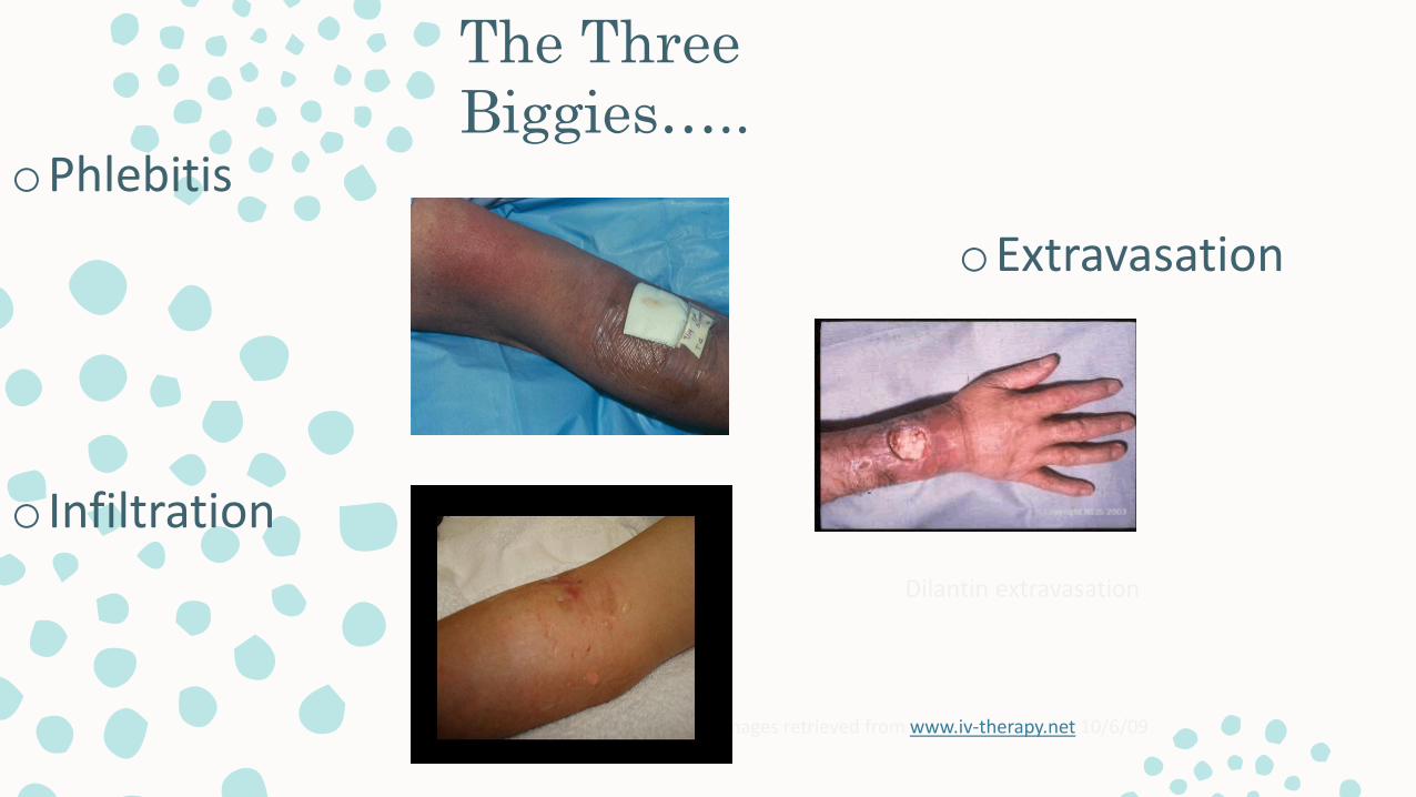
The Three Biggies…..
oPhlebitis
o Infiltration
oExtravasation
Dilantin extravasation
Images retrieved from www.iv-therapy.net 10/6/09

Phlebitis – in Peripheral IVs
o Definition: inflammation of a vein. May be mechanical, chemical, or bacterial
o Phlebitis has long been recognized as a risk for infection. o For adults, lower extremity insertion sites are associated
with a higher risk for infection than are upper extremity sites.
o Intravenous Nursing Society (INS) phlebitis scale;• Grade 0 no symptoms• Grade 1 erythema at insertion site with or without pain• Grade 2 pain at insertion site; with erythema and/or edema• Grade 3 pain at insertion site; with erythema and/or edema;
streak formation; palpable venous cord• Grade 4 pain at insertion site; with erythema and/or edema;
streak formation; venous cord > 1” in length; and purulent drainage
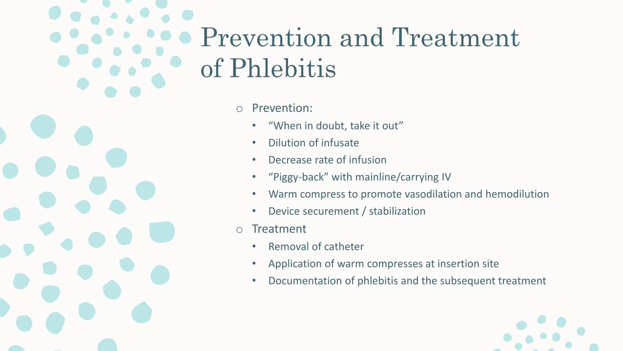
Prevention and Treatment of Phlebitis
o Prevention:• “When in doubt, take it out”• Dilution of infusate• Decrease rate of infusion• “Piggy-back” with mainline/carrying IV• Warm compress to promote vasodilation and hemodilution• Device securement / stabilization
o Treatment• Removal of catheter• Application of warm compresses at insertion site• Documentation of phlebitis and the subsequent treatment
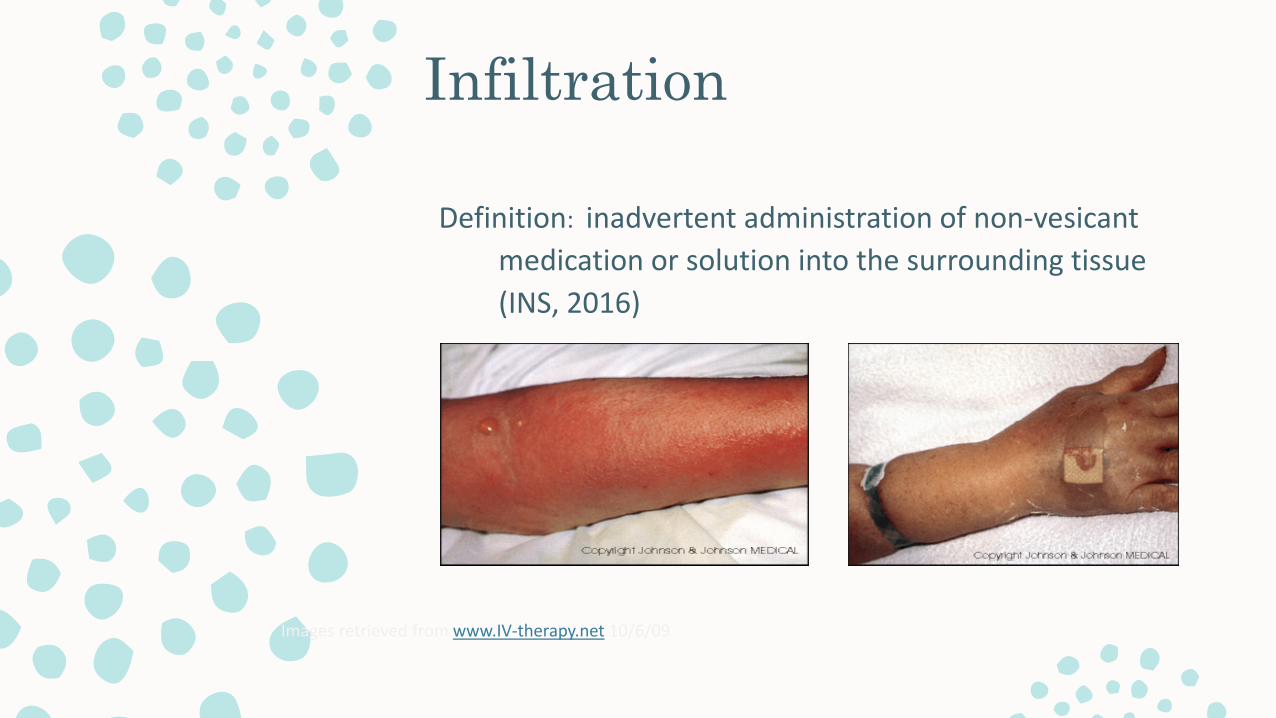
Infiltration
Definition: inadvertent administration of non-vesicant medication or solution into the surrounding tissue (INS, 2016)
Images retrieved from www.IV-therapy.net 10/6/09
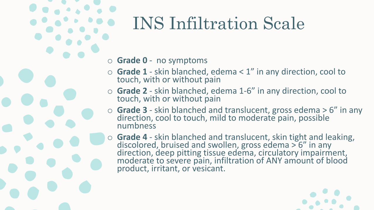
INS Infiltration Scale
o Grade 0 - no symptomso Grade 1 - skin blanched, edema < 1” in any direction, cool to
touch, with or without paino Grade 2 - skin blanched, edema 1-6” in any direction, cool to
touch, with or without paino Grade 3 - skin blanched and translucent, gross edema > 6” in any
direction, cool to touch, mild to moderate pain, possible numbness
o Grade 4 - skin blanched and translucent, skin tight and leaking, discolored, bruised and swollen, gross edema > 6” in any direction, deep pitting tissue edema, circulatory impairment, moderate to severe pain, infiltration of ANY amount of blood product, irritant, or vesicant.
Treatment of Infiltration
o Discontinue infusiono Elevate extremityo Warm compresses, NOT HOT, for normal or high pH/alkaline
solution (ex: D5W)o Cold compresses for low pH/acidic solutions ( ex: vanco)o Caution with infiltrated solution; ex.- morphine PCA resumption
with subcutaneous morphine infiltrate; “double-dosing”o Documentation of infiltrate and subsequent treatment

Extravasationo Inadvertent administration of vesicant medication or
solution into the surrounding tissue (INS, 2016)
o Definition of a vesicant drug – any IV drug that can cause blistering, severe tissue injury or tissue necrosis when extravasated
Image retrieved from www.IV-therapy.net 10/6/09
Extravasationo Extravasation should always be grade 4 on the infiltration scale. This
includes any amount of vesicant, blood product, or irritant.o Incidence is similar for peripheral and central line administrationo Risk factors, such as fragile vessels, location of peripheral iv (e.g.
areas of flexion), or catheter integrity are things to considero Antidotes may be used; refer to clinical resources for guidance, and
obtain order if indicated [Link to new Extravasation policy]o Many non-chemotherapy agents have vesicant properties (e.g.
Dopamine, Epinephrine, Gentamycin, Mannitol)o Extravasation is still possible, even in the presence of a positive
blood return.

Signs and Symptoms of Extravasation
o Early warning signs of possible extravasation• Swelling• Stinging, burning or pain at IV site• IV flow that stops or slows• Leaking around the port needle• Lack of blood return• Erythema, inflammation or blanching
o Other symptoms/damage resulting from extravasation: • Induration• Vesicle Formation• Necrotic tissue damage can progress for 6 months• Sloughing• Tendon, nerve, joint damage• Blistering at insertion site• Ulceration is usually seen 2-3 days to weeks following extravasation

Treatment of Extravasation
o IMMEDIATELY STOP INFUSIONo Remove tubing from IV, leave catheter or needle in place, attach syringe to IV
catheter o Attempt to aspirate residual drugo Elevate extremity o Notify Responding Clinician ASAP o Apply cold/heat as indicated (refer to policy for guidance on topical treatment). In
general:• Most drugs except Vinca alkaloids, etoposide, and catecholamines…apply COLD for 15-20
minutes (minimum of QID) for 48 hrs• For vinca alkaloids, etoposide and catecholamines…apply HEAT for 15-20 minutes
(minimum of QID) for 48 hours
o Refer to MGH Nursing Policies and Procedures in Ellucid or CALL PHARMACY for specific antidote
Extravasation Managemento DOCUMENTATION
• Medical record (“Hypersense/Extrav” Flowsheet in EPIC, serial progress notes)
• Safety report (Line/Tube > Extravasation)
o POST EXTRAVASATION CARE:• Document and consider photographing site
• Instruct patient about cold/heat application
• Patient and family education: symptoms to report immediately, care of site, follow-up appointment if needed
• Anticipate consult to plastic surgery or dermatology PRN• Ongoing evaluation & follow-up (serial photographs as needed)
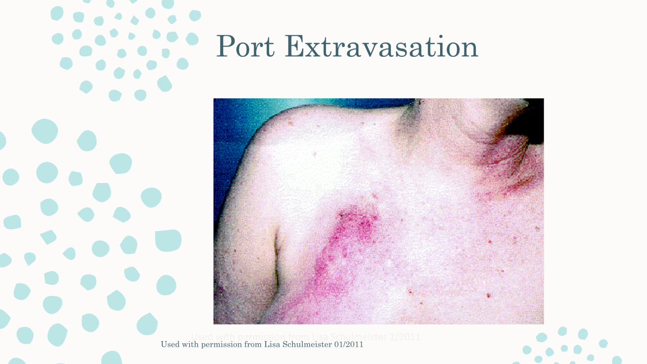
Used with permission from Lisa Schulmeister 1/2011Used with permission from Lisa Schulmeister 01/2011
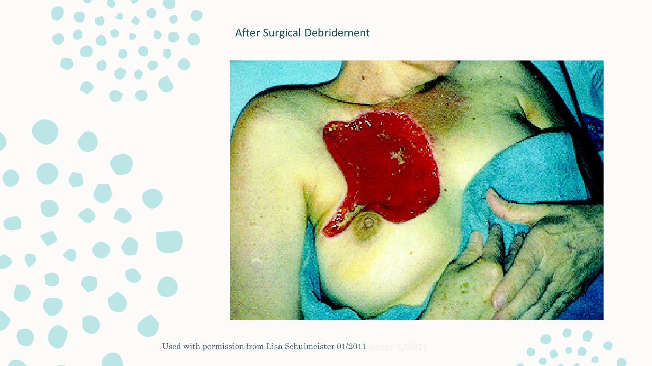
Used with permission from Lisa Schulmeister 1/2011Used with permission from Lisa Schulmeister 01/2011
After Surgical Debridement
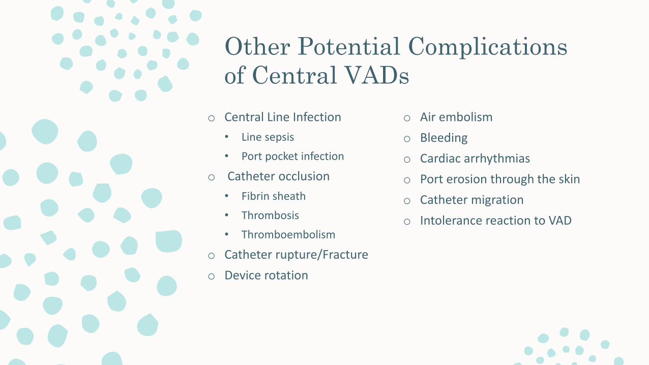
Other Potential Complications of Central VADs
o Central Line Infection• Line sepsis
• Port pocket infection
o Catheter occlusion• Fibrin sheath
• Thrombosis
• Thromboembolism
o Catheter rupture/Fracture o Device rotation
o Air embolismo Bleedingo Cardiac arrhythmiaso Port erosion through the skino Catheter migrationo Intolerance reaction to VAD
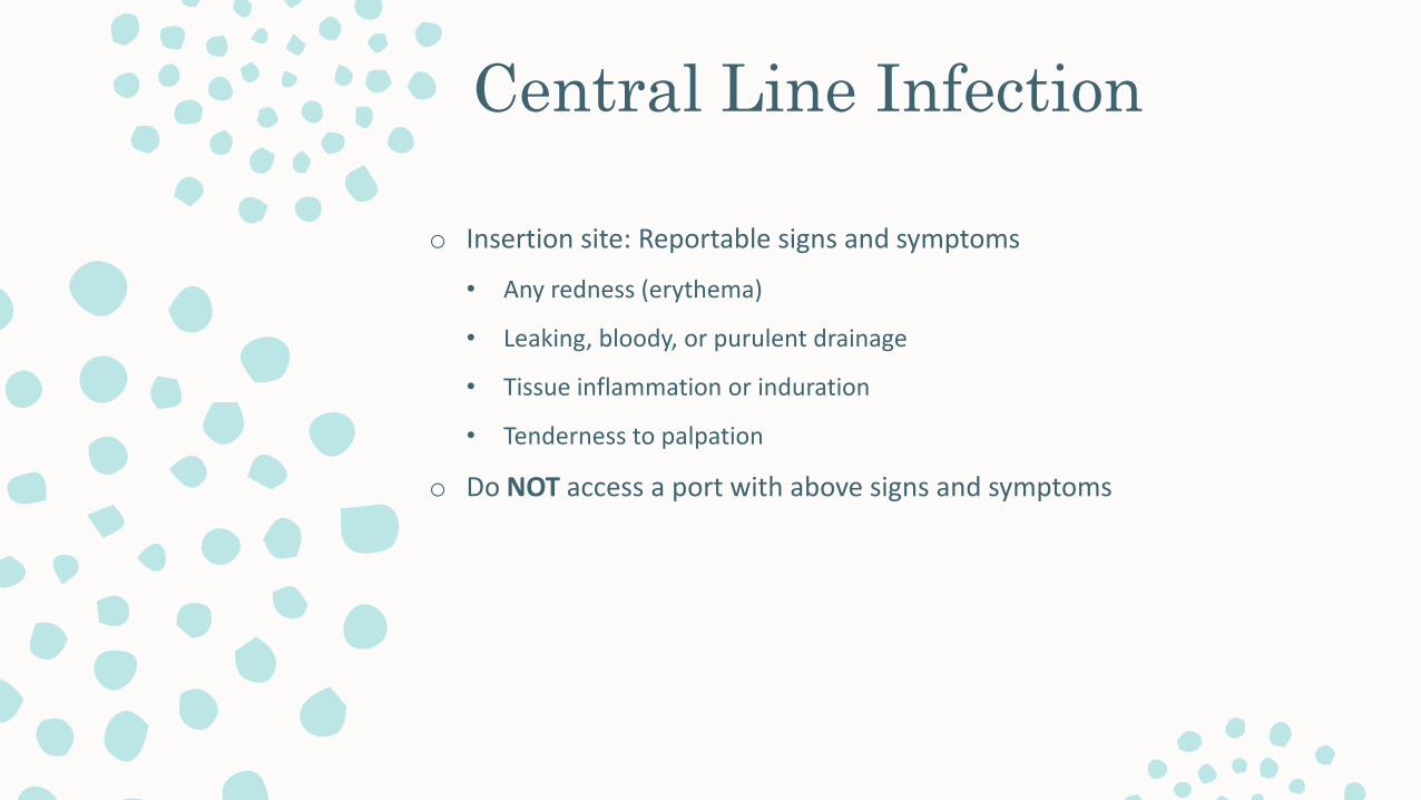
Central Line Infection
o Insertion site: Reportable signs and symptoms
• Any redness (erythema)
• Leaking, bloody, or purulent drainage
• Tissue inflammation or induration
• Tenderness to palpation
o Do NOT access a port with above signs and symptoms
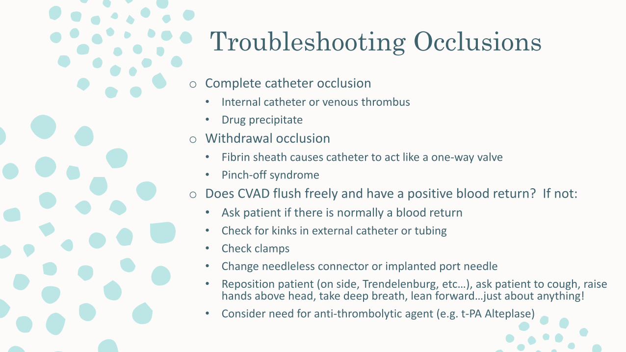
Troubleshooting Occlusionso Complete catheter occlusion
• Internal catheter or venous thrombus• Drug precipitate
o Withdrawal occlusion • Fibrin sheath causes catheter to act like a one-way valve• Pinch-off syndrome
o Does CVAD flush freely and have a positive blood return? If not:• Ask patient if there is normally a blood return• Check for kinks in external catheter or tubing• Check clamps• Change needleless connector or implanted port needle• Reposition patient (on side, Trendelenburg, etc…), ask patient to cough, raise
hands above head, take deep breath, lean forward…just about anything!• Consider need for anti-thrombolytic agent (e.g. t-PA Alteplase)
Troubleshooting Occlusions
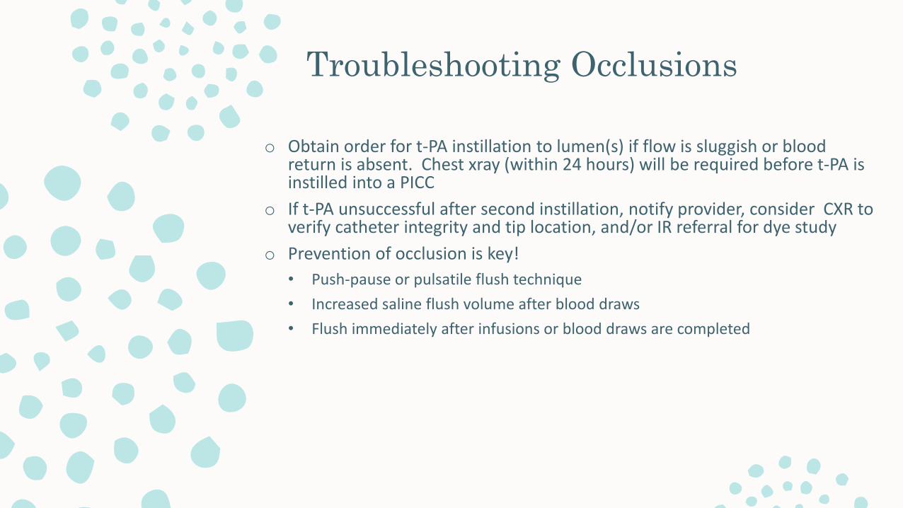
o Obtain order for t-PA instillation to lumen(s) if flow is sluggish or blood return is absent. Chest xray (within 24 hours) will be required before t-PA is instilled into a PICC
o If t-PA unsuccessful after second instillation, notify provider, consider CXR to verify catheter integrity and tip location, and/or IR referral for dye study
o Prevention of occlusion is key!• Push-pause or pulsatile flush technique• Increased saline flush volume after blood draws• Flush immediately after infusions or blood draws are completed
Tissue Plasminogen Activator:t-PA (Alteplase)
o Refer to MGH Medication Manual (see “Alteplase”)o Provider order and EMAR documentation required; separate t-PA order needed for
each lumeno IV nurses instill t-PA into PICCs; t-PA instillation to all other CVADs is responsibility of
unit RNo For inpatient PICCs, a CxR must have been done within 24 hrs before t-PA can be
instilled. For outpatient PICCs needing t-PA, a CxR is at the discretion of the provider.o Dosage (per lumen):
for patients weighing > 30kg (66lbs): 2mg/2ml– for patients weighing < 30kg (66 lbs): 110% of internal lumen volume of
catheter (up to 2mg)– Diluent: 2.2ml sterile water without preservative in a 10ml syringe– Do NOT clamp catheter while t-PA is instilled– Minimum dwell time of 30 minutes; 60 minutes is often required– Four hour t-PA dwelling times may be required for significant fibrin sheaths causing
withdrawal occlusions
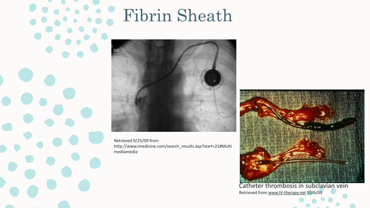
Fibrin Sheath
Retrieved 9/25/09 from http://www.imedicine.com/search_results.asp?start=21#Multimediamedia
Catheter thrombosis in subclavian veinRetrieved from www.IV-therapy.net 10/6/09
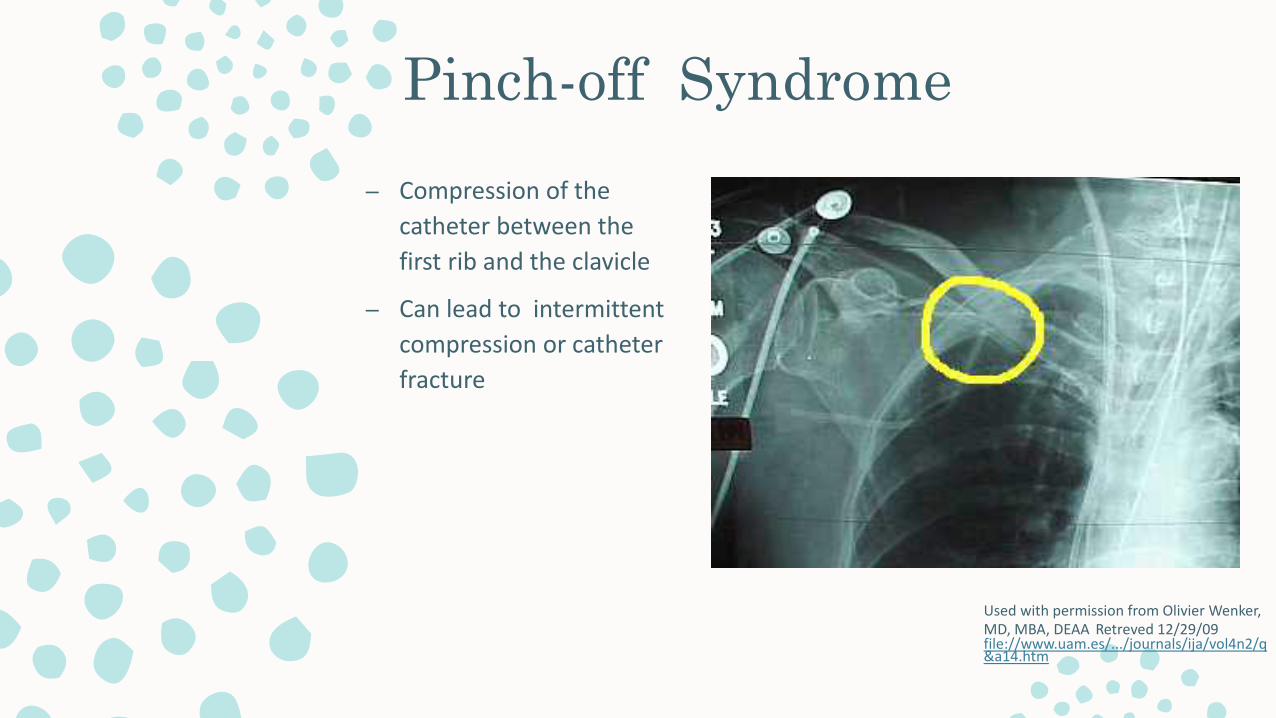
Pinch-off Syndrome– Compression of the
catheter between the first rib and the clavicle
– Can lead to intermittent compression or catheter fracture
Used with permission from Olivier Wenker, MD, MBA, DEAA Retreved 12/29/09 file://www.uam.es/.../journals/ija/vol4n2/q&a14.htm
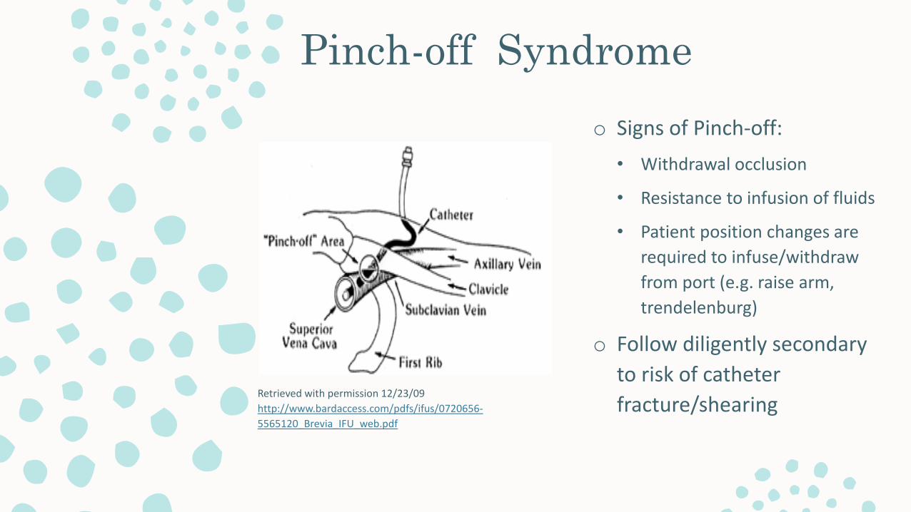
Pinch-off Syndrome
Retrieved with permission 12/23/09 http://www.bardaccess.com/pdfs/ifus/0720656-5565120_Brevia_IFU_web.pdf
o Signs of Pinch-off:• Withdrawal occlusion
• Resistance to infusion of fluids
• Patient position changes are required to infuse/withdraw from port (e.g. raise arm, trendelenburg)
o Follow diligently secondary to risk of catheter fracture/shearing

Miscellaneous Information
Related to peripheral and central
IV access
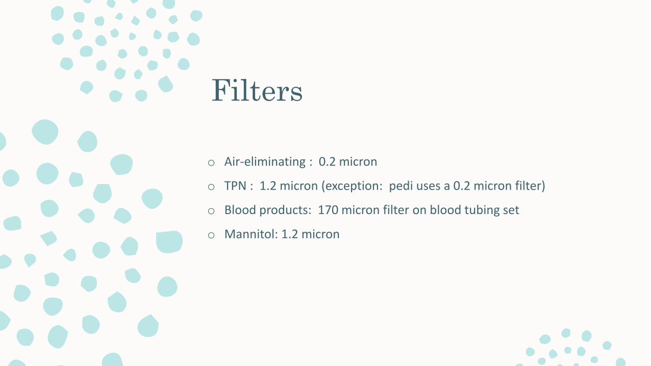
Filters
o Air-eliminating : 0.2 micron
o TPN : 1.2 micron (exception: pedi uses a 0.2 micron filter)
o Blood products: 170 micron filter on blood tubing set
o Mannitol: 1.2 micron
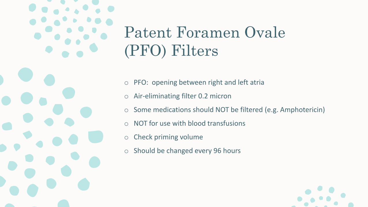
Patent Foramen Ovale(PFO) Filters
o PFO: opening between right and left atria
o Air-eliminating filter 0.2 micron
o Some medications should NOT be filtered (e.g. Amphotericin)
o NOT for use with blood transfusions
o Check priming volume
o Should be changed every 96 hours
PLEASE NOTE…o For more information on troubleshooting CVADs, refer to the
following resources:o “Guideline for Troubleshooting Central Venous Lines” in Ellucid
https://hospitalpolicies.ellucid.com/documents/view/1100/active/
o Hill, Jocelyn et. al. “Occlusion Management Guideline for Central Venous Access Devices (CVADs).” Vascular Access –Journal of the Canadian Vascular Access Association, Vol. 7; Supp 1 (2013). www.cvaa.info
o All information provided is subject to review and revision. Please continue to refer to MGH Policies and Procedures in Trove as your primary resource
References
o Original power point, 2011: Bartholomay, Dreher, Theresa Evans, Susan Finn, Deb Guthrie, Hannah Lyons, Janet Mulligan, Carol Tyksienski
o MGH Ellucido MGH Medication Manualo Bard Access Systems-Ports- MRI Implanted Ports Copyrights 2005 C.R. Bard
Inc http://www.bardamless.com/port-mri-port.phpo Bard Access Systems-Ports- MRI Implanted Ports Copyrights 2005 C.R. Bard
Inc http://www.bardamless.com/port-mri-port.phpo Cope, D., Ezzone, S., Hagle, M., Mmlorkindale, D., Moran, A., Sanoshy, J.,
Winkelman, l., and Camp-Sorrell, D. (editor)(2004) Access Device Guidelines: Recommendations for Nursing Practice and Education, 2nd ed. Pittsburgh: Oncology Nursing Society.
o Hill, Jocelyn et. al. “Occlusion Management Guideline for Central Venous Access Devices (CVADs).” Vascular Access – Journal of the Canadian Vascular Access Association, Vol. 7; Supp 1 (2013). www.cvaa.info