Embed Size (px)
Citation preview

Nutrition in Congenital HeartDisease
Cape Town Metropole PaediatricInterest Group
Final: April 2007Review: 2009
Christiaan Barnard Memorial Hospital

Cape Town Metropole Paediatric Working Group: Clinical Guidelines CHD 2
Nutrition in the Paediatric Cardiac Patient
Contents Page1. Glossary 3
2. Summary of recommendations for nutrition management of infants and 5 children with congenital heart disease2.1 Summary: Anthropometry 52.2 Summary: Biochemistry 52.3 Summary: Clinical 52.4 Summary: Dietary 62.5 Summary: Entry and exit criteria for nutrition support 82.6 Summary : Complications 92.6.1 Chylothorax 92.6.2 Chylous Ascites 92.6.3 Acute Myocarditis 92.7 Appendix 1 Treatment algorithm for congenital heart disease 102.8 Appendix 2 Treatment algorithm for chylothorax 112.9 Appendix 3 Minimal LCT diet 123. Nutrition in Paediatric Cardiac Patient 133.1 Aim 133.2 Objectives 133.3 Statement Regarding Promotion, Protection and 13 Support of Exclusive Breastfeeding in infants 133.3.1 Breastfeeding 133.3.2 Liquid Infant Formula 133.3.3 Infant Feeds 134. Introduction 145. Anthropometry 155.1 Nutritional Assessment 155.2 Expected weight gain 156. Biochemistry 157.Clinical 167.1 Pathogenesis of malnutrition in infant with congenital heart disease 167.2 Mechanisms of malnutrition 177.2.1 Type of cardiac lesion 177.2.2 Increased metabolic rate 177.2.3 Inadequate calorie intake 187.2.4 Weight at time of operation 197.2.5 Prenatal factors 197.2.6 Medication 197.2.6.1 Inotropes 197.2.6.2 Diuretics 207.2.7 Cardiac cachexia 218. Dietary 228.1 General guidelines 228.2 Nutrition strategies for children with congenital heart disease and failure to thrive 248.3 Type of feed 248.4 Method of administration 258.5 Iron supplementation 259. Complications 259.1 Chylothorax 259.2 Chylous ascites 279.3 Acute myocarditis 2910. Summary 2911. Appendix 4 3012. Authors page & metropole paediatric working group members 3113. References 3214. Developers Summary 33
Cape Town Metropole Paediatric Working Group: Clinical Guidelines CHD 3
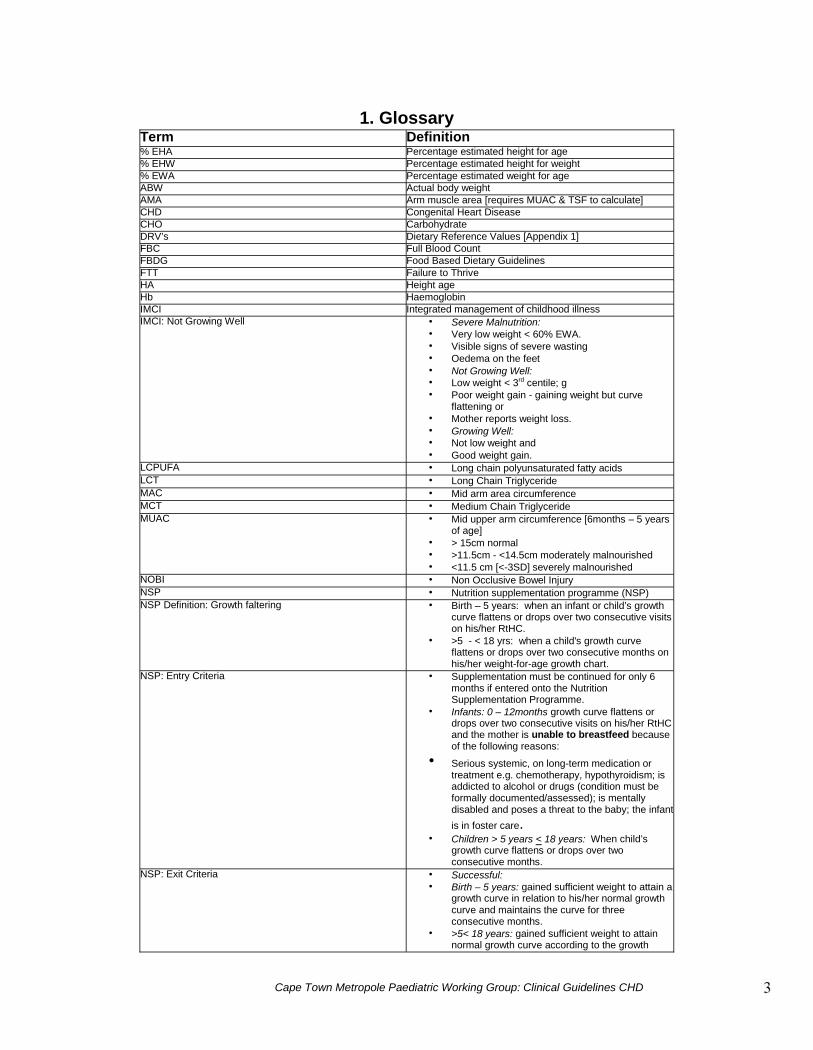
1. GlossaryTerm Definition% EHA Percentage estimated height for age% EHW Percentage estimated height for weight% EWA Percentage estimated weight for ageABW Actual body weightAMA Arm muscle area [requires MUAC & TSF to calculate]CHD Congenital Heart DiseaseCHO CarbohydrateDRV’s Dietary Reference Values [Appendix 1]FBC Full Blood CountFBDG Food Based Dietary GuidelinesFTT Failure to ThriveHA Height ageHb HaemoglobinIMCI Integrated management of childhood illnessIMCI: Not Growing Well Severe Malnutrition:
Very low weight < 60% EWA. Visible signs of severe wasting Oedema on the feet Not Growing Well: Low weight < 3rd centile; g Poor weight gain - gaining weight but curve
flattening or Mother reports weight loss. Growing Well: Not low weight and Good weight gain.
LCPUFA Long chain polyunsaturated fatty acidsLCT Long Chain TriglycerideMAC Mid arm area circumferenceMCT Medium Chain TriglycerideMUAC Mid upper arm circumference [6months – 5 years
of age] > 15cm normal >11.5cm - <14.5cm moderately malnourished <11.5 cm [<-3SD] severely malnourished
NOBI Non Occlusive Bowel InjuryNSP Nutrition supplementation programme (NSP)NSP Definition: Growth faltering Birth – 5 years: when an infant or child’s growth
curve flattens or drops over two consecutive visitson his/her RtHC.
>5 - < 18 yrs: when a child's growth curveflattens or drops over two consecutive months onhis/her weight-for-age growth chart.
NSP: Entry Criteria Supplementation must be continued for only 6months if entered onto the NutritionSupplementation Programme.
Infants: 0 – 12months growth curve flattens ordrops over two consecutive visits on his/her RtHCand the mother is unable to breastfeed becauseof the following reasons:
Serious systemic, on long-term medication ortreatment e.g. chemotherapy, hypothyroidism; isaddicted to alcohol or drugs (condition must beformally documented/assessed); is mentallydisabled and poses a threat to the baby; the infantis in foster care.
Children > 5 years < 18 years: When child’sgrowth curve flattens or drops over twoconsecutive months.
NSP: Exit Criteria Successful: Birth – 5 years: gained sufficient weight to attain a
growth curve in relation to his/her normal growthcurve and maintains the curve for threeconsecutive months.
>5< 18 years: gained sufficient weight to attainnormal growth curve according to the growthchart within the 6 months period on the scheme

Cape Town Metropole Paediatric Working Group: Clinical Guidelines CHD 4
chart within the 6 months period on the scheme Unsuccessful: Birth – 5 years: Failure to attain growth curve in
relation to his/her normal growth curve over aperiod of 6 months and if no underlyingdisease/condition is present e.g. Foetal AlcoholSyndrome
>5< 18 years: who do not attain a normal growthcurve according to the growth chart with in the 6months period.
Defaulter: Birth – 5 years: Failure to attend the clinic for a
period of three consecutive months. > 5 - <18 years: Failure to attend the clinic for a
period of three consecutive months within the 6months period.
Client has a history of irregular clinic attendance(less than three visits in a 6 month period) with inthe 6 months period.
** Re-entry: UNSUCCESSFUL and DEFAULT cases MAY
NOT be re-entered onto the programme. SUCCESSFUL cases MAY be re-entered onto
the programme according to entry criteria.RDA Recommended Daily AllowancesREE Resting Energy ExpenditureRTHC Road to health card (Clinic Card)RTU/RTU Ready to use/ Ready to hangSchofield Equation Predicting estimated energy requirements
[Appendix 1]SD Standard Deviations used to determine moderate
to severe malnutrition: 0 - <-1 SD Normally Nourished >-2 – -3 SD Moderately Malnourished >-3SD Malnourished
TSF Tricep Skinfold ThicknessTTO To Take OutWA Weight ageWaterlow Criteria (WHO) Used to determine malnutrition:
Acute malnutrition: Weight/ Height Normal WH >90%, Mild 81% - 90%, Moderate 70% - 80%, Severe <70%. Chronic malnutrition: height for age Normal >95%, Mild 90 –95%, Mild – moderate 85% to 89% Severe < 85%.
WCC White Cell CountWH Weight for height

Cape Town Metropole Paediatric Working Group: Clinical Guidelines CHD 5
2. Summary of recommendations for nutrition management ofinfants and children with congenital heart disease
Summary Recommendations: Congenital Heart Disease 1, 2, 12, 25
2.1 Anthropometry Complete on admission
& weekly untildischarge.
Height Weight MUAC Head Circumference < 3 years of ageCalculate % EWA % EWH % EHA HA WA WHClassify degree of malnutritionWaterlow: Acute malnutrition: Weight for Height(wasting) Normal WH >90% Mild 81% - 90% Moderate 70% - 80% Severe <70%Waterlow: Chronic malnutrition: height for age(stunting) Normal >95% Mild 90 –95% Mild – moderate 85% to 89% Severe < 85%Gomez: Acute wasting: Weight for age Obese >120% Normal > 90% Mild malnutrition 76 – 90% Moderate malnutrition 61 – 75% Severe malnutrition < 60%
Plot weight & height on appropriate growth charts.(CDC or WHO or disease specific e.g. DownsSyndrome)
Expected weight gain for an infant (< 6 months) withCHD is 10 – 20g per day and for infants (6 – 12months) 120 – 210g/week
2.2 Biochemistry Complete daily post
operatively whilst in ICU Once in recovery
complete x 2 week untildischarge
Monitor the following U & E: Urea, creatinine, sodium, potassium Calcium, magnesium and phosphorus Glucose FBC: Hb, platelets, WCC
2.3 Clinical Mechanisms of malnutrition effected by: Type of cardiac lesion Increased metabolic rate Inadequate caloric intake Weight at time of operation Prenatal factors Medication Cardiac cachexia Urinary sodium losses (especially on Frusemide-Lasix)
Low Hb Microcytic anaemia Macrocytic anaemia
Low Zinc or Selenium
Comments:
High urinary sodium losses canresult in failure thrive. Establishsodium balance over a 24 hourperiod
Check Iron status Check folate and Vit B12 levels Infants with cyanotic CHD may
have a normal Hb but irondeficient. Check iron status usingferritin, red cell indices and totaliron binding capacity.
Common clinical signs are: Fatigue on feeding Early satiety Anorexia Failure to thrive Frequent Infections

Cape Town Metropole Paediatric Working Group: Clinical Guidelines CHD 6
2.4 DietaryAt each follow up a thorough nutrition history should be completed
Components of a nutrition history include Weight Change Appetite Satiety Level Taste Changes/ aversions Nausea/ vomiting Bowel habits – constipation, diarrhoea Chewing/ swallowing ability Shortness of breath on feeding
Components of a nutrition history include (cont.) Long-term disease(s) affecting absorption/use of nutrients Dietary history – 24 hour recall/ food frequency Use of vitamin/ mineral or nutritional supplements Medications Level of activity/ exercise Ability to secure and prepare food Over the counter medications, vitamins and herbal remedies.
Diet history Review through 24 hour dietary recall quarterly or at each
follow up review. Use in conjunction with food frequency. Many patients may eat < 65% of RDI.
Method of feed administration (employ a stepwise downwardapproach) Offer smaller volumes and more frequent feeds orally Give any unfinished feeds via naso-gastric tube if required Give small frequent bolus feeds via naso-gastric tube Top up small frequent daytime feeds with continuous overnight feeds
via an enteral feeding pump Give feeds continuously over 24 hours via an enteral feeding pump.NOBI (Non occlusive Bowel Injury)Initiation of enteral feeds in patients with cirulatory compromise (sepsis,cardiogenic shock, haemodynamic instability) may lead to delteriouschanges on the structure and function of the gut. It is therefore imperitive tomonitor for any signs of feeding intollerance.
Fluid Fluid RangesAge (years)Premature
0-11-33-67-10
10-15Weight
Premature < 2kgNeonates and infants 2 – 10kg
0 – 6 months 6 – 12 months
Infants and children 10 -20kgChildren > 20kg
DO NOT ALTER MEDICALLY INDICATEDFLUID RESTRICTION
(Fluid requirements include fluid from feed,medication, IV fluids and oral sips)
ml/kg actual weight180-200
150100907060
Fluid Volume per 24 hours150ml/kg
150ml/kg120ml/kg
1000ml + 50ml/kg over 10kg1500ml + 20ml/kg over 20kg
Energy Infants:Pre-operative or post shunt Ventilated: 90 – 100 kcal/kg Non ventilated: 120 – 150kcal/kg
(maximum 170 kcal/kg)Post definitive operation (Cardiac Repair) Ventilated: 90 – 100 kcal/kg Not ventilated: Schoflied using abw + activity
1.2 + stress 1.5 – 1.6
In sedated, ventilated children’s energyexpenditure if often significantly reduced.
Aim to feed at 150kcal/kg and only increaseto 170kcal/kg if other factors effecting growthhave been excluded
Children:Pre-operative or post shunt Ventilated: Schofield equation or WHO/FAO/UNU x
1.3 – 1.5 [No activity factor] Non ventilated: 120 – 150% or DRV’sPost definitive operation (Cardiac Repair) Ventilated: Schofield equation or WHO/FAO/UNU x
1.3 – 1.5 [No activity factor] Non ventilated: Schofield using abw + activity 1.2 +
stress 1.5 – 1.6

Cape Town Metropole Paediatric Working Group: Clinical Guidelines CHD 7
Energy Supplementation Infants: No additional energy may be required in the pre-repair infant and breast milk and or standard ready to
use/ hang infant formula [0.67kcal/ml] should be given. If the patient is volume-restricted breast milk may be supplemented with a human milk fortifier or
carbohydrate/fat powder and or a ready to use/hang energy dense infant feed [1kcal/ml] should begiven.
Children: No additional energy may be required and a standard feed [1kcal/ml] should meet requirements in the
volume prescribed. If the patient is volume restricted an energy dense [1.5 kcal/ml] ready to use/hang feed should be given.NB: No powders or liquids e.g. oil should be added to a sterile ready to use feed. If additional energy is required in non-ventilated children then flushes of energy boluses should be
provided prior to a drink or feed including breastmilk. Recommendations for fat, protein and carbohydrate concentrations should not be exceeded. [See
sections below]Carbohydrate Glucose requirements
> According to toleranceInfants
8-9mg/kg/min [11.5g –12.9g/kg/day] Max 12.5mg/min/kg [18g/kg/day] Toddlers 7mg/kg/min [10g/kg/day] Adolescents 4mg/kg/min [5.7g/kg/day]
The following concentrations of CHO per 100mlwill be tolerated if a CHO/fat powder is used.
10-12% carbohydrate concentrations in infants under 6months (i.e. 7g from formula, 3-5g added)
12-15% in infants aged 6months to 1 year 15-20% in toddlers aged 1-2 years 20-30% in older children
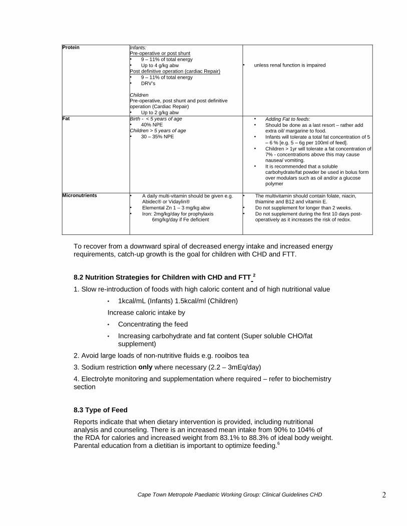
Protein Infants:Pre-operative or post shunt 9 – 11% of total energy Up to 4 g/kg abwPost definitive operation (Cardiac Repair) 9 – 11% of total energy DRV’s
ChildrenPre-operative, post shunt and post definitiveoperation (Cardiac Repair) Up to 2 g/kg abw
unless renal function is impaired
Fat Birth - < 5 years of age 40% NPEChildren > 5 years of age 30 – 35% NPE
Adding Fat to formula/enteral feeds: Should be done as a last resort – rather add
extra oil/ margarine to food. Infants will tolerate a total fat concentration of 5
– 6 % [e.g. 5 – 6g per 100ml of feed]. Children > 1yr will tolerate a fat concentration of
7% - concentrations above this may causenausea/ vomiting.
It is recommended that a solublecarbohydrate/fat powder be used in bolus formover modulars such as oil and/or a glucosepolymer
Micronutrients A daily multi-vitamin should be given e.g.Abidec® or Vidaylin®
Elemental Zn 1 – 3 mg/kg abw
Selenium 2mg/kg abw with a max of30mg/day
Iron: 2mg/kg/day for prophylaxis 6mg/kg/day if Fe deficient
The multivitamin should contain folate, niacin,thiamine and B12 and vitamin E.
Do not supplement Zinc for longer than 2 weeks. Selenium and Zinc should only be supplemented if
failing to thrive and low serum levels. Do not supplement iron during the first 10 days post-
operatively as it increases the risk of redox.

Cape Town Metropole Paediatric Working Group: Clinical Guidelines CHD 8
Discharge Planning Educate the parent/caregiver on thesupplementation of feeds (this can be startedprior to discharge). The caregiver is asked tomake up the child’s supplemented feeds atward level as a way of educating him/herunder supervision.
Provide caregiver with a date for dieteticfollow up or refer to a private practisingdietitian.
Ask the doctor to write up a prescription TTO(To Take Out) for multivitamins Government: Supply sufficient micronutrient supplements
until follow up appointmentPrivate: 7 day TTO on hospital discharge and provide asecond script for at least 1 month supply.Continue multi-vitamins until catch up growth has beenachieved.
Post Discharge Frequency of follow up Poor growth: monthly If failing to thrive, follow guidelines below Thriving: Quarterly
2.5 Entry and Exit Criteria for Nutrition SupportEntry Criteria Nutrition SupportNSP Supplementation must be continued for only 6 months if
entered onto the Nutrition Supplementation Programme. Children > 5 years < 18 years: When child’s growth curve
flattens or drops over two consecutive months.Or Private Patients Growth failure Downward crossing 2 or more centiles over a period of 1
month or 2 consecutive visits. MUAC < 12.5cm in children < 5years of ageAcute malnutrition: Weight/ Height < 80%Chronic malnutrition: height for age < 89%
Exit Criteria for nutrition supportNSP Birth – 5 years: gained sufficient weight to attain a growth curve in
relation to his/her normal growth curve and maintains the curve forthree consecutive months.
> 5yrs – 18 years who attain normal growth curve according to thegrowth chart within the 6 months period on the NSP scheme.
Or Private Patients Upward crossing of 2 or more centiles over a period of 1 month or 2
consecutive visits. MUAC >15cm in children < 5 years of age. WH >90%, HA >95%,
Referral NSP Scheme
Additional Requirements Nutritionally complete age appropriate supplement. Access from local day hospital/ CHC

Cape Town Metropole Paediatric Working Group: Clinical Guidelines CHD 9
2.6 Complications
2.6.1 ChylothoraxChylothorax is an uncommon post operative complication resulting in leakage of lymphatic fluid into thepleural space due to surgical disruption of the thoracic duct or increased venous pressure of one of it'smain tributatries resulting in increased pressure within the intrathoracic lymph systemDiagnosisThe following needs to be present in the pleural fluid:1) Triglycerides > 1.1 mmol/L2) Chylomicrons positive3) Chylomicrons negative with a lymphatic fraction > 80%Treatment1) Dietary
Bowel rest with total parenteral nutrition Fat free diet or high LCT diet: this type of diet should be followed in a clinical environment only
due to the high risk of developing essential fatty acid (EFA) deficiency and should not befollowed for more than 2 weeks.
High MCT enteral nutrition: Use monogen2) Surgical3) OctreotideRefer to Appendix 2 for the recommended treatment algorithm
2.6.2 Chylous Ascites
Chylous ascites is an accumulation of chyle in the peritoneal cavity due to obstruction or rupture of theperitoneal or reperitoneal lymphatic glands, increased venous pressure or congestive cardiac failure.Diagnosis (Paracentesis)The following needs to be present in the fluid1) Triglycerides 200mg/dl2) Predominance of lymphocytes > 75%TreatmentBowel rest with total parenteral nutrition
2.6.3 Acute MyocarditisTreatmentCCME:
L-carnitine 5 – 15 mg/kg (max 1g) 6H(IV) or orally 25 mg/kg 6 – 12H (max 3g/day) Co-enzyme Q10 1 – 4 mg/kg daily oral Magnesium Sulphate IV 2 mmol/ml (max 10ml) 12H slow IV Vitamin E 50 – 1000IU < 3yr or 200 – 400IU >3yr

Cape Town Metropole Paediatric Working Group: Clinical Guidelines CHD 10
2.7 Appendix 1The CHD patient
Goal: To ensure that each patient with congenital heart disease attains/ maintains anoptimal nutrition status.
To read the chart:Follow the arrows Start Here
Yes
Is there growth falteringor failing during lastmonth or over 2consecutive visits? No
Anthropometric assessment determine patient’s nutritional status & risk: Height MAC TSF %EHA HC Mid parental height MUAC Weight %EWA %EWH AMA Growth Velocity
Provide sufficient energy & protein tosupport growth and weight gain.Infants 3 – 4g/kg protein 120 – 150kcal/kg (max 170kcal/ml)Children 2.g/kg protein 1.2 – 1.5 x RDA OR Schofield equation or WHO x 1.5 –
1.6 [combined activity & stress factor]Paediatric Supplements available:
HospitalisedInfants Breast milk or 0.67kcal/ml RTU/H feed 1kcal/ml RTU/H feed (fluid restricted)Children 1kcal/ml RTU/H nutritionally complete
feed. (Standard) 1.5kcal/ml RTU/H nutritionally
complete feed. (Fluid Restricted)
For additional energy required for bothinfants and children consider a supersoluble CHO/fat powder.
Monitor 3 month review
Assess dietary intake Complete 24 hour diet recall Food frequency Analyse where possible Is the intake appropriate according to the
DRV’s?
Assess patient using the following approach: A = Anthropometry B = Biocehmistry C = Clinical D = Dietary Implement nutrition support where appropriate
Poor intake: Encourage caregiver and child. Advise caregiver around food based dietary guidelines
[FBDG] Promote small frequent meals x 3 and snacks 2 – 3 per
day. Recommend energy & nutrient dense foods & drinks
Yes
No
Good intake: Encourage caregiver and child around good food intake. Advise caregiver around food based dietary guidelines
[FBDG]
Paediatric Supplements available:Discharged
Infants Breast milk with a breast milk
fortifier if requiredChildren Enriched maize meal porridge Nutritionally complete age
appropriate supplement.
Entry to Nutrition Support:Calculate Dietary Requirements & recommendnutrition supplementation according to treatmentmodality (pre operative/post shunt or postdefinitive operation – cardiac repair)
Exit Nutrition Support when: Birth to 5 years – Normal
growth curve RTCH following3 months on NSP scheme.
> 5 – 18 yrs: Normal growthcurve RTCH ≤ 6 months onNSP scheme.
Upward crossing of 2 or morecentiles over a period of 1month or 2 consecutive visits.
MUAC >15cm in children < 5years of age.
WH >90%, HA >95%,
Biochemistry & ClinicalMonitor the following:
Urinary sodiumlosses
Hb Infection
Supplementation of complementaryfoods: Discharged
Infants Soluble CHO powder Long chain fat/oil
Children Soluble CHO powder Long chain fat/oil

Cape Town Metropole Paediatric Working Group: Clinical Guidelines CHD 11
Persistent Chest Tube Drainage (> 5mL/kg/day)
OrPresence of Milky Drainage
Pleural Fluid sent for:+/- Triglyceride > 1.1mmol/L+/- Lymphocyte fraction > 80%+/- Chylomicrons
YESChyle presentChyle not present withLymphocyte faction >80%
Continuediuresis
NO PHASE 1MCT Diet for 5 days
NOPHASE 2TPN NPO
Chest Tube Drainage < 2 mL/kg/dayYES
Fat challenge prior toremoval of chestdrain(page 12)
Diagnostic imaging(ultrasound, ECHO)to rule outthrombosis
For patients whohad aortic archreconstruction,notify surgeon toconsider earlythoracic ductligation.
If thrombosis, treatwith heparin +/- tPA
Chest Drainage <2mL/kg/day?
YESStop TPN
Return to low fatdiet
Chest Tube drainage <2mL/kg.day
YESFat challenge prior toremoval of chestdrain.
YESWean Octreotide by25% daily over 4daysContinue Low fat diet
Chest Tube drainage <2mL/kg.day
Chest Tube drainage <2mL/kg.day NO
Wean Octreotide by 25% dailyover 4 daysCardiac catheterisationConsider surgical interventionStop suction on chest tubes ifdecreasing drainage
NOReturn to Phase 3 with slowerwean of prednisoneReturn to prednisone dose givenprior to increase in drainage
YESFat challenge prior toremoval of chestdrain (page 12)
NOReturn toPHASE 2for minimum7 days
YESFat challenge prior toremoval of chestdrain (page 12)
Chest Tube drainage <2mL/kg.day
YESWean PrednisoneContinue Low fat diet
Chest Tube drainage <2mL/kg.day
NOPHASE 4
Start Octreotide (0.5 – 4 mcg/kg/hr IVcontinuous infusion) for 5 daysOctreotide 5mcg/kg/dose IV q8th iflimited IV access (increase q24th by5mcg/kg/day) for 5 daysStop prednisoneStop suction of chest tubes if decreasingdrainageContinue MCT/minimal fat diet
NOPHASE 3
Start Prednisone (1mg/kg/day twicedaily) for 5 – 7 daysStop suction of chest tubes ifdecreasing drainageReturn to MCT/minimal fat dietStop TPN when adequate enteralcaloric intake is reached
Start TPN/NPO for 5 daysAttain longer-term vascular accessStop suction of chest tube ifdecreasing drainage
2.8 APPENDIX 2 9 Chylothorax Management Diagram
PHASE 5Surgical interventionRe-start pathway aftersurgical intervention atPhase 1Consider pleuroperitonealshunt if chest tubescontinue to drain >2cc/kg/day
NOBack to Phase 4 with slowerOctreotide weanReturn to Octreotide dose prior toincrease in drainage
Fat ChallengeInfants5 – 6 g/kg fat given as a bolus using a 1kcal/mlRTH feed.Children5 – 6g/kg fat given as a bolus usingevaporated milk (19g fat/250ml).
Review drainage 90 – 180 minutes postingestion.If there is no chyle present then remove thechest drain
Recommend that a low fat diet be followed forat least 2 weeks and a follow up chest X-raybe done weekly.

Cape Town Metropole Paediatric Working Group: Clinical Guidelines CHD 12
2.9 APPENDIX 3 1 Minimal LCT (Long Chain Triglycerides) diet (In Hospital)
Maximum duration 10 – 14 days
Requirement of LCT: 1g LCT per year of life up to a maximum of 4 – 5g LCT per day
Suitable foods for use in a low LCT dietFood Average Portion Size (g) LCT per portion (g)Breakfast CerealsCornflakes 25 0.2Frosties 20 0.1Special K 20 0.2Cocopops 20 0.2Rice Crispies 25 0.2Weetbix 35 (1) 0.9
BreadWhite, large thin slice 35 0.4Matzos 20 0.2Crumpets Toasted 40(1) 0.4
Dairy FoodsReduced fat cottage cheese 50 0.7Condensed milk, skimmedsweetened
50 0.5
FishWhite hake fillet 100 0.7Fish fingers 25 (1) 1.8Tuna 100 0.5
Meat and PoultryRoast Turkey, light meat 70 1.0Roast chicken, light meat 25 1.0Roast lamb, lean 25 2.0Roast beef, lean topside 45 2.0Silverside, lean 40 2.0
Legumes, Pasta, RiceBaked beans in tomato sauce 200 1.0Tinned spaghetti in tomatosauce
125 0.5
White rice, boiled 150 0.4White pasta, boiled 130 1.0
Fortify skimmed milk with a glucose polymer.
Free Foods for a Minimal LCT Diet
3. Nutrition in Paediatric Liver Patient
All fruit, fresh, frozen or tinned (except olives and avocado)All vegetables fresh, tinned or frozenSugar, honey, golden syrup, treacle, jam and marmaladeJelly and jellied sweets such as Jelly Tots, Jelly Babies, wine gums, lollie pops or fruit pastillesBoiled sweets, mints (not butter mints)Fruit sorbets (milk free), water ices and ice lolliesMeringue, egg whiteSpices and essencesSalt, pepper, vinegar, herbs, tomato sauce, most chutneys, Marmite, Oxo and BovrilFruit juices, fruit squashes, bottle fruit saucesFizzy drinks, lemonade, cola

Cape Town Metropole Paediatric Working Group: Clinical Guidelines CHD 13
3.1 Aim
The aim of these guidelines is to outline appropriate nutrition care practices in thedietary management of children with congenital heart disease.
Nutrition management usually aims to maintain and support optimal nutrition status.
3.2 Objectives
The objectives of these guidelines are to:
Identify appropriate feeding practices in the paediatric cardiac patient. Identify appropriate routes of feeding in the paediatric cardiac patient. Identify appropriate nutrition care plan strategies for the maintenance and
support of optimal nutrition status. Promote early and appropriate nutrition intervention with non-volitional
nutrition support where oral feeding has failed in the paediatric cardiacpatient.
3.3 Statement Regarding Promotion, Protection and Support of ExclusiveBreastfeeding in infants with congenital heart disease
3.3.1 Breastfeeding
Exclusive breastfeeding should be encouraged for the first 6 months of life and up tothe age of 2 years following the introduction of a variety of safe and healthycomplimentary foods.
However, in some instances breastfeeding may not be possible and breast milksubstitutes are required. Breast milk substitutes should be prepared to standardsrecommended by Codex Alimentarius.
3.3.2 Liquid Infant Formula
Liquid infant formula is sterile and does not contain any pathogenic organisms and assuch does not present any potential source of infection. Wherever possible, in ahospital setting sterile liquid formula should be used. It is recommended that sterileinfant feed liquids are used in all Neonatal and Paediatric Intensive Care /High CareUnits, in addition in all infants considered to be immmunocompromised and or wherethe safe preparation of powdered infant feeds may not be guaranteed.
3.3.3 Infant Feeds
Breast-feeding should be supported at all times. However there may be instanceswhere specialized infant feeds (IF) are required and or breast milk is topped up with abreast milk substitute. However this should be provided under the guidance that areplacement feed should only be given if it is “acceptable, feasible, affordable andsustainable”.

Cape Town Metropole Paediatric Working Group: Clinical Guidelines CHD 14
4. Introduction
Congenital heart defects are classified into two broad categories: acyanotic andcyanotic lesions. The most common acyanotic lesions are ventricular septal defect(VSD), atrial septal defect (ASD), atrioventricular canal, pulmonary stenosis, patentductus arteriosis (PDA), aortic stenosis and coarctation of the aorta. Congestiveheart failure (CHF) is the primary concern in infants with acyanotic lesions. The mostcommon cyanotic lesions include tetralogy of Fallot (TOF) and transposition of thegreat arteries. 5
The reported incidence of congenital heart disease (CHD) is eight cases per 1000live births. Up to 30% of infants with CHD have features of various geneticsyndromes.
4.1 Features of Common Congenital Heart Defects 5
4.1.1 Acyanotic Lesions
Ventricular Septal Defect (VSD) Spontaneous closure in 30 –40% of cases in the first6 months. The most common 15 –20%.Surgical repair recommended if infant exhibits failureto thrive, pulmonary hypertension
Atrial Septal Defect (ASD) Often asymptomatic and 87% of secundum typesclose by age 4.Primary and sinus types require surgery
Atrioventricular canal Presentation similar to that of the VSD.
Pulmonary Stenosis May be asymptomatic or result in severe CHF.
Patent Ductus Arteriosis (PDA) In premature infants, spontaneous closure orindomethacin-induced closure may occur. In terminfant spontaneous closure is less likely. Recurrentpneumonia may occur. Surgical ligation is usuallyrequired.
Aortic Stenosis May be asymptomatic. Valve replacement and anti-coagulation may be required.
Coarctation of the aorta 98% of cases occur at origin of left subclavian artery.Blood pressure is higher in arms than in legs.Surgical repair usually required between 2 and 4years of age.
4.1.2 Cyanotic LesionsTetralogy of Fallot (TOF) Most common CHD beyond infancy. Intermittent
episodes of hyperapnoea, irritability, cyanosis.Surgical repair required with suitable anatomy.
Transposition of the greatarteries
Incidence of about 5% in children with CHD. Latecomplications include pulmonary stenosis, mitralregurgitation, aortic stenosis, coronary arteryobstruction, ventricular dysfunction and arrhythmias.

Cape Town Metropole Paediatric Working Group: Clinical Guidelines CHD 15
5. Anthropometry 2, 6
It has been reported that 52% of children with CHD were below the 16th percentile forheight, 55% were below the 16th percentile for weight and 27% were below the 3rd
percentile for both. Weight was more affected than height and boys tended to bemore malnourished that girls. Stunting was more frequent in children below 2 yearsof age at 49%. Children with CHD tended to fall behind in weight-for-height,especially between 6 and 12 months of age.2 Other studies found that acute andchronic malnutrition occurred in 33% and 64% respectively of hospitalized infantsand children with CHD. 1
5.1 Assessment1) Record weight, height and head circumference (below 3 years of age)2) Plot on relevant growth chart. (Down’s Syndrome charts and Gestational age
charts should be used where appropriate)3) If failing to thrive:
i) Calculate weight at 100% expected weight for child’scurrent length/height
ii) Calculate ideal body weight (IBW) using the followingequation:
IBW = (Expected weight-for-height + expected weight-for-age + expected weight-for-head circumference – if the child is less than 3 years of age) / Present body weight
The calculated ideal body weight will give you and indication of your aim, but alwaysuse current body weight when calculating requirements. To provide sufficient caloriefor catch up growth 1.2 – 1.5 DRV may be used in children >1 year of age
Refer to the clinical guideline on anthropometry.
5.2 Expected weight gain in children
Normal birth weight in the RSA is 3 kg for both sexes. There is some weight lossduring the first 5-7 days, but this is normally regained by day 10-14. Expected weightgain should be as follows.
Table 1: Expected weight gain in healthy children 1
Age g/week g/day g/month0-3 months 200 28.63-6 months 150 216-9 months 100 149-12 months 50-75 7-1112-18 months 56 818-24 months 42 62-7 years 387-9 years 56-629-11 yrs 67-7711-13yrs 85-110
Take note that normal weight gain would not normally be anticipated in a cardiac child and an averagegain of 10 –20g / day should be aimed for in children < 6 months, 120 – 210g per week 6 – 12 months.
6. Biochemistry
Monitor biochemistry values and in particular consider urea, creatinine and sodium. Ifa child is failing to gain weight despite appropriate nutritional support, the followingbiochemical growth factors may be contributing and should be evaluated.

Cape Town Metropole Paediatric Working Group: Clinical Guidelines CHD 16
High sodium losses in urine, normal or low serum sodium – a 24-hoururine sodium balance should be done to establish sodium balance.Sodium losses may be expected in the urine if the infant/child is onLasix. Children under 1 year normally require 3 mmol/kg sodium tosupport growth. Sodium may be supplemented orally via hypertonicsaline (1ml = 3mmol) which may be administered via NGT. If givenorally it may cause nausea due to the saltiness. Additional salt shouldnot be added to feeds.
Low potassium: As a result of diuresis. Consider a K sparing diuretic. Low HB: (microcytic anaemia – check iron status and macrocytic
anaemia – check folate and Vitamin B12 levels). Infants with cyanoticCHD may have a normal Hb but iron deficient. Check iron status usingferritin, red cell indices and total iron binding capacity. 12
Infection: An increase of 1 degree Celsius above normal temperaturecan result in a 10% increase in energy requirements. Monitor CRP.
Thyroid function:deranged thyroid function may cause growthretardation
Acidosis: may result in weight loss Serum zinc and selenium 1
Normal blood values for infants and childrenUnit Age Paediatric normal values
Potassium mmol/l <1mth>1mth
3.0-6.63.0-5.6
Sodium mmol/l 132-142Urea mmol/ l 2.5-6.5Creatinine mmol/lHaemoglobin g/dl 1wk
1month6month1-2yr4-5yr8-13yr
11.0-25.212.0-21.810.0-15.010.5-13.711.1-14.710.3-15.5
CRP mg/l 0 - 10
7. Clinical
7.1 Pathogenesis of malnutrition in infants with CHD
Many children with CHD have increased metabolic rate. There is increased brainmetabolism, cell number and over activity of the sympathetic nervous system. Thereis also an increased metabolic demand by specific tissues such as cardiac andrespiratory muscle and those tissues, which are haematopoietic. 1,2
There is often an increase in respiratory rate and hypertrophied cardiac muscle whichmay use up to 20-30% of the total oxygen consumption compared to the normal10%. Infections are common which may also impact on growth. 1,2
There are a number of factors associated with poor growth in infants with CHD ofwhich nutrition is one.1, 2
Inadequate calorie intake may be as a result of:1. Fatigue on feeding leading to low intake2. Fluid restriction
Cape Town Metropole Paediatric Working Group: Clinical Guidelines CHD 17
3. Poor absorption4. Increased metabolic expenditure5. Early satiety6. Anorexia7. Frequent infections8. Frequent use of antibiotics affecting gut flora. 1
7.2 Mechanisms of Malnutrition in Children with CHD7.2.1 Type of Cardiac LesionCyanotic lesions usually result in reduced height and weight, whereas acyanoticlesions tend to affect weight rather than height. Children with left to right shunts tendto weigh less than cyanotic children and this may be due to the greater occurrence ofpulmonary hypertension. In pulmonary stenosis and coarctation of the aorta, lineargrowth is usually more impaired than weight. Hypoxia and breathlessness iscommonly seen in children with CHD and while the duration of the hypoxia mayaffect growth, the severity does not appear to affect tissue metabolism profoundly. 2
7.2.2 Increased Metabolic RateIncreased metabolic rate is common especially if Congestive Heart Failure (CHF) ispresent. Energy expenditure usually comprises of five elements:
a. Resting metabolic rateb. Physical activityc. Thermic effect of food digestion/metabolismd. Energy cost of new tissue synthesise. Energy losses.
In children with CHD the most important difference is the higher energy cost formaintenance due to an increased metabolic rate, leaving little energy to be directedtoward growth.
Immediate post-operative oxygen consumption and resting energy expenditure(REE) appears to be close to the predicted values in healthy children. Oxygenconsumption in an infant with CHD and failure to thrive (FTT) is increased incomparison to infants with CHD without FTT. The same can be observed in severelymalnourished children with CHD when compared to children with CHD and normalgrowth. Malnourished children have an abnormal body composition, which is higherin lean body mass and lower in fat tissue as manifested by reduced skinfoldthickness measurements. The metabolic rate is affected by the fat content of thebody and once the fat is depleted, lean body mass is more metabolically active andconsumes relatively more oxygen resulting in an increased basal metabolic rate.
The following mechanisms explain this increased metabolic demand:a) Increased brain metabolism in undernourished children, with a twofoldincrease in energy expenditureb) Increased metabolism related to increased cell number rather than cellmassc) Over activity of the sympathetic nervous systemd) Increased demand of certain tissues e.g. hematopoietic tissue, cardiac andrespiratory muscle.
There is often an increased respiratory rate and hypertrophied cardiac muscle whichmay use up to 20-30% of the total oxygen consumption of the body compared to thenormal 10%. Infections are common which also impact on growth. 1,2
Cape Town Metropole Paediatric Working Group: Clinical Guidelines CHD 18
Infants with FTT due to ventricular septal defects (VSD) have been shown to have a40% elevation in total energy expenditure (TEE). REE was found to be the samebetween the control and VSD group. The difference between REE and TEE was 2.5times greater in the VSD group than the control indicating that their energyexpenditure during activity is much greater. In patients with a VSD there is asignificant mixing of venous and arterial blood reducing arterial oxygen saturation.Whilst at rest, this is not posing a problem and REE does not increase. Howeverwhen active, oxygen delivery to tissue is resulting in anaerobic metabolism. This isineffective and causes an increase in energy expenditure increasing TEE. Childrenwith CHD in which there is CHF present, often present with an increased REE. Thisis the heart muscle having to work much harder in order to pump an adequateamount of blood against a greater opposing force. In contrast to VSD, this type oflesion leads to a more inefficient use of energy at all times including rest leading toan elevation in REE. 2,6
Frequent respiratory infections also lead to growth impairment. Systemic andrespiratory illnesses increase body temperature and metabolic rate. Metabolic ratemay increase up to 10% for each degree Celsius above normal 37.5 degreesCelsius.
7.2.3 Inadequate Caloric IntakeInadequate caloric intake has been shown to be the most important cause of growthdisturbances in children with CHD. Evidence indicates that caloric intake of CHDpatients was 76% that of normal age matched controls. Inadequate caloric intakemay occur as a result, secondary to the body’s inability to utilize nutrients for growthas a result of anoxia, acidosis, malabsorption and the relative increase in nutrientrequirements.
Hypoxia leads to both dyspnoea and tachypnoea during feeding, causing the child totire easily reducing the quantity of food consumed. A child with CHD will typically eatin the following manner: a few minutes of sucking or chewing and swallowing,followed by a decrease in appetite, increased respiratory rate and sweating. Themeal is often not finished and the ingested part may be vomited. Anorexia and earlysatiety may also be related to the side effects of drugs such as diuretics. Acompressive hepatomegaly secondary to CHF in addition may reduce the gastricvolume and increasing the potential for gastroesophageal reflux and aspiration.
CHF is also responsible for oedema and hypoxia of the gut with subsequentdysmotility and malabsorption. It has been speculated that gastrointestinal maturationand function are delayed due to chronic hypoxia. Protein losing enteropathy andsteatorrhoea were two of the most common abnormalities. Ideopathic diarrhea iscommon and may be related to congestion of the mesenteric, splachnic and portalsystems.
Disorders of carbohydrate metabolism are commonly seen in children with CHD.These children are found to have lower fasting glucose levels and elevated insulinsecretion rates. This could be related to higher levels of circulating catecholamines ora switch from fatty acid β-oxidation to glycolytic metabolism, which is inefficient anduses more available glucose. This raises the possibility that CHD children arechronically hypoglycaemic which could contribute to the fatigue they experiencewhilst feeding. 2, 6

Cape Town Metropole Paediatric Working Group: Clinical Guidelines CHD 19
7.2.4 Weight at the time of operationChildren below 4.5kg have a high risk of intra-operative mortality. Children with PDAexperience a marked improvement in both weight and height after surgical correction.Normal birth weight children with large VSD’s and CHF show significant signs ofimprovement in weight, height and head circumference after early surgical closureoccurring within 6 – 12 months after surgery. Different results were found for low birthweight children with CHD. For these children, head circumference, length and weighthad limited postoperative increase and they never reached the norm. 2
7.2.5 Prenatal factorsTraditional prenatal factors effecting growth play a more significant role once thecardiac defect is corrected. Among these factors, parental height, genetic factors,intrauterine factors and birth weight have been identified as the most important.
7.2.6 Medication7.2.6.1 Inotropes
Vasoactive drugs are used to support cardiac output and blood pressure in patientswith cardiac failure. 13 – 16 Dopamine is used with the aim of decreasing afterloadresistance against which the heart contracts. Dopamine is still commonly used in lowdoses (1 – 3ug/kg/min) to improve renal perfusion and urinary output although thereis little evidence to suggest there is a subsequent improvement in renal function. 14
Splanchnic perfusion may be compromised following cardiac surgery due to poorcardiac output. As a result an increase in metabolic demands to the splanchnic bedmay be considered harmful if there is not committant increase in blood flow to theregion causing gastric mucosal acidosis, even in the presence of dopamine infusion.14,15
Dopamine results in local redistribution of blood flow in the splanchnic regionhowever there is controversy surrounding whether or not blood flow and oxygenconsumption in the splanchnic organs is always increased, remains unchanged orsometimes decreases. Some studies have found that in at least some of the subject’soxygen perfusion to the splanchnic bed is compromised. 14,15 This may be morecommonly seen in those patients who are receiving high doses of dopamine e.g. > 8ug/kg/min/day.However, in other clinical trials a dopamine infusion of 6 ug/kg/min does not effecthepatosplanchnic metabolism. In children following cardiac surgery dobutamine at 5ug/kg/min resulted in improved splanchnic circulation and resulted in the reversal ofintramucosal acidosis. 14
A recent study indicated, following the introduction of enteral feeds postoperativecardiac patient receiving inotropic support, indicated that cardiac index increased inconjunction with splachnic blood flow. This suggests that enteral feeding has atrophic effect on the gut. There were no signs of feeding intolerance with biochemicaland metabolic indices indication that nutrients were utilized. 17, 18
Non-occlusive bowel injury (NOBI)
This is an uncommon but potentially fatal complication, which is related to theadministration of enteral feeding in the acutely stressed patient. Criteria used to

Cape Town Metropole Paediatric Working Group: Clinical Guidelines CHD 20
diagnose non-occlusive bowel injury (NOBI) are patent mesenteric vascular bed inconjunction with the absence of bowel obstruction.Potential mechanisms for NOBI 19
Administration of intraluminal nutrients may increase energy demands negativelyimpacting on enterocytes. The reasons for this are multifactoral, which include animbalance in energy demand and supply, blood being shunted away from themucosa and a defect in the mitochondrial function of the enterocyte. 19
Recent studies have shown that early enteral feeding is not harmful and may evenhave a trophic effect on the gut; the initiation of enteral feeding in patients withcirculatory compromise who are at risk of splachnic hypoperfusion (sepsis,cardiogenic shock, haemodynamic instability) may lead to deleterious changes in thestructure and function of the gut. It is therefore imperative to monitor for ant signs offeeding intolerance and take early corrective measures. 19
Nutrients have different effects on splachnic flow during normal physiologicalconditions. Nutrients, which are found to be most capable of inducing mucosalhyperemia, are glucose and other carbohydatres, which is followed by proteins andpeptides with amino acids being the least capable nutrient inducers, althoughglutamine, aspartate and glycine are the exceptions promoting vasodilation. 20
7.2.6.2 Diuretics
Infants with CHD are often on diuretic therapy. Commonly used diuretics causeincreased urinary sodium, potassium, chloride and calcium losses. Whereas baselinesodium requirement for a healthy preterm infant is 3 – 4mEq/kg per day, diureticusage may increase this need to as high as 12mEq/kg/day. Similarly, the potassiumrequirement, which normally averages 2 – 4mEg/kg/day, will rise to 7 –10mEg/kg/day under diuretic pressure. Because the primary deficit is chloride,sodium and potassium, it must be repleted as sodium chloride or potassium chloride.Infants who have persistent hyponatraemia exhibit poor growth. Infants receivingdiuretics and electrolyte supplements may demonstrate wide swings in serumpotassium levels. Urinary calcium loss with diuretics exacerbates an already tenuousbalance and increases the risk of osteopaenia and nephrocalcinosis. 12
Metabolic Stress
NOBI
Bowel Distention
Impaired GutMotility
Increased EnergyDemand
Micro vascularIschaemia
Mucosal localinflammation
Intraluminaltoxins
Enteral Feeding Bacterialcolonisation

Cape Town Metropole Paediatric Working Group: Clinical Guidelines CHD 21
7.2.7 Cardiac Cachexia
Cachexia is a syndrome of tissue wasting, which was first described in chronic heartfailure (CHF) by Hippocrates 2300 years ago, and is associated with significantmorbidity and mortality. Many different mechanisms have been proposed for thepathogenesis of wasting in cardiac cachexia. It has been suggested that malnutrition,malabsorption, metabolic dysfunction, anabolic/catabolic imbalance and the loss ofnutrients via the urinary and digestive tracts are important for the development ofwasting, but the mechanisms of the transition from heart failure to cardiac cachexiaare not known. 3,4 Cachexia is defined as “accelerated loss of skeletal muscle in thecontext of a chronic inflammatory response. The development of cachexia in CHD isa dynamic process of non-intentional weight loss measured in a non-oedematousstate.
Congenital Cardiac Disease
Tissue Perfusion
Fatigue Dyspnoea SNS TNF &IL-1 Bowel Oedema
Nausea Drugs Nitric oxide
Physical Activity REE Protein Flux Anorexia
LBM (Cachexia)Nutrient Absorption
Functional Status Death Morbidity
Inotrope
Key: Congestive Heart Failure (CHF), Sympathetic nervous system (SNS), Tumour necrosisfactor (TNF), Interleukin – 1 (IL 1), Resting energy expenditure (REE), Lean body mass (LBM),nitric oxide (NO)

Cape Town Metropole Paediatric Working Group: Clinical Guidelines CHD 22
8. Dietary
Adequate nutrition is important for infants with CHD. Many infants with CHD are ableto breastfeed and gain adequate weight and in addition enjoy the other benefits ofbreastfeeding. In infants who are unable to gain sufficient weight throughbreastfeeding alone, breast milk can be supplemented with a super solublecarbohydrate and super soluble fat supplement to increase the feed concentration tofulfill requirements. 6
A child with CHF will typically eat in the following manner
1. Sucking and swallowing for a few minutes.2. Early satiety3. Increase in respiratory rate and sweating.4. The meal is often not finished and ingested portion will be vomited up.5. Compressive hepatomegaly secondary to CHF may reduce the gastric
volume and increase the risk of gastro-oesophageal reflux/ aspiration 1,2
8.1 General guidelines
1. Provide sufficient calorie and protein to facilitate weight gain2. Avoidance of large fluid loads if fluid restricted3. Monitor sodium requirement and intake4. Electrolyte monitoring.
DietaryAt each follow up a thorough nutrition history should be completed
Components of a nutrition history include Weight Change Appetite Satiety Level Taste Changes/ aversions Nausea/ vomiting Bowel habits – constipation, diarrhoea Chewing/ swallowing ability Shortness of breath on feeding
Components of a nutrition history include (cont) Long-term disease(s) affecting absorption/use of nutrients Dietary history – 24 hour recall/ food frequency Use of vitamin/ mineral or nutritional supplements Medications Level of activity/ exercise Ability to secure and prepare food Over the counter medications, vitamins and herbal remedies.
Diet history Review through 24-32hour dietary recall quarterly or at
each follow up review. Use in conjunction with food frequency. Many patients may eat < 65% of RDI.
Method of feed administration (employ a stepwise downwardapproach) Offer smaller volumes and more frequent feeds orally Give any unfinished feeds via naso-gastric tube if required Give small frequent bolus feeds via naso-gastric tube Top up small frequent daytime feeds with continuous overnight feeds
via an enteral feeding pump Give feeds continuously over 24 hours via an enteral feeding pump.

Cape Town Metropole Paediatric Working Group: Clinical Guidelines CHD 23
Fluid Fluid RangesAge (years)Premature
0-11-33-67-10
10-15Weight
Premature < 2kgNeonates and infants 2 – 10kg
0 – 6 months 6 – 12 months
Infants and children 10 -20kgChildren > 20kg
DO NOT ALTER MEDICALLY INDICATEDFLUID RESTRICTION
ml/kg actual weight180-200
150100907060
Fluid Volume per 24 hours150ml/kg
150ml/kg120ml/kg
1000ml + 50ml/kg over 10kg1500ml + 20ml/kg over 20kg
Energy Infants:Pre-operative or post shunt Ventilated: 90 – 100 kcal/kg Non ventilated: 120 – 150kcal/kg
(maximum 170 kcal/kg)Post definitive operation (Cardiac Repair) Ventilated: 90 – 100 kcal/kg Not ventilated: Schofiled using abw + activity
1.2 + stress 1.5 – 1.6
In sedated, ventilated children energyexpenditure if often significantly reduced.
Aim to feed at 150kcal/kg and only increaseto 170kcal/kg if other factors effecting growthhave been excluded.
Children:Pre-operative or post shunt Ventilated: Schofield equation or WHO/FAO/UNU x
1.3 – 1.5 [No activity factor] Non ventilated: 120 – 150% or DRV’sPost definitive operation (Cardiac Repair) Ventilated: Schofield equation or WHO/FAO/UNU x
1.3 – 1.5 [No activity factor] Non ventilated: Schofiled using abw + activity 1.2 +
stress 1.5 – 1.6
Energy Supplementation Infants: No additional energy should be required and breast milk and or standard ready to use/ hang infant
formula [0.67kcal/ml] should be given. If the patient is volume-restricted requirements breast milk may be supplemented with a human
milk fortifier or carbohydrate/fat powder and or a ready to use/hang energy dense infant feed[1kcal/ml] should be given.
Children: No additional energy should be required and a standard feed [1kcal/ml] should meet requirements
in the volume prescribed. If the patient is volume restricted an energy dense [1.5 kcal/ml] ready to use/hang feed should be
given. NB: No powders or liquids e.g. oil should be added to a sterile ready to use feed. If additional energy is required in non-ventilated children then flushes of energy boluses should be
provided prior to a drink or feed including breast milk. Recommendations for fat, protein and carbohydrate concentrations should not be exceeded. [See
sections below]Carbohydrate Glucose requirements
> According to toleranceInfants 8-9mg/kg/min [11.5g –12.9g/kg/day] Max 12.5mg/min/kg [18g/kg/day]Toddlers 7mg/kg/min [10g/kg/day]Adolescents 4mg/kg/min [5.7g/kg/day]
The following concentrations of CHO per 100mlwill be tolerated if a CHO/fat powder is used.
10-12% carbohydrate concentrations in infantsunder 6 months (i.e. 7g from formula, 3-5gadded)
12-15% in infants aged 6months to 1 year 15-20% in toddlers aged 1-2 years 20-30% in older children
Cape Town Metropole Paediatric Working Group: Clinical Guidelines CHD 24
Protein Infants:Pre-operative or post shunt 9 – 11% of total energy Up to 4 g/kg abwPost definitive operation (cardiac Repair) 9 – 11% of total energy DRV’s
ChildrenPre-operative, post shunt and post definitiveoperation (Cardiac Repair) Up to 2 g/kg abw
unless renal function is impaired
Fat Birth - < 5 years of age 40% NPEChildren > 5 years of age 30 – 35% NPE
Adding Fat to feeds: Should be done as a last resort – rather add
extra oil/ margarine to food. Infants will tolerate a total fat concentration of 5
– 6 % [e.g. 5 – 6g per 100ml of feed]. Children > 1yr will tolerate a fat concentration of
7% - concentrations above this may causenausea/ vomiting.
It is recommended that a solublecarbohydrate/fat powder be used in bolus formover modulars such as oil and/or a glucosepolymer
Micronutrients A daily multi-vitamin should be given e.g.Abidec® or Vidaylin®
Elemental Zn 1 – 3 mg/kg abw Iron: 2mg/kg/day for prophylaxis 6mg/kg/day if Fe deficient
The multivitamin should contain folate, niacin,thiamine and B12 and vitamin E.
Do not supplement for longer than 2 weeks. Do not supplement during the first 10 days post-
operatively as it increases the risk of redox.
To recover from a downward spiral of decreased energy intake and increased energyrequirements, catch-up growth is the goal for children with CHD and FTT.
8.2 Nutrition Strategies for Children with CHD and FTT 2
1. Slow re-introduction of foods with high caloric content and of high nutritional value 1kcal/mL (Infants) 1.5kcal/ml (Children)Increase caloric intake by Concentrating the feed Increasing carbohydrate and fat content (Super soluble CHO/fat
supplement)2. Avoid large loads of non-nutritive fluids e.g. rooibos tea3. Sodium restriction only where necessary (2.2 – 3mEq/day)4. Electrolyte monitoring and supplementation where required – refer to biochemistrysection
8.3 Type of FeedReports indicate that when dietary intervention is provided, including nutritionalanalysis and counseling. There is an increased mean intake from 90% to 104% ofthe RDA for calories and increased weight from 83.1% to 88.3% of ideal body weight.Parental education from a dietitian is important to optimize feeding.6

Cape Town Metropole Paediatric Working Group: Clinical Guidelines CHD 25
8.4 Method of Feed AdministrationOral intake in children with CHD is often impaired as they frequently tire duringfeedings and vomit a substantial amount of their intake. Continuous enteral feedingcan result in a 32% mean increase in caloric intake. 7
There are four proposed mechanisms for improved weight gain on continuous enteralfeeds:
1. Improved caloric and nitrogen intake2. Continuous infusion of feeds prevented gastric distention and vomiting3. Improved absorption due to continuous saturation of the small intestinal
absorptive sites4. Reduced metabolic demands.
8.5 Iron supplementationInfants who have cyanotic congenital heart disease have high iron requirements dueto their expanded red cell mass. Persistent cyanosis increases the production ofendogenous erythropoietin, resulting in secondary polycythemia, to improve oxygendelivery. The synthesis of each additional gram of hemoglobin requires an additional3.4 mg of elemental iron. It is important to screen infants who have cyanotic CHD foriron deficiency using ferritin, red cell indices and total iron binding saturation asheamoglobin may be within normal range even though the infant is iron deficient.Minimum dose recommended is 2mg/kg actual body weight of iron. 12
Post-operative iron supplementation has been shown to increase transferrinsaturations and results in smaller decreases in ferritin level and results in a lowerincidence of depleted iron stores. 24
Iron supplementation should not be given for the first 10 days post-operatively due toan increased risk of redox.
9. Complications
9.1 ChylothoraxChylous pleural effusion, or chylothorax, is an uncommon early postoperativecomplication. The postoperative leakage of lymphatic fluid into the pleural space mayresult from the surgical disruption of the thoracic duct or one of its main tributariesresulting in increased pressure within the intrathoracic lymph system. The incidenceof chylothorax is 2.5 – 4.7%. 9
Chyle is an opaque milky white odourless fluid consisting of protein, fat, lymphocytesand electrolytes absorbed through the gut into the lymph channels of thegastrointestinal tract. The lymph channels drain into the cisterna chyle and thentravel up the thoracic duct to re-enter the vascular system near the junction of thesubclavian and left internal jugular veins. It is mainly long chain fatty acids that areabsorbed from the intestines via lacteals and enter the circulation at the thoracicduct. 10
Because chyle fluid originates in the gut and chyle flow increases dramatically afterenteral intake, management of a patient with chylothorax has focused on altering

Cape Town Metropole Paediatric Working Group: Clinical Guidelines CHD 26
dietary intake. If this is not successful, other approaches include surgicalmanagement via thoracic duct ligation, pleurodesis or pleuroperitoneal shunt, morerecently; Octreotide infusion has also been used. 10
Buttiket and coworkers have defined chylothorax with the following parameters:a. Triglycerides > 1.1 mmol/Lb. Chylomicrons (+)c. Chylomicrons (-) plus lymphocyte fraction > 80%Present in the pleural fluid
When diagnosing chylothorax, cogniscance should be taken of previously poorenteral nutrition as this will decrease chylomicron and triglyceride levels in the pleuralfluid. As a longer time to diagnosis is correlated with increased drainage duration, itmay be useful to re-test pleural fluid after an enteral fat challenge. 9
Dietary ManagementDietary management includes complete gut rest with parenteral nutrition, relatively fatfree enteral nutrition, or very low long chain triglyceride (LCT), high medium chainchain triglyceride (MCT) enteral feeding. Medium chain fatty acids (6 – 12 carbonlengths) are absorbed directly into the portal system and do not enter the lymphaticsystem.
Adequate calories, fluid, protein and electrolytes must be provided regardless offeeding method. It is also important to provide enough essential fatty acids (linoleicand linolenic acid) to prevent essential fatty acid (EFA) deficiency. The general guideis to not give more than 1g of LCT per year of life up to a maximum of 4 to 5g LCTper day. (1) The American Academy of Paediatrics (AAP) recommends that at least3% of daily calories come from EFA’s. Others report adequate EFA if linoleic acidsupplies 1 – 2% of total calories and linolenic acid supplies 0.54% of total calories. 10
1) Total parenteral nutrition: Since the fat in TPN is delivered directly into theblood stream, it never enters the lymphatic system and therefore has noeffect on the thoracic duct.
2) Low fat nutrition: EFA deficiency has been found in infants and childrenreceiving a fat free diet for greater than three weeks. Symptoms of deficiencyare scaly skin, delayed wound healing, poor growth, diarrhoea, plateletdysfunction and hair loss. (10) It is important to monitor infants and childrenon the low fat diet to prevent EFA deficiency by providing EFA in the form ofwalnut oil 2 – 3% of total calories per day.
3) High MCT enteral nutrition: Monogen is a nutritionally complete, low fat,whole whey protein, powdered feed containing 93% of fat as MCT and 7% offat as LCT provided by walnut oil. It was designed for infants and children withlipid and lymphatic disorders. Monogen also has a lower osmolality than mostelemental, fat free formulas, which improves gastrointestinal tolerance. 11
Surgical ManagementSurgical management of chylothorax remains controversial and quite variable. Somecenters ligate the thoracic duct after a certain number of weeks of drainage, while

Cape Town Metropole Paediatric Working Group: Clinical Guidelines CHD 27
others use volume of drainage and rate of decline in drainage as determining factorsto plan surgery. Some hospitals use pleurodesis, the stripping of the pleura off thesurface of the lung, to control fluid drainage. Others use irritant agents (such as talc)to sclerose the pleura to the lung. Also reported is the placement of apleuroperitoneal shunt to shift the fluid from the thoracic cavity to the abdominalcavity where it can be reabsorbed. This approach prevents the problem of electrolyteand protein loss. This technique could be successful or at least minimally helpfuldepending on the volume of fluid shunted but could create an ascites like picture inthe patient. 10
OctreotideOctreotide is known to decrease splanchnic, hepatic and portal blood flow, therebydecreasing the volume of lymph produced and, ultimately, thoracic duct flow. Itinhibits the absorption of triglycerides and decreases acetylcholine release in the gut.Acetylecholine is known to increase lymph flow therefore reduced acetylcholinewould result in reduced lymph flow. A case report suggests 1 – 4mcg/kg/hr infusionwhen used in conjunction with dietary management. Anecdotal reports of side effectsinclude vomiting and other gastrointestinal issues related to decreased blood flow. 10
Chan et al. postulate that Octreotide may be more beneficial for use in mild tomoderate chylothoraces. 9
Recommended infusion rate: Octreotide infusion 1 – 4 mcg/kg/hr when used inconjunction with dietary intervention. 10
Refer to treatment Appendix 2. 3
9.2 Chylous AscitesChylous ascites, an uncommon disease usually caused by obstruction or rupture ofthe peritoneal or retroperitoneal lymphatic glands, is defined as the accumulation ofchyle in the peritoneal cavity.Many pathological conditions can result in this disease, including congenital defectsof the lymphatic system, non specific bacterial, parasitic and tuberculour peritonealinfection, blunt abdominal trauma and surgical injury. 21
The diagnosis is confirmed with paracentesis The fluid has:Elevated triglycerides 200mg/dlPredominance of lymphocytes > 75% 22, 23
Recommended treatment is conservative management with bowel rest, totalparenteral nutrition (TPN) in combination with Somatostatin therapy. Studies haveshown that fasting together with TPN can decrease the lymph flow in the thoracicduct dramatically from 220ml/ (kg.h) to 1 ml/ (kg.h). 21
Somatostatin has been shown to decrease intestinal absorption of fats, lowertriglyceride concentrations in the thoracic duct and attenuate lymph flow in the majorlymphatic channels. In addition it also decreases gastric, pancreatic and intestinalsecretions, inhibits motor activity of the intestine, slows the process of intestinalabsorption and decreases splachnic blood flow, which may further contribute todecreased lymph production. It has also been speculated that Somatostatin inhibitslymph fluid excretion through specific receptors found in normal lymphatic vessel ofthe intestinal wall. 21

Cape Town Metropole Paediatric Working Group: Clinical Guidelines CHD 28
No specific recommendations regarding the optimum dose of Somatostatin havebeen identified in the evidence.
9.3 Acute MyocarditisCCME stands for L-Carnitine, Coenzyme Q10, Magnesium and Vitamin E. Carnitine,coenzyme Q10, magnesium and vitamin E interact in the mitochodrial generation ofenergy. 26
Animal and human trials have shown specific nutrient deficiencies in the failingmyocardium. There is a reduction in L-carnitine, coenzyme Q10 and creatinine,which are important cofactors in energy metabolism. Deficiencies in carnitine andtaurine have been associated with dilated cardiomyopathy. Antioxidant endogenousdefences, including vitamin E and selenium, have also been shown to be reduced.Myocardial oxidative stress is increased during ischaemia-reperfusion. 17
L-carnitine increases ATP generation via its effect on beta oxidation, as well as itsrole in the removal of acetyl units from the mitochondria, which is important asaccumulation of acetyl units is known to inhibit various parts of the respiratoryprocess. It also acts as a vasodilator and has an increased ability to sustain cardiaccontraction. 26
Coenzyme Q10 is a rate-limiting carrier for the electron flow in the mitochondrialrespiratory change, and is also an endogenous antioxidant protecting membranesfrom oxidation. A reduction of up to 50% in myocardial coenzyme Q10 has beendocumented in heart failure. 17
Magnesium is considered to be a “natural” calcium channel blocker and it can alsomitigate the cardio toxic effects of catecholamines. 26
Vitamin E plays a role in the inhibition of platelet aggregation and is able to blockredox cycling of catecholamines resulting in diminution in abnormal sympatheticstimulation of the heart. 26
Folic acid supplementation has been shown to be essential in the prevention ofcoronary heart disease, but this type of supplementation is not an issue in acuteillness. 17
Recommended CCME: L-carnitine 5 – 15 mg/kg (max 1g) 6H(IV) or orally 25 mg/kg 6 – 12H
(max 3g/day) Co-enzyme Q10 1 – 4 mg/kg daily oral Magnesium Sulphate IV 2 mmol/ml (max 10ml) 12H slow IV Vitamin E 50 – 1000IU < 3yr or 200 – 400IU >3yr
10. Summary
Congenital heart disease results in growth failure and malnutrition. Aggressivenutrition support is required to promote good nutrition status. Entry and exit criteriafor nutrition support have been delineated in the summary tables and should be usedas a guide for the appropriate nutrition management of children with congenital heartdisease.
All children with growth failure should be provided with nutrition support to attainoptimal nutrition status and support linear growth. Children awaiting surgicalintervention should receive nutrition support.

Cape Town Metropole Paediatric Working Group: Clinical Guidelines CHD 29
All children with growth failure should be referred to the Nutrition SupplementationProgramme (NSP) and/or motivations written to medical aids to provide appropriatesupplements on a regular basis. The enrichment of food should be encouraged withsmall frequent meals and snacks.

Cape Town Metropole Paediatric Working Group: Clinical Guidelines CHD 30
11. Appendix 1: Energy CalculationsTable 1: Selected Dietary Reference Values (DRV’s) for Infants and Childrenrequiring Oral/Enteral Nutrition 1
Age Weight (kg) KJ/kg/day Kcal/kg/day Protein g/kg/day
Males0 – 3months 5.1 420 – 480 100 – 115 2.14 – 6 7.2 400 95 1.67 – 9 8.9 400 95 1.510 –12 9.6 400 95 1.51 – 3 years 12.9 400 95 1.14 – 6 19.0 380 90 1.17 – 10 8240/day 1970/day 28.3g/day11 – 14 9270/day 2220/day 42.1g/day15 – 18 11510/day 2755/day 55.2g.dayFemales0 – 3 months 4.8 420 – 480 100 – 115 2.14 – 6 6.8 400 95 1.67 – 9 8.1 400 95 1.510 –12 9.1 400 95 1.51 – 3 years 12.3 400 95 1.14 – 6 17.2 380 90 1.17 – 10 7280/day 1740/day 28.3g/day11 – 14 7920/day 1845/day 42.1g/day15 - 18 8830/day 2110/day 45.4g/day
Table 2: Schofield Equation for Calculating Resting Metabolic Rate (RMR) –Kcal/day 1,29
Age (yr) Male Female< 3 0.167(W) + 1517.4(H) –617.6 16.252(W) + 1023.2(H) – 413.53 – 10 19.59(W) + 130.3(H) + 414.9 16.696(W) + 161.8(H) + 371.210 –18 16.25(W) + 317.2(H) + 515.5 8.365(W) + 465(H) + 200.0> 18 15.057(W) + 10.04(H) + 705.8 13.623(W) + 283(H) + 98.2
Table 3: FAO/WHO/UNU kcal/day1,29
Age (yr) Male Female3 – 10 22.7 (W) + 495 22.5 (W) + 49910 - 18 17.5 (W) + 651 12.2 (W) + 746
PHYSICAL ACTIVITY FACTORS1
ACTIVITY ACTIVITY FACTOR (AF) Sleeping (ICU, Sedation and muscle relaxation) 1.0Hospitalized Non Ambulant Ambulant
1.21.3
At Home Relatively inactive Very active
1.41.9
STRESS FACTORSDISEASE STRESS FACTOR
Trauma Little (long bone fracture Central Nervous System Moderate to severe (multiple)
1.21.31.5
Sepsis Moderate Severe
1.31.6

Cape Town Metropole Paediatric Working Group: Clinical Guidelines CHD 31
12. Metropole Paediatric Working Group:
Principal Author: Sonja Stevens
Working Group members: Nadia Bowley [Netcare], Gina Stear[PPD], Laurentia van Wyk [TBH]; Elisnavan Wyk [TBH]; Luise Marino [RXH];Nazneen Osmany [TBH]
Ad Hoc Reviewer Claudia Schubl [TBH]; Shihaam Cader[RXH], Melanie Davids [RXH],Bernadette Saayman [RXH]; TamlynLippiat Moll [RXH]; Carmen van Zyl[RXH]
Clinical Reviewer: Dr S VoslooDr H PributDr. J LawrensonDr. S Shipton

Cape Town Metropole Paediatric Working Group: Clinical Guidelines CHD 32
13. References1. Editors: Lawson M, Shaw V. Clinical Paediatric Dietetics. 2nd Edition. Blackwell
Publishing. 2001.2. Foschilli M. McColl R, Walker A, Clifford L. Children with congenital heart disease. A
nutrition challenge. Nutrition Reviews. 1994; 52(10): 348-353.3. Freeman L. Reubenorff R. Nutritional Implications of cardiac cachexia. Nutrition Reviews.
1994; 52(10): 340-347.4. Filippatos G, Anker S, Kremastinos D. Pathophysiology of peripheral muscle wasting in
cardiac cachexia. Curr Opin in Clin Nutr Metab Care. 2005; 8:249 – 2545. Saenz R, Beebe D, Priplett L. Caring for infants with congenital heart disease and their
families. American Heart Association6. Wheat J. Nutritional management of children with congenital heart disease. Nutrition
Bytes. 2002, 8: Issue 2 Article 57. Vanderhoof J, Hofshire P, Baluff M, Guest J, Murray N, Pinsky W, Kugler J, Antonson D.
Continuous Eneteral Feeds. Am J Dis Child 1982,136:825 – 8278. Heymsfield S, Caspet K, Funfar J. Physiologic response and clinical implications of
nutrition support. Am J Cardiol 1987,60:75G – 81G9. Chan E, Russel J, Williams W, Arsdell G, Coles J, McCrindle B. Postoperative
chylothorax after cardiothoracic surgery in children. Ann Thorac Surg 2005,80:1864 – 7110. Suddaby E, Schiller S. Management of chylothorax in children. Pediatric Nursing 2004,
30,4:290 – 29511. Cormak B, Wilson N, Finucane K, West T. Use of monogen for paediatric chylothorax.
Ann Thorac Surg 2004; 77:301 – 30512. Premer D, Georgieff M. Nutrition for ill neonates. NeoReviews Sept 199913. Ensinger H, Geisser w, Brinkmann A, Wachter U, Vogt J, Radermacher P, Georgieff M,
Trager K. Metabolic effects of norepinephrine and dobutamine in healthy volunteers.Shock. 2002; 18(6):495-500.
14. Jakob SM, Ruokonen E, Takala J. Effects of dopamine on systemic and regional bloodflow and metabolism and cardiac surgery patients. Shock. 2002; 18(1):8-13.
15. Esinger H, Rantala A, Vogt J, Georgieff M, Takala J. Effect of dobutamine on splanchniccarbohydrate metabolism and amino acid balance after cardiac surgery. Anesthesiology.1999; 91:1:587-85
16. Joly LM, Monchi M, Cariou A, Criche JD, Bellenfant F, Brunet F, Dhainaut JF. Effects ofDobutamine on gastric mucosal perfusion and hepatic metabolism in patients with septicshock. Am J Respir Crit Care Med. 1999; 160:1983-1986.
17. Berger M, Mustafa I. Metabolic and nutritional support in acute cardiac failure. Currentopinion in Clinical Nutrition and Metabolic Care. 2003; 6: 195-2001
18. Revelly JP, Tappy L, Berger MM, Gerbash P, Cayeux C, Chiolero R. Early metabolic andsplanchnic responses to enteral nutrition in postoperative cardiac surgery patients withcirculatory compromise. Intensive Care Med. 2001; 27:540-547.
19. Rokyta R, Matejovic M, Krouzecky A, Novak I. Enteral Nutrition and hepatosphlanchnicregion in critically ill patients – Friends or foes? Physiol Res 2003;52:31-37.
20. Koznar RA. Hu S, Hassoun HT, Desoignie R, Moore FA. Specific intraluminal nutrientsalter mucosal blood flow during gut ischaemia/ reperfusion. JPEN. 2002; 26:226-229.
21. Huang Q, Jiang Z, Jiang J, Li N, Li J. Chylous ascites: Treatment with total parenteralnutrition and somatostatin.World J Gastroenterol 2004, 10(17): 2588 – 2591
22. Snyder C. Chylous ascites. www.emedicine.com/PED/topic2927.htm23. AlmakdisiT, Massoud S, Makdisi G. Lymphomas and chylous ascites: Review of the
literature. The Oncologist 2005, 10:632 – 63524. Ahfricht c, Ties M, Wimmer M, Haschke F, Pietschnig B, Herkner K. Iron supplementation
in children after cardiopulmonary bypass for surgical repair of congenital heart disease.Pediatr Cardiol. 1994 Jul – Aug, 15(4):167 – 169
25. UCSF ICN Vitamin Policy. UCSF Children’s Hospital. 200426. Leibovitz B. Heart disease: Nutritional treatment CCME. Journal of Optimal Nutirtion
(JON) 1994, Vol3(3)

14. Paediatric Working Group Guidelines: Developers Summary
Scope and Purpose
The Guidelines for Congenital Heart Disease have been developed by the WesternCape Paediatric Nutrition Working Group in response to the need for evidence-basedguidelines with respect to the nutrition management of Congenital Heart Disease.
The aim of this Guideline is to provide an evidence based nutrition managementresource tool, which may be used by health professionals involved in the prescriptionand supply of nutrition support to infants or children with Congenital Heart Disease.
This Guideline uses an “A, B, C, D” approach e.g. Anthropometry, Biochemistry,Clinical and Dietary, to provide a step by step reference as to how to approachnutrition support.
These guidelines outline nutrition support in children with Congenital Heart Diseasefrom the ages of 0 – 18 years of age. They are not meant to be prescriptive and theremay be individual case variations.
Stakeholder Involvement
Members of the Paediatric Working Group are outlined in table 1:
Table 1: Paediatric Working Group Members and ReviewersPrincipal Author Affiliations
Full Time MembersSonja Stevens Cardiac Disease Dietitian, Netcare Christian Barnard HospitalLourentia van Wyk Diabetes [May 2007] Department of Dietetics, Tygerberg HospitalNazneen Osmany Anthropometry Department of Dietetics, Tygerberg HospitalElisna van Wyk Pre Term Infants Department of Dietetics, Tygerberg HospitalLuise Marino Refeeding Syndrome
Total Parenteral Nutrition Liver Disease GORD
Department of Dietetics, Red Cross WarMemorial Children’s Hospital
Gina Stear Dietitian, Private Practice
Nadia Bowley Dietitian, Netcare Regional Office N1 CityAd Hoc MembersVivienne Norman GORD Department of Speech and Language Therapy,
Red Cross War Memorial Children’s HospitalShihaam Cader Short Bowel Syndrome Department of Dietetics, Red Cross War
Memorial Children’s HospitalBernadette Saayman Oncology Department of Dietetics, Red Cross War
Memorial Children’s HospitalClaudia Schubl Department of Dietetics, Tygerberg HospitalClinical ReviewersProf J Ireland Liver Disease
GORDDept of Gastroenterology, Red Cross WarMemorial Children’s Hospital
Dr. E Goddard Liver Disease GORD SBS
Dept of Gastroenterology, Red Cross WarMemorial Children’s Hospital
Prof M McCullough Liver Disease Department of Renal Medicine, Red Cross WarMemorial Children’s Hospital
Dr. L Cooke Liver Disease GORD
Ambulatory Medicine, Tygerberg Hospital
Dr. E Nel Liver Disease GORD
Department of Gastroenterology, TygerbergHospital
Mrs. Gordon Graham Refeeding Syndrome Pharmacy, Red Cross War Memorial Children’sHospital
Mrs. G Green Refeeding Syndrome Pharmacy, Red Cross War Memorial Children’sHospital
Dr. J Lawrenson Cardiac Disease Department of Cardiology, Red Cross WarMemorial Children’s Hospital
Dr. S Vosloo Cardiac Disease Private Cardio Thoracic Surgeon, Netcare:Christian Barnard Hospital

Christian Barnard HospitalProf. Hartley Oncology Department of Oncology, Red Cross War
Memorial Children’s HospitalProf H Rode Short Bowel Syndrome Department of General Surgery, Red Cross War
Memorial Children’s HospitalDr. Kapolosky Short Bowel Syndrome Department of General Surgery, Red Cross War
Memorial Children’s Hospital
Rigour of Development
A Pubmed search was completed using key words such as “nutritional management,Congenital Heart Disease, Chylothorax, Chylous Ascites”. Table 1 was used todefine the type of articles desired. 52 articles were identified using the key words.The search was narrowed to include papers graded as being 1+++ to 2+ levels ofevidence. [If this was not available change to which ones were included and rationale– e.g. consensus papers etc.]
Grading of levels of evidence (LOE) according to the Scottish Intercollegiate Guideline Network(SIGN) 2000Grading Level of evidence1+++ High quality meta analyses, systematic reviews of RCT’s or RCT’s with very low
risk of bias1+ Well conducted meta analyses, systematic review of RCT’s or RCT’s with low risk
of bias1- Meta analyses, systematic reviews of RCT’s or RCT’s with a high risk of bias2++ High quality systematic reviews of case controlled or cohort studies2+ Well conducted case control or cohort studies with a low risk of confounding,
bias, or chance and a moderate probability that the relationship is causal2- Case control or cohort studies with a high risk of confounding, bias or chance and
a significant risk that the relationship is not causal3 Non-analytical studies e.g. case reports, case series. Evidence from non
analytical studies e.g. case reports, case series4 Evidence from expert opinion
The principle author was responsible for compiling the Congenital Heart Diseaseguideline, which was circulated amongst members of the working group in additionsome of the ad hoc members.
All guidelines went through a process of first to third drafts. The recommendationswithin the guidelines were drafted following a review of the literature and discussionswithin the group.
All benefits and potential harm of the nutrition recommendations within the guidelineshave been discussed and reviewed by the panel at length. The recommendationsprovided within the text and summary tables are referenced and evidence based.
This guideline has been reviewed by Dr Vosloo, Dr Pribut, Dr Lawrensen and DrShipton are considered to be experts in their field. Comments received have beenincorporated into the clinical guidelines.
This guideline will be reviewed in 2008 and updated accordingly.
Clarity and Presentation
The format of this clinical guideline aims to direct the health professional through alogical Nutrition Care Plan approach using A, B, C, D e.g. Anthropometry,Biochemistry, Clinical and Dietary using a series of summary tables, which can beused as a quick reference abridged version for the key recommendations. In additionto these tables the full text may be consulted as required.

A variety of management options have been present targeting clients within thePublic and Private Health Care sector. The guideline provides a stratifiedmanagement approach and identifies current nutrition support systems through whichthey could be implemented.
Applicability
The working group did not perceive any potential barriers as all nutrition supportstrategies are currently available within Public and Private Health Care centres andare available on national tenders. All cost implications have been considered and themost cost effective nutrition management strategies have been recommended.
Within the Nutrition Care Plan Summary Tables appropriate review processes havebeen identified. In addition all tools are presented with an audit process.
Editorial Independence
The principal author, working group and or reviewers did not receive any funding tocomplete these guidelines and no conflicts of interest are recorded by the team.