Embed Size (px)
Citation preview
NWC EMS System
Pathophysiology & Management of shock Connie J. Mattera, M.S., R.N., EMT-P
EMS Administrative Director
Reading assignment: Text Vol.1; pp.205-212; 232-236
Assumed knowledge:
Genetics & Familial dx: pp 226-228; disease risks: pp. 228-232
SOP: Shock
UNIT TERMINAL OBJECTIVE
Upon the completion of this unit, the participant will be able to integrate pathophysiological principles and assessment
findings to formulate a field impression and implement a treatment plan for the patient with shock.
OBJECTIVES
1. At the completion, the paramedic will independently do the following with at least an 80% degree of accuracy:
2. Discuss hypoperfusion.
3. Differentiate the etiologies of cardiogenic, hypovolemic, neurogenic, anaphylactic and septic shock.
4. Describe the epidemiology, including the morbidity/ mortality and prevention strategies, for shock.
5. Discuss the anatomy and physiology of the cardiovascular system as it relates to perfusion and shock.
6. Discuss the pathophysiology of shock.
7. Discuss the general assessment findings associated with shock.
8. Define shock based on aerobic and anaerobic metabolism.
9. Describe the incidence, morbidity, and mortality of shock.
10. Describe the body's physiologic response to changes in perfusion.
11. Describe the effects of decreased perfusion at the capillary level.
12. Discuss the cellular ischemic phase related to shock.
13. Discuss the capillary stagnation phase related to shock.
14. Discuss the capillary washout phase related to hemorrhagic shock.
15. Relate pulse pressure changes to perfusion status.
16. Relate orthostatic vital sign changes to perfusion status.
17. Differentiate between compensated and decompensated shock.
18. Differentiate between the normotensive, hypotensive, or profoundly hypotensive patient.
19. Synthesize assessment findings and patient history to form a field impression for the patient with shock.
CJM: 11/13
Shock pathophysiology – all forms Connie J. Mattera, MS, RN, EMT-P
Northwest Community EMS System: Arlington Heights, IL
Shock syndrome defined
Nothing brings the body’s machinery to a grinding halt
quite like the process that occurs when essential nutrients
and metabolic fuel like oxygen fail to be delivered to cells
to meet their demands at the moment. While the word,
shock, may conjure up mental images of patients who are
cool, sweaty, hypotensive, and tachycardic, clinical signs
can vary remarkably based on the cause or etiology of the
problem. To understand the essence of shock, one needs
to consider what is happening at the cellular level.
All body cells require a constant
supply of fuel in the form of oxygen
and other nutrients like glucose.
They cannot storehouse O2 for even
a minute when breathing room air.
This just in time supply is provided by the constant
passage of oxygenated blood through the body's tissues in
a process called perfusion.
The simplest definition of shock can
begin with two words, cellular
hypoxia. This hypoxia usually stems
from a sustained perfusion deficit
where blood flow is restricted despite
compensatory adjustments. If
unchecked, the perfusion failure will
end in eventual organ failure. In a more complete
definition, shock is a metabolic condition resulting from a
sustained perfusion deficit leading to oxygen debt
(cellular hypoxia), anaerobic metabolism, cellular
membrane dysfunction, fluid influx, and cellular
death. The common denominator in shock, regardless of
cause, is a failure of the circulatory system to deliver the
chemical substances necessary for cells to survive and to
remove the waste products of cellular metabolism.
Factors necessary to maintain perfusion
Given that perfusion is absolutely necessary to maintain
cell function, understanding the components that
contribute to adequate perfusion will provide insight into
possible etiologies of shock.
Adequate pump: The heart must
generate the power necessary to
keep the vascular container filled and
to move blood forward to meet body
demands. It does this by generating a
cardiac output to maintain circulation.
Circulating fluid:
There must be sufficient blood volume
to fill the vascular container plus the
ability to carry O2 to the tissues,
release it to the cells, and to remove
waste products.
Intact vascular container: (pipes)
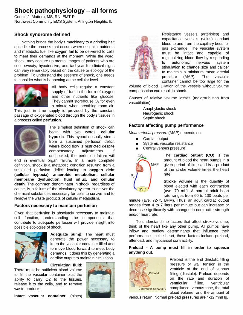
Resistance vessels (arterioles) and
capacitance vessels (veins) conduct
blood to and from the capillary beds for
gas exchange. The vascular system
must be intact and capable of
regionalizing blood flow by responding
to autonomic nervous system
stimulation to change size and caliber
to maintain a minimum mean arterial
pressure (MAP). The vascular
container cannot be too large for the
volume of blood. Dilation of the vessels without volume
compensation can result in shock.
Causes of relative volume losses (maldistribution from
vasodilation)
Anaphylactic shock
Neurogenic shock
Septic shock
Factors affecting pump performance
Mean arterial pressure (MAP) depends on:
■ Cardiac output
■ Systemic vascular resistance
■ Central venous pressure
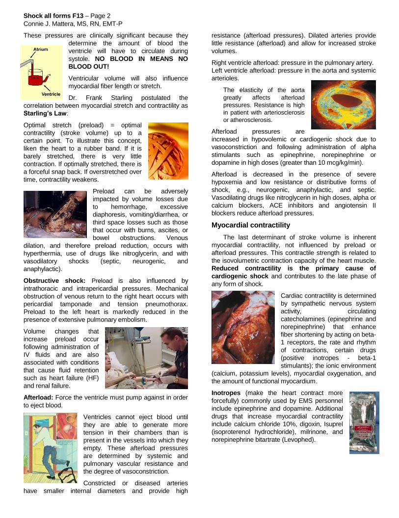
The cardiac output (CO) is the
amount of blood the heart pumps in a
given period of time and is a product
of the stroke volume times the heart
rate.
Stroke volume is the quantity of
blood ejected with each contraction
(ave. 70 mL). A normal adult heart
rate ranges from 60 to 100 beats per
minute (ave. 72-75 BPM). Thus, an adult cardiac output
ranges from 4 to 7 liters per minute but can increase or
decrease significantly with changes in contractile strength
and/or heart rate.
To understand the factors that affect stroke volume,
think of the heart like any other pump. All pumps have
inflow and outflow determinants that influence their
performance. In the heart, these factors include preload,
afterload, and myocardial contractility.
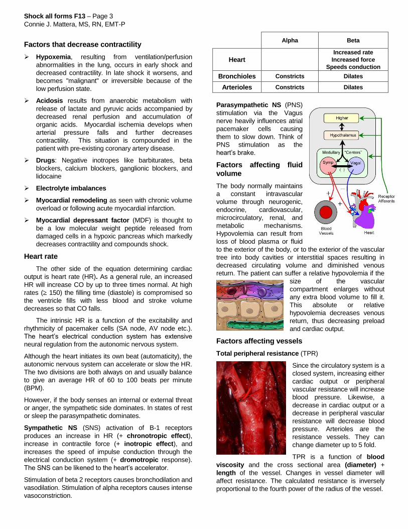
Preload - A pump must fill in order to squeeze
anything out.
Preload is the end diastolic filling
pressure or wall tension in the
ventricle at the end of venous
filling (diastole). Preload depends
on the rate and duration of
ventricular filling, ventricular
compliance, venous tone, the total
blood volume, and the amount of
venous return. Normal preload pressures are 4-12 mmHg.
Shock all forms F13 – Page 2
Connie J. Mattera, MS, RN, EMT-P
These pressures are clinically significant because they
determine the amount of blood the
ventricle will have to circulate during
systole. NO BLOOD IN MEANS NO
BLOOD OUT!
Ventricular volume will also influence
myocardial fiber length or stretch.
Dr. Frank Starling postulated the
correlation between myocardial stretch and contractility as
Starling’s Law:
Optimal stretch (preload) = optimal
contractility (stroke volume) up to a
certain point. To illustrate this concept,
liken the heart to a rubber band. If it is
barely stretched, there is very little
contraction. If optimally stretched, there is
a forceful snap back. If overstretched over
time, contractility weakens.
Preload can be adversely
impacted by volume losses due
to hemorrhage, excessive
diaphoresis, vomiting/diarrhea, or
third space losses such as those
that occur with burns, ascites, or
bowel obstructions. Venous
dilation, and therefore preload reduction, occurs with
hyperthermia, use of drugs like nitroglycerin, and with
vasodilatory shocks (septic, neurogenic, and
anaphylactic).
Obstructive shock: Preload is also influenced by
intrathoracic and intrapericardial pressures. Mechanical
obstruction of venous return to the right heart occurs with
pericardial tamponade and tension pneumothorax.
Preload to the left heart is markedly reduced in the
presence of extensive pulmonary embolism.
Volume changes that
increase preload occur
following administration of
IV fluids and are also
associated with conditions
that cause fluid retention
such as heart failure (HF)
and renal failure.
Afterload: Force the ventricle must pump against in order
to eject blood.
Ventricles cannot eject blood until
they are able to generate more
tension in their chambers than is
present in the vessels into which they
empty. These afterload pressures
are determined by systemic and
pulmonary vascular resistance and
the degree of vasoconstriction.
Constricted or diseased arteries
have smaller internal diameters and provide high
resistance (afterload pressures). Dilated arteries provide
little resistance (afterload) and allow for increased stroke
volumes.
Right ventricle afterload: pressure in the pulmonary artery.
Left ventricle afterload: pressure in the aorta and systemic
arterioles.
The elasticity of the aorta
greatly affects afterload
pressures. Resistance is high
in patient with arteriosclerosis
or atherosclerosis.
Afterload pressures are
increased in hypovolemic or cardiogenic shock due to
vasoconstriction and following administration of alpha
stimulants such as epinephrine, norepinephrine or
dopamine in high doses (greater than 10 mcg/kg/min).
Afterload is decreased in the presence of severe
hypoxemia and low resistance or distributive forms of
shock, e.g., neurogenic, anaphylactic, and septic.
Vasodilating drugs like nitroglycerin in high doses, alpha or
calcium blockers, ACE inhibitors and angiotensin II
blockers reduce afterload pressures.
Myocardial contractility
The last determinant of stroke volume is inherent
myocardial contractility, not influenced by preload or
afterload pressures. This contractile strength is related to
the isovolumetric contraction capacity of the heart muscle.
Reduced contractility is the primary cause of
cardiogenic shock and contributes to the late phase of
any form of shock.
Cardiac contractility is determined
by sympathetic nervous system
activity, circulating
catecholamines (epinephrine and
norepinephrine) that enhance
fiber shortening by acting on beta-
1 receptors, the rate and rhythm
of contractions, certain drugs
(positive inotropes - beta-1
stimulants); the ionic environment
(calcium, potassium levels), myocardial oxygenation, and
the amount of functional myocardium.
Inotropes (make the heart contract more
forcefully) commonly used by EMS personnel
include epinephrine and dopamine. Additional
drugs that increase myocardial contractility
include calcium chloride 10%, digoxin, Isuprel
(isoproterenol hydrochloride), milrinone, and
norepinephrine bitartrate (Levophed).
Shock all forms F13 – Page 3
Connie J. Mattera, MS, RN, EMT-P
Factors that decrease contractility
Hypoxemia, resulting from ventilation/perfusion
abnormalities in the lung, occurs in early shock and
decreased contractility. In late shock it worsens, and
becomes "malignant" or irreversible because of the
low perfusion state.
Acidosis results from anaerobic metabolism with
release of lactate and pyruvic acids accompanied by
decreased renal perfusion and accumulation of
organic acids. Myocardial ischemia develops when
arterial pressure falls and further decreases
contractility. This situation is compounded in the
patient with pre-existing coronary artery disease.
Drugs: Negative inotropes like barbiturates, beta
blockers, calcium blockers, ganglionic blockers, and
lidocaine
Electrolyte imbalances
Myocardial remodeling as seen with chronic volume
overload or following acute myocardial infarction.
Myocardial depressant factor (MDF) is thought to
be a low molecular weight peptide released from
damaged cells in a hypoxic pancreas which markedly
decreases contractility and compounds shock.
Heart rate
The other side of the equation determining cardiac
output is heart rate (HR). As a general rule, an increased
HR will increase CO by up to three times normal. At high
rates ( 150) the filling time (diastole) is compromised so
the ventricle fills with less blood and stroke volume
decreases so that CO falls.
The intrinsic HR is a function of the excitability and
rhythmicity of pacemaker cells (SA node, AV node etc.).
The heart’s electrical conduction system has extensive
neural regulation from the autonomic nervous system.
Although the heart initiates its own beat (automaticity), the
autonomic nervous system can accelerate or slow the HR.
The two divisions are both always on and usually balance
to give an average HR of 60 to 100 beats per minute
(BPM).
However, if the body senses an internal or external threat
or anger, the sympathetic side dominates. In states of rest
or sleep the parasympathetic dominates.
Sympathetic NS (SNS) activation of B-1 receptors
produces an increase in HR (+ chronotropic effect),
increase in contractile force (+ inotropic effect), and
increases the speed of impulse conduction through the
electrical conduction system (+ dromotropic response).
The SNS can be likened to the heart’s accelerator.
Stimulation of beta 2 receptors causes bronchodilation and
vasodilation. Stimulation of alpha receptors causes intense
vasoconstriction.
Alpha Beta
Heart
Increased rate
Increased force
Speeds conduction
Bronchioles Constricts Dilates
Arterioles Constricts Dilates
Parasympathetic NS (PNS)
stimulation via the Vagus
nerve heavily influences atrial
pacemaker cells causing
them to slow down. Think of
PNS stimulation as the
heart’s brake.
Factors affecting fluid
volume
The body normally maintains
a constant intravascular
volume through neurogenic,
endocrine, cardiovascular,
microcirculatory, renal, and
metabolic mechanisms.
Hypovolemia can result from
loss of blood plasma or fluid
to the exterior of the body, or to the exterior of the vascular
tree into body cavities or interstitial spaces resulting in
decreased circulating volume and diminished venous
return. The patient can suffer a relative hypovolemia if the
size of the vascular
compartment enlarges without
any extra blood volume to fill it.
This absolute or relative
hypovolemia decreases venous
return, thus decreasing preload
and cardiac output.
Factors affecting vessels
Total peripheral resistance (TPR)
Since the circulatory system is a
closed system, increasing either
cardiac output or peripheral
vascular resistance will increase
blood pressure. Likewise, a
decrease in cardiac output or a
decrease in peripheral vascular
resistance will decrease blood
pressure. Arterioles are the
resistance vessels. They can
change diameter up to 5 fold.
TPR is a function of blood
viscosity and the cross sectional area (diameter) +
length of the vessel. Changes in vessel diameter will
affect resistance. The calculated resistance is inversely
proportional to the fourth power of the radius of the vessel.
Shock all forms F13 – Page 4
Connie J. Mattera, MS, RN, EMT-P
There are a large number of factors influencing the
diameter of vessels in the microcirculation, which
ultimately determine resistance. Manipulation of these
factors allows the system to tolerate or compensate for
reduced CO.
Local auto-regulatory vascular control
Vessels have an intrinsic ability to autoregulate tone
and maintain blood flow over a wide range of perfusion
pressures - independent of neurogenic or humoral
influences. Different vascular beds vary in their capacity to
auto-regulate flow but the cerebral, coronary and renal
circulations are most potent.
The specific mediators of these local responses are not
known, but they are most likely triggered by changes in
osmolality, accumulation of metabolic waste and hypoxia
resulting from local ischemia due to low perfusion states.
Brain (via SNS) auto-regulation protects cerebral tissues
from low flow states - however, this ability is lost in the
presence of hypoxia or severe hypercarbia ( CO2).
Hemodynamics
Just because the patient has a blood pressure, does not
mean that tissues are being perfused. This concept is
explained by pressure, flow, and resistance relationships.
Blood flow = Pressure/resistance
Pressure = Flow X resistance
Resistance = Pressure/flow
Another way of calculating this
relationship is with the
following equation:
F = PA - PV
R
An increase in resistance will decrease flow at any given
perfusion pressure, in fact, a change in resistance (vessel
diameter) is the primary means of blood flow regulation.
Cellular metabolism: normal to hypoperfused
Normal flow in the microcirculation
The microcirculation is
composed of
arterioles, capillaries
and venules. There is
a sphincter at the
origin of the capillary
between the arteriole
and capillary (pre-
capillary sphincter)
and another at the end of the capillary between the
capillary and venule, called the post-capillary sphincter.
The arteriole component is concerned with homeostasis
and is innervated by adrenergic (SNS) fibers that control
muscular sphincters. These sphincters maintain peripheral
vascular resistance and determine blood flow through the
capillaries.
Each arteriole feeds a series of capillaries. Capillaries
open in rotation on demand of cells adjacent to them. The
opening of precapillary sphincters is facilitated by
histamine secretion in response to local tissue conditions,
such as acidosis and hypoxia. They open as more arterial
blood is needed.
When the arteriole is widely opened flow is rapid, the
pH drop is minimal, and the arterio-venous (AV) shunt is
closed. Oxygen and waste products are exchanged across
the capillary membrane based on hydrostatic and osmotic
pressure gradients. The post-capillary sphincter opens
when blood is to be emptied into the venule. Thus, blood
flow to cells is regulated by peripheral resistance and
pressure within the system.
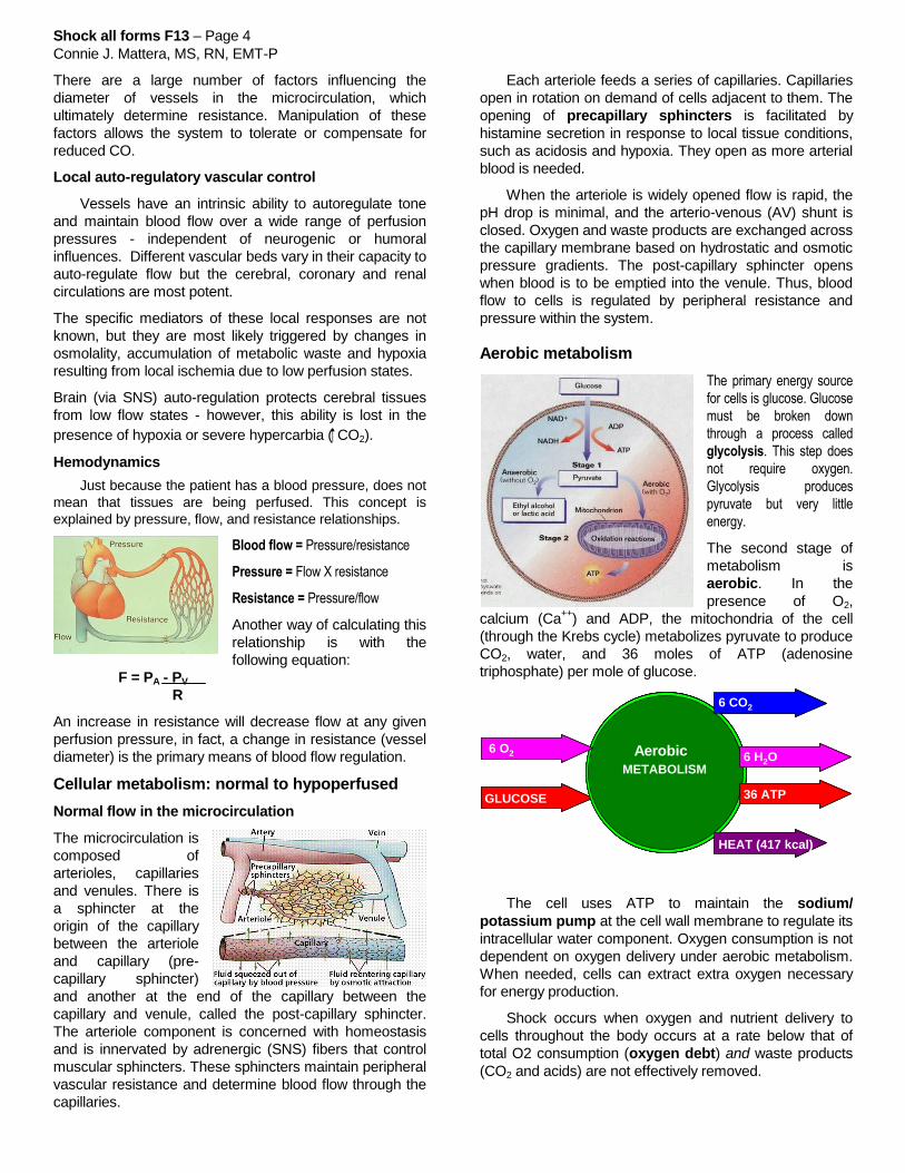
Aerobic metabolism
The primary energy source
for cells is glucose. Glucose
must be broken down
through a process called
glycolysis. This step does
not require oxygen.
Glycolysis produces
pyruvate but very little
energy.
The second stage of
metabolism is
aerobic. In the
presence of O2,
calcium (Ca++
) and ADP, the mitochondria of the cell
(through the Krebs cycle) metabolizes pyruvate to produce
CO2, water, and 36 moles of ATP (adenosine
triphosphate) per mole of glucose.
6 O2
GLUCOSE
METABOLISM
6 CO2
6 H2O
36 ATP
HEAT (417 kcal)
6 O2
GLUCOSE
METABOLISM
6 CO2
6 H2O
36 ATP
HEAT (417 kcal)
The cell uses ATP to maintain the sodium/
potassium pump at the cell wall membrane to regulate its
intracellular water component. Oxygen consumption is not
dependent on oxygen delivery under aerobic metabolism.
When needed, cells can extract extra oxygen necessary
for energy production.
Shock occurs when oxygen and nutrient delivery to
cells throughout the body occurs at a rate below that of
total O2 consumption (oxygen debt) and waste products
(CO2 and acids) are not effectively removed.
Aerobic
Shock all forms F13 – Page 5
Connie J. Mattera, MS, RN, EMT-P
Cells start to change from aerobic to anaerobic
metabolism.
GLUCOSE METABOLISM
2 LACTIC ACID
2 ATP
HEAT (32 kcal)
GLUCOSE METABOLISM
2 LACTIC ACID
2 ATP
HEAT (32 kcal)
Start connecting the dots…Causes of hypoperfusion:
Inadequate pump
Inadequate preload
Inadequate cardiac contractile strength
Inadequate HR
Excessive afterload
Inadequate fluid volume (absolute or relative)
Inadequate container (container failure)
Dilated vessels without change in fluid volume
Leak in the vessels
Stages of shock & compensatory mechanisms
The stages of shock reflect the severity of disruption in
tissue perfusion and the degree of cellular membrane
damage. If precipitating factors are promptly reversed,
compensatory mechanisms can usually restore perfusion.
The longer a patient remains in shock, the longer vital
organs are deprived of O2. After cellular destruction
begins, shock cannot be reversed and organs will fail.
Stages of shock
Initial stage (compensated/reversible)
Something occurs to cause a perfusion deficit with an
early drop in cardiac output that alters cellular function.
The body attempts to maintain hemodynamic stability
through compensatory mechanisms and by neutralizing
elevated lactate levels. Interrelated neural, hormonal, and
chemical mechanisms restore cardiac output and
perfusion to keep the circulatory system functioning at
normal or near normal levels so there are no early clinical
signs or symptoms.
Neural compensation - homeostatic neuroreflexes
The vasomotor center in the medulla receives
impulses from various receptor mechanisms in the body,
which either suppress or stimulate neural tone in the
sympathetic nervous system and adrenal glands in an
attempt to stabilize the BP.
Types of peripheral receptors
Baroreceptors (pressure
receptors in the aortic arch and
carotid sinuses) are triggered by
a decrease in cardiac output.
The carotid sinuses respond to
pressures of 60-180 mmHg. The aortic arch has a higher
threshold and is less sensitive than the carotid bodies.
Impulses travel to the medulla. The vasomotor center
responds by increasing sympathetic and decreasing
parasympathetic outflow. The SNS releases epinephrine
and norepinephrine that increase HR and strength of
contractions and cause venous then arterial
vasoconstriction.
Chemoreceptors detect hypoxia (pO2 < 80), high
pCO2 levels or a low pH (< 7.4). An increase in carbon
dioxide level will usually trigger ventilations. If
respiratory activity cannot correct the pH,
chemoreceptors activate the vagus nerve resulting in
bradycardia and coronary vasodilation. Thus, patients
with severe hypoxia may present with bradycardia.
Osmoreceptors in the hypothalamus sense the
concentration of body fluids.
Stretch receptors in the ventricles sense the
volume of blood return to the heart.
Hormonal compensation
In shock, the combination of
hypoxia, acidosis, hypotension
and volume abnormalities cause
simultaneous and synergistic
stimulation of all these receptors
to activate the sympathetic
nervous system and adrenal
glands as well as other hormonal responses.
Adrenal glands
Stimulation of the SNS leads to the
release of epinephrine and
norepinephrine from the adrenal
medulla. Venoconstriction precedes
arterial constriction during the initial
stage of shock. Given that the majority
of the blood volume is stored in the
veins (capacitance vessels),
constricting the veins may adequately
restore vascular volume. If the perfusion deficit worsens,
causing further O2 debt and acidosis, additional
compensation is required and more catecholamines are
released.
The SNS releases nor-epinephrine from nerve endings
Further activation of the SNS triggers the "fight or
flight" response. Heart rate and myocardial contractility
increase to augment cardiac output. Coronary arteries
dilate to supply additional O2 to heart muscle. Peripheral
vessels constrict to redistribute blood flow to the protected
vital organs (heart and brain) and shunt blood away from
non-priority organs. Constriction of dermal capillary beds
causes the skin to be pale and cool. Sweat glands are
activated to vent off heat. Pupils dilate to enhance vision.
Decreased GI perfusion slows peristalsis.
Anaerobic
Shock all forms F13 – Page 6
Connie J. Mattera, MS, RN, EMT-P
While norepinephrine secreted from sympathetic
nerve endings is rapidly dissipated, adrenal
catecholamines help to sustain the stress response for
hours to days.
Renin - Angiotensin - Aldosterone cycle
Renin is released from the
kidneys in response to
hypoperfusion and to input from
the SNS. Renin reacts with
alpha-2 globulin in the liver to
release angiotensin I.
Angiotensin converting enzyme
(ACE) converts angiotensin I to
angiotensin II. Angiotensin II is
a potent vasoconstrictor, helping to maintain the BP. It also
causes the adrenal gland to secrete aldosterone.
Aldosterone causes sodium retention and potassium
excretion by the kidneys. The net effects are to conserve
water by decreasing urinary output and to increase BP by
augmenting blood volume and prompting vasoconstriction.
Antidiuretic hormone (ADH or vasopressin)
ADH is made in the hypothalamus and stored in the
posterior pituitary gland. It is released in response to
hypovolemia or hyperosmolality sensed by receptors in the
carotid bodies and atria and by osmoreceptors in the
hypothalamus. When released, ADH stimulates water
reabsorption in the distal renal tubules and inhibits urinary
output. ADH is also a potent vasoconstrictor helping to
maintain the BP.
So if you are counting,
this is the 3rd mechanism
for vasoconstricting the
vessels and maintaining
mean arterial pressure
(MAP):
#1 Catecholamines
#2 Angiotensin II
#3 Vasopressin
Adrenocorticotropic hormone (ACTH)
During periods of stress or
trauma, the anterior
hypothalamus is affected by
input from the ascending
reticular activating system
(ARAS), brain stem, subcortex,
and limbic system. This causes
the hypothalamus to secrete a
releasing factor that acts on the anterior pituitary to secrete
ACTH. This hormone causes the adrenal cortex to
increase production of glucocorticoids, like cortisol, that
stimulate metabolic processes in the liver and kidneys to
increase blood glucose levels. Expect adults in shock to
have higher blood glucose levels due to this mechanism.
Gonadotrophins are inhibited. Women under prolonged
stress may experience amenorrhea.
Kinins (Bradykinin) are potent vasodilators .They are felt
to be responsible for the dramatic hypotension and
hyperemia associated with anaphylactic shock.
Serotonin and histamine: These vasoactive substances
are released from platelets and mast cells respectively and
regulate local vascular tone and capillary permeability.
Anaphylaxis and complement activation trigger their
release.
Prostaglandins are acidic lipid soluble materials
distributed widely in the body. They are generally released
in response to ischemia or hypoxia from the endothelial
tissue or platelets and may cause intravascular platelet
aggregation, clumping and vasoconstriction.
Chemical (respiratory) compensation
Redistribution of blood to priority organs causes
hypoperfusion of the lungs. Vasoconstriction in hypoxic
pulmonary beds results in alveoli that are ventilated but not
perfused, thus increasing alveolar dead space. This
produces a ventilation/perfusion (VA/Q) mismatch,
impaired gas exchange, and hypoxemia.
The body attempts to correct the acid-base imbalance
by increasing the ventilatory rate and depth in an effort to
exhale excess CO2 (acid to the body). On room air, there is
a 1:1 inverse correlation between pCO2 and pO2 levels.
For each 1 torr the pCO2 goes down, there is a
corresponding rise of 1 torr in pO2. Thus, one of the
earliest S&S of shock is an increase in RR.
The combination of hypoxemia and respiratory
alkalosis affects mental status resulting in restlessness,
agitation, excitability, confusion, and lethargy (Rice, 1997).
T
he total net result of compensatory mechanisms in
reversible shock is to successfully restore cardiac output
and tissue perfusion to vital organs at the expense of the
non-vital organs. If SNS fibers are intact, the patient will
have an increased heart rate, increased myocardial
contractility, increased diastolic BP; increased RR, pale,
cool, moist skin, and decreased peristalsis.
Shock all forms F13 – Page 7
Connie J. Mattera, MS, RN, EMT-P
Decompensated or progressive shock
Decompensated or progressive shock occurs when the
circulatory system starts to fail despite the body’s
maximum efforts to compensate and the systolic BP falls
below 100.
This leads to global hypoperfusion
and multiple organ dysfunction
syndrome (MODS). Arterioles are
constricted and AV shunts open
further reducing O2 delivery to cells.
There is slow flow in the upper
capillary and other capillaries may
open. When there is slow flow in all of
them, the pH drop is marked. Blood
vessels are unable to sustain
vasoconstriction. Vasodilation results
in decreased peripheral vascular resistance, hypotension,
and capillary flooding.
Anaerobic metabolism becomes widespread and is
only tolerated for only a limited amount of time. Anaerobic
metabolism is much less efficient than aerobic and leads
to systemic acidosis and depletion of high energy
reserves (ATP) producing only two moles of ATP (5-10%
of normal). Hypoxia will decrease the rate of ATP
synthesis in the cell but will not damage the mitochondria
unless it is sustained, severe, and associated with
ischemia.
You’re up to your ***** in alligators now!
Pathophysiology of acidosis
During anaerobic
metabolism, glucose
breakdown can only
complete the first
stage. This causes an
accumulation of
pyruvic acid. Pyruvic
acid cannot be
converted to Acetyl
Coenzyme A without
O2 so is transformed in greater amounts to lactate and
other acid by-products. Acidosis develops because ATP is
hydrolyzed to ADP and phosphate with the release of a
proton. Hydrogen ion accumulates, decreasing the pool of
bicarbonate buffer. Lactate also buffers protons and lactic
acid accumulates.
At the same time, ischemia causes an increased CO2
production by tissues. CO2 levels rise in the sublingual
area, esophagus, stomach, duodenum, jejunum, brain,
liver, and kidneys. The higher the organ's metabolic rate,
the higher the CO2 level in hypoperfused states. Excess
CO2 combines with intracellular water to produce
carbonic acid. Thus, acidosis can be used as a measure
of tissue perfusion.
The acidic condition of the blood reduces the ability of
hemoglobin in red blood cells to bind with and carry
oxygen. This adds to the cellular oxygen debt (shifts the
oxyhemoglobin dissociation curve to the right).
Circling the drain…game over.
Micro-circulatory failure & cell membrane injury
Sodium (Na) is more
abundant outside of the
cell than inside. It is
naturally inclined to
diffuse into the cells. The
sodium-potassium pump
is like a “bouncer” at the
cell membrane that
sends the sodium back
out against its
concentration gradient (active transport mechanism), but
needs an ample supply of ATP to fuel the process.
Reduced levels of ATP result in a dysfunctional Na/K
pump and alterations in cell membrane function. Loss of
the Na/K pump allows sodium to diffuse into the cell and
stay there. Water follows the sodium and shifts into
the cell, causing the cell to swell.
Intracellular enzymes that usually help to digest and
neutralize bacteria introduced into a cell are bound in a
relatively impermeable membrane. Cellular flooding
explodes that membrane and allows these lysosomal
enzymes to be released. Their job is to digest all intra and
extracellular proteins, and once released, they autodigest
the cell. If enough cells are destroyed, organ failure will
become evident. The release of the lysosomes heralds
the onset of irreversible shock.
Sluggish blood flow and pooling in the vessels coupled
with acidic blood leads to platelet agglutination and
formation of microthrombi in the capillary stagnation
phase.
Just to compound the problem, accumulating acids
and waste products act as potent vasodilators of post-
capillary sphincters, releasing hydrogen ion, lactic acid,
carbon dioxide and columns of coagulated red blood cells
(rouleaux formations) into the venous circulation.
Shock all forms F13 – Page 8
Connie J. Mattera, MS, RN, EMT-P
This is known as capillary washout. Rouleaux
formations microembolize in the lungs.
Arterial pressure falls to the point that even the "protected organs" such as the brain and heart are not perfused. Trend pulse pressures (normal 30-50) and mean arterial pressure (MAP) (normal 70-110). When aortic root pressures fall below a mean arterial pressure (MAP) of 60 mmHg, the coronary arteries do not fill, the heart is weakened, and cardiac output falls. Myocardial depressant factor is released from an ischemic pancreas, further decreasing the pumping action of the heart and decreasing CO.
Reduced blood supply to the vasomotor center in the
brain results in a slowing, then stopping of sympathetic
nervous system activity.
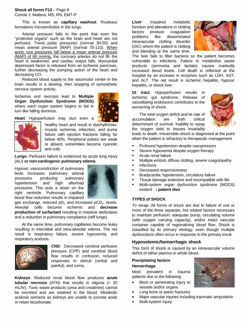
Ischemia and necrosis lead to Multiple
Organ Dysfunction Syndrome (MODS)
where each organ system begins to fail in
turn like falling dominos.
Heart: Hypoperfusion may stun even a
healthy heart and result in dysrhythmias,
muscle ischemia, infarction, and pump
failure with ejection fractions falling far
below 40%. Peripheral pulses are weak
or absent, extremities become cyanotic
and cold.
Lungs: Perfusion failure is evidenced by acute lung injury
(ALI) or non-cardiogenic pulmonary edema.
Hypoxic vasoconstriction of pulmonary
beds increases pulmonary arterial
pressures producing pulmonary
hypertension and high afterload
pressures. This puts a strain on the
right ventricle. Pulmonary capillary
blood flow reduction results in impaired
gas exchange, reduced pO2 and increased pCO2. levels.
Alveolar cells become ischemic and decrease
production of surfactant resulting in massive atelectasis
and a reduction in pulmonary compliance (stiff lungs).
At the same time, pulmonary capillaries become leaky
resulting in interstitial and intra-alveolar edema. The net
result is respiratory failure, severe hypoxemia, and
respiratory acidosis.
CNS: Decreased cerebral perfusion
pressure (CPP) and cerebral blood
flow results in confusion, reduced
responses to stimuli (verbal and
painful), and coma.
Kidneys: Reduced renal blood flow produces acute
tubular necrosis (ATN) that results in oliguria (< 20
mL/hr). Toxic waste products (urea and creatinine) cannot
be excreted and are retained in the blood. Metabolic
acidosis worsens as kidneys are unable to excrete acids
or retain bicarbonate.
Liver: Impaired metabolic
function and alterations in clotting
factors produce coagulation
problems like disseminated
intravascular clotting disorder
(DIC) where the patient is clotting
and bleeding at the same time.
The liver fails to filter bacteria so the patient becomes
vulnerable to infections. Failure to metabolize waste
products (ammonia and lactate) causes markedly
increased blood levels. Cell death is reflected at the
hospital by an increase in enzymes such as LDH, AST,
and ALT. The net result is ischemic hepatitis, hypoxic
hepatitis, or shock liver.
GI tract: Hypoperfusion results in
ischemic gut syndrome. Release of
vasodilating endotoxins contributes to the
worsening of shock.
The total oxygen deficit and its rate of
accumulation are both critical
determinant of survival. Inability to repay
the oxygen debt to tissues invariably
leads to death. Irreversible shock is diagnosed at the point
when the patient is refractory to therapeutic management.
Profound hypotension despite vasopressors
Severe hypoxemia despite oxygen therapy
Acute renal failure
Multiple emboli, diffuse clotting, severe coagulopathy
Infections
Decreased responsiveness
Bradycardia, hypotension, circulatory failure
Tissue damage extensive and incompatible with life
Multi-system organ dysfunction syndrome (MODS)
evident patient dies
TYPES of SHOCK
To recap: All forms of shock are due to failure of one or
more of the three separate, but related factors necessary
to maintain perfusion: adequate pump, circulating volume
(with oxygen carrying capacity), and/or intact vascular
container capable of regionalizing blood flow. Shock is
classified by its primary etiology, even though multiple
dysfunctions often occur in response to the primary insult.
Hypovolemic/hemorrhagic shock
This form of shock is caused by an intravascular volume
deficit of either plasma or whole blood.
Precipitating factors
Hemorrhage
Most prevalent in trauma
patients due to the following:
Blunt or penetrating injury to
vessels and/or organs
Long bone or pelvic fractures
Major vascular injuries including traumatic amputation
Multi-system injury
Shock all forms F13 – Page 9
Connie J. Mattera, MS, RN, EMT-P
Organs and organ systems with high incidence of
exsanguination from penetrating injuries:
Heart
Thoracic vascular system
Abdominal vascular system:
abdominal aorta, superior
mesenteric artery
Inferior vena cava, portal vein
Liver, spleen
Trunkey defines a severe hemorrhage as a blood loss
of greater than 150 mL/min. Others site a rate of 250
mL/min as leading to exsanguination, which will cause the
patient to lose ½ of their entire blood volume in
approximately 10 minutes.
Fluid (plasma) shifts: Plasma shifts from the
intravascular to interstitial spaces as a result of increased
capillary permeability in crush or burn injuries.
Other causes of body fluid deficits
Dehydration
Excess GI drainage; diarrhea
Ascites
Diabetes insipidus
Excess wound drainage
Acute renal failure; high output phase
Losses through skin and lungs
Osmotic diuresis secondary to hyperosmolar states
(DKA, HHNS)
Assessment/management
Shock resuscitation begins in the prehospital
environment and continues through the ED and possibly
the OR and ICU. Everyone knows when it begins, but the
end points of effective resuscitation are in the process of
being redefined. Classic end points were considered to be
a normalizing heart rate and BP and good urine output.
Traditional markers are global measures that reflect
the general circulation to large tissue beds and may be
slow to exhibit signs of severe perfusion deficits.
Another limitation is that preexisting diseases or the
aging process may alter a patient's response to volume
losses and blunt changes in vital signs or renal function.
Cardiogenic shock (pump failure)
Etiology
Cardiogenic shock is usually caused by extensive
myocardial infarction of the LV, diffuse ischemia, or
decompensated CHF resulting in primary pump failure. It
is also seen with cardiomyopathy, valvular abnormalities,
and dysrhythmias. A special type is compressive cardiac
shock due to an inadequate venous return to the heart
caused by extrinsic compression, i.e., tension
pneumothorax, pericardial tamponade. There is a poor
prognosis when > 40% of the LV is destroyed. Historically,
about 7.5% of patients with AMI develop cardiogenic
shock and mortality rates range as high as 80% even with
appropriate therapy.
Pathophysiology
Left ventricular function is so compromised that the
heart cannot meet the metabolic needs of the body and
compensatory mechanisms are maximized and
ineffective. Mean arterial pressures less than 60 mmHg
decrease coronary perfusion further suppressing cardiac
performance, and ultimately result in total pump failure. A
cardiogenic component to shock should be suspected
when hypoperfusion persists after correcting existing
dysrhythmias, hypovolemia, or altered vascular tone.
Distributive, vasogenic, low resistance, or
container failure shock: Loss of peripheral vascular
resistance (vasodilation) causes a relative fluid volume
deficit and maldistribution of blood flow to cells.
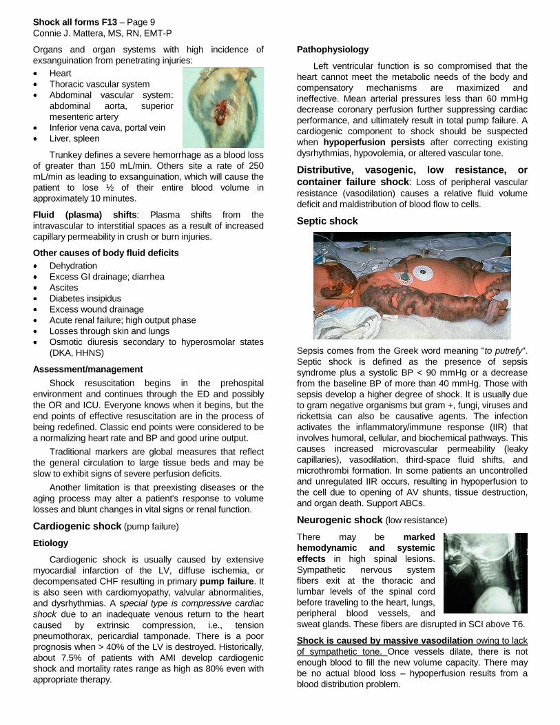
Septic shock
Sepsis comes from the Greek word meaning "to putrefy".
Septic shock is defined as the presence of sepsis
syndrome plus a systolic BP < 90 mmHg or a decrease
from the baseline BP of more than 40 mmHg. Those with
sepsis develop a higher degree of shock. It is usually due
to gram negative organisms but gram +, fungi, viruses and
rickettsia can also be causative agents. The infection
activates the inflammatory/immune response (IIR) that
involves humoral, cellular, and biochemical pathways. This
causes increased microvascular permeability (leaky
capillaries), vasodilation, third-space fluid shifts, and
microthrombi formation. In some patients an uncontrolled
and unregulated IIR occurs, resulting in hypoperfusion to
the cell due to opening of AV shunts, tissue destruction,
and organ death. Support ABCs.
Neurogenic shock (low resistance)
There may be marked
hemodynamic and systemic
effects in high spinal lesions.
Sympathetic nervous system
fibers exit at the thoracic and
lumbar levels of the spinal cord
before traveling to the heart, lungs,
peripheral blood vessels, and
sweat glands. These fibers are disrupted in SCI above T6.
Shock is caused by massive vasodilation owing to lack
of sympathetic tone. Once vessels dilate, there is not
enough blood to fill the new volume capacity. There may
be no actual blood loss – hypoperfusion results from a
blood distribution problem.

Shock all forms F13 – Page 10
Connie J. Mattera, MS, RN, EMT-P
Anaphylactic Shock
Pathophysiology
The onset of anaphylaxis is generally acute.
Symptoms can develop from 30-60 seconds to up to 20
minutes post-exposure and cause death within minutes.
IgE-mediated reactions occur as a
result of an immune response. The
immune system is exposed to an
allergen (antigen) and a specific
antibody (IgE) is formed and stored in
mast cells and basophils. With further
exposure, the antigen will bind with the
IgE antibody, triggering release of
vasoactive mediators. Non-IgE
reactions (anaphylactoid reactions) are
associated with nonsteroidal anti-inflammatory agents and
aspirin.
When a hypersensitivity reaction occurs, mast cells
and basophils rupture or secrete histamine, leukotrienes,
and other substances that cause systemic vasodilation,
increased capillary permeability, bronchoconstriction,
coronary vasoconstriction, and skin reactions. The relative
decrease in vascular volume owing to the enlarged
vascular space results in a decreased cardiac output and
inadequate tissue perfusion.
Risk: The severity of a reaction is affected by the quantity
of the antigen; the route and rapidity of absorption (highest
risk is produced by parenteral exposure; least risk by
topical exposure; oral ingestion is in between); a past
medical history of asthma or cardiac disease; and in
patients taking beta blocker drugs.
Signs and Symptoms
A severe systemic reaction can rapidly lead to respiratory
failure and cardiovascular collapse evidenced by BP <
90, cardiac dysrhythmias, shock and coma.

Shock all forms F13 – Page 12
Connie J. Mattera, MS, RN, EMT-P
References
Bledsoe, B.E., Porter, R.S., Cherry, R.A. (2013). Paramedic Care: Principles & Practice (4th ed). Boston:
Pearson.
Cherkas,D. (Nov. 2011). Traumatic hemorrhagic shock: Advances in fluid management. Emergency
Medicine Practice (EBMEDICINE.net), 13(11).
Haut, E.R. et al. (2011). Prehospital intravenous fluid administration is associated with higher mortality in
trauma patients: A national trauma data bank analysis. Annals of Surgery; 253(2), 371.
Sanders, M.J. (2012). Shock. In Mosby’s Paramedic Textbook (4th Ed) (pp. 1052-1073). St. Louis: Mosby
Elsevier.
Shock all forms F13 – Page 13
Connie J. Mattera, MS, RN, EMT-P
Homework Questions
1. Define shock:
2. List two causes of hypoperfusion due to an inadequate pump:
3. List three causes of hypoperfusion due to loss of vascular tone:
4. What is the most common etiology of shock in trauma patients?
A. Brain injury
B. Hemorrhage
C. Respiratory failure
D. Cardiac insufficiency
5. What two factors are multiplied to determine the cardiac output in one minute?
X
Insert the numbers for the above equation.
X = L/min
6. Define: Stroke volume
7. List three major factors that all influence stroke volume:
8. Define: Preload
9. Paraphrase the Frank-Starling mechanism or Starling’s law:
10. List one EMS intervention that increases preload in a patient with hypovolemic shock.
11. List one drug that is used by EMS personnel to decrease preload.
Shock all forms F13 – Page 14
Connie J. Mattera, MS, RN, EMT-P
12. Define: Afterload
13. List two drugs EMS personnel can give to increase afterload particularly in patients with a low
resistance forms of shock (list drug name, dose, and route).
14. Independent of preload and afterload, cardiac contractile force is affected by:
14. What specific sympathetic nervous system receptors are stimulated to increase heart rate, force of
contractility and speed of conduction?
15. Which nerve is stimulated to decrease heart rate?
16. What fuels are needed and how much ATP is produced during aerobic metabolism?
Fuels needed
Amount produced:
16. List the four peripheral receptors that all send feedback to the central nervous system in an effort to
maintain adequate perfusion:
17. Hormonal compensation in shock includes release of two catecholamines from the adrenal gland:
and which
cause peripheral blood vessels to .
18. Activation of the renin-angiotensin-aldosterone system causes blood vessels to
Constrict / dilate (circle one) and the kidneys to reabsorb and
.
19. Release of ADH causes blood vessels to Constrict / dilate (circle one) and the renal tubules to
reabsorb
20. Chemical compensation is achieved through an in respiratory rate in an effort to blow off:
.
21. What two substances accumulate to create the acidosis experienced by a patient in shock?
Shock all forms F13 – Page 15
Connie J. Mattera, MS, RN, EMT-P
22. What happens to hemoglobin's ability to bind with oxygen in an acidosis?
23. Anaerobic metabolism only produces (#) moles of ATP. This results in a dysfunctional
(cell transport mechanism) that
causes the movement of water from the fluid
compartment to the fluid compartment.
This fluid shift will cause the release of enzymes within the cell called:
What effect does the release of these enzymes have on the cell?
24. Which of these can hinder the clotting process?
A. Immobilization
B. Dehydration
C. Fever
D. Aspirin
25. Which of these is present if a patient's blood pressure decreases after changing from a supine to a
sitting position?
A. Compensatory vasoconstriction
B. Orthostatic hypotension
C. Supine hypotension
D. A negative "tilt test"
26. What is the minimum MAP needed to maintain aortic root pressures and perfuse coronary arteries?
27. What happens to the vasomotor center in the brain when it no longer receives adequate blood flow?
28. An adult presents with chest pain, dyspnea, dusky skin, and bilateral crackles. VS: BP 60/30; P 90; R
24; SpO2 86%; EtCO2 22 with square waveform. What etiology of shock should be suspected?
29. An adult presents following an MVC with paralysis of the arms and legs and loss of sensation below
the shoulders. VS: BP 70/40; P 48; R 16 and shallow with only abdominal and no chest wall
movement. There are beads of sweat on the patient’s upper lip but the skin is warm and dry below
the shoulders. What etiology of shock should be suspected?
30. An adult presents with extreme respiratory distress following the unsuspected ingestion of peanut oil.
Family members state that the patient is very allergic to peanuts. The patient’s tongue, lips and
eyelids are extremely swollen. There is audible stridor and wheezes. VS: BP 72/44; P 110; R 32 and
labored; SpO2 88%; EtCO2 18 with sharkfin waveform. What etiology of shock should be
suspected?
Shock all forms F13 – Page 16
Connie J. Mattera, MS, RN, EMT-P
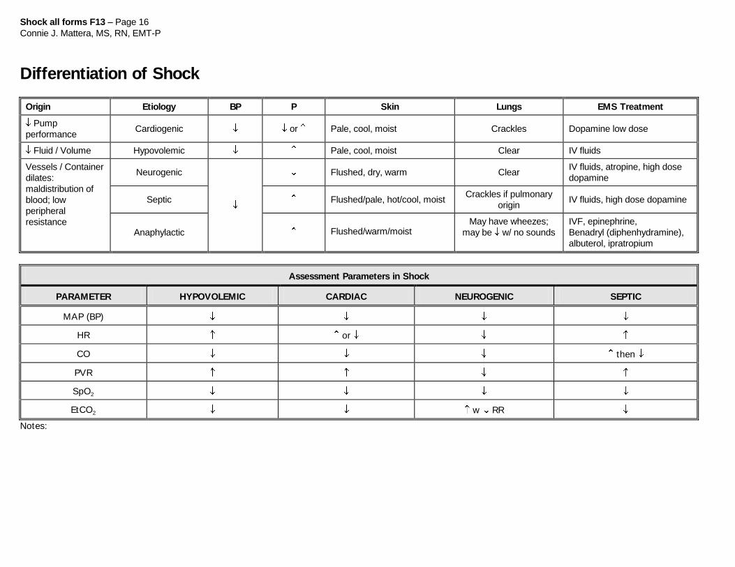
Differentiation of Shock
Origin Etiology BP P Skin Lungs EMS Treatment
Pump
performance Cardiogenic or Pale, cool, moist Crackles Dopamine low dose
Fluid / Volume Hypovolemic Pale, cool, moist Clear IV fluids
Vessels / Container
dilates:
maldistribution of
blood; low
peripheral
resistance
Neurogenic
Flushed, dry, warm Clear IV fluids, atropine, high dose
dopamine
Septic Flushed/pale, hot/cool, moist Crackles if pulmonary
origin IV fluids, high dose dopamine
Anaphylactic Flushed/warm/moist May have wheezes;
may be w/ no sounds
IVF, epinephrine,
Benadryl (diphenhydramine),
albuterol, ipratropium
Assessment Parameters in Shock
PARAMETER HYPOVOLEMIC CARDIAC NEUROGENIC SEPTIC
MAP (BP)
HR or
CO then
PVR
SpO2
EtCO2 w RR
Notes:


![SHOCK[1] - Hypovolemic Shock](https://img.pdfslide.net/doc/110x75/58edc1bc1a28abae538b4711/shock1-hypovolemic-shock.jpg)