Embed Size (px)
Citation preview
OBgyn Week 9Pregnancy Complications

Pregnancy Complications
• Common problems include:– Bleeding– SAB/miscarriage– Ectopic pregnancy– Gestational diabetes– Preeclampsia– Premature rupture of membranes (PROM)– Infections

Early Bleeding
Bleeding in early pregnancy
• Remember:– Any sexually active woman may be pregnant– Vaginal bleeding can be for reasons other
than those related to a pregnancy– Very common in 1st trimester due to SAB
(spontaneous abortion)

Early BleedingDifferential Diagnosis
– Salpingitis/ PID (mc misdiagnosis)• Mb bleeding, chandelier’s sign• Usu. no amenorrhea, usu. negative b-HCG
– Miscarriage- 2nd most common misdiagnosis• more profuse bleeding but less pain
– Rupture may cause internal bleeding but won’t see it
• Pain of SAB more localized and more rhythmic, cramp-like
– Appendicitis• No s/sx of PG, no positive b-hcg, no missed period• Other positive app signs (rebound tenderness, etc)

Early BleedingDiff Diagnosis
– Ovarian cyst/ rupture/ torsion• Usu. more discrete mass on u/s• Bleeding tends to be in abd cavity rather than vaginal• Usu. negative b-HCG
– Gastroenteritis• No PG s/sx, no vaginal bleeding• Ruptured peptic ulcer
– IUD: perforation, irritation– Other: diverticulitis, endometriosis, mittelschmerz,
fibroid, tumor

Early Bleeding DDX• Laboratory exams:• CBC w/ differential: anemia, infection,
appendicitis• Quantitative serum b-hcg and ultrasound
– Normal PG: >6500 units and intrauterine gestational sac
– Ectopic PG: >6500 units and no gestational sac– TAB: < 6000 units and gestational sac– Unclear: < 6000 units and no gestational sac
• Need serial b-hcg to see if rising or not and at what rate
Spontaneous Abortion
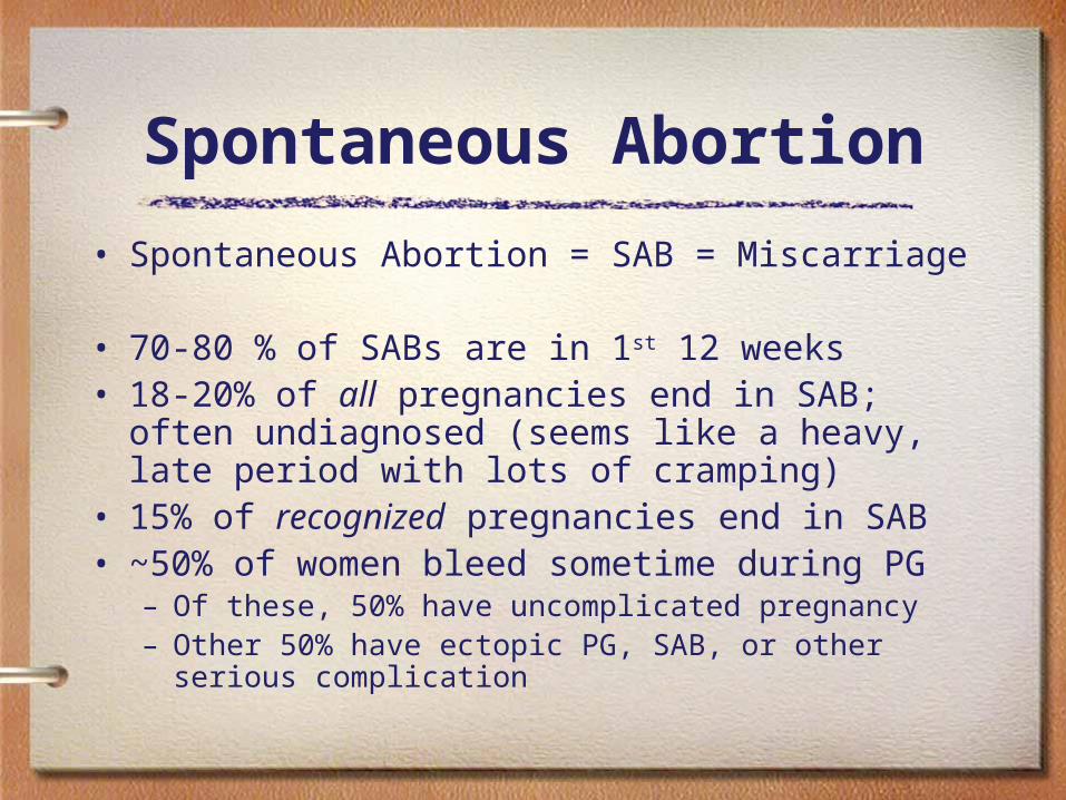
• Spontaneous Abortion = SAB = Miscarriage
• 70-80 % of SABs are in 1st 12 weeks• 18-20% of all pregnancies end in SAB; often
undiagnosed (seems like a heavy, late period with lots of cramping)
• 15% of recognized pregnancies end in SAB• ~50% of women bleed sometime during PG
– Of these, 50% have uncomplicated pregnancy– Other 50% have ectopic PG, SAB, or other serious
complication

Early Bleeding
• If bleeding occurs early, but reaches week 20, has 95% chance of an uncomplicated birth
• If early bleeding & SAB during weeks 17-20, commonly due to chromosomal abnormalities– Before that, is usually a problem with cell division
(e.g. - fertilized embryo that implants but does not develop; gestational sac may develop)

Timing and Terminology
QuickTime™ and aTIFF (Uncompressed) decompressor
are needed to see this picture.

Consecutive SABs
• “Habitual abortion” refers to a woman who has had 2 consecutive SABs (previously defined as 3)
• One previous SAB increases risk 2x of SAB on subsequent pregnancy
• In most 1st trimester SABs, fetus does not establish fetal heart tones – this is a sign that the fetus will probably SAB. Week 9 – 12.
Consecutive SABs
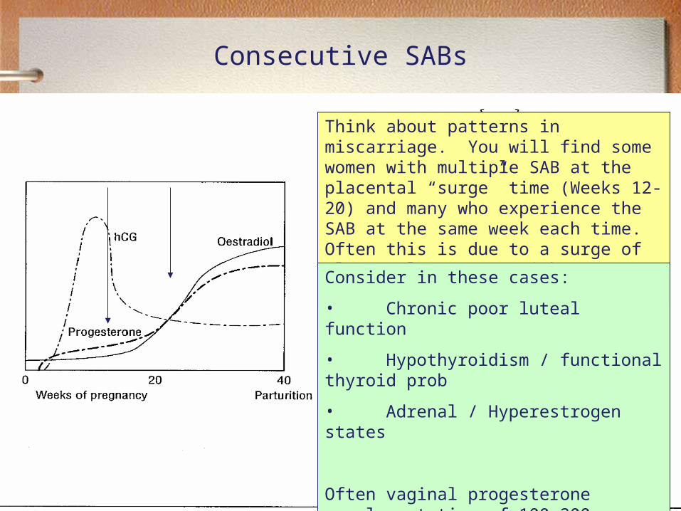
Think about patterns in miscarriage. You will find some women with multiple SAB at the placental “surge” time (Weeks 12-20) and many who experience the SAB at the same week each time. Often this is due to a surge of placental estrogen with a SLOW progesterone response.
Consider in these cases:
• Chronic poor luteal function
• Hypothyroidism / functional thyroid prob
• Adrenal / Hyperestrogen states
Often vaginal progesterone supplementation of 100-200 mg p.v. will get them through the normal SAB period – IF the pregnancy is viable otherwise.
SABs
• Emotional concerns with SAB– Desire (or lack of) for PG has strong effect on
response to SAB– Fear of pain: mini-labor; fear of bleeding to death,
fear of future fertility– Loss of control over events– Guilt or blame towards self or partner– “What did I do wrong?”– Emotional effects can last a long time, esp. difficult
at expected due date and 1 yr past SAB

Early Bleeding - is it SAB?
• Threatened abortion– Signs/ symptoms:
• Cramping (bleeding w/o cramping is not as worrisome)
• Blood may be pink, brown or bright red• Cervix may be open or closed (if closed,
chance of SAB low)
• Intercourse can cause benign spotting due to cervical friability

Early Bleeding - is it SAB?
• Labs:– Blood type and Rh– Quantitative b-hcg (may or may not show
SAB, ectopic PG)– CBC: needs to be done early in PG
anyway; if bleeding occurs later, only if concerned with anemia/ hemorrhage

Early Bleeding - is it SAB?
• FHTs not established in most 1st trimester = SABs• Imaging with ultrasound:
• 6 weeks: gestational sac – esp if can’t see it will = SAB• 8 weeks: beating heart (abdominal u/s)
• 80% accurate in predicting SAB if any of the following:– Poorly defined sac– Small sac for dates– Abnormal uterine echoes– Improper implant sac– Growth failure
Early Bleeding:Management
• Treat as viable PG until proven otherwise• Complete bed rest: no housework• Pelvic rest: no intercourse, tampons, douching, etc.• Decrease stress• Do not leave alone if possible• Go over s/sx of SAB
– Intensification of contractions– Increased bleeding/ passage of tissue– Signs of shock or infection (fever, dizziness, fainting, cold/
clammy, increased respirations, weak or rapid pulse, anxiety, low BP, etc.)

Inevitable Abortion
• SAB may be predicted if non-viability established via ultrasound
• No FHT at 8 weeks• Abnormal sac at 6 weeks
• Signs of inevitable SAB– Heavy bleeding– Dilated cervix– If at or > 20 weeks, may feel gush of amniotic fluid– May report passage of tissue – esp bean shaped
tissue. If > 12-14 weeks may see fetus

Inevitable Abortion
• Tell woman what to expect, explain options:– Can get D&C; may be necessary (i.e., no longer
optional) if tissue is retained– Can labor at home if < week 12
• Pain may be severe; 2-4 hours of mini-labor• Heavy bleeding for 1 day• Spotting common for 1-2 weeks• Should not be home alone; risk hemorrhage/ shock• If soaking > 2 maxipads/ hr, increased risk hemorrhage
Risks following SAB
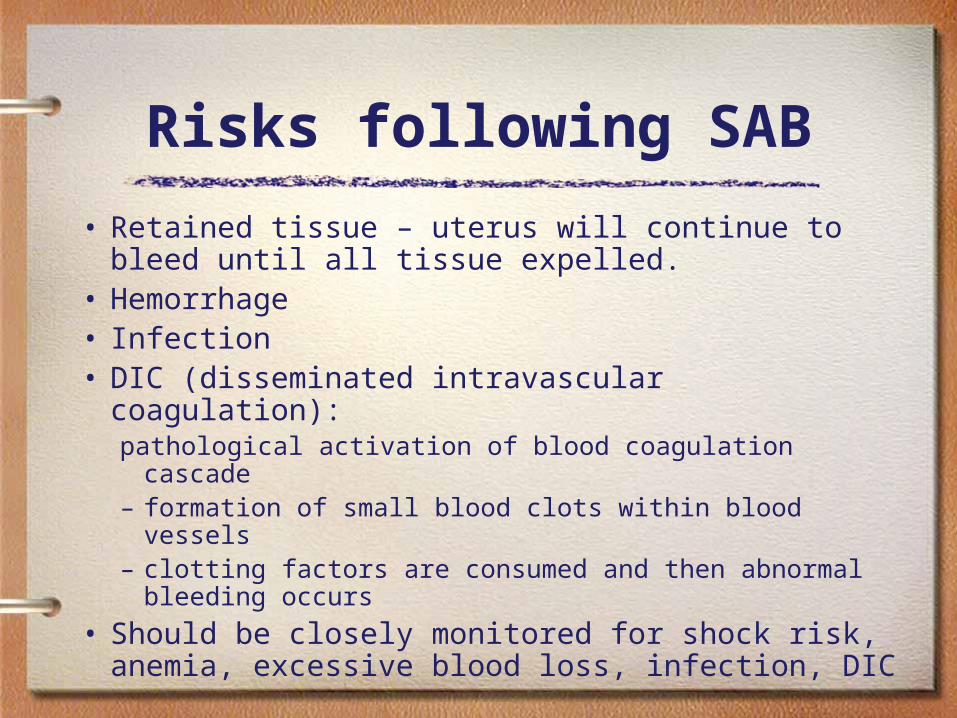
• Retained tissue – uterus will continue to bleed until all tissue expelled.
• Hemorrhage• Infection• DIC (disseminated intravascular coagulation):
pathological activation of blood coagulation cascade– formation of small blood clots within blood vessels– clotting factors are consumed and then abnormal
bleeding occurs
• Should be closely monitored for shock risk, anemia, excessive blood loss, infection, DIC

Increased Risk of SAB
• Environmental factors:– Toxic chemicals– Heavy metal accumulations– Pesticides, preservatives– Radiation– Alcohol and drug use (esp cocaine)– Smoking
• IUD – cuz that’s what it is supposed to do. • Insufficient maternal nutrition• Multiples: 1/35 twin PGs end in SAB for 1 or both
fetuses

Causes of SAB
• Developmental mechanism: abnormal cell division• Blighted ovum: stops growing around 4 weeks and aborts
around weeks 8-12
• Genetic: chromosomal abnormalities; more common in older fetus; mb abN organ development
• Endocrine imbalance: abnormal hormone levels• Infectious disease: chlamydia, toxoplasmosis,
bacterial/ viral • Endometriosis• Poor nutrition (low zinc levels can cause SAB)• Drugs, stress, grief
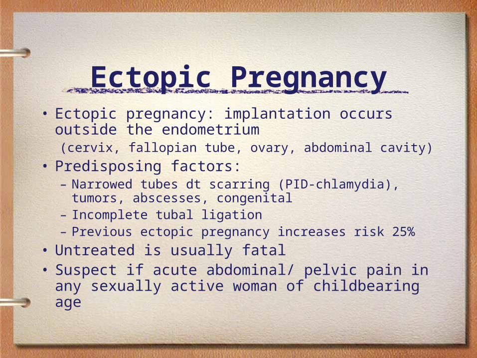
Ectopic Pregnancy• Ectopic pregnancy: implantation occurs outside
the endometrium (cervix, fallopian tube, ovary, abdominal cavity)
• Predisposing factors:– Narrowed tubes dt scarring (PID-chlamydia), tumors,
abscesses, congenital– Incomplete tubal ligation– Previous ectopic pregnancy increases risk 25%
• Untreated is usually fatal• Suspect if acute abdominal/ pelvic pain in any
sexually active woman of childbearing age
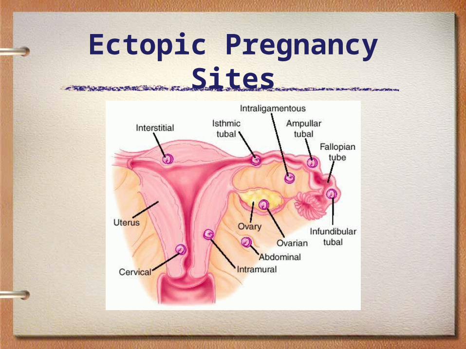
Ectopic Pregnancy Sites

Ectopic Pregnancy
• Complications:– Rupture– Shock– Death
– Rupture of tubal PG most likely 6-12 weeks– Rupture of ovarian PG < 3 months– Rupture of cervical PG usu early, may be >16 wks– Very few abdominal PG survive beyond 36 wks

Ectopic Pregnancy
Ectopic Pregnancy
• Signs/ symptoms of ectopic PG:– Spotting– Cramping pain– Amenorrhea (often has not yet missed period)– Rupture signaled by sudden, excruciating pain– Other s/sx of PG: fatigue, breast tenderness,
changes in appetite, etc.

Ectopic Pregnancy
• Gold standard of diagnosis: laparoscopic visualization
• Critical criteria:– History of spotting, cramping, amenorrhea– Tender pelvis, may palpate unilateral mass– Serum b-hcg

Ectopic Pregnancy• Treatment:• Medical: methotrexate (chemotherapy drug: stops
growth of rapidly dividing cells) IM or POQuantitative, serial b-hcg monitored to ensure termination; may require 2nd dose or surgery
• Surgical removal: laparoscopic surgery often performed at time of diagnosis via laparoscopy
• Visualization of Fallopian tubes (info for future fertility)– Entire tube may be removed if extensive scarring or
blockage

Early Bleeding - Placental Abruption
• Bleeding due to partial placental abruption before 20 weeks
• Sudden gush of blood ¼- ½ cup• Often pain, cramping, abd tenderness
• Monitor with – Ultrasound: blood clot behind placenta,
uterus larger than expected for dates– CBC: anemia– FHT: if good, reassuring

Partial Placental Abruption
• Risks factors– Maternal hypertension– Trauma during pregnancy (MVA, etc)– Cocaine use
• Treatment– Astringent herbs, herbs to stop bleeding– Bed and pelvic rest until bleeding stops;
continue pelvic rest for 1 week after– May go on to be uncomplicated birth

Placental Abruption• May not have vaginal bleeding as sx
• Can lead to maternal shock and fetal distress
• More common later in PG (>20 wks)

Early Bleeding - Molar Pregnancy
Hydatidiform mole or molar pregnancy– A nonviable growth and proliferation that implants in the
uterus but is not a fetus; can become choriocarcinoma and metastasize if not removed
– Common: 1/1000 PG in US; 1/100 in IndonesiaS/SX: – If bleeding, usu wks 16-20– Abnormally large uterus, but no FHTs– Abn high bHCG levels (often very high)– Abn form demonstrated on U/S– May also have hyperemesis, hyperthyroidism, increased
blood pressure, proteinuria

Molar Pregnancy
• Diagnosis– Ultrasound – cluster of grapes, water droplet, other
manifestations that are non fetus looking. – Histological exam of tissue from D&C– Very high levels of bHCG; if still high 2-3 months after
removal, repeat D&C – some cells remain
• Treatment: – D&C– Methotrexate– Hysterectomy

Early Bleeding - other causes
• Other forms of Gestational Trophoblastic dz:– Rare tumors - involve abnormal growth of cells that
would have normally developed into placenta• Invasive mole: hydatidiform mole that invades myometrium
– May require surgery and chemotherapy
• Choriocarcinoma: malignant, requires both surgery and chemo
• Placental site trophoblastic tumor: rare tumor at site where placenta attaches to uterus; treatment is surgical
Gestational Diabetes• GD: diabetes developed during the second half of pregnancy. • Inability to balance blood glucose levels along w insulin resistance, causes hyperglycemia• Should return to normal within 6 days of delivery• Does not increase congenital malformations*• Incidence: varies with populations (1.6%-3% in general OB population)
*unless insulin shots required

Gestational Diabetes
• Pathophysiology– Normal maternal adaptations to pregnancy are
diabetogenic
• Placenta secretes human placental lactogen (hPL) to increase fetal use of maternal blood glucose– keeps maternal blood glucose elevated for longer
• Insulin resistance increases due to effects of hPL, progesterone, cortisol, estrogen
Gestational Diabetes
• Population risks:– Maternal age > 30yo– Previous baby weighing > 9# at birth– Prior fetal or neonatal death– Excessive maternal weight gain or obesity– Hypertension– Proteinurea– Family history of diabetes; obesity

Gestational Diabetes
Screening:• Done between 24-28 weeks; earlier if at risk• Glucose tolerance test: checks blood glucose 2
hours post 50gm glucose challenge
– Diagnosis• Oral glucose tolerance test
– 8-14 hour fast, fasting glucose levels– Drink 100gm glucose, draw 1, 2, and 3 hours post
Gestational Diabetes
• Monitoring– Urine dip stick at every prenatal visit– Random blood glucose levels– Home glucometer monitoring by patient
after GD diagnosis– Serum HbA1c to monitor blood glucose
over past 3 months

Gestational Diabetes
• Treatment– Diet
• No refined sugars/carbs• High fiber (slows release of glucose from gut
into bloodstream)• Low fat

Gestational Diabetes
• Maternal sequelae– Short term
• Preeclampsia
• Infections (pyelonephritis, postpartum endometritis)
• Birth canal injury due to marcrosomia (excessive birth weight)
• Cesarean birth due to increased fetal distress and dystocia, causing increased maternal morbidity
• Cardiac and respiratory symptoms dt polyhydramnios (increased amniotic fluid)

Gestational Diabetes
• Maternal sequelaeLong term– 5-10 % are found to have type 2 diabetes
following pregnancy– 20-50% chance of developing type 2
diabetes within 5-10 years

Gestational Diabetes
• Effects on infant– Increased incidence of:
• Birth injuries due to marcrosomia• Hypoglycemia post-partum• Hypoxemia in utero • hyperbilirubinemia

Gestational Diabetes
• Effects on infant if mother requires insulin:– All of the prior effects (from last slide)– Prenatal and intrauterine mortality– Acidosis (dt maternal acidosis)– Hypocalcemia– Respiratory distress syndrome– Congenital anomalies (esp. skeletal, cardiac, and
CNS)– Possible predisposition to diabetes

Preeclampsia• aka Pregnancy Induced Hypertension or PIH
– Collection of symptoms due to endothelial dysfxn, includes dmg to maternal kidneys, liver
– Proteinuria– Thought to be caused by placental substances– May develop from 20 wks gestation (considered
early onset if before 32 wks)
• Gestational hypertension - no proteinuria• Chronic hypertension
– Elevated blood pressure prior to 20 weeks of pregnancy or present 12 weeks after delivery

Preeclampsia
• Possible sequelae of chronic and gestational hypertension– Preeclampsia– Placental abruption– IUGR (intra-uterine growth restriction)– 2nd trimester stillbirth
Preeclampsia
• Preeclampsia diagnosis:– New onset elevated BP of 140/ 90 – Proteinuria >300mg/ 24hrs (1+ or more in
urine dipstick)– Suspicion if accompanied by edema (but
not diagnostic)

Preeclampsia
• Severe preeclampsia– BP: 160/110 on 2 occasions ate least 6 hrs apart– Proteinuria: 5gm/ 24hr or >3-4+ on urine dipstick
testing of 2 samples at least 4 hours apart– May also see oliguria (<500ml/24hr) or altered
renal function tests, IUGR fetus, non-reassuring FHT, cerebral or visual disturbances, impaired liver function tests, thrombocytopenia, pulmonary edema, hyperreflexia, abd pain

Preeclampsia
• Eclampsia: development of seizures or coma not attributable to other neurological disorders in a patient with PIH
• HELLP syndrome: variant of PIH which is associated with severe liver compromise (Hemolysis, Elevated Liver enzyme levels and a Low Platelet count)

Preeclampsia
• Incidence varies by race, age, parity– Common - as high as 10% of all pregnancies
• Risk factors– Medical conditions that cause microvascular dz
• Diabetes, chronic HTN, vascular/ CT disorders, kidney dz, thrombophilias
– Nutritional factors• Low protein diet• Deficiencies in folic acid vitamins C and E
Preeclampsia
• Risk factors continued– Pregnancy-related risk factors:
• Chromosomal abnormalities, hydatidiform mole, multiple gestation, oocyte donation or donor insemination, structural congenital anomalies, UTI
– Maternal factors• Age less than 20 or greater than 35 years, family history
preeclampsia, previous PG with preeclampsia, first PG, gestational diabetes, stress, African descent
– Paternal factors• First time father, previously fathered a preeclamptic PG
in another woman

Preeclampsia
• Prognosis– Preeclampsia
• 20% prematurity
• 2.5% fetal mortality
• Leading cause of death in PG women (15%)
– Eclampsia• 10-20% fetal mortality
• 2-20% maternal mortality
• Maternal death usually due to cerebral hemorrhage, renal, or cardiac failure

(Preeclampsia - theories)
• Pathophysiology– Exact cause unclear– Theories/ some research indicates:
• Problem with placental implantation/ poor perfusion of trophoblast and localized inflammation
• Exaggerated inflammatory response• Systemic vasospasm decreases blood flow to all organs• Decreased perfusion to organs due to movement of fluid
to extravascular tissues (edema)

(Preeclampsia - theories)
– Liver/ malnutrition aka Metabolic toxemia• Chronic malnutrition causes the liver to be unable to
produce adequate albumin• Low albumin causes fluids to leak into tissues (edema)• Malnutrition mb dt malabsorption• With low calories mom burns protein for fuel instead of
using it to build albumin• Increased metabolic demand in 3rd trimester further taxes
the liver and exacerbates the problem. – K releases renin in response to hypovolemia, causing HTN

Preeclampsia
• Maternal complicationsKidneys– During a normal PG, renal blood flow and glomerular
filtration rate are increased dt increased blood volume
– Preeclamsia: renal ischemia decreases GFR, decreasing creatinine clearance, BUN, uric acid, oliguria, damage to glomeruli (worsens ischemia)
– Can cause acute renal failure from tubular necrosis

Preeclampsia
• Maternal complicationsLiver– Hepatic ischemia causes decreased liver fxn– N/V or general malaise from overload of
hormones and poor digestion– Associated with HELLP syndrome

Preeclampsia
• Maternal complications– CNS
• Cerebral edema may cause unrelenting headaches, blurred vision, cortical blindness, dulled senses, seizures, cerebral hemorrhage, coma
– Opthalmic • Visual changes (mb normal in PG dt expanded
blood volume)• Retinal detachment (may cause blindness)
Preeclampsia
• Maternal complicationsHematologic– Thrombocytopenia (platelets < 100,000 uL)
• Cause unknown• Associated with prolonged bleeding time• Associated with HELLP syndrome
– Disseminated intravascular coagulation (DIC)– Hemoconcentration
• Vasospasm decreases intravascular space, crowds RBCs and associated with hemolysis of RBCs

Preeclampsia
• Fetal complications– Intra-uterine growth restriction (IUGR)
• Decreased utero-placental perfusion leads to decreased nutrition and oxygen to fetus
• IUGR often seen before hypertension
– Placental abruption from vasospasm and ischemia
– Premature labor (hyper-excitable uterus)– Intra-uterine fetal death dt placental insufficiency,
abruptions

Preeclampsia
• Diagnosis– Physical exam:– Mb asymptomatic– Elevated BP– Fundal height measured to try to identify
IUGR– Track development of edema (upper body
edema more concerning)
Preeclampsia
• Diagnosis• Late signs/ symptoms of preeclampsia
– Proteinuria– Oliguria– Unrelenting h/a– Hyperreflexia– Visual changes– NV– Fetal distress

Preeclampsia
• Laboratory workup– CBC: Hgb, hct, platelet coung– 12 or 24 hour urinary protien collection – Serum creatinine and uric acid level– Serum albumin– Coagulation profile
– UA done at every prenatal visit

Preeclampsia
• Fetal monitoring– If mom at risk, baseline u/s at 25-28 weeks
gestation to evaluate fetal growth– If preeclampsia is diagnosed, weekly eval
Preeclampsia
• Management/ treatment– Ultimately the treatment is delivery of the baby and
placenta, which resolves the preeclampsia– Management based on balance of maternal and
fetal risks– Fetal indications for delivery:
• Severe IUGR, nonreassuring FHT, oligohydramnios
– Maternal indications for delivery:• Gestational age >38wks, low platelet count, worsening
hepatic or renal function, placental abruption

PROM
• Premature Rupture of Membranes• Considered PROM if rupture more than1 hour
before onset of labor at any gestational ageCan happen up to 24 hours prior
• Prolonged if >18 hours before onset of labor• If prior to week 37, called preterm premature
rupture of membranes or PPROM– Can lead to significant perinatal morbidity, including
respiratory distress syndrome, neonatal sepsis, umbilical cord prolapse, placental abruption, and fetal death

PROM• Maternal associated risks
– Chorioamnionitis – inflammation – Sepsis can result
• Fetal risks– Prematurity (1/3 of premature births due to PROM)– Cord prolapse– Malpresentation b/c can’t turn w/o amnio fluids– Infection – Death (1-2% risk assoc)

PROM
• Incidence: – Affects 8% of all term pregnancies– 75% of these will go into labor
spontaneously within 24 hours
– Treatment depends on whether it occurs prior or after week 34

PROM
• Causes: – Chorioamnionitis (inflammation of membranes): due to
infection - beta-strep, chlamydia, BV, gonorrhea – Abnormally weak membranes due to infection, placental
bleeding– Poor nutrition or poor health in general– Overdistended uterus (e.g. twins)– Polyhydramnios: overdistended amniotic sac creates
increased pressure– Cervical incompetence: pressure– Malpresentation: breech or transverse

PROM
Beta-strep• Severe risk for amnionitis (membranes
themselves), uterine infection, septicemia– Suspect if maternal temperature increases (fever)
after rupture– FHT: increased, may be tachycardic when infected– Uterine irritability (non-rhythmic contractions)– Strong unpleasant odor to amniotic fluid if
longstanding infection
PROM
Diagnosis:• Patient history: sudden gush of fluid, possible
constant leak or sensation of not being able to stop urinating
• Physical exam: pooling of fluid on speculum; Dr. should avoid digital exam as it may increase morbitidy/mortality
• Labwork: pH of fluid, ferning of discharge, cervical culture/ gram stain (want to check for beta-strep)
PROM
Management • Antibiotics to prevent infection, esp. if mom beta-
strep positive• Corticosteroids reduce perinatal morbidity and
mortality: reduces respiratory distress, intraventricular hemorrhage, necrotizing enterocolitis; appropriate prior to week 32; controversial between weeks 32 and 34; no beneficial effects after week 34– No more than one round of corticosteroids or risk
increases for decreased birthweight, length, and head circumference

PROM
• Management If prior to week 24:– Antibiotics/ corticosteroids– Likely spontaneous labor within 1 week– Transfer to a specialized facility (home care
controversial) – sterile area, complete bed rest. – If child survives, likely to have health problems
such as: chronic lung disease, developmental and neurologic abnormalities, hydrocephalus, and cerebral palsy
• Consultation with neonatologist may be helpful
PROM
• Management If during weeks 24-31
• Antibiotics/ corticosteroids• Fetal monitoring – fetus is viable at this point• Deliver sooner if signs of infection or nonreassuring fetal
testing
If during weeks 32-33• Antibiotics/ corticosteroids• Consider amniocentesis; deliver if lung maturity• Prolonging pregnancy after documentation of pulmonary
maturity unnecessarily increases the likelihood of maternal amnionitis, umbilical cord compression, prolonged hospitalization, and neonatal infection

PROM
– If during weeks 34-36• Administer antibiotics, deliver• Not corticosteroids as not trying to prolong
pregnancy• No benefit to conservative management; no
decrease in neotatal morbidity• Not delivering at this date increases risk for
infections

PROM
• Management– If no spontaneous labor, may have to induce labor
– pitocin induction– 24 time limit from ROM to active labor for home
birth– Monitor temperature of mother– Fetal movement counts (kick count test)– No baths, swimming, hot tubs
- birth tubs are ok. – No inserting into vagina at all.

PPROM
• Complications of preterm PROM
Complications Incidence (%)
Delivery within 1 week 50-75
Respiratory distress syndrome 35
Cord compression 32-76
Chorioamnionitis 13-60
Abruptio placentae 4-12
Antepartum fetal death 1-2

Placental Abruption
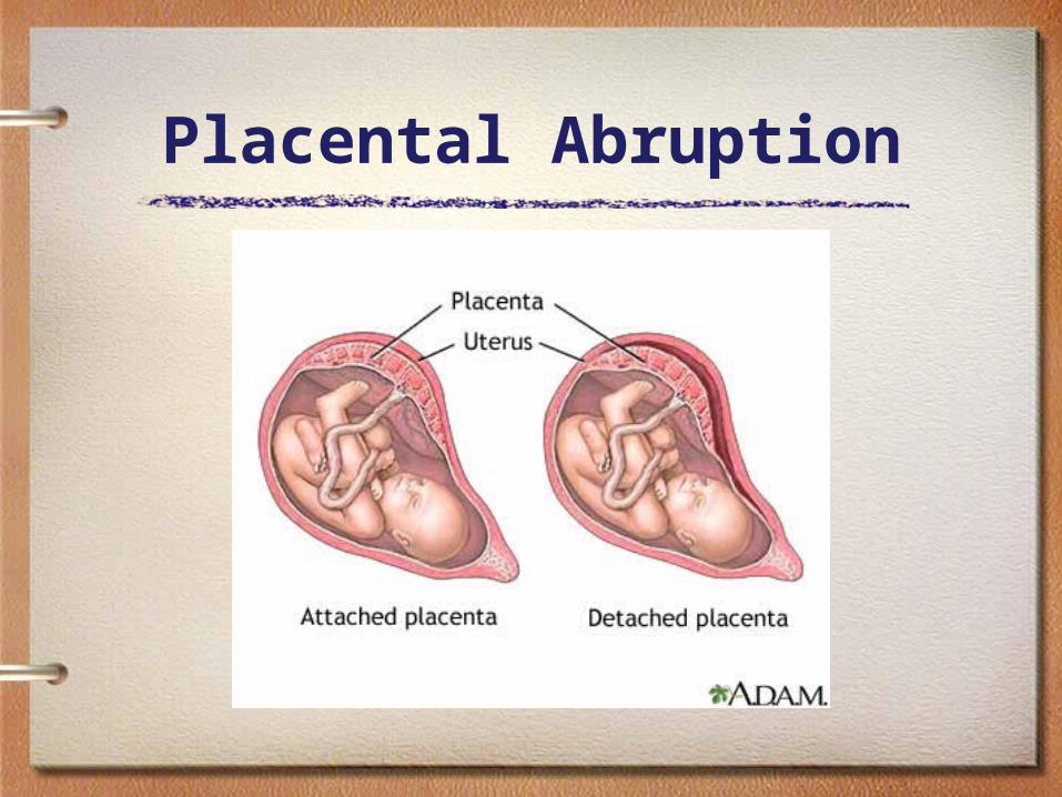
• Separation of placenta from implantation site in the uterus before the delivery of the fetus – May be only a few millimeters or may involve
entire placenta
• Bleeding can be external (exits via vagina) in 80% of cases, or can be concealed – Blood to amniotic fluid– Blood blocked by baby’s head or membranes
Placental Abruption

Placental Abruption
• Incidence– 1% of all pregnancies worldwide
Less in the US.
– Fetal mortality rate of 20-40% depending on degree of separation

Placental Abruption
• Effects on mom– Hemorrhage (may require transfusion)– Need for medications during delivery to
help uterus contract– Blood clotting problems/ DIC– Shock and subsequent problems of liver,
kidney, pituitary– May have extreme pain

Placental Abruption
• Effects on baby:– Fetal distress– Stillbirth– Prematurity– Low oxygen level after birth– Low blood count after birth– Brain damage
Placental Abruption
• Etiology– Trauma (MVA, assault)– Coagulopathies– Hypertension (a factor in 44% of
abruptions)
Placental Abruption
• Risk factors: – Short umbilical cord– Prolonged rupture of membranes (>24 hours)– Retroplacental fibromyoma– Maternal age: younger than 20 or older than 35– Previous abruption– Some infections are also diagnosed as a cause

Placenta Previa
• An obstetric complication where placenta is attached to the uterine wall close to or covering the cervix
• Affects 0.5% of all pregnancies • Often presents as painless, bright red vaginal
bleeding• No known etiology
– Perhaps abnormal vascularity due to scarring from previous instrumentation

Placenta Previa
• Risk factors– Previous placenta previa, caesarean delivery, or
abortion – scarring reduces where it can attach.– Women who have had previous pregnancies,
especially a large number of closely spaced pregnancies, are at higher risk
– Women who are younger than 20 are at higher risk and women older than 30 are at increasing risk as they get older
– Large placenta, twins– Women who smoke or use cocaine

Placenta Previa• Management
– No intervention necessary if mom and fetus not in distress
• Maybe bed rest
– Delivery if fetus mature and if in distress– Controversial if vaginal delivery or C-
section safer• C-section if distress or previa is in the way of
vaginal delivery• Vaginal delivery if DIC or if surgery unavailable

Infections
• Pelvic inflammatory disease – Infection of the uterus, fallopian tubes, and
adjacent pelvic structures that is not directly associated with pregnancy
– Route of transmission is from vagina and/or endocervix to endometrium, fallopian tubes, or pelvic structures
– Organisms: Chlamydia, gonorrhea, Gardnerella vaginalis, H. influenzae, several others

Infections
• PID symptoms– Lower abdominal pain– Adnexal tenderness– Chandelier’s sign– Purulent vaginal discharge– Possible palpable mass
Infections
• PID complications– Hydrosalpinx
• Accumulation of oviductal fluid in the lumen due to tubal occlusion
• Causes low conception rate• 50% chance first trimester SAB• Increased rate of ectopic pregnancy• Chronic pelvic pain

Infections
• PID complications • Tubo-ovarian abscess (TOA)
– Most serious complication of PID; occurs 1-4%– If ruptures, 8.6% mortality rate– Usually multi-organism infection– S/Sx: history PID, pelvic/ abdominal pain, fever,
leukocytosis, elevated CRP and ESR (inflammation), positive chandelier’s sign, pelvic tenderness, possible palpable mass
• If ruptures, peritonitis and shock

Infections• PID Complications
– Poor pregnancy outcome in general– Infant pneumonia– Neonatal death (20%)– Infertility (20%)– Chronic pelvic pain (9%)– Ectopic pregnancy– Increased risk for reproductive tract cancer– Vulnerability to HIV

Infections
• Predisposing factors for PID
Trichomonas infection Douching (dose related)
STI exposure Lower socioeconomic group
Failure to use contraception Multiple sexual partners
PID history IUD use
Cigarette smoking Sexual activity at young age
Invasive genital medical procedures
adolescence

Infections
• Diagnosis– Clinical symptoms and culture confirmed by rapid
improvement with antibiotic therapy• Clinical diagnosis is correct 65-90%
– Elevated ESR, CRP– Endometritis with endometrial biopsy– US or laparoscopy showing fluid-filled thickened
tubes, TOA• Laparoscopy gold standard
Infections
• Differential diagnosis for PID – rule these out• Ectopic PG• Endometriosis• Ovarian cysts• Cancer• Myoma• Appendicitis• Pancreatitis• Septic abortion• Acute cholecystitis• Mesenteric lymphadenitis

Infections
• PID during PG– High risk for maternal and fetal morbidity – High risk for preterm delivery, esp with
scarring– Treatment
• Medical: antibiotic therapy• Surgical: laparoscopic pelvic irrigation, lysis of
adhesions, drainage and irrigation of pyosalpinx, drainage and irrigation of TOA

Infections
• Sexually transmitted infections– Chancroid (painful ulcer H. ducreyi)
• No known additional hazard during PG
– Chlamydia • Incidence: 2-23% of PG women• 50% infant contract it at delivery (40% conjunctivitis)• Risks: premature labor, miscarriage, neonatal death• Ciprofolxacin and Tetracycline contraindicated in PG

Infections
• HIV/AIDS– Risks to fetus and birth attendants
• Use universal precautions with all patients
– Risk groups • IV drug users and their partners• Partners of gay/bisexual men• History of blood transfusions (to 1977), hemophiliacs• Partner of HIV positive person• Multiple sex partners• Sex industry workers• 42-50% of HIV+ cases not in an at-risk group!
Infections
• HIV risk to infant• 22-70% chance of infection with HIV+ mother• Symptomatic women are 9x more likely to transmit
disease to fetus than asymptomatic mothers
– Unclear if transmitted with vaginal delivery or breastfeeding
– Some babies seroconvert negative after stopping breastfeeding
Risk to mother: increased risk SAB

Infections
• HIV management during PG– Up to 22 weeks, may consider TAB– Considered high-risk PG, refer to
appropriate facility
Infections
• Syphillis– 30% fetus die in utero– Congenital syphillis
• May appear normal at birth or have rhinitis, hoarseness, rash (esp. palms and soles), hepatosplenomegaly, weight loss, bone and teeth changes, CNS lesions, very contagious
• State laws usually require testing of all PG women within 10 d of first prenatal visit
• Tx: penicillin or erythromycin

Infections
• Gonorrhea– Possibly asymptomatic or have vaginal or
rectal discharge, salpingitis in 1st trimester• 45% will also have chlamydia
– Newborns at risk for gonorrhea eye infection which is severe enough to cause blindness
• Erythromycin ointment in both eyes at birth• Silver nitrate no longer used (blindness risk)

Infections
Condyloma accuminata (HPV) • May require C-section if heavy vaginal infection• Risk to fetus: laryngeal papillomas, genital HPV
• Tuberculosis– Pregnancy does not alter course of TB– Congenital TB is rare, only from fetal aspiration of
infected fluid or hematogenous spread– At risk: IV drug users, alcoholics, correctional
institutions, nursing homes, Asia, Africa, Caribbean, Latin America

Infections
• Trichamonas vaginalis– Vaginitis symptoms– No risk to fetus
• Herpes simplex virus– Primary infection at delivery: 50% infants are
infected– Secondary infection at delivery: 2-5% infected
infants– Risk to mom: < 20wks 25% SAB– Risk to fetus: IUGR, prematurity, stillbirth, neonatal
herpes

Infections
• HSV– Neonatal disease symptoms develop after 3-15
days – Skin/ mucosal vesicles– Lethargy– Jaundice– Fever– Cyanosis– Can develop pneumonitis, encephalitis, hepatitis
Infections
• Neonatal HSV mortality – 2-3% if lesions confined to skin – 45% of babies have no skin lesions but have brain
infection– With desseminated infection, 15-50% die with
therapy; 85% die without therapy– 60% survivors will have CNS damage, blindness,
mental retardation– Only 5% of babies with CNS infection return to
normal– 80% of cases due to HSV2
Infections
• HSV transmission to neonate– Birth canal– Transplacental with or without primary
outbreak– Nosocomial: from one baby to another at
the hospital or another family member

Infections
• HSV management– C-section if prodrome or active lesion at
time of membrane rupture– Weekly prenatal cultures of no value– Prevention: low arginine foods + 1000mg
lysine

Infections
• Bacterial vaginosis– Risks
• Premature labor• PROM (bacteria weaken the amniotic sac)• Post partum endometritis• Salpingitis

Infections
• Candida– Common in PG, 25% at term due to
increased estrogen– If close to term, infant at risk for thrush if
vaginal birth– Best if mom has low sugar diet when
breast feeding. – Probiotics for infant.

Infections
• Beta strep– Neonate infected from contact with cervix
during labor– Neonatal infection can be very serious
• 5x more often in low birth weight babies• Infant can die within 8 hours• Infected infants have decreased respirations and
temperature
• Also: premature labor, PROM, uterine infection

Infections - bStrep
• Incidence – Up to 30% PG women may be colonized
• 5% constitutional susceptibility, no antibodies
– 25-75% of infants may get infected if mom colonized
• Only 1-8% of these will develop symptoms
– Premature infants, PROM and prolonged labor put the infant at greater risk (longer exposure)
– Standard cervical culture at 36 weeks, also if PROM or in labor if history of beta-strep infection

Infections - UTIs
• Urinary tract infection– During PG, increase chances of asymptomatic
bacteria progressing to infect urinary tract – Possible sequelae:
• Premature labor and preterm birth• PROM, IUGR• Fetal death – bacteria would have to travel. Uncommon. • Low APGAR scores• Mental retardation• Sepsis• Cerebral palsy
Infections - UTIs
• Predisposition during PG dt– Urinary stasis: dilation of renal pelvis and
ureters (progesterone)• Compression of ureters by uterus• Smooth muscle relaxation contributing to
urinary reflux
– Increased urine production and overfilling of bladder

Infections
• UTI predisposing factors
Kidney disease Previous UTI
Poor hygiene Frequent intercourse
Multigravida Hypertension
High sugar diet Vulvovaginitis
Infrequent or incomplete emptying of bladder
Systemic conditions: diabetes, sickle cell dz
Mechanical obstruction Urethral diverticula

Infections
• Acute pyelonephritis– One of most common medical conditions of PG– Most common during 2nd trimester– Sx: abrupt onset f/c, lumbar pain, n/v, malaise,
h/a, dehydration, tachycardia– Fetal sx: preterm labor, fetal tachycardia– Tx: hospitalization for IV antibiotics– Ddx: appendicitis, placental abruption, infarction of
myoma, labor
Infections
• Common cold– Some women have increased, others
decreased resistance to the common cold viruses during PG
– Risk is of development of pneumonia– Treament is REST, fluids– Echinacea ok during PG,
goldenseal/berberis/mahonia/agarita only in small amounts (stimulate contractions)

Infections
• Influenza– Women in 3rd trimester are at risk for
serious disease such as pneumonia due to pressure of baby on the lungs
– Monitor mom’s temp; 102F or higher risks fetal well-being
– Tx: rest, fluids, herbs, hydrotherapy, etc.

Infections
• Hepatitis B– Testing of all PG women recommended by CDC due
to risk of transmission during labor to both neonate and health care personnel
– Infant transmission is 10-70% depending on stage of maternal disease
– Of infected babies, 85-90% will become chronic carriers; 25% will die of hepatocellular CA or liver cirrhosis
– For exposed neonates: immunoglobulin IM within 12 hrs of birth and Hep B vaccine series at 7d, 6 mo, and 1 year
Infections
• Toxoplasmosis– 30% women are immune (more likely if been
handling cat litter boxes)• Source
– Cats who have eaten infected birds or rodents or have contacted infected cat feces
– Humans can contract it from dirty sandboxes or playgrounds where cats have left feces
– Raw meat or inadequately cooked pork, mutton
– Water contaminated by cat feces
– Milk from infected animals
– Organ transplants or transfusions
Infections
• Toxoplasmosis symptoms– Usually subclinical– Rash, lymphadenopathy, fever, malaise, generally
mildly sick– Risk: crosses placenta and affects fetus 50%– First trimester: SAB or severe CNS, liver– Third trimester, 60% of fetus affected but with less
severe disease than if infected during 1st trimester– IUGR, hepatosplenomegaly, jaundice, anemia,
retinopathy, hydrocephaly, convulsions, microcephaly, polyhydramnios

Infections
• Toxoplasmosis treatment– Toxic antimicrobials that cannot be used in
PG– Prevention is key!
• Avoid cat feces• Cook meat well• Wash hands after handling raw meat and cats• Avoid raw milk and eggs

Infections
• Rubella– Symptoms: fine rash, posterior cervical or occipital
lymph node enlargement, malaise, mild fever • Diagnosis: antibody titer
– Risk to fetus: crosses placenta (all viruses do; bacteria generally don’t)
• Malformations: deafness, cardiac defects, cataracts• Congenital rubella syndrome: low birth weight, marrow
damage, hepatitis, myocarditis, pneumonitis, encephalitis, chromosomal abnormalities, babies shed virus for months-- contagious

Infections
• Rubella infection:– At 1-8 weeks, 40-80% risk fetal defects– At 9-12 weeks, 20% risk– At 13-16 weeks, 5% risk– At 17-20 weeks, 1% risk
– Management: consider TAB in 1st and 2nd trimesters
– Immunize non-immune women after delivery

Infections• CMV (cytomegalovirus)
– Transmission: placenta, cervix, breast milk– Usu asx: diagnosis: titer or cervical culture
• Risk of infection: 12% of women secrete virus in urine, cervix, breast milk and affect offspring, birth attendants
– Risk to infant:• Subclinical and mild maternal infection: jaundice, petechieae,
feeding difficulties, irritability, muscle weakness, hepatosplenomegaly
• Severe maternal infection: SGA, microcephaly, meningioencephalitis, chorioretinitis, mental retardation
– CMV treatment• Enhance immune function• Stimulate digestive system
Infections
• Varicella (Chickenpox)• Risk to infant:
– 1st trimester: congenital varicella syndrome (developmental probs - CNS, bladder, digits, skin)
– 2nd and 3rd trimester: few problems
• Risk to mother:– Pneumonia, premature labor, dehydration, may be
fatal (esp. if pneumonia in 3rd trimester)
Infections
• Varicella – If infection at time of delivery, high risk of varicella
transmission to baby– If infxn within 5d of delivery, 30% transmission risk– If over 5d of delivery, 18% transmission risk– If baby gets rash within 5-10 days of being born,
20% will not survive– After 10 days, fetus receives passive immunity
from mother

Infections
• Mumps
• Risk to infant:– Increased SAB– Prematurity– Stillbirth– Endocardiofibroelastosis (heart problem)
– Risk to mother is variable

Infections
• Rubeola (measles)– Risk to infant:– SAB– SGA– Rare malformations– Most have no problems
– Risk to mother: none

Infections
• DO NOT TREAT ANYTHING BEFORE 10 WEEKS GESTATION UNLESS YOU ABSOLUTELY HAVE TO!