Embed Size (px)
Citation preview

8/25/2014
1
GI Malignancies
Trisha Marsolini RN, BS, OCN, CMSRN Septmeber 2014
Objectives
Review pathophysiology of GI systems
Discuss risk factors
Discuss presenting signs and symptoms
Discuss and Identify current treatment options
Review side effects and nursing management

8/25/2014
2
GI Malignancies Worldwide
Type Diagnosed Cases
Deaths US cases
Esophageal
482,300 406, 800 17, 460
Stomach
989,600 738,000 21,320
Pancreatic # fall under other
800,230 43, 920
Colorectal
1,167,020 602,958 143,460
Combined: 25% of all diagnosis and 32% of deaths
worldwide
•Numbers are based data collected by ACS in 2008
http://blogs.abcnews.com
Esophageal
Cancer
Anatomy and Function
• Located behind the trachea, at the hypopharynx • Starts at thoracic inlet, ending at GE junction
muscular tube 10-13 inches long • Facilitates swallowing and moves food and fluids • Movement controlled by sphincters and peristalsis
Esophageal Anatomy
• Mucosa – two components
• Epithelial layer – squamous cells
• Lamina layer – connective tissue
• Sub mucosa
• Glandular layer
• Muscularis Propria
• Muscle layer
• Adventia
• Connective tissue layer
• Lymph Nodes
•Types of Esophageal Cancer
• Squamous cell carcinoma (SCC)
Originate from mucosal layer
Usually occurs from mid to upper esophagus
90% used to be SCC now < 30%
More common in black men
Types of Esophageal Cancer
• Adenocarcinoma
• Does anyone remember where this type originates from?
• Originate from glandular cells of sub mucosa
• Squamous cells are replaced by
glandular cells
• Usually occurs near the stomach
• Increasing about 1% a year in white men

8/25/2014
3
Incidence of Esophageal Cancer
• 2% of U.S. cancer deaths
• 17, 460 estimated new cases - 2012 US
• 15,070 estimated deaths – 2012 US
• 8th most common cancer Worldwide
• 7th most common cancer death Worldwide
• 3 to 4 higher in men than women
startoncology.net
• Stats from 2004
Highest incidence :
•South Africa
•China
•Malawi
•Mongolia
•These areas have 20
to 30 times higher
than US
Worldwide Incidence 2008
Risk Factors for Esophageal Cancer
Age - most cases occur > 65
Male – 3 to 4x higher than women
Tobacco
44X risk more significant in SCC
Alcohol – heavy
Synergic relationship with smoking
Obesity/ High BMI
More significant in adenocarcinoma
Risk Factors for Esophageal Cancer
HPV – predisposes to adenocarcinoma
Diet
Diet low in fruits and vegetables Drinking hot fluids frequently Diet high in processes meats (high salt), pickled vegetables
Decline of h. pylori in esophagus
In squamous cell
Presenting Signs & Symptoms • Asymptomatic in early stage • Late Symptoms
• circumference of esophagus less than 13mm – Difficulty/painful swallowing – Weight loss – Chest or Epigastric Pain – Hoarse Voice – Hiccups – Hematemesis – Melena
• No screening tests available in US
Risk Factors for Esophageal Cancer
Barrett’s Esophagus
• 10% of people with GERD have Barrett’s
– GERD asst with high BMI
• Squamous cells replaced with glandular cells
• 30-125 times increased risk of adenocarcinoma
• Cancer risk higher when there is dysplasia

8/25/2014
4
Case Study • Jim a 67 yr old
– Hx: HTN, Hyperlipidemia, DM, GERD, obese and Barrett’s
• Progessive dyspaghia for last 4 to 6 weeks
– Previous 2 weeks only eating soft food
– 50 pd weight loss last 6 mo.
• Previous EGD 2 yrs ago – WNL
• 3/26 Had EGD
– Obstructing mass distal esophagus
– Biopsy
• High grade poorly differentiated adenocarcinoma
Diagnostic Work-up
Initial diagnostic exams
• Baruim Swallow
• Endoscopy and Biopsy
Staging exams
• CT chest and abdomen
• Bronchoscopy
• PET scan
• MRI
• Bone scan
• Laparoscopy
Diagnostic Work-up
Endoscopic Ultrasound
Identifies unseen
tumors and depth of
tumors
Can do biopsies
Even of lymph nodes
http://www.barrettsadvice.com
Case Study
• 4/4 CT/PET
– Obstructing mass distal esophagus and junction
– Enlarged perigastric node
– Bulky upper R mediastinum/paratracheal node
– No distant mets
• Family Hx
– Dad passed from esophageal ca
– Brother passed from a brain tumor
Metastatic Patterns
Local Spread through esophageal layers
Lymph Nodes
Surrounding organs
Distant Metastases
Liver
Lung & Pleura
Stomach
Peritoneum
NCCN Esophageal Staging
• TNM used
– Plus add Grade
• Makes difference in early stages
• Squamous Vs Adenocarcinoma
– Squamous staging
adds location
8/25/2014
5
TNM Staging - Squamous TNM staging - Adenocarcinoma
Staging TX Primary tumor cannot be assessed.
T0 No evidence of primary tumor.
Tis High-grade dysplasia.c
T1 Tumor invades lamina propria, muscularis mucosae, or submucosa.
T1a Tumor invades lamina propria or muscularis mucosae.
T1b Tumor invades submucosa.
T2 Tumor invades muscularis propria.
T3 Tumor invades adventitia.
T4 Tumor invades adjacent structures.
T4a Resectable tumor invading pleura, pericardium, or diaphragm.
T4b Unresectable tumor invading other adjacent structures, such as aorta, vertebral body, trachea, etc.
Nodes
Nx = unable to
assess LN
N0 = no LN
N1= 1-2 LN
N2 = 3-6 LN
N3 = >7 LN
Esophageal tumor
5 Year Survival Rates
• Localized disease survival rate is 37%
• Regional staged is 19%
• Distant metastasis 3%
Survival rates doubled last 40 years but remain poor
NCCN Treatment Guidelines • Surgery alone
– Early Stage - rare
• Chemotherapy
– Pre and postoperative
• Chemoradiation – Primary treatment
– Used alone in locally advanced SCC
– Combined with surgery in adenocarcinoma
• Radiation –
– Alone if tumor is locally advanced
– Palliative for symptom management
8/25/2014
6
Case Study • Jim is not a surgical candidate
– Why?
• 4/9 Port placed and chemo started
– Carboplatinum, taxotere and 5-FU
• 4/14 admitted to onc unit
– Dehydration, diarrhea and stomatitis/esophagitis
– WBC 8.8
– HCT 34.6
– Crt 1.7
• Stopped 5-FU 8 hrs early
– Time frame in general early for SE
NCCN Treatment Guidelines Combination Chemotherapies most common
Xeloda or 5-FU paired with Cisplatin Irinotecan Oxaliplatin Carboplatin Pacilataxel Docetaxel
Locally advanced or metastatic setting Trastuzamab and Lapitinib
2 drug regimens most common with Radiation 3 drug regimens most common when Radiation is not
indicated
Ongoing trials to find the best chemo and radiation combo Trials ongoing with radiosensitizers
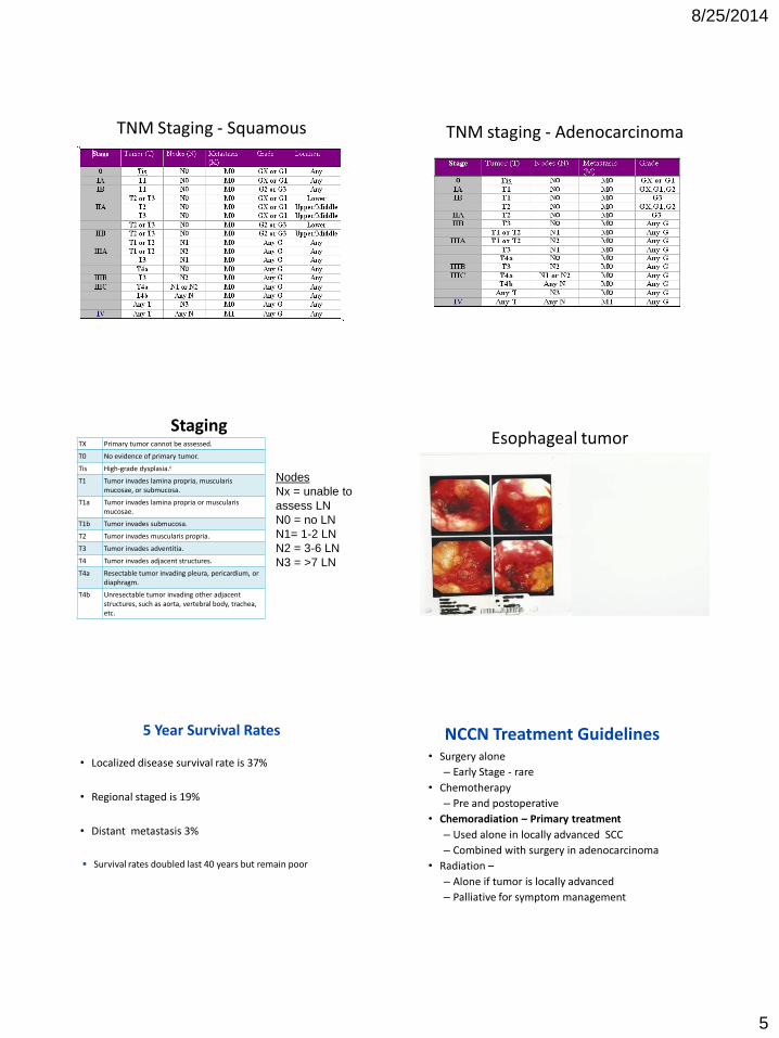
NCCN Treatment Guidelines Surgery
Esophago-gastrectomy
Less invasive approaches Thoracoscopy with limited laparotomy or laparoscopy & possible cervical incision
More invasive approaches
Thoracotomy with laparotomy & possible cervical incision
NCCN Treatment Guidelines Tumor can be unresectable – due to location
Close to cricopharyngeaus – cervical area
Extensive EGJ involved
Bulky tumor that involves surrounding organs
Bulky lymphadenopthy
Distant mets
Nursing Management
Post Surgical care
Pneumonia Obstruction or paralytic ileus Bleeding Anastomosis Leakage Blood clots Infection Pain management Recurrent laryngeal nerve paralysis (cervical
sites)
Nursing Management
Later phase surgical issues
Slow emptying and chronic nausea/vomiting Strictures Heartburn
Patient are going to have to learn to eat different
Nutrition Consult

8/25/2014
7
Case Study • Jim is not a surgical candidate
– Why?
• 4/9 Port placed and chemo started
– Carboplatinum, taxotere and 5-FU
• 4/14 admitted to onc unit
– Dehydration, diarrhea and stomatitis/esophagitis
– WBC 8.8
– HCT 34.6
– Crt 1.7
• Stopped 5-FU 8 hrs early
– Time frame in general early for SE
Case Study
• 4/17 Labs
– WBC 2.2
– HCT 25.1
• Treatment
– 2 units PRBCs
– GCSF
• Next treatment dosed reduced
Palliative Therapy
• Esophageal Dilatation
• Esophageal Stents
• PEG or PEJ placement
• Photodynamic Therapy
• Laser Endoscopy
Treatment
Photodynamic Therapy Used on superficial and mucosal lesions Procedure Given photosensitizing agent
Wait a few days Via endoscopy
Special laser is pointed at cancer cells causing cell death
Little harm to normal cells Patients need to stay inside for 4- 6 weeks
Case Study
• 5/22 CT showed
– Mild improvement in obstructing mass
• Dysphagia improved – Reg diet
– Creatinine climbing 2.7
• No more GCSF
• Stopped chemo except 5-FU
• 6/1 palliative radiation started
– Total 25 treatments
Case Study • 6/9 admitted
– increased confusion, dehydration, weakness
– Mucositis/dysphagia
– Currently on 5-FU CADD pump
– Labs
• Crt 2.06
• K 3.1
• BS 388
– Finished 5-FU took a few days off Radiation TX
• 7/8 last dose of 5-FU
• 7/26 finished radiation

8/25/2014
8
Case Study
• 8/4 CT – T12 compression fx
– New bulky distal esophageal adenopathy
• 8/9 started rad tx to new area
• 8/20 admitted LBP compression L-5 and constipation – IV narcotics for pain control
– Kyphoplasty – pain not improved after
– Relistor relieved constipation
Summary – Esophageal cancer
• Overall 5 yrs survival is 11 to 18%
• Males much higher risk than women
• Tobacco significant risk in SCC
• GERD significant risk in Adenocarcinoma
• Healthy weight and active lifestyle best defense
• Adenocarcinoma increasing in white men
• SCC decreasing in US but still significant problem worldwide
Colorectal Cancer
Colon Anatomy •Primary Function is water and mineral absorption and stool formation.
•70% of tumor occurrences are in the colon and 30% are in the rectum.
•Colon is approx. 5 to 6 ft long
•Rectum is 10 to 12 inches long
47
Normal to Adenoma to Carcinoma Human colon carcinogenesis
progresses by the dysplasia/adenoma to carcinoma pathway
•How long do you think this pathway takes?
Genetic Model of Colon Cancer
48
p53
Late Adenoma
Optimum phase for early detection
Many decades
APC
K-ras Mutation
Courtesy of Barry M. Berger. MD, FCAP EXACT Sciences
Late Cancer
Early Cancer
Adenoma Norma l Epithelium
Dwell Time: 2-5 years 2-5 years

8/25/2014
9
1 in 19 men and 1 in 20 women lifetime risk
143, 460 new cases - 2012 US
51,690 death – 2012 US
3rd leading cause of cancer death men and women
World-wide incidence similar in developed countries
African Americans have 20% higher incidence, 45% higher mortality
Colon cancer has decreased 15 years due to screening
1 million survivors of colorectal cancer in the US
Incidence of Colorectal Cancer
49
World-wide Incidence 2008
Risk Factors Age over 50
Obesity - especially abdominal obesity
High fat and red meat
Type II Diabetes
Smoking
Inflammatory Bowel Disease: Crohns, Ulcerative colitis
Previous cancerous polyps
Family history – 20% of all CRC
51
Risks Factors
• Familial Adenomatous Polyposis (FAP)
– 1% of CRC
– Adenomatous polyposis coli (APC) gene mutation
– Hundreds of polyps by age 39
– Not necessarily just located in colon and rectum
– 100% lifetime risk of CRC
– Autosomal dominant
Risks Factors
Hereditary Nonpolyposis Colorectal Cancer (HNPCC)
• 3-5% of CRC
• Also known as Lynch Syndrome
• Mismatch repair gene mutation
• Average age of onset is 44
• 70-80% lifetime risk of CRC
• Women have very high risk of endometrial cancer
• Increased risk of other cancers
Presenting Symptoms - Generalized
Persistent abdominal discomfort
Changes in bowel habits (over several weeks)
Rectal bleeding/Blood in the stool
A feeling that your bowel doesn't empty completely
Unexplained weight loss
Fatigue
Anemia
54

8/25/2014
10
Presenting Symptoms – Location
Ascending Large bulky tumor
Palpable mass uncommon
Dull pain
Tarry dark stool Late Symptoms:
Transverse Palpable mass
Occult blood in stool
Obstruction
Descending
Maroon colored in stool
Incomplete stool evacuation
Obstruction
Tenesmus
Sigmoid Constipation
Pencil-like stool
Tenesmus
Presenting Symptoms - Rectal
Mucous discharge/diarrhea
Bright red rectal bleeding (most common)
Tenesmus – spasmodic contraction
Sense of incomplete evacuation
Late Symptoms:
Feeling of rectal fullness
Constant ache
56
Screening
57
American Cancer Society-Screening Guidelines
Screening should begin at the age of 45 to 50 If there is no previous history of polyps or cancer
Tests that find polyps and cancer Flexible sigmoidoscopy every 5 years*, or Colonoscopy every 10 years, or Double-contrast barium enema every 5 years*, or CT colonography (virtual colonoscopy) every 5 years*
Tests that primarily find cancer Yearly fecal occult blood test (gFOBT)**, or Yearly fecal immunochemical test (FIT) every year**, or Stool DNA test (sDNA), interval uncertain** * If the test is positive, a colonoscopy should be done. ** The multiple stool take-home test should be used. One test done by the doctor in the office
is not adequate for testing. A colonoscopy should be done if the test is positive.
Colon Polyps Colon Cancers

8/25/2014
11
Diagnostic & Lab Procedures
• Barium Enema
• CAT Scan/PET Scan
• Colonoscopy, Sigmoidoscopy, Protoscopy
• MRI
• Hematocrit
• Liver Function Tests
• Carcinoembryonic Antigen (CEA) – Monitor for response to therapy and recurrence – not presence
of cancer
Case Study Jane 33 weeks of pregnancy
CT showed large obstructing mass in rectum and liver mets.
Flex sig showed innumerable polyps.
She was to have a C-Section at 36 weeks
contractions at 35 weeks
C-section = healthy boy
Pathology from flex sig
Familial Adenomatous Polyposis (FAP)
She has 10 other children at home.
What are their risk of getting FAP?
50/50
Case Study
CEA (Reference Range: 0-3 NG/ML)
Jan. 23 Result: 2126.94
Feb. 25 Result: 4176.6
• After delivery sent home
• Return to hospital 1 week later in terrible pain
• After about a week had to have a debulking surgery – Diverting ileostoy
– TPN
– PCA, frequent large boluses and pain consult
• DC home about 2 weeks later
Metastatic Patterns Local extension through penetration of layers of bowel
Invasion of submucosal layer: direct access to vascular and
lymphatic system
Distant metastasis most frequent in liver, then the lungs Less frequent in brain, bone and adrenal glands
64
Staging for Colon Cancer TNM – Staging
System
Tumor Nodes Metastasis 5 yr Survival
Stage 0 - Polyp Tis N0 M0
Stage I Tumor involves the inner lining of the intestine.
T1
T2
N0
N0
M0
M0 93%
Stage IIA Tumor invades muscle wall of the intestine but no LN.
T3 N0 M0 85%
Stage IIB T4 N0 M0 72%
Stage IIIA Lymph nodes are involved.
Any T N1 M0 83%
Stage IIIB Any T N2 M0 64%
Stage IIIC Any T N3 M0 44%
Stage IV Tumor spread to other organs.
Any T Any N M1
8%

8/25/2014
12
NCCN Treatment Guidelines
Recommendations for treatment by stage
Surgery
Primary treatment 75% of colorectal cancers goal being cure!
Chemotherapy
Monoclonal antibodies/Targeted Agents
Radiation – Rectal cancer mostly
67
Treatment: Chemotherapy
Used in combination with surgery in colon cancer
Can be adjuvant or neoadjuvant Fluorouracil (5-FU)/Xeloda and Leucovorin remains mainstay Other agents:
Irinotecan (Camptosar) oxaliplatin (Eloxatin), Cisplatin
Targeted therapy
Avastin – k-ras mutation Erbitux Vectibix
Treatment- Radiation
• Used in combination with chemo in rectal cancer
• Endocavitary radiation
• Early rectal cancers in low-rectal and mid-rectal regions
• Early anal cancers
• External Beam radiation
• Can be before or after surgery in rectal cancer
• Limited use in colon cancer
• Not used in metastatic setting
• Palliative – symptom management
Treatment: Surgery Colon
70
Right Hemicolectomy Left Hemicolectomy
•Laproscopic-assited colectomy – early stage colon cancer
•Hemicolectomy - sometimes many need temporary colostomy -rarely are
the permanent
•Debulking – pallative/symptom management – often ends up with
colostomy
Treatment: Surgery Rectum
Early stage surgeries
Transanal endoscopic microsurgery
Local Transanal Resection
Surgical procedure determined by location
Lower Anterior Resection – lesion in upper third of the rectum
temporary ileostomy may be neccessary
Treatment: Surgery Rectum Colo-anal Anastomosis – lesion in the middle and lower
third of the rectum
J-pouch and temporary ileostomy
Abdominoperineal (AP) Resection – lesion in the middle and lower third of the rectum, large and bulky
Permanent colostomy

8/25/2014
13
Nursing Management – Post Op Post Surgical care
Pneumonia
Obstruction or paralytic ileus
Bleeding
Anastomosis Leakage
Stoma complications
TPN
Discharge teaching
Ostomy Care
Self image
New and Exciting Treatments
Immunotherapy Using patients T-cells to attack antigens on the surface
of the colorectal cancer cells
Her-2/neu expression in GI
Malignancies
Vit D is being studied in recurrent
disease
An Aspirin a day may decrease risk of developing
recurrent colorectal cancer
Case study
• Jane started chemo 4 weeks after surgery – 5Fu. Leucovorin and oxaliplatin
• Tolerated treatment for many months
• CEA started to rise in November – Added Avastin
• Early on too much bleeding worry
• April following year tumor began to progress – Passed way in August
Summary – Colon cancer
3rd leading cause of cancer death
Most significant risk age & smoking!
Early detection SO IMPORTANT!
Get your colonoscopy
Stage is most significant prognostic indicator
Surgery is primary treatment
Cure is the goal!
76
Pancreatic Cancer Anatomy of Pancreas
Pear shaped dual-function gland
6 in long/about 15 cm
Located between stomach and spine
3 Parts
• Head - 78%
• Body – 11%
• Tail – 11%
• Tumor more resectable in the head
8/25/2014
14
Function of the Pancreas
Endocrine Function
Islets of Langerhans –
produce insulin and glucagon
Neuroendocrine tumors originate here
1% of pancreatic cancers
Exocrine Function Comprised of ducts and acini
Enzymes help in digestion
95% pancreas
95% are adenocarcinomas
Incidence of Pancreatic Cancer
10th most common cancer
4th leading cause of cancer deaths
43, 920 new cases – 2012 US
37,330 deaths – 2012 US
Lifetime risk 1 in 72
Higher incidence in African Americans
• At time of diagnosis > 50% have distant mets
Incidence the same worldwide – developed countries
World-wide incidence Risk Factors
Age – 55 and greater
Smoking – 2 to 3x greater risk Obesity Type 2 diabetes
Cirrhosis of the Liver Chronic pancreatitis – often related to smoking Helicobacter pylori (H. pylori) infection Genetic Syndromes 10% of pancreatic cancers
Presenting Signs & Symptoms
• Abdominal and back pain
• Dull and constant
• Radiates to mid or upper back
• Worse while supine
• Weight Loss and Poor Appetite
• Blockage of digestive enzymes
• Pale, bulky, greasy stool that may float
• Nausea and vomiting
Presenting Signs & Symptoms
Jaundice – usually painless
DVT/PE – paraneoplastic syndrome
Fatigue
Depression
Ascites
♦ Once there are signs and symptoms the disease is already advanced!!!

8/25/2014
15
Diagnostic Procedures • CT
• ERCP -No mass on CT
– Stent obstruction
– biopsies
• Endoscopic Ultrasound (EUS)
• MRCP- If ERCP can’t be done
• Total Bilirubin
• CA 19-9 – not pancreas specific
• Liver Enzymes
• PET scan
• Laparotomy
Case Study • Jill healthy 60 yr old was admitted Nov. 2011 with painless
jaundice
• HCT 24.2
• Total Bilirubin 8.8 (0.1-1.5)
• Alkaline Phosphate 444 (20-140)
• CA 19-9 = 125, 328
• EGD -showed bleeding duodenal mass
ERCP not possible D/T mass
• CT – mass at head of pancreas obstructing of biliary system and mets to the liver
• IR – biopsy done with biliary stent placement
Metastatic Pattern
• Local spread/invasion to surrounding tissue and organ
• Small bowel, CBD, Stomach
• Most common distant metastatic sites
• Liver
• Lungs
• Peritoneum/abdominal cavity
Staging/TNM Classification TNM Tumor Nodes Metastasi
s
5 YR Survival
Stage 0 Tis N0 M0
Stage IA T1 N0 M0 37%
Stage IB T2 N0 M0 21%
Stage IIA T3 N0 M0 12%
Stage IIB T1 N1 M0 6%
T2 N1 M0
T3 N1 M0
Stage III T4 Any N M0 2%
Stage IV Any T Any N M1 1%
Staging •T1 is a tumor < 2cm
•T2 is a tumor > 2cm
•T3 tumor beyond pancreas, major arteries or veins
not involved
•T4 tumor involves major arteries and veins

8/25/2014
16
NCCN Treatment Guidelines Chemotherapy – usually at all stages
• Gemcitabine and 5FU/Xeloda– Gold standard • proven to be chemo-resistant
Others you may see Xeloda, cisplatin, irinotecan,CPT-11, Taxol, docetaxol,
oxaliplatin
Targeted Therapy
• Erlotinib
Radiation – limited use
• After surgery to help prevent recurrence
• Tumor too large for surgery
Intra-operative Radiation
Clinical trials
Surgery – Whipple Procedure (Pancreaticoduodenectomy)
– Cancer much be contained with in the pancreas – Only potential cure and only 1 and 10 case
• A series of three anastomoses are created • Gastrojejunostomy tube
• 5 year survival even with surgery is 20%
Surgery
• Gastrojejunostomy
– Bypass tumor and attach stomach
to jejunum
– Second anastomoses done
from biliary systemt to if possible
• A palliative procedure for symptom management
Nursing Management Post operative care
Pneumonia
Bleeding
Infection
Anastomotic leaking
Blood sugar and electrolyte imbalances
TPN
Ileus
Dumping Syndrome
• Pain control
• Insulin Dependence – surgically induced
Nursing Management • Malnourishment/Malabsorption
• Anorexia
• Nausea
• Pancreatic insufficiency
• Blockage
• Interventions
• Pancreatic enzyme tablets
• Nutritional consult
• Feeding tube
Palliation treatment
• Biliary obstruction – in IR or by ERCP
• Permanent biliary stent
• Percutaneous biliary
stent with drain
• Gastric outlet obstruction
• Enteral stent
• PEG/PEJ tube
• Gastrojejunostomy

8/25/2014
17
Case Study • Jill was admitted in January for N/V and
weakness • Current Treatment
• Gemcitabine and Xeloda
• CA 19-9 = 513,626 (125, 328)
• Received bowel rest and IV hydration
• After several day N/V started again Upper GI series showed gastric outlet obstruction
Laprascopic Gastrojejunostomy with Moss tube placement and cholecystectomy
Summary – Pancreatic cancer
• Overall 5 yr survival is 3%
• Smoking increases risk 3 to 4 fold
• One of the most lethal cancers – in US
• Median survival is 9-12 months
After resection 15-19 months
• Treatment for majority pain and symptom management
• Pancreatic cancer has proven to be fairly chemo resistant
Chemotherapy Side Effects Side effect Management Drug
Mucositis Good Oral Care Magic Mouthwash
5 –Fu
Diarrhea Anti-diarrheal Hydration
Irinetecan 5-FU
Hand Foot Syndrome Monitor Patient education
5-FU/Xeloda Targeted therapies
Acne like rash Monitor Creams/antibotics
Erlotinib Oxaliplatin
Thrombocytopenia Patient Education on risk and signs and symptoms
Gemcitabine
Neurotoxicity (tingling/numbness)
Monitor Patient education
Oxaliplatin
Cold Sensitivity No cold food Scarf in cold weather
Oxaliplatin
Patient Education -Chemotherapy
• Nausea & Vomiting
• Antiemetic, keeping hydrated and when to call
• Myelosuppression
• Growth factors, blood transfusion, hand hygiene
• Fatigue
– > than 99% of patients complaining of fatigue
• Encourage mild exercise and energy conservation
• Decreased libido
Patient Education - Radiation
Inflammation of bowel or bladder
Blood in stool or urine
Ulceration of GI mucosa – pain
Necrosis of GI tract
Skin irritation/burns
Changes in sexual activity
Please refer to your handouts for nursing and patients resources.
Thankyou!
Q & A