Embed Size (px)
Citation preview
Clinical Connections April 16, 2014
1
2014
Dominique Kuzma, MPT, DPT
Patricia Malie, MOT, OTR/L
Rehabilitation of The Burn
Patient
Objectives
Identify Indications for PT/OT intervention Define initial therapeutic interventions
Identify appropriate positioning, ROM, splinting needs. Early mobilization. Adaptations needed to improve functional independence.
Define the focus of Acute Care versus Inpatient Rehab phase of therapy
intervention
Identify Follow-up/Outpatient services
Role of PT/OT Early Mobilization
OOB within 24 hours of admission if possible
Push towards OOB/ambulation while on ventilator
Prevention of Joint Contractures
ROM
Splinting/Positioning
Postural management
Promote functional independence
Transfer/gait training
Activities of Daily Living / Adaptations
Patient and Family Education
Importance of mobilization
Importance of Splinting/Positioning
HEP
Clinical Connections April 16, 2014
2
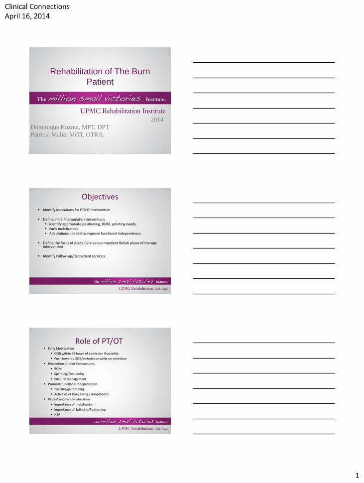
Activities of Daily Living
ADLS are the foundation of every patients successful outcome in rehab.
Ability to perform our own ADL’s provides us with a sense of self-worth, increases our self-esteem, gives us hope, and a feeling of independence.
Encourage patients to reach their highest level of independence in all aspects of their life: self-care, home management, work, leisure/play activities.
Current Medical History
Total Body Surface Area (TBSA)
Location of the Burn
Joints Crossed by injury
Degree of the Burn
Evidence of Inhalation Injury
Types of Dressings
Medications needed for pain, fluid resuscitation, pressors.
Occupational Profile Obtain social and environmental information from patient, family, and
medical chart. Patient may not always be able to provide.
Home environment, environmental barriers, DME used prior to admission.
Prior Level of Function in ADLS, IADLS, leisure, and vocational roles.
Family/caregivers involved prior to admission and if they will be available upon
discharge to assist.
Patients daily routine and activity demands prior to admission and what is
important to them when discharged home.

Clinical Connections April 16, 2014
3
Analysis of Occupational Performance
Measurement of Active and Passive ROM
Strength
Sensation
Coordination: manipulation of objects during functional activities.
Vision
Cognition/Communication
Functional transfers/mobility
Activity Tolerance
Patient Factors
Edema caused by initial injury and inflammatory process versus fluid resuscitation
Pain and how it is affecting patient vitals and functional performance.
Skin integrity: area of burn, size/depth, color and texture, exposed tendons, and status of graft.
Psychological: fear , anxiety, appearance.
Treatment of Burn Patients
Prevent the loss of mobility with the use of proper splinting and positioning devices, assistive devices, orthotics, and ROM.
Reduce edema with proper positioning, compression as appropriate, and ROM.
ROM/Exercise program based upon each individual patient’s needs to improve functional outcomes.
Reduce pain through activity or motion.
Involve the patient and family throughout the treatment process in order to decrease fear and anxiety.
Clinical Connections April 16, 2014
4
Achieving Patient Goals Encourage patients to work through the pain and frustration caused by the
injury.
May require modification of task in order to reach goal.
Adaptive equipment or Adaptations to the environment .
Change in routine.
May require increased time/sessions.
Early Mobilization
Recent push for OOB/early mobilization across all critical care settings
Benefits of early mobilization
Improvement of pulmonary status (inhalation injuries)
Aides in prevention of DVT/PE, PNA
Promotes functional independence
Promotes overall well-being for patients (psychological impact – ICU psychosis)
Decreases time in ICU setting
Take a multidisciplinary approach
Include nursing staff - allows for OOB multiple times/day
Discussion with physicians/PAs barriers to mobilization (i.e. refusals, pain control, sedation)
Early Mobilization Cont. Barriers to mobilization can include
PAIN Good communication with nursing staff Pre-medicate prior to therapy session!!!! Increases patient tolerance Achieve benefits of session Patient/therapist rapport (children)
Medical complications multiple trips to OR – ROM restrictions Resp status – too tenuous for mobilization Vitals Cognitive impairments
Initial trauma related vs hospital acquired (psychosis, meds)
Patient and family cooperation May circle back to pain Loss of a loved one Previous psychological involvement Fear Understanding of medical terms/diagnosis
Clinical Connections April 16, 2014
5
Proper Positioning Positioning
Begins upon therapy evaluation (if not sooner)
Burn wound healing begins as soon as injury occurs = need for early intervention
Patients will rest in a position that creates least amount of pain
“Position of comfort = Position of contracture”
Typically a flexed position
Importance of Patient and family education
Need to have continuity across all disciplines to ensure effectiveness of treatment
Importance of Communication
Daily rounds
Thorough documentation
Education
Proper Positioning Cont. JOINT POSITION OF COMFORT THERAPEUTIC POSITION
Neck Flexed Extended: towel roll/NO PILLOWS/collar
Shoulder Adducted/IR Abduct 90deg: wedge/air plane splint
Elbow Flexed Extended (-5deg): pillow/splint
Wrist Flexed Extended 30-60 deg: towel; splint
Hand Clawed w/MCP extension; PIP/DIP flexion; thumb adducted
MCP flex 70 deg/PIP&DIP ext/thumb abd : splint/washcloth
Hip Flexed/IR Extended/neutral rotation/abduction:
Pillow/wedge/abd pillow - NO PILLOWS
Knee Flexed Full Extension: NO PILLOWS
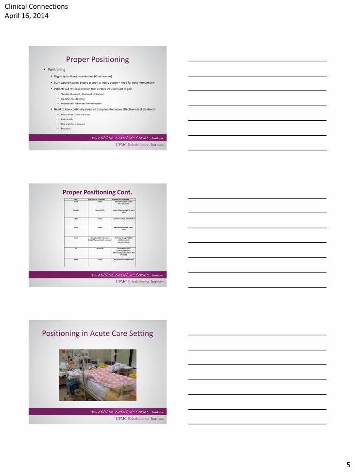
Positioning in Acute Care Setting
Clinical Connections April 16, 2014
6
Scar Formation
Scar formation is an ongoing process for the burn patient.
Scars are dynamic and will continue to grow and change throughout the maturation process for approximately eighteen months post injury.
Scars develop due to excessive amounts of collagen production seen during the healing process.
Types of Scars There are five main types of Scars:
Atrophic scars: are sunken in and often seen with acne or wounds where skin and muscle are removed in one specific area.
Hypertrophic Scars: Red, Raised, and Rigid due to increased collagen production. Tend to fade and flatten over time.
Contracture Scars: often happen with burns when increased collagen production causes disorganized fibers that attach to other structures limiting mobility.
Keloid Scars: Very elevated, pink/red/dark, and often grow larger than the site of original injury.
Stretch Marks: considered a unique type of scar since they occur as a response to the skin being stretched rapidly and not because of an injury.
Types of Scars Hypertrophic Scarring is the result of:
Tissue tension
Persistent inflammation/edema
Response of fibroblasts during the healing process which
deposit large amounts of non-elastic collagen that adheres to
other structures but are not as severe as keloid scars.
Stay within the boundaries of the original wound.
Clinical Connections April 16, 2014
7
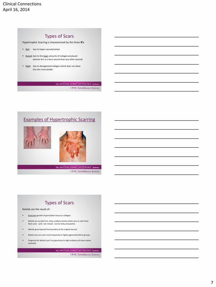
Types of Scars Hypertrophic Scarring is characterized by the three R’s:
Red: due to hyper-vascularization
Raised: due to the large amounts of collagen produced
(almost 4x’s in a burn wound than any other wound)
Rigid: due to disorganized collagen which does not allow
the skin to be pliable.
Examples of Hypertrophic Scarring
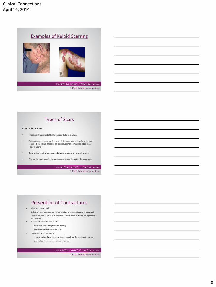
Types of Scars Keloids are the result of: Excessive growth of granulation tissue or collagen.
Keloids are usually firm, shiny, rubbery lesions which vary in color from
flesh color - pink- red- brown. Can be itchy and painful.
Keloids grow beyond the boundary of the original wound.
Keloid scars are seen more frequently in highly pigmented ethnic groups.
Prognosis for Keloid scars? not good due to high incidence of return when
removed.
Clinical Connections April 16, 2014
8
Examples of Keloid Scarring
Types of Scars
Contracture Scars:
This type of scar most often happens with burn injuries.
Contractures are the chronic loss of joint motion due to structural changes
in non-bony tissue. These non-bony tissues include muscles, ligaments,
and tendons.
Prognosis of contractures depends upon the cause of the contracture.
The earlier treatment for the contracture begins the better the prognosis.
Prevention of Contractures What is a contracture?
Definition : Contractures are the chronic loss of joint motion due to structural
changes in non-bony tissue. These non-bony tissues include muscles, ligaments,
and tendons.
Put patients at risk for complications
Medically: affect skin grafts and healing
Functional: limit mobility and ADLs
Patient Education is important
Understanding of why they have to go through painful treatment sessions
Less anxiety if patient knows what to expect
Clinical Connections April 16, 2014
9
Prevention of Contractures Cont. Limiting Factors can include:
Pain
Essential during all phases of care
Leads to high anxiety
Loss of trust between patient and provider
Conscious sedation may be necessary
Pre medicate
Education
Immobility
Global – Nature of the critical illness (complex)
Focal – burn itself
Poor Positioning
Muscle, soft tissue and bone pathology as a result of burn injury
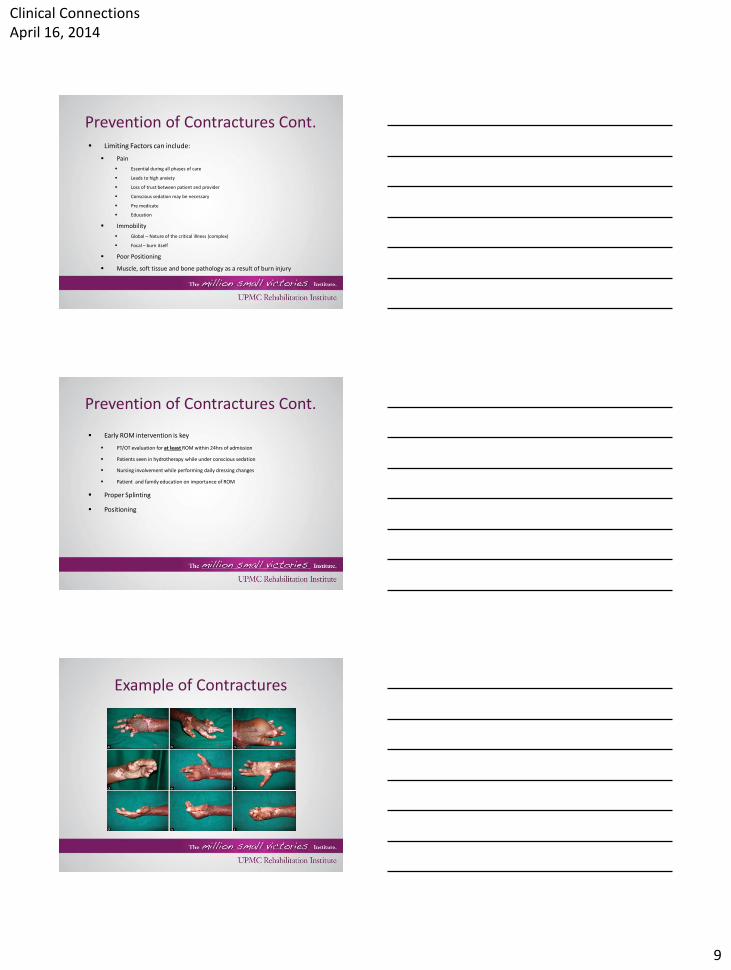
Prevention of Contractures Cont.
Early ROM intervention is key
PT/OT evaluation for at least ROM within 24hrs of admission
Patients seen in hydrotherapy while under conscious sedation
Nursing involvement while performing daily dressing changes
Patient and family education on importance of ROM
Proper Splinting
Positioning
Example of Contractures
Clinical Connections April 16, 2014
10
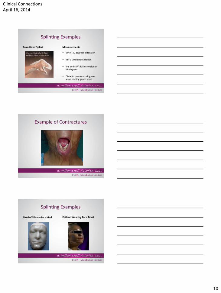
Splinting Examples
Burn Hand Splint Measurements
Wrist 30 degrees extension
MP’s 70 degrees flexion
IP’s and DIP’s full extension or (0) degrees.
Distal to proximal using ace wrap or cling gauze wrap.
Example of Contractures
Splinting Examples
Mold of Silicone Face Mask Patient Wearing Face Mask
Clinical Connections April 16, 2014
11
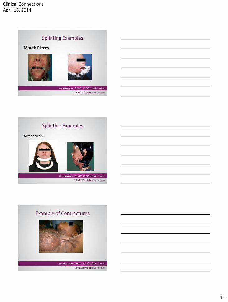
Splinting Examples
Mouth Pieces
Splinting Examples
Anterior Neck
Example of Contractures
Clinical Connections April 16, 2014
12
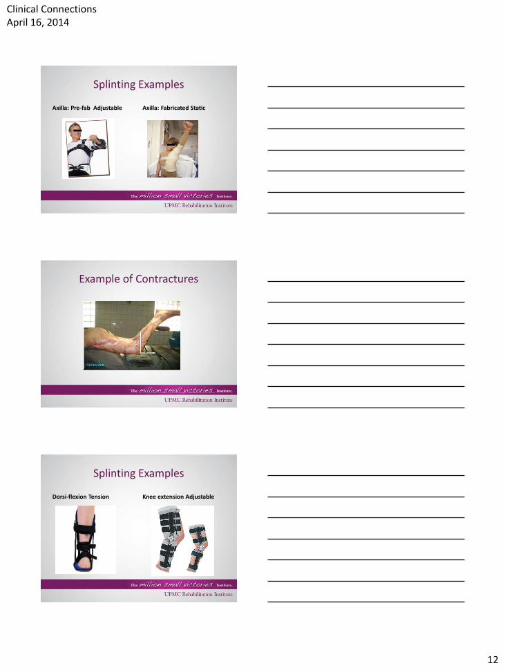
Splinting Examples
Axilla: Pre-fab Adjustable Axilla: Fabricated Static
Example of Contractures
Splinting Examples
Dorsi-flexion Tension Knee extension Adjustable
Clinical Connections April 16, 2014
13
Techniques for Scar Management
Preserve ROM
Splinting/Positioning
Protect underlying vulnerable structures
Increase ROM/Function
Prolonged Stretch
Apply pressure to scar area which promotes collagen remodeling
Scar Massage
Promotes collagen remodeling by applying pressure to scars.
Provides moisture and increased pliability of skin (Burn area and Donor sites).
Helps to decrease itching and discoloration.
Scar Massage Instructions
Apply lotion to healed burns, grafted areas, and donor sites. Avoid perfumed lotions or lotions with additives.
Massage while applying enough pressure to blanch the skin (white/yellow). Stop if skin begins to blister or tear.
Massaging in a circular pattern will avoid shearing.
Clinical Connections April 16, 2014
14
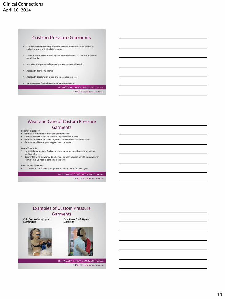
Custom Pressure Garments
Custom Garments provide pressure to a scar in order to decrease excessive collagen growth which leads to scarring.
They are meant to conform to a patient’s body contours to limit scar formation and deformity.
Important that garments fit properly to assure maximal benefit.
Assist with decreasing edema.
Assist with discoloration of skin and smooth appearance.
Patients report feeling better while wearing garments.
Wear and Care of Custom Pressure Garments
Does not fit properly:
Garment is too small if it binds or digs into the skin.
Garment should not ride up or down on patient with motion.
Garment should not cause the fingers or toes to become swollen or numb.
Garment should not appear baggy or loose on patient.
Care of Garments:
Patient should be given 2 sets of pressure garments so that one can be washed
and the other worn.
Garments should be washed daily by hand or washing machine with warm water or
a mild soap. Do not but garments in the dryer.
When to Wear Garments:
Patients should wear their garments 23 hours a day for over a year.
Examples of Custom Pressure Garments
Chin/Neck/Chest/Upper Extremities
Face Mask / Left Upper Extremity
Clinical Connections April 16, 2014
15
Therapy Treatment Techniques
Silicone for scar management.
Inserts to apply pressure while wearing splints and pressure garments.
Paraffin can be used on a case by case basis.
Ultrasound
Medical Treatment of Scar Formation
Steroid injections can help with hypertrophic or keloid scars but is almost always temporary and needs to be repeated.
Skin resurfacing using lasers/radiation.
Surgical release of contractures, further grafting, possible flap, or need for tissue expanders.
Z-plasty which is designed to relieve tension across the scar area.
Acute Care PT/OT Burn Care Thorough PT/OT evaluation/initiation of tx within 24 hours of
admission If able – may be too critical for thorough assessment
Despite pt participate need to initiate ROM/stretching and Splinting/positioning
Prioritize this patient population Due to quick onset of impairments
Quarterly audits completed to ensure compliance
Large focus on early ROM/Splinting and Functional Outcomes May need to treat BID to ensure proper ROM as well as focus on function
Early mobilization/ADLS
Communication Need to communicate with team regarding possible restrictions post-op
D/C recommendations as early as possible
Clinical Connections April 16, 2014
16
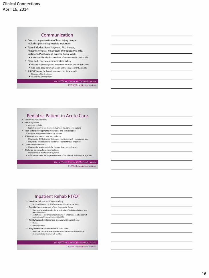
Communication Due to complex nature of burn injury care, a
multidisciplinary approach is important
Team includes: Burn Surgeons, PAs, Nurses, Anesthesiologists, Respiratory therapists, PTs, OTs, Dietitians, Psychosocial experts, Social work. Patient and family also members of team – need to be included
Clear and concise communication is key
With multiple disciplines miscommunication can easily happen
Also need good communication between covering therapists
At UPMC Mercy the burn team meets for daily rounds Discussion of barriers to care
d/c recs and patient progress
Pediatric Patient in Acute Care See infants > adolescents
Family dynamics
– Can hurt or help
– Lack of support vs too much involvement (i.e. refuse for patient)
Need to take developmental milestones into consideration
– May see a regression of skills s/p trauma
ROM/stretching under conscious sedation
– May require BID tx in order to include function as well – incorporate play
– May take a few sessions to build trust – consistency is important
Communication with CLS
– May require a set schedule for therapy times, schooling, etc.
Discharge planning/Recommendations
– More complex due to family dynamic
– Difficult due to MOI – large involvement of social work and case management
Inpatient Rehab PT/OT Continue to focus on ROM/stretching
Responsibility starts to shift from therapist to patient and family
Function becomes more of the therapists’ focus May need to adapt mobility due to contractures/limitations that may have
occurred early on
Acute focus on prevention of contractures vs rehab focus on adaptation of contractures which may limit mobility/ADLs
Family/support system more involved with patient care Ther ex
Dressing changes
May have some disconnect with burn team Need clear communication between acute care rep and rehab members
Communicate barriers in rehab huddles
Clinical Connections April 16, 2014
17
Follow-up/Discharge Planning
Outpatient Occupational and Physical Therapy
Psychology
Vocational/Work Hardening
Community Re-entry
References
• Fiona Procter, Rehabilitation of the burn patient. Indian J Plast Surg. 2010; 43: 101-113
• Troung AD, Fan E, Brower RG, Neeham DM. Bench-to-bedside review: Mobilizing patients in the intensive care unit- from pathophysiology to clinical trials. Critical Care. 2009;13:216
• Scheider JC, Holavanahalli R, Helm P, Goldstein R, Kowalske K. Contractures in Burn Injury: Defining the Problem. J Burn Care Res. 2006; 27: 508-514
• Al-Mousawi DN ,Mecott-Rivera GA, Jeschke MG, Herndon DN. Burn Teams and Burn Centers: The Importance of a Comprehensive Team Approach to Burn Care. Clin Plast Surg. 2009; 36:547-554
References • RUSH Project funded by the NIDRR completed by: The Rocky Mountain
Model System for Burn Injury Rehabilitation at the University of Colorado Health Sciences Center. NIDRR Project Number: H133ACB1402. An Easy Guide to Outpatient Burn Rehabilitation. Last Updated 10/27/2009. www.researchutilization.org/matrix/resources/burn/burnguide.html
• Garren L, Kelly N. Standard of Care : Inpatient Occupational Therapy Intervention for Burns. Brigham and Women’s Hospital Department of Rehabilitation Services. Boston, MA. 6/09.
• Piergrossi S. OT in the Burn Unit: Management of the Burned Patient in the Acute Rehabilitation Phase. Advance for Occupational Therapy Practitioners. Vol. 24. Issue 6. Page 37.
• Sheridan RL, Meier RH III. Burn Rehabilitation. 12/7/2012. http://emedicine.medscape.com
• Moore ML, Dewey WS, Richard RL. Rehab of the Burned Hand. Hand Clinic 25 (2009) 529-541.
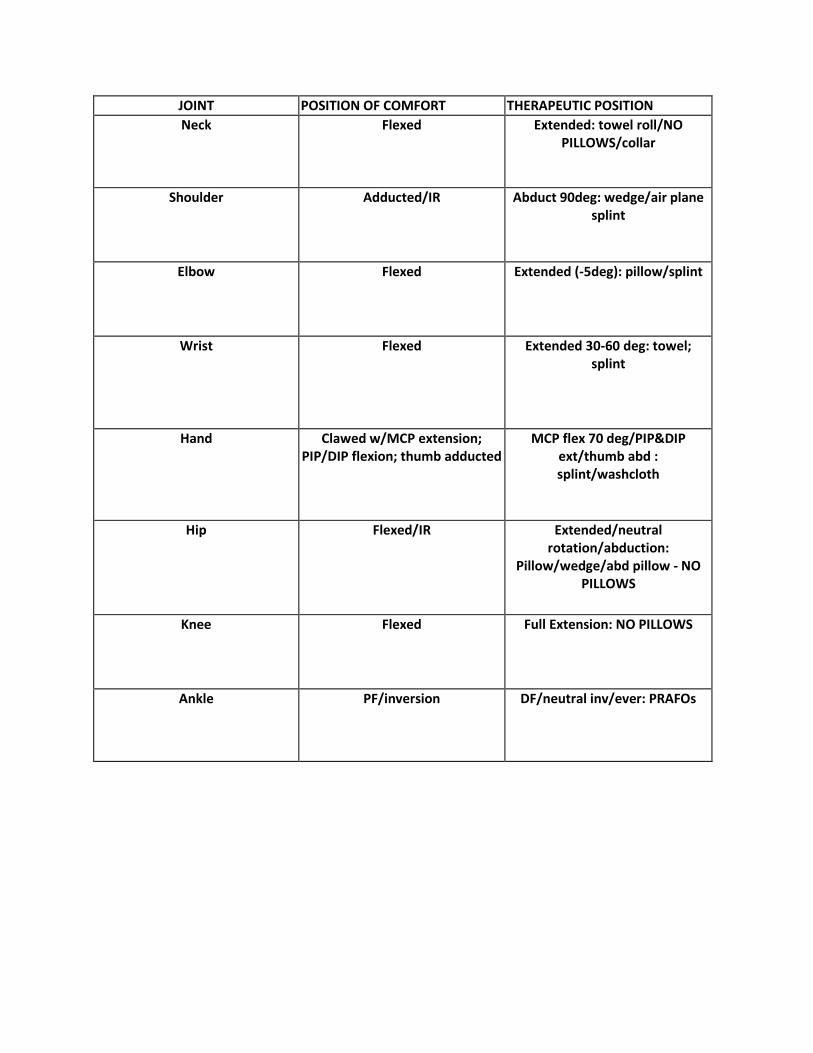
JOINT POSITION OF COMFORT THERAPEUTIC POSITION
Neck Flexed Extended: towel roll/NO PILLOWS/collar
Shoulder Adducted/IR Abduct 90deg: wedge/air plane splint
Elbow Flexed Extended (-5deg): pillow/splint
Wrist Flexed Extended 30-60 deg: towel; splint
Hand Clawed w/MCP extension; PIP/DIP flexion; thumb adducted
MCP flex 70 deg/PIP&DIP ext/thumb abd : splint/washcloth
Hip Flexed/IR Extended/neutral rotation/abduction:
Pillow/wedge/abd pillow - NO PILLOWS
Knee Flexed Full Extension: NO PILLOWS
Ankle PF/inversion DF/neutral inv/ever: PRAFOs