Embed Size (px)
Citation preview
1
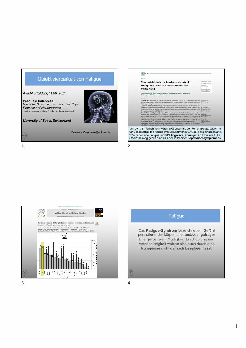
ASIM-Fortbildung 11.08. 2021
Pasquale CalabreseUniv.- Prof. Dr. rer. nat. med. habil., Dipl.-Psych.Professor of NeuroscienceHead of neuropsychology & behavioral neurology unit
Unversity of Basel, Switzerland
Objektivierbarkeit von Fatigue
1
192 journals.sagepub.com/home/msj
MULTIPLESCLEROSIS MSJJOURNAL
https://doi.org/10.1177/1352458517708685https://doi.org/10.1177/1352458517708685
Multiple Sclerosis Journal
2017, Vol. 23(2S) 192 –203
DOI: 10.1177/ 1352458517708685
© The Author(s), 2017. Reprints and permissions: http://www.sagepub.co.uk/journalsPermissions.nav
IntroductionThe role of economic studies in healthcare in Switzerland is less important than in some other European countries. The Swiss law stipulates that medical treatments have to be covered by the compul-sory health insurance scheme if they are considered ‘effective’, ‘appropriate’ and ‘cost-effective’. The methods required to assess cost-effectiveness for new and existing drugs are reference pricing (with a basket of nine European countries) and a comparative assess-ment of the therapeutic benefit to justify the price set by the manufacturer. Economic evaluations are accepted in a manufacturer submission yet not man-datory. Nevertheless, cost-of illness studies as a basis for health policy have the same importance as elsewhere.
The availability of disease-modifying treatments (DMTs) has led to changes in patient management and a focus on earlier and better diagnosis and
adjustments in the diagnostic criteria themselves. One of the consequences in this regard is that the recorded prevalence of the disease is quite different from that estimated two or three decades ago.1 In Switzerland, the current prevalence is estimated at 110 per 100,000, corresponding to a total of 10,000 individuals suffer-ing from multiple sclerosis (MS).2,3 With diagnosis already possible after a clinically isolated event,4 one must also expect a different distribution of the type of MS and the severity of the disease than 10 years ago: a larger proportion of patients with relapsing-remit-ting disease and thus of patients in the early stages of the disease and with less disability.
It is therefore important to update the information on the burden of MS, and the study presented here is part of a European-wide effort to update informa-tion on the burden of illness of MS in 16 countries, endorsed by the European Platform of MS Societies (EMSP)5 and carried out with the support of national
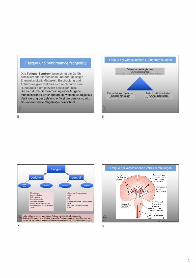
New insights into the burden and costs of multiple sclerosis in Europe: Results for Switzerland
Pasquale Calabrese, Gisela Kobelt, Jenny Berg, Daniela Capsa, Jennifer Eriksson and The European Multiple Sclerosis Platform
AbstractIntroduction: To estimate the value of interventions in multiple sclerosis (MS) – where lifetime costs and outcomes cannot be observed – outcome data have to be combined with costs. This requires that cost data be regularly updated.Objectives and methods: This study is part of a cross-sectional retrospective study in 16 countries col-lecting data on resource consumption and work capacity, health-related quality of life (HRQoL) and prevalent symptoms for patients with MS. Descriptive analyses are presented by level of severity, from the societal perspective, in CHF 2015.Results: A total of 721 patients (mean age 48 years) participated in Switzerland; 90% were below retire-ment age, and of these, 65% were employed. Employment was related to disease severity, and MS affected productivity at work for 69% of patients. Overall, 93% and 64% of patients experienced fatigue and cognition as a problem, respectively. The mean utility and annual costs were 0.799 and 29,600CHF at Expanded Disability Status Scale (EDSS) 0–3, 0.614 and 66,800CHF at EDSS 4–6.5 and 0.348 and 110,800CHF at EDSS 7–9, respectively. The mean cost of a relapse was estimated at 7600CHF.Conclusion: This study provides current data on MS in Switzerland that are important for development of health policies and to estimate the value of current and future treatments.
Keywords: Multiple sclerosis, burden of illness, fatigue, cognition, costs, HRQoL, Switzerland
Correspondence to: G Kobelt European Health Economics, 15 Rue Victor Schoelcher, 68200 Mulhouse, France. [email protected]
Pasquale Calabrese Division of Molecular and Cognitive Neuroscience, Department of Psychology, University of Basel, Basel, Switzerland
Gisela Kobelt European Health Economics, Mulhouse, France
Jenny Berg Daniela Capsa Jennifer Eriksson Mapi Group, Stockholm, Sweden
708685MSJ0010.1177/1352458517708685Multiple Sclerosis JournalP Calabrese, G Kobeltresearch-article2017
Article
Von den 721 Teilnehmern waren 90% unterhalb der Rentengrenze, davon nur 65% beschäftigt. Die Arbeits-Produktivität war in 69% der Fälle eingeschränkt. 93% gaben eine Fatigue und 64% kognitive Störungen an. Über alle EDSSStadien hinweg gaben rund 40% der Teilnehmer Depressionssymptome an.
2
Contents lists available at ScienceDirect
Multiple Sclerosis and Related Disorders
journal homepage: www.elsevier.com/locate/msard
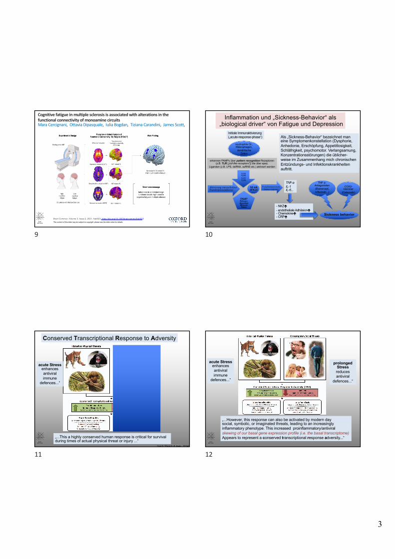
The disease burden of Multiple Sclerosis from the individual and populationperspective: Which symptoms matter most?
Laura Barina,⁎, Anke Salmenb,2, Giulio Disantoc,g,2, Haris Babačića, Pasquale Calabresed,Andrew Chanb, Christian P. Kammb,e, Jürg Kesselringf, Jens Kuhleg, Claudio Gobbic,Caroline Poth, Milo A. Puhana, Viktor von Wyla,1, for the Swiss Multiple Sclerosis Registry (SMSR)a Epidemiology, Biostatistics and Prevention Institute, University of Zurich, Zurich, SwitzerlandbDepartment of Neurology, University Hospital Bern and University of Bern, Bern, SwitzerlandcNeurocenter of southern Switzerland, Ospedale regionale di Lugano, Lugano, SwitzerlanddNeuropsychology and Behavioral Neurology Unit, Division of Molecular and Cognitive Neuroscience, Department of Psychology, University of Basel, Basel, SwitzerlandeNeurology and Neurorehabilitation Centre, Luzerner Kantonsspital, Lucerne, SwitzerlandfDepartment of Neurology & Neurorehabilitation, Rehabilitation Centre Kliniken Valens, Valens, SwitzerlandgNeurologic Clinic and Policlinic, Departments of Medicine, Biomedicine and Clinical Research, University Hospital Basel, University of Basel, Basel, Switzerlandh Laboratories of Neuroimmunology, Division of Neurology and Neuroscience Research Center, Department of Clinical Neurosciences, Lausanne University Hospital,Lausanne, Switzerland
A R T I C L E I N F O
Key-words:Quality of lifePatient care managementPatient reported outcomesEQ5DRegistriesRegression analysis
A B S T R A C T
Background: MS symptoms affect many functional domains. Knowing the specific impact of symptoms on health-related quality of life (HRQoL) is vital for successful disease and symptom management in MS. We aimed atinvestigating how specific MS symptoms contribute to the disease burden in individuals and from a populationperspective.Methods: We included 855 Swiss Multiple Sclerosis Registry participants with a relapsing-remitting form (RRMS)or a progressive form (PMS). HRQoL was measured with the EuroQol 5-Dimension EQ-5D-index and EQ-VisualAnalogue Scale (EQ-VAS) on 0–100% scales. Their associations with 20 symptoms, socio-demographic andclinical information were explored in median regression models, stratified by RRMS and PMS.Results: We included 611 participants with RRMS and 244 with PMS. In RRMS, gait (−6.5%) and balanceproblems (−5.1%) had the largest EQ-5D-index reductions, and were also important at the population level(frequencies 45% and 52%). Fatigue, depression, and spasticity (frequencies 74.1%, 31%, 38%) also contributedto the population disease burden. In PMS, spasticity, paralysis, and bowel problems had the largest impact onEQ-5D-index, both at the individual and population levels. The largest impact on EQ-VAS at population level wasassociated in RRMS with balance problems, depression, dizziness, and spasticity, while in PMS with weakness,pain, and paralysis.Conclusions: While HRQoL at population level is most affected by balance problems, spasticity, and depression inRRMS, the biggest HRQoL losses in PMS are caused by spasticity, paralysis, weakness, and pain. Many symptomswith the largest effects in individuals substantially contribute to the population disease burden.
https://doi.org/10.1016/j.msard.2018.07.013Received 2 May 2018; Received in revised form 22 June 2018; Accepted 8 July 2018
Abbreviations: AIC, Aikake's Information Criterion; CIS, Clinically Isolated Syndrome; DMT, Disease-modifying Therapy; EDSS, Expanded Disability Status Scale;EQ-5D-5L, European Quality of Life 5-Dimension 5 Level version; EQ-5D-index, European Quality of Life 5-Dimension Index; EQ-VAS, European Quality of Life VisualAnalogue Scale; EQ-VAS, European Quality of Life Visual Analogue Scale; EQ-VAS, European Quality of Life Visual Analogue Scale; HRQoL, Health-related quality oflife; MICE, Multivariate Imputation by Chained Equations; RRMS, Relapsing remitting multiple sclerosis; PwMS, Persons with multiple sclerosis; PMS, Progressivemultiple sclerosis; SMSR, Swiss Multiple Sclerosis Registry⁎ Corresponding author.
1 For the Swiss Multiple Sclerosis Registry (SMSR).2 Equal contribution.
E-mail addresses: [email protected] (L. Barin), [email protected] (A. Salmen), [email protected] (G. Disanto),[email protected] (P. Calabrese), [email protected] (A. Chan), [email protected] (C.P. Kamm),[email protected] (J. Kesselring), [email protected] (J. Kuhle), [email protected] (C. Gobbi), [email protected] (C. Pot),[email protected] (M.A. Puhan), [email protected] (V. von Wyl).
0XOWLSOH�6FOHURVLV�DQG�5HODWHG�'LVRUGHUV��������������²���
������������������(OVHYLHU�%�9��$OO�ULJKWV�UHVHUYHG�
7
Thefi
nalm
odel
forR
RMSwith
EQ-VAS
asou
tcom
ewas
adjuste
dfor
age,
sex,
diseasedu
ratio
n,recent
relapse,
anded
ucationleve
l.Fig.
2(right-han
dsid
e)show
sregressio
nco
efficien
tsan
d95
%CI.Th
esymptom
smoststr
ongly
associated
with
EQ-VAS
were
–in
orde
r-
depressio
n,tre
mor,a
ndsexu
aldy
sfunc
tion.
Themeancalib
ratio
nslo
pewas
0.82
(0.71–
0.94
),indicatin
gago
odcalib
ratio
n.Th
eco
mpletelisto
fcoe
fficien
tsfro
mtheun
ivariablean
dthemul-
tivariablemod
elsf
orRR
MSareshow
nin
theAp
pend
ix(Tab
leA4
).
Fig.
1.Freq
uenc
yof
repo
rtedsymptom
sin
parti
cipa
ntswith
RRMSan
dPM
S.RR
MS=
relapsingremittingMS,
PMS=
prog
ressiveMS,
dysf.
=dy
sfunc
tion,
p.=
prob
lems.
L.Ba
rinet
al.
0XOWLSOH�6
FOHURVLV�D
QG�5HODWHG�'LVRUG
HUV���
�����������²�
��
���
3
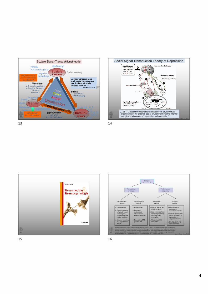
Das Fatigue-Syndrom bezeichnet ein Gefühl persistierender körperlicher und/oder geistiger Energielosigkeit, Müdigkeit, Erschöpfung und Antriebslosigkeit welche sich auch durch eine
Ruhepause nicht gänzlich beseitigen lässt.
Fatigue
4
2
Das Fatigue-Syndrom bezeichnet ein Gefühl persistierender körperlichen und/oder geistigen Energielosigkeit, Müdigkeit, Erschöpfung und Antriebslosigkeit welches sich auch durch eine Ruhepause nicht gänzlich beseitigen lässt.Die sich durch die Bearbeitung einer Aufgabe manifestierende Erschöpfbarkeit, welche als objektive Veränderung der Leistung erfasst werden kann, wird als «performance fatigability» bezeichnet.
Fatigue und performance fatigability
5
Fatigue bei neurologischen Grunderkrankungen
(neuroendokrin, neurodegenerativ, neuroimmunologisch, autonome Dysfunktion...)
Fatigue bei internistischen Grunderkrankungen
(Avitaminose, Organerkrankungen, Tumoren...)
Fatigue bei psychiatrischen Grunderkrankungen
(Depression, Angststörungen, PTSD...)
Fatigue bei verschiedenen Grunderkrankungen
6
Fatigue
peripher zentral
periph. Nerv Muskel physisch kognitiv
-Myasthenie-Lambert-Eaton-Polymyositis-Dermatomyositis-Muskeldystrophie-metabolische Myopathien-mitochondriale Erkrankungen- usw.
-(Meningo-)Enzephalitiden-MSA-MS-PD-Cerebrovaskuläre Erkrankungen-SHT-Metabol. Enzephalopathien- usw.
nota: während bei der peripheren Fatigue die kognitive KomponenteIndirekt, d.h. durch eine erhöhte physische Anstrengung (mit) betroffen sein kannist bei der zentralen Fatigue auch eine isolierte kognitive Erschöpfbarkeit möglich.
7Aus: Chaudhuri & Behan, LANCET 2004)
Fatigue bei verschiedenen ZNS-Erkrankungen
8
3
Brain Commun, Volume 3, Issue 2, 2021, fcab023, https://doi.org/10.1093/braincomms/fcab023The content of this slide may be subject to copyright: please see the slide notes for details.
Cognitive fatigue in multiple sclerosis is associated with alterations in the functional connectivity of monoamine circuitsMara Cercignani, Ottavia Dipasquale, Iulia Bogdan, Tiziana Carandini, James Scott,
9
TLR3TLR4TLR7TLR8...
neutrophile Gr.,Makrophagen,dendritische
Zellen
erkennen PAMPs über pattern recognition Rezeptoren(z.B. TLR [„toll-like-receptors“]) die über spez.
Liganden (z.B. LPS, dsRNA, ssRNS etc.) aktiviert werden
PAMPPathogen-AssociatedMolecularPattern
NF-kBIFN-rf
TNF-aIL-1IL-6...
- NKZé- endotheliale Adhäsioné- Chemokineé- CRPé Sickness behavior
COX2-Hemmer
(Celecoxib)
TNF-a-Antagonisten(Etanercept,Infliximab...)
Als „Sickness-Behavior“ bezeichnet maneine Symptomenkonstellation (Dysphorie,Anhedonie, Erschöpfung, Appetitlosigkeit,Schläfrigkeit, psychomotor. Verlangsamung,Konzentrationsstörungen) die üblicher-weise im Zusammenhang mich chronischenEntzündungs- und Infektionskrankheitenauftritt.
Initiale Immunaktivierung(„acute-response-phase“)
Inflammation und „Sickness-Behavior“ als„biological driver“ von Fatigue und Depression
Aktivierung intrazellulärerTranskriptionsfaktoren
ProinflammatorischeImmun-response
10
regulation of proinflammatory immune response genes, whichcombat bacteria and other extracellular pathogens, and a reciprocaldown-regulation of antiviral immune response genes, which targetintracellular pathogens such as viruses (Irwin & Cole, 2011). Asdepicted in Figure 1 and described more fully by Slavich and Cole(2013), this increased proinflammatory/reduced antiviral skewingof our basal gene expression profile, called the basal transcrip-tome, appears to represent a conserved transcriptional response toadversity (CTRA) that is adaptive in countering injuries associatedwith actual physical threat (see Antoni et al., 2012; S. W. Cole etal., 2012; Fredrickson et al., 2013; Irwin & Cole, 2011; Powell etal., 2013). Activation of this ancestral host defense program bynonphysical social, symbolic, anticipated, or imagined threats,however, can increase an individual’s risk for both viral infectionand inflammation-related disease.
Two physiological pathways are responsible for convertingsocial-environmental adversity into broad proinflammatory tran-scriptional programs such as the CTRA. The first pathway in-volves the sympathetic nervous system (SNS), and the secondpathway involves the hypothalamic–pituitary–adrenal (HPA) axis(Irwin & Cole, 2011). Additional evidence suggests that the para-sympathetic nervous system modulates immune responses at aregional level through both the efferent and afferent fibers of thevagus nerve, enabling it to prevent excessive inflammation (Bo-rovikova et al., 2000; Sternberg, 2006; Tracey, 2009). Because themechanisms underlying these pathways are described in detailelsewhere (Dantzer et al., 2008; Irwin & Cole, 2011; G. Miller,Chen, & Cole, 2009; Pavlov & Tracey, 2004; Slavich & Cole,2013; Sternberg, 2006; Waldburger & Firestein, 2010), we onlybriefly summarize the pathways here, with an emphasis on how
Figure 1. Conserved transcriptional response to adversity (CTRA). The innate immune system developed tocounter physical threats from predatory animals and hostile conspecifics that dominated our ancestral environ-ment. Exposure to these threats activates a CTRA that involves up-regulation of proinflammatory immuneresponse genes, which combat extracellular pathogens and wound-related bacterial infections, and down-regulation of antiviral immune response genes, which target intracellular pathogens such as viruses. Thisredeployment of the leukocyte basal transcriptome is adaptive in the context of actual physical threat because itenhances wound healing and recovery from injury and infection. The CTRA can also be activated by modern-daysocial, symbolic, anticipated, and imagined threats, however, leading to increased risk for several inflammation-related conditions, including depression (see Antoni et al., 2012; S. W. Cole et al., 2012; Fredrickson et al., 2013;Irwin & Cole, 2011; Powell et al., 2013; Slavich & Cole, 2013). Saber-toothed cat image copyright 2013 byDorling Kindersley; chimpanzee image copyright 2013 by Ronald van der Beek; pointing man image copyright2013 by Craig Wactor; all other images copyright 2013 by Getty Images. All images reprinted with permission.
ThisdocumentiscopyrightedbytheAmericanPsychologicalAssociationoroneofitsalliedpublishers.
Thisarticleisintendedsolelyforthepersonaluseoftheindividualuserandisnottobedisseminatedbroadly.
778 SLAVICH AND IRWIN
Conserved Transcriptional Response to Adversity
„...This a highly conserved human response is critical for survival during times of actual physical threat or injury ...“
acute Stressenhancesantiviral immune
defences...“
(nach Slavich & Irwin, 2014)
11
regulation of proinflammatory immune response genes, whichcombat bacteria and other extracellular pathogens, and a reciprocaldown-regulation of antiviral immune response genes, which targetintracellular pathogens such as viruses (Irwin & Cole, 2011). Asdepicted in Figure 1 and described more fully by Slavich and Cole(2013), this increased proinflammatory/reduced antiviral skewingof our basal gene expression profile, called the basal transcrip-tome, appears to represent a conserved transcriptional response toadversity (CTRA) that is adaptive in countering injuries associatedwith actual physical threat (see Antoni et al., 2012; S. W. Cole etal., 2012; Fredrickson et al., 2013; Irwin & Cole, 2011; Powell etal., 2013). Activation of this ancestral host defense program bynonphysical social, symbolic, anticipated, or imagined threats,however, can increase an individual’s risk for both viral infectionand inflammation-related disease.
Two physiological pathways are responsible for convertingsocial-environmental adversity into broad proinflammatory tran-scriptional programs such as the CTRA. The first pathway in-volves the sympathetic nervous system (SNS), and the secondpathway involves the hypothalamic–pituitary–adrenal (HPA) axis(Irwin & Cole, 2011). Additional evidence suggests that the para-sympathetic nervous system modulates immune responses at aregional level through both the efferent and afferent fibers of thevagus nerve, enabling it to prevent excessive inflammation (Bo-rovikova et al., 2000; Sternberg, 2006; Tracey, 2009). Because themechanisms underlying these pathways are described in detailelsewhere (Dantzer et al., 2008; Irwin & Cole, 2011; G. Miller,Chen, & Cole, 2009; Pavlov & Tracey, 2004; Slavich & Cole,2013; Sternberg, 2006; Waldburger & Firestein, 2010), we onlybriefly summarize the pathways here, with an emphasis on how
Figure 1. Conserved transcriptional response to adversity (CTRA). The innate immune system developed tocounter physical threats from predatory animals and hostile conspecifics that dominated our ancestral environ-ment. Exposure to these threats activates a CTRA that involves up-regulation of proinflammatory immuneresponse genes, which combat extracellular pathogens and wound-related bacterial infections, and down-regulation of antiviral immune response genes, which target intracellular pathogens such as viruses. Thisredeployment of the leukocyte basal transcriptome is adaptive in the context of actual physical threat because itenhances wound healing and recovery from injury and infection. The CTRA can also be activated by modern-daysocial, symbolic, anticipated, and imagined threats, however, leading to increased risk for several inflammation-related conditions, including depression (see Antoni et al., 2012; S. W. Cole et al., 2012; Fredrickson et al., 2013;Irwin & Cole, 2011; Powell et al., 2013; Slavich & Cole, 2013). Saber-toothed cat image copyright 2013 byDorling Kindersley; chimpanzee image copyright 2013 by Ronald van der Beek; pointing man image copyright2013 by Craig Wactor; all other images copyright 2013 by Getty Images. All images reprinted with permission.
ThisdocumentiscopyrightedbytheAmericanPsychologicalAssociationoroneofitsalliedpublishers.
Thisarticleisintendedsolelyforthepersonaluseoftheindividualuserandisnottobedisseminatedbroadly.
778 SLAVICH AND IRWIN
„...However, this response can also be activated by modern daysocial, symbolic, or imaginated threats, leading to an increasingly inflammatory phenotype. This increased proinflammatory/antiviral skewing of our basal gene expression profile (i.e. the basal transcriptome)Appears to represent a conserved transcriptional response adversity...“
acute Stressenhancesantiviral immune
defences...“
prolonged Stress reducesantiviral
defences...“
12
4
SozialeFaktoren
Gehirn
Immun-system
(epi-)Genetik
Bedrohung
Stress
Zurückweisung
Hirnstamm-nuclei
Verhalten
Soziale Signal-Transduktionstheorie
Verlust, Vernachlässigung
dACC
Amygdala
IL-1b, IL2 IL-6
TNF-aCRP
Hypothalamus
Insula
z.B. NFkBIL-1,6,8; TNF-Gene
z.B. HPAA u. ANS-Aktivierung
z.B. somato-vegetativeu. kognitive Symptome
(„Sickness Behavior“)
IL-8IL17, IL 21
negative Bewertung
...SorgeStressAngstDepressionCVO, Vagusnerv
self-promotion via„neuroinflammatory
sensitization“
bidirektionale„Darm-Hirn-Achse“
13
Figure 4. Social signal transduction theory of depression. Social signal transduction theory of depressiondescribes mechanisms that convert, or transduce, experiences of the external social environment into the internalbiological environment of depression pathogenesis. (1) Social-environmental experiences indicating possiblesocial threat or adversity (e.g., social evaluation, rejection, isolation, or exclusion) are represented neurally,especially in brain systems that process experiences of social and physical pain. Key nodes in this neural networkinclude the anterior insula (AI) and dorsal anterior cingulate cortex (dACC, shown in the insert). These regionsproject to lower level brain areas (e.g., hypothalamus, brainstem autonomic control nuclei) that have the abilityto initiate and modulate inflammatory activity via three pathways that involve (2) the hypothalamic–pituitary–adrenal axis, (3) sympathetic nervous system (SNS), and (4) efferent vagus nerve. (5) Activation of thesepathways leads to the production of glucocorticoids, epinephrine, norepinephrine (NE), and acetylcholine (ACh),which interact with receptors on cytokine-producing cells. Whereas glucocorticoids and acetylcholine haveanti-inflammatory effects, epinephrine and norepinephrine activate intracellular transcription factors (e.g.,nuclear factor-!B and activator protein 1) that bind to cis-regulatory DNA sequences to up-regulate inflamma-tory gene expression. When this occurs and immune response genes are expressed, DNA is transcribed into RNAand then translated into protein. The resulting change in cell function leads to the production of proinflammatorycytokines (e.g., interleukin-1" [IL-1"], interleukin-6 [IL-6], tumor necrosis factor-# [TNF-#]) that signal thebrain to induce cognitive, emotional, and behavioral alterations that include several hallmark symptoms ofdepression (e.g., sad mood, anhedonia, fatigue, psychomotor retardation, altered appetite and sleep, andsocial-behavioral withdrawal). Cytokines can exert these effects on the central nervous system by (6) passingthrough leaky or incomplete regions of the blood–brain barrier (e.g., circumventricular organs, organumvasculosum of the lamina terminalis) and by (7) stimulating primary afferent nerve fibers in the vagus nerve,which relays information to brain systems that regulate mood, motor activity, motivation, sensitivity to socialthreat, and arousal. Although these neurocognitive and behavioral responses are adaptive during times of actualthreat, as depicted in Figure 1, these social signal transduction pathways can also be initiated by purely symbolic,anticipated, or imagined threats—that is, situations that have not yet happened or that may never actually occur.Moreover, activation of these pathways can become self-promoting over time due to neuro-inflammatorysensitization and, as a result, remain engaged long after an actual threat has passed (see Figure 3). In suchinstances, these dynamics can increase risk for depression in the short-term and possibly promote physicaldisease, accelerate biological aging, and hasten mortality over the long run. ACTH $ adrenocorticotropichormone; mRNA $ messenger ribonucleic acid.
ThisdocumentiscopyrightedbytheAmericanPsychologicalAssociationoroneofitsalliedpublishers.
Thisarticleisintendedsolelyforthepersonaluseoftheindividualuserandisnottobedisseminatedbroadly.
793STRESS, INFLAMMATION, AND DEPRESSION
(aus Slavich & Irwin, 2014)
„...SSTTD describes mechanisms that convert, or „transduce“experiences of the external social environment into the internalbiological environment of depression pathogenesis...“
Social Signal Transduction Theory of Depression
14
15 for identifying and reporting which factors are mostrelevant in specific neurologic illnesses.
Homeostatic factors. Homeostasis refers to the ten-dency of an organism to maintain a stable functionalstate. This is accomplished by engaging feedback andfeedforward pathways that constrain variation in 1 ormore control variables. Within this context, perceptionsof fatigue likely contribute to homeostasis through reg-ulation of energy expenditure and protection from over-use injuries. Several metabolic stimuli are proposed toinduce the sensation of muscle fatigue, including deple-tion of muscle glycogen and phosphocreatine and theaccumulation of lactate, low pH, Pi, K1, ammonia,and ATP.55,56 More recently, a unique population ofdorsal root ganglion neurons has been discovered thatspecifically respond to low pH, ATP, and ammonia butnot other painful stimuli.57 Within the CNS, potentialcontributors to perceptions of fatigue may include cere-bral glycogen depletion, increased brain temperature,accumulation of ammonia, inflammatory cytokines(particularly IL-6), increases in serotonin, and decre-ments in dopamine.58–60,e1 Animal models demonstratethat the hypothalamus contributes to energy regulationand fatigue perception,e2 similar to its role in hunger andthirst. Other brain areas, including frontal lobes andbasal ganglia, are also likely important in regulating
behavior relative to homeostatic factors and other cues.43
One current hypothesis suggests that these CNS struc-tures act as a “central governor” to limit energy utiliza-tion and avoid potential energetic collapse.e3
Although fatigue can be associated with lesions ofthe hypothalamus, it is difficult to attribute fatiguedirectly to these lesions as patients frequently have addi-tional circadian rhythm and endocrine disturbances.However, disruption of the hypothalamic-pituitary-adrenal axis may contribute to other conditions associ-ated with symptomatic fatigue.e4,e5 While perceptionsof fatigue and effort may be altered in PD andMS,e6,e7
it is difficult to know whether these differences reflectchanges in energy regulation vs other psychologic orphysiologic factors. Research utilizing careful energeticmeasurements can test the role of homeostatic factorscontribution to perceptions of fatigue in neurologicillness.e8
Psychological factors. Psychological factors that con-tribute to the perception of fatigue and fatigabilityamong healthy volunteers include perceptions of effort,expectations, familiarity, motivation, temporal andperformance feedback, arousal, and mood.e9–e12 Someevidence suggests that these factors, particularly percep-tions of effort, may be the primary factors limitingprolonged performance for many motor and cognitive
Figure Diagram of major factors contributing to the 2 domains of fatigue: perceptions of fatigue andfatigability
While separated in this diagram, is should be noted that perceptions of fatigue and performance fatigability have the poten-tial to influence each other. Letters in boxes refer to the following: a) known neuroanatomic sites mediating this factor; b)normal function of this factor; c) pathologic states involving this factor. CFS 5 chronic fatigue syndrome; CVA 5 cerebro-vascular accident; EC 5 excitation/contraction; GBS 5 Guillain-Barré syndrome; GSD 5 glycogen storage diseases; MG 5
myasthenia gravis; MS 5 multiple sclerosis; PD 5 Parkinson disease; TBI 5 traumatic brain injury.
412 Neurology 80 January 22, 2013
ª 2013 American Academy of Neurology. Unauthorized reproduction of this article is prohibited.
(aus: Kluger et al., 2013)
for identifying and reporting which factors are mostrelevant in specific neurologic illnesses.
Homeostatic factors. Homeostasis refers to the ten-dency of an organism to maintain a stable functionalstate. This is accomplished by engaging feedback andfeedforward pathways that constrain variation in 1 ormore control variables. Within this context, perceptionsof fatigue likely contribute to homeostasis through reg-ulation of energy expenditure and protection from over-use injuries. Several metabolic stimuli are proposed toinduce the sensation of muscle fatigue, including deple-tion of muscle glycogen and phosphocreatine and theaccumulation of lactate, low pH, Pi, K1, ammonia,and ATP.55,56 More recently, a unique population ofdorsal root ganglion neurons has been discovered thatspecifically respond to low pH, ATP, and ammonia butnot other painful stimuli.57 Within the CNS, potentialcontributors to perceptions of fatigue may include cere-bral glycogen depletion, increased brain temperature,accumulation of ammonia, inflammatory cytokines(particularly IL-6), increases in serotonin, and decre-ments in dopamine.58–60,e1 Animal models demonstratethat the hypothalamus contributes to energy regulationand fatigue perception,e2 similar to its role in hunger andthirst. Other brain areas, including frontal lobes andbasal ganglia, are also likely important in regulating
behavior relative to homeostatic factors and other cues.43
One current hypothesis suggests that these CNS struc-tures act as a “central governor” to limit energy utiliza-tion and avoid potential energetic collapse.e3
Although fatigue can be associated with lesions ofthe hypothalamus, it is difficult to attribute fatiguedirectly to these lesions as patients frequently have addi-tional circadian rhythm and endocrine disturbances.However, disruption of the hypothalamic-pituitary-adrenal axis may contribute to other conditions associ-ated with symptomatic fatigue.e4,e5 While perceptionsof fatigue and effort may be altered in PD andMS,e6,e7
it is difficult to know whether these differences reflectchanges in energy regulation vs other psychologic orphysiologic factors. Research utilizing careful energeticmeasurements can test the role of homeostatic factorscontribution to perceptions of fatigue in neurologicillness.e8
Psychological factors. Psychological factors that con-tribute to the perception of fatigue and fatigabilityamong healthy volunteers include perceptions of effort,expectations, familiarity, motivation, temporal andperformance feedback, arousal, and mood.e9–e12 Someevidence suggests that these factors, particularly percep-tions of effort, may be the primary factors limitingprolonged performance for many motor and cognitive
Figure Diagram of major factors contributing to the 2 domains of fatigue: perceptions of fatigue andfatigability
While separated in this diagram, is should be noted that perceptions of fatigue and performance fatigability have the poten-tial to influence each other. Letters in boxes refer to the following: a) known neuroanatomic sites mediating this factor; b)normal function of this factor; c) pathologic states involving this factor. CFS 5 chronic fatigue syndrome; CVA 5 cerebro-vascular accident; EC 5 excitation/contraction; GBS 5 Guillain-Barré syndrome; GSD 5 glycogen storage diseases; MG 5
myasthenia gravis; MS 5 multiple sclerosis; PD 5 Parkinson disease; TBI 5 traumatic brain injury.
412 Neurology 80 January 22, 2013
ª 2013 American Academy of Neurology. Unauthorized reproduction of this article is prohibited.
16
5
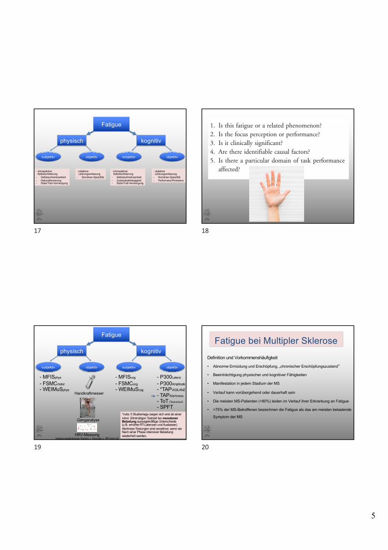
Fatigue
physisch kognitiv
subjektiv objektiv subjektiv objektiv
- introspektiveSelbstschilderung
- Selbstaufmerksamkeit- Dekonditionierung- State/Trait-Vermengung
- objektiveLeistungserfassung
- Domänen-Spezifität
- introspektiveSelbstschilderung
- Selbstaufmerksamkeit- Zustandsabhängigkeit- State/Trait-Vermengung
- objektiveLeistungserfassung
- Domänen-Spezifität- Performanz/Persistenz
17
progress, and the development of effective interventionsare constrained. Memory is a motivating example of afield where transformation of a commonly used wordinto a rigorous scientific taxonomy, for example distin-guishing between types of memory (e.g., working andprocedural memory) and memory processes (e.g.,retrieval and encoding), has led to scientific progress.
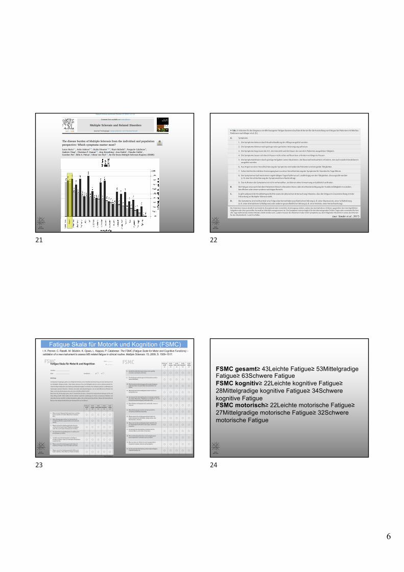
PROPOSAL FOR A UNIFIED TAXONOMY The termfatigue as used in the life sciences is not a unitary phe-nomenon and cannot be defined as such. Rather it isnecessary to identify its distinct domains and to distin-guish it from related phenomena. Our taxonomy mayapply to clinical or research settings and is based onanswering the following questions:
1. Is this fatigue or a related phenomenon?2. Is the focus perception or performance?3. Is it clinically significant?4. Are there identifiable causal factors?5. Is there a particular domain of task performance
affected?
Is this fatigue or a related phenomenon? Altered percep-tions of fatigue or fatigability may arise as either a pri-mary or secondary manifestation of disease. Secondarycauses include medications, chronic pain, physicaldeconditioning, anemia, respiratory dysfunction,depression, and sleep disorders. It is important to screenfor these issues in patients complaining of fatigue and totreat when present. In both clinical practice andresearch, it is also critical to distinguish fatigue frompotentially similar symptoms, including somnolence,depression, and apathy. Although there may be clinicaloverlap and interactions between these symptoms andfatigue, they are distinct phenomena. In PD, there is aclear dissociation between sleepiness and fatigue, sug-gesting different causes and hence the need for distincttreatments, whereas in MS there may be a more com-plex association between fatigue and sleep.45,46 Evenamong patients with major depressive disorder, manyreport continued fatigue after successful treatment oftheir depressed mood.47 From a research perspective,distinguishing fatigue from related phenomena can beaccomplished by including measures of mood and sleep-iness as covariates.
Is the focus perception or performance? One of themost important distinctions in a discussion of fatigueis that between perceptions of fatigue and perfor-mance fatigability. Perceptions of fatigue refer to sub-jective sensations of weariness, increasing sense ofeffort, mismatch between effort expended and actualperformance, or exhaustion.42,44,48 In contrast, fatiga-bility is defined as the magnitude or rate of change ina performance criterion relative to a reference valueover a given time of task performance or measure of
mechanical output. Perceptions of fatigue and fatiga-bility are not only distinct but also potentially inde-pendent. In PD, for example, Lou et al.31 foundobjective decrements in motor performance did notsignificantly correlate with perceived fatigue. Similarly,in MS, changes in objective cognitive performance dur-ing prolonged testing can occur independently ofchanges in perceived fatigue.49,50 In fact, establishingan association between fatigability and fatigue com-plaints is an important goal for clinical research buthas proven difficult for most conditions.
Is it clinically significant? Perceptions of fatigue and fat-igability are normal physiologic reactions to prolongedor intensive activity. In healthy adults, perceptions offatigue and fatigability are predictable and transient phe-nomena typically brought about by prolonged exertionthat diminish with rest and do not interfere with usualdaily activities. In some disorders, particularly neurologicillnesses, perceptions of fatigue or fatigability may bechronic, vary in their response to exertion or rest, decreasequality of life, and cause disability,51 and patients oftenreport a qualitative difference in their experience offatigue after acquiring a neurologic illness.52 In clinicalresearch, the term fatigue often implies a clinical syn-drome of chronic or disabling fatigue, as when speakingabout fatigue prevalence. Because perceptions of fatigueand fatigability also occur in healthy people, it is impor-tant to clearly define how clinical significance wasdetermined.
There is currently no consensus on how to determineclinical significance. One common approach is to definefatigue on the basis of exceeding a particular score onfatigue questionnaires. Similarly, quantitative definitionsof fatigability are typically based on a statistically signif-icant difference from a control group. As knowledge offatigue in neurologic disorders expands, clinical researchmay benefit from qualitative criteria such as those usedin chronic fatigue syndrome or major depressive disor-der.53,54 For measures of both fatigue and fatigability,future research needs to determine the optimal test char-acteristics of these measures using ecologically valid activ-ity level, disability, or quality of life outcomes.
Are there identifiable causal factors? Human and animalstudies have identified many factors that can influencethe perception of fatigue or fatigability. In discussingthese causal factors, we first distinguish between thosethat can influence perceptions of fatigue and fatigability,and then identify relevant factors for each: homeostaticand physiologic factors based on phenomenology forperceptions of fatigue, and peripheral and central factorsbased on anatomy for fatigability (figure). While thefatigue experienced by an individual is certainly influ-enced by interactions between these factors, the purposeof this aspect of the taxonomy is to provide a framework
Neurology 80 January 22, 2013 411
ª 2013 American Academy of Neurology. Unauthorized reproduction of this article is prohibited.
18
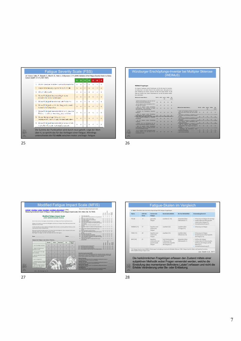
Fatigue
physisch kognitiv
subjektiv objektiv subjektiv objektiv
Handkraftmesser
Ganganalyse
- MFISphys
- FSMCmotor
- WEIMuSphys
- MFIScog
- FSMCcog
- WEIMuScog
- P300Latenz
- P300Amplitude
- *TAPVIGILANZ
- TAPAlertness
- ToT (Testverlauf)
- SPFT
HRV-Messungbelastungsabhängige Varianz v. Herzrate u. RR-Intervall
*nota: lt Studienlage zeigen sich erst ab einermind. 20minütigen Testzeit bei monotoner Belastung aussagekräftige Unterschiede (z.B. erhöhte RT-Latenzen und Auslasser). Alertness-Testungen sind sensitiver, wenn sieNach einer Phase intensiver Belastung wiederholt werden.
19
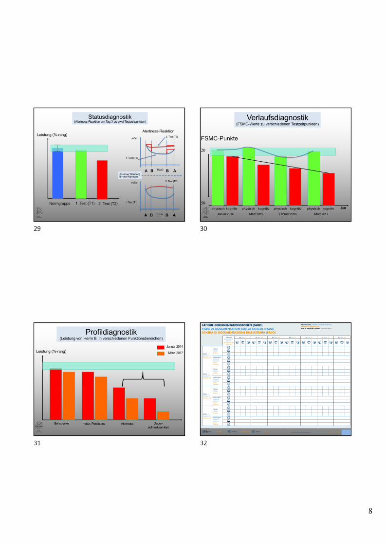
Definition und Vorkommenshäufigkeit
• Abnorme Ermüdung und Erschöpfung, „chronischer Erschöpfungszustand“
• Beeinträchtigung physischer und kognitiver Fähigkeiten
• Manifestation in jedem Stadium der MS
• Verlauf kann vorübergehend oder dauerhaft sein
• Die meisten MS-Patienten (>80%) leiden im Verlauf ihrer Erkrankung an Fatigue
• >75% der MS-Betroffenen bezeichnen die Fatigue als das am meisten belastende
Symptom der MS
Fatigue bei Multipler Sklerose
20
6
Contents lists available at ScienceDirect
Multiple Sclerosis and Related Disorders
journal homepage: www.elsevier.com/locate/msard
The disease burden of Multiple Sclerosis from the individual and populationperspective: Which symptoms matter most?
Laura Barina,⁎, Anke Salmenb,2, Giulio Disantoc,g,2, Haris Babačića, Pasquale Calabresed,Andrew Chanb, Christian P. Kammb,e, Jürg Kesselringf, Jens Kuhleg, Claudio Gobbic,Caroline Poth, Milo A. Puhana, Viktor von Wyla,1, for the Swiss Multiple Sclerosis Registry (SMSR)a Epidemiology, Biostatistics and Prevention Institute, University of Zurich, Zurich, SwitzerlandbDepartment of Neurology, University Hospital Bern and University of Bern, Bern, SwitzerlandcNeurocenter of southern Switzerland, Ospedale regionale di Lugano, Lugano, SwitzerlanddNeuropsychology and Behavioral Neurology Unit, Division of Molecular and Cognitive Neuroscience, Department of Psychology, University of Basel, Basel, SwitzerlandeNeurology and Neurorehabilitation Centre, Luzerner Kantonsspital, Lucerne, SwitzerlandfDepartment of Neurology & Neurorehabilitation, Rehabilitation Centre Kliniken Valens, Valens, SwitzerlandgNeurologic Clinic and Policlinic, Departments of Medicine, Biomedicine and Clinical Research, University Hospital Basel, University of Basel, Basel, Switzerlandh Laboratories of Neuroimmunology, Division of Neurology and Neuroscience Research Center, Department of Clinical Neurosciences, Lausanne University Hospital,Lausanne, Switzerland
A R T I C L E I N F O
Key-words:Quality of lifePatient care managementPatient reported outcomesEQ5DRegistriesRegression analysis
A B S T R A C T
Background: MS symptoms affect many functional domains. Knowing the specific impact of symptoms on health-related quality of life (HRQoL) is vital for successful disease and symptom management in MS. We aimed atinvestigating how specific MS symptoms contribute to the disease burden in individuals and from a populationperspective.Methods: We included 855 Swiss Multiple Sclerosis Registry participants with a relapsing-remitting form (RRMS)or a progressive form (PMS). HRQoL was measured with the EuroQol 5-Dimension EQ-5D-index and EQ-VisualAnalogue Scale (EQ-VAS) on 0–100% scales. Their associations with 20 symptoms, socio-demographic andclinical information were explored in median regression models, stratified by RRMS and PMS.Results: We included 611 participants with RRMS and 244 with PMS. In RRMS, gait (−6.5%) and balanceproblems (−5.1%) had the largest EQ-5D-index reductions, and were also important at the population level(frequencies 45% and 52%). Fatigue, depression, and spasticity (frequencies 74.1%, 31%, 38%) also contributedto the population disease burden. In PMS, spasticity, paralysis, and bowel problems had the largest impact onEQ-5D-index, both at the individual and population levels. The largest impact on EQ-VAS at population level wasassociated in RRMS with balance problems, depression, dizziness, and spasticity, while in PMS with weakness,pain, and paralysis.Conclusions: While HRQoL at population level is most affected by balance problems, spasticity, and depression inRRMS, the biggest HRQoL losses in PMS are caused by spasticity, paralysis, weakness, and pain. Many symptomswith the largest effects in individuals substantially contribute to the population disease burden.
https://doi.org/10.1016/j.msard.2018.07.013Received 2 May 2018; Received in revised form 22 June 2018; Accepted 8 July 2018
Abbreviations: AIC, Aikake's Information Criterion; CIS, Clinically Isolated Syndrome; DMT, Disease-modifying Therapy; EDSS, Expanded Disability Status Scale;EQ-5D-5L, European Quality of Life 5-Dimension 5 Level version; EQ-5D-index, European Quality of Life 5-Dimension Index; EQ-VAS, European Quality of Life VisualAnalogue Scale; EQ-VAS, European Quality of Life Visual Analogue Scale; EQ-VAS, European Quality of Life Visual Analogue Scale; HRQoL, Health-related quality oflife; MICE, Multivariate Imputation by Chained Equations; RRMS, Relapsing remitting multiple sclerosis; PwMS, Persons with multiple sclerosis; PMS, Progressivemultiple sclerosis; SMSR, Swiss Multiple Sclerosis Registry⁎ Corresponding author.
1 For the Swiss Multiple Sclerosis Registry (SMSR).2 Equal contribution.
E-mail addresses: [email protected] (L. Barin), [email protected] (A. Salmen), [email protected] (G. Disanto),[email protected] (P. Calabrese), [email protected] (A. Chan), [email protected] (C.P. Kamm),[email protected] (J. Kesselring), [email protected] (J. Kuhle), [email protected] (C. Gobbi), [email protected] (C. Pot),[email protected] (M.A. Puhan), [email protected] (V. von Wyl).
0XOWLSOH�6FOHURVLV�DQG�5HODWHG�'LVRUGHUV��������������²���
������������������(OVHYLHU�%�9��$OO�ULJKWV�UHVHUYHG�
7
Thefi
nalm
odel
forR
RMSwith
EQ-VAS
asou
tcom
ewas
adjuste
dfor
age,
sex,
diseasedu
ratio
n,recent
relapse,
anded
ucationleve
l.Fig.
2(right-han
dsid
e)show
sregressio
nco
efficien
tsan
d95
%CI.Th
esymptom
smoststr
ongly
associated
with
EQ-VAS
were
–in
orde
r-
depressio
n,tre
mor,a
ndsexu
aldy
sfunc
tion.
Themeancalib
ratio
nslo
pewas
0.82
(0.71–
0.94
),indicatin
gago
odcalib
ratio
n.Th
eco
mpletelisto
fcoe
fficien
tsfro
mtheun
ivariablean
dthemul-
tivariablemod
elsf
orRR
MSareshow
nin
theAp
pend
ix(Tab
leA4
).
Fig.
1.Freq
uenc
yof
repo
rtedsymptom
sin
parti
cipa
ntswith
RRMSan
dPM
S.RR
MS=
relapsingremittingMS,
PMS=
prog
ressiveMS,
dysf.
=dy
sfunc
tion,
p.=
prob
lems.
L.Ba
rinet
al.
0XOWLSOH�6
FOHURVLV�D
QG�5HODWHG�'LVRUG
HUV���
�����������²�
��
���
21
▶ Tab. 1 Kriterien für die Diagnose von MS-bezogener Fatigue basierend auf den Kriterien für die Feststellung von Fatigue bei Patienten mit MorbusParkinson nach Kluger et al. [5].
A. Symptome
1. Die Symptome können durch Routinehandlung des Alltags ausgelöst werden.
2. Die Symptome können nach geringer oder gar keiner Anstrengung auftreten.
3. Die Symptome begrenzen die Art, die Intensität und die Dauer der von dem Patienten ausgeübten Tätigkeit.
4. Die Symptome lassen sich durch Pausen nicht sicher auflösen bzw. erfordern verlängerte Pausen.
5. Die Symptomatik kann durch geistige Aufgaben sowie Situationen, die Daueraufmerksamkeit erfordern, wie auch soziale Interaktionenausgelöst werden.
6. Aus Angst vor einer Verschlechterung der Symptome vermeiden die Patienten anstrengende Tätigkeiten.
7. Schon leichte bis mittlere Anstrengung kann zu einer Verschlechterung der Symptome für Stunden bis Tage führen.
8. Der Symptomverlauf weist einen regelmäßigen Tagesrhythmus auf, unabhängig von denTätigkeiten, die ausgeübt werden(z. B. eine Verschlechterung der Symptomatik am Nachmittag).
9. Das Auftreten der Symptome ist nicht vorhersehbar, sie können ohne Vorwarnung und plötzlich auftreten.
B. Die Fatigue verursacht bei dem Patienten klinisch relevanten Stress oder eine Beeinträchtigung der Funktionsfähigkeit im sozialen,beruflichen oder einem anderen wichtigen Bereich.
C. Es gibt aufgrund der Krankheitsgeschichte sowie der physischen Untersuchung Hinweise, dass die Fatigue im Zusammenhang mit derErkrankung an Multipler Sklerose steht.
D. Die Symptome sind nicht primär eine Folge einer komorbiden psychiatrischen Störung (z. B. einer Depression), einer Schlafstörung(z. B. einer obstruktiven Schlafapnoe) oder anderer gesundheitlicher Störung (z. B. einer Anämie, einer Herzerkrankung).
Die Patienten müssen deutlich verminderte Energielevel oder vermehrte Anstrengung erleben, wobei das Ausmaß dieses Erlebens gegenüber den durchgeführtenAufgaben oder dem generellen Ausmaß an Aktivität unangemessen ist. Die Symptome müssen täglich für den überwiegenden Teil des Tages oder zumindest für fastalle Tage während des letzten Monats erlebt worden sein. Zudem müssen die Patienten 4 oder mehr Symptome aus dem folgenden Abschnitt A sowie die Kriterienfür die Abschnitte B, C und D erfüllen.
▶ Tab. 2 Überblick über die deutschsprachigen MS-Fatigue-Fragebögen.
Name Zahl der
Items
Formen der
Fatigue
Konstruktvalidität Re-Test-Reliabilität Anwendungsbereich
FSS [9] 9 generelleFatigue
exzellent [7, 10] akzeptabel (über6 Monate) [6, 7]
▪ Erfassung von Fatigue (Screening)▪ Insbesondere für Messung der
motorischen Fatigue [7]▪ Verlaufsbeobachtung [7]
WEIMUS [11] 17 kognitive undmotorischeFatigue
exzellent [12] exzellent (über14 Tage) [11]
▪ Erfassung von Fatigue
FSMC [13] 20 kognitive undmotorischeFatigue
exzellent [13] exzellent (über4 Wochen) [13]
▪ Erfassung von Fatigue▪ Einstufung des Schweregrades
der Fatigue [13]
MFIS [14] 21 kognitive,motorische undpsychosozialeFatigue
hoch (insbesonderein Bezug auf diemotorischen Aspekteder Fatigue) [7]
akzeptabel (über6 Monate) [6, 7]
▪ Einfluss der Fatigue▪ insbesondere für Messung des
Einflusses der Fatigue auf dieMotorik [7]
▪ Verlaufsbeobachtung [7]
FSS: Fatigue Severity Scale, WEIMUS: Würzburger Erschöpfungs-Inventar bei Multipler Sklerose, FSMC: Fatigue Scale for Motor and Cognitive Functions,MFIS: Modified Fatigue Impact Scale
254 Sander Carina et al. Diagnostik der Fatigue… Akt Neurol 2017; 44: 252–259
Übersicht
Her
unte
rgel
aden
von
: Fac
hste
lle E
-Med
ia. U
rheb
erre
chtli
ch g
esch
ützt
.
(aus: Sander et al., 2017)
22
Anleitung
Im folgenden Fragebogen geht es um alltägliche Probleme, die in direktem Zusammenhang mit einer extremen Form
von Müdigkeit (Fatigue) stehen. Unter dieser extremen Form der Müdigkeit wird ein nicht zu beherrschender Zu-
stand der Abgeschlagenheit, Erschöpfung und Energielosigkeit verstanden, der schlagartig eintritt, unabhängig von
eindeutigen äusseren Ursachen. Gemeint sind damit nicht Einzelereignisse, wie sie jeder Mensch im Verlaufe des
Tages, nach einer Anstrengung oder nach einer schlaflosen Nacht erlebt!
Bitte lesen Sie jede Aussage genau durch. Entscheiden Sie dann, inwieweit die entsprechende Aussage auf Sie und
Ihren Alltag zutrifft. Bitte treffen Sie Ihre Antwort möglichst unabhängig von Ihrem momentanen Befinden und
versuchen Sie uns ein Bild von Ihrem Zustand zu geben, wie sie ihn Tag für Tag erleben. Setzen Sie hierzu bitte ein
Kreuz in den entsprechenden Kreis (pro Aussage bitte nur ein Kreuz!).
1. Wenn ich mich längere Zeit konzentriere, erschöp-fe ich schneller als andere Menschen in meinem Alter.
2. Meine Bewegungen werden im Zustand der Er-schöpfung deutlich ungeschickter und unkoordi-nierter.
3. Wegen meiner Erschöpfungszustände brauche ich heute bei körperlichen Tätigkeiten häufigere und/oder auch längere Ruhepausen als früher.
4. Im Zustand der Erschöpfung bin ich unfähig, Ent-scheidungen zu treffen.
5. Ich fühle mich heute körperlich schneller er-schöpft als früher, wenn ich stressigen Situationen ausgesetzt bin.
6. Wegen meiner Erschöpfungszustände habe ich wesentlich weniger soziale Kontakte als früher.
7. Wegen meiner Erschöpfungszustände fällt es mir heute schwerer, etwas Neues zu lernen als früher.
Trifft gar nicht
zu
Trifft wenig
zu
Trifft teils-teils
zu
Trifft ziemlich
zu
Trifft völlig
zu
© Penner et al., 2005 1
FSMCFatigue Skala für Motorik und KognitionDatum:
ID:
Initialen:
Alter: Geschlecht: m w
Bitte umblättern
8. Berufliche Anforderungen lassen mich geistig schneller erschöpfen als früher.
9. Erschöpfungzustände spüre ich besonders stark in meinen Muskeln.
© Penner et al., 2005 2
20. Im Zustand der Erschöpfung nimmt meine Vergess-lichkeit merklich zu.
19. Wenn es heiss ist, fühle ich mich hauptsächlich körperlich extrem schwach und energielos.
10. Bei körperlicher Anstrengung über einen längeren Zeitraum habe ich mehr Mühe durchzuhalten als früher.
11. Meine Konzentrationsfähigkeit nimmt bei Stress beträchtlich ab.
12. Im Zustand der Erschöpfung bin ich weniger motiviert als andere Menschen, Tätigkeiten zu beginnen, die mit körperlicher Anstrengung verbunden sind.
13. Mein Denken verlangsamt sich zusehends, wenn es heiss ist.
14. Meine Bewegungen werden im Zustand der Er-schöpfung eindeutig langsamer.
15. Wegen meiner Erschöpfungszustände habe ich heute weniger Lust als früher, etwas zu tun, was Nachdenken erfordert.
16. Wenn sich ein Erschöpfungszustand einstellt, bin ich überhaupt nicht mehr in der Lage, schnell zu reagieren.
17. Im Zustand der Erschöpfung, kommen mir be-stimmte Worte nicht mehr in den Sinn.
18. Meine Aufmerksamkeit lässt im Erschöpfungszu-stand wesentlich schneller nach als früher.
Trifft gar nicht
zu
Trifft wenig
zu
Trifft teils-teils
zu
Trifft ziemlich
zu
Trifft völlig
zu
Bitte vergewissern Sie sich, dass Sie die Initialen Ihres Namens, Ihr Alter und Ihr Geschlecht auf Seite 1 angegeben
und bei jeder Aussage ein Kreuz gemacht haben. Vielen Dank.
FSMC
Fatigue Skala für Motorik und Kognition (FSMC)I. K. Penner, C. Raselli, M. Stöcklin, K. Opwis, L. Kappos, P. Calabrese: The FSMC (Fatigue Scale for Motor and Cognitive Functions) –validation of a new instrument to assess MS related fatigue in clinical routine. Multiple Sclerosis. 15, 2009, S. 1509–1517.
23
FSMC gesamt≥ 43Leichte Fatigue≥ 53Mittelgradige Fatigue≥ 63Schwere FatigueFSMC kognitiv≥ 22Leichte kognitive Fatigue≥ 28Mittelgradige kognitive Fatigue≥ 34Schwere kognitive FatigueFSMC motorisch≥ 22Leichte motorische Fatigue≥ 27Mittelgradige motorische Fatigue≥ 32Schwere motorische Fatigue
24
7
Die Summe der Punktzahlen wird durch neun geteilt. Liegt der Wert über 4, so spricht das für das Vorliegen einer Fatigue. Allerdings unterschiedet die FSS nicht zwischen motor. und kogn. Fatigue.
Fatigue Severity Scale (FSS)(dt. Version: Valko, P., Bassetti, C., Bloch K. E., Held, U., & Baumann, C. R. (2008) Validation of the Fatigue Severity Scale in a Swiss Cohort. SLEEP, 31 (11),1601-1607).
25
159
Anhang
WEIMuS-Fragebogen
Der folgende Fragebogen enthält Feststellungen, die Sie bitte darauf hin beurteilen,
wie häufig diese in der letzten Woche für Sie zutreffend waren. Bitte geben Sie zu
jeder Feststellung eine Antwort. Antworten Sie bitte ohne langes Grübeln. Es gibt
dabei kein „Richtig“ oder „Falsch“! Entscheidend ist, wie Sie die Situation erleben,
bzw. erlebt haben.
Während der letzten Woche ... fast nie selten manch-mal
häufig fast immer
... gehörte die Erschöpfung zu den drei, mich am meisten behindernden Beschwerden
�
�
�
�
�
... war ich aufgrund meiner Erschöpfung nicht in der Lage, klar zu denken
�
�
�
�
�
... hatte ich aufgrund meiner Erschöpfung Schwierigkeiten, meine Gedanken zu Hause oder bei der Arbeit zusammenzuhalten
�
�
�
�
�
... behinderte die Erschöpfung körperliche Betätigung
�
�
�
�
�
... hatte ich aufgrund meiner Erschöpfung Schwierigkeiten, mich zu konzentrieren
�
�
�
�
�
... führte körperliche Betätigung zu vermehrter Erschöpfung
�
�
�
�
�
... war ich aufgrund meiner Erschöpfung vergesslich
�
�
�
�
�
... hinderte mich die Erschöpfung bei der Ausführung bestimmter Aufgaben und Pflichten
�
�
�
�
�
... hatte ich aufgrund meiner Erschöpfung Schwierigkeiten, Sachen zu beenden, bei denen ich mich konzentrieren musste
�
�
�
�
�
... war ich aufgrund meiner Erschöpfung wenig motiviert, Sachen zu tun, bei denen ich mich konzentrieren musste
�
�
�
�
�
160
Anhang
Während der letzten Woche ... fast nie selten manch-mal
häufig fast immer
... hatte die Erschöpfung Einfluss auf meine Arbeit oder meine Familie oder mein soziales Leben
�
�
�
�
�
... verursachte die Erschöpfung häufig Probleme für mich
�
�
�
�
�
... war ich aufgrund meiner Erschöpfung weniger aufmerksam
�
�
�
�
�
... war aufgrund meiner Erschöpfung mein Denken verlangsamt
�
�
�
�
�
... hatte ich aufgrund meiner Erschöpfung Schwierigkeiten, über längere Zeit Dinge zu verfolgen
�
�
�
�
�
... beeinflusste die Erschöpfung meine körperliche Belastbarkeit
�
�
�
�
�
... war ich schnell erschöpft � � � � �
Würzburger Erschöpfungs-Inventar bei Multipler Sklerose(WEIMuS)
26
-ODIÚ�ED�&ATIGUE�)MPACT�3CALE�NICHT�VALIDIERTE�DEUTSCHE�6ERSION
(IER�IST�EINE�,ISTE�VON�!USSAGEN��DIE�BESCHREIBEN��WIE�&ATIGUE�EINE�0ERSON�BEEIN�USSEN�KANN�
&ATIGUE�IST�EIN�'EFÔHL�VON�PHYSISCHER�-ÔDIGKEIT�UND�6ERLUST�AN�%NERGIE��$IESE�%RFAHRUNG�MACHEN�VIELE�-ENSCHEN�IMMER�WIEDER�
)M�&ALLE�DER�-3�KANN�DIESES�'EFÔHL�DER�-ÔDIGKEIT�ÎFTER�EINTRETEN�UND�EINEN�GRηEREN�%INÛ�USS�AUF�DIE,EBENSQUALIT¼T�HABEN�ALS�GEWÎHNLICH�
"ITTE�LESEN�3IE�JEDE�!USSAGE�BEDACHT�UND�KREISEN�3IE�DIE�!USSAGE�EIN��DIE�AM�EHESTEN�AUF�)HRE�3YM PTOMATIK�ZUTRIFFT��DIE�IN�DEN�LETZTEN���7OCHEN�AUFTRAT��"EI�&RAGEN�NICHT�ZÎGERN�UND�DEN�)NTERVIEWER�BITTE�NACH�(ILFE�FRAGEN�
.AME��??????????????????� � � � � � � ���$ATUM��????????????
!UFGRUND�DER�&ATIGUE�IN�DEN�LETZTEN���7OCHEN���
.IEMALS 3ELTEN� -ANCHMAL /FT 3O�GUT�WIE�IMMER
����WAR�ICH�WENIGER�WACHSAM� 0 1 2 3 �
����HATTE�ICH�0ROBLEME�AUF MERKSAM�ZU�SEIN� 0 1 2 3 �
����WAR�ICH�NICHT�IN�DER�,AGE��KLAR�ZU�DENKEN� 0 1 2 3 �
����WAR�ICH�UNGESCHICKT�UNDUN KOORDINIERT� 0 1 2 3 �
���WAR�ICH�VERGESSLICH� 0 1 2 3 �
����MUSSTE�ICH�MICH�IN�MEINEN�PHYSISCHEN�!KTIVIT¼TEN�SELBST�ANTREIBEN�
0 1 2 3 �
����WAR�ICH�WENIGER�MOTIVIERT��ETWAS�ZU�TUN��DAS�PHYSISCHE�!NSTRENGUNG�ERFORDERT�
0 1 2 3 �
Modified Fatigue Impact Scale (MFIS)
Fatigue is a feeling of physical tiredness and lack of energy that many peopleexperience from time to time. But people who have medical conditions like MSexperience stronger feelings of fatigue more often and with greater impact than others.
Following is a list of statements that describe the effects of fatigue. Please read eachstatement carefully, the circle the one number that best indicates how often fatigue hasaffected you in this way during the past 4 weeks. (If you need help in marking yourresponses, tell the interviewer the number of the best response.) Please answer everyquestion. If you are not sure which answer to select choose the one answer that comesclosest to describing you. Ask the interviewer to explain any words or phrases that youdo not understand.
Because of my fatigue during the past 4 weeks
Nev
er
Rar
ely
Som
etim
es
Ofte
n
Alm
ost
Alw
ays
1. I have been less alert. 0 1 2 3 42. I have had difficulty paying attention for long periods of
time.0 1 2 3 4
3. I have been unable to think clearly. 0 1 2 3 44. I have been clumsy and uncoordinated. 0 1 2 3 45. I have been forgetful. 0 1 2 3 46. I have had to pace myself in my physical activities. 0 1 2 3 47. I have been less motivated to do anything that requires
physical effort.0 1 2 3 4
8. I have been less motivated to participate in socialactivities.
0 1 2 3 4
9. I have been limited in my ability to do things away fromhome.
0 1 2 3 4
10. I have trouble maintaining physical effort for longperiods.
0 1 2 3 4
11. I have had difficulty making decisions. 0 1 2 3 412. I have been less motivated to do anything that requires
thinking0 1 2 3 4
13. My muscles have felt weak 0 1 2 3 414. I have been physically uncomfortable. 0 1 2 3 415. I have had trouble finishing tasks that require thinking. 0 1 2 3 416. I have had difficulty organizing my thoughts when doing
things at home or at work.0 1 2 3 4
17. I have been less able to complete tasks that requirephysical effort.
0 1 2 3 4
Nev
er
Rar
ely
Som
etim
es
Ofte
n
Alm
ost
Alw
ays
18. My thinking has been slowed down. 0 1 2 3 419. I have had trouble concentrating. 0 1 2 3 420. I have limited my physical activities. 0 1 2 3 421. I have needed to rest more often or for longer periods. 0 1 2 3 4
Instructions for Scoring the MFISItems on the MFIS can be aggregated into three subscales (physical, cognitive, andpsychosocial), as well as into a total MFIS score. All items are scaled so that higherscores indicate a greater impact of fatigue on a person’s activities.
Physical SubscaleThis scale can range from 0 to 36. It is computed by adding raw scores onthe following items: 4+6+7+10+13+14+17+20+21.Cognitive SubscaleThis scale can range from 0 to 40. It is computed by adding raw scores onthe following items: 1+2+3+5+11+12+15+16+18+19.Psychosocial SubscaleThis scale can range from 0 to 8. It is computed by adding raw scores onthe following items: 8+9.Total MFIS ScoreThe total MFIS score can range from 0 to 84. It is computed by addingscores on the physical, cognitive, and psychosocial subscales.
usw... bis Aussage nr. 21
Modified Fatigue Impact Scale (MFIS)J D Fisk 1, P G Ritvo, L Ross, D A Haase, T J Marrie, W F Schlech (1994).Measuring functional impact of fatigue: initial validation of the fatigue impact scale; Clin. Infect. Dis. 18, 79-83.
27
▶ Tab. 1 Kriterien für die Diagnose von MS-bezogener Fatigue basierend auf den Kriterien für die Feststellung von Fatigue bei Patienten mit MorbusParkinson nach Kluger et al. [5].
A. Symptome
1. Die Symptome können durch Routinehandlung des Alltags ausgelöst werden.
2. Die Symptome können nach geringer oder gar keiner Anstrengung auftreten.
3. Die Symptome begrenzen die Art, die Intensität und die Dauer der von dem Patienten ausgeübten Tätigkeit.
4. Die Symptome lassen sich durch Pausen nicht sicher auflösen bzw. erfordern verlängerte Pausen.
5. Die Symptomatik kann durch geistige Aufgaben sowie Situationen, die Daueraufmerksamkeit erfordern, wie auch soziale Interaktionenausgelöst werden.
6. Aus Angst vor einer Verschlechterung der Symptome vermeiden die Patienten anstrengende Tätigkeiten.
7. Schon leichte bis mittlere Anstrengung kann zu einer Verschlechterung der Symptome für Stunden bis Tage führen.
8. Der Symptomverlauf weist einen regelmäßigen Tagesrhythmus auf, unabhängig von denTätigkeiten, die ausgeübt werden(z. B. eine Verschlechterung der Symptomatik am Nachmittag).
9. Das Auftreten der Symptome ist nicht vorhersehbar, sie können ohne Vorwarnung und plötzlich auftreten.
B. Die Fatigue verursacht bei dem Patienten klinisch relevanten Stress oder eine Beeinträchtigung der Funktionsfähigkeit im sozialen,beruflichen oder einem anderen wichtigen Bereich.
C. Es gibt aufgrund der Krankheitsgeschichte sowie der physischen Untersuchung Hinweise, dass die Fatigue im Zusammenhang mit derErkrankung an Multipler Sklerose steht.
D. Die Symptome sind nicht primär eine Folge einer komorbiden psychiatrischen Störung (z. B. einer Depression), einer Schlafstörung(z. B. einer obstruktiven Schlafapnoe) oder anderer gesundheitlicher Störung (z. B. einer Anämie, einer Herzerkrankung).
Die Patienten müssen deutlich verminderte Energielevel oder vermehrte Anstrengung erleben, wobei das Ausmaß dieses Erlebens gegenüber den durchgeführtenAufgaben oder dem generellen Ausmaß an Aktivität unangemessen ist. Die Symptome müssen täglich für den überwiegenden Teil des Tages oder zumindest für fastalle Tage während des letzten Monats erlebt worden sein. Zudem müssen die Patienten 4 oder mehr Symptome aus dem folgenden Abschnitt A sowie die Kriterienfür die Abschnitte B, C und D erfüllen.
▶ Tab. 2 Überblick über die deutschsprachigen MS-Fatigue-Fragebögen.
Name Zahl der
Items
Formen der
Fatigue
Konstruktvalidität Re-Test-Reliabilität Anwendungsbereich
FSS [9] 9 generelleFatigue
exzellent [7, 10] akzeptabel (über6 Monate) [6, 7]
▪ Erfassung von Fatigue (Screening)▪ Insbesondere für Messung der
motorischen Fatigue [7]▪ Verlaufsbeobachtung [7]
WEIMUS [11] 17 kognitive undmotorischeFatigue
exzellent [12] exzellent (über14 Tage) [11]
▪ Erfassung von Fatigue
FSMC [13] 20 kognitive undmotorischeFatigue
exzellent [13] exzellent (über4 Wochen) [13]
▪ Erfassung von Fatigue▪ Einstufung des Schweregrades
der Fatigue [13]
MFIS [14] 21 kognitive,motorische undpsychosozialeFatigue
hoch (insbesonderein Bezug auf diemotorischen Aspekteder Fatigue) [7]
akzeptabel (über6 Monate) [6, 7]
▪ Einfluss der Fatigue▪ insbesondere für Messung des
Einflusses der Fatigue auf dieMotorik [7]
▪ Verlaufsbeobachtung [7]
FSS: Fatigue Severity Scale, WEIMUS: Würzburger Erschöpfungs-Inventar bei Multipler Sklerose, FSMC: Fatigue Scale for Motor and Cognitive Functions,MFIS: Modified Fatigue Impact Scale
254 Sander Carina et al. Diagnostik der Fatigue… Akt Neurol 2017; 44: 252–259
Übersicht
Her
unte
rgel
aden
von
: Fac
hste
lle E
-Med
ia. U
rheb
erre
chtli
ch g
esch
ützt
.
(aus: Sander et al., 2017)
Fatigue-Skalen im Vergleich
Die herkömmlichen Fragebögen erfassen den Zustand mittels einer subjektiven Methodik wobei Fragen verwendet werden, welche die Einstufung des momentanen Befindens („state“) erfassen und nicht dieErlebte Veränderung unter Be- oder Entlastung.
28
8
Statusdiagnostik(Alertness-Reaktion am Tag X zu zwei Testzeitpunkten)
Leistung (%-rang)
1. Test (T1)Normgruppe 2. Test (T2)
A B B A
mSec
Trials
Alertness-Reaktion
A= ohne Warnton)B= mit Warnton)
1. Test (T1)
2. Test (T2)
A B B A
mSec
Trials
1. Test (T1)
2. Test (T2)
29
Zeitkognitiv
Verlaufsdiagnostik(FSMC-Werte zu verschiedenen Testzeitpunkten)
physisch kognitivphysisch kognitivphysischJanuar 2014 März 2015 Februar 2016
kognitivphysischMärz 2017
20
FSMC-Punkte
50
30
Leistung (%-rang)
Profildiagnostik(Leistung von Herrn B. in verschiedenen Funktionsbereichen)
Gehstrecke motor. Persistenz Dauer-aufmerksamkeit
Alertness
Januar 2014
März 2017
31
FATIGUE-DOKUMENTATIONSBOGEN (FADO) FICHE DE DOCUMENTATION SUR LA FATIGUE (FADO)SCHEDA DI DOCUMENTAZIONE DELL’ASTENIA (FADO)
TageszeitMoment de la
journéeMomento della
giornata
Mo, Lun, Lu Di, Mar, Ma Mi, Mer, Me Do, Jeu, Gi Fr, Ven, Ve Sa, Sam, Sa So, Dim, Do
Woche 1Semaine 1 Settimana 1
FatigueFatigueAstenia
NachtschlafSommeil nocturneSonno notturno
Woche 2Semaine 2 Settimana 2
FatigueFatigueAstenia
NachtschlafSommeil nocturneSonno notturno
Woche 3Semaine 3 Settimana 3
FatigueFatigueAstenia
NachtschlafSommeil nocturneSonno notturno
Woche 4Semaine 4 Settimana 4
FatigueFatigueAstenia
NachtschlafSommeil nocturneSonno notturno
mit freundlicher Unterstützung von, avec l’aimable soutien de, con il gentile contributo di
adaptiert nach, adapté d’après le projet du, adattato dal progetto del Prof. Dr. Pasquale Calabrese Universität Basel
morgens, matin, mattino mittags, midi, mezzogiorno abends, soir, sera
32
9
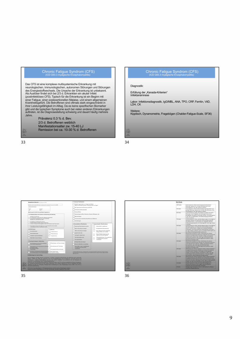
Chronic Fatigue Syndrom (CFS)(ICD G93.3 myalgische Enzephalomyelitis)
Prävalenz 0.3 % d. Bev.2/3 d. Betroffenen weiblichManifestationsalter zw. 15-40 LJRemission bei ca. 10-30 % d. Betroffenen
Das CFS ist eine komplexe multisystemische Erkrankung mit neurologischen, immunologischen, autonomen Störungen und Störungen des Energiestoffwechsels. Die Ursache der Erkrankung ist unbekannt. Als Auslöser findet sich bei 2/3 d. Erkrankten ein akuter Infekt (postinfektiöses CFS). Typisch für die Erkrankung ist ein Beginn mit einer Fatigue, einer postexertionellen Malaise, und einem allgemeinen Krankheitsgefühl. Die Betroffenen sind oftmals stark eingeschränkt in ihrer Leistungsfähigkeit im Alltag. Da es keine spezifischen Biomarker gibt und die typischen Symptome auch bei vielen anderen Erkrankungen auftreten, ist die Diagnosestellung schwierig und dauert häufig mehrere Jahre.
33
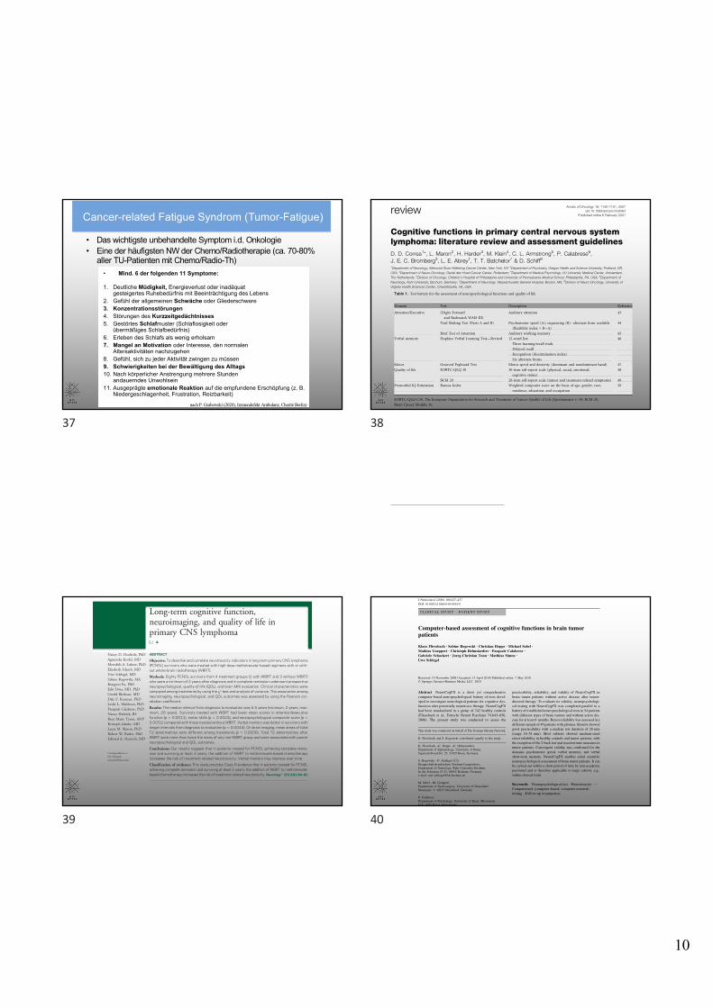
Chronic Fatigue Syndrom (CFS)(ICD G93.3 myalgische Enzephalomyelitis)
Diagnostik:
Erfüllung der „Kanada-Kriterien“Infektanamnese
Labor: Infektionsdiagnostik, IgG/MBL, ANA, TPO, CRP, Ferritin, VitD,LDH, CK
Weitere:Kipptisch, Dynamometrie, Fragebögen (Chalder-Fatigue-Scale, SF36)
34
7. Immunologische Manifestationen Schmerzhafte Lymphknoten Wiederkehrende Halsschmerzen
Neue Allergien/ Bereits bestehende Allergien haben sich verändert
Grippe Ähnliche Symptome oder allgemeines Krankheitsgefühl Überempfindlichkeit, Unverträglichkeit von Medikamenten und/oder Chemikalien
5. Autonome Manifestation Schnelle Lagewechsel (v.a. Liegen zum Stehen) führen zu Schwindel und/oder “Schwarzwerden vor Augen” Bei Lagewechsel tritt Herzrasen auf (POTS) Schwindel und Benommenheit Extreme Blässe Darmstörungen (diffuse Schmerzen, Brennen, Blähungen) oder Blasenstörungen Vasomotorische Instabilität (Instabilität des Gefäßtonus) Atemstörungen
6. Neuroendokrine Manifestation Anpassung Köpertemperatur gestört Hitze wird nicht gut vertragen Kälte wird nicht gut vertragen Appetitverlust oder Gesteigerter Appetit oder Gewichtszunahme oder Gewichtsabnahme Niedrige Blutzuckerspiegel Stress ist schlechter zu verarbeiten Stress führt zu einer Verstärkung der Erschöpfung und emotionaler Unsicherheit Carruthers BM, Kumar Jain A, De Meirleir KL, et al. Myalgic Encephalomyelitis/Chronic Fatigue Syndrom: Clinical Working Case Definition, Diagnostic and Treatment Protocols. Journal of Chronic Fatigue Syndrom 2003;11(1):7-97.
Wahrnehmungs- und Sinnesstörungen Desorientierung oder Verwirrung Bewegungskoordinationsstörungen
3. Schmerzen Gelenkschmerzen Muskelschmerzen Kopfschmerzen
Kanadische Kriterien für die Diagnose CFS/ME
modifiziert nach Carruthers BM Myalgic encephalomyelitis: International Consensus Criteria 2003
Bitte kreuzen Sie die für Sie zutreffenden Symptome an.
1. Erschöpfung/Fatigue und Zustandsverschlechterung nach Belastung deutliches Ausmaß einer neu aufgetretenen, anderweitig nicht erklärbaren körperlichen oder mentalen Erschöpfung, die zu einer erheblichen Reduktion des Aktivtätsniveaus führt Erschöpfung, Verstärkung des schweren Krankheitsgefühls und/oder Schmerzen Belastung mit einer verzögerten Erholungsphase (mehr als 24 Stunden, kann Tage dauern) Die Symptome können durch jede Art von nach körperlicher oder mentaler Anstrengung oder Stress verschlechtert werden 2. Schlafstörungen Einschlafstörungen Durchschlafstörungen Veränderter Tag-Nacht-Rhythmus Schlaf führt zu keiner Erholung 4. Neurologische/Kognitive Manifestation Beeinträchtigung der Konzentrationsfähigkeit und des Kurzzeitgedächtnisses Schwierigkeiten mit der Informationsverarbeitung, der Kategorisierung, Wortfindungsstörungen Lesestörungen Es kommt zu Überlastungserscheinungen (Rückfälle und/oder Ängste) durch: zu viele Informationen, zu viele Sinneseindrücke (zB Licht, Lärm) zu viel Stress
Patienteninformation Name …………………... Vorname …………………... geb. ……………………. Datum ……………………..
Erläuterung zur Auswertung Für die Diagnose CFS/ME müssen die Kriterien (1) Fatigue, Zustandsverschlechterung nach Belastung (post exertionelle Malaise = PEM) alle erfüllt sein, mindestens eines bei (2) Schlafstörungen und (3) Schmerzen; zwei oder mehr bei (4) neurologische / kognitive Manifestationen und eins oder mehrere Symptome von mindestens zwei der Kategorien (5) autonome, (6) neuroendokrine und (7) Immunmanifestationen. Zusätzlich muss die Erkrankung für mindestens 6 Monaten bestehen, um CFS von einer postinfektiösen Fatigue abzugrenzen. Die Zustandsverschlechterung tritt nach leichter körperlicher oder mentaler Belastung auf und ist ausgeprägt und länger anhaltend, für mehr als 24 Stunden. Während Sport Erschöpfung bei vielen Erkrankungen bessert, führt es bei CFS zu Zustandsverschlechterung. CFS ist eine Ausschlussdiagnose. CFS-Symptome können auch bei anderen Erkrankungen auftreten. Häufige komorbide Erkrankungen bei CFS sind: Fibromyalgie, Reizdarm, Hashimoto Thyreoiditis Wichtige Differentialdiagnosen von CFS
Rheumatologie Endokrinologie/Gynäkologie
Hämato/ Onkologie
Infektionen Gastro-enterologie
Neurologie
Undifferenzierte
Kollagenose/PMR/
Sjögren-Syndrom
Hashimoto-
Thyreoiditis*
Tumorfatigue Chronische
Hepatitiden
CED, Zöliakie HWS-Spinalstenosen/
Instabilität*
Fibromyalgie*
Endometriose* Lyme-Borreliose Reizdarm-
Syndrom*
Myasthenia gravis
M. Bechterew/
Psoriasisarthritis
PBC/PSC Multiple Sklerose
*kann auch als Komorbidität von CFS auftreten Häufigere Komorbiditäten von CFS
Immunologie Rheumatologie Autonome Dysfunktion
Gastro-enterologie
Neurologie Schlaf Endokrinologie/ Gynäkologie
Immunglobulin-
mangel/
Infektneigung
Fibromyalgie*
POTS/
Ruhetachykardie
Reizdarm-
Syndrom
HWS-Instabilität/
Spinalstenosen
Schlaf-
apnoe
Hashimoto-
Thyreoiditis*
Mast Cell
Aktivierungs
Syndrom
Ehlers-Danlos
Syndrom
Orthostatische
Hypotension
Nahrungsmittel
-intoleranzen
Small Fiber
Neuropathien
Restless
Leg
Syndrom
Metabolisches
Syndrom
Schwere
Allergien
Sicca Symptome
(Sjögren Syndrom
ausschließen!)
Migräne
Hypersensitivität
Endometriose*
35
Bell-Skala
100 Punkte Keine Symptome in Ruhe; keine Symptome bei körperlicher Belastung; insgesamt ein normales Aktivitätsniveau; ohne Schwierigkeiten in der Lage, Vollzeit zu arbeiten
90 Punkte Keine Symptome in Ruhe; leichte Symptome bei körperlicher und geistiger Belastung; insgesamt ein normales Aktivitätsniveau; ohne Schwierigkeiten in der Lage Vollzeit zu arbeiten
80 Punkte Leichte Symptome in Ruhe; die Symptome verstärken sich durch Belastung; nur bei Tätigkeiten, die anstrengend sind, ist eine geringfügige Leistungseinschränkungen spürbar; mit Schwierigkeiten in der Lage, an Arbeitsplätzen, die Kraftanstrengung erfordern, Vollzeit zu arbeiten
70 Punkte Leichte Symptome in Ruhe; deutliche Begrenzungen in den täglichen Aktivitäten spürbar; der funktionelle Zustand beträgt insgesamt etwa 90% der Norm – mit Ausnahme von Tätigkeiten, die einer Kraftanstrengung bedürfen; mit Schwierigkeiten in der Lage, Vollzeit zu arbeiten
60 Punkte Leichte Symptome in Ruhe; deutliche Begrenzungen in den täglichen Aktivitäten spürbar; der funktionelle Zustand beträgt insgesamt etwa 70—90% der Norm; unfähig, einer Vollzeitbeschäftigung nachzugehen, wenn dort körperliche Arbeit gefordert wird, aber in der Lage, Vollzeit zu arbeiten, wenn es um leichte Arbeiten geht und die Arbeitszeit flexibel gehandhabt werden kann
50 Punkte Mittelschwere Symptome in Ruhe; mittelschwere bis schwere Symptome bei körperlicher Belastung und Aktivität; der funktionelle Zustand ist auf 70% der Norm reduziert; unfähig, anstrengende Arbeiten durchzuführen, aber in der Lage, leichte Arbeiten oder Schreibtischarbeit für 4-5 Stunden täglich durchzuführen, wobei Ruhepausen benötigt werden
40 Punkte Mittelschwere Symptome in Ruhe; mittelschwere bis schwere Symptome bei Belastung oder Aktivität; der funktionelle Zustand ist auf 50-70% der Norm reduziert; unfähig, anstrengende Arbeiten durchzuführen, aber in der Lage, leichte Arbeiten oder Schreibtischarbeit für 3-4 Stunden täglich durchzuführen, wobei Ruhepausen benötigt werden
30 Punkte Mittelschwere bis schwere Symptome in Ruhe; schwere Symptome bei jeglicher Belastung oder Aktivität; der funktionelle Zustand ist auf 50% der Norm reduziert; in der Regel ans Haus gefesselt; unfähig, anstrengende Arbeiten durchzuführen, aber in der Lage, leichte Arbeiten oder Schreibtischarbeit für 2-3 Stunden täglich durchzuführen, wobei Ruhepausen benötigt werden
20 Punkte Mittelschwere bis schwere Symptome in Ruhe; schwere Symptome bei jeglicher Belastung oder Aktivität; der funktionelle Zustand ist auf 30-50% der Norm reduziert; bis auf seltene Ausnahmen unfähig, das Haus zu verlassen; den größten Teil des Tages ans Bett gefesselt; unfähig, sich mehr als eine Stunde am Tag zu konzentrieren
10 Punkte Schwere Symptome in Ruhe; die meiste Zeit bettlägerig; ein Verlassen des Hauses ist nicht möglich; deutliche kognitive Symptome, die eine Konzentration verhindern
0 Punkte Ständig schwere Symptome; immer ans Bett gefesselt; unfähig zu einfachsten Pflegemaßnahmen
36
10
• Das wichtigste unbehandelte Symptom i.d. Onkologie• Eine der häufigsten NW der Chemo/Radiotherapie (ca. 70-80%
aller TU-Patienten mit Chemo/Radio-Th)
Cancer-related Fatigue Syndrom (Tumor-Fatigue)
� Mind. 6 der folgenden 11 Symptome:
1. Deutliche Müdigkeit, Energieverlust oder inadäquat gesteigertes Ruhebedürfnis mit Beeinträchtigung des Lebens
2. Gefühl der allgemeinen Schwäche oder Gliederschwere3. Konzentrationsstörungen4. Störungen des Kurzzeitgedächtnisses5. Gestörtes Schlafmuster (Schlaflosigkeit oder
übermäßiges Schlafbedürfnis)6. Erleben des Schlafs als wenig erholsam7. Mangel an Motivation oder Interesse, den normalen
Altersaktivitäten nachzugehen8. Gefühl, sich zu jeder Aktivität zwingen zu müssen9. Schwierigkeiten bei der Bewältigung des Alltags10. Nach körperlicher Anstrengung mehrere Stunden
andauerndes Unwohlsein11. Ausgeprägte emotionale Reaktion auf die empfundene Erschöpfung (z. B.
Niedergeschlagenheit, Frustration, Reizbarkeit)
Tumorfatigue
Proposed ICD-10 criteria by: Cella D, et al. J Clin Oncol 2001
nach P. Grabowski (2020), Immundefekt Ambulanz, Charitè Berlin)
37
Annals of Oncology 18: 1145–1151, 2007doi:10.1093/annonc/mdl464
Published online 6 February 2007review
Cognitive functions in primary central nervous systemlymphoma: literature review and assessment guidelinesD. D. Correa1*, L. Maron2, H. Harder3, M. Klein4, C. L. Armstrong5, P. Calabrese6,J. E. C. Bromberg3, L. E. Abrey1, T. T. Batchelor7 & D. Schiff8
1Department of Neurology, Memorial Sloan-Kettering Cancer Center, New York, NY; 2Department of Psychiatry, Oregon Health and Science University, Portland, OR,
USA; 3Department of Neuro-Oncology, Daniel den Hoed Cancer Center, Rotterdam; 4Department of Medical Psychology, VU University Medical Center, Amsterdam,
The Netherlands; 5Division of Oncology, Children’s Hospital of Philadelphia and University of Pennsylvania Medical School, Philadelphia, PA, USA; 6Department of
Neurology, Ruhr-University, Bochum, Germany; 7Department of Neurology, Massachusetts General Hospital, Boston, MA; 8Division of Neuro-Oncology, University of
Virginia Health Sciences Center, Charlottesville, VA, USA
Received 30 August 2006; revised 9 November 2006; accepted 10 November 2006
Background: Treatment-related neurotoxicity has been recognized as a significant problem in patients with primary
central nervous system lymphoma (PCNSL) as effective treatment has increased survival rates. There is, however,
a paucity of research on cognitive functions in this population.
Design: In a review of the literature, a total of 17 articles that described cognitive outcome in adult PCNSL patients
were identified.
Results: The studies that assessed cognitive functions after whole-brain radiotherapy combined with chemotherapy
reported cognitive impairment in most patients. Patients treated with chemotherapy alone had either stable or
improved cognitive performance in most studies. Methodological problems, however, limited the ability to ascertain
the specific contribution of disease and various treatment interventions to cognitive outcome. On the basis of the
literature review, a battery of cognitive and quality-of-life (QoL) measures to be used in prospective clinical trials was
proposed. The battery is composed of five standardized neuropsychological tests, covering four domains sensitive to
disease and treatment effects (attention, executive functions, memory, psychomotor speed), and QoL questionnaires,
and meets criteria for use in collaborative trials.
Conclusion: The incorporation of formal and systematic cognitive evaluations in PCNSL studies will improve our
understanding of treatment-related neurotoxicity in this population.
Key words: chemotherapy, cognitive, neurotoxicity, primary CNS lymphoma, radiotherapy
introduction
Cognitive dysfunction in primary central nervous systemlymphoma (PCNSL) patients is related to multiple factorsincluding the effects of the tumor itself given its infiltrative andmultifocal pattern, age, and the delayed effects of whole-brainradiotherapy (WBRT) and high-dose methotrexate-basedchemotherapy (HD-MTX) either combined or alone [1].Delayed treatment-related cognitive dysfunction has beenrecognized as a significant problem as effective treatment ofPCNSL has increased survival rates [1–3]. Neurotoxicity isconsidered the most frequent complication among long-termsurvivors [4], and may interfere with the patient’s ability tofunction at premorbid levels, despite adequate disease control[5, 6]. The specific contribution of the disease itself and varioustreatment modalities to cognitive dysfunction remains to beelucidated as the neurotoxic potential of combined treatments is
difficult to determine when each can produce CNS damageindividually [7].Consistent with the current status of research on patients
with other brain tumor histologies there is also a paucity ofinformation regarding cognitive functions in PCNSL patients.Most studies reported performance status and mental statusexaminations [8], but neuropsychological evaluations wereseldom included. The former methods have low sensitivity todetect cognitive dysfunction in brain tumor patients [5, 9, 10].For instance, the widely used Mini-Mental State Examination(MMSE) does not assess domains often disrupted by treatmenteffects such as psychomotor speed and executive functions.Considering that insensitive instruments were used in mostclinical trials, only severe neurotoxicity resulting in significantdisability was documented, indicating that the incidence ofcognitive dysfunction in PCNSL was underestimated [11–13].The relevance of including cognitive outcome measures in
neurooncology research has been recognized by the NationalCancer Institute (BTPRG, 2000) and many professionals in thefield [10, 14, 15]. The International Primary CNS Lymphoma
review
*Correspondence to: Dr D. D. Correa, Department of Neurology, Memorial
Sloan-Kettering Cancer Center, 1275 York Avenue, New York, NY, USA 10021.
Tel: +1-212-639-2082; Fax: +1-646-422-2235; E-mail: [email protected]
ª 2007 European Society for Medical Oncology
of pretreatment cognitive assessments and treatmentrandomization; however, comparisons between patients treatedwith combined modality therapy versus chemotherapy aloneindicated greater cognitive impairments in patients who alsohad WBRT.The findings on cognitive outcome in PCNSL patients treated
with combined modality therapy are consistent with theliterature on treatment-related cognitive dysfunction in patientswith primary or metastatic brain tumors who receivedradiotherapy alone or in combination with high-dosechemotherapy, or prophylactic WBRT. These studies indicatethat cognitive impairments are diffuse, and the domains mostsensitive to treatment effects include attention and executivefunctions, learning and retrieval of new information, andpsychomotor speed [10, 32–34]. Deficits in these domains havealso been documented in diseases that affect the white matteror cortical–subcortical circuits [35].The studies that reported cognitive outcome in PCNSL
patients treated with HD-MTX alone or with BBBDchemotherapy were mostly prospective. Several studiesdocumented cognitive impairment before therapy in the areas ofattention, executive functions, memory, psychomotor speed,and language (word fluency, naming). Posttreatment follow-upintervals were variable across studies, but several reported eitherstable or improved cognitive performance [19–23]; in asubset of patients there was a decline in attention, memory,psychomotor speed, and word fluency [18, 20, 21, 24]. Of note,neuropsychological tests and follow-up intervals were notalways specified, and some studies included patients with onlya PR or tumor recurrence. Tumor progression is a factor thatoften confounds cognitive outcome in studies of brain tumortreatment-related neurotoxicity [32, 36], indicating that datashould be censored or separated once tumor progression hasbeen identified. The review indicated that methodologicalproblems in several of these studies limited the ability to discern
the specific contributions of the disease and chemotherapy-onlyregimens to cognitive dysfunction. Overall, only four studiesused self-report measures of QoL and three reported estimatesof premorbid IQ [5, 6, 21, 24].
proposed neuropsychological andQoL test battery
In light of the recognized relevance of studying cognitiveoutcome and QoL in PCNSL patients and the paucity ofsystematic studies in the literature, a core neuropsychologicaltest battery is proposed to be used in prospective clinical trials.The neuropsychological measures meet the following criteria:(i) assess several domains found to be most sensitive to tumorand treatment effects; (ii) have standardized materials andadministration procedures; (iii) have published normative data;(iv) have moderate to high test–retest reliability; (v) havealternate forms or are relatively insensitive to practice effects,and are therefore suitable to monitor changes in cognitivefunction over time; (vi) include tests that have been translatedinto several languages (i.e. Dutch, English, French, German,Italian) [37] or require translation primarily of test directions;(vii) can be administered by a trained psychometrician orclinical research associate under supervision of aneuropsychologist, and standardized training proceduresfollowed by certification should be required for all individualsinvolved in test administration [37]; and (viii) totaladministration time is !30–40 min for the cognitive testbattery, and 10 min for the self-report measures.The cognitive domains deemed essential to be evaluated
include attention, executive functions (i.e. working memory,processing speed, sequencing abilities), verbal memory, andmotor speed (Table 1). In the assessment of executive functions,it is important to include a measure that is not confoundedby motor difficulties considering that a significant number of
Table 1. Test battery for the assessment of neuropsychological functions and quality of life
Domain Test Description Reference
Attention/Executive (Digits Forward
and Backward; WAIS-III)
Auditory attention 43
Trail Making Test (Parts A and B) Psychomotor speed (A); sequencing (B)"alternate form available
(flexibility index = B"A)
44
Brief Test of Attention Auditory working memory 45
Verbal memory Hopkins Verbal Learning Test—Revised 12-word list: 46
Three learning/recall trials
Delayed recall
Recognition (discrimination index)
Six alternate forms
Motor Grooved Pegboard Test Motor speed and dexterity (dominant and nondominant hand) 47
Quality of life EORTC-QLQ 30 30-item self-report scale (physical, social, emotional,
cognitive status)
48
BCM 20 20-item self-report scale (tumor and treatment-related symptoms) 49
Premorbid IQ Estimation Barona Index Weighted composite score on the basis of age, gender, race,
residence, education, and occupation
50
EORTC-QLQ-C30, The European Organization for Research and Treatment of Cancer Quality of Life Questionnaire C-30; BCM 20,
Brain Cancer Module 20.
Annals of Oncology review
Volume 18 | No. 7 | July 2007 doi:10.1093/annonc/mdl464 | 114938
Nancy D. Doolittle, PhDAgnieszka Korfel, MDMeredith A. Lubow, PhDElisabeth Schorb, MDUwe Schlegel, MDSabine Rogowski, MARongwei Fu, PhDEdit Dósa, MD, PhDGerald Illerhaus, MDDale F. Kraemer, PhDLeslie L. Muldoon, PhDPasquale Calabrese, PhDNancy Hedrick, BSRose Marie Tyson, ANPKristoph Jahnke, MDLeeza M. Maron, PhDRobert W. Butler, PhDEdward A. Neuwelt, MD
Correspondence toDr. Neuwelt:[email protected]
Supplemental data atwww.neurology.org
Long-term cognitive function,neuroimaging, and quality of life inprimary CNS lymphoma
ABSTRACT
Objective: To describe and correlate neurotoxicity indicators in long-term primary CNS lymphoma(PCNSL) survivors who were treated with high-dose methotrexate–based regimens with or with-out whole-brain radiotherapy (WBRT).
Methods: Eighty PCNSL survivors from 4 treatment groups (1 with WBRT and 3 without WBRT)who were a minimum of 2 years after diagnosis and in complete remission underwent prospectiveneuropsychological, quality-of-life (QOL), and brain MRI evaluation. Clinical characteristics werecompared among treatments by using the x2 test and analysis of variance. The association amongneuroimaging, neuropsychological, and QOL outcomes was assessed by using the Pearson cor-relation coefficient.
Results: The median interval from diagnosis to evaluation was 5.5 years (minimum, 2 years; max-imum, 26 years). Survivors treated with WBRT had lower mean scores in attention/executivefunction (p 5 0.0011), motor skills (p 5 0.0023), and neuropsychological composite score (p 5
0.0051) compared with those treated without WBRT. Verbal memory was better in survivors withlonger intervals from diagnosis to evaluation (p 5 0.0045). On brain imaging, mean areas of totalT2 abnormalities were different among treatments (p 5 0.0006). Total T2 abnormalities afterWBRT were more than twice the mean of any non-WBRT group and were associated with poorerneuropsychological and QOL outcomes.
Conclusions: Our results suggest that in patients treated for PCNSL achieving complete remis-sion and surviving at least 2 years, the addition of WBRT to methotrexate-based chemotherapyincreases the risk of treatment-related neurotoxicity. Verbal memory may improve over time.
Classification of evidence: This study provides Class III evidence that in patients treated for PCNSLachieving complete remission and surviving at least 2 years, the addition of WBRT to methotrexate-based chemotherapy increases the risk of treatment-related neurotoxicity. Neurology! 2013;81:84–92
GLOSSARYASCT 5 autologous stem cell transplantation; BBBD 5 blood–brain barrier disruption; CI 5 confidence interval; EORTC 5European Organization for Research and Treatment of Cancer;MR5magnetic resonance; PCNSL5 primary CNS lymphoma;QLQ-C30 5 QOL Questionnaire–30; QOL 5 quality of life; WBRT 5 whole-brain radiotherapy.
High-dose methotrexate is the most widely used drug for primary CNS lymphoma (PCNSL). Incombination with whole-brain radiotherapy (WBRT), high-dose methotrexate improved survivalrates over WBRT alone. However, delayed treatment-related neurotoxicity emerged as a significantdisabling complication of the combined treatment.1–9 Single-drug and multidrug high-dose meth-otrexate–based regimens without WBRT have been used in an effort to increase survival whileavoiding the risk of delayed neurotoxicity.10–15 In patients older than 60 years treated with WBRT,virtually all long-term survivors develop this complication. In patients younger than 60 years,neurotoxicity rates ranging from 26% to 63% have been reported.2,6 However, the true risk of
From the Oregon Health & Science University (N.D.D., M.A.L., R.F., L.L.M., N.H., R.M.T., L.M.M., R.W.B., E.A.N.), Portland; Charité-Universitätsmedizin Berlin (A.K., K.J.), Campus Benjamin Franklin, Berlin; Freiburg University Medical Center (E.S., G.I.), Freiburg; Knapp-schaftskrankenhaus (U.S., S.R.), University Hospital Bochum, Bochum, Germany; Semmelweis University (E.D.), Budapest, Hungary; Universityof Florida (D.F.K.), Jacksonville; University of Basel (P.C.), Basel, Switzerland; and Department of Veterans Affairs Medical Center (E.A.N.),Portland, OR.
Presented in part at the 11th International Conference on Malignant Lymphoma, Lugano, Switzerland, June 15–18, 2011 (abstract 036), and at theAmerican Society of Clinical Oncology Annual Meeting, Chicago, IL, June 1–5, 2012 (abstract 2040).
Go to Neurology.org for full disclosures. Funding information and disclosures deemed relevant by the authors, if any, are provided at the end of the article.
84 © 2013 American Academy of Neurology
39
CLINICAL STUDY – PATIENT STUDY
Computer-based assessment of cognitive functions in brain tumorpatients
Klaus Fliessbach • Sabine Rogowski • Christian Hoppe • Michael Sabel •
Mathias Goeppert • Christoph Helmstaedter • Pasquale Calabrese •
Gabriele Schackert • Joerg-Christian Tonn • Matthias Simon •
Uwe Schlegel
Received: 10 November 2009 / Accepted: 13 April 2010 / Published online: 7 May 2010! Springer Science+Business Media, LLC. 2010
Abstract NeuroCogFX is a short yet comprehensivecomputer-based neuropsychological battery of tests devel-
oped to investigate neurological patients for cognitive dys-
function after potentially neurotoxic therapy. NeuroCogFXhad been standardized in a group of 242 healthy controls
(Fliessbach et al., Fortschr Neurol Psychiatr 74:643–650,
2006). The present study was conducted to assess the
practicability, reliability, and validity of NeuroCogFX inbrain tumor patients without active disease after tumor-
directed therapy. To evaluate its validity, neuropsychologi-
cal testing with NeuroCogFX was completed parallel to abattery of established neuropsychological tests in 54 patients
with different types of brain tumors and without active dis-
ease for at least 6 months. Retest reliability was assessed in adifferent sample of 49 patients with gliomas. Results showed
good practicability with a median test duration of 28 min
(range 16–51 min). Most subtests showed medium-sizedretest reliability in healthy controls and tumor patients, with
the exception of the 2-back test and reaction time measures in
tumor patients. Convergent validity was confirmed for thedomains psychomotor speed, verbal memory, and verbal
short-term memory. NeuroCogFX enables serial scientific
neuropsychological assessment of brain tumor patients. It canbe carried out within a short period of time by non-academic
personnel and is therefore applicable to large cohorts, e.g.,
within clinical trials.
Keywords Neuropsychological test ! Neurotoxicity !Computerized (computer-based, computer-assisted)
testing ! Follow-up examination
Introduction
Brain tumor patients often present with cognitive dysfunc-
tion such as psychomotor slowing, attention and memorydeficits, and executive dysfunction or focal deficits such as
aphasia or apraxia [1]. Successful tumor therapy ideally
improves these symptoms [2, 3]. However, with the adventof effective treatment regimens and with increasing survival
times delayed treatment-related cognitive dysfunction is
more frequently recognized as a significant problem [4]. For
This study was conducted on behalf of The German Glioma Network.
K. Fliessbach and S. Rogowski contributed equally to this study.
K. Fliessbach ! C. Hoppe ! C. HelmstaedterDepartment of Epileptology, University of Bonn,Sigmund-Freud-Str. 25, 53105 Bonn, Germany
S. Rogowski ! U. Schlegel (&)Knappschaftskrankenhaus Bochum-Langendreer,Department of Neurology, Ruhr University Bochum,In der Schornau 23-25, 44892 Bochum, Germanye-mail: [email protected]
M. Sabel ! M. GoeppertDepartment of Neurosurgery, University of Dusseldorf,Moorenstr. 5, 40225 Dusseldorf, Germany
P. CalabreseDepartment of Psychology, University of Basel, Missionsstr.62A, 4055 Basel, Switzerland
G. SchackertDepartment of Neurosurgery, University of Dresden, Fetscherstr.74, 01307 Dresden, Germany
J.-C. TonnDepartment of Neurosurgery, University of Munich,Marchioninistr. 15, 81377 Munich, Germany
M. SimonDepartment of Neurosurgery, University of Bonn,Sigmund-Freud-Str. 25, 53105 Bonn, Germany
123
J Neurooncol (2010) 100:427–437
DOI 10.1007/s11060-010-0194-9
40
11
Bell-Skala
100 Punkte Keine Symptome in Ruhe; keine Symptome bei körperlicher Belastung; insgesamt ein normales Aktivitätsniveau; ohne Schwierigkeiten in der Lage, Vollzeit zu arbeiten
90 Punkte Keine Symptome in Ruhe; leichte Symptome bei körperlicher und geistiger Belastung; insgesamt ein normales Aktivitätsniveau; ohne Schwierigkeiten in der Lage Vollzeit zu arbeiten
80 Punkte Leichte Symptome in Ruhe; die Symptome verstärken sich durch Belastung; nur bei Tätigkeiten, die anstrengend sind, ist eine geringfügige Leistungseinschränkungen spürbar; mit Schwierigkeiten in der Lage, an Arbeitsplätzen, die Kraftanstrengung erfordern, Vollzeit zu arbeiten
70 Punkte Leichte Symptome in Ruhe; deutliche Begrenzungen in den täglichen Aktivitäten spürbar; der funktionelle Zustand beträgt insgesamt etwa 90% der Norm – mit Ausnahme von Tätigkeiten, die einer Kraftanstrengung bedürfen; mit Schwierigkeiten in der Lage, Vollzeit zu arbeiten
60 Punkte Leichte Symptome in Ruhe; deutliche Begrenzungen in den täglichen Aktivitäten spürbar; der funktionelle Zustand beträgt insgesamt etwa 70—90% der Norm; unfähig, einer Vollzeitbeschäftigung nachzugehen, wenn dort körperliche Arbeit gefordert wird, aber in der Lage, Vollzeit zu arbeiten, wenn es um leichte Arbeiten geht und die Arbeitszeit flexibel gehandhabt werden kann
50 Punkte Mittelschwere Symptome in Ruhe; mittelschwere bis schwere Symptome bei körperlicher Belastung und Aktivität; der funktionelle Zustand ist auf 70% der Norm reduziert; unfähig, anstrengende Arbeiten durchzuführen, aber in der Lage, leichte Arbeiten oder Schreibtischarbeit für 4-5 Stunden täglich durchzuführen, wobei Ruhepausen benötigt werden
40 Punkte Mittelschwere Symptome in Ruhe; mittelschwere bis schwere Symptome bei Belastung oder Aktivität; der funktionelle Zustand ist auf 50-70% der Norm reduziert; unfähig, anstrengende Arbeiten durchzuführen, aber in der Lage, leichte Arbeiten oder Schreibtischarbeit für 3-4 Stunden täglich durchzuführen, wobei Ruhepausen benötigt werden
30 Punkte Mittelschwere bis schwere Symptome in Ruhe; schwere Symptome bei jeglicher Belastung oder Aktivität; der funktionelle Zustand ist auf 50% der Norm reduziert; in der Regel ans Haus gefesselt; unfähig, anstrengende Arbeiten durchzuführen, aber in der Lage, leichte Arbeiten oder Schreibtischarbeit für 2-3 Stunden täglich durchzuführen, wobei Ruhepausen benötigt werden
20 Punkte Mittelschwere bis schwere Symptome in Ruhe; schwere Symptome bei jeglicher Belastung oder Aktivität; der funktionelle Zustand ist auf 30-50% der Norm reduziert; bis auf seltene Ausnahmen unfähig, das Haus zu verlassen; den größten Teil des Tages ans Bett gefesselt; unfähig, sich mehr als eine Stunde am Tag zu konzentrieren
10 Punkte Schwere Symptome in Ruhe; die meiste Zeit bettlägerig; ein Verlassen des Hauses ist nicht möglich; deutliche kognitive Symptome, die eine Konzentration verhindern
0 Punkte Ständig schwere Symptome; immer ans Bett gefesselt; unfähig zu einfachsten Pflegemaßnahmen
AnamneseKanadische Kriterien InfektanamneseAutoimmunanamneseDD Fatigue
Labor• CrP nicht erhöht• Infektionsdiagnostik nach Anamnese• Immundefektdiagnostik bei Infekten• Autoantikörper (ANA, TPO, u,w.)• Ferritin, Vit D, (EBV-PCR)
CFS ± Diagnostik
Weitere DiagnostikTachykardie/POTSChalder Fatigue ScaleSF36COMPASS
41
Vigilanz
Konzentration geistiges TempoFlexibilität
Gedächtnis SpracheTermine
VereinbarungenFachwissen
Instruktionen...
KundenkontakteTeamwork
(Re-)PräsentationKommunikation...
ArbeitseffizienzAneignung neuer Techniken
zeitgebundene Aufträgeevtl. Akkordarbeit...
FokussierungAblenkbarkeit
Detailgenauigkeit...
DurchhaltevermögenDaueraufmerksamkeit
SchichtarbeitÜberstunden...
Multi-taskingPlanung von Abläufen
Absprache- u. Kritikfähigkeit...
42
Zusammenfassung I
• Relevante Kernsymptome beachten• Alltagsrelevanz berücksichtigen• Kausalen Zusammenhang mit
Grunderkrankung herstellen• Andere somatische und psychische
Ursachen ausschliessen• Fragen, zählen, messen (testen)
43
Zusammenfassung II• Selbsteinschätzungs- vs Performanzverfahren• Reaktionszeitbasierte („speed“) vs „Power“ Tests• Unterscheidung zw. eher körperl. u. eher kognitiver
Fatigue (z.B. Alertness vs SPFT)
• Erfassung eines Fatigue-Profils (z.B. FADO)• Mehrere Testzeitpunkte wählen• Dauer der Belastung mit eher monotonen Charakter
als aussagekräftiger Indikator• Beachtung der „Alertness-Reaktion“ nach intensiver
Belastung
44

![Original Article Chronic fatigue syndrome and … · 2, 1-16 Original Article Chronic fatigue syndrome and mitochondrial dysfunction Sarah Myhill 1 ... 10]. One consequence of mitochondrial](https://img.pdfslide.net/doc/110x75/5baf424e09d3f2c70e8c26a0/original-article-chronic-fatigue-syndrome-and-2-1-16-original-article-chronic.jpg)