Embed Size (px)
Citation preview
Obscure GI Bleeding
Kathy Bull-Henry, MD
Georgetown University Hospital
Division of Gastroenterology
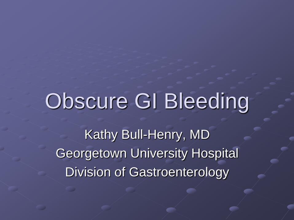
GI Bleeding Definitions
Obscure GI BleedingDefinition
Bleeding of unknown origin that persists or
recurs after negative colonoscopy and
negative upper endoscopy
Recurrent or persistent bleeding
FOBT positive
IDA
Visible bleeding
Melena, hematemesis, hematochezia, coffee
grounds
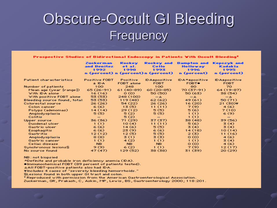
Obscure-Occult GI Bleeding Frequency
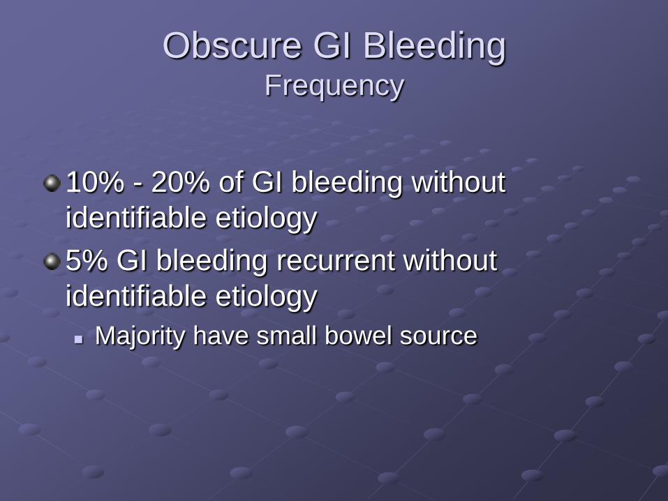
Obscure GI BleedingFrequency
10% - 20% of GI bleeding without
identifiable etiology
5% GI bleeding recurrent without
identifiable etiology
Majority have small bowel source
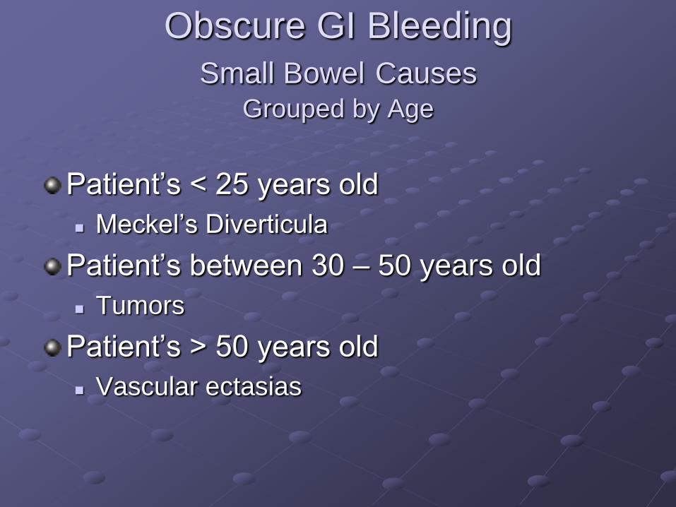
Obscure GI Bleeding
Small Bowel CausesGrouped by Age
Patient’s < 25 years old
Meckel’s Diverticula
Patient’s between 30 – 50 years old
Tumors
Patient’s > 50 years old
Vascular ectasias
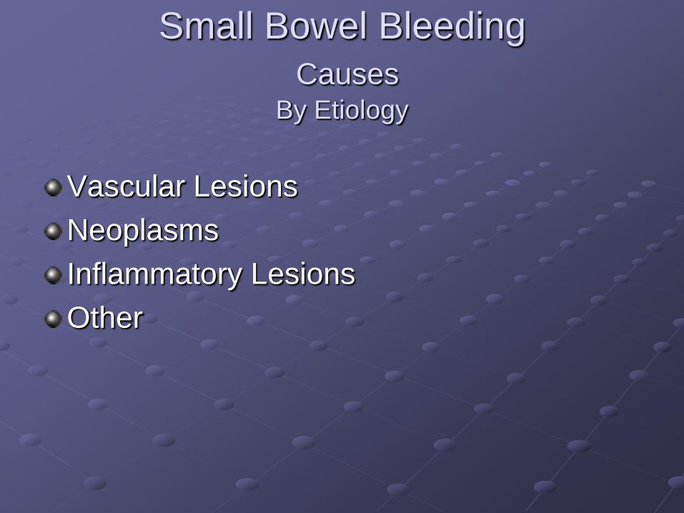
Small Bowel Bleeding
CausesBy Etiology
Vascular Lesions
Neoplasms
Inflammatory Lesions
Other
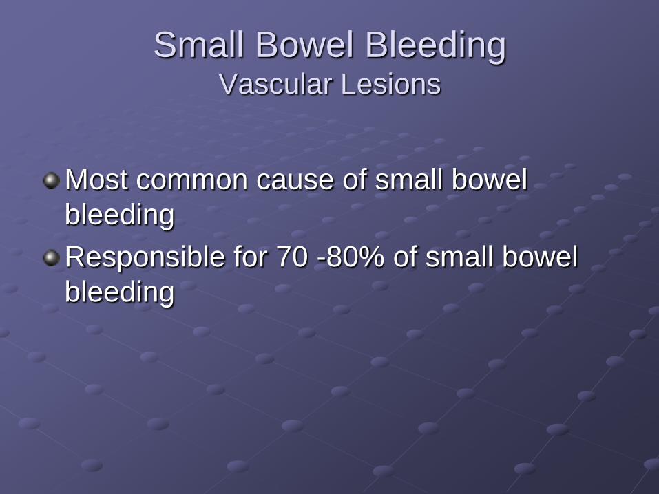
Small Bowel BleedingVascular Lesions
Most common cause of small bowel
bleeding
Responsible for 70 -80% of small bowel
bleeding
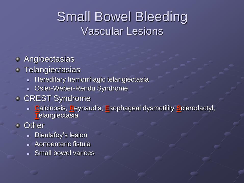
Small Bowel BleedingVascular Lesions
Angioectasias
Telangiectasias Hereditary hemorrhagic telangiectasia
Osler-Weber-Rendu Syndrome
CREST Syndrome Calcinosis, Reynaud’s, Esophageal dysmotility Sclerodactyl,
Telangiectasia
Other Dieulafoy’s lesion
Aortoenteric fistula
Small bowel varices
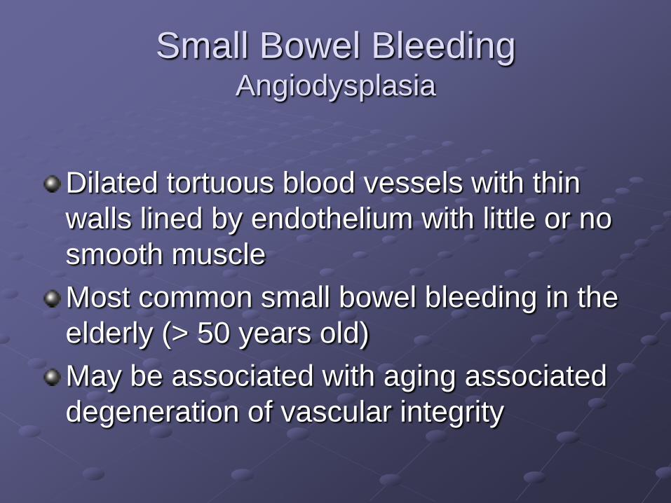
Small Bowel BleedingAngiodysplasia
Dilated tortuous blood vessels with thin
walls lined by endothelium with little or no
smooth muscle
Most common small bowel bleeding in the
elderly (> 50 years old)
May be associated with aging associated
degeneration of vascular integrity
Small Bowel Bleeding
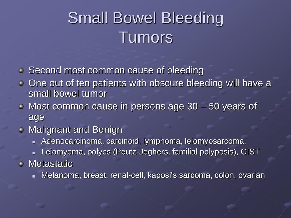
Tumors
Second most common cause of bleeding
One out of ten patients with obscure bleeding will have a small bowel tumor
Most common cause in persons age 30 – 50 years of age
Malignant and Benign Adenocarcinoma, carcinoid, lymphoma, leiomyosarcoma,
Leiomyoma, polyps (Peutz-Jeghers, familial polyposis), GIST
Metastatic Melanoma, breast, renal-cell, kaposi’s sarcoma, colon, ovarian
Causes of Small Bowel Bleeding
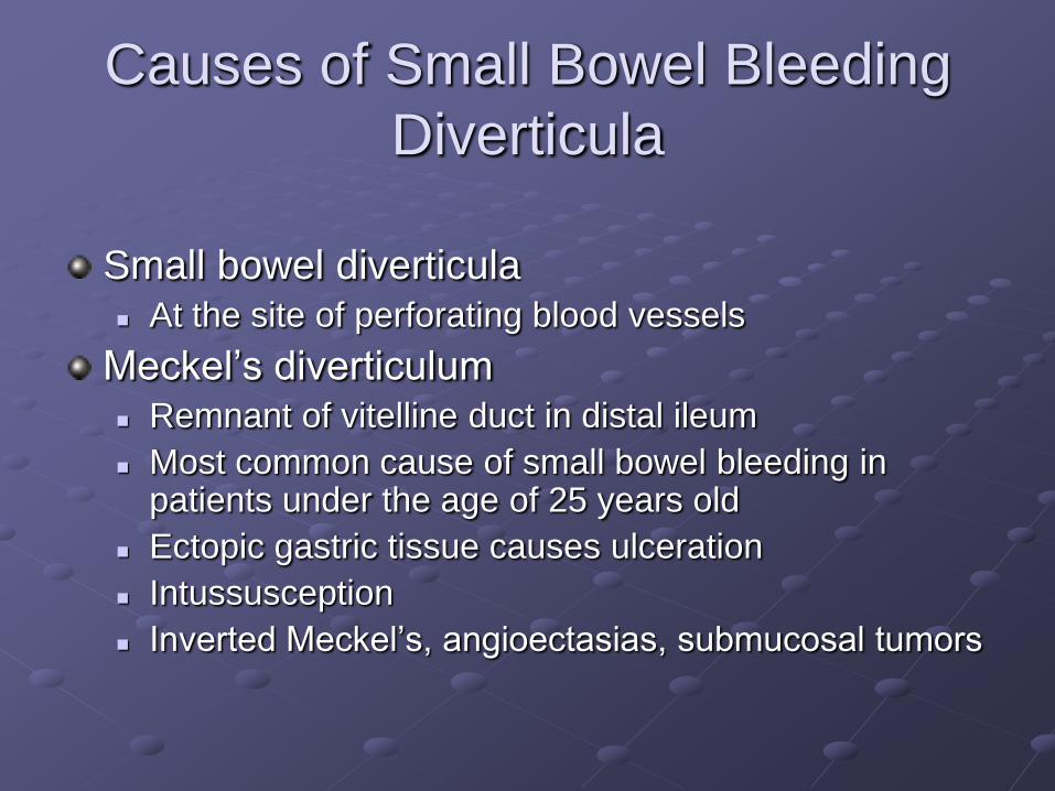
Diverticula
Small bowel diverticula At the site of perforating blood vessels
Meckel’s diverticulum Remnant of vitelline duct in distal ileum
Most common cause of small bowel bleeding in patients under the age of 25 years old
Ectopic gastric tissue causes ulceration
Intussusception
Inverted Meckel’s, angioectasias, submucosal tumors
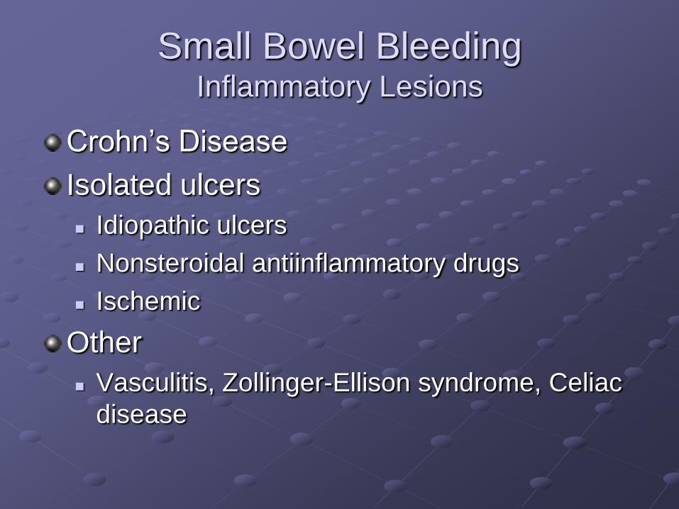
Small Bowel BleedingInflammatory Lesions
Crohn’s Disease
Isolated ulcers
Idiopathic ulcers
Nonsteroidal antiinflammatory drugs
Ischemic
Other
Vasculitis, Zollinger-Ellison syndrome, Celiac
disease
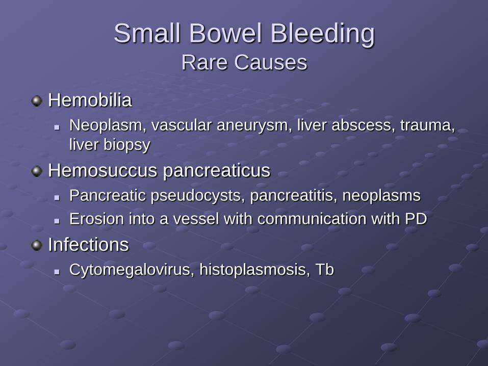
Small Bowel BleedingRare Causes
Hemobilia
Neoplasm, vascular aneurysm, liver abscess, trauma,
liver biopsy
Hemosuccus pancreaticus
Pancreatic pseudocysts, pancreatitis, neoplasms
Erosion into a vessel with communication with PD
Infections
Cytomegalovirus, histoplasmosis, Tb
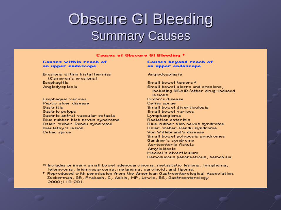
Obscure GI BleedingSummary Causes
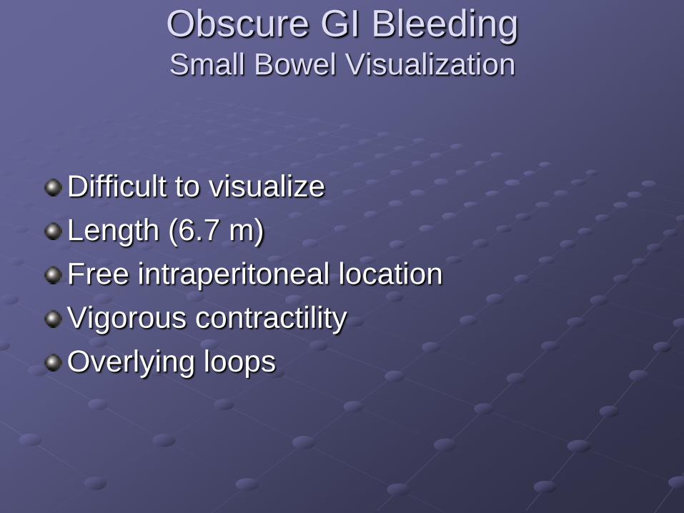
Obscure GI BleedingSmall Bowel Visualization
Difficult to visualize
Length (6.7 m)
Free intraperitoneal location
Vigorous contractility
Overlying loops
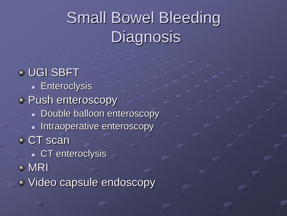
Small Bowel Bleeding
Diagnosis
UGI SBFT Enteroclysis
Push enteroscopy Double balloon enteroscopy
Intraoperative enteroscopy
CT scan CT enteroclysis
MRI
Video capsule endoscopy
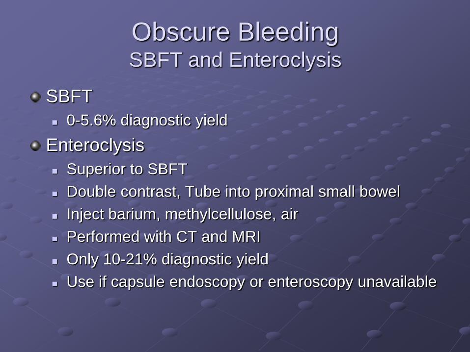
Obscure BleedingSBFT and Enteroclysis
SBFT
0-5.6% diagnostic yield
Enteroclysis
Superior to SBFT
Double contrast, Tube into proximal small bowel
Inject barium, methylcellulose, air
Performed with CT and MRI
Only 10-21% diagnostic yield
Use if capsule endoscopy or enteroscopy unavailable
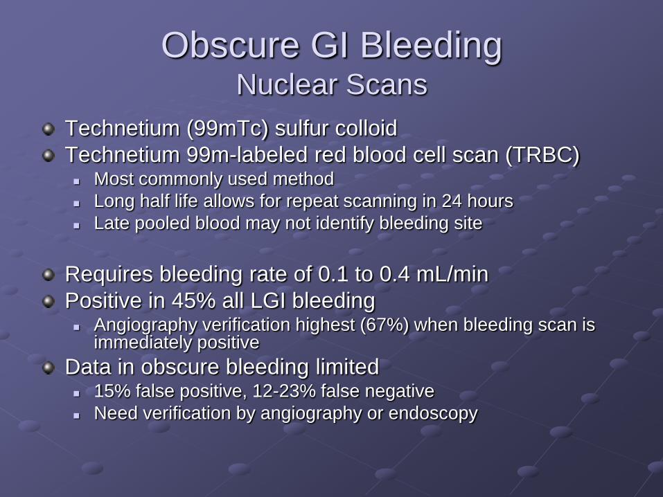
Obscure GI BleedingNuclear Scans
Technetium (99mTc) sulfur colloid
Technetium 99m-labeled red blood cell scan (TRBC) Most commonly used method
Long half life allows for repeat scanning in 24 hours
Late pooled blood may not identify bleeding site
Requires bleeding rate of 0.1 to 0.4 mL/min
Positive in 45% all LGI bleeding Angiography verification highest (67%) when bleeding scan is
immediately positive
Data in obscure bleeding limited 15% false positive, 12-23% false negative
Need verification by angiography or endoscopy
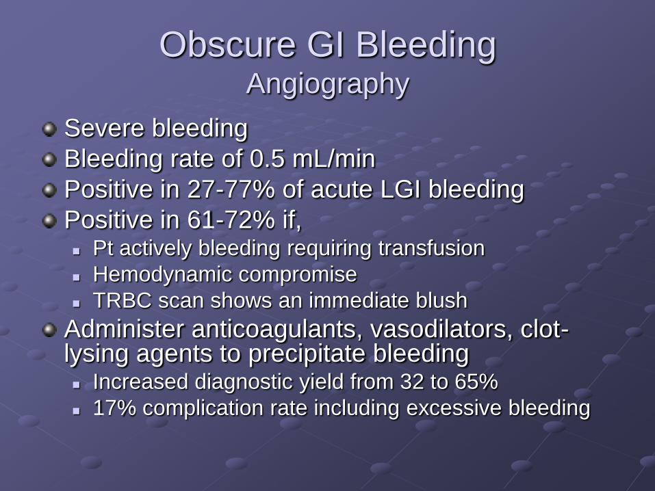
Obscure GI BleedingAngiography
Severe bleeding
Bleeding rate of 0.5 mL/min
Positive in 27-77% of acute LGI bleeding
Positive in 61-72% if, Pt actively bleeding requiring transfusion
Hemodynamic compromise
TRBC scan shows an immediate blush
Administer anticoagulants, vasodilators, clot-lysing agents to precipitate bleeding Increased diagnostic yield from 32 to 65%
17% complication rate including excessive bleeding
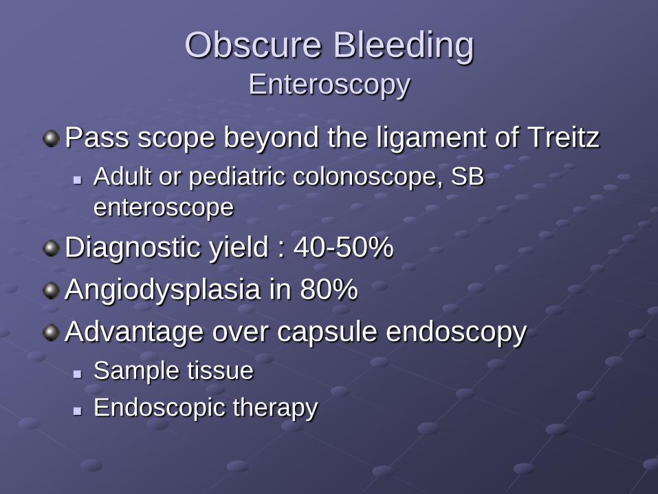
Obscure BleedingEnteroscopy
Pass scope beyond the ligament of Treitz
Adult or pediatric colonoscope, SB
enteroscope
Diagnostic yield : 40-50%
Angiodysplasia in 80%
Advantage over capsule endoscopy
Sample tissue
Endoscopic therapy
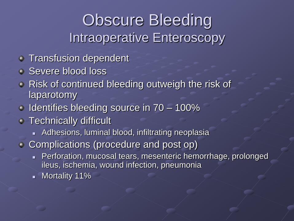
Obscure BleedingIntraoperative Enteroscopy
Transfusion dependent
Severe blood loss
Risk of continued bleeding outweigh the risk of laparotomy
Identifies bleeding source in 70 – 100%
Technically difficult Adhesions, luminal blood, infiltrating neoplasia
Complications (procedure and post op) Perforation, mucosal tears, mesenteric hemorrhage, prolonged
ileus, ischemia, wound infection, pneumonia
Mortality 11%
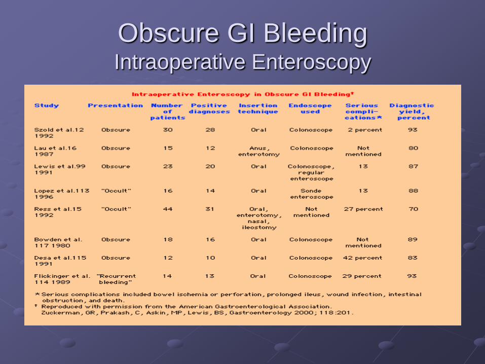
Obscure GI BleedingIntraoperative Enteroscopy
Obscure GI BleedingExploratory Laparotomy
Seldom without intraoperative enteroscopy
65% of 37 pt’s had lesion identified by
palpation or transillumination
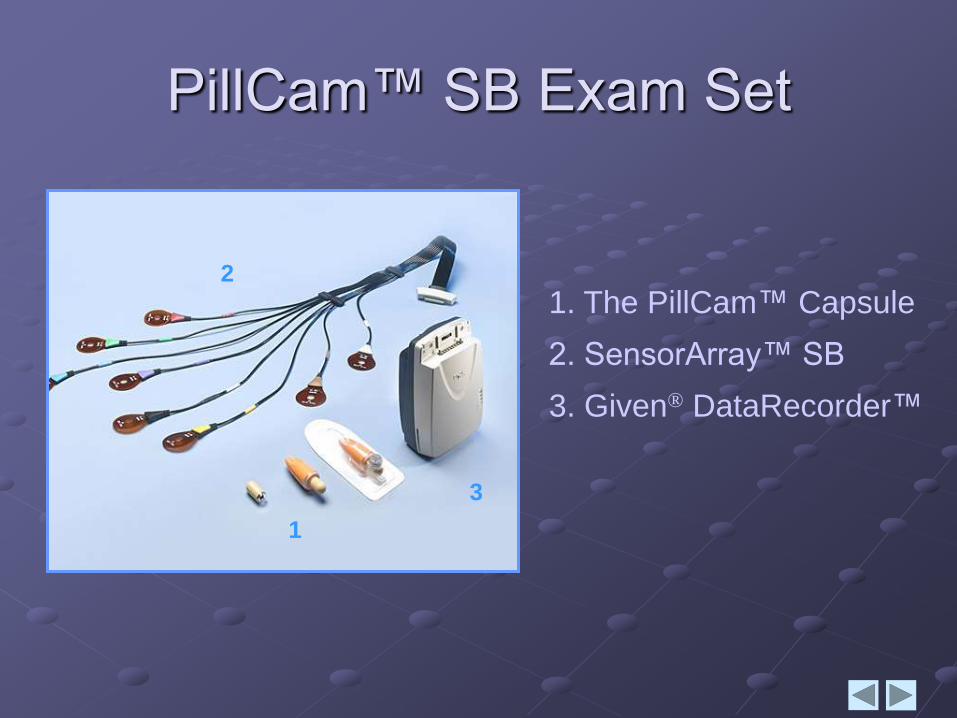
1. The PillCam™ Capsule
2. SensorArray™ SB
3. Given® DataRecorder™
PillCam™ SB Exam Set
2
1
3
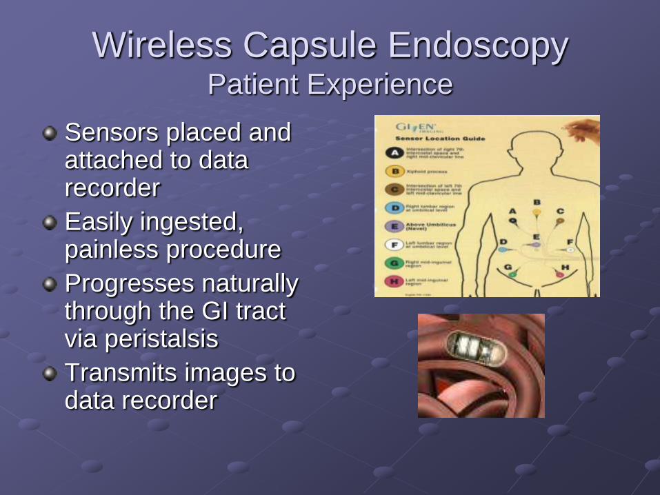
Wireless Capsule Endoscopy Patient Experience
Sensors placed and attached to data recorder
Easily ingested, painless procedure
Progresses naturally through the GI tract via peristalsis
Transmits images to data recorder
PillCam™ SBPatient Experience
Liquid diet from lunch the day before
Movie Prep the night before
12 hour fast the night before
Capsule ingested in the morning
Reglan or erythromycin for inpatients
Liquid diet after 2 hours
Light meal 4 hours after ingestion
Disconnect after 8 hours
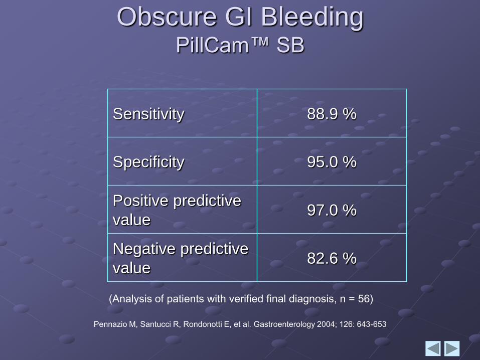
Pennazio M, Santucci R, Rondonotti E, et al. Gastroenterology 2004; 126: 643-653
82.6 %Negative predictive
value
97.0 %Positive predictive
value
95.0 %Specificity
88.9 %Sensitivity
(Analysis of patients with verified final diagnosis, n = 56)
Obscure GI BleedingPillCam™ SB
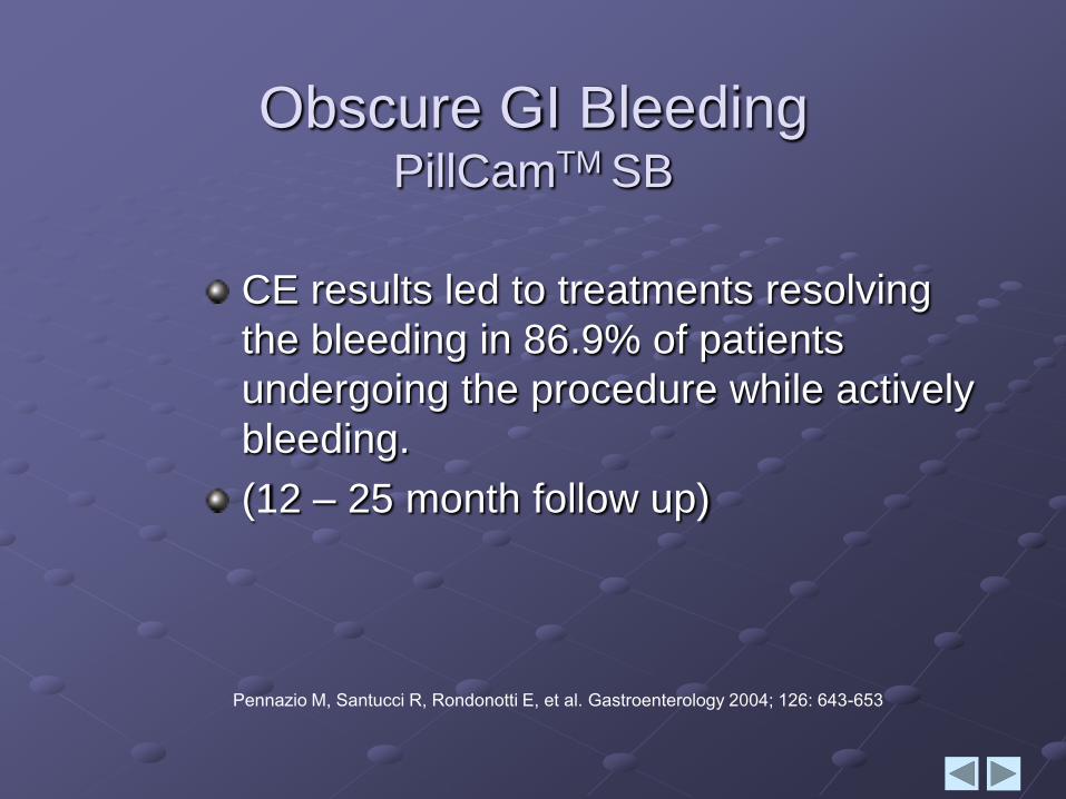
Obscure GI BleedingPillCamTM SB
CE results led to treatments resolving
the bleeding in 86.9% of patients
undergoing the procedure while actively
bleeding.
(12 – 25 month follow up)
Pennazio M, Santucci R, Rondonotti E, et al. Gastroenterology 2004; 126: 643-653
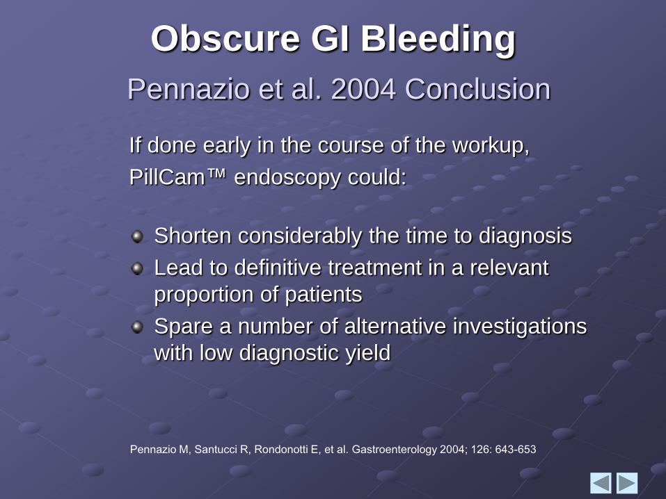
If done early in the course of the workup,
PillCam™ endoscopy could:
Shorten considerably the time to diagnosis
Lead to definitive treatment in a relevant
proportion of patients
Spare a number of alternative investigations
with low diagnostic yield
Obscure GI Bleeding
Pennazio et al. 2004 Conclusion
Pennazio M, Santucci R, Rondonotti E, et al. Gastroenterology 2004; 126: 643-653
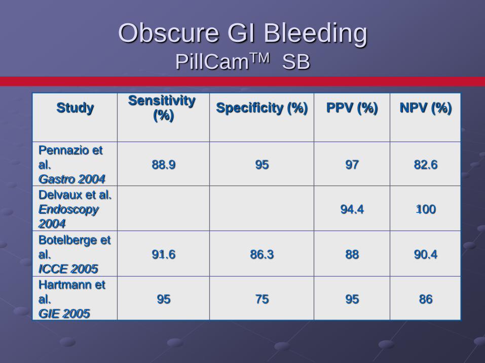
Obscure GI Bleeding PillCamTM SB
StudySensitivity
(%)Specificity (%) PPV (%) NPV (%)
Pennazio et
al.
Gastro 2004
88.9 95 97 82.6
Delvaux et al.
Endoscopy
2004
94.4 100
Botelberge et
al.
ICCE 2005
91.6 86.3 88 90.4
Hartmann et
al.
GIE 2005
95 75 95 86
First line diagnostic exam for visualization of small bowel mucosa.
Clinical data reviewed 32 independent studies which indicate CE diagnostic yield of 71% vs. 41% diagnostic yield for all other modalitiescombined1
Established as gold standard for diagnosis of disease of small intestine2
Now cleared in the US for pediatric populationfrom 10-18 years old
1. Internal data at Given Imaging Ltd. Reviewed by the FDA
2. Rex, et. Al; WIRELESS CAPSULE ENDOSCOPY DETECTS SMALL BOWEL ULCERS IN
PATIENTS WITH NORMAL RESULTS FROM STATE OF THE ART ENTEROCLYSIS The
American Journal of Gastroenterology, Vol. 98, No. 6
PillCam™ SBIndications
In patients with known or suspected
gastrointestinal obstruction, strictures, or fistulas
based on the clinical picture or pre-procedure
testing and profile.
In patients with cardiac pacemakers or other
implanted electromedical devices1.
In patients with swallowing disorders.
1 Leighton JA,, et al, SAFETY OF CAPSULE ENDOSCOPY IN PATIENTS WITH PACEMAKERS,
Gastrointest Endosc. 2004 Apr;59(4):567-9. Concludes that capsule endoscopy appears to be
safe in patients with cardiac pacemakers and does not appear to be associated with any
significant adverse cardiac event. Pacemakers do not interfere with capsule imaging.
PillCam™ SBContraindications
Small Bowel Bleeding
Causes Visualized by PillCamTM
Vascular Lesions
Angioectasias
Neoplasms
Inflammatory Lesions
Ulcers, Crohn’s Disease
Other
Diverticula, varices
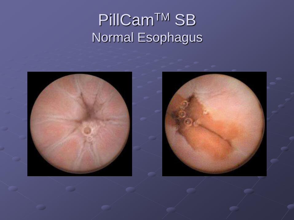
PillCamTM SB Normal Esophagus
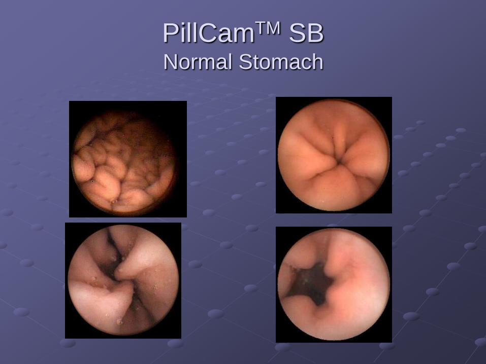
PillCamTM SB Normal Stomach
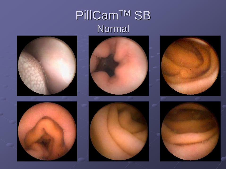
PillCamTM SBNormal
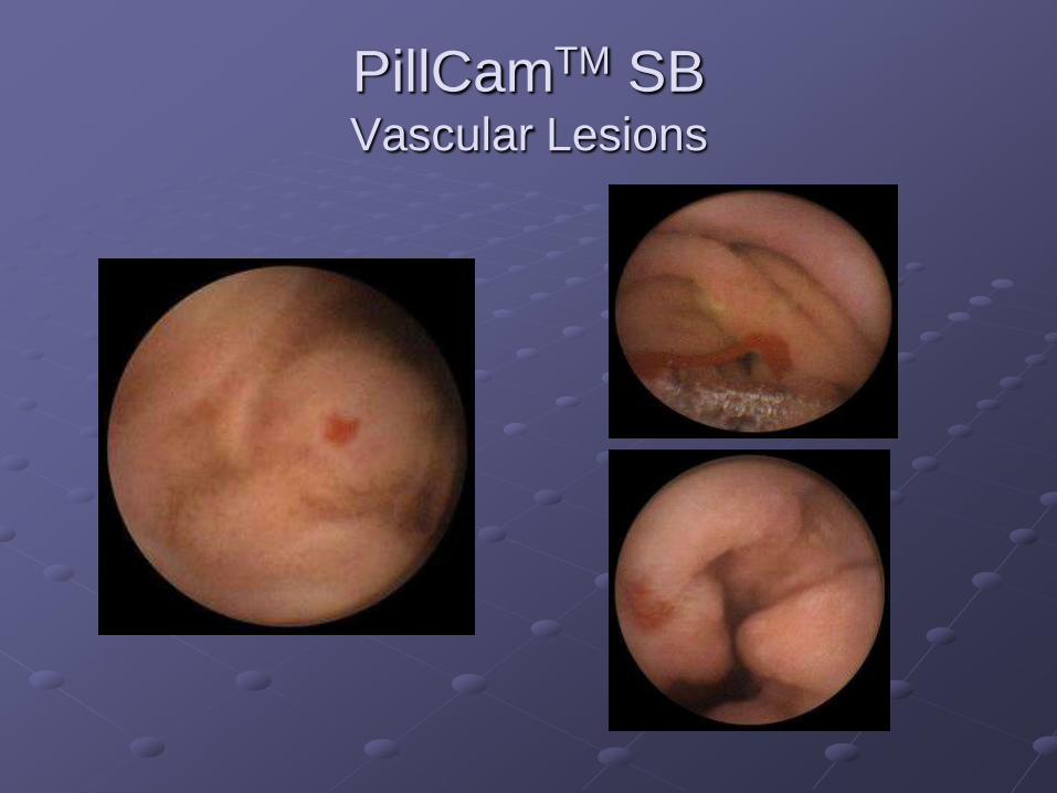
PillCamTM SBVascular Lesions
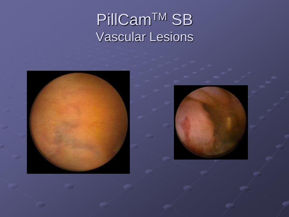
PillCamTM SBVascular Lesions
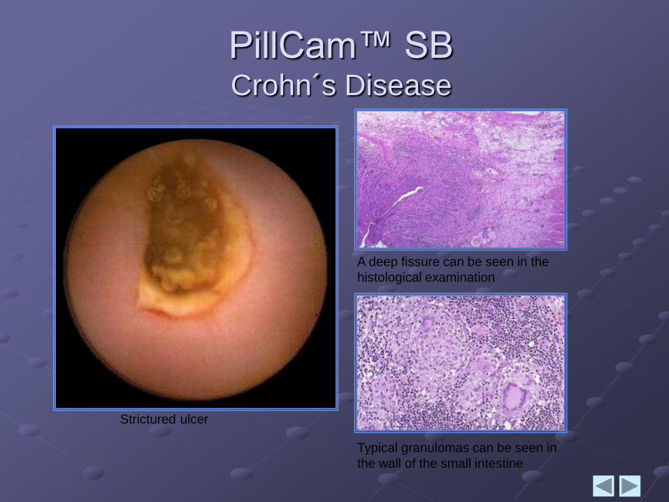
PillCam™ SBCrohn´s Disease
Strictured ulcer
A deep fissure can be seen in the
histological examination
Typical granulomas can be seen in
the wall of the small intestine
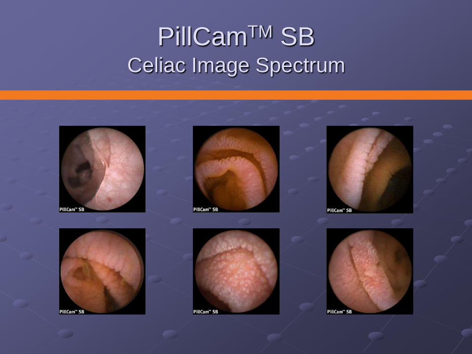
PillCamTM SBCeliac Image Spectrum
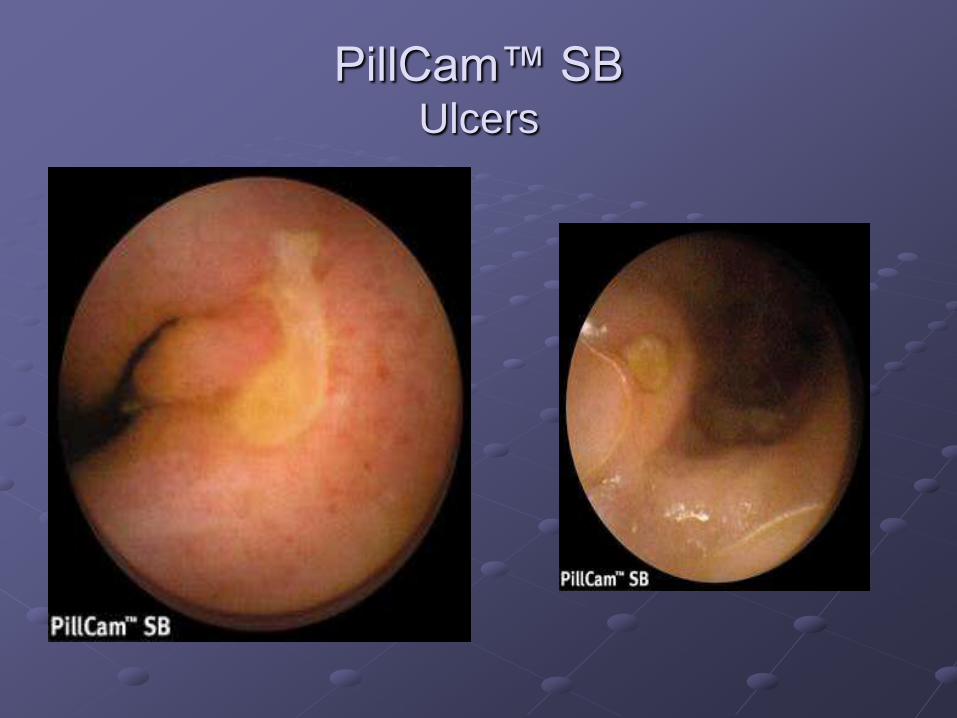
PillCam™ SBUlcers
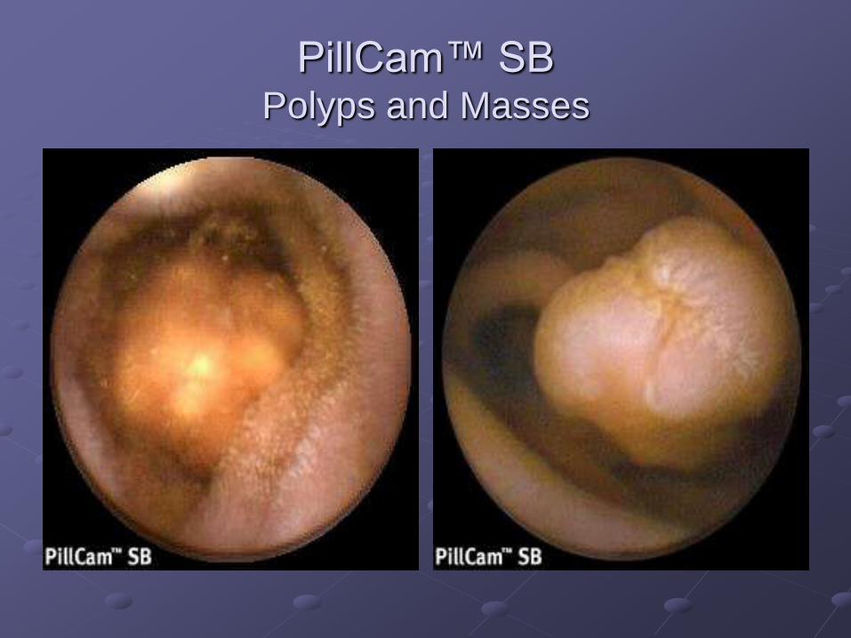
PillCam™ SBPolyps and Masses
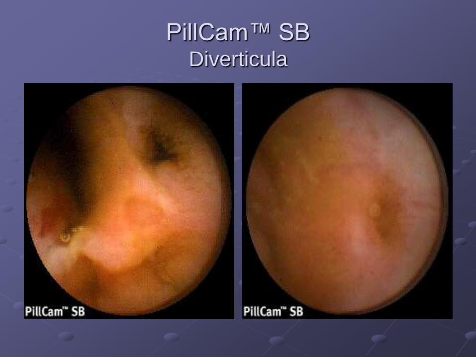
PillCam™ SBDiverticula
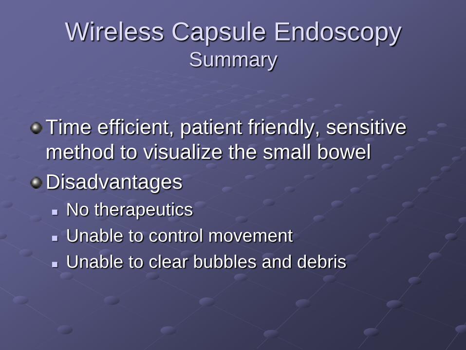
Wireless Capsule EndoscopySummary
Time efficient, patient friendly, sensitive
method to visualize the small bowel
Disadvantages
No therapeutics
Unable to control movement
Unable to clear bubbles and debris
Double Balloon Enteroscopy
First described by Yamamoto in 2001
Allows the diagnosis and treatment of disease
along the entire length of the small bowel
Entire SB visualized in 86% of patients (Yamamoto)
Fujinon enteroscope overtube system
230 cm total length
200-cm working length
140-cm overtube
2.8 mm channel for biopsy and therapeutic
intervention
Double Balloon Enteroscopy
Also called “push-pull enteroscopy”
Advanced antegrade or retrograde
Patient Prep
Antegrade: NPO 6-8 hrs
Retrograde: Colo prep
Moderate sedation, propofol, or general
anesthesia
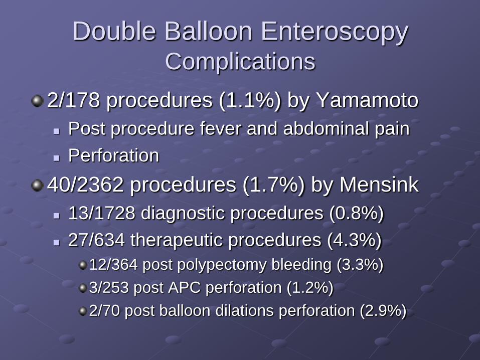
Double Balloon EnteroscopyComplications
2/178 procedures (1.1%) by Yamamoto
Post procedure fever and abdominal pain
Perforation
40/2362 procedures (1.7%) by Mensink
13/1728 diagnostic procedures (0.8%)
27/634 therapeutic procedures (4.3%)
12/364 post polypectomy bleeding (3.3%)
3/253 post APC perforation (1.2%)
2/70 post balloon dilations perforation (2.9%)
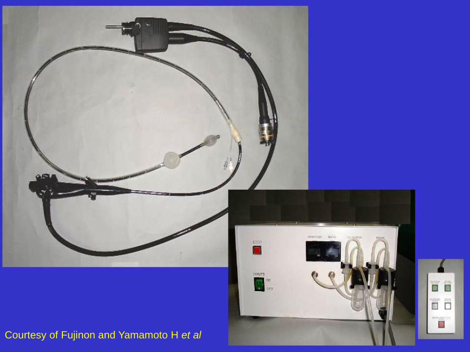
Courtesy of Fujinon and Yamamoto H et al
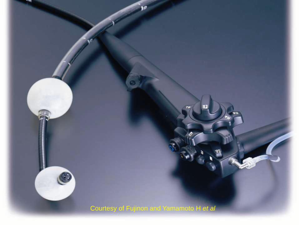
Courtesy of Fujinon and Yamamoto H et al
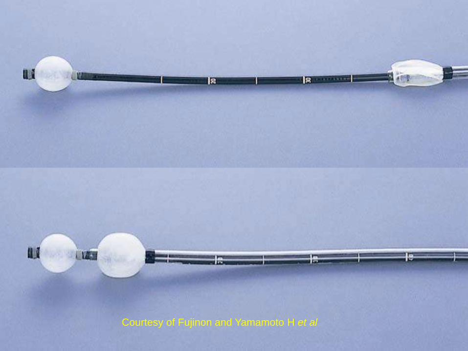
Courtesy of Fujinon and Yamamoto H et al
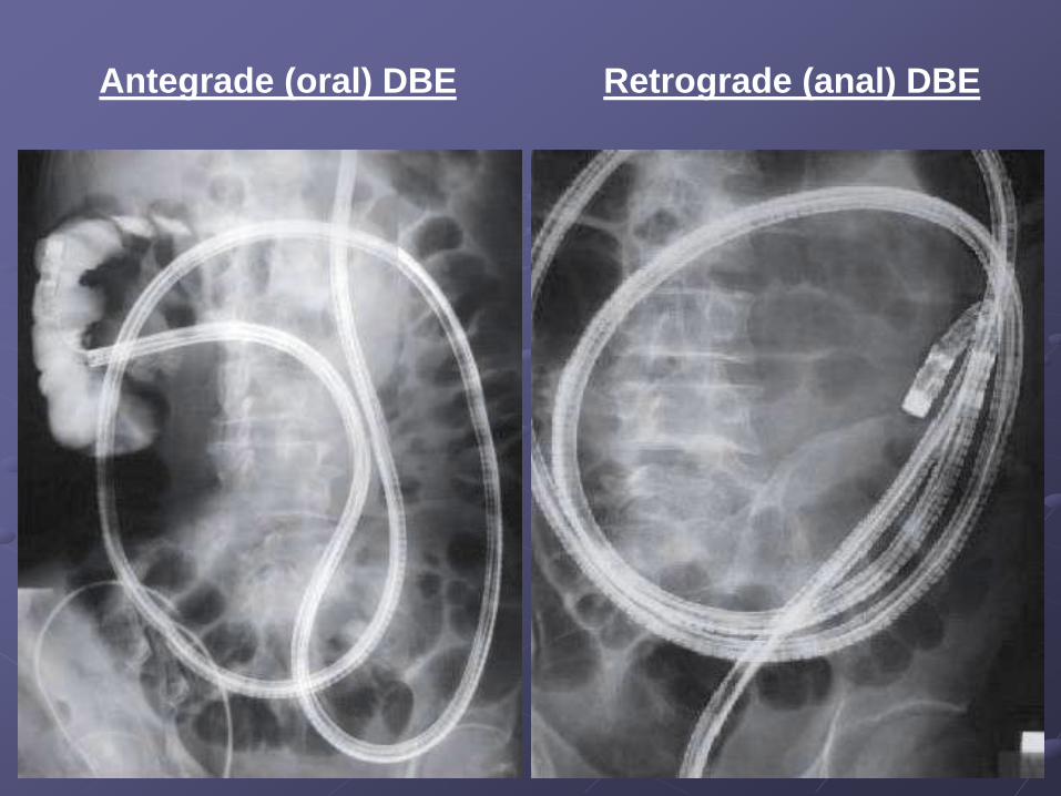
Antegrade (oral) DBE Retrograde (anal) DBE
Double Balloon Enteroscopy
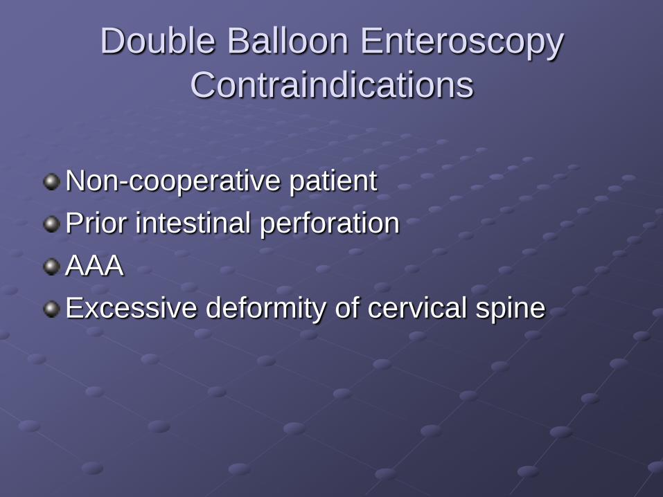
Contraindications
Non-cooperative patient
Prior intestinal perforation
AAA
Excessive deformity of cervical spine
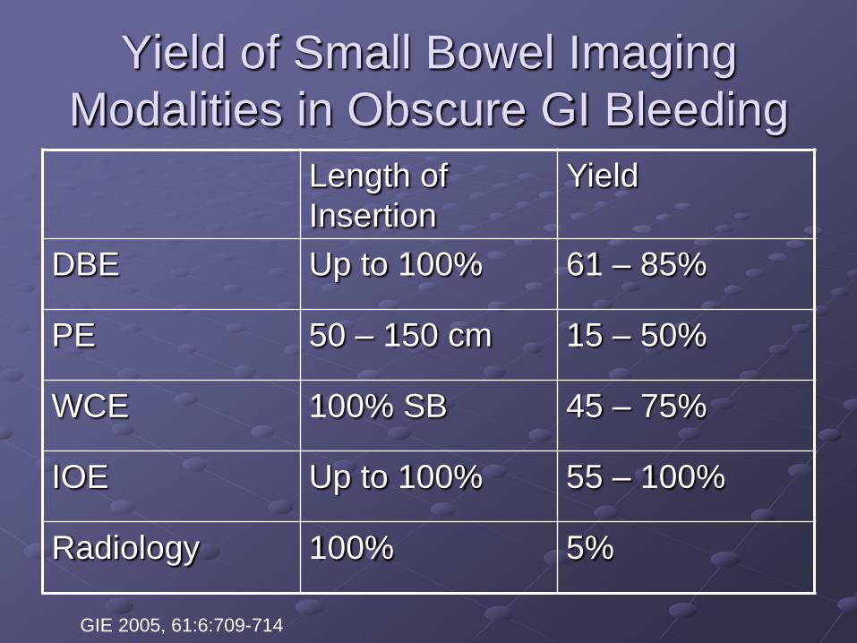
Yield of Small Bowel Imaging
Modalities in Obscure GI Bleeding
Length of
Insertion
Yield
DBE Up to 100% 61 – 85%
PE 50 – 150 cm 15 – 50%
WCE 100% SB 45 – 75%
IOE Up to 100% 55 – 100%
Radiology 100% 5%
GIE 2005, 61:6:709-714
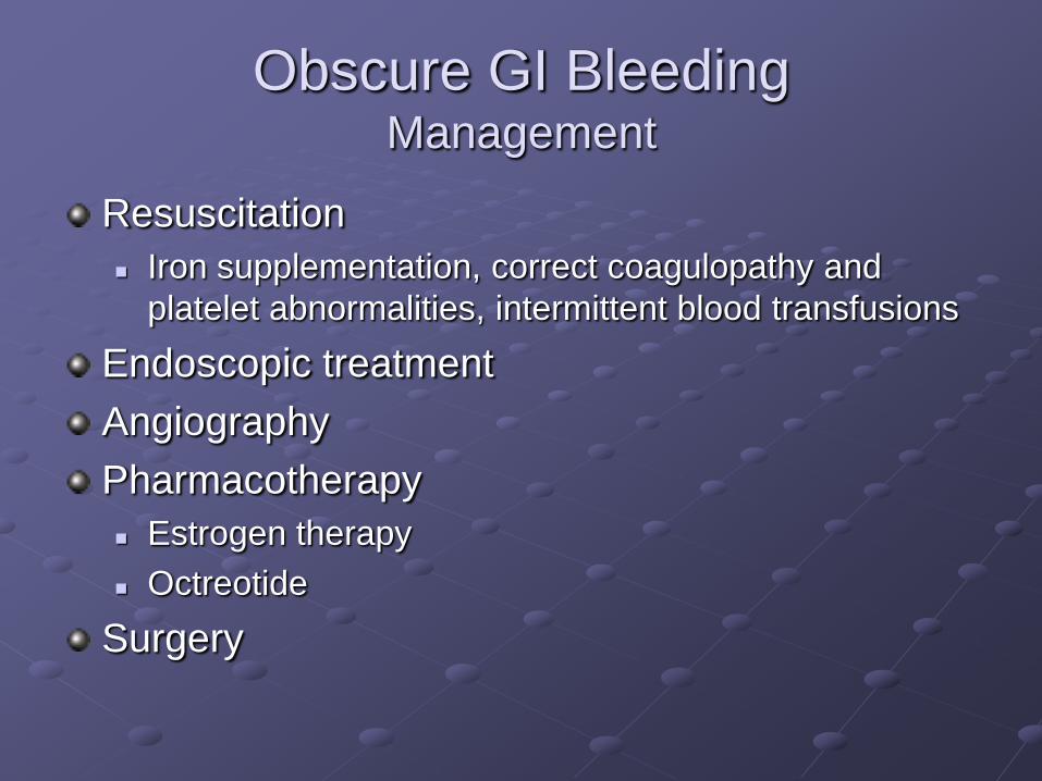
Obscure GI BleedingManagement
Resuscitation
Iron supplementation, correct coagulopathy and
platelet abnormalities, intermittent blood transfusions
Endoscopic treatment
Angiography
Pharmacotherapy
Estrogen therapy
Octreotide
Surgery
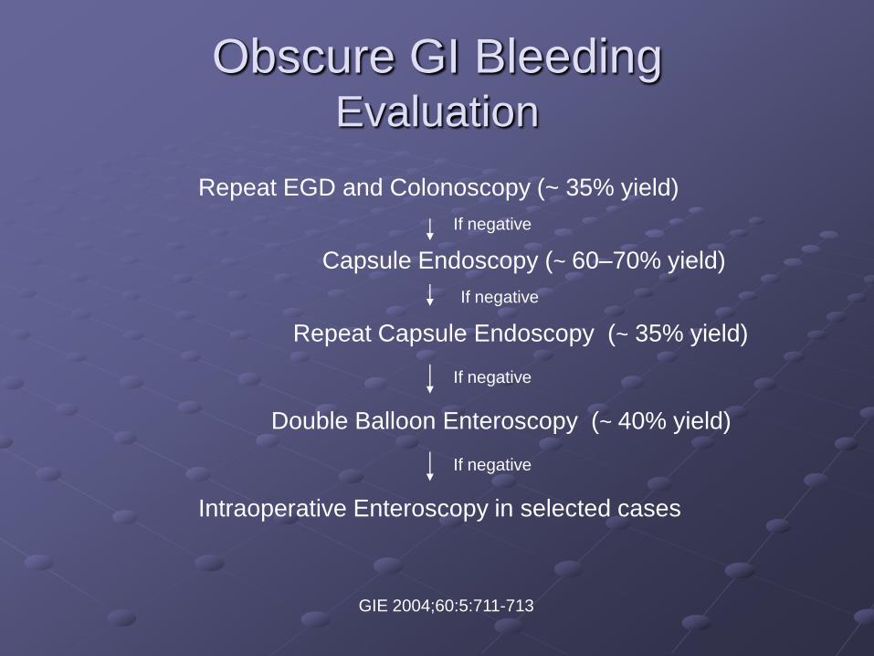
Obscure GI BleedingEvaluation
Repeat EGD and Colonoscopy (~ 35% yield)
If negative
Capsule Endoscopy (~ 60–70% yield)
If negative
Repeat Capsule Endoscopy (~ 35% yield)
If negative
Double Balloon Enteroscopy (~ 40% yield)
If negative
Intraoperative Enteroscopy in selected cases
GIE 2004;60:5:711-713


The FutureRobotics

The Magic Pill
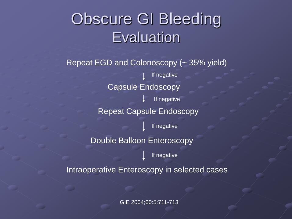
Obscure GI BleedingEvaluation
Repeat EGD and Colonoscopy (~ 35% yield)
If negative
Capsule Endoscopy
If negative
Repeat Capsule Endoscopy
If negative
Double Balloon Enteroscopy
If negative
Intraoperative Enteroscopy in selected cases
GIE 2004;60:5:711-713