Embed Size (px)
Citation preview
Sleep Medicine 15 (2014) 27–32
Contents lists available at ScienceDirect
Sleep Medicine
journal homepage: www.elsevier .com/locate /s leep
Original Article
Obstructive sleep apnea and neurocognitive performance: the roleof cortisol
1389-9457/$ - see front matter � 2013 Elsevier B.V. All rights reserved.http://dx.doi.org/10.1016/j.sleep.2013.08.789
⇑ Corresponding author at: University of Sydney, Exercise Health and Perfor-mance Research Group, C42–Cumberland Campus, 1825 Lidcombe, Australia. Tel.:+61 2 9036 7396; fax: +61 2 9351 9204.
E-mail address: [email protected] (K.M. Edwards).
Kate M. Edwards a,b,⇑, Rujvi Kamat c,d, Lianne M. Tomfohr c,d, Sonia Ancoli-Israel b,c,d, Joel E. Dimsdale b,c,d
a University of Sydney, Exercise Health and Performance Research Group, Lidcombe, Australiab Department of Psychiatry, University of California, San Diego, La Jolla, CA, USAc San Diego State University, Joint Doctoral Program in Clinical Psychology, San Diego, CA, USAd University of California, San Diego, Joint Doctoral Program in Clinical Psychology, San Diego, CA, USA
a r t i c l e i n f o a b s t r a c t
Article history:Received 28 March 2013Received in revised form 7 August 2013Accepted 23 August 2013Available online 31 October 2013
Keywords:Obstructive sleep apneaSleepNeurocognitive functionMemoryCortisolHypothalamic–pituitary–adrenal axis
Background: Obstructive sleep apnea (OSA) is a prevalent disorder with multiple consequences includingnegative effects on neurocognitive function. Several domains of cognitive function are impaired in OSApatients, but the mechanisms through which this sleep disorder results in impairment are not clear.Given the well-known effects of cortisol on cognitive function, in particular memory, the dysregulatingeffects of OSA on cortisol levels are hypothesized as a potential pathway leading to cognitive impairment.Methods: Fifty-five participants with OSA (mean apnea-hypopnea index [AHI], 30.3) were assessed over2 days. Over a 24-h period, blood samples were collected every 2 h to examine cortisol levels. The follow-ing night, sleep was monitored with polysomnography (PSG). Participants were given a battery ofneurocognitive tests, which assessed seven cognitive domains.Results: OSA severity assessed by oxygen desaturation index (ODI) was associated with 24-h cortisollevels. AHI, ODI, and nighttime cortisol levels were associated with global deficit scores (GDS) in cognitivefunctioning, particularly in domains of learning, memory, and working memory (P < .05 for all).Hierarchical linear regression analysis revealed that nighttime cortisol accounted for 9–16% of variancein learning (P = .018), memory (P = .003), and working memory (P = .016) domains, though apnea severitydid not significantly predict any additional variance.Conclusions: In our sample of patients with OSA, nocturnal cortisol levels were associated with neuropsy-chologic functioning above and beyond the influence of covariates and apnea severity. These findingssuggest that OSA-related alterations in cortisol activity may partially explain the pathophysiology ofneuropsychologic impairments in sleep apnea.
� 2013 Elsevier B.V. All rights reserved.
1. Introduction
Obstructive sleep apnea (OSA) is a sleep disorder involvingrepeated episodes of complete or partial obstruction of the upperairway, which cause transient cessations of breathing during sleep.These breathing disruptions cause intermittent hypoxia and sleepdisturbances which are associated with daytime sleepiness and fa-tigue [1]. OSA also has been demonstrated to have negative effectson cognitive functioning, which add to the public health risks forthe disease [2]. The neurobehavioral consequences of OSA extendto functional impairments, such as impaired driving, increased riskfor accidents, and decreased quality of life [3,4]. Numerous studieshave examined the effects of OSA on the cognitive abilitiesunderlying these neurobehavioral consequences. Impairments in
memory, vigilance, psychomotor performance, and executive func-tion all have been reported in OSA patients, but the presence anddegree of impairments is inconsistent between studies [5–12].A recent meta-analysis [2] of systematic reviews found supportfor impairments in attention or vigilance, delayed verbal and visuallong-term memory, visuospatial or constructional abilities, andexecutive function. Further, neuroimaging studies have reportedstructural changes in specific brain regions associated with cogni-tive function, and the loss of hippocampal volumes in OSA patientsis consistently reported [13].
Associations between cognitive function tests and magneticresonance imaging findings have been reported for verbal memoryand information processing [14], as well as for verbal memory andexecutive function [15].
The mechanisms by which OSA results in neurocognitivedysfunction are not entirely clear for several reasons. The comorbid-ities that accompany OSA (e.g., hypertension) are associated withneural injury, making it important to control for confounding associ-ations of OSA to neurocognitive impairment (see [1] for review).
28 K.M. Edwards et al. / Sleep Medicine 15 (2014) 27–32
In addition, the variety of tests used to assess neurocognitive functionalong with the varied sample characteristics are vast, which limit thepossibilities for comparing the reported cognitive sequelae of OSA[16]. Further, individual differences in the severity of sleep distur-bances also may add variability to the pattern of neurocognitive def-icits observed in patients with OSA, with greater deficits found inmore severe OSA, particularly in the case of executive function[17,18].
The neurochemical, vascular, and structural changes accompa-nying hypoxemia and sleep fragmentation have been implicatedin the adverse effect of OSA on cognitive functioning (see [16] forreview). One potential pathway of interest might be dysregulationof the hypothalamic–pituitary–adrenal (HPA) axis. The primaryhuman product of the HPA axis is cortisol, a hormone that evi-dences a strong diurnal rhythm. The fluctuation of cortisolthroughout the night is intricately related to sleep, and thus ithas been proposed as an important mechanism through whichsleep disorders manifest some of their physiologic changes. Fur-ther, both the hippocampus and amygdala, which are strongly in-volved in memory processes, express a high density ofcorticosteroid receptors [19]. Given the association between corti-sol and cognitive function and the association between OSA andcognitive dysfunction, the role that cortisol may play in cognitivedysfunction in OSA is worthy of investigation.
The literature linking OSA and HPA function is mixed, and fewstudies have found differences in cortisol levels between OSA andhealthy patients [20]. However, many studies are limited by theassessment of cortisol levels at a single time point [21,22]. Thoseinvestigations that have reported differences used extensive circa-dian sampling [23]; additionally, elevated cortisol levels appearedto be corrected after treatment with continuous positive airwaypressure (CPAP) [23–25]. Cortisol levels, especially during thenight, might be an important factor in the association betweenOSA and neurocognitive functioning. In our study, we examinedthe 24-h cortisol profile in 55 patients with OSA and its associationwith performance in cognitive function tests. It was hypothesizedthat increased OSA severity would be associated with both de-creased cognitive performance and elevated cortisol levels.
2. Methods
2.1. Participants
As part of a larger study (National Institutes of Health HL44915)examining the pathophysiology of the sympathetic nervous systemin patients with OSA [26], participants with known or suspectedOSA who had not received previous OSA treatment were recruitedvia advertisements, word of mouth referral, and referral from localmedical practices in the San Diego area. Fifty-five participants withOSA were included (age range, 29–65 y). Potential participantswere excluded if they (1) had a history of a major medical illness,with the exception of OSA and hypertension; (2) had current psy-chiatric diagnoses including alcohol or drug abuse; or (3) werereceiving psychotropic medications. Two patients who were takinghypertensive medication were slowly tapered off their medicationsfor 3 weeks prior to participation. In both cases, blood pressure(BP) remained within the inclusion range (<170/105 mmHg), andthus the patients were retained in the sample. The full details ofthe procedure are described elsewhere [26]. The project and allprocedures were approved by the University of California, SanDiego Human Subjects Committee.
2.2. Procedure
Written informed consent was obtained from all participantsbefore participation in the study. Participants were admitted to
the University of California, San Diego, General Clinical ResearchCenter Gillin Laboratory of Sleep and Chronobiology for two nights.The participants were prepared for standard polysomnography(PSG) on both nights; however, the recordings were discarded fromthe first night due to the possibility of first-night effects on sleepmetrics. A venous catheter was inserted at 5:00 pm. Starting at6:00 pm, a blood sample was collected every 2 h for 24 h. Bloodsamples were collected in ethylenediaminetetraacetic acid, placedon ice, and spun in a refrigerated centrifuge; the plasma was thenstored at �80 �C until assayed. Plasma cortisol was determinedusing commercial sandwich enzyme-linked immunosorbent as-says (Parameter assay; R&D Systems, Minneapolis, MN, USA).
Starting at 8:00 pm the following evening on completion of the24-h blood sampling, participants were prepared for PSG. The PSGsetup began at 9:00 pm and lights-off time occurred between10:00 pm and midnight. Parameters measured during PSG in-cluded electroencephalography, electrocardiography, electroocu-lography, chin and tibialis anterior electromyography, pulseoximetry, nasal-oral airflow by nasal cannula pressure transducerand thermistor, and thoracic and abdominal respiratory effort re-corded on a Grass Heritage digital PSG (Model PSG36-2, Astro-Med, Inc, West Warwick, RI). The next morning, participants wereawakened at 6:00 am and PSG recording equipment was removed.Experienced PSG technicians scored PSG sleep records according tothe Rechtschaffen and Kales criteria [27]. OSA can be quantified inseveral ways; each of these indices captures a slightly different as-pect of breathing cessation. The most commonly used assessmentsare recording the number of apneas and hypopneas, oxygen desat-uration events, and total sleep time (TST). Apneas were defined asdecrements in airflow of P90% from baseline lasting for P10 s.Hypopneas were defined as decrements in airflow of P50% but<90% from baseline lasting for P10 s regardless of the presenceor absence of associated desaturation or arousal. Significant tran-sient oxyhemoglobin desaturations were defined as transientdrops in oxyhemoglobin saturation by P3% from baseline lastingfor >10 s but <3 min. The oxygen desaturation index (ODI) was cal-culated as the number of transient oxygen desaturations per hourof sleep. TST was computed, and the numbers of apneas and hyp-opneas per hour of sleep were calculated to obtain the apnea–hypopnea index (AHI). Different criteria for OSA diagnosis exist;in our study, we used a cutoff of AHI P5 events per hour for diag-nosis of OSA and inclusion into the study, which is in accordancewith the criteria of the American Academy of Sleep Medicine[28]. The mean AHI for the included patients was 30.3 events perhour, with an interquartile range of 30.2 events per hour, thusshowing moderate to severe OSA.
2.3. Neuropsychologic battery
Participants were given the following fixed battery, which as-sessed seven cognitive domains: (1) Wechsler Adult IntelligenceScale-Revised [29]; (2) Digit Symbol, Digit Span, Letter-NumberSequencing Test, Symbol Search Test; (3) Brief Visuospatial Mem-ory Test-Revised (BVMT) [30]; (4) Hopkins Verbal Learning Test-Revised (HVLT) [31]; (5) Trail Making Test parts A and B [32]; (6)Digit Vigilance Test [33]; (7) Stroop Color-Word Test [34]; and(8) Word Fluency Test [35]. These tests produced 15 subscalescores per participant and assessed the following cognitive do-mains: (1) processing speed (Digit Symbol Test, Symbol SearchTest, Digit Vigilance Test, Trail Making Test part A, Stroop Color-Word Test); (2) working memory (Letter-Number Sequencing Test,Digit Span test, Digit Vigilance Test); (3) executive functions (TrailMaking Test part B, Digit Symbol Test, Symbol Search Test, Letter-Number Sequencing Test, Stroop Color-Word Test); (4) attention(Digit Vigilance Test); (5) learning (HVLT-total, BVMT-total), and(6) memory (HVLT-recall, BVMT-recall).
K.M. Edwards et al. / Sleep Medicine 15 (2014) 27–32 29
Raw scores were calculated for each neuropsychologic subtest.To investigate how many patients with OSA had neuropsychologicimpairment and in what domains, t scores were calculated for eachof the neuropsychologic subtests, controlling for ethnicity, sex, age,and education. Higher t scores indicated better performance.Domain-wise t scores were generated by averaging the scores onthe tests that contributed to each domain. A deficit score was com-puted for each of the 15 individual test scores according to the con-vention below, in which t scores were collapsed into groups from 0to 5 (with a t score of P40, the deficit score was 0; with a score ofP35 but <40, the deficit score was 1; with a score of P30 but <35,the deficit score was 2; with a score of P25 but <30, the deficitscore was 3; with a score of P20 but <25, the deficit score was4; and with a score <19, the deficit score was 5). The average ofthose scores was the global deficit score (GDS). A GDS cutoff pointof P0.5 was used to classify individuals as having neurocognitiveimpairment, as it yielded the optimal balance between sensitivityand specificity. A detailed explanation of GDS is described else-where [36].
2.4. Psychologic assessment
To assess depressive symptoms, participants completed theCenter for Epidemiologic Studies Depression Scale, a 20-itemself-report scale [37]. Scores of P16 indicated a likely diagnosisof major depression [38].
Table 1Sleep characteristics.
Sleep recording measures Mean (SD)
AHI (events/h) 30.3 (21.7)ODI (events/h) 21.7 (21.9)Total sleep time (min) 372.7 (45.3)Total arousal index (events/h) 22.2 (18.0)Wake after sleep onset (min) 53.6 (29.4)Stage 1 sleep (%) 12.6 (8.3)Stage 2 sleep (%) 55.86 (10.0)Slow-wave sleep (stage 3 and 4) (%) 12.01 (10.6)REM sleep (%) 20.3 (7.13)
2.5. Statistical analysis
Data were analyzed using SPSS 17.0 (SPSS Inc., Chicago, IL). Areaunder curve (AUC) values were calculated for cortisol levels basedon the trapezoid rule, with 24-h nighttime (10:00 pm–6:00 am)and daytime (8:00 am–8:00 pm) values adjusted to a per-hour ba-sis. Bivariate associations between neurocognitive measures, corti-sol levels, and respiratory variables were investigated usingPearson product moment correlation coefficients. A series of hier-archical linear regressions were then used to investigate if signifi-cant associations between neurocognitive measures, cortisollevels, and respiratory variables persisted after potentially con-founding covariates were considered (i.e., step 1: body mass index[BMI], smoking status); other potential cofounders were alreadyadjusted for in calculation of t scores (i.e., ethnicity, sex, age, edu-cation). Potential predictors (ODI for OSA severity; and nighttimecortisol for HPA activity) were sequentially entered in steps 2and 3 to allow for the determination of DR2 to which each variablebelonged. In all analyses, 2-sided t tests were used. A level ofP 6 .05 was accepted as level of significance and g2 was calculatedas a measure of effect size.
Abbreviations: AHI, apnea–hypopnea index; ODI, oxygen desaturation index; h,hour; min, minutes; REM, rapid eye movement; SD, standard deviation.The AHI, ODI, and total arousal index were calculated as events per hour of totalsleep time.
Table 2Mean (standard deviation) t scores and percent of participants classified as impaired.Higher t score indicated better performance.
t score % Impaired
Processing speed 48.2 (8.4) 16.3Verbal fluency 48.6 (10.5) 20.4Learning 44.2 (9.5) 36.7Memory 45.9 (9.1) 24.5Executive function 48.5 (9.5) 14.3Working memory 50.1 (9.6) 14.3
3. Results
3.1. Participant characteristics
Fifty-five participants qualified for the study (80% men). Partic-ipants were middle-aged (mean age, 50 y; standard deviation [SD],9.1) and overweight (mean BMI, 29.6 kg/m2; SD, 5.2). BP wasmildly elevated (mean systolic BP, 131.4 mmHg [SD, 17.2 mmHg];mean diastolic BP, 78.8 mmHg [SD, 9.1 mmHg]) and 25% of partic-ipants scored in the depressed range (Center for EpidemiologicStudies Depression Scale P16). Six participants were currentsmokers. Participants had severe OSA on average (mean AHI,30.3; SD, 21.7), Table 1 describes the sleep characteristics of theparticipants.
3.2. Neurocognitive function
Participants’ raw scores were converted to t scores for each do-main of cognitive function (speed of information processing; ver-bal, learning, memory, executive function; and working memory)and GDS scores were calculated. All t and GDS scores were adjustedfor gender, age, education, and ethnicity during calculations. In ahealthy population, a neurocognitive deficit (GDS, P0.5; t score,640) would be expected in 15% of participants [39]. In our sample,31% were impaired according to the GDS, showing a higher rate ofimpairment in our group. Table 2 shows average t scores and per-cent impairment for each domain.
3.3. Plasma cortisol
As shown in Fig. 1, the expected circadian effect was seen inplasma cortisol levels (reference range [morning], 5–25 lg/dL;[afternoon], 2–12 lg/dL). A significant time effect was found(F[11,43] = 48.2; P<.001; g2 = .925), with levels showing a nadir at10:00 pm–12:00 am and a peak at 6:00 am–8:00 am. Summaryvalues showed the expected lower cortisol AUC (adjusted to per-hour basis) during the nighttime than during the daytime(t[53] = 6.332; P < .001).
3.4. Univariate analyses
Our initial analyses examined associations between OSA sever-ity and cortisol levels. Partial correlations were performed to con-trol for potential confounding factors of BMI and smoking withcortisol levels [40–42]. Although we found no association withAHI, ODI was significantly related to 24-h cortisol AUC (r = .323;P=.025). However, there were no significant associations betweennighttime cortisol AUC with AHI or ODI (P > .05 for both). Analysesof sleep characteristics revealed that TST was correlated with 24-hcortisol AUC (r = �.365; P = .011) and with nighttime cortisol AUC(r = �.303; P = .036), but total arousal index; wake after sleep on-set; and proportion of sleep in stage 1, stage 2, slow-wave sleep,
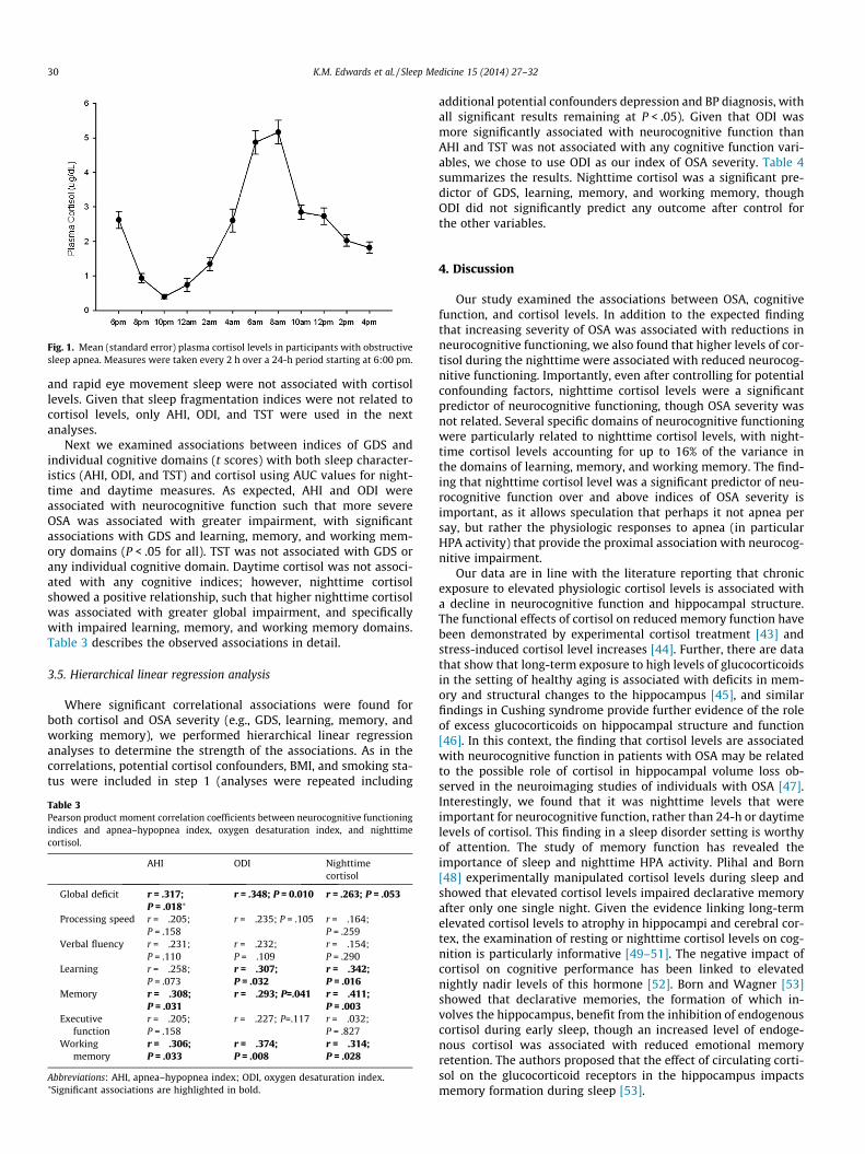
Fig. 1. Mean (standard error) plasma cortisol levels in participants with obstructivesleep apnea. Measures were taken every 2 h over a 24-h period starting at 6:00 pm.
30 K.M. Edwards et al. / Sleep Medicine 15 (2014) 27–32
and rapid eye movement sleep were not associated with cortisollevels. Given that sleep fragmentation indices were not related tocortisol levels, only AHI, ODI, and TST were used in the nextanalyses.
Next we examined associations between indices of GDS andindividual cognitive domains (t scores) with both sleep character-istics (AHI, ODI, and TST) and cortisol using AUC values for night-time and daytime measures. As expected, AHI and ODI wereassociated with neurocognitive function such that more severeOSA was associated with greater impairment, with significantassociations with GDS and learning, memory, and working mem-ory domains (P < .05 for all). TST was not associated with GDS orany individual cognitive domain. Daytime cortisol was not associ-ated with any cognitive indices; however, nighttime cortisolshowed a positive relationship, such that higher nighttime cortisolwas associated with greater global impairment, and specificallywith impaired learning, memory, and working memory domains.Table 3 describes the observed associations in detail.
3.5. Hierarchical linear regression analysis
Where significant correlational associations were found forboth cortisol and OSA severity (e.g., GDS, learning, memory, andworking memory), we performed hierarchical linear regressionanalyses to determine the strength of the associations. As in thecorrelations, potential cortisol confounders, BMI, and smoking sta-tus were included in step 1 (analyses were repeated including
Table 3Pearson product moment correlation coefficients between neurocognitive functioningindices and apnea–hypopnea index, oxygen desaturation index, and nighttimecortisol.
AHI ODI Nighttimecortisol
Global deficit r = .317;P = .018⁄
r = .348; P = 0.010 r = .263; P = .053
Processing speed r = �.205;P = .158
r = �.235; P = .105 r = �.164;P = .259
Verbal fluency r = �.231;P = .110
r = �.232;P = �.109
r = �.154;P = .290
Learning r = �.258;P = .073
r = �.307;P = .032
r = �.342;P = .016
Memory r = �.308;P = .031
r = �.293; P=.041 r = �.411;P = .003
Executivefunction
r = �.205;P = .158
r = �.227; P=.117 r = �.032;P = .827
Workingmemory
r = �.306;P = .033
r = �.374;P = .008
r = �.314;P = .028
Abbreviations: AHI, apnea–hypopnea index; ODI, oxygen desaturation index.⁄Significant associations are highlighted in bold.
additional potential confounders depression and BP diagnosis, withall significant results remaining at P < .05). Given that ODI wasmore significantly associated with neurocognitive function thanAHI and TST was not associated with any cognitive function vari-ables, we chose to use ODI as our index of OSA severity. Table 4summarizes the results. Nighttime cortisol was a significant pre-dictor of GDS, learning, memory, and working memory, thoughODI did not significantly predict any outcome after control forthe other variables.
4. Discussion
Our study examined the associations between OSA, cognitivefunction, and cortisol levels. In addition to the expected findingthat increasing severity of OSA was associated with reductions inneurocognitive functioning, we also found that higher levels of cor-tisol during the nighttime were associated with reduced neurocog-nitive functioning. Importantly, even after controlling for potentialconfounding factors, nighttime cortisol levels were a significantpredictor of neurocognitive functioning, though OSA severity wasnot related. Several specific domains of neurocognitive functioningwere particularly related to nighttime cortisol levels, with night-time cortisol levels accounting for up to 16% of the variance inthe domains of learning, memory, and working memory. The find-ing that nighttime cortisol level was a significant predictor of neu-rocognitive function over and above indices of OSA severity isimportant, as it allows speculation that perhaps it not apnea persay, but rather the physiologic responses to apnea (in particularHPA activity) that provide the proximal association with neurocog-nitive impairment.
Our data are in line with the literature reporting that chronicexposure to elevated physiologic cortisol levels is associated witha decline in neurocognitive function and hippocampal structure.The functional effects of cortisol on reduced memory function havebeen demonstrated by experimental cortisol treatment [43] andstress-induced cortisol level increases [44]. Further, there are datathat show that long-term exposure to high levels of glucocorticoidsin the setting of healthy aging is associated with deficits in mem-ory and structural changes to the hippocampus [45], and similarfindings in Cushing syndrome provide further evidence of the roleof excess glucocorticoids on hippocampal structure and function[46]. In this context, the finding that cortisol levels are associatedwith neurocognitive function in patients with OSA may be relatedto the possible role of cortisol in hippocampal volume loss ob-served in the neuroimaging studies of individuals with OSA [47].Interestingly, we found that it was nighttime levels that wereimportant for neurocognitive function, rather than 24-h or daytimelevels of cortisol. This finding in a sleep disorder setting is worthyof attention. The study of memory function has revealed theimportance of sleep and nighttime HPA activity. Plihal and Born[48] experimentally manipulated cortisol levels during sleep andshowed that elevated cortisol levels impaired declarative memoryafter only one single night. Given the evidence linking long-termelevated cortisol levels to atrophy in hippocampi and cerebral cor-tex, the examination of resting or nighttime cortisol levels on cog-nition is particularly informative [49–51]. The negative impact ofcortisol on cognitive performance has been linked to elevatednightly nadir levels of this hormone [52]. Born and Wagner [53]showed that declarative memories, the formation of which in-volves the hippocampus, benefit from the inhibition of endogenouscortisol during early sleep, though an increased level of endoge-nous cortisol was associated with reduced emotional memoryretention. The authors proposed that the effect of circulating corti-sol on the glucocorticoid receptors in the hippocampus impactsmemory formation during sleep [53].
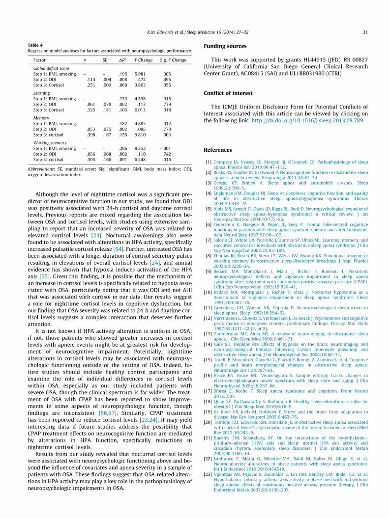
Table 4Regression model analyses for factors associated with neuropsychologic performance.
Factor b SE DR2 F Change Sig. F Change
Global deficit scoreStep 1: BMI, smoking – – .196 5.981 .005Step 2: ODI .114 .004 .008 .472 .495Step 3: Cortisol .251 .009 .060 3.863 .055
LearningStep 1: BMI, smoking – – .173 4.598 .015Step 2: ODI �.061 .078 .002 .112 .739Step 3: Cortisol �.325 .181 .103 6.013 .018
MemoryStep 1: BMI, smoking – – .182 4.885 .012Step 2: ODI �.053 .075 .002 .085 .773Step 3: cortisol �.398 .167 .155 9.810 .003
Working memoryStep 1: BMI, smoking – – .296 9.252 <.001Step 2: ODI �.056 .068 .002 .110 .742Step 3: cortisol �.305 .166 .091 6.248 .016
Abbreviations: SE, standard error; Sig., significant; BMI, body mass index; ODI,oxygen desaturation index.
K.M. Edwards et al. / Sleep Medicine 15 (2014) 27–32 31
Although the level of nighttime cortisol was a significant pre-dictor of neurocognitive function in our study, we found that ODIwas positively associated with 24-h cortisol and daytime cortisollevels. Previous reports are mixed regarding the association be-tween OSA and cortisol levels, with studies using extensive sam-pling to report that an increased severity of OSA was related toelevated cortisol levels [23]. Nocturnal awakenings also werefound to be associated with alterations in HPA activity, specificallyincreased pulsatile cortisol release [54]. Further, untreated OSA hasbeen associated with a longer duration of cortisol secretory pulsesresulting in elevations of overall cortisol levels [24], and animalevidence has shown that hypoxia induces activation of the HPAaxis [55]. Given this finding, it is possible that the mechanism ofan increase in cortisol levels is specifically related to hypoxia asso-ciated with OSA, particularly noting that it was ODI and not AHIthat was associated with cortisol in our data. Our results suggesta role for nighttime cortisol levels in cognitive dysfunction, butour finding that OSA severity was related to 24-h and daytime cor-tisol levels suggests a complex interaction that deserves furtherattention.
It is not known if HPA activity alteration is uniform in OSA;if not, those patients who showed greater increases in cortisollevels with apneic events might be at greatest risk for develop-ment of neurocognitive impairment. Potentially, nighttimealterations in cortisol levels may be associated with neuropsy-chologic functioning outside of the setting of OSA. Indeed, fu-ture studies should include healthy control participants andexamine the role of individual differences in cortisol levelswithin OSA, especially as our study included patients withsevere OSA, though the clinical spectrum is far wider. The treat-ment of OSA with CPAP has been reported to show improve-ments in some aspects of neuropsychologic function, thoughfindings are inconsistent [56,57]. Similarly, CPAP treatmenthas been reported to reduce cortisol levels [23,24]. It may yieldinteresting data if future studies address the possibility thatCPAP treatment effects on neurocognitive function are mediatedby alterations in HPA function, specifically reductions innighttime cortisol levels.
Results from our study revealed that nocturnal cortisol levelswere associated with neuropsychologic functioning above and be-yond the influence of covariates and apnea severity in a sample ofpatients with OSA. These findings suggest that OSA-related altera-tions in HPA activity may play a key role in the pathophysiology ofneuropsychologic impairments in OSA.
Funding sources
This work was supported by grants HL44915 (JED), RR 00827(University of California San Diego General Clinical ResearchCenter Grant), AG08415 (SAI) and UL1RR031980 (CTRI).
Conflict of interest
The ICMJE Uniform Disclosure Form for Potential Conflicts ofInterest associated with this article can be viewed by clicking onthe following link: http://dx.doi.org/10.1016/j.sleep.2013.08.789.
References
[1] Dempsey JA, Veasey SC, Morgan BJ, O’Donnell CP. Pathophysiology of sleepapnea. Physiol Rev 2010;90:47–112.
[2] Bucks RS, Olaithe M, Eastwood P. Neurocognitive function in obstructive sleepapnoea: a meta-review. Respirology 2013;18:61–70.
[3] George CF, Smiley A. Sleep apnea and automobile crashes. Sleep1999;22:790–5.
[4] Engleman HM, Douglas NJ. Sleep. 4: sleepiness, cognitive function, and qualityof life in obstructive sleep apnoea/hypopnoea syndrome. Thorax2004;59:618–22.
[5] Aloia MS, Arnedt JT, Davis JD, Riggs RL, Byrd D. Neuropsychological sequelae ofobstructive sleep apnea–hypopnea syndrome: a critical review. J IntNeuropsychol Soc 2004;10:772–85.
[6] Feuerstein C, Naegele B, Pepin JL, Levy P. Frontal lobe-related cognitivefunctions in patients with sleep apnea syndrome before and after treatment.Acta Neurol Belg 1997;97:96–107.
[7] Salorio CF, White DA, Piccirillo J, Duntley SP, Uhles ML. Learning, memory, andexecutive control in individuals with obstructive sleep apnea syndrome. J ClinExp Neuropsychol 2002;24:93–100.
[8] Thomas RJ, Rosen BR, Stern CE, Weiss JW, Kwong KK. Functional imaging ofworking memory in obstructive sleep-disordered breathing. J Appl Physiol2005;98:2226–34.
[9] Bedard MA, Montplaisir J, Malo J, Richer F, Rouleau I. Persistentneuropsychological deficits and vigilance impairment in sleep apneasyndrome after treatment with continuous positive airways pressure (CPAP).J Clin Exp Neuropsychol 1993;15:330–41.
[10] Bedard MA, Montplaisir J, Richer F, Malo J. Nocturnal hypoxemia as adeterminant of vigilance impairment in sleep apnea syndrome. Chest1991;100:367–70.
[11] Greenberg GD, Watson RK, Deptula D. Neuropsychological dysfunction insleep apnea. Sleep 1987;10:254–62.
[12] Verstraeten E, Cluydts R, Verbraecken J, De Roeck J. Psychomotor and cognitiveperformance in nonapneic snorers: preliminary findings. Percept Mot Skills1997;84:1211–22 [3, pt 2].
[13] Zimmerman ME, Aloia MS. A review of neuroimaging in obstructive sleepapnea. J Clin Sleep Med 2006;2:461–71.
[14] Gale SD, Hopkins RO. Effects of hypoxia on the brain: neuroimaging andneuropsychological findings following carbon monoxide poisoning andobstructive sleep apnea. J Int Neuropsychol Soc 2004;10:60–71.
[15] Torelli F, Moscufo N, Garreffa G, Placidi F, Romigi A, Zannino S, et al. Cognitiveprofile and brain morphological changes in obstructive sleep apnea.Neuroimage 2011;54:787–93.
[16] Bruce EN, Bruce MC, Vennelaganti S. Sample entropy tracks changes inelectroencephalogram power spectrum with sleep state and aging. J ClinNeurophysiol 2009;26:257–66.
[17] Sforza E, Roche F. Sleep apnea syndrome and cognition. Front Neurol2012;3:87.
[18] Quan SF, Parthasarathy S, Budhiraja R. Healthy sleep education—a salve forobesity? J Clin Sleep Med 2010;6:18–9.
[19] de Kloet ER, Joels M, Holsboer F. Stress and the brain: from adaptation todisease. Nat Rev Neurosci 2005;6:463–75.
[20] Tomfohr LM, Edwards KM, Dimsdale JE. Is obstructive sleep apnea associatedwith cortisol levels? a systematic review of the research evidence. Sleep MedRev 2012;16:243–9.
[21] Buckley TM, Schatzberg AF. On the interactions of the hypothalamic–pituitary–adrenal (HPA) axis and sleep: normal HPA axis activity andcircadian rhythm, exemplary sleep disorders. J Clin Endocrinol Metab2005;90:3106–14.
[22] Lanfranco F, Motta G, Minetto MA, Baldi M, Balbo M, Ghigo E, et al.Neuroendocrine alterations in obese patients with sleep apnea syndrome.Int J Endocrinol 2010;2010:474518.
[23] Vgontzas AN, Pejovic S, Zoumakis E, Lin HM, Bentley CM, Bixler EO, et al.Hypothalamic–pituitary–adrenal axis activity in obese men with and withoutsleep apnea: effects of continuous positive airway pressure therapy. J ClinEndocrinol Metab 2007;92:4199–207.
32 K.M. Edwards et al. / Sleep Medicine 15 (2014) 27–32
[24] Henley DE, Russell GM, Douthwaite JA, Wood SA, Buchanan F, Gibson R, et al.Hypothalamic–pituitary–adrenal axis activation in obstructive sleep apnea:the effect of continuous positive airway pressure therapy. J Clin EndocrinolMetab 2009;94:4234–42.
[25] Schmoller A, Eberhardt F, Jauch-Chara K, Schweiger U, Zabel P, Peters A, et al.Continuous positive airway pressure therapy decreases evening cortisolconcentrations in patients with severe obstructive sleep apnea. Metabolism2009;58:848–53.
[26] Tomfohr LM, Ancoli-Israel S, Loredo JS, Dimsdale JE. Effects of continuouspositive airway pressure on fatigue and sleepiness in patients withobstructive sleep apnea: data from a randomized controlled trial. Sleep2011;34:121–6.
[27] Rechtschaffen A, Kales A. A Manual of Standardized Terminology,Techniques and Scoring System of Sleep Stages in Human Subjects. LosAngeles, CA: Brain Information Service/Brain Research Institute, Universityof California; 1968.
[28] Epstein LJ, Kristo D, Strollo Jr PJ, Friedman N, Malhotra A, Patil SP, et al. Clinicalguideline for the evaluation, management and long-term care of obstructivesleep apnea in adults. J Clin Sleep Med 2009;5:263–76.
[29] Wechsler D. Weschler adult intelligence scale-III. San Antonio, TX: ThePsychological Corporation; 1997.
[30] Benedict RH. Brief visospacial memory tests-revised. Odessa, TX: PsychologicalAssessment Resources; 1997.
[31] Benedict RH, Schretlen C, Groninger L, Brandt J. Hopkins verbal learning test-revised: normative data and analysis of interfrom and test-retest reliability.Clin Neuropsychol 1998;12:43–55.
[32] Boll TJ. The Halstead-Reitan neuropsychological battery. In: Friskov SB, Boll TJ,editors. Handbook of clinical neuropsychology. New York, NY: John Wiley &Sons; 1981. p. 577–607.
[33] Lewis RF. Digit vigilance test. Odessa, TX: Psychological AssessmentResources; 1995.
[34] Golden CJ. A manual for clinical and experimental uses. Chicago, IL: StoeltingCo; 1978.
[35] Lezak MD. Neuropsychological assessment. Ney York, NY: Oxford UniversityPress; 1995.
[36] Treanor J, Keitel W, Belshe R, Campbell J, Schiff G, Zangwill K, et al. Evaluationof a single dose of half strength inactivated influenza vaccine in healthy adults.Vaccine 2002;20:1099–105.
[37] Eaton WW, Smith C, Muntaner C, Ybarra M, Tien A. The use of psychologicaltesting for treatment planning and outcomes assessment. In: Maruish ME,editor. Center for epidemiologic studies depression scale: review and revision(CESD and CESDR). Mahwah, NJ: Routledge; 2003. p. 363–77.
[38] Bush KA, McAnulty J, McPhie K, Reynolds R, Boomer M, Clarkson LM, et al.Antiviral prophylaxis in the management of an influenza outbreak in an agedcare facility. Commun Dis Intell 2004;28:396–400.
[39] Heaton RK, Grant I, Matthews CG. Comprehensive norms for an expandedHalstead-Reitan battery: demographic corrections, research findings, andclinical applications. Odessa, TX: Psychological Assessment Resources; 1991.
[40] Badrick E, Kirschbaum C, Kumari M. The relationship between smoking statusand cortisol secretion. J Clin Endocrinol Metab 2007;92:819–24.
[41] Kumari M, Chandola T, Brunner E, Kivimaki M. A nonlinear relationship ofgeneralized and central obesity with diurnal cortisol secretion in theWhitehall II study. J Clin Endocrinol Metab 2010;95:4415–23.
[42] Travison TG, O’Donnell AB, Araujo AB, Matsumoto AM, McKinlay JB. Cortisollevels and measures of body composition in middle-aged and older men. ClinEndocrinol (Oxf) 2007;67:71–7.
[43] Newcomer JW, Selke G, Melson AK, Hershey T, Craft S, Richards K, et al.Decreased memory performance in healthy humans induced by stress-levelcortisol treatment. Arch Gen Psychiatry 1999;56:527–33.
[44] Taverniers J, Van Ruysseveldt J, Smeets T, von Grumbkow J. High-intensitystress elicits robust cortisol increases, and impairs working memory andvisuo-spatial declarative memory in special forces candidates: a fieldexperiment. Stress 2010;13:323–33.
[45] Lupien SJ, Fiocco A, Wan N, Maheu F, Lord C, Schramek T, et al. Stress hormonesand human memory function across the lifespan. Psychoneuroendocrinology2005;30:225–42.
[46] Leon-Carrion J, Atutxa AM, Mangas MA, Soto-Moreno A, Pumar A, Leon-JustelA, et al. A clinical profile of memory impairment in humans due to endogenousglucocorticoid excess. Clinical Endocrinol (Oxf) 2009;70:192–200 [publishedonline ahead of print August 13, 2008].
[47] Macey PM, Henderson LA, Macey KE, Alger JR, Frysinger RC, Woo MA, et al.Brain morphology associated with obstructive sleep apnea. Am J Respir CritCare Med 2002;166:1382–7.
[48] Plihal W, Born J. Memory consolidation in human sleep depends on inhibitionof glucocorticoid release. Neuroreport 1999;10:2741–7.
[49] Rothschild AJ, Benes F, Hebben N, Woods B, Luciana M, Bakanas E, et al.Relationships between brain CT scan findings and cortisol in psychotic andnonpsychotic depressed patients. Biol Psychiatry 1989;26:565–75.
[50] Sapolsky RM. Glucocorticoid toxicity in the hippocampus: temporal aspects ofneuronal vulnerability. Brain Res 1985;359:300–5.
[51] Karlamangla AS, Singer BH, Chodosh J, McEwen BS, Seeman TE. Urinary cortisolexcretion as a predictor of incident cognitive impairment. Neurobiol Aging2005(26 suppl 1):S80–4.
[52] Belanoff JK, Gross K, Yager A, Schatzberg AF. Corticosteroids and cognition. JPsychiatr Res 2001;35:127–45.
[53] Born J, Wagner U. Memory consolidation during sleep: role of cortisolfeedback. Ann N Y Acad Sci 2004;1032:198–201.
[54] Spath-Schwalbe E, Gofferje M, Kern W, Born J, Fehm HL. Sleep disruption altersnocturnal ACTH and cortisol secretory patterns. Biol Psychiatry1991;29:575–84.
[55] Jacobson L, Dallman MF. ACTH secretion and ventilation increase at similararterial PO2 in conscious rats. J Appl Physiol 1989;66:2245–50.
[56] Lim W, Bardwell WA, Loredo JS, Kim EJ, Ancoli-Israel S, Morgan EE, et al.Neuropsychological effects of 2-week continuous positive airway pressuretreatment and supplemental oxygen in patients with obstructive sleep apnea:a randomized placebo-controlled study. J Clin Sleep Med 2007;3:380–6.
[57] Bardwell WA, Ancoli-Israel S, Berry CC, Dimsdale JE. Neuropsychologicaleffects of one-week continuous positive airway pressure treatment in patientswith obstructive sleep apnea: a placebo-controlled study. Psychosom Med2001;63:579–84.