Embed Size (px)
Citation preview
Obstructive sleep apnea in the adult obese
patient: implications for airway management
Jonathan L. Benumof, MDUCSD Medical Center, Department of Anesthesiology, 402 Dickinson Street (8812), San Diego,
CA 92103-8812, USA
Obstructive sleep apnea in the adult obese patient may be due, in part, to an
increased amount of pharyngeal tissue; therefore, there is an increased risk of
intubation and extubation difficulties, and pain management can be expected to be
complicated by narcotic/sedative-induced pharyngeal collapse.
The number of adult obese patients with obstructive sleep apnea (OSA) is very
large. It has been estimated, that among the middle-aged, 4% of men and 2% of
women have clinically significant symptomatic OSA [1,2]. Prevalence rates of
OSA and snoring increase with age [3,4] and the data in Table 1 is considered to be
representative of this relationship [5]. As another very important independent
factor, 60% to 90% of persons with OSA are obese (defined as a body mass index
[BMI] > 29 kg/m2) [5,6] with all indices of obesity, including BMI, waist, hip and
neck circumferences, and skin fold thickness strongly and directly related to the
severity of OSA [1,2,7]. Indeed, in 1993 the National Commission on Sleep
Disorders Research estimated that 18 million Americans have OSA [8].
At present, 80% to 90% [9] to 95% [8] of persons with OSA are undiagnosed;
obviously, this includes those who require anesthesia and surgery now. At the
same time, general physician recognition of the problem is rapidly growing [10]
resulting in an increase in polysomography testing of approximately 124% every
three years [11]. Thus, the incidence of adult obese patients presenting for
anesthesia and surgery with either a presumptive clinical and/or a sleep study
diagnosis of OSA can be expected to increase five- to ten-fold in the next decade.
Finally, on the basis of the facts that the mean age and weight of the USA
population is increasing steadily [7], and that most excess body fat accrues after
20 years of age [7], we can expect the incidence of adult obese patients with OSA
presenting for anesthesia and surgery, both with and without a diagnosis of OSA,
to increase for many years.
0889-8537/02/$ – see front matter D 2002, Elsevier Science (USA). All rights reserved.
PII: S0889 -8537 (02 )00020 -2
E-mail address: [email protected]
Anesthesiology Clin N Am
20 (2002) 789–811
This review is limited to considering OSA in adult obese patients for two
reasons. First, although other causes of OSA may coexist, an important mech-
anism of the obstruction in obese patients, namely, pharyngeal airway tissue
enlargement, is relatively specific to the obese patient. To the contrary, the etiology
of OSA in nonobese patients appears to be different and consists of predominantly
of craniofacial and orofacial bony abnormalities [12]. However, excess neck fat
may be present in nonobese snorers [13] and superimposition of obesity on any
pre-existing bony abnormality may increase the severity of OSA. Similarly, nasal
pathology and tonsillar hypertrophy are well-defined obvious causes of obstructed
breathing in patients of any weight and are not considered here. Because OSA in
pediatric patients is usually related to bony, nasal, and tonsillar pathology [14],
pediatric patients are also not considered in this review. Second, obesity is by far
the single most important physical characteristic associated with OSA in the adult
population (60%–90% incidence) [5,6] and therefore the large majority of the
adult OSA population is covered by this review.
Definition of OSA terms
OSA is defined as cessation of airflow for more than 10 seconds despite
continuing ventilatory effort, five or more times per hour of sleep, and is usually
associated with a decrease in arterial oxygen saturation (SaO2) of more than 4%
(Table 2) [15]. Obstructive sleep hypopnea (OSH) is defined as a decrease in
airflow of more than 50% for more than 10 seconds, 15 or more times per hour of
sleep, and is usually associated with snoring and may be associated with a
decrease in SaO2 of greater than 4% (Table 2). Both OSA and OSH repeatedly
disrupt sleep due to increased ventilatory effort-induced arousal which, in turn,
causes daytime sleepiness and altered cardiopulmonary function (Table 2) [15].
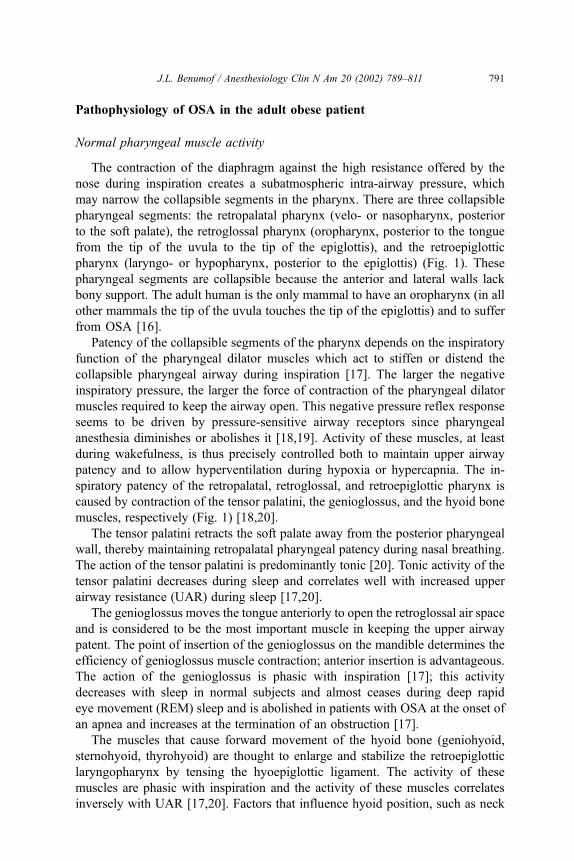
Table 1
Distribution by age of categorical levels of AHI* (AHI = Apneas + Hyponeas/Hour of sleep)
Age (y) Habitual snoring (%) AHI > 5 (%) AHI > 10 (%) AHI > 15 (%)
< 25 14 10 2 0
26–50 41 26 15 0
>50 46 61 50 36
AHI = Apnea Hyponea Index.
* From Strohl KP, Redline S. Recognition of sleep apnea. Am J Respir Crit Care Med 1996;154:
279–86.
Table 2
Major characteristics of obstructive sleep apnea (OSA) and obstructive sleep hypopnea (OSH)*
Obstruction #in Airflow >10 s Times/h #in O2 saturation Disrupted sleep Daytime sleepiness
OSA 100% >5 � 4% Yes Yes
OSH >50% >15 � 4% Yes Yes
* Data from Strollo PJ, Rogers RM. Obstructive sleep apnea. Current concepts. N Engl J Med
1996;334:99–104.
J.L. Benumof / Anesthesiology Clin N Am 20 (2002) 789–811790
Pathophysiology of OSA in the adult obese patient
Normal pharyngeal muscle activity
The contraction of the diaphragm against the high resistance offered by the
nose during inspiration creates a subatmospheric intra-airway pressure, which
may narrow the collapsible segments in the pharynx. There are three collapsible
pharyngeal segments: the retropalatal pharynx (velo- or nasopharynx, posterior
to the soft palate), the retroglossal pharynx (oropharynx, posterior to the tongue
from the tip of the uvula to the tip of the epiglottis), and the retroepiglottic
pharynx (laryngo- or hypopharynx, posterior to the epiglottis) (Fig. 1). These
pharyngeal segments are collapsible because the anterior and lateral walls lack
bony support. The adult human is the only mammal to have an oropharynx (in all
other mammals the tip of the uvula touches the tip of the epiglottis) and to suffer
from OSA [16].
Patency of the collapsible segments of the pharynx depends on the inspiratory
function of the pharyngeal dilator muscles which act to stiffen or distend the
collapsible pharyngeal airway during inspiration [17]. The larger the negative
inspiratory pressure, the larger the force of contraction of the pharyngeal dilator
muscles required to keep the airway open. This negative pressure reflex response
seems to be driven by pressure-sensitive airway receptors since pharyngeal
anesthesia diminishes or abolishes it [18,19]. Activity of these muscles, at least
during wakefulness, is thus precisely controlled both to maintain upper airway
patency and to allow hyperventilation during hypoxia or hypercapnia. The in-
spiratory patency of the retropalatal, retroglossal, and retroepiglottic pharynx is
caused by contraction of the tensor palatini, the genioglossus, and the hyoid bone
muscles, respectively (Fig. 1) [18,20].
The tensor palatini retracts the soft palate away from the posterior pharyngeal
wall, thereby maintaining retropalatal pharyngeal patency during nasal breathing.
The action of the tensor palatini is predominantly tonic [20]. Tonic activity of the
tensor palatini decreases during sleep and correlates well with increased upper
airway resistance (UAR) during sleep [17,20].
The genioglossus moves the tongue anteriorly to open the retroglossal air space
and is considered to be the most important muscle in keeping the upper airway
patent. The point of insertion of the genioglossus on the mandible determines the
efficiency of genioglossus muscle contraction; anterior insertion is advantageous.
The action of the genioglossus is phasic with inspiration [17]; this activity
decreases with sleep in normal subjects and almost ceases during deep rapid
eye movement (REM) sleep and is abolished in patients with OSA at the onset of
an apnea and increases at the termination of an obstruction [17].
The muscles that cause forward movement of the hyoid bone (geniohyoid,
sternohyoid, thyrohyoid) are thought to enlarge and stabilize the retroepiglottic
laryngopharynx by tensing the hyoepiglottic ligament. The activity of these
muscles are phasic with inspiration and the activity of these muscles correlates
inversely with UAR [17,20]. Factors that influence hyoid position, such as neck
J.L. Benumof / Anesthesiology Clin N Am 20 (2002) 789–811 791
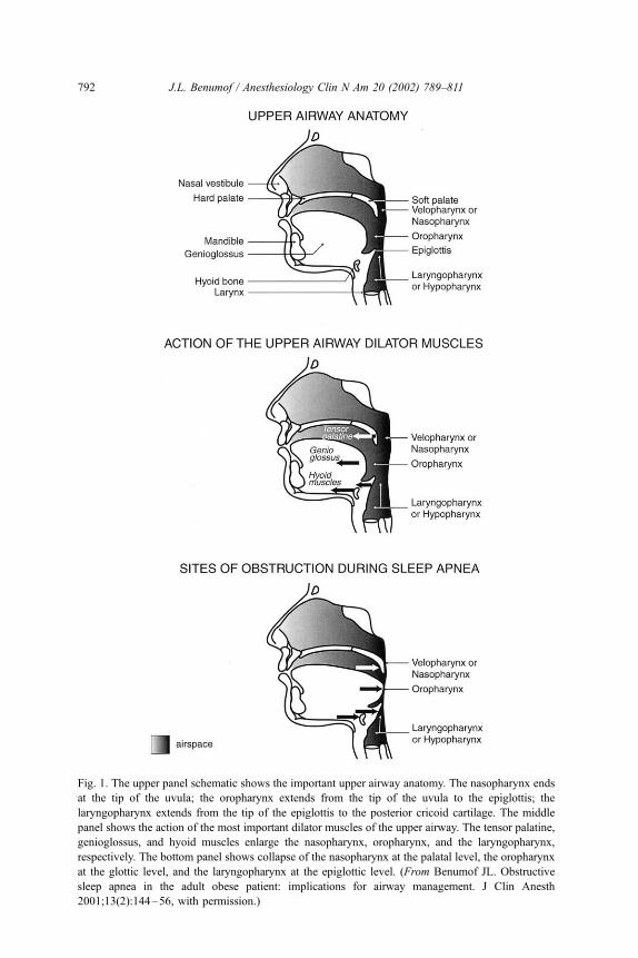
Fig. 1. The upper panel schematic shows the important upper airway anatomy. The nasopharynx ends
at the tip of the uvula; the oropharynx extends from the tip of the uvula to the epiglottis; the
laryngopharynx extends from the tip of the epiglottis to the posterior cricoid cartilage. The middle
panel shows the action of the most important dilator muscles of the upper airway. The tensor palatine,
genioglossus, and hyoid muscles enlarge the nasopharynx, oropharynx, and the laryngopharynx,
respectively. The bottom panel shows collapse of the nasopharynx at the palatal level, the oropharynx
at the glottic level, and the laryngopharynx at the epiglottic level. (From Benumof JL. Obstructive
sleep apnea in the adult obese patient: implications for airway management. J Clin Anesth
2001;13(2):144–56, with permission.)
J.L. Benumof / Anesthesiology Clin N Am 20 (2002) 789–811792
flexion or mandibular abnormalities, can adversely affect the function of these
muscles, leading to narrowing of the laryngopharynx.
Normal sleep
The relation between the anatomy and muscle function in the upper airway
becomes critical during sleep. In adults, a typical night of sleep consists of four to
six cycles of non-rapid eye movement (NREM) sleep followed by REM sleep.
There are four stages of NREM sleep which represent progressively deeper sleep
with progressive slowing of the electroencephalogram (EEG) waves. Stages 3 and
4 of NREM sleep differ only in the relative amount of slow waves and together
Stage 3 and 4 are called slow wave or deep sleep and is a restorative period of
sleep. REM sleep is also very deep sleep and is almost the exclusive domain of
dreaming [18]. REM sleep is characterized by a generalized loss of muscle tone as
evidenced by electromyography (EMG). However, the eye muscles are not
paralyzed, intermittent conjugate REMs still occur, and can be monitored by
electro-oculography (EOG).
During NREM sleep, the rhythmic activity of the upper airway muscles
decreases, UAR increases significantly and can be twice that during the awake
state [21,22]. In REM sleep, the activity of the upper airway muscles can disappear
completely and UAR increases even further. As UAR increases, the pharyngeal
subatmospheric pressure generated by a given diaphragmatic contraction increases
[23]. As pharyngeal pressure becomes more negative, pharyngeal collapse
increases. Magnetic resonance imaging, with and without nasal continuous
positive airway pressure (N-CPAP) (used as an experimental mechanism to
identify movement of various parts of the pharyngeal perimeter), shows that the
most important site of collapse is the compliant lateral pharyngeal walls [24].
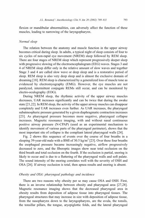
Fig. 2 shows this sequence of events over the course of four breaths in a
sleeping 39-year-old male with a BMI of 30.5 kg/m2 [23]. Over these four breaths
the esophageal pressure became increasingly negative, airflow progressively
decreased to zero, and the fiberoptic images show near total occlusion on the
third breath and total occlusion on the fourth. If the occlusion is partial, snoring is
likely to occur and is due to a fluttering of the pharyngeal walls and soft palate.
The sound intensity of the snoring correlates well with the severity of OSH and
OSA [26]. If airway occlusion is total, then apnea results and snoring is absent.
Obesity and OSA: pharyngeal pathology and incidence
There are two reasons why obesity per se may cause OSA and OSH. First,
there is an inverse relationship between obesity and pharyngeal area [27,28].
Magnetic resonance imaging shows that the decreased pharyngeal area in
obesity results from deposition of adipose tissue into pharyngeal tissues. The
pharyngeal structures that may increase in size with deposition of adipose tissue,
from the nasopharynx down to the laryngopharynx, are the uvula, the tonsils,
the tonsillar pillars, the tongue, aryepiglottic folds, and the lateral pharyngeal
J.L. Benumof / Anesthesiology Clin N Am 20 (2002) 789–811 793
walls. Increased fat deposition in the pharynx resulting in decreased patency of
the pharynx increases the likelihood that relaxation of the upper airway muscles
will cause collapse of the soft-walled pharynx between the uvula and epiglottis
[4,15,16].
The deposition of fat in the pharynx of OSA patients appears to be predom-
inantly into the lateral walls of the pharynx [13,29–33] and the volume of fat in
the lateral pharyngeal walls correlates well with the severity of OSA [31,32]. The
converse is also true; ie, weight loss improves the pharyngeal and glottic function
of OSA patients [34]. The deposition of fat into the lateral walls of the pharynx not
only narrows the airway but also changes the shape of the pharynx in obese
patients from being an ellipse with a long transverse (lateral) and a short anterior-
posterior axis to an ellipse with a short transverse and a long anterior-posterior axis
[20,30]. The external expression of very large internal lateral parapharyngeal fat
pads is submandibular (lateral) jowls.
Fig. 2. An original recording (Patient 6, BMI = 30.5 kg/m2 of Reference [23]) of airflow (flow;
inspiration positive) and esophageal pressure. Tracings show three breaths leading to an obstructive
apnea. Fiberoptic images are shown for the breath immediately preceding the apnea (Breath-1) and the
apneic period. During Breath-1, the images selected correspond to the smallest cross-sectional area that
occurred during inspiration (Nadir Inspiration), the largest cross-sectional area during expiration
(Maximum Expiraton), and the cross-sectional area at the end-expiration (End Expiration). During the
apnea, the images correspond to the time at which airway occlusion occurred (Occlusion), the time at
which the maximum esophageal pressure was generated (Nadir Inspiration), and the time at which the
airway reopened (Reopening). Within each image the dark area is the airway lumen, the lighter
horseshoe shape in the middle of the image is the epiglottis, and the narrow white triangular/cylindrical
shape on the right is the esophageal pressure catheter. (From Morrell MJ, Arabi Y, Zahn B, Badr MS.
Progressive retropalatal narrowing preceding obstructive apnea. AM J Respir Crit Care Med
1998;158:1974–81, with permission.)
J.L. Benumof / Anesthesiology Clin N Am 20 (2002) 789–811794
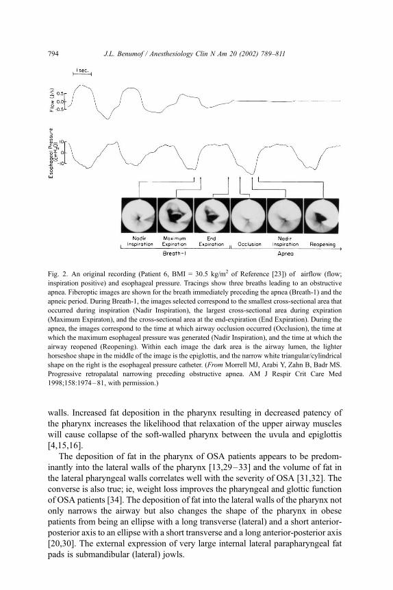
It has been theorized that the change in long axis from transverse to anterior-
posterior is functionally significant [35]. Since the muscles that increase upper air-
way size are all located along the anterior border of the pharynx (tensor palatini,
genioglossus, hyoid muscles), they pull the anterior wall of the pharynx forward
increasing the anterior-posterior axis (Fig. 1). Fig. 3 shows that this action of
the anterior pharyngeal dilator airway muscles is less efficient in a pharynx with a
long anterior-posterior elliptical axis than in one with a long transverse elliptical
axis [35].
Second, the patency of the pharynx (which is a collapsible tube) is determined
by the transmural pressure across its wall (the difference between the extraluminal
and intraluminal pressure) and the compliance of the wall. If the compliance of the
wall and intraluminal pressure (inspiratory airway pressure) are constant, then the
remaining important determinant of upper airway patency is extraluminal pressure.
In obese patients, extraluminal pressure is increased by superficially located fat
masses [36,37]; ie, the upper airway is compressed externally. This external
mechanism of increasing UAR is easily demonstrated in animals by experimentally
increasing the amount of anterior cervical neck fat [36]. Therefore, it is not surpris-
ing that the neck is significantly fatter in obese OSA patients compared to equally
obese non-OSA patients [38] and that the incidence and severity of OSA correlates
better with increased neck circumference than with general obesity [39–41].
Snoring occurs in 30% to 40% and 15% to 25% of obese (upper 25th percentile
of the BMI) and nonobese men, respectively, and in 15% to 25% and 5% to 10%
of obese and nonobese women, respectively [25]. Approximately half of individ-
uals that snore have some degree of OSA and virtually all patients with OSH and
OSA snore to some extent [4,15]. In two small series of persons with BMI >45.3
and > 50.2 (N = 250 and 27) 40% to 77% of men, but only 3% to 7% of women,
had significant OSA [42,43].
Fig. 3. The effect of a 5-mm change in the anteroposterior (AP) diameter of the airway on airway cross-
sectional area is shown for two equally elliptical airways with different lateral/AP ratios. (A) The lateral/
AP ratio = 0.5. (B) The lateral/AP ratio = 2. The lateral dimension of each ellipse was held constant. The
solid line represents the starting area (3 cm2 in both ellipses), and the dotted line represents the area after
a 5-mm increase in the AP diameter. The area change is greater in the ellipse with a more lateral
orientation (B). (From Leiter JC. Upper airway shape. Is it important in the pathogenesis of obstructive
sleep apnea? Am J Respir Crit Care Med 1996;153:894–8, with permission).
J.L. Benumof / Anesthesiology Clin N Am 20 (2002) 789–811 795
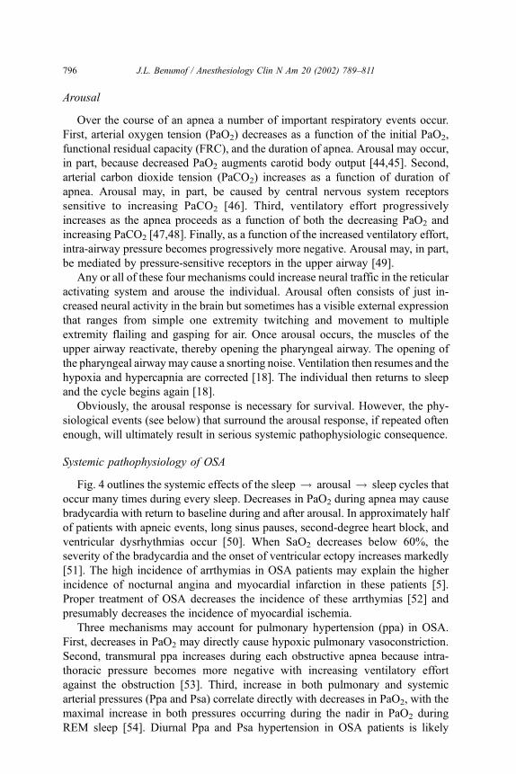
Arousal
Over the course of an apnea a number of important respiratory events occur.
First, arterial oxygen tension (PaO2) decreases as a function of the initial PaO2,
functional residual capacity (FRC), and the duration of apnea. Arousal may occur,
in part, because decreased PaO2 augments carotid body output [44,45]. Second,
arterial carbon dioxide tension (PaCO2) increases as a function of duration of
apnea. Arousal may, in part, be caused by central nervous system receptors
sensitive to increasing PaCO2 [46]. Third, ventilatory effort progressively
increases as the apnea proceeds as a function of both the decreasing PaO2 and
increasing PaCO2 [47,48]. Finally, as a function of the increased ventilatory effort,
intra-airway pressure becomes progressively more negative. Arousal may, in part,
be mediated by pressure-sensitive receptors in the upper airway [49].
Any or all of these four mechanisms could increase neural traffic in the reticular
activating system and arouse the individual. Arousal often consists of just in-
creased neural activity in the brain but sometimes has a visible external expression
that ranges from simple one extremity twitching and movement to multiple
extremity flailing and gasping for air. Once arousal occurs, the muscles of the
upper airway reactivate, thereby opening the pharyngeal airway. The opening of
the pharyngeal airway may cause a snorting noise. Ventilation then resumes and the
hypoxia and hypercapnia are corrected [18]. The individual then returns to sleep
and the cycle begins again [18].
Obviously, the arousal response is necessary for survival. However, the phy-
siological events (see below) that surround the arousal response, if repeated often
enough, will ultimately result in serious systemic pathophysiologic consequence.
Systemic pathophysiology of OSA
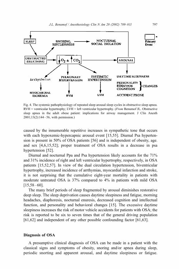
Fig. 4 outlines the systemic effects of the sleep ! arousal ! sleep cycles that
occur many times during every sleep. Decreases in PaO2 during apnea may cause
bradycardia with return to baseline during and after arousal. In approximately half
of patients with apneic events, long sinus pauses, second-degree heart block, and
ventricular dysrhythmias occur [50]. When SaO2 decreases below 60%, the
severity of the bradycardia and the onset of ventricular ectopy increases markedly
[51]. The high incidence of arrthymias in OSA patients may explain the higher
incidence of nocturnal angina and myocardial infarction in these patients [5].
Proper treatment of OSA decreases the incidence of these arrthymias [52] and
presumably decreases the incidence of myocardial ischemia.
Three mechanisms may account for pulmonary hypertension (ppa) in OSA.
First, decreases in PaO2 may directly cause hypoxic pulmonary vasoconstriction.
Second, transmural ppa increases during each obstructive apnea because intra-
thoracic pressure becomes more negative with increasing ventilatory effort
against the obstruction [53]. Third, increase in both pulmonary and systemic
arterial pressures (Ppa and Psa) correlate directly with decreases in PaO2, with the
maximal increase in both pressures occurring during the nadir in PaO2 during
REM sleep [54]. Diurnal Ppa and Psa hypertension in OSA patients is likely
J.L. Benumof / Anesthesiology Clin N Am 20 (2002) 789–811796
caused by the innumerable repetitive increases in sympathetic tone that occurs
with each hypoxemic-hypercapnic arousal event [15,55]. Diurnal Psa hyperten-
sion is present in 50% of OSA patients [56] and is independent of obesity, age.
and sex [4,6,15,52]; proper treatment of OSA results in a decrease in psa
hypertension [52].
Diurnal and nocturnal Ppa and Psa hypertension likely accounts for the 71%
and 31% incidence of right and left ventricular hypertrophy, respectively, in OSA
patients [15,52,57]. In view of the dual circulation hypertension, biventricular
hypertrophy, increased incidence of arrthymias, myocardial infarction and stroke,
it is not surprising that the cumulative eight-year mortality in patients with
moderate untreated OSA is 37% compared to 4% in patients with mild OSA
[15,58–60].
The many brief periods of sleep fragmented by arousal diminishes restorative
deep sleep. The sleep deprivation causes daytime sleepiness and fatigue, morning
headaches, diaphoresis, nocturnal enuresis, decreased cognition and intellectual
function, and personality and behavioral changes [15]. The excessive daytime
sleepiness increases the risk of motor vehicle accidents for patients with OSA; the
risk is reported to be six to seven times that of the general driving population
[61,62] and independent of any other possible confounding factor [61,63].
Diagnosis of OSA
A presumptive clinical diagnosis of OSA can be made in a patient with the
classical signs and symptoms of obesity, snoring and/or apnea during sleep,
periodic snorting and apparent arousal, and daytime sleepiness or fatigue.
Fig. 4. The systemic pathophysiology of repeated sleep arousal sleep cycles in obstructive sleep apnea.
RVH = ventricular hypertrophy; LVH = left ventricular hypertrophy. (From Benumof JL. Obstructive
sleep apnea in the adult obese patient: implications for airway management. J Clin Anesth
2001;13(2):144–56, with permission.)
J.L. Benumof / Anesthesiology Clin N Am 20 (2002) 789–811 797
Increased neck circumference is associated with OSA [39–41]; specifically, neck
circumference of OSA patients = 41.1 ± 3.5 cm versus neck circumference in
patients without OSA = 38.0 ± 3.5 cm, P < 0.001 [64].
Obesity is best expressed quantitatively as BMI:
BMI = mass/height2 = kg/m2 or 703 � lb/in2
where underweight, normal, overweight, obesity, and morbid obesity equals
< 19.0, 19.0–24.9, 25.0–29.9, 30.0–34.9, and > 35, respectively [65]. Ninety
percent of OSA patients may have a BMI > 28 kg/m2 [5]. Whereas the principal
limitation of use of the BMI is that it does not distinguish between fat and muscle,
in the general population it is much more likely that an individual with a BMI
> 30 kg/m2 is obese rather than muscular.
The definitive diagnosis of OSA and OSH, however, must be made by some
form of sleep study. A complete comprehensive sleep study exam is done by a
technologist in a formal soundproof infrared video monitored sleep study
laboratory. However, such studies can be logistically complex and expensive to
perform and may be relatively inaccessible some areas. Under these circumstances
some simpler derivation (eg, portable abbreviated monitoring/screening system) is
often performed and may be cost-effective [15,52].
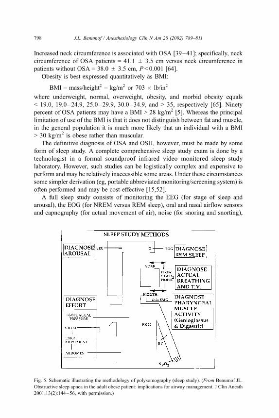
A full sleep study consists of monitoring the EEG (for stage of sleep and
arousal), the EOG (for NREM versus REM sleep), oral and nasal airflow sensors
and capnography (for actual movement of air), noise (for snoring and snorting),
Fig. 5. Schematic illustrating the methodology of polysomography (sleep study). (From Benumof JL.
Obstructive sleep apnea in the adult obese patient: implications for airway management. J Clin Anesth
2001;13(2):144–56, with permission.)
J.L. Benumof / Anesthesiology Clin N Am 20 (2002) 789–811798
esophageal pressure and chest and abdominal movements (for breathing effort),
submental and extremity EMG (for pharyngeal [genioglossus] muscle activity and
extremity movement, respectively), oximetry (pulse, ear, transcutaneous) (for
SpO2 and noninvasive blood pressure), and ECG (for cardiovascular function)
(Fig. 5). Rarely, direct systemic and pulmonary artery pressure monitoring is
performed for more precise determination of cardiopulmonary function.
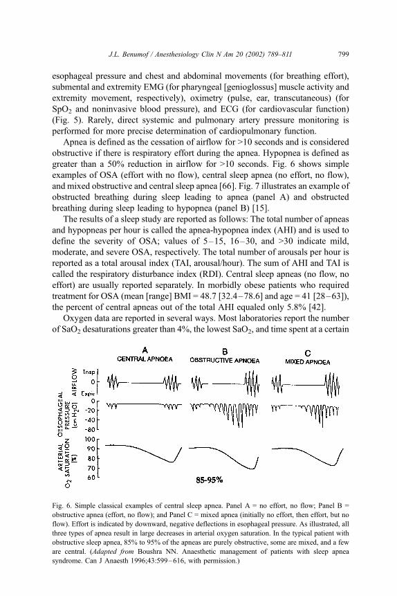
Apnea is defined as the cessation of airflow for >10 seconds and is considered
obstructive if there is respiratory effort during the apnea. Hypopnea is defined as
greater than a 50% reduction in airflow for >10 seconds. Fig. 6 shows simple
examples of OSA (effort with no flow), central sleep apnea (no effort, no flow),
and mixed obstructive and central sleep apnea [66]. Fig. 7 illustrates an example of
obstructed breathing during sleep leading to apnea (panel A) and obstructed
breathing during sleep leading to hypopnea (panel B) [15].
The results of a sleep study are reported as follows: The total number of apneas
and hypopneas per hour is called the apnea-hypopnea index (AHI) and is used to
define the severity of OSA; values of 5–15, 16–30, and >30 indicate mild,
moderate, and severe OSA, respectively. The total number of arousals per hour is
reported as a total arousal index (TAI, arousal/hour). The sum of AHI and TAI is
called the respiratory disturbance index (RDI). Central sleep apneas (no flow, no
effort) are usually reported separately. In morbidly obese patients who required
treatment for OSA (mean [range] BMI = 48.7 [32.4–78.6] and age = 41 [28–63]),
the percent of central apneas out of the total AHI equaled only 5.8% [42].
Oxygen data are reported in several ways. Most laboratories report the number
of SaO2 desaturations greater than 4%, the lowest SaO2, and time spent at a certain
Fig. 6. Simple classical examples of central sleep apnea. Panel A = no effort, no flow; Panel B =
obstructive apnea (effort, no flow); and Panel C = mixed apnea (initially no effort, then effort, but no
flow). Effort is indicated by downward, negative deflections in esophageal pressure. As illustrated, all
three types of apnea result in large decreases in arterial oxygen saturation. In the typical patient with
obstructive sleep apnea, 85% to 95% of the apneas are purely obstructive, some are mixed, and a few
are central. (Adapted from Boushra NN. Anaesthetic management of patients with sleep apnea
syndrome. Can J Anaesth 1996;43:599–616, with permission.)
J.L. Benumof / Anesthesiology Clin N Am 20 (2002) 789–811 799
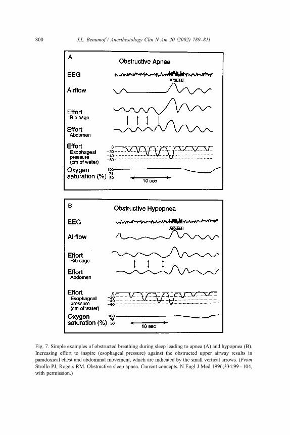
Fig. 7. Simple examples of obstructed breathing during sleep leading to apnea (A) and hypopnea (B).
Increasing effort to inspire (esophageal pressure) against the obstructed upper airway results in
paradoxical chest and abdominal movement, which are indicated by the small vertical arrows. (From
Strollo PJ, Rogers RM. Obstructive sleep apnea. Current concepts. N Engl J Med 1996;334:99–104,
with permission.)
J.L. Benumof / Anesthesiology Clin N Am 20 (2002) 789–811800
range of SaO2 (eg, 89%–80%, 79%–70%, etc.). The cardiovascular manifesta-
tions of SaO2 desaturation are variously described but always include maximum
and minimum heart rate and blood pressure during the event and the occurrence of
any arrthymias or ECG changes that are consistent with myocardial ischemia. If
N-CPAP was used for part of the sleep period, then all of the above data will be
reported with and without N-CPAP.
Effect of anesthetic drugs on airway patency in the adult obese OSA patient
All central depressant drugs diminish the action of the pharyngeal dilator
muscles in adult obese OSA patients, thereby promoting pharyngeal collapse
around a fat laden pharynx [67–73]. The commonly-used anesthetic drugs that
have been demonstrated to cause pharyngeal collapse are propofol [74], thio-
pental [75,76], narcotics [70,77,78], benzodiazepines [70,79–81], small doses
of neuromuscular blockers [82–84], and nitrous oxide [85]. The anesthetic drug-
induced pharyngeal collapse around the excessive airway tissue ‘‘makes the
airway lumen resemble the interior of the intestine and the path of the airway
easily becomes lost among the folds’’ [86]. Furthermore, if opioids cause airway
obstruction, then the opioids may also cause a poor ventilatory response to the
ensuing hypoxemia and hypercapnia [87].
It is important to understand that in the postoperative setting, sleep architecture
is disturbed. During the first three days after surgery, pain scores are highest and
deep stage 3 and 4 NREM and REM sleep are often suppressed [88]. High levels
of pain result in increased analgesic requirements during the first three post-
operative days, and from this perspective, the danger of life-threatening apnea
during drug-induced sleep is increased. In the next three days, deep REM sleep
rebounds [88,89]. During this phase of recovery the danger of life-threatening
natural deep sleep-induced apnea is increased. Thus, for separate in-series reasons
(increased analgesic requirement followed by increased amount of REM sleep),
the risk of prolonged apnea during sleep is increased for approximately one week
for the postoperative OSA patient [90,91].
Given that airway obstruction is more likely when either drug- or REM sleep-
induced pharyngeal collapse occurs, it is not surprising that heavy snorers
(identified preoperatively) have more severe decreases in SpO2 during sleep
postoperatively than normal patients [92]. This consideration is highlighted by
numerous reports of the need for rescue airway management in postoperative OSA
patients [93–100].
Implications for airway management
Preoperative evaluation: OSA and airway status
Since most adult obese patients with OSA are undiagnosed, many who presently
require anesthesia and surgery have neither a presumptive clinical nor sleep study
J.L. Benumof / Anesthesiology Clin N Am 20 (2002) 789–811 801
diagnosis of OSA. The essential items on history that must be present for a pre-
sumptive clinical diagnosis of OSA in the adult obese patient are history of snoring
and/or snorting and/or apnea during sleep and daytime sleepiness. The severity of
these historical items correlates with the severity of sleep study–proven OSA
[6,7,15,26,101]. Since a firm diagnosis of OSA will likely impact on anesthetic
management, it is reasonable to suggest that all adult obese patients (or those who
observe them while asleep) be routinely asked about nocturnal snoring/snorting/
apnea and diurnal sleepiness [91,92,102]. Prediction of OSA is increased if there is
a history of hypertension [64] or a neck circumference >40–42 cm [39,40,64].
Other signs and symptoms consistent with a clinical presumptive diagnosis of OSA
are nocturnal diaphoresis and enuresis, frequent nocturia, morning headaches,
abnormal cardiovascular, and neuropsychiatric function (see fig. 4).
If the anesthesiologist is the first care-giver to diagnose OSA, then it may
sometimes be prudent to postpone the surgery and refer the patient to an
appropriate physician and perhaps a formal sleep study obtained to quantify the
severity of OSA. Alternatively, if general anesthesia is required, the patient could
be treated as though severe OSA existed (see remainder of article). Finally,
regional anesthesia is worth considering if it can be technically performed, the
awake patient can tolerate the surgical position, and the respiratory effects of the
regional anesthetic, access to the airway is adequate, and the surgery can be
quickly terminated. Regional anesthesia may obviate the need for sedative and
narcotic drugs both intra- and postoperatively.
Tracheal intubation
Several lines of evidence in the literature indicate that obese OSA patients are, in
general, more difficult to intubate than normal controls. First, obesity is signifi-
cantly related to difficult intubation [103–105]. Indeed, in two series of morbidly
obese patients undergoing upper abdominal surgery, the incidence of difficult of
intubation under general anesthesia was 13% and 24% and the incidence of patients
requiring awake intubation was 8% in both studies [68,106]. Second, a short thick
neck is significantly related to difficult intubation [103,107]. Third, obesity [1,2,
5–7] and a short thick neck [36–41,64] are significantly related to OSA and to each
other [103]. Fourth, since excess pharyngeal tissue is deposited in the lateral walls
of the pharynx of obese OSA patients [13,29–33], the excess tissue may not be
visualized during routine oropharyngeal classification. Finally, based on the above,
it is not surprising that difficult intubation and OSA had been found to be
significantly related [108]. In fact, the strength of the relationship was such that
the author felt that ‘‘all patients who have a trachea that is difficult to intubate
should be regarded as having OSA until excluded by clinical features and, where
doubt exist, sleep studies.’’ Indeed, in one large series of patients undergoing
surgery for OSA, the incidence of failed intubation was 5% [93]. Thus, given the
above literature, and the fact that excess pharyngeal tissue may not be revealed by
routine examination, it is reasonable that the practitioner have an increased index of
suspicion regarding intubation difficulty.
J.L. Benumof / Anesthesiology Clin N Am 20 (2002) 789–811802
Several more tracheal intubation points are especially relevant to obese OSA
patients. Within the context of an increased index of suspicion of intubation
difficulty, the decision as to whether to do tracheal intubation with the patient
awake or under general anesthesia must be individualized on the basis of a
complete preoperative airway evaluation. If difficulty with either mask ventilation
or tracheal intubation is expected, then according to the ASA Difficult Airway
Algorithm, intubation and extubation should be performed while the patient is
awake [109–111].
If tracheal intubation is to be done while the patient is awake, it is essential that
the patient be properly prepared. One component of proper preparation is judicious
administration of sedative and analgesic medication [68,71]. The danger of pre-
medication in these patients is well illustrated by descriptions of several cases of
complete airway obstruction [112–115]. Thus, proper preparation should depend
on thorough topical and nerve block anesthesia of the upper airway [106,110]. Use
of a flexible fiberscope through a rigid oropharyngeal conduit technique of in-
tubation permits visualization of structures in an atraumatic manner [111,116].
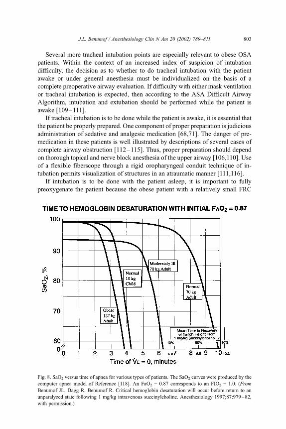
If intubation is to be done with the patient asleep, it is important to fully
preoxygenate the patient because the obese patient with a relatively small FRC
Fig. 8. SaO2 versus time of apnea for various types of patients. The SaO2 curves were produced by the
computer apnea model of Reference [118]. An FaO2 = 0.87 corresponds to an FIO2 = 1.0. (From
Benumof JL, Dagg R, Benumof R. Critical hemoglobin desaturation will occur before return to an
unparalyzed state following 1 mg/kg intravenous succinylcholine. Anesthesiology 1997;87:979–82,
with permission.)
J.L. Benumof / Anesthesiology Clin N Am 20 (2002) 789–811 803
(small oxygen reservoir) and high oxygen consumption desaturates much more
rapidly during obstructive apnea compared with a normal patient [117,118]
(Fig. 8).Maximal total body preoxygenation (filling of the alveolar, arterial,
venous, and tissue spaces) requires that the patient breathe FIO2 = 1.0 for
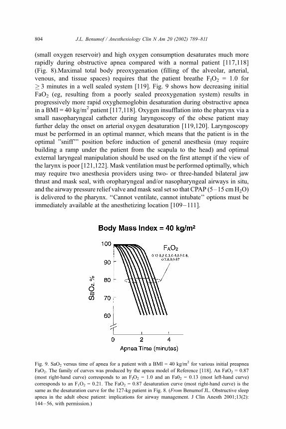
� 3 minutes in a well sealed system [119]. Fig. 9 shows how decreasing initial
FaO2 (eg, resulting from a poorly sealed preoxygenation system) results in
progressively more rapid oxyghemoglobin desaturation during obstructive apnea
in a BMI = 40 kg/m2 patient [117,118]. Oxygen insufflation into the pharynx via a
small nasopharyngeal catheter during laryngoscopy of the obese patient may
further delay the onset on arterial oxygen desaturation [119,120]. Laryngoscopy
must be performed in an optimal manner, which means that the patient is in the
optimal ’’sniff’’’ position before induction of general anesthesia (may require
building a ramp under the patient from the scapula to the head) and optimal
external laryngeal manipulation should be used on the first attempt if the view of
the larynx is poor [121,122]. Mask ventilation must be performed optimally, which
may require two anesthesia providers using two- or three-handed bilateral jaw
thrust and mask seal, with oropharyngeal and/or nasopharyngeal airways in situ,
and the airway pressure relief valve andmask seal set so that CPAP (5–15 cmH2O)
is delivered to the pharynx. ‘‘Cannot ventilate, cannot intubate’’ options must be
immediately available at the anesthetizing location [109–111].
Fig. 9. SaO2 versus time of apnea for a patient with a BMI = 40 kg/m2 for various initial preapnea
FaO2. The family of curves was produced by the apnea model of Reference [118]. An FaO2 = 0.87
(most right-hand curve) corresponds to an FIO2 = 1.0 and an Fa02 = 0.13 (most left-hand curve)
corresponds to an F1O2 = 0.21. The FaO2 = 0.87 desaturation curve (most right-hand curve) is the
same as the desaturation curve for the 127-kg patient in Fig. 8. (From Benumof JL. Obstructive sleep
apnea in the adult obese patient: implications for airway management. J Clin Anesth 2001;13(2):
144–56, with permission.)
J.L. Benumof / Anesthesiology Clin N Am 20 (2002) 789–811804
Extubation: awake versus leaving the tube in
The risk of airway obstruction following extubation is increased in OSA
patients [123]. The risk is further increased in OSA patients who have had nasal
packing following nasal surgery [124]; therefore, packing around a nasopha-
ryngeal airway (creating a central conduit for gas exchange) should be considered
[69,91]. In a retrospective review of 135 patients undergoing surgery for the
treatment of OSA, the incidence of life-threatening postextubation obstruction was
5%; those patients who obstructed were extubated in the operating room [93].
Aside from the threat of death from airway obstruction, another great danger of
spontaneous ventilation against an obstructed airway is rapid development of
severe negative pressure pulmonary edema [93,125]. The treatment of negative
pressure pulmonary edema in this setting usually requires re-intubation [125].
Depending on the mask ventilation and tracheal intubation experience at the
beginning of the case, the length and type of surgery, and the severity of the OSA,
one should consider leaving the patient intubated for a period of postoperative me-
chanical ventilation. Whenever the patient is to be extubated (either in the
operating room or later in the PACU or ICU) the patient should be fully awake. A
dangerous mistake is to interpret mindless movement, such as reflex reaching for
an endotracheal tube or suddenly trying to sit up, for purposeful movement. Full
recovery from neuromuscular blockade should be proven by a neuromuscular
blockade monitor, sustained head lift for >5 seconds and, in the ICU, with an
adequate vital capacity and peak inspiratory pressure. The patient should not have
a high blood level of narcotic as indicated by a respiratory rate < 12–14 breaths/
minute while the endotracheal tube is in situ. It is helpful for regional analgesia to
be operative at the time of extubation. Extubation in the reverse Trendelenburg or
semi-upright position minimizes compression of the diaphragm by abdominal
contents [126].
When extubating, an oropharyngeal and/or a long nasopharyngeal airway (ie, it
is desirable for the distal end to be retroglossal) should be in situ if possible and
two-person mask ventilation should be ready to be applied. If doubt exists about
the ability of the patient to breathe adequately and the practitioner to re-intubate if
the patient does breathe inadequately, then the tracheal tube should be removed
over an airway exchange catheter or fiberscope [127]. If the patient does well
initially, one should consider pneumatically splinting the oropharynx by applying
N-CPAP, first with oxygen and then later with air [123,128]. Beyond the initial
immediate application of N-CPAP, the FIO2 should only be increased if the SpO2
is significantly decreased [94].
Opioid pain management: location of patient
Obese OSA patients have an increased risk of opioid-induced upper airway
obstruction (even epidural and patient controlled analgesia may be problematic)
[96,97] and is a reason why these patients may need a monitored care environment
(ie, continuous electronic and frequent visual monitoring [respiratory rate,
J.L. Benumof / Anesthesiology Clin N Am 20 (2002) 789–811 805
sedation level, snoring]) [95]. Factors to be considered in this risk benefit analysis
are the BMI of the patient, the AHI (ie, the severity of the OSA), the degree of
associated cardiopulmonary disease, and the postoperative narcotic requirement.
When all of these factors are mild, then the patient may reasonably go to a
relatively unmonitored environment, and when any of these factors are severe, the
patient should go to an ICU. The large gray zone in between these extremes
requires careful judgment. Putting aside the one negative report involving epidural
opioids in a OSA patient [96], the risk of opioid-induced airway obstruction may
be avoidable by using alternative techniques, for example, regional block with
local anesthesia to provide postoperative analgesia [129].
Summary
Adult obese patients with suspected or sleep test confirmed OSA present a
formidable challenge throughout the perioperative period. Life-threatening prob-
lems can arise with respect to tracheal intubation, tracheal extubation, and
providing satisfactory postoperative analgesia. Tracheal intubation and extubation
decisions in obese patients with either a presumptive and/or sleep study diagnosis
of OSA must be made within the context that there may be excess pharyngeal
tissue that cannot be visualized by routine examination, and the literature indicates
an increased risk of intubation difficulty. Regional anesthesia for postoperative
pain control is desirable (although such management is not necessary or possible
for many of these patients). If opioids are used for the extubated postoperative
patient, then one must keep in mind an increased risk of pharyngeal collapse and
consider the need for continuous visual and electronic monitoring. The exact
management of each sleep apnea patient with regard to intubation, extubation, and
pain control requires judgment and is a function of many anesthesia, medical, and
surgical considerations.
References
[1] Young T, Palta M, Dempsey J, Skatrud J, Weber S, Badr S. The occurrence of sleep-disordered
breathing among middle-aged adults. N Engl J Med 1993;328:1230–5.
[2] Bresnitz E. Epidemiology of obstructive sleep apnea. Epidemiol Rev 1994;16:210–27.
[3] Anocli-Israel S, Kripke DF, Klauber MR, Mason WJ, Fell R, Kaplan O. Sleep-disordered
breathing in community-dwelling elderly. Sleep 1991;14:485–95.
[4] Barthel SW, Strome M. Snoring, obstructive sleep apnea and surgery. Med Clin North Am
1999;83:85–96.
[5] Strohl KP, Redline S. Recognition of obstructive sleep apnea. Am J Respir Crit Care Med
1996;154:279–86.
[6] Guilleminault C, Tikian A, Dement W. The sleep apnea syndromes. Annu Rev Med 1976;
27:465–84.
[7] Willett WC, Dietz WH, Colditz GA. Guidelines for healthy weight. N Engl J Med 1999;341:
427–34.
[8] National Commission on Sleep Disorders Research. Wake up America: a national sleep alert.
Washington, DC: Government Printing Office; 1993.
J.L. Benumof / Anesthesiology Clin N Am 20 (2002) 789–811806
[9] Young T, Evans L, Finn L, Palta M. Estimation of the clinically diagnosed proportion of sleep
apnea syndrome in middle-aged men and women. Sleep 1997;20:705–6.
[10] Callop NA. Conundrums in sleep medicine. Chest 1999;115:607–8.
[11] Pack AI, Gurubhagavatula I. Economic implications of the diagnosis of obstructive sleep apnea.
Ann Intern Med 1999;130:533–4.
[12] Sakakibara H, Tong M, Matsushita K, Hirata M, Konishi Y, Suetsugh S. Cephalometric ab-
normalities in non-obese and obese patients with obstructive sleep apnea. Eur Respir J 1999;
13:403–10.
[13] Mortimore IL, Marshal I, Wraith PK, Sellar RJ, Douglas NJ. Neck and total body fat deposition
in nonobese and obese patients with sleep apnea compared with that in control subjects. Am J
Respir Crit Care Med 1998;157:280–3.
[14] Davidson-Ward SL, Marcu CL. Obstructive sleep apnea in infants and young children. J Clin
Neurophysiol 1996;13:198–207.
[15] Strollo PJ, Rogers RM. Obstructive sleep apnea. current concepts. N Engl J Med 1996;334:
99–104.
[16] Barsh CI. The origin of pharyngeal obstruction during sleep. Sleep and breathing 1999;3:
17–21.
[17] Deegan PC, McNicholas WT. Pathophysiology of obstructive sleep apnea. Eur Respir J
1995;8:1161–78.
[18] White DP. Pathophysiology of obstructive sleep apnea. Thorax 1995;50:797–804.
[19] Horner RL, Innes JA, Holden HB, Guy A. Afferent pathways for pharyngeal dilator reflex
to negative pressure in man: a study using upper airway anesthesia. J Physiol 1991;436:
31–44.
[20] Pierce RJ, Worsnop CJ. Upper airway function and dysfunction in respiration. Clin Exp Pharm
Physiol 1999;26:1–10.
[21] Hudgel DW. Palate and hypopharynx: sites of inspiratory narrowing of the upper airway during
sleep. Am Rev Respir Dis 1988;138:1542–7.
[22] Wiegand I, Zwillich CW, White DP. Collapsibility of the human airway during normal sleep.
J Appl Physiol 1989;66:1800–8.
[23] Morrell MJ, Arabi Y, Zahn B, Badr MS. Progressive retropalatal narrowing preceding obstruc-
tive apnea. Am J Respir Crit Care Med 1998;158:1974–81.
[24] Schwab RI, Pack AI, Gupta KB, Metzger JL, Oh E, Getsy JE, et al. Upper airway and soft
tissue structural changes induced by CPAP in normal subjects. Am J Respir Crit Care Med
1996;154:1106–16.
[25] Bloom JW, Kaltenborn WT, Quan SF. Risk factors in a general population for snoring: Im-
portance of cigarette smoking an obesity. Chest 1988;93:678–83.
[26] Wilson K, Stoohs RA, Mulrooney TF, Johnson LJ, Guilleminault C, Huang Z. The snoring
spectrum: acoustic assessment of snoring sound intensity in 1,139 individuals undergoing
polysomography. Chest 1999;115:765–70.
[27] White DP, Lombard RM, Cadieux RJ, Zwillich CW. Pharyngeal resistance in normal humans:
influence of gender, age and obesity. J Appl Physiol 1985;58:365–71.
[28] Brown IG, Zamel N, Hoffstein V. Pharyngeal cross-sectional area in normal men and women.
J Appl Physiol 1986;61:890–5.
[29] Horner RL, Mohiaddin RH, Lowell DG, Shea SA, Burman ED, Longmore DB, et al. Sites and
sizes of the fat deposits around the pharynx in obese patients with obstructive sleep apnea and
weight matched controls. Eur Respir J 1989;2:613–22.
[30] Mayer P, Pepin JL, Bettega G, Veale D, Ferrette G, Deschaux C, et al. Relationship between
body mass index, age and upper airway measurements in snorers and sleep apnea patients. Eur
Respir J 1996;9:1801–9.
[31] Shelton KE, Gay SB, Hollowell DE, Woodson H, Suratt PM. Mandible enclosure of upper
airway and weight in obstructive sleep apnea. Am Rev Respir Dis 1993;148:195–200.
[32] Shelton KE, Gay SB, Woodson H, Gay S, Suratt PM. Pharyngeal fat in obstructive sleep apnea.
Am Rev Respir Dis 1993;148:462–6.
J.L. Benumof / Anesthesiology Clin N Am 20 (2002) 789–811 807
[33] Schwab RJ, Gupta KB, Gefter WB, Metzger LJ, Hoffman EA, Pack AI. Upper airway soft
tissue anatomy in normal subjects and patients with sleep-disordered breathing. Significance of
the lateral pharyngeal walls. Am J Respir Crit Care Med 1995;152:1673–89.
[34] Rubinstein I, Colapinto N, Rotstein LE, Brown IG, Hoffstein V. Improvement in upper airway
function after weight loss in patients with obstructive sleep apnea. Am Rev Resp Dis 1988;
138:1192–5.
[35] Leiter JC. Upper airway shape. Is it important in the pathogenesis of obstructive sleep apnea?
Am J Respir Crit Care Med 1996;153:894–8.
[36] Koenig JE, Thach BT. Effects of mass loading on the upper airway. J Appl Physiol 1988;64:
2294–9.
[37] Koopmann CF, Field RA, Coulthard SW. Sleep apnea syndrome associated with a neck mass.
Otolaryngol Head Neck Surg 1981;89:949–52.
[38] Hoffstein V, Mateika S. Differences in abdominal and neck circumferences in patients with and
without obstructive sleep apnea. Eur Respir J 1992;5:377–81.
[39] Davies RJO, Stradling JR. The relationship between neck circumference, radiographic
pharyngeal anatomy, and the obstructive sleep apnoea syndrome. Eur Respir J 1990;3:
509–14.
[40] Stradling JR, Crosby JH. Predictors and prevalence of obstructive sleep apnoea and snoring in
1,001 middle aged men. Thorax 1991;46:85–90.
[41] Davies RJO, Ali NJ, Stradling JR. Neck circumference and other clinical features in the
diagnosis of the obstructive sleep apnea syndrome. Thorax 1992;47:101–5.
[42] Vgontzas AN, Tan TL, Bixler EO, Martin LF, Shubert D, Kales A. Sleep apnea and sleep
disruption in obese patients. Arch Intern Med 1994;154:1705–11.
[43] Rajala R, Partinen M, Sane T, Pelkonen R, Huikur K, Seppalainen AM. Obstructive sleep
apnoea syndrome in morbidly obese patients. J Intern Med 1991;230:125–9.
[44] Bowes G, Townsend ER, Bromley SM, Kozar LF, Phillipson EA. Role of carotid body and of
afferent vagal stimuli in the arousal response to airway occlusion in sleeping dogs. Am Rev
Respir Dis 1981;123:644–7.
[45] Bowes G, Townsend ER, Kozar LF, Bromley SM, Phillipson EA. Effect of carotid body
denervation on arousal response in sleeping dogs. J Appl Physiol 1981;51:40–5.
[46] Berthon-Jones M, Sullivan CE. Ventilation and arousal responses to hypercapnea in normal
sleeping humans. J Appl Physiol 1984;54:59–67.
[47] Kimoff RJ, Cheong TH, Olha AE, Charbonneau M, Levy RD, Cosio MG, et al. Mechanisms of
apnea termination in obstructive sleep apnea: role of chemoreceptor and mechanoreceptor
stimuli. Am J Respir Crit Care Med 1994;149:707–14.
[48] Gleeson K, Zwillich CW, White DP. The influence of increasing ventilatory effort on arousal
from sleep. Am Rev Respir Dis 1990;142:295–300.
[49] Issa FG, McNamara SG, Sullivan CE. Arousal responses to airway occlusion in sleeping dogs:
comparison of nasal and tracheal occlusions. J Appl Physiol 1987;62:1832–6.
[50] Guilleminault C, Connolly SJ, Winkle RA. Cardiac arrhythmia and conduction disturbances
during sleep in 400 patients with sleep apnea syndrome. Am J Cardiol 1983;52:490–4.
[51] Orr WC. Sleep apnea, hypoxemia and cardiac arrhythmias [editorial]. Chest 1986;89:1–2.
[52] Man GCW. Obstructive sleep apnea: ciagnosis and treatment. Med Clin North Am 1996;80:
803–20.
[53] Schafer H, Hasper SE, Koehler U, Latzelsberger J, Tasci S, Luderitz B. Pulmonary hemody-
namics in obstructive sleep apnea: time course and associated factors. Eur Respir J 1998;12:
679–84.
[54] Shepard JW. Gas exchange and hemodynamics during sleep. Med Clin North Am 1985;
69:1243–64.
[55] Fletcher EC. The relationship between systemic hypertension and obstructive sleep apnea: facts
and theory. Am J Med 1995;98:118–28.
[56] Millman RP, Redline S, Carlisle CC, Assaf AR, Levinson PD. Daytime hypertension in ob-
structive sleep apnea. Prevalence and contributing risk factors. Chest 1991;99:861–6.
J.L. Benumof / Anesthesiology Clin N Am 20 (2002) 789–811808
[57] Berman EJ, DiBenedetto RJ, Causey DE, Mims T, Conneff M, Goodman LS, et al. Right
ventricular hypertrophy detected by echocardiography in patients with newly diagnosed ob-
structive sleep apnea. Chest 1991;100:347–50.
[58] He J, Kryger MH, Zorick FJ, Conway W, Roth T. Mortality and apnea index in obstructive
sleep apnea: experience in 385 male patients. Chest 1988;94:9–14.
[59] Palomaki H, Partinen M, Erkinjuntti J, Kaste M. Snoring, sleep apnea syndrome, and stroke.
Neurology 1992;42(Suppl 6):75–82.
[60] Partinen M, Guilleminault C. Daytime sleepiness and vascular morbidity at seven-year follow-
up in obstructive sleep apnea patients. Chest 1990;97:27–32.
[61] Teran-Santos J, Jimenez-Gomez A, Cordero-Guevara J, Cooperative Group Burgos-Santander.
The association between sleep apnea and the risk of traffic accidents. N Engl J Med 1999;340:
847–51.
[62] Findley LJ, Unverzagt ME, Suratt PM. Automobile accidents involving patients with obstruc-
tive apnea sleep. Am Rev Respir Dis 1988;138:337–40.
[63] Suratt PM, Findley LJ. Driving with sleep apnea. N Engl J Med 1999;340:881–3.
[64] Flemons WW, Whitelaw WA, Brant R, Remmers JE. Likelihood ratios for a sleep apnea clinical
prediction rule. Am J Respir Crit Care Med 1994;150:1279–85.
[65] Obesity: preventing and managing the global epidemic: report of a WHO consultation on
obesity, Geneva, June 3–5, 1997. Geneva: World Health Organization; 1998.
[66] Orr WC. Utilization of polysomnography in the assessment of sleep disorders. Med Clin North
Am1985;69:1153–67.
[67] Boushra NN. Anaesthetic management of patients with sleep apnoea syndrome. Can J Anaesth
1996;43:599–616.
[68] Buckley FP, Robinson NB, Simonowitz DA, Dillinger EP. Anaesthesia in the morbidly obese: a
comparison of anaesthetic and analgesic regimens for upper abdominal surgery. Anaesthesia
1983;38:840–51.
[69] Hanning CD. Obstructive sleep apnea. Br J Anaesth 1989;63:477–88.
[70] Chung F, Crago RR. Sleep apnea syndrome and anaesthesia. Can Anaesth Soc J 1982;
29:439–43.
[71] Connolly CA. Anesthetic management of obstructive sleep apnea patients. J Clin Anesth
1991;3:461–9.
[72] Hwang JC, St John WM, Bartlett D. Respiratory related hypoglossal nerve activity: influence of
anesthetics. J Appl Physiol 1983;55:785–92.
[73] Nishino T, Shirahata M, Yonezawa T, Honda Y. Comparison of changes in the hypoglossal and
phrenic nerve activity in response to increasing depth of anesthesia in cats. Anesthesiology
1984;60:19–24.
[74] Mathru M, Esch O, Lang J, Herbert ME, Chaljub G, Goodacre B, et al. Magnetic resonance
imaging of the upper airway: Effect of propofol anesthesia and nasal continuous positive airway
pressure in humans. Anesthesiology 1996;84:273–9.
[75] Drummond GB. Influence of thiopentone on upper airway muscles. Br J Anaesth 1989;63:
12–21.
[76] Nandi PR, Charlesworth CH, Taylor SJ, Nunn JF, Dore CJ. Effect of general anaesthesia on the
pharynx. Br J Anaesth 1991;66:157–62.
[77] Catley DM, Thornton C, Jordan C, Lehane JR, Royston D, Jones JG. Pronounced, episodic
oxygen desaturation in postoperative period: its association with ventilatory pattern and anal-
gesic regimen. Anesthesiology 1985;63:20–8.
[78] Robinson RW, Zwillich CW, Bixler EO, Cadieux RJ, Kales A, White DP. Effects of oral
narcotics on sleep-disordered breathing in healthy adults. Chest 1987;91:197–203.
[79] Leiter JC, Knuth SL, Krol RC, Bartlett Jr D. The effect of diazepam on genioglossal muscle
activity in normal human subjects. Am Rev Respir Dis 1985;132:216–9.
[80] Montravers P, Dureuil B, Desmonts JM. Effects of IV midazolam on upper airway resistance.
Br J Anaesth 1992;68:27–31.
[81] Zivkovic B, Perrault G, Morel E, Sanger DJ. Comparative pharmacology of zolpidem and other
J.L. Benumof / Anesthesiology Clin N Am 20 (2002) 789–811 809
hypnotics and sleep inducers. In: Sauvant JP, Langer SZ, Morselli PL, editors. Imidazopyridines
in sleep disorders. New York: Raven Press; 1988. p. 97–109.
[82] Bruce DL, Downs JB, Kulkarni PS, Capan LM. Precurarization inhibits maximal ventilatory
effort. Anestheisology 1984;61:618–21.
[83] Musich J, Walts LF. Pulmonary aspiration after a priming dose of vecuronium. Anesthesiology
1986;64:517–9.
[84] Pavlin EG, Holle RH, Schoene RB. Recovery of airway protection compared with ventilation in
humans after paralysis with curare. Anesthesiology 1989;70:381–5.
[85] Beydon L, Lafaso F, Heyer L, Delaunay L, Goldenberg F. Nitrous oxide induces central and
obstructive apneas in normal subjects. Br J Anaesth 1994;72(Suppl 1):A113.
[86] Craddock M, Lees DE. Anesthesia for obstructive sleep apnea: risks, precautions, and manage-
ment. In: Fairbanks DNF, Fujita S, Ikematsu T, Simmons FB, editors. Snoring and obstructive
sleep apnea. New York: Raven Press; 1987. p. 235.
[87] Phillipson EA, Sullivan CE. Arousal: the forgotten response to respiratory stimuli. Am Rev
Respir Dis 1978;118:807–9.
[88] Knill RL, Moote CA, Skinner MI, Rose EA. Anesthesia with abdominal surgery leads to
intensive REM sleep during the first postoperative week. Anesthesiology 1990;73:52–61.
[89] Reeder MK, Goldman MD, Loh L, Muir AD, Casey KR, Lehane JR. Late postoperative
nocturnal dips in oxygen saturation in patients undergoing major abdominal vascular surgery.
Predictive value of pre-operative overnight pulse oximetry. Anaesthesia 1992;47:110–5.
[90] Reeder MK, Goldman MD, Loh L, Muir AD, Casey KR, Gitlin DA. Postoperative obstruc-
tive sleep apnoea. Haemodynamic effects of treatment with nasal CPAP. Anaesthesia 1991;
46:849–53.
[91] Dodds C. Sleep apnoea and anaesthesia. Recent Advances in Anaesthesia and Analgesia
1994;18:179–85.
[92] Gentil B, Lienhart A, Fleury B. Enhancement of postoperative desaturation in heavy snorers.
Anesth Analg 1995;81:389–92.
[93] Esclamado RM, Glenn MG, McCulloch TM, Cummings CW. Perioperative complications and
risk factors in the surgical treatment of obstructive sleep apnea syndrome. Laryngoscope 1989;
99:1125–9.
[94] Jones JG, Jordan C, Scudder C, Rocke DA, Barrowcliffe M. Episodic postoperative oxygen
desaturation: the value of added oxygen. J R Soc Med 1985;78:1019–22.
[95] Keamy MF, Cadieux RJ, Kofke WA, Kales A. The occurrence of obstructive sleep apnea in a
recovery room patient. Anesthesiology 1987;66:232–4.
[96] Larmarche Y, Martin R, Reiher J, Blaise G. The sleep apnoea syndrome and epidural morphine.
Can Anaesth Soc J 1986;33:231–3.
[97] VanDercar DH, Martinez AP, De Liser EA. Sleep apnea syndromes: a potential contraindication
for patient-controlled analgesia. Anesthesiology 1991;74:623–4.
[98] Tierney NM, Pollard BJ, Doran BRH. Obstructive sleep apnoea. Anaesthesia 1989;44:235–7.
[99] Gabrielczyk MR. Acute airway obstruction after uvulopalatopharyngoplasty for obstructive
sleep apnea syndrome. Anesthesiology 1988;69:941–3.
[100] Fairbanks DNF. Uvulopalatopharyngoplasty complications and avoidance strategies. Otolar-
yngol Head Neck Surg 1990;102:239–45.
[101] Grunstein R, Wilcox I, Yang TS, Hedner J. Snoring and sleep apnoea in men: association with
central obesity and hypertension. Int J Obesity 1993;17:533–40.
[102] Vidhani K, Langham BT. Obstructive sleep apnoea syndrome: Is this an overlooked cause of
desaturation in the immediate postoperative period. Br J Anaesth 1997;78:442–3.
[103] Rocke DA, Murray WB, Rout CC, Gouws E. Relative risk analysis of factors associated with
difficult intubation in obstetric anaesthesia. Anesthesiology 1992;77:67–73.
[104] Rose DK, Cohen MM. The airway: problems and predictions in 18,500 patients. Can J Anaesth
1994;41:372–82.
[105] Wilson ME, Spiegelhalter D, Robertson JA, Lesser P. Predicting difficult intubation. Br J
Anaesth 1988;61:211–6.
J.L. Benumof / Anesthesiology Clin N Am 20 (2002) 789–811810
[106] Cherit GD, Gonzalez R, Borunda D, Pedroza J, Barranco JG, Herrera MF. Anesthesia for
morbidly obese patients. World J Surg 1998;22:969–73.
[107] Cass NM, James NR, Lines V. Difficult direct laryngoscopy complicating intubation for anaes-
thesia. BMJ 1956;1:488–9.
[108] Hiremath AS, Hillman DR, James AL, Noffsinger WJ, Platt PR, Singer SL. Relationship
between difficult intubation and obstructive sleep apnea. Br J Anaesth 1998;80:606–11.
[109] Caplan RA, Benumof JL, Berry FA, Blitt CD, Bode RH, Cheney FW, et al. Practice guidelines
for management of the difficult airway. A report by the American Society of Anesthesiologists
Task Force on the Management of the Difficult Airway. Anesthesiology 1993;78:597–602.
[110] Benumof JL. Management of the Difficult Airway. With special emphasis on awake tracheal
intubation. Anesthesiology 1991;75:1087–110.
[111] Benumof JL. The laryngeal mask airway and the ASA difficult airway algorithm. Anesthesi-
ology 1996;88:686–99.
[112] Hishikawa Y, Furjuya E, Wakamatsu H. Hypersomnia and periodic respiration. Presentation of
two cases and comment on the physiopathogenesis of the Pickwickian syndrome. Folia Psy-
chiatry and Neurology of Japan 1970;24:163–73.
[113] Rafferty T, Ruskis A, Sasaki C, Gee J. Perioperative considerations in the management of
tracheostomy for the obstructive sleep apnoea patient. Br J Anaesth 1980;52:619–21.
[114] Samuels SI, Rabinov W. Difficulty reversing drug-induced coma in a patient with sleep apnea.
Anesth Analg 1986;65:1222–4.
[115] Simmons FB, Hill MW. Hypersomnia caused by upper airway obstructions: a new syndrome in
otolaryngology. Ann Otol Rhinol Laryngol 1974;83:670–3.
[116] Rogers S, Benumof JL. New and easy techniques for fiberoptic endoscopy-aided tracheal
intubation. Anesthesiology 1983;59:569–72.
[117] Benumof JL, Dagg R, Benumof R. Critical hemoglobin desaturation will occur before return to
an unparalyzed state following 1 mg/kg intravenous succinylcholine. Anesthesiology 1997;87:
979–82.
[118] Farmery AD, Roe PG. A model to describe the rate of oxyhemoglobin desaturation during
apnoea. Br J Anaesth 1996;76:284–91.
[119] Benumof JL. Preoxygenation: best method for both efficacy and efficiency [editorial]. Anes-
thesiology 1999;91:342–4.
[120] Teller LE, Alexander CM, Frumin MJ, Gross JB. Pharyngeal insufflation of oxygen prevents
arterial desaturation during apnea. Anesthesiology 1988;69:980–2.
[121] Benumof JL, Cooper SD. Quantitative improvement in laryngoscopic view by optimal external
laryngeal manipulation. J Clin Anesth 1996;8:136–40.
[122] Benumof JL. Difficult laryngoscopy: Obtaining the best view [editorial]. Can J Anaesthesia
1994;41:361–5.
[123] Rennotte MT, Baele P, Aubert G, Rodenstein DO. Nasal continuous positive airway pressure in
the perioperative management of patients with obstructive sleep apnea submitted to surgery.
Chest 1995;107:367–74.
[124] Fairbanks DNF. Risks of nasal packing for epistaxis. Otolaryngol Head Neck Surg 1986;94:
412–5.
[125] Lang SA, Duncan PG, Shepard DA, Ha HC. Pulmonary oedema associated with airway
obstruction. Can J Anaesth 1990;37:210–8.
[126] Burns SM, Egloff MB, Ryan B, Carpenter R, Burns JE. Effect of body position on spontaneous
respiratory rate and tidal volume in patients with obesity, abdominal distension and ascites. Am
J Crit Care 1994;3:102–6.
[127] Benumof JL. Airway exchange catheter: Simple concept, potentially very dangerous [editorial].
Anesthesiology 1999;91:342–4.
[128] Powell NB, Riley RW, Guilleminault C, Murcia G. Obstructive sleep apnea, continuous pos-
itive airway pressure, and surgery. Otolaryngol Head Neck Surg 1988;99:362–9.
[129] Pellacchia DJ, Bretz KA, Barnette RE. Postoperative pain control by means of epidural nar-
cotics in a patient with obstructive sleep apnea. Anesth Analg 1987;66:280–2.
J.L. Benumof / Anesthesiology Clin N Am 20 (2002) 789–811 811