Embed Size (px)
Citation preview

OBTURATOR HERNIA Irina Kovatch, MD Brooklyn VA Hospital Morbidity and Mortality September 22nd, 2011
www.downstatesurgery.org

Case Presentation – 8/6/11 Xx yo M c/o abdominal pain, constipation,
N/V x 4 days PMH: Afib, HTN, ESRD (last HD 8/4/11),
COPD PSH: RIHR x2, LUE AV fistula Meds: ASA, plavix, etc All: ACE inhibitors
www.downstatesurgery.org

Case Presentation – 8/6/11 VS: 97.6, 183/113, 118, 19 PE: mild respiratory distress; dry oral
mucosa; bilateral crackles; afib; abd soft, mildly tender, distended; b/l LE edema
Labs: CBC 13.6/ 12/ 36.7/ 140 Chem 142/ 4/ 105/ 25/ 4.1/ 117 LFTs, Coags – wnl
www.downstatesurgery.org
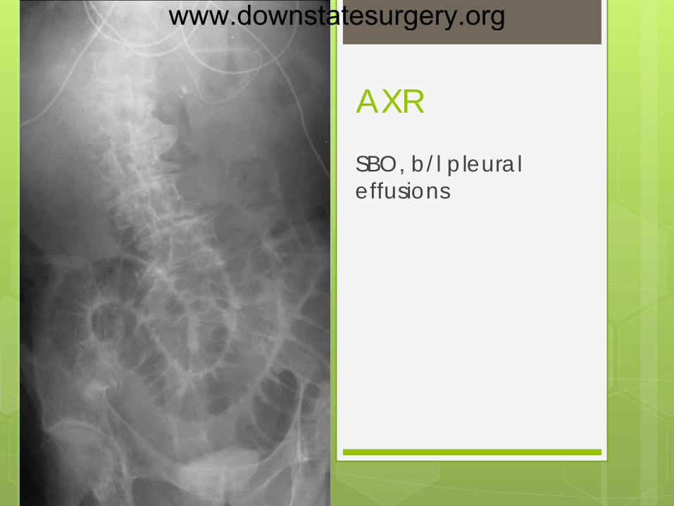
AXR SBO, b/l pleural effusions
www.downstatesurgery.org

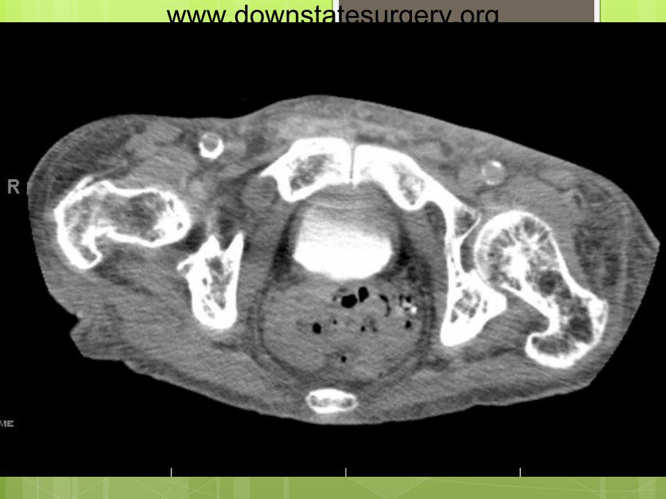
CT Abdomen
Small bowel obstruction secondary to right obturator hernia, bibasilar pneumonia, bilateral pleural effusions
www.downstatesurgery.org

CT Abdomen Small bowel obstruction secondary to right obturator hernia, bibasilar pneumonia, bilateral pleural effusions
www.downstatesurgery.org

www.downstatesurgery.org

www.downstatesurgery.org
www.downstatesurgery.org

www.downstatesurgery.org

www.downstatesurgery.org

www.downstatesurgery.org

Hospital Course 8/6 – 8/8 Admision to ICU NGT, NPO/IVF Dialysis Cardizem drip for afib Abx for PNA Refused surgical intervention No improvement of SBO
www.downstatesurgery.org
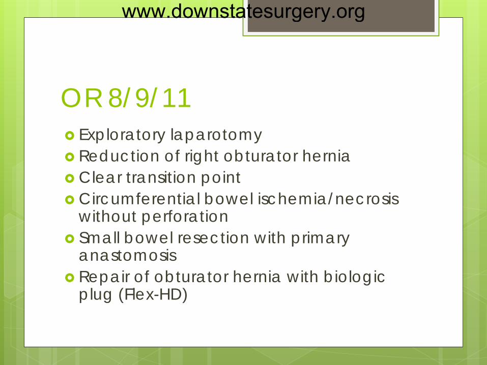
OR 8/9/11 Exploratory laparotomy Reduction of right obturator hernia Clear transition point Circumferential bowel ischemia/necrosis
without perforation Small bowel resection with primary
anastomosis Repair of obturator hernia with biologic
plug (Flex-HD)
www.downstatesurgery.org

Hospital Course 8/10 – 8/30 8/11 – extubated 8/15 – clear diet, thoracenthesis (1500cc) 8/16 – full liquids, transfer to floor 8/19 – tolerating regular diet 8/20 – 8/29 awaiting subacute rehab 8/30 - discharged
www.downstatesurgery.org

Questions
www.downstatesurgery.org

Obturator Hernia Protrusion of sac through obturator
foramen and canal along the obturator nerve and vessels
Represents <0.1% of all hernias High incidence of strangulation “the skinny old lady hernia” - thin, frail,
multiparous elderly woman with SBO of unclear etiology
www.downstatesurgery.org
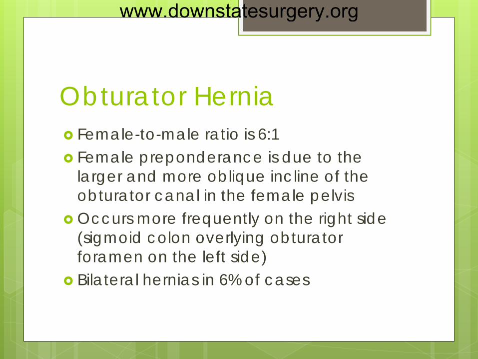
Obturator Hernia Female-to-male ratio is 6:1 Female preponderance is due to the
larger and more oblique incline of the obturator canal in the female pelvis
Occurs more frequently on the right side (sigmoid colon overlying obturator foramen on the left side)
Bilateral hernias in 6% of cases
www.downstatesurgery.org
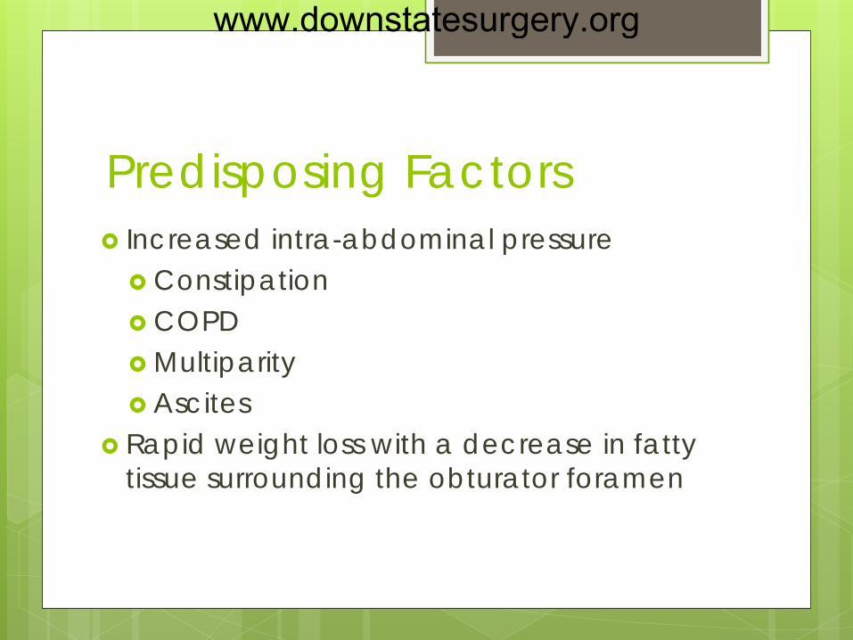
Predisposing Factors Increased intra-abdominal pressure Constipation COPD Multiparity Ascites
Rapid weight loss with a decrease in fatty tissue surrounding the obturator foramen
www.downstatesurgery.org
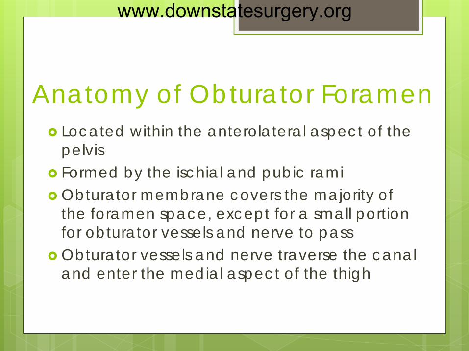
Anatomy of Obturator Foramen Located within the anterolateral aspect of the
pelvis Formed by the ischial and pubic rami Obturator membrane covers the majority of
the foramen space, except for a small portion for obturator vessels and nerve to pass
Obturator vessels and nerve traverse the canal and enter the medial aspect of the thigh
www.downstatesurgery.org

www.downstatesurgery.org
www.downstatesurgery.org

Obturator Canal 2-3 cm long tunnel begins in the pelvis exits through the obturator foramen passes obliquely downward to the
obturator region of the thigh The canal is bounded superiorly and laterally by the pubic bone inferiorly by the obturator membrane and
obturator muscles
www.downstatesurgery.org

Obturator Anatomy The direction of the obturator hernia through the obturator canal
www.downstatesurgery.org
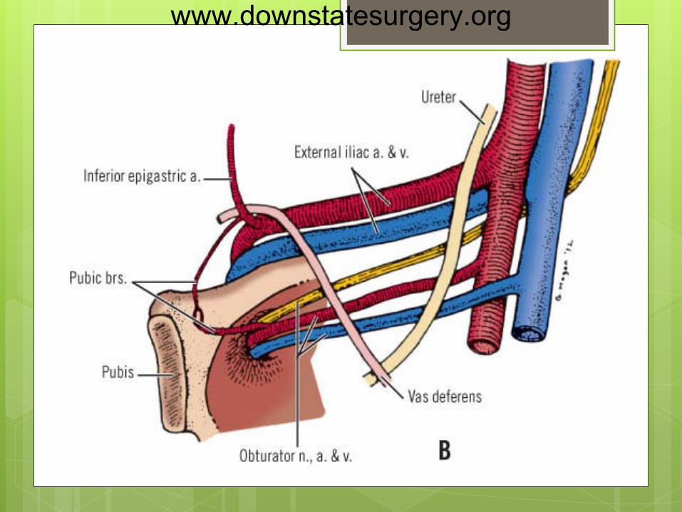
Obturator Canal Contents Obturator nerve, artery, and vein enter
the canal through an opening in the anterosuperior aspect of the obturator membrane
Obturator nerve lies superior to the obturator artery and divides immediately on exiting the canal into anterior and posterior branches
www.downstatesurgery.org
www.downstatesurgery.org

www.downstatesurgery.org

Obturator Nerve Anterior branch emerges between the
adductor longus and adductor brevis muscles supplies sensory innervation to the medial
aspect of the thigh, hip and knee joints and motor innervation to the adductor longus/brevis, gracilis, and pectineus muscles
Posterior division emerges between the adductor brevis and adductor magnus muscles supplies motor innervation to the obturator
externus and adductor magnus muscles
www.downstatesurgery.org

www.downstatesurgery.org
Potential Hernia Pathways Most common - sac lies in front of the obturator
externus and underneath the pectineus, accompanied by the anterior division of the obturator nerve
Hernia emerges between the middle and superior fasciculi of the obturator externus along with the posterior division of the nerve
Most rare - sac emerges between the internal and external obturator muscles and membranes
Recognition of the three variants is important when repair is attempted through the thigh
www.downstatesurgery.org

www.downstatesurgery.org

Obturator Hernia Formation Consists of three stages: prehernia stage - which involves
preperitoneal fat, or “pilot tags” second stage - formation of a true sac third stage - hernia becomes clinically
significant Diagnosis during the first two stages is
uncommon
www.downstatesurgery.org

Clinical Manifestations: Small Bowel Obstruction Up to 80% of cases present with obstruction,
either intermittent or acute and complete Intestinal obstruction results from involvement
of the jejunum or ileum within the hernia sac Approximately 50% of patients have an
incomplete obstruction secondary to a Richter-type hernia
History of repeated episodes of bowel obstruction that pass quickly and without intervention is present in up to 30% of cases
www.downstatesurgery.org
Clinical Manifestations: Obturator Neuralgia
Obturator neuralgia is manifested as cramping or as hypoesthesia or hyperesthesia extending from the inguinal crease to the anteromedial aspect of the thigh
www.downstatesurgery.org

Clinical Manifestations: Howship-Romberg Sign Pain radiating down the medial aspect of the
thigh to the knee and less often to the hip Result from compression of the anterior division of
the obturator nerve relieved by flexion and external rotation of the
thigh exacerbated by extension, adduction, and
medial rotation of the leg Considered pathognomonic Present in up to 50% of patients
www.downstatesurgery.org

Clinical Manifestations: Hannington-Kiff Sign Absence of the obturator reflex in the thigh,
caused by compression on the obturator nerve Reflex can usually be elicited by percussing
over an extended index finger placed across the adductor muscle approximately 5 cm above the knee
If the patellar reflex of the ipsilateral side is present in the absence of an obturator reflex, it is highly likely that the obturator nerve is compressed
www.downstatesurgery.org

Clinical Manifestations: Palpable Mass In 20% of cases a palpable mass is found in
the proximal medial aspect of the thigh at the origin of the adductor muscles The mass is best palpated with the thigh
flexed, abducted, and rotated outward or laterally on a vaginal exam
In rare cases, ecchymoses may be noted in the upper medial thigh due to effusion from the strangulated hernia contents
www.downstatesurgery.org

Modalities Used to Assist in Diagnosis
Both CT and ultrasound (transvaginal or inner thigh views) are useful in the diagnosis of obturator hernia
MRI is as good as but not superior to CT AXR may show air in the obturator region Laparoscopy may be used as a
diagnostic tool, as well as a treatment modality
www.downstatesurgery.org
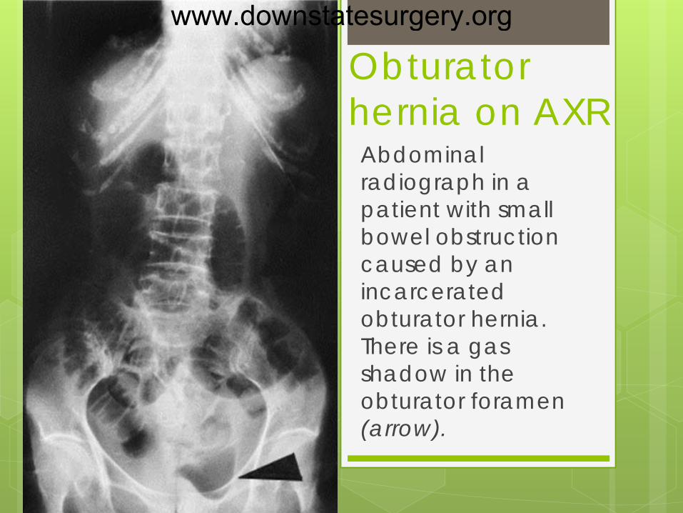
Obturator hernia on AXR Abdominal radiograph in a patient with small bowel obstruction caused by an incarcerated obturator hernia. There is a gas shadow in the obturator foramen (arrow).
www.downstatesurgery.org

Treatment In >50% of cases an obturator hernia is found
intraoperatively during a diagnostic laparoscopy or laparotomy for SBO
When diagnosis is made preoperatively, alternative approaches for repair include abdominal extraperitoneal anterior thigh exposure laparoscopic
www.downstatesurgery.org

Transperitoneal Approach Lower midline laparotomy Run bowel, reduce hernia Incise obturator membrane in antero-
posterior direction Avoid injury to small bowel, obturator
vessels and nerve Make counter-incision in the medial groin Bowel resection required in 25% of cases
www.downstatesurgery.org
Transperitoneal Approach Close hernia opening around the
obturator vessels with a running non-absorbable suture
Closure should include the periosteum of the superior pubic ramus and the fascia on the internal obturator muscle
In a clean case, a piece of mesh can be placed over the obturator foramen (may be sutured to Cooper's ligament)
www.downstatesurgery.org

Extraperitoneal Approach Lower midline incision Enter preperitoneal plane, peel bladder from the
peritoneum Expose superior pubic ramus and the obturator
internus muscle Identify the hernia sac (projection of peritoneum
passing inferiorly into the obturator canal) Reduce the hernia Close the internal opening to the obturator canal Preperitoneal mesh may be placed
www.downstatesurgery.org

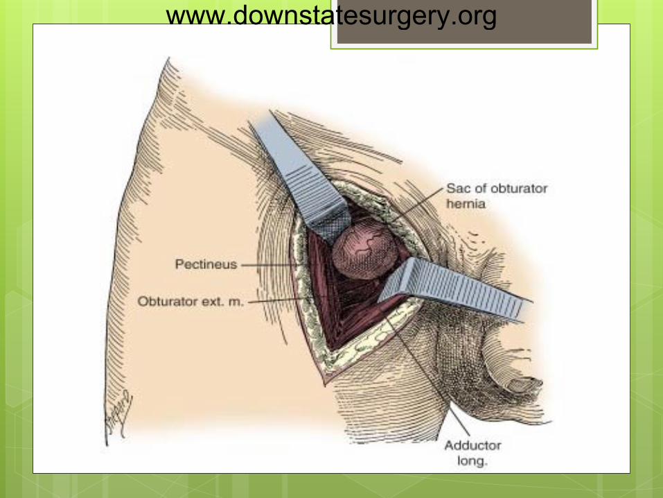
Thigh Approach Vertical incision in the upper medial thigh along
the adductor longus muscle Retract the muscle medially to expose the
pectineus muscle Cut pectineus muscle across to expose the sac Reduce hernia, excise the sac (if viable contents) Close hernial opening If the bowel contents within the hernia sac do not
appear viable, midline laparotomy is usually performed
www.downstatesurgery.org
www.downstatesurgery.org

www.downstatesurgery.org
Laparoscopic Approach Both totally extraperitoneal (TEP) and
transabdominal preperitoneal (TAPP) laparoscopic approaches are highly effective in the treatment of obturator hernia
During laparoscopy, the defect is repaired with a prosthetic mesh
www.downstatesurgery.org
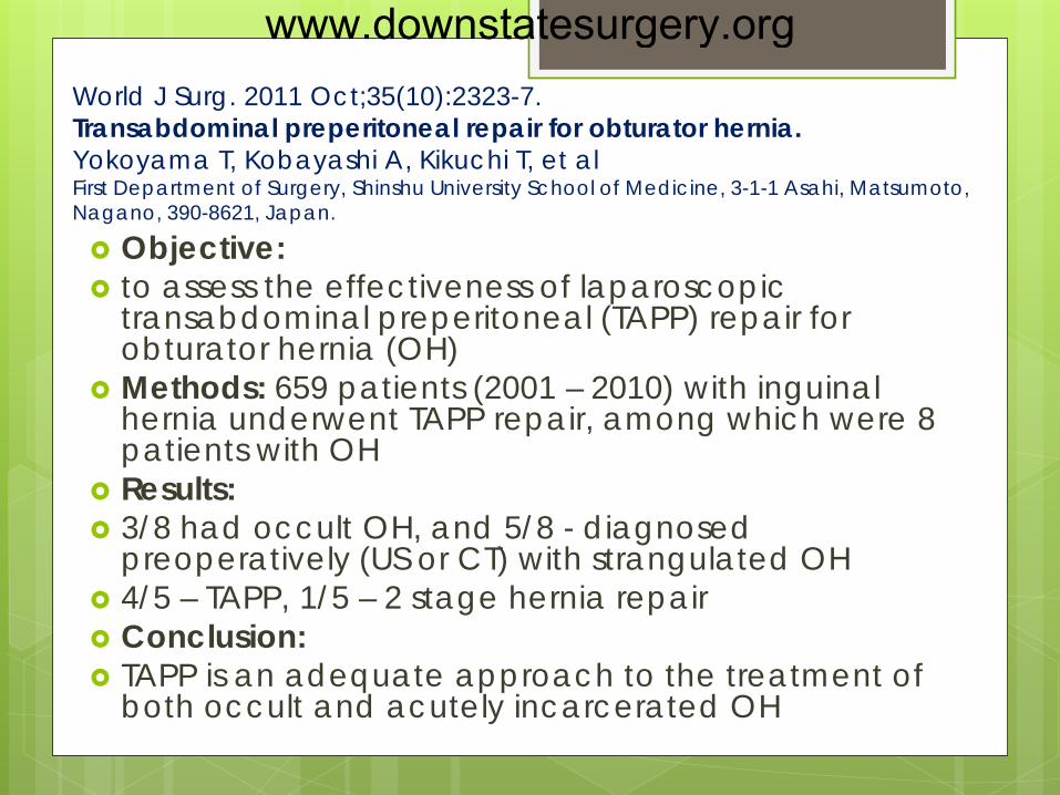
World J Surg. 2011 Oct;35(10):2323-7. Transabdominal preperitoneal repair for obturator hernia. Yokoyama T, Kobayashi A, Kikuchi T, et al First Department of Surgery, Shinshu University School of Medicine, 3-1-1 Asahi, Matsumoto, Nagano, 390-8621, Japan.
Objective: to assess the effectiveness of laparoscopic
transabdominal preperitoneal (TAPP) repair for obturator hernia (OH)
Methods: 659 patients (2001 – 2010) with inguinal hernia underwent TAPP repair, among which were 8 patients with OH
Results: 3/8 had occult OH, and 5/8 - diagnosed
preoperatively (US or CT) with strangulated OH 4/5 – TAPP, 1/5 – 2 stage hernia repair Conclusion: TAPP is an adequate approach to the treatment of
both occult and acutely incarcerated OH
www.downstatesurgery.org
References Nir Wasserberg, Howard S. Kaufman, “Chapter 48 – Lumbar and Pelvic Hernias”
(Chapter). Yeo: Shackelford's Surgery of the Alimentary Tract, 6th ed. Javid Patrick J, Brooks David C, "Chapter 5. Hernias" (Chapter). Zinner MJ, Ashley SW:
Maingot's Abdominal Operations, 11th Edition. Gene L. Colborn, Robert M. Rogers Jr., John E. Skandalakis, “Chapter 28. Pelvis and
Perineum” (Chapter). Skandalakis' Surgical Anatomy Skandalakis LJ, Androulakis J, Colborn GL, et al: Obturator hernia. Embryology,
anatomy, and surgical applications. Surg Clin North Am 2000; 80:71. Chang SS, Shan YS, Lin YJ, et al: A review of obturator hernia and a proposed
algorithm for its diagnosis and treatment. World J Surg 2005; 29:450. Yokoyama T, Mulnakata Y, Ogiwara M, et al: Preoperative diagnosis of strangulated
obturator hernia using ultrasonography. Am J Surg 1997; 174:76 Yokoyama Y, Yamaguchi A, Isogai M, et al: Thirty-six cases of obturator hernia: Does
CT contribute to the postoperative outcome?. World J Surg 1999; 23:214. Nishina M, Fujii C, Ogino R, et al: Preoperative diagnosis of obturator hernia by
computed tomography in six patients. J Emerg Med 2001; 20:277. Schmidt PH, Bull WJ, Jeffery KM, et al: Typical versus atypical presentation of
obturator hernia. Am Surg 2001; 67:191. Shapiro K, Patel S, Choy C, et al: Totally extraperitoneal repair of obturator
hernia. Surg Endosc 2004; 18:954. Kammori M, Mafune K, Kirashima T, et al. Forty-three cases of obturator hernia. Am J
Surg 2004;187:549 Skandalakis JE. Obturator hernia. In: Skandalakis JE, Gray SW, Mansberger AR, et al
(eds). Hernia Surgical Anatomy and Technique. New York, NY: McGraw-Hill; 1989:174 Tucker JG, Wilson RA, Ramshaw BJ, et al. Laparoscopic herniorraphy: technical
concerns in prevention of complications and early recurrence. Am Surg 1995;61:36
www.downstatesurgery.org


![Immediate Obturator with Airway for Maxillary Resection ... · Palatal plate of the surgical obturator can easily be modified and used as an interim obturator [16-18]. Benefits of](https://img.pdfslide.net/doc/110x75/5f25b8b636c20c5f147362fe/immediate-obturator-with-airway-for-maxillary-resection-palatal-plate-of-the.jpg)