Embed Size (px)
Citation preview

Occupational Injuries and Diseases of the Hip and Pelvis
John C. Schumpert, MD, MPH, FACOEM Resources for Environmental and Occupational Health
www.reoh.com

Disclosures
• 2001 to current: Founder, President, and Consulting Physician, Resources for Environmental and Occupational Health (REOH)
• 2009 to current: Faculty Affiliate, Center for Environmental Health Sciences, University of Montana
• 2016 to current: Co-medical Director, Employment Relations Division, Montana Department of Labor and Industry

Introduction
• Many conditions masquerade hip joint and pelvic
• Non-occupational cases of pelvis and hip joint • Work-related cases of the pelvis and hip joint

Non-occupational hip cases

Avascular necrosis

Avascular necrosis
• 48-year-old, right-hand dominant male logger and choker setter for 20 years was pinned between a tree stump and a vehicle initially complaining of bilateral shoulder pain
Photo source: https://forestpolicypub.com/2016/11/29/montana-timber-industrys-old-growth-liquidation-strategy/

Avascular necrosis
Photo source: https://sites.google.com/site/loggingandkudzu/

Avascular necrosis
• Past medical history – right carpal tunnel syndrome – right elbow fracture – Hypertension – Hypercholesterolemia – right hand gunshot wound – right knee injury

Avascular necrosis
• Past surgical history – right knee arthroscopy x 2 – right carpal tunnel release – right elbow open reduction and internal fixation – left shoulder arthroscopy x 2
Avascular necrosis
• His habits – No smoking – No chewing tobacco – Occasional alcohol
consumption
• His hobbies and interests – Fishing – Hunting – Watching spectator
sports
Avascular necrosis
• Physical exam – Painful left hip flexion, internal rotation, and
external rotation – Pain-free right hip passive range of motion

Avascular necrosis
• MR scan of the left hip bilateral avascular necrosis of the femoral heads
• Subchondral ischemia in both femoral heads was observed
• No evidence of fracture or deformity to the articular surface of either femoral head or femoral neck

Blood supply of the femoral head http://www.physio-pedia.com/Legg-Calve-Perthes_Disease

Risk factors for avascular necrosis • Systemic lupus
erythematosus (SLE) • Sickle cell disease • Renal transplants • Fat emboli • Bone disease and
neoplasms • Osteomyelitis • Pancreatitis • Pheochromocytoma
• Long-term high-dose corticosteroid treatment
• Alcoholism • Femoral neck fractures • Decompression sickness • High dose radiation
exposures • Legg-Calve-Perthes
disease

Avascular necrosis
• Kang et al. (2013) • Fraitzl et al. (2013)
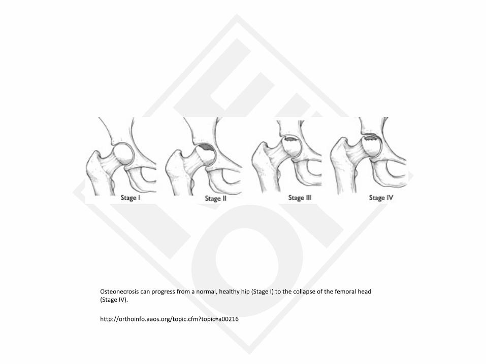
Osteonecrosis can progress from a normal, healthy hip (Stage I) to the collapse of the femoral head (Stage IV). http://orthoinfo.aaos.org/topic.cfm?topic=a00216

Avascular necrosis
• Medical laboratory tests revealed antinuclear antibody (ANA) screen was positive with a titer of 1:160 and a speckled pattern
• Diagnosed with unspecified auto-immune arthropathy (likely systemic lupus erythematosus)
• Recommended rheumatology evaluation

Femoroacetabular impingement
Femoroacetabular impingement
• 44-year-old right hand dominant female who developed right hip pain which she associated with her employment as a long haul trucker
Photo source: www.redrockcollisionrepair.com/st-george-heavy-equipment/semi-trucks/

Femoroacetabular impingement
• 44-year-old right hand dominant female who developed right hip pain which had she associated with her employment as a long haul trucker

Femoroacetabular impingement
• She reported pain in the right groin wrapping around the hip and into the right sacral region
• She also reported muscle spasms in the right buttock

Femoroacetabular impingement
• Pelvic MR scan – Moderate osteoarthritic changes with right side
worse than left – Slight lateral subluxation of the right hip – Enlargement of the superior labrum – Right hip joint effusion

Femoroacetabular impingement

Femoroacetabular impingement
Source: http://www.phongtran.com.au/femoroacetabular-impingement-fai/

Femoroacetabular impingement
• History from medical records contradictory – Several years earlier, she slipped on the ice and
landed on her right hip, and had low back and right hip pain
– MRI showed revealed a right hip acetabular labral tear
– She underwent a right hip arthroscopy labral debridement and arthroscopic decompression of a cam lesion of the femur

Femoroacetabular impingement
• Medical records clearly showed a longstanding hip problem dating back to her fall on ice
• Most recent right hip MRI – significant right hip degeneration – no bone marrow edema – no new or recurrent labral tear
• No indication either a new injury or an aggravation of the pre-existing labral tear

Occupational hip cases

Femoroacetabular impingement (FAI)

Femoroacetabular Impingement
Source: https://mikereinold.com/femoroacetabular-impingement-etiology-diagnosis-and-treatment-of-fai/

FAI Case
• 57-year-old right-hand dominant male who was injured when he fell off a 6 ft ladder landing on his right hip
Source: https://www.fmlainsights.com/when-an-employee-falls-off-a-ladder-at-work-is-his-absence-covered-by-fmla/

FAI Case

FAI Case
• Radiographs of the right hip – mild bony overgrowth
of femoral head and neck
• Findings consistent with cam-type femoral acetabular impingement

FAI Case
• Right hip MR arthrogram – complex horizontal tear
from the 12 o’clock position through the 1:30 position
– paralabral cartilage irregularity with fissuring and adjacent delamination

FAI Case
• In femoroacetabular impingement, bone spurs develop around the femoral head and/or along the acetabulum
• The bone overgrowth causes the hip bones to hit against each other, rather than to move smoothly
• Over time, this can result in the tearing of the labrum and breakdown of hip joint cartilage

Femoroacetabular impingement
• Three types of femoroacetabular impingement – Pincer – Cam – Mixed FAI

Pincer-type FAI
• Pincer type impingement occurs because extra bone extends out over the normal rim of the acetabulum
• The labrum can be crushed under the prominent rim of the acetabulum.

Cam-type FAI
• In cam type impingement, the femoral head is not round and cannot rotate smoothly inside the acetabulum
• A bony outgrowth forms on the edge of the femoral head and neck that grinds the cartilage inside the acetabulum

Mixed FAI
• Combined impingement just means that both the pincer and cam types are present

Risk factors for aggravated FAI
• Some people may live long, active lives with FAI and never have problems
• Femoroacetabular impingement can become aggravated by vigorous athletic activity (e.g., tennis, racquetball, ballet, martial arts, etc.) and by traumatic injury

FAI Case
• The evidence suggested cam-type impingement, based upon the irregularity of the femoral head, decreased femoral head-neck offset, and evidence of labral fissuring and adjacent delamination.

FAI Case
• Trauma from the fall off a 6 foot ladder onto the right hip was the likely cause of aggravated FAI

Gluteal nerve axonotmesis

Gluteal nerve axonotmesis
• 67-year-old left hand dominant male who was working for cattle company; he "attempted to get on his horse, horse must have spooked, was thrown into fence."
Photo source: https://boydranch.net/available-horses/

Gluteal nerve axonotmesis
• Past medical history – peripheral neuropathy – Morton's neuroma – carpal tunnel syndrome

Gluteal nerve axonotmesis
• Past surgical history – left total knee arthroplasty – left hip arthroplasty – right foot surgery

Gluteal nerve axonotmesis
• Initial diagnoses – left 2nd – 8th ribs fractured – left pneumothorax – left humeral head dislocation with a Hill-Sachs
deformity and glenoid rim displaced fracture – non-displaced left acetabular rim fracture – small fatty-containing left inguinal hernia – left mid-clavicle fracture

Acetabular rim fracture

Gluteal nerve axonotmesis

Gluteal nerve axonotmesis
• During hospitalization, he underwent: – open reduction and internal fixation of left
humeral head fracture – left thoracostomy tube – open reduction and internal fixation of left inferior
glenoid rim fracture
Gluteal nerve axonotmesis
• Within 1 ½ months, he complained of – worsening lumbar region – worsening left hip pain – burning in left leg and buttock – fluid collection in his low back and that moved
into the left buttock

Gluteal nerve axonotmesis
• Lumbar radiographs – disc degeneration at L2-3, L3-4, L4-5, and L5-S1 – no subluxation on flexion and extension views

Gluteal nerve axonotmesis
• Lumbar spine MRI – L1-2: mild bilateral facet degeneration – L2-3: there was a symmetric disc bulge, mild facet – L3-4: disc bulge causing mild spinal canal narrowing,
right lateral recess effacement of right L4 nerve root, mild bilateral L3 neural foraminal encroachment
– L4-5: disc-osteophyte complex causing mild spinal canal stenosis, and osteophytes causing bilateral foraminal stenosis
– L5-S1: osteophytes causing left foraminal narrowing

Gluteal nerve axonotmesis
• Symptoms – superior left gluteal muscle pain and "there’s a big
knot“ – gluteal pain provoked by prolonged sitting – gluteal numbness that did not extend into the
thigh – pain moved through the buttock, wrapped the left
greater trochanter, then through the anterolateral thigh stopping at the mid-thigh level
– No right leg numbness, tingling, weakness, or pain

Gluteal nerve axonotmesis

Gluteal nerve axonotmesis
• Physical exam – strength was 4+/5 in all muscle groups of both
legs – deep tendon reflexes were symmetric – no ankle clonus – obvious left buttock muscle atrophy

Gluteal nerve axonotmesis
• A contrast-enhanced pelvic MR scan – asymmetric atrophy
and edematous signal within the left gluteus maximus muscle with minimal increased enhancement

Gluteal nerve axonotmesis
• Electrodiagnostics considered abnormal with evidence of a left lower extremity peroneal motor neuropathy – non-contributory findings – no weakness on great toe extension – no weakness on ankle dorsiflexion
Gluteal nerve axonotmesis
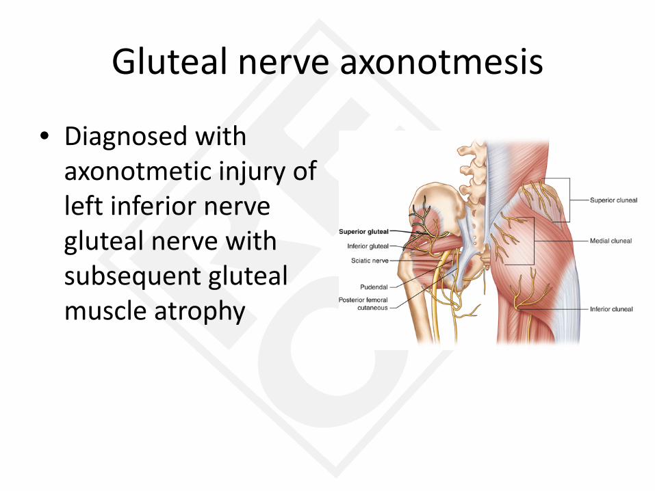
• Diagnosed with axonotmetic injury of left inferior nerve gluteal nerve with subsequent gluteal muscle atrophy

What’s the difference between axonotmesis and neuropraxia?

Neuropraxia
• Mild injury – ischemia, mechanical compression, metabolic or
toxic factors • Results in focal demyelination, but no loss of
axonal integrity • Weakness and sensory loss are due to
conduction block • No muscle atrophy • Excellent recovery is expected

Axonotmesis
• Severe injury – motor vehicle accidents, falls or percussion
injuries (e.g., gunshot wounds) • Axon and myelin sheath degenerates, a
process is known as Wallerian degeneration • Muscle atrophy occurs within weeks • Partial recovery is the expected outcome, but
the time course is significantly protracted as compared with neurapraxia

Gluteal nerve axonotmesis • Diagnosed with axonotmesis
injury of left inferior nerve gluteal nerve with fairly severe gluteal muscle atrophy
• Likely cause was the severe left hemipelvic blunt trauma which also caused left acetabular rim fracture
• Unlikely L5 or S1 radiculopathy because no hip extension weakness
• Unlikely superior gluteal nerve injury because no gluteus medius weakness
Photo source: http://thinklikeahorse.org/index-2.html/

Avascular necrosis

Avascular necrosis
• Obese 54-year-old male computer technician had to replace internet broadcast antenna on a 300 foot tall grain silo in January
• Used an interior elevator to reach top of silo
Photo source: https://bobsmisanthropicadventures.wordpress.com/category/silo/

Avascular necrosis
• After installing the antenna, he found the elevator didn’t work; he had to climb down using an exterior ladder
• He was out of breath after about 30 feet
Photo source: https://www.tennessean.com/story/money/2017/03/23/massive-mural-planned-grain-silo-nations/99506626/

Avascular necrosis
• He wore a harness that he clipped onto the ladder while he caught his breath
• He had to stop repeatedly because of shortness of breath

Avascular necrosis
• He said that he thought he might develop hypothermia and die on the ladder
• The descent required over an hour

Avascular necrosis
• When he finally got to the ground, his whole body was aching, but this resolved in a couple of hours

Avascular necrosis
• A week later – right groin and medial thigh pain traveling to the
knee – lateral hip pain extending through the lateral thigh
to the lateral knee – shooting pain in the right leg to the foot – right leg buckling and falls

Avascular necrosis
• Symptoms – Right leg pain in the medial and lateral thigh,
sharp pain in the right groin – Right leg buckling – Nocturnal awakening with right groin cramping, – Right hip externally rotates when walking (i.e.,
Trendelenburg gait)

Avascular necrosis
• Physical exam – right hip with limited passive flexion, internal
rotation, and external rotation – moderate pain with all of these movements – Pain on Stinchfield’s test – no evidence of inguinal hernia

• Why did I worry about a hernia? • What is a hernia?


Avascular necrosis
• What’s Stinchfield’s test? – aka Stinchfield Resisted Hip Flexion Test – Tests for a pain response caused by an increase in
hip joint reactive force – Is a way to distinguish between intra-articular and
extra-articular hip pathology causing groin, thigh, buttock, and even pretibial leg pain

Avascular necrosis
• What’s Stinchfield’s test? – From a supine position
with the knee extended, the patient is asked to actively elevate the leg against
– Positive test is reproduces pain in groin

Avascular necrosis
• Right hip x-rays – Narrowing of the femoral acetabular joint with
mild sclerosis and spurring present – Flattening and irregularity of the femoral head
with increased sclerosis suggesting avascular necrosis
– Cam-type femoroacetabular impingement with hypertrophic change of the femoral neck

Avascular necrosis
• Bone scan – increased activity in the right hip most
pronounced at the femoral head – no evidence of fracture

Avascular necrosis
• Hip MR arthrogram – Large right hip joint effusion – Diffuse abnormal signal intensity in the right
femoral head, neck, and subtrochanteric region – Femoral head articular surface cortical defect – Overall appearance of avascular necrosis of the
femoral head

Avascular necrosis
• Medical lab – Glycosylated hemoglobin level was slightly
elevated at 5.7% – Slightly low calcium level – Vitamin D deficiency

Avascular necrosis
• Diagnosis – Right hip avascular necrosis – Type 2 obesity (BMI=38.5 kg/m2)

Avascular necrosis
• Risk factors of avascular necrosis: – Systemic lupus erythematosus – Rheumatoid arthritis – Gaucher’s disease (heritable lipid storage disease
caused by glucocerebrosidase enzyme deficiency) – Legg-Calvé-Perthes syndrome (childhood
osteonecrosis) – Long-term (months-years) high-dose steroid
treatment

Avascular necrosis
• Risk factors of avascular necrosis: – HIV infection (100-fold increased risk) – Caisson disease (decompression sickness) – Work in high pressure environments (divers) – Alcoholism, tobacco smoking – Untreated hypertension – Vasculitis, arterial embolism and thrombosis – Bisphosphonates (jaw only) – Chemo and radiation therapy for cancer – Sickle cell disease

Avascular necrosis
• Discussion: –Kang et al. (2013) –Fraitzl et al. (2013)

Cam-type FAI
• In cam type impingement, the femoral head is not round and cannot rotate smoothly inside the acetabulum
• Prolonged hip flexion while resting during hour-long ladder descent combined with cam-type FAI likely caused avascular necrosis

Compromised blood supply of the femoral head http://emedicine.medscape.com/article/386808-overview

Blood supply of the femoral head http://www.physio-pedia.com/Legg-Calve-Perthes_Disease

Osteonecrosis can progress from a normal, healthy hip (Stage I) to the collapse of the femoral head (Stage IV). http://orthoinfo.aaos.org/topic.cfm?topic=a00216

(Left) An x-ray of a healthy hip joint. (Right) In this x-ray, the osteonecrosis has progressed to collapse of the femoral head. http://orthoinfo.aaos.org/topic.cfm?topic=a00216

Conclusion
• Hip and groin pain are common and can have lots of causes – Inguinal hernia – Avascular necrosis of femoral head – Labral tear – Iliopsoas muscle tear from its insertion at the lesser
trochanter – L1-2 or L2-3 disc herniation – Pelvic muscle tendinopathy – Hip fracture

Conclusion
• History can provide clues to cause of problem – Congenital abnormality – Lifestyle choices – Other diseases – Previous injuries – Medical treatments

Conclusion
• Mechanism of injury can provide clues to cause of pain – Severe blunt trauma – Abrupt strain/sprain – Overuse/repetitive use

Conclusion
• Accurate diagnosis is absolutely necessary for successful treatment

Questions?
REOH Resources for Environmental and Occupational Health
210 East Pine Street Missoula, Montana
406-549-6520 www.reoh.com


![Appendix 1 HIP Male and Female - University of East Anglia · App14.1!HIP!v3.2_02_05_2012!!!!!Health’Improvement’Profile[HIP]’ ’’’’’’’’’’’’’’’’’’’’’’’’’’’’(HIP)–’Male](https://img.pdfslide.net/doc/110x75/5f0af26b7e708231d42e1f1c/appendix-1-hip-male-and-female-university-of-east-anglia-app141hipv3202052012healthaimprovementaprofilehipa.jpg)