Embed Size (px)
Citation preview
Article
Oct4-induced oligodendrocyte progenitor cellsenhance functional recovery in spinal cordinjury modelJeong Beom Kim1,2,*, Hyunah Lee1,2, Marcos J Araúzo-Bravo3,4, Kyujin Hwang5, Donggyu Nam1,2,
Myung Rae Park1,2, Holm Zaehres6, Kook In Park5 & Seok-Jin Lee1,2
Abstract
The generation of patient-specific oligodendrocyte progenitor cells(OPCs) holds great potential as an expandable cell source for cellreplacement therapy as well as drug screening in spinal cord injuryor demyelinating diseases. Here, we demonstrate that inducedOPCs (iOPCs) can be directly derived from adult mouse fibroblastsby Oct4-mediated direct reprogramming, using anchorage-independent growth to ensure high purity. Homogeneous iOPCsexhibit typical small-bipolar morphology, maintain their self-renewal capacity and OPC marker expression for more than 31passages, share high similarity in the global gene expression profileto wild-type OPCs, and give rise to mature oligodendrocytes andastrocytes in vitro and in vivo. Notably, transplanted iOPCscontribute to functional recovery in a spinal cord injury (SCI)model without tumor formation. This study provides a simplestrategy to generate functional self-renewing iOPCs and yieldsinsights for the in-depth study of demyelination and regenera-tive medicine.
Keywords direct conversion; myelination; Oct4; oligodendrocyte progenitor
cell; self-renewal
Subject Categories Neuroscience; Stem Cells
DOI 10.15252/embj.201592652 | Received 23 July 2015 | Revised 14 September
2015 | Accepted 17 September 2015 | Published online 23 October 2015
The EMBO Journal (2015) 34: 2971–2983
Introduction
Oligodendrocyte progenitor cells (OPCs) are originated from the
neuroepithelium during embryonic development (Noll & Miller,
1993). OPCs are a bipotent glial cell type in the central nervous
system (CNS) that can differentiate into both astrocytes and
myelinogenic oligodendrocytes, responsible for forming myelin
sheath (Franklin & Ffrench-Constant, 2008) that insulates axons for
the efficient propagation of action potentials (Najm et al, 2011).
Demyelinating conditions, such as traumatic spinal cord injury
(SCI), cause immediate cell damage, which can subsequently lead to
both motor and sensory dysfunctions that result in devastating
paralysis (Lee et al, 2014). Since there are no full restorative treat-
ments yet available and current strategies including surgical inter-
ventions and medications carry severe side effects (Silva et al,
2014), cell replacement therapy by transplanting neural/glial
progenitors (Ben-Hur & Goldman, 2008; Jin et al, 2011) for restora-
tion of demyelinated lesions is a promising treatment option. In
particular, transplantation of oligodendrocytes or OPCs derived from
pluripotent stem cells promotes axonal remyelination and facilitates
functional recovery of the damaged spinal cord (Faulkner &
Keirstead, 2005; Keirstead et al, 2005; Sharp et al, 2010; All et al,
2012; Wang et al, 2013; Douvaras et al, 2014). However, the clinical
application of pluripotent stem cell derivatives is limited due to the
tumorigenic potential of the residual undifferentiated stem cells after
transplantation (Miura et al, 2009; Fong et al, 2010). Direct lineage
conversion of one somatic cell type into other cell types, such as
myoblasts, neuronal cells, hepatocytes, cardiomyocytes, and
endothelial cells, has been developed, which eliminates tumori-
genicity through bypassing an intermediate pluripotent state (Davis
et al, 1987; Efe et al, 2011; Huang et al, 2011, 2014; Sekiya &
Suzuki, 2011; Li et al, 2013; Han et al, 2014; Morita et al, 2015).
Meanwhile, this strategy is still challenging since the induced cells
are in the postmitotic state, which impedes the large-scale expansion
of desired cell types. Previous studies reported generation of expand-
able induced neural progenitor cells (iNPCs) from fibroblasts using
distinct sets of neural transcription factors or reprogramming factors
(Kim et al, 2011; Mitchell et al, 2014b). However, the low differenti-
ation efficiency of iNPCs to mature oligodendrocytes limits its appli-
cation as a SCI treatment. Thus, self-renewing and bipotent OPCs
rather than iNPCs are considered as more appropriate cell source for
developing cell-based therapies for SCI. Recently, two research
1 Hans Schöler Stem Cell Research Center (HSSCRC), School of Life Sciences, Ulsan National Institute of Science and Technology (UNIST), Ulsan, South Korea2 Max Planck Partner Group-Molecular Biomedicine Laboratory (MPPG-MBL), UNIST, Ulsan, South Korea3 Group of Computational Biology and Bioinformatics, Biodonostia Health Research Institute, San Sebastián, Spain4 IKERBASQUE, Basque Foundation for Science, Bilbao, Spain5 Department of Pediatrics and BK21 Project for Medical Sciences, Yonsei University College of Medicine, Seoul, South Korea6 Department of Cell and Developmental Biology, Max Planck Institute for Molecular Biomedicine, Münster, Germany
*Corresponding author. Tel: +82 52 217 5201; Fax: +82 52 217 5269; E-mail: [email protected]
ª 2015 The Authors The EMBO Journal Vol 34 | No 23 | 2015 2971
Published online: October 23, 2015
groups have reported generation of induced oligodendrocyte pro-
genitor cells (iOPCs) from murine fibroblasts with combinations of
transcription factors including Sox10, Olig2, and Nkx6.2 (Najm et al,
2013) or Sox10, Olig2, and Zfp536 (Yang et al, 2013). However,
these cells can be expanded for only up to five passages and are
restricted exclusively to the oligodendrocyte lineage (Najm et al,
2013). Moreover, the global gene expression profile of these iOPCs is
distinct from the wild-type OPCs (wtOPCs) (Yang et al, 2013).
Our previous studies revealed that ectopic expression of Oct4
alone can induce pluripotency in both mouse and human NSCs
(Kim et al, 2009a,b,c). Remarkably, a number of recent studies
showed Oct4-mediated direct reprogramming (Mitchell et al, 2014a)
can promote direct lineage conversion of fibroblasts into blood
progenitor cells (Szabo et al, 2010) as well as neural progenitor cells
(Mitchell et al, 2014b), astrocytes into neural stem cells (Corti et al,
2012), or peripheral blood cell into neural progenitor cells (Lee et al,
2015) through passing cell plasticity stage (Mitchell et al, 2014a).
Here, we demonstrated that the ectopic expression of Oct4 with
defined culture conditions is sufficient to generate homogeneous,
self-renewing and bipotent iOPCs from fully differentiated somatic
cells through passing the aggregate stage and demonstrates its
functionality in rat SCI models. Our strategy reduces host genome
modifications by minimizing the use of transcription factors to a
single factor and facilitates future therapeutic applications for
demyelinating conditions, including SCI.
Results
Generation of iOPCs from adult mouse fibroblasts by Oct4
To generate induced oligodendrocyte progenitor cells (iOPCs) using
a minimal number of transcription factors, we employed Oct4-
mediated direct reprogramming strategy, which induces cell fate
plasticity at the early phase of reprogramming through mediating
ectopic expression of Oct4 (Mitchell et al, 2014a; Xu et al, 2015).
The procedure for generating iOPCs is summarized in a schematic
design (Stage 1; Fig 1A). Briefly, we first isolated the mouse skin
fibroblasts from 6-week-old adult mice as parental cells and con-
firmed that the cells did not express pluripotency or neural-lineage
markers (Fig EV1A–C). The fibroblasts were transduced with retro-
viruses encoding Oct4 and cultured in defined OPC induction
medium. The transduced cells underwent morphological changes
into spindle-shaped cells 14–21 days after Oct4 induction (Fig 1B
and C), whereas uninfected cells did not change during the entire
process (Fig 1D). Next, we mechanically isolated the cells exhibiting
spindle-shaped morphology and replated them in OPC medium
supplemented with platelet-derived growth factor AA (PDGF–AA),
an essential mitogen for OPCs (Noble et al, 1988; Raff et al, 1988;
Richardson et al, 1988; Hu et al, 2012). Within 35 days after infec-
tion, these cells formed floating OPC aggregates (OPC-AGs) (Stage 2;
Fig 1E), and the OPC-AGs were subsequently transferred onto
gelatin-coated plates in OPC medium. Bipolar OPC-like cells outgrew
from the attached OPC-AGs and could be maintained in a highly
homogeneous monolayer culture (Stage 3; Fig 1F and G). After
consecutive passages, we established two iOPC clones named iOPC-
C1 and iOPC-C2. Immunostaining revealed that the iOPC clones co-
expressed OPC-specific surface markers, A2B5, PDGFRa, and NG2,
and also expressed an early OPC marker, Olig2 (Fig 1H). We further
evaluated whether iOPCs can maintain the proliferation after
multiple passages. Both iOPC clones were stably expanded for more
than 31 passages (Fig 1I). The mean population doubling times
(mDT) of iOPC clones were approximately 26 h at both passages 3
(P3) and 31 (P31) (Fig 1J). We next confirmed the integration of
Oct4 transgene in the host genome of iOPC clones by genomic poly-
merase chain reaction (PCR) (Fig EV1D). Exogenous expression of
Oct4 mRNA was dramatically silenced in both clones at passage 5
(P5), as examined by quantitative reverse transcription–PCR (qRT–
PCR) (Fig EV1E). Furthermore, the iOPCs maintained a normal
mouse chromosome karyotype (2n = 40) after Oct4 induction
(Fig EV1F). These results support that Oct4 expression with our
defined culture condition is sufficient to convert the cell fate of adult
mouse fibroblasts into expandable iOPCs.
Self-renewing iOPCs can differentiate into matureoligodendrocytes and astrocytes
Next, we investigated the self-renewal capacity and bipotency of
iOPCs through examining populations of iOPCs, oligodendrocyte,
astrocyte, and neurons at early (P5) and late passage (P35) in the
OPC culture condition. Both iOPC clones homogeneously expressed
A2B5, PDGFRa, and NG2. In contrast, glial fibrillary acidic protein
(GFAP)+ astrocytes were rarely present, and neither receptor-
interacting protein (RIP)+ mature oligodendrocytes nor neuron-
specific class III beta-tubulin (Tuj1)+ neurons were detected
(Figs 2A and B, and EV2A). The mean percentage of positive cells
for each marker is presented in Appendix Table S1. We next examined
the bipotency of iOPCs to differentiate to bothmature oligodendrocytes
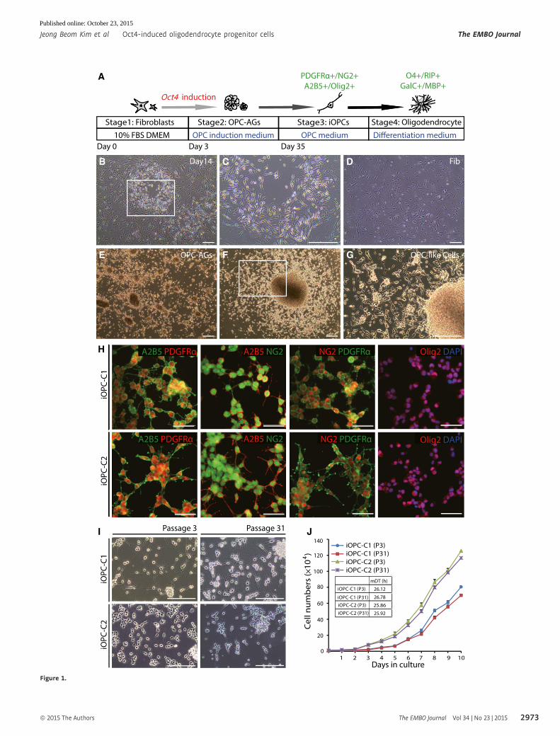
▸Figure 1. Generation and characterization of Oct4-induced OPCs from fibroblasts.
A Experimental scheme for generating iOPCs from fibroblasts through Oct4-mediated reprogramming and in vitro differentiation into mature oligodendrocytes (OPC-AGs, OPC aggregates).
B–G Morphology of Oct4-infected fibroblasts in OPC induction medium (B) at 14 days post-infection. (C) Zoomed image of the white square in (B), which shows aspindle-shaped morphology. (D) Typical morphology of fibroblasts in OPC medium without Oct4 induction. (E) Appearance of OPC-AGs within 35 days afterinfection. (F) OPC-like cells outgrew from OPC-AGs on gelatin-coated plates. (G) Zoomed image of the white square in (F), which shows the bipolar morphology ofOPC-like cells. Scale bars, 250 lm.
H Immunofluorescence images of iOPC-C1 and iOPC-C2 stained with OPC-specific markers, A2B5, PDGFRa, NG2, and Olig2, in OPC medium. Cells were co-stainedwith A2B5 and PDGFRa (left-most columns), A2B5 and NG2 (left-middle columns), NG2 and PDGFRa (right-middle columns), and Olig2 and DAPI (right-mostcolumn). Cells were counterstained with DAPI. Scale bars, 75 lm.
I Morphology of iOPC clones at early (passage 3) and late passages (passage 31). Scale bars, 125 lm.J Growth curves and mean doubling time (mDT) of iOPC clones at passage 3 (P3) and passage 31 (P31). Each point refers to the cell numbers of two iOPC clones
every 24 h. Data are presented as the means � SD (n = 3).
The EMBO Journal Vol 34 | No 23 | 2015 ª 2015 The Authors
The EMBO Journal Oct4-induced oligodendrocyte progenitor cells Jeong Beom Kim et al
2972
Published online: October 23, 2015
A
H
I J
B C D
E F G
Figure 1.
ª 2015 The Authors The EMBO Journal Vol 34 | No 23 | 2015
Jeong Beom Kim et al Oct4-induced oligodendrocyte progenitor cells The EMBO Journal
2973
Published online: October 23, 2015
I
A
J
B
DC FE
G H
Figure 2.
The EMBO Journal Vol 34 | No 23 | 2015 ª 2015 The Authors
The EMBO Journal Oct4-induced oligodendrocyte progenitor cells Jeong Beom Kim et al
2974
Published online: October 23, 2015
and astrocytes. iOPCs at P5 were seeded onto PDL/laminin-coated
plates in oligodendrocyte differentiation medium supplemented with
thyroid hormone (T3) (Stage 4; Fig 1A). Cellular morphology was
dramatically changed within 5 days (Figs 2C and EV2B), and oligo-
dendrocytes with branched structures and star-shaped astrocytes
were detected throughout 28 days of differentiation (Figs 2D–F and
EV2C–E). Furthermore, immunostaining revealed the co-existence
of O4+ oligodendrocytes (~94 � 1.3%) and GFAP+ astrocytes
(~16.2 � 1.9%) after differentiation (Fig 2G–I), whereas none of the
cells differentiated to neurons or oligodendrocytes in neuronal
differentiation medium (Fig EV2F). These results indicate that
iOPCs are glial lineage-restricted bipotent progenitors. Moreover, we
observed differentiation of iOPCs to galactosylceramidase (GalC)+
premyelinating oligodendrocytes, and RIP and myelin basic protein
(MBP) co-expressing mature oligodendrocytes with complex
branches in the late stage of maturation (Fig 2J). Remarkably, the
iOPCs were able to differentiate to mature oligodendrocytes at late
passage (P35) as well (Fig EV2G). The differentiation efficiency of
iOPCs to each mature oligodendrocyte marker-positive cell along
with GFAP+ astrocyte is presented in Appendix Table S2. These
results demonstrate that self-renewing and bipotent Oct4-iOPCs can
differentiate to mature oligodendrocytes and astrocytes, but not to
the neuronal lineage.
Oct4-iOPCs exhibit similar global gene expression profiles aswild-type OPCs
To investigate whether the iOPCs exhibit molecular similarity with
wild-type OPCs (wtOPCs), we compared global gene expression
patterns among fibroblasts (Fib), Oct4-infected cells at day 3 (FOct4-
D3) and day 10 (FOct4-D10), mock-infected cells, iOPC clones at P5,
and wtOPCs derived from pluripotent stem cells (Najm et al, 2013).
Heat map analysis revealed that the global gene expression patterns
of both iOPC clones were very similar to wtOPCs (Fig 3A), but
distinct from the parental fibroblasts and the mock-infected cells.
We analyzed the heat map further by selecting 80 genes that are
commonly up-regulated in the iOPCs and wtOPC and classified them
through Gene Ontology (GO) term enrichment profiling. We found
most of the GO terms associated with the 80 genes are “oligodendro-
cyte/neural development” and “myelination” (Appendix Table S3).
We narrowed down the 80 genes to 32 OPC and oligodendrocyte
lineage-specific genes (Cahoy et al, 2008) and found high level of
similarity in the expression levels of the 32 genes (Fig 3B). More-
over, pairwise scatter plots demonstrated high similarity between
wtOPCs and iOPCs especially in OPC-specific genes, including Mag,
Nkx2.2, Olig1, Olig2, Sox10, Cnp, Cspg4, and Ptprz1, distinct from
the fibroblast (Fig 3C). The hierarchical clustering, 3D PCA analysis,
and the distance map showed that iOPCs and wtOPCs are tightly
correlated (Fig 3D and Appendix Fig S1A and B). To validate the
microarray data, we examined the mRNA expression of OPC-specific
genes including Ptprz1, Siat8a, Nkx2.2, Olig1, Olig2, Sox10, Cnp,
Mag, and Myrf by qRT–PCR. Consistent with the microarray result,
the expression level of OPC-specific genes was up-regulated in the
iOPC clones relative to the fibroblasts (Fig 3E). Together, these
results revealed a high degree of similarity in molecular identity
between iOPCs and wtOPCs.
Oct4-iOPCs enhance recovery in a rodent spinal cordinjury model
To examine the in vivo functionality of the iOPCs, we transplanted
GFP-labeled iOPCs into adult rat SCI models (n = 8). We induced
contusive damage to thoracic vertebrae 9 (T9) of the spinal cord
and injected 1.2 × 10⁵ iOPCs at P5 into the upper (T8) and lower
(T10) vertebrate after 1 week of injury. To confirm the tissue recov-
ery of the injury site, we conducted hematoxylin and eosin (H&E)
staining of coronal and sagittal sections of the rat spinal cord
after 6 weeks of transplantation. Transplanted iOPCs promoted the
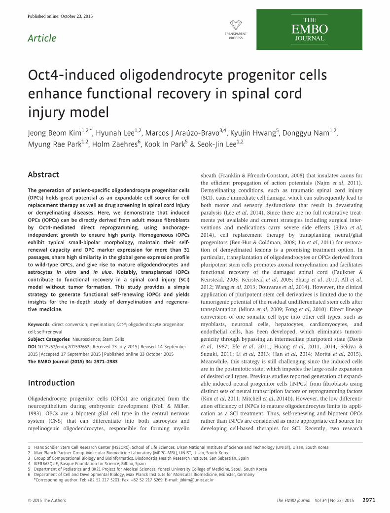
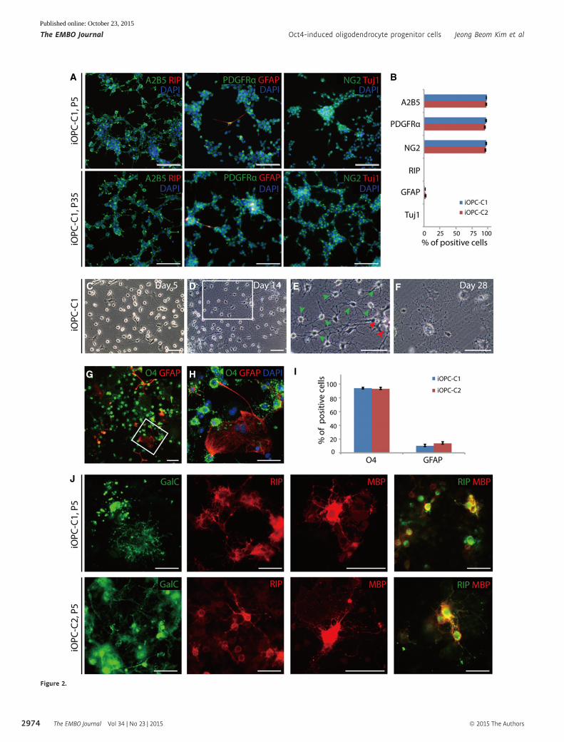
◀ Figure 2. Self-renewal capacity and biopotency of iOPCs.
A Immunofluorescence images of iOPC-C1 at passage 5 (P5) and 35 (P35) stained with A2B5 and RIP (left columns), PDGFRa and GFAP (middle columns), and NG2 andTuj1 (right columns). Cells were counterstained with DAPI. Scale bars, 125 lm.
B Quantification of A2B5+, PDGFRa+, NG2+, RIP+, GFAP+, and Tuj1+ cells in undifferentiated iOPCs. Each type of marker-positive cells was counted in three biologicalreplicates. Data are presented as the means � SD (n = 3).
C–F Morphological changes of iOPC-C1 during differentiation on PDL/laminin-coated plates at (C) days 5, (D, E) 14, and (F) 28. (E) Zoomed image of the white square in(D) shows the mature oligodendrocytic morphology with complex branches (green arrowhead) and heterogeneous astrocytic morphology (red arrowhead). Scalebars, 50 lm.
G, H Biopotency of iOPC in vitro after 2 weeks of differentiation showing (G) O4-immunostained oligodendrocytes and GFAP-stained astrocytes derived from iOPC-C1.(H) Zoomed image of the white square in (G). Scale bars, 75 lm.
I The differentiation efficiency of iOPCs into O4+ oligodendrocytes and GFAP+ astrocytes. Data are presented as the means � SD (n = 3).J Expression of the premyelinating oligodendrocytes markers GalC (left-most columns), mature oligodendrocytes RIP (left–middle columns), and MBP (right–middle
columns) and co-expression of RIP and MBP (right-most columns) at late stage of maturation of iOPC clones (P5). Scale bars, 75 lm.
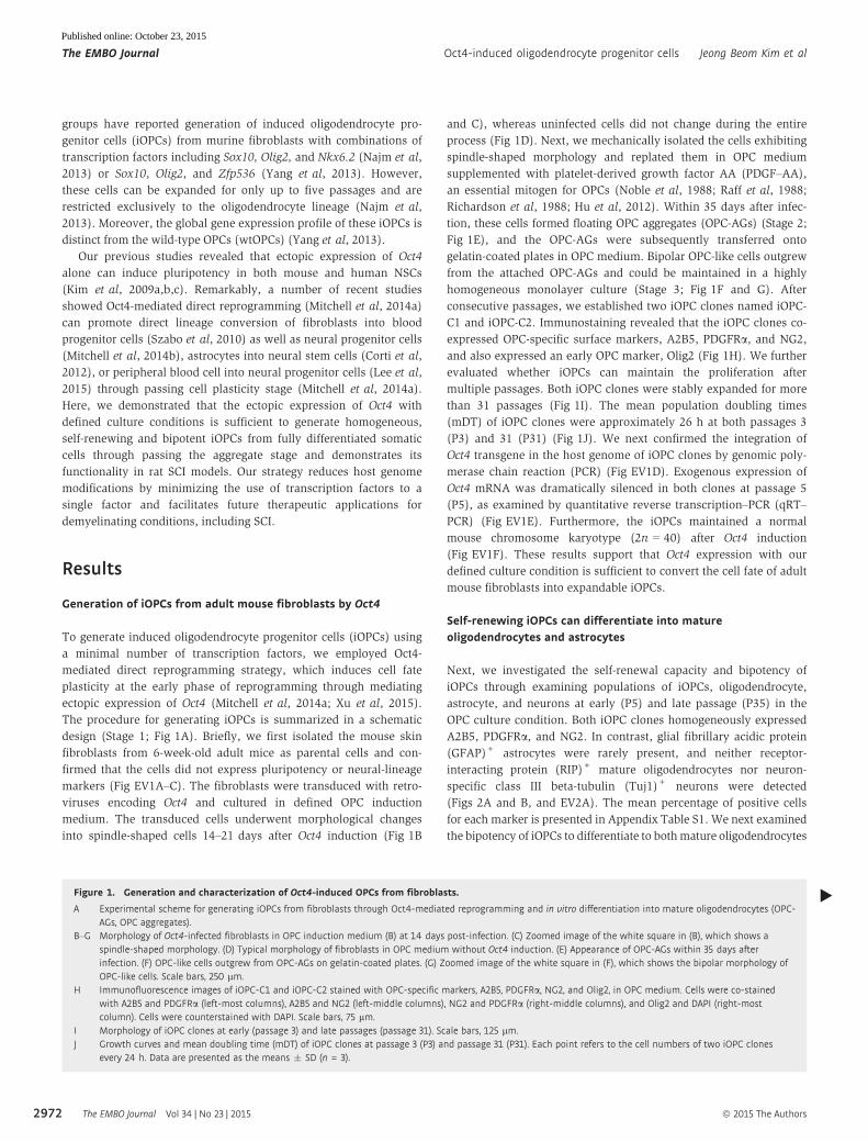
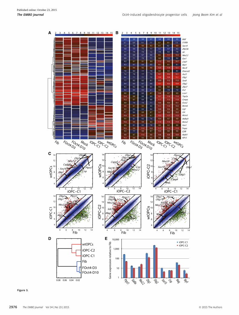
Figure 3. Global gene expression profiles of Oct4-induced OPCs.
A, B Heat map analysis of (A) the global gene expression profiles and (B) 32 OPC and oligodendrocyte lineage enriched genes of Fib, FOct4-D3, FOct4-D10, mock, iOPC-C1, iOPC-C2, and wtOPCs. The upper color bar codifies the gene expression in the log2 scale. Shown are 767 probes selected based on codified variation ≥ � 4across all samples.
C Pairwise scatter plots of the global gene expression of wtOPCs versus iOPC-C1/iOPC-C2, iOPC-C1 versus iOPC-C2 and Fib versus iOPC-C1/iOPC-C2/wtOPC. The blacklines indicate the boundaries of twofold changes in gene expression. The OPC-enriched genes (Mag, Nkx2.2, Olig1, Olig2, Sox10, Cnp, Cspg4, and Ptprz1) arehighlighted with orange circles. Gene expression levels are shown on a log2 scale.
D Hierarchical clustering of wtOPCs, iOPC-C1, iOPC-C2, Fib, FOct4-D3, and FOct4-D10.E qRT–PCR analysis of mRNA expression level for OPC lineage-specific genes (Ptprz1, Siat8a, Nkx2.2, Olig1, Olig2, Sox10, Cnp, Mag, and Myrf) in iOPCs relative to
fibroblasts. Graphs represent log2-fold changes after normalization to GAPDH. Data are presented as the means � SD (n = 3).
▸
ª 2015 The Authors The EMBO Journal Vol 34 | No 23 | 2015
Jeong Beom Kim et al Oct4-induced oligodendrocyte progenitor cells The EMBO Journal
2975
Published online: October 23, 2015
A
C
D E
B
Figure 3.
The EMBO Journal Vol 34 | No 23 | 2015 ª 2015 The Authors
The EMBO Journal Oct4-induced oligodendrocyte progenitor cells Jeong Beom Kim et al
2976
Published online: October 23, 2015
recovery of spinal cord in the shape and reduced cavity size
compared with vehicle-injected control (Fig 4A and B). Further-
more, we observed robust engraftment of the transplanted GFP+
iOPCs that migrated toward the injury site (Fig 4C and D). Next, we
immunostained tissue slides with MBP to investigate whether
engrafted iOPCs contribute to myelination in vivo (Fig 4E and F).
GFP+ iOPCs were distributed in the vicinity of the myelinated nerve
fibers in the white matter (Fig 4G and H). This result suggests that
myelination was associated with the transplanted iOPCs. To identify
the cellular features of iOPCs in the SCI model, we assessed in vivo
differentiation potential of the iOPCs. Most of the GFP+-engrafted
cells robustly expressed mature oligodendrocyte markers such as
CNPase, MBP, APC-CC1, and O4 (Figs 4I and J, and EV3A and B)
and were mainly localized near neurofilament (NF)+ host neurons
in the injury site (Figs 4K–N and EV3C and D). The interaction
between iOPC-derived mature oligodendrocyte (GFP+CNPase+/
GFP+MBP+/GFP+AP-CC1+) and the host NF+ neurons in the
injury site was clearly observed in the three-dimensional recon-
structed images (Figs 4O and P and EV3E, and Movie EV1). These
results strongly indicate differentiation of the transplanted iOPCs
into myelin-producing oligodendrocytes that enhance the remyelina-
tion process by ensheathing damaged neurons. In addition, a
few astrocytes (GFP+GFAP+) (Fig EV3F–H) and undifferentiated
iOPCs (GFP+PDGFRa+/GFP+A2B5+/GFP+NG2+/GFP+Olig2+)
were observed as well (Fig EV3I). We further examined in vivo dif-
ferentiation potential of iOPCs in mouse SCI models (n = 4) and
observed expression of A2B5, PDGFRa, CNPase, and GFAP from the
engrafted cells in the mouse spinal cord tissue as well (Fig EV3J–Q).
Next, we evaluated the motor function recovery of the hind limbs
by measuring Basso–Beattie–Bresnahan (BBB) scores for 10 weeks
in rat SCI models that were treated either with PBS (control)
(n = 10) or with iOPCs injection (n = 8). Both groups exhibited
regaining motor ability for the first 2 weeks of the injury due to the
natural reflex recovering ability in rats (Basso et al, 1995).
However, rats transplanted with iOPCs improved in BBB scores
from week 2 (n = 8) that persisted steadily through week 10 (n = 4)
showing significant improvement in locomotor recovery (P < 0.05),
which is distinct from the control group which reached the plateau
in the BBB score by week 2 (Fig 4Q). To assess the risk of tumor
formation, we subcutaneously transplanted iOPCs (P5) into severe
combined immunodeficient (SCID) mice and no tumors were
observed for 9 months of experiment period (Fig EV3R). These
results show that iOPCs can differentiate into mature oligodendro-
cytes and astrocytes without tumor formation in vivo and success-
fully promote functional recovery of locomotion in a SCI model.
Discussion
Here, we have successfully generated self-renewing iOPCs from
adult mouse fibroblasts through Oct4-mediated direct reprogram-
ming methodology. Conventionally, OPCs were isolated from fetal
brain or derived from pluripotent stem cells (Sim et al, 2011; All
et al, 2012; Wang et al, 2013). However, primary OPCs are limited
in availability, and pluripotent stem cell-derived OPCs have the risk
of tumor formation after transplantation and are low in differentia-
tion efficiency. In contrast, the direct lineage conversion strategy
has the advantage in high-yield generation of the desired cells with-
out the tumorigenic potential by bypassing an intermediate pluripo-
tent state (Najm et al, 2013; Yang et al, 2013). Recent studies
revealed that iOPCs can be generated from mouse and rat fibroblasts
by defined sets of lineage-specific transcription factors, including
Sox10, Olig2, and Nkx6.2 (Najm et al, 2013) or Sox10, Olig2, and
Zfp536 (Yang et al, 2013). However, the iOPCs generated by Sox10,
Olig2, and Nkx6.2 transgenes were not bipotent OPCs as they cannot
be differentiated into astrocytes, and they were expandable up to
only 5 passages (Najm et al, 2013). In addition, the global gene
expression pattern of the iOPCs generated by Sox10, Olig2, and
Zfp536 transgenes exhibited a transcription profile that is more
similar to primary pre-oligodendrocytes than the OPCs (Yang et al,
2013).
In contrast to the previously reported direct conversion methods
for generating iOPCs, we showed that a single transcription factor
Oct4 in combination with defined OPC induction medium is suffi-
cient to generate iOPCs from the mouse fibroblast. Previous studies
reported that fewer viral integrations were detected in single-factor-
derived iPSCs (Kim et al, 2009b) compared with iPSCs generated by
four transcription factors, thereby reducing chances of insertional
mutagenesis (Wernig et al, 2007; Aoi et al, 2008). Thus, our single-
factor-derived iOPCs would have higher levels of genomic stability
by having lower chance of viral integration into the host genome
than the previously reported three-factor-derived iOPCs. Moreover,
the previous studies isolated iOPCs by purifying proteolipid protein
(Plp)+ or O4+ cells from a heterogeneous population (Najm et al,
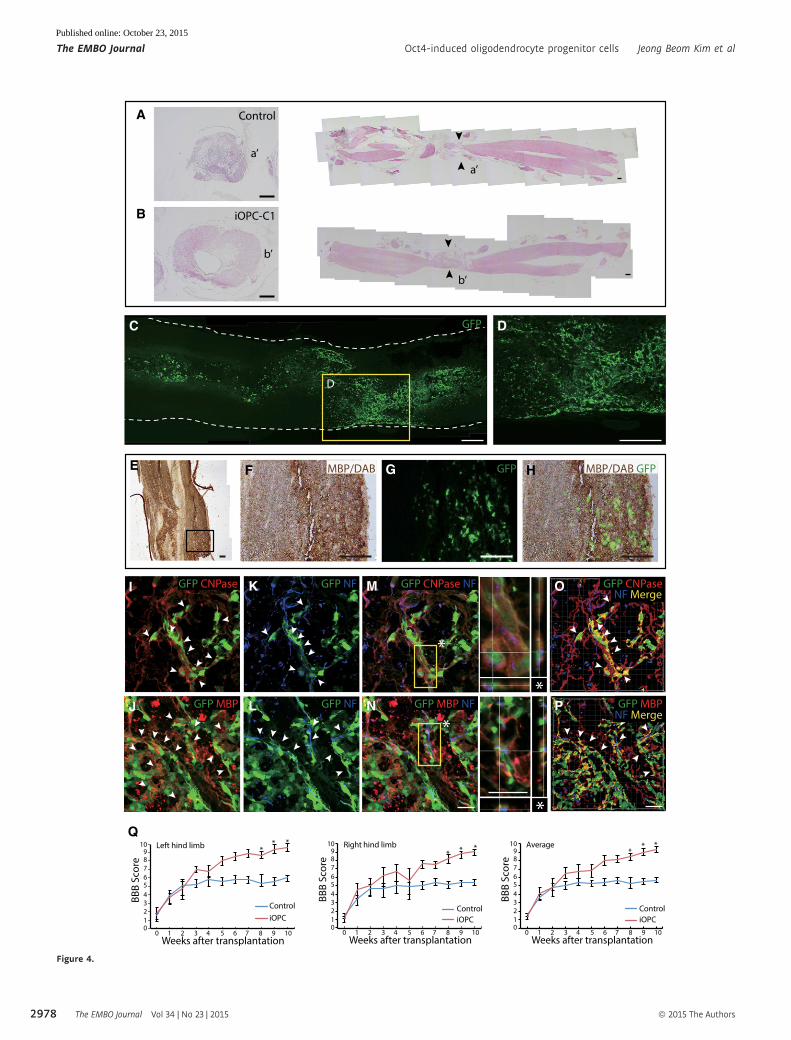
Figure 4. Therapeutic potential and differentiation capacity of transplanted iOPCs in a spinal cord injury model.
A, B H&E staining of rat spinal cords at 6 weeks after transplantation with (A) vehicle or (B) iOPC-C1 (left, coronal plane section; right, sagittal plane section). Blackarrowheads indicate the injury sites (a’ and b’) of each group. Scale bars, 400 lm.
C, D Immunofluorescence image of (C) GFP-labeled iOPC-C1 in a sagittal section of the rat spinal cord. (D) Zoomed image of the yellow square in (C) showing themigration of transplanted cells toward the injury site. Scale bars, 500 lm.
E–H Immunohistochemical analysis of the white matter of the rat spinal cord transplanted with iOPCs (E) for MBP revealing localization of GFP+ iOPC-derivedoligodendrocytes at myelinated nerve fiber structures. Staining was visualized by diaminobenzidine (DAB). Zoomed image of the black square in (E) shows (F) DAB-stained MBP, (G) locality of GFP+ oligodendrocytes, and (H) co-localization of GFP+ oligodendrocytes and MBP. Scale bars, 150 lm.
I–P Expression of the mature oligodendrocyte markers (I) CNPase and (J) MBP in transplanted GFP+ iOPC-C1. Cells co-expressing GFP and oligodendrocyte markers areindicated with white arrowheads. (K, L) GFP+ cells (white arrowheads) wrap neurofilaments (NF) stained host neurons. Confocal z-stacks of the indicated area(asterisks) show that NF+ host neurons are surrounded by (M) GFP+CNPase+ or (N) GFP+MBP+ cells. (O, P) Three-dimensional reconstructions of rat spinal cordsections using IMARIS software show the complex structure of the host neurons and GFP+ cells forming axon ensheathments. Co-expression (yellow) of GFP+ iOPCs(green) and mature oligodendrocyte markers (red) is observed around neurons (blue). Scale bars, 30 lm.
Q BBB score evaluation of the left (left panel), right (middle panel), and average (right panel) of both hind limbs during the 10 weeks following transplantation. Dataare presented as the means � SD (n = 3–8). *P < 0.05 (one-way ANOVA).
▸
ª 2015 The Authors The EMBO Journal Vol 34 | No 23 | 2015
Jeong Beom Kim et al Oct4-induced oligodendrocyte progenitor cells The EMBO Journal
2977
Published online: October 23, 2015
A
B
C
E F
Q
HG
I K M O
J L N P
D
Figure 4.
The EMBO Journal Vol 34 | No 23 | 2015 ª 2015 The Authors
The EMBO Journal Oct4-induced oligodendrocyte progenitor cells Jeong Beom Kim et al
2978
Published online: October 23, 2015
2013; Yang et al, 2013). However, the iOPCs in these studies may
include heterogeneous population having various viral integration
sites in the host genome. Moreover, Plp and O4 are also expressed
in pre-oligoendrocytes as well as mature oligodendrocytes which
interrupt isolation of homogeneous OPC population. Therefore, the
purifying method used in the previous studies yields difficulty in
developing a single homogeneous cell line, which leads to the diffi-
culty in determining the accurate conversion efficiency. In contrast,
we demonstrated a clonal reprogramming method, as we have
shown previously in generation of iPSCs from NSCs (Kim et al,
2009c). This approach enables the establishment of homogeneous
cell lines from single colonies. Additionally, as the previous direct
conversion strategies have demonstrated low conversion efficien-
cies, it is not feasible to obtain iOPCs with high yield and purity.
Our iOPCs are self-renewing and bipotent progenitors that maintain
typical OPC features including bipolar morphology, OPC marker
expression, and high differentiation efficiency in vitro and in vivo
throughout multiple passages (> 31 passages). Strikingly, the global
gene expression pattern of Oct4-iOPCs exhibits high levels of simi-
larity to the publically available GEO data sets of wtOPCs (Najm
et al, 2013). Lastly, we generated iOPCs through sequential induc-
tion with defined medium at each stage to induce the target cell fate
more precisely. Thus, our strategy allows generation of a pure popu-
lation of proliferative iOPCs that resembles key morphological and
molecular features of wtOPCs in large scale.
Recent studies demonstrated that transplantation of OPCs dif-
ferentiated from ESCs or iPSCs is a promising strategy to promote
recovery after SCI (Keirstead et al, 2005; Sharp et al, 2010; Czepiel
et al, 2015). However, the risk of tumorigenicity from undifferenti-
ated cells remains as a major challenge for its therapeutic use. In
this study, Oct4-iOPCs differentiated to mature oligodendrocytes and
surrounded host neurons, displaying extensive ensheathment when
transplanted into the rodent SCI model. Notably, the transplantation
of iOPCs facilitated remyelination and significant locomotor func-
tional recovery without tumor formation. Thus, Oct4-iOPCs can
serve as a prospective cell source for the establishment of cell-based
therapy for SCI. To note, we mainly focused on a rat SCI model
because of the small size of the mouse SCI models that limits surgi-
cal maneuvers and device implantations (Nakae et al, 2011).
However, rat models are commonly used interchangeably with
mouse models due to its cost and convenience in surgical maneu-
vers (Talac et al, 2004).
Several lines of evidence have demonstrated that Oct4 is involved
in the initial cell fate commitment during reprogramming. Previ-
ously, we reported that Oct4 is required to induce NSCs towards the
pluripotent state, which validates the critical role of Oct4 in cell fate
determination (Kim et al, 2008; Kim et al, 2009a,b,c). Here, we
generated iOPCs by using a similar approach to the recent works on
generation of iNPCs or blood progenitors from somatic cells through
OCT4-mediated direct reprogramming (Szabo et al, 2010; Mitchell
et al, 2014b; Lee et al, 2015). The previous studies signify the
importance of the lineage-specific induction medium since Oct4-
induced somatic cell fate determination seems to depend on the
lineage-specific culture condition (Szabo et al, 2010; Mitchell et al,
2014a,b; Lee et al, 2015). Specifically, the recent work on blood-
derived hiNPCs showed that blood cells can be directly repro-
grammed into iNPCs by using OCT4 in combination with bFGF and
epidermal growth factor (EGF)-supplemented NPC induction
medium, and small molecules (SMAD and GSK-3 inhibitors) (Lee
et al, 2015). In the same line, we used OPC induction medium
supplemented with PDGF-AA, an essential OPC-inducing growth
factor (Hu et al, 2012), to convert the cell fate of Oct4-induced
fibroblast into iOPC. Therefore, our study demonstrated Oct4-
induced somatic cell fate can be determined to OPC lineage by
providing OPC lineage-specific culture condition. Importantly, we
did not obtain any Tuj1+ neurons in the process of generating iOPCs
(Figs 2A and EV3A) and from the iOPCs under neuronal induction
condition (Fig EV3F). Thus, these results suggest that transient NPC
state was not involved during the reprogramming, possibly due to
the OPC lineage-specific stimuli in our OPC induction medium.
However, a detailed signaling pathway that mediates generation of
iOPCs through bypassing NPC state remains to be elucidated.
In conclusion, we demonstrated that Oct4 with defined iOPCs
induction condition reprograms adult somatic cells into self-
renewing and bipotent iOPCs, thereby allowing large-scale expan-
sion of the oligodendrocytes and astrocytes. This single-factor-
mediated conversion method is a safer and simpler protocol that
potentially reduces the chance of viral insertional mutagenesis.
Although further investigation is needed to study the mechanism of
Oct4-triggered lineage conversion, our strategy may provide the
basis for future human iOPC generation and new insights to investi-
gate high-throughput drug screening and stem cell-based therapies
for SCI and demyelinating disorders.
Materials and Methods
Isolation of mouse skin fibroblasts
The experimental procedures were approved by the Animal Care
and Use Committee at Ulsan National Institute of Science and Tech-
nology (Ulsan, South Korea). Primary dermal fibroblasts were
isolated from skin biopsies of 6-week-old CF1 mice (Jackson Labora-
tories) as described previously (Driskell et al, 2013) and cultivated
in fibroblast medium [DMEM high glucose (GIBCO), 10% fetal
bovine serum (GIBCO), penicillin/streptomycin (GIBCO), non-
essential amino acids (GIBCO), and b-mercaptoethanol (GIBCO)]
MEF medium. Parental fibroblasts were passaged 2–3 times before
Oct4 induction.
Virus production
To produce viruses, a pMX-based retroviral vector containing Oct4
cDNA (Addgene #13366) and the SFFV–GFP lentiviral vector, a gift
from Dr. Axel Schambach (Warlich et al, 2011), were transfected
into 293T cells using the X-tremeGENE9 DNA transfection reagent
(Roche) to package the VSV-G-pseudotyped virus. After 48 h of
transfection, the virus-containing supernatants were collected and
filtered through a 0.45-lm syringe filter. The viruses were harvested
as previously described (Zaehres & Daley, 2006).
Generation of iOPCs
Primary adult mouse fibroblasts were seeded at 1 × 10⁴ cells on
gelatin-coated 6-well plates. On the next day, the fibroblasts
were infected with a pMX retroviral vector expressing Oct4 in
ª 2015 The Authors The EMBO Journal Vol 34 | No 23 | 2015
Jeong Beom Kim et al Oct4-induced oligodendrocyte progenitor cells The EMBO Journal
2979
Published online: October 23, 2015
MEF medium containing 6 lg/ml protamine sulfate. The viral
supernatant was removed after 24 h of infection and replaced with
fresh MEF medium. At 3 days post-infection, the medium was
switched to OPC induction medium (1:1 mixture of DMEM/F12
(GIBCO) and neurobasal medium (GIBCO) supplemented with N2
(GIBCO), B27 (GIBCO), penicillin/streptomycin, 10 ng/ml PDGF-AA
(Peprotech), and 10 ng/ml FGF2 (Peprotech)). Oct4-infected cells
were mechanically isolated by a glass micropipette and transferred
into defined OPC medium (DMEM/F12 supplemented with N2, peni-
cillin/streptomycin, 20 ng/ml PDGF-AA (Peprotech), and 10 ng/ml
FGF2). The floating aggregates were re-plated onto gelatin-coated
plate, and the OPC-like cells were subsequently passaged 3–5 times
until the cells become homogeneous. We conducted further charac-
terization of the iOPCs at early passages (P3 and P5) and late
passages (P31 and P35).
Oct4 transgene silencing analysis
Total RNA of iOPC-C1 and iOPC-C2 was extracted using an RNeasy
mini kit (Qiagen). cDNAs were synthesized by SuperScript� III
reverse transcriptase (Invitrogen), and 500 ng total RNA was
obtained per reaction. Real-time PCR analysis was performed on a
LightCycler 480 and SYBR Green I Master mix (Roche) in a total
volume of 20 ll. The experiments were performed in triplicate, and
expression was normalized to GAPDH. Gene expression was
measured by the Ct calculation method. The sequences of the
primers used in this experiment are listed in Appendix Table S5.
RT–PCR and quantitative RT–PCR
DNA-free total RNA of iOPC-C1 and iOPC-C2 (P5) was extracted
using the RNeasy mini kit (Qiagen). Total RNA (500 ng) was used
to synthesize cDNAs using SuperScript� III reverse transcriptase
(Invitrogen). RT–PCR was performed using recombinant Taq DNA
polymerase (Invitrogen). qRT–PCR analysis was conducted on a
LightCycler 480 instrument with SYBR Green I Master mix (Roche).
The experiments were performed in triplicate, and expression was
normalized to the housekeeping gene GAPDH. Gene expression was
measured by calculating Ct values. All of the experiments were
conducted according to the manufacturer’s instructions. The
sequences of the primers used are listed in Appendix Table S5.
In vitro differentiation of iOPCs
1 × 10⁴ iOPCs (P5 and P35) were seeded on PDL/laminin-coated
4-well plates in OPC medium. For oligodendrocyte differentiation,
the medium was switched to oligodendrocyte differentiation
medium 1, which contains DMEM/F12 supplemented with N2, peni-
cillin/streptomycin, 10 ng/ml FGF2 (Peprotech), and 10 mM
forskolin (Sigma-Aldrich), for 4–5 days. For further maturation, the
medium was changed to oligodendrocyte differentiation medium 2,
which contains 30 ng/ml 3,30,5-triiodothyronine (T3; Sigma-
Aldrich) and 20 ng/ml ascorbic acid (AA; Sigma-Aldrich). For astro-
cyte differentiation, we differentiated iOPCs in DMEM containing
2% FBS for 7–9 days. For neuronal differentiation, the cells
were cultured in neuronal differentiation medium 1 (DMEM/F12
supplemented with N2, B27, and 10 ng/ml FGF-2) for 5 days and
then switched to neuronal differentiation medium 2 (DMEM/F12:
neurobasal (1:1) supplemented with 0.5% N2 and 0.5% B27) (Kim
et al, 2009b).
Immunocytochemistry (ICC) analysis
iOPCs or iOPC derivatives were fixed with 4% paraformaldehyde
for 10 min and permeabilized with 0.1% Triton X-100 for 10 min.
The cells were then incubated in 4% FBS blocking solution for
30 min and subsequently incubated in primary antibodies diluted in
blocking solution for 1 h at room temperature. After the primary
antibody incubation, cells were washed with PBST (0.05% Tween-
20) three times. The secondary antibodies were diluted in PBS and
applied for 1 h: Alexa Fluor 488/568 anti-mouse IgG1, IgG3, IgM,
and anti-goat IgG (Invitrogen, 1:1,000). Nuclei were stained with
DAPI (Invitrogen). The primary antibodies used for ICC analysis are
listed in Appendix Table S4.
Microarray analysis
Global gene expression of the following cell populations were pro-
filed by microarray analysis: fibroblasts, FOct4-D3 (3 days after Oct4
induction), FOct4-D10 (10 days after Oct4 induction cultured in OPC
medium), mock (mock-infected fibroblasts cultured in OPC medium
for 10 days), iOPC-C1, and iOPC-C2. We compared these samples
with previously published data for wtOPCs (undifferentiated OPCs
from pluripotent stem cells) (Yang et al, 2013). Total RNA of iOPC
clones (P5) was isolated using an RNeasy mini kit (Qiagen) accord-
ing to the manufacturer’s instructions. The samples were hybridized
to an Affymetrix Mouse Gene 1.0 ST array. Normalization was
performed with the robust multi-array analysis (RMA) algorithm
(Irizarry et al, 2003). Data processing and graphic production were
performed with in-house-developed functions in MATLAB. The hier-
archical clustering of genes and samples was performed with the one
minus correlation metric and the unweighted average distance
(UPGMA) linkage method. Differentially expressed genes were
analyzed by comparing gene expression of iOPC clones (iOPC-C1,
iOPC-C2) relative to fibroblasts. Among the highly up-regulated
genes (˃ 1.2-fold), genes that are related to neural/oligodendrocyte/
OPC development or myelin function were screened according to
Gene Ontology (GO) term enrichment profiling. Microarray data
from the wtOPCs were downloaded from the GEO database (data
accessible at NCBI GEO database (Najm et al, 2013), accession
GSE45440). The data discussed in this publication were deposited in
NCBI Gene Expression Omnibus (Edgar et al, 2002) and are
accessible through GEO Series accession number GSE52035.
Contusion spinal cord injury and transplantation
The procedures were approved by the Animal Care and Use Commit-
tees of Yonsei University College of Medicine (Seoul, Korea). Adult
male Sprague Dawley rats (220–240 g) and 6-week-old male severe
combined immunodeficient (SCID) mice were anesthetized with
intraperitoneal injections of ketamine (90 mg/kg), rompun (0.2 ml/kg),
and promazine (1 mg/kg). The skin was shaved and decontami-
nated with serial 70% ethanol and povidone–iodine washes. A
dorsal laminectomy was performed on the T9 vertebrae to expose
the spinal cord. The contusion injury was induced at spinal segment
T9 using the Infinite Horizon Impactor (Precision Systems,
The EMBO Journal Vol 34 | No 23 | 2015 ª 2015 The Authors
The EMBO Journal Oct4-induced oligodendrocyte progenitor cells Jeong Beom Kim et al
2980
Published online: October 23, 2015
Kentucky, IL, USA) with a force of 230 Kdyn. After contusion, the
muscle and skin were sutured. For transplantation, iOPC-C1 (P5)
was labeled with GFP by the lentiviral system, and the integrated
cells were detected by green fluorescence. After 1 week of injury,
1.2 × 10⁵ iOPCs were injected into T8 and T10 using a glass micro-
pipette. The animals were provided food and water regularly and
received manual bladder expression twice daily for urination.
Basso-Beattie-Bresnahan (BBB) test
The locomotor recovery of the animals was assessed by two inde-
pendent observers using the BBB open-field locomotor score. The
experiment was performed by blinded observation. The rat SCI
models were separated into PBS-injected control group (n = 10) and
1.2 × 10⁵ iOPCs (P5)-injected experimental group (n = 8) and were
tested weekly for up to 10 weeks after transplantation. Hind limb
locomotion was assessed by the BBB open-field scale as described
previously (Basso et al, 1995).
Histological process and immunohistochemical (IHC) analysis
The rats were anesthetized and perfused in PBS at 6 weeks after
transplantation. The spinal cords were fixed overnight in 4%
paraformaldehyde and dehydrated using a series of alcohol and
xylene. The tissue was embedded in paraffin blocks and sectioned
sequentially at 16 lm in the sagittal or coronal planes. The sections
were permeabilized with 0.2% Triton X-100 and blocked with CAS-
Block solution (Invitrogen) to prevent non-specific binding. For IHC
analysis, the sections were incubated with primary antibodies
diluted in CAS-Block solution overnight at 4°C. After the primary
antibody incubation, the cells were washed with PBST (0.05%
Tween-20) three times for 10 min each. The following secondary
antibodies were diluted in PBS and applied for 1 h: Alexa Fluor
405/488/568 anti-mouse IgG1, IgG3, IgM, anti-goat IgG, and anti-rat
IgG (Invitrogen, 1:200). Fluorescence images were visualized with
Olympus IX81-ZDC and FV1000 microscopes. The primary anti-
bodies used for IHC analysis are listed in Appendix Table S4.
For diaminobenzidine (DAB) immunostaining, the tissue sections
were incubated in the primary antibody and then treated with 0.3%
H2O2 in PBS for 30 min at room temperature to block endogenous
peroxidases. The sections were then incubated with a horseradish
peroxidase-conjugated anti-rat IgG antibody (Invitrogen, 1:200) for
2 h. We used DAB (Invitrogen) to visualize the sections by treating
them for 5 min at room temperature.
For hematoxylin and eosin staining, paraffin blocks were
prepared by aforementioned method with SCI rats 6 weeks after
transplantation. The paraffin blocks were sectioned sequentially at
4 lm in the sagittal or coronal planes. Nucleus was stained with
hematoxylin (Sigma-Aldrich) and blued in 0.2% ammonium
hydroxide. Tissue were then placed in Eosin Y solution (Sigma-
Aldrich) and followed by several washings and dehydrating. Slides
were mounted with mounting solution (Leica).
Karyotyping
The fibroblasts and iOPCs were treated with 0.1 lg/ml demecolcine
(Sigma-Aldrich) for 8 h in a 37°C CO2 incubator. The cells were
collected and trypsinized into a single-cell suspension and then
incubated in 75 mM KCl for 20 min at 37°C. Carnoy’s solution
(methanol:acetic acid = 1:3) was slowly added to the cells, which
were then fixed for 5 min at room temperature. The cells were fixed
twice more with fresh Carnoy’s solution for 20 min each and
dropped onto humid glass slides. The chromosomes clearly spread
and were counted in individual cells.
Growth curve and mean doubling time
iOPC clones (1 × 10⁴ cells) at P3 and P31 were seeded onto 4-well
plates and cultivated for 10 days. The cells were collected from trip-
licate wells and manually counted each day (24 h) using a hemacy-
tometer (Marienfeld). The average cell numbers on each day were
plotted, and the mean doubling time was calculated based on the
growth curve.
Data deposition
The microarray data reported in this article have been deposited in
the NCBI Gene Expression Omnibus database under accession
number GSE52035.
Statistical analysis
All data in this article are presented as the means � SD (standard
deviation). Data from at least three independent samples were used
for statistical analysis. A P-value < 0.05 (one-way ANOVA) was
considered significant.
Expanded View for this article is available online:
http://emboj.embopress.org
AcknowledgementsWe thank Dr. Axel Schambach for providing SFFV-GFP, JY and SW for generous
assistance in preparation of the manuscript. This research was partly
supported by the Bio & Medical Technology Development Program of the
National Research Foundation (NRF) and funded by the Ministry of Science,
ICT & Future Planning [2012M3A9C6049788], the ICT R&D Program of MSIP/
IITP [R0190-15-2072], and Max Planck Partner Group, Max Planck Society
(MPG), Germany.
Author contributionsJBK designed and supervised the study. JBK and HL conducted most of the
experiments and wrote the manuscript. MJA contributed to the microarray
analysis. KIP and KH contributed to the animal experiments. DN and MRP
collected the data and DN, MRP, HZ, and S-JL were involved in preparing the
manuscript.
Conflict of interestThe authors declare that they have no conflict of interest.
References
All AH, Bazley FA, Gupta S, Pashai N, Hu C, Pourmorteza A, Kerr C (2012)
Human embryonic stem cell-derived oligodendrocyte progenitors aid in
functional recovery of sensory pathways following contusive spinal cord
injury. PLoS ONE 7: e47645
ª 2015 The Authors The EMBO Journal Vol 34 | No 23 | 2015
Jeong Beom Kim et al Oct4-induced oligodendrocyte progenitor cells The EMBO Journal
2981
Published online: October 23, 2015
Aoi T, Yae K, Nakagawa M, Ichisaka T, Okita K, Takahashi K, Chiba T,
Yamanaka S (2008) Generation of pluripotent stem cells from adult mouse
liver and stomach cells. Science 321: 699 – 702
Basso DM, Beattie MS, Bresnahan JC (1995) A sensitive and reliable locomotor
rating scale for open field testing in rats. J Neurotrauma 12: 1 – 21
Ben-Hur T, Goldman SA (2008) Prospects of cell therapy for disorders of
myelin. Ann N Y Acad Sci 1142: 218 – 249
Cahoy JD, Emery B, Kaushal A, Foo LC, Zamanian JL, Christopherson KS, Xing
Y, Lubischer JL, Krieg PA, Krupenko SA, Thompson WJ, Barres BA (2008) A
transcriptome database for astrocytes, neurons, and oligodendrocytes: a
new resource for understanding brain development and function. J
Neurosci 28: 264 – 278
Corti S, Nizzardo M, Simone C, Falcone M, Donadoni C, Salani S, Rizzo F,
Nardini M, Riboldi G, Magri F, Zanetta C, Faravelli I, Bresolin N, Comi GP
(2012) Direct reprogramming of human astrocytes into neural stem cells
and neurons. Exp Cell Res 318: 1528 – 1541
Czepiel M, Boddeke E, Copray S (2015) Human oligodendrocytes in
remyelination research. Glia 63: 513 – 530
Davis RL, Weintraub H, Lassar AB (1987) Expression of a single transfected
cDNA converts fibroblasts to myoblasts. Cell 51: 987 – 1000
Douvaras P, Wang J, Zimmer M, Hanchuk S, O’Bara Melanie A, Sadiq S, Sim
Fraser J, Goldman J, Fossati V (2014) Efficient generation of myelinating
oligodendrocytes from primary progressive multiple sclerosis patients by
induced pluripotent stem cells. Stem Cell Rep 3: 250 – 259
Driskell RR, Lichtenberger BM, Hoste E, Kretzschmar K, Simons BD,
Charalambous M, Ferron SR, Herault Y, Pavlovic G, Ferguson-Smith AC,
Watt FM (2013) Distinct fibroblast lineages determine dermal architecture
in skin development and repair. Nature 504: 277 – 281
Edgar R, Domrachev M, Lash AE (2002) Gene Expression Omnibus: NCBI gene
expression and hybridization array data repository. Nucleic Acids Res 30:
207 – 210
Efe JA, Hilcove S, Kim J, Zhou H, Ouyang K, Wang G, Chen J, Ding S (2011)
Conversion of mouse fibroblasts into cardiomyocytes using a direct
reprogramming strategy. Nat Cell Biol 13: 215 – 222
Faulkner J, Keirstead HS (2005) Human embryonic stem cell-derived
oligodendrocyte progenitors for the treatment of spinal cord injury.
Transpl Immunol 15: 131 – 142
Fong CY, Gauthaman K, Bongso A (2010) Teratomas from pluripotent stem
cells: a clinical hurdle. J Cell Biochem 111: 769 – 781
Franklin RJM, Ffrench-Constant C (2008) Remyelination in the CNS: from
biology to therapy. Nat Rev Neurosci 9: 839 – 855
Han JK, Chang SH, Cho HJ, Choi SB, Ahn HS, Lee J, Jeong H, Youn SW, Lee HJ,
Kwon YW, Cho HJ, Oh BH, Oettgen P, Park YB, Kim HS (2014) Direct
conversion of adult skin fibroblasts to endothelial cells by defined factors.
Circulation 130: 1168 – 1178
Hu JG, Wang YX, Wang HJ, Bao MS, Wang ZH, Ge X, Wang FC, Zhou JS, Lu HZ
(2012) PDGF-AA mediates B104CM-induced oligodendrocyte precursor cell
differentiation of embryonic neural stem cells through Erk, PI3K, and p38
signaling. J Mol Neurosci 46: 644 – 653
Huang P, He Z, Ji S, Sun H, Xiang D, Liu C, Hu Y, Wang X, Hui L (2011)
Induction of functional hepatocyte-like cells from mouse fibroblasts by
defined factors. Nature 475: 386 – 389
Huang P, Zhang L, Gao Y, He Z, Yao D, Wu Z, Cen J, Chen X, Liu C, Hu Y, Lai
D, Hu Z, Chen L, Zhang Y, Cheng X, Ma X, Pan G, Wang X, Hui L (2014)
Direct reprogramming of human fibroblasts to functional and expandable
hepatocytes. Cell Stem Cell 14: 370 – 384
Irizarry RA, Bolstad BM, Collin F, Cope LM, Hobbs B, Speed TP (2003) Summaries
of Affymetrix GeneChip probe level data. Nucleic Acids Res 31: e15
Jin K, Xie L, Mao X, Greenberg MB, Moore A, Peng B, Greenberg RB, Greenberg
DA (2011) Effect of human neural precursor cell transplantation on
endogenous neurogenesis after focal cerebral ischemia in the rat. Brain
Res 1374: 56 – 62
Keirstead HS, Nistor G, Bernal G, Totoiu M, Cloutier F, Sharp K, Steward O
(2005) Human embryonic stem cell-derived oligodendrocyte progenitor
cell transplants remyelinate and restore locomotion after spinal cord
injury. J Neurosci 25: 4694 – 4705
Kim JB, Zaehres H, Wu G, Gentile L, Ko K, Sebastiano V, Araúzo-Bravo MJ,
Ruau D, Han DW, Zenke M, Schöler HR (2008) Pluripotent stem cells
induced from adult neural stem cells by reprogramming with two factors.
Nature 454: 646 – 650
Kim JB, Greber B, Arauzo-Bravo MJ, Meyer J, Park KI, Zaehres H, Scholer HR
(2009a) Direct reprogramming of human neural stem cells by OCT4.
Nature 461: 649 – 653
Kim JB, Sebastiano V, Wu G, Araúzo-Bravo MJ, Sasse P, Gentile L, Ko K, Ruau
D, Ehrich M, van den Boom D, Meyer J, Hübner K, Bernemann C, Ortmeier
C, Zenke M, Fleischmann BK, Zaehres H, Schöler HR (2009b) Oct4-induced
pluripotency in adult neural stem cells. Cell 136: 411 – 419
Kim JB, Zaehres H, Arauzo-Bravo MJ, Scholer HR (2009c) Generation of induced
pluripotent stem cells from neural stem cells. Nat Protoc 4: 1464 – 1470
Kim J, Efe JA, Zhu S, Talantova M, Yuan X, Wang S, Lipton SA, Zhang K, Ding S
(2011) Direct reprogramming of mouse fibroblasts to neural progenitors.
Proc Natl Acad Sci USA 108: 7838 – 7843
Lee BB, Cripps RA, Fitzharris M, Wing PC (2014) The global map for traumatic
spinal cord injury epidemiology: update 2011, global incidence rate. Spinal
Cord 52: 110 – 116
Lee JH, Mitchell RR, McNicol JD, Shapovalova Z, Laronde S, Tanasijevic B,
Milsom C, Casado F, Fiebig-Comyn A, Collins TJ, Singh KK, Bhatia M (2015)
Single transcription factor conversion of human blood fate to NPCs with
CNS and PNS developmental capacity. Cell Rep 11: 1367 – 1376
Li J, Huang NF, Zou J, Laurent TJ, Lee JC, Okogbaa J, Cooke JP, Ding S (2013)
Conversion of human fibroblasts to functional endothelial cells by defined
factors. Arterioscler Thromb Vasc Biol 33: 1366 – 1375
Mitchell R, Szabo E, Shapovalova Z, Aslostovar L, Makondo K, Bhatia M
(2014a) Molecular evidence for OCT4-induced plasticity in adult human
fibroblasts required for direct cell fate conversion to lineage specific
progenitors. Stem Cells 32: 2178 – 2187
Mitchell RR, Szabo E, Benoit YD, Case DT, Mechael R, Alamilla J, Lee JH,
Fiebig-Comyn A, Gillespie DC, Bhatia M (2014b) Activation of neural cell
fate programs toward direct conversion of adult human fibroblasts into
tri-potent neural progenitors using OCT-4. Stem Cells Dev 23: 1937 – 1946
Miura K, Okada Y, Aoi T, Okada A, Takahashi K, Okita K, Nakagawa M,
Koyanagi M, Tanabe K, Ohnuki M, Ogawa D, Ikeda E, Okano H, Yamanaka
S (2009) Variation in the safety of induced pluripotent stem cell lines. Nat
Biotech 27: 743 – 745
Morita R, Suzuki M, Kasahara H, Shimizu N, Shichita T, Sekiya T, Kimura A,
Sasaki K, Yasukawa H, Yoshimura A (2015) ETS transcription factor ETV2
directly converts human fibroblasts into functional endothelial cells. Proc
Natl Acad Sci USA 112: 160 – 165
Najm FJ, Zaremba A, Caprariello AV, Nayak S, Freundt EC, Scacheri PC, Miller
RH, Tesar PJ (2011) Rapid and robust generation of functional
oligodendrocyte progenitor cells from epiblast stem cells. Nat Methods 8:
957 – 962
Najm FJ, Lager AM, Zaremba A, Wyatt K, Caprariello AV, Factor DC, Karl RT,
Maeda T, Miller RH, Tesar PJ (2013) Transcription factor-mediated
reprogramming of fibroblasts to expandable, myelinogenic
oligodendrocyte progenitor cells. Nat Biotech 31: 426 – 433
The EMBO Journal Vol 34 | No 23 | 2015 ª 2015 The Authors
The EMBO Journal Oct4-induced oligodendrocyte progenitor cells Jeong Beom Kim et al
2982
Published online: October 23, 2015
Nakae A, Nakai K, Yano K, Hosokawa K, Shibata M, Mashimo T (2011) The
animal model of spinal cord injury as an experimental pain model. J
Biomed Biotechnol 2011: 939023
Noble M, Murray K, Stroobant P, Waterfield MD, Riddle P (1988) Platelet-
derived growth factor promotes division and motility and inhibits
premature differentiation of the oligodendrocyte/type-2 astrocyte
progenitor cell. Nature 333: 560 – 562
Noll E, Miller RH (1993) Oligodendrocyte precursors originate at the ventral
ventricular zone dorsal to the ventral midline region in the embryonic rat
spinal cord. Development 118: 563 – 573
Raff MC, Lillien LE, Richardson WD, Burne JF, Noble MD (1988) Platelet-
derived growth factor from astrocytes drives the clock that times
oligodendrocyte development in culture. Nature 333: 562 – 565
Richardson WD, Pringle N, Mosley MJ, Westermark B, Dubois-Dalcg M (1988)
A role for platelet-derived growth factor in normal gliogenesis in the
central nervous system. Cell 53: 309 – 319
Sekiya S, Suzuki A (2011) Direct conversion of mouse fibroblasts to
hepatocyte-like cells by defined factors. Nature 475: 390 – 393
Sharp J, Frame J, Siegenthaler M, Nistor G, Keirstead HS (2010) Human
embryonic stem cell-derived oligodendrocyte progenitor cell transplants
improve recovery after cervical spinal cord injury. Stem Cells 28:
152 – 163
Silva NA, Sousa N, Reis RL, Salgado AJ (2014) From basics to clinical: a
comprehensive review on spinal cord injury. Prog Neurobiol 114: 25 – 57
Sim FJ, McClain CR, Schanz SJ, Protack TL, Windrem MS, Goldman SA (2011)
CD140a identifies a population of highly myelinogenic,
migration-competent and efficiently engrafting human oligodendrocyte
progenitor cells. Nat Biotechnol 29: 934 – 941
Szabo E, Rampalli S, Risueno RM, Schnerch A, Mitchell R, Fiebig-Comyn A,
Levadoux-Martin M, Bhatia M (2010) Direct conversion of human
fibroblasts to multilineage blood progenitors. Nature 468: 521 – 526
Talac R, Friedman JA, Moore MJ, Lu L, Jabbari E, Windebank AJ, Currier BL,
Yaszemski MJ (2004) Animal models of spinal cord injury for evaluation of
tissue engineering treatment strategies. Biomaterials 25: 1505 – 1510
Wang S, Bates J, Li X, Schanz S, Chandler-Militello D, Levine C, Maherali N,
Studer L, Hochedlinger K, Windrem M, Goldman Steven A (2013) Human
iPSC-derived oligodendrocyte progenitor cells can myelinate and rescue a
mouse model of congenital hypomyelination. Cell Stem Cell 12: 252 – 264
Warlich E, Kuehle J, Cantz T, Brugman MH, Maetzig T, Galla M, Filipczyk AA,
Halle S, Klump H, Scholer HR, Baum C, Schroeder T, Schambach A (2011)
Lentiviral vector design and imaging approaches to visualize the early
stages of cellular reprogramming. Mol Ther 19: 782 – 789
Wernig M, Meissner A, Foreman R, Brambrink T, Ku M, Hochedlinger K,
Bernstein BE, Jaenisch R (2007) In vitro reprogramming of fibroblasts into
a pluripotent ES-cell-like state. Nature 448: 318 – 324
Xu J, Du Y, Deng H (2015) Direct lineage reprogramming: strategies,
mechanisms, and applications. Cell Stem Cell 16: 119 – 134
Yang N, Zuchero JB, Ahlenius H, Marro S, Ng YH, Vierbuchen T, Hawkins JS,
Geissler R, Barres BA, Wernig M (2013) Generation of oligodendroglial cells
by direct lineage conversion. Nat Biotech 31: 434 – 439
Zaehres H, Daley GQ (2006) Transgene expression and RNA interference in
embryonic stem cells. Methods Enzymol 420: 49 – 64
ª 2015 The Authors The EMBO Journal Vol 34 | No 23 | 2015
Jeong Beom Kim et al Oct4-induced oligodendrocyte progenitor cells The EMBO Journal
2983
Published online: October 23, 2015