Embed Size (px)
Citation preview

Volume - 2 Number - 4
Oct.-Dec. 2013
Journal of Pediatrics Association of India
NIJP : ISSN No.2277-9507RNI No. MAHENG13347/13/1/1013-TC

Advertisement in NIJP
Advertisements tariff are as follows :-
1. Back Cover - Rs 15000/-
2. Front inner - Rs 12000/-
3. Back inner - Rs 12000/-
4. Full page inside - Rs 8000/-
5. Halfpage inside - Rs 5000/-
Directions for sending advertisements : -
1. Please send a high resolution ad, approx 2000 x 1800 or more pixels , DPI 300, in Corel Draw format or .jpg image in a CD to Dr SatishTiwari, Editor in Chief,NIJP, Yashoda Nagar No.2, Amravati - 444 606 (Maharashtra) INDIA. Phone : 0721-2541252, Mobile 09422857204 or by email to [email protected], [email protected].
2. Money has to be paid in advance by DD or multi city cheque at following address - Dr Satish Tiwari, Yashoda Nagar No. 2, Amravati, 444606, Maharashtra, India
Subscription of NIJPAnnual subscription rates are:
Rs. 600/- only for subscribers in India)
Rs. l200/- only (for subscribers abroad)
Payment can be made by DD or multi city cheque drawn in favour of PAI and to be sent to Dr Satish Tiwari, Yashoda Nagar No. 2, Amravati - 444606, Maharashtra, India.

Editor- in- Chief: Dr Satish Tiwari
Managing Editors: Dr Amar Varma, Dr Jayant Vagha,
Associate Editors: Dr Utpal kanta Singh, Dr Sandhya Khadse, Dr Prasanth Saboth
Ethical Issues: Dr Akash Bang
Legal Issues: Dr Balraj Yadav, Dr Vishesh Kumar
Executive Members: Dr Sukanto Chattarjee, Dr Arun Thakur, Dr Jyanindranath Behera, Dr Satish Agrawal, Dr Ranbir LaishramDr Nimain Mohanty
International Members:Dr Pushpa Chaturvedi (UAE)Dr Sudipta Misra (USA)Dr Salma S. Syed (USA)
New Indian Journal of PediatricsNIJP: ISSN no. 2277-9507
Editorial Team
NIJP
Volume 2 Oct.-Dec. 2013 H
Contents Page No
Editorial:Human Milk Banks: The unexplored venture in child 148nutrition : Dr Satish Tiwari, Dr R K Agarwal
Research Study: A Study of Clinical markers indicating the onset of 151Lactogenesis II: Dr Zaheeruddin Mohammed, Dr Gayathri Aradhya, Dr N.K Kalappanavar, Dr C.R. Banapurmath.
Epidemiological determinants of children attending anti rabies vaccination clinic: 157Dr Sushama S Thakre, Dr Neelam D Sukhsohale,
Dr Charuhas Akre, Dr Sanket Pande , Sushma S Pande. Non febrile seizure in children: Dr Vijay Kamale, Dr.Jeetendra Gavhane, Dr.Rakesh Thamke, 163Dr.Nitin Kadam, Dr.Nimain Mohanty
Accuracy and reliability of clinical assessment for detecting hyperbilirubinemia in hospitalized neonates: 169Dr Amar Taksande, Dr Goral Gondale, Dr. Krishna Vilhekar
Case Reports:1. Gaucher's disease: 178Dr Amar Verma, Dr. Anita Verma, Dr. Vidhya Shankari
2. Epidermolysis bullosa: 182Dr Amit Thakur, Dr Bhavana Lakhkar
Media Watch / Around the World: 184Dr Anil Lohar, Dr Satish Agrawal
Subject Index 188List of Reviewers 189Author Index 190Membership forms 192

B Published by:Pediatrics Association of IndiaCuttack, Odisha
B Editorial office:Dr Satish TiwariEditor-in-chief, Professor of PediatricsDr PDM Medical College Amravati
B Address for correspondence:Yashodanagar no 2 Amravati, 444606Maharashtra, IndiaPh: 0721-2541252, 9422857204E-mail: [email protected], [email protected]
B Disclaimer:Views expressed in the articles or letters are of individual
writers. Publisher or editor may not necessarily agree with the same.
All efforts have been taken not to include any unethical or unlawful advertisements in this journal. Publisher or editor may not necessarily agree with the advertisements or exaggerated claims in any particular advertisements.
Important information: All possible care has been taken to ensure accuracy of the
material, but, the editor, printer or publisher shall not be responsible for any inadvertent error(s) or consequences arising out of it.
All rights reserved:Any part of these publications can't be reproduced or transmitted
in any form, by any means or otherwise without the permission of the editor, printer or publisher
NIJP: ISSN no. 2277-9507
NIJP
Volume 2 Oct.-Dec. 2013 H

Editorial:Human Milk Banks: The unexplored venture in child nutritionDr. Satish Tiwari*, Dr. R K Agarwal**
*Prof. of Pediatrics, Dr PDM Medical College Amravati, ** President IAP 2008, e-mail: [email protected]
"Where it is not possible for the biological mother to breast feed, the first alternative, if available, should be the use of human milk from other sources. Human milk banks should be made available in appropriate situations." ………WHO and UNICEF Joint Statement 1980.
The basic necessities of human life include “Roti, Kapda, Makan and Health”. (1) The mother's milk is supposed to be 'Gold standard' as far as the child's health and nutrition are concerned. Still, there are many doubts, myths and misconceptions in the community regarding infant feeding practices. This results not only in under-nutrition but also increases the childhood mortality and morbidity. One of the reasons for not achieving our Millennium Developments Goals is supposed to be the wide gaps in the need and availability of the child's nutrition. If we want to improve the health of our future generation there is urgent need to look into the nutrition and feeding of the children.
The Mother's Milk
The breast milk secretion is natural, physiological, instinctive and 'tailor-made' as per the needs of the child. It is also 'Species specific' i.e. the mammalian milk is as per the body needs of individual mammalian species. Those mammals which grow fast have more protein in their milk so as to meet the extra needs for growing muscle and other body tissues. The mother's milk is not only species specific but also gestation specific. In case of premature birth the protein content varies as per the body needs of preterm child. It is not only the protein but also the other components of the human milk like carbohydrate, fat, minerals and vitamins
that make it unique as per the needs of the child. This uniqueness makes it perfect for the optimal growth and development of the child. The mother's milk is as vital as the blood for the body and hence it is also called as “ The White blood”. (2)
Human beings are supposed to be slowly growing amongst all mammals. In contrast, off-springs of other mammals, specially the four legged, are almost developed and mature enough to follow their mother for species specific mother's milk. The concept of 'Wet nursing' is not new in Indian mythology or history. Mother Yashoda has probably nursed and fed Lord Krishna while Panna Dhay has saved Udaysingh at the cost of her own son. The history of Rajasthan (Mewar and Marwar) and many parts of India are well known for the wet nursing.
The Animal Milk
The animal milk is not suited for most of the human babies and results in various health hazards. It not only endangers the physical growth and developments but also results in impaired cognitive, emotional and intellectual development. Sometimes it may also result in many allergic disorders. Still, the unregulated and uncontrolled use of animal or formula milk continues in our society endangering the health of the future generation. If we want to preserve the health of our babies there is need to curb this tendency of using animal milk and avoiding breastmilk.
The National statistics:
The National statistics are also not very encouraging when we look towards the child health and nutrition. The aim of Millennium Development
NIJPNIJP
Volume 2 Oct.-Dec. 2013 148

NIJP
Volume 2 Oct.-Dec. 2013 149
Goal- 4 is to reduce under-five child mortality by two-thirds by 2015. In order to accelerate the progress on child survival there is heightened global interest in increasing the rates of optimal IYCF practices, especially the human milk for the first six months. The National Health and Family Survey-3 suggest that most of the parameters as far as initiation of breastfeeding, exclusive breastfeeding, complimentary feeding etc are far from satisfactory. (3)
Role of various Stake- holders :
Various health personals and activists working for the maternal and child health have to look in this aspect of child nutrition. The obstetricians, pediatricians, community health experts, various NGOs, celebrities, policy makers and judiciary must come together to decide what is good for the child's nutrition, growth and development. It has been documented that initiation of breastfeeding within first hour after birth cuts 22% mortality.
Available options :
When we consider the child nutrition many options are available. But the human milk has definite advantages in comparison to any artificial or animal sources. If for any reason, mother's milk is not available for a particular child human milk from other source is the best option. (4) This is possible if there is some other lactating mother in the family or from a human milk bank. Such situation highlights the importance and the need of human milk banks in various set ups. Initially the media hype or invasion has resulted in decreasing trends of breastfeeding but present scenario or trend is again moving towards breastfeeding or mother's milk especially in educated mothers as now they are convinced about the role and advantages of breastfeeding in child development.
With technological advances, artificial feeding products are continually improving but human milk factors cannot be replicated or reproduced in laboratory. Awareness of the special nutritional needs of the premature infant have stimulated interest in human donor milk banking and such milk from the banks will definitely be
superior to formula or animal milk. This will further help in reducing the childhood mortality or morbidity. The need is to establish such banks especially in health set ups taking care of compromised babies at the age of viability. Human milk banks and its quality assurance in the country need to be maintained uniformly so that best outcomes are possible.
Human Milk Banks
The history of first milk bank dates back to 1911 in the US started by two Boston physicians who were concerned about the high death rate in an orphan asylum in their community. Early in 20th century, milk banking grew with increased use of donor milk for ill and premature infants. Mothers with abundant milk supplies were asked to provide milk for ill infants by either nursing the babies directly or expressing milk. With technological and hygienic advances, milk banks were established as collection and storage of milk was possible with the development of refrigeration and a greater knowledge of safe food processing. In India, the first human milk bank was probably established in LTMG hospital, Mumbai in 1989 by Dr. Armida Fernandez. Now, there are about 15 human milk banks all over India. (5)
The commercialization of human milk
Recently there is information or news regarding commercialization and selling of human milk. In the era of HIV or AIDS the dangers of such selling is obvious. Purchase of milk over the Internet is even more risky. The producer of the milk may not even be human! Cow milk or goat milk could easily be substituted. The cleanliness of the milk is certainly not monitored. And there are no safeguards through pasteurization and donor screening. A US firm is looking to commercialize breast milk by selling it to hospitals for the treatment of sick babies. (6)
The future
The breastmilk also supposed to be one of the excellent sources of stem cells. (7) Stem cells can be sourced from breast milk and have the potential to help people suffering from debilitating diseases

such as Parkinson`s and diabetes. The benefit of obtaining stem cells from breast milk is that they can be accessed non-invasively, unlike getting them from the bone marrow, umbilical cord blood or peripheral blood. The limitations of the current therapies are that the transplanted stem cells are accessed using invasive methods and have limited differentiation potential.
In future, human milk powder may be available for anybody who needs it, even at a grocer's shop. We have to build a concept, an industry, a revolution and a rich and healthy India. The cow's milk powder industry is very well established. So can be the human milk powder industry. Human milk is the richest asset India has. Even if we presume the cost of human milk equivalent to cow's milk, it is a multi-crore asset. If tapped and utilized properly, human milk powder industry has the potential to make India rich and healthy. This has a potential of becoming another revolution viz. green, white, mobile, computer, TV etc.
What to do
There is need to protect, promote and support breastfeeding especially for the newborns struggling for their survival. The value of human milk in preventing the childhood morbidity or mortality is well established. The need is to propagate this practice in the community. The health workers, media, activists should come under one banner in implementing these child survival strategies in the society. The nature's gift must be protected and utilized fully over any available unnatural, artificial and commercial alternatives. There is need to formulate National guidelines, policy, planning for execution of policy and establishment of milk banks specially in centers providing services to large number of critically sick neonates and infants specially those who are delivered at the edge of viability. There is need to have budgetary provisions for infrastructure, technical support and evidence based facilities and
coordination mechanism. Hence the Government, health experts and the civil society must join hands to propagate the concept of human milk banking for the sake of future of thousands of low birth weight and preterm babies born in our society. The Human Milk Banking is an important medical-social initiative as far as future of human race is concerned.
References:
1) Tiwari S. Introduction of semisolids & solids. In; Gupte S Editor; Text Book of Nutrition New Delhi, Peepee brothers 2006; 106-113
2) Elisabet Hels ing, F. Savage King: Breastfeeding in practice, A Manual for health
stworkers. 1 edition, Delhi Oxford University Press 1984 pg. 171-181
3) National Family Health Survey (NFHS-3), 2005–06
http://www.measuredhs.com/pubs/pdf/FRIND3/00FrontMatter00.pdf Accessed on 5 Dec 2013
4) National Consultative committee for formulation of guidelines on IYCF practices, Indian Academy of Pediatrics & IYCF chapter, Tiwari SK, Dubey AP, Rajeshwari K. & others. Infant and Young Child Feeding guidelines: 2010. Indian Pediatr 2010: 47, 995-1004
5) A s i a ' s f i r s t h u m a n m i l k b a n k . http://www.harmonyindia.org/hportal/VirtualPageView.jsp?page_id=5833 Accessed on 8 Dec 2013
6) Move to commercialize breast milk. http://news.bbc.co.uk/2/hi/health/4744651.stm Accessed on 8 Dec 2013
7) Stem Cells in Breastmilk - A New Discovery. http://www.pregnancy.com.au/breastfeeding/breastfeeding_information/stem-cells-in-breastmilk-a-new-discovery.shtml Accessed on 10 Dec 2013
NIJP
Volume 2 Oct.-Dec. 2013 150
EeE

Key Words -
Breast feeding, Clinical markers of lactogenesis II, delayed lactation, prelacteal feeds, yellow tinge in the stool.
Abstract -
Objective:
To know the factors governing the onset of lactogenesis II and to find out the clinical markers of establishment of lactogenesis II.
Design :
Cross section observational study,200 mother infant pairs who were selected by
thsystematic sampling procedure (every 10 case) in two hospitals attached to the medical college.
Results:
A total of 200 mother-infant pairs were studied during their hospital stay. Among them 139(69.5%) cases had normal lactation and 61(30.5%) cases had delayed onset of lactation .37.7% of primiparous mothers had delayed lactation in contrast to 19.3% of multiparous mothers (p<0.01). Among cesarean deliveries, 48.7% had delayed lactogenesis as compared to 28.3% of vaginal deliveries (p<0.01). There was a direct relation between time of initiation of feeding and subsequent delay in lactation (p<0.001) .Mean percentage of weight loss was 9.3% in delayed lactogenesis group as against 5.4% in early group. On day 4 mean no. of voids was 5.4 in early group, whereas it was 2.6 in delayed lactation group. Similarly mean no. of stools passed on day 4 was
4.4 in early lactation group and 2.6 in delayed lactation group. Various factors associated with delayed onset of lactation were: wrong techniques of feeding, cesarean deliveries, use of pacifiers and inverted nipple.
Conclusions :
Appearance of yellow tinge in the stool can be used as a field marker in the peripheries by health workers for assessment of establishment of adequate breastfeeding.
Introduction
The first week postpartum is a critical period in the establishment of breast feeding. Normally the amount of milk produced is minimal for the first 1 to 2 days postpartum, but increases dramatically by 2-3 days postpartum as lactogenesis occurs in response to the drop in progesterone after delivery .(1)
Socio cultural factors are strongly associated with the initiation of breastfeeding, but lactation problems (2) are common even among mothers who are highly motivated to breastfeed. Problems such as delayed onset of lactation and suboptimal breastfeeding among newborns, especially those exposed to labor associated medications during delivery (3) are frequently reported. If the situation is not handled appropriately, inadequate milk transfer can lead to excessive infant weight loss, dehydration and serious medical complications, even death.(4)
Some reports suggest that the incidence of
Research Study: A study of clinical markers indicating the onset of Lactogenesis IIDr Zaheeruddin Mohammed*, Dr Gayathri Aradhya**, Dr N.K Kalappanavar***, Dr C.R. Banapurmath**.
*Specialist Pediatrician, Dubai. **Dept of Pediatrics, J.J.M medical college, ***Dept of pediatrics, S.S. Institute of medical sciences, Davangere. e-mail: [email protected]
NIJP
Volume 2 Oct.-Dec. 2013 151

NIJP
Volume 2 Oct.-Dec. 2013 152
breast feeding malnutrition has increased due to shorter hospital stays (5) have become more common. Although serious outcomes are rare, lactation difficulties during the first week postpartum are associated with greater risk of early termination of breastfeeding (6,7) and lower breastfeeding success with subsequent children.(6)
Several risk factors have been associated with delayed onset including primiparity(2,8,9), cesarean section delivery(2,9,10), stress during labor and delivery(2,8,11), maternal diabetes and high maternal body mass index (BMI).(2,12)
These studies have demonstrated that physiological factors, not just behavioral factors, can strongly influence early lactation success.
In order to know all the factors which are governing the onset of lactogenesis, and to find out the clinical markers for establishment of lactogenesis we conducted a study at two baby friendly hospitals attached to our college.
Methods:
The study was conducted at two hospitals attached to JJM Medical College i.e. Chigateri General Hospital and Women and Children Hospital. It was a cross section observational study.
Study period- Jan 2007 to Jan 2010.
During the one year period of study, there were 2000 deliveries, out of which 200 cases were selected by systematic sampling procedure (every
th10 case). During the study period, if any one case became sick or was admitted to NICU, that case was dropped out from the study group and the next baby which satisfies the inclusion criteria was taken up for the study.
All apparently normal mothers who delivered a single live term baby weighing >2000 gm and those mothers who were willing to stay in the hospital for at least 72 hours after delivery were included in the study. Mothers who didn't secrete breast milk adequately in 72 hours were considered in to be in delayed lactational group.
All preterm, low birth weight, malformed babies, babies born out of multiple pregnancies and mothers with systemic diseases were excluded from the study.
Data was collected using a structured proforma like name of the father, mother, age, address, information of the baby, sex, mode of delivery. The birth weight of the baby was recorded on an electronic weighing scale having an accuracy of 10 gm (Model SW-1 of CAS Company) which was periodically standardized. Weight was recorded by the same observer every day and was recorded in grams.
Mother was enquired about the time of shifting the baby to mother and time of first feed. Mother was advised to keep a record of number of voids baby passed and its quantity, number of stools and their color.
Statistical Analysis:
Quantitative analysis of the data was expressed as mean and standard deviation and the difference between the two groups were compared by student test. Categorical analysis was also undertaken. Chi square test was used to study the significance of difference between factors affecting lactogenesis.
Odds Ratio, also known as cross product ratio is a measure of the association between exposure to certain risk factors and outcome. In the present study, variables with the odds ratio more than 1 were considered significant. Odds ratio was calculated with 95% confidence interval. A 'p' value of <0.05 was considered for statistical significance.
Data was analyzed using SPSS version 10 and Minitab version13. Informed consent of mothers was taken after explaining in detail about the methods involved in this study in their own language.
Results:
A total of 200 mother-infant pairs were studied during their hospital stay. Among them 139

NIJP
Volume 2 Oct.-Dec. 2013 153
techniques, cesarean deliveries, use of pacifiers, inverted nipple had significant p value.
Symptoms of lactogenesis were present in early lactation group by Day 4, whereas it was delayed beyond 4 days in the delayed group.
Discussion:
Lactogenesis is the onset of milk secretion and includes all of the changes in mammary ep i the l ium necessary to go f rom the undifferentiated mammary gland in early pregnancy to full lactation sometime after parturition.Stage I occurs during pregnancy, when the gland becomes sufficiently differentiated to secrete small quantities of specific milk components , such as casein and lactose, it occurs around mid pregnancy in humans.Stage II is the onset of copius milk secretion associated with parturition, the progesterone level does not decrease prepartum but decrease approximately 10- fold during the first days after birth, accompanied by a programmed transformation of the mammary epithelium,which leads to transfer of milk to the infant.(14) Onset of good amount of milk secretion within 72 hours is called early lactogenesis whereas it is considered delayed if it is more than 72 hours.(21)
Previous studies across the country mainly studied the individual factors causing delayed lactogenesis II. For example one study was conducted at AIIMS, New Delhi, to know the breastfeeding practices among mothers undergoing cesarean section, they concluded that physical discomfort and sedation were the major reasons of delayed initiation.(15) Another study by Anthony and Oviawe reported that prelacteal feeds were the cause for delayed onset of lactation.(16) A study from CMC Vellore, observed that socio demographic factors are not reliable predictors of breastfeeding behavior.(17)
The incidence of delayed lactation in this study was 30.5%. Similar incidence was observed in a sample of breastfeeding women in Connecticut
cases had normal lactation and 61 cases had delayed onset of lactation out of which 42% of female babies had lactational difficulties whereas 22.7% of males were affected. On applying chi square test, this difference was seen to be statistically significant (P<0.01). 37.7% of primiparous mothers had delayed lactation in contrast to 19.3% of multiparous mothers, which was statistically significant (p<0.01). Among cesarean deliveries, 48.7% had delayed lactogenesis as compared to 28.3% of vaginal deliveries which was also statistically significant (p<0.01). There was a direct relation between time of initiation of feeding and subsequent delay in lactation (p<0.001) statistically highly significant.
Mean percentage of weight loss was 9.3% in delayed lactogenesis group as against 5.4% in early group. On day 4 mean no. of voids was 5.4 in early group, whereas it was 2.6 in delayed lactation group, p value was <0.001, which is highly significant. Similarly mean no. of stools passed on day 4 was 4.4 in early lactation group and 2.6 in delayed lactation group. p value was <0.001, which is again highly significant.In a study done by Macdonald and Ross et al( 13),Median weight loss: formula fed babies is 3.5%, breast fed babies is 6.6%. Median time of maximum weight loss- 2.7 days for breast fed and formula fed. Recovery of birth weight in breast fed babies is median 8.3 days, formula fed babies is median 6.5 days.
In this study, intiation of breast feeding was done within half an hour in all the mothers and babies
ndwere fed every 2 hour and on hunger cues, which might have hastened lactogenesis II.
Appearance of yellow tinge in stool was noticed early, in early lactation group by day 3, showed high Specificity (98.8%) and high Positive Predictive Value (98%), whereas it started appearing on day 5 in delayed lactation group as shown in table 3. Chi square test was found to be statistically highly significant (P<0.001). Various factors associated with delayed onset of lactation are shown in Table 2 out of which wrong

NIJP
Volume 2 Oct.-Dec. 2013 154
Table 1 : Relationship between Sex, Parity, Mode of delevery, Initiation of lactation on Lactogenesis
(31%).(2) In another community based study of 280 mother-infant pairs in Davis, CA, incidence was 24%.(18)They found that both maternal and fetal stress during labor and delivery was associated with impaired lactogenesis II. A recent
study done in 2003, reported an incidence of 22% and concluded that important factors for delayed onset of lactation were primiparity, cesarean section, stage 2 labor(>1 hour), maternal body
2index>27kg/m , flat or inverted nipple and birth
dsa

NIJP
Volume 2 Oct.-Dec. 2013 155
Table 2 : Factors associated with delayed onset of lactation.
Table 3 : Appearance of yellow tinge in the stool
Day Normal lactation Delayed lactation Difference
n P% A n P% A P value
1 139 00(0.0) 139 61 0 (0.0) 61 00 1.0, NS
2 139 00(0.0) 139 61 0 (0.0) 61 00 1.0, NS
3 139 85(61.0) 54 61 01 (1.6) 60 58.8 <0.001, HS
4 54 54(100) 30 61 07 (11.4) 54 90.1 <0.001, HS
5 24 00(0.0) 00 61 34 (55.7) 27 4.8 <0.05, HS
6 14 00(0.0) 00 40 26 (65.0) 14 14.8 <0.001, HS
7 12 00(0.0) 00 14 07 (50.0) 07 2.6 0.1, NS
8 06 00(0.0) 00 07 07 (100.0) 00 00 1.0, NS
weight>3600gm.(19)
The present study documents that significant number of Indian mothers face difficulties when evaluated at 72 hours postpartum. However all the 61 mothers were able to overcome the difficulties and breastfeeding was established in 100% of the mothers by day 8, due to peer support groups, help from the doctors, nursing staff and family members.In a study done in Scotland
Group-based and one-to-one peer coaching for pregnant women and breastfeeding mothers increased breastfeeding initiation and duration in an area with below average breastfeeding rates. (20)
Onset of lactation has been defined as the initiation of copious milk production in the mammary gland and measured as the time at which women report a perception that their breast milk

NIJP
Volume 2 Oct.-Dec. 2013 156
has “come in” based on cues such as breast hardness, fullness, heaviness or smelling and leakage of colostrum or breast milk.
In the present study the symptoms of lactogenesisII, appeared in all the cases by day 4 in normal lactation group, whereas in the affected
thgroup, symptoms started appearing from the 5 day onwards indicating lactation was delayed.(21)
A study by Chapman and Prez-Escamilla (21) strongly suggest that maternal perception of the onset of lactation is a rated, public health indicator of lactogenesis stage II. They defined delayed onset of lactation on the basis of milk transfer <9.2g/feeds at 60hpp and maternal perception >12hrs post partum
According to Daly and Hartmann (22) test weighing is the “gold standard” for documenting lactogenesis stage II. But, unfortunately, test weighing is costly, invasive and impractical to use in population studies. Arthur et al (23) have used breast milk biomarkers such as citrate and lactose to determine when lactogenesis stage II occurs. However these methods require milk sampling and laboratory analysis rendering them impractical for routine clinical assessment or use in large scale studies.
From a public health perspective, the onset of lactogenesis II i.e. volume increase is perceived by parturient women as the “coming in” of the milk and reflects a massive increase in the rates of synthesis and secretion of almost all of the components of mature milk(24), In a study done by Elizabeth Brownwel et al on 2491 mothers in Connecticut showed Delayed lactogenesis II was associated with cessation of any and exclusive breastfeeding at 4weeks postpartum.(25) Delayed lactogenesis II may be a useful indicator to identify women at risk of early postpartum breast feeding cessation.
Stool output is a useful and reliable indicator of adequate breast milk intake, as a reflection of adequate intake the meconium should
change to a large, loose, watery and yellow stool. Stools that are consistently green or scant after the
th th5 or 6 day indicate inadequate milk intake and a need for evaluat ion and in tervent ion. (26).Appearance of yellow tinge in the stool early in the early lactation group can be used as a field marker in the peripheries by health workers for assessment of establishment of adequate breast feeding . Any baby beyond the age of 5 days not having yellow tinge in the stools ,should be referred to a lactation counsellor.
Acknowledgements
Superintendent of Chigateri General Hospital, Superintendent of Women and children Hospital
Funding - None
References:
1. Neville MC, Morton J. Physiology and Endocrine Changes underlying Human Lactogenesis II. J Nutr 2001;131:3005S-3008S
2. C h a p m a n D J , P e r e z - E s c a m i l l a R . Identification of Risk Factors for Delayed Onset of Lactation. J Am Diet Assoc. 1999;99:450-454
3. Ransjo-Arvindson AB, Matthiesen AS, Lilja G, Nissen E, Widstrom AM, Uvnas-Moberg K. Material Analgesia during Labor Disturbs NewbornBehavior. Effects on Breast Feeding, Temperature and Crying. Birth 2001;28:5-12.
4. Neifert MR. Prevention of breast feeding tragedies. In: Schanler RJ, ed. The pediatric clinics of North America, Breastfeeding 2001, Part II: The management of Breast Feeding, Volume 48. Philadelphia, PA: WB Saunders; 2001:273-298.
5. Cooper WO, Atherton HD, Kahana M, Kotagal UR. Increased incidence of severe breastfeeding malnutrition and hypernatremia in a metropol i tan area . Pediat r ics 1995;96:957-960.
6. Hall R, Mercer A, Teasley S. A breastfeeding

assessment score to evaluate the risk of cessation of breastfeeding by 7 to 10 days of age. Jpediatr 2002;141:659-664
7. Mcleod D, Pullon S, Cookson T. Factors influencing continuation of breastfeeding in a cohort of women. J Hum Lact 2002;18:335-343.
8. Chen DC, Nommsen-Rivers L, Dewey KG, Lonnerdal B, Stress during labour and delivery and early lactation performance. Am J ClinNutr 1998;68:335-344.
9. Hildebrandt H. Maternal perception of lactogenesis time: a clinical report. JHumLact 1999;15:317-323.
10. Vestermark V, HogdallCK,, Binch M, Plenor G, Toftager-Larsen K. Influence of mode of delivery on initiation of breastfeeding. Eur J ObstetGynecolReprodBiol 1991;38:33-38
11. Dewey K. Maternal and fetal stress are associated with impaired lactogenesis in humans. J Nutr 2001;131:3012S-3015S.
12. Rasmussen KM, Hitson JA, Kjolhede. Obesity may impair lactogenesis II. J Nutr 2001;131:3009S-3011S.
13. PD Macdonald, SRM Ross, L Grant, D Young. Neonatal weight loss in breast and formula fed infants.Arch Dis Child Fetal andNeonatal Ed 2003;88:F472-76.
14. Neville MC, Morton J, Umemura S. Lactogenesis. The transition from pregnancy to lactation. PediatrClin North Am. 2001 Feb;48(1):35-52.
15. Kapil V, Kaul S, Vohra G, Chaturvedi S. Breastfeeding practices among mothers having undergone cesarean section. Indian Pediatr 1992;29:222-224.
16. Anthony E and Oviawe O. Prelacteal Feeds and breastfeeding problems. Indian J Pediatrics 1987;54:89-96.
17. Jesson VC, Richard J. Factors influencing breastfeeding behaviour. Indian Pediatr
1989;26:995-1000.
18.Dewey KG, Nommsen-Rivers LA, Heining MS, Cohen RJ. Lactogenesis and infant weight change in the first weeks of life. Integrating population outcomes, biological mechanisms and research methods in the study of human milk and lactation.Klueuer Academic plenum Publishers New York 2001.
19.Dewey KG, Nommsen-Rivers LA, Heining MS, Cohen RJ. Risk Factors for suboptimal infant breastfeeding behaviour, Delayed onset of lactation,, and excess neonatal weight loss Pediatrics 2003;112:607-619.
20. Hoddinott P, Lee AJ, Pill R. Effectiveness of a breastfeeding peer coaching intervention in rural Scotland. Birth. 2006 Mar;33(1):27-36
21. Chapman DJ, Perez-Escamilla. Maternal perception of the onset of lactogenesis is a valid, public health indicator of lactogenesis stage II. J Nutr 2000;130:2972-2980
22. Daly S, Hartman P. Infant demand and milk supply: Part 2: the short term control of milk synthesis in lactating women. J Hum Lact 1995;11:27-31.
23. Arthur P, Smith M, Hartmann P. Milk lactose, citrate and glucose as markers of lactogenesis in normal and diabet ic women. J Pediatr.GastroenterolNutr 1989;90:488-496.
24. Neville MC, Allen JC, Archer P. Studies in human lactation: Milk volume and nutrient composition during weaning and lactogenesis. Am j clinNutr 54:81-93,1991.
25. Elizabeth B, Cynthia RH, Ruth AL, Ann MD. Delayed Onset Lactogenesis II Predicts the Cessation of Any or Exclusive Breastfeeding. J Pediatr.2012;161:608-14.
26. Linda S, Black MD. Incorporating Breastfeeding care into daily newborn rounds and pediatric office practice.PediatrClin North Am. 2001 Apr;48(2):299-319.
NIJP
Volume 2 Oct.-Dec. 2013 157
EeE

Key words : Socio-demographic profile, children, animal
bite, anti- rabies vaccine, wound toilet
AbstractIntroduction:
Rabies is a vaccine preventable disease. Still it poses a significant public health problem in developing countries like India particularly in children. Nearly half of those bitten by suspect rabid animals are children less than 15 years of age. Objectives:
To study the epidemiological determinants of children with animal bite.Methods:
A hospital based cross-sectional study was conducted in 50 children. Detailed socio-demographic profile, type of bites including site, duration, category of exposure, wound toilet, home treatment, clinical treatment including active and passive immunization etc. was inquired. Results :
Out of 50 patients 74% were male and 26% were female. Majority of patients i.e. 94% were from urban areas and only 6% were from rural areas.74% animal bites were of category III with 69% being unprovoked.72% injuries were of abrasion type and 24% were deep wounds and only 4% were licking type of wound. Maximum number i. e. 70% bites were on lower limb, 20% were on upper limb and only 6% on trunk and 4% were on head. Wound toileting was done by 58% of patients
and 26% patients had given history of local application of turmeric. Out of total patients, 80% were of dog bites, 12% were pig bites followed by 6% monkey bites and 2% cat bites. Active immunization (Anti rabies vaccine) was administered to 56% of cases whereas passive immunization (Immunoglobulin - equirab) was given to 20% cases.Conclusion :
Our study findings suggest that, majority of the patients were bitten by dogs (80%) and most of them did not follow the proper wound care in the form of immediate washing of wound with soap and water. All these call for concerted effort for a mass awareness campaign.Introduction
Rabies is a vaccine preventable disease. Still it poses a significant public health problem in many countries in Asia and Africa. Deaths due to rabies are common in these countries even though safe, effective vaccines for both human and veterinary use exist. Nearly half of those bitten by suspect rabid animals are children less than 15 years of age.(1,2)
Although all age groups are susceptible, rabies is most common in patients younger than 15 years; on an average 40% of the post-exposure prophylaxis (PEP) given in Asia and Africa are to children aged 5-14 years, and the majority receiving the PEP are male.(3) Children under the age of 15 years account for nearly 30-60% of reported rabies. In India, about 17.4 million people
Research Study:
anti rabies vaccination clinic Dr Sushama S Thakre*, Dr Neelam D Sukhsohale*, Dr Charuhas Akre*,
Dr Sanket Pande** , Dr Sushma S Pande***.
Epidemiological determinants of children attending
*Dept of PSM, Indira Gandhi Government Med College, Nagpur, India, **Dept. of Pediatrics, MVPS Dr.Vasantrao Pawar Med College, Nashik, *** Dept of Physiology, Dr.P.D.M. Med College, Amravati. E-mail: [email protected]
NIJP
Volume 2 Oct.-Dec. 2013 158

are bitten by animals, mostly dogs, every year and need post-exposure prophylaxis.(4) With this background, the present study has been undertaken to highlight the epidemiological determinants of children suffering from animal bite attending anti rabies vaccination clinic.Material & Methods:
A hospital based cross-sectional descriptive study was carried out in 50 children attending anti-rabies vaccination OPD of Indira Gandhi Government Medical College, Nagpur. The study was conducted from January 2013 to May 2013. After obtaining written informed consent from the parents of children, they were interviewed as per the preformed structured questionnaire. All patients were subjected to sociodemographic profile and detailed history of animal bite including type of bites, site of bite, duration since bite, category of exposure, wound toilet, home treatment, clinical treatment including active and passive immunization etc. was inquired. Statistical analysis was done by simple proportions and percentages.Results and Discussion:
Out of total 50 children studied bitten by animals, it was observed that majority of patients i.e. 47 (94%) were from urban areas and only 3 (6%) were from rural areas. Considering the gender of children, 37 (74%) were male and 13 (26%) were female. When patients were categorized as per WHO classification of animal bite, it was seen that 37 (74%) animal bites were of category III exposure; 10 (20%) belonged to category II animal exposure and only 3 (6%) belonged to category I exposure (as shown in Graph no 1, 2 and 3 respectively). The finding reveal that incidence is common in urban boys. As far as types of injuries are concerned 36 (72%) injuries were of abrasion type and 12 (24%) were deep wounds and only 2 (4%) were licking type of wound with 32 (64%) being unprovoked
and 18 (36%) were provoked. Osaghae DO (5)reported that there were 62 (74.7%) cases of dog
bites, 17 (20.5%) of human bites, 3 (3.6%) rat bites and 1 (1.2%) monkey bite. Of the dog bites, 68% children were bitten by vagrant and unvaccinated animals. The children presented with superficial and deep tissue injuries.
In our study, the commonest site of animal bite was found to be lower limb in 35 (70%) followed by upper limb in 10 (20%), trunk in 3 (6%) and head in only 2 (4%) of cases of animal bites. Our study finding is consistent with the findings of study done by S Tepsumethanon(6) who observed that the most common site of injury was on the legs (56.6%) and hands (30.7%). 31.7 per cent and 68.3 per cent of the bitten children incurred WHO category II and III potential rabies exposures (moderate and severe). 61.9 per cent had performed wound cleansing on each bite injury site and 34 per cent did not. This indicates that there is improper wound care. Proper wound toileting calls for the enhancing the awareness in the community. Mass organizations of such campaigns is required.
Results show that 29 (58%) of patients performed wound toileting.; whereas 21 (42%) of the patients had not done any wound toileting. 13 (26%) patients had given history of local application of turmeric, whereas 10 (20%) had applied salt and oil over the wound. 6 (12%) had given history of application of soap and water and only 1 (2%) had applied antiseptic on the wound. 27 (54%) did not apply anything over the wound. Out of total patients 40 (80%) were of dog bites, 6 (12%) were pig bites followed by 3 (6%) monkey bites and 1 (2%) cat bites. Active immunization (Anti rabies vaccine) was administered to 27 (56%) of cases whereas passive immunization (Immunoglobulin - equirab) was given to 10 (20%) children. The findings are summarized in Tables 1 to 6. In a study carried out by Bernardo LM et al(7) in children < or = 5 years of age reported that children accounted for 49% of the injuries. The biting dog's owner was generally a parent or neighbor. Only 2 children had received rabies prophylaxis.
NIJP
Volume 2 Oct.-Dec. 2013 159

Conclusions:Our study findings suggest that, majority of
the children in our anti rabies vaccination OPD were bitten by dogs (80%) and most of them did not follow the proper wound care in the form of immediate washing of wound with soap and water. All these call for concerted effort for a mass awareness campaign.References:
1. Manjunath M, Subhas Babu P, Vinay M, Nagaraja Goud B, Harish B R, Anil Kumar K, et al. Factors influencing Intra-annual variation in the number of animal bite cases among children aged less than 15 years of age attending Anti Rabies Clinic in Mandya city. APCRI journal 2012;14(1):16-19.
2 Human and Animal Rabies - Human Rabies'' ht tp:/ /www.who.int/ topics/rabies/en/
staccessed on 1 June 2012.
3. Human and Animal Rabies - Human Rabies'' ht tp:/ /www.who.int/ topics/rabies/en/
staccessed on 21 June 2012.
4. Association for prevention and control of Rabies in India. Assessing the burden of rabies in India: WHO Sponsored National multi centric rabies survey, 2004.
5. Osaghae DO. Animal and human bites in children. West Afr J Med. 2011 Nov-Dec;30(6):421-4.
6. Tepsumethanon S, Tepsumethanon V, Wilde H. Risk of rabies after mammal bites in Thai children. J Med Assoc Thai. 2002 Jan;85(1):77-81.
7. Bernardo LM, Gardner MJ, O'Connor J, Amon N. Dog bites in children treated in a pediatric emergency department. J Soc Pediatr Nurs. 2000 Apr-Jun;5(2):87-95.
Graph 1: Gender distribution of patients Graph 2: Distribution of patients according to residential area
Graph 3: Distribution of patients according to the category of bite
NIJP
Volume 2 Oct.-Dec. 2013 160

Type of wound No. of patients Percentage
Licking 2 4
Abrasion 36 72
Deep 12 24
Contusion / scratch 0 0
Table 1: Distribution of patients according to the type of wound
Type of bite No. of patients Percentage
Provoked 18 36
Unprovoked 32 64
Table 2: Distribution of patients according to the type of bite
Site of bite No. of patients Percentage
Head 2 4
Trunk 3 6
Upper limb 10 20
Lower limb 35 70
Table 3 : Distribution of patients according to the site of bite
Toileting
Done 29 58
Not done 21 42
Type of applicant
Salt and oil 10 20
Turmeric 13 26
Soap and water 6 12
Antiseptic 1 2
None 27 54
Character No. of patients Percentage
Table 4 : Distribution of cases according to wound care
NIJP
Volume 2 Oct.-Dec. 2013 161

Treatment given No. of patients Percentage
Injection TT
Yes 39 78
No 11 22
(Active immunisation) ARV
Yes 27 56
No 23 46
(Passive immunisation) Immunoglobulin
Yes 10 20
No 40 80
Table 6 : Distribution of cases according to treatment given
Type of animal Pet Street Stray Wild Others Total
Dog 16 (32) 19 (38) 5 (10) 0 0 40 (80)
Cat 0 0 1 (2) 0 0 1 (2)
Monkey 0 0 0 3 (6) 0 3 (6)
Pig 0 0 0 6 (12) 0 6 (12)
Mangoose 0 0 0 0 0 0
Rat/Rabbit/Mice 0 0 0 0 0 0
Total 16 (32) 19 (38) 6 (12) 9 (18) 0 50 (100)
Figures in parentheses indicate percentage.
Table 5 : Distribution of cases according to type of animal
EeE
NIJP
Volume 2 Oct.-Dec. 2013 162

Keywords :Nonfebri le seizure, Tuberculoma,
Unprovoked seizure.Introduction :
First episode of seizure is a dramatic and frightening event. Acute Seizure is a common pediatric emergency. Seizures are described by internationals league against epilepsy criteria for classification of epilepsy(1). Seizures can be provoked or unprovoked. Provoked seizures are mainly due to fever, head trauma, intracranial infection/ space occupying lesion or metabolic reasons. The simple classification is febrile seizure and non febrile seizures. Febrile seizure, is special category and well studied(2). Epilepsy and CNS infections are also well studied (3,4). American academy of neurology has suggested practice parameter for evaluating first nonfebrile seizures in children(5). They have commented that nonfebrile seizures are not studied well. There are a few epidemiologic studies on incidence of acute seizures (3,4,6,7). Idro et.al., in 2008, studied incidence, etiology and outcome of acute seizures in 900 Kenyan children and found that infections viz. malaria , meningitis and other accounted for 80% of first seizure. Hypoglycemia and electrolyte imbalance was present in 3 % each (8).Rationale of Study: This Study addresses the evaluation of children age 1 month to 12 years who have experienced a first nonfebrile seizure that cannot be explained by
an immediate, obvious provoking cause such as head trauma or intracranial infection. Reports concerning serum laboratory studies, CSF examination, EEG, CT, and MRI are reviewed. Setting: A medical college hospital casualty department and inpatient department including PICU. The medical college is catering rapidly growing habitation. The population covered also is poor and middle income group.Design:
Prospective analytical study.Definition :
The seizure types covered in this study include partial (simple or complex partial, or partial with secondary generalization), generalized (tonic clonic, or tonic) seizures. We are specifically not including children diagnosed with epilepsy, defined as two or more seizures without acute provocation. For this reason, myoclonic and atonic seizures are excluded because they typically are not recognized until there have been multiple occurrences. We defined the first seizure using the International League Against Epilepsy (ILAE) criteria to include multiple seizures within 24 hours with recovery of conscious-ness between seizures (1). Children with provoked seizures and neonatal seizures are diagnostically and therapeutically different.Hypoglycemia,Hyponatremia and hypocalcemia were defined as blood glucose level <50 mg % and serum sodium and calcium level below 135 mEq/L and 7 mg % respectively. Hypernatremia was defined as serem sodium level
Research Study: Non febrile seizure in children: Dr Vijay Kamale, Dr.Jeetendra Gavhane, Dr.Rakesh Thamke, Dr.Nitin Kadam, Dr.Nimain Mohanty
Dept. of Pediatrics MGM Medical College Kamothe Navi Mumbai. E-mail: [email protected]
NIJP
Volume 2 Oct.-Dec. 2013 163

of more than 150 mEq/L (9). Inclusion criteria :Age : 1 month – 12 years Motor seizure or staring episode & as per definition above.Bl.Sugar, S. Electrolytes done at admissionEEG mandatoryNeuroimaging(CT/MRI Brain) doneExclusion Criteria: EEG suggestive of seizure disorder / incomplete workup. Aims and objective :1) To determine etiology of first episode of
nonfebrile seizure2) To study clinical parameters associated with
seizure3) To know immediate outcome.Goals of immediate evaluation :
After stabilization of the child, a physician has determined if a seizure has occurred, and if so, if it is the child's first episode. It is critical to obtain a detailed history as possible at the time of presentation. A careful history and neurologic examination may allow a diagnosis without need for further evaluation. Children can present with seizure-like symptoms that may not in fact represent actual seizures, but rather breath-holding spells, syncope, gastro–esophageal reflux, pseudoseizures (psychogenic), and other non epileptic events (10). The next goal of assessment is to determine the cause of the seizure. In many children, the history and physical examination alone will provide adequate information regarding probable cause of the seizure (11, 12) or the need for other tests including neuroimaging. (13). The etiology of the seizure may necessitate prompt treatment or provide important prognostic information. Material and methods :
stTotal 329 children with 1 seizure episode
from January 2010 to December 2011 were evaluated out of which184 had typical or atypical febrile seizure, 86 had nonfebrile seizure. 40 children were excluded because EEG was
suggestive of epilepsy or not done. Thus 46 children fulfilled the above mentioned criteria.
Results : 46 children who fulfilled the criteria were
analyzed for clinical factors etiology and outcome. The average age of studied children was 4.1 years (range 1 month to 12 years). The nonfebrile seizures were common in 1 month to 1 year of age 22/46 (47.83%). The single seizure occurrence (36/46) was more common than multiple seizures (10/46) during single admission (78.26 % vs. 21.74 %). The focal onset seizure (30/46) was more common than generalized seizure (16/46) 65.22 % vs. 34.88 %. The duration of seizure was more than 30 minutes in 20/46 children (43.78). However 24/46(52.17) children recovered well with standard treatment. The post-ictal neurological examination was normal in 38/46(82.61%) children. Focal slowing of activities was seen in 15/36 EEG, rest 31/46 EEG were normal (32.61% vs. 67.39%)
The etiology of seizure was hypoglycemia in 12 / 46 (26.09%), hypocalcemia in 10 / 46 (21.74%), tuberculoma in 12 / 46 (26.09%), hyponatremia in 6 / 46 (8 .70%) and neurocysticercosis in 3/46(6.52), cerebral malformation in 2(4.35%) and drug over dose in 1 / 46 (2.17%) case each. Out of 46, two children died, two had neurodeficit and other 42 recovered completely.Discussion :
In one study of 30 children ages 0 to 18 years, and 133 adults with seizures, of whom 24
NIJP
Volume 2 Oct.-Dec. 2013 164

(15%) had new onset seizures, the standard diagnostic laboratory workup, which included complete blood count (CBC), serum electrolytes, blood urea nitrogen (BUN), creatinine, glucose, calcium, and magnesium, revealed one case of hyperglycemia that was unsuspected clinically (95% CI 0, 4.9%). This patient's age was not noted, nor were those with new onset seizures identified by age. Another prospective study of 136 new onset seizure patients found no clinically significant laboratory abnormalities in the 16 children in the study who were ages 12 to 19 years15 (95% CI 0, 19%).in our study magnesium and ionized calcium levels were not done. The fact that a first nonfebrile seizure occurred in the absence of any suggestive history or symptoms in a child who is older than age 6 months and has returned to baseline has not been shown to be sufficient reason to perform routine laboratory testing in the child with a first nonfebrile seizure. However, the number of children reported is too small to be confident that in rare circumstances, routine laboratory screening such as blood glucose determination might not provide important information, even without specific clinical indications. There were only two reports of positive toxicology screens, but no studies that systematically evaluated the yield from doing routine toxicology screening in children with first seizures. If no cause for the seizure has been identified, it is important to ask questions regarding possible toxic ingestions or exposures.In our study percentage of hypoglycemia ,hypocalcemia and hyponatremia were very high . We have taken 135 mEq/L has cut off value, so probably number of patients with hypo-natremia is high. Idro et al also had similar finding in kenyan study.Lumbar puncture :
In the child with a first nonfebrile seizure, LP is of limited value and should be used primarily when there is concern about possible meningitis or encephalitis. So we have not taken CSF reports for
evaluation in our study.EEG : The EEG is recommended as part of the neurodiagnostic evaluation of the child with an apparent first unprovoked seizure. (14,15). The majority of evidence from Class I and Class II studies confirms that an EEG helps in determination of seizure type, epilepsy syndrome, and risk for recurrence, and therefore may affect further management decisions. Experts commonly recommend that an EEG be performed after all first nonfebrile seizures.(13) It is not clear what the optimal timing should be for obtaining an EEG. Although an EEG done within 24 hours of the seizure is most likely to show abnormalities. Physicians should be aware that some abnormalities such as post-ictal slowing that can be seen on EEG done within 24 to 48 hours of a seizure may be transient and must be interpreted with caution. There is no evidence that the EEG must be done before discharge from the emergency department; the study may be arranged on an outpatient basis. Epileptiform EEG abnormalities may be useful in confirming that the event was a seizure; however, an EEG abnormality by itself is not sufficient to make a diagnosis that an epileptic seizure occurred, nor can its absence rule out a seizure.(14,15) The EEG is necessary to determine the epilepsy syndrome and the diagnosis of an epilepsy syndrome may be helpful in determining the need for imaging studies.(15) The EEG is also useful in predicting the prognosis for recurrences.(13,17) EEG was done in all the children included in our study. The timing of EEG was dependant on clinical scenario and senior pediatrician advice. The EEG abnormality was seen in 15/46 children. Neuroimaging :
There was one Class I report regarding MRI in children presenting with a first seizure and another Class I report of newly diagnosed epilepsy in children.(16,18,19,)60 Of 411 children who presented with a first seizure, 218 had
NIJP
Volume 2 Oct.-Dec. 2013 165

neuroimaging studies. Four had lesions seen on MRI or CT (2 brain tumors, 2 neurocysticercosis) that potentially altered management. (20, 21) If a neuroimaging study is obtained, MRI is the preferred modality. Emergency neuroimaging should be performed in a child of any age who exhibits a post-ictal focal deficit (Todd's paresis) not quickly resolving, or who has not returned to baseline within several hours after the seizure. Nonemergency imaging studies with MRI should be seriously considered in any child with a significant cognitive or motor impairment of unknown etiology, unexplained abnormalities on neurologic examination, a seizure of partial (focal) onset with or without secondary generalization, an EEG that does not represent a benign partial
epilepsy of childhood or primary generalized epilepsy, or in children under 1 year of age. Abnormal neuroimaging findings were quiet high, especially Tuberculoma in our study. This may be possibly because the incidence of focal convulsion was high in our study and many children were from very low socioeconomic conditions. Most of the CT scans were done on emergency basis within 48 hrs as cost of MRI was prohibitive to many.
Idro et.al studied incidence, etiology and outcome of acute seizures and found incidence 425 per100000/year. It was not possible to find incidence in our study because our population was from diverse area attending medical college hospital. Still the proportion of infection,
Age 1 mo. – 1 yr. 22 (47.83%)
2 yr. – 5 yr 7 (15.22 %)
6 yr – 12 yr 17 (36.95 %)
Type of seizure Generalized 16(34.88 %)
Focal 30 (65.22 %)
No. of seizures Single 36 (78.26 %)
Multiple 10 (21.74 %)
Duration of seizure < 30 mins 26 (55.22 %)
>30 mins 20 (43.78 %)
Recovery Within 1 hr. 24 (52.17 %)
After 1 hr. 20 (43.48 %)
No 2 (4.35 %)
Postictal examination Normal 38 (82.61 %)
Abnormal 8 (17.39 %)
EEG Normal 31 (67.39 %)
abnormal 15 (32.61 %)
Outcome at discharge Normal 42 (91.30 %)
neurodeficit 2 (4.35 %)
Death 2 (4.35 %)
Table 1: Relationship between age, type, number and duration of seizures including invetigations and out come in non febrile seizures
NIJP
Volume 2 Oct.-Dec. 2013 166

Etiology n=46 Present Percentage (%)
Hypoglycemia 12 26.09
Tuberculoma 12 26.09
Hypocalcemia 10 21.74
Hyponatremia 6 8.70
Neurocysticercosis 3 6.52
CNS malformation 2 4.35
Drug ingestion 1 2.17
Table 2: Etiology of non febrile seizures.
hypoglycemia and hyponatremia are comparable i.e. 80% vs.73%, 3% vs.3.6% and 3% vs 1.8%. Moreover, idro et .al have not done S.Ca, EEG & neuroimaging mandatory. In our study of 46 children, all necessary investigations were done to find out probable cause. The proportion of tuberculoma in our population was unusual finding.CONCLUSION :
Nonfebrile seizures are commonly associated with metabolic causes. The tuberculomas although infectious etiology is not associated with fever and is found to be one of the common cause of afebrile seizure.This implies importance of neuroimaging and metabolic work up in nonfebrile seizure. The EEG also important investigation can be carried out when strongly indicated.References:
1. Commission on Epidemiology and Prognosis, International League Against Epilepsy. Guidelines for epidemiologic s tud ies on ep i lepsy. Ep i leps ia 1993;37:592–596.
2. American Academy of Pediatrics. Clinical practice guidelines-febril seizures: Guideline for neurodiagnostic evaluation of the child with simple febrile seizures. Pediatrics 2011;127:389-396
3. Hauser WA, Annegers JF, Kurland LT. The
incidence of epilepsy and unprovoked seizures in Rochesters, Minnesota: 1935-1984. Epilepsia 1995;34:453-68.
4. Jallon P, Goumaz M, Haenggeli C, Morabia A. Incidence of first epileptic seizures in the canton of Geneva. Epilepsia 1997;38:547-52.
5. Practice parameter: Evaluating a first nonfebrile seizures in children. Report of quality standards Subcommittee of the American Academy of Neurology, the Child Neurology Society, and the American Epilepsy Society. neurology
6. Liyanage G, de Silva T U N, Perera B J C. A survey of febrile seizures at the Lady Ridgeway Hospital for children. Sri Lanka Journal of Child Health 2005;34:109-13.
7. Birbeck GL. Seizures in rural Zambia. Epilepsia 2000;41(3):277-81.
8. Idro R, Gwer S, Michael K, Gatakaa H, Kazunga T, Ndiritu M et al. The incidence, etiology and outcome of acute seizure disorders in children living in malaria endemic areas. BMC Pediatrics 2008; 8:5-10.
9. Kliegman RM, Stanton BF, Schor NF, St. Geme III JW , Behrman RE in Nelsons
thTextbook of Pediatrics, 19 Edition. ELSEVIER 2011
NIJP
Volume 2 Oct.-Dec. 2013 167

10. Williams J, Grant M, Jackson M. Behavioral descriptors that differentiate between seizure and nonseizure events in a pediatric population. Clin Pediatr 1996;35:243–249.
11. Eisner RF, Turnbull TL, Howes DS, Gold IW. Efficacy of a “standard” seizure workup in the emergency department. Ann Emerg Med 1986;15:69–75.
12. Garvey MA, Gaillard WD, Rusin JA. Emergency brain computed tomography in children with seizures: who is most likely to benefit? J Pediatr 1998;133:664–669.
13. Hirtz DG. First unprovoked seizure. In: Maria BL, ed. Current management in child neurology. London: B.C. Decker, 1999:125–129.
14. King MA, Newton MR, Jackson GD. Epileptology of first seizure presentation: a clinical, electroencephalographic and magnetic resonance imaging study of 300 consecutive patients. Lancet 1998; 352:1007–1011.
15. Gilbert DL, Buncher RC. An EEG should not be obtained routinely after first unprovoked seizure in childhood. Neurology 2000;54:635–641.
16. Commission on Neuroimaging of the International League Against Epilepsy. Recommendations for neuroimaging of patients with epilepsy. Epilepsia 1997;38:1255–1256.
17. Gibbs J, Appleton RE, Carty H, Beirne M, Acomb BA. Focal electroencephalographic abnorma l i t i e s and compu te r i s ed tomography findings in children with seizures. J Neurol Neurosurg Psychiatry 1993;56:369–371.
18. Resta M, Palma M, Dicuonzo F. Imaging studies in partial epilepsy in children and adolescents. Epilepsia 1994; 35 : 1187 – 1193.
19. Sachdev HPS, Shiv VK, Bhargava SK, Dubey AP, Choudhury P, Puri RK. Reversible computerized tomographic lesions following childhood seizures. J Trop Pediatr 1991;37:121–126.
20. O'Dell C, Shinnar S, Mitnick R, Berg AT, Moshe SL. Neuroimaging abnormalities in children with a first afebrile seizure. Epilepsia 1997;38(suppl 8):184.
21. Klug JM, deGrauw A, Taylor CNR, Eglehoff JC. Magnetic resonance evaluation in children with new onset of seizure. Ann Neurol 1996;40:71.
22. Reddy DB, Kameshwarrao V. Tuberculoma of the brain. Indian J.Tubercul. 2004;51:92-8.
EeE
NIJP
Volume 2 Oct.-Dec. 2013 168

Key words : Hyperbilirubinemiam, Clinical assessment, Serum bilirubin, Kernicterus.
Abstract:
Introduction :
Hyperbilirubinemia develops in 13% of stbreast fed infants in the 1 wk of life because of
decreased milk intake with dehydration or reduced caloric intake. Clinical examination to detect hyperbilirubinemia is done by visual assessment, a method whose diagnostic accuracy remains unclear.
Aim :
To study the diagnostic accuracy of clinical examination to detect hyperbilirubinemia.
Material and Methods:
This study was performed in the Neonatology unit at Mahatma Gandhi Institute of Medical Sciences, Sevagram, from May 2009 and July 2009.
Study Design:
A diagnostic study of the index test (clinical assessment of jaundice) as compared to a reference s t a n d a r d ( l a b o r a t o r y c o n f i r m e d hyperbilirubinemia) in a blind and independent manner.
Inclusion criteria:
All the healthy full-term newborns who will be admitted in postnatal ward.
Exclusion criteria:
Neonates who would have received exchange transfusion or phototherapy prior to inclusion (day 3) would be excluded.
Performance of the index test:
Medical student, Nurse and Doctor had rd
examined the entire neonate on 3 day of life for jaundice in the manner suggested by the American
Academy of Pediatrics, which is by (a) blanchingthe skin and (b) determine the cephalocaudal progression of jaundice as well as (c) predict the total serum bilirubin. The total serum bilirubin estimation will be done by calorimetrically using green filter with 540 nm wavelength (KLETTE) method.
Statistical analysis:
The diagnostic accuracy of each clinical sign was measured by computation of sensitivity, specificity, positive likelihood ratios (LR+), negative likelihood ratios (LR–), and positive and negative predictive values. We used ĸ statistic to assessed reliability of physical findings between the pair of physicians. STATA 13 software was used for statistical analysis.
eligible
2.6±0.7kg
3.2±0.4days The infants were examined by
Result :
Of 99 neonates, 54 were male and 45 were female. The mean gestational age was 39 weeks; mean birth weight was ; and the mean age at the time of data collection was
.
Research Study:
hyperbilirubinemia in hospitalized neonatesAccuracy and reliability of clinical assessment for detecting
Dr Amar Taksande*, Dr Goral Gondale**, Dr. Krishna Vilhekar**
*Dept of Pediatrics, J. N. M. College, AVBR Hospital, Sawangi (M), Wardha, **Dept of Pediatrics, MGIMS, Sewagram, Wardha, E mail: [email protected]
NIJP
Volume 2 Oct.-Dec. 2013 169

student, nurse and doctor . The bilirubin levels ranged from 1.8 to 27.3 mg/dL with mean of 12.03±5.69. The diagnostic accuracy was better for prediction of higher levels of hyperbilirubinemia. At lower levels clinical assessment had modest sensitivity but poor specificity. The Area under curve(AUC) by ROC was 0.870 ( CI: 0.80-0.95) for predicting the s.bilirubin >15mg/dL by the doctor , as well as the AUC of the nurse and student were 0.80(CI: 0.70-0.90) and 0.72 ( CI: 0.60-0.83). Similarly, the Area under curve by ROC was 0.81 (CI: 0.72-0.89) for predicting the s.bilirubin >12mg/dL by the doctor , as well as the AUC of the nurse and student were 0.78 (CI: 0.69-0.88) and 0.75 (CI:0.65-0.85). Similarly, the AUC by ROC was 0.86 (CI: 0.78-0.94) for predicting the s.bilirubin >10mg/dL by the doctor , as well as the AUC of the nurse and student were 0.81 (CI:0.71-0.90) and 0.76(CI:0.65-0.86).
Conclusion:
Clinical examination for neonatal jaundice was reliable, and prediction of serum bilirubin concentration using clinical examination alone had good accuracy in our study.
Introduction:
Jaundice in newborn is quite common affecting nearly 60% of term and 80% of preterm neonates during first week of life. It is the most frequently encountered diagnostic and therapeutic problem in the newborn. Hyperbilirubinemia
stdevelops in 13% of breast fed infants in the 1 wk of life because of decreased milk intake with dehydration or reduced caloric intake. The greatest risk associated with hyperbilirubinemia is the development of bilirubin induced neurologic dysfunction (kernicterus), which typically occurs with high indirect bilirubin levels. The standard method of serum bilirubin estimation requires blood specimen taken by heel prick or venepuncture which is both painful and expensive. Clinical evaluation of a neonate through a comprehensive history and physical examination is
generally considered as a cornerstone in neonatology (1-2). Clinical examination to detect hyperbilirubinemia is done by visual assessment, a method whose diagnostic accuracy remains unclear. Most clinicians have a low threshold for investigating jaundiced babies, and would r o u t i n e l y s c r e e n a l l n e o n a t e s f o r hyperbilirubinemia, while others would perform a screening only when jaundice is clinically visible. Both these approaches could potentially have a limitation. If clinical assessment is good, many unnecessary skin punctures could be avoided. On the other hand if clinical assessment is a poor tool, this would be of critical importance because of the potential for missing the deadly or damaging kernicterus (5-6).
The clinical assessment is performed by midwives as well as doctors. MadLon Kay DJ(3)reported that, bilirubin levels were more
strongly correlated with the Nurse s estimates of
bilirubin levels based on their usual method of assessing jaundice than with their determination of
the caudal progression of jaundice or with icterometer readings. In another study, the author
found that there was only moderate agreement between physicians, nurses, and parents about whether an infant was jaundiced (4). The aim of study was to know the diagnostic accuracy of clinical examination to detect hyperbilirubinemia.
Material and Methods
This study was performed in the Neonatology unit at Mahatma Gandhi Institute of Medical Sciences, Sevagram, from May 2009 and July 2009.
Study Design : A diagnostic study of the index test (clinical assessment of jaundice) as compared to a reference standard (laboratory confirmed hyperbilirubinemia) in a blind and independent manner. The clinical assessment of jaundice would be done independently by three professional categories of health workers, who have different educational background and years of experience.
NIJP
Volume 2 Oct.-Dec. 2013 170

Inclusion criteria : All the healthy full-term newborns who will be admitted in postnatal ward. A written informed consent will be sought from one of the parents (preferably the mother) of the neonates for inclusion in the study.
Exclusion criteria : Neonates who would have received exchange transfusion or phototherapy prior to inclusion (day 3) would be excluded. In addition, eligible neonates, whose parent does not agree for inclusion in the study, will be excluded.
Performance of the index test:
Medical student, nurse and doctor had rdexamined the entire neonate on 3 day of life for
jaundice in the manner suggested by the American Academy of Pediatrics, which is by
(a) blanching the skin and (b) determine the cephalocaudal progression of jaundice as well as (c) predict the total serum bilirubin (5,6).
The clinical assessment of jaundice would be done in natural light. The pulp of finger or thumb would be press on the baby's skin, preferably over a bony part, till it blanches and noted the underlying yellow color of skin and determined the extent of jaundice. For determining the caudal progression
of the jaundice, the distance from the top of the
newborn's head to the horizontal line on newborn
corresponding to where the jaundice end was noted and ppredicted the total serum bilirubin on the basis of extent of jaundice as given in Table 1(5).
To assess the reliability of general examination, medical student evaluated each neonate along with doctor and nurse independently and in succession. The time for all examination per patient was not exceeded 5 minutes. Clinical examination for detecting the level of jaundice and serum bilirubin estimation was carried out by observers independently in a
hospitalized baby. Each observer was noted the
prediction of the total serum bilirubinconcentration based on the clinical appearance of neonate. A
serum bilirubin test was performed immediately within 10 minute after the clinical assessments of
the neonate. The total serum bilirubin estimation will be done by calorimetrically using green filter with 540 nm wavelength (KLETTE) method. Protocol for the study was reviewed and approved by the Institutional Ethical Committee (IEC) of the MGIMS, Sewagram.
Statistical analysis
The diagnostic accuracy of each clinical sign was measured by computation of sensitivity, specificity, positive likelihood ratios (LR+), negative likelihood ratios (LR–), and positive and negative predictive values. The precision of these estimates was evaluated by using 95% confidence intervals (95% CI). The likelihood ratio was computed by means of sensitivity and specificity values. Assessment of jaundice based on all the three clinical signs was compared against the reference standard. A multi-level sensitivity and specificity estimates would be obtained and a ROC curve would be constructed to determine the optimal cut-off level which is best detected by a clinical sign. We used ĸ statistic to assess reliability of physical findings between the pair of physicians. We used the following criteria to grade the k statistic value: (0 to 0.2, slight agreement; 0.2-0.4, fair agreement; 0.4-0.6, moderate agreement; 0.6-0.8 substantial agreement and 0.8-1.0, perfect agreement).
eligible
2.6±0.7kg
3.2±0.4days The infants were examined by student, nurse and doctor . The bilirubin levels ranged from 1.8 to 27.3 mg/dL with mean of 12.03±5.69. The agreement between student and Nurse regarding facial jaundice was slight agreement (k value = 0.14); for upper trunk jaundice was moderate agreement ( k value =0.49) and similarly moderate agreement for the other area of body. The agreement between student and
Result :
Of 99 neonates, 54 were male and 45 were female. The mean gestational age was 39 weeks; mean birth weight was ; and the mean age at the time of data collection was
.
NIJP
Volume 2 Oct.-Dec. 2013 171

doctor regarding upper trunk jaundice was moderate agreement (k value = 0.43) as well as same agreement was observed for the other area of body except facial areas. Whereas the agreement between observer for the palm and soles was fair (k=0.29). The agreement between nurse and doctor regarding upper trunk jaundice was substantial ( k value= 0.75), similarly, there was a substantial agreement for the other area of body except facial and palms and soles areas. In short, the agreement was better between a nurse and a doctor as well as the agreement was moderate to good over trunk, arms, thighs and leg. It was fair to poor over extremes of body such as face / soles/palms (Table no.2)
The likelihood of hyperbilirubinemia above 15 mg/dL was increased by 5.8 times, when medical student clinically assessed it to be above 15. However the likelihood of hyperbilirubinemia above 12 and 10mg/dL was increased by 3 and 2.3 times, when medical student assessed it to be above 9 and 7 respectively (Table 3). Overall the diagnostic accuracy was better for prediction of higher levels of hyperbilirubinemia. At lower levels clinical assessment had modest sensitivity but poor specificity. At higher levels of serum bilirubinemia clinical assessment had poor sensitivity but high specificity estimates. Compared to doctor , the diagnostic accuracy estimates were not different from those of a Nurse at any bilirubin levels. The accuracy estimates of medical student assessment were significantly lower as compared to a doctor for prediction of serum bilirubin level above 15mg/dL (table 4). The Area under curve(AUC) by ROC was 0.870 ( CI: 0.80-0.95) for predicting the s.bilirubin >15mg/dL by the doctor , as well as the AUC of the nurse and student were 0.80(CI: 0.70-0.90) and 0.72 ( CI: 0.60-0.83) as shown in fig 1. Similarly, the Area under curve by ROC was 0.81 (CI: 0.72-0.89) for predicting the s.bilirubin >12mg/dL by the doctor , as well as the AUC of the nurse and student were 0.78 (CI: 0.69-0.88) and 0.75 (CI:0.65-0.85) as
shown in fig 1. Similarly, the AUC by ROC was 0.86 (CI: 0.78-0.94) for predicting the s.bilirubin >10mg/dL by the doctor , as well as the AUC of the Nurse and student were 0.81 (CI:0.71-0.90) and 0.76(CI:0.65-0.86) as shown in fig 1. As considering the doctor as reference for predicting the s.bilirubin >15mg/dL, statistically significant difference was found (P <0.05) between doctor and student. Whereas there was no statistically significant difference was found between doctor and nurse as well as doctor and student for predicting the s.bilirubin >12mg/dL and >10 mg/dL respectively.
Discussion :
Hyperbilirubinemia are the most frequently evaluated conditions and the most common reason for readmission after early hospital discharge (6-7). Visual assessment of neonatal jaundice is still widely used for assessing neonatal jaundice. Neonatal dermal icterus is not noticeable at total serum bilirubin levels below 4 mg per dL (68 µmol/L). Riskin et al(8) stated that the trained human eye can still discriminate between the jaundiced and nonjaundiced newborn, and clinical impression of jaundice remains a reliable primary screening tool for significant neonatal hyperbilirubinemia. Previous studies unable to demonstrate the clinical application and reliability of the visual assessment of jaundice to predict subsequent hyperbilirubinemia, especially in darker skinned babies (1,9). But, the recent articles has mentioned an hour specific TSB(total serum bilirubin), before hospital discharge, can predict newborns at high, intermediate, or low risk for d e v e l o p i n g c l i n i c a l l y s i g n i f i c a n t hyperbilirubinemia. Sarici et al(10) also reported an early six-hour serum bilirubin measurement was useful in predicting the development of significant hyperbilirubinemia in newborns with ABO incompatibility. The American Academy of Pediatrics (AAP) recommendations for management of hyperbilirubinemia presume that clinical examination will be sufficient for
NIJP
Volume 2 Oct.-Dec. 2013 172

identification of infants who need serum bilirubin testing.
B e c a u s e o f e a r l y e s t i m a t i o n o f hyperbilirubinemia by transcutaneous bilirubin measurement, nurses and physicians may not comfortably attempt to carefully assess jaundice severity clinically. Most of the clinicians are totally dependent on investigation which is painful for the small neonate. The importance of using clinical skill for identifying the jaundice in neonate is replacing by the use of newer advance technique. We found that the estimates by nurses and doctor of bilirubin levels in neonates thought to be jaundiced were significantly correlated with actual bilirubin levels. As well as it also demonstrated that an agreement was better between a nurse and a doctor as well as the agreement was moderate to good over trunk, arms, thighs and leg. The perception of color may vary among individuals so that it may be unreasonable to expect individuals to agree. There did not seem to be systematic variation between observers in assessment of jaundice, and no one observer was better than others in predicting the actual bilirubin levels from the clinical appearance. Riskin A et al(11) reported that the TSB and visual assessment of jaundice were with good correlation (Pearson's r = 0.752, P < .0001), but other measures of agreement were poor. Similarly, Davidson et al(12), found that the variability between skin color and bilirubin level was peculiar to each infant rather than just observer dependent. MadLon Kay DJ et al(4) concluded that there was only moderate agreement between physicians, nurses, and parents about whether an infant was jaundiced. The pair wise k comparing physician versus nurse , physician versus parent, and nurse versus parent examinations were all 0.48. In another study, MadLon DJ et al (3) observed that, bilirubin levels were more strongly correlated with the Nurse s' estimates of bilirubin levels based on their usual method of assessing jaundice than with their determination of the caudal progression of jaundice or with icterometer readings.
We found that, student assessment of cephalocaudal progression of jaundice had the best correlation with actual bilirubin levels. Therefore, determining the cephalocaudal progression of jaundice is a reasonable method to assess jaundice severity even in minimally trained observers. Selecting the rreliable predictors can reduce hospital stay for normal babies resulting in early discharge and identifying at risk or high-risk neonates likely to develop pathological jaundice. Previous studies (5,13) have demonstrated the possibility of prediction of serum bilirubin in neonates by analysis of the spectral reflectance from the skin. Szabo P(14) showed the best performance, with ROC area under the curve of 0.96, followed by clinical assessment by nurses (0.73) and by a physician (0.70). MadLon Kay DJ et al (4) reported that , all infants with bilirubin levels >12 mg/dL were correctly identified as jaundiced by all examiners. The parents' assessment of cephalocaudal progression and the icterometer readings were most highly correlated with serum bilirubin levels. We found that as considering the doctor as reference for predicting the s.bilirubin >15mg/dL, statistically significant difference was found (P <0.05) between doctor and student. Whereas there was no statistically significant difference was found between doctor and nurse as well as doctor and student for predicting the s.bilirubin >12mg/dL and >10mg/dL subsequently.
Conclusion : In conclusion, prediction of serum bilirubin concentration using clinical examination alone had good accuracy in our study. At higher levels of serum bilirubinemia clinical assessment had poor sensitivity but high specificity estimates.
References
1. Moyer VA, Ahn C, Sneed S. Accuracy of Clinical Judgment in Neonatal Jaundice. Arch Pediatr Adolesc Med 2000;154:391-394.
2. Samanta S, Tan M, Kissack C, Nayak S,
NIJP
Volume 2 Oct.-Dec. 2013 173
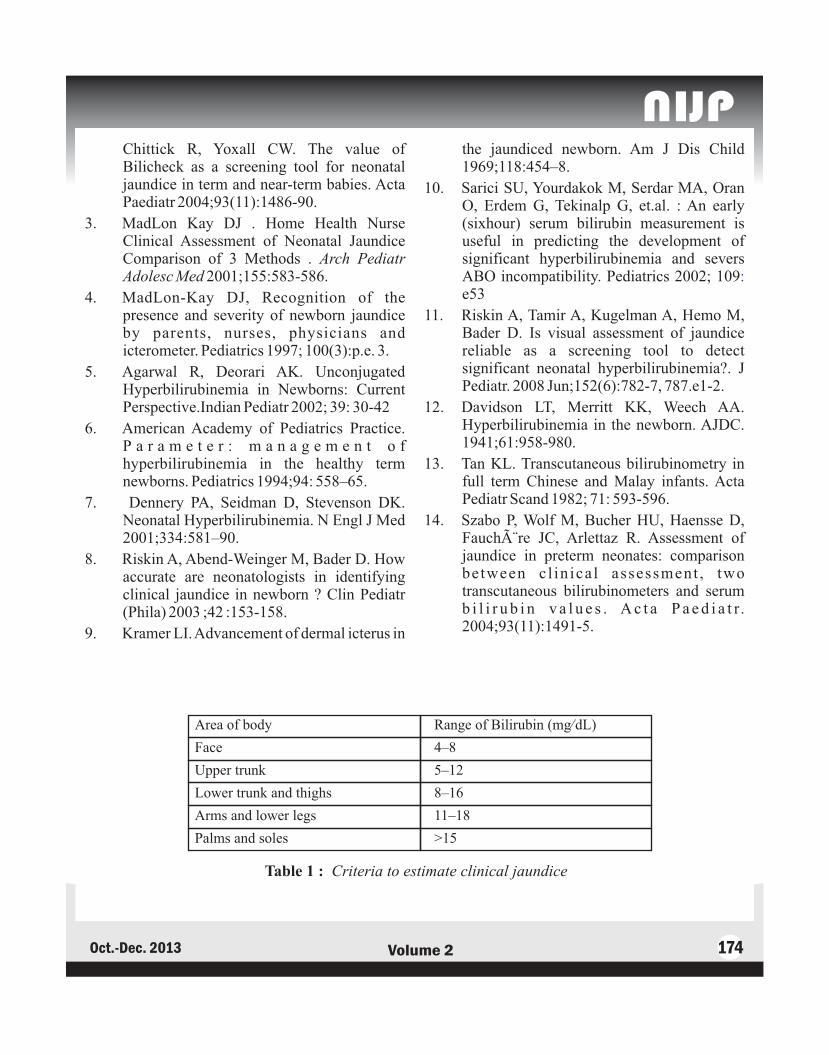
Chittick R, Yoxall CW. The value of Bilicheck as a screening tool for neonatal jaundice in term and near-term babies. Acta Paediatr 2004;93(11):1486-90.
3. MadLon Kay DJ . Home Health Nurse Clinical Assessment of Neonatal Jaundice Comparison of 3 Methods . Arch Pediatr Adolesc Med 2001;155:583-586.
4. MadLon-Kay DJ, Recognition of the presence and severity of newborn jaundice by parents, nurses, physicians and icterometer. Pediatrics 1997; 100(3):p.e. 3.
5. Agarwal R, Deorari AK. Unconjugated Hyperbilirubinemia in Newborns: Current Perspective.Indian Pediatr 2002; 39: 30-42
6. American Academy of Pediatrics Practice. P a r a m e t e r : m a n a g e m e n t o f hyperbilirubinemia in the healthy term newborns. Pediatrics 1994;94: 558–65.
7. Dennery PA, Seidman D, Stevenson DK. Neonatal Hyperbilirubinemia. N Engl J Med 2001;334:581–90.
8. Riskin A, Abend-Weinger M, Bader D. How accurate are neonatologists in identifying clinical jaundice in newborn ? Clin Pediatr (Phila) 2003 ;42 :153-158.
9. Kramer LI. Advancement of dermal icterus in
the jaundiced newborn. Am J Dis Child 1969;118:454–8.
10. Sarici SU, Yourdakok M, Serdar MA, Oran O, Erdem G, Tekinalp G, et.al. : An early (sixhour) serum bilirubin measurement is useful in predicting the development of significant hyperbilirubinemia and severs ABO incompatibility. Pediatrics 2002; 109e53
11. Riskin A, Tamir A, Kugelman A, Hemo M, Bader D. Is visual assessment of jaundice reliable as a screening tool to detect significant neonatal hyperbilirubinemia?. J Pediatr. 2008 Jun;152(6):782-7, 787.e1-2.
12. Davidson LT, Merritt KK, Weech AA. Hyperbilirubinemia in the newborn. AJDC. 1941;61:958-980.
13. Tan KL. Transcutaneous bilirubinometry in full term Chinese and Malay infants. Acta Pediatr Scand 1982; 71: 593-596.
14. Szabo P, Wolf M, Bucher HU, Haensse D, Fauchère JC, Arlettaz R. Assessment of jaundice in preterm neonates: comparison between c l in ica l assessment , two transcutaneous bilirubinometers and serum b i l i r u b i n v a l u e s . A c t a P a e d i a t r. 2004;93(11):1491-5.
:
NIJP
Volume 2 Oct.-Dec. 2013 174
Area of body Range of Bilirubin (mg/dL)
Face 4–8
Upper trunk 5–12
Lower trunk and thighs 8–16
Arms and lower legs 11–18
Palms and soles >15
Table 1 : Criteria to estimate clinical jaundice

NIJP
Volume 2 Oct.-Dec. 2013 175
Figure 2. ROC for predicting the s.bilirubin >12mg/dl
Figure 3. ROC for predicting the s.bilirubin >15mg/dl
Figure 1. ROC for predicting the s.bilirubin >10mg/dL

Face Upper trunk Lower trunk Arms and Palms andand Thighs lower legs Soles
Student vs Percent 90 77 78 81 86Nurse agreement
Kappa 0.14 0.49 0.54 0.53 0.40(95%CI) (0.14 - 0.41) (0.32-0.66) (0.38-0.71) (0.36-0.71) (0.16-0.64)
Student vs Percent 93 75 73 80 83Doctor agreement
Kappa -0.03 0.43 0.44 0.52 0.29(95%CI) (-0.06 to 0) (0.26-0.61) (0.26-0.62) (0.34-0.7) (0.05-0.53)
Nurse vs Percent 91 88 83 93 93Doctor agreement
Kappa 0.36 0.75 0.63 0.79 0.5(95%CI) (0.03 - 0.68) (0.62 -0.88) (0.47-0.79) (0.64-0.94) (0.17- 0.82)
Table 2. Percent Agreement between (Student, Nurse and Doctor ) observers for jaundice in infants
Reference standard predicted S. Sn* Sp* Correctly LR+* LR-*S.Bilirubin >15mg/dL Bilirubin by each Classified*
observer
Student ≥15 34.48% 94.12% 76.29% 5.8621 0.6961
Nurse ≥ 14 34.48% 98.36% 77.78% 21.0345 0.6661
Doctor ≥ 12 65.52% 94.03% 85.42% 10.9741 0.3667
Reference standardS.Bilirubin >12mg/dL
Student ≥ 9 63.27% 79.17% 71.13% 3.0367 0.464
Nurse ≥ 6 77.08% 69.05% 73.33% 2.4904 0.3319
Doctor ≥ 8 69.39% 82.98% 76.04% 4.0765 0.3689
Reference standardS.Bilirubin >10mg/dL
Student ≥ 7 83.61% 63.89% 76.29% 2.3153 0.2566
Nurse ≥ 5 90.00% 56.67% 78.89% 2.0769 0.1765
Doctor ≥ 5 93.55% 58.82% 81.25% 2.2719 0.1097
Best cut-off for
* These are estimates at the best predicted cut-off for each observer, Sn=Sensitivity; Sp=Specificity
Table 3. Prediction of hyperbilirubinaemia by doctor , nurse and student with cut off points, sensitivity, specificity and likelihood ratio.
NIJP
Volume 2 Oct.-Dec. 2013 176

S. Bilirubin >15mg/dL S. Bilirubin >12mg/dL S. Bilirubin >10mg/dL
Area Under curve P value Area Under P value Area Under P value(confidence curve (CI) curve (CI)Interval)
Doctor 0.87 (0.80-0.95) Reference 0.81 (0.72-0.89) Reference 0.86 (0.78-0.94) Reference
Nurse 0.80 (0.70-0.90) 0.11 0.78 (0.69-0.88) 0.70 0.81 (0.71-0.90) 0.54
Student 0.72 ( 0.60-0.83) 0.002 0.75 (0.65-0.85) 0.17 0.76(0.65-0.86) 0.04
Table 4 : The area under curve by roc for predicting the s.bilirubin at different levels
EeE
NIJP
Volume 2 Oct.-Dec. 2013 177

Key words : Gaucher disease, Lipid storage disorder, Glucocerebrosidase, Lysosomal storage disorder.Background :
Gaucher disease is an inherited metabolic disorder among lipid storage diseases. It is autosomal recessive in mode of inheritance. Gaucher's disease is caused by a deficiency of the enzyme glucocerebrosidase. Fatty materials can accumulate in the spleen, liver, lungs, bone marrow, and brain. Symptoms may include skeletal disorders, enlarged spleen and liver, liver malfunction, anemia, and yellow spots in the eyes. Following is a case of type 1 gaucher's disease -Clinical Presentation :
A 17 year old female patient, Miss X, a resident of Ranchi, Jharkhand, presented to us with the complaints of bone pain and fatigue. She had recurrent abdominal pain, which was never completely resolved even with medication.Past history:
The patient is born out of a non-consanguineous marriage. Her growth and development milestones were normal. Her parents and siblings are apparently healthy.
Her antenatal, natal and postnatal history was uneventful. She had been immunized with pneumococcal, meningococcal and haemophilus influenza vaccines in 2012.
She was apparently asymptomatic till the age of 8 months, after which she developed cough and fever which lasted for more than a week. She
was given anti-tubercular drug by a local physician but after 3 months of ATT, she developed jaundice and ATT was stopped.
Then gradually she developed abdominal distension, which started first on left side later involving whole of abdomen. This was associated with severe pain over right hypochondrium. As the pain grew unbearable they consulted another physician and ultimately Cholelithiasis was demonstrated. She was operated for gallstones in 2011 at CMC Vellore.
She was symptom-free for a few days but again had episodes of dull aching type of pain over left hypochondrium of abdomen, for which she was evaluated and diagnosed as multiple splenic abscesses. Splenectomy was done in 2012 at AIIMS.Clinical examination:
She is a well-built, well-nourished child with normal anthropometric measurements and normal vital signs. She does not have any spinal abnormality or joint contractures except for tenderness over lumbar spine and bilateral sacroiliac joint. Her general examination is within normal limit.
On systemic examination, we found that her respiratory and cardiovascular systems are normal. On examination of the abdomen, multiple scar marks of previous operative procedures were found. Liver was palpable, about 4 cm below right subcostal margin, which was firm, with a smooth surface, round margin and without any tenderness.
Case Report :Gaucher's diseaseDr Amar Verma*, Dr. Anita verma**, Dr. Vidhya Shankari*.
*Department of Pediatrics, Rajendra Institute of Medical Sciences, Ranchi, **Department of Pharmacology I.Q. Medical College, Durgapur, West Bengal E-mail: [email protected]
NIJP
Volume 2 Oct.-Dec. 2013 178

She does not have any other organomegaly. There is absence of free fluid in the abdomen.
On examination of the central nervous system, no abnormality was detected except for a slight diminution in recent memory. Her speech and gait are normal.Investigation:
1. The blood indices such as MCV, MCH, MCHC, RDW, TC, Hb%, Platelet, RBC, PCV, SGOT, SGPT, ALP, S.Urea, serum creatinine were within normal limits.
2. Serology for HIV, HbsAg, HCV is negative.3. Repeat examination for platelet count was
75000/cumm, Hb% was 9.9gm%. Repeat CBC was within normal limits.
4. The blood indices repeated again were within normal limits. Further reticulocyte count-3.04% and, Absolute eosinophil count-100/ cumm, PT -13.1 sec., INR-1.19, APTT-39.6,MCV-78.5 were almost within normal limits
5. A s s a y f o r l e u c o c y t e s b e t a -glucocerebrosidase enzyme - which was found to be 0.7nmol/hr/ml.[normal range:17-84 nmol/hr./ml] This is reduced in case of Gaucher's disease.
6. Further investigations for CRP-1.6 mg/l, Rheumatoid factor-<10.5 IU/ml, Anti CCP antibodies-59.1 U/ml. [normal range <15 U/ml]
7. The bone density scan of lumbar spine reported osteoporotic features with high fracture risk.
8. Assay for chitotriosidase level in plasma was 8495.3 nmol/hr/ml.[normal range:<76]. The low value of beta-glucocerebrosidase and high value of chitotriosidase in patient are consistent with the diagnosis of Gaucher disease.
9. The genetic analysis report of exon10 of GBA gene done on 3-9-2012 shows “homozygous p .L483P(c .1448T>c) mutation” [previously known as PL444P].
Case discussion:M a n y d i s e a s e s p r e s e n t w i t h
Hepatosplenomegaly. Malaria, Kala-zar, Thalassemia, sickle cell disease, leukaemia etc. are few common diseases found in Jharkhand and Bihar present with Hepatosplenomegaly along with other symptoms and signs specific for them. Most of these patients are diagnosed by their classical presentation and pathological finding without any difficulty. But around 5-8% patients with hepatosplenomegaly and nonspecific symptoms remain undiagnosed. Many times these conditions are assumed as tropical splenomegaly or other common condition like tuberculosis without any definite diagnostic findings. Even empirical treatment like for tuberculosis is initiated without any evidence. Doctors rarely thought about storage and other rare disorders due to lack of awareness or unavailability of diagnostic tools. In recent years with availability of diagnostic tools and increased awareness, acceptance of these conditions in differential diagnosis has increased. That's why we are now diagnosing these cases in our practice though they may have existed earlier also.
T h i s p a t i e n t p r e s e n t e d w i t h Hepatosplenomegaly and mult isystemic presentation especially involvement of skeletal system with pain in multiple bones and joints. Initially she was treated like common conditions present in this part of country like malaria or congenital hemolytic anemia or even tuberculosis. But later the possibility of storage disorder was suspected. Finally diagnosis was made by assay for leucocytes beta-glucocerebrosidase enzyme. It was possible to label the clinical condition of this child as gaucher's disease.
Gaucher's disease is a multisystemic l ipidosis characterized by hematologic abnormalities, organomegaly and skeletal involvement, the latter usually manifesting as bone pain and pathological fractures.(1) It is the most common lysosomal storage disease.
1. Type 1- adult , non-neuronopathic form
NIJP
Volume 2 Oct.-Dec. 2013 179

(accounts for 99% cases)2. Type 2- the infantile or acute neuronopathic
form 3. Type 3-juvenile or subacute neuronopathic
form.Gaucher's disease results from the deficient
activity of the lysosomal hydrolase, acid beta-glucosidase, located on chromosome 1q21-q31, leading to accumulation of undergraded glycolipid substrates, particularly glucosylceramide, in cells of the reticuloendothelial system. Four mutations – N370S, L444P, 84insG and IVS2 are commonly found.
Clinical manifestations of type-1 have a variable age at onset, from early childhood to late adulthood and patients may have bruising from thrombocytopenia, chronic fatigue secondary to anemia, hepatomegaly with or without elevated LFT, splenomegaly, bone pain and occasional pulmonary involvement. Clinically apparent bony involvement present as bone pain, a pseudo-os t eomye l i t i s pa t t e rn o r pa tho log ica l fractures.(1,2)
Clinical manifestation of type-2 is a rapid neurodegenerative course with extensive visceral involvement and death within first year of life. This usually presents in infancy with increased tone, strabismus, organomegaly, laryngospasm and failure to thrive.
Clinical manifestations of type-3 are intermediate between 1 and 2, with presentation in childhood and death by 10-15 yrs. Type-3a: progressive myotonia and dementia. Type-3b:isolated supranuclear gaze palsy.(3)
This adolescent girl presented to us with symptoms of bone pain [tenderness over the lumbar spine and sacroiliac joint], easy fatigability with previous history of jaundice. She has hepatomegaly at present. She was operated for multiple splenic abcesses by splenectomy earlier. She has no neurological abnormality except for a slight regression of recent memory. She has anemia and
thrombocytopenia. Her liver function tests are within normal limit. Her bone density scan is suggestive of osteoporotic findings. Assay for leucocytes beta-glucocerebrosidase enzyme - which was found to be 0.7 nmol/ hr/ml.[normal range : 17-84 ] This is reduced in case of Gaucher's disease.
Assay for chitotriosidase level in plasma d o n e o n t h e s a m e d a y w a s 8 4 9 5 . 3 nmol/hr/ml.[normal range:<76]. The low value of b-glucocerebrosidase and high value of chitotriosidase in patient are consistent with the diagnosis of Gaucher's disease.(4,5)
The genetic analysis report of exon10 of GBA gene done shows “homozygous p.L483P(c.1448T>c) mutation” [previously known as PL444P]. (6)
All above mentioned investigations are consistent with the diagnosis of Gaucher's disease. Her symptoms suggest type 1 gaucher's disease, which is commonest and milder form.
It may now be stressed that in any case of chronic hepatosplenomegaly (with or without fever) which is not responding to standard or empirical therapy and having multisystemic presentation, one must look for the possibility of storage disorders.
Now the patient is in need of enzyme replacement therapy but could not afford it. We expect that in near future, she will get her enzyme replacement through government aid.(7)Contributors :
VS was involved in search of literature and drafting the manuscript. AV and AV were involved in the management of the case and finalized the manuscript and will be the guarantor.Funding: NoneCompeting interests : None statedReferences:
1. Grabowski GA: Phenotype, diagnosis, and treatment of Gaucher's disease, Lancet 2008; 372:1263-1271.
2. Kaplan P, Anderson HC, Kacena KA,: The
NIJP
Volume 2 Oct.-Dec. 2013 180

clinical and demographic characteristics of nonneuronpathic Gaucher disease in 887 children at diagnosis, Arch Pediatr Adolesc Med 2006; 160:603-608.
3. Meikle PJ, Ranieri E, Simonsen H,: Newborn screening for lysosomal storage disorders: clinical evaluation of a two-tier strategy. Paediatrics 2004; 114:909-916.
4. Burtis CA, Ashwood ER, Bruns D, eds. Tietz, Textbook of clinical chemistry and molecular diagnostics. 4th edn. Philadelphia: WB Saunders; 2005
5. Pastores GM, Hughes DA: Gaucher disease.
G e n e R e v . A v a i l a b l e a t <http://www.ncbi.nlm.nih.gov/bookshelf/br.fcgi?book=gene&part=gaucher>2008 Accessed on 30 Sept.2013.
6 Ensenauer RE,: Primer on medical genomics. Part VIII: essentials of medical genetics for the practicing physician. Mayo Clin Proc 2003; 78:846
7 . Rimoin DL: Nature and frequency of genetic disease. In: Rimoin DL, et al ed. Emery and Rimoin's Principles and Practice of Medical Genetics, 3rd ed.. New York: Churchill Livingstone; 1997:32.
Figure1 -Report of Bone densitometry showing osteoporosis & High Fracture risk
Figure 2 : File photo of patient with her mother
EeE
NIJP
Volume 2 Oct.-Dec. 2013 181

Key words : Epidermolysis bullosa, Skin fragility, scarring, Skin biopsy.
Epidermolysis bullosa (EB)
According to the National EB registry project from USA, the incidence and prevalence of EB are estimated to be 19.60 per million live births and 8.22 per million population, respectively. The incidence and prevalence rates of EB simplex are 10.75 and 4.65, of junctional EB are 2.04 and 0.44, and dystrophic EB dominant type 2.86 and 0.99 and recessive dystrophic EB 2.04 and 0.92, respectively(1).
It is a group of congenital, hereditary blistering disorder that cause the skin to be very fragile and to blister easily, generally in warm weather. Blisters and skin erosions form in response to minor injury or friction, such as rubbing or scratching. It is a result of a defect in anchoring between the epidermis and dermis, resulting in friction induced skin fragility. Its severity ranges from mild to lethal.
The dreaded complications are bleeding ,infection, scarring and death.
Herewith we are presenting a classical case of Dystrophic epidermolysis bullosa (DEB).
Case Report:
A 5 yr old male child presented with drooling of saliva and refusing to feed since last 5 hours following ingestion of non-vegetarian food .Child had repeated episodes of similar problems and each episode took long time to correct itself.
On examination patient had blistering and scarring over oral mucosa. Multiple Erosions, hypo-
pigmented lesions were seen in extremities along with loss of nails and contractures, multiple digital fusions were present. Child had grade IV malnutrition . Segmental height was measured due to contractures and stunting was obvious. Vitals were stable.
Foreign body aspiration was ruled out, HRCT scan showed dilated esophagus just below the level of vocal cords and distended with air column within it at the level of C7-T1 disc space.
Transmission electron microscopy is useful for diagnosis but could not be done due to non-availability. Skin biopsy, electron microscopy and immuno- fluorescent microscopy are diagnostic.
Based upon clinical features diagnosis of autosomal recessive epidermolysis bullosa was made which is the most severe form.
Fig 1 : Dystrophic epidermolysis bullosa
Discussion
There are three main types of EB.(1) The condition is classified according to site of skin
Case Report :Epidermolysis bullosaDr Amit Thakur, Dr Bhavana Lakhkar
Department of Pediatrics, J. N. M. College, AVBR Hospital, Sawangi (M), Wardha, Maharashtra, India, 442004 E-mail: [email protected]
NIJP
Volume 2 Oct.-Dec. 2013 182

blistering in layers of skin.
.Epidermolysis bullosa simplex (EBS), involves the upper layer of the skin (the epidermis). This is the most common type of EB, accounting for 70% of cases, and tends to be milder than the other types.
* Junctional epidermolysis bullosa (JEB),
* Occurs at the junction between the epidermis and the dermis (lower layer of the skin) . Accounts for around 5% of cases .
* Dystrophic epidermolysis bullosa (DEB), where blistering occurs in the upper part of the dermis. DEB accounts for around 25% of cases. Based on severity 3 types of DEB have been described.
A u t o s o m a l r e c e s s i v e d y s t r o p h i c epidermolysis bullosa (DEB) is the most severe, classic form of the condition. Affected infants are typically born with widespread blistering and areas of missing skin, often caused by trauma during birth. Most often, blisters are present over the whole body and affect mucous membranes such as the moist lining of the mouth and digestive tract. As the blisters heal, they result in severe scarring. Scarring in the mouth and esophagus can make it difficult to chew and swallow food, leading to chronic malnutrition and slow growth. Additional complications of progressive scarring can include fusion of the fingers and toes, loss of fingernails and toenails, joint deformities (contractures) that restrict movement, and eye inflammation leading to vision loss. Additionally, young adults have a very high risk of developing a form of skin cancer called squamous cell carcinoma, which tends to be unusually aggressive and is often life-threatening. (2)
The overall prognosis is poor. Foods that traumatize the buccal or esophageal mucosa should be avoided. A semiliquid diet and esophageal dilatations may be required in case of esophageal scarring. Stricture excision or colonic interposition may be needed to relieve esophageal obstruction. In infants, severe oropharyngeal involvement may necessitate the use of special feeding devices such as a gastrostomy tube. Iron therapy for anemia, intermittent antibiotic therapy for secondary infections, which are a common cause of death, and periodic surgery for release of digits may reduce morbidity.(3) Tissue-engineered skin grafts and allogeneic bone marrow transplantation (4) may also be beneficial. Once mutations are identified in family, prenatal diagnosis is useful. Other two forms are mild. Mildest one involves only nails.
Bibliography
1) Fine JD, Eady RA, Bauer EA: The classification of inherited epidermolysis bullosa (EB): report of the Third International Consensus Meeting on Diagnosis and Classification of EB. J Am Acad Dermatol 2008; 58:931-950.
2) Sarkar R, Bansal S, Garg VK. Epidermolysis bullosa: Where do we stand?. Indian J Dermatol Venereol Leprol 2011;77:431-8.
3) Fine JD, Hall M, Weiner M : The risk of cardiomyopathy in inherited epidermolysis bullosa. Br J Dermatol 2008; 159:677-682.
4) Wagner JE, Ishida-Yamamoto A, McGrath JA : Bone marrow transplantation for recessive dystrophic epidermolysis bullosa. N Engl J Med 2010; 363(7):629-638
EeE
NIJP
Volume 2 Oct.-Dec. 2013 183

Can serum Tenascin- C be used as a marker of inflammation in patients with Dilated Cardiomyopathy?
Myocardial inflammatory diseases are an important cause of dilated cardiomyopathy (DCM) in children. Epidemics of viral myocarditis have been reported, particularly Coxsackie B virus, and the enteroviruses which are considered to be the most common cause of viral myocarditis.
There are few biomarkers for myocarditis. Troponin I has a high specificity for diagnosing myocarditis but a sensitivity of only 34%, and creatine kinase and its cardiac isoform CK-MB are less sensitive and specific than troponin.
Tenascin-C (TN-C) is an extracellular matrix glycoprotein that is not expressed in normal adult hearts but is expressed in various myocardial diseases such as acute myocarditis , dilated cardiomyopathy , myocardial infarction.
Speckletracking echocardiography (STE) is a noninvasive ultrasound imaging technique that allows for an objective and quantitative evaluation of global and regional myocardial function independently, study hypothesized that serum TN-C might serve as a marker of active myocardial inflammation in children with new onset DCM.
The study was done to evaluate the role of TN-C as a marker for active inflammation in children with dilated cardiomyopathy (DCM). 24 consecutive patients with primary nonfamilial DCM aged 6–72months (mean45.19±11.03) were divided into 2 groups: group I, twelve patients with acute onset DCM (<6 months duration), and group II, twelve patients with chronic DCM (>6 months duration), and compared to 20 healthy age- and sex-matched controls. Clinical diagnosis of DCM was based on the WHO and American Heart Association criteria. At the time of diagnosis all
patients had an ejection fraction <45% and/or a fractional shortening of <25% and a left ventricular end diastolic dimension (LVEDD) of >112% of the predicted value corrected for age and body surface area. Investigations included estimation of serum TN-C and echocardiographic evaluation using M-mode and 2D speckle tracking echocardiography (STE).
Statistical analysis was done ,results were Serum TN-C showed a higher significant statistical elevation among patients than controls p< 0.001) and in group I than group II ( p < 0.001). EF was significantly decreased, and LVEDD and EDV increased in patients than controls and in GI than GII. STE showed a statistically significant difference in global peak strain longitudinal (GPSL) average in patients than controls ( p < 0.05) and between GI and GII (p< 0.001). STE wall motion scoring showed normokinesia (33.5%), hypokinesia (8.33%), and akinesia (50%) in GI and hypokinesia (100%) in GII. There was a statistically significant positive correlation between serum TN-C and GPSL average.
Serum-TN-C levels are increased in children with DCM and can be used as a marker of inflammation in acute cases. Its persistence in chronic DCM might lead to progressive myocardial disease and ventricular dilation. STE is more sensitive than conventional echocardiography in the assessment of myocardial performance in DCM. Its values correlate with serum TN-C.Source: International Journal of Pediatrics Volume 2013, Article ID 608563, 6 pagesComment : Increased serum TN-C can be used as a marker of inflammation in DCM and is associated with the severity of heart failure and LV dysfunction as detected by STE. Drugs targeting the expression or function of tenascin-C might offer new
Media Watch / Around the WorldDr. Anil Lohar*, Dr. Satish Agrawal**
*Senior Resident, Dr.PDMC, Amravati. ** Practicing Pediatrician, Amravati. e-mail: [email protected]
NIJP
Volume 2 Oct.-Dec. 2013 184
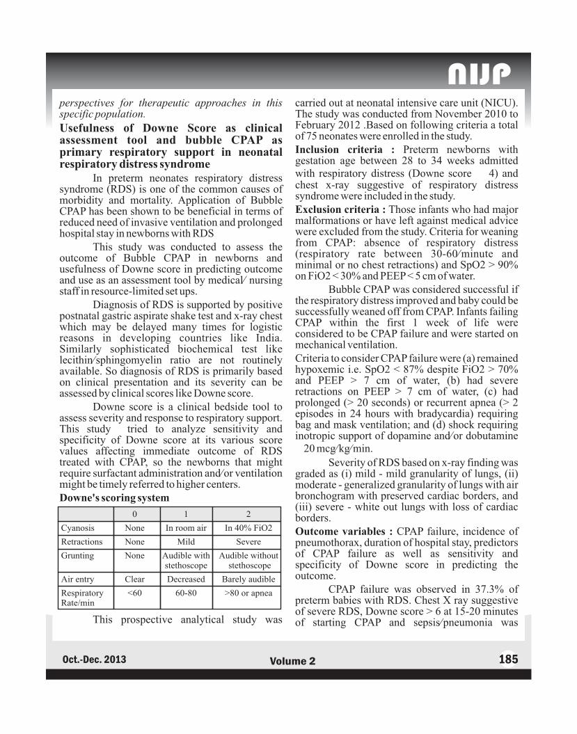
perspectives for therapeutic approaches in this specific population.
Usefulness of Downe Score as clinical assessment tool and bubble CPAP as primary respiratory support in neonatal respiratory distress syndrome
In preterm neonates respiratory distress syndrome (RDS) is one of the common causes of morbidity and mortality. Application of Bubble CPAP has been shown to be beneficial in terms of reduced need of invasive ventilation and prolonged hospital stay in newborns with RDS
This study was conducted to assess the outcome of Bubble CPAP in newborns and usefulness of Downe score in predicting outcome and use as an assessment tool by medical/ nursing staff in resource-limited set ups.
Diagnosis of RDS is supported by positive postnatal gastric aspirate shake test and x-ray chest which may be delayed many times for logistic reasons in developing countries like India. Similarly sophisticated biochemical test like lecithin/sphingomyelin ratio are not routinely available. So diagnosis of RDS is primarily based on clinical presentation and its severity can be assessed by clinical scores like Downe score.
Downe score is a clinical bedside tool to assess severity and response to respiratory support. This study tried to analyze sensitivity and specificity of Downe score at its various score values affecting immediate outcome of RDS treated with CPAP, so the newborns that might require surfactant administration and/or ventilation might be timely referred to higher centers.Downe's scoring system
This prospective analytical study was
carried out at neonatal intensive care unit (NICU). The study was conducted from November 2010 to February 2012 .Based on following criteria a total of 75 neonates were enrolled in the study.Inclusion criteria : Preterm newborns with gestation age between 28 to 34 weeks admitted with respiratory distress (Downe score ≥ 4) and chest x-ray suggestive of respiratory distress syndrome were included in the study.Exclusion criteria : Those infants who had major malformations or have left against medical advice were excluded from the study. Criteria for weaning from CPAP: absence of respiratory distress (respiratory rate between 30-60/minute and minimal or no chest retractions) and SpO2 > 90% on FiO2 < 30% and PEEP < 5 cm of water.
Bubble CPAP was considered successful if the respiratory distress improved and baby could be successfully weaned off from CPAP. Infants failing CPAP within the first 1 week of life were considered to be CPAP failure and were started on mechanical ventilation. Criteria to consider CPAP failure were (a) remained hypoxemic i.e. SpO2 < 87% despite FiO2 > 70% and PEEP > 7 cm of water, (b) had severe retractions on PEEP > 7 cm of water, (c) had prolonged (> 20 seconds) or recurrent apnea (> 2 episodes in 24 hours with bradycardia) requiring bag and mask ventilation; and (d) shock requiring inotropic support of dopamine and/or dobutamine ≥ 20 mcg/kg/min.
Severity of RDS based on x-ray finding was graded as (i) mild - mild granularity of lungs, (ii) moderate - generalized granularity of lungs with air bronchogram with preserved cardiac borders, and (iii) severe - white out lungs with loss of cardiac borders.Outcome variables : CPAP failure, incidence of pneumothorax, duration of hospital stay, predictors of CPAP failure as well as sensitivity and specificity of Downe score in predicting the outcome.
CPAP failure was observed in 37.3% of preterm babies with RDS. Chest X ray suggestive of severe RDS, Downe score > 6 at 15-20 minutes of starting CPAP and sepsis/pneumonia was
NIJP
Volume 2 Oct.-Dec. 2013 185

significantly associated with CPAP failure and also were independent predictors of outcome. Area under curve (AUC) for Downe score at 15-20 minutes of starting CPAP was 78.5% (95% CI = 67.9 to 89.1)
Study concluded that Bubble CPAP for RDS is effective and safe mode of treating mild to moderate RDS. Nearly 37.3% newborns with RDS failed CPAP. Chest x-ray suggestive of severe RDS, Downe score >6 at 15-20 minutes of starting CPAP and sepsis/pneumonia were associated with CPAP failure and also independent predictors of outcome. Downe score can be used at periphery to monitor response and to decide about referral in absence of sophisticated tests by the medical and paramedical nursing staff.Source: Journal of Pediatric Sciences. 2013;5(1):e176Comment : Bubble CPAP was found to be effective and safe mode of treating mild to moderate RDS and Downe score can be used at periphery to monitor response and to decide about referral in absence of sophisticated tests.
Propranolol treatment for severe infantile hemangiomas: a single-centre 3-year experience
Infantile hemangiomas( IHs) are common benign tumours of infancy, characterized by a phase of rapid growth within the first months of life, followed by a variable involution phase over the next several months to years. Because of the benign natural course of hemangiomas, a wait-and-see policy seems reasonable, and therapeutic intervention is required in approximately 10% of patients with problematic infantile hemangioma.
Systemic corticosteroids, mostly oral prednisolone, had been the mainstay of treatment for severe IHs for many years with good but whatsoever variable results. More elevated daily doses yielded more satisfactory results but have also been proved to result in more serious side effects.
Undoubted ly, the mos t exc i t ing development in the treatment for IHs over the last 3
years has been the serendipitous discovery of propranolol's inhibitory effects during the proliferative phase of the hemangioma cycle.
This study was conducted to evaluate the effectiveness, safety and tolerability of propranolol as single agent treatment in patients with problematic, proliferative-phase, infantile hemangiomas (IHs).
Twenty-eight patients admitted in our department between January 2009 and December 2011 with problematic IH were included in the study. Patients were enrolled in the study if they had: (i) problematic IHs, defined as hemangiomas impairing vital or sensory functions and causing local complications as ulceration, bleeding, local infection or disfigurement, and additionally; (ii) negative history of asthma or recent episode of wheezing, hypoglycaemia, cardiovascular disorders contraindicating beta-blocker use, impaired renal function and neurological disorders. Cardiologic evaluation was performed before treatment initiation. Hemodynamic variables and blood glucose levels were monitored during the first 24 h of treatment, while the children were hospitalized. Clinical response and tolerance were assessed every month, along with photographic documentation.. Propranolol was started at a dose of 2 mg / kg / day divided in two doses, and HR and BP were monitored every hour for 6 h following each dose.
Blood glucose was tested by finger sampling 1 h after the ingestion of the medicament. Short-term efficacy was evaluated upon changes in texture, colour and size within the first month of treatment. Therapy was continued until the macroscopic regression of the lesions or until a point where no further improvement was noted for over a month and was then tapered over a period of 2 weeks. We considered patients to have complete response if their lesions regressed over 90% of their initial size.
Twenty-four patients completed treatment after a mean duration of 7.56 months, and their hemangiomas were successfully regressed. Propranolol was administered again, with satisfactory results, in three patients (12.5%) because of hemangioma regrowth.
NIJP
Volume 2 Oct.-Dec. 2013 186

A growing number of case reports and several retrospective case series support the efficacy of propranolol for the treatment of hemangiomas. This study contributes to the enforcement of the opinion that propranolol could be used as first-line treatment for IHs management and adds further data on establishing a safe and effective treatment protocol.
So propranolol, as first-line treatment, yielded excellent results with very good clinical tolerance and also seems to be effective in relapses. Source: Acta Paediatrica 2012 , 101, pp. e469–e474Comment : Further studies are required with large number of patients to reach the conclusion that propranolol could be used as first-line treatment for Infantile hemangioma management.
Flow Cytometry in the Detection of Neonatal Sepsis
Neonatal sepsis remains a burden problem by showing minimal initial symptoms of subtle character, nonspecific manifestation, and diagnostic pitfalls. The clinical course can be fulminant and fatal if treatment is not commenced promptly. It is therefore crucial to establish early diagnosis and initiate adequate therapy.
Besides clinical symptoms, the most reliable laboratory markers in establishing diagnosis is currently the combined measurement of CRP and a cytokine (IL-6 and IL-8).
Due to their different kinetics, a diagnostic gap might occur and thus withholding antimicrobial therapy in clinical suspicion of infection is not acceptable. We therefore need parameters which unerringly differentiate between infants in need for antimicrobial therapy and those who are not.
With a delayed start of antimicrobial treatment, the fulminant course of sepsis may lead to major sequelae. Therefore, the demand for laboratory markers in the detection of sepsis is high.
Despite diagnostic improvements in neonatal sepsis over the past decades, flow cytometry remains a confidential diagnostic tool. Advanced flow cytometry is undeniably the best tool for analyzing signaling processes, proliferation and differentiation, cell-cell interactions, surface markers, intracellular molecules, and proteins secreted by cells.
A number of cell surface markers have been studied and seem eligible for diagnostic use: a Pubmed search was performed with the keywords “neonatal sepsis” + “CD64”, or “CD11b”, or “HLA-DR”; studies since 2002 were accepted. Most studies show weak or even a lack of differentiation of gestational age, birth weight, and/or onset of sepsis, respectively, and thus, were excluded.
In the end, we included 5 of 16 studies for reviewing and estimating sensitivity, specificity, positive, and negative predictive values of CD64 and 4/5 for CD11b.
Nevertheless, none of the parameters presented is able to be the one parameter in diagnosis of early neonatal sepsis. So far, the combination of CRP and IL-6 remains the diagnostic resource of choice in detection of Early onset neonatal septisemia and Late onset neonatal septisemia. Solely CD64 seems to have the potential to complement the existing combination of CRP and a cytokine (IL-6 or IL-8) to increase sensitivity up to 100%.Source : International Journal of Pediatrics Volume 2013, Article ID 763191, 6 pages Comment : Flow cytometry promises to be a useful tool in this field, allowing the determination of different cellular, dissolved, and functional pathophysiological components of sepsis. Despite technical and methodical advances in flow cytometry , its use in clinical routine is still limited. Larger trials to define standard measurement protocols and reference values are highly desirable.
NIJP
Volume 2 Oct.-Dec. 2013 187
EeE

AAnimal bite 158Anti Rabies vaccine 158
BBilirubin serum 169BMI 119Breastfeeding, 151
CCalcification Intracranial 36Calcium Metabolism 36Community Sindhi 69Clinical assessment 169Clinical trials 50Conflict of interest 4Crigglar Najjar Syndrome 39
DDehydration, 15Diarrhea, 15
EEctodermal dysplasia 73Hypohidrotic 126Emphysema subcutaneous 33Enteritis Hemorrhagic 108Ischemic 108Necrotizing 108 Epidermolysis bullosa 182
GGaucher's disease 178Glucocerebrosidase 178Glucoronyl transferase 39Granulomatous diseases 28
HHelminthic infestations 62Hemiparesis 79Hemoglobin D 69Hemoglobinopathy 69Hepatic manifestations 9Hydrocephalus 77Hyperbilirubinemia indirect 39, 169Hypodontia 73Hypoparathyroidism 36Hypotrichosis 73
IImmunization 55India incredible 98Intestinal obstruction 130
JJejunitis necrotizing 108
KKernicterus 169Knowledge 55
LLactation delayed 151Lactogenesis II 151Lipid storage disorders 178Lipodystrophy 134Lymphadenopathy 28Lysosomal storage disease 178
MMalnutrition, 119Mesentric cyst 130Mid arm circumference 119Milk bank human 148
Subject IndexNew Indian Journal of Pediatrics
Volume 2, January-December, 2013
NIJP
Volume 2 Oct.-Dec. 2013 188

NNeonatal screening 102Neurologic manifestations 9
O ORS 15
PPerception 55Pneumocephalus 77Pneumomediastinum 33Pneumothorax 33Polypharmacy 15Prelacteal feeds 151Prescription audit 15
SSarcoidosis 28Scarring, 182Schizencephaly 79Seizures nonfebrile 163Unprovoked 163Sickle cell 102Sign Mount Fuji 77Skin Biopsy 182Skin fragility 182Socio-demographic profile 158Stool yellow tinge 151Syndrome Berardinelli – Seip 134
TThallassemia beta 69Trap sequence 82Tuberculoma 163Twin Acardiac 82Dichorionic 82Monochorionic 82
UUnder five children 62
VVaccination status 55Ventilation 33
Ventricular tap 77
WWilson disease 9Wound toilet 158
ZZinc 15
Dr A KutheDr Akash BangDr Amar Taksande, Dr Amar VarmaDr A P DubeyDr Balraj Yadav, Dr Dipty JainDr G Sarangi, Dr Jayant ShahDr Jayant Vagha Dr Kanya Mukhopadhyay Dr N C MohantyDr Nilofer MujawarDr Pushpa Chaturvedi Dr Pratibha KaleDr Pushpa Junghare, Dr Sandhya KhadseDr Sanjay AgrawalDr Santosh PrabhuDr Satish C Agrawal (Bareilly)Dr Satish Agrawal (Amravati)Dr Satish TiwariDr Sudhir MishraDr Vishesh KumarDr Yogesh Zawar
List of Reviewer
NIJP
Volume 2 Oct.-Dec. 2013 189

AAchouba Th 62Agrawal R K 4, 50, 98, 148Agrawal Satish 43, 85, 137, 184Agrawal Satish C 9, Agrawal G 134Akre Charu 157Akoijam B 55Amita M 73, Anita Kumari 9,Anthopia Devi N 126
BBajaj AP 69Banapurmath CR 151Barik K L 36, 134Bhagat P 15Boricha BG 82
CChaki B 134Chaudhuri N 36, 134Chaudhuri R 79Chaudhuri M S 82Chaudhuri S 62Chauhan Varsha 79,Chhajer CM 4, 50Chourjit Singh K 73
DDamke S 77Dandge VP 39Das S 28Dasgupta M 28
De S 33, 36, 134Deshmukh AT 69Deshmukh Y 15
EEdbor A 39
G Gavhane J 163Gayathri A 151Golmei A 62Golmei N 130Gondale G 169Ghosh TN 33Girish M 119Guha D 28
HHarwani AT 69
K Kadam N 15, 163Kalappanavar NK 151Kamale V 163Karki S 15Khan Sarosh 39Konjengbam S 55Kulkarni S 102, Kumar P 36, 134
LLaishram J 55Laishram R 130Lakhkar B 182
Author IndexNew Indian Journal of Pediatrics
Volume 2, January-December, 2013
NIJP
Volume 2 Oct.-Dec. 2013 190

Lohar A 43, 85, 137, 184
MMangi Ch 130Mohanty Nimain 15, 82, 163Mohanty Ray 15Mohapatra S 108Mujawar N 119Mukhia Salona 62
NNagpal A 77Nayek K 28Nimbhorkar S 39
OOpendra S 130
PPande Sushma 157Pande Sanket 157Pati S 33, 36Patil SK 82Patra C 28Pazare P 39
RRamola Devi 55Ramola Pukhrambam 62Ranbir Singh L 73Ranjan RK 62Rupbati Devi 73
SSanayaima Devi H 55Sarangi G 108, Sarkar S 28Satpute P 119Shankari V 178Shyamsunder Ch 130Singh KS 126Singh M 79
Singh RL 55Singh WR 126Shrivastav M 79Soni RR 69Subhashchandra S 130Sukhsohale ND 157Suman RK 15Sunilbala K 73
T Taksande A 169,Thakre AS 69Thakre S 157Thakur A 182Thakur GSP 77Thamke R 163Tiwari Satish 4, 50, 98, 148
VVagha Jayant 77, 102Verma Amar 178 Verma Anita 178 Vilhekar K Y 79, 169
WWanjari M 119
YYumkham R 126
ZZaheeruddin Mohammad 151
NIJP
Volume 2 Oct.-Dec. 2013 191

INDIAN MEDICO-LEGAL & ETHICS ASSOCIATIONMembership Form
Name of the applicant:…….....................................……………...............................……………………………………...............
(Surname) (First name) (Middle name)
Date of Birth:…………….......……………........……. Sex: …………………......
Address for Correspondence:…………………………...............................……………………………………...……........................
………………………………………………………………………………………………................................………...................................
Telephone No:- Resi. ................................................................ Hosp:..................................................
Mobile:……………….......................……………….. Other:…………………………………….......
Fax:….......……………………….. E-mail …………………………………………………………….......................................................
Registration no. ……………………………………. Date of Reg. ………………………………....................................
Name of the Council (MCI/Dental/Homeo/Ayurvedic/ Others: ..................................................................................
Medical/legal qualification University Year
Experience in legal field (if any) ...................................................................................................................................
Name, membership no. & signature of proposer :.........................................................................................................
Name, membership no & signature of seconder :..........................................................................................................
A) Was / Is there any med-legal case against you /your Hospital : Yes / No
If, Yes (Give details) .........................................................................................
(Attach separate sheet)....................................................................................
B) Do you have a Professional Indemnity Policy: Yes / No
Name of the Company:............................................................... Amount:
C) Do you have Hospital Insurance: Yes / No
Name of the Company:............................................................... Amount:
D) Do you have Risk Management Policy: Yes / No
Name of the company: ............................................................... Amount:
E) Is your relative / friend practicing Law: Yes / No
If Yes, Name:............................................................................. Qualification:............................................
Place of Practice:....................................................................... Specialization:...........................................
F) Any other information you would like to share: (Please attach the details)
I hereby declare that above information is correct. I shall be responsible for any incorrect / fraudulent declarations.
Place:............................................
Date: ............................................ (signature of applicant)
Enclosures: Degree & Registration Certificates
Life Membership fee (individual 2500/-, couple 4000/-) by CBS (At Par, Multicity Cheque) or DD, in the name of Indian medico-legal & ethics association (IMLEA) payable at Amravati. Please send the Cheque/DD to Dr. Satish Tiwari, Yashodanagar No.2 Amravati 444606.
NIJP
Volume 2 Oct.-Dec. 2013 192

NIJP
Volume 2 Oct.-Dec. 2013 193
Fig 1 : Dystrophic epidermolysis bullosa (Pg.No.182)
Fig.1 -Report of Bone densitometry showing osteoporosis & High Fracture risk
(Pg.No.181)
Fig.2 : file photo of patient with her mother (Pg.No.181)

Professional AssistanceScheme
Professional AssistanceScheme
Professional AssistanceScheme
Professional AssistanceScheme
Human Medico-Legal Consultants (P) Ltd.Office
9/3, KADAMBARI APTS., UJJWAL NAGAR,WARDHA ROAD, NAGPUR - 440025, MAHARASHTRA, INDIA.
For further details contact :
08882006159 08879442026 0721254125209422857204
Ms. Ruchita Shukla Sh. Piyush Dwivedi Dr. Satish Tiwari
Special Features :B Professional Indemnity for individual as well as hospital insurance,
B In Collaboration with recognized insurance companies,
B Competitive charges,
B Special discounts for scheme extending more than one year,
B Special discounts for couples, hospital (in future),
B Services of distinguished medico-legal experts across the country,
B Services for all branches, specialties,
B Services of crisis management committee at the city / district level,
B PREFERABLY FOR THE MEMBERS OF IMLEA, IAP & PAI.