Embed Size (px)
Citation preview
Page 1/37
Analysis of BMP3 Variants In The Causality ofOcular ColobomaSabrina Fox
University of Alberta https://orcid.org/0000-0003-1534-2006Sonya A. Widen
University of AlbertaMika Asai-Coakwell
University of SaskatchewanSerhiy Havrylov
University of AlbertaMatthew Benson
University of AlbertaLisa B. Prichard
MacEwan UniversityPranidhi Baddam
University of AlbertaDaniel Graf
University of AlbertaOrdan J. Lehmann
University of AlbertaAndrew J. Waskiewicz ( [email protected] )
University of Alberta
Research Article
Keywords: Coloboma, ophthalmology, Ocular development, periocular mesenchyme, zebra�sh, BMP, TGF-β
Posted Date: September 22nd, 2021
DOI: https://doi.org/10.21203/rs.3.rs-876946/v1
License: This work is licensed under a Creative Commons Attribution 4.0 International License. Read Full License
Page 2/37
AbstractColoboma, a congenital disorder characterized by gaps in ocular tissues, is caused when the choroid�ssure fails to close during embryonic development. Several loci have been associated with coloboma,but these represent less than 40% of those that are involved with this disease. Here, we describe a novelcoloboma-causing locus, BMP3. Whole exome sequencing and Sanger sequencing of patients withcoloboma identi�ed three variants in BMP3, two of which are predicted to be disease causing. Consistentwith this, bmp3 mutant zebra�sh have aberrant �ssure closure. bmp3 is expressed in the ventral headmesenchyme and regulates phosphorylated Smad3 in a population of cells adjacent to the choroid�ssure. Furthermore, mutations in bmp3 sensitize embryos to Smad3 inhibitor treatment resulting in openchoroid �ssures. Micro CT scans and Alcian blue staining of zebra�sh demonstrate that mutations inbmp3 cause midface hypoplasia, suggesting that bmp3 regulates cranial neural crest cells. Consistentwith this, we see active Smad3 in a population of periocular neural crest cells, and bmp3 mutantzebra�sh have reduced neural crest cells in the choroid �ssure. Taken together, this data suggests thatBmp3 controls Smad3 phosphorylation in neural crest cells to regulate early craniofacial and oculardevelopment.
IntroductionVertebrate ocular development is a deeply conserved and highly coordinated developmental process. Assuch, even minor perturbations can lead to ocular malformations which, in many cases, are blinding.During gastrulation, retinal tissues in the eye �eld evaginate to form bilateral optic vesicles (Zuber et al.2003). These vesicles migrate laterally until they contact the overlying ectoderm and induce lensformation. Subsequent coordinated invagination of the vesicles forms the bi-layered structure of the opticcup, with the lens-facing layer forming the neural retina and the lens-averted layer becoming the retinalpigmented epithelium (Schmitt and Dowling 1994; Kagiyama et al. 2005; Kwan et al. 2012). During opticcup invagination, a transient opening forms on the ventral side of the optic cup known as the optic orchoroid �ssure (Schmitt and Dowling 1994; Kwan et al. 2012). The choroid �ssure acts as a conduit forthe early embryonic ocular vasculature and optic nerve, and closure of the choroid �ssure is necessary fornormal ocular development (Schmitt and Dowling 1994). Failure of choroid �ssure closure results in gapsin ocular tissues such as the iris and retina (Shah et al. 2012). In humans, this congenital ocularmalformation is referred to as coloboma and is present in 2-19 of 100,000 live births (Williamson andFitzPatrick 2014; ALSomiry et al. 2019; Patel and Sowden 2019; Yoon et al. 2020). Coloboma and itsrelated disorders microphthalmia (reduced ocular size) and anophthalmia (absence of one or both eyes),frequently described as MAC, account for up to 11% of cases of pediatric blindness (Yoon et al. 2020).
Although coloboma can be caused by changes to environmental factors during gestation, it is primarilycaused by mutations in genes necessary for ocular development (Williamson and FitzPatrick 2014;ALSomiry et al. 2019; Patel and Sowden 2019; Yoon et al. 2020). To date, more than 40 coloboma-causing loci have been identi�ed (ALSomiry et al. 2019; Yoon et al. 2020). These genes have wellcharacterized roles in critical developmental processes essential to choroid �ssure closure, including axial
Page 3/37
patterning of the optic cup (proximal-distal, nasal-temporal and dorsal-ventral), cell movement and shapechanges, cell adhesion, extracellular matrix remodeling, and apoptosis (ALSomiry et al. 2019; Yoon et al.2020). Despite the substantial genetic heterogeneity, the majority of patients with coloboma do not havemutations in any known loci (Yoon et al. 2020). In addition, coloboma exhibits highly variable inheritance,with instances of coloboma pedigrees that display incomplete penetrance and reduced expressivity,suggesting that modi�er loci, environmental factors, and stochastic developmental events may confoundthe identi�cation of disease-causing loci. This is further complicated by marked phenotypic heterogeneitywith a diverse spectrum of severity, involvement of distinct ocular structures, and frequent unilateraldisease (Morrison et al. 2002; Hornby et al. 2003; Shah et al. 2012; Prokudin et al. 2014).
Of the loci that regulate choroid �ssure closure, a signi�cant proportion belong to the TransformingGrowth Factor Beta (TGF-b) superfamily of signaling ligands (Williamson and FitzPatrick 2014; ALSomiryet al. 2019; Patel and Sowden 2019; Yoon et al. 2020). All TGF-b superfamily members contain threecentralized disul�de bonds termed a “cysteine knot”, as well as an additional disul�de bond that allowsfor homo- or heterodimerization of ligands (Goebel et al. 2019). This cysteine knot structure andadditional disul�de bond are essential for the secretion, stability, and function of these ligands, and lossof this structure severely impairs the activity of these ligands (Goebel et al. 2019). Binding of theextensive family of TGF-b ligand dimers to a smaller cohort of tetrameric complex of Type I and Type IIreceptors triggers the phosphorylation of receptor-associated Sma- and Mad-related (r-Smad) proteins(Wrana et al. 1992; Bassing et al. 1994; Zhang et al. 1996). Phosphorylated r-Smad recruits co-Smad4,and this complex translocates to the nucleus, where it regulates target gene expression (Liu et al. 1996).TGF-b ligands can be divided into two groups: the Bone Morphogenetic Protein (BMP) subgroup, whichtypically trigger the phosphorylation of Smad1/5/8, and the Activin/Nodal/TGF-b subgroup, whichpredominantly induce the phosphorylation of Smad2/3 (Graff et al. 1996).
Of the two groups of TGF-b ligands, BMPs have been studied most extensively in ocular development anddisease. Mutation of BMP pathway members (including ligands, antagonists, co-receptors, receptors andSmads) in patients and/or model organisms contributes to MAC, and mutations in these pathwaycomponents are also associated with related phenotypes such as orofacial clefts, craniofacial disorders,and neural tube closure defects (Sakuta et al. 2001; Hanel and Hensey 2006; Morcillo et al. 2006; Asai-Coakwell et al. 2007, 2009; Bakrania et al. 2008; French et al. 2009; Suzuki et al. 2009; Ye et al. 2010;Wyatt et al. 2010; Abouzeid et al. 2011; Reis et al. 2011; Okada et al. 2011; Zhang et al. 2013; Beleggia etal. 2015; P�rrmann et al. 2015; Yan et al. 2020). BMP ligands are required for ocular cell survival,maintenance of stem cell pools, migration of ocular precursors, and axial patterning of the developingretina (Peters and Cepko 2002; Adler and Belecky-Adams 2002; Morcillo et al. 2006; Asai-Coakwell et al.2007). Yet, despite intensive investigation, a clear molecular mechanism for how these ligands regulate�ssure closure or a plausible explanation for complex patterns of inheritance for genes encoding thesepathway components has yet to be deduced.
Early eye development is dependent not only on the intrinsic population of optic cup neurectoderm cells,but also on a population of extraocular cells that surround the developing eye cup known as the
Page 4/37
periocular mesenchyme (POM). These cells, which are principally derived from neural crest and cranialmesoderm, migrate over the eye cup and through the choroid �ssure and contribute to many structures ofthe anterior segment, including the iris stroma, the cornea, and components of the intraocular drainagesystem (Creuzet et al. 2003; Gage et al. 2005; Langenberg et al. 2008). POM cells have also beenimplicated in choroid �ssure closure. Notably, neural crest cells and mesodermally-derived endothelialcells are present in the �ssure at times that are critical for choroid �ssure closure (James et al. 2016;Bernstein et al. 2018; Gestri et al. 2018). Embryos with no appreciable neural crest contribution have openchoroid �ssures, suggesting that neural crest cells are necessary for closure of the choroid �ssure (Gestriet al. 2018; Bryan et al. 2020). Additionally, mutations in transcription factors and signaling moleculesassociated with neural crest function, including BMP7, ZIC2, FOXC1, LMX1B, ALX1, and TFAP2A, havebeen shown to cause coloboma, further implicating this population of cells in choroid �ssure closure(Morcillo et al. 2006; McMahon et al. 2009; Skarie and Link 2009; Gestri et al. 2009; Lupo et al. 2011; Deeet al. 2012; Sedykh et al. 2017).
Here, we characterize a novel coloboma causing locus that was identi�ed by exome sequencing a three-generation pedigree of patients with autosomal dominant coloboma and microphthalmia. Additionalvariants in BMP3 were identi�ed in unrelated MAC patients using Sanger sequencing. Use of in-silicopredictions and protein secretion assays provided the �rst evidence that variants have deleterious effectsto BMP3 protein function. Zebra�sh CRISPR-generated bmp3 mutants display both delayed choroid�ssure closure and altered jaw development. This prompted us to test the hypothesis that Bmp3functions in periocular cells to regulate neural crest cell behavior. We demonstrated that bmp3 mutantsdisplay marked reduced Smad phosphorylation in periocular neural crest cells. Furthermore, overallmigration of periocular neural crest cells is dramatically reduced in bmp3 mutants. Our data thus providethe �rst evidence of a contribution of BMP3 to coloboma and highlight the importance of signalingpathways in regulating the function of periocular neural crest during ocular development.
ResultsPatients with coloboma and/or microphthalmia have mutations in BMP3
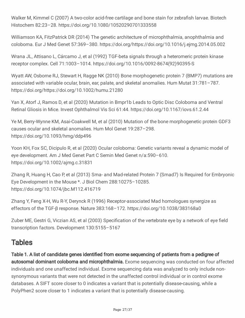
To reveal the genetic cause of coloboma in archival samples, we �rst performed whole exomesequencing (WES) on samples from four individuals affected by autosomal dominant coloboma and oneunaffected control individual from a three-generation European pedigree (Figure 1A). Identi�ed variantswere �ltered using standard criteria (Beaulieu et al. 2014). Comparison to SNP databases (allelefrequency < 1%), in-silico prediction algorithms (MutationTaster, SIFT and PolyPhen), and ocularexpression patterns were used to identify rare, potentially damaging variants within genes expressedduring ocular morphogenesis (Figure 1A). This strategy identi�ed 10 variants present in the affectedindividual’s samples (II:2, III:6, III:1, and IV:4) and absent in the unaffected individual’s sample (IV:5), ofwhich the point mutations (c.1408G>C) in BMP3 represented the most promising candidate pathogenicvariant (Table 1). Notably, this variant exhibited Mendelian segregation in the larger pedigree, is absentfrom local control DNA samples, and is absent from the National Heart, Lung and Blood Institute, 1000
Page 5/37
Genomes, and gnomAD databases (total of 87,888 exomes and genomes) (Table 2). In addition to thisBMP3 variant, the variant present in CRYAA was also an attractive candidate. CRYAA encodes Crystallinalpha A, a protein that confers light-focusing properties to the lens (Brady et al. 1997). Mutations inCRYAA cause congenital cataracts with high penetrance (Litt et al. 1998; Pras et al. 2000; Mackay et al.2003; Richter et al. 2008; Laurie et al. 2013). A small number of patients with CRYAA mutations and MACbeen reported, but most patients with CRYAA do not have any features of MAC (Beby et al. 2007; Sun etal. 2017). Such patients always present with congenital cataracts, and the coloboma present in thesepatients is predicted to be secondarily caused by defects of the lens (Sun et al. 2017). Since none of thepatients in the pedigree from Figure 1 presented with cataracts, we reasoned that the CRYAA variantpresent in the family from Figure 1 is likely non-pathogenic. Therefore, the variant in BMP3 still representsthe most plausible variant for causing disease in this family. This mutation alters a residue (p.Ala470Pro(A470P)) that is located between cysteines that mediate the disul�de bonding necessary to form thecysteine knot structure that is essential for TGF-b ligand stability (Figure S1). This residue is also highlyconserved among vertebrate species, further suggesting that it is necessary for the stability and/orfunction of the mature BMP3 protein (Figure S1). Collectively, this strongly indicates that changes to thisresidue will deleteriously impact protein function. Given that numerous TGF-b superfamily members haveessential roles in ocular development and mutations in multiple TGF-b paralogs cause coloboma, weconcluded that this BMP3 variant represented the most plausible pathogenic variant of those identi�ed inthis family (Lo et al. 1998; Asai-Coakwell et al. 2007; Bakrania et al. 2008; Ye et al. 2010; Abouzeid et al.2011).
Additional BMP3 variants are present in an unrelated cohort of MAC patients.
To determine whether this exceedingly rare mutation indicates that BMP3 might contribute to coloboma,we searched for additional BMP3 mutations by conducting Sanger sequencing of BMP3 in patientsunrelated to the pedigree family. DNA samples from a cohort of individuals with microphthalmia,anophthalmia and coloboma phenotypes were collected over the past 20 years through national andinternational collaboration. Sanger sequencing of 154 samples identi�ed two additional point mutationsin BMP3 that cause single amino acid substitutions in the mature TGF-b domain of the BMP3 protein(c.1178C>T (p.Ser393Phe) and c.1349T>A (p.Phe450Tyr)) (Figure 1A-B). The Ser393Phe variant ispresent at exceedingly low rates in genome databases (1/76, 069 in gnomAD), whereas the Phe450Tyrvariant is present in some control individuals in several databases (1/6732 in NHBLI, 1/5000 in 1KG, and29/76, 064 in gnomAD) (Table 2). In-silico modeling using SIFT, PolyPhen2, and MutationTaster predictthat the three variants are damaging to protein function, further suggesting that these are pathogenicvariants (Table 3). Subsequent use of ANOLEA to bioinformatically predict protein structure indicated thateach variant would also increase the free energy required to fold the BMP3 protein (Fig S2) (Melo, F.,Devos, D., Depiereux, E., Feytmans 1997). Such predictions, together with the rarity of the alleles in thegeneral population, suggested that S393F and A470P represented pathogenic variants (Table 3). TheF450Y allele, since it is present in a larger portion of the general population is less likely to represent adisease-causing variant.
Page 6/37
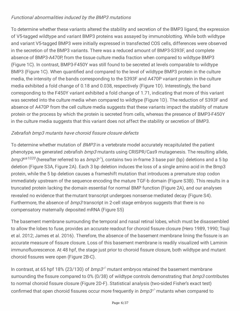
Functional abnormalities induced by the BMP3 mutations
To determine whether these variants altered the stability and secretion of the BMP3 ligand, the expressionof V5-tagged wildtype and variant BMP3 proteins was assayed by immunoblotting. While both wildtypeand variant V5-tagged BMP3 were initially expressed in transfected COS cells, differences were observedin the secretion of the BMP3 variants. There was a reduced amount of BMP3-S393F, and completeabsence of BMP3-A470P, from the tissue culture media fraction when compared to wildtype BMP3(Figure 1C). In contrast, BMP3-F450Y was still found to be secreted at levels comparable to wildtypeBMP3 (Figure 1C). When quanti�ed and compared to the level of wildtype BMP3 protein in the culturemedia, the intensity of the bands corresponding to the S393F and A470P variant protein in the culturemedia exhibited a fold change of 0.18 and 0.038, respectively (Figure 1D). Interestingly, the bandcorresponding to the F450Y variant exhibited a fold change of 1.71, indicating that more of this variantwas secreted into the culture media when compared to wildtype (Figure 1D). The reduction of S393F andabsence of A470P from the cell culture media suggests that these variants impact the stability of matureprotein or the process by which the protein is secreted from cells, whereas the presence of BMP3-F450Yin the culture media suggests that this variant does not affect the stability or secretion of BMP3.
Zebra�sh bmp3 mutants have choroid �ssure closure defects
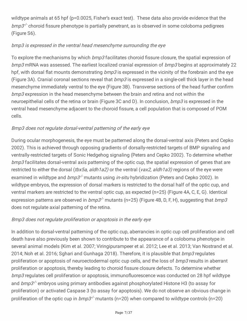
To determine whether mutation of BMP3 in a vertebrate model accurately recapitulated the patientphenotype, we generated zebra�sh bmp3 mutants using CRISPR/Cas9 mutagenesis. The resulting allele,bmp3ua1020 (hereafter referred to as bmp3-/-), contains two in-frame 3 base pair (bp) deletions and a 5 bpdeletion (Figure S3A, Figure 2A). Each 3 bp deletion induces the loss of a single amino acid in the Bmp3protein, while the 5 bp deletion causes a frameshift mutation that introduces a premature stop codonimmediately upstream of the sequence encoding the mature TGF-b domain (Figure S3B). This results in atruncated protein lacking the domain essential for normal BMP function (Figure 2A), and our analysesrevealed no evidence that the mutant transcript undergoes nonsense mediated decay (Figure S4).Furthermore, the absence of bmp3 transcript in 2-cell stage embryos suggests that there is nocompensatory maternally deposited mRNA (Figure S5)
The basement membrane surrounding the temporal and nasal retinal lobes, which must be disassembledto allow the lobes to fuse, provides an accurate readout for choroid �ssure closure (Hero 1989, 1990; Tsujiet al. 2012; James et al. 2016). Therefore, the absence of the basement membrane lining the �ssure is anaccurate measure of �ssure closure. Loss of this basement membrane is readily visualized with Lamininimmuno�uorescence. At 48 hpf, the stage just prior to choroid �ssure closure, both wildtype and mutantchoroid �ssures were open (Figure 2B-C).
In contrast, at 65 hpf 18% (23/130) of bmp3-/- mutant embryos retained the basement membranesurrounding the �ssure compared to 0% (0/38) of wildtype controls demonstrating that bmp3 contributesto normal choroid �ssure closure (Figure 2D-F). Statistical analysis (two-sided Fisher’s exact test)con�rmed that open choroid �ssures occur more frequently in bmp3-/- mutants when compared to
Page 7/37
wildtype animals at 65 hpf (p=0.0025, Fisher’s exact test). These data also provide evidence that thebmp3-/- choroid �ssure phenotype is partially penetrant, as is observed in some coloboma pedigrees(Figure S6).
bmp3 is expressed in the ventral head mesenchyme surrounding the eye
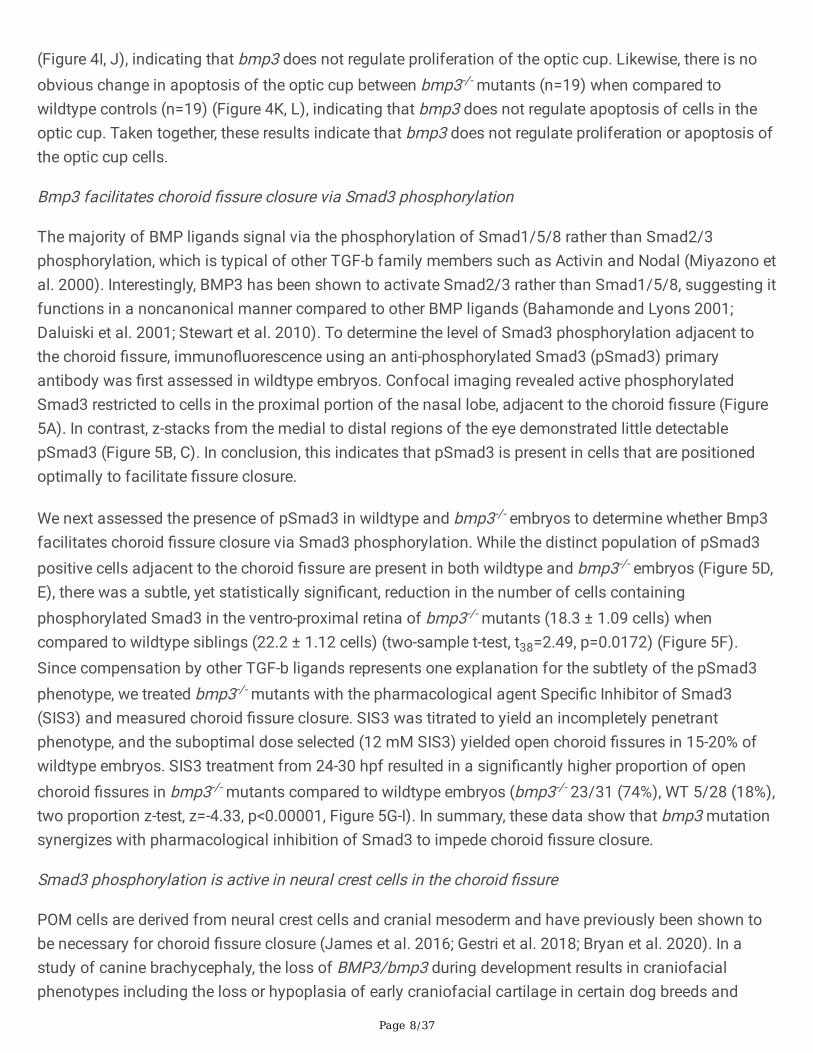
To explore the mechanisms by which bmp3 facilitates choroid �ssure closure, the spatial expression ofbmp3 mRNA was assessed. The earliest localized cranial expression of bmp3 begins at approximately 22hpf, with dorsal �at mounts demonstrating bmp3 is expressed in the vicinity of the forebrain and the eye(Figure 3A). Cranial coronal sections reveal that bmp3 is expressed in a single-cell thick layer in the headmesenchyme immediately ventral to the eye (Figure 3B). Transverse sections of the head further con�rmbmp3 expression in the head mesenchyme between the brain and retina and not within theneuroepithelial cells of the retina or brain (Figure 3C and D). In conclusion, bmp3 is expressed in theventral head mesenchyme adjacent to the choroid �ssure, a cell population that is composed of POMcells.
Bmp3 does not regulate dorsal-ventral patterning of the early eye
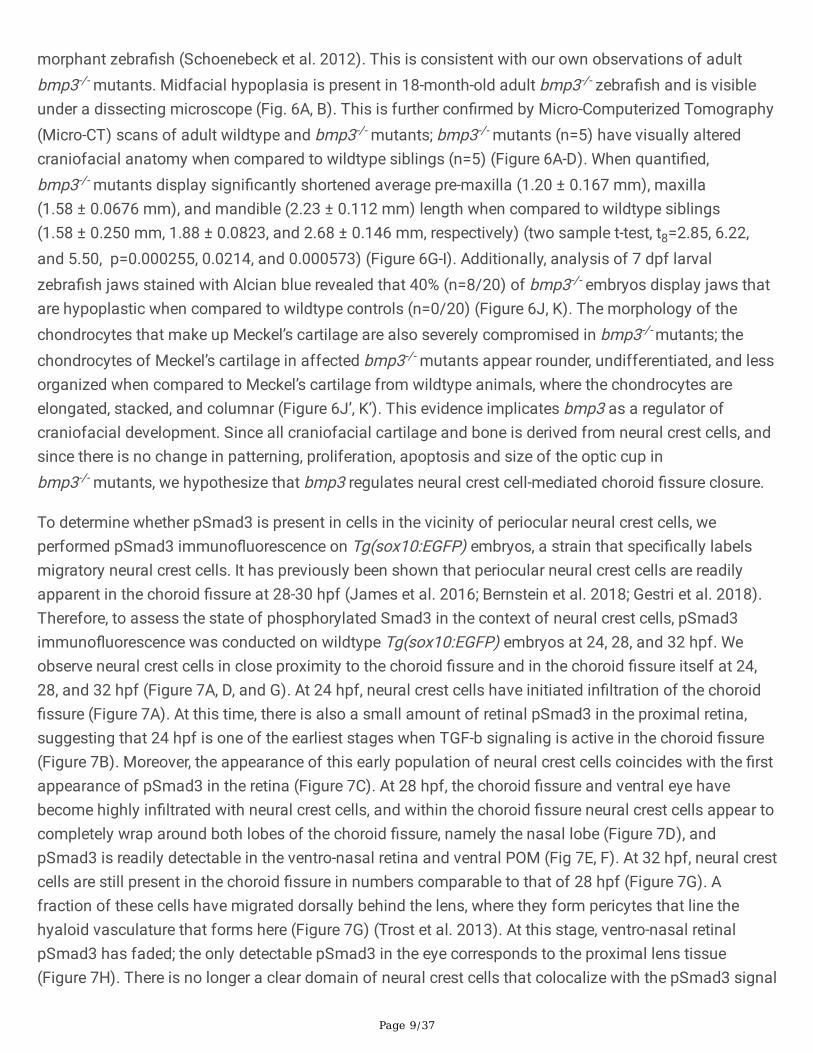
During ocular morphogenesis, the eye must be patterned along the dorsal-ventral axis (Peters and Cepko2002). This is achieved through opposing gradients of dorsally-restricted targets of BMP signaling andventrally-restricted targets of Sonic Hedgehog signaling (Peters and Cepko 2002). To determine whetherbmp3 facilitates dorsal-ventral axis patterning of the optic cup, the spatial expression of genes that arerestricted to either the dorsal (tbx5a, aldh1a2) or the ventral (vax2, aldh1a3) regions of the eye wereexamined in wildtype and bmp3-/- mutants using in-situ hybridization (Peters and Cepko 2002). Inwildtype embryos, the expression of dorsal markers is restricted to the dorsal half of the optic cup, andventral markers are restricted to the ventral optic cup, as expected (n=25) (Figure 4A, C, E, G). Identicalexpression patterns are observed in bmp3-/- mutants (n=25) (Figure 4B, D, F, H), suggesting that bmp3does not regulate axial patterning of the retina.
Bmp3 does not regulate proliferation or apoptosis in the early eye
In addition to dorsal-ventral patterning of the optic cup, aberrancies in optic cup cell proliferation and celldeath have also previously been shown to contribute to the appearance of a coloboma phenotype inseveral animal models (Kim et al. 2007; Viringipurampeer et al. 2012; Lee et al. 2013; Van Nostrand et al.2014; Noh et al. 2016; Sghari and Gunhaga 2018). Therefore, it is plausible that bmp3 regulatesproliferation or apoptosis of neuroectodermal optic cup cells, and the loss of bmp3 results in aberrantproliferation or apoptosis, thereby leading to choroid �ssure closure defects. To determine whetherbmp3 regulates cell proliferation or apoptosis, immuno�uorescence was conducted on 28 hpf wildtypeand bmp3-/- embryos using primary antibodies against phosphorylated Histone H3 (to assay forproliferation) or activated Caspase 3 (to assay for apoptosis). We do not observe an obvious change inproliferation of the optic cup in bmp3-/- mutants (n=20) when compared to wildtype controls (n=20)
Page 8/37
(Figure 4I, J), indicating that bmp3 does not regulate proliferation of the optic cup. Likewise, there is noobvious change in apoptosis of the optic cup between bmp3-/- mutants (n=19) when compared towildtype controls (n=19) (Figure 4K, L), indicating that bmp3 does not regulate apoptosis of cells in theoptic cup. Taken together, these results indicate that bmp3 does not regulate proliferation or apoptosis ofthe optic cup cells.
Bmp3 facilitates choroid �ssure closure via Smad3 phosphorylation
The majority of BMP ligands signal via the phosphorylation of Smad1/5/8 rather than Smad2/3phosphorylation, which is typical of other TGF-b family members such as Activin and Nodal (Miyazono etal. 2000). Interestingly, BMP3 has been shown to activate Smad2/3 rather than Smad1/5/8, suggesting itfunctions in a noncanonical manner compared to other BMP ligands (Bahamonde and Lyons 2001;Daluiski et al. 2001; Stewart et al. 2010). To determine the level of Smad3 phosphorylation adjacent tothe choroid �ssure, immuno�uorescence using an anti-phosphorylated Smad3 (pSmad3) primaryantibody was �rst assessed in wildtype embryos. Confocal imaging revealed active phosphorylatedSmad3 restricted to cells in the proximal portion of the nasal lobe, adjacent to the choroid �ssure (Figure5A). In contrast, z-stacks from the medial to distal regions of the eye demonstrated little detectablepSmad3 (Figure 5B, C). In conclusion, this indicates that pSmad3 is present in cells that are positionedoptimally to facilitate �ssure closure.
We next assessed the presence of pSmad3 in wildtype and bmp3-/- embryos to determine whether Bmp3facilitates choroid �ssure closure via Smad3 phosphorylation. While the distinct population of pSmad3positive cells adjacent to the choroid �ssure are present in both wildtype and bmp3-/- embryos (Figure 5D,E), there was a subtle, yet statistically signi�cant, reduction in the number of cells containingphosphorylated Smad3 in the ventro-proximal retina of bmp3-/- mutants (18.3 ± 1.09 cells) whencompared to wildtype siblings (22.2 ± 1.12 cells) (two-sample t-test, t38=2.49, p=0.0172) (Figure 5F).Since compensation by other TGF-b ligands represents one explanation for the subtlety of the pSmad3phenotype, we treated bmp3-/- mutants with the pharmacological agent Speci�c Inhibitor of Smad3(SIS3) and measured choroid �ssure closure. SIS3 was titrated to yield an incompletely penetrantphenotype, and the suboptimal dose selected (12 mM SIS3) yielded open choroid �ssures in 15-20% ofwildtype embryos. SIS3 treatment from 24-30 hpf resulted in a signi�cantly higher proportion of openchoroid �ssures in bmp3-/- mutants compared to wildtype embryos (bmp3-/- 23/31 (74%), WT 5/28 (18%),two proportion z-test, z=-4.33, p<0.00001, Figure 5G-I). In summary, these data show that bmp3 mutationsynergizes with pharmacological inhibition of Smad3 to impede choroid �ssure closure.
Smad3 phosphorylation is active in neural crest cells in the choroid �ssure
POM cells are derived from neural crest cells and cranial mesoderm and have previously been shown tobe necessary for choroid �ssure closure (James et al. 2016; Gestri et al. 2018; Bryan et al. 2020). In astudy of canine brachycephaly, the loss of BMP3/bmp3 during development results in craniofacialphenotypes including the loss or hypoplasia of early craniofacial cartilage in certain dog breeds and
Page 9/37
morphant zebra�sh (Schoenebeck et al. 2012). This is consistent with our own observations of adultbmp3-/- mutants. Midfacial hypoplasia is present in 18-month-old adult bmp3-/- zebra�sh and is visibleunder a dissecting microscope (Fig. 6A, B). This is further con�rmed by Micro-Computerized Tomography(Micro-CT) scans of adult wildtype and bmp3-/- mutants; bmp3-/- mutants (n=5) have visually alteredcraniofacial anatomy when compared to wildtype siblings (n=5) (Figure 6A-D). When quanti�ed,bmp3-/- mutants display signi�cantly shortened average pre-maxilla (1.20 ± 0.167 mm), maxilla(1.58 ± 0.0676 mm), and mandible (2.23 ± 0.112 mm) length when compared to wildtype siblings(1.58 ± 0.250 mm, 1.88 ± 0.0823, and 2.68 ± 0.146 mm, respectively) (two sample t-test, t8=2.85, 6.22,and 5.50, p=0.000255, 0.0214, and 0.000573) (Figure 6G-I). Additionally, analysis of 7 dpf larvalzebra�sh jaws stained with Alcian blue revealed that 40% (n=8/20) of bmp3-/- embryos display jaws thatare hypoplastic when compared to wildtype controls (n=0/20) (Figure 6J, K). The morphology of thechondrocytes that make up Meckel’s cartilage are also severely compromised in bmp3-/- mutants; thechondrocytes of Meckel’s cartilage in affected bmp3-/- mutants appear rounder, undifferentiated, and lessorganized when compared to Meckel’s cartilage from wildtype animals, where the chondrocytes areelongated, stacked, and columnar (Figure 6J’, K’). This evidence implicates bmp3 as a regulator ofcraniofacial development. Since all craniofacial cartilage and bone is derived from neural crest cells, andsince there is no change in patterning, proliferation, apoptosis and size of the optic cup inbmp3-/- mutants, we hypothesize that bmp3 regulates neural crest cell-mediated choroid �ssure closure.
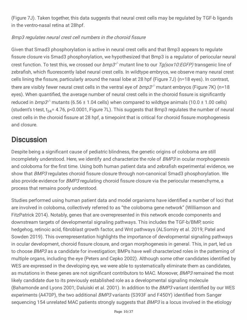
To determine whether pSmad3 is present in cells in the vicinity of periocular neural crest cells, weperformed pSmad3 immuno�uorescence on Tg(sox10:EGFP) embryos, a strain that speci�cally labelsmigratory neural crest cells. It has previously been shown that periocular neural crest cells are readilyapparent in the choroid �ssure at 28-30 hpf (James et al. 2016; Bernstein et al. 2018; Gestri et al. 2018).Therefore, to assess the state of phosphorylated Smad3 in the context of neural crest cells, pSmad3immuno�uorescence was conducted on wildtype Tg(sox10:EGFP) embryos at 24, 28, and 32 hpf. Weobserve neural crest cells in close proximity to the choroid �ssure and in the choroid �ssure itself at 24,28, and 32 hpf (Figure 7A, D, and G). At 24 hpf, neural crest cells have initiated in�ltration of the choroid�ssure (Figure 7A). At this time, there is also a small amount of retinal pSmad3 in the proximal retina,suggesting that 24 hpf is one of the earliest stages when TGF-b signaling is active in the choroid �ssure(Figure 7B). Moreover, the appearance of this early population of neural crest cells coincides with the �rstappearance of pSmad3 in the retina (Figure 7C). At 28 hpf, the choroid �ssure and ventral eye havebecome highly in�ltrated with neural crest cells, and within the choroid �ssure neural crest cells appear tocompletely wrap around both lobes of the choroid �ssure, namely the nasal lobe (Figure 7D), andpSmad3 is readily detectable in the ventro-nasal retina and ventral POM (Fig 7E, F). At 32 hpf, neural crestcells are still present in the choroid �ssure in numbers comparable to that of 28 hpf (Figure 7G). Afraction of these cells have migrated dorsally behind the lens, where they form pericytes that line thehyaloid vasculature that forms here (Figure 7G) (Trost et al. 2013). At this stage, ventro-nasal retinalpSmad3 has faded; the only detectable pSmad3 in the eye corresponds to the proximal lens tissue(Figure 7H). There is no longer a clear domain of neural crest cells that colocalize with the pSmad3 signal
Page 10/37
(Figure 7J). Taken together, this data suggests that neural crest cells may be regulated by TGF-b ligandsin the ventro-nasal retina at 28hpf.
Bmp3 regulates neural crest cell numbers in the choroid �ssure
Given that Smad3 phosphorylation is active in neural crest cells and that Bmp3 appears to regulate�ssure closure vis Smad3 phosphorylation, we hypothesized that Bmp3 is a regulator of periocular neuralcrest function. To test this, we crossed our bmp3-/- mutant line to our Tg(sox10:EGFP) transgenic line ofzebra�sh, which �uorescently label neural crest cells. In wildtype embryos, we observe many neural crestcells lining the �ssure, particularly around the nasal lobe at 28 hpf (Figure 7J) (n=18 eyes). In contrast,there are visibly fewer neural crest cells in the ventral eye of bmp3-/- mutant embryos (Figure 7K) (n=18eyes). When quanti�ed, the average number of neural crest cells in the choroid �ssure is signi�cantlyreduced in bmp3-/- mutants (6.56 ± 1.04 cells) when compared to wildtype animals (10.0 ± 1.00 cells)(student’s t-test, t34= 4.76, p<0.0001, Figure 7L). This suggests that Bmp3 regulates the number of neuralcrest cells in the choroid �ssure at 28 hpf, a timepoint that is critical for choroid �ssure morphogenesisand closure.
DiscussionDespite being a signi�cant cause of pediatric blindness, the genetic origins of coloboma are stillincompletely understood. Here, we identify and characterize the role of BMP3 in ocular morphogenesisand coloboma for the �rst time. Using both human patient data and zebra�sh experimental evidence, weshow that BMP3 regulates choroid �ssure closure through non-canonical Smad3 phosphorylation. Wealso provide evidence for BMP3 regulating choroid �ssure closure via the periocular mesenchyme, aprocess that remains poorly understood.
Studies performed using human patient data and model organisms have identi�ed a number of loci thatare involved in coloboma, collectively referred to as “the coloboma gene network” (Williamson andFitzPatrick 2014). Notably, genes that are overrepresented in this network encode components anddownstream targets of developmental signaling pathways. This includes the TGF-b/BMP, sonichedgehog, retinoic acid, �broblast growth factor, and Wnt pathways (ALSomiry et al. 2019; Patel andSowden 2019). This overrepresentation highlights the importance of developmental signaling pathwaysin ocular development, choroid �ssure closure, and organ morphogenesis in general. This, in part, led usto choose BMP3 as a candidate for investigation; BMPs have well characterized roles in the patterning ofmultiple organs, including the eye (Peters and Cepko 2002). Although some other candidates identi�ed byWES are expressed in the developing eye, we were able to systematically eliminate them as candidates,as mutations in these genes are not signi�cant contributors to MAC. Moreover, BMP3 remained the mostlikely candidate due to its previously established role as a developmental signaling molecule(Bahamonde and Lyons 2001; Daluiski et al. 2001). In addition to the BMP3 variant identi�ed by our WESexperiments (A470P), the two additional BMP3 variants (S393F and F450Y) identi�ed from Sangersequencing 154 unrelated MAC patients strongly suggests that BMP3 is a locus involved in the etiology
Page 11/37
of coloboma. These variants are extremely rare, suggesting that they are not benign variants found in thegeneral population; rather, it is likely that these variants contribute to the etiology of MAC in thesepatients. This is supported by both predictive algorithms of protein folding and cell secretion assays,which show that at least two of the variants, A470 and S393, result in the absence of these variantproteins from the media of COS-7 cells. Both A470 and S393 are located near cysteine residues thatparticipate in the disul�de bonds that are critical for the stability of mature BMP3 dimers. Therefore, thepresence of amino acid substitutions at either of these residues may result in aberrant cysteine knotformation, resulting in lowered extracellular stability of BMP3 and thus its absence from the cell culturemedia (Goebel et al. 2019). Interestingly, there is another variant at the A470 residue listed in gnomAD(p.Ala470Thr) that is present in control individuals (7/75 435 individuals). Although this may suggestthat change to the A470 residue do not cause changes to BMP3 activity, the A470T amino acid change is,relative to A470P, conservative; threonine is small and uncharged, making it unlikely that it dramaticallyaffects BMP3 protein structure and function. Proline, on the other hand, forms a pyrrolidine loop with thenitrogen from the amino acid backbone. This pyrrolidine loop has previously been observed todramatically disrupt the secondary structure of proteins, therefore making it exceptionally likely that thisamino acid disrupts BMP3 protein structure (Levitt 1978). Taken together, this evidence strongly suggeststhat the patient variants identi�ed through WES and Sanger sequencing have deleterious consequencesto protein activity, further implicating BMP3 as a novel coloboma-causing locus.
For the third variant detected, F450Y, we do not observe any secretion aberrancies. This residue is locatedin close proximity to the receptor-binding domain of the BMP3 protein (Allendorph et al. 2007). Thereceptor binding domain of BMP3 is extremely sensitive to amino acid composition, and single aminoacid substitutions have been shown to reduce its a�nity for its receptor (Allendorph et al. 2007).Alternatively, this residue may not be pathogenic; this is supported by the presence of this variant incontrol genome databases (Table 2). Therefore, more investigation is needed to con�rm whether BMP3-F450Y is a pathogenic variant.
We show that zebra�sh bmp3-/- mutants have an incompletely penetrant coloboma phenotype.Additionally, we show that this phenotype is a delay in �ssure closure, as all bmp3-/- mutants assayedhave closed �ssures at 72 hpf. The nature of the choroid �ssure closure defects observed in thesemutants may give some indication as to bmp3’s role in �ssure closure. It has been proposed thatcoloboma can be broadly separated into two classes: morphogenetic coloboma, which arises fromdefects the morphogenesis of the eye cup, and fusion defective coloboma, which arises from defects indegradation of the basal lamina or adhesion of the retinal cells that make up the choroid �ssure (Jameset al. 2016; Eckert et al. 2020). Eyes from bmp3-/- mutants are not smaller in size than those fromwildtype embryos, and the �ssure lobes are fully apposed without any gross morphological defects,suggesting that bmp3 is not a regulator of early ocular morphogenetic events such as optic vesicleevagination or choroid �ssure formation, as perturbations to these process usually cause overtmorphological defects such as microphthalmia and severe, very obvious coloboma (Patel and Sowden2019). Due to the subtlety of the choroid �ssure closure defects in bmp3 mutants, it is more likely that
Page 12/37
bmp3 facilitates processes that occur later in choroid �ssure closure, such as basement membranebreakdown or intercalation of the retinal cells that make up the lobes of the choroid �ssure. Indeed, thephenotypes of bmp3 mutants resemble other zebra�sh models of defective basement membranedegradation and cellular intercalation, further suggesting that bmp3 plays a role in these processes(James et al. 2016; Hardy et al. 2019).
Previous studies of bmp3 function have shown that it non-canonically activates the phosphorylation ofSmad2/3 rather than Smad1/5/8 (Bahamonde and Lyons 2001; Daluiski et al. 2001; Stewart et al. 2010).We show that bmp3 is likely facilitating choroid �ssure closure through this mechanism, as indicated bythe presence of phospho-Smad3 adjacent to the �ssure and the increased sensitivity of bmp3-/- embryosto SIS3. Recent work on zebra�sh has shown that there are other TGF-b ligands expressed in the �ssure,and that TGF-b signaling is active in the �ssure at the same time we detect phospho-Smad3 in the retina(Knickmeyer et al. 2020). Therefore, bmp3 may act in concert with other TGF-b ligands to facilitatechoroid �ssure closure (Knickmeyer et al. 2020). The mechanism behind this process is still not clear;others have proposed that TGF-b signaling via Smad2/3 phosphorylation in the �ssure promotes theexpression of BMP antagonists which leads to the inhibition of dorsal BMP signaling (Knickmeyer et al.2020). This thereby creates a restricted domain of BMP expression in the dorsal eye and conferringdorsal identity (Knickmeyer et al. 2020). Indeed, BMP3 has been shown to inhibit both canonical BMPsignaling and Activin signaling (Gamer et al. 2005; Knickmeyer et al. 2020). This mechanism has alsobeen well documented in other developmental processes including axis patterning and gastrulation (Littleand Mullins 2006). However, this is somewhat at odds with our results; we do not observe any changesto the expression of dorsal markers, especially tbx5a, which is a direct transcriptional output of BMPsignaling in the dorsal eye, suggesting that bmp3 doesn’t antagonize dorsal BMP signaling. Therefore,more research is needed to determine the precise pathway through which Bmp3 inhibits canonical BMPsignaling in eye development.
Interestingly, bmp3 is not expressed in the dorsal eye like other ocular BMP ligands, such asgdf6a (French et al. 2009; Gosse and Baier 2009). Rather, it is expressed in the ventral head mesenchymeadjacent to the �ssure. The head mesenchyme is populated by both cranial neural crest cells and paraxialand splanchnic mesoderm (Creuzet et al. 2003; Gage et al. 2005). Accordingly, there has been evidencethat bmp3 functions in both the mesoderm and the cranial neural crest; bmp3 expression has beendetected in the tail paraxial mesoderm of zebra�sh and has also been shown to be a regulator of neuralcrest-derived craniofacial structures (Mueller et al. 2010; Schoenebeck et al. 2012). Here, we provideevidence that bmp3 is a regulator of cranial neural crest cells rather than cranial mesoderm, as indicatedby microCT scans of adult �sh and Alcian blue staining of larval jaw cartilage. Although this is consistentwith other reports of bmp3 function, we �nd that the larval mutant phenotypes are far less severe andhave lower penetrance than previously reported (Schoenebeck et al. 2012). However, these previousstudies were performed using morpholino oligonucleotides, which have been shown to produce moresevere phenotypes when compared to mutants (Eve et al. 2017). Regardless, we provide further evidencethat bmp3 regulates neural crest-derived structures and is novel regulator of craniofacial development,
Page 13/37
particularly structures that contribute to the jaw. This is somewhat unsurprising, as craniofacialabnormalities and coloboma commonly occur together in syndromes including CHARGE syndrome andBOFS syndrome (Siebert et al. 1985; Gestri et al. 2009). These studies, together with our own studies ofBMP3’s involvement in coloboma, suggest that there is likely a shared neural crest-derived etiology ofcoloboma and craniofacial abnormalities in many contexts (Gestri et al. 2009; Bajpai et al. 2010).Moreover, it wouldn’t be entirely surprising to �nd craniofacial abnormalities in patients with damagingmutations in BMP3, given that we see craniofacial abnormalities in our zebra�sh model. In our studies ofcartilage development in bmp3-/- mutants, we observe jaw chondrocytes that are less organized andundifferentiated when compared to wildtype controls, suggesting that bmp3 doesn’t affect the migrationof neural crest cells to form the cartilage, but rather regulates the differentiation of cranial neural crestcells into chondrocytes. Moreover, only anterior structures derived from pharyngeal arch 1 and 2 (such asMeckel’s cartilage, the palatoquadrate, and the ceratohyal) are affected, whereas structures derived fromthe more posterior pharyngeal arches 3-7 (such as the ceratobranchials) remain largely unaffected,suggesting that bmp3’s role on craniofacial development is restricted to the neural crest cells of the �rstpharyngeal arch; indeed, other studies of bmp3’s role on craniofacial development found its expression tobe highest in the anterior pharyngeal arches and lower or absent in the more posterior arches(Schoenebeck et al. 2012). This suggests that bmp3 may be regulating the differentiation of arch 1 and 2neural crest cells, and the absence of bmp3 in the �rst arch results in undifferentiated chondrocytes and,thus, the craniofacial defects we observe in bmp3-/- mutants. Alternatively, bmp3 could be regulatingcranial neural crest cell division, survival, or death; a lack of bmp3 results in an incorrect number ofchondrocyte precursors, which, ultimately, results in disorganization of the cartilage seen inbmp3 mutants. Consistent with this, there appears to be more chondrocytes present in the jaws ofbmp3-/- mutants. Once the cranial neural crest cells condense into cartilage, convergence extensionelongates the chondrocytes and facilitates chondrocyte stacking (Kimmel et al. 1998). Other studies haveshown that disrupting genes necessary for convergence extension results in jaw phenotypes similar tobmp3-/- mutants, suggesting that bmp3 could have a role in regulating the number of cells necessary forconvergence extension to occur properly (Rochard et al. 2016). Further research is needed to establish theprecise role of bmp3 in craniofacial development and cranial neural crest function.
In addition to regulating the neural crest-derived craniofacial skeleton, we also show that bmp3 regulatesperiocular neural crest cells during choroid �ssure closure. There have been several studies that haveshown that these populations of cells are necessary for choroid �ssure closure, further solidifying the roleof these cells as necessary mediators of choroid �ssure closure (James et al. 2016; Gestri et al. 2018).This is also supported by our studies, where ventral periocular neural crest cells colocalize with pSmad3signal, suggesting that neural crest cells are responsive to TGF-b signaling. This is in agreement withother studies of periocular neural crest cells and TGF-b signals, which show that neural crest-speci�cdeletion of Tgfbr2 results in compound ocular defects in mice (Ittner et al. 2005). Therefore, thisrepresents a plausible mechanism for TGF-b signaling in the eye, where cranial neural crest cells areresponsive to Bmp3 and potentially other TGF-b ligands. This also �ts with our model ofBmp3 facilitating later stages of choroid �ssure closure; although some reports of neural crest-mediated
Page 14/37
choroid �ssure closure have shown that periocular neural crest cells are necessary for early eye vesiclemorphogenesis, others have proposed that neural crest cells are necessary for later stages of choroid�ssure (James et al. 2016; Gestri et al. 2018; Hardy et al. 2019; Bryan et al. 2020). We also observe areduction in the number of neural crest cells in the choroid �ssures of bmp3-/- mutants at 28 hpf,suggesting that Bmp3 regulates the number of neural crest cells in the choroid �ssure. However, it isunclear whether Bmp3 regulates neural crest number, guidance, or differentiation. Previous reports andour own studies of Bmp3 function in craniofacial development have shown that the neural crest-derivedcraniofacial cartilage is severely reduced in bmp3 morphants, suggesting that bmp3 plays a role in neuralcrest differentiation (Schoenebeck et al. 2012). In contrast, other reports of periocular neural crestmigration suggest that TGF-b signals emanating from the lens regulate periocular neural crest migrationinto the anterior segment (Takamiya et al. 2020). Therefore, it is plausible that ocular TGF-b signals arealso necessary for facilitating migration of periocular neural crest cells into the choroid �ssure.
In summary, we present the discovery of a novel locus involved with choroid �ssure closure, BMP3. Wehave shown that BMP3 likely facilitates �ssure closure through Smad3 phosphorylation, and we alsoprovide evidence that BMP3 may regulate periocular neural crest cells during the process of �ssureclosure. Although the present study has advanced our understanding of choroid �ssure closure biologyand, thus, coloboma, further work will need to be done to identify additional loci that are involved in thisprocess. Additionally, more investigation is warranted to identify and characterize loci that are expressedby the periocular mesenchyme and their role in choroid �ssure closure as well as paracrine factors thatregulate the periocular mesenchyme which will, in turn, inform our understanding of vertebrate ocularmorphogenesis.
Materials And MethodsEthics Statement
Whole exome and Sanger sequencing of human patient DNA
Whole exome sequencing (WES) was performed on genomic DNA from each proband as
part of FORGE Canada Consortium at the McGill University and Genome Quebec Innovation Centre.Exome target enrichment was performed using the Agilent SureSelect 50Mb (V3) All Exon Kit andsequencing was performed on the Illumina HiSeq 2000, multiplexing three samples per lane. The meancoverage of coding sequence regions, after accounting for duplicate reads was greater than 70x. WESdata was analyzed by performing alignment with BWA, duplicate read removal with Picard, local indelrealignment with GATK, variant calling with SAMtools, and annotation with Annovar and custom scripts.
For Sanger sequencing of the MAC patient cohort unrelated to individuals in the pedigree from Figure 1,intronic primers were used to amplify BMP3 in 154 patients with MAC. The amplicons were then puri�ed
Page 15/37
and sent for Sanger sequencing at the at the Genome Quebec Innovation Centre.
Zebra�sh husbandry
Adult zebra�sh were kept on a 10-hour dark/14-hour light schedule. Embryos were raised at 25.5°C,28.5°C or 33°C in embryo media and staged according to standardized developmental hallmarks (Kimmelet al. 1995). Embryos that were grown past 24 hpf were treated with 0.004% 1-phenyl 2-thiourea (PTU,Sigma Aldrich P7629) prior to 22hpf to block pigment formation. Anesthesia of larval and adult zebra�shwas performed with a 4% dilution of 0.4% tricaine methanesulfonate (TMS) stock solution (Sigma AldrichE10521). The AB wildtype strain, the bmp3-/- mutant line, and the Tg(sox10:EGFP) transgenic line wereused. The bmp3-/- line contains two in-frame 3 basepair (bp) deletions (NM_001077765.1:c.543_545deland NM_001077765.1:c.840_842del) and a 5bp deletion (NM_001077765.1:c.886_890del). Both of the3bp deletions cause the deletion of a single glutamine residue (NP_001071233.1:p.Gln182del andNP_001071233.1:p.Gln281del). The 5bp results in a frameshift and premature stop codon upstream ofthe mature TGF-b signaling domain (NP_001071233.1:p.Thr296GlyfsTer2).
Atomic Non-Local Environment Assessment (ANOLEA)
Using the previously solved crystal structure for BMP3, the effect of patient amino acid substitutions onprotein function were modeled in silico using Swiss-pbd Veiwer and analyzed using the ANOLEA server(http://melolab.org/anolea/) (Melo, F., Devos, D., Depiereux, E., Feytmans 1997; Allendorph et al. 2007).Using the ANOLEA data, the difference in energy requirement needed to fold the BMP3 protein with eachamino acid substitution was plotted in Microsoft Excel.
Site-directed mutagenesis
The open reading frame for wildtype human BMP3 was obtained from ASU BioDesign Institute in pDNR-Dual and was moved into pcDNA3.2/V5. Oligonucleotide primers for site-directed mutagenesis weredesigned to generate constructs carrying c.1178C>T (p.S393F), c.1349T>A (p.F450Y), and c.1408G>C(p.A470P) variants individually. The PCR reaction for site-directed mutagenesis contained: 500 ng oftemplate plasmid DNA, 17 μl of master mix (100 μl 10x PFU buffer, 792 μl water, 2 μl each of 100 mMdNTPs), 1 μl (10 mM) NAD, 0.5 μl (200 ng) each primer, 0.5 μl DMSO, 0.3 μl Taq DNA Ligase (NEB,M0208S), 1 μl PfuUltra DNA Polymerase (Agilent, 600670), and 1.5 μl water. The PCR cycle conditionswere denaturation at 95°C for 2 mins, then 30 cycles of 95°C for 1 min, 55°C for 1 min, and 65°C for 10mins. PCR products were then digested with 1 μl of DpnI (NEB, R0176S) for 30 mins at 37°C to digest theoriginal plasmid. Constructs were veri�ed by Sanger sequencing to ensure the mutation was introducedcorrectly. DNA for the four constructs was isolated by maxi prep (Qiagen, 12165) and Sanger sequencedto con�rm the mutation sites.
Tissue Culture and Immunoblotting
Page 16/37
COS-7 cells were plated to 6-well dishes and transfected at 80% con�uency. 3 mg of DNA was transfectedper well using Lipofectamine 2000 (ThermoFisher Scienti�c, 12566014) according to the manufacturer’sinstructions. Transfected cells were grown in low serum (0.1%) conditions for 48 hours and thenharvested. Media was removed and saved for secreted protein analysis. Cell monolayers were rinsed oncein phosphate buffered saline (PBS), and 500 ml of cold lysis buffer was added. Lysis buffer componentswere 50mM Tris, 150mM NaCl, 1mM EDTA, 1.5% SDS, and one complete mini protease inhibitor tablet(Millipore/Sigma 11836170001), ph7.5. Cells sat on ice for 10 minutes and were then scraped andcollected in Eppendorf tubes. Samples were boiled, transferred to QiaShredder tubes (Qiagen, 79656),centrifuged, and the �ow-through was collected in new tubes. Proteins from media were concentrated byacetone precipitation (1:1.5 media to acetone volume) and air dried. A minimal volume of lysis bufferwas added to resuspend the pellets. Protein concentration was determined using the Bradfordcolorimetric protein assay (BioRad, 5000201). Cell lysates and concentrated media lysates were run on 4-12% Bis-Tris gradient gels (Invitrogen NP0336) for 1 hour at 200 volts. Resolved proteins were transferredto polyvinylidene di�uoride (PVDF) membranes (Millipore IPVH00010) at 22 volts for 1 hour on a semi-dryblotter (Amersham Biosciences). Blots were blocked in 5% milk in Triton buffered saline with Tween 20(TBST) for 2 hours and primary antibody was added for overnight incubation at 4°C. Mouse monoclonalanti-V5 primary antibody (ThermoFisher, MA5-15253) was used at 1:5000 dilution in 5% milk and mousemonoclonal anti-tubulin primary antibody (Sigma, T9026) was used at 1:10,000. After washing, sheepanti-mouse IgG HRP-conjugated secondary (Amersham, NA931) was added at 1:5000 for 1 hour at roomtemperature. Membranes were washed in SuperSignal West Pico PLUS ECL reagent (ThermoFisher,34577) for 3 minutes and detection was performed using the ChemiDoc MP Imaging System (BioRad).
CRISPR/Cas9 Mutagenesis
Short guide RNA (sgRNA) design and synthesis were performed as previously described (Gagnon et al.2014). The sequences for the oligonucleotides used to generate the templates for sgRNA synthesis are:5’-GGGACTTCATCTCATGGCAGTGG-3’, 5’-GGGAGCTCATTGTTCTGCAGTGG-3’ and 5’-GGCTGGCCTCATCCCATGTAGGG-3’. Cas9 protein (PNA Bio, CP01) was reconstituted in sterile water to aconcentration of 50 mg/mL and 2 mL was mixed with 1 mL of each sgRNA. One- or Two-cell stageembryos were injected with 1nL of Cas9/sgRNA mixture and allowed to develop to sexual maturity.Carriers for frameshift mutations were identi�ed by high resolution melt (HRM) analysis using the Type-ItHRM PCR kit (Qiagen, 206544) on a Rotor Gene Q qPCR machine (Qiagen, 9001560) and Sangersequencing.
Zebra�sh genotyping
bmp3-/- mutants were genotyped using polymerase chain reaction (PCR) followed by gel electrophoresis.Genomic DNA was extracted from �n clips in 20 mL of 50mM NaOH at 95°C for 15 minutes andsubsequently neutralized using 2 mL Tris-HCl, pH 8.0. Samples were diluted 1/2 in sterile water and usedas template for PCR using the following primers: Forward: 5’-CTTCATATGCTGGAATCGCATAAC-3’,Reverse: 5’-TTGCTCTCCATCGGATCATAAG-3’. PCR was performed using the following conditions:
Page 17/37
Denaturation at 94°C for 2 minutes, 40 cycles of 94°C for 15 seconds, 58°C for 15 seconds, and 72°C for12 seconds, followed by a �nal extension cycle at 72°C for 3 minutes. PCR products were then run on a3% agarose gel to resolve the wildtype and mutant amplicons (167bp and 156bp, respectively).
In-situ hybridization
Antisense riboprobes labeled with digoxygenin (DIG) were synthesized from puri�ed, linearizedexpression plasmid containing a gene-speci�c insert or from a gene-speci�c PCR product with anintegrated T7 RNA polymerase site. Probe synthesis was performed as previously described (Thisse andThisse 2008).
Embryos were �xed overnight at 4°C in 4% paraformaldehyde (PFA) and subsequently permeabilized for 5minutes at room temperature (RT) using 10 mg/ml Proteinase K in PBST. In-situ hybridization wasperformed as previously described except probes were not hydrolyzed (Prince et al. 1998). Forexperiments analyzing bmp3 expression, embryos were either manually de-yolked and �at-mounted undera coverslip in 70% glycerol or embedded in para�n wax, sectioned, and mounted under a coverslip usingDibutylphthalate Polystyrene Xylene (DPX) mountant. Flat-mounts and sections were imaged on a ZeissAxioImager Z1 compound microscope with Axiocam HR digital camera. For experiments analyzing tbx5a,alhd1a2, vax2 and alhd1a3 expression, embryos were mounted in 3% methyl-cellulose on a spot plateand imaged with an Olympus SZX12 stereomicroscope and Qimaging micropublisher camera.
Immuno�uorescence
For laminin immuno�uorescence, embryos were �xed n 4% PFA for two hours at RT. Embryos werepermeabilized with 10 mg/ml Proteinase K for 30 minutes. After permeabilization, embryos were blockedfor at least 1 hour at RT using 5% normal goat serum and 2% bovine serum albumin (BSA) in PBST.Embryos were incubated O/N in rabbit anti-laminin primary antibody diluted in block (1:200, L-9393,Sigma-Aldrich). After washing, embryos were incubated for 2 hours at RT in goat anti-rabbit Alexa Fluor555 secondary antibody (1:1000, A32732, Invitrogen).
For Phosphorylated Histone H3 immuno�uorescence, embryos were �xed in 4% PFA for wo hours at RT.Embryos were permeabilized in ice-cold acetone for 7 minutes at -20°C. After permeabilization, embryoswere incubated in 10mM ctric acid at 95C for 10 minutes for antigen retrieval. Embryos were then blockedin 3% BSA in 0.5% TritonX-100 in PBS for 1 hour at RT. Embryos were then incubated in rabbit anti-phospho-Histone H3 antibody (1:1000, ab183626, Abcam) O/N at 4°C, washed, and incubated in goatanti-rabbit Alexa Fluor 488 secondary antibody (1:1000, A32732, Invitrogen) plus TO-PRO-3 (1:1000,T3605, Invitrogen) for 2 hours at RT.
For cleaved Caspase 3 immuno�uorescence, embryos were �xed in 4% PFA for two hours at RT. Embryoswere permeabilized with ice-cold acetone for 7 minutes at -20°C. After permeabilization, embryos wereblocked in 5% goat serum (GS) in PBSDTT (PBST + 1% DMSO + 0.1% TritonX-100) for 2 hours at RT.Embryos were then incubated in rabbit anti-cleaved-Caspase3 (1:400, 559565, BDBiosciences) O/N at
Page 18/37
4°C, washed, and incubated in goat anti-rabbit Alexa Fluor 488 secondary antibody (1:1000, A32732,Invitrogen) plus TO-PRO-3 (1:1000, T3605, Invitrogen) for 2 hours at RT.
For phosphorylated Smad3 (pSmad3) immuno�uorescence, embryos were �xed overnight at 4°C.Embryos were permeabilized with 10 mg/ml Proteinase K for 5 minutes After permeabilization, embryoswere incubated in 4% BSA in 0.15% TritonX-100 in PBS. Embryos were incubated in rabbit anti-pSmad3primary antibody (1:200, ab59203, Abcam) O/N at 4°C, washed, and incubated in goat anti-rabbit AlexaFluor 488 or 568 secondary antibody O/N at 4°C (1:1000, A32732, Invitrogen).
After immuno�uorescence was performed, embryos were washed and passed through a 30, 50, and 70%glycerol series. Eyes and the surrounding head mesenchyme were dissected from whole embryos andmounted in 70% glycerol on a glass slide. All immuno�uorescence experiments were imaged using aZeiss AxioImager Z1, Zeiss LSM700 laser scanning confocal microscope.
Pharmacological inhibition of TGF-b signaling
To inhibit TGF-b signaling, Speci�c Inhibitor of Smad3 (SIS3) was used (566405-1MG, Calbiochem). Thestructure and activity of SIS3 has been described elsewhere (Jinnin et al. 2006). 1mg of SIS3 wasresuspended in DMSO to a stock concentration of 3mM, aliquoted, and stored at -20°C. Prior to treatment,the stock was mixed with pre-warmed embryo media to a �nal concentration of 12 mM. For controls, anequivalent volume of DMSO alone was added to pre-warmed embryo media. Embryos were stage-matched at 10 hpf. At 24 hpf, embryos were de-chorionated, transferred to 35mm petri dishes in groupsof 15 embryos per dish, and 5 mL of SIS3- or DMSO-treated media was added to the appropriate dishes.Embryos were incubated for six hours at 28.5°C until 30 hpf. The media was then removed, embryos werewashed 3 times with fresh embryo media, and all dishes were returned to 28.5°C. Embryo media waschanged at 48 hpf, and embryos were �xed at 72 hpf for 2 hours at RT with 4% PFA. Embryos were thenused for laminin immuno�uorescence. Eyes from these embryos were then dissected, mounted on glassslides in 70% glycerol, and imaged using a Zeiss AxioImager Z1, Zeiss LSM700 laser scanning confocalmicroscope.
Micro-computed tomography (micro-CT) of adult zebra�sh
18-month-old wildtype (n=5) and bmp3 -/- (n=5) zebra�sh were scanned using MILabs μCT at the Schoolof Dentistry, University of Alberta. Zebra�sh were �xed at 4% PFA for 24 h. The parameters for scanningand reconstruction were conducted as previously described (Miyashita et al. 2020). AVIZO (LifeTechnologies) software was used to visualize and quantify premaxilla, maxilla, and mandible length.Landmarks used to quantify the midfacial region are demonstrated in Figure S7.
Alcian blue staining of larval zebra�sh
Alcian blue staining of 7 dpf larval zebra�sh was performed as previously described, except Alizarin redstaining was not performed (Walker and Kimmel 2007). Brie�y, larval zebra�sh were euthanized using a

Page 19/37
4% dilution of 0.4% TMS and �xed in 1% PFA for one hour at RT. Embryos were washed for 10 minutes in10 mM MgCl2 buffered with 100 mM Tris pH 7.5, and then stained overnight in 0.04% Alcian blue stainingsolution. Embryos were removed from staining solution, rehydrated, bleached for 10 minutes in 3%H2O2/0.5% KOH, and de-stained with two 10-minute washes of 25% Glycerol/0.1% KOH and two 10-minute washes of 50% Glycerol/0.1% KOH. Embryos were then transferred to 70% glycerol and mountedon glass slides for imaging. Images were taken using a Zeiss AxioImager Z1 compound microscope withAxiocam HR digital camera.
Statistical analysis
Statistical signi�cance was determined using a two-proportion z-test (Figure 5) or an unpaired t-test(Figure 5, 6, and 7). Quantitative results are presented as percentage or mean ± S.E.
DeclarationsAcknowledgements- The authors are grateful to Science Animal Support Services for zebra�sh animalcare and facility maintenance
Con�ict of Interest Statement- The authors declare no competing con�icts of interest.
Funding - This works was funded by the Women and Children’s Health Research Institute (UOFABWCHRIIG 2879) and the National Science and Engineering Research Council of Canada (NSERC RGPIN-2016-04682)
Con�icts of Interest - The authors declare no con�icts of interest.
Availability of data and material - The data that support the �ndings of this study are available from thecorresponding author upon reasonable request.
Code availability - Not applicable.
Ethics approval - Embryonic, larval, and adult zebra�sh were cared for according to guidelines set by theCanadian Council of Animal Care and protocols were approved by the University of Alberta’s Animal Careand Use Committee (Protocol #427).
Consent to participate - Not applicable.
Consent to publish - Not applicable.
References
Page 20/37
Abouzeid H, Boisset G, Favez T, et al (2011) Mutations in the SPARC-related modular calcium-bindingprotein 1 gene, SMOC1, cause waardenburg anophthalmia syndrome. Am J Hum Genet 88:92–98
Adler R, Belecky-Adams TL (2002) The role of bone morphogenetic proteins in the differentiation of theventral optic cup. Development 129:3161 – 3171
Allendorph GP, Isaacs MJ, Kawakami Y, et al (2007) BMP-3 and BMP-6 Structures Illuminate the Nature ofBinding Speci�city with Receptors,. Biochemistry 46:12238–12247. https://doi.org/10.1021/bi700907k
ALSomiry AS, Gregory-Evans CY, Gregory-Evans K (2019) An update on the genetics of ocular coloboma.Hum Genet 138:865–880. https://doi.org/10.1007/s00439-019-02019-3
Asai-Coakwell M, French CR, Berry KM, et al (2007) GDF6, a novel locus for a spectrum of oculardevelopmental anomalies. Am J Hum Genet 80:306–315
Asai-Coakwell M, French CR, Ye M, et al (2009) Incomplete penetrance and phenotypic variabilitycharacterize Gdf6-attributable oculo-skeletal phenotypes. Hum Mol Genet 18:1110–1121.https://doi.org/10.1093/hmg/ddp008
Bahamonde ME, Lyons KM (2001) BMP3: To Be or Not To Be a BMP. JBJS 83: BJS 83: S56-S62
Bajpai R, Chen DA, Rada-Iglesias A, et al (2010) CHD7 cooperates with PBAF to control multipotent neuralcrest formation. Nature 463:958–962. https://doi.org/10.1038/nature08733
Bakrania P, Efthymiou M, Klein JC, et al (2008) Mutations in BMP4 cause eye, brain, and digitdevelopmental anomalies: overlap between the BMP4 and hedgehog signaling pathways. Am J HumGenet 82:304–319
Bassing CH, Howe DJ, Segarini PR, et al (1994) A single heteromeric receptor complex is su�cient tomediate biological effects of transforming growth factor-beta ligands. J Biol Chem 269:14861–14864.https://doi.org/https://doi.org/10.1016/S0021-9258(17)36543-2
Beaulieu CL, Majewski J, Schwartzentruber J, et al (2014) FORGE Canada Consortium: Outcomes of a 2-Year National Rare-Disease Gene-Discovery Project. Am J Hum Genet 94:809–817.https://doi.org/10.1016/j.ajhg.2014.05.003
Beby F, Commeaux C, Bozon M, et al (2007) New Phenotype Associated With an Arg116Cys Mutation inthe CRYAA Gene: Nuclear Cataract, Iris Coloboma, and Microphthalmia. Arch Ophthalmol 125:213–216.https://doi.org/10.1001/archopht.125.2.213
Beleggia F, Li Y, Fan J, et al (2015) CRIM1 haploinsu�ciency causes defects in eye development inhuman and mouse. Hum Mol Genet 24:2267–2273. https://doi.org/10.1093/hmg/ddu744

Page 21/37
Bernstein CS, Anderson MT, Gohel C, et al (2018) The cellular bases of choroid �ssure formation andclosure. Dev Biol 440:137–151. https://doi.org/https://doi.org/10.1016/j.ydbio.2018.05.010
Brady JP, Garland D, Duglas-Tabor Y, et al (1997) Targeted disruption of the mouse αA-crystallin geneinduces cataract and cytoplasmic inclusion bodies containing the small heat shock protein αB-crystallin.Proc Natl Acad Sci 94:884 – 889. https://doi.org/10.1073/pnas.94.3.884
Bryan CD, Casey MA, Pfeiffer RL, et al (2020) Optic cup morphogenesis requires neural crest-mediatedbasement membrane assembly. Development 147:dev181420. https://doi.org/10.1242/dev.181420
Creuzet S, Vincent C, Couly G (2003) Neural crest derivatives in ocular and periocular structures. Int J DevBiol 49:161–171
Daluiski A, Engstrand T, Bahamonde ME, et al (2001) Bone morphogenetic protein-3 is a negativeregulator of bone density. Nat Genet 27:84–88. https://doi.org/10.1038/83810
Dee CT, Szymoniuk CR, Mills PED, Takahashi T (2012) Defective neural crest migration revealed by aZebra�sh model of Alx1-related frontonasal dysplasia. Hum Mol Genet 22:239–251.https://doi.org/10.1093/hmg/dds423
Eckert P, Knickmeyer MD, Heermann S (2020) In Vivo Analysis of Optic Fissure Fusion in Zebra�sh:Pioneer Cells, Basal Lamina, Hyaloid Vessels, and How Fissure Fusion is Affected by BMP. Int J Mol Sci21:2760
Eve AMJ, Place ES, Smith JC (2017) Comparison of Zebra�sh tmem88a mutant and morpholinoknockdown phenotypes. PLoS One 12:e0172227
French CR, Erickson T, French D V, et al (2009) Gdf6a is required for the initiation of dorsal–ventral retinalpatterning and lens development. Dev Biol 333:37–47
Gage PJ, Rhoades W, Prucka SK, Hjalt T (2005) Fate Maps of Neural Crest and Mesoderm in theMammalian Eye. Invest Ophthalmol Vis Sci 46:4200–4208. https://doi.org/10.1167/iovs.05-0691
Gagnon JA, Valen E, Thyme SB, et al (2014) E�cient Mutagenesis by Cas9 Protein-MediatedOligonucleotide Insertion and Large-Scale Assessment of Single-Guide RNAs. PLoS One 9:e98186
Gamer LW, Nove J, Levin M, Rosen V (2005) BMP-3 is a novel inhibitor of both activin and BMP-4signaling in Xenopus embryos. Dev Biol 285:156–168.https://doi.org/https://doi.org/10.1016/j.ydbio.2005.06.012
Gestri G, Bazin-Lopez N, Scholes C, Wilson SW (2018) Cell Behaviors during Closure of the ChoroidFissure in the Developing Eye. Front. Cell. Neurosci 12:42
Page 22/37
Gestri G, Osborne RJ, Wyatt AW, et al (2009) Reduced TFAP2A function causes variable optic �ssureclosure and retinal defects and sensitizes eye development to mutations in other morphogeneticregulators. Hum Genet 126:791–803. https://doi.org/10.1007/s00439-009-0730-x
Goebel EJ, Hart KN, McCoy JC, Thompson TB (2019) Structural biology of the TGFβ family. Exp Biol Med244:1530–1546. https://doi.org/10.1177/1535370219880894
Gosse NJ, Baier H (2009) An essential role for Radar (Gdf6a) in inducing dorsal fate in the zebra�shretina. Proc Natl Acad Sci 106:2236 – 2241. https://doi.org/10.1073/pnas.0803202106
Graff JM, Bansal A, Melton DA (1996) Xenopus Mad Proteins Transduce Distinct Subsets of Signals forthe TGFβ Superfamily. Cell 85:479–487. https://doi.org/https://doi.org/10.1016/S0092-8674(00)81249-0
Hanel ML, Hensey C (2006) Eye and neural defects associated with loss of GDF6. BMC Dev Biol 6:43.https://doi.org/10.1186/1471-213X-6-43
Hardy H, Prendergast JGD, Patel A, et al (2019) Detailed analysis of chick optic �ssure closure revealsNetrin-1 as an essential mediator of epithelial fusion. Elife 8:e43877
Hero I (1990) Optic �ssure closure in the normal cinnamon mouse. An ultrastructural study. InvestOphthalmol Vis Sci 31:197–216
Hero I (1989) The optic �ssure in the normal and microphthalmic mouse. Exp Eye Res 49:229–239.https://doi.org/https://doi.org/10.1016/0014-4835(89)90093-6
Hornby SJ, Dandona L, Jones RB, et al (2003) The familial contribution to non-syndromic ocularcoloboma in south India. Br J Ophthalmol 87:336 – 340. https://doi.org/10.1136/bjo.87.3.336
Ittner LM, Wurdak H, Schwerdtfeger K, et al (2005) Compound developmental eye disorders followinginactivation of TGFβ signaling in neural-crest stem cells. J Biol 4:11
James A, Lee C, Williams AM, et al (2016) The hyaloid vasculature facilitates basement membranebreakdown during choroid �ssure closure in the zebra�sh eye. Dev Biol 419:262–272.https://doi.org/https://doi.org/10.1016/j.ydbio.2016.09.008
Jinnin M, Ihn H, Tamaki K (2006) Characterization of SIS3, a Novel Speci�c Inhibitor of Smad3, and ItsEffect on Transforming Growth Factor-β1-Induced Extracellular Matrix Expression. Mol Pharmacol 69:597– 607. https://doi.org/10.1124/mol.105.017483
Kagiyama Y, Gotouda N, Sakagami K, et al (2005) Extraocular dorsal signal affects the developmentalfate of the optic vesicle and patterns the optic neuroepithelium. Dev Growth Differ 47:523–536.https://doi.org/10.1111/j.1440-169X.2005.00828.x
Page 23/37
Kim T-H, Goodman J, Anderson K V, Niswander L (2007) Phactr4 Regulates Neural Tube and Optic FissureClosure by Controlling PP1-, Rb-, and E2F1-Regulated Cell-Cycle Progression. Dev Cell 13:87–102.https://doi.org/https://doi.org/10.1016/j.devcel.2007.04.018
Kimmel CB, Ballard WW, Kimmel SR, et al (1995) Stages of embryonic development of the zebra�sh. DevDyn 203:253–310
Kimmel CB, Miller CT, Kruze G, et al (1998) The Shaping of Pharyngeal Cartilages during EarlyDevelopment of the Zebra�sh. Dev Biol 203:245–263.https://doi.org/https://doi.org/10.1006/dbio.1998.9016
Knickmeyer MD, Mateo JL, Eckert P, et al (2020) TGFβ-facilitated optic �ssure fusion and the role of bonemorphogenetic protein antagonism. Open Biol 8:170134. https://doi.org/10.1098/rsob.170134
Kwan KM, Otsuna H, Kidokoro H, et al (2012) A complex choreography of cell movements shapes thevertebrate eye. Development 139:359 – 372. https://doi.org/10.1242/dev.071407
Langenberg T, Kahana A, Wszalek JA, Halloran MC (2008) The eye organizes neural crest cell migration.Dev Dyn 237:1645–1652. https://doi.org/10.1002/dvdy.21577
Laurie KJ, Dave A, Straga T, et al (2013) Identi�cation of a Novel Oligomerization Disrupting Mutation inCRYΑA Associated with Congenital Cataract in a South Australian Family. Hum Mutat 34:435–438.https://doi.org/https://doi.org/10.1002/humu.22260
Lee J, Lee B-K, Gross JM (2013) Bcl6a function is required during optic cup formation to prevent p53-dependent apoptosis and colobomata. Hum Mol Genet 22:3568–3582.https://doi.org/10.1093/hmg/ddt211
Levitt M (1978) Conformational preferences of amino acids in globular proteins. Biochemistry 17:4277–4285. https://doi.org/10.1021/bi00613a026
Litt M, Kramer P, LaMorticella DM, et al (1998) Autosomal Dominant Congenital Cataract Associated witha Missense Mutation in the Human Alpha Crystallin Gene CRYAA. Hum Mol Genet 7:471–474.https://doi.org/10.1093/hmg/7.3.471
Little SC, Mullins MC (2006) Extracellular modulation of BMP activity in patterning the dorsoventral axis.Birth Defects Res Part C Embryo Today Rev 78:224–242.https://doi.org/https://doi.org/10.1002/bdrc.20079
Liu F, Hata A, Baker JC, et al (1996) A human Mad protein acting as a BMP-regulated transcriptionalactivator. Nature 381:620–623. https://doi.org/10.1038/381620a0
Lo RS, Chen YG, Shi Y, et al (1998) The L3 loop: a structural motif determining speci�c interactionsbetween SMAD proteins and TGF-beta receptors. EMBO J 17:996–1005.
Page 24/37
https://doi.org/10.1093/emboj/17.4.996
Lupo G, Gestri G, O’Brien M, et al (2011) Retinoic acid receptor signaling regulates choroid �ssure closurethrough independent mechanisms in the ventral optic cup and periocular mesenchyme. Proc Natl AcadSci USA 108:8698–8703. https://doi.org/10.1073/pnas.1103802108
Mackay DS, Andley UP, Shiels A (2003) Cell death triggered by a novel mutation in the alphaA-crystallingene underlies autosomal dominant cataract linked to chromosome 21q. Eur J Hum Genet 11:784–793.https://doi.org/10.1038/sj.ejhg.5201046
McMahon C, Gestri G, Wilson SW, Link BA (2009) Lmx1b is essential for survival of periocularmesenchymal cells and in�uences Fgf-mediated retinal patterning in zebra�sh. Dev Biol 332:287–298.https://doi.org/10.1016/j.ydbio.2009.05.577
Melo, F., Devos, D., Depiereux, E., Feytmans E (1997) ANOLEA: a www server to assess protein structures.Intell Syst Mol Biol 97:110–113
Miyashita T, Baddam P, Smeeton J, et al (2020) nkx3.2 mutant zebra�sh accommodate jaw joint lossthrough a phenocopy of the head shapes of Paleozoic jawless �sh. J Exp Biol 223:.https://doi.org/10.1242/jeb.216945
Miyazono K, Ten Dijke P, Heldin C-H (2000) TGF-β signaling by Smad proteins. Academic Press, pp 115–157
Morcillo J, Martínez-Morales JR, Trousse F, et al (2006) Proper patterning of the optic �ssure requires thesequential activity of BMP7 and SHH. Development 133:3179 LP – 3190.https://doi.org/10.1242/dev.02493
Morrison D, FitzPatrick D, Hanson I, et al (2002) National study of microphthalmia, anophthalmia, andcoloboma (MAC) in Scotland: investigation of genetic aetiology. J Med Genet 39:16 – 22.https://doi.org/10.1136/jmg.39.1.16
Mueller RL, Huang C, Ho RK (2010) Spatio-temporal regulation of Wnt and retinoic acid signaling bytbx16/spadetail during zebra�sh mesoderm differentiation. BMC Genomics 11:492.https://doi.org/10.1186/1471-2164-11-492
Noh H, Lee H, Park E, Park S (2016) Proper closure of the optic �ssure requires ephrin A5-EphB2-JNKsignaling. Development 143:461–472. https://doi.org/10.1242/dev.129478
Okada I, Hamanoue H, Terada K, et al (2011) SMOC1 Is Essential for Ocular and Limb Development inHumans and Mice. Am J Hum Genet 88:30–41. https://doi.org/10.1016/j.ajhg.2010.11.012
Patel A, Sowden JC (2019) Genes and pathways in optic �ssure closure. Semin Cell Dev Biol 91:55–65.https://doi.org/https://doi.org/10.1016/j.semcdb.2017.10.010
Page 25/37
Peters MA, Cepko CL (2002) The Dorsal–Ventral Axis of the Neural Retina Is Divided into MultipleDomains of Restricted Gene Expression Which Exhibit Features of Lineage Compartments. Dev Biol251:59–73. https://doi.org/https://doi.org/10.1006/dbio.2002.0791
P�rrmann T, Emmerich D, Ruokonen P, et al (2015) Molecular mechanism of CHRDL1-mediated X-linkedmegalocornea in humans and in Xenopus model. Hum Mol Genet 24:3119–3132.https://doi.org/10.1093/hmg/ddv063
Pras E, Frydman M, Levy–Nissenbaum E, et al (2000) A Nonsense Mutation (W9X) in CRYAA CausesAutosomal Recessive Cataract in an Inbred Jewish Persian Family. Invest Ophthalmol Vis Sci 41:3511–3515
Prince VE, Moens CB, Kimmel CB, Ho RK (1998) Zebra�sh hox genes: expression in the hindbrain regionof wild-type and mutants of the segmentation gene, valentino. Development 125:393 LP – 406
Prokudin I, Simons C, Grigg JR, et al (2014) Exome sequencing in developmental eye disease leads toidenti�cation of causal variants in GJA8, CRYGC, PAX6 and CYP1B1. Eur J Hum Genet 22:907–915.https://doi.org/10.1038/ejhg.2013.268
Reis LM, Tyler RC, Schilter KF, et al (2011) BMP4 loss-of-function mutations in developmental eyedisorders including SHORT syndrome. Hum Genet 130:495–504. https://doi.org/10.1007/s00439-011-0968-y
Richter L, Flodman P, Barria von-Bischhoffshausen F, et al (2008) Clinical variability of autosomaldominant cataract, microcornea and corneal opacity and novel mutation in the alpha A crystallin gene(CRYAA). Am J Med Genet Part A 146A:833–842. https://doi.org/https://doi.org/10.1002/ajmg.a.32236
Rochard L, Monica SD, Ling ITC, et al (2016) Roles of Wnt pathway genes wls, wnt9a, wnt5b, frzb andgpc4 in regulating convergent-extension during zebra�sh palate morphogenesis. Development143:2541–2547. https://doi.org/10.1242/dev.137000
Sakuta H, Suzuki R, Takahashi H, et al (2001) Ventroptin: A BMP-4 Antagonist Expressed in a Double-Gradient Pattern in the Retina. Science (80- ) 293:111 LP – 115.https://doi.org/10.1126/science.1058379
Schmitt EA, Dowling JE (1994) Early-eye morphogenesis in the zebra�sh, Brachydanio rerio. J CompNeurol 344:532–542. https://doi.org/10.1002/cne.903440404
Schoenebeck JJ, Hutchinson SA, Byers A, et al (2012) Variation of BMP3 Contributes to Dog Breed SkullDiversity. PLOS Genet 8:e1002849
Sedykh I, Yoon B, Roberson L, et al (2017) Zebra�sh zic2 controls formation of periocular neural crest andchoroid �ssure morphogenesis. Dev Biol 429:92–104. https://doi.org/10.1016/j.ydbio.2017.07.003

Page 26/37
Sghari S, Gunhaga L (2018) Temporal Requirement of Mab21l2 During Eye Development in Chick RevealsStage-Dependent Functions for Retinogenesis. Invest Ophthalmol Vis Sci 59:3869–3878.https://doi.org/10.1167/iovs.18-24236
Shah SP, Taylor AE, Sowden JC, et al (2012) Anophthalmos, Microphthalmos, and Coloboma in the UnitedKingdom: Clinical Features, Results of Investigations, and Early Management. Ophthalmology 119:362–368. https://doi.org/10.1016/j.ophtha.2011.07.039
Siebert JR, Graham Jr JM, MacDonald C (1985) Pathologic features of the CHARGE association: supportfor involvement of the neural crest. Teratology 31:331–336
Skarie JM, Link BA (2009) FoxC1 Is Essential for Vascular Basement Membrane Integrity and HyaloidVessel Morphogenesis. Invest Ophthalmol Vis Sci 50:5026–5034. https://doi.org/10.1167/iovs.09-3447
Stewart A, Guan H, Yang K (2010) BMP-3 promotes mesenchymal stem cell proliferation through the TGF-β/activin signaling pathway. J Cell Physiol 223:658–666. https://doi.org/10.1002/jcp.22064
Sun Z, Zhou Q, Li H, et al (2017) Mutations in crystallin genes result in congenital cataract associatedwith other ocular abnormalities. Mol Vis 23:977–986
Suzuki S, Marazita ML, Cooper ME, et al (2009) Mutations in BMP4 Are Associated with Subepithelial,Microform, and Overt Cleft Lip. Am J Hum Genet 84:406–411.https://doi.org/10.1016/j.ajhg.2009.02.002
Takamiya M, Stegmaier J, Kobitski AY, et al (2020) Pax6 organizes the anterior eye segment by guidingtwo distinct neural crest waves. PLoS Genet 16:. https://doi.org/10.1371/journal.pgen.1008774
Thisse C, Thisse B (2008) High-resolution in situ hybridization to whole-mount zebra�sh embryos. NatProtoc 3:59–69. https://doi.org/10.1038/nprot.2007.514
Trost A, Schroedl F, Lange S, et al (2013) Neural Crest Origin of Retinal and Choroidal Pericytes. InvestOphthalmol Vis Sci 54:7910–7921. https://doi.org/10.1167/iovs.13-12946
Tsuji N, Kita K, Ozaki K, et al (2012) Organogenesis of mild ocular coloboma in FLS mice: Failure ofbasement membrane disintegration at optic �ssure margins. Exp Eye Res 94:174–178.https://doi.org/https://doi.org/10.1016/j.exer.2011.12.004
Van Nostrand JL, Brady CA, Jung H, et al (2014) Inappropriate p53 activation during developmentinduces features of CHARGE syndrome. Nature 514:228–232. https://doi.org/10.1038/nature13585
Viringipurampeer IA, Ferreira T, DeMaria S, et al (2012) Pax2 regulates a fadd-dependent molecular switchthat drives tissue fusion during eye development. Hum Mol Genet 21:2357–2369.https://doi.org/10.1093/hmg/dds056
Page 27/37
Walker M, Kimmel C (2007) A two-color acid-free cartilage and bone stain for zebra�sh larvae. BiotechHistochem 82:23–28. https://doi.org/10.1080/10520290701333558
Williamson KA, FitzPatrick DR (2014) The genetic architecture of microphthalmia, anophthalmia andcoloboma. Eur J Med Genet 57:369–380. https://doi.org/https://doi.org/10.1016/j.ejmg.2014.05.002
Wrana JL, Attisano L, Cárcamo J, et al (1992) TGF-beta signals through a heteromeric protein kinasereceptor complex. Cell 71:1003–1014. https://doi.org/10.1016/0092-8674(92)90395-S
Wyatt AW, Osborne RJ, Stewart H, Ragge NK (2010) Bone morphogenetic protein 7 (BMP7) mutations areassociated with variable ocular, brain, ear, palate, and skeletal anomalies. Hum Mutat 31:781–787.https://doi.org/https://doi.org/10.1002/humu.21280
Yan X, Atorf J, Ramos D, et al (2020) Mutation in Bmpr1b Leads to Optic Disc Coloboma and VentralRetinal Gliosis in Mice. Invest Ophthalmol Vis Sci 61:44. https://doi.org/10.1167/iovs.61.2.44
Ye M, Berry-Wynne KM, Asai-Coakwell M, et al (2010) Mutation of the bone morphogenetic protein GDF3causes ocular and skeletal anomalies. Hum Mol Genet 19:287–298.https://doi.org/10.1093/hmg/ddp496
Yoon KH, Fox SC, Dicipulo R, et al (2020) Ocular coloboma: Genetic variants reveal a dynamic model ofeye development. Am J Med Genet Part C Semin Med Genet n/a:590–610.https://doi.org/10.1002/ajmg.c.31831
Zhang R, Huang H, Cao P, et al (2013) Sma- and Mad-related Protein 7 (Smad7) Is Required for EmbryonicEye Development in the Mouse *. J Biol Chem 288:10275–10285.https://doi.org/10.1074/jbc.M112.416719
Zhang Y, Feng X-H, Wu R-Y, Derynck R (1996) Receptor-associated Mad homologues synergize aseffectors of the TGF-β response. Nature 383:168–172. https://doi.org/10.1038/383168a0
Zuber ME, Gestri G, Viczian AS, et al (2003) Speci�cation of the vertebrate eye by a network of eye �eldtranscription factors. Development 130:5155–5167
TablesTable 1. A list of candidate genes identi�ed from exome sequencing of patients from a pedigree ofautosomal dominant coloboma and microphthalmia. Exome sequencing was conducted on four affectedindividuals and one unaffected individual. Exome sequencing data was analyzed to only include non-synonymous variants that were not detected in the unaffected control individual or in control exomedatabases. A SIFT score closer to 0 indicates a variant that is potentially disease-causing, while aPolyPhen2 score closer to 1 indicates a variant that is potentially disease-causing.

Page 28/37
Gene Amino acid variant SIFT score PolyPhen2 score
BMP3 p.Ala470Pro 0 1
VPS72 p.Tyr89Cys 0 0.992
NLRC3 p.Arg51Trp 0 0.998
SARM1 p.Arg230Cys 0 1
SEMA5B p.Arg273His 0 0.992
PDZD9 p.Glu104* n/a n/a
TTC3 p.Pro1239Ser n/a 0.999
ZNF658 p.Pro657Leu 0 0.999
MYH3 p.Asp1178Asn 0.01 0.994
CRYAA p.Arg96Cys 0 1
Table 2. BMP3 variants identi�ed in this study are found at extremely low frequencies in genomedatabases. None of the variants are detected in unaffected control samples. The p.Ala470Pro variant isnot detected in any genome databases, while the p.Phe450Tyr and p.Ser393Phe variants are found onlyat extremely low frequencies in genome databases. (CDS=coding sequence, AA=amino acid,NHLBI=National Heart, Lung, and Blood Institute Exome Variant Server, 1KG=1000 Genomes Project,gnomAD=Genome Aggregation Database).
CDS Change AA Change Controls NHLBI 1KG gnomAD
c.1178C>T p.Ser393Phe 0/192 0/6732 0/5000 1/76, 069
c.1349T>A p.Phe450Tyr 0/268 1/6732 1/5000 29/76, 064
c.1408G>C p.Ala470Pro 0/257 0/6732 0/5000 0/76, 156
Table 3. The BMP3 variants identi�ed in this study are predicted to be deleterious to protein function. Allthree variants in this study are predicted to be damaging to protein function by SIFT, PolyPhen2, andMutationTaster with the exception of pPhe450Tyr, which is predicted by SIFT to be tolerated.(CDS=coding sequence, AA=amino acid).
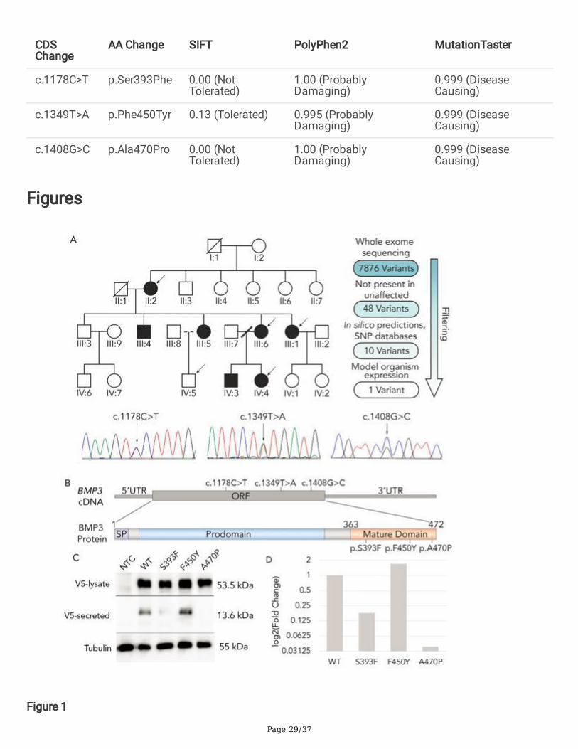
Page 29/37
CDSChange
AA Change SIFT PolyPhen2 MutationTaster
c.1178C>T p.Ser393Phe 0.00 (NotTolerated)
1.00 (ProbablyDamaging)
0.999 (DiseaseCausing)
c.1349T>A p.Phe450Tyr 0.13 (Tolerated) 0.995 (ProbablyDamaging)
0.999 (DiseaseCausing)
c.1408G>C p.Ala470Pro 0.00 (NotTolerated)
1.00 (ProbablyDamaging)
0.999 (DiseaseCausing)
Figures
Figure 1
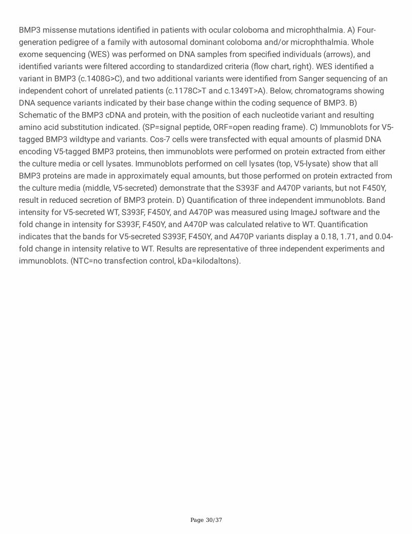
Page 30/37
BMP3 missense mutations identi�ed in patients with ocular coloboma and microphthalmia. A) Four-generation pedigree of a family with autosomal dominant coloboma and/or microphthalmia. Wholeexome sequencing (WES) was performed on DNA samples from speci�ed individuals (arrows), andidenti�ed variants were �ltered according to standardized criteria (�ow chart, right). WES identi�ed avariant in BMP3 (c.1408G>C), and two additional variants were identi�ed from Sanger sequencing of anindependent cohort of unrelated patients (c.1178C>T and c.1349T>A). Below, chromatograms showingDNA sequence variants indicated by their base change within the coding sequence of BMP3. B)Schematic of the BMP3 cDNA and protein, with the position of each nucleotide variant and resultingamino acid substitution indicated. (SP=signal peptide, ORF=open reading frame). C) Immunoblots for V5-tagged BMP3 wildtype and variants. Cos-7 cells were transfected with equal amounts of plasmid DNAencoding V5-tagged BMP3 proteins, then immunoblots were performed on protein extracted from eitherthe culture media or cell lysates. Immunoblots performed on cell lysates (top, V5-lysate) show that allBMP3 proteins are made in approximately equal amounts, but those performed on protein extracted fromthe culture media (middle, V5-secreted) demonstrate that the S393F and A470P variants, but not F450Y,result in reduced secretion of BMP3 protein. D) Quanti�cation of three independent immunoblots. Bandintensity for V5-secreted WT, S393F, F450Y, and A470P was measured using ImageJ software and thefold change in intensity for S393F, F450Y, and A470P was calculated relative to WT. Quanti�cationindicates that the bands for V5-secreted S393F, F450Y, and A470P variants display a 0.18, 1.71, and 0.04-fold change in intensity relative to WT. Results are representative of three independent experiments andimmunoblots. (NTC=no transfection control, kDa=kilodaltons).

Page 31/37
Figure 2
Zebra�sh with mutations in bmp3 exhibit delayed �ssure closure. A) Partial coding sequence alignmentof wild type and bmp3ua1020 mutant zebra�sh (top) and schematics of each resulting protein (bottom).A 5bp deletion (c.887_891del) was generated with CRISPR-Cas9, resulting in a frameshift and subsequentpremature stop codon (p.Thr96GlyfsTer2). The resulting truncated protein lacks the entire TGF-β maturedomain. (SP=Signal peptide) (B-E) Larvae from a bmp3-/- heterozygous incross were �xed at 48 or 65 hpfand laminin immuno�uorescence was conducted to visualize the extracellular matrix. Eyes weremanually dissected and mounted in pairs, then imaged on a confocal microscope. Both wild type andbmp3 mutant larvae at 48 hpf (B,C) show �ssure lobes in apposition but incomplete �ssure closure(indicated by arrows). In contrast, at 65 hpf (D,E) wild type larvae have completed �ssure closure, while23/130 bmp3 mutants show bilateral open �ssures (arrowheads). F) Quanti�cation of �ssure phenotypesin B-E, showing 18% bilateral open �ssures in bmp3 mutant larvae at 65 hpf. (WT=Wild-type, Open=Openchoroid �ssures, Closed=Closed choroid �ssures).
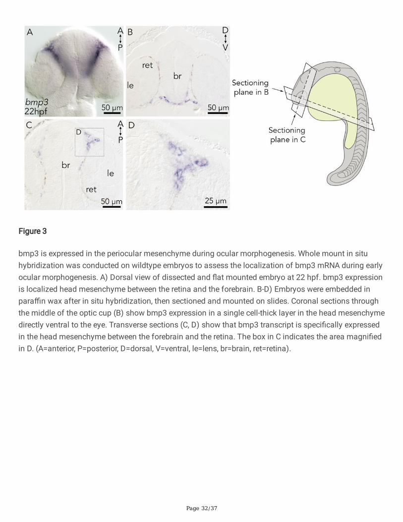
Page 32/37
Figure 3
bmp3 is expressed in the periocular mesenchyme during ocular morphogenesis. Whole mount in situhybridization was conducted on wildtype embryos to assess the localization of bmp3 mRNA during earlyocular morphogenesis. A) Dorsal view of dissected and �at mounted embryo at 22 hpf. bmp3 expressionis localized head mesenchyme between the retina and the forebrain. B-D) Embryos were embedded inpara�n wax after in situ hybridization, then sectioned and mounted on slides. Coronal sections throughthe middle of the optic cup (B) show bmp3 expression in a single cell-thick layer in the head mesenchymedirectly ventral to the eye. Transverse sections (C, D) show that bmp3 transcript is speci�cally expressedin the head mesenchyme between the forebrain and the retina. The box in C indicates the area magni�edin D. (A=anterior, P=posterior, D=dorsal, V=ventral, le=lens, br=brain, ret=retina).
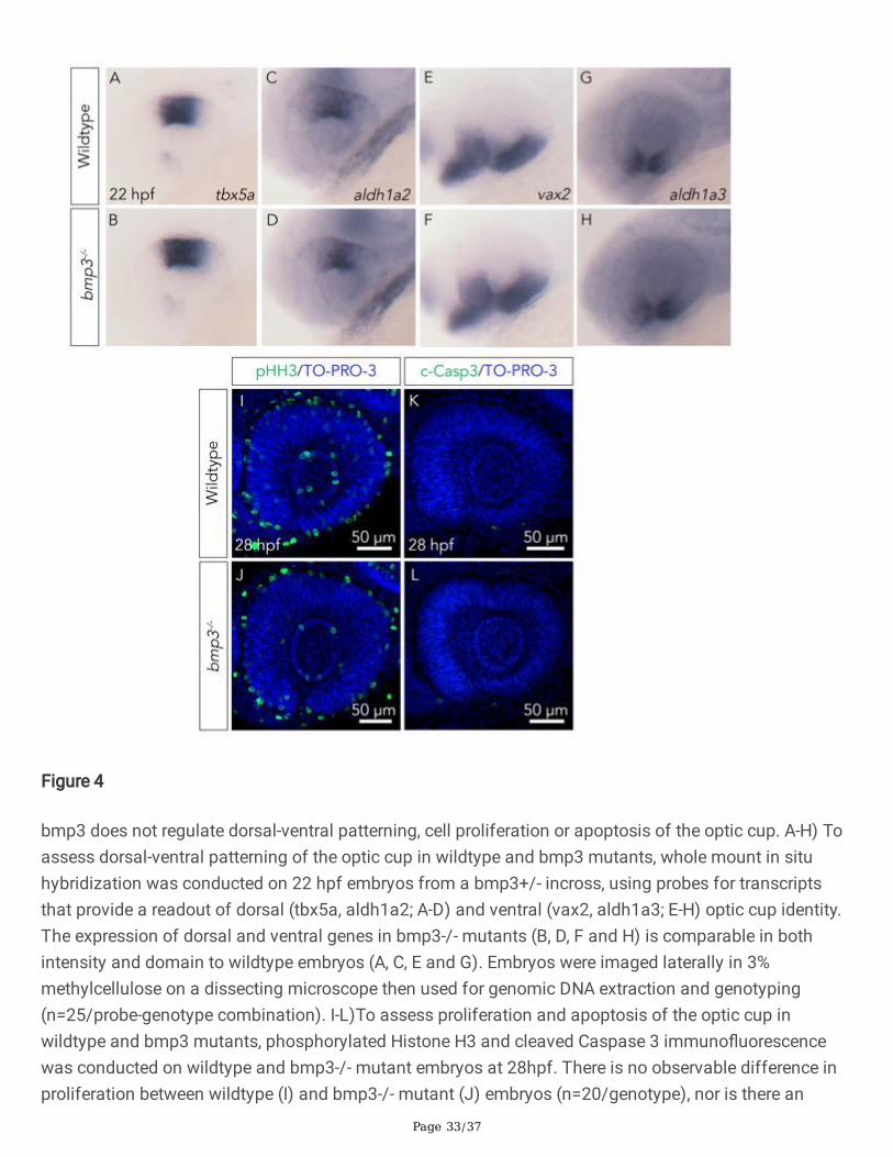
Page 33/37
Figure 4
bmp3 does not regulate dorsal-ventral patterning, cell proliferation or apoptosis of the optic cup. A-H) Toassess dorsal-ventral patterning of the optic cup in wildtype and bmp3 mutants, whole mount in situhybridization was conducted on 22 hpf embryos from a bmp3+/- incross, using probes for transcriptsthat provide a readout of dorsal (tbx5a, aldh1a2; A-D) and ventral (vax2, aldh1a3; E-H) optic cup identity.The expression of dorsal and ventral genes in bmp3-/- mutants (B, D, F and H) is comparable in bothintensity and domain to wildtype embryos (A, C, E and G). Embryos were imaged laterally in 3%methylcellulose on a dissecting microscope then used for genomic DNA extraction and genotyping(n=25/probe-genotype combination). I-L)To assess proliferation and apoptosis of the optic cup inwildtype and bmp3 mutants, phosphorylated Histone H3 and cleaved Caspase 3 immuno�uorescencewas conducted on wildtype and bmp3-/- mutant embryos at 28hpf. There is no observable difference inproliferation between wildtype (I) and bmp3-/- mutant (J) embryos (n=20/genotype), nor is there an
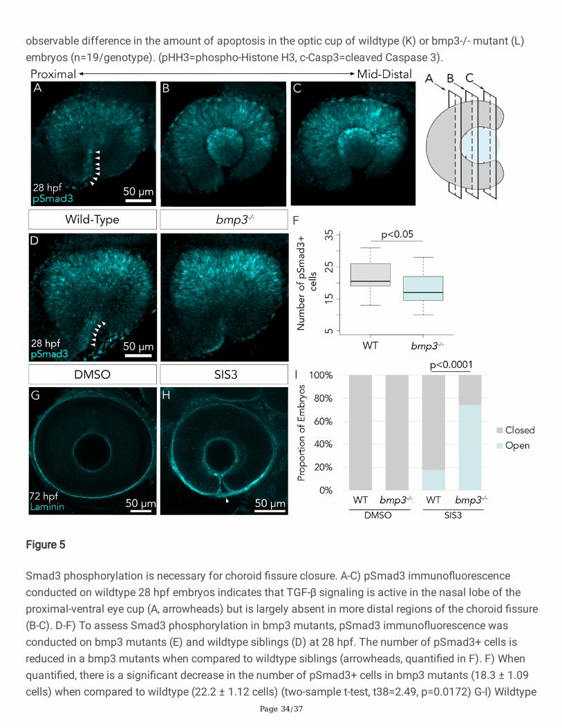
Page 34/37
observable difference in the amount of apoptosis in the optic cup of wildtype (K) or bmp3-/- mutant (L)embryos (n=19/genotype). (pHH3=phospho-Histone H3, c-Casp3=cleaved Caspase 3).
Figure 5
Smad3 phosphorylation is necessary for choroid �ssure closure. A-C) pSmad3 immuno�uorescenceconducted on wildtype 28 hpf embryos indicates that TGF-β signaling is active in the nasal lobe of theproximal-ventral eye cup (A, arrowheads) but is largely absent in more distal regions of the choroid �ssure(B-C). D-F) To assess Smad3 phosphorylation in bmp3 mutants, pSmad3 immuno�uorescence wasconducted on bmp3 mutants (E) and wildtype siblings (D) at 28 hpf. The number of pSmad3+ cells isreduced in a bmp3 mutants when compared to wildtype siblings (arrowheads, quanti�ed in F). F) Whenquanti�ed, there is a signi�cant decrease in the number of pSmad3+ cells in bmp3 mutants (18.3 ± 1.09cells) when compared to wildtype (22.2 ± 1.12 cells) (two-sample t-test, t38=2.49, p=0.0172) G-I) Wildtype
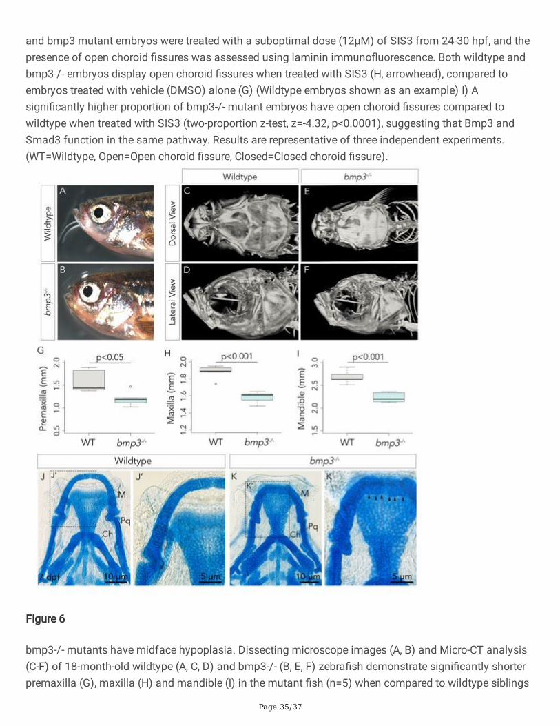
Page 35/37
and bmp3 mutant embryos were treated with a suboptimal dose (12μM) of SIS3 from 24-30 hpf, and thepresence of open choroid �ssures was assessed using laminin immuno�uorescence. Both wildtype andbmp3-/- embryos display open choroid �ssures when treated with SIS3 (H, arrowhead), compared toembryos treated with vehicle (DMSO) alone (G) (Wildtype embryos shown as an example) I) Asigni�cantly higher proportion of bmp3-/- mutant embryos have open choroid �ssures compared towildtype when treated with SIS3 (two-proportion z-test, z=-4.32, p<0.0001), suggesting that Bmp3 andSmad3 function in the same pathway. Results are representative of three independent experiments.(WT=Wildtype, Open=Open choroid �ssure, Closed=Closed choroid �ssure).
Figure 6
bmp3-/- mutants have midface hypoplasia. Dissecting microscope images (A, B) and Micro-CT analysis(C-F) of 18-month-old wildtype (A, C, D) and bmp3-/- (B, E, F) zebra�sh demonstrate signi�cantly shorterpremaxilla (G), maxilla (H) and mandible (I) in the mutant �sh (n=5) when compared to wildtype siblings
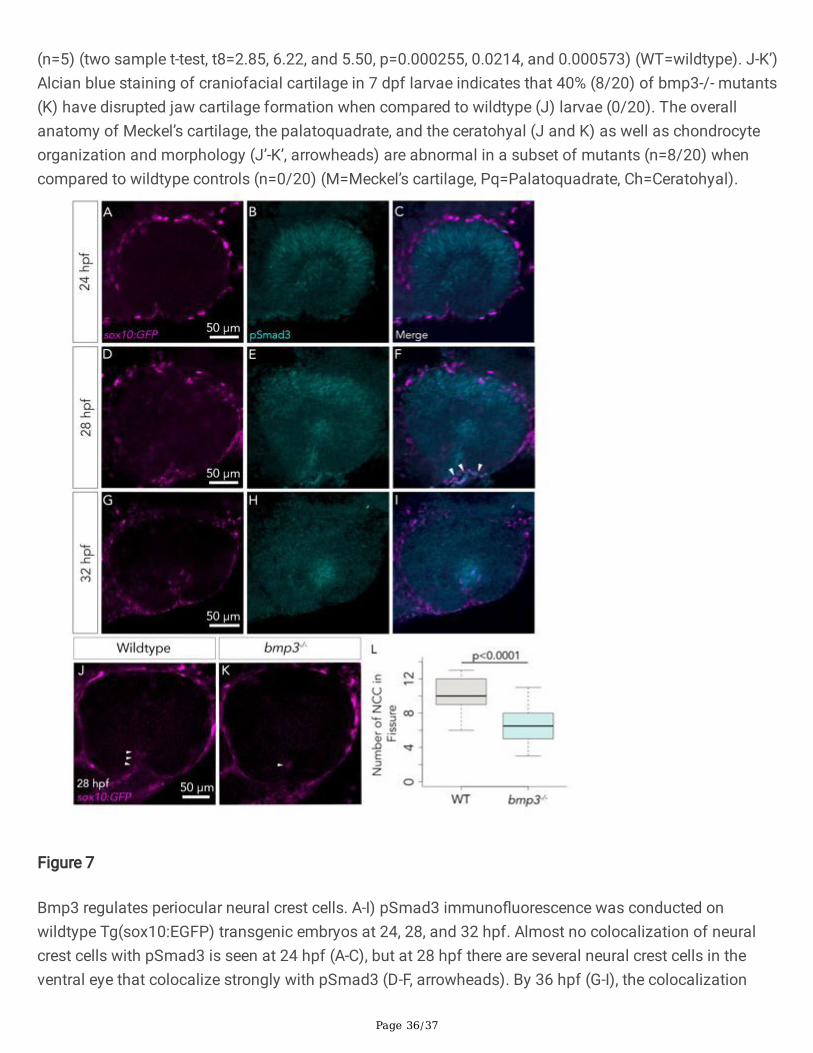
Page 36/37
(n=5) (two sample t-test, t8=2.85, 6.22, and 5.50, p=0.000255, 0.0214, and 0.000573) (WT=wildtype). J-K’)Alcian blue staining of craniofacial cartilage in 7 dpf larvae indicates that 40% (8/20) of bmp3-/- mutants(K) have disrupted jaw cartilage formation when compared to wildtype (J) larvae (0/20). The overallanatomy of Meckel’s cartilage, the palatoquadrate, and the ceratohyal (J and K) as well as chondrocyteorganization and morphology (J’-K’, arrowheads) are abnormal in a subset of mutants (n=8/20) whencompared to wildtype controls (n=0/20) (M=Meckel’s cartilage, Pq=Palatoquadrate, Ch=Ceratohyal).
Figure 7
Bmp3 regulates periocular neural crest cells. A-I) pSmad3 immuno�uorescence was conducted onwildtype Tg(sox10:EGFP) transgenic embryos at 24, 28, and 32 hpf. Almost no colocalization of neuralcrest cells with pSmad3 is seen at 24 hpf (A-C), but at 28 hpf there are several neural crest cells in theventral eye that colocalize strongly with pSmad3 (D-F, arrowheads). By 36 hpf (G-I), the colocalization

Page 37/37
between pSmad3 and sox10+ neural crest cells in the ventral eye has diminished. J-L) To assess theeffect of bmp3 on periocular neural crest cells, the number of sox10+ cells was examined in bmp3-/-mutants (K) and wildtype siblings (J) on a Tg(sox10:EGFP) transgenic background at 28 hpf. L) There aresigni�cantly fewer neural crest cells in the choroid �ssures of bmp3-/- embryos (6.56 ± 1.04 cells, n=18eyes) compared to wildtype siblings (10.1 ± 1.00 cells, n=18 eyes) (two sample t-test, t34= 4.76,p<0.0001). (WT=wildtype).
Supplementary Files
This is a list of supplementary �les associated with this preprint. Click to download.
SupplementalInformation.docx