Embed Size (px)
Citation preview

1
Ocular Pharmacokinetics of Brimonidine Drug Delivery System in
Monkeys and Translational Modeling for Selection of Dose and
Frequency in Clinical Trials
Authors: Mitalee Tamhane, Kenneth T. Luu,1 Mayssa Attar
Affiliations: Allergan, an AbbVie company, Irvine, CA, USA
This article has not been copyedited and formatted. The final version may differ from this version.JPET Fast Forward. Published on July 1, 2021 as DOI: 10.1124/jpet.120.000483
at ASPE
T Journals on N
ovember 25, 2021
jpet.aspetjournals.orgD
ownloaded from

2
Running title: Brimonidine Drug Delivery System ‒ Ocular Pharmacokinetics
Correspondence: Mitalee Tamhane, PhD, AbbVie Inc., RD2-2B, 2525 Dupont
Drive, Irvine, CA 92612, USA; E-mail: [email protected]
Telephone: 714-246-4211
Fax: 714-246-6510
Number of text pages: 15
Number of tables: 3
Number of figures: 3
Number of references: 42
Number of words in Abstract: 250
Number of words in Introduction: 715
Number of words in Discussion: 1489
ABBREVIATIONS: AMD, age-related macular degeneration; ARVO, Association for
Research in Vision and Ophthalmology; AUC, area under concentration-time curve;
BLQ, below limit of quantification; DDS, drug delivery system; FOCE, first-order
conditional estimation; HPLC, high-performance liquid chromatography; OFV,
objective function value; RPE, retinal pigmented epithelium; RSE, relative standard
error; SD, standard deviation; SE, standard error; VEGF, vascular endothelial growth
factor.
SECTION ASSIGNMENT:
Drug Discovery and Translational Medicine
This article has not been copyedited and formatted. The final version may differ from this version.JPET Fast Forward. Published on July 1, 2021 as DOI: 10.1124/jpet.120.000483
at ASPE
T Journals on N
ovember 25, 2021
jpet.aspetjournals.orgD
ownloaded from

3
ABSTRACT
Brimonidine, a selective alpha2-adrenoceptor agonist, displays putative retinal cyto-
neuroprotective activity in vitro and in vivo. An intravitreal sustained-release
brimonidine implant, Brimonidine Posterior Segment Drug Delivery System
(brimonidine DDS), allowing targeted drug delivery to the retina, has been developed
for potential clinical application. This study evaluates the in vivo posterior segment
pharmacokinetics of brimonidine DDS implant in the monkey eye, and applies
translational pharmacokinetic modeling to predict tissue exposure in the human eye.
Anesthetized Cynomolgus monkeys received a single intravitreal injection of
brimonidine DDS 400 µg implant before removal of study eyes at Days 7, 30, 60, 92,
120, and 150 post-implant (3-4 animals per time point) for assay of brimonidine in
aqueous humor, vitreous, and retina samples. Brimonidine concentrations in the
human eye were modeled using a linear, 3-compartment model, assuming
bidirectional distribution to/from the aqueous humor and retina, and elimination from
the aqueous humor. Monkey tissue volumes were scaled up to human values;
intercompartmental and elimination rate constants were assumed to be identical in
the two species. Modeling and simulations were performed using NONMEM v. 7.3, R
3.5.1. Brimonidine exposure was highest in the monkey vitreous and retina;
concentrations in the central (macula) and peripheral retina were maintained at high
levels (>100 ng/g) for 3 to 4 months. Simulated brimonidine concentration-time
profiles in human macula indicated that brimonidine DDS 400 µg implant would
deliver effective drug concentrations (20.7‒82.2 ng/g, based on animal
pharmacology) for approximately 3 months. Accordingly, administration of the 400 µg
implant at 3-month intervals is recommended.
This article has not been copyedited and formatted. The final version may differ from this version.JPET Fast Forward. Published on July 1, 2021 as DOI: 10.1124/jpet.120.000483
at ASPE
T Journals on N
ovember 25, 2021
jpet.aspetjournals.orgD
ownloaded from

4
SIGNIFICANCE STATEMENT
Brimonidine, an alpha2-adrenoceptor agonist, is cyto-neuroprotective in animal
models of retinal/optic nerve injury. Brimonidine Drug Delivery System (brimonidine
DDS) is an intravitreal sustained-release implant with potential ophthalmological
applications. This study explores the pharmacokinetics of brimonidine DDS 400 µg
implant in the monkey eye, and uses compartmental modeling to predict human
ocular tissue exposure. Targeted retinal brimonidine delivery from vitreous was
demonstrated in monkeys. Simulated tissue concentration-time profiles indicated
persistence of pharmacologically effective brimonidine concentrations for ≈3 months
in human retina.
This article has not been copyedited and formatted. The final version may differ from this version.JPET Fast Forward. Published on July 1, 2021 as DOI: 10.1124/jpet.120.000483
at ASPE
T Journals on N
ovember 25, 2021
jpet.aspetjournals.orgD
ownloaded from

5
Introduction
Brimonidine is a highly selective alpha2-adrenergic receptor agonist that is
currently approved in the United States and Europe for the treatment of open-angle
glaucoma and ocular hypertension. In addition to its intraocular pressure–lowering
effect, experimental studies have demonstrated that brimonidine possesses cyto-
and neuroprotective activity in a variety of animal models of retinal and optic nerve
injury, including acute retinal ischemia (Donello et al., 2001; Lafuente et al., 2001;
Vidal-Sanz et al., 2001; Lai et al., 2002), excitotoxic retinal injury (Galindo-Romero et
al., 2016), blue light phototoxicity (Ortín-Martínez et al., 2014; Valiente-Soriano et al.,
2019), chronic ocular hypertension (WoldeMussie et al., 2001; Hernández et al.,
2008), and optic nerve crush (Yoles et al., 1999; Saylor et al., 2009). In a chronic
progressive outer retinal degeneration model of geographic atrophy in monkeys,
brimonidine prevented light-induced damage to retinal pigment epithelium and
photoreceptors (Rajagopalan et al., 2019). The cyto- and neuroprotective effect of
brimonidine is thought to be related to its enhancement of the ability of retinal
neuronal cells to resist cellular stress, an effect possibly achieved through activation
of cell-survival signaling pathways or interference with cytotoxic signaling (Peng et
al., 1998; Wheeler et al., 2003; Saylor et al., 2009). Research indicates that the
alpha2-adrenergic receptor is expressed throughout the neurosensory retina
(Woldemussie et al., 2007), and that its activation has cascading effects on signal
pathways that block apoptosis (Wheeler et al., 2003). Brimonidine demonstrates
cyto- and neuroprotective activity in vitro, reducing the toxic effects of hydroquinone
exposure on human retinal pigment epithelium and retinal Muller cells and rendering
them more resistant to injury (Ramírez et al., 2016). These data show that
This article has not been copyedited and formatted. The final version may differ from this version.JPET Fast Forward. Published on July 1, 2021 as DOI: 10.1124/jpet.120.000483
at ASPE
T Journals on N
ovember 25, 2021
jpet.aspetjournals.orgD
ownloaded from

6
brimonidine has pharmacological properties consistent with protection of retinal
cellular and neuronal function.
Clinical evidence of a potential cyto- and neuroprotective effect of brimonidine
is provided by the Low-Pressure Glaucoma Treatment Study, in which topical
brimonidine was found to reduce the risk of visual field progression compared with
timolol in normal-tension glaucoma patients despite the two drugs showing similar
ocular hypotensive effect (Krupin et al., 2011). Additionally, the cyto- and
neuroprotective effect of brimonidine has been explored in patients with age-related
macular degeneration (AMD) (Ferencz et al., 2005), geographic atrophy
(Kuppermann et al., 2020), retinitis pigmentosa (Merin et al., 2008), diabetic
retinopathy (Mondal et al., 2004; Simó et al., 2019), and acute nonarteritic anterior
ischemic optic neuropathy (Wilhelm et al., 2006).
To exert a durable cyto- and neuroprotective effect, a drug should be
delivered in adequate concentrations to the retina over an extended period. Topical
brimonidine (brimonidine tartrate 0.2% ophthalmic solution) shows only limited
posterior segment penetration, achieving vitreous concentrations of ≤5 ng/mL in the
phakic human eye (Kent et al., 2001). Intravitreal administration enables targeted
delivery of brimonidine to the retina. However, brimonidine undergoes rapid
clearance following intravitreal injection, with a vitreous elimination half-life of 1.45
hours after single bolus administration (928 ng in 50-µL injection volume) in the
rabbit eye (unpublished Allergan data). Due to the short duration of ocular exposure,
frequent intravitreal injections would be necessary to achieve sustained retinal tissue
concentrations. To address these constraints, an intravitreal implant consisting of
brimonidine 400 µg free base in a slow-release polymer matrix—the sustained-
release Brimonidine free-base Posterior Segment Drug Delivery System
This article has not been copyedited and formatted. The final version may differ from this version.JPET Fast Forward. Published on July 1, 2021 as DOI: 10.1124/jpet.120.000483
at ASPE
T Journals on N
ovember 25, 2021
jpet.aspetjournals.orgD
ownloaded from

7
(brimonidine DDS; Allergan, an AbbVie company, Irvine, CA)—has been developed
for potential clinical application in the treatment of geographic atrophy and for cyto-
and neuroprotection in glaucoma. The implant evaluated in this study offers the
advantage over earlier prototypes of providing a higher drug load, faster drug
release, and more rapid matrix bioerosion, resulting in higher ocular tissue exposure.
Despite interspecies differences in ocular anatomic and physiological
parameters, animal studies can provide reliable estimates of vitreous
pharmacokinetics in the human eye (Del Amo et al. 2015). For this purpose,
monkeys are commonly used to evaluate ocular pharmacokinetic profiles
(Gaudreault et al. 2005; Chang-Lin et al. 2011; Shen et al. 2014; Niwa et al. 2015).
The objectives of this study were (i) to evaluate the in vivo posterior segment
pharmacokinetics of brimonidine DDS 400 µg implant in the monkey eye; (ii) to
perform compartmental modeling of the monkey pharmacokinetic data to enable
translation to humans; and (iii) to use the translational model to predict vitreous and
macula exposure in the human eye and select an appropriate dosage regimen for
clinical testing.
Methods
Animal Experiments
The in vivo portion of this study was conducted at Covance Laboratories Inc.
(Madison, WI) in compliance with the Animal Welfare Act (Regulations 9 CFR Parts
1, 2 and 3), and the Association for Research in Vision and Ophthalmology (ARVO)
Statement for the Use of Animals in Ophthalmic and Vision Research.
Male Cynomolgus monkeys weighing approximately 2.3 to 3.7 kg were
obtained from Covance Research Products Inc. (Denver, PA and Alice, TX). The
This article has not been copyedited and formatted. The final version may differ from this version.JPET Fast Forward. Published on July 1, 2021 as DOI: 10.1124/jpet.120.000483
at ASPE
T Journals on N
ovember 25, 2021
jpet.aspetjournals.orgD
ownloaded from

8
animals were housed in a temperature-controlled facility with a 12-hour light/dark
cycle, with unrestricted access to food and water, and were acclimated to study
conditions for 4 weeks.
In preparation for dosing with brimonidine DDS 400 µg implant, monkeys were
lightly anesthetized with intramuscular ketamine 10 mg/kg and dexmedetomidine
0.025 mg/kg, with additional anesthesia provided as needed. Ocular preparation for
intravitreal injection involved administration of 1 or 2 drops of a topical ophthalmic
anesthetic (proparacaine hydrochloride 0.5%) followed by a broad-spectrum
ophthalmic microbicide (povidone-iodine 5% solution; Betadine®). After 2–3 minutes,
study eyes were irrigated with sterile saline and a drop of topical ophthalmic
anesthetic (lidocaine hydrochloride 2%) was applied, followed by an ophthalmic
fluoroquinolone antibiotic ointment for infection prophylaxis. A 25-gauge needle and
pre-loaded brimonidine DDS applicator was used to deliver a single intravitreal
injection of brimonidine DDS 400 µg implant (Allergan, an AbbVie company, Irvine,
CA) to the study eye. Injections were performed in the dorsotemporal quadrant,
approximately 2.5 mm posterior to the limbus, through the sclera and pars plana.
The bevel of the needle was directed downward and posteriorly to avoid the lens.
Brimonidine DDS 400 µg implant was deployed in the center of the vitreous by
depressing an actuator button on the applicator.
At predetermined time intervals over 5 months post-implant (Days 7, 30, 60,
92, 120, and 150), 3 or 4 animals per time point were euthanized via exsanguination
under sodium pentobarbital anesthesia. Blood samples (approximately 5 mL) were
collected via cardiac puncture at the time of sacrifice, stored in K3 EDTA-coated
tubes on wet ice, and centrifuged to obtain plasma. Study eyes were enucleated at
the time of sacrifice, aqueous humor was removed, and the eyes were then flash-
This article has not been copyedited and formatted. The final version may differ from this version.JPET Fast Forward. Published on July 1, 2021 as DOI: 10.1124/jpet.120.000483
at ASPE
T Journals on N
ovember 25, 2021
jpet.aspetjournals.orgD
ownloaded from

9
frozen in liquid nitrogen before collection (within 2 days) of vitreous, retina, and
choroid with retinal pigmented epithelium (RPE). Vitreous was collected together
with the implant remnants, and the latter were separated from the defrosted vitreous
before assay of vitreous drug concentrations. Retina samples consisted of 8 mm-
diameter circular punch biopsies obtained from the macula (‘central retina’) and from
the remaining retinal tissue (‘peripheral retina’).
External ophthalmic examination, slit-lamp biomicroscopy of the adnexa and
anterior eye, and indirect ophthalmoscopy of the fundus were conducted in
ketamine-anesthetized monkeys prior to intravitreal dosing and at Days 3, 14, 92,
180, and 270 post-dose by a board-certified veterinary ophthalmologist.
Bioanalysis
Concentrations of brimonidine in plasma and ocular tissue samples were
assayed by liquid chromatography–tandem mass spectrometry (LC-MS/MS), using
an API 3000 or API 5000 triple quadrupole spectrometer (AB Sciex™) interfaced with
a high-performance liquid chromatography (HPLC) system (Shimadzu Scientific
Instruments, Columbia, MD) and auto-sampler (Shimadzu Scientific Instruments,
Columbia, MD; Perkin-Elmer). HPLC was performed on a Synergi Polar-RP column
(2.0 × 50 mm, 4 µm; Phenomenex Corp.) using mobile phase A with 0.1% formic
acid in 70:30 acetonitrile:water and mobile phase B with 0.1% ammonium hydroxide
in 70:30 acetonitrile:water at flow rates of 0.3 or 0.5 mL/min. Mass spectrometric
detection was performed using multiple reaction monitoring scan mode with
precursor/product ion pairs m/z 292 → m/z 212 for brimonidine and m/z 296 → m/z
216 for brimonidine-d4. Assay ranges for brimonidine in plasma and ocular tissues
This article has not been copyedited and formatted. The final version may differ from this version.JPET Fast Forward. Published on July 1, 2021 as DOI: 10.1124/jpet.120.000483
at ASPE
T Journals on N
ovember 25, 2021
jpet.aspetjournals.orgD
ownloaded from

10
were 0.05–10.0 ng/mL (plasma), 0.1–50.0 ng/mL (aqueous humor), 0.25–200.0
ng/mL (vitreous), and 0.1–100.0 ng (retina, and choroid‒RPE).
Pharmacokinetic Analysis
Noncompartmental analysis was used to determine the following
pharmacokinetic parameters for brimonidine: maximum concentration, time to
maximum concentration, and area under the concentration-time curve from time zero
to the last quantifiable sampling time (AUC0-tlast). For AUC0-tlast, mean and standard
error (SE) of mean values were calculated using the composite AUC calculation
available in Watson™ version 7.3 software (Waltham, MA). Mean (standard
deviation [SD] and SE) tissue concentrations of brimonidine were calculated for each
time point using Watson™ version 7.3 software. Within each data group, if ≤50% of
individual readings were below the limit of quantification (BLQ), a value of zero was
substituted for BLQ readings, and these substituted values were included in the
calculation. If >50% of readings were BLQ, the mean value was reported as “not
calculable”.
Pharmacokinetic Modeling and Human Dose Prediction
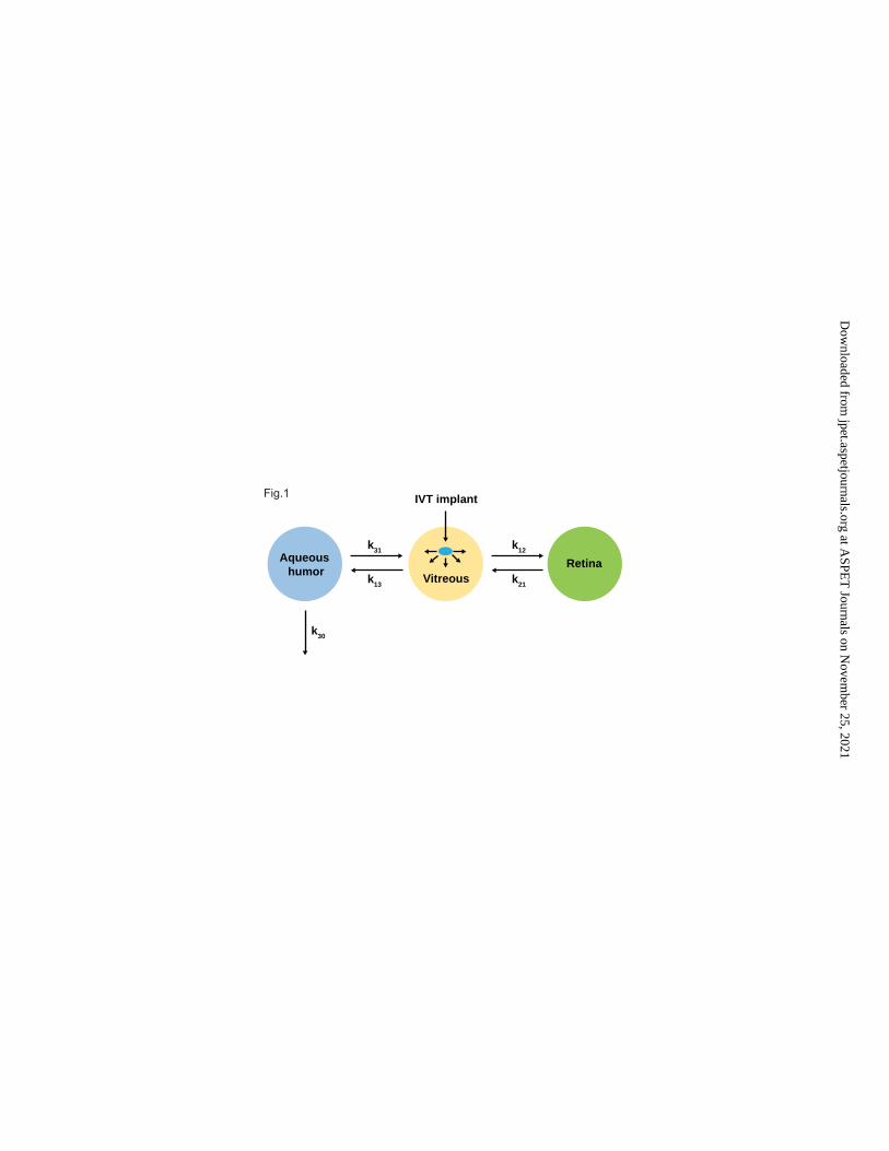
Brimonidine concentrations in human aqueous humor, vitreous, and retina
(macula) were modeled using a linear, 3-compartment model (Figure 1). As shown in
the figure, the model assumes that brimonidine undergoes slow release from the
implant into the vitreous, and is distributed bidirectionally to and from the aqueous
humor and retina. Different structural models were tested to fit the data in all 3
tissues: (i) a model involving drug elimination from both the retina and the aqueous
humor; and (ii) a model with drug elimination occurring from either the retina or the
This article has not been copyedited and formatted. The final version may differ from this version.JPET Fast Forward. Published on July 1, 2021 as DOI: 10.1124/jpet.120.000483
at ASPE
T Journals on N
ovember 25, 2021
jpet.aspetjournals.orgD
ownloaded from

11
aqueous humor. Based on estimates of the objective function value (OFV) of the
various models, the model involving drug elimination from the aqueous humor was
chosen (see Results and Discussion sections). A bioavailability factor was included
to offset the tendency of the model to overpredict aqueous humor concentrations,
and to account for potential binding of brimonidine to melanin (see Discussion). The
device release rate (zero-order) was previously determined from in vitro experiments
to be 5.9 µg/day; the same value was assumed to apply under in vivo conditions and
was fixed in the model. Volume of drug distribution was fixed to the specific
anatomical characteristics (weight or volume) of each tissue (Table 1). This
approach allows for interspecies scaling, substituting human for monkey tissue
volumes. Intercompartmental and elimination rate constants were assumed to be
identical for monkeys and humans. Once the model was scaled to the human eye,
simulations were performed to predict a suitable dose and regimen for testing in the
clinic.
Modeling and simulations were performed using NONMEM v. 7.3, R 3.5.1.
The data were kept as naïve pool for model fitting. The first-order conditional
estimation (FOCE) method was implemented in NONMEM. The final model applied
additive and proportional residual error model. The model was fitted to concentration
data for the aqueous humor, vitreous, and retina simultaneously to obtain parameter
estimates. Concentrations below the limit of detection were treated with the M5
method (dividing the BLQ by 2). Parameter precision was obtained by importance
sampling following FOCE.
Results
This article has not been copyedited and formatted. The final version may differ from this version.JPET Fast Forward. Published on July 1, 2021 as DOI: 10.1124/jpet.120.000483
at ASPE
T Journals on N
ovember 25, 2021
jpet.aspetjournals.orgD
ownloaded from

12
A total of 22 monkeys (22 eyes) received the brimonidine DDS 400 µg
implant, and safety and pharmacokinetic data were obtained from 22 and 18 eyes,
respectively. Ophthalmic examination indicated that the intravitreal implant was well
tolerated over 9 months of follow-up. All animals appeared healthy with no overt
signs of toxicity relating to the implant. In a minority of animals the implant elicited a
mild and transient increase in vitreous cell numbers that subsided within
approximately 90 days. Injection-related effects (subconjunctival hemorrhage at the
injection site, mildly degraded view of the fundus, and fibrin/blood on the tip of the
implant or in the vitreous) were short-lived, being evident only at Day 3 post-dose.
Pharmacokinetics of Brimonidine DDS in Monkeys
Noncompartmental pharmacokinetic parameters for brimonidine in each
tissue are summarized in Table 2. Brimonidine exposure was highest in the vitreous
and retina (macula and periphery), with lower concentrations observed in the
aqueous humor, indicating targeted retinal drug delivery from the vitreous. Mean
brimonidine concentration peaked in the vitreous (10,900 ng/mL) and peripheral
retina (12,200 ng/g) at 60 days post-implant, and in the central retina (714 ng/g) at
30 days post-implant. Brimonidine concentrations in the retina and vitreous were
maintained at high levels (>100 ng/g and >1,000 ng/mL, respectively) for the first 2
or 3 months post-implant before falling sharply to <10 ng/g and <1 ng/mL,
respectively, at 5 months. Brimonidine concentrations in retina were sustained for
longer than those in the vitreous, remaining above 30 ng/g for the first 4 months
post-implant compared with <2 ng/mL at the same time-point for vitreous.
Brimonidine concentrations in macula remained quantifiable for at least 120 days
post-implant. Systemic drug exposure was negligible after intravitreal administration
This article has not been copyedited and formatted. The final version may differ from this version.JPET Fast Forward. Published on July 1, 2021 as DOI: 10.1124/jpet.120.000483
at ASPE
T Journals on N
ovember 25, 2021
jpet.aspetjournals.orgD
ownloaded from

13
of brimonidine DDS 400 µg implant, with plasma brimonidine concentrations
remaining below the limit of quantification (0.05 ng/mL) at all time-points.
Pharmacokinetic Modeling of Monkey Data
The final model was a 3-compartment, linear model with a single exit rate
constant coming from the aqueous humor compartment (OFV = 549). Adding a
second exit compartment from the retina did not improve the fit or alter the OFV, but
resulted in inclusion of an additional parameter. The model with elimination occurring
solely from the retina produced a higher OFV (562). The final model was selected on
the basis of having the lowest OFV and fewest parameters of those models
evaluated. The final model parameters are summarized in Table 3, and a schematic
of the model is presented in Figure 1. Addition of the bioavailability factor to the
model improved the OFV by >100 points. Differential rates of drug distribution from
vitreous to retina and from retina to vitreous provided optimal data fit and were
adopted for the final model. Both additive and proportional (constant coefficient of
variation) error models were tested to capture the residual error in concentration data
for vitreous and retina. The final model had both additive and proportional error
structures. Figure 2 presents the final model fit to the pharmacokinetic data in
monkeys.
The volume or weight of the ocular tissues were changed from the values
reported for monkeys to those reported for humans. Simulated brimonidine
concentration-time profiles in human aqueous humor, vitreous, and retina (macula)
are shown in Figure 3. The brimonidine DDS 400 µg implant is anticipated to deliver
pharmacologically effective drug concentrations in the range of 20.7 to 82.2 ng/g
(based on internal data generated in animal pharmacology models) to the human
This article has not been copyedited and formatted. The final version may differ from this version.JPET Fast Forward. Published on July 1, 2021 as DOI: 10.1124/jpet.120.000483
at ASPE
T Journals on N
ovember 25, 2021
jpet.aspetjournals.orgD
ownloaded from

14
retina for approximately 3 months. These simulated brimonidine concentrations
(~100 ng/g) in human retina are 160-fold higher than the in vitro EC50 value at the
human alpha2 adrenergic receptor (0.6 ng/mL or 2 nM). For estimating potential
efficacious in vivo levels in humans, we assumed a multiple of 10- to 100-fold over
the in vitro EC50 value to account for in vitro to in vivo translation. The ‘in vivo’
efficacious concentrations generated in animal models fall within this multiple and
hence were used as target levels.
Discussion
Intravitreal injection is a widely accepted route of administration for ensuring
targeted drug delivery to the posterior segment of the eye. This route is approved for
administration of anti-vascular endothelial growth factor therapy (pegaptanib,
ranibizumab, and aflibercept) for choroidal neovascularization caused by “wet” AMD
and also for corticosteroid treatments (triamcinolone acetonide) for intraocular
inflammation (Novack, 2009). In addition to the above treatments, which are given as
solution formulations, dexamethasone intravitreal implant (Ozurdex®) is approved for
treatment of posterior segment uveitis, retinal vein occlusion, and diabetic macular
edema. In the case of small molecules, which usually exhibit a vitreous half-life of
several hours, the implant delivery system ensures sustained drug delivery to the
target tissue for long periods, thereby minimizing the frequency of injections.
Brimonidine DDS, an intravitreal implant consisting of 400 µg brimonidine free base
loaded in a slow-release polymer matrix, was developed to achieve
pharmacologically effective concentrations in the retina for prolonged periods.
The in vivo study in monkeys was performed to characterize the ocular
pharmacokinetics of single-dose brimonidine DDS 400 µg implant following
This article has not been copyedited and formatted. The final version may differ from this version.JPET Fast Forward. Published on July 1, 2021 as DOI: 10.1124/jpet.120.000483
at ASPE
T Journals on N
ovember 25, 2021
jpet.aspetjournals.orgD
ownloaded from

15
intravitreal administration. Findings indicated that brimonidine concentrations in the
central retina (macula) and peripheral retina were maintained at high levels (>100
ng/g) for 3 to 4 months in this species.
The monkey is a well-established model for studying the pharmacokinetics of
intravitreally administered drugs (Short, 2008), and allows simultaneous sampling of
ocular tissues at serial time points in different animals. The monkey eye resembles
the human eye closely with regard to anatomy and physiology, including the
presence of a macula, but it too has a small vitreous cavity (2 mL), which needs to
be taken into consideration when extrapolating pharmacokinetic findings. Since the
monkey is an appropriate translational species with regard to macular
pharmacokinetics, monkey data were used to predict human aqueous humor,
vitreous, and macula concentrations using pharmacokinetic modeling. Aqueous
humor and vitreous are the most commonly sampled matrices in humans to predict
pharmacokinetics as well as biomarker response to correlate with efficacy endpoints
for retinal disorders. Therefore, predicting concentrations in both human aqueous
humor and vitreous, in addition to the target tissue, is of value. In building our
pharmacokinetic model, we used brimonidine concentrations from retina central
punch (macula) rather than peripheral or total retina, since the macula represents the
key site of action of brimonidine.
Brimonidine has been shown to bind reversibly, and with high affinity, to
ocular melanin in vitro (Tang-Liu et al., 1992), and to distribute preferentially to, and
clear more slowly from, pigmented than non-pigmented ocular tissues in the monkey
(Acheampong et al., 2002). We conjectured that the need to incorporate the
bioavailability term to improve the fit of the aqueous humor data was attributable to
extensive melanin binding, which lowers the concentration of free drug. Without
This article has not been copyedited and formatted. The final version may differ from this version.JPET Fast Forward. Published on July 1, 2021 as DOI: 10.1124/jpet.120.000483
at ASPE
T Journals on N
ovember 25, 2021
jpet.aspetjournals.orgD
ownloaded from

16
integrating the bioavailability term, the model consistently overfitted the aqueous
humor data. The discrepancy between the duration of brimonidine release from the
implant (2 to 3 months) and the prolonged duration of retinal brimonidine
concentrations (>5 months) is consistent with drug-melanin binding in the retina.
Likewise, the low levels of brimonidine seen in the vitreous between 90 and 150
days post-implant (ie, after drug release from the implant has ceased) might be due
to release of melanin-bound brimonidine and redistribution of free drug from retina to
vitreous.
The model appeared to point to brimonidine being predominantly eliminated
via the aqueous humor. Smaller and more lipophilic drugs are thought to be
predominantly eliminated from the retina by crossing blood-retinal barriers, whereas
larger or more hydrophilic drugs are eliminated from the anterior segment through
aqueous humor outflow (Del Amo et al., 2017; Varela-Fernández et al., 2020). When
we tested the model with elimination from both the aqueous humor and the retina,
the estimated elimination rate constant from the aqueous humor (k30) was 0.022 day-
1, whereas the retinal elimination rate constant (k20) was <1 × 10-14 day-1. Since
retinal elimination was near zero and inclusion of this parameter did not improve the
OFV, we decided to exclude retinal elimination from the final model. Although, in
general, an empirical model such as this provides no proof of the mechanism of drug
clearance, it is feasible that brimonidine might be eliminated predominantly from the
aqueous humor on account of its highly hydrophilic nature and small molecular size,
with the latter potentially allowing clearance through the chamber angle or
Schlemm’s canal (Agrahari et al., 2016).
Brimonidine’s distribution to the posterior ocular tissues following intravitreal
administration differs quantitatively from that reported after topical administration. In
This article has not been copyedited and formatted. The final version may differ from this version.JPET Fast Forward. Published on July 1, 2021 as DOI: 10.1124/jpet.120.000483
at ASPE
T Journals on N
ovember 25, 2021
jpet.aspetjournals.orgD
ownloaded from

17
another study, twice-daily topical instillation of 0.5% [14C]-brimonidine tartrate
solution (119 µg of brimonidine) for 14 days (total dose = 3,332 µg) into monkey
eyes resulted in the following order of drug distribution: choroid/retina > aqueous
humor > vitreous (Acheampong et al., 2002). Mean brimonidine concentrations
peaked in vitreous at 1 hour post-dose (0.071 µg-Eq/g) and in choroid/retina at 24
hours post-dose (30.6 µg-Eq/g), while estimated AUC0-90 days was 0.960 µg-Eq·day/g
in vitreous and 1,499.0 µg-Eq·day/g in choroid/retina. (Data are reported in units of
radioactivity since intact brimonidine was the major radioactive component in all
tissues at all time-points, accounting for 83%‒98% of total radioactivity.) Thus,
brimonidine exposure in vitreous after administration of brimonidine DDS 400 µg was
>500-fold higher than that achieved after topical administration of brimonidine
tartrate 0.5% solution, despite the implant containing an 8-fold lower total
brimonidine dose (total topical dose of brimonidine over 14 days = 3,332 µg). In the
study of topically instilled 0.5% [14C]-brimonidine tartrate solution, retinal and
choroidal tissues were not separated, and therefore total radioactivity or brimonidine
concentrations were presented for combined retina/choroid. In our study with
intravitreal brimonidine DDS, retina was separated from choroid-RPE and,
furthermore, was subdivided into “retina central punch” (macula) and “retina
periphery” (remaining retina). This difference in tissue sampling has a profound
impact on estimates of brimonidine concentrations available for α2-adrenoceptor
activation in the retina. Brimonidine binds strongly but reversibly to ocular melanin,
and this can affect the drug’s disposition and lead to overestimation of therapeutic
drug levels in ocular tissue.
A study of the ocular distribution of topically administered 0.1% brimonidine
tartrate ophthalmic solution (35 µL) in the pigmented rabbit demonstrated that free
This article has not been copyedited and formatted. The final version may differ from this version.JPET Fast Forward. Published on July 1, 2021 as DOI: 10.1124/jpet.120.000483
at ASPE
T Journals on N
ovember 25, 2021
jpet.aspetjournals.orgD
ownloaded from

18
brimonidine concentrations in retina/choroid were 100-fold lower than total
brimonidine concentrations (Shinno et al., 2017). Therefore, retina separated from
choroid-RPE should provide a better estimate of brimonidine levels available for α2-
adrenoceptor activation. As demonstrated in Figure 2, after administration of
brimonidine DDS 400 µg implant, drug concentrations in monkey retina (macula),
representing free brimonidine concentrations, are maintained above the
pharmacologically relevant concentrations (20.7 ‒ 82.2 ng/g; determined in
pharmacology models of retinal degeneration) for 3 months post-dose. Thus,
comparison of posterior ocular tissue exposure following topical and intravitreal
brimonidine implant administration suggests that the intravitreal delivery route
provides higher posterior tissue concentrations, and that these are sustained over a
longer period, thereby avoiding the burden to the patient of daily eyedrop
administration.
Our simulations of brimonidine concentration-time profiles in human aqueous
humor, vitreous and macula, based on pharmacokinetic modeling of the monkey
data, suggest that, at a conservative estimate, the brimonidine DDS 400 µg implant
will provide sustained, pharmacologically effective drug concentrations in the human
macula for approximately 3 months following intravitreal administration. Based on
these model predictions, a dosage regimen of 400 µg, administered intravitreally
every 3 months, was recommended for clinical trials.
Limitations to the data collection and model are worth noting. Firstly, we did
not account for the location of the implant, which could impact drug distribution in the
posterior tissues. Secondly, the device release rate was assumed to be constant
over the duration of drug release and the same as the in vivo release rate. There
could be differences in in vitro and in vivo release rates, which could impact the
This article has not been copyedited and formatted. The final version may differ from this version.JPET Fast Forward. Published on July 1, 2021 as DOI: 10.1124/jpet.120.000483
at ASPE
T Journals on N
ovember 25, 2021
jpet.aspetjournals.orgD
ownloaded from

19
model predictions. Notwithstanding this, preliminary internal data (data not shown)
indicated that there was good correlation between in vitro and in vivo release rates,
and this assumption resulted in a reasonable fit of the model to the data. The model
also assumed the intercompartmental and elimination rate constants to be identical
for monkeys and humans since we are limited by the paucity of drug concentration
data generated in human ocular tissues. Thirdly, since ocular pharmacokinetic
sampling is a terminal procedure, serial pharmacokinetic data could not be collected
from the same animal. Consequently, estimates of inter-individual variability in
pharmacokinetic parameters were unavailable, and the model was fitted to naïve
pool data. Finally, the model assumes linear pharmacokinetics based on a single
dose level. For this study, only the highest feasible drug dose was evaluated with the
optimized implant formulation, due to the long duration of follow-up and the need to
minimize the number of study animals. As studies with monkeys are resource
intensive, particularly those involving terminal procedures for collection of ocular
tissues, we typically limit implant investigations to the final or near-final formulation.
Although our linear pharmacokinetic model provided good data-fit, the possibility of
nonlinear pharmacokinetics should not be ruled out.
Acknowledgments
Medical writing and editorial assistance were provided to the authors by
Andrew Fitton, PhD, of Evidence Scientific Solutions (Horsham, UK) and funded by
Allergan, an AbbVie company, Irvine, CA, USA. All authors met the ICMJE
authorship criteria. No honoraria or payments were made for authorship.
Authorship Contributions Participated in research design: Attar, Luu, Tamhane
This article has not been copyedited and formatted. The final version may differ from this version.JPET Fast Forward. Published on July 1, 2021 as DOI: 10.1124/jpet.120.000483
at ASPE
T Journals on N
ovember 25, 2021
jpet.aspetjournals.orgD
ownloaded from

20
Conducted experiments: Tamhane
Performed data analysis: Luu, Tamhane
Wrote or contributed to the writing of the manuscript: Attar, Luu, Tamhane
This article has not been copyedited and formatted. The final version may differ from this version.JPET Fast Forward. Published on July 1, 2021 as DOI: 10.1124/jpet.120.000483
at ASPE
T Journals on N
ovember 25, 2021
jpet.aspetjournals.orgD
ownloaded from

21
References
Acheampong AA, Shackleton M, John B, Burke J, Wheeler L and Tang-Liu D (2002)
Distribution of brimonidine into anterior and posterior tissues of monkey,
rabbit, and rat eyes. Drug Metab Dispos 30:421-429.
Agrahari V, Mandal A, Agrahari V, Trinh HM, Joseph M, Ray A, Hadji H, Mitra R, Pal
D and Mitra AK (2016) A comprehensive insight on ocular pharmacokinetics.
Drug Deliv Transl Res 6:735-754.
Bill A and Hellsing K (1965) Production and drainage of aqueous humor in the
cynomolgus monkey (Macaca irus). Invest Ophthalmol 4:920-926.
Chang-Lin J-E, Attar M, Acheampong AA, Robinson MR, Whitcup SW, Kuppermann
BD and Welty D (2011) Pharmacokinetics and pharmacodynamics of a
sustained-release dexamethasone intravitreal implant. Invest Ophthalmol Vis Sci
52:80-86.
Del Amo EM, Rimpelä AK, Heikkinen E, Kari OK, Ramsay E, Lajunen T, Schmitt M,
Pelkonen L, Bhattacharya M, Richardson D, Subrizi A, Turunen T, Reinisalo
M, Itkonen J, Toropainen E, Casteleijn M, Kidron H, Antopolsky M, Vellonen
KS, Ruponen M and Urtti A (2017) Pharmacokinetic aspects of retinal drug
delivery. Prog Retin Eye Res 57:134-185.
Del Amo EM and Urtti A (2015) Rabbit as an animal model for intravitreal
pharmacokinetics: clinical predictability and quality of the published data. Expt
Eye Res 137:111-124.
Donello JE, Padillo EU, Webster ML, Wheeler LA and Gil DW (2001) alpha(2)-
Adrenoceptor agonists inhibit vitreal glutamate and aspartate accumulation
This article has not been copyedited and formatted. The final version may differ from this version.JPET Fast Forward. Published on July 1, 2021 as DOI: 10.1124/jpet.120.000483
at ASPE
T Journals on N
ovember 25, 2021
jpet.aspetjournals.orgD
ownloaded from

22
and preserve retinal function after transient ischemia. J Pharmacol Exp Ther
296:216-223.
Feke GT, Tagawa H, Deupree DM, Goger DG, Sebag J and Weiter JJ (1989) Blood
flow in the normal human retina. Invest Ophthalmol Vis Sci 30:58-65.
Ferencz JR, Gilady G, Harel O, Belkin M and Assia EI (2005) Topical brimonidine
reduces collateral damage caused by laser photocoagulation for choroidal
neovascularization. Graefes Arch Clin Exp Ophthalmol 243:877-880.
Galindo-Romero C, Harun-Or-Rashid M, Jiménez-López M, Vidal-Sanz M, Agudo-
Barriuso M and Hallböök F (2016) Neuroprotection by α2-adrenergic receptor
stimulation after excitotoxic retinal injury: a study of the total population of
retinal ganglion cells and their distribution in the chicken retina. PLoS One
11:e0161862.
Gaudreault J, Fei D, Rusit J, Suboc P and Shiu V (2005) Preclinical
pharmacokinetics of ranibizumab (rhuFabV2) after a single intravitreal
administration. Invest Ophthalmol Vis Sci 46:726-733.
Hernández M, Urcola JH and Vecino E (2008) Retinal ganglion cell neuroprotection
in a rat model of glaucoma following brimonidine, latanoprost or combined
treatments. Exp Eye Res 86:798-806.
Kaufman PL, Calkins BT and Erickson KA (1981) Ocular biometry of the cynomolgus
monkey. Curr Eye Res 1:307-309.
Kent AR, Nussdorf JD, David R, Tyson F, Small D and Fellows D (2001) Vitreous
concentration of topically applied brimonidine tartrate 0.2%. Ophthalmology
108:784-787.
Krupin T, Liebmann JM, Greenfield DS, Ritch R, Gardiner S and Low-Pressure
Glaucoma Study Group (2011) A randomized trial of brimonidine versus
This article has not been copyedited and formatted. The final version may differ from this version.JPET Fast Forward. Published on July 1, 2021 as DOI: 10.1124/jpet.120.000483
at ASPE
T Journals on N
ovember 25, 2021
jpet.aspetjournals.orgD
ownloaded from

23
timolol in preserving visual function: results from the Low-Pressure Glaucoma
Treatment Study. Am J Ophthalmol 151:671-681.
Kuppermann BD, Patel SS, Boyer DS, Augustin AJ, Freeman WR, Kerr KJ, Guo Q,
Schneider S and Lopez FJ (2020) Phase 2 study of the safety and efficacy of
Brimonidine Drug Delivery System (Brimo DDS) Generation 1 in patients with
geographic atrophy secondary to age-related macular degeneration. Retina.
E-pub March 3.
Lafuente MP, Villegas-Pérez MP, Sobrado-Calvo P, García-Avilés A, Miralles de
Imperial J and Vidal-Sanz M (2001) Neuroprotective effects of alpha(2)-
selective adrenergic agonists against ischemia-induced retinal ganglion cell
death. Invest Ophthalmol Vis Sci 42:2074-2084.
Lai RK, Chun T, Hasson D, Lee S, Mehrbod F and Wheeler L (2002) Alpha-2
adrenoceptor agonist protects retinal function after acute retinal ischemic
injury in the rat. Vis Neurosci 19:175-185.
Merin S, Obolensky A, Farber MD and Chowers I (2008) A pilot study of topical
treatment with an alpha2-agonist in patients with retinal dystrophies. J Ocul
Pharmacol Ther 24:80-86.
Mondal LK, Baidya KP, Bhattacharya B, Chatterjee PR and Bhaduri G (2004) The
efficacy of topical administration of brimonidine to reduce ischaemia in the
very early stage of diabetic retinopathy in good controlled type-2 diabetes
mellitus. J Indian Med Assoc 102:724-725, 729.
Niwa Y, Kakinoki K, Sawada T, Wang X and Ohji M (2015) Ranibizumab and
aflibercept: intraocular pharmacokinetics and their effects on aqueous VEGF
level in vitrectomized and nonvitrectomized Macaque eyes. Invest Ophthalmol
Vis Sci 56:6501-6505.
This article has not been copyedited and formatted. The final version may differ from this version.JPET Fast Forward. Published on July 1, 2021 as DOI: 10.1124/jpet.120.000483
at ASPE
T Journals on N
ovember 25, 2021
jpet.aspetjournals.orgD
ownloaded from

24
Novack GD (2009) Ophthalmic drug delivery: development and regulatory
considerations. Clin Pharmacol Ther 85:539-543.
Ortín-Martínez A, Valiente-Soriano FJ, Garcia-Ayuso D, Alarcón-Martínez L,
Jiménez-López M, Bernal-Garro JM, Nieto-López L, Nadal-Nicolás FM,
Villegas-Pérez MP, Wheeler LA and Vidal-Sanz M (2014) A novel in vivo
model of focal light emitting diode-induced cone-photoreceptor phototoxicity:
neuroprotection afforded by brimonidine, BDNF, PEDF or bFGF. PLoS One
9:e113798.
Peng M, Li Y, Luo Z, Liu C, Laties AM and Wen R (1998) Alpha2-adrenergic
agonists selectively activate extracellular signal-regulated kinases in Müller
cells in vivo. Invest Ophthalmol Vis Sci 39:1721-1726.
Rajagopalan L, Ghosn C and Tamhane M (2019) Cyto-/neuro-protective effect of
brimonidine drug delivery system (DDS) in a nonhuman primate progressive
retinal degeneration model of geographic atrophy (GA) secondary to age-
related macular degeneration (AMD). Invest Ophthalmol Vis Sci 60:2993,
Abstract.
Ramírez C, Cáceres-del-Carpio J, Chu J, Chu J, Moustafa MT, Chwa M, Limb GA,
Kuppermann BD and Kenney MC (2016) Brimonidine can prevent in vitro
hydroquinone damage on retinal pigment epithelium cells and retinal Müller
cells. J Ocul Pharmacol Ther 32:102-108.
Saylor M, McLoon LK, Harrison AR and Lee MS (2009) Experimental and clinical
evidence for brimonidine as an optic nerve and retinal neuroprotective agent:
an evidence-based review. Arch Ophthalmol 127:402-406.
Shen J, Durairaj C, Lin T, Liu Y and Burke J (2014) Ocular pharmacokinetics of
intravitreally administered brimonidine and dexamethasone in animal models
This article has not been copyedited and formatted. The final version may differ from this version.JPET Fast Forward. Published on July 1, 2021 as DOI: 10.1124/jpet.120.000483
at ASPE
T Journals on N
ovember 25, 2021
jpet.aspetjournals.orgD
ownloaded from

25
with and without blood–retinal barrier breakdown. Invest Ophthalmol Vis Sci
55:1056-1066.
Shinno K, Kurokawa K, Kozai S, Kawamura A, Inada K and Tokushige H (2017) The
relationship of brimonidine concentration in vitreous body to the free
concentration in retina/choroid following topical administration in pigmented
rabbits. Curr Eye Res 42:748-753.
Short BG (2008) Safety evaluation of ocular drug delivery formulations: techniques
and practical considerations. Toxicol Pathol 36:49-62.
Simó R, Hernández C, Porta M, Bandello F, Grauslund J, Harding SP, Aldington SJ,
Egan C, Frydkjaer-Olsen U, García-Arumí J, Gibson J, Lang GE, Lattanzio R,
Massin P, Midena E, Ponsati B, Ribeiro L, Scanlon P, Lobo C, Costa MA,
Cunha-Vaz J and European Consortium for the Early Treatment of Diabetic
Retinopathy (EUROCONDOR) (2019) Effects of topically administered
neuroprotective drugs in early stages of diabetic retinopathy: Results of the
EUROCONDOR clinical trial. Diabetes 68:457-463.
Struble C, Howard S and Relph J (2014) Comparison of ocular tissue weights
(volumes) and tissue collection techniques in commonly used preclinical
animal species. Poster presentation at European Association for Vision and
Eye Research (EVER), Nice, France, October 1-4, 2014.
Tang-Liu D, Acheampong A, Chien D-S and Luo A (1992) The effect of melanin
binding on brimonidine disposition in rabbit eyes. Invest Ophthalmol Vis Sci
33:1015, Abstract.
Valiente-Soriano FJ, Ortín-Martínez A, Di Pierdomenico J, García-Ayuso D, Gallego-
Ortega A, Miralles de Imperial-Ollero JA, Jiménez-López M, Villegas-Pérez
MP, Wheeler LA and Vidal-Sanz M (2019) Topical brimonidine or intravitreal
This article has not been copyedited and formatted. The final version may differ from this version.JPET Fast Forward. Published on July 1, 2021 as DOI: 10.1124/jpet.120.000483
at ASPE
T Journals on N
ovember 25, 2021
jpet.aspetjournals.orgD
ownloaded from

26
BDNF, CNTF, or bFGF protect cones against phototoxicity. Transl Vis Sci
Technol 8:36.
Varela-Fernández R, Díaz-Tomé V, Luaces-Rodríguez A, Conde-Penedo A, García-
Otero X, Luzardo-Álvarez A, Fernández-Ferreiro A and Otero-Espinar FJ
(2020) Drug delivery to the posterior segment of the eye: biopharmaceutic
and pharmacokinetic considerations. Pharmaceutics 12:269.
Vidal-Sanz M, Lafuente MP, Mayor S, de Imperial JM and Villegas-Pérez MP (2001)
Retinal ganglion cell death induced by retinal ischemia. Neuroprotective
effects of two alpha-2 agonists. Surv Ophthalmol 45:S261-267; discussion
S273-266.
Weir AB and Collins M (2010) Assessing Ocular Toxicology in Laboratory Animals.
Springer: New York Heidelberg Dordrecht London.
Wheeler L, WoldeMussie E and Lai R (2003) Role of alpha-2 agonists in
neuroprotection. Surv Ophthalmol 48 Suppl 1:S47-51.
Wilhelm B, Ludtke H, Wilhelm H and Group BS (2006) Efficacy and tolerability of
0.2% brimonidine tartrate for the treatment of acute non-arteritic anterior
ischemic optic neuropathy (NAION): a 3-month, double-masked, randomised,
placebo-controlled trial. Graefes Arch Clin Exp Ophthalmol 244:551-558.
WoldeMussie E, Ruiz G, Wijono M and Wheeler LA (2001) Neuroprotection of retinal
ganglion cells by brimonidine in rats with laser-induced chronic ocular
hypertension. Invest Ophthalmol Vis Sci 42:2849-2855.
Woldemussie E, Wijono M and Pow D (2007) Localization of alpha 2 receptors in
ocular tissues. Vis Neurosci 24:745-756.
This article has not been copyedited and formatted. The final version may differ from this version.JPET Fast Forward. Published on July 1, 2021 as DOI: 10.1124/jpet.120.000483
at ASPE
T Journals on N
ovember 25, 2021
jpet.aspetjournals.orgD
ownloaded from

27
Yoles E, Wheeler LA and Schwartz M (1999) Alpha2-adrenoreceptor agonists are
neuroprotective in a rat model of optic nerve degeneration. Invest Ophthalmol
Vis Sci 40:65-73.
This article has not been copyedited and formatted. The final version may differ from this version.JPET Fast Forward. Published on July 1, 2021 as DOI: 10.1124/jpet.120.000483
at ASPE
T Journals on N
ovember 25, 2021
jpet.aspetjournals.orgD
ownloaded from

28
Footnotes
This study was supported by Allergan plc, Irvine, CA (before its acquisition by
AbbVie).
Parts of this analysis were accepted as an Abstract submission for presentation at
the Association for Research in Vision and Ophthalmology 2016 Annual Meeting,
May 1–5, 2016, Seattle, WA.
Reprint requests: Mitalee Tamhane, PhD, AbbVie Inc., RD2-2B, 2525 Dupont Drive,
Irvine, CA 92612, USA; e-mail: [email protected]
All authors are current or former full-time employees and stockholders of AbbVie,
Inc.
1Current affiliation (K.T.L.): AnaptysBio Inc., San Diego, CA
This article has not been copyedited and formatted. The final version may differ from this version.JPET Fast Forward. Published on July 1, 2021 as DOI: 10.1124/jpet.120.000483
at ASPE
T Journals on N
ovember 25, 2021
jpet.aspetjournals.orgD
ownloaded from

29
Figure Legends
Fig. 1. Schematic presentation of the pharmacokinetic model used to describe the
monkey data. Parameter descriptions are provided in Table 3.
IVT, intravitreal.
Fig. 2. Observed (symbols) and model-fitted (solid lines) brimonidine concentration‒
time profiles in monkey aqueous humor, vitreous, and retina (macula) following
intravitreal administration of brimonidine DDS 400 µg implant.
Fig. 3. Model-simulated brimonidine concentrations in human aqueous humor,
vitreous, and retina (macula) following intravitreal administration of brimonidine DDS
400 µg implant. aBrimonidine concentration in ng/mL for aqueous humor and
vitreous, and in ng/g for retina (macula). Stippled lines denotes effective retinal
tissue concentration range for brimonidine (20.7 ‒ 82.2 ng/g) from in vivo animal
pharmacology model.
This article has not been copyedited and formatted. The final version may differ from this version.JPET Fast Forward. Published on July 1, 2021 as DOI: 10.1124/jpet.120.000483
at ASPE
T Journals on N
ovember 25, 2021
jpet.aspetjournals.orgD
ownloaded from

30
TABLE 1
Ocular tissue volumes and/or weights used to develop the pharmacokinetic model
Cynomolgus monkey Human
Vitreous (mL) 1.8-2.0 (used 1.8) (Kaufman et al.,
1981)
4.0 (Weir and Collins, 2010)
Retina (g) 0.073 (Struble et al., 2014) 0.326 (Feke et al., 1989)
Aqueous humor (mL) 0.123 (Bill and Hellsing, 1965) 0.310 (Weir and Collins, 2010)
Values were fixed in either the monkey model or scaled-up human model.
This article has not been copyedited and formatted. The final version may differ from this version.JPET Fast Forward. Published on July 1, 2021 as DOI: 10.1124/jpet.120.000483
at ASPE
T Journals on N
ovember 25, 2021
jpet.aspetjournals.orgD
ownloaded from

31
TABLE 2
Pharmacokinetic parameters of single-dose intravitreal brimonidine DDS 400 µg
implant in monkey eyes
Tissue Pharmacokinetic parameter (mean ± SD)
Tmax
(days)
Cmax (ng/mL or
ng/g)
AUC (ng•day/mL or
ng•day/g)*
AUC
interval
(days)
Aqueous humor 120 253 ± 307 14,400 ± 5,550 0–150
Vitreous humor 60 10,900 ± 1,140 555,000 ± 152,000 0–150
Retina–macula
punch
30 715 ± 605 40,200 ± 12,800 0–120
Retina–
remaining
60 12,200 ± 5,100 624,000 ± 101,000 0–150
Choroid-RPE–
punch
92 25,100 ± 16,000 1,980,000 ± 308,000 0–150
Choroid-RPE–
remaining
60 115,000 ± 15,900 1,1800,000 ± 626,000 0–150
AUC, area under concentration-time curve; Cmax, maximum mean concentration; DDS, drug delivery
system; RPE, retinal pigmented epithelium; SD, standard deviation; Tmax, time to maximum mean
concentration.
* Values are presented as mean ± standard error.
This article has not been copyedited and formatted. The final version may differ from this version.JPET Fast Forward. Published on July 1, 2021 as DOI: 10.1124/jpet.120.000483
at ASPE
T Journals on N
ovember 25, 2021
jpet.aspetjournals.orgD
ownloaded from

32
TABLE 3
Pharmacokinetic model parameters, their descriptions, estimates and relative
standard errors (RSE)
Model
parameter Parameter description Estimate RSE (%)
k31
Linear distribution rate constant from the
aqueous humor to the vitreous
1.44 x 10⁻⁵ day-1 6.76 x 10⁻⁵
k13
Linear distribution rate constant from the
vitreous to the aqueous humor
0.402 day-1 4.88 x 10⁻⁶
k12
Linear distribution rate constant from the
vitreous to the retina
2.64 x 10⁻⁴ day-1 1.14 x 10⁻⁵
k21
Linear distribution rate constant from the retina
to the vitreous
0.0644 day-1 2.93 x 10⁻⁶
k30
Elimination rate constant from the aqueous
humor
0.0222 day-1 3.00 x 10⁻⁵
V1 Vitreous volume 1.80 mL (fixed) NA
V2 Retina volume (tissue weight) 0.0730 g (fixed) NA
V3 Aqueous humor volume 0.123 mL (fixed) NA
BA factor Aqueous humor bioavailability factor 1.02 x 10⁻⁴ 3.05 x 10⁻⁵
This article has not been copyedited and formatted. The final version may differ from this version.JPET Fast Forward. Published on July 1, 2021 as DOI: 10.1124/jpet.120.000483
at ASPE
T Journals on N
ovember 25, 2021
jpet.aspetjournals.orgD
ownloaded from
Aqueous humor Vitreous
Retina
IVT implant
k30
k31 k12
k13 k21
This article has not been copyedited and formatted. The final version may differ from this version.JPET Fast Forward. Published on July 1, 2021 as DOI: 10.1124/jpet.120.000483
at ASPE
T Journals on N
ovember 25, 2021
jpet.aspetjournals.orgD
ownloaded from

1e+06
1e+05
1e+04
1e+03
1e+02
1e+01
1e+00
1e-01
1e-02
1e+06
1e+05
1e+04
1e+03
1e+02
1e+01
1e+00
1e-01
1e-02
1e+06
1e+05
1e+04
1e+03
1e+02
1e+01
1e+00
1e-01
1e-02
Time (day)
Vitreous
Retina (macula)
Aqueous humor
Con
cent
ratio
n (n
g/m
L or
ng/
g)
Aqueous humor Retina (macula) Vitreous
Compartment
0 5025 10075 150125
This article has not been copyedited and formatted. The final version may differ from this version.JPET Fast Forward. Published on July 1, 2021 as DOI: 10.1124/jpet.120.000483
at ASPE
T Journals on N
ovember 25, 2021
jpet.aspetjournals.orgD
ownloaded from

1e+06
1e+05
1e+04
1e+03
1e+02
1e+01
1e+00
1e-01
Time (day)
Vitreous
Retina (macula)
Aqueous humor
1e+06
1e+05
1e+04
1e+03
1e+02
1e+01
1e+00
1e-01
1e+06
1e+05
1e+04
1e+03
1e+02
1e+01
1e+00
1e-01
Con
cent
ratio
n (n
g/m
L or
ng/
g)
0 5025 10075 150125
Aqueous humor Retina (macula) Vitreous
Compartment
Expected efficaciousconcentration range:
20.7 to 82.2 ng/g
This article has not been copyedited and formatted. The final version may differ from this version.JPET Fast Forward. Published on July 1, 2021 as DOI: 10.1124/jpet.120.000483
at ASPE
T Journals on N
ovember 25, 2021
jpet.aspetjournals.orgD
ownloaded from