Embed Size (px)
Citation preview
ODONTOLOGICAL ESSAYS
BY L. BOLK, M.D.,Director of the Anatomical Institute of the University of Amsterdam
FIRST ESSAY
ON THE DEVELOPMENT OF THE PALATE AND ALVEOLARRIDGE IN MAN
IT seems desirable, not to say necessary, as a general introduction to thefollowing essays, to record briefly the results of my researches upon thedevelopment of the palate, tooth-band and alveolar ridge in man. The com-munication of these results is justified by the consideration that it brings outsome ontogenetical principles, which will find their application in the followingessays, and further that it shows that some of the readings of the developmentof this part of the human mouth which are adopted in textbooks of Embryologyare not entirely true.
Upon the earliest stages of the development of the human palate until thecomplete separation of cavum oris and cavum nasi by the coalescence of thetwo palatine processes, I have nothing to say; the purpose of this essay is toelucidate the developmental phenomena in the palate after the uniting of theprocessus palatinus in the middle-line.
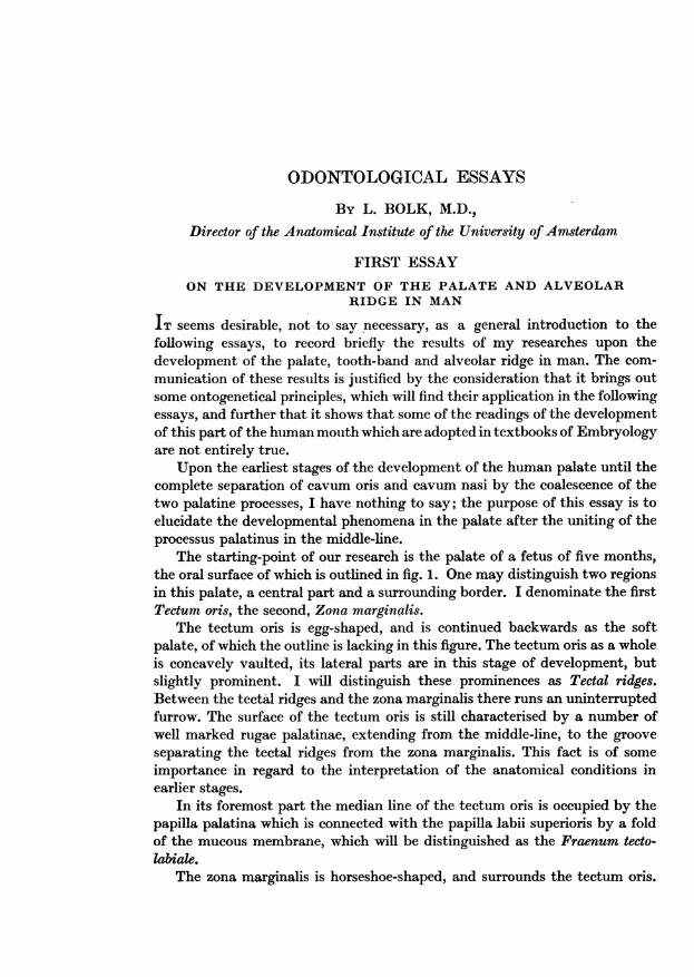
The starting-point of our research is the palate of a fetus of five months,the oral surface of which is outlined in fig. 1. One may distinguish two regionsin this palate, a central part and a surrounding border. I denominate the firstTectum oris, the second, Zona marginalis.
The tectum oris is egg-shaped, and is continued backwards as the softpalate, of which the outline is lacking in this figure. The tectum oris as a wholeis concavely vaulted, its lateral parts are in this stage of development, butslightly prominent. I will distinguish these prominences as Tectal ridges.Between the tectal ridges and the zona marginalis there runs an uninterruptedfurrow. The surface of the tectum oris is still characterised by a number ofwell marked rugae palatinae, extending from the middle-line, to the grooveseparating the tectal ridges from the zona marginalis. This fact is of someimportance in regard to the interpretation of the anatomical conditions inearlier stages.
In its foremost part the median line of the tectum oris is occupied by thepapilla palatina which is connected with the papilla labii superioris by a foldof the mucous membrane, which will be distinguished as the Fraenum tecto-labiale.
The zona marginalis is horseshoe-shaped, and surrounds the tectum oris.
Odontological Essays
In the front part of the mouth it lies between the latter and the lip, laterallybetween the cheeks and the tectum. It is separated from lips and cheeks bya rather deep furrow-the vestibular groove-interrupted in the median lineby the fraenum already referred to. The internal boundary line of the zonamarginalis is a somewhat more complicated one. A furrow, which separatesthe zona from the tectum, begins at the fraenum tecto-labiale and deepens asit passes outward and backward. A little behind the mouth this furrow turnsobliquely outward, finally fusing with the vestibular groove. Thus the zonamarginalis is divided into an anterior and a posterior part. The latter isseparated from the tectum oris by a special furrow which is apparently abackward prolongation from the furrow in the front part of the palate. Ongrounds, which will become intelligible in the course of this essay, I shalldistinguish the first furrow-that ending the vestibular groove-as theinternal alveolarfurrow, that limitating the posterior part of the zona marginalison the inner side as the transitory palatine furrow.
The posterior part of the internal alveolar furrow divides the zonamarginalis into two parts, whose significance in the future development of thepalate is quite different. For the anterior part is the alveolar ridge, in thisphase but partially developed, whereas the posterior part has nothing to dowith this ridge; being, as development advances, incorporated in the tectumoris by the disappearance of the transitory palatine furrow. This fusion withthe tectum is as a rule already complete when the fetus is full grown: so thatin the new-born child only some small fragments of the furrow persist on theinner side of the alveolar ridge, which by this time has extended considerablyfurther backward. The different signification and behaviour of the two partsof the zona marginalis necessitate the application of a special name to each;the anterior part, as the true alveolar ridge, shall retain that name, the namepseudo-alveolar ridge being appropriate to the posterior part in the light of itslater development. In fetuses, more advanced in development than that offig. 1, the alveolar ridge has penetrated between the cheek and the pseudo-alveolar ridge.
The surface of the pseudo-alveolar ridge is smooth, that of the alveolar onthe contrary is rugged; small irregular prominences indicating the spots wherethe teeth are lying under the surface of the gum.
In a fetus in an earlier stage of development, anatomical conditions aremet with, which prove that the above described configuration of the palatedoes not originate by a simple enlargement, but by a development of veryparticular nature. This becomes obvious by a comparison of fig. 1 with fig. 2,in which the palate of a fetus from the fourth month is outlined.
The most striking difference between the palate sketched in fig. 1 and thatof the younger subject drawn in fig. 2, is that in the earlier stage the zonamarginalis is incomplete, being represented only by its anterior and posteriorextremities. The anterior part, the alveolar ridge, is narrower and sickle-shaped,lying between the lips and the tectum oris. The corrugations by which it is
139
140 L. Boli
characterised in the older fetus have not appeared, its surface being smooth.In the median line it is divided into two halves by the Fraenum tecto-labiale.The papilla palatina and papilla labii superioris lie closer to one another, andare connected by the short and broad Fraenum. A little behind the corner ofthe mouth the internal alveolar furrow and the vestibular furrow unite,forming a common groove, which runs backwards between the cheek and thetectum oris. Examination of the rugae palatinae makes it clear, that thetectum oris in this stage extends laterally as far as the cheeks, for the rugaereach these lateral walls of the mouth. In this region therefore the zonamarginalis is lacking. In the most posterior part of the palate it reappears, thepseudo-alveolar ridge intercalating itself between the cheek and the tectum oris,being bounded internally by the transitory palatine furrow, and externally bythe vestibular groove.
Summarising, one finds that in a younger stage of development the zonamarginalis is wanting in part, and the tectum oris extends laterally as far as
Fig. 1 Fig. 2 Fig. 3
the cheeks. Moreover the lateral parts of the tectum project more considerably,and the palate is therefore more strongly vaulted from side to side.
Knowledge of the structure at this stage of development facilitates theunderstanding of the conditions in a still earlier stage, drawn in fig. 3. This ischaracterized by the total absence of the zona marginalis, the palate beingrepresented in this subject solely by the tectum oris. In the front part of themouth the latter reaches the lips; laterally along its whole length it touches thecheeks. That such is really the case is proved by the fact, that the rugaepalatinae on the front part of the palate reach the furrow bounding the lip,and posteriorly extend outwards to the groove between the cheeks and thepalate. If the lateral groove in this stage is called the vestibular groove, stressmust be laid on the fact that its anatomical value is not the same as thevestibular groove in the stage represented in fig. 1. The conditions in the two
subjects are quite different. For in the elder stage this groove runs between thezona marginalis and the cheeks and lip, whilst in the younger it separates thesefrom the tectum oris. Therefore it is advisable to distinguish the groove in
Odontological Essays
the latter case by a special name at the labio-tectal and bucco-tectal furrow.The total absence of the zona marginalis in fig. 3 involves lack of the internalalveolar furrow and the transitory palatine furrow.
A very remarkable phenomenon is presented by the eminence lying in thefront part of the palate. In older stages two papillae are situated in the medianline: a papilla palatina and a papilla labii superioris, which are connected witheach other by the Fraenum tecto-labiale. In the present case both papillaeare represented by a single egg-shaped eminence, the Fraenum being stillundeveloped.
The three stages of development above described, enable us to explain thehistory of the differentiation of the human palate after the fusion of the twopalatinal processes. I wish to emphasise the fact that this description appliesonly to the development of the palate in man, and must not be extended toother mammals. I am aware of the circumstance that the developmentalhistory of this part of the mouth in some other mammals differs not incon-siderably from that in man, especially so far as it concerns the alveolar ridge.In the following paragraphs I will try to give a short summary of this history.
The so-called secondary palate is formed by the fusion in the middle lineof the left and right palatine processes, the resulting plate separating theprimitive cavum oris into an upper and a lower cavity. This secondary palateis in man transitory, representing only that part of the definitive palate whichlies within the alveolar ridge. It is thus not identical with the final palate,which includes this ridge and may therefore be distinguished as the tertiarypalade.
The secondary palate is strongly vaulted from side to side, the lateral partsforming two prominences, along the inner surface of the cheeks, from whichthey are separated by the labio-bucco-tectal furrow. In the median line thisfurrow is interrupted by a rather large papilla: the labio-tectal papilla. Thetwo prominences, just referred to, may be distinguished as tectal ridges.
The tertiary palate arises from this by the growing up of a ridge, whichoriginates from the bottom of the labio-bucco-tectal furrow, and sandwichesitself between the tectal ridges on the inner side, and the lip and cheeks on theouter side. This ridge embraces the secondary palate like a horse-shoe. Twodivisions of the palate so constituted are to be distinguished: a central part-the true roof of the mouth-tectum oris, and a border, the zona marginaliswhich surrounds it. This zona originates in each half of the palate from twocentres, an anterior and a posterior one. The latter becomes visible before theformer. The anterior part of the zona makes its first appearance at the surfaceimmediately laterally to the tecto-labial papilla, and extends graduallybackwards to touch the posterior part. The two parts however do not unitewith each other as a rather deep furrow remains between them after theircontact. This furrow is the continuation of that between the tectum oris andthe frontal part of the zona marginalis.
The two parts of the zona have different relation to the set of teeth, and it
141
is therefore necessary to give them different names, the anterior part I callthe alveolar ridge, whilst the posterior part may be distinguished as the pseudo-alveolar ridge. The former is bounded by the vestibular groove and the internalalveolar furrow, the latter by the vestibular groove and the transitory palatinefurrow.
The surface of the alveolar ridge shows some prominences, caused by thedeveloping teeth, the pseudo-alveolar ridge on the contrary remains smoothand flattens gradually as the transitory palatine groove disappears. At birth,therefore, the limits of this part of the zona marginalis are difficult to determine.The main cause of this difficulty is the backward extension of the alveolarridge which insinuates itself between the cheek and the pseudo-alveolar ridge,so as to give that prominence an internal position, where, on the total dis-appearance of the transitory palatine furrow it becomes fused with the tectumoris. In an early stage of development the foremost part of this median lineof the palate is interrupted by a small eminence, the papilla tecto-labialis. Inconsequence of the growing up of the alveolar ridge between the lip and thetectum, this papilla is divided into an anterior labial papilla and a posteriortectal papilla, united by a mucous fold, the Fraenum tecto-labiale. Till shortlybefore birth the presence of this fraenum is easy to demonstrate. After birththis fold is partly flattened by the gradually accentuated prominence of thealveolar ridge, and only the anterior part-known as the Fraenum labiisuperioris-persists. In some Prosimiae (Lemur, e.g.) the whole Fraenum ispersistent, running between the two central incisors from the lip to the palate.It seems to me not impossible that the diastema sometimes occurring in manin the median line of the set of teeth is caused by the persistence of thisfraenum during a longer period than usual.
I have controlled and completed this developmental history of the humanpalate based on its surface anatomy in fetuses of different ages by the micro-scopical researches with which the following account deals. It is obvious thatthe value and significance of such a microscopical investigation are greaterones than those of a purely macroscopical study. For this research ought alsoto include the genetical and topographical phenomena concerning the teethand the dental lamina and their relation to the superficial furrows abovedescribed.
I have examined by means of serial sections a large number of humanfetuses with regard to the anlage and further development of teeth and palate,from these I choose for description five specimens varying in age from the secondtill the fifth month of development. The youngest embryo measured 25 mm.whole length.
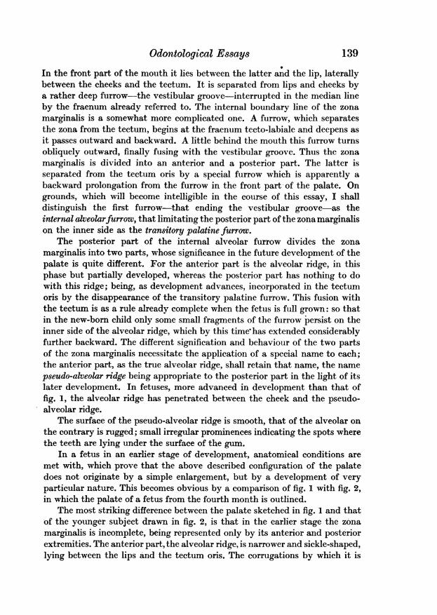
I begin with this embryo, which is represented in figs. 4-7. As shown bythese figures in this embryo the separation between the cavum oris and thecavum nasi is not yet formed, the palatine processes still lying lateral andventral to the dorsum of the tongue. Figs. 6 and 7 are drawn from transversesections behind the corner of the mouth, figs. 4 and 5 from similar sections in
142 L. Bolk
Odontological Essays 143
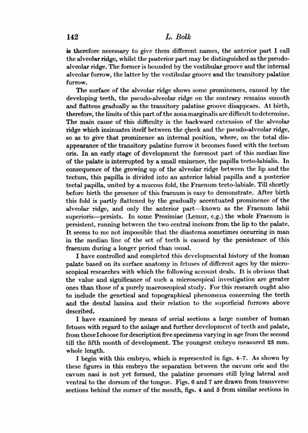
front of this corner. In this embryo there is not the minutest trace of theteeth-germs. In figs. 4-7, as well as in all those which follow, the interestingepithelial formations are indicated by dotting. In fig. 4 one sees nearly in themiddle of the surface of the common anlage for lip and palate a shallow furrow(fig. 4, 1) which may be distinguished as the labio-tectal furrow. Below, withthis furrow an epithelial thickening has grown into the subjacent tissue, whichshows two offshoots, a medial and a lateral one. I shall call the medial thedento-gingival sheet (fig. 4, 2) and the lateral the labio-gingival sheet (fig. 4, 3).These names indicate the future significance of these epithelial formations;from the former are differentiated the enamel-organs of the teeth, whilst inaddition a part of the gum takes origin from it; the latter gives rise to theepithelium on the inner surface of the lip and furthermore to a part of the gum.I lay stress upon the fact that neither of these sheets is distinguished by me asdental lamina, being convinced that such a name is too restricted, as I shallshow in the course of the present paper.
The two epithelial sheets above described separate three ridges ofmesenchyme. Of these the medial one will be called the tectal ridge (fig. 4, 4),
Fig. 4 Fig. 5 Fig. 6 Fig. 7
in the later parts of this paper, the middle the alveolar ridge (fig. 4, 5) andthe lateral one the labial ridge (fig. 4, 6). The appropriateness of these nameswill be shown by the behaviour of the ridges in later embryos.
The conditions of these ridges do not alter much as they are traced backwardthrough the serial sections until at a point a little in advance of the corner ofthe mouth, the labio-gingival sheet disappears and the common epithelialmass is carried inward only by the dento-gingival sheet, which is slightlyinclined medially (fig. 5). That this is a normal structure and not an individualpeculiarity of the embryo is shown by conditions in later stages. The regionwhere the labio-gingival sheet is absent is short because it reappears immedi-ately behind the corner of the mouth (see fig. 6).
As this region is in the cheek the name labio-gingival must be replaced bybucco-gingival. The whole epithelial formation in this region arises from thesurface epithelium forming the lateral border of the cavum oris, the labio-tectal furrow has disappeared, and the groove between the tectal ridge(fig. 6, 4) and the cheek may be distinguished as the bucco-tectal groove.
Further back the bucco-gingival sheet becomes lower and finally disappearsAnatomy LV 10
(fig. 7) and the dento-gingival sheet above is continued to the posterior part ofthe mouth where it disappears.
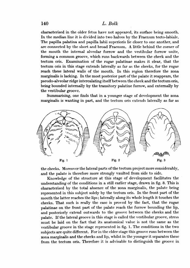
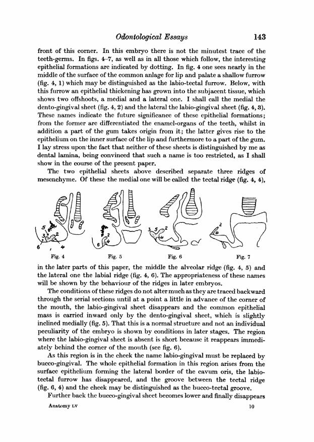
The conditions in a fetus of 40 mm. are shown in figs. 8, 9, 10 and 11. Thecavum oris in this specimen is separated from the cavum nasi, the secondarypalate is complete and the germs of all the milk teeth are present. In fig. 8a section is drawn between the germ of the lateral incisor and the canine tooth,nearly the same level as the section drawn in fig. 4 of the younger embryo.The changes which have taken place may be recognised in the easiest mannerby a comparison of the two figures.
Fig. 8 Fig. 9
k .0/~~.4
Fig. 10
INAJ7I,32
}Fig. 11i
The labio-tectal furrow has deepened, so that the lip becomes moreprominent and acquires a free inner surface. A considerable heaping up ofepithelium separates the labial ridge (6) from the tectal ridge (4) and from thiscommon epithelial strand the dento-gingival sheet (2) goes out in a mesialdirection, whilst the labio-gingival sheet is directed upwards and laterally.By the considerable elongation of both sheets the alveolar ridge is muchbroadened.
The section represented in fig. 9 runs through the germ of the canine tooth;at this level the upper and lower lip are united with each other. This sectioncorresponds nearly with that in fig. 5.
There is a rather broad bucco-tectal cleft and between the cheek and tectalridge (4) is intercalated the backward prolongation of that large epithelial
144 L. Bolk
) .r 4
Odontological Essays 145
mass, which in the preceding figure is shown to separate the tectal and labialridges. In the present section as in that of fig. 5 there is but a single offshootgoing out from this thickening of the surface epithelium, namely the mesiallyinclined dento-gingival sheet (2), carrying at its free edge the germ of thecanine tooth. As in the younger fetus the bucco-gingival sheet is absent in theregion of this tooth. Hence a lateral limitation of the alveolar ridge (5) islacking in this region.
A little further backwards this sheet reappears, as is shown by fig. 10,which section runs through the germ of the first milk molar. The cleft betweenthe cheek and the tectal ridge is deepened and widened out. This bucco-tectalgroove cannot be identified with the vestibulum oris, for the vestibular cavityis not in existence in the present stage of development. The cleft between thecheek and the tectum oris is only of short duration, it disappears in the courseof the further development, as will be demonstrated in older embryos.
The section drawn in fig. 10 is taken through nearly the same region as thatof the younger embryo which is the original of fig. 6. It shows a new featurewhich is worthy of special notice. This is that the dento-gingival sheet no
longer arises from the lateral edge of the tectal ridge (4) but originates morelaterally. Fig. 11, which represents a section through the posterior part ofthe palate, shows that this condition is accentuated as the structures arefollowed backward. In it the bujeco-gingival sheet (3) has become very low,penetrating only a little way into the subjacent tissue. The dento-gingivalsheet (2) has still a considerable depth. On the surface of the palate the tectalridge (4) is much flattened, and the tecto-buccal cleft, although considerablybroadened, has become very shallow. Between the cheek and the tectal ridgea new ridge is formed (7) which has nothing to do with the alveolar ridge (5).For the dento-gingival sheet extends laterally from this new formation. Theridge in question is that described by me earlier in this paper as pseudo-alveolar ridge, it is separated from the tectal ridge (4) by the shallow furrowwhich I have distinguished as the transitory palatine furrow (8).
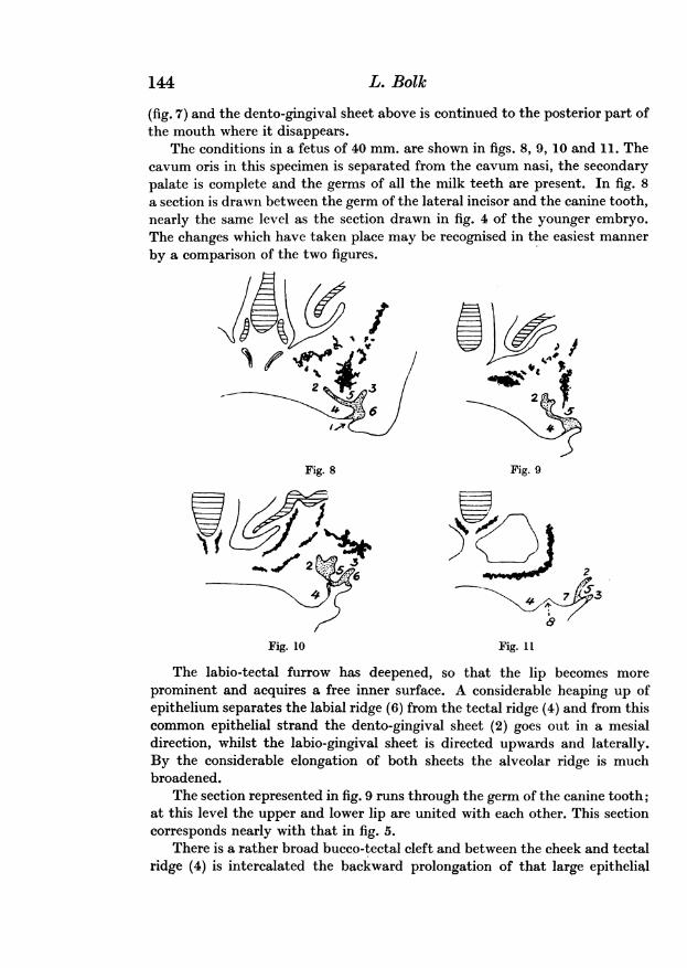
I choose an embryo of the age of two and a half months as a third stage.The figs. 12, 13, 14 and 15 are drawn from it.
The section in fig. 12 cuts the germ of the lateral incisor, and correspondstherefore with those in figs. 4 and 8. There is an enormous heaping lip ofepithelium between the tectal ridge (4) and the labial ridge (6). By thisconsiderable thickening of the epithelium these ridges are pushed away fromeach other and in consequence the alveolar ridge (5) is broadened. By thepenetration of the labio-tectal furrow into this mass of epithelial cells the lipbecomes more and more independent and acquires a free inner surface.
The deepening of this furrow is due to an atrophy of the cells which fillthe interior of the epithelial mass. The dento-gingival sheet (2) is stronglyinclined mesially, so as to take nearly a horizontal direction. The labio-gingival sheet (3) bounding the alveolar ridge (5) along its lateral edge, is alsothickened and penetrates more deeply into the mesenchyme.
F ~~~~~~~~~10-2
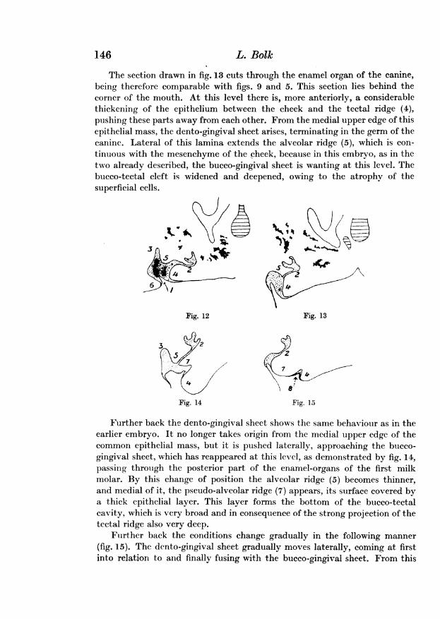
The section drawn in fig. 13 cuts through the enamel organ of the canine,being therefore comparable with figs. 9 and 5. This section lies behind thecorner of the mouth. At this level there is, more anteriorly, a considerablethickening of the epithelium between the cheek and the tectal ridge (4),pushing these parts away from each other. From the medial upper edge of thisepithelial mass, the dento-gingival sheet arises, terminating in the germ of thecanine. Lateral of this lamina extends the alveolar ridge (5), which is con-tinuous with the mesenchyme of the cheek, because in this embryo, as in thetwo already described, the bucco-gingival sheet is wanting at this level. Thebucco-tectal cleft is widened and deepened, owing to the atrophy of thesuperficial cells.
r*~~~~~~I
Fig. 12 Fig. 1334
7~~~~~~~~
Fig. 14 Fig. 15
Further back the dento-gingival sheet shows the same behaviour as in theearlier embryo. It no longer takes origin from the medial upper edge of thecommon epithelial mass, but it is pushed laterally, approaching the bucco-gingival sheet, which has reappeared at this level, as demonstrated by fig. 14,passing through the posterior part of the enamel-organs of the first milkmolar. By this change of position the alveolar ridge (5) becomes thinner,and medial of it, the pseudo-alveolar ridge (7) appears, its surface covered bya thick epithelial layer. This layer forms the bottom of the bucco-tectalcavity, which is very broad and in consequence of the strong projection of thetectal ridge also very deep.
Further back the conditions change gradually in the following manner(fig. 15). The dento-gingival sheet gradually moves laterally, coming at firstinto relation to and finally fusing with the bucco-gingival sheet. From this
146 L. Bolk
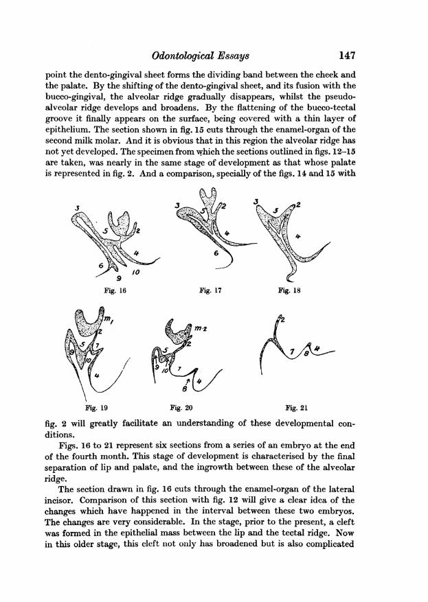
Odontological Essayspoint the dento-gingival sheet forms the dividing band between the cheek andthe palate. By the shifting of the dento-gingival sheet, and its fusion with thebucco-gingival, the alveolar ridge gradually disappears, whilst the pseudo-alveolar ridge develops and broadens. By the flattening of the bucco-tectalgroove it finally appears on the surface, being covered with a thin layer ofepithelium. The section shown in fig. 15 cuts through the enamel-organ of thesecond milk molar. And it is obvious that in this region the alveolar ridge hasnot yet developed. The specimen from which the sections outlined in figs. 12-15are taken, was nearly in the same stage of development as that whose palateis represented in fig. 2. And a comparison, specially of the figs. 14 and 15 with
3
Fig. 16 Fig. 17 Fig. 18
49
Fig. 19 fig. 20 fig. 21
fig. 2 will greatly facilitate an understanding of these developmental conl-ditions.
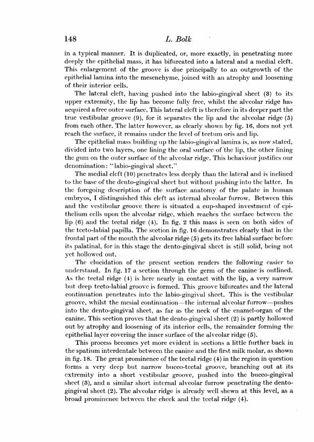
Figs. 16 to 21 represent six sections from a series of an embryo at the endof the fourth month. This stage of development is characterised by the finalseparation of lip and palate, and the ingrowth between these of the alveolar
/.jj
ridge.The section drawn in fig. 16 cuts through the enamel-organ of the lateral
incisor. Comparison of this section with fig. 12 will give a clear idea of thechanges which have happened in the interval between these two embryos.The changes are very considerable. In the stage, prior to the present, a cleftwas formed in the epithelial mass between the lip and the tectal ridge. Nowin this older stage, this cleft not only has broadened but is also complicated
147
148 L. Bolk
in a typical manner. It is duplicated, or, more exactly, in penetrating moredeeply the epithelial mass, it has bifurcated into a lateral and a medial cleft.This enlargement of the groove is due principally to an outgrowth of theepithelial lamina into the mesenchyme, joined with anl atrophy and looseningof their interior cells.
The lateral cleft, having pushed into the labio-gingival sheet (3) to itsupper extremity, the lip has become fully free, whilst the alveolar ridge hasacquired a free outer surface. This lateral cleft is therefore in its deeper part thetrue vestibular groove (9), for it separates the lip and the alveolar ridge (5)from each other. The latter however, as clearly shown by fig. 16, does not yetreach the surface, it remains under the level of tectum oris and lip.
The epithelial mass building up the labio-gingival lamina is, as now stated,divided into two layers, one lining the oral surface of the lip, the other liningthe gum on the outer surface of the alveolar ridge. This behaviour justifies ourdenomination: "labio-gingival sheet.'
The medial cleft (10) penetrates less deeply than the lateral and is inclinedto the base of the dento-gingival sheet but without pushing into the latter. Inthe foregoing description of the surface anatomy of the palate in humanembryos, I distinguished this cleft as internal alveolar furrow. Between thisand the vestibular groove there is situated a cup-shaped investment of epi-thelium cells upon the alveolar ridge, which reaches the surface between thelip (6) and the tectal ridge (4). In fig. 2 this mass is seen on both sides ofthe tecto-labial papilla. The section in fig. 16 demonstrates clearly that in thefrontal part of the mouth the alveolar ridge (5) gets its free labial surface beforeits palatinal, for in this stage the dento-gingival sheet is still solid, being notyet hollowed out.
The elucidation of the present section renders the following easier tounderstand. In fig. 17 a section through the germ of the canine is outlined.As the tectal ridge (4) is here nearly in contact with the lip, a very narrowbut deep tecto-labial groove is formed. This groove bifurcates and the lateralcontinuation penetrates into the labio-gingival sheet. This is the vestibulargroove, whilst the mesial continuation -the internal alveolar furrow pushesinto the dento-gingival sheet, as far as the neck of the enamel-organ of thecanine. This section proves that the dento-gingival sheet (2) is partly hollowedout by atrophy and loosening of its interior cells, the remainder forming theepithelial layer covering the inner surface of the alveolar ridge (5).
This process becomes yet more evident in sections a little further back inthe spatium interdentale between the canine and the first milk molar, as shownin fig. 18. The great prominence of the tectal ridge (4) in the region in questionforms a very deep but narrow bucco-tectal groove, branching out at itsextremity into a short vestibular groove, pushed into the bucco-gingivalsheet (3), and a similar short internal alveolar furrow penetrating the dento-gingival sheet (2). The alveolar ridge is already well shewn at this level, as abroad prominence between the cheek and the tectal ridge (4).
Odontological Essays 149
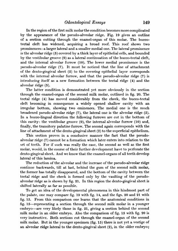
In the region of the first milk molar the condition becomes more complicatedby the appearance of the pseudo-alveolar ridge. Fig. 19 gives an outlineof a section cutting through the enamel-organ of this molar. The bucco-tectal cleft has widened, acquiring a broad roof. This roof shows twoprominences: a larger lateral and a smaller medial one. The lateral prominenceis the alveolar ridge (5) covered by a thick layer of epithelial cells, and boundedby the vestibular groove (9) as a lateral continuation of the bucco-tectal cleft,and the internal alveolar furrow (10). The lower medial prominence is thepseudo-alveolar ridge (7). It must be noticed that the line of attachmentof the dento-gingival sheet (2) to the covering epithelial layer correspondswith the internal alveolar furrow, and that the pseudo-alveolar ridge (7) isintroducing itself as a new formation between the tectal ridge (4) and thealveolar ridge (5).
The latter condition is demonstrated yet more obviously in the sectionthrough the enamel-organ of the second milk molar, outlined in fig. 20. Thetectal ridge (4) has moved considerably from the cheek, the bucco-tectalcleft becoming in consequence a widely opened shallow cavity with anirregular bottom, showing two eminences. The medial one is the muchbroadened pseudo-alveolar ridge (7), the lateral one is the alveolar ridge (5).In a bucco-lingual direction the following furrows are cut in the bottom ofthis cavity: the vestibular groove (9), the internal alveolar furrow (10) and,finally, the transitory palatine furrow. The second again corresponds with theline of attachment of the dento-gingival sheet (2) to the superficial epithelium.
This section proves in a conclusive manner the fact that the pseudo-alveolar ridge (7) cannot be a formation which later enters into relation to theset of teeth. For if such was really the case, the second as well as the firstmolar, would, in the course of their further development have to perforate thedento-gingival sheet. And we know that the enamel-organs of all teeth developlateral of this lamina.
The reduction of the alveolar and the increase of the pseudo-alveolar ridgecontinue backwards, till at last, behind the gum of the second milk molar,the former has totally disappeared, and the bottom of the cavity between thetectal ridge and the cheek is formed only by the vaulting of the pseudo-alveolar ridge as is shown by fig. 21. In this region the dento-gingival sheet isshifted laterally as far as possible.
To get an idea of the developmental phenomena in this hindmost part ofthe palate, one may compare fig. 19 with fig. 14, and the figs. 20 and 21 withfig. 15. From this comparison one learns that the anatomical conditions infig. 15-representing a section through the second milk molar in a youngerembryo-are very likely those in fig. 21, giving a section behind the secondmilk molar in an older embryo. Also the comparison of fig. 15 with fig. 20 isvery instructive. Both sections cut through the enamel-organ of the secondmilk molar. But in the younger specimen (fig. 15) there is not yet a vestige ofan alveolar ridge lateral to the dento-gingival sheet (2), in the older embryo;
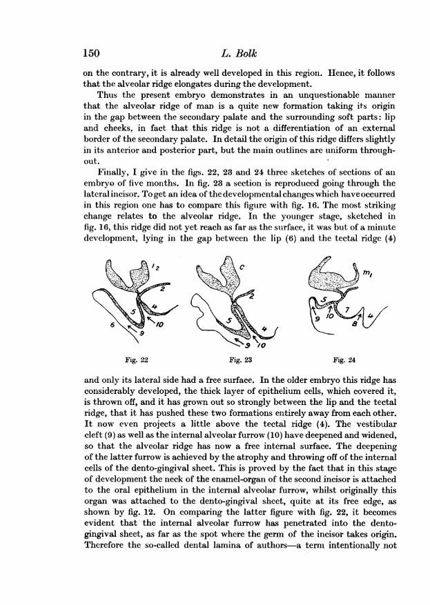
on the contrary, it is already well developed in this region. Hence, it followsthat the alveolar ridge elongates during the development.
Thus the present embryo demonstrates in an unquestionable mannerthat the alveolar ridge of man is a quite new formation taking its originin the gap between the secondary palate and the surrounding soft parts: lipand cheeks. in fact that this ridge is not a differentiation of an externalborder of the secondary palate. In detail the origin of this ridge differs slightlyin its anterior and posterior part, but the main outlines are uniform through-out.
Finally, I give in the figs. 22, 23 and 24 three sketches of sections of anembryo of five months. In fig. 23 a section is reproduced going through thelateral incisor. To get an idea of the developmental changes which have occurredin this region one has to compare this figure with fig. 16. The most strikingchange relates to the alveolar ridge. In the younger stage, sketched infig. 16, this ridge did not yet reach as far as the surface, it was but of a minutedevelopment, lying in the gap between the lip (6) and the tectal ridge (4)
2
;2
5 910
.9/0
Fig. 22 Fig. 23 Fig. 24
and only its lateral side had a free surface. In the older embryo this ridge hasconsiderably developed, the thick layer of epithelium cells, which covered it,is thrown off, and it has grown out so strongly between the lip and the tectalridge, that it has pushed these two formations entirely away from each other.It now even projects a little above the tectal ridge (4). The vestibularcleft (9) as well as the internal alveolar furrow (10) have deepened and widened,so that the alveolar ridge has now a free internal surface. The deepeningof the latter furrow is achieved by the atrophy and throwing off of the internalcells of the dento-gingival sheet. This is proved by the fact that in this stageof development the neck of the enamel-organ of the second incisor is attachedto the oral epithelium in the internal alveolar furrow, whilst originally thisorgan was attached to the dento-gingival sheet, quite at its free edge, asshown by fig. 12. On comparing the latter figure with fig. 22, it becomesevident that the internal alveolar furrow has penetrated into the dento-gingival sheet, as far as the spot where the germ of the incisor takes origin.Therefore the so-called dental lamina of authors-a term intentionally not
150 L. Bolk
Odontological Essays
used by me-is an epithelial band, which forms not only the germs of the teeth,but also the gum on the inner surface of the alveolar ridge. After comparingfig. 12 with fig. 22 there cannot remain the least doubt upon this fact. Hencemy distinguishing of this band as dento-gingival sheet.
Fig. 23 is a drawing of a section through the germ of the canine. Thisfigure should be compared with fig. 17, showing a section through the sametooth in a younger embryo. The differences between these sections are in themain identical with those in the front part of the mouth. In the older stage thealveolar ridge (5) nearly fills up the cleft between the tectal ridge (4) and thecheek, its internal surface has become free by the hollowing out of the dento-gingival sheet, as far as the neck of the enamel-organ of the canine. For therest this figure asks no further explanation.
Finally, fig. 24 shews a section of the same embryo, going through theenamel-organ of the first milk molar. This figure is to be compared with fig. 19.Such a comparison shows that the tectal ridge (4) in the older embryo hasrelatively increased in size; the cavity between the cheek and the alveolarridge, originally deep and narrow, is now very wide and shallow, the bottom ofit is formed by the broad pseudo-alveolar ridge (7) and the also broadenedalveolar ridge. This bottom is incised by three furrows: the transitorypalatine furrow (8) between the tectal and the pseudo-alveolar ridge; theinternal alveolar furrow (10) between the alveolar and pseudo-alveolar ridge,and the vestibular groove in its well-known situation.
After the description of the five stages of development now given, it is veryeasy to reproduce by means of some simple diagrams the main features in thedevelopmental history of the human palate, specially the origin of thealveolar ridge and its genetical relation to the epithelial formations growinginto the mesenchymatous tissue of the palate. Before doing so, it seemsnecessary to me to remark, that I have never met in human embryos withthe considerable heaping up of epithelium extending along the length of thejaw, distinguished as "Zahnwall" by the German authors. I have had underobservation a large number of serial sections of the mouth of human embryos.But, as shown by figs. 5 and 6, even in the youngest stage there is no trace ofan outward thickening of the epithelium. On the contrary, from the outset,the surface of the jaw shows a shallow furrow, marking the position of theingrowing epithelium. In this matter I agree with Leche, that this heapingup of epithelium does not represent an occurrence typical of all mammals.At all events it does not occur in man.
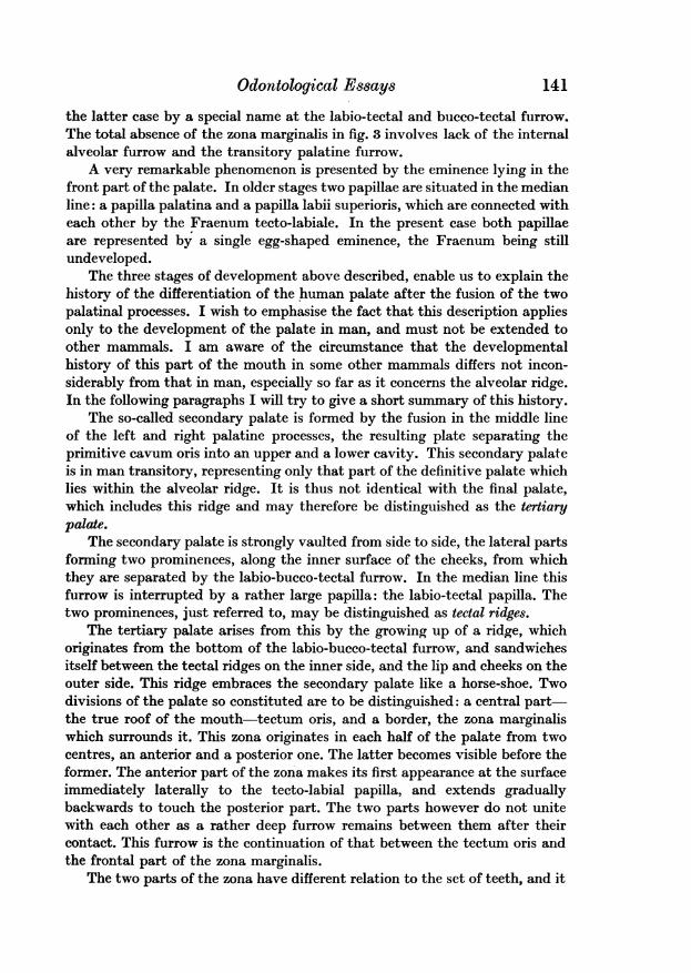
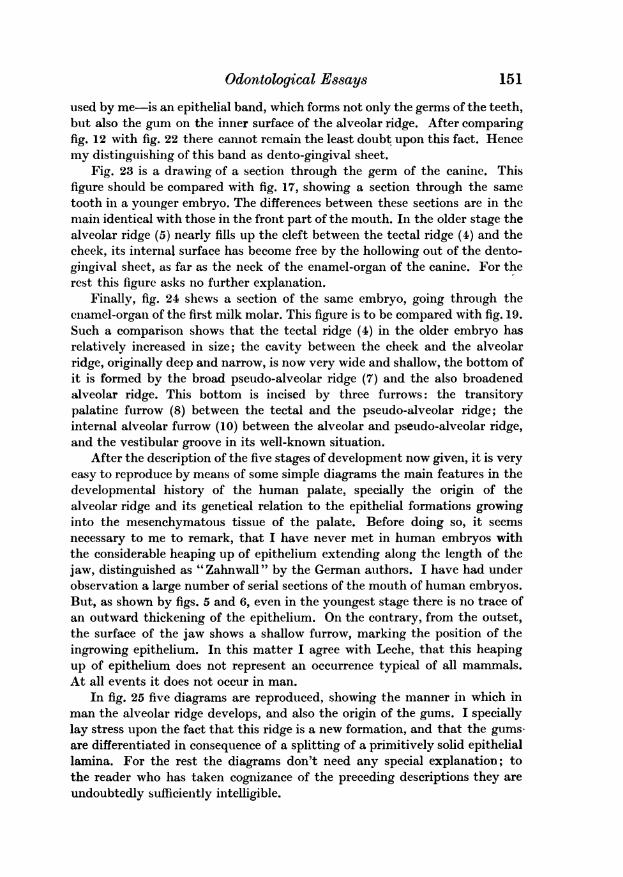
In fig. 25 five diagrams are reproduced, showing the manner in which inman the alveolar ridge develops, and also the origin of the gums. I speciallylay stress upon the fact that this ridge is a new formation, and that the gums-are differentiated in consequence of a splitting of a primitively solid epitheliallamina. For the rest the diagrams don't need any special explanation; tothe reader who has taken cognizance of the preceding descriptions they areundoubtedly sufficiently intelligible.
151
152 L. Bolk
Finally, the question as to the significance of the pseudo-alveolar ridgesmust be considered. These organs which ina young stage form the very prominentposterior half of the zona marginalis on bothsides of the palate, are, as already sufficientlyindicated, transitory. In the older stages they dIare surrounded by the alveolar ridges, pro- clongating backwards, and become incorpor-ated in the central part of the palate, which ()rI distinguished as tectum oris. To solve the -vquestion of their significance seems to me arather difficult matter. Klatch, after havingconfirmed the occurrence of these organs firstdescribed by me in human embryos, is per-haps right, when he considers them as rudi-
F
mentary organs, inherited by us from our Fig. 25marsupial ancestors, where they may have assisted the young in holdingon to the nipple.
SECOND ESSAYON THE DEVELOPMENT OF THE ENAMEL-GERM
To write a special essay upon the development of the enamel-germ, worthreading, may seem a somewhat bold endeavour. For the development of thispart of the tooth-germ has already so often been the subject of investigations,that it may be considered as improbable that particularities of some im-portance are as yet unknown. However, at the very outset of this essay itmust be said that the descriptions of the development of the enamel-germ asgiven in the textbooks of Anatomy or Embryology are incomplete.
So simple a matter as the development and internal structure of the enamel-germ is without exception described in an incomplete manner. This is the casenot only in the textbooks written in English, but also those written inGerman and French. And often during my researches upon this subject, Ihave wondered at the fact, that phenomena so simple and appearances soeasily observable as those described in the present paper, are still unknown.For the facts referred to occur with such a regularity and are so unmistakable,that they may easily be verified by anyone desirous to do so. But a primerequisite for such a study is the examination of complete serial sections ofseveral subjects of different ages. Only in this way does it become possibleto survey the relation between the changes in the succeeding stages, and tobuild up the complete history of the development of the enamel-organ.
Not without intention, at the outset of this essay, I lay stress upon thevalue of the facts to be described hereafter, for they are of primary significance
Odontological Essays
as a starting point to the dental theory which will be gradually developed inthe succeeding essays. In saying so, I remember, that in my own mind, thefirst conception of the principles of this theory was founded on observationsof some phenomena during the development of the enamel-organ in embryosof man and monkeys.
The facts to be dealt with in the present paper, possess therefore a doublesignification: as ontogenetic phenomena and as the principal elements ofa theoretical construction. In this latter significance they can only be utilisedlater on, in connection with facts and meanings, worked out in succeedingessays. Therefore in the present communication, I confine myself to the factsalone.
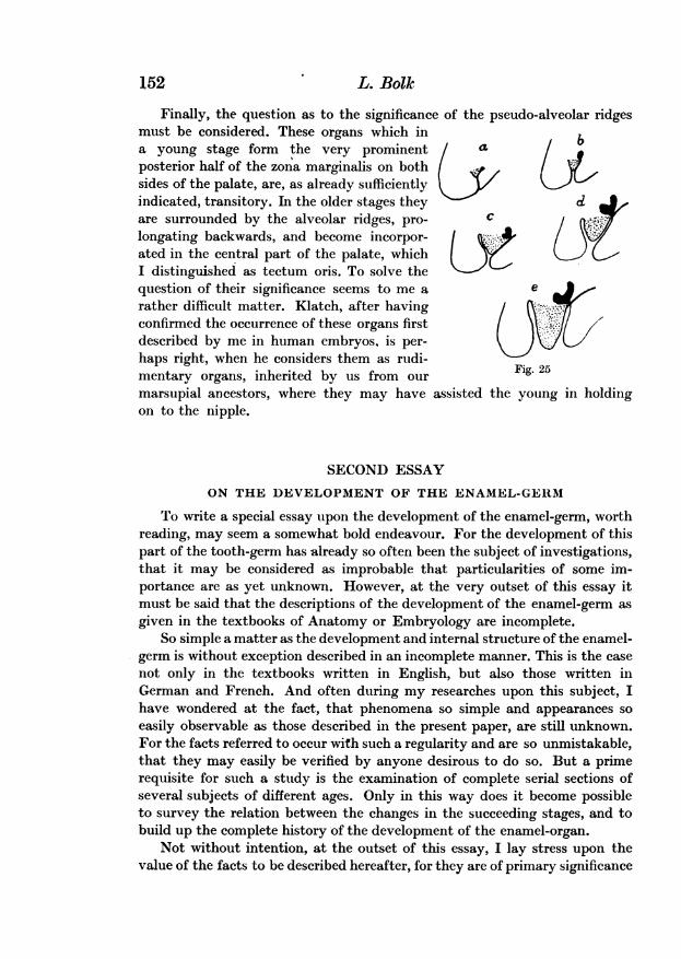
The first principal fact which I intend to demonstrate in the present essayis as follows: enamel-organs are, during a certain phase of their development,connected with the tooth-band or dental lamina by means of two strands, a
Vkkk~
Fig. 26
lateral and a medial one. The large number of embryological series of man andmonkeys at my disposal has led me to use these primates and especially manas the basis of my investigations. Therefore I will begin by trying to justifythe above assertion by observations made upon the teeth of man and otherPrimates; after that I will give a number of examples to prove that thisassertion holds good also with regard to other mammals. In fig. 26 tensuccessive sections are reproduced through the inferior lateral incisor of ahuman embryo of 39 mm. whole length. This embryo is registered in thecollection as: Homo Y. The thickness of the sections is 10 ,u. After the10th section there followed three more through the samc tooth showing a
gradual diminishing of the enamel-organ. It was not necessary to reproducethese also.
In looking over the figures, it seems to me that the last of the series(fig. 26) is intelligible without further explanation, the enamel-organ possessing
153
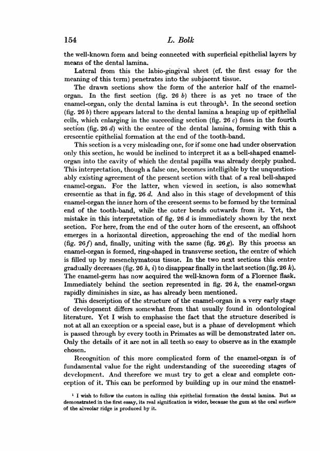
the well-known form and being connected with superficial epithelial layers bymeans of the dental lamina.
Lateral from this the labio-gingival sheet (cf. the first essay for themeaning of this term) penetrates into the subjacent tissue.
The drawn sections show the form of the anterior half of the enamel-organ. In the first section (fig. 26 b) there is as yet no trace of theenamel-organ, only the dental lamina is cut through'. In the second section(fig. 26 b) there appears lateral to the dental lamina a heaping up of epithelialcells, which enlarging in the succeeding section (fig. 26 c) fuses in the fourthsection (fig. 26 d) with the centre of the dental lamina, forming with this acrescentic epithelial formation at the end of the tooth-band.
This section is a very misleading one, for if some one had under observationonly this section, he would be inclined to interpret it as a bell-shaped enamel-organ into the cavity of which the dental papilla was already deeply pushed.This interpretation, though a false one, becomes intelligible by the unquestion-ably existing agreement of the present section with that of a real bell-shapedenamel-organ. For the latter, when viewed in section, is also somewhatcrescentic as that in fig. 26 d. And also in this stage of development of thisenamel-organ the inner horn of the crescent seems to be formed by the terminalend of the tooth-band, while the outer bends outwards from it. Yet, themistake in this interpretation of fig. 26 d is immediately shown by the nextsection. For here, from the end of the outer horn of the crescent, an offshootemerges in a horizontal direction, approaching the end of the medial horn(fig. 26f) and, finally, uniting with the same (fig. 26 g). By this process anenamel-organ is formed, ring-shaped in transverse section, the centre of whichis filled up by mesenchymatous tissue. In the two next sections this centregradually decreases (fig. 26 h, i) to disappear finally in the last section (fig. 26 k).The enamel-germ has now acquired the well-known form of a Florence flask.Immediately behind the section represented in fig. 26 k, the enamel-organrapidly diminishes in size, as has already been mentioned.
This description of the structure of the enamel-organ in a very early stageof development differs somewhat from that usually found in odontologicalliterature. Yet I wish to emphasise the fact that the structure described isnot at all an exception or a special case, but is a phase of development whichis passed through by every tooth in Primates as will be demonstrated later on.Only the details of it are not in all teeth so easy to observe as in the examplechosen.
Recognition of this more complicated form of the enamel-organ is offundamental value for the right understanding of the succeeding stages ofdevelopment. And therefore we must try to get a clear and complete con-ception of it. This can be performed by building up in our mind the enamel-
1 I wish to follow the custom in calling this epithelial formation the dental lamina. But asdemonstrated in the first essay, its real signification is wider, because the gum at the oral surfaceof the alveolar ridge is produced by it.
154 L. Bolk
Odontological Essays
organ represented in fig. 26 by superposition of the sections. On doing so,the enamel-organ presents itself as a low flask-like organ, with a broad base,slightly concave and with a niche-shaped dimple in its front side. In thepresent case the niche is still very shallow, only the three sections, g, h, i offig. 26 running through it; but the whole organ is as yet a very minute one.Thit niche shall be distinguished henceforth as enamel-niche.
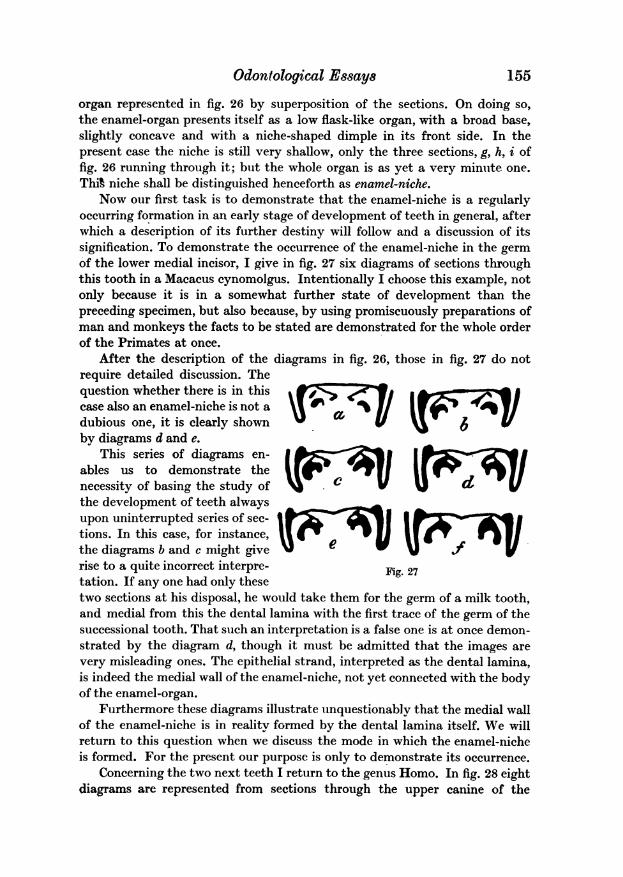
Now our first task is to demonstrate that the enamel-niche is a regularlyoccurring formation in an early stage of development of teeth in general, afterwhich a description of its further destiny will follow and a discussion of itssignification. To demonstrate the occurrence of the enamel-niche in the germof the lower medial incisor, I give in fig. 27 six diagrams of sections throughthis tooth in a Macacus cynomolgus. Intentionally I choose this example, notonly because it is in a somewhat further state of development than thepreceding specimen, but also because, by using promiscuously preparations ofman and monkeys the facts to be stated are demonstrated for the whole orderof the Primates at once.
After the description of the diagrams in fig. 26, those in fig. 27 do notrequire detailed discussion. Thequestion whether there is in thiscase also an enamel-niche is not adubious one, it is clearly shownby diagrams d and e.
This series of diagrams en-ables us to demonstrate thenecessity of basing the study ofthe development of teeth alwaysupon uninterrupted series of sec-tions. In this case, for instance,the diagrams b and c might giverise to a quite incorrect interpre-tation. If any one had only these
ND V
IV UVUT
Fig.. 27
two sections at his disposal, he would take them for the germ of a milk tooth,and medial from this the dental lamina with the first trace of the germ of thesuccessional tooth. That such an interpretation is a false one is at once demon-strated by the diagram d, though it must be admitted that the images arevery misleading ones. The epithelial strand, interpreted as the dental lamina,is indeed the medial wall of the enamel-niche, not yet connected with the bodyof the enamel-organ.
Furthermore these diagrams illustrate unquestionably that the medial wallof the enamel-niche is in reality formed by the dental lamina itself. We willreturn to this question when we discuss the mode in which the enamel-nicheis formed. For the present our purpose is only to demonstrate its occurrence.
Concerning the two next teeth I return to the genus Homo. In fig. 28 eightdiagrams are represented from sections through the upper canine of the
155
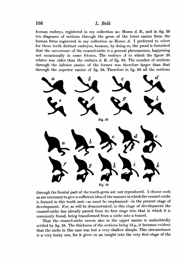
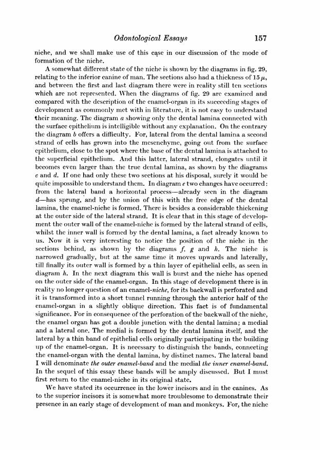
human embryo, registered in my collection as: Homo d. R., and in fig. 29ten diagrams of sections through the germ of the lower canine from thehuman fetus registered in my collection as Homo A. I preferred to selectfor these teeth distinct embryos, because, by doing so, the proof is furnishedthat the occurrence of the enamel-niche is a general phenomenon, happeningnot occasionally in some fetuses. The embryo A to which the figure 29relates was older than the embryo d. R. of fig. 28. The number of sectionsthrough the inferior canine of the former was therefore larger than thatthrough the superior canine of fig. 28. Therefore in fig. 29 all the sections
Fig. 28
Fig. 29
through the frontal part of the tooth-germ are not reproduced. I choose suchas are necessary to give a sufficient idea of the manner in whichthe enamel-nicheis formed in this tooth and-as must be emphasised-in the present stage ofdevelopment. For, as will be demonstrated, in this stage of development theenamel-niche has already passed from its first stage into that in which it iscommonly found, being transformed from a niche into a tunnel.
That the enamel-niche occurs also in the upper canine is undoubtedlysettled by fig. 28. The thickness of the sections being 15 u, it becomes evidentthat the niche in this case was but a very shallow dimple. This circumstanceis a very lucky one, for it gives us an insight into the very first stage of the
156 L. Bolk
dC
Odontological Essays 157
niche, and we shall make use of this ease in our discussion of the mode offormation of the niche.
A somewhat different state of the niche is shown by the diagrams in fig. 29,relating to the inferior canine of man. The sections also had a thickness of 15 I,and between the first and last diagram there were in reality still ten sectionswhich are not represented. When the diagrams of fig. 29 are examined andcompared with the description of the enamel-organ in its succeeding stages ofdevelopment as commonly met with in literature, it is not easy to understandtheir meaning. The diagram a showing only the dental lamina connected withthe surface epithelium is intelligible without any explanation. On the contrarythe diagram b offers a difficulty. For, lateral from the dental lamina a secondstrand of cells has grown into the mesenchyme, going out from the surfaceepithelium, close to the spot where the base of the dental lamina is attached tothe superficial epithelium. And this latter, lateral strand, elongates until itbecomes even larger than the true dental lamina, as shown by the diagramsc and d. If one had only these two sections at his disposal, surely it would bequite impossible to understand them. In diagram e two changes have occurred:from the lateral band a horizontal process-already seen- in the diagramd-has sprung, and by the union of this with the free edge of the dentallamina, the enamel-niche is formed. There is besides a considerable thickeningat the outer side of the lateral strand. It is clear that in this stage of develop-ment the outer wall of the enamel-niche is formed by the lateral strand of cells,whilst the inner wall is formed by the dental lamina, a fact already known tous. Now it is very interesting to notice the position of the niche in thesections behind, as shown by the diagrams f, g and h. The niche isnarrowed gradually, bitt at the same time it moves upwards and laterally,till finally its outer wall is formed by a thin layer of epithelial cells, as seen indiagram h. In the next diagram this wall is burst and the niche has openedon the outer side of the enamel-organ. In this stage of development there is inreality no longer question of an enamel-niche, for its backwall is perforated andit is transformed into a short tunnel running through the anterior half of theenamel-organ in a slightly oblique direction. This fact is of fundamentalsignificance. For in consequence of the perforation of the backwall of the niche,the enamel organ has got a double junction with the dental lamina; a medialand a lateral one. The medial is formed by the dental lamina itself, and thelateral by a thin band of epithelial cells originally participating in the buildingtip of the enamel-organ. It is necessary to distinguish the bands, connectingthe enamel-organ with the dental lamina, by distinct names. The lateral bandI will denominate the outer enamel-band and the medial the inner enamel-band.In the sequel of this essay these bands will be amply discussed. But I mustfirst return to the enamel-niche in its original state.
We have stated its occurrence in the lower incisors and in the canines. Asto the superior incisors it is somewhat more troublesome to demonstrate theirpresence in an early stage of development of man and monkeys. For, the niche
158 L. Bolk
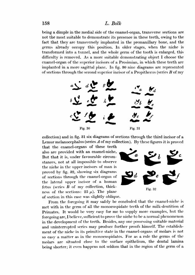
being a dimple in the medial side of the enamel-organ, transverse sections arenot the most suitable to demonstrate its presence in these teeth, owing to thefact that they are transversely implanted in the premaxillary bone, and thegerms already occupy this position. In older stages, when the niche istransformed into a tunnel, and the whole germ of the tooth is enlarged, thisdifficulty is removed. As a more suitable demonstrating object I choose thecnamel-organ of the superior incisors of a Prosimian, in which these teeth areimplanted in a more sagittal plane. In fig. 30 nine diagrams are representedof sections through the second superior incisor of a Propithecus (series B of my
bg e e hFo t
4'SfF isp _C_bFig. 30 Fig. 31
collection) and in fig. 31 six diagrams of sections through the third incisor of aLemur melanocephalus (series A of my collection). By these figures it is provedthat the enamel-organs of these teethalso are provided with an enamel-niche.But that it is, under favourable circum-stances, not at all impossible to observethe niche in the upper incisors of man isproved by fig. 32, showing six diagramsof sections through the enamel-organ ofthe lateral upper incisor of a humanfetus (series B of my collection, thick- F 32ness of the sections: 25 su). The plane gof section in this case was slightly oblique.
From the foregoing it may safely be concluded that the enamel-niche ismet with in the germ of all the monocuspidate teeth of the milk-dentition ofPrimates. It would be very easy for me to supply more examples, but theforegoing are, I believe, sufficient to prove the niche to be a normal phenomenonin the development of the teeth. Besides, any one possessing suitable materialand uninterrupted series may produce further proofs himself. The establish-ment of the niche in its primitive state in the enamel-organs of molars is not
so easy a matter as in the monocuspidates. For as a rule the germs of themolars are situated close to the surface epithelium, the dental laminabeing shorter; it even happens not seldom that in the region of the germ of a
Odontological Essays1
molar the dental bond seems to be absent totally, the germ taking origindirectly from the surface epithelium. This fact renders the interpretation ofthe appearances often somewhat troublesome. Moreover, the niche itself in theenamel-organ of molars behaves so as to render its demonstration ratherdifficult. Firstly it seems to exist in these tooth-germs only a very short time,passing rapidly into its second state, namely the canal or tunnel alreadyalluded to.
And secondly, it seems that in the germs of molars, the niche is not alwayssituated in the mesial or frontal side of the germ, but occasionally the dimpleappears at the posterior side of the germ. This variability in the situation ofthe niche is a very indifferent fact, for whether beginning at the front side orat the back side of the organ it is in all cases soon transformed into a canalby perforating the organ either in a forward or in a backward direction.
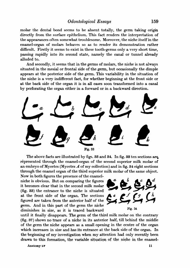
Fig. 33
The above facts are illustrated by figs. 33 and 34. In fig. 38 ten sections are.represented through the enamel-organ of the second superior milk molar ofan embryo of Mycetes (Mycetes A of my collection) and in fig. 34 eight sectionsthrough the enamel organ of the third superior milk molar of the same object.Now in both figures the presence of the enamel-niche is obvious. But on comparing the figures a, c adit becomes clear that in the second milk molar(fig. 33) the entrance to the niche is situatedat the front side of the organ. The sections ae
_
figured are taken from the anterior half of thegerm. And in this part of the germ the nichediminishes in size, as it is traced backward Fig. 34until it finally disappears. The germ of the third milk molar on the contrary(fig. 37) shows no trace of a niche in its anterior half, till behind the middleof the germ the niche appears as a small opening in the centre of the organwhich increases in size and has its entrance at the back side of the organ. Inthe beginning of my investigation when my attention had only recently beendrawn to this formation, the variable situation of the niche in the enamel-
159
Anatomy Lv 11
organs of molar teeth rendered the phenomena not always easy to understand,the object being not always in the most suitable state of development toprocure a clear insight into the relations. But in examining a larger numberof series this point became more intelligible to me. I thought it necessaryto intercalate this remark as a warning to the reader anxious to check thecorrectness of my description, that he must procure himself a rather numerousseries of specimens of different ages. Only in this manner will he be able toobserve all the particular facts described above.
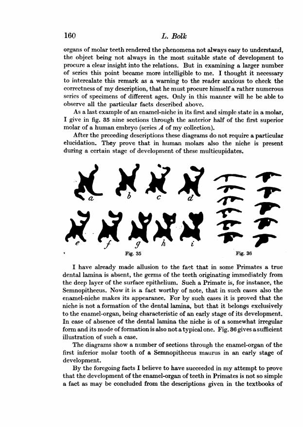
As a last example of an enamel-niche in its first and simple state in a molar,I give in fig. 35 nine sections through the anterior half of the first superiormolar of a human embryo (series A of my collection).
After the preceding descriptions these diagrams do not require a particularelucidation. They prove that in human molars also the niche is presentduring a certain stage of development of these multicupidates.
Fig. 35 Fig. 36
I have already made allusion to the fact that in some Primates a truedental lamina is absent, the germs of the teeth originating immediately fromthe deep layer of the surface epithelium. Such a Primate is, for instance, theSemnopithecus. Now it is a fact worthy of note, that in such cases also theenamel-niche makes its appearance. For by such cases it is proved that theniche is not a formation of the dental lamina, but that it belongs exclusivelyto the enamel-organ, being characteristic of an early stage of its development.In case of absence of the dental lamina the niche is of a somewhat irregularform and its mode of formation is also not a typical one. Fig. 36 gives a sufficientillustration of such a case.
The diagrams show a number of sections through the enamel-organ of thefirst inferior molar tooth of a Semnopithecus maurus in an early stage ofdevelopment.
By the foregoing facts I believe to have succeeded in my attempt to provethat the development of the enamel-organ of teeth in Primates is not so simplea fact as may be concluded from the descriptions given in the textbooks of
160 L. Bolk
Odontological Essays
Anatomy. In none of these have I met with any allusion to the formationof the enamel-niche. In German literature it was recognised at nearly thesame time by myself' and by Ahrens2.
This author, however, had restricted his investigation to human teeth only,whereas I have examined a large number of other mammals and havedemonstrated that the niche is a common appearance in the organ of nearlyall the objects studied by me.
The examples given all agree in one point, namely, that they relate tomilk teeth of the Primates. And the question arises, whether this appearanceis restricted to this set of teeth only, or if in the enamel-organ of the suc-cessional teeth a niche is formed. The answer to this question must be in theaffirmative but the demonstration of the correctness of this assertion is noteasy. For it is a rather troublesome matter to obtain an uninterrupted seriesof sections through the germs of these teeth, and having succeeded in it, it isa mere chance that the object was in the proper stage of development.
Yet, I have been able to gather some examples out of my collection ofpreparations of which I now will give a brief account.
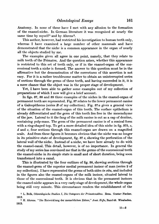
In figs. 37, 38 and 39 three examples of the niches in the enamel-organ ofpermanent teeth are represented. Fig. 37 relates to the lower permanent canineof a Galeopithecus (series B of my collection). Fig. 37a gives a general viewof the situation of the enamel-organ of this tooth. The surface epithelium isalready differentiated and the germ of this tooth lies free in the mesenchymeof the jaw. Lateral to it the fang of the milk canine is cut as a cap of dentine,containing pulp-mass. The germ of the permanent canine is of a conical formwith a ring-shaped top. To get a more detailed idea of this niche in fig. 37b, c,d and e, four sections through this enamel-organ are drawn on a magnifiedscale. And from these figures it becomes obvious that the niche was no longerin its primitive state of development, fig. 37 e, showing the perforation of thelateral wall of the niche. Instead of a niche, we have here already to do withthe enamel-canal, This detail, however, is of no importance. In general thestudy of my series has convinced me that in the germs of the successional teeththe enamel-niche in its simple state is small and of short duration, being soontransformed into a canal.
This is illustrated by the four outlines of fig. 38, showing sections throughthe enamel-germ of the superior medial permanent incisor of man (series 2 ofmy collection). I have represented the germs of both sides in situ, and includedin the figures also the enamel-organs of the milk incisor, situated lateral tothose of the successional teeth. It is obvious that in the permanent incisorsthe enamel-niche appears in a very early stage of development, the whole organbeing still very minute. This circumstance renders the establishment of the
1 L. Bolk, Odontologieche Studien I, Die Ontogenre der Primatenzahne. Zena. Gustav Fischer.1913.
2 H. Ahrens. "Die Entwicklung der menschlichen ZUhne," Anat. Hefte, Band 48. Wiesbaden.1913.
11-2
161
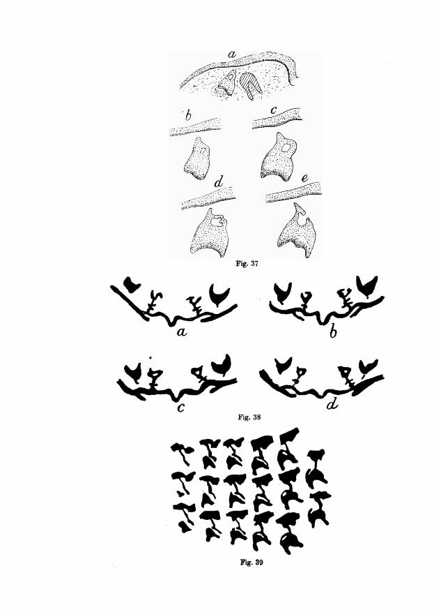
Fig. 37
Fig. 38
Fig. 39
C
C4'b
Odontological Essays 163
presence of the niche in such organs rather difficult. A very fine specimen ofan enamel-niche in the germ of a permanent tooth is finally represented infig. 89, showing a number of diagrams of succeeding sections through theenamel-organ of the inferior first permanent molar of Mycetes (series B of mycollection). The diagrams are arranged in vertical rows. In this case we haveto do with a niche whose backwall is already perforated, as shown by the twodiagrams of the last row.
With regard to theoretical considerations upon the morphological significa-tion of the enamel-niche, the statement of its occurrence in the organ ofpermanent teeth is of the utmost interest. And therefore I will here addthat Sicher has lately demonstrated the presence of an enamel-niche in thepermanent teeth of the mole'.
From the foregoing it may safely be concluded that the development of theenamel-organ in Primates (and as will be demonstrated later on also in othermammals) does not happen in such a simple manner as would be concludedfrom the descriptions given in the textbooks of Embryology. It is of a morecomplicated nature, and owing to the appearance of the enamel-niche, therelation between the enamel-organ and the dental lamina becomes somewhatmore intricate than would be concludedfrom the current descriptions. Our firsttask will now be to demonstrate this by dealing with the principal points in thefurther behaviour of the enamel-niche.
There was already occasion in the foregoing discussion to remark that theenamel-niche as such is but of short duration. For, by penetrating graduallydeeper into the enamel-organ, its backwall becomes successively thinner, untilat last it is perforated and instead of a niche the enamel-organ is provided witha canal of conical form.
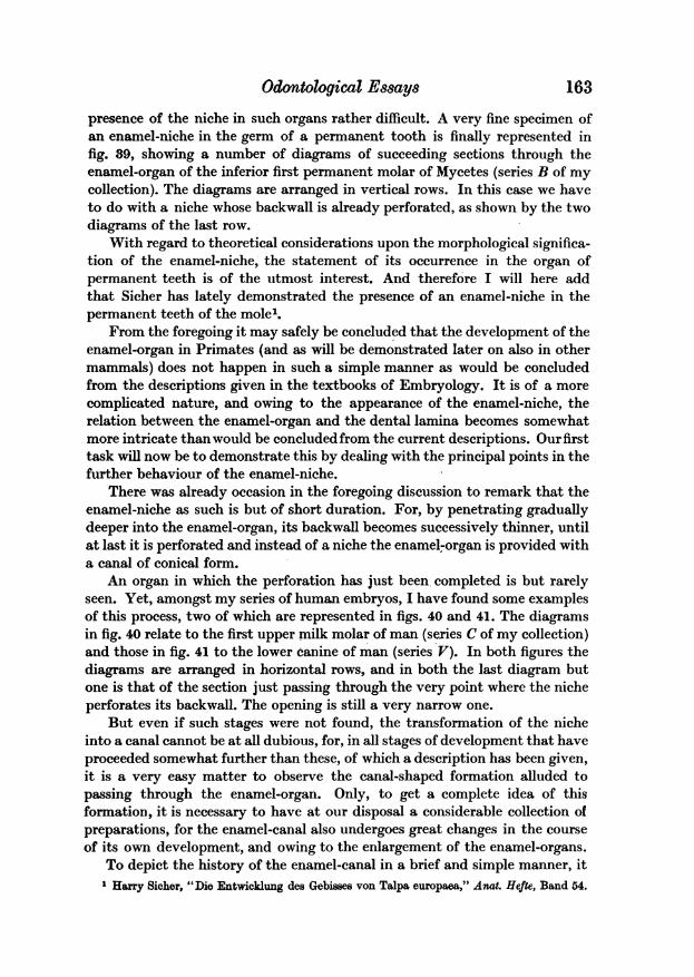
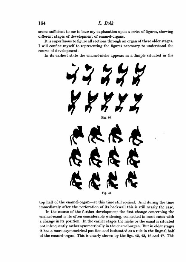
An organ in which the perforation has just been completed is but rarelyseen. Yet, amongst my series of human embryos, I have found some examplesof this process, two of which are represented in figs. 40 and 41. The diagramsin fig. 40 relate to the first upper milk molar of man (series C of my collection)and those in fig. 41 to the lower canine of man (series V). In both figures thediagrams are arranged in horizontal rows, and in both the last diagram butone is that of the section just passing through the very point where the nicheperforates its backwall. The opening is still a very narrow one.
But even if such stages were not found, the transformation of the nicheinto a canal cannot be at all dubious, for, in all stages of development that haveproceeded somewhat further than these, of which a description has been given,it is a very easy matter to observe the canal-shaped formation alluded topassing through the enamel-organ. Only, to get a complete idea of thisformation, it is necessary to have at our disposal a considerable collection ofpreparations, for the enamel-canal also undergoes great changes in the courseof its own development, and owing to the enlargement of the enamel-organs.
To depict the history of the enamel-canal in a brief and simple manner, itHarry Sicher, "Die Entwicklung des Gebisses von Talpa europaea," Anat. Hefte, Band 54.
164 L. Bolk
seems sufficient to me to base my explanation upon a series of figures, showingdifferent stages of development of enamel-organs.
It is superfluous to figure all sections through an organ of these older stages.I will confine myself to representing the figures necessary to understand thecourse of development.
In its earliest state the enamel-niche appears as a dimple situated in the
'9b 6
Fig. 40
Fig. 41
top half of the enamel-organ-at this time still conical. And during the timeimmediately after the perforation of its backwall this is still nearly the case.
In the course of the further development the first change concerning theenamel-canal is its often considerable widening, connected in most cases witha change in its position. In the earlier stages the niche or the canal is situatednot infrequently rather symmetrically in the enamel-organ. But in older stagesit has a more asymmetrical position and is situated as a rule in the lingual halfof the enamel-organ. This is clearly shown by the figs. 42, 43, 46 and 47. This
Odomtological Essays
situation is due to the fact that the proper enamel-organ is developing andenlarging principally in a buccal direction.
That this mode of development does however not always take place isshewn by the figs. 44, 45, 48 and 49, to which I will return later on. By thedilatation of the enamel-canal the character of the whole germ is very muchchanged. For the canal is now enclosed between a very thin lingual wall,which is indeed the prolongation of the dental lamina, and the enamel-organitself.
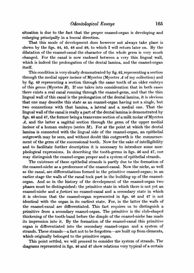
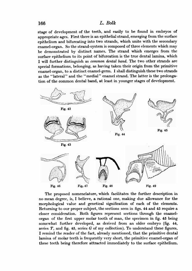
This condition is veryclearly demonstrated by fig. 42, representing a sectionthrough the medial upper incisor of Mycetes (Mycetes A of my collection) andby fig. 43 representing a section through the same tooth of an older embryoof this genus (Mycetes B). If one takes into consideration that in both casesthere exists a real canal running through the enamel-germ, and that the thinlingual wall of this canal is the prolongation of the dental lamina, it is obviousthat one may describe this state as an enamel-organ having not a single, buttwo connections with that lamina, a lateral and a medial one. That thelingual wall of the canal is really a part of the dental lamina is demonstrated byfigs. 46 and 47, the former beinga transverse section of a milk molar of MycetesA, and the latter a sagittal section through the germ of the upper medialincisor of a human embryo (series M). For at the point at which the dentallamina is connected with the lingual side of the enamel-organ, an epithelialoutgrowth may be seen, and without doubt this outgrowth is the commence-ment of the germ of the successional tooth. Now for the sake of intelligibilityand to facilitate further description it is necessary to introduce some mor-phological expressions. In describing the tooth-germs in figs. 46 and 47, onemay distinguish the enamel-organ proper and a system of epithelial strands.
The existence of these epithelial strands is partly due to the formation ofthe enamel-niche as a predecessor of the enamel-canal. Now the niche, as wellas the canal, are differentiations formed in the primitive enamel-organ; in anearlier stage the walls of the canal took part in the building up of the enamel-organ. And so in the history of the development of the enamel-organ twophases must be distinguished: the primitive state in which there is not yet anenamel-niche and a fortiori no enamel-canal and a secondary state in whichit is obvious that the enamel-organ represented in figs. 46 and 47 is notidentical with the organ in its earliest state. For, in the latter the walls ofthe enamel-canal are differentiated. This fact requires us to distinguish aprimitive from a secondary enamel-organ. The primitive is the club-shapedthickening of the tooth-band before the dimple of the enamel-niche has madeits impression into it. By the formation of the enamel-canal this primitiveorgan is differentiated into the secondary enamel-organ and a system ofstrands. These strands-a fact not to be forgotten-are built up from elements,which originally belonged to the primitive organ.
This point settled, we will proceed to consider the system of strands. Thediagrams represented in figs. 46 and 47 show relations very typical of a certain
165
166 L. Bolk
stage of development of the teeth, and easily to be found in embryos ofappropriate ages. First there is an epithelial strand, emerging from the surfaceepithelium and bifurcating into two strands, which unite with the secondaryenamel-organ. So the strand-system is composed of three elements which maybe demonstrated by distinct names. The strand which emerges from thesurface epithelium to its point of bifurcation is the true dental lamina, whichI will further distinguish as common dental band. The two other strands arespecial formations, belonging, as having taken their origin from the primitiveenamel-organ, to a distinct enamel-germ. I shall distinguish these two strandsas the " lateral " and the " medial " enamel strand. The latter is the prolonga-tion of the common dental band, at least in younger stages of development.
Fig. 42 -
Fig. 45Fig. 44
Fig 43
Fig. 46 Fig. 47 Fig. 48 Fig. 49
The proposed nomenclature, which facilitates the further description inno mean degree, is, I believe, a rational one, making due allowance for themorphological value and genetical signification of each of the elements.Returning to our proper subject, the sections seen in figs. 44 and 45 require acloser consideration. Both figures represent sections through thie enamel-organ of the first upper molar tooth of man, the specimen in fig. 45 beingsomewhat further developed, as derived from an older embryo (fig. 44,series T, and fig. 45, series G of my collection). To understand these figures,I remind the reader of the fact, already mentioned, that the primitive dentallamina of molar teeth is frequently very short, the primitive enamel-organ ofthese teeth being therefore attracted immediately to the surface epithelium.
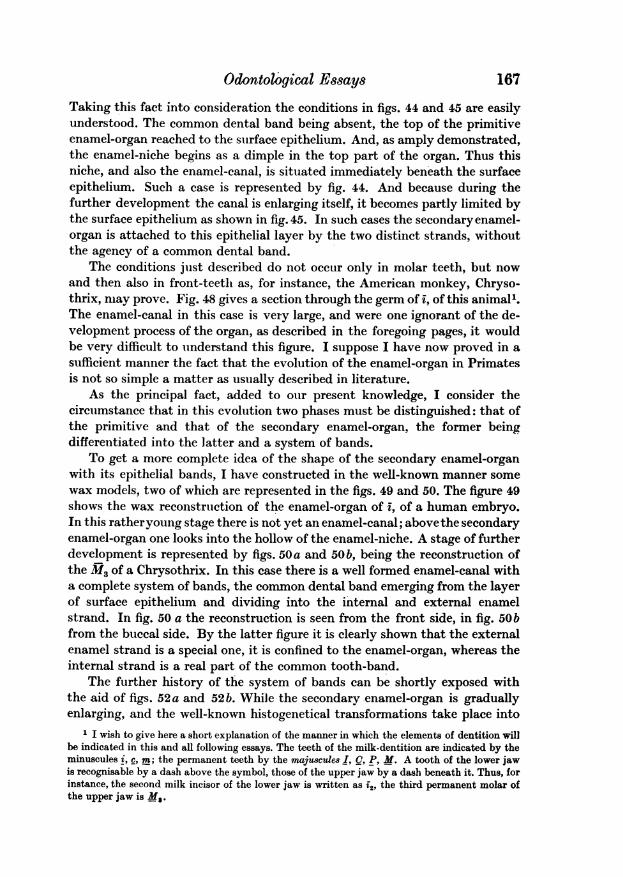
Odontol6gical Essays 167Taking this fact into consideration the conditions in figs. 44 and 45 are easilyunderstood. The common dental band being absent, the top of the primitiveenamel-organ reached to the surface epithelium. And, as amply demonstrated,the enamel-niche begins as a dimple in the top part of the organ. Thus thisniche, and also the enamel-canal, is situated immediately beneath the surfaceepithelium. Such a case is represented by fig. 44. And because during thefurther development the canal is enlarging itself, it becomes partly limited bythe surface epithelium as shown in fig. 45. In such cases the secondaryenamel-organ is attached to this epithelial layer by the two distinct strands, withoutthe agency of a common dental band.
The conditions just described do not occUr only in molar teeth, but nowand then also in front-teeth as, for instance, the American monkey, Chryso-thrix, may prove. Fig. 48 gives a section through the germ of i, of this animal'.The enamel-canal in this case is very large, and were one ignorant of the de-velopment process of the organ, as described in the foregoing pages, it wouldbe very difficult to understand this figure. I suppose I have now proved in asufficient manner the fact that the evolution of the enamel-organ in Primatesis not so simple a matter as usually described in literature.
As the principal fact, added to our present knowledge, I consider thecircumstance that in this evolution two phases must be distinguished: that ofthe primitive and that of the secondary enamel-organ, the former beingdifferentiated into the latter and a system of bands.
To get a more complete idea of the shape of the secondary enamel-organwith its epithelial bands, I have constructed in the well-known manner somewax models, two of which are represented in the figs. 49 and 50. The figure 49shows the wax reconstruction of the enamel-organ of i, of a human embryo.In this ratheryoung stage there is not yet an enamel-canal; abovethe secondaryenamel-organ one looks into the hollow of the enamel-niche. A stage of furtherdevelopment is represented by figs. 50a and 50b, being the reconstruction ofthe M3 of a Chrysothrix. In this case there is a well formed enamel-canal witha complete system of bands, the common dental band emerging from the layerof surface epithelium and dividing into the internal and external enamelstrand. In fig. 50 a the reconstruction is seen from the front side, in fig. 50bfrom the buccal side. By the latter figure it is clearly shown that the externalenamel strand is a special one, it is confined to the enamel-organ, whereas theinternal strand is a real part of the common tooth-band.
The further history of the system of bands can be shortly exposed withthe aid of figs. 52a and 52b. While the secondary enamel-organ is graduallyenlarging, and the well-known histogenetical transformations take place into
1 I wish to give here a short explanation of the manner in which the elements of dentition willbe indicated in this and all following essays. The teeth of the milk-dentition are indicated by theminuscules i, c, m; the permanent teeth by the majuscules I, C, P, M. A tooth of the lower jawis recognisable by a dash above the symbol, those of the upper jaw by a dash beneath it. Thus, forinstance, the second milk incisor of the lower jaw is written as f2, the third permanent molar ofthe upper jaw is Ms.
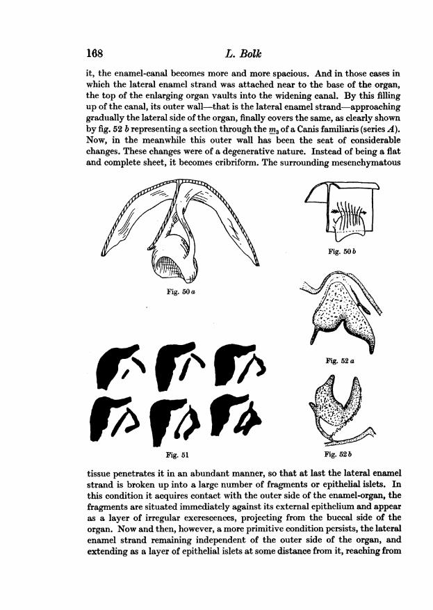
168 L. Bolkit, the enamel-canal becomes more and more spacious. And in those cases inwhich the lateral enamel strand was attached near to the base of the organ,the top of the enlarging organ vaults into the widening canal. By this fillingup of the canal, its outer wall-that is the lateral enamel strand-approachinggradually the lateral side of the organ, finally covers the same, as clearly shownby fig. 52 b representing a section through the rn3 of a Canis familiaris (series A).Now, in the meanwhile this outer wall has been the seat of considerablechanges. These changes were of a degenerative nature. Instead of being a flatand complete sheet, it becomes cribriform. The surrounding mesenchymatous
Fig. 50b
Fig. 50a /
Fig. 52a
Fig. 51 Fig. 52 b
tissue penetrates it in an abundant manner, so that at last the lateral enamelstrand is broken up into a large number of fragments or epithelial islets. Inthis condition it acquires contact with the outer side of the enamel-organ, thefragments are situated immediately against its external epithelium and appearas a layer of irregular excrescences, projecting from the buccal side of theorgan. Now and then, however, a more primitive condition persists, the lateralenamel strand remaining independent of the outer side of the organ, andextending as a layer of epithelial islets at some distance from it, reaching from
Odontological Essays 169
the base of the organ to the common dental band. This condition is shown byfig. 52a (mi, of a Mycetes) and fig. 58 (I of Equus caballus, series D).
In the course of the further development, the common dental band andits prolongation, the medial enamel strand, degenerate and atrophy. Themanner in which this process goes on is similar to that in the lateral enamelstrand. In the meantime the organ recedes from the medial enamel strand;during a short time it is still connected with it by a narrow line of cells, by theinterruption of which the organ becomes quite independent of the system ofbands. To our knowledge of this well-known process, I have nothing to add.
As pointed out before, we intended, after the description of the earlierstages of tooth-genesis in Primates in a more detailed manner, to prove thatthe ontogenetical conditions, which we have met with in this group ofmammals,occur also in the other groups. As we can do so in a brief account, there is noneed of the pointing out afresh of all observations which I have made con-cerning these points, and I will confine myself to giving some examples fromdifferent groups, which I choose among my considerable collection of seriesof mammalian embryos. In doing so I accomplish, as I believe, my purpose:to demonstrate that the enamel-niche and canal are universal phenomena inthe tooth-genesis of mammals. The conviction, however, that the enamel-organ, during a short period of tooth-genesis, has a double connection with thetooth-band, can only be acquired by examining a large number of mammalianembryos of different species and different ages. For it may be repeated thatthe recognition of the presence either of the niche or of the canal is not alwaysvery easy, the development of both being often rudimentary. Of thegeneral occurrence of niche and canal I was so strongly convinced, that itwas a very interesting and surprising fact to me, to find that there are formsin which any trace of a niche or canal is wanting. I will advance two examplesof these exceptions. The first concerns the milk teeth of Cheiroptera. Of thisgroup I have studied a number of series, in close stages of development,of the Malayan form: Roussettus amplexicaudatus. And it is very interesting,that while I looked in vain for an enamel-niche or canal in the organs of theset of milk teeth, the organs of the successional teeth behave quite regularly,as may be seen in fig. 56, showing a section through the anlage of P2 (series Cof my collection) in which the double connection between tooth-band andenamel-organ is obvious. There is a short enamel-canal running in the typicalmanner through the top part of the organ.
This case is a very welcome one from another point of view, for it is afurther proof of the occurrence of the enamel-canal in the organs of permanentteeth.
I believe I know the reason of this lack of an enamel-niche in the milkteeth of Cheiroptera, but I think it is premature to advance my opinion uponthis point, before I have given a plain and integral exposition of my viewsupon the genetical signification of the enamel-canal in general. And so I willreturn to the facts later on.
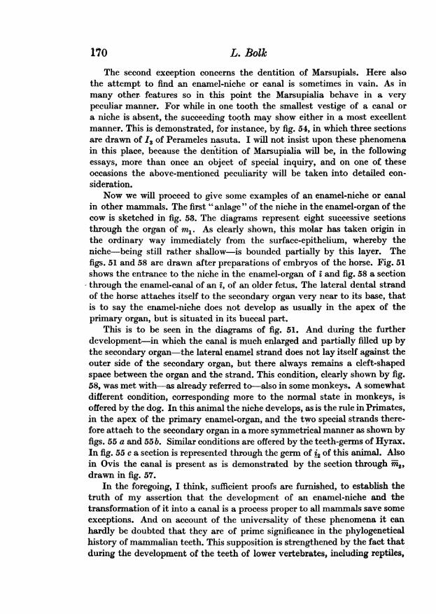
170 L. BolkThe second exception concerns the dentition of Marsupials. Here also
the attempt to find an enamel-niche or canal is sometimes in vain. As inmany other. features so in this point the Marsupialia behave in a verypeculiar manner. For while in one tooth the smallest vestige of a canal ora niche is absent, the succeeding tooth may show either in a most excellentmanner. This is demonstrated, for instance, by fig. 54, in which three sectionsare drawn of I3 of Perameles nasuta. I will not insist upon these phenomenain this place, because the dentition of Marsupialia will be, in the followingessays, more than once an object of special inquiry, and on one of theseoccasions the above-mentioned peculiarity will be taken into detailed con-sideration.
Now we will proceed to give some examples of an enamel-niche or canalin other mammals. The first " anlage " of the niche in the enamel-organ of thecow is sketched in fig. 53. The diagrams represent eight successive sectionsthrough the organ of in. As clearly shown, this molar has taken origin inthe ordinary way immediately from the surface-epithelium, whereby theniche-being still rather shallow-is bounded partially by this layer. Thefigs. 51 and 58 are drawn after preparations of embryos of the horse. Fig. 51shows the entrance to the niche in the enamel-organ of i and fig. 58 a sectionthrough the enamel-canal of an i, of an older fetus. The lateral dental strandof the horse attaches itself to the secondary organ very near to its base, thatis to say the enamel-niche does not develop as usually in the apex of theprimary organ, but is situated in its buccal part.
This is to be seen in the diagrams of fig. 51. And during the furtherdevelopment-in which the canal is much enlarged and partially filled up bythe secondary organ-the lateral enamel strand does not lay itself against theouter side of the secondary organ, but there always remains a cleft-shapedspace between the organ and the strand. This condition, clearly shown by fig.58, was met with-as already referred to-also in some monkeys. A somewhatdifferent condition, corresponding more to the normal state in monkeys, isoffered by the dog. In this animal the niche develops, as is the rule in Primates,in the apex of the primary enamel-organ, and the two special strands there-fore attach to the secondary organ in a more symmetrical manner as shown byfigs. 55 a and 55 b. Similar conditions are offered by the teeth-germs of Hyrax.In fig. 55 c a section is represented through the germ of i2 of this animal. Alsoin Ovis the canal is present as is demonstrated by the section through m2,drawn in fig. 57.
In the foregoing, I think, sufficient proofs are furnished, to establish thetruth of my assertion that the development of an enamel-niche and thetransformation of it into a canal is a process proper to all mammals save someexceptions. And on account of the universality of these phenomena it canhardly be doubted that they are of prime significance in the phylogeneticalhistory of mammalian teeth. This supposition is strengthened by the fact thatduring the development of the teeth of lower vertebrates, including reptiles,
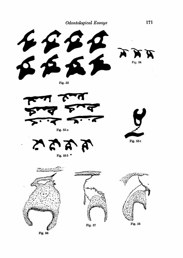
Odontological Essays
Fig. 53
Fig. 55 a
Fig. 55 b w
171
Fitg. 54
Fig. 55c
Fig. 57 Fig. 58
Fir. 56
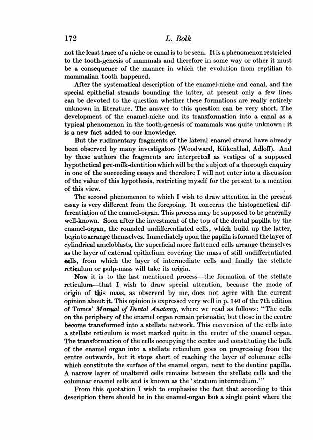
not the least trace of a niche or canal is to be seen. It is a phenomenon restrictedto the tooth-genesis of mammals and therefore in some way or other it mustbe a consequence of the manner in which the evolution from reptilian tomammalian tooth happened.
After the systematical description of the enamel-niche and canal, and thespecial epithelial strands bounding the latter, at present only a few linescan be devoted to the question whether these formations are really entirelyunknown in literature. The answer to this question can be very short. Thedevelopment of the enamel-niche and its transformation into a canal as atypical phenomenon in the tooth-genesis of mammals was quite unknown; itis a new fact added to our knowledge.
But the rudimentary fragments of the lateral enamel strand have alreadybeen observed by many investigators (Woodward, Kukenthal, Adloff). Andby these authors the fragments are interpreted as vestiges of a supposedhypothetical pre-milk-dentition which will be the subject of a thorough enquiryin one of the succeeding essays and therefore I will not enter into a discussionof the value of this hypothesis, restricting myself for the present to a mentionof this view.
The second phenomenon to which I wish to draw attention in the presentessay is very different from the foregoing. It concerns the histogenetical dif-ferentiation of the enamel-organ. This process may be supposed to be generallywell-known. Soon after the investment of the top of the dental papilla by theenamel-organ, the rounded undifferentiated cells, which build up the latter,beginto arrange themselves. Immediatelyupon the papilla is formed the layer ofcylindrical ameloblasts, the superficial more flattened cells arrange themselvesas the layer of external epithelium covering the mass of still undifferentiatedcells, from which the layer of intermediate cells and finally the stellatereticulum or pulp-mass will take its origin.
Now it is to the last mentioned process-the formation of the stellatereticulum-that I. wish to draw special attention, because the mode oforigin of this mass, as observed by me, does not agree with the currentopinion about it. This opinion is expressed very well in p. 140 of the 7th editionof Tomes' Manual of Dental Anatomy, where we read as follows: "The cellson the periphery of the enamel organ remain prismatic, but those in the centrebecome transformed into a stellate network. This conversion of the cells intoa stellate reticulum is most marked quite in the centre of the enamel organ.The transformation of the cells occupying the centre and constituting the bulkof the enamel organ into a stellate reticulum goes on progressing from thecentre outwards, but it stops short of reaching the layer of columnar cellswhich constitute the surface of the enamel organ, next to the dentine papilla.A narrow layer of unaltered cells remains between the stellate cells and thecolumnar enamel cells and is known as the 'stratum intermedium."'
From this quotation I wish to emphasise the fact that according to thisdescription there should be in the enamel-organ but a single point where the
172 L. Bolk
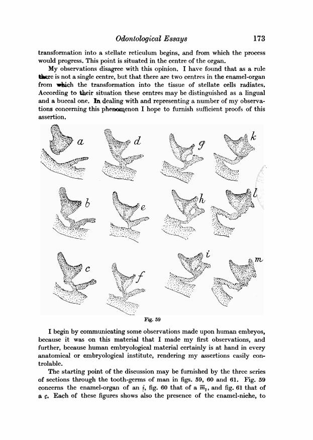
Odontological Es8ays
transformation into a stellate reticulum begins, and from which the processwould progress. This point is situated in the centre of the organ.
My observations disagree with this opinion. I have found that as a ruletire is not a single centre, but that there are two centres in the enamel-organfrom wbich the transformation into the tissue of stellate cells radiates.According to their situation these centres may be distinguished as a lingualand a buccal one. In dealing with and representing a number of my observa-tions concerning this phenomenon I hope to furnish sufficient proofs of thisassertion.
X t
I.~~~~~~~~~~~~I
Fig. 59
I begin by communicating some observations made upon human embryos,because it was on this material that I made my first observations, andfurther, because human embryological material certainly is at hand in everyanatomical or embryological institute, rendering my assertions easily con.trolable.
The starting point of the discussion may be furnished by the three seriesof sections through the tooth-germs of man in figs. 59, 60 and 61. Fig. 59concerns the enamel-organ of an i, fig. 60 that of a m,, and fig. 61 that ofa £. Each of these figures shows also the presence of the enamel-niche, to
173
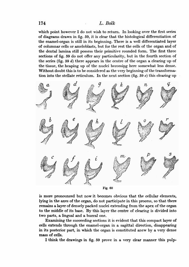
which point however I do not wish to return. In looking over the first seriesof diagrams drawn in fig. 59, it is clear that the histological differentiation ofthe enamel-organ is still in its beginning. There is a well differentiated layerof columnar cells or ameloblasts, but for the rest the cells of the organ and ofthe dental lamina still possess their primitive rounded form. The first threesections of fig. 59 do not offer any particularity, but in the fourth section ofthe series (fig. 59 d) there appears in the centre of the organ a clearing up ofthe tissue, the heaping up of the nuclei becoming here somewhat less dense.Without doubt this is to be considered as the very beginning of the transforma-tion into the stellate reticulum. In the next section (fig. 59 e) this clearing up
Fig. 60
is more pronounced but now it becomes obvious that the cellular elements,lying in the axes of the organ, do not participate in this process, so that thereremains a layer of densely packed nuclei extending from the apex of the organto the middle of its base. By this layer the centre of clearing is divided intotwo parts, a lingual and a buccal one.
Examining the succeeding sections it is evident that this compact layer ofcells extends through the enamel-organ in a sagittal direction, disappearingin its posterior part, in which the organ is constituted anew by a very densemass of cells.
I think the drawings in fig. 59 prove in a very clear manner this pulp.,
174 L. Bolk
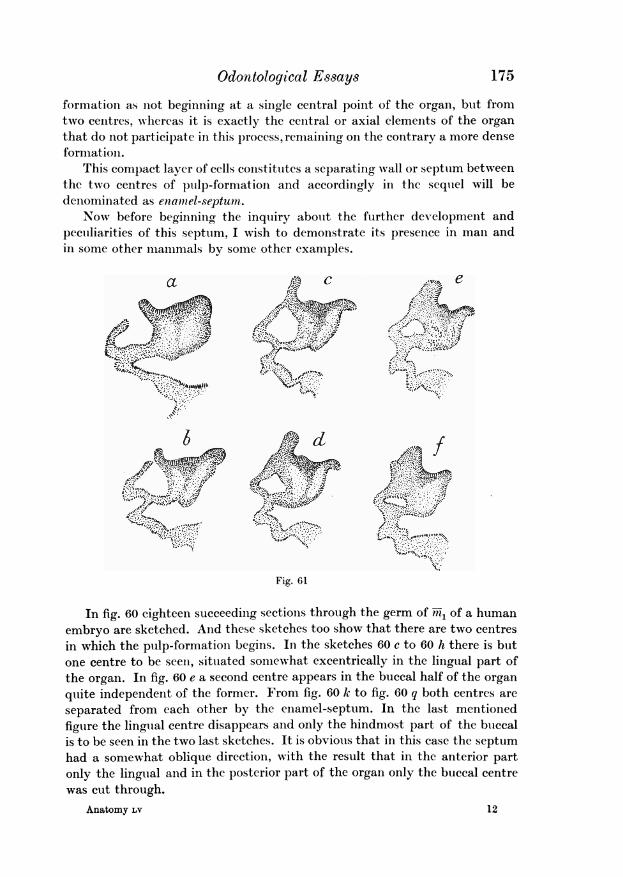
Odontological Essays 175
formation as not beginning at a single central point of the organ, but fromtwo centres, whereas it is exactly the central or axial elements of the organthat do not participate in this process, remaining on the contrary a more denseformation.
This compact layer of cells constitutes a separating wvall or septum betweenthe two centres of puilp-formation and accordingly in the sequel will bedenominated as eniamel-septum.
Now before beginning the inquiry about the further development andpeculiarities of this septum, I wish to demonstrate its presence in man andin some other mammals by some other examples.
a C
M..~~~~~~~~~~~~~~~~~~~~~~~~~~~~~~i.
.I,.~~~~~~A-1
Fig. 61
In fig. 60 eighteen succeeding sections through the germ of -m1 of a humanembryo are sketched. And these sketches too show that there are two centresin which the pulp-formation begins. In the sketches 60 c to 60 h there is butone centre to be seen, situated somewhat excentrically in the lingual part ofthe organ. In fig. 60 e a second centre appears in the buccal half of the organquite independent of the former. From fig. 60 k to fig. 60 q both centres areseparated from each other by the enamel-septum. In the last mentionedfigure the lingual centre disappears and only the hindmost part of the buccalis to be seen in the two last sketches. It is obvious that in this case the septumhad a somewhat oblique direction, with the result that in the anterior partonly the lingual and in the posterior part of the organ only the buccal centrewas cut through.
Anatomy LV 12
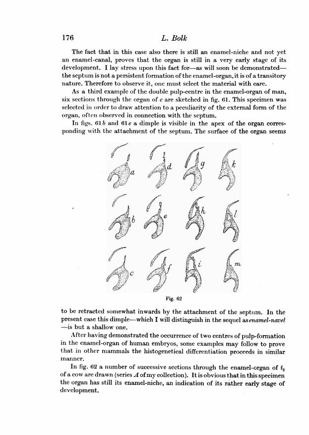
The fact that in this case also there is still an enamel-niche and not yetan enamel-canal, proves that the organ is still in a very early stage of itsdevelopment. I lay stress upon this fact for-as will soon be demonstrated-the septum is not a persistent formation of the enamel-organ, it is of a transitorynature. Therefore to observe it, one must select the material with care.
As a third example of the double pulp-centre in the enamel-organ of man,six sections through the organ of c are sketched in fig. 61. This specimen wasselected in order to draw attention to a peculiarity of the external form of theorgan, often observed in connection with the septum.
In figs. 61 b and 61 e a dimple is visible in the apex of the organ corres-ponding with the attachment of the septum. The surface of the organ seems
d a So 49d
Fig. 62
to be retracted somewhat inwards by the attachment of the septum. In thepresent case this dimple-which I will distinguish in the sequel as enamel-navel-is but a shallow one.
After having demonstrated the occurrence of two centres of pulp-formationin the enamel-organ of human embryos, some examples may follow to provethat ili other mammals the histogenetical differentiation proceeds in similarmanner.
In fig. 62 a number of successive sections through the enamel-organ of i2of a cow are drawn (series A ofmy collection). It is obvious that in this specimenthe organ has still its enamel-niche, an indication of its rather early stage ofdevelopment.
176 L. Bolk
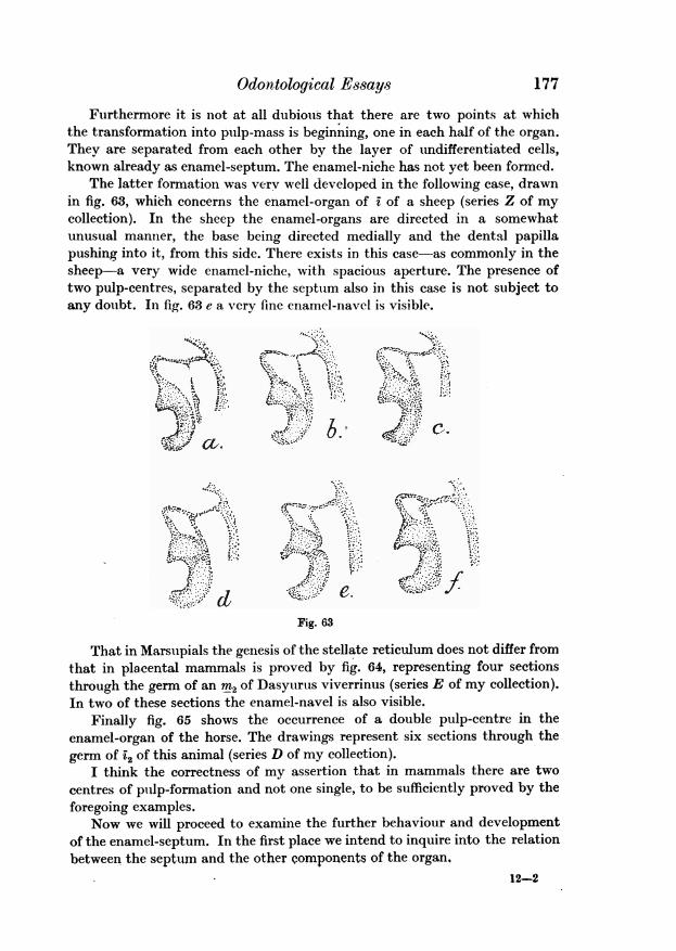
Odontological E8says9
Furthermore it is not at all dubious that there are two points at whichthe transformation into pulp-mass is beginning, one in each half of the organ.They are separated from each other by the layer of undifferentiated cells,known already as enamel-septum. The enamel-niche has not yet been formed.
The latter formation was verv well developed in the following case, drawnin fig. 63, which concerns the enamel-organ of i of a sheep (series Z of mycollection). In the sheep the enamel-organs are directed in a somewhatunusual manner, the base being directed medially and the dental papillapushing into it, from this side. There exists in this case-as commonly in thesheep-a very wide enamel-niche, with spacious aperture. The presence oftwo pulp-centres, separated by the septum also in this case is not subject toany doubt. In fig. 63 e a very fine enamel-navel is visible.
ale
I /Fig. 63
That in Marsupials the genesis of the stellate reticulum does not differ fromthat in placental mammals is proved by fig. 64, representing four sectionsthrough the germ of an M2 of Dasyurtus viverrinus (series E of my collection).In two of these sections the enamel-navel is also visible.
Finally fig. 65 shows the occurrence of a double pulp-centre in theenamel-organ of the horse. The drawings represent six sections through thegerm of '2 of this animal (series D of my collection).
I think the correctness of my assertion that in mammals there are twocentres of pulp-formation and not one single, to be sufficiently proved by theforegoing examples.
Now we will proceed to examine the further behaviour and developmentof the enamel-septum. In the first place we intend to inquire into the relationbetween the septum and the other components of the organ.
12-2
177
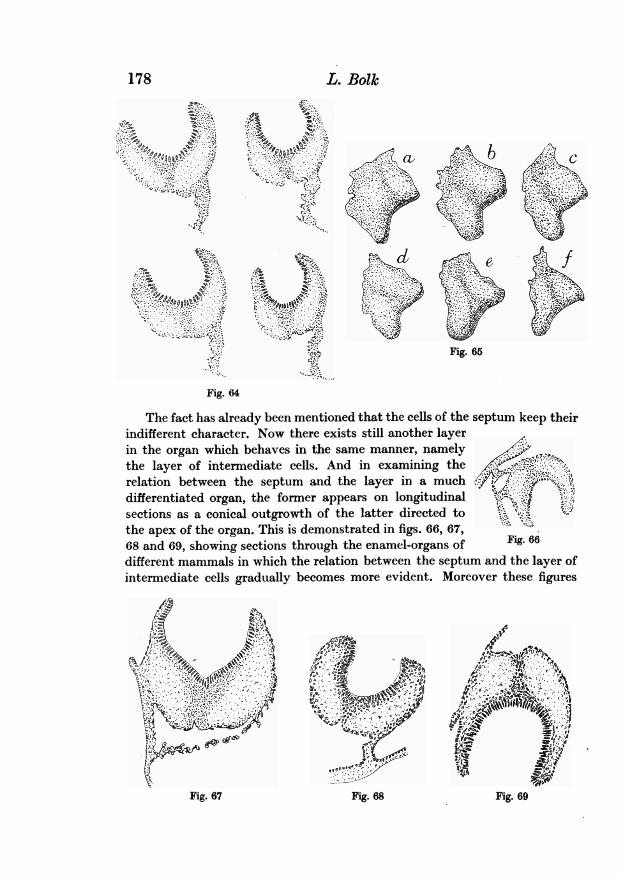
178 L. Bolk
S. ~ ~ ~ ~ ~ ~
F czAi t
Fig. 65
Fig. 64
The fact has already been mentioned that the cells of the septum keep theirindifferent character. Now there exists still another layerin the organ which behaves in the same manner, namelythe layer of intermediate cells. And in examining the ..K;relation between the septum and the layer in a much KII
differentiated organ, the former appears on longitudinal t .sections as a conical outgrowth of the latter directed to *J Vthe apex of the organ. This is demonstrated in figs. 66, 67,68 and 69, showing sections through the enamel-organs of Fig. 66different mammals in which the relation between the septum and the layer ofintermediate cells gradually becomes more evident. Moreover these figures
Fig.67ig. 68Fig. 67 Fvig. 69
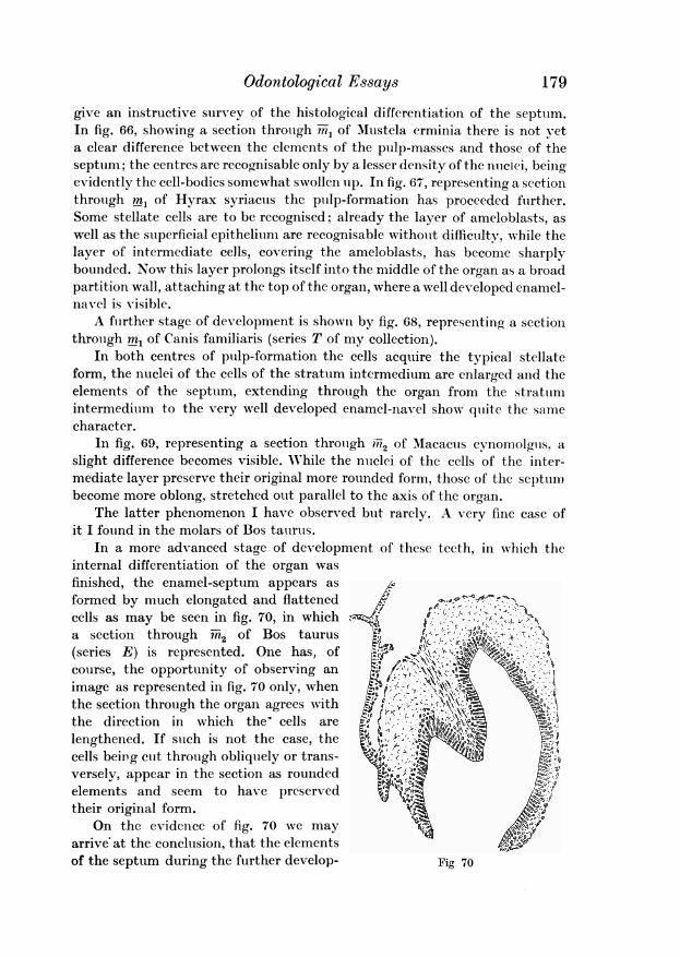
Odontological Essays 179
give an instructive survey of the histological differentiation of the septum.In fig. 66, showing a section through -m, of Mustela erminia there is not veta clear difference between the elements of the pulp-masses and those of theseptum; the centres are recognisable only by a lesser density of the nuclei, beingevidently the cell-bodies somewhat swollen up. In fig. 67, representing a sectionthrough ml of Hyrax syriacus the pulp-formation has proceeded further.Some stellate cells are to be recognised; already the layer of ameloblasts, aswell as the superficial epithelium are recognizable without difficulty, while thelayer of intermediate cells, covering the ameloblasts, has become sharplybounded. Now this layer prolongs itself into the middle of the organ as a broadpartition wall, attaching at the top of the organ, where a well developed enamel-nav el is visible.
A further stage of development is shown by fig. 68, representing a sectionthrough ml of Canis familiaris (series T of my collection).
In both centres of pulp-formation the cells acquire the typical stellateform, the nuclei of the cells of the stratum intermedium are enlarged and theelements of the septum, extending through the organ from the stratumintermedium to the very well developed enamel-navel show quite the samecharacter.
In fig. 69, representing a section through iiR2 of M1acacus cynomolgus, aslight difference becomes visible. While the nuclei of the cells of the inter-mediate layer preserve their original more rounded form, those of the septumbecome more oblong, stretched out parallel to the axis of the organ.
The latter phenomenon I have observed but rarely. A very fine case ofit I found in the molars of Bos tatirus.
In a more advanced stage of development of these teeth, in which theinternal differentiation of the organ wasfinished, the enamel-septum appears asformed by much elongated and flattened U - Acells as may be seen in fig. 70, in whichsection through m2ofBstuu l;r ; - d
(series E) is represented. One has, of .,w .course, the opportunity of observing animage as represented in fig. 70 only, when j -r8, Kthe section through the organ agrees with ;the direction in which the- cells arelengthened. If such is not the case, the I,;cells being cut through obliquely or trans- <versely, appear in the section as rounded ,elements and seem to have preservedtheir original form.
On the evidence of fig. 70 we mayarrive at the conclusion, that the elementsof the septum during the further develop- Fig. 70
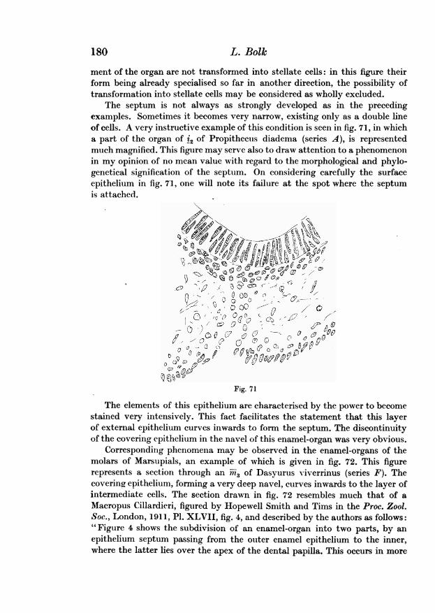
180 L. Bolk
ment of the organ are not transformed into stellate cells: in this figure theirform being already specialised so far in another direction, the possibility oftransformation into stellate cells may be considered as wholly excluded.
The septum is not always as strongly developed as in the precedingexamples. Sometimes it becomes very narrow, existing only as a double lineof cells. A very instructive example of this condition is seen in fig. 71, in whicha part of the organ of t2 of Propithecus diadema (series A), is representedmuch magnified. This figure may serve also to draw attention to a phenomenonin my opinion of no mean value with regard to the morphological and phylo-genetical signification of the septum. On considering carefully the surfaceepithelium in fig. 71, one will note its failure at the spot where the septumis attached.
00
0)C~0
Fig. 71
The elements of this epithelium are characterised by the power to becomestained very intensively. This fact facilitates the statement that this layerof external epithelium curves inwards to form the septum. The discontinuityof the covering epithelium in the navel of this enamel-organ was very obvious.
Corresponding phenomena may be observed in the enamel-organs of themolars of Marsupials, an example of which is given in fig. 72. This figurerepresents a section through an rn3 of Dasyurus v-iverrinus (series F). Thecovering epithelium, forming a very deep navel, curves inwards to the layer ofintermediate cells. The section drawn in fig. 72 resembles much that of aMacropus Cillardieri, figured by Hopewell Smith and Tims in the Proc. Zool.Soc., London, 1911, P1. XLVII, fig. 4, and described by the authors as follows:"Figure 4 shows the subdivision of an enamel-organ into two parts, by anepithelium septum passing from the outer enamel epithelium to the inner,where the latter lies over the apex of the dental papilla. This occurs in more
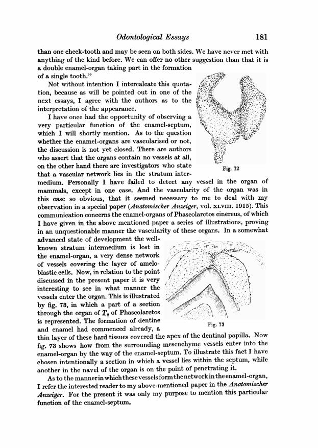
Odontological Es8ays
than one cheek-tooth and may be seen on both sides. We have never met withanything of the kind before. We can offer no other suggestion than that it isa double enamel-organ taking part in the formationof a single tooth."
Not without intention I intercalcate this quota- 4tion, because as will be pointed out in one of the Knext essays, I agree with the authors as to the -interpretation of the appearance.
I have once had the opportunity of observing a Hi -:. t ..Q . - -very particular function of the enamel-septum, ..- ?@which I will shortly mention. As to the questionwhether the enamel-organs are vascularised or not,the discussion is not yet closed. There are authorswho assert that the organs contain no vessels at all,on the other hand there are investigators who statethat a vascular network lies in the stratum inter- Fig. 72
medium. Personally I have failed to detect any vessel in the organ ofmammals, except in one case. And the vascularity of the organ was inthis case so obvious, that it seemed necessary to me to deal with myobservation in a special paper (Anatornischer Anzeiger, vol. xLviii. 1915). Thiscommunication concerns the enamel-organs of Phascolaretos cinereus, of whichI have given in the above mentioned paper a series of illustrations, provingin an unquestionable manner the vascularity of these organs. In a somewhatadvanced state of development the well- _known stratum intermedium is lost in ,-the enamel-organ, a very dense network ; . - 'of vessels covering the layer of amelo- / 0,'';blastic cells. Now, in relation to the point ,discussed in the present paper it is veryinteresting to see in what manner thevessels enter the organ. This is illustratedby fig. 73, in which a part of a section Bthrough the organ of T2 of Phascolarctos Ga.is represented. The formation of dentine F.and enamel had commenced already, a Fig. 73
thin layer of these hard tissues covered the apex of the dentinal papilla. Nowfig. 73 shows how from the surrounding mesenchyme vessels enter into the
enamel-organ by the way of the enamel-septum. To illustrate this fact I havechosen intentionally a section in which a vessel lies within the septum, whileanother in the navel of the organ is on the point of penetrating it.
As to the mannerinwhichthesevessels formthe network inthe enamel-organ,I refer the interested reader to my above-mentioned paper in the AnatomischerAnzeiger. For the present it was only my purpose to mention this particularfunction of the enamel-septum.
181
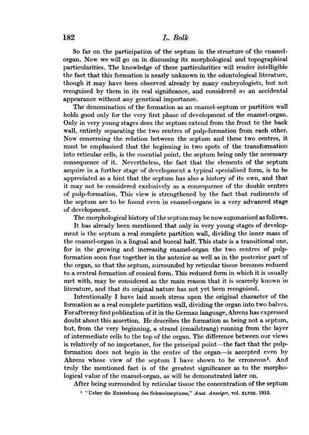
So far on the participation of the septum in the structure of the enamel-organ. Now we will go on in discussing its morphological and topographicalparticularities. The knowledge of these particularities will render intelligiblethe fact that this formation is nearly unknown in the odontological literature,though it may have been observed already by many embryologists, but notrecognised by them in its real significance, and considered as an accidentalappearance without any genetical importance.
The denomination of the formation as an enamel-septum or partition wallholds good only for the very first phase of development of the enamel-organ.Only in very young stages does the septum extend from the front to the backwall, entirely separating the two centres of pulp-formation from each other.Now concerning the relation between the septum and these two centres, itmust be emphasised that the beginning in two spots of the transformationinto reticular cells, is the essential point, the septum being only the necessaryconsequence of it. Nevertheless, the fact that the elements of the septumacquire in a further stage of development a typical specialised form, is to beappreciated as a hint that the septum has also a history of its own, and thatit may not be considered exclusively as a consequence of the double centresof pulp-formation. This view is strengthened by the fact that rudiments ofthe septum are to be found even in enamel-organs in a very advanced stageof development.
The morphological history of the septum may be now sumnmarised as follows.It has already been mentioned that only in very young stages of develop-
ment is the septum a real complete partition wall, dividing the inner mass ofthe enamel-organ in a lingual and buccal half. This state is a transitional one,for in the growing and increasing enamel-organ the two centres of pulp-formation soon fuse together in the anterior as well as in the posterior part ofthe organ, so that the septum, surrounded by reticular tissue becomes reducedto a central formation of conical form. This reduced form in which it is usuallymet with, may be considered as the main reason that it is scarcely known inliterature, and that its original nature has not yet been recognised.
Intentionally I have laid much stress upon the original character of theformation as a real complete partition wall, dividing the organ into two halves.For aftermy first publication of it in the German language, Ahrens has expresseddoubt about this assertion. He describes the formation as being not a septum,but, from the very beginning, a strand (emailstrang) running from the layerof intermediate cells to the top of the organ. The difference between our viewsis relatively of no importance, for the principal point-the fact that the pulp-formation does not begin in the centre of the organ-is accepted even byAhrens whose view of the septum I have shown to be erroneous'. Andtruly the mentioned fact is of the greatest significance as to the morpho-logical value of the enamel-organ, as will be demonstrated later on.
After being surrounded by reticular tissue the concentration of the septum1 "Ueber die Entstehung des Schmelzseptums," Anat. Anzeiger, vol. XLVIII. 1915.
182 L. Bolk
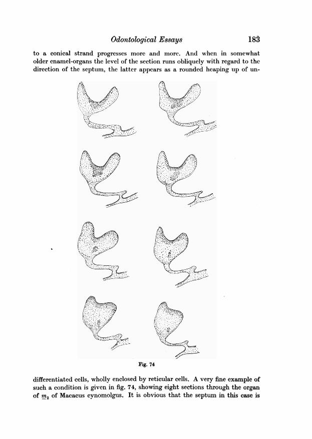
Odontological Essays 183to a conical strand progresses more and more. And when in somewhatolder enamel-organs the level of the section runs obliquely with regard to thedirection of the septum, the latter appears as a rounded heaping up of un-
Fig. 74
differentiated cells, wholly enclosed by reticular cells. A very fine example ofsuch a condition is given in fig. 74, showing eight sections through the organof m2 of Macacus cynomolgus. It is obvious that the septum in this case is
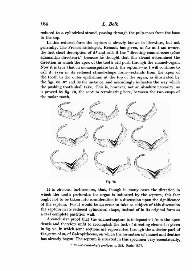
184 L. Bolk
reduced to a cylindrical strand, passing through the pulp-mass from the baseto the top.
In this reduced form the septum is already known in literature, but notgenerally. The French histologist, Renaut, has given, as far as I am aware,the first short description of iti and calls it the "directing enamel-cone (c6neadamantin directeur)," because he thought that this strand determined thedirection in which the apex of the tooth will push through the enamel-organ.Now it is true that in monocuspidate teeth the septum-as I will continue tocall it, even in its reduced strand-shape form-extends from the apex ofthe tooth to the outer epithelium at the top of the organ, as illustrated bythe figs. 66, 67 and 68 for instance, and accordingly indicates the way whichthe pushing tooth shall take. This is, however, not an absolute necessity, asis proved by fig. 70, the septum terminating here, between the two cusps ofthe molar tooth.
Fig. 75
It is obvious, furthermore, that, though in many cases the direction inwhich the tooth perforates the organ is indicated by the septum, this factought not to be taken into consideration in a discussion upon the significanceof the septum. For it would be an error to take as subject of this discussionthe septum in its reduced cylindrical shape, instead of in its original form asa real complete partition wall.
A conclusive proof that the enamel-septum is independent from the apexdentis and therefore unfit to accomplish the task of directing element is givenin fig. 75, in which some sections are represented through the anterior part ofthe germ of rn2 of Galeopithecus, on which the formation of enamel and dentinehas already begun. The septum is situated in this specimen very eccentrically,
1 Traite' d'kitiologie pratiqque, p. 248. Paris, 1897.

Odontological Essays
without any topographical relation with the apex of the tooth, which hasnearly reached the outer epithelium of the enamel-organ.
Such an eccentric situation is met with very seldom and finally in fig. 76there is reproduced yet another case of it, beinga section through m3 of Chrysothrix sciurea.
With this case I wish to finish the descriptionof the enamel-septum. It is-as mentionedalready-my intention to restrict myself in the _present paper to the communication of my *;observations only, putting off the discussion tiupon their significance to one of the next essays. XYet it seems to me not wholly out of place toanticipate some general considerations.
A first remark concerns the septum. It is of 9great importance, as bearing on the significanceof this formation, that it is met with exclusivelyin the enamel-organs of mammals. I have Fig. 76examined the genesis of the teeth in reptilesas thoroughly as that of mammals. And I have never seen in any enamel-organ of this group of vertebrates the smallest trace of an enamel-septum.It may be stated as a matter of fact that the septum is a formationtypical of the teeth of the highest class of the vertebrates, and in some wayor other its origin must be the consequence of the manner in which the teethof the mammalia have taken origin from those of reptiles. For that thedentition of mammals must be derived from that of reptiles is no longer subjectto any doubt, only as to the manner in which this evolution has taken placeare the opinions of the authors not unanimous. A special essay will be devotedto my own views upon this subject.
Now one may remember that after my description of the external develop-mental appearances of the enamel-organ, I made the remark that theenamel-niche and canal fail in the tooth-genesis of reptiles, appearing exclusivelyduring the development of mammalian teeth. And the conclusion at which Iarrived was necessarily the same as that just given: the enamel-niche mustbe a result of the manner in which the evolution of the mammalian teeth tookplace. Therefore a relation must exist between the external developmentaland the internal histogenetical phenomena, for both result from the samecause, and an attempt to solve the problem of the evolution of mammalianteeth must demonstrate at the same time the origin of the enamel-niche (canal)as well as that of the enamel-septum as a consequence of this process.
Now a very striking agreement between both phenomena may be pointedout here, by which a main morphological character of the mammalian enamel-organ comes to light.
Our researches have taught us, firstly, that in the tooth-genesis of mammalsthere is a common dental band from which two special bands emerge,
185
attaching themselves at the buccal and lingual sides respectively of the enamel-organ. In reptiles on the contrary, there is but one single connection betweenthe organ and the dental lamina. This fact gives rise to the question whetherthe enamel-organ of the mammals is a single organ, or a compound one, beinghomologous with two organs of reptiles and not with a single one. If this pointof view were true, the mammalian organ should be, so to say, a twin-organ,in which the constituent elements are situated as a buccal and a lingual one.
Now, this point of view is surely strengthened by our second observation,described in the present paper. We have demonstrated that in the organ ofmammals there is not a single centre of pulp-formation, arising in the middleof the organ, but there are two centres, one in its buccal and one in its lingualhalf. This fact too is favourable to the appreciation of this organ as a doubleone, built up by two elementary organs, a buccal and lingual one.
I confine myself to throwing simply this light upon the morphologicalnature of the enamel-organ of mammals, the working out of this idea is to beput off to one of the next essays, after the exposition of some details on theorigin of teeth in reptiles.
Yet I cannot end this essay without pointing out a consequence of theabove view concerning the teeth. If, in reality, the mammalian enamel-organ be a compound one, the tooth itself also must be of the same nature,built up by two elements each homologous with a reptilian tooth. The buccalcusps of the mammalian tooth should represent then the one, and the lingualcusps the other reptilian tooth. The mammalian tooth should be then, what Iwill call a "dimer" tooth.
This consequence forms the fundamental point of my hypothesis upon themorphological value of the mammalian tooth and its relation to the teethof reptiles.
186 L. Bolk