Embed Size (px)
Citation preview
OECD Series on Adverse Outcome Pathways No. 3
Adverse Outcome Pathwayon Alkylation of DNA in Male
Pre-Meiotic Germ CellsLeading to Heritable
Mutations
Carole Yauk,Iain Lambert,
Francesco Marchetti,George Douglas
https://dx.doi.org/10.1787/5jlsvvxn1zjc-en

Foreword This Adverse Outcome Pathway (AOP) on Alkylation of DNA in male pre-meiotic germ cells leading to heritable mutations has been developed under the auspices of the OECD AOP Development Programme, overseen by the Extended Advisory Group on Molecular Screening and Toxicogenomics (EAGMST), which is an advisory group under the Working Group of the National Coordinators for the Test Guidelines Programme (WNT). The AOP has been reviewed internally by the EAGMST, externally by experts nominated by the WNT, and has been endorsed by the WNT and the Task Force on hazard Assessment (TFHA) in April 2016. Through endorsement of this AOP, the WNT and the TFHA express confidence in the scientific review process that the AOP has undergone and accept the recommendation of the EAGMST that the AOP be disseminated publicly. Endorsement does not necessarily indicate that the AOP is now considered a tool for direct regulatory application. The Joint Meeting of the Chemicals Committee and the Working Party on Chemicals, Pesticides and Biotechnology agreed to declassification of this AOP on 17 June 2016. This document is being published under the responsibility of the Joint Meeting of the Chemicals Committee and the Working Party on Chemicals, Pesticides and Biotechnology.

Table of Contents Authors ................................................................................................................................................ 3 Abstract ............................................................................................................................................... 3 Summary of the AOP: Graphical Representation ............................................................................... 5 Key events ........................................................................................................................................... 6
Molecular Initiating Event............................................................................................................... 6 Key events ..................................................................................................................................... 10 Adverse Outcome .......................................................................................................................... 19
Key Event Relationships: Scientific evidence supporting the linkages in the AOP ......................... 24 Overall Assessment of the AOP ....................................................................................................... 46 Considerations for Potential Applications of the AOP ..................................................................... 51 References ......................................................................................................................................... 51
ADVERSE OUTCOME PATHWAY ON ALKYLATION OF DNA IN MALE PRE-MEIOTIC GERM CELLS LEADING TO HERITABLE MUTATIONS
Short name: Alkylation of DNA leading to heritable mutations
Authors
Carole Yauk (1)* Iain Lambert (2) Francesco Marchetti (1) George Douglas (1) (1) Environmental Health Science and Research Bureau, Health Canada, Ottawa, ON, Canada (2) Dept. of Biology, Carleton University, Ottawa, ON, Canada
* Communicating author: [email protected]
Abstract
Germ cell/heritable mutations are important regulatory endpoints for international agencies interested in protecting the health of future generations. However, germ cell mutation analysis has been hampered by a lack of efficient tools. With the publication of the OECD test guideline TG488 (rodent transgene mutation assay) and new technologies (including next generation sequencing) this field is experiencing renewed focus. Indeed, regulatory approaches to assess germ cell mutagenicity were the focus of a recent IWGT workshop (Yauk et al., 2013). Of particular concern is the inability to address this endpoint through high-throughput screening assays (because spermatogenesis cannot be carried out in culture), and mutagenesis is an important gap in existing high-throughput tests. The motivation for developing this AOP was to provide context for new assays in this field, identify research gaps and facilitate the development of new methods. In this AOP, a compound capable of alkylating DNA is delivered to the testes causing germ cell mutations and subsequent mutations in the offspring of the exposed parents. DNA alkylation in male pre-meiotic germ cells is the molecular initiating event. A variety of different DNA adducts are formed that are subject to DNA repair; however, at high doses the repair machinery becomes saturated or overwhelmed. The fate of remaining adducts includes: (1) attempted DNA repair by alternative DNA repair machinery, or (2) no repair. Key event (KE) 1 is insufficient or incorrect DNA repair. Lack of repair can lead to replication of adducted DNA and ensuing mutations in male pre-meiotic germ cells (KE2). Mutations that do not impair spermatogenic processes will persist in these cells and will eventually be present in the mature sperm. Thus, the mutations can be transmitted to the offspring (adverse outcome – inherited mutations). It is well documented that mice and other animals exposed to alkylating agents develop mutations in male pre-meiotic germ cells that are then found in sperm, resulting in the transmission of mutations to their offspring. There is a significant amount of empirical evidence supporting the AOP and the overall weight of evidence is strong. Although there are some gaps surrounding some mechanistic aspects of this AOP, the overarching AOP is widely accepted and applies broadly to any species that produces sperm.

Background
De novo germ cell mutations are changes in the DNA sequence of sperm or egg that can be inherited by offspring. De novo mutations contribute to a wide range of human disorders including cancer, infertility, autism, schizophrenia, intellectual disability, and epilepsy (Girirajan et al. 2010; Hoischen et al. 2010; Ku et al. 2012; Lupski 2010; Morrow 2010; Vissers et al. 2010). Each child inherits, on average, approximately one de novo mutation per 100 million nucleotides delivered via the parental egg and sperm (Conrad et al. 2011; Kong et al. 2012; O'Roak et al. 2012; Roach et al. 2010). The precise locations and types of mutations in the genomic DNA sequence govern the outcome of these mutations (e.g. protein coding versus intergenic sequences, conserved versus non-conserved mutations, etc.). Although a large portion of human DNA is of unknown function, recent literature suggests that at least 80% of the genome is transcribed, and most DNA is expected to have a biological function (Bernstein et al. 2012). It has been estimated that the proportion of coding and splice-site base substitutions that result in truncating mutations is ~5% (Kryukov et al. 2007), and that as many as 30% of missense mutations are also likely to be highly deleterious due to loss of function (Boyko et al. 2008). When they occur in functional sites, de novo mutations can cause embryonic or fetal lethality, or if viable, can produce a broad spectrum of inherited genetic disorders. Recent estimates suggest that a human genome contains approximately 100 loss-of-function variants, with as many as 20 exhibiting complete loss of gene function (McLaughlin et al. 2010). Therefore, de novo mutations contribute to the overall population genetic disease burden. The present AOP focuses on DNA alkylation in spermatogonia that causes inherited mutations transmitted via sperm, arguably one of the most well characterised modes of action in genetic toxicology. Humans are exposed to alkylating agents from external (e.g. abiotic plant materials, tobacco smoke, combustion products, chemotherapeutic agents) and internal (e.g. byproducts of oxidative damage and cellular methyl donors) sources.

5
Summary of the AOP: Graphical Representation

6
Key Events
Molecular Initiating Event
Molecular Initiating Event
DNA, Alkylation
DNA, alkylation
AOPs including this Key Event
AOP Name Event Type Alkylation of DNA in male pre-meiotic germ cells leading to heritable mutations MIE Alkylation of DNA leading to cancer MIE
Chemical Initiators
The following are chemical initiators that operate directly through this Event:
1. Diethyl nitrosamine 2. Diethyl sulfate 3. Dimethyl nitrosamine 4. Dimethyl sulfate 5. Ethyl methanesulfonate 6. Ethyl nitrosourea 7. Ethyl-N'-nitro-N-nitrosoguanidine 8. Isopropyl methanesulfonate 9. Methyl methanesulfonate 10. Methyl-l-N'-nitro-N-nitroguanidine
How this Key Event works
Level of biological organisation
Molecular
The event involves DNA alkylation to form a variety of DNA adducts (i.e. alkylated nucleotides). Alkylation occurs at various sites in DNA and can include alkylation of adenine- Nl, - N3, - N7, guanine- N3, - O6, - N7, thymine-O2, - N3, - O4, cytosine- O2, -N3, and the phosphate (diester) group (reviewed in detail in Beranek 1990). In addition, alkylation can involve modification with different sizes of alkylation groups (e.g. methyl, ethyl, propyl). It should be noted that many of these adducts are not stable or are readily repaired (discussed in more detail below). A small proportion of adducts are stable and remain bound to DNA for long periods of time.
7
How it is measured or detected
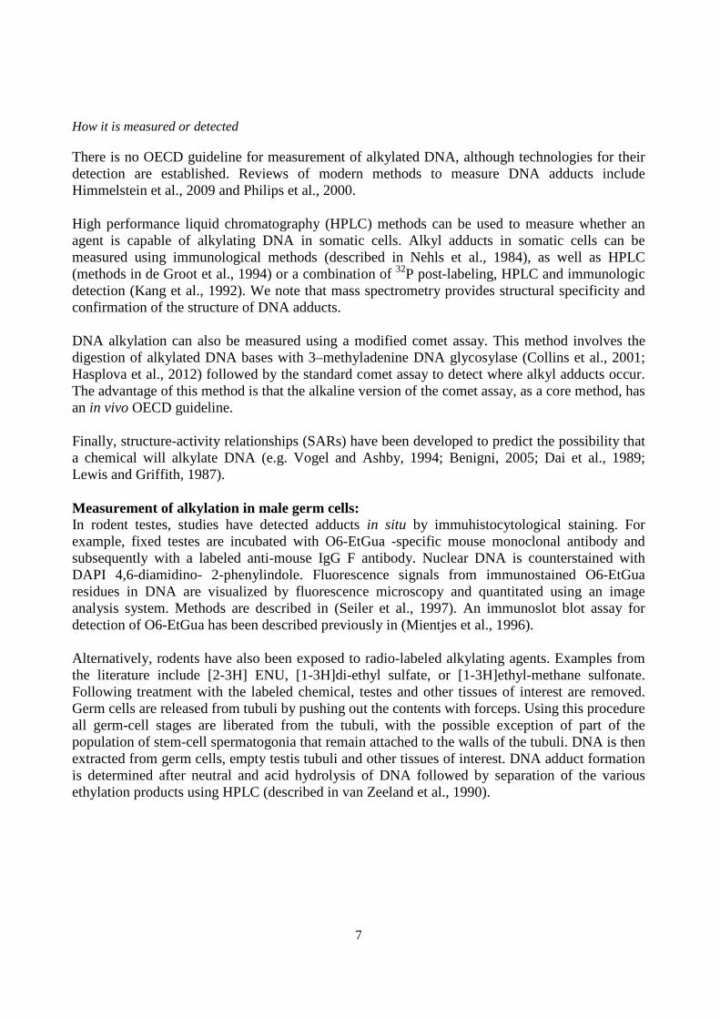
There is no OECD guideline for measurement of alkylated DNA, although technologies for their detection are established. Reviews of modern methods to measure DNA adducts include Himmelstein et al., 2009 and Philips et al., 2000. High performance liquid chromatography (HPLC) methods can be used to measure whether an agent is capable of alkylating DNA in somatic cells. Alkyl adducts in somatic cells can be measured using immunological methods (described in Nehls et al., 1984), as well as HPLC (methods in de Groot et al., 1994) or a combination of 32P post-labeling, HPLC and immunologic detection (Kang et al., 1992). We note that mass spectrometry provides structural specificity and confirmation of the structure of DNA adducts. DNA alkylation can also be measured using a modified comet assay. This method involves the digestion of alkylated DNA bases with 3–methyladenine DNA glycosylase (Collins et al., 2001; Hasplova et al., 2012) followed by the standard comet assay to detect where alkyl adducts occur. The advantage of this method is that the alkaline version of the comet assay, as a core method, has an in vivo OECD guideline. Finally, structure-activity relationships (SARs) have been developed to predict the possibility that a chemical will alkylate DNA (e.g. Vogel and Ashby, 1994; Benigni, 2005; Dai et al., 1989; Lewis and Griffith, 1987). Measurement of alkylation in male germ cells: In rodent testes, studies have detected adducts in situ by immuhistocytological staining. For example, fixed testes are incubated with O6-EtGua -specific mouse monoclonal antibody and subsequently with a labeled anti-mouse IgG F antibody. Nuclear DNA is counterstained with DAPI 4,6-diamidino- 2-phenylindole. Fluorescence signals from immunostained O6-EtGua residues in DNA are visualized by fluorescence microscopy and quantitated using an image analysis system. Methods are described in (Seiler et al., 1997). An immunoslot blot assay for detection of O6-EtGua has been described previously in (Mientjes et al., 1996). Alternatively, rodents have also been exposed to radio-labeled alkylating agents. Examples from the literature include [2-3H] ENU, [1-3H]di-ethyl sulfate, or [1-3H]ethyl-methane sulfonate. Following treatment with the labeled chemical, testes and other tissues of interest are removed. Germ cells are released from tubuli by pushing out the contents with forceps. Using this procedure all germ-cell stages are liberated from the tubuli, with the possible exception of part of the population of stem-cell spermatogonia that remain attached to the walls of the tubuli. DNA is then extracted from germ cells, empty testis tubuli and other tissues of interest. DNA adduct formation is determined after neutral and acid hydrolysis of DNA followed by separation of the various ethylation products using HPLC (described in van Zeeland et al., 1990).

8
Evidence supporting taxonomic applicability
Name Scientific Name Evidence Links
mouse Mus musculus Strong NCBI
Syrian golden hamster Mesocricetus auratus Strong NCBI
rat Rattus norvegicus Strong NCBI
Homo sapiens Homo sapiens Strong NCBI
Alkylated DNA has been measured in somatic cells in a variety of species, from prokaryotic organisms, to rodents in vivo, to human cells in culture. Theoretically, DNA alkylation can occur in any cell type in any organism.
Evidence for chemical initiation of this Molecular Initiating Event
Alkylating agents are prototypical DNA-reactive compounds and have been extensively studied for decades (reviewed in Beranek, 1990). The chemicals can be direct-acting electrophiles, or can be converted from non-reactive substances to reactive metabolites via metabolism. A prototypical alkylating agent is N-ethyl-N-nitrosourea (chemical formula C3H7N3O2) (ENU). ENU is rapidly absorbed following oral exposure and intraperitoneal injections and distributed widely across the tissues. ENU is unstable and readily reacts with somatic and germ cell DNA in mice, rats, flies and hamsters, to alkylate DNA. Very generally, mono-functional (referring to the transfer of a single alkyl group) alkylating agents include: 1. Alkyl sulfates: e.g. diethyl (DES) and dimethyl sulfate (DMS); 2. Alkyl alkanesulfonates: e.g. methyl (MMS) and ethyl methanesulfonate (EMS); 3. Nitrosamides: e.g. methyl (MNU) and ethyl nitrosourea (ENU), methyl- (MNNG) and ethyl-N'-nitro-N-nitrosoguanidine (ENNG), and the indirect-acting (i.e. requiring metabolic activation) dimethyl (DMN) and diethyl nitrosamines (DEN). ENU is the most widely studied and understood alkylating agent and as such has been instrumental in contributing to the knowledgebase in this field. Immunohistochemistry studies clearly indicate the presence of alkylated DNA following exposure to ENU in both somatic cells and spermatogonia (Kamino et al., 1995; Seiler et al., 1997; van Zeeland et al., 1990).
References
Benigni, R. (2005), "Structure-activity relationship studies of chemical mutagens and carcinogens: mechanistic investigations and prediction and approaches", Chem. Rev., 105: 1767-1800.
Beranek, D.T. (1990), "Distribution of methyl and ethyl adducts following alkylation with monofunctional alkylating agents", Mutat. Res., 231: 11-30.
Collins, A.R., M. Dusinská and A. Horská (2001), "A Detection of alkylation damage in human lymphocyte DNA with the comet assay" Acta Biochim Pol., 48: 611-4.
Dai, Q.H. and R.G. Zhong (1989), "Quantitative pattern recognition for structure-carcinogenic activity relationship of N-nitroso compounds based upon Di-region theory", Sci China B., 32:776-790.

9
de Groot, A.J., J.G. Jansen, C.F. van Valkenburg and A.A. van Zeeland (1994), "Molecular dosimetry of 7-alkyl- and O6-alkylguanine in DNA by electrochemical detection", Mutat Res., 307: 61-6.
Hašplová, K., A. Hudecová, Z. Magdolénová, M. Bjøras, E. Gálová, E. Miadoková and M. Dušinská (2012), "DNA alkylation lesions and their repair in human cells: modification of the comet assay with 3-methyladenine DNA glycosylase (AlkD)", Toxicol. Lett., 208: 76-81.
Himmelstein, M.W., P.J. Boogaard, J. Cadet, P.B. Farmer, J.J. Kim, E.A. Martin, R. Persaud and D.E. Shuker (2009), "Creating context for the use of DNA adduct data in cancer risk assessment: II. Overview of methods of identification and quantitation of DNA damage", Crit. Rev. Toxicol., 39: 679-94.
Kamino, K., F. Seiler, M. Emura, J. Thomale, M.F. Rajewsky and U. Mohr (1995), "Formation of O6-ethylguanine in spermatogonial DNA of adult Syrian golden hamster by intraperitoneal injection of diethylnitrosamine", Exp. Toxicol. Pathol., 47: 443-445.
Kang, H.I., C. Konishi, G. Eberle, M.F. Rajewsky, T. Kuroki and N.H. Huh (1992), "Highly sensitive, specific detection of O6-methylguanine, O4-methylthymine, and O4-ethylthymine by the combination of high-performance liquid chromatography prefractionation, 32P postlabeling, and immunoprecipitation", Cancer Res., 52: 5307-5312.
Lewis, D.F. and V.S. Griffiths (1987), "Molecular electrostatic potential energies and methylation of DNA bases: a molecular orbital-generated quantitative structure-activity relationship", Xenobiotica, 17: 769-776.
Mientjes, E.J., K. Hochleitner, A. Luiten-Schuite, J.H. van Delft, J. Thomale, F. Berends, M.F. Rajewsky, P.H. Lohman and R.A. Baan (1996), "Formation and persistence of O6-ethylguanine in genomic and transgene DNA in liver and brain of lambda(lacZ) transgenic mice treated with N-ethyl-N-nitrosourea", Carcinogenesis, 17: 2449-2454.
Nehls, P., M.F. Rajewsky, E. Spiess, D. Werner (1984), "Highly sensitive sites for guanine-O6 ethylation in rat brain DNA exposed to N-ethyl-N-nitrosourea in vivo", EMBO J., 3:327-332.
Phillips, D.H., P.B. Farmer, F.A. Beland, R.G. Nath, M.C. Poirier, M.V. Reddy and K.W. Turteltaub (2000), "Methods of DNA adduct determination and their application to testing compounds for genotoxicity", Environ. Mol. Mutagen., 35: 222-233.
Scherer, E., A.A. Jenner and L. den Engelse (1987), "Immunocytochemical studies on the formation and repair of O6-alkylguanine in rat tissues", IARC Sci. Publ., 84: 55-58.
Sega, G.A., C.R. Rohrer, H.R. Harvey and A.E. Jetton (1986), "Chemical dosimetry of ethyl nitrosourea in the mouse testis", Mutat. Res., 159: 65-74.
Seiler, F., K. Kamino, M. Emura, U. Mohr and J. Thomale (1997), "Formation and persistence of the miscoding DNA alkylation product O6-ethylguanine in male germ cells of the hamster", Mutat. Res., 385: 205-211.
van Zeeland, A.A., A. de Groot and A. Neuhäuser-Klaus (1990), "DNA adduct formation in mouse testis by ethylating agents: a comparison with germ-cell mutagenesis", Mutat. Res. 231: 55-62.
Vogel, E.W., Ashby, J. (1994), "Structure-activity relationships: experimental approaches." In: Methods to asses DNA Damage and repair: Interspecies comparisons. Edited by R.T. Tardiff, P.H.M. Lohman and G.N. Wogan, SCOPE, Wiley and Sons LTD.

10
Key events
Key Event
Mutations, Increase
Insufficient or incorrect DNA repair, N/A
1. Mutations, Increase
AOPs including this Key Event
AOP Name Event Type Alkylation of DNA in male pre-meiotic germ cells leading to heritable mutations KE Alkylation of DNA leading to cancer KE
How this Key Event works
Level of biological organisation
Molecular
A mutation is a change in DNA sequence. Mutations can be propagated to daughter cells upon cellular replication. Mutations in stem cells (versus terminally differentiated non-replicating cells) are the most concerning, as these will persist in the organism. The consequence of the mutation, and thus the fate of the cell, depends on the location (e.g. coding versus non-coding) and the type (e.g. nonsense versus silent) of mutation. Mutations can occur in somatic cells or germ cells (sperm or egg).
How it is measured or detected
Mutations can be measured using a variety of both OECD and non-OECD mutagenicity tests. Some examples are given below. Somatic cells: The Salmonella mutagenicity test (Ames Test) is generally used as part of a first tier screen to determine if a chemical can cause gene mutations. This well-established test has an OECD test guideline (TG 471). A variety of bacterial strains are used, in the presence and absence of a metabolic activation system (e.g. rat liver microsomal S9 fraction), to determine the mutagenic potency of chemicals by dose-response analysis. A full description is found in Test No. 471: Bacterial Reverse Mutation Test (OECD). A variety of in vitro mammalian cell gene mutation tests are described in OECD’s Test Guidelines 476 and 490. TG 476 is used to identify substances that induce gene mutations at the Hprt (hypoxanthine-guanine phosphoribosyl transferase) gene, or the transgenic Xprt (xanthine-guanine phosphoribosyl transferase) reporter locus. The most commonly used cells for the HPRT test include the CHO, CHL and V79 lines of Chinese hamster cells, L5178Y mouse lymphoma

11
cells, and TK6 human lymphoblastoid cells. The only cells suitable for the XPRT test are AS52 cells containing the bacterial xprt (or gpt) transgene (from which the hprt gene was deleted). The new OECD TG 490 describes two distinct in vitro mammalian gene mutation assays using the thymidine kinase (tk) locus and requiring two specific tk heterozygous cells lines: L5178Y tk+/-3.7.2C cells for the mouse lymphoma assay (MLA) and TK6 tk+/- cells for the TK6 assay. The autosomal and heterozygous nature of the thymidine kinase gene in the two cell lines enables the detection of cells deficient in the enzyme thymidine kinase following mutation from tk+/- to tk-/-. It is important to consider that different mutation spectra are detected by the different mutation endpoints assessed. The non-autosomal location of the Hprt gene (X-chromosome) means that the types of mutations detected in this assay are point mutations, including base pair substitutions and frameshift mutations resulting from small insertions and deletions. Whereas, the autosomal location of the transgenic xprt, tk, or gpt locus allows the detection of large deletions not readily detected at the hemizygous hprt locus on X-chromosomes. Genetic events detected using the tk locus include both gene mutations (point mutations, frameshift mutations, small deletions) and large deletions. The transgenic rodent mutation assay (OECD TG 488) is the only assay capable of measuring gene mutation in virtually all tissues in vivo. Specific details on the rodent transgenic mutation reporter assays are reviewed in Lambert et al. (2005, 2009). The transgenic reporter genes are used for detection of gene mutations and/or chromosomal deletions and rearrangements resulting in DNA size changes (the latter specifically in the lacZ plasmid and Spi- test models) induced in vivo by test substances (OECD, 2009, OECD, 2011; Lambert et al., 2005). Briefly, transgenic rodents (mouse or rat) are exposed to the chemical agent sub-chronically. Following a manifestation period, genomic DNA is extracted from tissues, transgenes are rescued from genomic DNA, and transfected into bacteria where the mutant frequency is measured using specific selection systems. The Pig-a (phosphatidylinositol glycan, Class A) gene on the X chromosome codes for a catalytic subunit of the N-acetylglucosamine transferase complex that is involved in glycosylphosphatidyl inositol (GPI) cell surface anchor synthesis. Cells lacking GPI anchors, or GPI-anchored cell surface proteins are predominantly due to mutations in the Pig-a gene. Thus, flow cytometry of red blood cells expressing or not expressing the Pig-a gene has been developed for mutation analysis in blood cells from humans, rats, mice, and monkeys. The assay is described in detail in Dobrovolsky et al. (2010). Development of an OECD guideline for the Pig-a assay is underway. In addition, experiments determining precisely what proportion of cells expressing the Pig-a mutant phenotype have mutations in the Pig-a gene are in progress (e.g. Nicklas et al., 2015, Drobovolsky et al., 2015). A recent paper indicates that the majority of CD48 deficient cells from 7,12-dimethylbenz[a]anthracene-treated rats (78%) are indeed due to mutation in Pig-a (Drobovolsky et al., 2015). Germ cells: Tandem repeat mutations can be measured in bone marrow, sperm, and other tissues using single-molecule PCR. This approach has been applied most frequently to measure repeat mutations occurring in sperm DNA. Isolation of sperm DNA is as described above for the transgenic rodent mutation assay, and analysis of tandem repeats is done using electrophoresis for size analysis of allele length using single-molecule PCR. For expanded simple tandem repeat this

12
involves agarose gel electrophoresis and Southern blotting, whereas for microsatellites sizing is done by capillary electrophoresis. Detailed methodologies for this approach are found in Yauk et al. (2002) and Beal et al. (2015). Mutations in rodent sperm can also be measured using the transgenic reporter model (OECD TG 488). A description of the approach is found within this published TG. Further modifications to this protocol have now been made for the analysis of germ cells. Detailed methodology for detecting mutant frequency arising in spermatogonia is described in Douglas et al. (1995), O'Brien et al. (2013); and O'Brien et al. (2014). Briefly, male mice are exposed to the mutagen and killed at varying times post-exposure to evaluate effects on different phases of spermatogenesis. Sperm are collected from the vas deferens or caudal epididymis (the latter preferred). Modified protocols have been developed for extraction of DNA from sperm. A similar transgenic assay can be used in transgenic medaka (Norris and Winn, 2010). Please note, gene mutations that occur in somatic cells in vivo (OECD Test. No. 488) or in vitro (OECD Test No. 476: In vitro Mammalian Cell Gene Mutation Test), or in bacterial cells (i.e. OECD Test No. 471) can be used as an indicator that mutations in male pre-meiotic germ cells may occur for a particular agent (sensitivity and specificity of other assays for male germ cell effects is given in Waters et al., 1994). However, given the very unique biological features of spermatogenesis relative to other cell types, known exceptions to this rule, and the small database on which this is based, inferring results from somatic cell or bacterial tests to male pre-meiotic germ cells must be done with caution. That mutational assays in somatic cells predict mutations in germ cells has not been rigorously tested empirically (Singer and Yauk, 2010). The IWGT working group on germ cells specifically acknowledged this gap in knowledge in their report (Yauk et al., 2015) and recommended that additional research address this issue. Mutations can be directly measured in humans (and other species) through the application of next-generation sequencing. Although single-molecule approaches are growing in prevalence, the most robust approach to measure mutation using next-generation sequencing today requires clonal expansion of the mutation to a sizable proportion (e.g. sequencing tumours; Shen et al., 2015), or analysis of families to identify germline derived mutations (reviewed in Campbell and Eichler, 2013; Adewoye et al., 2015).
Evidence supporting taxonomic applicability
Name Scientific Name Evidence Links
Mus musculus Mus musculus Strong NCBI
medaka Oryzias sp. Moderate NCBI
rat Rattus norvegicus Strong NCBI
Homo sapiens Homo sapiens Moderate NCBI
Mutations can occur in any organism and in any cell type, and are the fundamental material of evolution. The test guidelines described above range from analysis in prokaryotes, to rodents, to

13
human cells in vitro. Mutations have been measured in virtually every human tissue sampled in vivo.
References
Adewoye, A.B., Lindsay, S.J., Dubrova, Y.E. and M.E. Hurles (2015), "The genome-wide effects of ionizing radiation on mutation induction in the mammalian germline", Nat. Commun., 6:6684.
Campbell, C.D. and E.E. Eichler (2013), "Properties and rates of germline mutations in humans", Trends Genet., 29(10): 575-84.
Dobrovolsky, V.N., J. Revollo, M.G. Pearce, M.M. Pacheco-Martinez and H. Lin (2015), "CD48-deficient T-lymphocytes from DMBA-treated rats have de novo mutations in the endogenous Pig-a gene. CD48-Deficient T-Lymphocytes from DMBA-Treated Rats Have De Novo Mutations in the Endogenous Pig-a Gene", Environ. Mol. Mutagen., 6(: 674-683.
Douglas, G.R., J. Jiao, J.D. Gingerich, J.A. Gossen and L.M. Soper (1995), "Temporal and molecular characteristics of mutations induced by ethylnitrosourea in germ cells isolated from seminiferous tubules and in spermatozoa of lacZ transgenic mice", Proceedings of the National Academy of Sciences of the United States of America, 92(16): 7485-7489.
Nicklas, J.A., E.W. Carter and R.J. Albertini (2015), "Both PIGA and PIGL mutations cause GPI-a deficient isolates in the Tk6 cell line", Environ. Mol. Mutagen., 6(8):663-73.
Norris, M.B. and R.N. Winn (2010), "Isolated spermatozoa as indicators of mutations transmitted to progeny", Mutat Res., 688(1-2): 36–40.
O'Brien, J.M., A. Williams, J. Gingerich, G.R. Douglas, F. Marchetti and C.L. Yauk (2013), "No evidence for transgenerational genomic instability in the F1 or F2 descendants of Muta™Mouse males exposed to N-ethyl-N-nitrosourea", Mutat. Res., 741-742:11-7.
O'Brien, J.M., M.A. Beal, J.D. Gingerich, L. Soper, G.R. Douglas, C.L. Yauk and F. Marchetti (2014), "Transgenic rodent assay for quanitifying male germ cell mutation frequency", Journal of Visual Experimentation, Aug 6;(90).
O’Brien, J.M., M. Walker, A. Sivathayalan, G.R. Douglas, C.L. Yauk and F. Marchetti (2015), "Sublinear response in lacZ mutant frequency of Muta™ Mouse spermatogonial stem cells after low dose subchronic exposure to N-ethyl-N-nitrosourea", Environ. Mol. Mutagen., 6(4): 347-355.
OECD (1997), Test No. 471: Bacterial Reverse Mutation Test, OECD Guidelines for the Testing of Chemicals, Section 4, OECD Publishing, Paris.
OECD (1997), Test No. 476: In vitro Mammalian Cell Gene Mutation Test, OECD Guidelines for the Testing of Chemicals, Section 4, OECD Publishing, Paris.
OECD (2009), Detailed Review Paper on Transgenic Rodent Mutation Assays, Series on Testing and Assessment, N° 103, ENV/JM/MONO 7, OECD, Paris.
OECD (2011), Test No. 488: Transgenic Rodent Somatic and Germ Cell Gene Mutation Assays, OECD Guidelines for the Testing of Chemicals, Section 4, OECD Publishing, Paris.
OECD (2015), Test. No. 490: In vitro mammalian cell gene mutation mutation tests using the thymidine kinase gene, OECD Guidelines for the Testing of Chemicals, Section 4, OECD Publishing, Paris.
Lambert, I.B., T.M. Singer, S.E. Boucher and G.R. Douglas (2005), "Detailed review of transgenic rodent mutation assays", Mutat Res., 590(1-3):1-280.
Shen, T., S.H. Pajaro-Van de Stadt, N.C. Yeat and J.C. Lin (2015), "Clinical applications of next generation sequencing in cancer: from panels, to exomes, to genomes" Front. Genet., 6: 215.
Singer, T.M. and C.L. Yauk CL (2010), "Germ cell mutagens: risk assessment challenges in the 21st century", Environ. Mol. Mutagen., 51(8-9): 919-928.

14
Waters, M.D., H.F. Stack, M.A. Jackson, B.A. Bridges and I.D. Adler (1994), "The performance of short-term tests in identifying potential germ cell mutagens: a qualitative and quantitative analysis", Mutat. Res., 341(2): 109-31.
Yauk, C.L., Y.E. Dubrova, G.R. Grant and A.J. Jeffreys (2002), "A novel single molecule analysis of spontaneous and radiation-induced mutation at a mouse tandem repeat locus", Mutat. Res., 500(1-2): 147-56.
Yauk, C.L., M.J. Aardema, J. van Benthem, J.B. Bishop, K.L. Dearfield, D.M. DeMarini, Y.E. Dubrova, M. Honma, J.R. Lupski, F. Marchetti, M.L. Meistrich, F. Pacchierotti, J. Stewart, M.D. Waters and G.R. Douglas (2015), "Approaches for Identifying Germ Cell Mutagens: Report of the 2013 IWGT Workshop on Germ Cell Assays", Mutat. Res. Genet. Toxicol. Environ. Mutagen., 783: 36-54.
2. Insufficient or incorrect DNA repair, N/A
AOPs including this Key Event
AOP Name Event Type Essentiality
Alkylation of DNA in male pre-meiotic germ cells leading to heritable mutations KE Moderate
Alkylation of DNA leading to cancer KE
How this Key Event works
Level of biological organisation
Cellular
DNA lesions result from the formation of DNA adducts (i.e. covalent modification of DNA by chemicals), or by the action of agents such as radiation that produce strand breaks or modified nucleotides within the DNA molecule. These DNA lesions are repaired through several mechanistically distinct pathways that can be categorized as follows. 1) Damage reversal acts to reverse the damage without breaking any bonds within the sugar phosphate backbone of the DNA. The most prominent enzymes associated with damage reversal are photolyases (Sancar, 2003) that can repair UV dimers in some organisms, and O6-alkylguanine-DNA alkyltransferase (AGT) (Pegg, 2011) and oxidative demethylases (Sundheim et al., 2008), which can repair some types of alkylated bases. 2) Excision repair involves the removal of a damaged nucleotide(s) through cleavage of the sugar phosphate backbone followed by re-synthesis of DNA within the resultant gap. Excision repair of DNA lesions can be mechanistically divided into base excision repair (BER) (Dianov and Hübscher, 2013), in which the damaged base is removed by a damage-specific glycosylase prior to incision of the phosphodiester backbone at the resulting abasic site, and nucleotide excision repair (NER) (Schärer, 2013), in which the DNA strand containing the damaged nucleotide is incised at sites several nucleotides 5’ and 3’ to the site of damage, and a polynucleotide

15
containing the damaged nucleotide is removed prior to DNA resynthesis within the resultant gap. A third form of excision repair is mismatch repair (MMR), which does not act on DNA lesions but does recognize mispaired bases resulting from replication errors. In MMR the strand containing the misincorporated base is removed prior to DNA resynthesis. 3) Double strand break repair (DSBR) is necessary to preserve genomic integrity when breaks occur in both strands of a DNA molecule. There are two major pathways for DSBR: homologous recombination (HR), which operates primarily during S phase in dividing cells, and nonhomologous end joining (NHEJ), which can function in both dividing and non-dividing cells (Iyama and Wilson, 2013). Most DNA repair pathways are extremely efficient. However, in principal, all DNA repair pathways can be overwhelmed when the DNA lesion burden exceeds the capacity of a given DNA repair pathway to recognize and remove the lesion. Such DNA repair insufficiency may lead to toxicity or mutagenesis following DNA damage. Apart from extremely high DNA lesion burden, DNA insufficiency may arise through several different specific mechanisms. For example, during repair of DNA containing O6-alkylguanine adducts, AGT irreversibly binds a single O6-alkylguanine lesion and as a result is inactivated (this is termed suicide inactivation, as its own action causes it to become inactivated). Thus, the capacity of AGT to carry out alkylation repair can become rapidly saturated when the DNA repair rate exceeds the de novo synthesis of AGT (Pegg, 2011). A second mechanism relates to cell specific differences in the cellular levels or activity of some DNA repair proteins. For example, XPA is an essential component of the NER complex. The level of XPA that is active in NER is low in the testes, which may reduce the efficiency of NER in testes compared to other tissues (Köberle et al., 1999). Likewise, both NER and BER have been reported to be deficient in cells lacking functional p53 (Adimoolam and Ford, 2003; Hanawalt et al., 2003; Seo and Jung, 2004). A third mechanism relates to the importance of the DNA sequence context of a lesion in its recognition by DNA repair enzymes. For example, 8-oxoguanine (8-oxoG) is repaired primarily by BER; the lesion is initially acted upon by a bifunctional glycosylase, OGG1, which carries out the initial damage recognition and excision steps of 8-oxoG repair. However, the rate of excision of 8-oxoG is modulated strongly by both chromatin components (Menoni et al., 2012) and DNA sequence context (Allgayer et al., 2013) leading to significant differences in the repair of lesions situated in different chromosomal locations. DNA repair is also remarkably error-free. However, misrepair can arise during repair under some circumstances. DSBR is notably error prone, particularly when breaks are processed through NHEJ, during which partial loss of genome information is common at the site of the double strand break (Iyama and Wilson, 2013). Excision repair pathways require the resynthesis of DNA and rare DNA polymerase errors during gap resynthesis will result in mutations (Brown et al., 2011). Errors may also arise during gap resynthesis when the strand that is being used as a template for DNA synthesis contains DNA lesions (Kozmin and Jinks-Robertson, 2013). In addition, it has been shown that tandemly repeated sequences, such as CAG triplet repeats, are subject to expansion during gap resynthesis that occurs during BER of 8-oxoG damage (Liu et al., 2009).

16
How it is measured or detected
There is no test guideline for this event. The event is usually inferred from measuring the retention of DNA adducts or the creation of mutations as a measure of lack of repair or incorrect repair. These ‘indirect’ measures of its occurrence are crucial to determining the mechanisms of genotoxic chemicals and for regulatory applications (i.e. determining the best approach for deriving a point of departure). More recently, a fluorescence-based multiplex flow-cytometric host cell reactivation assay (FM-HCR) has been developed to directly measures the ability of human cells to repair plasmid reporters (Nagel et al., 2014). INDIRECT MEASUREMENT In somatic and spermatogenic cells, measurement of DNA repair is usually inferred by measuring DNA adduct formation/removal. Insufficient repair is inferred from the retention of adducts and from increasing adduct formation with dose. Insufficient DNA repair is also measured by the formation of increased numbers of mutations and alterations in mutation spectrum. The methods will be specific to the type of DNA adduct that is under study. Some EXAMPLES are given below for alkylated DNA. DOSE-RESPONSE CURVE FOR ALKYL ADDUCTS/MUTATIONS: It is important to consider that some adducts are not mutagenic at all because they are very effectively repaired. Others are effectively repaired, but if these repair processes become overwhelmed mutations begin to occur. The relationship between exposure to mutagenic agents and the presence of adducts (determined as adducts per nucleotide) provides an indication of whether the removal of adducts occurs, and whether it is more efficient at low doses. A sub-linear DNA adduct curve suggests that less effective repair occurs at higher doses (i.e. repair processes are becoming saturated). A sub-linear shape for the dose-response curves for mutation induction is also suggestive of repair of adducts at low doses, followed by saturation of repair at higher doses. Measurement of a clear point of inflection in the dose-response curve for mutations suggests that repair does occur, at least to some extent, but reduced repair efficiency arises above the breakpoint. A lack of increase in mutation frequencies (i.e. flat line for dose-response) for a compound showing a dose-dependent increase in adducts would imply that the adducts formed are either not mutagenic or are effectively repaired. RETENTION OF ALKYL ADDUCTS: Alkylated DNA can be found in cells long after exposure has occurred. This indicates that repair has not effectively removed the adducts. For example, DNA adducts have been measured in hamster and rat spermatogonia several days following exposure to alkylating agents, indicating lack of repair (Seiler et al., 1997; Scherer et al., 1987). MUTATION SPECTRUM: Shifts in mutation spectrum (i.e. the specific changes in the DNA sequence) following a chemical exposure (relative to non-exposed mutation spectrum) indicates that repair was not operating effectively to remove specific types of lesions. The shift in mutation spectrum is indicative of the types of DNA lesions (target nucleotides and DNA sequence context) that were not repaired. For example, if a greater proportion of mutations occur at guanine nucleotides in exposed cells, it can be assumed that the chemical causes DNA adducts on guanine that are not effectively repaired.

17
DIRECT MEASUREMENT Nagel et al. (2014) developed a fluorescence-based multiplex flow-cytometric host cell reactivation assay (FM-HCR) to measures the ability of human cells to repair plasmid reporters. These reporters contain different types and amounts of DNA damage and can be used to measure repair through by NER, MMR, BER, NHEJ, HR and MGMT. Evidence supporting taxonomic applicability
Name Scientific Name Evidence Links
mouse Mus musculus Strong NCBI
rat Rattus norvegicus Moderate NCBI
Syrian golden hamster Mesocricetus auratus Moderate NCBI
Homo sapiens Homo sapiens Strong NCBI
The retention of adducts has been directly measured in many different types of eukaryotic somatic cells (in vitro and in vivo). In male germ cells, work has been done on hamsters, rats and mice. The accumulation of mutations and changes in mutation spectrum has been measured in mice and human cells in culture. Theoretically, saturation of DNA repair occurs in every species (prokaryotic and eukaryotic). The principles of this work were established in prokaryotic models. Nagel et al. (2014) have produced an assay that directly measures DNA repair in human cells in culture.
References
Adimoolam, S. and J.M. Ford (2003), "p53 and regulation of DNA damage recognition during nucleotide excision repair", DNA Repair (Amst), 2(9): 947-54.
Allgayer, J., N. Kitsera, C. von der Lippen, B. Epe and A. Khobta (2013), "Modulation of base excision repair of 8-oxoguanine by the nucleotide sequence", Nucleic Acids Res., 41(18): 8559-8571.
Beranek, D.T. (1990), "Distribution of methyl and ethyl adducts following alkylation with monofunctional alkylating agents", Mutat. Res., 231(1): 11-30.
Bronstein, S.M., J.E. Cochrane, T.R. Craft, J.A. Swenberg and T.R. Skopek (1991), "Toxicity, mutagenicity, and mutational spectra of N-ethyl-N-nitrosourea in human cell lines with different DNA repair phenotypes", Cancer Research, 51(19): 5188-5197.
Bronstein, S.M., T.R. Skopek and J.A. Swenberg (1992), "Efficient repair of O6-ethylguanine, but not O4-ethylthymine or O2-ethylthymine, is dependent upon O6-alkylguanine-DNA alkyltransferase and nucleotide excision repair activities in human cells", Cancer Research, 52(7): 2008-2011.
Brown, J.A., L.R. Pack, L.E. Sanman and Z. Suo (2011), "Efficiency and fidelity of human DNA polymerases λ and β during gap-filling DNA synthesis", DNA Repair (Amst), 10(1):24-33.
Dianov, G.L. and U. Hübscher (2013), "Mammalian base excision repair: the forgotten archangel", Nucleic Acids Res., 41(6):3483-90.
Douglas, G.R., J. Jiao, J.D. Gingerich, J.A. Gossen and L.M. Soper (1995), "Temporal and molecular characteristics of mutations induced by ethylnitrosourea in germ cells isolated from seminiferous

18
tubules and in spermatozoa of lacZ transgenic mice", Proceedings of the National Academy of Sciences of the United States of America, 92(16):7485-7489.
Hanawalt, P.C., J.M. Ford and D.R. Lloyd (2003), "Functional characterization of global genomic DNA repair and its implications for cancer", Mutat. Res., 544(2-3): 107–114.
Iyama, T. and D.M. Wilson III (2013), "DNA repair mechanisms in dividing and non-dividing cells", DNA Repair, 12(8): 620– 636.
Köberle, B., J.R. Masters, J.A. Hartley and R.D. Wood (1999), "Defective repair of cisplatin-induced DNA damage caused by reduced XPA protein in testicular germ cell tumours", Curr. Biol., 9(5):273-6.
Kozmin, S.G. and S. Jinks-Robertson (2013), “The mechanism of nucleotide excision repair-mediated UV-induced mutagenesis in nonproliferating cells”, Genetics, 193(3): 803-17.
Liu, Y., R. Prasad, W.A. Beard, E.W. Hou, J.K. Horton, C.T. McMurray and S.H. Wilson (2009), "Coordination between polymerase beta and FEN1 can modulate CAG repeat expansion", J. Biol. Chem., 284(41): 28352-28366.
Menoni, H., M.S. Shukla, V. Gerson, S. Dimitrov and D. Angelov (2012), "Base excision repair of 8-oxoG in dinucleosomes", Nucleic Acids Res., 40(2): 692-700.
Nagel, Z.D., C.M. Margulies, I.A. Chaim, S.K. McRee, P. Mazzucato, A.A. Ahmad, R.P. Abo, V.L. Butty, A.L. Forget and L.D. Samson (2014), "Multiplexed DNA repair assays for multiple lesions and multiple doses via transcription inhibition and transcriptional mutagenesis", Proc. Natl. Acad. Sci. USA, 111(18):E1823-32.
O’Brien, J.M., M. Walker, A. Sivathayalan, G.R. Douglas, C.L. Yauk, and F. Marchetti (2015), "Sublinear response in lacZ mutant frequency of Muta™ Mouse spermatogonial stem cells after low dose subchronic exposure to N-ethyl-N-nitrosourea", Environ. Mol. Mutagen., 56(4): 347-55.
Pegg, A.E. (2011), "Multifaceted roles of alkyltransferase and related proteins in DNA repair, DNA damage, resistance to chemotherapy, and research tools", Chem. Res. Toxicol., 4(5): 618-39.
Sancar, A. (2003), "Structure and function of DNA photolyase and cryptochrome blue-light photoreceptors", Chem Rev., 103(6): 2203-37.
Schärer, O.D. (2013), "Nucleotide excision repair in eukaryotes", Cold Spring Harb. Perspect. Biol., 5(10): a012609.
Scherer, E., A.A. Jenner and L. den Engelse (1987), "Immunocytochemical studies on the formation and repair of O6-alkylguanine in rat tissues", IARC Sci Publ., 84: 55-8.
Seiler, F., K. Kamino, M. Emura, U. Mohr and J. Thomale (1997), "Formation and persistence of the miscoding DNA alkylation product O6-ethylguanine in male germ cells of the hamster", Mutat. Res., 385(3): 205-211.
Shelby, M.D. and K.R. Tindall (1997), "Mammalian germ cell mutagenicity of ENU, IPMS and MMS, chemicals selected for a transgenic mouse collaborative study", Mutat. Res., 388(2-3): 99-109.
Seo, Y.R. and H.J. Jung (2004), "The potential roles of p53 tumor suppressor in nucleotide excision repair (NER) and base excision repair (BER)", Exp. Mol. Med., 36(6): 505-509.
Sundheim, O., V.A. Talstad, C.B. Vågbø, G. Slupphaug and H.E. Krokan (2008), "AlkB demethylases flip out in different ways", DNA Repair (Amst)., 7(11): 1916-1923.
van Zeeland, A.A., A. de Groot and A. Neuhäuser-Klaus (1990), "DNA adduct formation in mouse testis by ethylating agents: a comparison with germ-cell mutagenesis", Mutat. Res., 231(1): 55-62.

19
Adverse Outcome
Adverse Outcome
Heritable mutations in offspring, Increase
Heritable mutations in offspring, Increase
AOPs including this Key Event
AOP Name Event Type Essentiality
Alkylation of DNA in male pre-meiotic germ cells leading to heritable mutations AO
Affected Organs
Synonym Scientific Name Evidence Links Offspring
How this Key Event works
Level of biological organisation
Individual
Mutations occurring in offspring are the adverse effect. These mutations may have many eventual consequences including embryonic or fetal death, or genetic disease in the offspring. If mutations are viable, the specific sites and sequence changes of the mutations will govern the phenotypic outcome of the inherited mutation. DETAILS: Evolutionarily advantageous or beneficial mutations are expected to be rare. Thus, the majority of inherited mutations will be neutral, with a somewhat smaller proportion expected to be harmful. For example, Keightley (2012) used phylogenetic analysis to estimate that approximately 70 new mutations occur per generation, 2.2 of which, on average, are deleterious. These deleterious mutations affect the fitness of the organism (decreasing probability of reproducing) and thus impact the population. Alternatively, one must also consider pathogenic mutations, including those that do not affect fitness (e.g. diseases that may occur later in life and do not affect ability to reproduce). It is currently not possible to fully measure the consequences of pathogenic mutations, because we lack appropriate methods to measure their penetrance (e.g. mutations with low odds ratios, diverse phenotypes, or that contribute to multigenic disorders, etc.). Thus, we currently do not have precise mechanisms to evaluate the full impacts of de novo mutations. However, increasing use of whole genome sequencing is shedding light on the rate, spectrum, and consequences of de novo mutations. Evidence is accumulating on the major role of de novo mutations in rare Mendelian and genetically heterogeneous diseases (e.g. reviewed in Walsch et al., 2010; Veltman and Brunner, 2012; Geschwind and Flint, 2015). The rate and

20
spectrum of human mutations is reviewed in Campbell and Eichler (2013), and potential consequences of mutations explored in Shendure and Akey (2015). Estimates indicate approximately 100 loss-of-function variants in a human genome, with as many as 20 exhibiting complete loss of gene function (McLaughlin et al., 2010). As an example, based on full genome sequencing data, paternal de novo sequence mutations are expected to account for an equal amount of the genetic burden of disease in ageing fathers as maternal aneuploidies due to ageing (Hurles, 2012). It is important to note that although mutations in coding regions are expected to have large effects on fitness, the absolute number of mutations in non-coding sequence that is under selection is actually greater than coding sequence (Green and Ewing, 2013). In general, it is widely accepted that de novo mutations contribute to the overall population genetic disease burden. The application of whole genome sequencing in the clinic is providing new knowledge on the unprecedented extent to which de novo mutations are contributing to a whole host of idiopathic human genetic disorders (e.g. Lupski et al., 2011; Ku et al., 2013; Gilissen et al., 2014).
How it is measured or detected
A heritable mutation is measured as a mutation occurring in the offspring that is not present in the parents and that is present in every cell type (the latter is not typically measured). Heritable mutations were previously measured using the Mouse Specific Locus Test (SLT) and variations on this assay (in rodents, fish and Drosophila). The Oak Ridge National Laboratory's SLT, established by William and Lianne Russell, was the gold standard for heritable mutation screening for several decades. Transmission of mutations from exposed males to their offspring can also be measured by analysis of tandem repeat mutations, an accepted though not widely used method. No OECD guideline exists for either assay. Mouse SLT or variations of this assay: The SLT and dominant cataract methods are no longer used today because they require too many rodents, but there is a fairly large database from the application of these methods. The SLT is based on the use of seven dominant visible trait markers in mice (Russell et al., 1979; Davis and Justice, 1998). Male mice are exposed to the mutagen and mated at varying times post-exposure to evaluate effects on different phases of spermatogenesis. Males are mated with females carrying recessive alleles at the seven loci screened in the assay. Functional mutations at the dominant (male) locus results in expression of the recessive phenotype in the offspring. These phenotypes include changes in coat colour, skeletal malformations, and other traits. Variations of this assay include looking at other visible traits including 34 common skeletal malformations and dominant cataracts. Additional variations include protein electrophoresis to explore protein changes (e.g. Lewis et al., 1991). Tandem repeat mutations: Tandem repeat mutations can be measured in offspring using a similar approach. Male mice are exposed to the mutagen and mated various times post-exposure to non-exposed females. DNA fingerprinting is used to measure changes in the length of tandem repeat loci in offspring relative to their parents. This is currently the only assay that is able to measure the same mutational endpoint in sperm as in offspring, supporting that transmission of mutations from sperm to the offspring occurs. For methodologies please see Vilarino-Guell et al. (2003). A wide range of human genetic disorders are associated with de novo length change mutations in tandem repeat sequences (Mirkin, 2007). However, it should also be noted that mutations in tandem repeat sequences are induced through indirect mechanisms that are likely to be associated

21
with polymerase errors during cell cycle arrest, rather than direct lesions at the locus (Yauk et al., 2002). Next generation sequencing: With the advent and improvement in sequencing technologies, it is anticipated that heritable mutations will be measured by directly sequencing the offspring of males exposed to mutagenic agents. Current approaches require the exposure of parental gametes to a mutagenic agent, followed by mating and collection of offspring. Whole genome sequencing is applied to compare the genome sequences of parents and offspring to identify and haplotype (i.e. determine the parental origin) of de novo mutations (identified as mutations occurring in offspring but not their parents). Studies such as these have demonstrated that increasing paternal age causes an increase in both single nucleotide variants and tandem repeats in the offspring (Kong et al., 2012; Sun et al., 2012). Proof of principle of the ability of genomics tools (array comparative genome hybridization and next generation sequencing) has been published for male mice exposed to radiation (Adewoye et al., 2015). The authors show that the frequency of de novo copy number variants (CNVs) and insertion/deletion events (indels) are significantly elevated in offspring of radiation-exposed fathers. Several papers have described how research in this field should proceed (Beal and Somers, 2011; Yauk et al., 2012; Yauk et al., 2015) and propose that this will be a paradigm-changing technology. Note: The Dominant Lethal test (OECD TG 478) is used to measure the effects of DNA damage in sperm on dominant lethality in the offspring. The overwhelming majority of dominant lethal mutations are due to chromosomal effects rather than gene mutations (Marchetti et al., 2005). Thus, this TG is not generally suited to the measurement of inherited gene mutations.
Evidence supporting taxonomic applicability
Name Scientific Name Evidence Links
Mus musculus Mus musculus Very Strong NCBI
medaka Oryzias sp. Moderate NCBI
Drosophila melanogaster Drosophila melanogaster Moderate NCBI
Heritable mutations are the basis of evolution and occur in every species.
References
Adewoye, A.B., S.J. Lindsay, Y.E. Dubrova and M.E. Hurles (2015), "The genome-wide effects of ionizing radiation on mutation induction in the mammalian germline", Nat Commun., 6: 6684.
Beal, M.A., T.C. Glenn and C.M. Somers (2011), "Whole genome sequencing for quantifying germline mutation frequency in humans and model species: cautious optimism", Mutat. Res., 750(2): 96-106
Beal, M.A., A. Rowan-Carroll, C. Campbell, A. Williams, C.M. Somers, F. Marchetti and C.L. Yauk (2015), "Single-molecule PCR analysis of an unstable microsatellite for detecting mutations in sperm of mice exposed to chemical mutagens", Mutat. Res., 775: 26-32.

22
BEIR VII (2006), "Health Risks from Exposure to Low Levels of Ionizing Radiation", Academies NRCotN, editor. Washington, D.C.: National Academies Press.
Campbell, C.D. and E.E. Eichler (2013), "Properties and rates of germline mutations in humans", Trends Genet., 29(10): 575-584.
Cimino, M.C. (2006), "Comparative overview of current international strategies and guidelines for genetic toxicology testing for regulatory purposes", Environ. Mol. Mutagen., 47: 362–390.
Davis, A.P. and M.J. Justice (1998), "An Oak Ridge legacy: the specific locus test and its role in mouse mutagenesis", Genetics, 148(1): 7-12.
Geschwind, D.H. and J. Flint (2015), "Genetics and genomics of psychiatric disease", Science, 349(6255): 1489-1494
Green, P. and B. Ewing (2013), "Comment on “Evidence of abundant purifying selection in humans for recently acquired regulatory functions”", Science, 340(682) discussion 682.
Gilissen, C., J.Y. Hehir-Kwa, D.T. Thung, M. van de Vorst, B.W. van Bon, M.H. Willemsen, M. Kwint, I.M. Janssen, A. Hoischen, A. Schenck, R. Leach, R. Klein, R. Tearle, T. Bo, R. Pfundt, H.G. Yntema, B.B. de Vries, T. Kleefstra, H.G. Brunner, L.E. Vissers and J.A. Veltman (2014), "Genome sequencing identifies major causes of severe intellectual disability", Nature, 511(7509): 344-347.
Eastmond, D.A., A. Hartwig, D. Anderson, W.A. Anwar, M.C. Cimino, I. Dobrev, G.R. Douglas, T. Nohmi, D.H. Phillips and C. Vickers (2009), "Mutagenicity testing for chemical risk assessment: update of the WHO/IPCS Harmonized Scheme", Mutagenesis, 24(4): 341-349.
Hurles, M. (2012), Older males beget more mutations", Nature Genetics, 44(11): 1174-1176. International Conference on Harmonisation (ICH) (2011), "Guidance On Genotoxicity Testing And Data
Interpretation For Pharmaceuticals Intended For Human Use S2(R1)" ICH Harmonised Tripartite Guideline, International Conference on Harmonization, Geneva, Switzerland.
Keightly, P.D. (2012), “Rates and fitness consequences of new mutations in humans”, Genetics, 190(2): 295-304.
Kong, A., M.L. Frigge, G. Masson, S. Besenbacher, P. Sulem, G. Magnusson, S.A. Gudjonsson, A. Sigurdsson, A. Jonasdottir, W.S. Wong, G. Sigurdsson, G.B. Walters, S. Steinberg, H. Helgason, G. Thorleifsson, D.F. Gudbjartsson, A. Helgason, O.T. Magnusson, U. Thorsteinsdottir and K. Stefansson K. (2012), "Rate of de novo mutations and the importance of father's age to disease risk", Nature, 488(7412): 471-475.
Ku, C. S., E.K. Tan and D.N. Cooper (2013), "From the periphery to centre stage: de novo single nucleotide variants play a key role in human genetic disease", J. Med. Genet., 50(4): 203-211.
Lewis, S.E., L.B. Barnett, B.M. Sadler and M.D. Shelby MD (1991), "ENU mutagenesis in the mouse electrophoretic specific-locus test, 1. Dose-response relationship of electrophoretically-detected mutations arising from mouse spermatogonia treated with ethylnitrosourea", Mutat Res., 249(2): 311-5.
Lupski, J.R., J.W. Belmont, E. Boerwinkle and R.A. Gibbs (2011), "Clan genomics and the complex architecture of human disease", Cell, 147(1): 32-43.
Marchetti, F. and A.J. Wyrobek (2005), "Mechanisms and consequences of paternally-transmitted chromosomal abnormalities", Birth Defects Res C Embryo Today, 75(2): 112-129.
Mirkin, S.M. (2007), "Expandable DNA repeats and human disease", Nature, 447(7147): 932-940. Russell, W.L., E.M. Kelly, P.R. Hunsicker, J.W. Bangham, S.C. Maddux and E.L. Phipps (1979),
"Specific-locus test shows ethylnitrosourea to be the most potent mutagen in the mouse" Proceedings of the National Academy of Sciences of the United States of America, 76(11): 5818-5819.
Russel, L.B. (2004), "Effects of male germ-cell stage on the frequency, nature and spectrum of induced specific-locus mutations in the mouse", Genetica, 122: 25-36.

23
Shendure, J. and J.M. Akey (2015), "The origins, determinants, and consequences of human mutations", Science, 349(6255): 1478-1483.
Sun, J.X., A. Helgason, G. Masson, S.S. Ebenesersdottir, H. Li, S. Mallick, S. Gnerre, N. Patterson, A. Kong, D. Reich and K. Stefansson (2012), "A direct characterization of human mutation based on microsatellites", Nat. Genet., 44(10): 1161-1165.
Veltman, J.A. and H.G. Brunner (2012), "De novo mutations in human genetic disease", Nat. Rev. Genet., 13(8): 565-575.
Vilarino-Guell, C., A.G. Smith and Y.E. Dubrova (2003), "Germline mutation induction at mouse repeat DNA loci by chemical mutagens" Mutat. Res., 526(1-2): 63-73.
Walsh, T., M.K. Lee, S. Casadei, A.M. Thornton, S.M. Stray, C. Pennil, A.S. Nord, J.B. Mandell, E.M. Swisher and M.C. King (2010) "Detection of inherited mutations for breast and ovarian cancer using genomic capture and massively parallel sequencing", Proc Natl Acad Sci U S A., 107(28): 12629-12633.
United Nations (UN) (2013), "Globally Harmonized System of Classification and Labelling of Chemicals (GHS)", United Nations, New York, USA.
Yauk, C.L., Y.E. Dubrova, G.R. Grant and A.J. Jeffreys (2002), "A novel single molecule analysis of spontaneous and radiation-induced mutation at a mouse tandem repeat locus" Mutat. Res., 500(1-2): 147-56.
Yauk, C.L., L.J. Argueso, S.S. Auerbach, P. Awadalla, S.R. Davis, D.M. DeMarini, G.R. Douglas, Y.E. Dubrova, R.K. Elespuru, T.M. Glover, B.F. Hales , M.E. Hurles, C.B. Klein, J.R. Lupski, D.K. Manchester, F. Marchetti, A. Montpetit, J.J. Mulvihill, B. Robaire, W.A. Robbins, G.A. Rouleau, D.T. Shaughnessy, C.M. Somers, J.G. Taylor 6th, J. Trasler, M.D. Waters, T.E. Wilson, K.L. Witt and J.B. Bishop (2013), "Harnessing genomics to identify environmental determinants of heritable disease" Mutation Research, 752(1): 6-9.
Yauk, C.L., M.J. Aardema, J. van Benthem, J.B. Bishop, K.L. Dearfield, D.M. DeMarini, Y.E. Dubrova, M. Honma, J.R. Lupski, F. Marchetti, M.L. Meistrich, F. Pacchierotti, J. Stewart, M.D. Waters and G.R. Douglas (2015), "Approaches for Identifying Germ Cell Mutagens: Report of the 2013 IWGT Workshop on Germ Cell Assays", Mutat. Res. Genet. Toxicol. Environ. Mutagen. 783:36-54.

24
Key Event Relationships: Scientific evidence supporting the linkages in the AOP
Event Description Triggers
DNA, Alkylation Directly Leads to Insufficient or incorrect DNA repair, N/A
Insufficient or incorrect DNA repair, N/A Directly Leads to Mutations, Increase
DNA, Alkylation Indirectly Leads to Mutations, Increase
DNA, Alkylation Indirectly Leads to Heritable mutations in offspring, Increase
Mutations, Increase Directly Leads to Heritable mutations in offspring, Increase
1. DNA, Alkylation leads to Insufficient or incorrect DNA repair, N/A
How does this Key Event Relationship work
Alkylated DNA may be tolerated and/or repaired error-free by a variety of DNA repair pathways. However, at high doses, it is established that the primary DNA repair pathway (O6-Alkylguanine-DNA alkyltransferase) responsible for removing alkylated DNA becomes saturated. This may lead to several potential adduct fates: (i) error-free repair of the DNA adduct using alternative DNA repair mechanisms; (ii) no repair (DNA damage is retained); or (iii) instability in the DNA duplex leading to DNA strand breaks and possibly activation of DNA damage signaling. For repair of alkyl adducts it is well established that the O6-alkylguanine-DNA alkyltransferase pathway becomes saturated at high doses leading to insufficient repair at high doses.
Weight of Evidence
Biological Plausibility
General details: The weight of evidence for this KER is strong. It is widely accepted that damaged DNA is subject to repair, and that in the absence of DNA repair, mutations will ensue. Specifically, AGT, also known as O6-methylguanine-DNA methyltransferase (MGMT), reverses alkylation damage by directly transferring alkyl groups from the O6 position of guanine to a cysteine residue on the AGT (or MGMT) molecule, restoring the DNA in a single step. However, transfer of the alky group to AGT results in concomitant inactivation of AGT (Pegg, 2011). The mammalian protein is also active on O6-ethylguanine and can remove only one ethyl group from DNA, following which the protein is degraded. Thus, high levels of alkylation damage overwhelm the cellular AGT capacity to remove lesions. In mammalian cells, O4-ethylthymine and O2-ethylthymine are poor substrates for AGT (Fang et al. 2010) and no other DNA repair pathway has been identified that is able to efficiently repair these lesions; consequently, these lesions are extremely persistent in cells. Reviews on this topic have been published (Kaina et al., 2007; Pegg, 2011). In the absence of the AGT/MGMT pathway, other DNA repair pathways may be invoked, but the relative efficiency of these pathways is not well understood (further details described below).
25
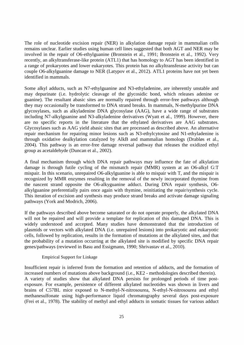
The role of nucleotide excision repair (NER) in alkylation damage repair in mammalian cells remains unclear. Earlier studies using human cell lines suggested that both AGT and NER may be involved in the repair of O6-ethylguanine (Bronstein et al., 1991; Bronstein et al., 1992). Very recently, an alkyltransferase-like protein (ATL1) that has homology to AGT has been identified in a range of prokaryotes and lower eukaryotes. This protein has no alkyltransferase activity but can couple O6-alkylguanine damage to NER (Latypov et al., 2012). ATL1 proteins have not yet been identified in mammals. Some alkyl adducts, such as N7-ethylguanine and N3-ethyladenine, are inherently unstable and may depurinate (i.e. hydrolytic cleavage of the glycosidic bond, which releases adenine or guanine). The resultant abasic sites are normally repaired through error-free pathways although they may occasionally be transformed to DNA strand breaks. In mammals, N-methylpurine DNA glycosylases, such as alkyladenine DNA glycosylase (AAG), have a wide range of substrates including N7-alkylguanine and N3-alkyladenine derivatives (Wyatt et al., 1999). However, there are no specific reports in the literature that the ethylated derivatives are AAG substrates. Glycosylases such as AAG yield abasic sites that are processed as described above. An alternative repair mechanism for repairing minor lesions such as N3-ethylcytosine and N1-ethyladenine is through oxidative dealkylation catalyzed by AlkB and mammalian homologs (Drabløs et al., 2004). This pathway is an error-free damage reversal pathway that releases the oxidized ethyl group as acetaldehyde (Duncan et al., 2002). A final mechanism through which DNA repair pathways may influence the fate of alkylation damage is through futile cycling of the mismatch repair (MMR) system at an O6-alkyl G:T mispair. In this scenario, unrepaired O6-alkylguanine is able to mispair with T, and the mispair is recognized by MMR enzymes resulting in the removal of the newly incorporated thymine from the nascent strand opposite the O6-alkyguanine adduct. During DNA repair synthesis, O6-alkylguanine preferentially pairs once again with thymine, reinitiating the repair/synthesis cycle. This iteration of excision and synthesis may produce strand breaks and activate damage signaling pathways (York and Modrich, 2006). If the pathways described above become saturated or do not operate properly, the alkylated DNA will not be repaired and will provide a template for replication of this damaged DNA. This is widely understood and accepted. Many studies have demonstrated that the introduction of plasmids or vectors with alkylated DNA (i.e. unrepaired lesions) into prokaryotic and eukaryotic cells, followed by replication, results in the formation of mutations at the alkylated sites, and that the probability of a mutation occurring at the alkylated site is modified by specific DNA repair genes/pathways (reviewed in Basu and Essigmann, 1990; Shrivastav et al., 2010).
Empirical Support for Linkage
Insufficient repair is inferred from the formation and retention of adducts, and the formation of increased numbers of mutations above background (i.e., KE2 - methodologies described therein). A variety of studies show that alkylated DNA persists for prolonged periods of time post-exposure. For example, persistence of different alkylated nucleotides was shown in livers and brains of C57BL mice exposed to N-methyl-N-nitrosourea, N-ethyl-N-nitrosourea and ethyl methanesulfonate using high-performance liquid chromatography several days post-exposure (Frei et al., 1978). The stability of methyl and ethyl adducts in somatic tissues for various adduct
26
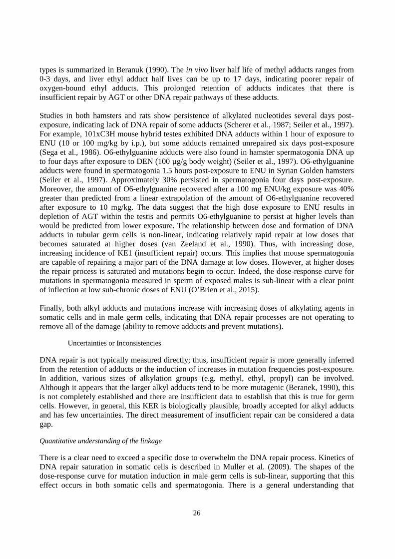
types is summarized in Beranuk (1990). The in vivo liver half life of methyl adducts ranges from 0-3 days, and liver ethyl adduct half lives can be up to 17 days, indicating poorer repair of oxygen-bound ethyl adducts. This prolonged retention of adducts indicates that there is insufficient repair by AGT or other DNA repair pathways of these adducts. Studies in both hamsters and rats show persistence of alkylated nucleotides several days post-exposure, indicating lack of DNA repair of some adducts (Scherer et al., 1987; Seiler et al., 1997). For example, 101xC3H mouse hybrid testes exhibited DNA adducts within 1 hour of exposure to ENU (10 or 100 mg/kg by i.p.), but some adducts remained unrepaired six days post-exposure (Sega et al., 1986). O6-ethylguanine adducts were also found in hamster spermatogonia DNA up to four days after exposure to DEN (100 µg/g body weight) (Seiler et al., 1997). O6-ethylguanine adducts were found in spermatogonia 1.5 hours post-exposure to ENU in Syrian Golden hamsters (Seiler et al., 1997). Approximately 30% persisted in spermatogonia four days post-exposure. Moreover, the amount of O6-ethylguanine recovered after a 100 mg ENU/kg exposure was 40% greater than predicted from a linear extrapolation of the amount of O6-ethylguanine recovered after exposure to 10 mg/kg. The data suggest that the high dose exposure to ENU results in depletion of AGT within the testis and permits O6-ethylguanine to persist at higher levels than would be predicted from lower exposure. The relationship between dose and formation of DNA adducts in tubular germ cells is non-linear, indicating relatively rapid repair at low doses that becomes saturated at higher doses (van Zeeland et al., 1990). Thus, with increasing dose, increasing incidence of KE1 (insufficient repair) occurs. This implies that mouse spermatogonia are capable of repairing a major part of the DNA damage at low doses. However, at higher doses the repair process is saturated and mutations begin to occur. Indeed, the dose-response curve for mutations in spermatogonia measured in sperm of exposed males is sub-linear with a clear point of inflection at low sub-chronic doses of ENU (O’Brien et al., 2015). Finally, both alkyl adducts and mutations increase with increasing doses of alkylating agents in somatic cells and in male germ cells, indicating that DNA repair processes are not operating to remove all of the damage (ability to remove adducts and prevent mutations).
Uncertainties or Inconsistencies
DNA repair is not typically measured directly; thus, insufficient repair is more generally inferred from the retention of adducts or the induction of increases in mutation frequencies post-exposure. In addition, various sizes of alkylation groups (e.g. methyl, ethyl, propyl) can be involved. Although it appears that the larger alkyl adducts tend to be more mutagenic (Beranek, 1990), this is not completely established and there are insufficient data to establish that this is true for germ cells. However, in general, this KER is biologically plausible, broadly accepted for alkyl adducts and has few uncertainties. The direct measurement of insufficient repair can be considered a data gap.
Quantitative understanding of the linkage
There is a clear need to exceed a specific dose to overwhelm the DNA repair process. Kinetics of DNA repair saturation in somatic cells is described in Muller et al. (2009). The shapes of the dose-response curve for mutation induction in male germ cells is sub-linear, supporting that this effect occurs in both somatic cells and spermatogonia. There is a general understanding that

27
methyl adducts are more readily repaired that ethyl adducts, which contributes to quantitative differences between chemicals in their genotoxic potency. There are no models that exist for this to our knowledge.
Evidence supporting taxonomic applicability
Name Scientific Name Evidence Links
Syrian golden hamster Mesocricetus auratus Moderate NCBI
mouse Mus musculus Moderate NCBI
DNA adducts can occur in any cell type. While there are differences across taxa, all species have some DNA repair systems in place and it is common to extrapolate conclusions across eukaryotic species.
References
Basu, A.K. and J.M. Essigmann (1990), "Site-specific alkylated oligodeoxynucleotides: Probes for mutagenesis, DNA repair and the structure effects of DNA damage", Mutat. Res., 233: 189-201.
Beranek, D.T. (1990), "Distribution of methyl and ethyl adducts following alkylation with monofunctional alkylating agents", Mutat. Res., 231(1): 11-30.
Bronstein, S.M., J.E. Cochrane, T.R. Craft, J.A. Swenberg and T.R. Skopek (1991), "Toxicity, mutagenicity, and mutational spectra of N-ethyl-N-nitrosourea in human cell lines with different DNA repair phenotypes", Cancer Research, 51(19): 5188-5197.
Bronstein, S.M., T.R. Skopek and J.A. Swenberg (1992), "Efficient repair of O6-ethylguanine, but not O4-ethylthymine or O2-ethylthymine, is dependent upon O6-alkylguanine-DNA alkyltransferase and nucleotide excision repair activities in human cells", Cancer Research, 52(7): 2008-2011.
Drabløs, F., E. Feyzi, P.A. Aas, C.B. Vaagbø, B. Kavli, M.S. Bratlie, J. Peña-Diaz, M. Otterlei, G. Slupphaug and H.E. Krokan (2004), "Alkylation damage in DNA and RNA - repair mechanisms and medical significance", DNA Repair, 3(11): 1389-1407.
Duncan, T., S.C. Trewick, P. Koivisto, P.A. Bates, T. Lindahl and B. Sedgwick B (2002), "Reversal of DNA alkylation damage by two human dioxygenases", Proc. Natl. Acad. Sci. USA, 99(26): 16660-16665.
Fang, Q., S. Kanugula, J.L. Tubbs, T.A. Tainer and A.E. Pegg (2010), "Repair of O4-alkylthymine by O6-alkylguanine-DNA alkyltransferases", J. Biol. Chem. 12(285): 885-895.
Frei, J.V., D.H .Swenson, W. Warren, P.D. Lawley (1978), "Alkylation of deoxyribonucleic acid in vivo in various organs of C57BL mice by the carcinogens N-methyl-N-nitrosourea, N-ethyl-N-nitrosourea and ethyl methanesulphonate in relation to induction of thymic lymphoma. Some applications of high-pressure liquid chromatography", Biochem. J., 174(3): 1031-1044.
Kaina, B., M. Christmann, S. Naumann and W.P. Roos (2007), "MGMT: Key node in the battle against genotoxicity, carcinogenicity and apoptosis induced by alkylating agents", DNA Repair, 6: 1079–1099.
Latypov, V.F., J.L. Tubbs, A.J. Watson, A.S. Marriott, G. McGown, M. Thorncroft, O.J. Wilkinson, P. Senthong, A. Butt, A.S. Arvai, C.L. Millington, A.C. Povey, D.M. Williams, M.F. Santibanez-Koref, J.A. Tainer and G.P. Margison GP (2012), "Atl1 regulates choice between global genome and transcription-coupled repair of O(6)-alkylguanines", Mol. Cell, 47(1): 50-60.

28
Muller, L., E. Gocke, T. Lave and T. Pfister (2009), "Ethyl methanesulfonate toxicity in Viracept – a comprehensive assessment based on threshold data for genotoxicity", Toxicol. Lett., 190: 317-329.
O’Brien, J.M., M. Walker, A. Sivathayalan, G.R. Douglas, C.L. Yauk and F. Marchetti (2015), "Sublinear response in lacZ mutant frequency of Muta™ Mouse spermatogonial stem cells after low dose subchronic exposure to N-ethyl-N-nitrosourea", Environ. Mol. Mutagen., 56(4): 347-55.
Pegg, A.E. (2011), "Multifaceted roles of alkyltransferase and related proteins in DNA repair, DNA damage, resistance to chemotherapy, and research tools", Chem. Res. Toxicol., 24(5): 618-639.
Scherer, E., A.A. Jenner and L. den Engelse (1987), "Immunocytochemical studies on the formation and repair of O6-alkylguanine in rat tissues", IARC Sci. Publ., 84: 55-8.
Sega, G.A., C.R. Rohrer, H.R. Harvey and A.E. Jetton (1986), "Chemical dosimetry of ethyl nitrosourea in the mouse testis", Mutat. Res., 159(1-2): 65-74.
Seiler, F., K. Kamino, M. Emura, U. Mohr and J. Thomale (1997), "Formation and persistence of the miscoding DNA alkylation product O6-ethylguanine in male germ cells of the hamster", Mutat. Res., 385(3): 205-211.
Shrivastav, N., D. Li and J.M. Essignmann (2010), "Chemical biology of mutagenesis and DNA repair: cellular response to DNA alkylation", Carcinogenesis, 31(1): 59-70.
van Zeeland, A.A., A. de Groot and A. Neuhäuser-Klaus (1990), "DNA adduct formation in mouse testis by ethylating agents: a comparison with germ-cell mutagenesis", Mutat. Res., 231(1):55-62.
Wyatt, M.D., J.M. Allan, A.Y. Lau, T.E. Ellenberger, L.D. Samson (1999), "3-methyladenine DNA glycosylases: structure, function, and biological importance", Bioessays, 21(8): 668-676.
York S.J. and P. Modrich (2006), "Mismatch repair-dependent iterative excision at irreparable O6-methylguanine lesions in human nuclear extracts", J. Biol. Chem., 281(32): 22674-22683.
2. Insufficient or incorrect DNA repair, N/A leads to Mutations, Increase
How does this Key Event Relationship work
Insufficient repair results in the retention of damaged DNA that is then used as a template during DNA replication. During replication of damaged DNA, incorrect nucleotides may be inserted, and upon replication these become ‘fixed’ in the cell. Further replication propagates the mutation to additional cells. For example, it is well established that replication of alkylated DNA can cause insertion of an incorrect base in the DNA duplex (i.e. mutation). Replication of non-repaired O4 thymine alkylation leads primarily to A:T→G:C transitions. Retained O6 guanine alkylation causes primarily G:C→A:T transitions.
Weight of Evidence
Biological Plausibility
If DNA repair is able to correctly and efficiently repair DNA lesions introduced by a genotoxic stressor, then no increase in mutation frequency will occur. For example, for alkylated DNA, efficient removal by AGT will result in no increases in mutation frequency. However, above a certain dose AGT becomes saturated and is no longer able to efficiently remove the alkyl adducts. Replication of O-alkyl adducts leads to mutation. The evidence demonstrating that replication of

29
unrepaired O-alkylated DNA causes mutations is extensive in somatic cells and has been reviewed (Basu and Essigmann, 1990; Shrivastav et al., 2010); specific examples are given below. It is important to note that not all DNA lesions will cause mutations. It is well documented that many are bypassed error-free. For example, N-alkyl adducts can quite readily be bypassed error-free with no increase in mutations (Philippin et al., 2014).
Empirical Support for Linkage
INSUFFICIENT REPAIR OF ALKYLATED DNA Evidence in somatic cells Empirical evidence to support this KER is primarily from studies in which synthetic oligonucleotides containing well-characterized DNA lesions were genetically engineered in viral or plasmid genomes and subsequently introduced into bacterial or mammalian cells. Example of alkylated DNA: Mutagenicity of each lesion is ascertained by sequencing, confirming that replication of alkylated DNA (i.e. unrepaired DNA) causes mutations in addition to revealing the important DNA repair pathways and polymerases involved in the process. For example, plasmids containing O6-methyl or O6-ethylguanine were introduced into AGT deficient or normal Chinese hamster ovary cells (Ellison et al., 1989). Following replication, an increase in mutant fraction to 19% for O6-methylguanine and 11% for O6-ethylguanine adducts was observed in AGT deficient cells versus undetectable levels for control plasmids. The relationship between input of alkylated DNA versus recovered mutant fractions revealed that a large proportion of alkyl adducts were converted to mutations in the AGT deficient cells (relationship slightly sublinear, with more adducts than mutations). The primary mutation occurring was G:C-A:T transitions. The results indicate that replication of the adducted DNA caused mutations and that this was more prevalent with reduced repair capacity. The number of mutations measured is less than the unrepaired alkyl adducts transfected into cells, supporting that insufficient repair occurs prior to mutation. Moreover, the alkyl adducts occur prior to mutation formation, demonstrating temporal concordance. Various studies in cultured cells and microorganisms have shown that the expression of AGT/MGMT (repair machinery – i.e. decrease in KE1) greatly reduces the incidence of mutations caused by exposure to methylating agents such as MNU and MNNG (reviewed in Kaina et al., 2007; Pegg, 2011). Thomas et al. (2013) used O6-benzylguanine to specifically inhibit MGMT activity in AHH-1 cells. Inhibition was carried out for one hour prior to exposure to MNU, a potent alkylating agent. Inactivation of MGMT resulted in increased MNU-induced HPRT (hypoxanthine-guanine phosphoribosyltransferase) mutagenesis and shifted the concentrations at which induced mutations occurred to the left on the dose axis (10 fold reduction of the lowest observed genotoxic effect level from 0.01 to 0.001 µg/ml). The ratio of mutants recovered in DNA repair deficient cells was 3-5 fold higher than repair competent cells at concentrations below 0.01 µg/ml, but was approximately equal at higher concentrations, indicating that repair operated effectively to a certain concentration. Only at this concentration (above 0.01 µg/ml when repair machinery is overwhelmed and repair becomes deficient) do the induced mutations in the repair competent cells approach those of repair deficient. Thus, induced mutation frequencies in wild

30
type cells are suppressed until repair is overwhelmed for this alkylating agent. The mutations prevented by MGMT are predominantly G:C-A:T transitions caused by O6-methylguanine. Evidence in germ cells That saturation of repair leads to mutation in spermatogonial cells is supported by work using the OECD TG488 rodent mutation reporter assay in sperm. A sub-linear dose-response was found using the lacZ MutaTMMouse assay in sperm exposed as spermatogonial stem cells, though the number of doses was limited (van Delft and Baan, 1995). This is indirect evidence that repair occurs efficiently at low doses and that saturation of repair causes mutations at high doses. Lack of additional data motivated a dose-response study using the MutaTMMouse model following both acute and sub-chronic ENU exposure by oral gavage (O’Brien et al., 2015). The results indicate a linear dose-response for single acute exposures, but a sub-linear dose-response occurs for lower dose sub-chronic (28 day) exposures, during which mutation was only observed to occur at the highest dose. This is consistent with the expected pattern for dose-response based on the hypothesized AOP. The same sub-linear dose-response in lacZ mutant frequency is found in MutaTMMouse male germ cells following exposure to benzo[a]pyrene, a bulky DNA-adduct forming chemical (O’Brien et al., 2016), extending this KER to other types of DNA damage (i.e. bulky adducts). Thus, this sub-linear curve for mutation at low doses following sub-chronic ENU and benzo[a]pyrene exposure suggests that DNA repair in spermatogonia is effective in preventing mutations until the repair processes becomes overwhelmed at higher doses. Mutation spectrum: Following exposure to alkylating agents, the most mutagenic adducts to DNA in pre-meiotic male germ cells include O6-ethylguanine, O4-ethylthymine and O2-ethylthymine (Beranek, 1990; Shelby and Tindall, 1997). Studies on sperm samples collected post-ENU exposure in transgenic rodents have shown that 70% of the observed mutations are at A:T sites (Douglas et al., 1995). The mutations observed at G:C base pairs are almost exclusively G:C-A:T transitions, presumably resulting from O6-ethylguanine. It is proposed that the prevalence of mutations at A:T basepairs is the result of efficient removal of O6-alkylguanine by AGT in spermatogonia, which is consistent with observation in human somatic cells (Bronstein et al. 1991; Bronstein et al. 1992). This results in the majority of O6-ethylguanine adducts being removed, leaving O4- and O2-ethylthymine lesions to mispair during replication. Thus, lack of repair predominantly at thymines and guanines at increasing doses leads to mutations in these nucleotides, consistent with the concordance expected between diminished repair capabilities at these adducts and mutation induction (i.e. concordance relates to seeing these patterns across multiple studies, species and across the data in germ cells and offspring).
Uncertainties or Inconsistencies
There were no inconsistencies in the empirical data reviewed or in the literature relating to biological plausibility. Much of the support for this KER comes predominantly from data in somatic cells and in prokaryotic organisms. For germ cells, we note that all of the data in germ cells used in this KER are produced exclusively from ENU exposure. Data on other chemicals are required. We consider the overall weight of evidence of this KER to be strong because of the obvious biological plausibility of the KER, and documented temporal association and incidence concordance based on studies over-expressing and repressing DNA repair in somatic cells.

31
Quantitative understanding of the linkage
Thresholds for mutagenicity indicate that the response at low doses is modulated by the DNA repair machinery, which is effectively able to remove alkylated DNA at low doses [Gocke and Muller, 2009; Lutz and Lutz, 2009; Pozniak et al., 2009]. Kinetics of DNA repair saturation in somatic cells is described in Muller et al. [Muller et al., 2009]. For O-methyl adducts, once the primary repair process is saturated, in vitro data suggest that misreplication occurs almost every time a polymerase encounters a methylated guanine [Ellison et al., 1989; Singer et al., 1989]; however, it should be noted that this process can be modulated by flanking sequence. This conversion of adducts to mutations also appears to be reduced substantially in vivo [Ellison et al., 1989]. The probability of mutation will also depend on the type of adduct (e.g. O-alkyl adducts are more mutagenic than N-alkyl adducts; larger alkyl groups are generally more mutagenic, etc.). Overall, a substantive number of factors must be considered in developing a quantitative model.
Evidence supporting taxonomic applicability
Name Scientific Name Evidence Links
mouse Mus musculus Moderate NCBI
All organisms, from prokaryotes to eukaryotes, have DNA repair systems. Indeed, much of the empirical evidence on the fundamental principles described in this KER are derived from prokaryotic models. DNA adducts can occur in any cell type, and may or may not be repaired, leading to mutation. While there are differences among DNA repair systems across eukaryotic taxa, all species develop mutations following excessive burdens of DNA lesions like DNA adducts. Theoretically, any sexually reproducing organism (i.e. producing gametes) can also acquire DNA lesions that may or may not be repaired, leading to mutations in gametes.
References
Basu, A.K. and J.M. Essigmann (1990), "Site-specific alkylated oligodeoxynucleotides: Probes for mutagenesis, DNA repair and the structure effects of DNA damage", Mutat. Res., 233: 189-201.
Beranek, D.T. (1990), "Distribution of methyl and ethyl adducts following alkylation with monofunctional alkylating agents", Mutat. Res., 231(1): 11-30.
Douglas, G.R., J. Jiao, J.D. Gingerich, J.A. Gossen and L.M. Soper (1995), "Temporal and molecular characteristics of mutations induced by ethylnitrosourea in germ cells isolated from seminiferous tubules and in spermatozoa of lacZ transgenic mice", Proc. Natl. Acad. Sci. USA, 92(16): 7485-7489.
Ellison, K.S., E. Dogliotti, T.D. Connors, A.K. Basu and J.M. Essigmann (1989), "Site-specific mutagenesis by O6-alkyguanines located in the chromosomes of mammalian cells: Influence of the mammalian O6-alkylguanine-DNA alkyltransferase", Proc. Natl. Acad. Sci. USA, 86: 8620-8624.
Gocke, E. and L. Muller (2009), "In vivo studies in the mouse to define a threhold for the genotoxicity of EMS and ENU", Mutat. Res., 678, 101-107.

32
Kaina, B., M. Christmann, S. Naumann and W.P. Roos (2007), "MGMT: Key node in the battle against genotoxicity, carcinogenicity and apoptosis induced by alkylating agents", DNA Repair, 6: 1079–1099.
Muller, L., E. Gocke, T. Lave and T. Pfister (2009), "Ethyl methanesulfonate toxicity in Viracept – A comprehensive human risk assessment based on threshold data for genotoxicity", Toxicol. Lett., 190: 317-329.
O'Brien, J.M., A. Williams, J. Gingerich, G.R. Douglas, F. Marchetti and C.L. Yauk (2013), "No evidence for transgenerational genomic instability in the F1 or F2 descendants of Muta™Mouse males exposed to N-ethyl-N-nitrosourea", Mutat. Res., 741-742:11-7
O’Brien, J.M., M. Walker, A. Sivathayalan, G.R. Douglas, C.L. Yauk and F. Marchetti (2015), "Sublinear response in lacZ mutant frequency of Muta™ Mouse spermatogonial stem cells after low dose subchronic exposure to N-ethyl-N-nitrosourea", Environ. Mol. Mutagen., 56(4): 347-55.
O'Brien, J.M., M.A. Beal, C.L. Yauk and F. Marchetti (2016), “Benzo(a)pyrene is mutagenic in mouse spermatogonial stem cells and dividing spermatogonia”, Toxicol. Sci., May 13. pii: kfw088. [Epub ahead of print]
Pegg, A.E. (2011), "Multifaceted roles of alkyltransferase and related proteins in DNA repair, DNA damage, resistance to chemotherapy, and research tools", Chem. Res. Toxicol., 24(5): 618-639.
Philippin, G., J. Cadet, D. Gasparutto, G. Mazon, R.P. Fuchs (2014), "Ethylene oxide and propylene oxide derived N7-alkylguanine adducts are bypassed accurately in vivo", DNA Repair (Amst), 22:133-6.
Pzoniak, A., L. Muller, M. Salgo, J.K. Jone, P. Larson and D. Tweats (2009), "Elevated ethyl methansulfonate in nelfinavir mesylate (Viracept, Roche): overview", Aids Research and Therapy, 6: 18.
Shelby, M.D. and K.R. Tindall (1997), "Mammalian germ cell mutagenicity of ENU, IPMS and MMS, chemicals selected for a transgenic mouse collaborative study”, Mutat. Res., 388(2-3):99-109.
Shrivastav, N., D. Li and J.M. Essignmann (2010), "Chemical biology of mutagenesis and DNA repair: cellular response to DNA alkylation", Carcinogenesis, 31(1): 59-70.
Singer, B., F. Chavez, M.F. Goodman, J.M. Essigman and M.K. Dosanjh (1989), "Effect of 3' flanking neighbors on kinetics of pairing of dCTP or dTTP opposite O6-methylguanine in a defined primed oligonucleotide when Escherichia coli DNA polymerase I is used", Proc. Natl. Acad. Sci. USA, 86(21): 8271-8274.
Thomas, A.D., G.J. Jenkins, B. Kaina, O.G. Bodger, K.H. Tomaszowski, P.D. Lewis, S.H. Doak and G.E. Johnson (2013), "Influence of DNA repair on nonlinear dose-responses for mutation", Toxicol. Sci., 132(1): 87-95.
van Delft, J.H. and R.A. Baan (1995), "Germ cell mutagenesis in lambda lacZ transgenic mice treated with ethylnitrosourea; comparison with specific-locus test", Mutagenesis, 10(3): 209-214.

33
3. DNA, Alkylation leads to Mutations, Increase
How does this Key Event Relationship work
Alkylated DNA may be ‘misread’ during DNA replication, leading to insertion of incorrect nucleotides. Upon replication, these changes become fixed as mutations. Subsequent replication propagates these mutations to daughter cells. Mutations in stem cells are of the greatest concern, as these will persist throughout the organism’s lifetime. Thus, increased mutations will be found in the cells of organisms that possess alkylated DNA.
Weight of Evidence
Biological Plausibility
Alkylating agents can cause a variety of adducts and DNA damage (e.g. alkali labile sites, DNA strand breaks, etc.) that are potentially mutagenic and clastogenic. This KER focuses on the probability that an alkyl DNA adduct will lead to a mutation. Not all adducts are equally mutagenic. Very generally, chemicals that preferentially cause O-alkylation in DNA induce DNA sequence changes, whereas chemicals that cause N-alkylation of DNA are more efficient inducers of structural chromosomal aberrations (reviewed in Beranek 1990). Indeed, a review of the biological significance of N7 alkyl-guanine adducts concluded that these adducts simply be used to confirm exposure to target tissue (Boysen et al., 2009), because the vast majority of studies shows that these adducts do not cause mispairing. A variety of work has demonstrated that N7-alkylguanine adducts can be bypassed essentially error free (e.g. Philippin et al., 2014; Shrivastav et al., 2010). Moreover, alkylation can involve modification with different sizes of alkylation groups (e.g. methyl, ethyl, propyl). Although response to these is qualitatively similar with respect to the key events, in general, larger alkylating groups tend to be more mutagenic (Beranek, 1990). It is widely known that chemicals that preferentially cause O-alkylation in DNA induce mutations. ENU (N-ethyl-N-nitrosourea) is a prototypical O-alkylating agent and the most studied male germ cell mutagen. Alkylating agents are prototypical somatic and male germ cell mutagens.
Empirical Support for Linkage
Evidence in somatic cells It is well established that transfection of cells with alkylated DNA leads to mutation at the sites of alkyl damage. The design of these experiments requires waiting for cellular replication in order to produce the mutation, confirming the temporal concordance of DNA alkylation and subsequent mutation. A summary of the empirical data to support this is reviewed in Shrivastav et al. (2010). Various studies have examined the dose-response of DNA adduct levels and mutations. These studies demonstrate that alkyl adducts can be seen at lower doses in the absence of increased mutations both in vitro and in vivo, or demonstrate equal or increased incidence of adducts relative to mutations at the same doses. For example, following exposure of AHH-1 cells to increasing concentrations of MMS, a linear increase in alkylated DNA is measured with significant increases occurring in adduct levels at 0.25 µg/ml (Swenberg et al. 2008). Significant

34
increases in mutations in the HPRT gene in the same cells are not measurable until 1.25 µg MMS/ml (Swenberg et al. 2008) (Figure 1). In vivo, time-series analyses of λlacZ transgenic mice exposed to a single dose of either ENU or DEN demonstrate that global and lacZ-specific O6-EtGua adducts occur within hours of exposure in the liver, with the bulk of adducts removed by three days post-exposure (Mientjes et al., 1996 and 1998). In contrast, mutant frequency does not begin to significantly increase until three days post-exposure, demonstrating temporal concordance of adduct and mutation formation (see Figure 1 in Mientjes et al., 1998). Levels of O6-EtGua adducts are also consistently higher than the induced mutation frequency per nucleotide. In the bone marrow, DEN is not metabolized and thus is unable to create O6-EtGua adducts. The finding of lack of O6-alkyl adducts in bone marrow is consistent with the lack of an increase in mutations observed in this tissue (Mientjes et al., 1998). This is in contrast to ENU exposure (a direct acting mutagen that does not require metabolic activation), where both adducts and mutations increase in a concordant fashion in the bone marrow. This pattern of adduct incidence versus mutation incidence is consistent for somatic tissues in rodents in vivo for other types of adducts (we were not able to find suitable dose-response studies to compare oxygen-alkyl adducts to mutation frequencies in vivo). For example, MutaTMMouse males were exposed to increasing doses of the polycyclic aromatic hydrocarbon dibenzo[a,l]pyrene (forms mutagenic bulky DNA adducts) for 28 days (followed by a 3 day break for mutation fixation) following OECD protocol TG488 (Malik et al., 2013). Significant increases in hepatic DNA adducts were found at 25 mg/kg, but increases in lacZ mutant frequency in liver did not occur until 50 mkg/kg. Bulky DNA adducts in both the livers and lungs of MutaTMMouse males exhibit an order of magnitude greater incidence per nucleotide than mutations in the lacZ gene using a similar experimental design following exposure to benzo[a]pyrene (Labib et al., 2012; Malik et al., 2012). Lower tissue adduct burden is correlated with lower tissue-specific gene mutation frequencies in Big Blue® mice exposed to benzo[a]pyrene (Skopek et al. 1996). Thus, adducts in DNA occur at lower doses than mutations and are correlated with mutation burden in somatic tissues for different types of DNA adducts. Evidence in germ cells: No study has compared dose-response for adduct formation and mutation in a single experiment on germ cells. However, it is possible to look across experiments. It is important to note that adducts are measured immediately following exposure because they are relatively quickly repaired. However, analysis of lacZ mutation requires collection of mature sperm from the caudal epididymis. Thus, sperm is collected 42 or 49 days post-exposure (OECD TG488). This is because spermatogonia can not be sampled directly for these purposes. Therefore, comparison of adducts to mutations in pre-meiotic male germ cells requires sampling at different time points for these endpoints (early for adducts, much later for mutations), which is consistent with the expected temporal order of events, with adducts occurring before mutations. Dose-response for alkyl adduct levels has been very well characterized in mouse testicular DNA for ENU, EMS and DES (van Zeeland et al., 1990). A summary of dose-response data for mouse exposure to ENU and mutation analysis using the transgenic rodent mutation assay in sperm is given in the attached Table I and Figure 2. These studies involve acute injections or oral gavage studies only. Alkyl adducts are evident in gonadal tissues within 2 hours of exposure (van Zeeland et al., 1990) and are fairly efficiently removed within days of exposure in the absence of continued exposure. For this analysis, transgene mutant frequencies were converted to per nucleotide mutation frequency by dividing mutant frequency by the length of the lacZ gene (3096

35
bp). The data demonstrate that alkyl adduct incidence at low doses is much greater per nucleotide than transgene mutations in the lacZ locus. Adducts are observed to increase substantially at the lowest exposure dose (10 mg/kg), whereas mutation increases in lacZ are marginal at 25 mg/kg. Alkyl adducts in mouse testes following ENU exposure were in the range of approximately 40 in 10E7 nucleotides for 80 mg/kg ENU exposure (van Zeeland et al., 1990). Conversion of the data in O’Brien et al. (2015) to mutations per nucleotide (by dividing mutant frequency by the length of the lacZ locus, which is 3096 bp) produces an estimated induced mutation frequency in spermatogonia of approximately 4 mutations per 10E7 nucleotides for the highest dose (100 mg/kg ENU), an increase of approximately 2 in 10E7 above controls. This suggests that the incidence of adducts is an order of magnitude greater than incidence of mutations. Similarly, exposure to a single dose of 250 mg/kg EMS leads to an over 10-fold increase in the number of alkyl adducts (although the majority are on nitrogen atoms, with only a small proportion on oxygen) (van Zeeland et al., 1990), but only a marginal 2-fold increase in lacZ mutation frequency [van Delft et al. 1997]. Indeed, Van Zeeland et al. (1990) estimate that approximately 10 O6-ethylguanine adducts are required in the gene-coding region to generate a mutation. Analysis of germ cell mutation during a sub-chronic exposure was carried out by O’Brien et al. (2015). The lower doses used in that study revealed that significant increases in mutations occurred only after 28 days of exposure to the highest dose of 5 mg/kg ENU (cumulative dose of 140 mg/kg), again supporting that higher adduct burdens are required to lead to mutations. In general, many studies in different mouse strains have used similar experimental designs to conclusively demonstrate that exposure to a variety of alkylating agents that form alkyl adducts in DNA causes mutations in spermatogonia (Brooks and Dean, 1997; Douglas et al., 1995; Katoh et al., 1997; Katoh et al., 1994; Liegibel and Schmezer, 1997; Mattison et al., 1997; O'Brien et al., 2014; O’Brien et al., 2015; Renault et al., 1997; Suzuki et al., 1997; Swayne et al., 2012; Tinwell et al., 1997; van Delft et al., 1997). These studies have been done using a single dose and thus do not enable further comparison of the concordance of dose-response. We also note that ENU exposure of pre-meiotic male germ cells in fish (transgenic medaka) also causes significant increases in mutations observed in spermatozoa (Norris and Winn 2010), supporting the effects of alkylating agents on mutations in male pre-meiotic germs across taxa using similar experimental designs.
Uncertainties or Inconsistencies
As described above, not all alkyl adducts are mutagenic. The proportion of oxygen-alkylation and the type of adduct (with ethylation > methylation) will govern mutagenicity, but there are few empirical data to support this. There are no inconsistencies or uncertainties for ENU or iPMS; other alkylating agents (EMS, MMS) have yielded some discrepancies in the transgenic rodent mutation assay. However, the experimental protocols applied were sub-standard (the OECD TG for this analysis was revised and published in 2013). Overall, more work is needed on alkylating agents other than ENU to fill important data gaps.
Quantitative understanding of the linkage
The shape of the dose-response curve for alkyl adduct formation versus mutation demonstrates that a threshold exists whereby alkyl adducts can be seen at low doses in the absence of increased

36
mutations occurring. For example, following exposure of AHH-1 cells to increasing concentrations of MMS, a linear increase in alkylated DNA is measured. However, a hockey-stick shaped curve was found for mutations at HPRT in the same cells (Thomas et al., 2013). Thus, alkylation of DNA occurs at lower doses than mutation, and above a certain dose (where repair is saturated), mutation frequencies increase. That DNA alkylation leads to mutation in spermatogonia in a similar hockey stick-shaped response (implying that a minimal dose must be exceeded) is supported by work using the lacZ Muta™Mouse assay. Exposure of male mice to the prototypical agent ENU was used to examine effects on spermatogonial stem cells, though the number of doses was limited (van Delft and Baan, 1995). This analysis revealed that mutations did not occur at the lowest dose, where adducts are known to be measurable in other studies (van Zeeland et al., 1990). This data gap motivated a dose-response study using the Muta™Mouse model following both acute and sub-chronic ENU exposure by oral gavage at Health Canada (O’Brien et al., 2015). These data indicate a clear dose-response for single acute exposures, whereas a hockey stick-shaped dose-response occurs for lower dose sub-chronic (28 day) exposures. At the single acute high doses where the DNA repair machinery is expected to be overwhelmed (and thus higher levels of alkylation occur), significantly more mutations occur relative to the same dose spread out over 28 daily oral gavage exposures (O’Brien et al., 2015). Additional contributors to the probability that an adduct will cause mutation include the site of alkylation, with agents that cause O-alkyl lesions being the primary mutagens, and the size of the alkyl group, with larger alkyl groups generally being more mutagenic. A computational model to describe the mutational efficiency of different alkyl adducts has not yet been developed to our knowledge.
Evidence supporting taxonomic applicability
Name Scientific Name Evidence Links
mouse Mus musculus Strong NCBI
medaka Oryzias latipes Moderate NCBI
Alkylating agents are well-established to cause mutation in virtually any cell type in any organism.
References
Beranek, D.T. (1990), "Distribution of methyl and ethyl adducts following alkylation with monofunctional alkylating agents", Mutat. Res., 231(1): 11-30.
Boysena, G., B.F. Pachkowski, J. Nakamura and J.A. Swenberg (2009), "The formation and biological significance of N7-guanine adducts", Mutat. Res., 678: 76–94.
Brooks, T.M. and S.W. Dean (1997), "The detection of gene mutation in the tubular sperm of Muta Mice following a single intraperitoneal treatment with methyl methanesulphonate or ethylnitrosourea", Mutat. Res., 388(2-3): 219-222.
37
Douglas, G.R., J. Jiao, J.D. Gingerich, J.A. Gossen and L.M. Soper (1995), "Temporal and molecular characteristics of mutations induced by ethylnitrosourea in germ cells isolated from seminiferous tubules and in spermatozoa of lacZ transgenic mice", Proc. Natl. Acad. Sci. USA, 92(16): 7485-7489.
Katoh, M., N. Horiya and R.P. Valdivia (1997), "Mutations induced in male germ cells after treatment of transgenic mice with ethylnitrosourea", Mutat Res., 388(2-3):229-37.
Katoh, M., T. Inomata, N. Horiya, F. Suzuki, T. Shida, K. Ishioka and T. Shibuya (1997), "Studies on mutations in male germ cells of transgenic mice following exposure to isopropyl methanesulfonate, ethylnitrosourea or X-ray", Mutat. Res., 388(2-3):213-8.
Liegibel, U.M. and P. Schmezer (1994), "Detection of the two germ cell mutagens ENU and iPMS using the LacZ/transgenic mouse mutation assay" Mutat. Res., 341(1):17-28.
Mattison, J.D., L.B. Penrose and B. Burlinson (1997), "Preliminary results of ethylnitrosourea, isopropyl methanesulphonate and methyl methanesulphonate activity in the testis and epididymal spermatozoa of Muta Mice", Mutat. Res. 388(2-3): 123-7.
Mientjes, E.J., K. Hochleitner, A. Luiten-Schuite, J.H. van Delft, J. Thomale, F. Berends, M.F. Rajewsky, P.H. Lohman and R.A. Baan (1996), "Formation and persistence of O6-ethylguanine in genomic and transgene DNA in liver and brain of lambda(lacZ) transgenic mice treated with N-ethyl-N-nitrosourea", Carcinogenesis, 17(11): 2449-2454.
Mientjes, E.J., A. Luiten-Schuite, E. van der Wolf, Y. Borsboom, A. Bergmans, F. Berends, P.H. Lohman, R.A. Baan RA, J.H. van Delft (1998), "DNA adducts, mutant frequencies, and mutation spectra in various organs of lambda lacZ mice exposed to ethylating agents", Environ. Mol. Mutagen., 31(1): 18-31
Norris, M.B. and R.N. Winn (2010), "Isolated spermatozoa as indicators of mutations transmitted to progeny", Mutat. Res., 688(1-2): 36–40.
Labib, S., C. Yauk, A. Williams, V.M. Arlt, D.H. Phillips, P.A. White and S. Halappanavar (2012)," Subchronic oral exposure to benzo(a)pyrene leads to distinct transcriptomic changes in the lungs that are related to carcinogenesis”, Toxicol. Sci., 129(1):213-224.
Malik, A.I., A. Williams, C.L. Lemieux, P.A. White and C.L. Yauk (2012), "Hepatic mRNA, microRNA, and miR-34a-target responses in mice after 28 days exposure to doses of benzo(a)pyrene that elicit DNA damage and mutation", Environ. Mol. Mutagen., 53(1): 10-21.
Malik, A.I., A. Rowan-Carroll, A. Williams, C.L. Lemieux, A.S. Long, V.M. Arlt, D.H. Phillips, P.A. White and C.L. Yauk (2013), "Hepatic genotoxicity and toxicogenomic responses in MutaMouse males treated with dibenz[a,h]anthracene", Mutagenesis, 28(5): 543-554.
O'Brien, J.M., M.A. Beal, J.D. Gingerich, L. Soper L, G.R. Douglas, C.L. Yauk and F. Marchetti (2014), "Transgenic rodent assay for quantifying male germ cell mutant frequency", J. Vis. Exp., (90): e51576.
O’Brien, J.M., M. Walker, A. Sivathayalan, G.R. Douglas, C.L. Yauk and F. Marchetti (2015), "Sublinear response in lacZ mutant frequency of Muta™ Mouse spermatogonial stem cells after low dose subchronic exposure to N-ethyl-N-nitrosourea", Environ. Mol. Mutagen., 56(4): 347-55.
Renault, D., D. Brault and V. Thybaud (1997), "Effect of ethylnitrosourea and methyl methanesulfonate on mutation frequency in MutaMouse germ cells (seminiferous tubule cells and epididymis spermatozoa)", Mutat. Res., 388(2-3): 145-153.
Shrivastav, N., D. Li and J.M. Essigmann (2010), "Chemical biology of mutagenesis and DNA repair: cellular responses to DNA alkylation", Carcinogenesis, 31(1): 59-70.
Skopek, T.R., K.L. Kort, D.R. Marino, L.V. Mittal, D.R. Umbenhauer, G.M. Laws and S.P. Adams (1996), "Mutagenic response of the endogenous hprt gene and lacI transgene in benzo[a]pyrene-treated Big Blue B6C3F1 mice", Environ. Mol. Mutagen., 28(4): 376-384.

38
Suzuki, T., S. Itoh, N. Takemoto, N. Yajima, M. Miura, M. Hayashi, H. Shimada and T. Sofuni (1997), "Ethyl nitrosourea and methyl methanesulfonate mutagenicity in sperm and testicular germ cells of lacZ transgenic mice (Muta Mouse)", Mutat. Res., 388(2-3): 155-163.
Swenberg, J.A., E. Fryar-Tita, Y. Jeong, G. Boysen, T. Starr, V.E. Walker and R.J. Albertini (2008), "Biomarkers in toxicology and risk assessment: informing critical dose-response relationships", Chem. Res. Toxicol., 21(1): 253-265.
Swayne, B.G., A. Kawata, N.A. Behan, A. Williams, M.G. Wade, A.J. Macfarlane and C.L. Yauk (2012), "Investigating the effects of dietary folic acid on sperm count, DNA damage and mutation in Balb/c mice", Mutat. Res., 737(1-2): 1-7.
Tinwell, H., P. Lefevre, C.V. Williams and J. Ashby (1997), "The activity of ENU, iPMS and MMS in male mouse germ cells using the Muta Mouse positive selection transgenic mutation assay", Mutat. Res., 388(2-3): 179-185.
van Delft J.H., A. Bergmans and R.A. Baan RA (1997), "Germ-cell mutagenesis in lambda lacZ transgenic mice treated with ethylating and methylating agents: comparison with specific-locus test", Mutat. Res., 388(2-3): 165-173.
van Delft, J.H. and R.A. Baan (1995), "Germ cell mutagenesis in lambda lacZ transgenic mice treated with ethylnitrosourea; comparison with specific-locus test", Mutagenesis, 10(3): 209-214.
van Zeeland, A.A., A. de Groot and A. Neuhäuser-Klaus (1990), "DNA adduct formation in mouse testis by ethylating agents: a comparison with germ-cell mutagenesis", Mutat. Res., 231(1):55-62.
4. DNA, Alkylation leads to Heritable mutations in offspring, Increase
How does this Key Event Relationship work
Pre-meiotic male germ cells are outside of the blood-testis barrier and thus are exposed if there is systemic distribution. Exposure of pre-meiotic male germ cells to DNA alkylating agents results in DNA alkyl adducts. Replication of DNA with alkyl adducts thus can cause mutations in these cells. Fertilization of an egg by sperm containing mutations causes an increase in the number of mutations that are transmitted to their offspring.
Weight of Evidence
Biological Plausibility
Alkylating agents are prototypical mutagens in laboratory animals. It is established that these agents, especially those chemicals that preferentially cause O-alkylation in DNA, induce heritable mutations. ENU (N-ethyl-N-nitrosourea) is a prototypical agent used to derive offspring with de novo mutations inherited from exposed males (e.g. http://ja.brc.riken.jp/lab/mutants/genedriven.htm). In fact, ENU mutagenicity is a standard bench tool for genetic screens used to identify new mutations associated with a phenotype of interest. A variety of alkylating agents are positive in the mouse specific locus test demonstrating that they cause heritable mutations in offspring as a result of exposure of pre-meiotic male germ cells. These agents include ENU, methyl nitrosourea (MNU), procarbazine and melphalan. This has been thoroughly reviewed by Marchetti and Wyrobek (2005) and Witt and Bishop (1996). It should be noted that procarbazine and melphalan predominantly cause N-alkyl adducts and yield a

39
weaker response in the specific locus test in male pre-meiotic germs (these agents yield higher responses in post-meiotic stages of spermatogenesis).
Empirical Support for Linkage
Dose-response: No study has directly compared alkyl adducts in sperm and mutations in offspring within a single experiment. However, comparisons can be made across experiments. The shape of the dose-response curve for adducts in testes (van Zeeland et al., 1990) and offspring with mutations (using the SLT – described in Favor et al., 1990) are similarly sub-linear, and incidence of adducts exceeds mutations at similar doses. For example, alkyl adducts in spermatogonia of mice exposed to 80 mg/kg bw ENU (van Zeeland et al., 1990) were in the range of approximately 1 in 10E6 nucleotides (range 0.23 to 1.92 x 10E-6). Conversion of the data in the specific locus test to per bp mutation (using exon sizes published in (Russell, 2004)) reveals a control mutation rate of 8.23 per 10E11 nucleotides, and 1.47 in 10E9 nucleotides for mice treated with 80 mg/kg ENU (from Favor et al., 1990) (see Table I and Figure 2 for summary of data). These are conservative estimates of mutation rate because they only account for functional mutations. However, it is clear that the incidence of adducts is much greater than the increased incidence of offspring with mutations. Moreover, alkyl adducts occur within hours of exposure in spermatogonia (van Zeeland et al., 1990), whereas studies in mice that focus on spermatogonial stem cells wait a minimum of 49 days prior to mating to confirm that this is the phase of spermatogenesis that was affected. Thus, alkylation of DNA occurs prior to the mutations in the offspring supporting temporal concordance of these events. Mutations in the offspring of males exposed to alkylating agents occur in tandem repeat DNA sequences (Dubrova et al., 2008; Vilarino-Guell et al. 2003) and genes associated with visible phenotypic traits in mice (Ehling and Neuhäuser-Klaus, 1991; Favor, 1986; Russell et al., 1979; Selby et al., 2004). Specific locus mutations in the offspring of males exposed to alkylating agents has also been demonstrated in Drosophila (Tosal et al., 1998) and medaka (fish) (Shima and Shimada, 1994). A substantive number of studies have demonstrated inherited mutations caused by exposure to ENU, but data also exist showing increases incidence of mutations in offspring with increasing doses of the two alkylating agents MNU and iPMS (Ehling and Neuhäuser-Klaus, 1991; Nagao, 1987; Russell et al., 2007; Vilarino-Guell et al., 2003).
Uncertainties or Inconsistencies
As described above, not all alkylating agents cause heritable mutations as a result of mutations arising in spermatogonia. O-alkylation is critical, and the size of the alkyl group is important, with ENU exhibiting an order of magnitude greater response than MNU. Although there are no inconsistencies based on knowledge of the spectrum of adducts expected for specific alkylating agents, the database on which this KER is assessed is nearly exclusively centered on ENU. Moreover, a key data gap includes evidence of the effect of alkylating agents in the offspring of exposed humans. Very little data are available on exposed humans despite the fact that humans may be exposed to high doses of alkylating agents during chemotherapy. Thus far the evidence has not supported that the cancer treatments pose heritable mutagenic hazards based on assessment of cancer (Madanat-Harjuoja et al., 2011), minisatellite mutations (Tawn et al., 2013) and congenital anomalies (Signorello et al., 2012) in offspring, or minisatellite mutation analysis in sperm ( Zheng et al.,

40
2000; Armour et al., 199). However, cancer therapies are complex combinations of drugs that include agents that generally induce N-alkylation rather than O-alkylation. It has been suggested that the search for human germ cell mutagens has been flawed by lack of appropriate power, focus on the wrong agents, and using the wrong tools (DeMarini, 2012).
Quantitative understanding of the linkage
As with mutations arising in sperm, it is established that the levels of O-alkylation must exceed a specific threshold before mutations begin to measurably increase in frequency above controls in the descendants of exposed males (Favor et al., 1990; Russell et al., 1982). In addition, fractionation of the dose reduces the recovery of mutations, indicating that more of the DNA damage is repaired (Favor et al., 1997). A quantitative model has not been developed because of insufficient data.
Evidence supporting taxonomic applicability
Name Scientific Name Evidence Links
mouse Mus musculus Strong NCBI
Drosophila melanogaster Drosophila melanogaster Moderate NCBI
That alkylation of DNA causes heritable mutations has been demonstrated specifically in flies, fish, and rodents. However, it is assumed that alkylating agents would act broadly on virtually any DNA sequence, in any organism, in any cell type. Thus, as long as the species has male germ cells, this KER would be relevant to that species.
References
Armour, J.A., M.H. Brinkworth and A. Kamischke (1999), "Direct analysis by small-pool PCR of MS205 minisatellite mutation rates in sperm after mutagenic therapies", Mutat. Res., 445(1): 73-80.
DeMarini, D.M. (2012), "Declaring the existence of human germ-cell mutagens", Environ. Mol. Mutagen., 53(3): 166-172.
Dubrova, Y.E., P. Hickenbotham, C.D. Glen, K. Monger, H.P. Wong and R.C. Barber (2008), "Paternal exposure to ethylnitrosourea results in transgenerational genomic instability in mice", Environ. Mol. Mutagen., 49(4): 308-311.
Ehling, U.H. and A. Neuhäuser-Klaus (1991), "Induction of specific-locus and dominant lethal mutations in male mice by 1-methyl-1-nitrosourea (MNU)", Mutat. Res., 250(1-2): 447-456.
Favor, J. (1986), "The frequency of dominant cataract and recessive specific-locus mutations in mice derived from 80 or 160 mg ethylnitrosourea per kg body weight treated spermatogonia." Mutat. Res., 162(1): 69-80.
Favor, J., M. Sund, A. Neuhauser-Klaus and U.H. Ehling (1990), "A dose-response analysis of ethylnitrosourea-induced recessive specific-locus mutations in treated spermatogonia of the mouse", Mutat. Res., 231(1): 47-54.
Favor, J., A. Neuhäuser-Klaus, U.H. Ehling, A. Wulff and A.A. van Zeeland (1997), "The effect of the interval between dose applications on the observed specific-locus mutation rate in the mouse following fractionated treatments of spermatogonia with ethylnitrosourea", Mutat. Res., 374(2): 193-199.

41
Lewis, S.E., L.B. Barnett, B.M. Sadler and M.D. Shelby (1991), "ENU mutagenesis in the mouse electrophoretic specific-locus test, 1. Dose-response relationship of electrophoretically-detected mutations arising from mouse spermatogonia treated with ethylnitrosourea", Mutat. Res., 249(2): 311-315.
Madanat-Harjuoja, L.M., N. Malila, P. Lähteenmäki, E. Pukkala, J.J. Mulvihill, J.D. Boice Jr and R. Sankila (2010), "Risk of cancer among children of cancer patients - a nationwide study in Finland," Int. J. Cancer, 126(5): 1196-1205.
Marchetti, F. and A.J. Wyrobek (2005), "Mechanisms and consequences of paternally-transmitted chromosomal abnormalities", Birth Defects Res C Embryo Today, 75(2): 112-129.
Nagao, T. (1987), "Frequency of congenital defects and dominant lethals in the offspring of male mice treated with methylnitrosourea", Mutat. Res., 177(1): 171-178.
Russell, W.L., E.M. Kelly, P.R. Hunsicker, J.W. Bangham, S.C. Maddux and E.L. Phipps (1979), "Specific-locus test shows ethylnitrosourea to be the most potent mutagen in the mouse", Proc. Natl. Acad. Sci. USA, 76(11): 5818-5819.
Russell, W.L., P.R. Hunsicker, G.D. Raymer, M.H. Steele, K.F. Stelzner and H.M. Thompson HM (1982), "Dose-response curve for ethylnitrosourea-induced specific-locus mutations in mouse spermatogonia", Proc. Natl. Acad. Sci. USA, 79(11): 3589-3591.
Russell, L.B. (2004), "Effects of male germ-cell stage on the frequency, nature, and spectrum of induced specific-locus mutations in the mouse", Genetica, 122(1): 25-36.
Russell, L.B., P.R. Hunsicker and W.L. Russell (2007), "Comparison of the genetic effects of equimolar doses of ENU and MNU: while the chemicals differ dramatically in their mutagenicity in stem-cell spermatogonia, both elicit very high mutation rates in differentiating spermatogonia", Mutat. Res., 616(1-2): 181-195.
Selby, P.B., V.S. Earhart, E.M. Garrison and G. Douglas Raymer (2004), "Tests of induction in mice by acute and chronic ionizing radiation and ethylnitrosourea of dominant mutations that cause the more common skeletal anomalies", Mutat. Res., 545(1-2): 81-107.
Signorello, L.B., J.J. Mulvihill, D.M. Green, H.M. Munro, M. Stovall, R.E. Weathers, A.C. Mertens, J.A. Whitton, L.L. Robison and J.D. Boice Jr. (2012), "Congenital anomalies in the children of cancer survivors: a report from the childhood cancer survivor study", J. Clin. Oncol., 30(3): 239-245.
Shima, A. and A. Shimada (1994), "The Japanese medaka, Oryzias latipes, as a new model organism for studying environmental germ-cell mutagenesis", Environ. Health Perspect., 102 Suppl 12: 33-35.
Tosal, L., M.A. Comendador and L.M. Sierra (1998), "N-ethyl-N-nitrosourea predominantly induces mutations at AT base pairs in pre-meiotic germ cells of Drosophila males", Mutagenesis, 13(4): 375-380.
Van Zeeland, A.A., A. de Groot and A. Neuhauser-Klaus (1990), "DNA adduct formation in mouse testis by ethylating agents: a comparison with germ cell mutagenesis", Mutat. Res., 231(1): 55-62.
Vilarino-Guell, C., A.G. Smith and Y.E. Dubrova (2003), "Germline mutation induction at mouse repeat DNA loci by chemical mutagens", Mutat. Res., 526(1-2): 63-73.
Witt, K.L. and J.B. Bishop (1996), "Mutagenicity of anticancer drugs in mammalian germ cells", Mutat. Res., 355(1-2): 209-234.
Zheng, N., D.G. Monckton, G. Wilson, F. Hagemeister, R. Chakraborty, T.H. Connor, M.J. Siciliano, M.L. Meistrich (2000), "Frequency of minisatellite repeat number changes at the MS205 locus in human sperm before and after cancer chemotherapy", Environ. Mol. Mutagen., 36(2): 134-145.

42
5. Mutations, Increase leads to Heritable mutations in offspring, Increase
How does this Key Event Relationship work
If a mutation arises in spermatogonial stem cells and does not influence cellular function, the mutation can become fixed and/or propagated within the stem cell population. Mutations that do not affect sperm maturation will persist through the succeeding stages of spermatogenesis and will be found in the mature sperm of the organism throughout its reproductive lifespan. Mutations can also occur in differentiating spermatogonia; however, once the sperm generated by these dividing spermatogonia are ejaculated there will be no ‘record’ of the induced mutation. Mutations that impair spermatogenesis or the viability of the cell will be lost via apoptosis and cell death, potentially contributing to decreased fertility. Mutations that do not impact sperm motility, morphology or ability to penetrate the zona pellucida (or other important sperm parameters), and that are present in mature sperm, may be transmitted to the egg resulting in the development of an offspring with a mutation. Thus, increased incidence of mutations in germ cells leads to increased incidence of mutations in the offspring. There is a great deal of evidence demonstrating that exposure to a variety of DNA alkylating agents induces mutations in male spermatogenic cells.
Weight of Evidence
Biological Plausibility
Evolution requires heritable mutations that are transmitted to offspring via parental gametes. This is the primary mechanism by which an offspring would have a sequence variant in every single one of its cells that is not found in its parents. Indeed, as stated in a recent review in Science by Shendura and Aikey "Germline mutations are the principal cause of heritable disease and the ultimate source of evolutionary change." Thus, this KER is supported by substantive understanding of genetics and evolution, with heritable germ cell mutations forming the basis for the selective evolution of species. In addition, in toxicology, a large body of literature demonstrates that exposure to specific genotoxic chemicals during pre-meiotic stages of spermatogenesis leads to mutations in both the sperm and the offspring, supporting that mutations occurring in sperm fertilize the egg and result in offspring with mutations (reviewed in DeMarini, 2012; Marchetti and Wyrobek, 2005; Yauk et al., 2012). Indeed, ENU is used as a tool in genetics to create offspring carrying mutations for the purposes of understanding gene function (e.g. http://www.riken.jp/en/research/labs/brc/mutagen_genom). In these studies, male mice are mutagenized by exposure to ENU and mated to females. The offspring of these males have a much higher incidence of mutation; the function of new mutations occurring in genes in these offspring is used to study gene function. Thus, overall this KER is biologically plausible and widely understood.
Empirical Support for Linkage
Identification of mutations in sperm requires the destruction of the sperm. Thus, tracking a mutated spermatogonial stem cell through to fertilization and characterization of the mutation in the offspring is not possible, and the empirical evidence to support this KER is weak. No single

43
study has looked at the dose-response relationship of the same mutation endpoint in germ cells and offspring because technologies are not currently available to do this. We caution that comparing mutation rates across different genes or genetic loci is imprecise, because factors intrinsic to specific loci govern mutation rates (e.g. length, GC content, transcribed versus non-transcribed, coding versus non-coding, chromatin structure, DNA methylation, sequence, etc.). Increased numbers of germ cell mutations occur in mature sperm 42+ days post-exposure in mice (indicating that mutations arose in pre-meiotic male germ cells). Mating in this time interval to produce offspring also results in increased incidence of mutation in the descendants of exposed males, indicating temporal concordance. By virtue of the required experimental designs, mutations measured in the offspring occur after the mutations in germ cells. For example, mutations identified in proteins via electrophoresis (a variation of the SLT test) are found in the offspring of male mice mated 10+ weeks post-exposure to ENU (Lewis et al., 1991). These inherited changes are the result of mutations in stem cells that persist through spermatogenesis and are transmitted to offspring. The only assay that presently can measure mutations in both sperm and offspring is the tandem repeat mutation assay. A single study on one dose of radiation (1 Gy X-ray) against matched controls has shown that increases in mutation frequencies in exposed sperm are similar to the increases observed in the offspring of exposed males for tandem repeats (Yauk et al., 2002), suggesting that tandem repeat mutations in sperm are transmitted to offspring. Alkylating agents cause similar increases in tandem repeat mutations in both sperm and in the offspring through comparison across studies (Dubrova et al., 2008; Swayne et al., 2012; Vilarino-Guell et al., 2003), but dose-response studies have not been conducted. It is advisable that dose-response experiments in sperm for tandem repeats be conducted in the future to address this gap. Many studies have shown the induction of transgene mutations recovered in mature sperm derived from toxicant-exposed pre-meiotic male germ cells in transgenic mutation reporter mice (e.g. Brooks and Dean, 1997; Liegibel and Schmezer, 1997; Mattison et al., 1997). One study measured transgene mutations in the offspring of mice exposed to three single i.p. doses of 100 mg/kg ENU (in 7 day intervals) (Barnett et al., 2002). Four inherited transgene mutations were found from 280 mice (confirmed in multiple somatic tissues), for a mutant frequency of 35.7 x 10E-5 per locus. There is no comparable study on the sperm of lacI transgenic mice, or a similar exposure in another transgenic strain for comparison. However, conversion of the per locus lacI offspring mutant frequency to per nucleotide reveals a mutant frequency of 3.31 x 10⁻⁷ per nucleotide. LacZ mutant frequencies for 150 mg/kg (half the exposure level of the lacI transgenic mice) exposures of MutaTMMouse males to ENU results in a per nucleotide mutation frequency ranging from 0.37 to 2.21 x 10E-7 (Liegibel and Schmezer, 1997; van Delft and Baan, 1995). Thus, the induced mutation frequency in sperm and offspring are within the same range, despite higher doses in the offspring study, supporting incidence concordance for these events. Finally, it has been documented that DNA damage and mutation accumulates as human males age (reviewed in Paul and Robaire, 2013), which is concordant with increased incidence of mutation in the offspring of ageing fathers (Kong et al., 2012; Sun et al., 2012). Comparison of the dose-response characteristics of this relationship is not possible because of differences in the mutagenic endpoints measured in sperm versus offspring.

44
Uncertainties or Inconsistencies
There are no inconsistencies in the data for this KER, although the data are limited. There is a possibility that mutations can arise in the early embryo instead of in the spermatogenic cells. However, given the study designs for this type of work (where > 42 days pass prior to sperm collection or mating – see OECD TG488 for the time-series required for transgene mutation analysis in sperm), it is unlikely that this contributes significantly. Limitations in technology currently prevent the analyses required to describe the incidence of mutations in sperm versus offspring, but this is a future research direction. It should be noted that the locations and types of mutations would influence the probably of transmission; this relationship has not been confirmed empirically and limits extrapolation across studies applying different endpoints.
Quantitative understanding of the linkage
Mutations conferring a selective disadvantage to sperm or to the embryo will not be measured in live born offspring and will be eliminated. Thus, mutation frequency in sperm should be equal to mutation rate derived by measuring mutations in the offspring for non-selective loci (as is seen in the rodent tandem repeat and transgene mutation examples described above); or, sperm mutation frequency should be greater than mutation rate measured by identifying mutations in the offspring. However, quantitative data to demonstrate this are lacking because of current technical limitations to study this. It is anticipated that improved models will be developed to predict the likely outcome of increased rates of heritable mutation from sperm mutation frequency data when more data are available from studies applying next generation sequencing technologies in sperm and pedigrees.
Evidence supporting taxonomic applicability
Name Scientific Name Evidence Links
mouse Mus musculus Strong NCBI
human Homo sapiens Strong NCBI
Mutation is the underlying source of evolution and occurs in every species. Theoretically, any sexually reproducing organism can acquire mutations in their gametes and transmit these to descendants. Thus, the present KER is relevant to any species producing sperm.
References
Barnett, L.B., R.W. Tyl, B.S. Shane, M.D. Shelby and S.E. Lewis (2002), "Transmission of mutations in the lacI transgene to the offspring of ENU-treated Big Blue male mice", Environ. Mol. Mutagen., 40(4): 251-257.
Brooks, T.M. and S.W. Dean (1997), "The detection of gene mutation in the tubular sperm of Muta Mice following a single intraperitoneal treatment with methyl methanesulphonate or ethylnitrosourea", Mutat. Res., 388(2-3): 219-222.
DeMarini, D.M. (2012), "Declaring the existence of human germ-cell mutagens", Environ. Mol. Mutagen., 53(3): 166-172.

45
Dubrova, Y.E., P. Hickenbotham, C.D. Glen, K. Monger, H.P. Wong and R.C. Barber (2008), "Paternal exposure to ethylnitrosourea results in transgenerational genomic instability in mice", Environ. Mol. Mutagen., 49(4): 308-311.
Kong, A., M.L. Frigge, G. Masson, S. Besenbacher, P. Sulem, G. Magnusson, S.A. Gudjonsson, A. Sigurdsson, A. Jonasdottir, W.S. Wong, G. Sigurdsson, G.B. Walters, S. Steinberg, H. Helgason, G. Thorleifsson, D.F. Gudbjartsson, A. Helgason, O.T. Magnusson, U. Thorsteinsdottir and K. Stefansson (2012), "Rate of de novo mutations and the importance of father's age to disease risk", Nature, 488(7412): 471-475.
Lewis, S.E., L.B. Barnett, B.M. Sadler and M.D. Shelby (1991), "ENU mutagenesis in the mouse electrophoretic specific-locus test, 1. Dose-response relationship of electrophoretically-detected mutations arising from mouse spermatogonia treated with ethylnitrosourea", Mutat. Res., 249(2): 311-315.
Liegibel, U.M. and P. Schmezer (1994), "Detection of the two germ cell mutagens ENU and iPMS using the LacZ/transgenic mouse mutation assay" Mutat. Res., 341(1):17-28.
Marchetti, F. and A.J. Wyrobek (2005), "Mechanisms and consequences of paternally-transmitted chromosomal abnormalities", Birth Defects Res C Embryo Today, 75(2): 112-129.
Mattison, J.D., L.B. Penrose and B. Burlinson (1997), "Preliminary results of ethylnitrosourea, isopropyl methanesulphonate and methyl methanesulphonate activity in the testis and epididymal spermatozoa of Muta Mice", Mutat. Res. 388(2-3): 123-7.
O'Brien, J.M., A. Williams, J. Gingerich, G.R. Douglas, F. Marchetti and C.L. Yauk (2013), "No evidence for transgenerational genomic instability in the F1 or F2 descendants of Muta™Mouse males exposed to N-ethyl-N-nitrosourea", Mutat Res., 741-742:11-7
Paul, C. and B. Robaire (2013), "Ageing of the male germ line", Nat. Rev. Urol., 10(4): 227-234. Shendura, J. and M. Akey (2015), "The origins, determinants, and consequences of human mutations",
Science, 349(6255): 1478-1483. Sun, J.X., A. Helgason, G. Masson, S.S. Ebenesersdottir, H. Li, S. Mallick, S. Gnerre, N. Patterson, A.
Kong, D. Reich and K. Stefansson (2012), "A direct characterization of human mutation based on microsatellites", Nat. Genet., 44(10): 1161-1165.
Swayne, B.G., A. Kawata, N.A. Behan, A. Williams, M.G. Wade, A.J. Macfarlane and C.L. Yauk (2012), "Investigating the effects of dietary folic acid on sperm count, DNA damage and mutation in Balb/c mice", Mutat. Res., 737(1-2): 1-7.
Vilarino-Guell, C., A.G. Smith and Y.E. Dubrova (2003), "Germline mutation induction at mouse repeat DNA loci by chemical mutagens", Mutat. Res., 526(1-2): 63-73.
Yauk, C.L., Y.E. Dubrova, G.R. Grant and A.J. Jeffreys (2002), "A novel single molecule analysis of spontaneous and radiation-induced mutation at a mouse tandem repeat locus", Mutat. Res., 500(1-2): 147-156.
Yauk, C.L., L.J. Argueso, S.S. Auerbach, P. Awadalla, S.R. Davis, D.M. DeMarini, G.R. Douglas, Y.E. Dubrova, R.K. Elespuru, T.M. Glover, B.F. Hales , M.E. Hurles, C.B. Klein, J.R. Lupski, D.K. Manchester, F. Marchetti, A. Montpetit, J.J. Mulvihill, B. Robaire, W.A. Robbins, G.A. Rouleau, D.T. Shaughnessy, C.M. Somers, J.G. Taylor 6th, J. Trasler, M.D. Waters, T.E. Wilson, K.L. Witt and J.B. Bishop (2013), "Harnessing genomics to identify environmental determinants of heritable disease" Mutat. Res., 752(1): 6-9.
46
Overall Assessment of the AOP
See Annex 1: Assessment Summary of Aop-15 Before developing this AOP a review of the literature was undertaken to identify studies in which male germ cells were exposed to alkylating agents and measures of DNA adducts, DNA repair and mutations, as well as mutations in offspring, were evaluated. The focus of this AOP (as described in the KERs) is on O-alkylating agents, which are signficantly more mutagenic than N-alkylation chemicals. During our review, studies where sufficient information relating to the chemicals used, dose, tissue, time-point, animal model, experimental procedures and experimental results were available were considered to assess empirical data in germ cells for each of the KEs and KERs in the AOP. The germ cell database on which the AOP was based is found in Supplementary Table I (Annex 1) and is comprised of 32 studies. No study measured multiple KEs within it; however, for each KE there were at least two dose-response and time-series analyses for at least one alkylating agent. We consider this overall number of high quality studies to be fairly extensive evidence of the ability of O-alkylating agents to cause adducts and mutations in germ cells, and mutations in offspring, although no studies were ideally suited to establish the empirical linkages between the KERs. We thus compared results across studies where possible to attempt to do this. All of the studies either used ENU as the primary study compound, or applied ENU as one of the positive controls to assess other alkylating agents. Strong dose-response data for mutations occurring in exposed pre-meiotic germ cells and mutations in offspring are only available for ENU. The other alkylating agents show varying degrees mutagenicity, but single doses were used in most studies. Thus, the evaluation of concordance of the dose-response could only be undertaken with ENU for in vivo germ cell and heritable effects. However, where possible we used information from research on somatic cells to provide additional support for the KERs. In particular, experiments in somatic cells were necessary to assess the involvement of DNA repair in removing adducts and preventing mutations. Overall, we note that the rationale for claiming high confidence in this AOP and its KERs is based primarily on the more influential Bradford Hill consideration of biological plausibility, with decades of research having been done in somatic and germ cells on DNA damage, repair and mutation following exposure to agents that cause alkyl adducts. Much of the data, then, supporting AOP evaluation derives from historical studies from the 1990’s, with less recent evidence. As noted, a primary motivation for developing this AOP was the recent release of TG 488, and newly available whole generation sequencing methods, which we expect to be increasingly applied. Thus, additional well-designed experiments that dissect the relationships between alkyl adducts, DNA repair, mutations in sperm, and mutations in offspring to assess essentiality and empirical support are expected in the future through application of these improved approaches. Below we describe each KE and KER in detail, using the wiki entries as a guide to the order of presentation and the content described.

47
Weight of Evidence Summary
Event Description Triggers Weight of Evidence
DNA, Alkylation Directly Leads to
Insufficient or incorrect DNA repair, N/A Strong
Insufficient or incorrect DNA repair, N/A
Directly Leads to Mutations, Increase Strong
DNA, Alkylation Indirectly Leads to Mutations, Increase Strong
DNA, Alkylation Indirectly Leads to
Heritable mutations in offspring, Increase Strong
Mutations, Increase Directly Leads to
Heritable mutations in offspring, Increase Strong
Biological plausibility of the KERs: Strong. There is extensive understanding of the ability of alkylating agents to cause DNA adducts, the requirement for overcoming DNA repair, and the resulting mutations that arise in both somatic and germ cells. It is established that exposure to alkylating agents produced mutations in germ cells – ENU is used in genetic screening to produce mutations to derive new phenotypes for research. Empirical support for the KERs: Across the KERs the degree of support ranges from weak to strong (Annex 1 - Table II). Support from somatic cells in culture contributes to moderate calls for the relationships between adduct formation, insufficient DNA repair and mutation. The weak call is based on lack of empirical data to support that mutations in germ cells are transmitted to offspring. However, increased mutation frequencies in germ cells occur following exposure to the same types of chemicals that cause increased mutations in the offspring. It should be noted that biological plausibility for this KER is strong as it is based on understanding of molecular biology and evolution. The strongest support is associated with the indirect KER linking alkylation of DNA to mutation in germ cells (KER4). This is primarily based on extensive evidence in both somatic and germ cells demonstrating that chemicals that alkylate DNA cause mutations, that alkyl adducts occur at a greater incidence than mutations at matching doses, and that alkyl adducts precede mutations. In somatic cells, work has been done on many different chemicals, whereas the germ cell data were primarily for the chemical ENU (but data were also available for a few select other chemicals) (Annex 1 - Table I, Figure 2). In addition, data are available for multiple species to support this indirect KER. There is a large degree of consistency in the germ cell literature to show that a variety of O-alkylating agents cause male germ cell mutations in many species (Drosophila, fish and rodent) and that these effects occur at many mutational loci (e.g. mutations in genes that are inherited measured with the Specific Locus Test, sperm mutations in tandem repeat DNA sequences, tandem repeat mutations in offspring, transgene mutations in sperm). Many alkylating agents have been tested to show that they create adducts in male rodent germ cells (e.g. DEN, ENU, EMS, DES), mutations in male mouse germ cells (ENU, IPMS and MNU) and mutations in the offspring of exposed male mice (ENU, MNU and IPMS). In

48
summary, we consider the overall empirical data supporting the AOP to be MODERATE (the median call). Rank order (provided in the overall assessment Table - Annex 1): Rank order of the KERs and the weight of evidence for the essentiality all point to the overall weight of evidence for this AOP as strong. Biological plausibility is strong for all KERs, with primarily moderate evidence for KER linkages and relatively few uncertainties or inconsistencies.
Essentiality of the Key Events
Molecular Initiating Event Support for Essentiality
DNA, Alkylation
Key Event Support for Essentiality
Mutations, Increase Insufficient or incorrect DNA repair, N/A Moderate
Essentiality was not directly tested for all of the KEs. The MIE cannot be ‘blocked’ in any way to our knowledge (e.g. as you might block a receptor-binding MIE). However, as described in the KERs, enhanced DNA repair of alkylated DNA reduces mutation frequencies and reduction in repair increases mutation frequencies, supporting the essentiality of KE1 (i.e. moderate support). Correct repair of the alkylated DNA (i.e. a block of KE1) will not lead to mutation. For example, MGMT overexpression protects mgt1 mutant yeast against alkylation-induced mutation (Xiao and Fontanie, 1995). In addition, Big Blue® mice over-expressing human AGT exhibit greatly reduced O6-methylguanine-mediated lacI and K-ras mutations in the thymus following treatment with MNU (Allay et al., 1999) relative to wild type Big Blue® mice. Insufficient DNA repair is well-established to lead to mutations. In addition, inactivation of MGMT sensitizes cells to alkylation-induced mutagenesis resulting in an increased number of mutations per adduct (Thomas et al., 2013). The remainder of the AOP requires transmission of mutations in sperm to offspring. There are no means to study the essentiality of mutations in sperm. Once mutations occur in male pre-meiotic germ cells, they cannot be removed to observe whether occurrence in offspring is decreased. In addition, mutations that occur in stem cells are propagated clonally and can become fixed in the spermatogonial cell population. Thus, waiting a longer period of time, or removing the exposure, is not effective in causing a decline in the mutation frequency. Therefore, the essentiality of this KE is inferred by the biology of the pathway and cannot be addressed directly with experimental evidence.

49
Quantitative Considerations
Event Description Triggers Quantitative Understanding
DNA, Alkylation Directly Leads to
Insufficient or incorrect DNA repair, N/A Moderate
Insufficient or incorrect DNA repair, N/A
Directly Leads to Mutations, Increase Moderate
DNA, Alkylation Indirectly Leads to Mutations, Increase Moderate
DNA, Alkylation Indirectly Leads to
Heritable mutations in offspring, Increase Moderate
Mutations, Increase Directly Leads to
Heritable mutations in offspring, Increase Moderate
As described above, it is established that alkyl adducts, mutations in spermatogonia and mutations in offspring all increase with dose in a manner that is consistent with the AOP. Alkylation must exceed a threshold (determined by saturation of the relevant DNA repair pathways) before alkyl DNA lesions persist, and mutations subsequently begin to occur. However, the precise quantitative relationship has not been modeled. Existing data published in the literature could be mined to do this and thresholds for specific adduct types (i.e. estimates of how many adducts are needed to cause a mutation in a gene on average) have been published for certain cell types, which should theoretically correlate with germ cell mutagenicity for ENU and other alkylating agents. The quantitative relationship between mutations in sperm and mutations in the offspring has not been determined and will be locus- and mutation-type specific (e.g. stronger selection against coding mutations than non-coding mutations, which will influence transmission probability); however, although many mutations will lead to embryonic loss, a large subset of mutations is expected to be heritable and viable. It is expected that quantitative understanding of this relationship will increase as advanced single cell sequencing technologies are more developed to query mutations in sperm versus offspring. For non-coding sites (e.g. transgenic reporter genes and non-coding DNA like tandem repeats), the relationship is expected to approach 1:1. Overall, the variables that could be used to predict whether a heritable mutation is probable following exposure to an alkylating agent are the number and types of adducts per nucleotide (and knowledge of their repair efficiency). Generally, the probability of a mutation occurring is highly dependent on the type of adduct formed (mutagenicity of the adduct is based on repair efficiency and probability of error-free replication over the lesion) and abundance of the adducts, and could be modeled using existing published data.

50
Applicability of the AOP
Life Stage Applicability
Life Stage Evidence
Adult Strong
Taxonomic Applicability
Name Scientific Name Evidence Links
Mus musculus Mus musculus Strong NCBI
Drosophila melanogaster Drosophila melanogaster Strong NCBI
Oryzias latipes Oryzias latipes Weak NCBI
Syrian golden hamster Mesocricetus auratus Weak NCBI
Sex Applicability
Sex Evidence
Male Strong
This AOP is relevant exclusively to mature males and their pre-meiotic germ cells. Although not considered in this AOP, progenitor germ cells from earlier life stages may also be susceptible to induced mutations from alkylating agents, which could then be transmitted to offspring after sexual maturity. Relevant endpoints have been characterized across different taxa: (1) alkyl adduct levels in this AOP were from hamsters, mice and rats; (2) repair of alkylated DNA has been studied in prokaryotes to higher eukaryotes, including human cells in culture (while there are differences across taxa, all species have some DNA repair systems in place and it is common to extrapolate conclusions across eukaryotic species); (3) mutations in male germ cells were measured in mice and fish; and (4) mutations in offspring were measured in Drosophila, Japanese Medaka and mice. Quite generally, the AOP applies to any species that produces sperm. The similarity in spermatogenesis and in DNA repair of alkyl adducts is well documented across rodents and humans (Adler, 1996). Heritable mutations are the basis of evolution and occur in every species. That mutations in sperm are transmitted to offspring in humans is best demonstrated by studies exploring the effects of ageing. Significant increases are observed in the amount of DNA damage and mutation as human males age (reviewed in Paul and Robaire, 2013). Similarly, increased incidence of single nucleotide mutations and microsatellite mutation in the offspring of ageing fathers has recently been measured by advanced genomics technologies (Kong et al., 2012; Sun et al., 2012). Lifestyle factors including smoking and lower income brackets in human fathers in associated with increased minisatellite mutations in their offspring (LinSchooten et al., 2013).

51
Considerations for Potential Applications of the AOP
The information in this AOP will provide context for understanding how to interpret new data produced from the rodent transgene mutation assay applied to sperm (OECD TG 488) [OECD, 2013], which is being increasingly applied, as well as data produced using tandem repeat mutation assays. In addition, it is envisioned that next generation sequencing technologies will enable the analysis of germ cell mutations in human populations and the eventual discovery of human germ cell mutagens. It is important to note that the regulation of chemicals that can induce heritable effects has, to date, been based heavily on extrapolation from somatic cell data. Although regulatory agencies around the world have policies in place for germ cell mutagens, risk management based on an agent that is classified as a germ cell mutagen has not yet occurred because of lack of solid evidence that these exist. This AOP demonstrates strong evidence to support the existence of male rodent germ cell mutagens, supported by data in other species (fish, flies, birds), and strongly implies that such mutagens will also affect human germ cells.
References
Allay, E., M. Veigl and S.L. Gerson (1999), "Mice over-expressing human O6 alkylguanine-DNA alkyltransferase selectively reduce O6 methylguanine mediated carcinogenic mutations to threshold levels after N-methyl-N-nitrosourea", Oncogene, 18(25): 3783-3787.
Bernstein, B.E., E. Birney, I. Dunham, E.D. Green, C. Gunter and M. Snyder (2012), "An integrated encyclopedia of DNA elements in the human genome", Nature, 489(7414): 57-74.
Boyko, A.R., S.H. Williamson, A.R. Indap, J.D. Degenhardt, R.D. Hernandez, K.E. Lohmueller, M.D. Adams, S. Schmidt, J.J. Sninsky, S.R. Sunyaev, T.J. White, R. Nielsen, A.G. Clark and C.D. Bustamante (2008), "Assessing the evolutionary impact of amino acid mutations in the human genome", PLoS Genetics, 4: e1000083.
Conrad, D.F., J.E. Keebler, M.A. DePristo, S.J. Lindsay, Y. Zhang, F. Casals, Y. Idaghdour, C.L. Hartl, C. Torroja, K.V. Garimella, M. Zilversmit, R. Cartwright, G.A. Rouleau, M. Daly, E.A. Stone, M.E. Hurles and P. Awadalla (2011) "Variation in genome-wide mutation rates within and between human families", Nature Genetics, 43(7): 712-714.
Girirajan, S., C.D. Campbell and E.E. Eichler (2010) "Human Copy Number Variation and Complex Genetic Disease", Annual Review of Genetics, 45: 203-226.
Hoischen, A., B.W. van Bon, C. Gilissen, P. Arts, B. van Lier, M. Steehouwer, P. de Vries, R. de Reuver, N. Wieskamp, G. Mortier, K. Devriendt, M.A. Amorim, N. Revencu, A. Kidd, M. Barbosa, A. Turner, J. Smith, C. Oley, A. Henderson, I.M. Hayes, E.M. Thompson, H.G. Brunner, B.B. de Vries and J.A. Veltman (2010) "De novo mutations of SETBP1 cause Schinzel-Giedion syndrome", Nat. Genet., 42(6): 483-485.
Kong, A., M.L. Frigge, G. Masson, S. Besenbacher, P. Sulem, G. Magnusson, S.A. Gudjonsson, A. Sigurdsson, A. Jonasdottir, W.S. Wong, G. Sigurdsson, G.B. Walters, S. Steinberg, H. Helgason, G. Thorleifsson, D.F. Gudbjartsson, A. Helgason, O.T. Magnusson, U. Thorsteinsdottir and K. Stefansson (2012), "Rate of de novo mutations and the importance of father's age to disease risk", Nature, 488(7412): 471-475.
Kryukov, G.V., L.A. Pennacchio and S.R. Sunyaev (2007), "Most rare missense alleles are deleterious in humans: implications for complex disease and association studies", American Journal of Human Genetics, 80(4): 727-739.

52
Ku, C.S., V. Vasiliou and D.N. Cooper (2012), "A new era in the discovery of de novo mutations underlying human genetic disease", Human Genomics, 12(6):27.
Lupski, J.R. (2010), "New mutations and intellectual function", Nature Genetics, 42(12): 1036-1038. Linschooten, J.O., N. Verhofstad, K. Gutzkow, A.K. Olsen, C. Yauk, Y. Oligschläger, G. Brunborg, F.J.
van Schooten and R.W. Godschalk (2013), “Paternal lifestyle as a potential source of germline mutations transmitted to offspring”, FASEB J, 27: 2873-28749. Paul C, Robaire B. 2013. Ageing of the male germ line. Nat Rev Urol 10(4):227-234.
McLaughlin, H.M., R. Sakaguchi, C. Liu, T. Igarashi, D. Pehlivan, K. Chu, R. Iyer, P. Cruz, P.F. Cherukuri, N.F. Hansen, J.C. Mullikin, Program NCS, L.G. Biesecker, T.E. Wilson, V. Ionasescu, G. Nicholson, C. Searby, K. Talbot, J.M. Vance, S. Zuchner, K. Szigeti, J.R. Lupski, Y.M. Hou, E.D. Green and A. Antonellis (2010), "Compound heterozygosity for loss-of-function lysyl-tRNA synthetase mutations in a patient with peripheral neuropathy", Am. J. Hum. Genet., 87(4): 560-566.
Morrow, E.M. (2010), "Genomic copy number variation in disorders of cognitive development", Journal of the American Academy of Child and Adolescent Psychiatry, 49(11): 1091-1104.
O'Roak, B.J., L. Vives, S. Girirajan, E. Karakoc, N. Krumm, B.P. Coe, R. Levy, A. Ko, C. Lee, J.D. Smith, E.H. Turner, I.B. Stanaway, B. Vernot, M. Malig, C. Baker, B. Reilly, J.M. Akey, E. Borenstein, M.J. Rieder, D.A. Nickerson, R. Bernier, J. Shendure and E.E. Eichler (2012), "Sporadic autism exomes reveal a highly interconnected protein network of de novo mutations", Nature, 485(7397): 246-250.
Roach, J.C., G. Glusman, A.F. Smit, C.D. Huff, R. Hubley, P.T. Shannon, L. Rowen, K.P. Pant, N. Goodman, M. Bamshad, J. Shendure, R. Drmanac, L.B. Jorde, L. Hood, D.J. Galas (2010), "Analysis of genetic inheritance in a family quartet by whole-genome sequencing", Science, 328(5978): 636-639.
Sun, J.X., A. Helgason, G. Masson, S.S. Ebenesersdottir, H. Li, S. Mallick, S. Gnerre, N. Patterson, A. Kong, D. Reich and K. Stefansson (2012), "A direct characterization of human mutation based on microsatellites", Nature Genetics, 44(10): 1161-1165.
Thomas, A.D., G.J. Jenkins, B. Kaina, O.G. Bodger, K.H. Tomaszowski, P.D. Lewis, S.H. Doak and G.E. Johnson (2013), "Influence of DNA repair on nonlinear dose-responses for mutation", Toxicol. Sci., 132(1): 87-95.
Vissers, L.E., J. de Ligt, C. Gilissen, I. Janssen, M. Steehouwer, P. de Vries, B. van Lier, P. Arts, N. Wieskamp, M. del Rosario, B.W. van Bon, A. Hoischen, B.B. de Vries, H.G. Brunner and J.A. Veltman (2010), "A de novo paradigm for mental retardation", Nature Genetics, 42(12): 1109-1112.
Yauk C.L., Aardema, M.J., Benthem, J., Bishop, J.B., Dearfield, K.L., DeMarini, D.M., Dubrova, Y.E., Honma, M., Lupski, J.R., Marchetti, F., Meistrich, M.L., Pacchierotti, F., Stewart, J., Waters, M.D. and G.R. Douglas (2013), "Approaches for identifying germ cell mutagens: Report of the 2013 IWGT workshop on germ cell assays", Mutat. Res., 783: 36-54.
Xiao, W. and T. Fontanie (1995), "Expression of the human MGMT O6-methylguanine DNA methyltransferase gene in a yeast alkylation-sensitive mutant: its effects on both exogenous and endogenous DNA alkylation damage", Mutat. Res., 336(2): 133-42.

53
Annex 1: Assessment summary

Supplementary Table 1. Summary of the data used for analysis of weight of evidence in support of the AOP.
Key Event Model
Target cell/tissue Chemical Dose Endpoint Response or conclusion Reference
1. Adduct formation in male pre-‐meiotic germ cells
Syrian golden hamster
Spermatogonia
ENU and DEN
100 µg/g body weight and time-‐series analysis.
single-‐cell quantitative immunocytological assay and anti-‐(O6-‐EtGua) monoclonal antibodies to detect O6-‐Ethyl Guanine (O6-‐EtGua) adducts
O6-‐EtGua adducts were found in spermatogonia 1.5 hrs post-‐exposure to ENU. Approximately 30% persisted in spermatogonia 4-‐days post-‐exposure. O6-‐EtGua was also in DNA after exposure to DEN (100 ug/g body weight).
Mutat Res. 1997 Dec;385(3):205-‐11. Formation and persistence of the miscoding DNA alkylation product O6-‐ethylguanine in male germ cells of the hamster. Seiler F, Kamino K, Emura M, Mohr U, Thomale J.
1. Adduct formation in male pre-‐meiotic germ cells
Syrian golden hamster
Spermatogonia
ENU and DEN
100 mg/kg single i.p. injection DEN; 100 mg/kg single i.p. ENU
Immunofluorescence staining of O6-‐ethylguanine measured in the spermatogonia
O6-‐ethylguanine was formed in the spermatogonia. Binding was mainly to spermatogonia A and B (and a few Sertoli cells). ENU and DEN stain identically.
Exp Toxicol Pathol. 1995 Dec;47(6):443-‐5. Formation of O6-‐ethylguanine in spermatogonial DNA of adult Syrian golden hamster by intraperitoneal injection of diethylnitrosamine. Kamino K, Seiler F, Emura M, Thomale J, Rajewsky MF, Mohr U.
1. Adduct formation in male premeiotic germ cells Rat testes ENU
2x100 mg/kg injection of ENU with a time-‐series analysis.
Immunocytochemical visualization of O6-‐ethylguanine (O6-‐etGua) and O6-‐methylguanine (O6-‐meGua) in histological sections
O6-‐etGua was fournd in testes 3 and 6 hours post-‐expsoure.
IARC Sci Publ. 1987;(84):55-‐8. Immunocytochemical studies on the formation and repair of O6-‐alkylguanine in rat tissues. Scherer E, Jenner AA, den Engelse L.

1. Adduct formation in male premeiotic germ cells
102/El x C3H/E1 F1 mice
tubular germ cells, not including spermatogonial stem cells
ENU, ethyl methanesulfonate (EMS), and diethyl sulfate (DES)
single i.p. injection of 10, 20, 40 , 80 and 160 mg/kg ENU; 50, 100, 175 and 250 mg/kg EMS; 28, 48, 56, 97, 112, 193 mg/kg DES
DNA adduct formation (various types of alkylation products) was determined after neutral and acid hydrolysis of DNA and separation of the vanous ethylation products using a high-‐performance liquid chromatography system.
DNA adducts were measured 2 hours post-‐injection. The frequency of adduct formation for all three chemcals in DNA from germ cells and in DNA from empty tubuli was very similar, suggesting that similar transport of the chenucals through the tubuli walls. Potency to react either at the O6-‐guanine or N7-‐guanine was correlated with mutation [potency in the specific-‐locus mouse assay]. ENU is primarily mutagenic in spermatogonia of the mouse, whereas EMS and DES (which cause higher levels of N-‐alkylations (7-‐ethylguanine)) are much more mutagenic in post-‐meiotic stages of male germ cells. The dose-‐response relationshipENU and O6-‐ethylguanine per nucleotide in testicular DNA is sub-‐linear.
Mutat Res. 1990 Jul;231(1):55-‐62. DNA adduct formation in mouse testis by ethylating agents: a comparison with germ-‐cell mutagenesis. van Zeeland AA, de Groot A, Neuhäuser-‐Klaus A.
1. Adduct formation in male premeiotic germ cells
101 X C3H mouse testes ENU
Single i.p. injection of 10 or 100 mg/kg and time-‐series analysis.
ratio of O6-‐ethylguanine to N7-‐ethylguanine in testis DNA measured by HPLC
DNA adducts were found 1 hour to 6 days post-‐exposure. O6-‐EtGua and N7-‐EtGua adducts were found as early as 1 hour post-‐exposure and persisted up to 6 days post-‐exposure.
Mutat Res. 1986 Jan-‐Feb;159(1-‐2):65-‐74. Chemical dosimetry of ethyl nitrosourea in the mouse testis. Sega GA, Rohrer CR, Harvey HR, Jetton AE.
2. Exceeding DNA repair capacity
101 X C3H mouse testes ENU
Single i.p. injection 10 or 100 mg/kg
ratio of O6-‐ethylguanine to N7-‐ethylguanine in testis DNA
The amount of O6-‐EtGua recovered after the 100 mg/kg exposure was 40% greater than predicted from a linear extrapolation of the amount of O6-‐EtGua recovered after exposure to 10 mg/kg. Authors conclude that the high dose exposure to ENU results in depletion of the O6-‐alkylguanine acceptor protein within the testes and permits O6-‐ethylguanine to persist at higher levels than would be predicted from lower
Mutat Res. 1986 Jan-‐Feb;159(1-‐2):65-‐74. Chemical dosimetry of ethyl nitrosourea in the mouse testis. Sega GA, Rohrer CR, Harvey HR, Jetton AE.

exposure data.
2. Exceeding DNA repair capacity
10-‐12-‐week-‐old (102/El x C3H/E1)F1 hybrid males mated with Test-‐stock tester mouse
spermatogonia ENU
Meta-‐analysis of two datasets (Ehling and Neuhauser-‐Klaus, 1984; and Russell et al., 1982). Covered i.p. injection 0, 25, 40, 50, 75, 80, 100, 150, 160, 200 and 250 mg/kg combined.
Specific-‐locus mutations
Threshold model was the best dose-‐response fit for specific locus mutation induction; the threshold dose was estimated to be between 34 and 39 mg/kg. The authors interpret the thresholded response for ENU-‐induced mutations to be the result of saturation of the DNA repair process. At lower doses, ENUS is less efficient in inducing mutation. Once this repair process is saturated, a clear dose-‐related increase in the mutation rate is observed.
Mutat Res. 1990 Jul;231(1):47-‐54. A dose-‐response analysis of ethylnitrosourea-‐induced recessive specific-‐locus mutations in treated spermatogonia of the mouse. Favor J, Sund M, Neuhäuser-‐Klaus A, Ehling UH.
2. Exceeding DNA repair capacity
10-‐12-‐week-‐old (102/El x C3H/E1)F 1 hybrid mated with Test-‐stock tester mouse
spermatogonia ENU
Different fractionations of ENU doses tested by i.p. injection: (a) 4 x 10, 2 x 40. 4 x 20 or 4 x 40 mg/kg with 24 h between applications; (b) 4 x 40 mg/kg with 72 h between dose applications; (c) 2 x 40, 4 x 20 and 4 x 40 mg /kg with
Specific locus mutations
24-‐h intervals between dose: no effect due to dose fractionation on the observed mutation rates (time interval between dose applications to be shorter than the recovery time of the repair processes acting on alkyl DNA adducts). 168 h intervals: significant reduction in the observed mutation rate due to recovery of the repair process. However, although reduced, the observed mutation rates for fractionation intervals of 168 h were higher than the spontaneous specific-‐locus mutation rate. Supports that repair can be 'saturated'.
Mutat Res. 1997 Mar 21;374(2):193-‐9. The effect of the interval between dose applications on the observed specific-‐locus mutation rate in the mouse following fractionated treatments of spermatogonia with ethylnitrosourea. Favor J, Neuhäuser-‐Klaus A, Ehling UH, Wulff A, van Zeeland AA.

168 h between dose applications.
2. Exceeding DNA repair capacity
10-‐12-‐week-‐old (102/El x C3H/E1)F 1 hybrid mated with Test-‐stock tester mouse
spermatogonia ENU
Experiment was done in two series by i.p. injection: (1) 100, 150, 200, and 250 mg/kg body weight; (2) 11 wks later, doses of 25, 50, 75, and 100 mg/kg.
Specific locus mutations
A thresholded dose-‐response was observed. Below a dose of 100 mg/kg, the datapoints for mutation rate fell statistically significantly below a maximum likelihood fit to a straight line. The authors argue that over this dose range, the amount of ENU that reaches the testis is directly proportional to the injected dose, but that the spermatogonia are able to repair at least a major part of the mutational damage when the repair process is not swamped at higher doses.
Proc Natl Acad Sci U S A. 1982 Jun;79(11):3589-‐91. Dose-‐response curve for ethylnitrosourea-‐induced specific-‐locus mutations in mouse spermatogonia. Russell WL, Hunsicker PR, Raymer GD, Steele MH, Stelzner KF, Thompson HM.
2. Exceeding DNA repair capacity Mouse
spermatogonia and other stages
ENU and MNU
Single i.p. injection of 150 or 300 mg/kg ENU OR 40 or 80 mg/kg MNU.
Unscheduled DNA synthesis (UDS)
At equimolar concentrations, MNU was more efficient than ENU in eliciting a UDS response in all germ cells. After ENU treatment, type A spermatogonia showed the highest UDS response, while round and elongating spermatids showed the lowest. After MNU treatment, pachytene spermatocytes exhibited the highest UDS response while type A spermatogonia showed the lowest.
Teratog Carcinog Mutagen. 1999;19(5):339-‐51. Differential sensitivities of spermatogonial and postspermatogonial cell stages of the mouse to induction of unscheduled DNA synthesis by ethyl-‐ and methyl-‐nitrosourea. Sotomayor RE, Ehling UH, Sega GA.

3. Mutation in germ cell
Drosophila melanogaster
spermatozoa
ENU, ethyl methanesulfonate (MMS)
0.4, 1, 2 and 5mM ENU; 0.1, 0.5, 1 and 5 mM EMS
Dose-‐response curves were constructed for both ENU and EMS, where dose was measured as total adducts per deoxynucleotide (APdN) and response as sex-‐linked recessive lethals (SLRL) induced in Drosophila melanogaster spermatozoa.
Dose response curve was linear and extrapolated to the origin for both chemicals. ENU was 1.9 times more efficient per adduct in inducing SLRL mutations than EMS. In vitro studies supported that this was due to a much higher proportion of O-‐alkyation for ENU relative to EMS. Specifically, ENU induced 9.5% of its total adducts on O6-‐G while EMS induced 2.0% of its adducts on O6-‐G. The authors conclude that 'if O6-‐G was the sole genotoxic site, then ENU should be 4.8 times more efficient per adduct than EMS. In contrast, if N-‐7 G was the sole genotoxic site, ENU would be only 0.19 as effective as EMS. It was concluded that while O6-‐G was the principal genotoxic site, N-‐7 G made a significant contribution to germ-‐line mutagenesis.'
Mutat Res. 1990 Jul;231(1):31-‐45. Comparison of dose-‐response relationships for ethyl methanesulfonate and 1-‐ethyl-‐1-‐nitrosourea in Drosophila melanogaster. Lee WR, Beranek DT, Byrne BJ, Tucker AB.
3. Mutations in germ cells
Big Blue mouse
spermatogonia ENU
Single i.p. injection of 150 mg/kg ENU
Big blue Lac I mutations
ENU induced a significant increase in mutant frequency (MF) above controls in cells derived from exposed spermatogonial stages.
Mutat Res. 1997 Feb 14;388(2-‐3):229-‐37. Mutations induced in male germ cells after treatment of transgenic mice with ethylnitrosourea. Katoh M, Horiya N, Valdivia RP.
3. Mutations in germ cells
MutaMouse
Spermatogonia and other stages ENU
Single i.p. injections over 5 days of 50 mg/kg ENU. LacZ mutations
Maximum mutation achieved from exposed differentiated spermatogonia (35 days post-‐exposure). 70% of spermatogonial mutations were in AT base bairs (only 16% in spontaneous).
Proc Natl Acad Sci U S A. 1995 Aug 1;92(16):7485-‐9. Temporal and molecular characteristics of mutations induced by thylnitrosourea in germ cells isolated from seminiferous tubules and in spermatozoa of lacZ transgenic mice. Douglas GR, Jiao J, Gingerich JD, Gossen JA,

Soper LM.
3. Mutations in germ cells
MutaMouse
Spermatogonia and other stages
X-‐rays, isopropyl methanesulfonate (iPMS) and ENU (acute injections)
Single i.p. injections of: 200 mg/kg iPMS, 150 mg/kg ENU or 500 rads of X-‐rays.
MutaMouse LacZ mutation in sperm
The lacZ-‐ MFs in all treatment groups were increased over the control animals. The iPMS-‐induced MF in postmeiotic stages was low; greater MF for iPMS in pre-‐meiotic cells. ENU induced the greatest increase in MF in the spermatogonia. X-‐rays induced MF in the late spermatid and early spermatid stages that were higher than the MF in spermatogonia.
Mutat Res. 1994 Nov;341(1):17-‐28. Studies on mutations in male germ cells of transgenic mice following exposure to isopropyl methanesulfonate, ethylnitrosourea or X-‐ray. Katoh M, Inomata T, Horiya N, Suzuki F, Shida T, Ishioka K, Shibuya T.
3. Mutations in germ cells
MutaMouse
spermatid (tubular sperm 14 days post-‐exposure)
ENU and MMS
Single i.p. injection of 150 mg/kg ENU or 40 mg/kg MMS
MutaMouse LacZ mutation in tubular sperm
MF of 2.1 x 10E5 (64 mutants per 3.05 x 10E6 plaque forming units (PFU)) for the 10 vehicle-‐treated mice, a mean MF of 2.8 x 10E5 (78 mutants per 2.75 x 10E6 PFU) for the 10 MMS-‐treated mice and a mean MF of 9.1 x 10E5 (194 mutants per 2.14 x 10E6 PFU) for the 8 ENU-‐treated mice; this latter value representing a 4.5-‐fold increase over the vehicle control values.
Mutat Res. 1997 Feb 14;388(2-‐3):219-‐22. The detection of gene mutation in the tubular sperm of Muta Mice following a single intraperitoneal treatment with methyl methanesulphonate or ethylnitrosourea. Brooks TM, Dean SW.
3. Mutations in germ cells
LacZ transgenic mouse (plasmid mouse)
Spermatogonia and other stages
ENU, MMS, iPMS
Single i.p. injections of ENU (150 mg/kg), MMS (80 mg/kg) and iPMS (100 mg/kg)
MutaMouse LacZ mutation in tubular sperm sampled 52 days post-‐exposure
The spontaneous MF observed in the control animals (n = 7) ranged from 3.5 to 17.9 x 10E-‐5 (mean value 9.5 +/-‐ 5.3 x 10E-‐5). ENU (150 mg/kg; n = 8) induced a 6.9-‐fold increase over controls, iPMS (100 mg/kg; n = 7) a 2.4-‐fold increase, and no effect at all was found following MMS treatment (80 mg/kg; n = 8).
Mutat Res. 1997 Feb 14;388(2-‐3):213-‐8. Detection of the two germ cell mutagens ENU and iPMS using the LacZ/transgenic mouse mutation assay. Liegibel UM, Schmezer P.

3. Mutations in germ cells
MutaMouse
Spermatogonia and spermatozoa -‐ sampled 3 or 63 days post-‐exposure. DNA extracted from testes or sperm from epid.
ENU, MMS, iPMS
Single i.p. injections of MMS 40 mg/kg, ENU 150 mg/kg or iPMS 200 mg/kg
MutaMouse LacZ mutation in cauda sperm and testes
No increase in MF in testes was observed for the animals treated with either MMS or iPMS. A slight increase in the MF was seen in the ENU-‐treated animals with a 3 day expression time. A 4-‐fold increase was observed in the ENU-‐treated animals samples after 63 days. In the epididymal spermatozoa, all of the test chemicals induced increases in mutation frequency, at both timepoints, with the exception of a negative result for MMS after 3 days. ENU induced a 2.5 and iPMS a induced a 4-‐fold increase above the control mutation frequency after 3 days. For all treatments, the later sampling time of 63 days gave an approximate 2-‐fold increase above the results of the 3-‐day timepoint. These increases amounted to a 2, 4.5 and 11-‐fold increase above control for MMS, ENU and iPMS, respectively.
Mutat Res. 1997 Feb 14;388(2-‐3):123-‐7. Preliminary results of ethylnitrosourea, isopropyl methanesulphonate and methyl methanesulphonate activity in the testis and epididymal spermatozoa of Muta Mice. Mattison JD, Penrose LB, Burlinson B.
3. Mutations in germ cells
MutaMouse
mature stages -‐ sampled 3 or 14 days but collected testes or sperm
ENU, MMS
Single i.p. injections of ENU (150 mg/kg) or MMS (40 mg/kg)
MutaMouse LacZ mutation in testes and sperm
Spontaneous MF in vehicle-‐treated control mice were approximately 1 x 10E-‐5 and 3 x 10E-‐5 in testicular germ cells and sperm, respectively. ENU treatment increased the MF in the testicular germ cells to 5 x 10E-‐5 three and 14 days post-‐exposure, but did not affect sperm MF. MMS was not mutagenic.
Mutat Res. 1997 Feb 14;388(2-‐3):155-‐63. Ethyl nitrosourea and methyl methanesulfonate mutagenicity in sperm and testicular germ cells of lacZ transgenic mice (Muta Mouse). Suzuki T, Itoh S, Takemoto N, Yajima N, Miura M, Hayashi M, Shimada H, Sofuni T.
3. Mutations in germ cells
MutaMouse
seminiferous tubule cells and
ENU, MMS
Single i.p. injections of 150 mg/kg ENU and 40 mg/kg MMS
MutaMouse LacZ mutation in seminiferous and cauda sperm (nothing in testes)
Three and 14 days after ENU treatment, the MF was significantly increased in seminiferous tubule cells (3.5-‐ and 3.6-‐fold, respectively) but unchanged in epididymis spermatozoa. After 25 and 50
Mutat Res. 1997 Feb 14;388(2-‐3):145-‐53. Effect of ethylnitrosourea and methyl methanesulfonate on mutation frequency in Muta

epididymal sperm 3, 14, 25 and 50 days after a single i.p. of 150 mg/kg ENU and 3 and 14 days after a single i.p. of 40 mg/kg MMS
days, MF increased in seminiferous tubule cells (8.9-‐ and 14.3-‐fold, respectively) and epididymis spermatozoa (3.4-‐ and 7.9-‐fold, respectively), confirming the sensitivity of premeiotic cells to the mutagenic activity of ENU. Three and 14 days after MMS administration, the mutation frequency remained unchanged in seminiferous tubule cells and epididymis spermatozoa.
Mouse germ cells (seminiferous tubule cells and epididymis spermatozoa). Renault D, Brault D, Thybaud V.
3. Mutations in germ cells
LacZ plasmid Mice
sperm or spermatogonia
ENU, MMS, MNU
Single i.p. of 150 mg/kg ENU; 250 mg/kg EMS, 60 mg/kg MMS and 70 mg.kg MNU. Sperm sampled 7 or 100 days post-‐exposure. A dose-‐response for ENAU (50, 100 and 150 mg/kg) was
LacZ mutations in sperm
ENU at 150 mg/kg significantly increased MF in spermatogonial stem cells (analysis at 100 days post-‐treatment), but not in post-‐stem cells (7 days post-‐treatment). EMS (250 mg/kg) and MMS (60 mg/kg) induced mutations only in post-‐stem cells (7 days), but not in stem cells (100 days). MNU (70 mg/kg) resulted in an increase of mutations in both post-‐stem cells (14 and 37 days) and stem cells (100 days), although the latter, due to a limited number of data, was not statistically significant.
Mutat Res. 1997 Feb 14;388(2-‐3):165-‐73. Germ-‐cell mutagenesis in lambda lacZ transgenic mice treated with ethylating and methylating agents: comparison with specific-‐locus test. van Delft JH, Bergmans A, Baan RA.

also done.
3. Mutations in germ cells
LacI transgenic mice
Testicular seminiferous tubules
ENU, iPMS, MMS
Single intraperitoneal injections of ENU (150 mg/kg), iPMS (200 mg/kg), or MMS (40 mg/kg)
LacI mutations in seminiferous tubules 3 days after exposure.
MF in seminiferous tubules (average +/-‐ SEM) increased significantly compared with untreated controls (7.2 +/-‐ 0.7 x 10E-‐5) following treatment with ENU (11.7 +/-‐ 0.8 x 10E-‐5, p = 0.003) or with IPMS (9.6 +/-‐ 0.5 x 10E-‐5, p = 0.018) but not following treatment with MMS (8.1 +/-‐ 0.8 x 10E-‐5, p = 0.213).
Mutat Res. 1997 Feb 14;388(2-‐3):187-‐95. Evaluation of lacI mutation in germ cells and micronuclei in peripheral blood after treatment of male lacI transgenic mice with ethylnitrosourea, isopropylmethane sulfonate or methylmethane sulfonate. Gorelick NJ1, Andrews JL, Gibson DP, Carr GJ, Aardema MJ.
3. Mutations in germ cells BalbC
spermatogonia (10 weeks post-‐exposure) ENU
i.p. injection of 75 mg/kg ENU
ESTR mutation in sperm using SM-‐PCR
2-‐fold increase in sperm mutation at ESTRs arising from exposed spermatogonia.
Mutat Res. 2012 Jul 20. [Epub ahead of print] Investigating the effects of dietary folic acid on sperm count, DNA damage and mutation in Balb/c mice. Swayne BG, Kawata A, Behan NA, Williams A, Wade MG, Macfarlane AJ, Yauk CL.
4. Inheritance of mutation
10-‐12-‐week-‐old (102/El x C3H/E1)F1 hybrid mated with Test-‐stock tester mouse
spermatogonia ENU
This was a meta-‐analysis of two datasets (Ehling and Neuhauser-‐Klaus, 1984; and Russell et al., 1982). Covered i.p. injection 0, 25, 40, 50, 75, 80, 100, 150, 160, 200 and 250 mg/kg
Dose-‐response analysis with two independent datasets for ENU-‐induced specific-‐locus mutations
The combined datasets demonstrate a significantly increase in mutation rate with dose of alkylating agent in the offspring exposed males. The lowest dose (40 mg/kg) induced an approximate 5-‐fold increase in mutation rate above the historical controls, peaking at 90-‐fold above controls at the top dose used. Mathematical modelling revelaed a sub-‐linear (threshold) dose response curve.
Mutat Res. 1990 Jul;231(1):47-‐54. A dose-‐response analysis of ethylnitrosourea-‐induced recessive specific-‐locus mutations in treated spermatogonia of the mouse. Favor J, Sund M, Neuhäuser-‐Klaus A, Ehling UH.

combined.
4. Inheritance of mutations
Drosophila melanogaster
postmeiotic male germ cell stages MMS
0, 0.25, 0.35, 0.5, 1.5,2 and 3mM MMS
Mutations in F1 (immediate mutation fixation -‐ after first round of replication in embryo) and F2 (the result of mosaic F1 -‐ mutations arising later) offspring in the Vermilion gene
Spectrum of 40 intralocus MMS-‐induced mutations was dominated by AT-‐GC transitions (23%), AT-‐TA transversions (54%) and deletions (14%). The small deletions were preferentially found among mutants isolated in the F1 (8/18), whereas the AT-‐GC transitions exclusively occurred in the F2 (6/22). The spectrum was compared with that of ENU. ENU exposure in Drosophila predominantly leads to transition mutations (61% GC-‐AT and 18% AT-‐GC) in both the F1 and F2. The spectrum is consistent with preferential N-‐alkylation by MMS relative to ENU (which preferentially is an O-‐alkylator).
Genetics. 1992 Jul;131(3):673-‐82. Molecular analysis of mutations induced in the vermilion gene of Drosophila melanogaster by methyl methanesulfonate. Nivard MJ, Pastink A, Vogel EW.
4. Inheritance of mutations
Drosophila
all stages including spermatogonia ENU 1 mM ENU
Drosophila specific locus at Vermillion locus
The results showed that ENU is mutagenic in male pre-‐meiotic germ cells in Drosophila. ENU is as potent an inducer of mutations in Drosophila as in mice. Weaker mutagenicity was observed in spermatogonia than post-‐meiotic cell types. The mutation spectrum established for ENU (Table III for paper) is made up of: AT—»TA transversions (50%); AT—>GC transitions (35%); GC-‐>AT transitions (10%), and GC-‐>TA transversion (5%). No significant differences were found with respect to the nature of F, and F2 mutations.
Mutagenesis. 1998 Jul;13(4):375-‐80. N-‐ethyl-‐N-‐nitrosourea predominantly induces mutations at AT base pairs in pre-‐meiotic germ cells of Drosophila males. Tosal L, Comendador MA, Sierra LM.

4. Inheritance of mutations
10-‐12-‐week-‐old (102/El x C3H/E1)F1 hybrid mated with Test-‐stock tester mouse
spermatogonial stem cells, differentiating spermatogonia and post-‐meiotic stages
ENU and MNU
Single i.p. injection of 50, 100 and 250 mg/kg ENU; 44 and 75 mg/kg MNU
Mouse specific locus test
In stem cell spermatogonials cells ENU was more than an order of magnitude more mutagenic than MNU (but MNU still significantly increased above control). Both chemicals induce high mutation rates in differentiating spermatogoniaand preleptotene spermatocytes. Mutation rate following 75mg MNU/kg) in these later stages was the highest induced by any single-‐exposure mutagenic treatment (below overtly toxic doses). The authors concluded that 'there is thus a vast difference between stem cell and differentiating spermatogonia in their sensitivity to MNU, but little difference between these stages in their sensitivity to ENU.'
Mutat Res. 2007 Mar 1;616(1-‐2):181-‐95. Epub 2006 Dec 14. Comparison of the genetic effects of equimolar doses of ENU and MNU: while the chemicals differ dramatically in their mutagenicity in stem-‐cell spermatogonia, both elicit very high mutation rates in differentiating spermatogonia. Russell LB, Hunsicker PR, Russell WL.
4. Inheritance of mutations
10-‐12-‐week-‐old (102/El x C3H/E1)F1 hybrid mated with Test-‐stock tester mouse
spermatogonial stages
Acute X-‐rays and ENU
600 R of either partial body X-‐radiation (89 R/min) or Cs137 gamma radiation (< 4mR/min); single i.p. injections of 77.5 mg/kg ENU weekly until a total dose of 310 mg.kg was achieved.
34 more common skeletal anomalies (i.e., those found in 0.5% or more of the control offspring).
Mutation induction was demonstrated for eight anomalies in the acute X-‐ray experiment and for 17 anomalies (including those same eight anomalies) in the ENU experiment.
Mutat Res. 2004 Jan 12;545(1-‐2):81-‐107. Tests of induction in mice by acute and chronic ionizing radiation and ethylnitrosourea of dominant mutations that cause the more common skeletal anomalies. Selby PB, Earhart VS, Garrison EM, Douglas Raymer G.

4. Inheritance of mutations
Japanese medaka
spermatogonia and other stage
Acute X-‐rays and ENU
0, 0.64. 1.90, 4.75 and 9.5 Gy OR 0, 0.1, 0.5 and 1mM of ENU for 2 hrs.
Specific locus mutations
Approximately 90% of the mutants recovered from ENU-‐exposed spermatogonia were viable mutants. The quantitative relationship between induction of specific-‐locus mutations and dominant lethals is the same among spermatogenesis stages for gamma-‐rays, but is biased excessively to the induction of specific-‐locus mutations in ENU-‐exposed spermatogonia.
Environ Health Perspect. 1994 Dec;102 Suppl 12:33-‐5. The Japanese medaka, Oryzias latipes, as a new model organism for studying environmental germ-‐cell mutagenesis. Shima A, Shimada A.
4. Inheritance of mutations
10-‐12-‐week-‐old (102/El x C3H/E1)F1 hybrid mated with Test-‐stock tester mouse
spermatogonia and other stage MNU
i.p. injection of 70 mg/kg boday weight MNU.
Specific locus and dominant lethal mutations
1-‐Methyl-‐1-‐nitrosourea (MNU) induced specific-‐locus mutations in mice in all spermatogenic stages except spermatozoa. The highest mutational yield was induced in differentiating spermatogonia.
Mutat Res. 1991 Sep-‐Oct;250(1-‐2):447-‐56. Induction of specific-‐locus and dominant lethal mutations in male mice by 1-‐methyl-‐1-‐nitrosourea (MNU). Ehling UH, Neuhäuser-‐Klaus A.
4. Inheritance of mutations:Transgene mutations transmitted to offspring
Big Blue mouse
spermatogonia ENU
Three single i.p. injections of 100 mg/kg ENU (in 7 day intervals)
LacI transgene mutations in OFFSPRING
BB mice mated to T stock females and offspring screened for SL mutations (6 loci) and mutations in lacI transgene in somatic tissues and germ cells. Five offspring had SL mutations from 597 offspring (MF = 139.6 x 10E-‐5 per locus). Four offspring had lacI mutations of 280 examined (MF = 35.7 x 10E-‐5 per locus -‐ assuming 40 target loci per transgene). Each of the 4 lacI mutant offspring had a different mutation (3 A:T-‐-‐>G:C transitions; 1 G:C-‐-‐>A:T transition). The induced mutations in the transgene were in every tissue scored, consistent with a
Environ Mol Mutagen. 2002;40(4):251-‐7. Transmission of mutations in the lacI transgene to the offspring of ENU-‐treated Big Blue male mice. Barnett LB, Tyl RW, Shane BS, Shelby MD, Lewis SE.

parental germ cell mutation transmitted to a conceptu. The authors conclude that 'these data provide preliminary evidence for the biological validity of assessing induced, heritable mutations using transgenic mice, without the need for generating an F(1) generation.'
4. Inheritance of mutations
CBA/Ca male mouse crossed with C57Bl/6 unexposed females
differentiated spermatogonia (not stem) and spermatids
ENU, iPMS
Singel i.p. injections from 12.5-‐75 mg/kg for ENU; and up to 38 mg/kg for iPCMS
ESTR mutation in offspring of exposed males
ENU induced mutation in differentiated spermatogonia. Mutation frequency increased at 12.5 and 25 mg/kg, and plateaud at 25 and above (up to 75 mg/kg). iPMS induced same profile (linear to 25 mg/kg then plateau)
Mutat Res. 2003 May 15;526(1-‐2):63-‐73. Germline mutation induction at mouse repeat DNA loci by chemical mutagens. Vilariño-‐Güell C, Smith AG, Dubrova YE.
4. Inheritance of mutations
CBA/Ca male mice and Balb/C mice
spermatogonia and spermatozoa ENU
Single i.p. injection of 150 mg/kg ENU
ESTR mutation in offspring of exposed males
No effect on spermatozoa; significant increase in ESTR mutation frequency (3.5-‐4.5-‐fold above control) for exposed spermatogonial stem cells in both strains of mice.
Environ Mol Mutagen. 2008 May;49(4):308-‐11. Paternal exposure to ethylnitrosourea results in transgenerational genomic instability in mice. Dubrova YE, Hickenbotham P, Glen CD, Monger K, Wong HP, Barber RC.
4. Inheritance of mutations
10-‐12-‐week-‐old (102/El x C3H/E1)F1 hybrid mated with Test-‐stock tester
differentiating spermatogonia (42 days post-‐exposure) ENU
Single i.p. injection 250 mg/kg ENU
Specific locus mutations and dominant cataracts
Increase in induced mutation rate was evident; the ratio of recessive visibles to dominant cataracts for chemically induced mutations in spermatogonia was about 5.4:1. (which is different from the radiation ratio -‐ different spectra).
Mutat Res. 1982 Feb 22;92(1-‐2):181-‐92. Dominant cataract mutations and specific-‐locus mutations in mice induced by radiation or ethylnitrosourea. Ehling UH, Favor J, Kratochvilova J, Neuhäuser-‐Klaus A.

mouse
4. Inheritance of mutations
10-‐12-‐week-‐old (102/El x C3H/E1)F 1 hybrid mated with Test-‐stock tester mouse
spermatogonia ENU
Singe i.p. injection of 160 mg/kg or two injections of 80 mg/kg, 24-‐h fractionation interval
Specific locus mutations and dominant cataracts
Both treatments caused an increase in mutation rate in the offspring derived from exposed spermatogonia. The fractionated exposure exhibited an additive effect.
Mutat Res. 1988 Apr;198(2):269-‐75. The effect of dose fractionation on the frequency of ethylnitrosourea-‐induced dominant cataract and recessive specific locus mutations in germ cells of the mouse. Favor J, Neuhäuser-‐Klaus A, Ehling UH.
4. Inheritance of mutations
10-‐12-‐week-‐old (102/El x C3H/E1)F 1 hybrid mated with Test-‐stock tester mouse
spermatocytes (+spermatids and spermatozoa) ENU
I.p. injections of ENU: 2x 80 mg/kg, 160 or 250 mg/kg
Specific locus mutations and dominant cataracts
The induced mutation rate for the specific locus alleles was linearly related to dose. In contrast, the dominant cataract results showed that for lower doses there were relatively more recovered mutations per unit dose than at higher doses.
Mutation Res., 1986. 162, 69-‐80.The frequency of dominant cataract and recessive specific-‐locus mutations in mice derived from 80 or 160 mg ethylnitrosourea per kg body weight treated spermatogonia. Favor, J. (1986)

4. Inheritance of mutations
10-‐12-‐week-‐old (102/El x C3H/E1)F1 hybrid mated with Test-‐stock tester mouse
spermatogonia ENU
I.p. injections were given with different fractionations -‐ 4 x 10, 2 x 40. 4 x 20 or 4 x 40 mg/kg with 24 h between applications; OR 4 x 40 mg /kg with 72 h between dose applications; OR 2 x 40, 4 x 20 and 4 x 40 mg /kg with 168 h between dose applications.
Specific locus mutations
24-‐h intervals between dose: no effect due to dose fractionation on the observed mutation rates (hypothesized that the time interval between dose applications is shorter than the recovery time of the repair processes). 168 h intervals: significant reduction in the observed mutation rate due to recovery of the repair process. However, these mutation rates (168 h fractionated doses) were still higher than the spontaneous specific-‐locus mutation rate.
Mutat Res. 1997 Mar 21;374(2):193-‐9. The effect of the interval between dose applications on the observed specific-‐locus mutation rate in the mouse following fractionated treatments of spermatogonia with ethylnitrosourea. Favor J, Neuhäuser-‐Klaus A, Ehling UH, Wulff A, van Zeeland AA.
4. Inheritance of mutations
10-‐12-‐week-‐old (102/El x C3H/E1)F 1 hybrid mated with Test-‐stock tester mouse
spermatogonia
ENU -‐ dose-‐response
Specific locus mutations
THRESHOLD DOSE-‐RESPONSE: In the lower portion of the curve, below a dose of 100 mg/kg, the data fall statistically significantly below a maximum likelihood fit to a straight line. Independent evidence indicates that, over this dose range, ethylnitrosourea reaches the testis in amounts directly proportional to the injected dose. It is concluded that, despite the mutagenic effectiveness of ethylnitrosourea, the spermatogonia are apparently capable of repairing at least a major part of the mutational damage when the repair process is not swamped by a high dose.
Proc Natl Acad Sci U S A. 1982 Jun;79(11):3589-‐91. Dose-‐-‐response curve for ethylnitrosourea-‐induced specific-‐locus mutations in mouse spermatogonia. Russell WL, Hunsicker PR, Raymer GD, Steele MH, Stelzner KF, Thompson HM.

4. Inheritance of mutations
ICR mice mated to untreated virgin females
Spermatogonial stem cells, early smpermatids, late spermatids and spermatozoa
MNU dose response
daily doses by i.p. over 5 days to cummulatives doses of
Congenital defects through analysis of uterine contents and dominant lethality
MNU causes a dose-‐dpeendnt significant increase in the incidence of both congenital defects and dominant lethality in both post-‐meitoc stages and in spermatogonial stem cells, with stem cells exhibiting the highest sensitivity.
Mutat Res. 1987 Mar;177(1):171-‐8. Frequency of congenital defects and dominant lethals in the offspring of male mice treated with methylnitrosourea. Nagao T.