Embed Size (px)
Citation preview
Page 1/9
Recurrent Acute Necrotizing Encephalopathy Associated With Family Historyof Encephalopathy: Clinical Experience And Literature ReviewYanli Yang
A�liated Hospital of Zunyi Medical University https://orcid.org/0000-0001-7570-5468Yu Yin
A�liated Hospital of Zunyi Medical UniversityJingjing Zhang
A�liated Hospital of Zunyi Medical UniversityYang Yang
A�liated Hospital of Zunyi Medical UniversityCheng He
Chongqing University Central HospitalHeng Liu ( [email protected] )
A�liated Hospital of Zunyi Medical University
Case report
Keywords: Acute necrotizing encephalopathy, child, recurrent, magnetic resonance imaging
Posted Date: May 5th, 2021
DOI: https://doi.org/10.21203/rs.3.rs-421692/v1
License: This work is licensed under a Creative Commons Attribution 4.0 International License. Read Full License
Page 2/9
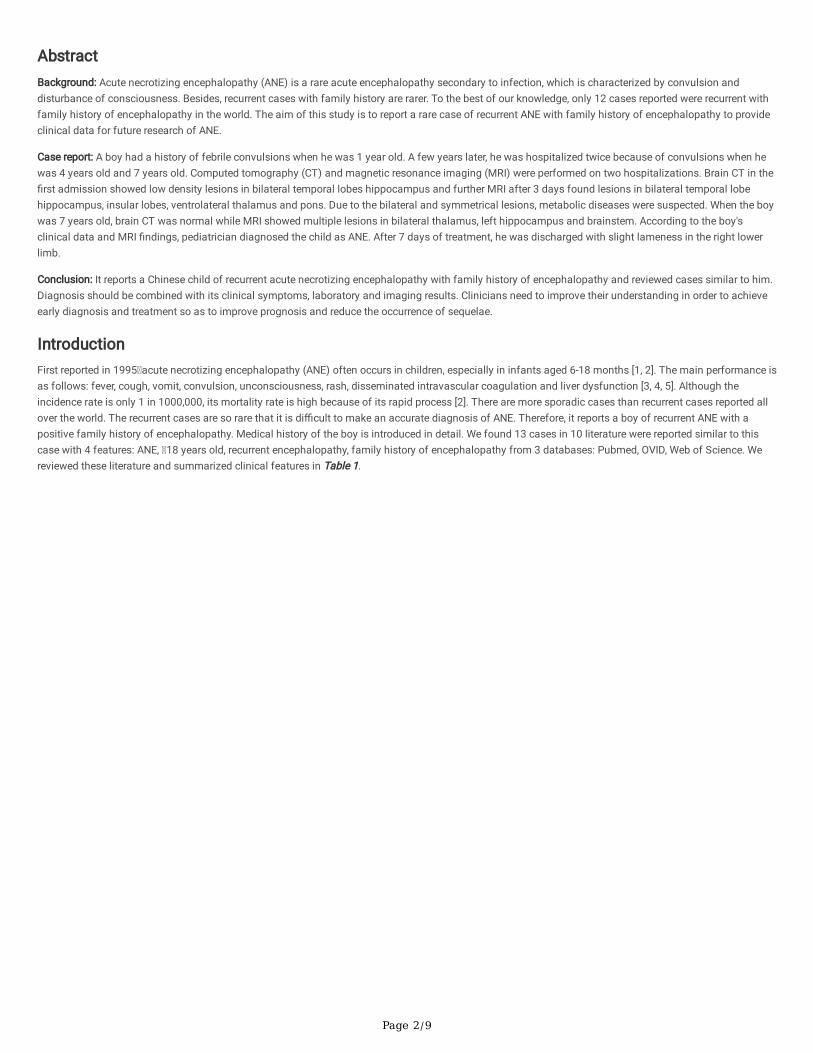
AbstractBackground: Acute necrotizing encephalopathy (ANE) is a rare acute encephalopathy secondary to infection, which is characterized by convulsion anddisturbance of consciousness. Besides, recurrent cases with family history are rarer. To the best of our knowledge, only 12 cases reported were recurrent withfamily history of encephalopathy in the world. The aim of this study is to report a rare case of recurrent ANE with family history of encephalopathy to provideclinical data for future research of ANE.
Case report: A boy had a history of febrile convulsions when he was 1 year old. A few years later, he was hospitalized twice because of convulsions when hewas 4 years old and 7 years old. Computed tomography (CT) and magnetic resonance imaging (MRI) were performed on two hospitalizations. Brain CT in the�rst admission showed low density lesions in bilateral temporal lobes hippocampus and further MRI after 3 days found lesions in bilateral temporal lobehippocampus, insular lobes, ventrolateral thalamus and pons. Due to the bilateral and symmetrical lesions, metabolic diseases were suspected. When the boywas 7 years old, brain CT was normal while MRI showed multiple lesions in bilateral thalamus, left hippocampus and brainstem. According to the boy'sclinical data and MRI �ndings, pediatrician diagnosed the child as ANE. After 7 days of treatment, he was discharged with slight lameness in the right lowerlimb.
Conclusion: It reports a Chinese child of recurrent acute necrotizing encephalopathy with family history of encephalopathy and reviewed cases similar to him.Diagnosis should be combined with its clinical symptoms, laboratory and imaging results. Clinicians need to improve their understanding in order to achieveearly diagnosis and treatment so as to improve prognosis and reduce the occurrence of sequelae.
IntroductionFirst reported in 1995 acute necrotizing encephalopathy (ANE) often occurs in children, especially in infants aged 6-18 months [1, 2]. The main performance isas follows: fever, cough, vomit, convulsion, unconsciousness, rash, disseminated intravascular coagulation and liver dysfunction [3, 4, 5]. Although theincidence rate is only 1 in 1000,000, its mortality rate is high because of its rapid process [2]. There are more sporadic cases than recurrent cases reported allover the world. The recurrent cases are so rare that it is di�cult to make an accurate diagnosis of ANE. Therefore, it reports a boy of recurrent ANE with apositive family history of encephalopathy. Medical history of the boy is introduced in detail. We found 13 cases in 10 literature were reported similar to thiscase with 4 features: ANE, 18 years old, recurrent encephalopathy, family history of encephalopathy from 3 databases: Pubmed, OVID, Web of Science. Wereviewed these literature and summarized clinical features in Table 1.
Page 3/9
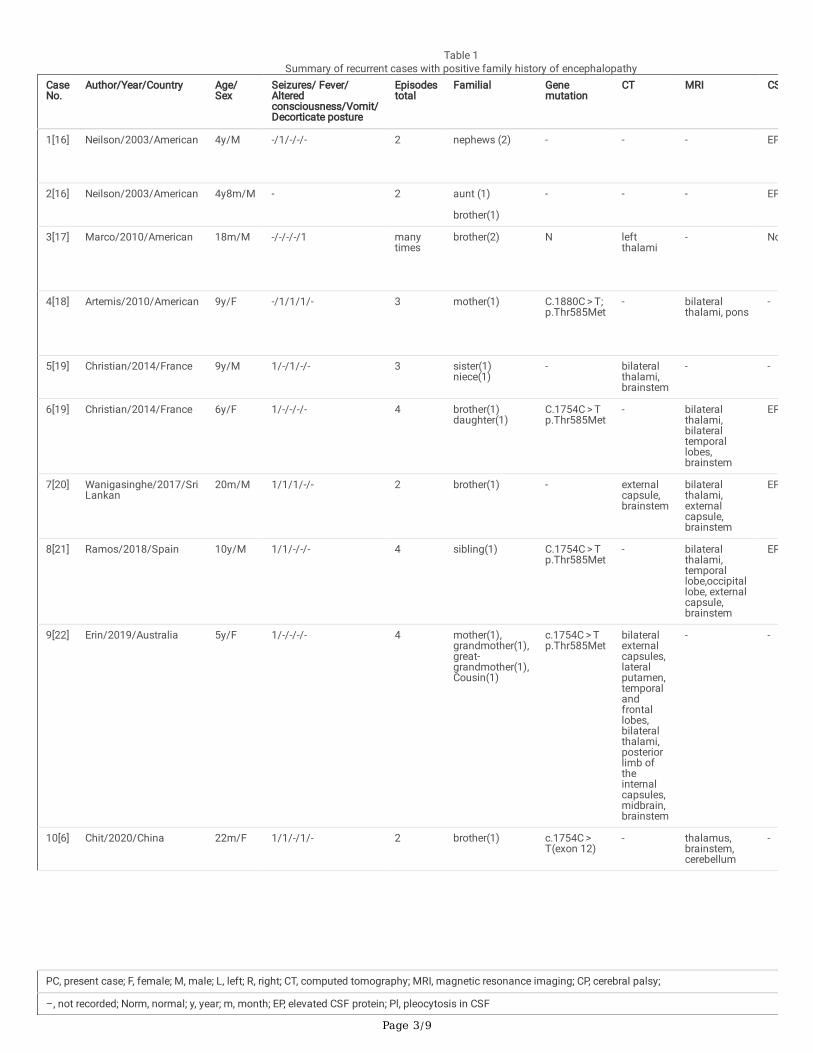
Table 1Summary of recurrent cases with positive family history of encephalopathy
CaseNo.
Author/Year/Country Age/Sex
Seizures/ Fever/Alteredconsciousness/Vomit/Decorticate posture
Episodestotal
Familial Genemutation
CT MRI CS
1[16] Neilson/2003/American 4y/M -/1/-/-/- 2 nephews (2) - - - EP
2[16] Neilson/2003/American 4y8m/M - 2 aunt (1)
brother(1)
- - - EP
3[17] Marco/2010/American 18m/M -/-/-/-/1 manytimes
brother(2) N leftthalami
- No
4[18] Artemis/2010/American 9y/F -/1/1/1/- 3 mother(1) C.1880C > T;p.Thr585Met
- bilateralthalami, pons
-
5[19] Christian/2014/France 9y/M 1/-/1/-/- 3 sister(1)niece(1)
- bilateralthalami,brainstem
- -
6[19] Christian/2014/France 6y/F 1/-/-/-/- 4 brother(1)daughter(1)
C.1754C > Tp.Thr585Met
- bilateralthalami,bilateraltemporallobes,brainstem
EP
7[20] Wanigasinghe/2017/SriLankan
20m/M 1/1/1/-/- 2 brother(1) - externalcapsule,brainstem
bilateralthalami,externalcapsule,brainstem
EP
8[21] Ramos/2018/Spain 10y/M 1/1/-/-/- 4 sibling(1) C.1754C > Tp.Thr585Met
- bilateralthalami,temporallobe,occipitallobe, externalcapsule,brainstem
EP
9[22] Erin/2019/Australia 5y/F 1/-/-/-/- 4 mother(1),grandmother(1),great-grandmother(1),Cousin(1)
c.1754C > Tp.Thr585Met
bilateralexternalcapsules,lateralputamen,temporalandfrontallobes,bilateralthalami,posteriorlimb oftheinternalcapsules,midbrain,brainstem
- -
10[6] Chit/2020/China 22m/F 1/1/-/1/- 2 brother(1) c.1754C > T(exon 12)
- thalamus,brainstem,cerebellum
-
PC, present case; F, female; M, male; L, left; R, right; CT, computed tomography; MRI, magnetic resonance imaging; CP, cerebral palsy;
–, not recorded; Norm, normal; y, year; m, month; EP, elevated CSF protein; Pl, pleocytosis in CSF
Page 4/9
CaseNo.
Author/Year/Country Age/Sex
Seizures/ Fever/Alteredconsciousness/Vomit/Decorticate posture
Episodestotal
Familial Genemutation
CT MRI CS
11[9] Gayatri/2020/India 15m/M 1/1/1/-/- 3 brother(1)sister(1)
- - pons,cerebellardentatenuclei,thalami,posteriorputamen,internal andexternalcapsule,hemisphericwhite matter
No
12[9] Gayatri/2020/India 13m/F 1/1/1/1/- 2 brothers(2) - - bilateralthalami,externalcapsule
-
14a PC 4y/M 1/-/1/-/- the 1st brothers(1)sister(2)grandpa(1)
- bilateraltemporallobes
bilateraltemporallobes, insularlobe, bilateralthalami andpons
EP
14b PC 7y/M 1/1/1/1- the 2nd brothers(1)sister(2)grandpa(1)
- Norm bilateralthalami andexternalcapsule, lefthippocampus,brainstem,pons
EP
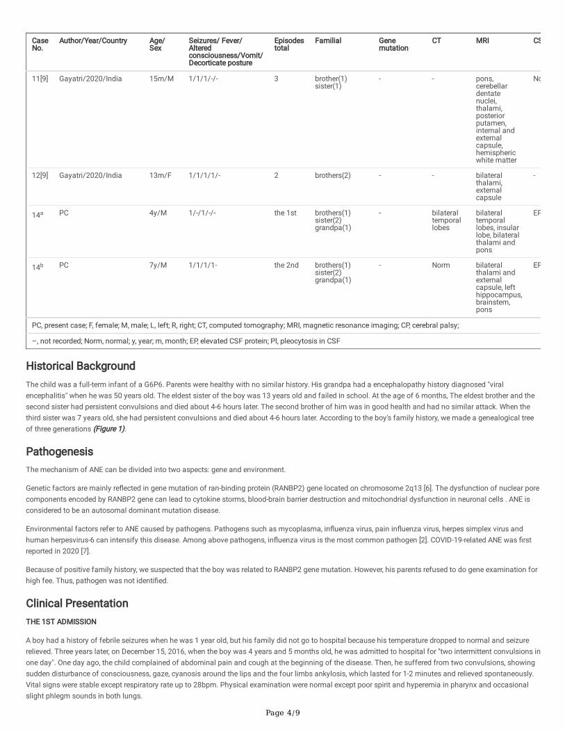
PC, present case; F, female; M, male; L, left; R, right; CT, computed tomography; MRI, magnetic resonance imaging; CP, cerebral palsy;
–, not recorded; Norm, normal; y, year; m, month; EP, elevated CSF protein; Pl, pleocytosis in CSF
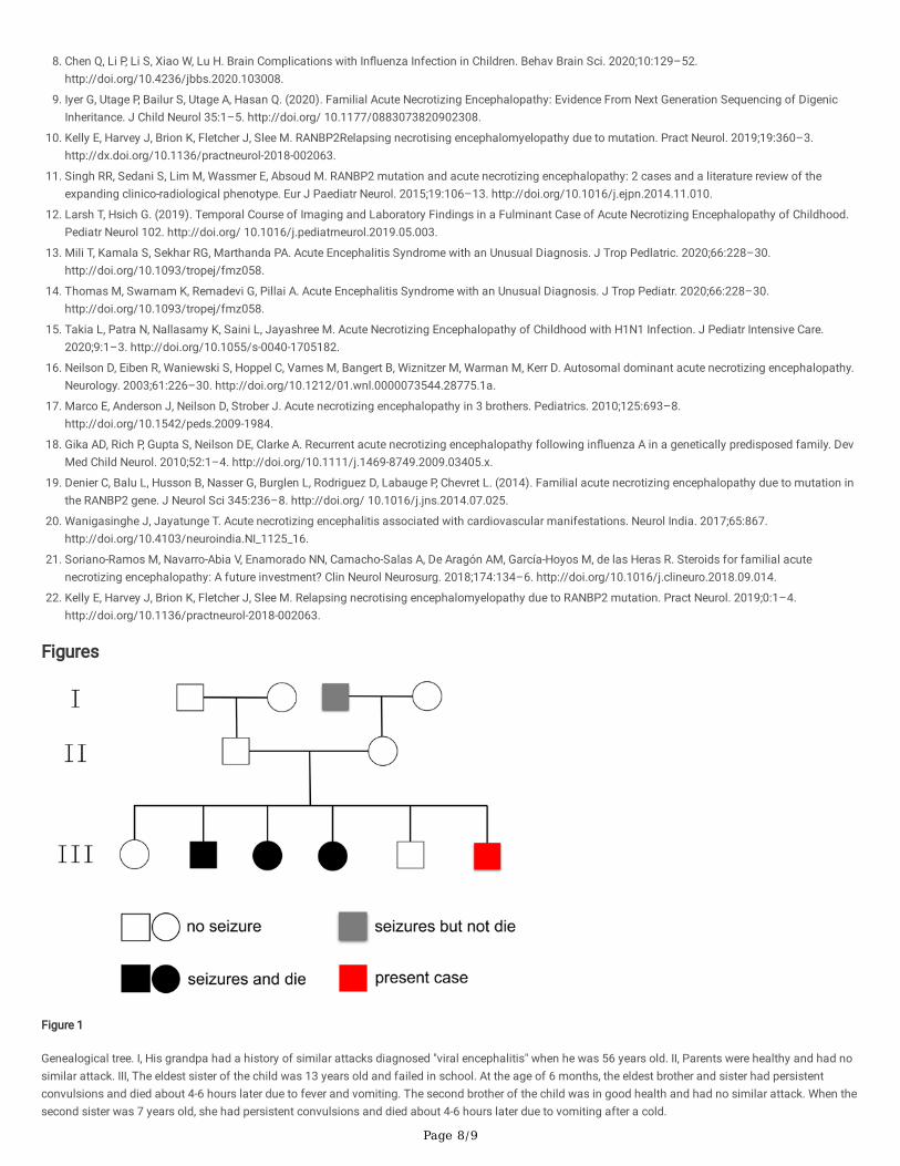
Historical BackgroundThe child was a full-term infant of a G6P6. Parents were healthy with no similar history. His grandpa had a encephalopathy history diagnosed "viralencephalitis" when he was 50 years old. The eldest sister of the boy was 13 years old and failed in school. At the age of 6 months, The eldest brother and thesecond sister had persistent convulsions and died about 4-6 hours later. The second brother of him was in good health and had no similar attack. When thethird sister was 7 years old, she had persistent convulsions and died about 4-6 hours later. According to the boy's family history, we made a genealogical treeof three generations (Figure 1).
PathogenesisThe mechanism of ANE can be divided into two aspects: gene and environment.
Genetic factors are mainly re�ected in gene mutation of ran-binding protein (RANBP2) gene located on chromosome 2q13 [6]. The dysfunction of nuclear porecomponents encoded by RANBP2 gene can lead to cytokine storms, blood-brain barrier destruction and mitochondrial dysfunction in neuronal cells . ANE isconsidered to be an autosomal dominant mutation disease.
Environmental factors refer to ANE caused by pathogens. Pathogens such as mycoplasma, in�uenza virus, pain in�uenza virus, herpes simplex virus andhuman herpesvirus-6 can intensify this disease. Among above pathogens, in�uenza virus is the most common pathogen [2]. COVID-19-related ANE was �rstreported in 2020 [7].
Because of positive family history, we suspected that the boy was related to RANBP2 gene mutation. However, his parents refused to do gene examination forhigh fee. Thus, pathogen was not identi�ed.
Clinical PresentationTHE 1ST ADMISSION
A boy had a history of febrile seizures when he was 1 year old, but his family did not go to hospital because his temperature dropped to normal and seizurerelieved. Three years later, on December 15, 2016, when the boy was 4 years and 5 months old, he was admitted to hospital for "two intermittent convulsions inone day". One day ago, the child complained of abdominal pain and cough at the beginning of the disease. Then, he suffered from two convulsions, showingsudden disturbance of consciousness, gaze, cyanosis around the lips and the four limbs ankylosis, which lasted for 1-2 minutes and relieved spontaneously.Vital signs were stable except respiratory rate up to 28bpm. Physical examination were normal except poor spirit and hyperemia in pharynx and occasionalslight phlegm sounds in both lungs.
Page 5/9
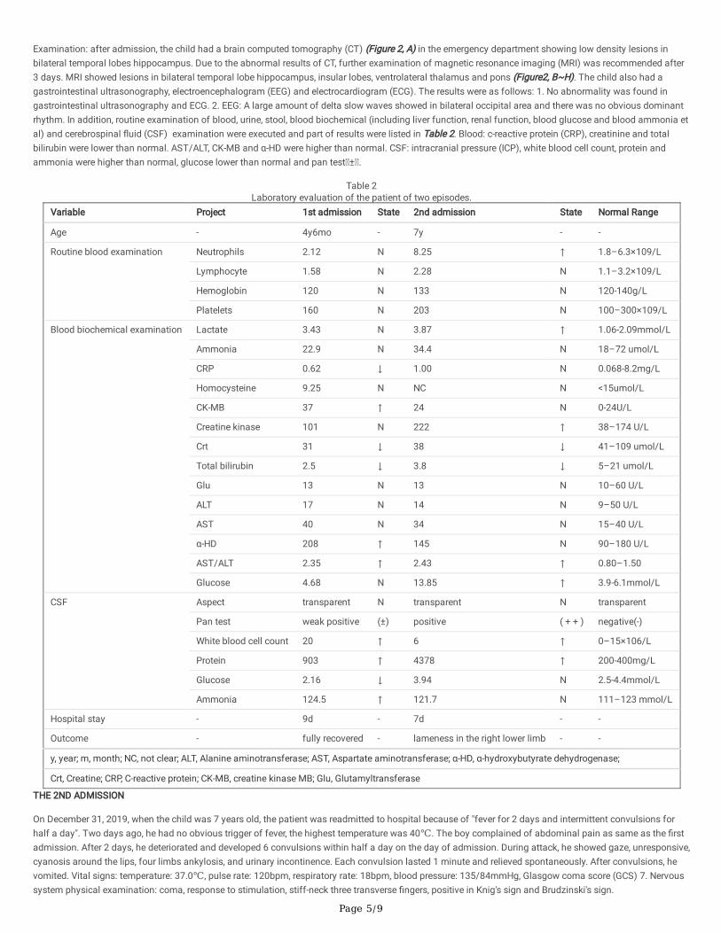
Examination: after admission, the child had a brain computed tomography (CT) (Figure 2, A) in the emergency department showing low density lesions inbilateral temporal lobes hippocampus. Due to the abnormal results of CT, further examination of magnetic resonance imaging (MRI) was recommended after3 days. MRI showed lesions in bilateral temporal lobe hippocampus, insular lobes, ventrolateral thalamus and pons (Figure2, B~H). The child also had agastrointestinal ultrasonography, electroencephalogram (EEG) and electrocardiogram (ECG). The results were as follows: 1. No abnormality was found ingastrointestinal ultrasonography and ECG. 2. EEG: A large amount of delta slow waves showed in bilateral occipital area and there was no obvious dominantrhythm. In addition, routine examination of blood, urine, stool, blood biochemical (including liver function, renal function, blood glucose and blood ammonia etal) and cerebrospinal �uid (CSF) examination were executed and part of results were listed in Table 2. Blood: c-reactive protein (CRP), creatinine and totalbilirubin were lower than normal. AST/ALT, CK-MB and α-HD were higher than normal. CSF: intracranial pressure (ICP), white blood cell count, protein andammonia were higher than normal, glucose lower than normal and pan test ± .
Table 2Laboratory evaluation of the patient of two episodes.
Variable Project 1st admission State 2nd admission State Normal Range
Age - 4y6mo - 7y - -
Routine blood examination Neutrophils 2.12 N 8.25 ↑ 1.8–6.3×10⁹/L
Lymphocyte 1.58 N 2.28 N 1.1–3.2×10⁹/L
Hemoglobin 120 N 133 N 120-140g/L
Platelets 160 N 203 N 100–300×109/L
Blood biochemical examination Lactate 3.43 N 3.87 ↑ 1.06-2.09mmol/L
Ammonia 22.9 N 34.4 N 18–72 umol/L
CRP 0.62 ↓ 1.00 N 0.068-8.2mg/L
Homocysteine 9.25 N NC N <15umol/L
CK-MB 37 ↑ 24 N 0-24U/L
Creatine kinase 101 N 222 ↑ 38–174 U/L
Crt 31 ↓ 38 ↓ 41–109 umol/L
Total bilirubin 2.5 ↓ 3.8 ↓ 5–21 umol/L
Glu 13 N 13 N 10–60 U/L
ALT 17 N 14 N 9–50 U/L
AST 40 N 34 N 15–40 U/L
α-HD 208 ↑ 145 N 90–180 U/L
AST/ALT 2.35 ↑ 2.43 ↑ 0.80–1.50
Glucose 4.68 N 13.85 ↑ 3.9-6.1mmol/L
CSF Aspect transparent N transparent N transparent
Pan test weak positive (±) positive ( + + ) negative(-)
White blood cell count 20 ↑ 6 ↑ 0–15×106/L
Protein 903 ↑ 4378 ↑ 200-400mg/L
Glucose 2.16 ↓ 3.94 N 2.5-4.4mmol/L
Ammonia 124.5 ↑ 121.7 N 111–123 mmol/L
Hospital stay - 9d - 7d - -
Outcome - fully recovered - lameness in the right lower limb - -
y, year; m, month; NC, not clear; ALT, Alanine aminotransferase; AST, Aspartate aminotransferase; α-HD, α-hydroxybutyrate dehydrogenase;
Crt, Creatine; CRP, C-reactive protein; CK-MB, creatine kinase MB; Glu, Glutamyltransferase
THE 2ND ADMISSION
On December 31, 2019, when the child was 7 years old, the patient was readmitted to hospital because of "fever for 2 days and intermittent convulsions forhalf a day". Two days ago, he had no obvious trigger of fever, the highest temperature was 40℃. The boy complained of abdominal pain as same as the �rstadmission. After 2 days, he deteriorated and developed 6 convulsions within half a day on the day of admission. During attack, he showed gaze, unresponsive,cyanosis around the lips, four limbs ankylosis, and urinary incontinence. Each convulsion lasted 1 minute and relieved spontaneously. After convulsions, hevomited. Vital signs: temperature: 37.0℃, pulse rate: 120bpm, respiratory rate: 18bpm, blood pressure: 135/84mmHg, Glasgow coma score (GCS) 7. Nervoussystem physical examination: coma, response to stimulation, stiff-neck three transverse �ngers, positive in Knig's sign and Brudzinski's sign.

Page 6/9
Examination: after admission, the child underwent imaging examinations: brain CT was normal (Figure 3, A). MRI showed multiple lesions in bilateralthalamus, left hippocampus and brainstem (Figure 3, B~F). Laboratory tests was the same as the �rst admission and results were listed in Table 2. Blood:neutrophils, lactate, creatine kinase, AST/ALT and glucose were higher than normal. Creatinine and total bilirubin were lower than normal. CSF: ICP, whiteblood cell count, protein and ammonia were higher than normal.
The clinical process can be divided into three stages: prodromal stage, acute encephalopathy stage and convalescent stage. The �rst stage lasted about 3days and children had respiratory, gastrointestinal symptoms such as fever, cough and vomit [8]. After a short prodromal period, rapid alteration ofconsciousness and convulsions occurred. About 30% children may die in acute encephalopathy stage. Among patients who were lucky enough to get throughthe second period, less than 10% of them recovered completely in the third period [3]. This child admitted to hospital twice for seizures, the �rst time hisprodromal period was so short that the boundary between prodromal and acute encephalopathy stage was not clear. The child showed abdominal painwithout any other common prodromal symptoms. At the 2nd admission, he fevered for 2 days and the highest temperature up to 41℃. In acuteencephalopathy stage, the child showed 6 seizures and alteration of consciousness.
Most ANE cases are sporadic and will not recur. But the neurological function of a few ANE patients with recurrent encephalopathy deteriorated [9, 10]. For thislittle boy, he left the right lower limb slightly lameness at the 2nd discharge even though the treatment was similar to the �rst admission which leaded to fullyrecovery. A study in Japan, it was pointed out that the independent factors related to the death of ANE included AST > 500U/L, blood glucose > 150U/dl,hematuria/ albuminuria. For this boy, he was not satis�ed with any of the independent factors.
In radiological research, the identi�cation of ANE is the most important factor in determining the outcome [11]. Imaging shows multiple and symmetricallesions located mostly in bilateral thalamus, brain stem, periventricular white matter, cerebellar medulla etc[12]. Thalamus is the most frequently involved area,which can be seen in most ANE patients. The typical signs are “concentric/laminar structure” or “tricolor pattern” in appearance. At the �rst of admission, thechild had a brain CT showing low density lesions in bilateral temporal lobe hippocampus. After 3 days, MRI showed bilateral and symmetrical long T1 signaland long T2 signal in bilateral thalami, insular lobes. Axial �uid attenuated inversion recovery (FLAIR) showed abnormal, high signal of the bilateral thalami,insular lobes, temporal lobes hippocampus and the pons. Apparent diffusion coe�cient (ADC) map showed “concentric/laminar structure” in bilateral insularlobes, large areas of restriction of diffusion in bilateral insular lobes and temporal lobes hippocampus. Diffusion-weighted imaging (DWI) showed high signalin the bilateral thalami, insular lobes. The 2st time, brain CT showed normal. But MRI showed long T1 and long T2 signal in bilateral thalamus, lefthippocampus and brain stem. FLAIR, ADC, DWI showed high signal. According to the literature, hemorrhagic transformation may occur in the focus. Comparedwith MRI on the �rst admission, lesions on the second time were reduced and absorbed. Therefore, the performance was consistent with literature.
Laboratory examination showed that thrombocytopenia, liver enzyme increased but blood ammonia decreased in hematological and increased proteinwithout pleocytosis in CSF [13]. Due to cytokine storms, cytokines may cause damage to multiple organs and lead to the above abnormality. It may showplatelets reduced or liver enzymes increased in blood examinations. However, the results of platelet count and liver function were unremarkable during twohospitalizations. CSF protein increased obviously while no pleocytosis is one of the characteristics of ANE. Although the mechanism is unclear, it can helpdistinguish ANE from other encephalitis such as acute diffuse encephalomyelitis (ADE). CSF showed no pleocytosis and protein results of two admission were903mg/L, 4378mg/L respectively, which distinguished ANE from ADE. In addition, CSF protein is one of the indexes to predict the prognosis of ANE. HigherCSF protein is associated with worse prognosis. 94% of the seriously ill patients had CSF protein > 0.45g/l [9]. For this boy, CSF protein was much higher thanthe �rst one. And he recovered completely the �rst time but left a sequelae the second time the second time.
We believe that it is necessary to test RANBP2 gene in ANE patients because one of the risk factors of ANE is RANBP2 gene mutation. It is associated withfamilial ANE and recurrent ANE. In addition, 40% of children carrying RANBP2 mutations are likely to develop ANE and there is a 50% chance of recurring ANE[6]. Moreover, infection-induced acute encephalopathy (IIAE) is a group of neurological diseases caused by infection. IIAE has many subtypes. ANE is one ofthe subtypes which is known as IIAE3 [9]. As for this family, the boy presented with IIAE3. His grandfather had “viral encephalitis”. His three siblings alsoshowed acute encephalopathy although the cause was unknown. It was a pity that even though we highly suspected that his families carried RANBP2 genemutation, the little boy and his families did not test RANBP2 gene. Nevertheless, we still believe that it is very important to establish a diagnosis of gene-related ANE [9, 10]. Because genetic test of ANE patient may be a bene�t to family members. It provides opportunities for preventive vaccination, earlyintervention and prenatal detection of ANE. Some experts suggest that children more than 6 months old could be vaccinated against in�uenza every year,which is meaningful for ANE survivors and caregivers.
DiagnosisDiagnostic criteria: 1. Convulsions and deterioration of consciousness after viral infection. 2. CSF Protein increases with no pleocytosis. 3. Multiple,symmetrical lesions involving bilateral thalamic, frontal lobes, parietal lobes, temporal lobes, brainstem, internal capsule and cerebellum. Bilateral thalamic areimaging markers. 4. Liver dysfunction as hepatic transaminase increased in various degree while blood ammonia did not increase. 5. Exclude resemblingdisease.
Differential diagnosis mainly from two aspects [8]: Clinically, ANE needs to be differentiated from viral encephalitis, fulminant hepatitis, heatstroke andhemolytic uremic syndrome. In imaging, ANE should be differentiated from carbon monoxide toxic encephalopathy, Reyes syndrome and ADE. Among thesethree diseases, ADE and Reyes syndrome are the focus of differential diagnosis. Brain lesions of ANE are symmetrical, while ADE are asymmetrical. CSFprotein is normal in ADE, while increased in ANE. Reyes syndrome is characterized by liver dysfunction, high serum ammonia and hypoglycemia [14]. But ANEshowed liver dysfunction but normal serum ammonia. Brain lesions of this patient were bilateral, symmetrical, and blood examination was normal, so it canbe distinguished from ADE and Reyes syndrome.
MANAGEMENT AND PROGNOSIS
Page 7/9
General treatment includes intensive care management, detection and treatment of elevated ICP and therapeutic hypothermia. Antiviral drugs and early use ofimmunosuppressive therapy such as hormone and immunoglobulin or plasma exchange can improve prognosis [15]. Although the exactly mechanism isunclear, it is well-known that timely diagnosis and treatment may lead to successful effection. The child was treated with intensive care, mannitol to reduceICP, improving brain metabolism, nourishing brain cells, resolving phlegm and maintaining internal environment stability. Dexamethasone and oseltamivirwere also treated. Hospitalization time was 9 days, 7 days respectively. Although the treatment were similar, the boy’s neurological function deteriorated andleft the right lower limb slightly lameness at the second admission.
Literature ReviewAcute necrotizing encephalopathy is very rare. We searched the Pubmed, Web of Science and Ovid databases for English-language literature and case seriesof encephalopathy by key words: acute and encephalopathy. We screened the literature published between January 1, 1995 and February 28, 2021. A total of13 cases involved in 10 articles were contained with 4 features: ANE, 18 years old, recurrent encephalopathy, family history of encephalopathy. For each case,publication year, the �rst author and country were documented, along with the patient’s age, sex, symptoms, imaging characteristics, gene test, episode time,encephalopathy history of relatives and follow-up results (Table 1).
ConclusionWe report a Chinese child of recurrent ANE with a positive family history of encephalopathy, providing clinical data for future research of ANE. Doctors need toimprove their understanding of ANE because diagnosis is di�cult. It should combine with clinical symptoms, laboratory examination and gene detection ofsuspicious patients. In order to improve the prognosis and reduce the occurrence of sequelae, we should try our best to achieve early diagnosis and treatment.
DeclarationsAuthor contributions
Yu Yin and Jingjing Zhang collect the patient data. Yang Yang and Cheng He analyzed data. Yanli Yang and Yu Yin made pictures. Yanli Yang was a majorcontributor in writing the manuscript. Heng Liu modi�ed the manuscript. All authors read and approved the �nal manuscript.
Funding
Not applicable.
Availability of data and materials
The datasets generated and/or analyzed during the current study are available from the corresponding author on reasonable request.
Ethics approval and consent to participate
The case report was approved and supervised by the ethics committee of the A�liated Hospital of Zunyi Medical University.
Consent for publication
Written informed consent for publication of patients’ clinical details and clinical images was obtained.
Competing interests
The authors declare that they have no competing interests.
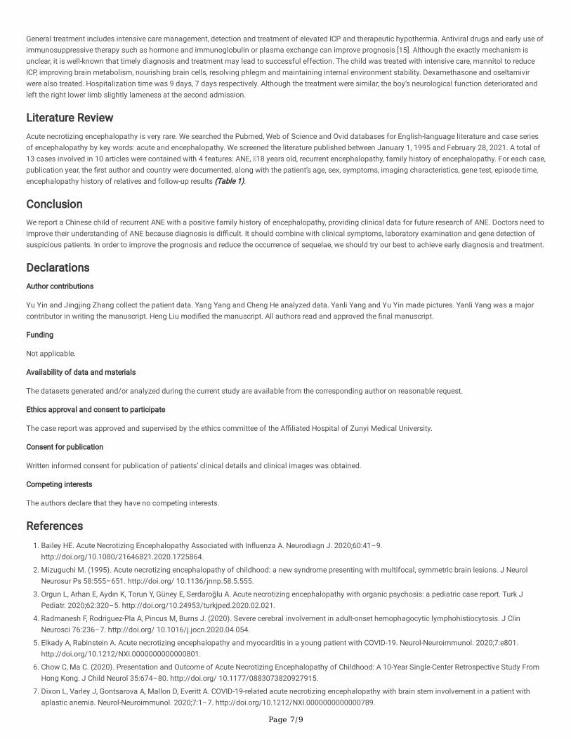
References1. Bailey HE. Acute Necrotizing Encephalopathy Associated with In�uenza A. Neurodiagn J. 2020;60:41–9.
http://doi.org/10.1080/21646821.2020.1725864.
2. Mizuguchi M. (1995). Acute necrotizing encephalopathy of childhood: a new syndrome presenting with multifocal, symmetric brain lesions. J NeurolNeurosur Ps 58:555–651. http://doi.org/ 10.1136/jnnp.58.5.555.
3. Orgun L, Arhan E, Aydın K, Torun Y, Güney E, Serdaroğlu A. Acute necrotizing encephalopathy with organic psychosis: a pediatric case report. Turk JPediatr. 2020;62:320–5. http://doi.org/10.24953/turkjped.2020.02.021.
4. Radmanesh F, Rodriguez-Pla A, Pincus M, Burns J. (2020). Severe cerebral involvement in adult-onset hemophagocytic lymphohistiocytosis. J ClinNeurosci 76:236–7. http://doi.org/ 10.1016/j.jocn.2020.04.054.
5. Elkady A, Rabinstein A. Acute necrotizing encephalopathy and myocarditis in a young patient with COVID-19. Neurol-Neuroimmunol. 2020;7:e801.http://doi.org/10.1212/NXI.0000000000000801.
�. Chow C, Ma C. (2020). Presentation and Outcome of Acute Necrotizing Encephalopathy of Childhood: A 10-Year Single-Center Retrospective Study FromHong Kong. J Child Neurol 35:674–80. http://doi.org/ 10.1177/0883073820927915.
7. Dixon L, Varley J, Gontsarova A, Mallon D, Everitt A. COVID-19-related acute necrotizing encephalopathy with brain stem involvement in a patient withaplastic anemia. Neurol-Neuroimmunol. 2020;7:1–7. http://doi.org/10.1212/NXI.0000000000000789.
Page 8/9
�. Chen Q, Li P, Li S, Xiao W, Lu H. Brain Complications with In�uenza Infection in Children. Behav Brain Sci. 2020;10:129–52.http://doi.org/10.4236/jbbs.2020.103008.
9. Iyer G, Utage P, Bailur S, Utage A, Hasan Q. (2020). Familial Acute Necrotizing Encephalopathy: Evidence From Next Generation Sequencing of DigenicInheritance. J Child Neurol 35:1–5. http://doi.org/ 10.1177/0883073820902308.
10. Kelly E, Harvey J, Brion K, Fletcher J, Slee M. RANBP2Relapsing necrotising encephalomyelopathy due to mutation. Pract Neurol. 2019;19:360–3.http://dx.doi.org/10.1136/practneurol-2018-002063.
11. Singh RR, Sedani S, Lim M, Wassmer E, Absoud M. RANBP2 mutation and acute necrotizing encephalopathy: 2 cases and a literature review of theexpanding clinico-radiological phenotype. Eur J Paediatr Neurol. 2015;19:106–13. http://doi.org/10.1016/j.ejpn.2014.11.010.
12. Larsh T, Hsich G. (2019). Temporal Course of Imaging and Laboratory Findings in a Fulminant Case of Acute Necrotizing Encephalopathy of Childhood.Pediatr Neurol 102. http://doi.org/ 10.1016/j.pediatrneurol.2019.05.003.
13. Mili T, Kamala S, Sekhar RG, Marthanda PA. Acute Encephalitis Syndrome with an Unusual Diagnosis. J Trop Pedlatric. 2020;66:228–30.http://doi.org/10.1093/tropej/fmz058.
14. Thomas M, Swarnam K, Remadevi G, Pillai A. Acute Encephalitis Syndrome with an Unusual Diagnosis. J Trop Pediatr. 2020;66:228–30.http://doi.org/10.1093/tropej/fmz058.
15. Takia L, Patra N, Nallasamy K, Saini L, Jayashree M. Acute Necrotizing Encephalopathy of Childhood with H1N1 Infection. J Pediatr Intensive Care.2020;9:1–3. http://doi.org/10.1055/s-0040-1705182.
1�. Neilson D, Eiben R, Waniewski S, Hoppel C, Varnes M, Bangert B, Wiznitzer M, Warman M, Kerr D. Autosomal dominant acute necrotizing encephalopathy.Neurology. 2003;61:226–30. http://doi.org/10.1212/01.wnl.0000073544.28775.1a.
17. Marco E, Anderson J, Neilson D, Strober J. Acute necrotizing encephalopathy in 3 brothers. Pediatrics. 2010;125:693–8.http://doi.org/10.1542/peds.2009-1984.
1�. Gika AD, Rich P, Gupta S, Neilson DE, Clarke A. Recurrent acute necrotizing encephalopathy following in�uenza A in a genetically predisposed family. DevMed Child Neurol. 2010;52:1–4. http://doi.org/10.1111/j.1469-8749.2009.03405.x.
19. Denier C, Balu L, Husson B, Nasser G, Burglen L, Rodriguez D, Labauge P, Chevret L. (2014). Familial acute necrotizing encephalopathy due to mutation inthe RANBP2 gene. J Neurol Sci 345:236–8. http://doi.org/ 10.1016/j.jns.2014.07.025.
20. Wanigasinghe J, Jayatunge T. Acute necrotizing encephalitis associated with cardiovascular manifestations. Neurol India. 2017;65:867.http://doi.org/10.4103/neuroindia.NI_1125_16.
21. Soriano-Ramos M, Navarro-Abia V, Enamorado NN, Camacho-Salas A, De Aragón AM, García-Hoyos M, de las Heras R. Steroids for familial acutenecrotizing encephalopathy: A future investment? Clin Neurol Neurosurg. 2018;174:134–6. http://doi.org/10.1016/j.clineuro.2018.09.014.
22. Kelly E, Harvey J, Brion K, Fletcher J, Slee M. Relapsing necrotising encephalomyelopathy due to RANBP2 mutation. Pract Neurol. 2019;0:1–4.http://doi.org/10.1136/practneurol-2018-002063.
Figures
Figure 1
Genealogical tree. I, His grandpa had a history of similar attacks diagnosed "viral encephalitis" when he was 56 years old. II, Parents were healthy and had nosimilar attack. III, The eldest sister of the child was 13 years old and failed in school. At the age of 6 months, the eldest brother and sister had persistentconvulsions and died about 4-6 hours later due to fever and vomiting. The second brother of the child was in good health and had no similar attack. When thesecond sister was 7 years old, she had persistent convulsions and died about 4-6 hours later due to vomiting after a cold.
Page 9/9
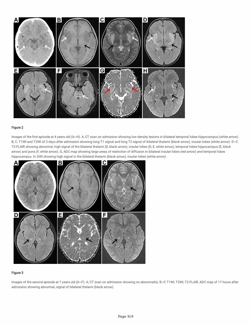
Figure 2
Images of the �rst episode at 4 years old (A~H). A, CT scan on admission showing low density lesions in bilateral temporal lobes hippocampus (white arrow).B, C. T1WI and T2WI of 3 days after admission showing long T1 signal and long T2 signal of bilateral thalami (black arrow), insular lobes (white arrow). D~F,T2-FLAIR showing abnormal, high signal of the bilateral thalami (D, black arrow), insular lobes (D, E, white arrow), temporal lobes hippocampus (E, blackarrow) and pons (F, white arrow). G, ADC map showing large areas of restriction of diffusion in bilateral insular lobes (red arrow) and temporal lobeshippocampus. H, DWI showing high signal in the bilateral thalami (black arrow), insular lobes (white arrow).
Figure 3
Images of the second episode at 7 years old (A~F). A, CT scan on admission showing no abnormality. B~F, T1WI, T2WI, T2-FLAIR, ADC map of 17 hours afteradmission showing abnormal, signal of bilateral thalami (black arrow).