Embed Size (px)
Citation preview


Promotors: Prof. dr. D. Geelen
Department of Plant Production
Prof. dr. ir. S. Werbrouck
Department of Plant Production
University College Ghent
Dean: Prof. dr. ir. G. Van Huylenbroeck
Rector: Prof. dr. P. Van Cauwenberghe

Hans MOTTE
FACTORS CONTROLLING SHOOT REGENERATION FROM
ARABIDOPSIS ROOT EXPLANTS
Thesis submitted in fulfillment of the requirements
For the degree of doctor (PhD) in Applied Biological Sciences: Cell and Gene Biotechnology

Dutch translation of the title:
Factoren die scheutregeneratie vanuit Arabidopsis wortelexplantaten reguleren
Illustrations on the cover (from top, left, to bottom, right):
- Root explant incubated on callus induction medium. Green fluorescence represents CUC2
expression and marks organogenesis competent sites.
- Shoot development during phenyl-adenine treatment.
- Shoot regeneration from root explants in a 96-well plate.
- Manhattan plot representing the association of 215,000 single nucleotide polymorphisms with
shoot regeneration.
- Development of a presumptive shoot primordium. Green fluorescence represents the
localization of the RPK1 receptor. Red fluorescence represents cell membranes.
Citation:
Motte H. (2013). Factors controlling shoot regeneration from Arabidopsis root explants. PhD thesis,
Ghent University, Ghent, Belgium.
ISBN-number: 978-90-5989-592-8
The author and the promoters give the authorization to consult and to copy parts of this work for
personal use only. Every other use is subject to the copyright laws. Permission to reproduce any
material contained in this work should be obtained from the author


Woord vooraf
“ When we discovered that crown gall induction on plants by Agrobacterium tumefaciens
is a natural event of genetic engineering, we were convinced that this was the dawn of a
new era for plant science. Now, more than 30 years later, I remain overawed by how far
and how rapidly we progressed with our knowledge of the molecular basis of plant
growth, development, stress resistance, flowering, and ecological adaptation, thanks to
the gene engineering technology. I am impressed, but also frustrated by the difficulties
of applying this knowledge to improve crops and globally develop a sustainable and
improved high-yielding agriculture. Now that gene engineering has become so efficient, I
had hoped that thousands of teams, all over the world, would work on improving our
major food crops, help domesticate new ones, and succeed in doubling or tripling
biomass yields in industrial crops. We live in a world where more than a billion people
are hungry or starving, while the last areas of tropical forest and wild nature are
disappearing. We urgently need a better supply of raw material for our chemical
industry because petroleum-based products pollute the environment and are limited in
supply. Why could this new technology not bring the solutions to these challenges? Why
has this not happened yet; what did we do wrong?“
(Marc Van Montagu (2011). "It is a long way to GM agriculture." Annual Review of Plant
Biology
62(1): 1-23)
Ik herhaal hier graag de samenvatting van een review over GGO-technologie en hoe Universiteit Gent
mee aan de bakermat lag van de ontwikkeling hiervan. Deze technologie is werkelijk fantastisch -
iedereen die ermee vertrouwt is of de review leest, is dat vast met mij eens - en heeft me dan ook
reeds van voor mijn universitaire studies gefascineerd. Het bepaalde tevens mee mijn keuze voor een
doctoraat rond plantenbiotechnologie. Ik ben op dit moment dan ook zeer blij dat ik via moleculaire
en biologische analyses mee heb geholpen aan het verwerven van nieuwe inzichten in
scheutregeneratie, een proces dat onder andere wordt toegepast binnen de GGO-technologie.
Echter, ik zou vandaag niet staan waar ik nu sta zonder de hulp van een aantal mensen. Ik zou dan
ook meerderen willen bedanken, want zonder jullie was dit doctoraat nooit gelukt.

Vooreerst wil ik mijn promotoren bedanken, die mij de kans gaven om dit onderzoek uit te voeren.
Stefaan, bedankt om mij de kneepjes van het plantenweefselteelt-vak te leren, en Danny, bedankt
voor de hulp en uitleg bij de eerder moleculair getinte problemen. Ik wil jullie beide bedanken voor
het vertrouwen dat jullie in mij stelden, voor het vele naleeswerk en voor de constructieve feedback
die ik telkens van jullie mocht ontvangen. Jullie hebben er mee voor gezorgd dat dit doctoraat een
werk van hoge kwaliteit is geworden.
Ook wil ik alle Hogent collega’s bedanken: iedereen sowieso voor de sfeer, velen voor nuttige
discussies en nog anderen voor praktische hulp. Een volledige opsomming lijkt me moeilijk, maar
volgende collega’s verdienen zeker een vermelding:
Joris, niet alleen voor het vooronderzoek, maar in het bijzonder voor de man te zijn die alles
organiseert, alles weet en alles kan oplossen. Man, ik kan mij het labo niet voorstellen zonder jou.
Danny, Joris, Katrien, Pieter en Stefaan voor de fijne tijd in het labo en hulp allerlei.
Filip, Jan en Stephanie, omdat wij er samen in slaagden de tofste bureau te vormen.
Geert, voor de onderbrekingen in de tofste bureau: ze waren soms van onschatbare waarde.
Sofie, voor de hulp bij de correlatiestudies.
Kris, voor de beste ondervoorzitter die ik ken te zijn, en de andere FC-leden, voor de goede
samenwerking.
Ellen, Griet, Marijke en Noëlla, niet allen voor de administratieve ondersteuning, maar ook zeker
voor de aangename sfeer die ik altijd bij jullie vond.
Ook alle Ugent collega’s wil ik bedanken, zowel van op het ‘boerenkot’ als van op het VIB. Wederom
is een volledige lijst niet mogelijk, maar volgenden wil ik zeker vermelden:
Patricia, voor vele hulp in het labo, en samen met Machteld, voor de administratie.
Christophe, voor het vermeerderen van heel wat zaad en andere hulp.
Inge, voor zowel de praktische hulp als de hulp bij het schrijven.
Nadine, voor de hulp met de glimlach.
Annelies en Marnik, voor de aangename en vruchtvolle samenwerking m.b.t. de QTL- en GWA-
studies.
Long, voor het verdelen van mijn 10,000 compounds.
Stephen, voor de hulp met confocale microscopie.
I stayed a short but very important period during my PhD-research at the Palacky University in
Olomouc, Czech Republic. This stay learned me a lot and yielded some results of big importance. Petr
(G), this was not possible without you. You are one of the best imaginable supervisors, and

moreover, you are also a very agreeable person. And it seems that this affect your whole lab: David,
Edita, Eva, Hanna, Jarek, Jitka, Josef, Katarína, Kateřina, Mária, Mária, Marta, Ondřej and Tomáš, I
really enjoyed working with you guys. I had a wonderful time in Olomouc and I’m looking forward to
future collaborations, both professional as for leisure. I also want to thank Petr (T), Pavel and Lukáš.
Thanks for all practical help, fruitful discussions and wonderful pub-time!
During this research, I talked a lot with experienced scientists on congresses or other meetings,
resulting in interesting discussions. Some of them are now members of my examination committee,
which is a great honor for me. I’m especially thankful to the reading committee, whose constructive
suggestions increased the quality of this dissertation. I really appreciated the open conversations we
had about this work.
Ook uit persoonlijke hoek zijn er verschillende mensen die ik graag bedank, misschien iets minder
voor de wetenschappelijke input, des te meer voor de ondersteuning of voor het begrip van mijn, op
het einde te vaak voorkomende, afwezigheid. Naast mijn ouders en familie, wens ik zeker mijn ruime
vriendengroep te bedanken, waaronder enkele goede vrienden in het bijzonder:
Aldrik en Soetkin, voor het ongelooflijk warme hart dat jullie altijd hebben.
Jan, super die telefoontjes, die hebben me er op het einde ongelooflijk geholpen om er door te
geraken.
Stijn en An, voor in de vriendenkring, toch ook af en toe nog eens over wetenschap te kunnen
babbelen.
Wouter en Melanie, voor bij mij te komen wonen en me, dag in dag uit, te steunen.
Verder verdient ook de belangrijkste persoon in mijn leven, Floor, een speciale vermelding, niet
alleen om het te tolereren dat ik soms meer dacht aan wortelexplantaten dan aan jou, maar vooral
om me te blijven verwennen gedurende het dikwijls late werk. Liefje, merci!
And you always have to save the best for last: ik vermoed dat je wel beseft wat je voor mij en mijn
doctoraat hebt betekend, maar, Danny V., ik wil je hier toch wel eens serieus in de bloemetjes zetten.
Het is slechts door jou dat ik Petr G als ‘one of the best’ moest klasseren… Echt waar, ik kan me geen
betere supervisor inbeelden dan jou, en dit op alle vlakken! Tot de laatste snik werkte je alle foutjes
weg en hielp je dit doctoraatswerk een prachtig werk te worden. Je stond altijd paraat, voor het
overlopen van de zoveelste proefopzet, voor het geven van nieuwe ideeën, om teksten te reviseren
tot ze perfect waren, voor een goede en motiverende babbel,… en zelfs om wortelstukjes te helpen
snijden. Danny, super merci!!


Table of contents
Summary ..................................................................................................................................... 1
Chapter 1 – In vitro propagation ................................................................................................... 9
Chapter 2 – Shoot regeneration: the importance of auxins and cytokinins .................................... 29
Chapter 3 – Scopes and objectives ............................................................................................... 55
Chapter 4 – Testing markers for shoot regeneration from Arabidopsis root explants ..................... 61
Chapter 5 – Phenyl-adenine, identified in an LIGHT-SENSITIVE HYPOCOTYLS4-assisted chemical screen, is a potent compound for shoot regeneration through the inhibition of CYTOKININ OXIDASE/DEHYDROGENASE activity ............................................................................................ 75
Chapter 6 – Combining linkage and association mapping as a gene discovery tool for the regeneration capacity of Arabidopsis thaliana ............................................................................ 105
Chapter 7 – Discussion and perspectives .................................................................................... 135
Bibliography .............................................................................................................................. 143
Curriculum vitae ........................................................................................................................ 171


Summary
| 1
Summary In vitro shoot regeneration, or the development of shoots from non-meristematic tissue, is a widely
applied process in plant biotechnology, including tissue culture and genetic transformation. Although
it is a well-studied developmental process, it is poorly understood why tissues of some plant species
can readily be induced to regenerate into whole plants, whereas others remain recalcitrant to do so.
To study factors controlling regeneration capacity, we adopted a two-step protocol for shoot
regeneration from Arabidopsis thaliana root explants. In this protocol, root explants are first
incubated on an auxin-rich callus induction medium (CIM), which lead to organogenesis competence,
and subsequently transferred to a cytokinin-rich shoot induction medium (SIM), resulting in
morphogenesis and development of shoots.
To better time and quantify shoot regeneration, several shoot expressed marker constructs were
evaluated via live-imaging during the course of a shoot induction protocol. Although all tested
markers were expressed in shoots, none of them were able to strictly predict shoot formation. These
results indicated that there is a certain flexibility in the shoot organogenesis program and hence
shoot formation may be interrupted at late stages of development. However, the marker CUP-
SHAPED COTELYDON2 (CUC2), which is already expressed during CIM incubation, was useful as it
predicted organogenesis competence and marked sites that could develop into shoots or lateral
roots. On the other hand, expression of LIGHT-DEPENDENT SHORT HYPOCOTYLS4 (LSH4) highly
correlated with sites of shoot formation. LHS4 was not expressed elsewhere in the root explant and
was the most predictive regeneration marker. Therefore, we used an LSH4-GFP line in a high-
throughput chemical screen to identify shoot inducing compounds. Among the 10,000 small
molecules applied on the Arabidopsis regeneration protocol, one appeared to induce shoots: phenyl-
adenine (Phe-Ade). Comprehensive molecular, enzymatic and chemical analyses revealed that Phe-
Ade exhibits a dual mode of action: it is a weak activator of the cytokinin receptors and a strong
competitive inhibitor of the cytokinin degrading CYTOKININ OXIDASE/DEHYDROGENASE (CKX)

Summary
2 |
enzymes. Phe-Ade is a cytokinin-like molecule that in contrast to classic cytokinins does not exhibit
cytotoxicity at high concentrations. This property, together with the strong stimulation of shoot
induction qualify Phe-Ade as a promising compound for future biotechnological applications.
In another approach, we used the allelic variance of Arabidopsis to determine the genetic
requirements for shoot regeneration. 88 accessions were subjected to the two-step regeneration
protocol and the regeneration capacity was evaluated. In doing so, we demonstrated that, among
the different accessions, there is hardly any pair-wise correlation between shoot regeneration and
accompanying traits, such as callus formation or greening of the explant. To further identify genetic
correlations with regeneration, two strategies were followed: (i) a quantitative trait locus (QTL)-
analysis with an inbred population of two divergent accessions, revealing five regeneration QTLs and
(ii) a genome-wide association study with 215,000 single nucleotide polymorphisms (SNPs), revealing
about 30 candidate genes associated with shoot regeneration. We further combined the results of
the two studies with a local association mapping, using data of completely sequenced accessions.
This approach narrowed down the number of candidate genes revealing the probable quantitative
trait gene (QTG) and regeneration-associated gene RECEPTOR-LIKE PROTEIN KINASE1 (RPK1).
Remarkably, RPK1 is related to abscisic acid (ABA), which is to our knowledge not reported to be
involved in shoot regeneration. Mutant analysis finally corroborated the importance of this ABA-
related gene in shoot regeneration. This result demonstrated that next-generation mapping is a
useful technique to identify genes related to a complex trait as shoot regeneration.
Altogether, we applied two sophisticated technologies to enhance our understanding of shoot
regeneration capacity. These approaches allowed the identification of Phe-Ade as an inducer of
regeneration through the inhibition of CKXs and revealed the importance of RPK1 and possibly ABA
as new players in the shoot regeneration process.

| 3


Samenvatting
| 5
Samenvatting In vitro scheutregeneratie, of de ontwikkeling van scheuten uit niet-meristematisch weefsel, is een
veel toegepast proces in de plantenbiotechnologie, onder andere voor plantenweefselteelt en
genetische transformatie. Hoewel het een goed bestudeerd proces is, is het amper geweten waarom
weefsel van bepaalde planten gemakkelijk kan geïnduceerd worden om te regenereren tot volledige
planten, terwijl anderen recalcitrant blijven. Om regulerende factoren in de regeneratiecapaciteit te
bestuderen, maakten we gebruik van een twee-staps protocol voor scheutregeneratie vanuit
Arabidopsis thaliana wortelexplantaten. In dit protocol worden wortelexplantaten eerst geïncubeerd
op een auxine-rijk callusinductie medium (CIM), dat tot competentie voor organogenese leidt, en
vervolgens verplaatst naar een cytokinine-rijk scheutinductie medium (SIM), wat tot morfogenese en
ontwikkeling van scheuten leidt.
Om scheutregeneratie beter te kunnen controleren en kwantificeren werden verschillende, in de
scheut tot expressie komende, merkers geëvalueerd via live beeldvorming gedurende het verloop
van het scheutinductieprotocol. Hoewel alle geteste merkers effectief in de scheut tot expressie
kwamen, kon geen enkele de ontwikkeling van scheuten betrouwbaar voorspellen. Deze resultaten
duiden erop dat er een zekere flexibiliteit is in het scheutorganogenese proces, waardoor
scheutvorming kan onderbroken worden tijdens late stadia in de ontwikkeling. Echter, de merker
CUP-SHAPED COTELYDON2 (CUC2), die reeds tijdens CIM incubatie tot expressie komt, bleek nuttig te
zijn voor het voorspellen van competentie voor organogenese en duidde plaatsen aan die tot
scheuten of laterale wortels konden ontwikkelen. Anderzijds vertoonde expressie van LIGHT-
DEPENDENT SHORT HYPOCOTYLS4 (LSH4) een hoge correlatie met plaatsen waar scheuten werden
gevormd. LSH4 kwam nergens anders in het wortelexplantaat tot expressie en was de meest
predictieve regeneratie merker. Daarom gebruikten we een LSH4-GFP lijn in een high-throughput
screen om scheutinducerende moleculen te vinden. Onder de 10,000 moleculen toegepast op het
Arabidopsis regeneratie protocol, bleek er één scheuten te induceren: phenyl-adenine (Phe-Ade).

Samenvatting
6 |
Uitgebreide moleculaire, enzymatische en chemische analyses wezen uit dat Phe-Ade een dubbel
werkingsmechanisme vertoont: het is een zwakke activator van de cytokinine receptoren en een
sterke competitieve inhibitor van de cytokinine afbrekende CYTOKININ OXIDASE/DEHYDROGENASE
(CKX) enzymen. Phe-Ade is een cytokinine-achtige molecule die, in tegenstelling tot de gebruikelijke
cytokininen, in hoge concentratie geen cytotoxiciteit vertoont. Deze eigenschap, samen met de
sterke stimulatie van scheutinductie, maakt dat Phe-Ade een beloftevolle molecule is voor
toekomstige biotechnologische toepassingen.
In een andere aanpak, maakten we gebruik van de allelische variatie van Arabidposis om genetische
vereisten voor scheutregeneratie te bepalen. 88 accessies werden onderworpen aan het twee-staps
regeneratieprotocol en de regeneratiecapaciteit werd geëvalueerd. Op die manier toonden we aan
dat er onder de verschillende accessies nauwelijks paarsgewijze correlatie is tussen
scheutregeneratiecapaciteit en bijgaande responsen zoals callusvorming of groenvorming van het
explantaat. Om verder de genetische correlaties met regeneratie te bepalen, werden twee
strategieën gevolgd: (i) een quantitative trait locus (QTL)-analyse gebruik makend van een inteelt
populatie van twee divergerende ouders, wat vijf regeneratie-QTLs onthulde, en (ii), een genoom-
wijde associatiestudie met 215,000 single nucleotide polymorphisms (SNPs), wat een 30-tal
kandidaatgenen geassocieerd met scheutregeneratie opleverde. Vervolgens combineerden we deze
resultaten met een grondige lokale associatie mapping, gebruik makend van de gegevens van
volledige gesequeneerde accessies. Dit verkleinde het aantal kanidaatgenen en onthulde de
vermoedelijke quantitative trait gene (QTG) en regeneratie-geassocieerd gen RECEPTOR-LIKE
PROTEIN KINASE1 (RPK1). Het is opmerkelijk dat RPK1 gerelateerd is aan abscisinezuur (ABA),
waarover, voor zover we weten, nog geen verband met scheutregeneratie werd gerapporteerd.
Mutantenanalyse bevestigde ten slotte het belang van dit gen in scheutregeneratie. Dit resultaat
toonde aan dat next-generation mapping een bruikbare methode is voor de identificatie van genen
gerelateerd aan een complex kenmerk, zoals scheutregeneratie.
Alles samengenomen, hebben we hier twee geavanceerde technologieën gebruikt, die onze kennis
over regeneratiecapacteit hebben verruimd. Door deze aanpak hebben we Phe-Ade geïdentificeerd
als een krachtig scheutinducerend molecule dat werkt via de inhibitie van CKX enzymen en hebben
we de betrokkenheid van RPK1 en mogelijks ook van ABA als nieuwe spelers in scheutregeneratie
aangetoond.

| 7


In vitro propagation
| 9
1. 1
Chapter 1 In vitro propagation
Adapted from: Motte, H., S. Werbrouck and D. Geelen (submitted). In vitro propagation. In: Plant
chemical biology. D. Audenaert and P. Overvoorde (ed.). New York, John Wiley and Sons, Inc.


In vitro propagation
| 11
Plant tissue culture as a historical basis for the discovery of plant
growth regulators
Plant tissue culture was truly on its way when phytohormones or plant growth regulators were used
to control growth and development. In 1926 Went was the first to isolate such a plant growth
regulator from oat coleoptiles: the auxin indole-3-acetic acid (Went, 1926; Went, 1928). Previously,
Haberlandt (1913) suggested already that a plant growth regulator might be responsible for cell
division but it took until 1955 before the first cytokinin, kinetin, was discovered as a degradation
product from DNA (Miller et al., 1955). During more than 40 years, kinetin was considered as a
synthetic phytohormone but more recently it is reported to be endogenously present in coconut and
Australian pine (Barciszewski et al., 1996; Raman and Elumalai, 1996). The first cytokinin that was
extracted from plants was zeatin (Miller, 1961). Skoog and Miller (1957) showed that organ
differentiation could be manipulated by changing the relative concentrations of auxin and cytokinin.
This concept of hormonal regulation of organogenesis is now applicable to a lot of plant species and
made plant tissue culture a widely applied technique for propagation and genetic modification.
Although in vitro propagation still mainly uses auxins and cytokinins, other plant growth regulators
are implemented for specific applications. By screening collections of small molecules the toolbox of
plant growth regulators available for plant tissue culture has been steadily growing since the early
days of plant growth regulator discovery. The recent discovery of pyrabactin as an abscisic acid (ABA)
agonist (Park et al., 2009) and the novel plant hormone strigolactone (Umehara et al., 2008) are
exemplary for the prospect of identifying additional compounds with growth regulatory activity in
tissue culture. Still more molecules are being discovered by screening commercial and custom-made
chemical libraries. This approach, adopted from cancer drug screening, generates great opportunities
for the plant tissue culturist who is trying to develop protocols for plants that are difficult to cultivate
and for the commercial grower who seeks the most cost-effective method for plant reproduction and
propagation.
Cytokinins used in tissue culture
Cytokinins play a central role in the regulation of proliferation and differentiation of plant cells.
Exogenous cytokinins allow to control the adventitious shoot/root balance, degree of apical
dominance and delay of senescence (Mok, 1994). All natural cytokinins are N6
Figure 1.1
-substituted adenine
derivatives ( ) that are classified, according to the configuration of their N6
1975
-side chain, as
isoprenoid or aromatic compounds. In the early years of cytokinin research, only cytokinins with an
isoprenoid side chain were thought to be endogenous. However, Horgan et al. ( ) isolated and

Chapter 1
12 |
identified ortho-topolin (oT), a benzyladenine derivative in mature poplar leaves. Later on, other
aromatic cytokinins were extracted from plants (Barciszewski et al., 1996; Ge et al., 2004; Ge et al.,
2005). Some synthetic molecules with a cytokinin activity will be discussed in the next paragraphs. An
overview of the natural cytokinins can be found in Table 1.1.
The active form of an adenine-type cytokinin is the free base, but different conjugates also occur in
plants (reviewed by Sakakibara, 2006). Nucleosides (cytokinin ribosides) and nucleotides (cytokinin
riboside 5’- mono/di/tri-phosphates)(Figure 1.1) are important transport forms and precursors of the
free bases. Some of these conjugates are also able to activate certain cytokinin receptors (Spíchal et
al., 2004) and hence, are sometimes applied in in vitro tissue culture (see below). Inactivation of the
free bases can occur by N-glucosylation on the N7- or N9 Hou et al., 2004-position ( ) or, dependent on
the side chain, by O-glucosylation (Dixon et al., 1989).
Figure 1.1: N6
R represents the substituted structure. The natural occurring substitions are presented in
-substituted adenine.
Table 1.1. Nucleosides and nucleotides include a β-D-ribose or β-D-ribose 5'-mono/di/tri-phosphate at the N9
-position (red), respectively.
Urea-type cytokinins
Other types of cytokinins that, at first sight, have no structural relationship with the natural
cytokinins are phenylurea compounds. The discovery of phenylureas with cytokinin-like activity was
an accidental finding. Coconut milk is a rich source of cytokinins which was used by Shantz and
Steward (Shantz and Steward, 1955) to identify N,N’-diphenylurea (DPU) as a highly active substance.
DPU is however not naturally present in coconut milk but it was in fact a contaminant derived from
equipment used in experiments with chemically synthesized herbicides. Nevertheless, the fortunate
mistake led to the discovery of a group of synthetic compounds with some derivatives having a very
strong cytokinin-like activity (reviewed by Ricci and Bertoletti, 2009).

In vitro propagation
| 13
Table 1.1: Natural cytokinins. Trivial Name Class Abbreviation IUPAC name Substituted
structure (R)
Molecular
formula
Molecular weight
(g/mol)
2-isopentenyladenine isoprenoid 2-iP; iP N-(3-methylbut-2-enyl)-7H-purin-6-amine C10H13N 203.24 5
trans-zeatin isoprenoid tZ (E)-2-methyl-4-(7H-purin-6-ylamino)but-2-en-1-ol C10H13N5 219.24 O
cis-zeatin isoprenoid cZ (Z)-2-methyl-4-(7H-purin-6-ylamino)but-2-en-1-ol
C10H13N5 219.24 O
dihydrozeatin isoprenoid DHZ 2-methyl-4-(7H-purin-6-ylamino)butan-1-ol C10H15N5 221.26 O
N6 aromatic -benzyladenine BA N-benzyl-7H-purin-6-amine C12H11N 225.25 5
ortho-topolin aromatic oT 2-[(8,9-dihydro-7H-purin-6-ylamino)methyl]phenol
C12H13N5 243.26 O
meta-topolin aromatic mT 3-[(8,9-dihydro-7H-purin-6-ylamino)methyl]phenol
C12H13N5 243.26 O
ortho-methoxytopolin aromatic MeoT N-[(2-methoxyphenyl)methyl]-7H-purin-6-amine
C13H13N5 255.28 O
meta-methoxytopolin aromatic MemT N-[(3-methoxyphenyl)methyl]-7H-purin-6-amine
C13H13N5 255.28 O
kinetin aromatic Kin N-(furan-2-ylmethyl)-7H-purin-6-amine C10H9N5 215.21 O

Chapter 1
14 |
The common structure of the urea-type cytokinins is phenylurea, with variations on the phenyl
moiety or on N’ (Figure 1.2). As there are many variations, the urea derivatives provide a large
potential to develop chemical libraries for screening compounds with putative cytokinin-like activity.
With this purpose, a group of about 500 urea and thiourea derivatives were evaluated by testing the
tobacco pith cell division, senescence retardation in radish, lettuce seed germination and pea lateral
bud development, leading to relationships between chemical structure and biological activity (Bruce
et al., 1965; Bruce and Zwar, 1966). Similar screens were repeated and enlarged by other labs, and
hence the relationship between structure and biological activity was refined, expanded and
sometimes adjusted. Additional urea derivatives with activity in the standard tobacco callus assay
were found to carry specific substitutions on the phenyl ring (X, Figure 1.2) and unsubstituted or
substituted pyridyl rings on N’ (R, Figure 1.2) (Takahashi et al., 1978; Okamoto et al., 1981). The
highly complex phenylureum derivative N-phenyl-N’-(1,2,3-thiadiazol-5-yl)ureum (thidiazuron, TDZ)
was originally developed as a cotton defoliant (Arndt et al., 1976), named Dropp®. Its cytokinin-like
activity was compared with other urea and thiadazolylurea derivatives by Mok et al. (1982) in the
Phaseolus callus growth bioassay. Others have synthesized and tested a wide range of urea
derivatives (Ricci et al., 2001; Ricci et al., 2004; Yonova and Stoilkova, 2004; Song et al., 2007). Based
on such screens, multiple bioactive phenylurea derivatives have been identified, with a diversity of
physiological effects. For example, N-(2-chloro-4-pyridyl)-N’-phenylurea (CPPU) is highly effective to
promote bud break (Kapchina-Toteva et al., 2000; Suttle, 2008; Ku et al., 2010) and parthenocarpic
fruit development (Ricci and Bertoletti, 2009 and refs therein), whereas TDZ is effective for
promoting shoot regeneration, even for some recalcitrant species, and somatic embryogenesis
(Murthy et al., 1998 and refs therein). N-phenyl-N’-benzothiazol-6-ylurea (PBU) was shown to have
shoot morphogenetic activity (Ricci et al., 2001; Carra et al., 2006). In contrast to the isoprenoid-type
cytokinins, some diarylurea derivatives have been shown to stimulate root formation while they lack
other cytokinin- or auxin-like activities (Ricci et al., 2006).
Figure 1.2: Phenylurea. Derivatives carry different R and X side chains.
There is no clear structural similarity between DPU derivatives and the adenine-type cytokinins.
Miller (1961) proposed that DPU were precursors of unknown natural cytokinins, but Mok et al.
(1979) suggested that DPU enhanced endogenous cytokinin biosynthesis as this compounds induced

In vitro propagation
| 15
cytokinin autonomy. This idea was rejected by Chatfield and Armstrong (1986) and Laloue and Fox
(1989), who hypothesized that phenylurea derivatives inhibit the cytokinin degrading CYTOKININ
OXIDASE/DEHYDROGENASE (CKX) enzymes and thus mimic cytokinin action by raising the
endogenous cytokinin levels. This mode of action was recently demonstrated by structural data
analysis (Kopečný et al., 2010) although some derivatives also act as cytokinin signal transduction
molecules by directly interacting with cytokinin receptors. For example, TDZ can activate the
ARABIDOPSIS HISTIDINE KINASE3 (AHK3) and AHK4/CYTOKININ RESPONSE1 (CRE1) cytokinin
receptors similar to adenine-type cytokinins (Yamada et al., 2001; Spíchal et al., 2004; Hothorn et al.,
2011),while DPU does not activate the AHKs (Spíchal et al., 2004), but only influences the
endogenous cytokinin level via CKX inhibition.
Novel cytokinins
Since the discovery of kinetin, a lot of chemically related compounds were synthesized and tested for
cytokinin activity. For instance Skoog et al. (1967) tested 69 compounds related to the natural
cytokinins and found that variations in N6
Doležal et al., 2006
-monosubstituted aminopurines could have a positive
effect on the cytokinin-like activity. The research group of Miroslav Strnad synthesized different
libraries by adding side groups or modifying substructures of cytokinins, which resulted in a total
collection of more than 1000 cytokinin-like molecules. For example, 84 aromatic cytokinin derivatives
were synthesized and compared with BA for their cytokinin activity ( ; Doležal et
al., 2007). The majority of these compounds exhibited a high activity in the tobacco callus, wheat
senescence and Amaranthus bioassays. Concerning the substitutions on the aromatic ring, the results
suggested a general trend of cytokinin activity being: meta ≥ ortho > para. The meta hydroxy-
substituted compounds were already previously noted to be more active than the ortho and para
isomers (Holub et al., 1998). Generally the fluoro derivatives were recognized as the most active
compounds. Doležal et al. (2007) concluded that, as there were strong differences in activities of the
same cytokinin compounds in the different bioassays, it may be possible to design specific
compounds that can be used to target particular cytokinin-dependent processes and to eliminate
undesirable side effects.
Some more complex variants of compounds with adenine as substructure have been tested as well.
Szüčová et al. (2009) tested 6-benzylamino-9-tetrahydropyran-2-yl and 9-tetrahydrofuran-2-ylpurine
derivatives and found a higher resistance to enzymatic degradation if the benzyl ring contains a
hydroxy or methoxy group in the meta position. Experiments with additional structural variants of
adenine, revealed compounds enhancing the cytokinin response indirectly, by inhibiting CYTOKININ
OXIDASE/DEHYDROGENASE (CKX) (Zatloukal et al., 2008). Other research groups synthesized and

Chapter 1
16 |
tested cytokinin variants, mainly starting from the isoprenoid cytokinins (eg. Haidoune et al., 1998;
Marival-Hodebar et al., 1999) and found inhibitors of CKX that, in contrast to the phenylurea
derivatives, irreversibly bind the cytokinin degrading enzyme (Suttle and Mornet, 2005). An
alternative way to indirectly increase cytokinin activity is inhibition of glycosylation. A screen for such
inhibitors led to the identification of a number of BA-related structures (Tao et al., 1991).
Cytokinin antagonists are another group of growth regulators that have been identified from screens
that could be very useful to study the mode of action of cytokinins. The chemical structure of some
of these antagonists is similar to cytokinins. Spíchal et al. (2009) screened BA-related structures that
do not have cytokinin activity and were shown not to activate the cytokinin receptors AHK4 and
AHK3, for their ability to compete for cytokinin receptor binding. In their experiments, recombinant
E. coli expressing AHK4 or AHK3 receptor genes were used to determine radiolabeled tZ binding
capacity in the presence of cytokinin derivatives. This screening method allowed the identification of
the cytokinin antagonist PI-55 (6-(2-hydroxy-3-methylbenzylamino)purine). Based on the structure of
PI-55, a second library of compounds was synthesized (Nisler et al., 2010) that led to the
identification of a stronger antagonist, LGR-991 (6-(2,5-dihydroxybenzylamino)purine). Arata et al.
(2010) screened for potential cytokinin antagonists by means of a yeast complementation assay. The
yeast strain ∆sln1[CRE1] lacks an essential histidine kinase that is complemented by AHK4 expressed
via the yeast CYC promoter. In the presence of BA or by introducing Arabidopsis
ISOPENTENYLTRANSFERASE4 (AtIPT4) AtIPT5, the modified yeast strain will grow. Cytokinin-like
compounds that inhibit the growth of the yeast strain ∆sln1[CRE1] are potential cytokinin
antagonists. Two compounds, sharing a 4-phenylquinazoline structure, were identified using this
yeast selective growth system (Arata et al., 2010). There are also several earlier reports of cytokinin
antagonists (Hecht et al., 1971; Skoog et al., 1973; Iwamura et al., 1974; Iwamura et al., 1975; Skoog
et al., 1975), but these seemed to inhibit the cell cycle progression and not necessarily targeted the
cytokinin receptors (Spíchal et al., 2007).
Synthesis of novel cytokinin-like molecules has also lead to other unexpected applications. For
example, BA derivatives have been found to specifically inhibit several protein kinases (Veselý et al.,
1994). Because some of these kinases are important for tumor cell divisions, the cytokinin-like
compound library from Miroslav Strnad was screened for cytotoxic activity and strong anticancer
properties (reviewed by Sharma et al., 2008). Olomoucin (I and II) and roscovitin are two examples
that have been identified from these screens (DeAzevedo et al., 1997; Havlíc ̌ ek et al., 1997; Kryštof et
al., 2002).

In vitro propagation
| 17
Micro-organisms, mainly pathogens, could be a source for novel cytokinins. An example is the
actinomycete Rhodococcus fascians that produces, beside some common plant cytokinins, also
methylaminopurin, 2-methylthioisopentenyladenine (2MeSiP), 2-methylthio-cis-zeatin (2MeScZ) and
their respective ribosides, which are tRNA degradation products in plants (Armstrong et al., 1976;
Murai et al., 1980). Bacterial infection can be used in vitro and gives some advantages compared to
the classical micropropagation technique, such as improvement of shoot regeneration for a wide
range of species (Vereecke et al., 2000). Also Streptomyces turgidiscabies possesses homologues
cytokinin biosynthesis genes (Joshi and Loria, 2007). Some other examples of novel bacterial
cytokinins are 5′-deoxyisopentenyladenosine produced by Pantoea agglomerans (Omer et al., 2004),
2′-deoxyzeatin riboside produced by Pseudomonas amygdali (Evidente et al., 1989) and 1’’-methyl
zeatin riboside produced by Pseudomonas syringae ssp. savastanoi (Surico et al., 1985), but to our
knowledge, these have not been applied in plant tissue culture. The fungus Cladosporium sp. 501-7W
produces two molecules that show no structural relationship with cytokinins, but do have cytokinin-
like activity. As they were found in a cytokinin bioassay regarding cotyledon growth, these were
called cotylenin A and B (Sassa et al., 1970; Sassa et al., 1998). Remarkably, cotylenin A is also tested
for its anticancer properties and was found to affect the differentiation of leukemia cells (Yamamoto-
Yamaguchi et al., 2001).
Auxins used in tissue culture
A plethora of auxins
The term auxin refers to a group of chemicals that have the capacity to stimulate plant growth. In
addition to the stimulation of cell division, auxin has a role in cell differentiation and elongation. The
cumulative effect of division and elongation controls physiological processes including tropism, apical
dominance, and root growth (Hagen and Guilfoyle, 2002; Vanneste and Friml, 2009). In contradiction
to other growth regulators, auxins are less unambiguously defined and can regulate a large diversity
of morphological or developmental processes (Vanneste and Friml, 2009). A more clearly identified
property is that it, independently from other exogenously applied hormones, stimulates adventitious
root formation. This property has led to its general application in plant clonal reproduction through
stem cuttings and in in vitro shoot micropropagation protocols (Debergh and Read, 1991).
Auxins were initially defined as substances that have the ability to promote growth in the Avena
coleoptile bioassay (Went, 1926). A quite diverse group of chemicals have been shown to exert
auxin-like growth effects. As early as the 1930’s, about 50 chemicals were tested which share some
structural relationships to indole-3-acetic acid (IAA)(Koepfli et al., 1938). Many more auxin-like

Chapter 1
18 |
compounds that are similar to IAA in structure or function have been identified by testing derivatives
of molecules with auxin-like activity (Robison and Robison, 1956; Thimann, 1958; Hamilton et al.,
1960). Because the wide diversity of molecules belonging to this hormone group, a structural
classification is not evident, but in general, the most common auxins could be classified according to
their basic structure: indole, , benzene, naphthalene or phenol. The most important auxins are listed
in Table 1.2.
Surprisingly, there is no correlation between the structural class and the kind of physiological
response, suggesting that the signal-to-response correlation follows a complex path. Indeed, several
auxin receptors have been demonstrated to occur in a single species. The first receptor identified is
the TRANSPORT INHIBITOR RESPONSE1 (TIR1) protein which is required for the changes in gene
expression patterns provoked by exogenous auxin (Dharmasiri et al., 2005a; Kepinski and Leyser,
2005; Tan et al., 2007). Members of the same family as TIR1, AUXIN SIGNALING F-BOX1-5 (AFB1-5)
also function as auxin receptors and are partially redundant to TIR1 (Dharmasiri et al., 2005a;
Dharmasiri et al., 2005b; Greenham et al., 2011; Calderon Villalobos et al., 2012) In addition to TIR1,
the Auxin Binding Protein1 (ABP1) is another receptor shown to be involved in cell division and
expansion (Braun et al., 2008). Although the molecular function of ABP1 is not known, it has been
shown to regulate at least two auxin-sensitive processes: the clathrin-dependent endocytosis of PIN-
FORMED1 (PIN1) (Robert et al., 2010) and the interdigitated pavement cell expansion (Xu et al.,
2010). Although IAA causes an auxin effect via binding on ABP1, the response of certain other auxins,
like napthalene-1-acetic acid (NAA), is caused via an ABP1-independent pathway (Yamagami et al.,
2004). More recently, the S-PHASE KINASE-ASSOCIATED PROTEIN 2A (SKP2A), an E3 ligase SCF
complex constituting F-box protein, was also reported as a new auxin receptor, involved in cell
division (Jurado et al., 2010). Furthermore, the lack of direct measurements of auxin-protein
interactions has confounded the structure-function relationship analysis and an overall common
structural feature that includes all of the auxins has not been unambiguously established (Ferro et
al., 2010). These interacting proteins are receptors, transporters and metabolic enzymes involved in
biosynthesis, degradation and conjugation (Chapman and Estelle, 2009; Del Bianco and Kepinski,
2011). Besides, most of the endogenous auxins are not found as a free and biologically active form,
but as conjugates (Tam et al., 2000). A plethora of conjugates are reported. Low molecular weight
ester conjugates with sugars or amide conjugates with amino acids are involved in auxin storage,
transport or homeostasis (reviewed by Bajguz and Piotrowska, 2009). The function of high molecular
weight conjugates with for example proteins or glycoproteins is not completely clear (Ludwig-Müller,
2011). It gets even more complicated as different plant species have different conjugate profiles and
distinct catabolism mechanisms (Bajguz and Piotrowska, 2009). Surprisingly, the set of free auxins

In vitro propagation
| 19
Table 1.2: Common auxins. Trivial name Class Abbreviation IUPAC name Structure Molecular
formula
Molecular
weight
(g/mol)
indole-3-acetic acid indole IAA 2-(1H-indol-3-yl)acetic acid
C10H9NO 175.19 2
indole-3-butyric acid indole IBA 1H-indole-3-butanoic acid
C12H13NO2 203.24
4-chloroindole-3-acetic acid indole 4-Cl-IAA 2-(4-chloro-1H-indol-3-yl)acetic acid
C10H8ClNO 209.63 2
indole-3-pyruvic acid indole IPA 3-(1H-indol-3-yl)-2-oxopropanoic acid
C11H9NO 203.20 3
phenylacetic acid benzene PAA phenylacetic acid
C8H8O2 136.15
napthalene-1-acetic acid naphthalene NAA 2-(1-naphthyl)acetic acid
C12H10O 186.21 2
2,4-dichlorophenoxyacetic acid phenol 2,4-D (2,4-dichlorophenoxy)acetic acid
C8H6Cl2O 221.04 3

Chapter 1
20 |
isolated from plants is less complex and only a few molecules have been chemically identified in
extracts: IAA, indole-3-butyric acid (IBA), phenylacetic acid (PAA) and 4-Cl-IAA (Marumo et al., 1968;
Wightman and Lighty, 1982; Epstein and Ludwigmüller, 1993). Interestingly, the synthetic auxins
exert usually the strongest effects in tissue culture. Many of these synthetic auxins are used as
herbicides and in particular useful against dicotyledonous weeds. A common effect of synthetic
auxins is that they induce ectopic cell division, but most of the damage is presumably exerted by an
increased production of ABA and ethylene (Grossmann, 2009 and refs. therein). Therefore, some
synthetic auxins, in particular 2,4-dichlorophenoxyactetic acid (2,4-D), are mostly used to induce the
formation of callus.
Some of the synthetic auxin-like molecules turned out to show an inhibitory effect on auxin-
mediated physiological responses. These compounds have been named anti-auxins but because of
possible confusion with regard to the mode of action - several have been shown to inhibit auxin
transport rather than to prevent the binding of auxin to its receptor - this term is no longer used in
auxin research. The most commonly used auxin transport inhibitors are N-1-naphthylphthalamic acid
(NPA), 2,3,5-triiodobenzoic acid (TIBA) and p-chlorophenoxyisobutyric acid (PCIB). The mode of
action has not yet been fully clarified, but they seem to block cycling of auxin efflux carriers between
the plasma membrane and endosomal compartments (Geldner et al., 2001). Moreover, NPA also
inhibits an ABC subfamily B auxin efflux transporter (Geisler et al., 2005; Nagashima et al., 2008).
Recently, auxin transport has been shown to be selectively inhibited by alkoxy-auxins (Tsuda et al.,
2011). These compounds do not exert auxin signaling responses and therefore hold the premise to
separate transport-dependent processes from signaling processes. Some other auxin antagonists
preventing binding with the TIR1 receptor were found by synthesizing variants of IAA (Hayashi et al.,
2008). Interestingly, the variants with short side chains showed auxin activity. The use in tissue
culture of molecules with auxin antagonistic action is limited to situations where one aims to reduce
the activity of auxin. This can sometimes also be achieved by simply transferring the explant to auxin-
free medium. Morphogenesis, somatic embryogenesis and adventitious organogenesis often ensues
when auxin is removed from the medium (Tran Thanh Van, 1981). In a few examples, the application
of auxin antagonists promotes or speeds up somatic embryogenesis (Newcomb and Wetherel, 1970)
and in some cases it inhibits this process (Fujimura and Komamine, 1979). These opposite effects
have to be attributed to either the differential specificity with regard to the treated plant species or
to a differential requirement during the consecutive developmental steps in embryogenesis and
organogenesis. In the end, the application of a single auxin might be a too simplified approach to
manipulate and redirect plant development with the purpose of clonal reproduction and rooting.
In vitro propagation
| 21
The search for new auxins is still going on. Through compound screens of large chemical libraries
such as the DIVERSet library from ChemBridge Corporation, many more auxins or auxin antagonists
have been identified (Grozinger et al., 2001; Armstrong et al., 2004; Sungur et al., 2007; Christian et
al., 2008; Savaldi-Goldstein et al., 2008). Screening of compounds of the Korea Chemical Bank
resulted in the identification of a new transport inhibitor (Kim et al., 2010). The availability of new
compounds may boost in vitro tissue culture research and help to realize higher reproduction rates
and uniformity in root induction.
Hormones in tissue culture applications
Induction of callus
Most plant tissues and even single microspores and protoplasts react to auxin by ectopic cell
divisions which on solid medium lead to the formation of callus. By and large, synthetic auxins like
2,4-D and NAA are most frequently used for callus induction. The powerful response to 2,4-D may be
attributed to its strong accumulation inside the cell (Meijer et al., 1999) and the inability to be
exported out of the plant cell (Delbarre et al., 1996). Moreover, 2,4-D and other auxinic herbicides
seem to persist for a long time with little apparent conversion which may also explain the sustained
cell divisions (Grossmann, 2005). Monocotyledonous plants are usually more resistant to 2,4-D than
dicotyledonous plants, but 2,4,5-trichlorophenoxyacetic acid (2,4,5-T) and 3,6-dichloroanisic acid
(dicamba) have been successfully applied to induce callus in grasses (Heyser et al., 1983; Gray and
Conger, 1985). Woody species also show a more restricted sensitivity to auxin with 4-amino-3,5,6-
trichloropicolinic acid (picloram) as a potent callus-inducing auxin (eg. Beyl and Sharma, 1983).
During the initial phase of callus induction the presence of cytokinin may be required to sustain
growth. After a series of subcultures, the dependency for cytokinin may be lost, a phenomenon that
is known as cytokinin habituation (Meins, 1989). Cytokinin habituation has been shown to correlate
with an increase in expression of cytokinin signal transduction genes (Pischke et al., 2006) and has
been shown to be installed upon a single application of the diphenylurea compound DFU in
Phaseolus lunatus (Mok et al., 1979). Surprisingly, the cytokinin TDZ can also induce callus in a variety
of plant culture systems (Murthy et al., 1998), probably because, besides binding the cytokinin
receptor in Arabidopsis (Yamada et al., 2001; Spíchal et al., 2004), it also regulates auxin signaling,
presumably by influencing polar auxin transport (Suttle, 1988).
Callus comes in various shapes, colors and textures indicating that auxin-mediated dedifferentiation
of the explant cells is not complete and that some level of differentiation is maintained in the
presence of auxin. The same explant may generate several types of callus that have different

Chapter 1
22 |
morphogenetic regeneration capacities. Most studies show that the tissue, incubation conditions,
light and temperature are critical parameters determining the regenerative capacity of callus,
suggesting that environmental factors are more important than the auxin applied (George and
Debergh, 2008). The growth of callus has been associated with increased genetic and epigenetic
changes which could also be a source for variation in callus types (Smulders and de Klerk). The
prolonged cultivation of callus usually leads to selection with the fastest growing cells taking over the
slow growing ones, generating a more uniform growth pattern. 2,4-D has been reported to inflict
cytogenetic damage in plants (eg. Bayliss, 1977) and in mosquito (Ali and Ahmad, 1996). Because
polyploid cells tend to pass quicker through the cell cycle, 2,4-D has been used to generate polyploid
cultures (Kubalakova et al., 1996). This technique is still exploited to produce diploid regenerants
from haploid microspores and ovules (Kasha et al., 2006). The novel auxin-like compounds have not
been systematically analyzed with respect to genetic stability. Alternative compounds that do not
have genetic side effects would be very valuable for tissue culture applications (Machakova et al.,
2008). Irvine et al. (1983) tested 79 compounds for their callus inducing capacity on sugarcane and
selected picloram as an alternative for 2,4-D, but in later studies, it seemed also to induce genetic
variability (Soniya et al., 2001).
Adventitious rooting
Adventitious roots arise from non-root tissue, in contrast to lateral roots that originate from the
pericycle, an organogenesis competent cell layer surrounding the central cylinder of the root
(Casimiro et al., 2001). In natural conditions, adventitious roots emerge upon wounding or from
preformed primordia that for example activate after a flood. In tissue culture, cut shoots are
obviously wounded and this can suffice to stimulate adventitious rooting, but generally an auxin,
mostly IAA or IBA, is supplied to promote root organogenesis. Usually, the latter has a higher root-
inducing capacity because it is more stable and can be metabolized to IAA (Bartel et al., 2001), but
the response is also species-dependent (Li et al., 2009). Adventitious roots arise directly from a
differentiated cell or indirectly from callus tissue. Callus-derived roots do not provide a vascular
connection and therefore are inefficient in transport of nutrients. For this reason, classical auxins,
and not callus-inducing auxins like 2,4-D, are used for micropropagation. Unfortunately, the root
induction capacity of auxin is not universal and some species are difficult to root. The mechanisms
underlying recalcitrance to root is not fully resolved, but cytokinin accumulation during the
reproduction phase of micropropagation is in many cases causing a poor root induction in
subsequent stages. Some cytokinins like BA are metabolized to stable N-glucosides which accumulate
in the shoot base where they block rooting (Werbrouck et al., 1995). Molecular studies have shown
that adventitious rooting is a heritable quantitative genetic trait (Han et al., 1994; Price and Tomos,

In vitro propagation
| 23
1997; King and Stimart, 1998; Marques et al., 1999; Zheng et al., 2003; Mano et al., 2005a; Mano et
al., 2005b; Horii et al., 2006; Ochoa et al., 2006; Shepherd et al., 2006; Shepherd et al., 2008)
involving multiple genes (Li et al., 2009 and refs. therein) and therefore, recalcitrance for rooting may
be difficult to overcome.
Nevertheless, exploring new compounds can in some cases resolve the problem. For example,
wounding stimulates in addition to auxin accumulation also the biosynthesis of phenolic compounds,
some of which have positive or negative effects on rooting. The formation of polyphenolic
compounds is a byproduct from tissue culture and wounding and occurs frequently in woody species
where it has been shown to inhibit root formation. Some of the phenolics, like flavonoids, stimulate
adventitious rooting, presumably by influencing auxin transport (Murphy et al., 2000). Other
compounds may prevent degradation of auxins by inhibiting decarboxylation (Wilson and van
Staden, 1990; De Klerk et al., 2011). De Klerk et al. (2011) tested different phenolic compounds in
combination with a suboptimal IAA concentration and found that ferulic acid, a methylated ortho-
diphenol, had the strongest effect on rooting. For other small molecules like polyamines and
ethylene there are contradicting reports on their root induction capacities (Li et al., 2009).
Optimization of micropropagation
In vitro shoots can be derived directly from shoot primordia, via de novo shoot morphogenesis or via
somatic embryogenesis. Each method requires an appropriate combination of plant growth
regulators, which mainly consists of a combination of cytokinin and auxin. The most popular way of
micropropagation is via shoot culture, because this method is easy, rapid and does not involve a
callus step and hence has a low risk of generating somaclonal variation (Piccioni et al., 1997). The
shoots are transplanted on a medium with cytokinins, often combined with a smaller amount of
auxin, in order to inhibit apical dominance and to stimulate bud outgrowth (George and Debergh,
2008). Apical dominance can also be disrupted by manually tipping the shoots or applying an anti-
auxin (Machakova et al., 2008). The rate of propagation of a specific species may vary depending on
the growth regulator applied. For example 2-isopentenyladenine (2-iP) is very effective for shoot
proliferation in plant species from the Ericaceae family (Norton and Norton, 1985), while TDZ is
highly effective for woody plants (Huetteman and Preece, 1993) or for certain legumes (Malik and
Saxena, 1992).
However, optimal shoot multiplication conditions are sometimes disadvantageous for other
processes, such as general fitness or subsequent rooting. Therefore, topolins are in some cases
preferred for multiplication, as a number of reports indicate that they have less negative side-effects.

Chapter 1
24 |
Werbrouck et al. (1996) showed that mT combined a good axillary shoot production with root
formation in micropropagated Spathiphyllum. The main metabolite of mT was its O-glucoside, which
was degraded easily during acclimatization and hence showed no negative effect on rooting during
acclimatization. Salvi et al. (2002) studied the effect of various cytokinins for in vitro propagation of
Curcuma longa. mT caused a higher shoot multiplication rate and greener and more robust shoots
compared to BA .Bairu et al. (2007) reported that mT was the preferred cytokinin both in terms of
multiplication rate and rooting of Aloë polyphylla. It completely controlled the hyperhydricity
problem in this species. Also application of the non- or less active nucleosides, precursors of the free
cytokinin bases, seems to be beneficial in some cases. For example, Baroja-Fernández et al. (2002)
reported how a low dose of the riboside of mT could stimulate plantlet growth and survival of a
weakly growing potato cultivar. Bogaert et al. (2006) used a leaf-variegated petunia for
micropropagation experiments and found that the use of meta-methoxytopolinriboside (MemTR)
resulted in the production of a considerable number of new variegated shoots, with only a small
number of off-type, fully green or albino shoots, while BA induced many non-variegated shoots.
These results suggested that, in petunia, MemTR stimulates axillary shoot production in a way that
maintains the histogenic integrity of the shoot meristem, whereas BA-derived primordia are mainly
adventitious in origin and are composed of cells with a single genotype. Bogaert et al. (2006) also
demonstrated that MemTR had a much better anti-senescing effect in micropropagated roses than
BA, mT, MemT, FmT or FmTR. Bairu et al. (2008) compared the effect of mT, mTR, MemT and MemTR
to BA, on the micropropagation of the banana cvs ‘Williams’ and ‘Grand Naine’. Superior
multiplication rates were recorded for mTR treatments. Plants were categorized as normal or
abnormal based upon morphological appearance and an abnormality index was calculated. Plants
multiplied on BA showed the highest abnormality index. On an equimolar concentration of MemTR
the plantlets showed the lowest abnormality index. Hence, topolins or ribosides are valuable
compounds to test in the optimization of multiplication protocols. However, the use of these
compounds does not guarantee a minimalization of the disadvantageous side-effects of cytokinin
treatments. Indeed, several studies that reported BA or other cytokinins as the least deleterious
(Aremu et al., 2012).
If apical dominance cannot be broken, nodes can be used for propagation as each node contains a
bud. In this case, it is often unnecessary to add hormones to the medium as shoots elongate easily on
hormone free medium (George and Debergh, 2008). Alternatively, explants can be propagated
through de novo or adventitious shoot organogenesis. Adventitious shoot formation has the
advantage that it can occur in any type of explant in which a subpopulation of cells dedifferentiates
and starts to divide and hence, it can also be used for the initiation of a culture (George and Debergh,

In vitro propagation
| 25
2008). The pool of dividing cells forms either a meristemoid mass of cells that directly generates a
new shoot or an undifferentiated callus. Not every callus is the same. Some variations in appearance
are usually observed as well as variations in regeneration capacity. Organs that originate from callus
are identified as indirect and typically show an increased risk in somaclonal variation. Somaclonal
variants show inheritable phenotypic changes compared to the mother plant from which the explant
is derived and are normally unwanted during mass clonal production of elite plant selections. The
frequency at which somaclonal variation occurs depends on the plant species propagated and on the
cultivation protocol. In general, indirect shoot organogenesis involves two steps: first the explant is
incubated on callus inducing medium (CIM), usually containing a synthetic auxin, to initiate cell
divisions (Cary et al., 2002) and second, it is incubated on shoot inducing medium (SIM), usually
containing a high cytokinin dose. Similar to direct shoot organogenesis, a subpopulation of cells, here
within the callus, becomes competent and develops into shoot progenitor cells. Thus, callus is not a
homogenous mass of cells and cell-cell interactions are likely contributing to organ initiation (Chapter
2; Gordon et al., 2007; Sugimoto et al., 2010). Besides the hormones in the media, many other
factors influence the capacity for shoot regeneration. What is generally described as the "quality" of
an explant, determines whether regeneration is successful. These quality parameters are the age,
type, size and position of the explant, period of incubation and other conditioning factors (Gahan and
George, 2008). Developing a protocol for shoot regeneration is therefore an empirical undertaking.
Although there are some indicators for the acquirement of shoot competence (Ochatt et al., 2010;
Motte et al., 2011), it is still unknown which are the triggers for cells to become competent for shoot
regeneration. Interestingly, competence of callus is not determined toward either root or shoot
formation. The final identity of the organ emerging from regenerating callus depends on the
composition of the medium and can be reversed by changing the auxin to cytokinin ratio. Hence,
cells acquire organogenesis competence, rather than shoot forming competence (Pernisová et al.,
2009; Motte et al., 2011), which corresponds with the finding that the initially formed shoot
primordium in Arabidopsis root explants actually resembles a lateral root meristem (Atta et al., 2008;
Sugimoto et al., 2010). The auxin in the CIM induces the G1-to-S transition of the cell cycle
(Dubrovsky et al., 2008) and is critically important for the formation of a lateral root primordium
(Himanen et al., 2002). On the contrary, cytokinin blocks the lateral root development (Laplaze et al.,
2007) and converses the initial root primordium into a shoot primordium (Atta et al., 2008).
Depending on the plant species, different cytokinins may induce more efficiently shoot formation.
For example, zeatin and 2-iP are better than BA for inducing shoots from Arabidopsis root explants,
while BA is more efficient for cotyledon explants (Zhao et al., 2002). Sometimes, phenylurea

Chapter 1
26 |
derivatives give better results than the classical cytokinins (Read et al., 1986) or can induce shoot
regeneration where classical cytokinins fail (eg. Malik and Saxena, 1992).
Somatic embryogenesis
The emergence of bipolar structures which resemble somatic embryos was first discovered in carrot
cultures (Wetherell and Halperin, 1963). On virtually every type of carrot tissue it is possible to
induce callus that has regenerative capacity. Before the somatic embryos appear, the carrot callus
produces cell clumps, proembryogenic masses (PEM), that start to develop when the 2,4-D levels
drop or the PEM are cultivated in the absence of 2,4-D. The callus cells that are committed to
develop embryos, called embryonic cells, accumulate starch and form a small cluster that only
proceeds further to embryos when the auxin is no longer inhibitory. Therefore, the formation of
somatic embryos has to be seen as a spontaneous process and not as induced by auxin (Thorpe,
1995). Expression analyses revealed that different group of genes are differentially expressed during
somatic embryogenesis. Besides cell cycle, cell wall genes, shoot-related and hormone-responsive
genes, which serve more general functions, certain genes are specifically expressed during somatic
embryogenesis (Yang and Zhang, 2010). For instance, various genes involved in the signal
transduction pathway in somatic embryogenesis are identified and play crucial roles. As such,
SOMATIC EMBRYOGENESIS RECEPTOR LIKE KINASE (SERK) genes are used as markers for somatic
embryogenesis (Schmidt et al., 1997) and their overexpression enhances the somatic embryogenesis
competence (Hecht et al., 2001). However, the identification of multiple roles of SERKs raised
questions about the specificity of particular members of this subfamily (Li, 2010). Some transcription
factors that regulate embryo development are more specifically expressed during embryogenesis.
For example ABA-INSENSITIVE3 (ABI3), FUSCA3 (FUS3), LEAFY COTYLEDON1 (LEC1) and LEC2 are
reported as markers that are able to distinguish between embryogenic and nonembryogenic cell
cultures (Lotan et al., 1998; Luerssen et al., 1998; Shiota et al., 1998). Over- or misexpression lines in
these genes might spontaneously induce somatic embryos, while loss-of-function mutants in these
genes are recalcitrant for embryogenesis (Meinke et al., 1994; Parcy et al., 1994; Lotan et al., 1998;
Shiota et al., 1998; Nambara et al., 2000; Stone et al., 2001; Gaj et al., 2005).
Herbaceous plants show a fair chance of producing embryonic callus, but woody species show more
recalcitrance. There are major genotype or cultivar differences and the choice of explant can be of
paramount importance for obtaining embryogenic callus (Lo Schiavo, 1995). It usually helps to start
with juvenile explants, in particular immature embryos, as a suitable source for generating embryos.
In analogy to adventitious shoot or root formation, indirect - via an intermediary callus phase - or

In vitro propagation
| 27
direct somatic embryos are distinguished. However, direct somatic embryogenesis is rare and does
usually not allow mass propagation. A callus on the contrary, produces numerous small embryoids,
which is required for commercial production. Because callus usually cannot be cultured in the
absence of an auxin source, it is difficult to dissect the role of auxin in cell proliferation and the
induction of embryogenesis. 2,4-D is the most commonly used auxin to stimulate callus growth and
hence, appears frequently in somatic embryogenesis protocols. Anti-auxins have also been shown to
be effective in inducing somatic embryos in a few cases (Machakova et al., 2008). In some species,
the addition of cytokinins promotes callus growth and consequently is also part of some
embryogenesis induction protocols. Moreover, to induce somatic embryogenesis, TDZ can substitute
for both auxin and cytokinin (Murthy et al., 1998). In view of the economic potential of somatic
embryogenesis, researchers have tested many kinds of additives, including other hormones and
chemicals as well as “nutritive” chemicals (Jimenez, 2005). However, so far, a true inducer of somatic
embryogenesis has not been reported. Embryogenic callus is usually very compact and consists of
small cytoplasmic dense cells. When embryogenic cultures are established, the callus is transferred
to hormone-free medium, upon which the embryogenic cells develop into mature somatic embryos.
ABA and media with a reduced osmotic potential are often used to arrest embryonic growth and to
install dormancy for easier germination. The somatic embryos finally develop into plants in the
absence of any growth regulator (Von Arnold, 2008).
Concluding remarks
In vitro tissue culture is, beside its use in basic research, of great value for propagating plants. The
application of plant growth regulators is essential in many protocols and since the discovery of the
first phytohormone, a lot of different plant hormones have been tested. The last decade, however,
the screening of compounds for tissue culture purposes is rare and despite the diversity of plant
growth regulators already available, most published tissue culture methods use a limited selection.
The discovery of new plant hormones such as strigolactone and new chemicals with hormone-like
action like pyrabactin, supports the idea that the screenings done in the past have not exhausted the
realm of useful plant growth regulators. It is up to the tissue culturist to recognize this potential and
further explore the chemical world possibly through compound screening, automation of tissue
culture methods and by including new model plant species and tissue culture methods.


Shoot regeneration
| 29
2. 2
Chapter 2 Shoot regeneration
The importance of auxins and cytokinins
Manuscript in preparation


Shoot regeneration
| 31
In a limited number of plant species, de novo shoot regeneration is a natural
strategy for vegetative propagation, while in vitro culture is needed to make
use of this remarkable feature for micropropagation or biotechnological
breeding of a large group of other plants. For many plants, cultivars or
tissues, efficient regeneration protocols are difficult to establish, while the
cause of this regeneration recalcitrance is often not known. Shoot
regeneration is a complex trait, dependent on multiple factors that might
influence regeneration capacity. Here, we review the different steps that are
required for a proper shoot induction in a two-step regeneration protocol.
First, organogenic callus is produced that is mainly dependent on auxin-
related processes. The callus resembles root tissue but still has the capacity
to convert into shoots. Subsequent shoot initiation is mainly orchestrated by
cytokinins and several important shoot-related genes. By summarizing the
different aspects of shoot regeneration, we reveal hinge points that may be
useful to circumvent shoot regeneration recalcitrance.


Shoot regeneration
| 33
Introduction
At the beginning of the previous century, the first in vitro plant tissue culture techniques were
developed by Haberlandt, who was driven by the idea of totipotency: “Theoretically all plant cells are
able to give rise to a complete plant” (Haberlandt, 1902). He succeeded only in the survival of
aseptically grown tissue, but not in division of cells. Somewhat later, Hannig (1904) obtained in vitro
plant cell division and Simon (1908) was the first to regenerate buds on callus tissue. However, it
would take until the 1950’s to get to the understanding of the control of organogenesis: Skoog and
Miller (1957) showed that organ differentiation could be manipulated by changing the relative
concentrations of auxin and cytokinin. Since then, based on their experiments, shoot regeneration
has been established for many plants, as a first and crucial step in the initiation of an in vitro culture
(George and Debergh, 2008). Tissue culture procedures are important for many applications. They
allow plant production at a low price, propagation of clones of elite plants, , conservation of
threatened species and production of secondary metabolites (Garcia-Gonzales et al., 2010; Barnicoat
et al., 2011; Engelmann, 2011; Reed et al., 2011; Korkina and Kostyuk, 2012). Finally, shoot
regeneration is also used for genetic transformation of plants to improve traits, such as stress
tolerance, yield, nutritional value, etc. (Garcia-Gonzales et al., 2010).
Although the experiments of Skoog and Miller gave many insights in shoot regeneration, the tobacco
pith cultures they used were considered as easy to regenerate and their relatively simple findings
could not straightforwardly be adopted for other plants. Indeed, a series of events have to occur
before regeneration takes place (Christianson and Warnick, 1983; Cary et al., 2002; Che et al., 2007)
and, depending on the plant species and tissues used, different treatments have to be applied.
Thus, the development of efficient regeneration protocols for many important plants remains
cumbersome and mainly a matter of trial and error, and therefore many plants can still not used in
tissue culture practices
Birnbaum and Alvarado, 2008
. The last two decades, in part due to the rise of the model plant Arabidopsis
thaliana, but also because of the fast development of new molecular techniques, many new insights
have been gained in the molecular and developmental mechanisms underlying shoot regeneration.
Intensive research in the auxin and cytokinin field contributed enormously as well with findings that
could be applied to regeneration via tissue culture. Moreover, these lines of study challenged the
totipotency hypothesis ( ) and lead to the rejection of the generally
accepted assumption that callus tissue is composed of dedifferentiated cells (Sugimoto et al., 2010).

Chapter 2
34 |
Depending on the origin of the regenerating tissue, adventitious shoots can arise either from
differentiated cells via de- or trans-differentiation or from pre-existing stem cells (Sugimoto et al.,
2011). Both these founder cells must acquire organogenesis competence, which is correlated with
local auxin responses, prior to shoot development (Atta et al., 2008; Dubrovsky et al., 2008). So, it
may come as no surprise that the acquisition of competence can be obtained by a pre-treatment on
auxin-rich medium (Cary et al., 2002). Anatomical studies on Convolvulus arvensis roots
demonstrated that this first stage of regeneration is identical for shoots and lateral roots (Bonnett
and Torrey, 1966), leading to the hypothesis that an early stage primordium has the capacity to
develop either into a shoot or a root. This hypothesis was recently confirmed by using molecular
markers, and was extended to the finding that besides root callus, also leaf- or hypocotyl-derived
callus resembles so-called root primordia (Atta et al., 2008; Sugimoto et al., 2010). In a second stage
of regeneration, the presence of cytokinin determines the shoot identity of the developing organ by
modulating the auxin distribution (Atta et al., 2008; Pernisová et al., 2009). Thus, regeneration is
mainly dependent on two successive events: an auxin-induced organogenesis competence
acquirement and a cytokinin-induced assignment of shoot identity (Figure 2.1). Moreover, a proper
auxin-cytokinin crosstalk is required during shoot organogenesis. Consequently, regeneration
recalcitrance can be caused by defects in one or more steps in these hormone-related events.The
identification of the common elements behind plant cell totipotency which provides plant cells with
the remarkable ability to regenerate in vitro would be extremely helpful in establishing efficient
propagation protocols.
Table 2.1
In this review, we give an overview of the developmental and genetic steps
occurring during shoot regeneration from Arabidopsis root explants and, as such, we attempt to
pinpoint possible causes of regeneration recalcitrance ( ).
Figure 2.1: Overview of the widely used two-step procedure for shoot regeneration starting from Arabidopsis root explants. The initial incubation on callus induction medium (CIM) is followed by an incubation on shoot induction medium (SIM). The figure is modified from Che et al. (2007).

Shoot regeneration
| 35
Auxin accumulation specifies founder cells
Auxin initiates the formation of organogenic callus
Prior to shoot commitment, cells need to acquire organogenesis competence. Due to the common
initial stage of lateral root and adventitious shoot formation (Atta et al., 2008; Sugimoto et al., 2010),
insights from lateral root research can be used to understand particular steps in shoot regeneration.
During lateral root formation, the first cell divisions are initiated by local auxin maxima, which in a
differentiated root are only detected in certain pericycle cells (Benkova et al., 2003). These local
auxin maxima are necessary and sufficient to respecify the pericycle cells into founder cells of root
primordia and are the earliest detectable events in founder cell specification (Atta et al., 2008;
Dubrovsky et al., 2008). Interestingly, the pericycle cells are absolutely necessary for regeneration, as
ablation prevents shoot formation (Che et al., 2007). The callus-inducing medium (CIM), frequently
used to pre-treat explants, often contains synthetic auxins like 2,4-dichlorophenoxyacetic acid (2,4-D)
that are not transported out of the cells by the efflux carriers, allowing a building up of local auxin
maxima in almost all cells (Delbarre et al., 1996). A prolonged incubation on 2,4-D-containing CIM
results in explants with many proliferating cells generating a callus, which actually resembles a root
primordium, irrespective of the origin of the explant (Sugimoto et al., 2010). Another common
synthetic auxin that can be used in CIM is naphthaleneacetic acid (NAA). Because NAA is metabolized
more slowly than natural auxins (Beyer and Morgan, 1970), it presumably attains a much higher
activity in the cells inducing a lot of founder cells. In Arabidopsis, NAA is less efficient than 2,4-D in
inducing competence for both root or shoot organogenesis (Motte et al., 2011), but it has the
advantage that it induces less somaclonal variation (Ahmed et al., 2004).
The auxin influx carriers AUXIN-RESISTANT1 (AUX1), LIKE AUX1-1 (LAX1), LAX2 and LAX3 all
contribute to the local auxin accumulation, but malfunctioning of any of these genes appears not to
hamper regeneration. For instance, root explants of the aux1 mutant are not capable of forming
callus under standard culture conditions, but by increasing the auxin concentration in the medium,
callus is formed and regeneration takes place (Kakani et al., 2009). Probably, redundancy between
the different influx carriers assures a sufficient auxin supply and, consequently, the establishment of
a local auxin accumulation (Bainbridge et al., 2008). The PIN-FORMED (PIN) auxin efflux carriers on
the other hand, important in generating auxin gradients during organ formation (Benkova et al.,
2003), appear to negatively affect regeneration at this stage, since the inhibition of polar auxin
transport was shown to stimulate organogenic callus formation (Pernisová et al., 2009).
Nevertheless, together with the auxin biosynthetic YUCCA (YUC) genes, PINs are required during
shoot morphogenesis (see below)(Zhao, 2008).

Chapter 2
36 |
Table 2.1: Genes involved in regeneration-related processes, of which overexpression (OE), induced expression (IE) and/or loss of function (LOF) alters the regeneration capacity.
Gene Process Phenotype Reference
AUX1 Auxin influx LOF requires increased auxin level for callus
formation
(Kakani et al., 2009)
TIR1 Auxin perception LOF decreases and OE increases regeneration
capacity
(Qiao et al., 2012b)
SLR/IAA14 Auxin signaling OE decreases regeneration capacity (Atta et al., 2008)
LBD16, LBD17,
LBD18, LBD29
Auxin signaling OE induces spontaneous callus formation with
efficient regeneration capacity, LOF inhibits
callus formation on CIM
(Fan et al., 2012)
ARF10 Auxin signaling LOF decreases and OE increases regeneration
capacity
(Qiao et al., 2012a)
IAA3/SHY2 Auxin signaling OE suppresses the formation of shoot
meristems, LOF induces CUC expression
(Koyama et al., 2010)
ALF4 Lateral root cell
division
LOF blocks callus formation (Sugimoto et al., 2010)
IPTs Cytokinin
biosynthesis
IE induces spontaneous shoot formation, LOF
decreases regeneration capacity
(Kunkel et al., 1999;
Cheng et al., 2013)
CYP735A2 trans-zeatin
synthesis
Presumable IE induces STM-marked meristems
on leafs
(Uchida et al., 2011)
CKXs Cytokinin
degradation
OE decreases regeneration capacity (Yang et al., 2003)
GLU Cytokinin activation OE increases regeneration capacity (Klemš et al., 2011)
AHK2, AHK3 Cytokinin perception LOF decreases regeneration capacity (Nishimura et al.,
2004)
AHK4 Cytokinin perception LOF blocks shoot formation (Nishimura et al.,
2004)
CKI1 Cytokinin signaling OE induces cytokinin independent shoot
formation
(Hwang and Sheen,
2001)
Type B ARRs Cytokinin signaling OE induces cytokinin independent shoot
formation, LOF decreases regeneration capacity
(Hwang and Sheen,
2001; Ishida et al.,
2008)
CRFs Cytokinin signaling LOF decreases regeneration capacity (Rashotte et al., 2006)
Type C ARRs Cytokinin signaling OE blocks shoot formation (Kiba et al., 2004)
Type A ARRs Cytokinin response LOF increases and OE decreases regeneration
capacity
(Buechel et al., 2010)
PIN1 Shoot-related auxin
transport
LOF decreases regeneration capacity (Gordon et al., 2007;
Cheng et al., 2013)

Shoot regeneration
| 37
PID Shoot-related auxin
transport
LOF decreases regeneration capacity (Matsuo and Banno,
2012)
ESRs Shoot-related auxin
transport
LOF decreases and OE increases regeneration
capacity and induces CUC expression
(Banno et al., 2001;
Ikeda et al., 2006;
Matsuo et al., 2011)
YUCs Shoot-related auxin
biosynthesis
LOF decreases regeneration capacity (Cheng et al., 2013)
ARF3 Cytokinin controlling
auxin signaling
LOF decreases regeneration capacity (Cheng et al., 2013)
CUCs Shoot development LOF decreases and OE increases regeneration
capacity
(Daimon et al., 2003)
LSHs Shoot development OE induces WUS-expressing meristem-like
tissues, WUS- and STM-expressing shoot-like
primordia and shoots on flowers
(Takeda et al., 2011)
STM Shoot development IE induces multiple ectopic shoot meristems,
LOF blocks shoot formation
(Brand et al., 2002;
Daimon et al., 2003)
WUS Shoot development LOF decreases and OE increases regeneration
capacity
(Gallois et al., 2004;
Gordon et al., 2007)
ETR1, EINs Ethylene signaling LOF decreases regeneration capacity (Chatfield and
Raizada, 2008)
CTRs, HLS1 Ethylene response LOF increases regeneration capacity (Chatfield and
Raizada, 2008)
ETO1 Ethylene
biosynthesis
inhibition
LOF increases regeneration capacity (Chatfield and
Raizada, 2008)
CDKB2s Cell division OE blocks shoot formation (Andersen et al., 2008)
WIND1 Wound response OE induces CIM-independent shoot formation (Iwase et al., 2011)
ATHB15 Unknown Protein modification induces cytokinin-
independent shoot formation
(Duclercq et al.,
2011a)
SRDs Unknown LOF blocks shoot formation at high
temperatures
(Yasutani et al., 1994)
RAP2.6L Unknown LOF decreases regeneration capacity (Che et al., 2006)
FLA1 Unknown LOF decreases regeneration capacity (Johnson et al., 2011)
GLB1, GLB2 Unknown LOF decreases and OE increases regeneration
capacity
(Wang et al., 2011b)

Chapter 2
38 |
The importance of auxin signaling in regeneration
Upon perception of the accumulating auxin by the SCFTIR1/AFB1-5
Dharmasiri et al., 2005a
complexes, which can include the F-
box auxin receptor proteins TRANSPORT INHIBITOR RESISTANCE1 (TIR1) or AUXIN F-BOX BINDING1-5
(AFB1–5), the Aux/IAA transcriptional repressors are degraded ( ; Dharmasiri
et al., 2005b; Kepinski and Leyser, 2005; Tan et al., 2007; Greenham et al., 2011). Subsequently,
Auxin Response Factors (ARFs) are released from their inhibitor and can mediate auxin-dependent
gene expression (Ulmasov et al., 1997; Ulmasov et al., 1999). Different Aux/IAA-ARF modules are
consecutively and sometimes simultaneously responsible for different events in root primordium
initiation and emergence:
(i) Auxin accumulation in pericycle cells leads to the degradation of the Aux/IAA28
transcriptional repressor which controls the founder cell-specifying GATA23 transcription factor (De
Rybel et al., 2010).
(ii) Division of the founder cells for organogenesis initiation is dependent on the auxin activated
degradation of SOLITARY ROOT (SLR/IAA14) (Fukaki et al., 2002) and the subsequent activation of
ARF7 and ARF19 (Okushima et al., 2005), because SLR/IAA14 affects the expression of several cell
cycle regulatory genes (Vanneste et al., 2005). ARF7 and ARF19 are negatively regulated by
PICKLE/SUPPRESSOR OF SLR2 (PKL/SSL2) (Fukaki et al., 2006) and regulate the transcriptional
activation of LATERAL ORGAN BOUNDARIES-DOMAIN29/ASYMMETRIC LEAVES2-LIKE16
(LBD29/ASL16), LBD16/ASL18 and LBD18/ASL20, which function in the initiation and emergence of
lateral roots (Okushima et al., 2007; Lee et al., 2009). LBD16/ASL18 is specifically expressed in
founder cells resulting in nuclear migration and subsequent asymmetric division of each founder cell
which preludes organogenesis (Goh et al., 2012a).
(iii) Further development and organ emergence is also dependent on auxin. Indeed, after the
first cell division and following the SLR/IAA14-ARF7/19 auxin response module, the BODENLOS
(BDL)/IAA12-MONOPTEROS (MP)/ARF5–mediated auxin response is required for root organogenesis
(De Smet et al., 2010). At the same time, the receptor-like kinase ARABIDOPSIS CRINKLY4 (ACR4),
which normally prevents lateral root initiation, is now transcribed specifically in the small daughter
cells of the asymmetrically divided founder cell and suppresses proliferative cell divisions in nearby
pericycle cells (De Smet et al., 2008). The regulation of ACR4 expression is not yet completely
understood, but auxin is probably important as SLR/IAA14 is required for its expression (Vanneste et
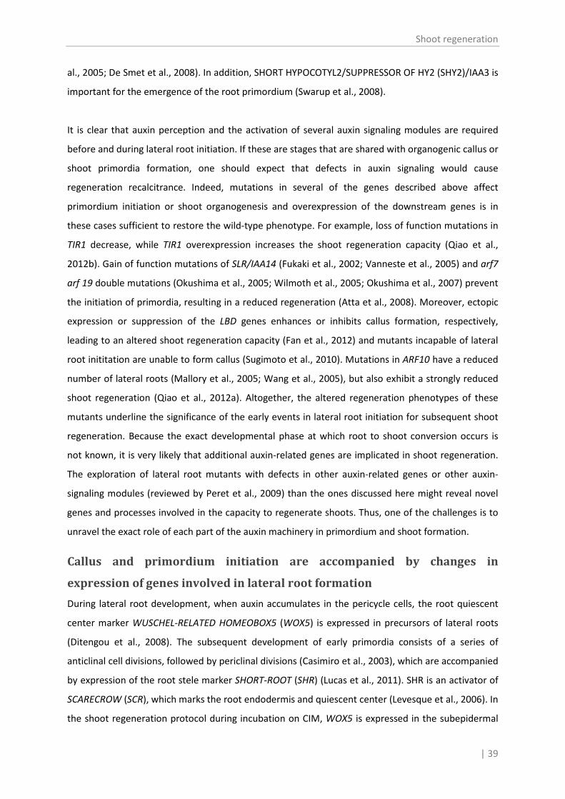
Shoot regeneration
| 39
al., 2005; De Smet et al., 2008). In addition, SHORT HYPOCOTYL2/SUPPRESSOR OF HY2 (SHY2)/IAA3 is
important for the emergence of the root primordium (Swarup et al., 2008).
It is clear that auxin perception and the activation of several auxin signaling modules are required
before and during lateral root initiation. If these are stages that are shared with organogenic callus or
shoot primordia formation, one should expect that defects in auxin signaling would cause
regeneration recalcitrance. Indeed, mutations in several of the genes described above affect
primordium initiation or shoot organogenesis and overexpression of the downstream genes is in
these cases sufficient to restore the wild-type phenotype. For example, loss of function mutations in
TIR1 decrease, while TIR1 overexpression increases the shoot regeneration capacity (Qiao et al.,
2012b). Gain of function mutations of SLR/IAA14 (Fukaki et al., 2002; Vanneste et al., 2005) and arf7
arf 19 double mutations (Okushima et al., 2005; Wilmoth et al., 2005; Okushima et al., 2007) prevent
the initiation of primordia, resulting in a reduced regeneration (Atta et al., 2008). Moreover, ectopic
expression or suppression of the LBD genes enhances or inhibits callus formation, respectively,
leading to an altered shoot regeneration capacity (Fan et al., 2012) and mutants incapable of lateral
root inititation are unable to form callus (Sugimoto et al., 2010). Mutations in ARF10 have a reduced
number of lateral roots (Mallory et al., 2005; Wang et al., 2005), but also exhibit a strongly reduced
shoot regeneration (Qiao et al., 2012a). Altogether, the altered regeneration phenotypes of these
mutants underline the significance of the early events in lateral root initiation for subsequent shoot
regeneration. Because the exact developmental phase at which root to shoot conversion occurs is
not known, it is very likely that additional auxin-related genes are implicated in shoot regeneration.
The exploration of lateral root mutants with defects in other auxin-related genes or other auxin-
signaling modules (reviewed by Peret et al., 2009) than the ones discussed here might reveal novel
genes and processes involved in the capacity to regenerate shoots. Thus, one of the challenges is to
unravel the exact role of each part of the auxin machinery in primordium and shoot formation.
Callus and primordium initiation are accompanied by changes in
expression of genes involved in lateral root formation
During lateral root development, when auxin accumulates in the pericycle cells, the root quiescent
center marker WUSCHEL-RELATED HOMEOBOX5 (WOX5) is expressed in precursors of lateral roots
(Ditengou et al., 2008). The subsequent development of early primordia consists of a series of
anticlinal cell divisions, followed by periclinal divisions (Casimiro et al., 2003), which are accompanied
by expression of the root stele marker SHORT-ROOT (SHR) (Lucas et al., 2011). SHR is an activator of
SCARECROW (SCR), which marks the root endodermis and quiescent center (Levesque et al., 2006). In
the shoot regeneration protocol during incubation on CIM, WOX5 is expressed in the subepidermal

Chapter 2
40 |
layer of the callus and SHR an SCR are expressed in the callus as well (Sugimoto et al., 2010).
Moreover, the PLETHORA1 (PLT1) gene, expressed downstream of WOX5 (Ding and Friml, 2010), is
activated in premature shoot primordia (Atta et al., 2008).
Both in precursors of shoots and roots, PIN genes are expressed, which requires PLT1, SHR and SCR
(Xu et al., 2006), a polarized PIN accumulation occurs and local auxin maxima are generated
(Benkova et al., 2003; Gordon et al., 2007; Atta et al., 2008). Nevertheless, in the callus initiated on
CIM, the PINs are less polarized and the auxin is less concentrated (Atta et al., 2008).
Besides these genes involved in lateral root formation, the root apical meristem (RAM) specific
marker genes RCH1 (Casamitjana-Martinez et al., 2003), QC25 (Sabatini et al., 2003) and GL2 (Lin and
Schiefelbein, 2001) as well were detected prior to shoot development (Atta et al., 2008; Sugimoto et
al., 2010). Finally, the cytokinin response gene, ARABIDOPSIS RESPONSE REGULATOR5 (ARR5) and the
cytokinin biosynthesis gene, ISOPENTENYLTRANSFERASE5 (IPT5), show similar local expression
patterns in root and shoot primordia (Atta et al., 2008).
With the availability of several genome-wide transcript datasets from callus formation (Che et al.,
2006; Sugimoto et al., 2010; He et al., 2012; Xu et al., 2012) or from specific root cells or tissues
involved in lateral root initiation (Himanen et al., 2004; Vanneste et al., 2005; Brady et al., 2007; De
Smet et al., 2008), it became possible to do comparative analyses in order to obtain new insights in
these processes. For instance, Sugimoto et al. (2010) noticed that about a third of the genes
upregulated during callus induction, were actually genes upregulated in the tip zone of the root
(Brady et al., 2007). Xu et al. (2012) profiled the transcriptome of shoot precursory callus during the
first 4 days of incubation on CIM at different time points and with short intervals. When we compare
their data to those of De Smet et al. (2008), 847 of the 1109 genes upregulated in pericycle cells
undergoing lateral root initiation were also upregulated (> 2-fold) in callus. Furthermore, if their
results are compared with those of Vanneste et al. (2005) who identified 913 lateral root initiation
genes, we found 643 genes that were significantly upregulated during callus formation. However, it is
not clear to what extent the callus upregulated genes are important for callus formation and
organogenesis competence. To address this question, these genes were plotted in a bar chart
according to their upregulation on the different time points. Figure 2.2 shows that the total number
of upregulated genes increased over time, but as the cumuluative number of upregulated genes
rapidly increased, there is an enrichment of new upregulated genes at each time point. Hence, many
genes upregulated during callus formation, showed a transient expression profile and were
upregulated during a short period. Therefore, they are presumably not important for callus or
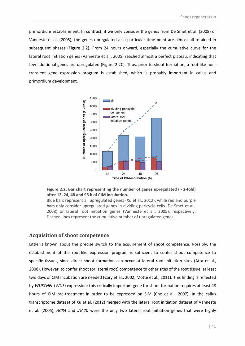
Shoot regeneration
| 41
primordium establishment. In contrast, if we only consider the genes from De Smet et al. (2008) or
Vanneste et al. (2005), the genes upregulated at a particular time point are almost all retained in
subsequent phases (Figure 2.2). From 24 hours onward, especially the cumulative curve for the
lateral root initiation genes (Vanneste et al., 2005) reached almost a perfect plateau, indicating that
few additional genes are upregulated (Figure 2.2C). Thus, prior to shoot formation, a root-like non-
transient gene expression program is established, which is probably important in callus and
primordium development.
Figure 2.2: Bar chart representing the number of genes upregulated (> 2-fold) after 12, 24, 48 and 96 h of CIM incubation. Blue bars represent all upregulated genes (Xu et al., 2012), while red and purple bars only consider upregulated genes in dividing pericycle cells (De Smet et al., 2008) or lateral root initiation genes (Vanneste et al., 2005), respectively. Dashed lines represent the cumulative number of upregulated genes.
Acquisition of shoot competence
Little is known about the precise switch to the acquirement of shoot competence. Possibly, the
establishment of the root-like expression program is sufficient to confer shoot competence to
specific tissues, since direct shoot formation can occur at lateral root initiation sites (Atta et al.,
2008). However, to confer shoot (or lateral root) competence to other sites of the root tissue, at least
two days of CIM incubation are needed (Cary et al., 2002; Motte et al., 2011). This finding is reflected
by WUSCHEL (WUS) expression: this critically important gene for shoot formation requires at least 48
hours of CIM pre-treatment in order to be expressed on SIM (Che et al., 2007). In the callus
transcriptome dataset of Xu et al. (2012) merged with the lateral root initiation dataset of Vanneste
et al. (2005), ACR4 and IAA20 were the only two lateral root initiation genes that were highly

Chapter 2
42 |
upregulated (> 10-fold) after 48 hours of CIM incubation, but not yet (< 2-fold) after 24 hours of CIM
incubation and thus might indicate the acquisition of shoot competence. Although the exact roles of
these genes during shoot regeneration have not been studied in detail, they are both involved in
shoot-related processes: ACR4 is, besides its role in primordium initiation (De Smet et al., 2008), also
required for proper shoot morphogenesis and is mainly expressed in the apical regions during
embryogenesis (Tanaka et al., 2002), while IAA20 is highly upregulated in response to overexpression
of ENHANCER OF SHOOT REGENERATION2 (ESR2), which confers cytokinin-independent shoot
regeneration to root explants (Ikeda et al., 2006).
Another possible marker for the acquisition of shoot competence is CUC2 because its transcript
locally accumulates during callus induction prior to shoot formation. Moreover, CUC2 expressing
sites have the capacity to develop into shoots or roots, depending on the hormones used in the
medium following CIM (Motte et al., 2011). In tomato, REGENERATION1 (RG1), a gene identified by
the characterization of a highly regenerative natural variant (Koornneef et al., 1993), and PROCERA
(PRO)(Bassel et al., 2008), are proposed to be key genes in the acquirement of shoot competence
(Lombardi-Crestana et al., 2012). Finally, the cytokinin receptor ARABIDOPSIS HISTIDINE KINASE4
(AHK4) might also be an important factor, since during CIM incubation localized AHK4 expression
marks sites of cytokinin-induced WUS transcription during the subsequent incubation on shoot
induction medium (SIM) (Gordon et al., 2009). Possibly, the other two cytokinin receptors AHK2 and
AHK3 might have similar expression profiles and functions, because mutants in these receptors have
a decreased regeneration capacity (Nishimura et al., 2004).
Organ determination
CIM as used in the shoot regeneration protocols, is actually a root induction medium, but because of
the high levels of auxin in the medium, the roots are hardly capable to elongate (Rahman et al.,
2007). When 2,4-D, which induces callus that resembles roots (Sugimoto et al., 2010), is replaced by
NAA, shoot regeneration still occurs. Nevertheless, a prolonged incubation period on NAA causes the
development of outgrowths that are morphologically identical to lateral roots (Atta et al., 2008).
Under these conditions, organ identity is determined and root to shoot conversion is no longer
possible (Christianson and Warnick, 1983). Consequently, the optimization of the incubation time on
CIM is a crucial step in the establishment of regeneration protocols (Christianson and Warnick, 1983;
Valvekens et al., 1988; Che et al., 2007). For example, the Arabidopsis accession C24 has an optimal
CIM incubation time of 4 days, after a treatment of 7 days the regeneration efficiency significantly
decreases, and after 14 days only degenerative callus is obtained (Valvekens et al., 1988). In contrast,

Shoot regeneration
| 43
the accession Landsberg erecta (Ler) still regenerates efficiently after 14 days of CIM incubation
(Gordon et al., 2007).
As far as we know, no genetic or morphological markers have been described that indicate the
irreversible commitment of a primordium to develop into a true root. Indeed, several so-called root
identity markers, such as WOX5, SCR, SHR, PLT1, RCH1, QC25, GL2 or J0121 are also expressed in
premature shoot primordia and cannot be used to define the root identity (Atta et al., 2008;
Sugimoto et al., 2010). Because a lateral root initiation-like process is at the basis of shoot
regeneration, it is likely that a genuine root identity determining factor only occurs in the later stages
of lateral root development. Although BDL/IAA12-MP/ARF5 and SHY2/IAA3 are both implicated in
root emergence, it is unlikely that BDL/IAA12-MP/ARF5 is associated with primordium fate
determination, because it is also involved in the control of the expression of ARABIDOPSIS RESPONSE
REGULATOR (ARR)15 (Zhao et al., 2010) and ESR1 (Cole et al., 2009) during shoot development.
SHY2/IAA3 suppresses shoot meristem formation and the expression of the CUC genes (Koyama et
al., 2010), which are essential for shoot regeneration (Aida et al., 1999; Daimon et al., 2003; Hibara et
al., 2003; Vroemen et al., 2003; Hibara et al., 2006) and causes cell differentiation through the
regulation of PIN expression (Dello Ioio et al., 2008). Moreover, while shy2 loss of function mutants
still develop root primordia but no lateral roots, overexpression of SHY2 reduces the number of
primordia (Swarup et al., 2008; Goh et al., 2012b). On the other hand, SHY2 expression is also
observed in leaves and cotyledons (Koyama et al., 2010) and is strongly induced by auxin in different
tissues (Weijers et al., 2005). So if SHY2/IAA3 determines root identity, it is only locally during lateral
root initiation, which should be evidenced by reporter analysis.
Cytokinin-mediated root to shoot conversion
Importance of cytokinin uptake
After the auxin-mediated formation of primordia that are in fact premature root meristems (Atta et
al., 2008; Sugimoto et al., 2010), high cytokinin levels are important for the subsequent development
into shoots (Gordon et al., 2009). The uptake of cytokinins is very fast and relatively complete. For
example cZ or tZ added to liquid medium is taken up for more than 80% within 15 minutes by
tobacco BY-2 cells (Gajdošová et al., 2011). On the other hand, reduced cytokinin uptake has been
reported as a probable cause of recalcitrance (Cortizo et al., 2009). Because the kinetics of cytokinin
uptake are multiphasic and its transport is almost complete abolished by adding a protonophore, it is
assumed that uptake is mainly mediated by multiple proton-coupled cytokinin transport systems
(Cedzich et al., 2008). PURINE PERMEASEs (PUP), such as PUP1 and 2, transport cytokinin bases and,

Chapter 2
44 |
to a minor extent, also cytokinin ribosides (Gillissen et al., 2000; Bürkle et al., 2003), but the kinetic
properties indicate that PUP-mediated transport is inefficient (Frébort et al., 2011). EQUILIBRATIVE
NUCLEOSIDE TRANSPORTER (ENT) proteins, such as ENT3 and ENT8 in Arabidopsis and ENT2 in rice,
transport nucleosides (Mohlmann et al., 2001; Li et al., 2003; Wormit et al., 2004; Hirose et al., 2005;
Sun et al., 2005). Because Arabidopsis ent3 or ent8 mutants show a severely reduced uptake of the
ribosides (Sun et al., 2005), it seems that specific transporters largely determine riboside transport
rather than uptake by simple diffusion. On the other hand, if PUPs in Arabidopsis seedlings are
disabled by adding a protonophore, there is still minimal cytokinin uptake (Cedzich et al., 2008). The
effect of pup or ent mutations on shoot regeneration has not been reported, but it would be highly
informative to determine the importance of cytokinin transport in this developmental process.
Cytokinin biosynthesis and metabolism
Cytokinin biosynthesis and metabolism affect the endogenous cytokinin level (for excellent reviews,
see Sakakibara, 2006; Hirose et al., 2008; Frébort et al., 2011; Spíchal, 2012). In general, metabolic
reactions aim at the maintenance of a homeostatic cytokinin equilibrium mainly by controlling
biosynthesis and activation if the cytokinin level is too low, or by degradation and inactivation if the
cytokinin level exceeds the appropriate homeostasis level.
For cytokinin biosynthesis, the rate-limiting step is the addition of an isoprenoid chain to the adenine
moiety of ADP or ATP by adenosine phosphate-isopentenyltransferases (IPTs) (Kakimoto, 2001; Takei
et al., 2001). Overexpression of IPTs eliminates the requirement of cytokinin in the medium during
regeneration and results in spontaneous shoot formation on callus (Kunkel et al., 1999; Kakimoto,
2001; Sun et al., 2003). As such, inducible IPT expression has been used for marker-free
transformation of plants (Zuo et al., 2002). Loss of function ipt mutants show a decreased
regeneration capacity (Cheng et al., 2013) and have reduced shoot meristem sizes (Miyawaki et al.,
2006). The latter can partially be rescued by cytokinin application, implying that in addition to the
cytokinin level, the spatial cytokinin distribution is an important developmental factor (Zhao, 2008).
The cytochrome P450 monooxygenases CYP735A1 and CYP735A2 are specifically involved in trans-
zeatin synthesis from 2-iP nucleotides (Takei et al., 2004). Although the knowledge about their role in
homeostasis is still limited, they may have a positive effect on shoot formation. Indeed, the
phenotype of the semi-dominant Arabidopsis mutant uni-1D, that forms SHOOT MERISTEMLESS
(STM)-marked meristem on leaves (Igari et al., 2008), was suggested to be caused by induction of
CYP735A2 (Uchida et al., 2011). Besides biosynthesis, the cytokinin level can be increased by the
conversion of inactive cytokinin nucleotides to the active free bases by LONELY GUY (LOG) enzymes
(Kurakawa et al., 2007). Loss of function mutants have similar phenotypes as ipt mutants (Kuroha et

Shoot regeneration
| 45
al., 2009), including reduced (Tokunaga et al., 2012) or prematurely terminated (Kurakawa et al.,
2007) shoot meristems. Overexpressing LOG genes results in cytokinin response phenotypes (Kuroha
et al., 2009), but the effect on shoot regeneration has not been explored.
One of the downregulating mechanisms for cytokinin homeostasis is the irreversible degradation by
CYTOKININ OXIDASE/DEHYDROGENASE (CKX) enzymes. CKX overexpression lines lack a functional
shoot meristem (Werner et al., 2003) and their explants have a reduced regeneration capacity (Yang
et al., 2003). Interestingly, the CKX content, which is dependent on conditions and genotype, has
been suggested to be an important cause of regeneration recalcitrance (Auer et al., 1992; Auer et al.,
1999; Sriskandarajah et al., 2006). If so, this type of recalcitrance could be nihilated by inhibitors of
CKX enzymes. The use of CKX inhibitors has indeed proven to be an effective approach to stimulate
shoot regeneration (Chapter 5). Since CKX genes are expressed in organ primordia (Werner et al.,
2003), CKX inhibitors might locally affect the cytokinin content in regions were shoots can be
induced. Such a local mode of action might be more effective for shoot regeneration than the
classical use of traditional cytokinins (see Chapter 5). Inactivation by glucosylation is another
mechanism to reduce the cytokinin content. In Arabidopsis, the glucosyltransferases UGT76C1 and
UGT76C2 are able to irreversibly glucosylate cytokinins at the N7 and N9 Hou et al., 2004 positions ( ).
UGT76C2 was demonstrated to be involved in cytokinin homeostasis, but loss- and gain-of-function
mutants did not cause any obvious phenotypic changes (Wang et al., 2011a). O-glucosylation of
zeatin is reversible and because these conjugated forms are resistant to CKX degradation (Galuszka
et al., 2007), they are considered as alternative storage forms (Mok and Mok, 2001) that have a role
in rapidly maintaining cytokinin homeostasis (Kakimoto, 2001; Takei et al., 2001). The O-glycosyl
derivatives can be cleaved by specific β-glucosidases (Brzobohaty et al., 1993), but the corresponding
genes have only been identified in Zea mays and Brassica napus (Brzobohaty et al., 1993; Falk and
Rask, 1995). Although overexpression of ZmGLU genes results in a higher regeneration capacity
(Klemš et al., 2011), it is not known whether this effect is a consequence of higher cytokinin levels or
results from affecting the metabolic regulation itself. BGLU19 from Arabidopsis has been proposed to
encode a β-glucosidase as well (Xu et al., 2004). Nevertheless, proteins closely related to BGLU19
appear to have different substrates (Zhao et al., 2012), casting doubt on the ability of BGLU19 to
cleave O-glucosylated cytokinins. Moreover, no biochemical evidence supports the occurrence of this
metabolic step in Arabidopsis and hence, O-glucosylation might be an irreversible inactivation
mechanism in this plant.

Chapter 2
46 |
Cytokinin signaling and responses
In Arabidopsis, three histidine kinases, AHK2, AHK3 and AHK4/WOODENLEG (WOL)/CYTOKININ
RESPONSE1 (CRE1), perceive cytokinins and mediate downstream signaling (Inoue et al., 2001; Suzuki
et al., 2001; Ueguchi et al., 2001; Yamada et al., 2001). Loss-of-function mutations result in a reduced
regeneration capacity and the lack of a functional AHK4 even leads to a complete recalcitrance, even
with elevated cytokinin concentrations (Inoue et al., 2001; Ueguchi et al., 2001; Higuchi et al., 2004;
Nishimura et al., 2004). AHK4 is predominantly expressed in the vascular bundle and the pericycle of
the root (Mähönen et al., 2000; Higuchi et al., 2004) and it accumulates in preluding shoot sites
during CIM incubation, increasing the cytokinin sensitivity at these sites (Gordon et al., 2009).
Whereas the cytokinin receptors are composed of a receptor and a kinase domain, the histidine
kinase CYTOKININ INDEPENDENT KINASE (CKI1), originally hypothesized to be a receptor as well
(Kakimoto, 1996), only shares the kinase function. Overexpression of CKI1 increases cytokinin
signaling, resulting in cytokinin-independent shoot regeneration (Hwang and Sheen, 2001).
When the kinase activity of AHK2, 3 or 4 is triggered by cytokinin binding into the receptor active site,
it results in the phosphorylation of an ARABIDOPSIS HISTIDINE PHOSPHOTRANSFER PROTEIN (AHP),
which serves as a shuttle to transfer the phosphoryl group to ARRs and is important for the
movement of CYTOKININ RESPONSE FACTORs (CRFs) into the nucleus. There, type B ARRs and CRFs
activate transcription of cytokinin-controlled genes and type A ARRs, which act in a negative
feedback loop of cytokinin signaling(Imamura et al., 1999; Hwang and Sheen, 2001; Rashotte et al.,
2006; Cutcliffe et al., 2011; Hwang et al., 2012). Overexpression or elimination of AHP1-5 induces
only moderate cytokinin-like effects due to functional redundancy (Hutchison et al., 2006). AHP6,
which is unable to receive a phosphoryl group, is suggested to interact with the phosphorelay
machinery and hence inhibits cytokinin signaling. Elimination of this gene results in protoxylem
differentiation, which is also induced by cytokinin addition (Mähönen et al., 2006). Importantly,
because these effects have particularly been observed during root development (Mähönen et al.,
2006; Bishopp et al., 2011), it is possible that AHP6 influences shoot regeneration as well. The
stimulation or inhibition of the cytokinin signaling downstream of the AHPs dramatically affects
shoot regeneration. For example, loss-of-function mutations in type B ARRs, which activate the
cytokinin response, or in CRFs, reduce the regeneration capacity (Rashotte et al., 2006; Ishida et al.,
2008). In contrast, overexpression of the type B ARR2 and ARR11 results in cytokinin-independent
shoot regeneration (Hwang and Sheen, 2001) and in spontaneous outgrowth of adventitious shoots
on the junction of cotyledons and of leaf and petioles (Imamura et al., 2003), respectively. Ectopic
expression of these Arabidopsis transcription factors increases regeneration in other plants, such as

Shoot regeneration
| 47
tobacco (Rashid and Kyo, 2010). Interestingly, expression profiling of a group of recalcitrant
Arabidopsis lines within an inbred population, revealed that the expression level of the type B ARR18
was much lower than in the highly regenerative lines (Lall et al., 2004), implying that the level of type
B ARR expression might be at the basis of regeneration recalcitrance.
Because the primary response type A ARRs mediate a negative feedback on cytokinin signaling
(Hwang and Sheen, 2001), overexpression of for instance ARR7 or ARR15 reduces cytokinin signaling
and decreases regeneration capacity (Kiba et al., 2003; Buechel et al., 2010). Silencing of ARR7 and
ARR15 on the other hand increases the meristem size (Zhao et al., 2010). Because SIM contains a
high cytokinin level, A-type ARR genes are highly expressed in root explants incubated on this
medium (Che et al., 2002; Che et al., 2006). Upregulation of ARR15 on SIM seemed to be dependent
of CIM pre-incubation. Since the time required for the formation of green foci and that for the
expression of ARR15 is comparable in Col-0, ARR15 has been proposed as a marker for this
developmental step during shoot regeneration (Che et al., 2007). Nevertheless, an arr15 mutant does
not exhibit a regeneration defect and root explants from a septuple arr mutant form shoots on SIM
with a low cytokinin content without CIM pre-incubation (Buechel et al., 2010). Moreover, ARRs are
suppressed by WUS, which controls meristem function (Leibfried et al., 2005).
Finally, the type C ARRs, ARR22 and ARR24, also block cytokinin signaling. They differ from the true
type A ARRs because they are not induced by cytokinins and they are more related to histidine
kinases (Kiba et al., 2004; Gupta and Rashotte, 2012). Type C ARRs act as phosphatases that receive
phosphoryl groups and reduce cytokinin signaling. They are assumed to be very important for the
local regulation of cytokinin signaling, because even a slight mis expression severely interferes with
cytokinin homeostasis (Horak et al., 2008). For example, ARR22 overexpression results in complete
recalcitrance (Kiba et al., 2004).
Shoot initiation and morphogenesis
Auxin-cytokinin crosstalk
Although shoots arise during SIM incubation, and are induced by cytokinins, the crosstalk with auxin
is also crucially important. For instance, cytokinin regulates the expression of the PIN auxin efflux
carrier genes in a concentration dependent manner (Růžička et al., 2009). During SIM incubation, PIN
genes are upregulated (Benkova et al., 2003; Atta et al., 2008) specifically on AHK4 expressing sites,
which were induced by auxin (Gordon et al., 2009). In addition, cytokinins also induce the auxin
biosynthetic YUCCA (YUC) genes (Jones et al., 2010), contributing to the establishment of a polar

Chapter 2
48 |
auxin gradient. Initially, PIN1 carriers in epidermal cells are directed toward the apical tip, but during
meristem morphogenesis, the PIN1 expression sites are shifted towards incipient organ primordium
sites, creating auxin maxima essential for new organ formation (Heisler et al., 2005; Gordon et al.,
2007). Auxin, in turn, controls the cytokinin distribution by negative regulation of IPTs, which involves
at least the auxin response factors ARF3 and MP/ARF5 and the A-type ARRs ARR7 and ARR15 (Zhao
et al., 2010; Cheng et al., 2013). Auxin also suppresses STM expression, that promotes cytokinin
biosynthesis in the shoot meristem (Heisler et al., 2005; Yanai et al., 2005). PIN1 expression is
functionally required for efficient shoot regeneration, as shoot formation is severely reduced in pin1
mutants (Gordon et al., 2007), but correct PIN1 localization is essential as well: shoot regeneration
can be completely blocked by application of auxin transport inhibitors which affect the cytokinin
localization (Cheng et al., 2013). Also PIN2 and PIN7 are involved in auxin transport in the shoot, but
they are apparently less important than PIN1 since mutations in these genes do not significantly
influence shoot development (Müller et al., 1998; Friml et al., 2003). The auxin influx carriers AUX1
and LAX1-3, are less crucial for polar auxin transport and the establishment of auxin maxima, but
they are necessary to supply sufficient auxin to the epidermal layer (Vernoux et al., 2010).
It is not exactly known how PIN1 polarization is shifted during shoot development, but PINOID (PID),
which is expressed in the shoot, is probably involved in this process (Christensen et al., 2000;
Benjamins et al., 2001). Consequently, just like PIN1, PID is important for shoot development and
regeneration capacity (Furutani et al., 2004; Matsuo and Banno, 2012) and its expression is
upregulated especially in presumptive shoot sites during SIM incubation (Matsuo and Banno, 2012).
ESR1/DÖRNROSCHEN (DRN) and ESR2/DRN-LIKE (DRNL) are two partially redundant transcription
factors that interact during shoot development with PIN1 and PID, respectively (Chandler et al.,
2011a), and further regulate the shoot-related auxin-transport. ESR1 was first discovered in a cDNA
overexpressor screen for cytokinin-independent shoot regeneration, but overexpression also greatly
increased shoot regeneration efficiency in the presence of cytokinin. ESR1 is rapidly induced during
SIM incubation and its expression is dependent on CIM pre-treatment (Banno et al., 2001).
Overexpression of ESR2, selected based on its similarity with ESR1, causes a similar regeneration
phenotype, but it is expressed later during SIM incubation (Ikeda et al., 2006; Matsuo et al., 2009).
Based on this temporal difference in expression, it has been suggested that ESR1, interacting with
PIN1, is mainly important during early root meristem into shoot meristem conversion, while ESR2,
interacting with PID, functions during subsequent shoot development (Matsuo and Banno, 2012).
Moreover, ESR2 precedes the establishment of auxin maxima in the incipient primordia during shoot
development (Chandler et al., 2011b), which supports its role during late shoot formation. The

Shoot regeneration
| 49
importance of the ESR genes in shoot regeneration is corroborated by the reduced shoot
regeneration of mutants lacking the functional genes, where esr1 esr2 double mutant show a
stronger reduction compared to the single mutants (Matsuo et al., 2011).
Shoot meristem pathways
Overexpression of ESR1 induces CUC2 expression (Matsuo et al., 2009), while ESR2 is shown to
activate CUC1 (Ikeda et al., 2006). This finding supports the different temporal function of the ESR
genes: CUC2 is expressed during CIM and is further upregulated during SIM incubation, while CUC1 is
expressed in presumptive shoot sites later during SIM incubation (Cary et al., 2002; Che et al., 2006).
The expression of CUC3, partially redundant to the other CUC genes, is not changed during CIM or
SIM incubation (Che et al., 2006). Expression of the CUC genes results in STM upregulation (Aida et
al., 1999; Hibara et al., 2003), which is essential in shoot meristem initiation and required for
maintenance of undifferentiated cells in the meristem (Barton and Poethig, 1993; Endrizzi et al.,
1996; Long et al., 1996). During shoot regeneration, STM is initially expressed in a ring of cells
surrounding CUC2 expressing promeristems and, subsequently, after the relocalization of PIN1 during
shoot meristem morphogenesis, it is expressed throughout the entire meristem (Gordon et al.,
2007). STM, in turn, is required to restrict the expression of the CUC genes to the meristem
boundaries in the developing shoot (Aida et al., 1999; Takada et al., 2001; Cary et al., 2002; Vroemen
et al., 2003; Gordon et al., 2007; Spinelli et al., 2011). Loss-of-function mutations in STM completely
block adventitious shoot formation, while mutations in the CUC genes reduce shoot regeneration
(Aida et al., 1997; Daimon et al., 2003). Although overexpression of CUC1 or CUC2 increases the
regeneration capacity in a wild-type background, it is not sufficient to undo the regeneration defect
of stm mutants (Daimon et al., 2003). Overexpression of STM causes multiple ectopic shoot
meristems (Brand et al., 2002), which remain undifferentiated and do not divide nor develop into
shoots (Gallois et al., 2002).
CUC1 activates, dependent upon CUC2, the transcription factors LIGHT-DEPENDENT SHORT
HYPOCOTYLS3 (LSH3)/ORGAN BOUNDARY1 (OBO1) and LSH4/OBO4 (Takeda et al., 2011). During the
early phases in shoot regeneration, LSH4 seems to have a similar, but slightly delayed, expression
pattern as CUC1 (Cary et al., 2002) and during further shoot development, LSH3 and LSH4 expression
resembles that of the CUC genes (Cary et al., 2002; Cho and Zambryski, 2011; Takeda et al., 2011).
Their exact role in shoot formation or if they interact with STM is not known, but LSH3 and LSH4 are
important for meristem maintenance and organ differentiation in the boundary region of the shoots
(Cho and Zambryski, 2011; Takeda et al., 2011). Moreover, LSH3 or LSH4 overexpression induces

Chapter 2
50 |
WUS expressing meristem-like tissues, shoot-like primordia expressing WUS and STM, and shoots on
flowers (Takeda et al., 2011).
In parallel to the the CUC-STM reorganization and during the shoot meristem morphogenesis, WUS
expression, initially expressed in cells surrounding the CUC2-marked shoot progenitors, gets localized
within the center of the shoot meristem (Gordon et al., 2007). This expression is induced by
cytokinins in regions where the AKH4 cytokinin receptor is sufficiently expressed and a strong
cytokinin response is detected (Gordon et al., 2009; Cheng et al., 2013). WUS defines the organizing
center (Mayer et al., 1998) and directly represses the transcription of the type A ARRs (Leibfried et
al., 2005), which is important for proper meristem formation (Buechel et al., 2010). WUS is required
for meristem maintenance (Laux et al., 1996) and confers stem cell identity to the overlying neighbor
cells (Mayer et al., 1998) by inducing CLAVATA3 (CLV3) in these cells. CLV3 in turn, represses WUS
expression, and thus controls the meristem size (Schoof et al., 2000). CLV3 is processed by
proteolysis into a small secreted peptide ligand (Brand et al., 2000; Lenhard and Laux, 2003; Ni and
Clark, 2006) which binds to different leucine-rich receptors or receptor complexes with kinase
domains, such as CLV1 and RECEPTOR-LIKE PROTEIN KINASE2 (RPK2) homomers, and CLV1-BARELY
ANY MERISTEM1 (BAM1), CLV1-BAM2 and CLV2-CORYNE (CRN)/SUPPRESSOR OF LLP1 2 (SOL2)
heteromers (Clark et al., 1997; Kayes and Clark, 1998; DeYoung and Clark, 2008; Müller et al., 2008;
Guo et al., 2010; Kinoshita et al., 2010). Subsequent signaling leading to WUS suppression occurs
through post-transcriptional inhibition of the phosphatases POLTERGEIST (POL) and POL-LIKE1 (PLL1)
(Song et al., 2006; Gagne and Clark, 2010).
The WUS-CLV mechanism is important for proper organization of the shoot meristem and for
regeneration. Loss-of-function wus mutants have terminated shoot apices with numerous leaf
primordia and secondary shoot meristems (Laux et al., 1996) and are almost completely regeneration
recalcitrant (Gordon et al., 2007). Overexpression of WUS causes improved shoot regeneration
(Gallois et al., 2004). Mutants in the genes involved in the WUS-CLV feedback loop, exhibit WUS
overexpressing or wus-like phenotypes (Clark et al., 1993; Kayes and Clark, 1998; Fletcher et al.,
1999; DeYoung et al., 2006; Song et al., 2006; Müller et al., 2008; Kinoshita et al., 2010) and will
probably also have similar regeneration phenotypes.
Interestingly, Gallois et al. (2002) demonstrated that the combined overexpression of WUS and STM
causes ectopic shoot formation, while overexpression of only one of the genes does not. Hence, WUS
and STM together are required and sufficient for de novo shoot formation. Therefore, the moment
where WUS and STM expression is initiated just after the PIN1 shift, might mark organ determination
Shoot regeneration
| 51
and assignment of shoot identity. Indeed, at least for STM the timing of expression more or less
coincides with the timing that explants can be transferred from SIM to hormone free medium,
without affecting the regeneration capacity (Zhao et al., 2002). In addition, the few shoots that
regenerate on root explants of a dysfunctional wus mutant, have an autonomous development once
STM expression is activated (Gordon et al., 2007). Hence, it seems that STM, at least when expressed
within the meristem, assigns shoot determination.
Concluding remarks
By giving a chronological overview of the processes that are necessary for the establishment of de
novo shoot formation during a two-step regeneration protocol (Valvekens et al., 1988), we pinpoint
possible hinge points that may be at the basis of regeneration recalcitrance. Although we mainly
discussed data from the model organism Arabidopsis thaliana, we believe that the conditions and
different phases that are necessary to achieve shoot regeneration in other plants are largely similar.
In support of such similarities, the historical research on Convolvulus arvensis of Beijerick (1887)
already demonstrated the requirement of the pericycle, where buds arise at sites opposite the
protoxylem poles, and Bonnett and Torrey (1966) described the common stages between lateral root
and adventitious shoot initiation. However, research in non-model plants can benefit from the
findings in the Arabidopsis research (for a recent review, see Neelakandan and Wang, 2011). For
instance, although cacti are generally easy to propagate by shoot regeneration from leaf-derived
calli, recalcitrant cultivars appeared to have excessive CKX activity (Sriskandarajah et al., 2006).
Consequently, a possible approach to overcome recalcitrance in these cultivars might be the use of
CKX inhibitors. Another example is the use of IPT genes as high-efficiency markers for transformation
(Kunkel et al., 1999).
For this review we chose to focus on the most important events that occur during incubation on
auxin-rich CIM and cytokinin-rich SIM, but obviously multiple other factors affect shoot regeneration.
Hormones, such as ethylene (Chatfield and Raizada, 2008) and gibberellins (Jasinski et al., 2005), and
regulatory proteins, such as class III type HD-ZIP transcription factors (Catterou et al., 2002; Green et
al., 2005; Duclercq et al., 2011a; Gardiner et al., 2011) or cyclin-dependent kinases (Andersen et al.,
2008; Meng et al., 2010) are but a few. Moreover, new key factors implicated in shoot regeneration
are continuously identified although their exact role in the process remains to be explored. Examples
are the transcription factor RAP2.6L (Che et al., 2006), the fasciclin-like arabinogalactan-protein FLA1
(Johnson et al., 2011) and the oxygen-binding hemoglobins (Wang et al., 2011b).

Chapter 2
52 |
Finally, with the identification of the transcription factor WOUND INDUCED DEDIFFERENTIATION1
(WIND1) it has become clear that auxin- and cytokinin-independent processes can lead to shoot
regeneration as well. Indeed, WIND1 overexpression omits the requirement of auxin to form callus
and allows the formation of shoots without CIM pre-incubation. Interestingly, the WIND1-induced
callus does not show a lateral root-like gene expression program and is not disturbed in the slr
mutant (Iwase et al., 2011). Hence, WIND1-dependent shoot regeneration follows a different
pathway, opening novel opportunities to solve regeneration recalcitrance by using this different type
of callus.
With the recent advances in live-imaging, expression profiling, gene discovery and cell biology tools,
further expansion of our knowledge of shoot regeneration is guaranteed. Additionally, it would be
highly beneficial for tissue culture practices if the progress made in basic research on shoot
regeneration would be implemented more intensively during the establishment of working protocols
for recalcitrant plants. Indeed, by studying different cultivars and explants one might get insight into
the basis of recalcitrance of the plant of interest and step away from trial and error towards
approaches inspired by more educated guesses to solve tissue culture problems.

| 53


Scopes and objectives
| 55
3. 3
Chapter 3 Scopes and objectives


Scopes and objectives
| 57
Adventitious shoot formation is, besides being a subject of study in developmental biology, applied in
in vitro micropropagation and plant biotechnology. One major concern in tissue culture is
regeneration recalcitrance, i.e the incompetency of some plant species and cultivars to regenerate in
vitro. Possible factors causing recalcitrance are in general poorly understood and efforts to
circumvent this problem comprise mainly trial and error procedures. De novo shoot formation is a
complex process involving different developmental events. Recent advances in molecular analysis of
shoot meristems and shoot organogenesis have substantially widened our knowledge about the
underlying processes occurring during shoot regeneration. Yet, many questions remain unanswered.
The objective of this study was to improve the methodologies for shoot regeneration in plants and to
obtain information that would help us to get insights into the factors controlling regeneration
capacity and the possible cause(s) of recalcitrance. For these purposes we have adopted the
Arabidopsis root explants system as it is widely applied in physiological and molecular studies of
plant regeneration. In addition, forward and reverse genetics using this system have already lead to
the identification of several genetic factors required for regeneration. These studies have provided
an important framework for further research and directed the focus of this work. To address our
research questions we opted to use the two-step regeneration protocol for the implementation of
two recently developed technologies to study diverse biological processes.
A first strategy was to apply chemical genetics to the shoot regeneration process and to screen a
chemical library to identify molecules that induce shoot regeneration. Besides revealing new
molecules that could possibly break through recalcitrance, this technique would be instrumental in
finding novel proteins or networks that are involved in the regeneration process. Chemical genetics is
indeed commonly used as an alternative for traditional genetics by the use of bio-active small
molecules that, analogous to a specific mutation, perturb a process of interest (McCourt and
Desveaux, 2010; Toth and van der Hoorn, 2010). As chemical genetics involves the screening of large
libraries, high-throughput manipulations are required. Therefore, such compound screens are in
general performed on seeds or young seedlings, evaluating marker gene expression or altered
phenotypes of young plants (Hicks and Raikhel, 2009). As we wanted to do this chemical screen on a
much more complex biological process, a prerequisite was to find suitable conditions so that efficient
shoot formation in multiwell plates would be possible. By using a reporter gene for shoot
regeneration, evaluation of the screen could be facilitated. Multiple genes marking shoots or shoot
regeneration are reported (Sijacic and Liu, 2010), however, their usefulness and reliability in
predicting shoot formation is in general poorly documented. Thus, a first goal was to select a reliable
and informative shoot marker by live-imaging of fluorescent reporters. We were particularly

Chapter 3
58 |
interested in compounds that induce regeneration, therefore, we substituted the shoot-inducing
cytokinin with a library of small molecules. Any hit might provide new insights about the
regeneration process. Therefore, a main objective was also to determine the mode of action of the
revealed compounds and identify the target proteins.
In a second and very different approach, we wanted to identify novel players involved in shoot
regeneration and took advantage of the wide natural variation of Arabidopsis accessions (Mitchell-
Olds and Schmitt, 2006). As shoot regeneration is a quantitative trait involving many factors (Lall et
al., 2004; Meng et al., 2010), we expected that regeneration capacity would vary among accessions,
ranging from strongly recalcitrant to highly regenerative. By classifying different accessions according
to their regeneration capacity and regeneration-related traits, we aimed to identify strains that could
be useful for further research regarding regeneration efficiency and recalcitrance. The analysis of
natural variation in wild populations has a great potential in identifying novel genes involved in
regeneration as current Arabidopsis research mainly focusses on a few accessions, and therefore, the
available mutant collections are mainly constructed using this limited number of laboratory strains,
which harbor only a small portion of the Arabidopsis natural variation (Alonso-Blanco et al., 2009).
For exploring this variation regarding shoot regeneration, we opted to use two mapping methods: on
the one hand we wanted to run a QTL analysis with an inbred population of less explored Arabidopsis
accessions, and on the other hand to we wanted to set out the accession classification for a genome-
wide association (GWA) mapping. With these approaches, we aimed at the identification of new
genes involved in regeneration which might help to improve regeneration capacity.
Altogether, with the combination of different experimental approaches to gain novel insights in the
process of shoot regeneration, this work had the purpose to provide useful information for the
improvement of tissue culture techniques.

| 59


Shoot markers
| 61
4. 4
Chapter 4 Testing markers for shoot regeneration from
Arabidopsis root explants Adapted from: Motte, H., I. Verstraeten, S. Werbrouck and D. Geelen (2011). "CUC2 as an early marker for
regeneration competence in Arabidopsis root explants." Journal of Plant Physiology 168: 1598-1601.


Shoot markers
| 63
Although many shoot markers have already been described, their reliability
and practical usefulness are generally poorly documented. To monitor and
facilitate the optimization of in vitro regeneration of Arabidopsis thaliana,
CUP SHAPED COTELYDON1 and 2 (CUC1 and 2), SHOOT MERISTEMLESS
(STM), and LIGHT-DEPENDENT SHORT HYPOCOTYLS4 (LSH4) were tested as
markers for shoot induction. The root explants of the different lines were
first incubated on an auxin-rich callus induction medium (CIM) and then
transferred to a cytokinin-rich shoot induction medium (SIM). Although the
expression of all four markers occurred prior to visible shoot formation, only
CUC2 was expressed during incubation of the root explants on CIM. Shoot
formation was invariably preceded by the expression of all four genes, but
the expression of CUC1 and STM proved to have a very weak predictive
value for subsequent shoot formation. In contrast, CUC2 appeared to be a
predictive marker for the acquisition of root explant competence for root
and shoot organogenesis, whereas LSH4 proved to be a reliable marker for
shoot formation.


Shoot markers
| 65
Introduction
Shoot regeneration from root explants has been studied in detail in Arabidopsis thaliana (see
Chapter 2). The regeneration involves a two-step process with a pre-incubation on auxin-rich callus
induction medium (CIM) to induce acquirement of shoot competence, followed by transfer to
cytokinin-rich shoot induction medium (SIM) to stimulate the formation of shoots (Valvekens et al.,
1988; Cary et al., 2002). Using molecular reporters, it has been shown that during incubation of root
explants on CIM, auxin maxima in pericycle cells give rise to founder cells which further divide and
will develop into shoot primordia when placed on SIM (Atta et al., 2008). During both incubation
phases, specific gene expression programs are established (Che et al., 2006). Shoot formation is a
multistep process involving cell dedifferentiation, acquisition of competence, primordium initiation,
and the formation and outgrowth of a shoot meristem (Che et al., 2007). Different marker genes
have been identified of which the expression coincides with the emergence of primordia or
subsequent steps in shoot development (Gordon et al., 2007).
In an approach to facilitate the monitoring of shoot organogenesis, we tested four of such marker
genes for their association with the shoot induction program. One of the early genes expressed
during the induction of shoots from Arabidopsis root explants is CUP-SHAPED COTELYDON2 (CUC2),
which is already expressed during the pre-incubation on CIM (Gordon et al., 2007). Although CUC1 is
functionally redundant to CUC2 during shoot formation (Takada et al., 2001), it is expressed in callus
after 4 days of SIM incubation and thereafter, its expression is intensified in the presumptive sites of
shoot formation (Cary et al., 2002). CUC1 and CUC2 are involved in the establishment of organ
boundaries during shoot development and both are important for SHOOT MERISTEMLESS (STM)
expression (Aida et al., 1999; Daimon et al., 2003; Laufs et al., 2004). STM is expressed in the shoot
meristem cells (Aida et al., 1999; Gordon et al., 2007) and is required for the maintenance of a
population of undifferentiated cells within the shoot meristem (Endrizzi et al., 1996). During shoot
regeneration, STM is suggested to be specifically expressed in cells determined for shoot
organogenesis (Zhao et al., 2002). Besides its activation by the CUC transcription factors (Daimon et
al., 2003), STM expression is dependent on cytokinins (Rupp et al., 1999). More recently, CUC1 was
found to stimulate the expression of LIGHT-DEPENDENT SHORT HYPOCOTYLS4 (LSH4) and its
homolog LSH3 in shoot organ boundary cells (Takeda et al., 2011). Both LSH genes were marked in
enhancer trap lines (Haseloff, 1999; Cary et al., 2002; Cho and Zambryski, 2011) and because of their
specific expression pattern have also been named ORGAN BOUNDARY1 (OBO1 /LSH3) and OBO4 (Cho
and Zambryski, 2011).

Chapter 4
66 |
Although STM, CUC1, CUC2, and LSH4 have previously been described as shoot markers (Cary et al.,
2002; Heisler et al., 2005; Gordon et al., 2007), their usefulness as predictors of shoot formation
during the regeneration procedure had not been assessed. Here, the expression of these four genes
was evaluated at different time points during CIM and SIM incubation. We provide evidence that
CUC1 nor STM are reliable markers for shoot regeneration. CUC2 and LSH4 on the other hand proved
to be valuable markers for the acquisition of organogenesis competence and for shoot regeneration,
respectively, and both genes may be implemented as markers to study regeneration of root explants.
Results
To determine the usefulness of CUC1, CUC2, STM and LSH4 as shoot regeneration markers, their
expression was followed during shoot regeneration.
CUC1 and STM expression do not reliably predict shoot formation
CUC1 expression was monitored using the M0167 GAL4-GFP enhancer trap line (in C24 background).
No CUC1 expression occurred during incubation on CIM, but shoot formation was always preceded
by its expression on SIM (data not shown). Nevertheless, CUC1 expression was at times transient and
then, no development occurred at these sites (Figure 4.1). Moreover, CUC1 expression did not always
coincide with subsequent development into shoots. Indeed, on SIM with 12.5 µM or 25 µM 2-iP, up
to 30% of the CUC1 expression sites did not result in shoot formation.
Figure 4.1: CUC1 expression. Cells exhibiting transient CUC1 expression (arrowhead) do not develop into shoots. Bars = 1 mm.
When the expression of STM with the pSTM::GFP-ER marker (in Col-0 background) was followed,
similar observations were made. No STM expression was detected on CIM, but shoot formation was
consistently preceded by its expression on SIM (Figure 4.2A). Importantly, just like for CUC1, STM
expression was often not followed by shoot formation. Even under optimal conditions, around 30%
of the STM expressing sites did not progress to form shoots (Figure 4.2B).

Shoot markers
| 67
Figure 4.2: STM expression during shoot
regeneration from root explants.
(A) An example of the pSTM::GFP-ER marker
preceding shoot formation during SIM incubation.
Bars = 100 µm. (B) The percentage of STM expressing
sites that develop into shoots on SIM with different
2-iP concentrations. The fluorescent sites and shoots
were counted every two days for 30 days. Error bars
indicate standard errors (N ≥ 10).
Expression of CUC2 during regeneration
Expression of CUC2 was followed with the pCUC2::3XVENUS-N7-marker (in Ler background). Roots of
young Arabidopsis seedlings exhibited CUC2 expression in the root tip which was conserved after
excision and transfer to CIM (Figure 4.3A). After two days on CIM, the CUC2 fluorescence pattern
changed toward regularly-spaced, bright patches in the region close behind the root tip. These
patches occurred in the periphery of the vascular bundle, presumably in the pericycle (Figure 4.3B)
and may correspond to precursors of newly emerging shoots (Atta et al., 2008). After 4 days
incubation on CIM, the fluorescent CUC2 patches became wider and brighter, forming a regular
pattern along the entire root explant (Figure 4.3C). Upon transfer to SIM, the distinct CUC2
expression sites on the root explants were retained and they radially expanded to form primordia
(Figure 4.3D). Most, but not all of the CUC2 expressing primordia progressed further to form shoots
(indicated by red chlorophyll fluorescence in Figure 4.3E), implying that increased CUC2 expression is
not sufficient to allow shoot primordium formation. Nevertheless, the capacity to form shoots was
always associated with a patchy induction of CUC2 expression.
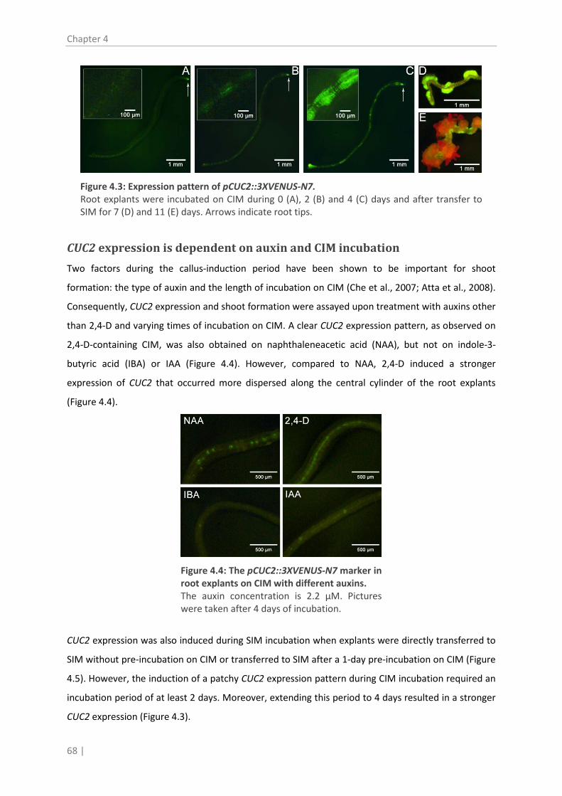
Chapter 4
68 |
Figure 4.3: Expression pattern of pCUC2::3XVENUS-N7. Root explants were incubated on CIM during 0 (A), 2 (B) and 4 (C) days and after transfer to SIM for 7 (D) and 11 (E) days. Arrows indicate root tips.
CUC2 expression is dependent on auxin and CIM incubation
Two factors during the callus-induction period have been shown to be important for shoot
formation: the type of auxin and the length of incubation on CIM (Che et al., 2007; Atta et al., 2008).
Consequently, CUC2 expression and shoot formation were assayed upon treatment with auxins other
than 2,4-D and varying times of incubation on CIM. A clear CUC2 expression pattern, as observed on
2,4-D-containing CIM, was also obtained on naphthaleneacetic acid (NAA), but not on indole-3-
butyric acid (IBA) or IAA (Figure 4.4). However, compared to NAA, 2,4-D induced a stronger
expression of CUC2 that occurred more dispersed along the central cylinder of the root explants
(Figure 4.4).
Figure 4.4: The pCUC2::3XVENUS-N7 marker in root explants on CIM with different auxins. The auxin concentration is 2.2 µM. Pictures were taken after 4 days of incubation.
CUC2 expression was also induced during SIM incubation when explants were directly transferred to
SIM without pre-incubation on CIM or transferred to SIM after a 1-day pre-incubation on CIM (Figure
4.5). However, the induction of a patchy CUC2 expression pattern during CIM incubation required an
incubation period of at least 2 days. Moreover, extending this period to 4 days resulted in a stronger
CUC2 expression (Figure 4.3).

Shoot markers
| 69
Figure 4.5: The pCUC2::3XVENUS-N7 marker in a root explant directly transferred to SIM. The picture was taken after 9 days of incubation.
Interestingly, the conditions resulting in the highest CUC2 expression also lead to the highest number
of formed shoots. Indeed, pre-treatment on 2,4-D-containing CIM was more efficient than CIM with
NAA (Figure 4.6C). Moreover, direct incubation on SIM or a 1-day pre-incubation on CIM did not
result in shoot formation (Figure 4.6A). In contrast, the number of shoots increased steadily after 2,
3, and 4 days pre-incubation on CIM (Figure 4.6A).
Altogether these data suggest that the acquisition of competence for shoot formation is correlated
with CUC2 expression and that the level of CUC2 expression determines the efficiency of shoot
formation.
Shoot-forming capacity is positively correlated with root-forming capacity
Because CUC2 was expressed in the root tip, we further investigated the local expression of CUC2
during lateral root initiation. When explants are transferred from CIM to hormone-free basal
medium (BM), the CUC2-expressing patches differentiated into root primordia that developed into
roots. Therefore, the impact of the auxin type in CIM and the CIM incubation period on the
competence of root explants to form roots was investigated by determining the number of emerging
lateral roots under different growth conditions. Interestingly, similar results were obtained as for
shoot induction. When either 2,4-D or NAA were present in the CIM, but not IBA or IAA, root
formation was strongly stimulated (Figure 4.6C). The same correlation was found varying the CIM
incubation time. In the absence of CIM or after 1-day pre-incubation on CIM, no lateral roots were
induced, except for a few explants that produced a single lateral root near the root tip. A more
pronounced induction of lateral roots occurred after 2 days of CIM incubation, and the number of
roots increased with extending CIM incubation times (Figure 4.6B). Direct transfer of root explants to
BM, or transfer after one day CIM, induced CUC2 expression on BM, but none of the fluorescent foci
developed into roots. We therefore conclude that the acquisition of competence to form shoots
seems very similar to the acquisition of competence to form lateral roots, in particular with respect
to the timing of the cellular processes and the auxin used.

Chapter 4
70 |
Figure 4.6: Influence of the pre-incubation conditions on the number of roots formed on BM or shoots formed on SIM. (A) Average number of shoots per explant on SIM after 0-4 days of pre-incubation on CIM. (B) Average number of lateral roots per explant on BM after 0-4 days of pre-incubation on CIM. (C) Influence of the auxin type in CIM on the number of roots formed on BM (X-axis) or shoots formed on SIM (Y-axes). The number of shoots and roots were counted after 17 days. Different letters indicate significantly different means.
LSH4 as a predictor of shoot formation
LSH4 expression was monitored with the M0223 GAL4-GFP enhancer trap line (in C24 background).
No LSH4 expression occurred during incubation on CIM, but when shoots formed this was always
preceded by its expression on SIM (data not shown). Although LSH4 expression was also not strictly
correlated with subsequent shoot development on 12.5 µM or 25 µM 2-iP, just like for CUC2, only a
minority of the LSH4 expressing sites (5%) did not predict shoot formation. Moreover, LSH4
expressing cells consistently exhibited some level of development, albeit that sometimes it arrested
at a spherical primordium-like structure (Figure 4.7A). LSH4 expression generally became apparent
between 7 and 11 days of incubation on SIM and on average 4 to 7 days later the first shoots could
be observed (Figure 4.7B).

Shoot markers
| 71
Figure 4.7: LSH4 expression during shoot regeneration. (A) LSH4 expressing cells with a developmental arrest at a spherical primordium-like structure. (B) LSH4 expressing sites that predict shoot formation. Bars = 1 mm.
Discussion
To facilitate visualization of early steps in the regeneration process, we tested four different shoot
markers. Because of the very high discrepancy between CUC1 and STM expression and subsequent
shoot formation, these genes cannot be considered as valuable predictive markers for shoot
regeneration.
The expression of CUC2 on the other hand proved to be informative for the acquisition of
organogenesis competence. CUC2 was highly expressed at the root tip (Figure 4.1; Gordon et al.,
2007), where most of the cell divisions are taking place and the auxin concentration reaches a
maximum (Sabatini et al., 1999; Sauer et al., 2006). Shoots are only induced on root explants after a
pre-treatment with auxin and when local CUC2 expression occurs before transfer to SIM. The cut site
nor the root tip generate shoot primordia under the conditions analyzed, indicating that other
factors inherent to the regenerating tissue play a role. Our analyses revealed that upon incubation of
excised root segments on CIM, CUC2 is expressed in patches dispersed along the root explant, that,
upon incubation on SIM, will form shoot primordia. The NAA-induced CUC2 expressing sites are
smaller and less frequent compared to those induced by 2,4-D, presumably because 2,4-D is, unlike
the natural auxins, not transported out of the cell by the efflux carriers, allowing it to build-up inside
cells (Delbarre et al., 1996). These high intracellular auxin concentrations might provoke the
activation of CUC2 throughout the pericycle, which then gives rise to the formation of very broad and
numerous primordia. Although NAA is transported out of the cell, it is metabolized more slowly than
IAA and IBA (Beyer and Morgan, 1970). Thus, compared to IAA and IBA, NAA can attain a much
higher activity that is sufficient to stimulate CUC2 expression and to support cell dedifferentiation
and division required for primordium induction. As auxins are known to trigger organogenesis
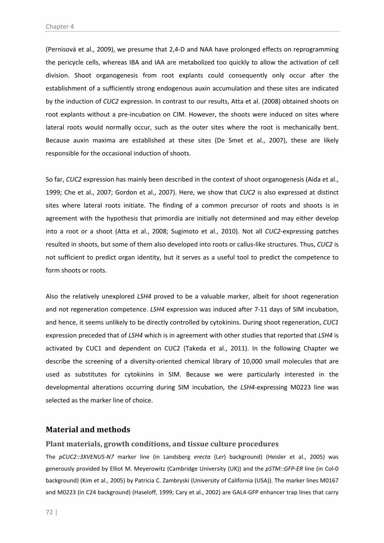
Chapter 4
72 |
(Pernisová et al., 2009), we presume that 2,4-D and NAA have prolonged effects on reprogramming
the pericycle cells, whereas IBA and IAA are metabolized too quickly to allow the activation of cell
division. Shoot organogenesis from root explants could consequently only occur after the
establishment of a sufficiently strong endogenous auxin accumulation and these sites are indicated
by the induction of CUC2 expression. In contrast to our results, Atta et al. (2008) obtained shoots on
root explants without a pre-incubation on CIM. However, the shoots were induced on sites where
lateral roots would normally occur, such as the outer sites where the root is mechanically bent.
Because auxin maxima are established at these sites (De Smet et al., 2007), these are likely
responsible for the occasional induction of shoots.
So far, CUC2 expression has mainly been described in the context of shoot organogenesis (Aida et al.,
1999; Che et al., 2007; Gordon et al., 2007). Here, we show that CUC2 is also expressed at distinct
sites where lateral roots initiate. The finding of a common precursor of roots and shoots is in
agreement with the hypothesis that primordia are initially not determined and may either develop
into a root or a shoot (Atta et al., 2008; Sugimoto et al., 2010). Not all CUC2-expressing patches
resulted in shoots, but some of them also developed into roots or callus-like structures. Thus, CUC2 is
not sufficient to predict organ identity, but it serves as a useful tool to predict the competence to
form shoots or roots.
Also the relatively unexplored LSH4 proved to be a valuable marker, albeit for shoot regeneration
and not regeneration competence. LSH4 expression was induced after 7-11 days of SIM incubation,
and hence, it seems unlikely to be directly controlled by cytokinins. During shoot regeneration, CUC1
expression preceded that of LSH4 which is in agreement with other studies that reported that LSH4 is
activated by CUC1 and dependent on CUC2 (Takeda et al., 2011). In the following Chapter we
describe the screening of a diversity-oriented chemical library of 10,000 small molecules that are
used as substitutes for cytokinins in SIM. Because we were particularly interested in the
developmental alterations occurring during SIM incubation, the LSH4-expressing M0223 line was
selected as the marker line of choice.
Material and methods
Plant materials, growth conditions, and tissue culture procedures
The pCUC2::3XVENUS-N7 marker line (in Landsberg erecta (Ler) background) (Heisler et al., 2005) was
generously provided by Elliot M. Meyerowitz (Cambridge University (UK)) and the pSTM::GFP-ER line (in Col-0
background) (Kim et al., 2005) by Patricia C. Zambryski (University of California (USA)). The marker lines M0167
and M0223 (in C24 background) (Haseloff, 1999; Cary et al., 2002) are GAL4-GFP enhancer trap lines that carry

Shoot markers
| 73
a GFP fusion to CUC1 and LSH4, respectively and they were obtained from the Nottingham Arabidopsis Stock
Centre (NASC). Seeds were fumigated for 4 hours in a desiccator jar with chlorine gas by adding 5 mL
concentrated HCl to 100 mL 5% (v/v) NaOCl. Sterilized seeds were incubated on basal medium (BM):
Gamborg’s B5 medium (Gamborg et al., 1968) supplemented with 0.05% (w/v) 2-(4-morpholino-)ethane
sulfonic acid (MES) at pH 5.8, 2% (w/v) glucose and 0.7% (w/v) agar. Before germination, a cold treatment for 4
days at 4°C was applied to the seeds. Seedlings were grown for 6 days at 20°C and a 16 h photoperiod with a
light irradiance of 45 µmol m-2 s-1
1988
provided by cool-white fluorescent tungsten tubes. Shoot formation from
root explants was basically as described by Valvekens et al. ( ). Except when stated otherwise, root
segments (7 mm) were cut from 6 days old seedlings and explanted onto callus induction medium (CIM; BM
supplemented with 2.2 µM (0.5 mg/L) 2,4-dichlorophenoxy acetic acid (2,4-D) and 0.2 µM (0.05 mg/L) kinetin
(Kin)) for 4 days. Explants were then transferred to shoot induction medium (SIM; BM supplemented with 25
µM (5 mg/l) 2-isopentenyl adenine (2-iP) and 0.86 µM (0.15 mg/L) 3-indoleacetic acid (IAA)). Hormones were
dissolved in DMSO and supplied to the medium after autoclaving.
Microscopy
Explants were imaged directly on the media using a Leica MZ FLIII stereomicroscope. For fluorescence imaging,
a 425-460 nm excitation filter was used together with a 470 nm dichromatic beam splitter and a GG475 barrier
filter. Images were captured using ProgRess® CaptureBasic.
Statistical analysis
Statistical analysis was performed using S-Plus 8.0 software. Normality of data was checked using the
Kolmogrov-Smirnov test. Normally distributed data were treated using analysis of variance procedures and
means were separated using the simulation-based method (P=0.05). For non-normally distributed data, means
were compared using the nonparametric Wilcoxon two-sample test.
Acknowledgements
The authors thank Elliot M. Meyerowitz for providing the pCUC2::3XVENUS-N7 marker, Patricia C. Zambryski for
the pSTM::GFP-ER line, and Danny Vereecke for critical reading of the manuscript. This work was supported by
grants from the Research Fund of the University College Ghent to HM, the agency for Innovation by Science
and Technology to IV, the Hercules Foundation (project AUGE/013) and the framework of the
“Associatieonderzoeksgroep Primaire Plantaardige Productie”.


Shoot regeneration by Phe-Ade
| 75
5. 5
Chapter 5 Phenyl-adenine, identified in a LIGHT-DEPENDENT SHORT
HYPOCOTYLS4-assisted chemical screen, is a potent compound for shoot regeneration through the inhibition of CKX activity
Manuscript in press (Motte, H., P. Galuszka, L. Spíchal, P. Tarkowski, O. Plíhal, M. Šmehilová, P.
Jaworek, D. Vereecke, S. Werbrouck and D. Geelen (2013). Plant Physiology: doi:
10.1104/pp.1112.210716)


Shoot regeneration by Phe-Ade
| 77
In vitro shoot regeneration is implemented in basic plant research and
commercial plant production, but for some plant species still difficult to
achieve by means of the currently available cytokinins and auxins. To
identify novel compounds that promote shoot regeneration, we screened a
library of 10,000 small molecules. The bioassay consisted of a two-step
regeneration protocol adjusted and optimized for high throughput
manipulations of root explants of Arabidopsis (Arabidopsis thaliana)
carrying the shoot regeneration marker LIGHT-DEPENDENT SHORT
HYPOCOTYLS4 (LSH4). The screen revealed a single compound, the
cytokinin-like phenyl-adenine (Phe-Ade), as a potent inducer of adventitious
shoots. Although Phe-Ade triggered diverse cytokinin-dependent
phenotypical responses, it did not inhibit shoot growth and was not
cytotoxic at high concentrations. Transcript profiling of cytokinin-related
genes revealed that Phe-Ade treatment established a typical cytokinin
response. Moreover, Phe-Ade activated the cytokinin receptors AHK3 and
AHK4 in a bacterial receptor assay, albeit at relatively high concentrations,
illustrating that it exerts genuine but weak cytokinin activity. In addition, we
demonstrated that Phe-Ade is a strong competitive inhibitor of CYTOKININ
OXIDASE/DEHYDROGENASE (CKX) enzymes, leading to an accumulation of
endogenous cytokinins. Collectively, Phe-Ade exhibits a dual mode of action
which results in a strong shoot inducting activity.


Shoot regeneration by Phe-Ade
| 79
Introduction
The capacity to regenerate shoots, broadly applied for tissue culture purposes or biotechnological
breeding methods (Duclercq et al., 2011b), is not an uncommon trait of plants nevertheless many
species remain recalcitrant. Thus, a major challenge for tissue culture practices is the development of
efficient protocols in which plant growth regulators (PGRs) are pivotal. Currently, besides natural
plant hormones, a whole range of synthetic PGRs is being applied in tissue culture. For example,
compared to natural cytokinins, the synthetic cytokinin thidiazuron (TDZ) is more effective as an
inducer of shoot formation in some plants (van Staden et al., 2008) and because it can substitute
both for cytokinin and auxin, TDZ is also widely used for callus induction and somatic embryogenesis
(Murthy et al., 1998). In a continuous search for new hormone-like substances to expand the
collection of suitable compounds and to increase the efficiency of specific protocols, libraries of
structural variants of known PGRs or of structurally unrelated small molecules are being tested
(Kumari and van der Hoorn, 2011). Whereas the first approach mainly results in compounds that
function in a similar but more efficient way as known PGRs, the latter tactic may potentially yield
compounds that exhibit an alternative mode of action. Successful chemical screens have for instance
led to the identification of growth promoting auxin analogs (Savaldi-Goldstein et al., 2008), root
growth promoting cytokinin antagonists (Arata et al., 2010) and the non-auxin-like lateral root
inducer naxillin (De Rybel et al., 2012).
Although shoot regeneration is a complex developmental process, a lot of insights have been gained
from studies on the model plant Arabidopsis thaliana (Meng et al., 2010). In general, two major
phases are distinguished in shoot organogenesis: the acquisition of competence and the
commitment to form shoots (Cary et al., 2002). In the two-step shoot regeneration protocol
described by Valvekens et al. (1988) for Arabidopsis root explants, these two phases are
accomplished through incubation on auxin-rich callus induction medium (CIM) followed by
incubation on cytokinin-rich shoot induction medium (SIM). During the CIM treatment, auxin
maxima, essential for the acquisition of competence, are established in the pericycle cells (Atta et al.,
2008; Pernisová et al., 2009); then, tissues proliferate that resemble premature roots (Atta et al.,
2008; Sugimoto et al., 2010). The auxin treatment also leads to a broad transcriptional modulation
(Che et al., 2006) including a strong and local up-regulation of the ARABIDOPSIS HISTIDINE KINASE4
(AHK4) cytokinin receptor (Gordon et al., 2009). The subsequent incubation on SIM converts the
premature roots into shoots, particularly in those regions where the cytokinin receptor genes are
upregulated (Pernisová et al., 2009). Indeed, in these regions cytokinin signaling is sufficiently high to

Chapter 5
80 |
activate WUSCHEL (WUS) and SHOOT MERISTEMLESS (STM) expression (Gordon et al., 2009), which
are critically important for shoot meristem development (Gallois et al., 2002; Lenhard et al., 2002).
The importance of the cytokinin response and cytokinin-controlled genes in shoot regeneration has
also been demonstrated by genetic approaches. For example, the constitutive expression of cytokinin
signaling genes, such as CYCLIN-DEPENDENT KINASE INHIBITOR1 (CKI1)(Kakimoto, 1996) and
ARABIDOPSIS RESPONSE REGULATOR2 (ARR2)(Hwang and Sheen, 2001), eliminates the requirement
for cytokinins in SIM. Interestingly, however, overexpression of ENHANCER OF SHOOT
REGENERATION1 (ESR1) and ESR2, which are not directly involved in cytokinin metabolism or
signaling, also confer cytokinin-independent shoot formation from Arabidopsis root explants. Still,
these lines are not insensitive to cytokinins since their regeneration efficiency can be increased by
cytokinin application (Banno et al., 2001; Ikeda et al., 2006). Whereas for all these transgenics the
pre-treatment with the synthetic auxin 2,4-dichlorophenoxy acetic acid (2,4-D) remains a
prerequisite for shoot formation, overexpression of WOUND INDUCED DEDIFFERENTIATION 1
(WIND1) abolishes the need both for 2,4-D in CIM and cytokinins in SIM. Analysis of the regeneration
process in these plants revealed that the meristems originated from another cell type than in wild-
type plants implying that there is a second regeneration pathway (Iwase et al., 2011), opening
perspectives to identify alternative regeneration protocols.
Because shoot regeneration can be achieved by cytokinin application to the cultivation medium, but
also using transgenics approaches, we reasoned that it would be conceivable to obtain shoot
formation with cytokinin-like compounds or downstream effectors different from cytokinins. We
therefore set out to search for novel shoot-inducing compounds by combining a chemical screen
with the regeneration procedure described for Arabidopsis by Valvekens et al. (1988). First, we
optimized the protocol to allow high throughput screening of a chemical library. Of all compounds
tested only one, phenyl-adenine (Phe-Ade), stimulated shoot formation. To assess the specificity of
this compound, we scored its activity in typical cytokinin-related processes. Then, to unravel the
mode of action of Phe-Ade, we profiled the expression of cytokinin-related marker genes in response
to Phe-Ade, analyzed its effect on the regeneration of Arabidopsis cytokinin receptor mutants,
assessed its perception by the cytokinin receptors, and tested its interaction with different
CYTOKININ OXIDASE/DEHYDROGENASE (CKX) enzymes. Based on our results we conclude that Phe-
Ade is a weak cytokinin that strongly inhibits endogenous cytokinin degradation.

Shoot regeneration by Phe-Ade
| 81
Results
A chemical screen reveals Phe-Ade as a potent shoot-inducing molecule
To identify novel compounds that promote shoot regeneration, we first optimized the regeneration
protocol of Valvekens et al. (1988) allowing high throughput manipulations (Supplemental Procedure
S5.1; Supplemental Table S5.1; Supplemental Figure S5.1). In short, 7 mm root explants with a root
apical meristem were incubated on CIM containing 2,4-D and kinetin for 4 days in petri dishes. Then,
the explants were transferred to 96-well plates, two per well, containing solid SIM without 3-
indoleacetic acid (IAA) and with either 10 µM 2-isopentenyladenine (2-iP) or 10 µM of the individual
compounds of a diversity-oriented library of 10,000 small molecules (molecular weight less than 500
g/mol). To facilitate the observation of shoot primordia, we used the accession C24 GAL4-GFP
enhancer trap line M0167 (Haseloff, 1999). This line visualizes the expression of the shoot marker
LIGHT-DEPENDENT SHORT HYPOCOTYLS4 (LSH4)(Cary et al., 2002). LSH4 expression was scored after
12 days on SIM and shoot formation 7 days later.
Figure 5.1: Chemical structure and activity of Phenyl-Adenine (N-phenyl-9H-purin-6-amine; Phe-Ade). (A) Chemical structure. (B) LSH4 expression after 12 d (top panel) and shoot induction after 19 d (lower panel) of SIM incubation containing 10 µM Phe-Ade. Bars = 1 mm.
Of all compounds tested, only one, phenyl-adenine (Phe-Ade; N-phenyl-9H-purin-6-amine) induced
LSH4 expression and shoot formation (Figure 5.1). None of the other compounds activated LSH4
expression or induced shoot primordia or shoots. To evaluate the shoot-inducing capacity of Phe-
Ade, we tested different concentrations within the activity range of 2-iP in the 96-well format.
Because Arabidopsis accession C24 is highly regenerative, counting the number of shoots formed per
explant in the 96-well plate format was difficult. Therefore, the projected area encompassed by the

Chapter 5
82 |
emerging shoots was adopted as a quantitative measure. As shown in Figure 5.2A, at the lower
concentrations, Phe-Ade was a better inducer of shoot formation than 2-iP and at the highest
concentration tested, it performed equally well.
Figure 5.2: Phe-Ade is an efficient inducer of shoot formation on C24 and Col-0 root explants. (A) Regeneration of C24 root explants after 15 days on SIM with Phe-Ade or 2-iP using shoot area as a quantitative measure (see Material and methods). (B) Regeneration rate of Col-0 root explants expressed as % responsiveness, i.e. the average number of root explants forming at least one shoot. Responsive explants were counted 14 days after transfer to SIM. Data represent the average of 3 biological repeats with each 19 root explants. (A,B) Different letters indicate statistical differences evaluated with the Duncan’s multiple range test in conjunction with an analysis of variance. Error bars represent standard errors.
To further assess the potential of Phe-Ade as a shoot inducer, its effect on shoot regeneration on
roots of accession Col-0, which has been reported to have a lower shoot regeneration capacity
(Siemens et al., 1993; Cary et al., 2002), was tested in petri dishes. The regeneration rate was
quantified by counting the number of responsive explants, i.e. explants that produced one or more
shoots, on different concentrations of Phe-Ade and 2-iP. As shown in Figure 5.2B, the effect of both
compounds was optimal at a concentration of 10 µM, confirming the results obtained with the 96-
well plates for accession C24 (Figure 5.2A). Moreover, Phe-Ade had a stronger shoot inducing effect
than 2-iP for the tested concentration range and the difference in the regeneration rate under
optimal and sub- or supra-optimal Phe-Ade concentrations was only moderate (Figure 5.2B),
indicating that Phe-Ade is effective at a much broader concentration range than 2-iP. When the
concentration of Phe-Ade was lowered to 0.1 µM or increased to 500 µM, induction of shoots no
longer occurred (data not shown).
Phe-Ade provokes cytokinin-like biological responses
To assess whether Phe-Ade affected other biological processes besides shoot regeneration, its
activity in different cytokinin-mediated processes was evaluated. Col-0 seeds were germinated on
medium containing 10 µM Phe-Ade, 10 µM 2-iP, or 0.1% dimethyl sulfoxide (DMSO) as a control, and
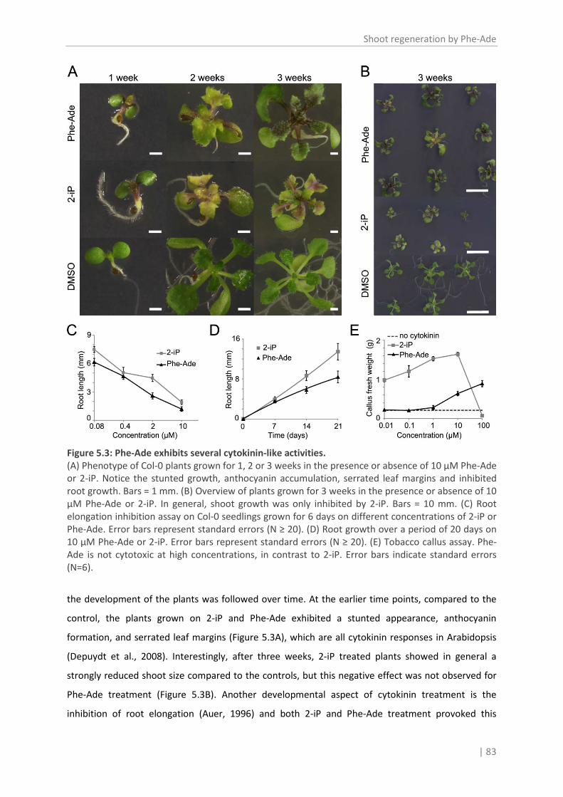
Shoot regeneration by Phe-Ade
| 83
Figure 5.3: Phe-Ade exhibits several cytokinin-like activities. (A) Phenotype of Col-0 plants grown for 1, 2 or 3 weeks in the presence or absence of 10 µM Phe-Ade or 2-iP. Notice the stunted growth, anthocyanin accumulation, serrated leaf margins and inhibited root growth. Bars = 1 mm. (B) Overview of plants grown for 3 weeks in the presence or absence of 10 µM Phe-Ade or 2-iP. In general, shoot growth was only inhibited by 2-iP. Bars = 10 mm. (C) Root elongation inhibition assay on Col-0 seedlings grown for 6 days on different concentrations of 2-iP or Phe-Ade. Error bars represent standard errors (N ≥ 20). (D) Root growth over a period of 20 days on 10 µM Phe-Ade or 2-iP. Error bars represent standard errors (N ≥ 20). (E) Tobacco callus assay. Phe-Ade is not cytotoxic at high concentrations, in contrast to 2-iP. Error bars indicate standard errors (N=6).
the development of the plants was followed over time. At the earlier time points, compared to the
control, the plants grown on 2-iP and Phe-Ade exhibited a stunted appearance, anthocyanin
formation, and serrated leaf margins (Figure 5.3A), which are all cytokinin responses in Arabidopsis
(Depuydt et al., 2008). Interestingly, after three weeks, 2-iP treated plants showed in general a
strongly reduced shoot size compared to the controls, but this negative effect was not observed for
Phe-Ade treatment (Figure 5.3B). Another developmental aspect of cytokinin treatment is the
inhibition of root elongation (Auer, 1996) and both 2-iP and Phe-Ade treatment provoked this

Chapter 5
84 |
response (Figure 5.3A). When the extent of the root growth inhibition was quantified for seedlings
grown on different concentrations of Phe-Ade or 2-iP, it was clear that after 6 days, Phe-Ade showed
a stronger root growth inhibition than 2-iP for all concentrations tested (Figure 5.3C). Moreover,
evaluation of the plants after 21 days of growth on 10 µM of the PGRs showed that Phe-Ade, in
contrast to its effect on shoot growth, clearly inhibited root elongation stronger than 2-iP (Figure
5.3D): the root length after Phe-Ade or 2-iP treatment was 12% or 19% of the DMSO control,
respectively. Finally, the tobacco (Nicotiana tabacum) callus growth bioassay was reassessed with
different concentrations of 2-iP and Phe-Ade. Phe-Ade only moderately induced callus in this assay,
confirming the data by Zatloukal et al. (2008). Interestingly however, at 100 µM, when commonly
used cytokinins were toxic and caused cell death (Zatloukal et al., 2008; Pertry et al., 2009), Phe-Ade
stimulated the division of the tobacco callus cells (Figure 5.3E). Altogether these results illustrate that
Phe-Ade, in contrast to previous reports, has a cytokinin activity that is comparable to that of 2-iP.
Importantly, when compared to 2-iP, negative effects such as the inhibition of shoot growth and the
induction of cell death at higher concentrations are not observed upon Phe-Ade treatment.
Transcript profiling suggests that Phe-Ade triggers a cytokinin-like gene
regulation
To gain insight into the mode of action of Phe-Ade, its effect on the expression of cytokinin-related
genes was examined in roots and leaves. To this end, 14 d old Arabidopsis Col-0 plants were
incubated in liquid medium containing 10 µM Phe-Ade or 2-iP for 15, 30, and 60 min, and 1, 3, and 7
days. Incubation in hormone-free medium served as a negative control. Generally, in shoot tissues,
the expression profiles obtained upon Phe-Ade treatment were comparable to those on 2-iP (Figure
5.4). The primary cytokinin response genes, the type A ARRs ARR5, ARR15 and ARR16, showed a
typical transient change in expression with a very fast and strong upregulation after 15 min of PGR
treatment and the subsequent gradual dampening of induction due to feedback mechanisms (Figure
5.4). The transcript levels of B-type ARRs, ARR2, ARR10 and ARR14, coding for transcription factors
that induce cytokinin response genes, were not altered by Phe-Ade or by 2-iP (data not shown). The
cytokinin biosynthesis genes, ISOPENTENYLTRANSFERASE3 (IPT3) and IPT7, were down-regulated
after longer periods of Phe-Ade and 2-iP treatment, pointing to the establishment of cytokinin
homeostasis mechanisms (Figure 5.4). The expression of other IPTs, IPT1, IPT2 and IPT8, as well as
Figure 5.4: Transcript profiling of cytokinin-related genes in Col-0 shoot tissues at different time points of 10 µM Phe-Ade or 2-iP treatment. Different letters indicate statistical differences between the samples evaluated with the Tukey range test in conjunction with an analysis of variance. Asterisks (*) indicate statistical differences from the hormone-free control, evaluated with a two-tailed Student’s t-test and adjusted with the Benjamini-Hochberg FDR method. Error bars represent standard errors (N=4).

Shoot regeneration by Phe-Ade
| 85

Chapter 5
86 |
the cytokinin activating genes, LONELY GUY6 (LOG6) and LOG8, and the cytochrome P450 gene
CYP735A2, implicated in the biosynthesis of trans-Zeatin (tZ), were not altered by either PGR (data
not shown). The very fast up-regulation by Phe-Ade and 2-iP of the CYTOKININ
OXIDASE/DEHYDROGENASE (CKX) genes, CKX3, CKX4 and CKX5, mediating the irreversible
degradation of cytokinins, further supports the activation of homeostatic mechanisms (Figure 5.4).
CKX2 and CKX6 expression was only activated after longer incubation periods with both PGRs, but
CKX2 up-regulation by Phe-Ade was strongly delayed compared to that by 2-iP (Figure 5.4). The
transcript levels of CKX1 and CKX7 were not altered by either compound (data not shown). Cytokinin
homeostasis is further established through irreversible conjugation into inactive N-glucosides by
UDP-GLUCOSYL TRANSFERASEs (UGTs)(Wang et al., 2011a). Expression of UGT76C2 (Figure 5.4), but
not of UGT76C1 (data not shown), was up-regulated in response to Phe-Ade and 2-iP treatment,
although the induction by Phe-Ade was slightly delayed. The expression of the O-glucosyltransferases
UGT73C5 and UGT85A1 (Figure 5.4), but not of UGT73C1 (data not shown), putatively mediating the
reversible conjugation of cytokinins (Hou et al., 2004), was induced after longer incubation times
with both PGRs. Finally, neither Phe-Ade nor 2-iP treatment affected the expression of the cytokinin
receptor genes AHK2, AHK3 and AHK4 (data not shown). Similar expression trends were observed in
the root tissues for all tested genes (data not shown).
Taken together, the expression profiles obtained upon Phe-Ade treatment imply a typical cytokinin-
mediated gene regulation and an activation of cytokinin homeostasis mechanisms. The lack of
significant differences in the kinetics and in the amplitude of the expression profiles upon Phe-Ade
and 2-iP treatments, shows that Phe-Ade establishes a cytokinin-like gene regulation.
Importance of cytokinin receptors in the Phe-Ade-induced responses
The fast induction of expression of the A-type ARRs (Figure 5.4) suggests that Phe-Ade acts as a
cytokinin. Further evidence for the activation of ARR5 expression by Phe-Ade was obtained by
histochemical staining of 14 d old Col-0 plants harboring an ARR5:GUS reporter fusion, treated for 5
days with 10 µM Phe-Ade or 10 µM 2-iP on solid medium. As shown in Figure 5.5A, both compounds
induced ARR5 expression to the same extent and in the same tissues (especially the shoot apical
meristem and the roots). Next, to assess whether a specific cytokinin receptor was implicated in the
ARR5 induction, similar analyses were done using the double cytokinin receptor mutants, ahk2 ahk3,
ahk2 ahk4, and ahk3 ahk4. As observed by Pertry et al. (2010), the basal ARR5 expression level in
these receptor mutants was elevated compared to wild-type plants (Figure 5.5A). Both Phe-Ade and
2-iP induced ARR5 expression in these mutants to the same extent with a similar tissue specificity.

Shoot regeneration by Phe-Ade
| 87
hjkjkjkjkjkj
Figure 5.5: Interaction between Phe-Ade and the cytokinin receptors. (A) ARR5:GUS expression in 14 d old plants of Col-0 and the cytokinin receptor double mutants ahk2 ahk3, ahk2 ahk4, and ahk3 ahk4 treated for 5 days with 10 µM of Phe-Ade or 2-iP. (B) Root elongation inhibition assay on Col-0 on double ahk mutant seedlings grown for 7 days on 10 µM 2-iP or Phe-Ade. Error bars represent standard errors (N ≥ 10). (C) In silico binding of Phe-Ade in the active site of AHK4. (D) Effect of Phe-Ade, adenine (Ade), and tZ on the specific binding of 2 nM [³H]tZ in a “live-cell hotmone binding assay” employing E. coli cells expressing AHK4. NS, nonspecific binding in the presence of 5000-fold excess of unlabeled tZ. Error bars indicate standard errors (N=3). (E) Activation of the cytokinin receptors AHK3 and AHK4 by Phe-Ade and tZ in the E. coli receptor assay. Error bars indicate standard errors (N=3). (F) Regeneration rate of root explants of Col-0 and the double ahk mutants on 10 µM Phe-Ade or 2-iP. Error bars indicate standard errors. The data are the average of 3 biological repeats with 10 explants each.

Chapter 5
88 |
Compared to treated Col-0 plants, the expression level was much higher in ahk2 ahk4 and ahk3 ahk4
mutants and occurred throughout the plant, while in ahk2 ahk3 mutants, it occurred especially in the
root (Figure 5.5A). These results imply that the Phe-Ade-mediated cytokinin response is
notdependent on a specific cytokinin receptor, albeit that their individual contribution to ARR5
induction is not the same. Similar conclusions could be drawn when the effect of Phe-Ade and 2-iP on
the development of the ahk double mutants was evaluated. Compared to the wild-type controls, the
DMSO-treated cytokinin receptor mutants had longer roots and smaller rosette leaves (Figure 5.5B
and Figure 5.3), which is in agreement with previous reports (Nishimura et al., 2004). Phe-Ade and 2-
iP treatment of in particular ahk3 ahk4, but also of ahk2 ahk4 plants had only moderate effects on
the rosette, the root growth, the bleaching of the shoots, the anthocyanin production and the
serrations of the leave margins. In contrast, treatment of the ahk2 ahk3 mutant with both PGRs did
cause considerable effects on plant development which were comparable to those described for the
wild-type plants (Figure 5.3A, B, and D, Figure 5.5B and Supplemental Figure S5.2). After one week,
anthocyanin accumulation and a strongly reduced root elongation were observed. After two weeks,
2-iP showed a strong inhibition of rosette growth, while rosettes treated by Phe-Ade, although
clearly serrated, were comparable in size as the DMSO controls. This was also in a lesser extent
noticed for the ahk2 ahk4 and ahk3 ahk4 mutants (Supplemental Figure S5.2). Thus, for the evaluated
cytokinin-like responses triggered both by Phe-Ade and 2-iP, a functional AHK4 receptor seems
essential and sufficient.
With the availability of the crystal structure of AHK4 (Hothorn et al., 2011; PDB id: 3t4q), we assessed
AHK4 Phe-Ade binding in silico using Autodock Vina (Trott and Olson, 2010). Although Phe-Ade
docked into AHK4, in contrast to tZ, it lacked the interaction of the polar hydroxyl group (Figure 5.5C)
and therefore, only a weak interaction of -5.6 kcal/mol was predicted. This prediction was confirmed
with a “live-cell hormone binding assay” using AHK4 expressing E. coli cells (Romanov et al., 2005). In
this assay, the potential of different concentrations of Phe-Ade to compete with radiolabeled tZ for
AHK4 binding was evaluated; adenine was used as a negative control. As shown in Figure 5.5D, Phe-
Ade reduced the binding of labeled tZ, but only at relatively high concentrations. The subsequent
activation of the cytokinin receptors was demonstrated using AHK3 and AHK4 expressing E. coli cells
carrying a cytokinin-activated reporter gene cps::lacZ (Suzuki et al., 2001; Yamada et al., 2001;
Spíchal et al., 2004). Although Phe-Ade activated both receptors (Figure 5.5E), the required
concentration was 1000-fold higher than for tZ, which is in agreement with the results of the
competition assay.

Shoot regeneration by Phe-Ade
| 89
The importance of the cytokinin receptors in the shoot-inducing activity of Phe-Ade was tested by
quantifying the shoot regeneration rate of root explants from the double cytokinin receptor mutants
on Phe-Ade and 2-iP. For both compounds, no regeneration occurred for ahk2 ahk4 and ahk3 ahk4
(Figure 5.5F), suggesting that AHK4 is essential for this developmental process. Indeed, treatment of
root explants of the ahk2 ahk3 mutant, in which the AHK4 receptor is functional, with Phe-Ade and
2-iP resulted in the formation of a comparable number of shoots. However, the regeneration rate of
this mutant was up to 8-fold lower compared to that of Col-0 explants (Figure 5.5F), implying that
although AHK4 is essential it is not sufficient for a wild-type regeneration level. Hence, shoot
regeneration both by Phe-Ade and 2-iP depends on cytokinin perception through the all three AHKs.
Phe-Ade is a competitive inhibitor of CKX enzymes
Although Phe-Ade is only a weak activator of the cytokinin receptors, its activity in the shoot
regeneration assay nevertheless is strong. Moreover, the induced expression of the cytokinin
metabolism genes, CKX2 and UGT76C2, in response to Phe-Ade is delayed when compared to 2-iP
(Figure 5.4). Overall, these observations suggested that besides its direct but weak cytokinin activity,
another indirect mode of action could be at the basis of the biological activity of Phe-Ade. Based on
the transcript profiling results (Figure 5.4), a differential regulation of the most important cytokinin
metabolism genes was ruled out as potential mechanism. So we hypothesized that Phe-Ade would
affect cytokinin degradation, and therefore, the ability of Phe-Ade to inhibit the activity of CKX
enzymes was tested. Using the method of Libreros-Minotta and Tipton (1995), the degradation of 2-
iP by recombinant CKX2 in the absence or presence of Phe-Ade was measured. As observed
previously (Galuszka et al., 2007), CKX2 exhibited oxidase activity, but it was much more active as a
dehydrogenase (Figure 5.6A). More importantly however, both in the dehydrogenase (with the
synthetic electron acceptors 2,6-dichlorophenol indophenol (DCPIP) or 2,3-dimethoxy-5-methyl-1,4-
benzoquinone (Q0
Figure 5.6
)) and the oxidase mode (with oxygen as electron acceptor), Phe-Ade inhibited the
degradation of 2-iP ( A). To determine whether the reduced degradation of 2-iP resulted
from a competitive degradation of Phe-Ade as an alternative substrate for CKX2, we omitted 2-iP
from the reaction mixture and monitored the consumption of the electron acceptor DCPIP in the
presence of only Phe-Ade. Benzyladenine (BA), which is reported to be a very weak substrate for CKX
enzymes (Frébortová et al., 2007), was used as a control. As shown in Figure 5.6C, BA was degraded
by CKX2 very slowly, whereas Phe-Ade was not.
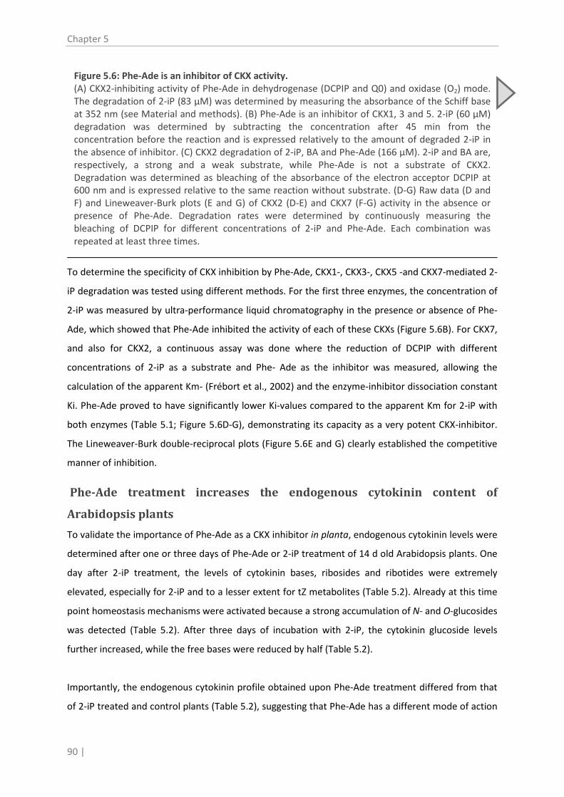
Chapter 5
90 |
To determine the specificity of CKX inhibition by Phe-Ade, CKX1-, CKX3-, CKX5 -and CKX7-mediated 2-
iP degradation was tested using different methods. For the first three enzymes, the concentration of
2-iP was measured by ultra-performance liquid chromatography in the presence or absence of Phe-
Ade, which showed that Phe-Ade inhibited the activity of each of these CKXs (Figure 5.6B). For CKX7,
and also for CKX2, a continuous assay was done where the reduction of DCPIP with different
concentrations of 2-iP as a substrate and Phe- Ade as the inhibitor was measured, allowing the
calculation of the apparent Km- (Frébort et al., 2002) and the enzyme-inhibitor dissociation constant
Ki. Phe-Ade proved to have significantly lower Ki-values compared to the apparent Km for 2-iP with
both enzymes (Table 5.1; Figure 5.6D-G), demonstrating its capacity as a very potent CKX-inhibitor.
The Lineweaver-Burk double-reciprocal plots (Figure 5.6E and G) clearly established the competitive
manner of inhibition.
Phe-Ade treatment increases the endogenous cytokinin content of
Arabidopsis plants
To validate the importance of Phe-Ade as a CKX inhibitor in planta, endogenous cytokinin levels were
determined after one or three days of Phe-Ade or 2-iP treatment of 14 d old Arabidopsis plants. One
day after 2-iP treatment, the levels of cytokinin bases, ribosides and ribotides were extremely
elevated, especially for 2-iP and to a lesser extent for tZ metabolites (Table 5.2). Already at this time
point homeostasis mechanisms were activated because a strong accumulation of N- and O-glucosides
was detected (Table 5.2). After three days of incubation with 2-iP, the cytokinin glucoside levels
further increased, while the free bases were reduced by half (Table 5.2).
Importantly, the endogenous cytokinin profile obtained upon Phe-Ade treatment differed from that
of 2-iP treated and control plants (Table 5.2), suggesting that Phe-Ade has a different mode of action
Figure 5.6: Phe-Ade is an inhibitor of CKX activity. (A) CKX2-inhibiting activity of Phe-Ade in dehydrogenase (DCPIP and Q0) and oxidase (O2) mode. The degradation of 2-iP (83 µM) was determined by measuring the absorbance of the Schiff base at 352 nm (see Material and methods). (B) Phe-Ade is an inhibitor of CKX1, 3 and 5. 2-iP (60 µM) degradation was determined by subtracting the concentration after 45 min from the concentration before the reaction and is expressed relatively to the amount of degraded 2-iP in the absence of inhibitor. (C) CKX2 degradation of 2-iP, BA and Phe-Ade (166 µM). 2-iP and BA are, respectively, a strong and a weak substrate, while Phe-Ade is not a substrate of CKX2. Degradation was determined as bleaching of the absorbance of the electron acceptor DCPIP at 600 nm and is expressed relative to the same reaction without substrate. (D-G) Raw data (D and F) and Lineweaver-Burk plots (E and G) of CKX2 (D-E) and CKX7 (F-G) activity in the absence or presence of Phe-Ade. Degradation rates were determined by continuously measuring the bleaching of DCPIP for different concentrations of 2-iP and Phe-Ade. Each combination was repeated at least three times.

Shoot regeneration by Phe-Ade
| 91
Table 5.1: Kinetic parameters calculated from Figure 5.6D-G. CKX2 CKX7
Vmax 0.62 ± 0.03 (µkat/mg protein) 0.016 ± 0.001 Km 12.9 ± 1.7 (µM) 55.5 ± 5.6 Ki 0.59 ± 0.08 (µM) 10.7 ± 1.6

Chapter 5
92 |
Table 5.2: Endogenous cytokinin content of 14 d old Arabidopsis plants after 1 or 3 days treatment with 10 µM 2-iP or Phe-Ade. Data shown are in pmol/g fresh weight ± standard deviation (N=3). cis-zeatin-O-glucoside (cZOG) was below the detection limit. 2-iP, 2-isopentenyladenine; tZ, trans-zeatin; cZ, cis-zeatin; iPR, 2-isopentenyladenosine; tZR, trans-zeatin riboside; cZR; cis-zeatin riboside; iPRMP, 2-isopentenyladenosine-5’-monophosphate; tZRMP, trans-zeatin riboside-5’-monophosphate; cZRMP, cis-zeatin riboside-5’-monophosphate; iP9G, 2-isopentenyladenine-9-glucoside; tZ9G, trans-zeatin-9-glucoside; cZ9G, cis-zeatin-9-glucoside; tZOG, trans-zeatin-O-glucoside; tZROG, trans-zeatin riboside-O-glucoside; cZROG, cis-zeatin riboside-O-glucoside.
Cytokinin 2-iP Phe-Ade DMSO
type 1 day 3 days 1 day 3 days 1 day 3 days 2-iP 5843.5 ± 1666.4 2553.2 ± 733.5 9.0 ± 0.9 63.1 ± 5.0 1.1 ± 0.2 1.5 ± 0.1
tZ 228.7 ± 9.9 185.3 ± 10.3 0.6 ± 0.0 1.9 ± 0.0 0.6 ± 0.0 0.6 ± 0.0 cZ 7.9 ± 0.2 9.4 ± 0.3 0.2 ± 0.0 0.4 ± 0.0 0.1 ± 0.0 0.1 ± 0.0
iPR 768.5 ± 14.6 851.5 ± 34.3 0.6 ± 0.0 8.6 ± 0.7 0.4 ± 0.1 1.2 ± 0.1 tZR 70.7 ± 3.4 110 ± 4.2 0.2 ± 0.0 0.8 ± 0.0 0.2 ± 0.0 0.3 ± 0.0 cZR 7.0 ± 1.2 27.1 ± 1.2 0.3 ± 0.0 1.5 ± 0.2 0.3 ± 0.0 0.3 ± 0.0
iPRMP 48320.2 ± 7023.5 62529.8 ± 11476.8 33.7 ± 0.3 957.2 ± 369.2 26.9 ± 1.0 108.4 ± 36.1 tZRMP 2638.1 ± 240.5 9800.3 ± 3132.7 6.2 ± 0.6 22.8 ± 4.7 8.1 ± 1.0 17.5 ± 1.9 cZRMP 216.8 ± 36.4 1386.2 ± 496.3 9.4 ± 1.8 43.2 ± 20.7 7.5 ± 1.4 13.3 ± 2.7
iP9G 3033.0 ± 245.0 3660.1 ± 292.6 4.6 ± 0.7 76.4 ± 7.1 2.4 ± 0.3 2.3 ± 0.1 tZ9G 93.8 ± 5.5 264.3 ± 13.6 2.8 ± 0.1 2.6 ± 0.4 4.3 ± 0.2 4.1 ± 0.1 cZ9G 6.8 ± 0.5 46.5 ± 4.3 0.4 ± 0.0 0.5 ± 0.0 0.4 ± 0.0 0.4 ± 0.0 tZOG 59.7 ± 3.5 165.4 ± 51.9 1.8 ± 0.3 2 ± 0.8 2.6 ± 1.2 3.1 ± 0.2
tZROG 121.2 ± 22.6 243.2 ± 8.4 1 ± 0.4 0.9 ± 0.4 0.7 ± 0.2 0.7 ± 0.0 cZROG 16.6 ± 1.9 32.9 ± 4.4 1 ± 0.2 4 ± 0.5 1 ± 0.1 0.7 ± 0.1

Shoot regeneration by Phe-Ade
| 93
than 2-iP and implying that it affects cytokinin homeostasis. One day after Phe-Ade incubation, a
modest increase in 2-iP levels was measured. Later, after 3 days of incubation, a steady accumulation
of the 2-iP base, riboside and ribotide was detected, evidencing that the CKX enzymes were not
functioning optimally. The concentration of 2-isopentenyladenine-9-glucoside (iP9G) increased as
well, indicating that some level of inactivating glucosylation occurred. The effect of Phe-Ade on the
zeatin metabolites remained very low, even after 3 days (Table 5.2). Altogether these results support
an indirect mode of action of Phe-Ade leading to a distinct increase in endogenous cytokinin level
Discussion
Screening of a diversity-oriented chemical library of small molecules as substitutes for cytokinins in
SIM in the two-step regeneration protocol starting from Arabidopsis root explants (Valvekens et al.,
1988) yielded only phenyl-adenine (Phe-Ade) as a shoot-inducing compound, implying that the
cytokinin-mediated step in shoot regeneration is difficult to bypass through exogenous chemicals.
Phe-Ade had been described as a cytokinin before (Miller, 1961), but compared to other cytokinins it
was classified as significantly less active in several typical cytokinin bioassays, such as tobacco callus
growth, anthocyanin production in Amaranthus seedlings, and chlorophyll retention of detached
wheat leaves (Hahn and Bopp, 1968; Zatloukal et al., 2008). Consequently, it has only rarely been
used as a PGR. Why then does Phe-Ade have such a strong activity as a shoot inducer in the
regeneration protocol?
In planta, the effect on shoot and root development, the activation of ARR5 expression, and the fast
and strong differential regulation of a set of cytokinin-related marker genes was comparable for Phe-
Ade and 2-iP treatment. These results implied that Phe-Ade functioned as a cytokinin. Indeed, as
predicted by in silico docking tests, Phe-Ade could bind to the cytokinin receptor AHK4 (and AHK3) in
in vitro assays and triggered downstream signaling, albeit only at relatively high concentrations. This
moderate activation of the receptors seemed to be in contrast with the strong shoot
regeneration
Nishimura et al., 2004
activity of Phe-Ade. Nonetheless, AHK4 was essential for Phe-Ade- and 2-iP-mediated
regeneration, as reported for tZ-induced shoot formation ( ), but the
functionality of all three cytokinin receptors appeared to be imperative for shoot induction.
Importantly, several observations suggested that Phe-Ade might have an additional and indirect
mode of action. Shoot growth inhibition typically associated with high cytokinin levels was not
observed upon Phe-Ade treatment and at high concentrations Phe-Ade did not induce cell death in a
tobacco callus bioassay. Moreover, the activation of several cytokinin metabolism genes by Phe-Ade
was delayed compared to 2-iP treatment.

Chapter 5
94 |
Since Phe-Ade could possibly target cytokinin homeostasis, cytokinin degradation by different CKX
enzymes in the presence or absence of Phe-Ade was assessed using several experimental
approaches. The in vitro data clearly demonstrated that Phe-Ade was a strong and competitive
inhibitor of these enzymes. Actually, Phe-Ade had been used as a scaffold structure for the
generation of a group of analogs that exhibit CKX-inhibiting activity (Zatloukal et al., 2008),
supporting our results. Profiling endogenous cytokinin levels validated the relevance of the inhibition
of the CKX enzymes in plants upon Phe-Ade treatment. Thus, the activation of the AHKs and the
downstream cytokinin signaling during Phe-Ade treatment is caused both by Phe-Ade itself and by
the accumulating endogenous cytokinins.
This specific dual mode of action of Phe-Ade has several implications in planta which might explain
the strong activity in the shoot regeneration process. (i) 2-iP treatment resulted in a very fast and
extremely high accumulation of the cytokinin content in plant tissues, which is likely cytotoxic and
causes stress and reduced responsiveness. Although Phe-Ade treatment triggered an increase in the
endogenous cytokinin levels as well, the accumulation resulting from CKX inhibition occurred
gradually and was much less pronounced, likely preventing cytotoxic and deleterious effects, and
allowing a continuous developmental response. (ii) Since Phe-Ade is a competitive inhibitor of the
CKX enzymes, its effect on homeostasis in tissues with increased cytokinin levels is assumed to be
less pronounced. Thus, at a certain moment during the incubation on SIM, the level of endogenous
cytokinin, accumulating as a result of Phe-Ade action, will become high enough to compete for CKX
binding, resulting in a dampening of the Phe-Ade-mediated CKX inhibition. Consequently, the levels
of endogenous cytokinins will reach a new homeostatic equilibrium that might favor shoot
regeneration. (iii) Phe-Ade is a very potent inhibitor of CKX as suggested by the low Ki
Werner et al., 2003
values. Hence,
relatively low concentrations of Phe-Ade will still cause inhibition of CKX activity, explaining the high
regeneration rate under these conditions. (iv) Phe-Ade is not degraded by CKXs and in the root
growth inhibition assay it had a stronger effect than 2-iP after prolonged incubation. Because shoot
regeneration takes from 11 up to 18 days, these characteristics of Phe-Ade would be beneficial in the
regeneration protocol. (v) Since the inhibition of cytokinin degradation affects the homeostasis,
cytokinin levels will be especially modified by Phe-Ade in those cells or tissues where the CKX
enzymes are most active or abundant. Interestingly, CKX1, CKX5 and CKX6 are expressed in the
precursors of lateral root primordia and in the root primordia themselves ( ). As
the first step in the regeneration process involves the formation of lateral root-like primordia (Atta et
al., 2008; Sugimoto et al., 2010), it is conceivable that in these tissues Phe-Ade will affect CKX activity.
This would result in a local increase in endogenous cytokinin levels, which is essential to convert the

Shoot regeneration by Phe-Ade
| 95
root-like tissue into shoot primordia (Pernisová et al., 2009). Such a localized modification of
cytokinin homeostasis could explain why Phe-Ade is a strong shoot inducer but does not perform
well in other cytokinin bioassays. Based on several aspects of the shoot biology we can speculate that
Phe-Ade might be efficient for shoot multiplication as well. Indeed, multiple axillary meristems are
induced by increased cytokinin levels in the shoot (Kurakawa et al., 2007), local hormone levels are
important for the activity of shoot meristems (Kurakawa et al., 2007; Zhao, 2008), and, importantly,
CKX2, a target of Phe-Ade, is highly expressed in the shoot (Werner et al., 2003).
In conclusion, Phe-Ade appears to exhibit cytokinin-like activities in a very broad concentration
range. It targets the cytokinin receptors and inhibits the activity of CKX enzymes resulting in an
accumulation of endogenous cytokinin levels and an enhanced cytokinin signaling without provoking
negative side-effects such as cytotoxicity and inihibited shoot growth. Moreover, Phe-Ade only
moderately induces callus growth, an aspect that is highly desirable in tissue culture. Thus, the dual
mode of action of Phe-Ade is very different from the classical cytokinins used in tissue culture
practices. Preliminary results on Anthurium and Melia Volkensii (our unpublished data) indicate that
these properties of Phe-Ade also apply in other plants besides Arabidopsis. Therefore, although
additional experiments are required, it seems that Phe-Ade might be a potentially promising
compound for application purposes.
Material and methods
Plant materials and growth conditions
The Arabidopsis thaliana marker lines M0223 and M0167 (C24 background) and accessions Columbia-0 and
C24 were obtained from the Nottingham Arabidopsis Stock Centre (NASC). The ahk2 ahk3, ahk2 ahk4, and ahk3
ahk4 double mutants (Col-0 background) harboring the ARR5:GUS reporter fusion (D'Agostino et al., 2000)
were described previously (Spíchal et al., 2009). Seeds were sterilized by fumigation for 4 hours in a desiccator
jar with chlorine gas generated by adding 5 mL of concentrated HCl to 100 mL 5% (v/v) NaOCl. Sterilized seeds
were sown on square petri dishes with basal medium (BM)(Gamborg’s B5 salts, 0.05% (w/v) 2-(4-morpholino-
)ethane sulfonic acid (pH 5.8), 2% (w/v) glucose, and 0.7% (w/v) agar). After a cold treatment at 4°C for 3 days,
the plates were placed vertically in a growth chamber at 22°C under a 16 h/8 h light/dark photoperiod (45 µmol
m-2 s-1
Optimization of the shoot regeneration assay
light irradiance from cool-white fluorescent tungsten tubes).
To use shoot regeneration as a bioassay in a chemical screen, the protocol of Valvekens et al. (1988) had to be
adjusted to allow high throughput manipulations. A detailed description of the different aspects of the
optimization procedure is given in Supplemental Procedure S1. We compared liquid versus solid SIM, explants
Chapter 5
96 |
with or without the root apical meristem (RAM), SIM with or without IAA and with different concentrations of
2-iP, and single or multiple explants per well (Supplemental Table S5.1; Supplemental Figure S5.1).
The final protocol was as follows: 7 mm primary root segments containing the RAM were harvested from 7 d
old seedlings grown on BM medium and placed on CIM (BM supplemented with 2.2 µM 2,4-D and 0.2 µM
kinetin) for 4 days. Explants were then used for the chemical screen or transferred to SIM (BM supplemented
with 10 µM 2-iP or Phe-Ade; no IAA). Hormones were dissolved in DMSO and supplied to the medium after
autoclaving. The success rate of shoot formation is expressed as the regeneration rate (number of explants
producing at least one shoot).
Chemical screen
To visualize primordia prior to shoot formation, the chemical screen was executed with root explants of the
GAL4-GFP enhancer trap line M0167 in C24 background using the optimized 96-well protocol. A subset of
10,000 small molecules of the CNS-setTM
Microscopy
(ChemBridge) was provided in 96-well plates by the VIB Compound
Screening Facility of the Ghent University (Belgium). Compounds were dissolved in DMSO and after addition of
BM (cooled to 65°C) to a final volume of 200 µl, a compound concentration of 10 µM per well was obtained;
the final DMSO concentration was 1%. In each 96-well plate, 8 positive controls with 10 µM 2-iP and 8
hormone-free negative controls with only 1% DMSO were included. Root explants were pre-treated on CIM as
described above and then, two explants were placed into each well. Root explants were evaluated for LSH4
expression after 12 days of SIM incubation as described below. Shoot formation was scored after 19 days.
Explants were imaged directly on the media using a Leica MZ FLIII stereomicroscope. For fluorescence imaging,
a 425-460 nm excitation filter was used together with a 470 nm dichromatic beam splitter and a GG475 barrier
filter. Images were captured using ProgRess® CapturePro 2.8.
Shoot area measurement
After 15 days on SIM, the projected area of shoots formed from root explants of accession C24 in individual
wells of 96-well plates was quantified based on pictures analyzed with the Image-Pro Plus 5.1 software. The
spatial scale was calibrated using the 7.3 mm diameter of a well as reference. The area was selected by defining
an ‘area of interest’ that encompassed the green color range of the shoots. First, for 6 out of 48 wells per
treatment, it was manually confirmed that the software was imaging only the shoots, and if not, the color
range was adjusted. The projected area of the shoot was automatically calculated for each well. Afterwards,
the accurate selection of shoots was manually confirmed for each well.
Cytokinin bioassays
The cytokinin-mediated inhibition of Arabidopsis root growth was assayed as previously described (Auer, 1996).
Col-0 plants were germinated and grown in vertically placed petri dishes under the conditions given above on

Shoot regeneration by Phe-Ade
| 97
BM supplemented with 2-iP or Phe-Ade in concentrations ranging from 0.08 µM to 10 µM. The root length of at
least 20 seedlings per treatment was measured after 6 or 7, 14 and 21 days.
To assess the effect of these compounds on general plant development, Col-0 plants were germinated and
grown on BM with 10 µM 2-iP or Phe-Ade. Anthocyanin production, leaf shape and general plant stature were
evaluated after one, two and three weeks.
For the visualization of ARR5 expression, Col-0 plants or the double cytokinin receptor mutants carrying an
ARR5:GUS fusion were grown under the same conditions described above and histochemical GUS stainings
were done after five days. Therefore, plant material was submerged in 90% (v/v) acetone at 4°C for 30 min,
rinsed in 100 mM Na2HPO4 (pH 7.0) and transferred to a GUS-staining solution (1 mM 5-bromo-4-chloro-3-
indolyl-β-D-glucuronide, 0.1% (v/v) Triton X-100 and 100 mM Na2HPO4
(pH 7.0)). After 24 h of incubation at
37°C in the dark, the tissue was cleared in 70% ethanol.
The tobacco callus bioassay was carried out as previously described (Holub et al., 1998). Six replicates were
prepared for each cytokinin concentration and the entire test was repeated twice.
Quantitative RT-PCR
14 d old Arabidopsis plants were transferred to 250 mL flasks containing 100 mL of liquid half strength MS
medium, 0.1% (w/v) sucrose with or without addition of 10 µM of Phe-Ade or 10 µM 2-iP. RNA-isolation, cDNA
synthesis and qRT-PCR were performed as described previously (Vyroubalová et al., 2009; Mik et al., 2011).
Each treatment included 4 biological repeats for RNA-isolation. Each RNA sample was transcribed in two
independent reactions and each cDNA sample was run in at least two technical replicates on a StepOnePlusTM
Real-Time PCR System (Applied Biosystems) using the comparative CT method. The CT values were obtained by
StepOneTM
Quilliam et al., 2006
software version 2.2.2. Two household genes, ACTIN2 (ACT2) and SMALL NUCLEAR RIBONUCLEAR
PROTEIN D1 (SNRNPD1)( ), were used as internal standards. Primer sequences are listed in
Supplemental Table S5.2. Relative expression levels were calculated with DataAssistTM
Receptor binding and activation assay
Software version 3.0
(Applied Biosystems).
The binding assays, modified from Romanov et al. (2005), were done with Escherichia coli strain KMI001
harboring the plasmid pIN-III-AHK4 or pSTV28-AHK3 (Suzuki et al., 2001; Yamada et al., 2001). Cultures were
grown overnight in M9 minimal medium with 0.1% (w/v) casamino acids. [³H]tZ (2 nM final concentration) and
different concentrations of cytokinins were added to 1 mL aliquots of culture, which were then incubated for
30 min at 4°C. Subsequently, the bacteria were pelleted at 13,000 g for 3 min and the pellet was suspended in 1
mL ACS-II scintillation cocktail (Amersham Biosciences) by vortexing and sonication. The radioactivity was
measured with a Beckman LS 6500 scintillation counter.

Chapter 5
98 |
The receptor activation assay was described previously (Spíchal et al., 2004; Doležal et al., 2006). Briefly,
cultures were grown under the same conditions as described above and diluted 1:600 before Phe-Ade or tZ
were added in different concentrations. The cultures were grown further at 25 °C for 16 h (AHK4) or 28 h
(AHK3). Then, 5 µL of 10 mM 4-methyl-umbelliferyl-galactoside was added to 50 µL-aliquots of culture , which
was incubated for 20 min at 37°C; the reaction was stopped by adding 100 µL of 0.2 M Na2CO3. Fluorescence
was measured using a Synergy H4 Hybrid Multi-Mode Microplate Reader (BioTec) at excitation and emission
wavelengths of 365 and 460 nm. The OD600 of the remaining culture was determined and the β-galactosidase
activity was calculated as nmol 4-methylumbelliferone x OD600-1 x h-1
CKX inhibition measurements
.
Production and purification of the recombinant CKX enzymes was described previously (Pertry et al., 2009;
Kowalska et al., 2010). CKX activity was measured using different methods to determine the degradation of 2-iP
in the presence or absence of Phe-Ade. The end-point method was modified from Frébort et al. (2002). For
CKX1, 2, 5 and 7 the reaction mixture contained aliquots of individual CKX enzymes, 2-iP and/or Phe-Ade in
various concentrations, and an electron acceptor (0.3 mM 2,6-dichlorophenol indophenol (DCPIP) or 2,3-
dimethoxy-5-methyl-1,4-benzoquinone (Q0
Libreros-Minotta and Tipton, 1995
)) in 150 mM imidazole/HCl buffer (pH 7.5). For CKX3, 0.3 mM
ferricyanide (FC) was used as electron acceptor and the reaction was done in 100 mM imidazole/HCl buffer
(pH6.5). Consumption of 2-iP was determined spectrophotometrically as the formation of a Schiff base from 4-
aminophenol and 3-methyl-2-butanal ( ) or with an ultraperformance liquid
chromatograph (Shimadzu Nexera). For the latter, 45 µL aliquots of the reaction mixture were mixed with a 2-
fold excess of ethanol immediately after starting the reaction with 2-iP or after 45 min, in triplicate for each
reaction. The samples were centrifuged at 12,000 g for 10 min and a 5.7-fold excess of 15 mM HCOONH4, pH
4.0 (A) was added. The samples, purified through 0.22 µm nylon filters, were injected onto a C18 reverse-phase
column (ZORBAX RRHD Eclipse Plus 1.8 µm, 2.1 x 50 mm, Agilent). The column was eluted with a linear gradient
of A and methanol (B): 0 min, 20% B; 3–8 min, 100% B; flow rate of 0.40 mL min-1
; column temperature of 40°C.
Depletion of 2-iP from the reaction mixture during the 45 min interval was subtracted from the peak areas
using LabSolutions software (Shimadzu).
Finally, a continuous method was used based on bleaching of DCPIP (Laskey et al., 2003). The same reaction
conditions were used as described above except that the final concentration of DCPIP was 0.03 mM. Based on
DCPIP reduction rates at different concentrations of 2-iP (6 to 180 µM) and Phe-Ade (0.66 to 6.66 µM), the
kinetic parameters were calculated with the software GraFit Version 4.0.12.
Quantification of endogenous cytokinin levels
Twenty 14 d old Arabidopsis plants were transferred to 250 mL flasks containing 100 mL of liquid half strength
MS medium, 0.1% (w/v) sucrose and 0.1% DMSO as control or 10 µM of Phe-Ade or 2-iP. After one or three
days of treatment, the plants were washed, dried on paper tissues and immediately frozen in liquid nitrogen.
Subsequently, cytokinins were extracted, purified and analyzed by LC-MS/MS as described previously (Pertry et
al., 2010).

Shoot regeneration by Phe-Ade
| 99
Acknowledgments
The authors thank Marta Greplová and Jarek Kábrt for assistance with CKX purification, Tomáš Hluska for
assistance with UPLC and Karel Berka for predicting the docking model.
Supplemental material
Procedure S5.1: Optimization of the shoot regeneration assay
To use shoot regeneration as a bioassay in a chemical screen, the protocol of Valvekens et al. (1988) had to be
adjusted to allow high throughput manipulations. C24 plants were grown in petri dishes on BM medium for 7 days
and, for homogeneity of all explants, one 7 mm root explant containing the root apical meristem (RAM) was
harvested per seedling and transferred to CIM containing 2,4-D and kinetin. After 4 days the root explants have to
be moved to SIM containing 2-iP and IAA and at this stage, for automation purposes, the use of 96-well plates
with liquid SIM was desirable. Therefore, we determined the regeneration rate of single root explants per well in
liquid and solidified SIM in 96-well plates. In liquid SIM only 54% of the explants formed shoots, while a 71%
responsiveness was obtained for the explants incubated on solid medium (Supplemental Table S5.1;
Supplemental Figure S5.1). Moreover, all explants not forming shoots on solid medium acquired other
characteristics that are reported to precede shoot regeneration, such as greening, formation of callus, and/or
development of primordia-like protuberances (Cary et al., 2002; Che et al., 2006; Atta et al., 2008). This general
responsiveness in liquid medium was only 84% (Supplemental Figure S5.1B). These observations indicate that
liquid medium is not well suited for the shoot regeneration bioassay and thus, since only 71% of the explants on
solid SIM developed shoots, we continued to optimize the protocol with solid SIM. First, we assessed whether the
presence of the RAM was important for the regeneration potential of the explant. When root explants without
RAM were harvested, the regeneration rate dropped to almost 20% (Supplemental Table S5.1). Then, we
evaluated the effect of different concentrations of 2-iP in SIM (ranging from 0.5-100 µM). Only for 5, 10 and 25
µM, efficient shoot regeneration rates were obtained (Supplemental Table S5.1; Supplemental Figure S5.1),
with a maximum of 96% for 10 µM 2-iP. We could also simplify the composition of SIM by omitting IAA which did
not significantly affected the regeneration rate (Supplemental Figure S5.1C). Finally, a shoot regeneration rate of
100% could reproducibly be obtained by placing 2 or 3 explants per well (Supplemental Figure S5.1D and E).
Thus, in the optimized protocol we used two explants per well and SIM with 10 µM 2-iP and no IAA.
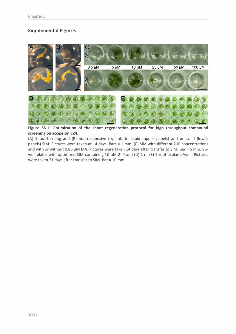
Chapter 5
100 |
Supplemental Figures
Figure S5.1: Optimization of the shoot regeneration protocol for high throughput compound screening on accession C24. (A) Shoot-forming and (B) non-responsive explants in liquid (upper panels) and on solid (lower panels) SIM. Pictures were taken at 14 days. Bars = 1 mm. (C) SIM with different 2-iP concentrations and with or without 0.86 µM IAA. Pictures were taken 14 days after transfer to SIM. Bar = 5 mm. 96-well plates with optimized SIM containing 10 µM 2-iP and (D) 1 or (E) 2 root explants/well. Pictures were taken 21 days after transfer to SIM. Bar = 10 mm.

Shoot regeneration by Phe-Ade
| 101
Figure S5.2: Phenotype of ahk double mutants grown for 1, 2 or 3 weeks in the presence or absence of 10 µM Phe-Ade or 2-iP. Bars = 1 mm.
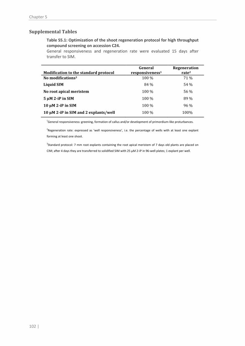
Chapter 5
102 |
Supplemental Tables
Table S5.1: Optimization of the shoot regeneration protocol for high throughput compound screening on accession C24. General responsiveness and regeneration rate were evaluated 15 days after transfer to SIM.
Modification to the standard protocol General
responsivenessRegeneration
1 rateNo modifications
2 100 % 3 71 %
Liquid SIM 84 % 54 %
No root apical meristem 100 % 56 %
5 µM 2-iP in SIM 100 % 89 %
10 µM 2-iP in SIM 100 % 96 %
10 µM 2-iP in SIM and 2 explants/well 100 % 100%
1General responsiveness: greening, formation of callus and/or development of primordium-like proturbances.
2Regeneration rate: expressed as ‘well responsiveness’, i.e. the percentage of wells with at least one explant
forming at least one shoot.
3
Standard protocol: 7 mm root explants containing the root apical meristem of 7 days old plants are placed on
CIM; after 4 days they are transferred to solidified SIM with 25 µM 2-iP in 96-well plates; 1 explant per well.

Shoot regeneration by Phe-Ade
| 103
Table S5.2: Sequences of primers used for qRT-PCR amplification. Primers were designed according to Mik et al. (2011), except the primers for AHK2, AHK3, AHK4 and CYP735A2, which were designed in this work.
Target Forward primer Reverse primer ACTIN2 AGACCTTTAACTCTCCCGCTATGTAT GATTGGCACAGTGTGAGACACA
AHK2 GGAGAAGAAGACCGTGAAAACATC ACACGCTGTTCTTCTGTAGCATCA
AHK3 GATCCTGACATTTGCAGTGTACAAG CGAGATACCCGTTAGTAGCCTCAA
AHK4 CCACAGATGGACGGATTTGAA TCCTCGTAGGTCGCGTGTATC
ARR10 CGTATTCTCCAGACTTTGCTTC CTGATTCGTTGTTGTAACGTGA
ARR14 CTTCCTGTCATTATGATGTCTGTTGA TGCCAAATGTTCTTAAGCTCTTCA
ARR15 ATGGAGACAATGGATCTTCTGG CTGAAGACTCTTTGATCTTCTTG
ARR16 GAATGCGCTTAGAGCATTGGAG CCTTCATAACGTTACAGGTCAAT
ARR2 TGACTTGTCCTCCAGCAAATAC TTATCTACGGATGGGATGCCTT
ARR5 AGAATTACCAGTTGTGATTATGTCT CTTCACATCAGCTAATTTCACAG
CKX1 CGGTCCTATCCTTATTTATCCAGTCA GGGAGAAAGGCTACGAGATAGAATATAT
CKX2 GGACTCGCTCTTCTCTATCCAACA TCTGGTATCATCGCCGACATAC
CKX3 CAACAAGTGGAATGATCGGATGT AGCCTCCCAATTGTCAAAACC
CKX4 TCTTCTAAACCAAACCTCAACTTCTG TGCGGTTGTTCCATTTGTTTC
CKX5 GGCGTTTTCAAGGGCATTTT TCGTCCCATTTGTCTTTGTTCA
CKX6 AACACCGGAGGAAGAGGTATTCT ACCCTGGAGATGCCGATGT
CKX7 GTCAATGGTCCAATGCTTGTGT CGAAGCAATGCCACAATGTAGA
CYP735A2 GCTCTTCCATCCACCACAACA CGGATTGTGCTTCGTTAGCA
IPT1 TCTCGTCAAAAGGTTCCGATTATC CGGATGAAAAAGGATCGAACTT
IPT2 CAATAAAGTCACCGTCGATGAACA AATAAGAGGAACAGTGAAGTCTCGAAA
IPT3 GCAAACAACCATTGCCTCCTT TTTCGAGACTTCACTGTTCCTCTTATT
IPT7 AAAGCGAATTACTCCGTAGGGATA CGGTCCACTAGAGATTCGTTACGT
IPT8 ATCCATTCTCTTCTGAGACATCCAT CTGAGACATCCACCCAAAGGA
LOG2 CACACTAAACCGGTGGGACTATT TGGAGCCGAGACAATGATACG
LOG8 CAGCGAAGCAGATTCAGAAAAA TTCCTCTTCACGAGTTCATTGC
SNRNPD1 GCTTGAACCTCGAGACTTTACTTGT CAACAGCTTTCCCAGCAACA
UGT73C1 GAGATTTTGGACTTGCTCGATT GATTATGCAGTTTGGCCTAGGT
UGT73C5 CAATTTAAAGTCAGATAAGGAGCT AGATATCTTTCCAGTCTCCAGCT
UGT76C1 TCTTGTTCCTCAGATTCGTCGT AAGCAAGTAAGCATCTAGAGGC
UGT76C2 GAACCAAGGGATATCTTCCAGTT TATATTAACCCTGAAGATCTTATTGTC
UGT85A1 ACAACCAGTGGCTGTGCG TGGTATAAAATCTATAACCGTGTCT


QTL and GWA analysis of regeneration capacity
| 105
6. 6
Chapter 6 Combining linkage and association mapping as a
gene discovery tool for the regeneration capacity of Arabidopsis thaliana
Manuscript in preparation


QTL and GWA analysis of regeneration capacity
| 107
Shoot regeneration, i.e. the induction of shoots on non-meristematic tissue,
is widely applied in plant biotechnology. However, the capacity to
regenerate shoots is highly variable and dependent on plant species and
cultivar. Despite intensive research to unravel the molecular mechanisms of
shoot regeneration, the factors that determine regeneration capacity are
still poorly understood. Here, we evaluated the regeneration capacity of 88
Arabidopsis accessions, subjecting root explants to a two-step regeneration
protocol. We used these data for correlation studies with other phenotypical
responses that accompany shoot regeneration, such as callus formation and
the greening of the explant. Intriguingly, we found only low correlations
within these traits and hence, callus nor green foci predict regeneration
capacity. By using a Nok-3 x Ga-0 inbred population, we identified 5
regeneration QTLs, which were not previously described. Furthermore, we
performed a genome-wide and a local association study for a QTL fine
mapping, resulting in the identification of a plausible QTG: the RECEPTOR-
LIKE PROTEIN KINASE1 (RPK1). The importance of this ABA-related receptor
in shoot regeneration was corroborated with mutant analyses. Altogether
our results demonstrate that association mapping is a powerful method to
discover novel important genes implicated in a biological process as
complex as shoot regeneration .

QTL and GWA analysis of regeneration capacity
| 109
Introduction
The capacity or recalcitrance to regenerate shoots or to form adventitious shoots in tissue culture is
of major importance for biotechnological breeding and for commercial in vitro initiation and
propagation of plants (Duclercq et al., 2011b). Unfortunately, amongst species, varieties, cultivars,
and even explants, the efficiency of shoot regeneration is highly variable and currently unpredictable
(George and Debergh, 2008). The importance of shoot regeneration for horticulture and agriculture
is illustrated by the plentitude of studies assessing natural allelic variation in regeneration capacity in
diverse crops, such as tomato, wheat, rice, barley, sunflower, cabbage, and potato (eg. Koornneef et
al., 1993; BenAmer et al., 1997; Jan et al., 1997; Taguchi-Shiobara et al., 1997; Berrios et al., 2000;
Takeuchi et al., 2000; Kwon et al., 2001; Mano and Komatsuda, 2002; Holme et al., 2004; Nishimura
et al., 2005; Tyagi et al., 2010; Trujillo-Moya et al., 2011), to name but a few. As such, using
quantitative trait loci (QTL) mapping, valuable information has been obtained on genetic linkages,
genomic regions and/or markers associated with regeneration in these species. Nevertheless, using
this approach little insight has been gained into the molecular basis of regeneration. Moreover,
because the QTL mapping is done in biparental populations, the linkages can often not be
extrapolated to global populations (Würschum, 2012).
With the two-step regeneration protocol described by Valvekens et al. (1988) starting from root
explants of the model plant Arabidopsis thaliana using diverse experimental methods, the questions
about the molecular mechanism behind shoot regeneration can be met. The explants first acquire
organogenesis competence on an auxin-rich callus inducing medium (CIM) and, subsequently, they
become committed to shoot formation on a cytokinin-rich shoot inducing medium (SIM) (Cary et al.,
2002). Genome-wide analyses of the gene expression profiles accompanying these successive steps
in the regeneration process revealed multiple key regulators and genes implicated in phytohormonal
signaling during shoot regeneration (Cary et al., 2002; Che et al., 2002; Che et al., 2006; Che et al.,
2007; Sugimoto et al., 2010). Reporter gene fusions with marker genes allowed visualization of their
spatio-temporal expression patterns during regeneration, contributing to the elucidation of the
function of important shoot-related genes, such as CUP SHAPED COTYLEDON1 (CUC1), CUC2, SHOOT
MERISTEMLESS (STM), WUSCHEL (WUS) or CLAVATA3 (CLV3) (Cary et al., 2002; Heisler et al., 2005;
Gordon et al., 2007; Gordon et al., 2009; Motte et al., 2011).
In classical gene-based forward and reverse genetics approaches a number of additional genes
essential for or involved in shoot regeneration have been identified (reviewed by Meng et al., 2010;

Chapter 6
110 |
Duclercq et al., 2011b). Examples are the cytokinin signaling genes CYTOKININ INDEPENDENT1 (CKI1)
(Kakimoto, 1996) and ARABIDOPSIS RESPONSE REGULATOR2 (ARR2) (Hwang and Sheen, 2001), the
ETHYLENE-RESPONSIVE FACTOR family genes ENHANCER OF SHOOT REGENERATION1 (ESR1) (Banno
et al., 2001), ESR2 (Ikeda et al., 2006) and WOUND INDUCED DEDIFFERENTIATION1 (WIND1) (Iwase
et al., 2011), and the shoot markers CUC1 and CUC2 (Daimon et al., 2003). Although these
approaches have proven to be useful in gene discovery, they focus on a single gene and thus, might
overlook important genes implicated in polygenic traits (Vreugdenhil et al., 2007).
Quantitative trait loci (QTL) mapping is an alternative method used to discover major genes involved
in regeneration in Arabidopsis. QTL mapping using recombinant inbred lines (RILs) of Ler x Col
(Schiantarelli et al., 2001; Lall et al., 2004) or Ler x Cvi (Velázquez et al., 2004), accessions with
different regeneration capacities, clearly demonstrated that shoot regeneration is a polygenic trait
exhibiting continuous variation (Schiantarelli et al., 2001; Lall et al., 2004; Velázquez et al., 2004). The
subsequent identification of the quantitative trait gene (QTG) or nucleotide (QTN) responsible for a
QTL, includes high resolution mapping which is very slow and labor intensive. If the resulting list of
candidate genes is short enough, phenotypic analyses of mutants or transgenic plants can identify
the QTG. The main drawback of a QTL analysis is that it makes use of segregating lines originating
from two or a few parents. Hence, loci that contribute to the variation in regeneration in a larger
population, but not in the selected parents, can easily be overlooked.
Here, to overcome this problem, we assessed the natural allelic variation in the shoot regeneration
capacity of 88 accessions of Arabidopsis by combining a traditional QTL analysis with a genome-wide
association (GWA) mapping. GWA mapping is a fairly recent technique that allows a more
comprehensive study of trait loci in a population by searching for associations between trait variation
and allelic variants using very dense marker maps, generally single nucleotide polymorphisms (SNPs),
resulting in a higher mapping resolution (Nordborg and Weigel, 2008). First, following the
regeneration procedure described for Arabidopsis root explants by Valvekens et al. (1988), the
formation of callus and roots, the greenness of the explants, and the development of shoot
primordia and shoots were scored at different time points during SIM incubation for the various
accessions. Next, pair-wise correlations between the different features revealed a relative low
correlation between the regeneration capacity and the other traits, indicating that no early
observations could predict shoot formation. Therefore, the 88 accessions were ranked based on the
number of explants forming shoots. From these data, Nok-3 and Ga-0 were selected for linkage
mapping, as accessions with a high and low regeneration ability, respectively. Using their
recombinant inbred lines (RILs)(O’Neill et al., 2008), five QTLs were identified. Subsequently, a GWA

QTL and GWA analysis of regeneration capacity
| 111
study was done resulting in a list of 32 SNPs associated with regeneration. Local association mapping
of the QTL regions revealed RECEPTOR-LIKE PROTEIN KINASE1 (RPK1) as a candidate QTG. Finally,
mutant analysis confirmed the importance of this gene in shoot regeneration. Thus, combining
linkage and association mapping proved to be a powerful gene discovery tool for a phenotype as
complex as regeneration.
Results
Natural variation in shoot regeneration and correlation with accompanying
phenotypical responses
To assess the natural variation in shoot regeneration, root explants of 88 A. thaliana accessions were
subjected to a two-step regeneration procedure (Valvekens et al., 1988) and the number of shoots
per explant, the regeneration rate (the number of explants forming at least one shoot), the number
of primordia and roots that developed, the degree of callus formation and the greenness of the
explants (see Material and Methods) were recorded after different incubation times on SIM (7, 11,
14 and 21 days). The regeneration rate ranged between 0% (for accessions CIBC-17 and Sq-8) and
100% (for accessions NFA-10, Spr1-6 and Yo-0)(Figure 6.1), but also for the other parameters a wide
diversity in the responses between the 88 accessions was observed (Figure 6.2A and B; Supplemental
Table S6.1). Compared to the number of shoots per explant, the regeneration rate exhibited a more
continuous distribution and showed relatively less variation within the repeats (Figure 6.1) and
therefore, the latter was used in following mapping studies. To determine whether two particular
phenotypical responses were correlated with each other during the regeneration process of all
accessions tested, Spearman correlation coefficients were calculated for each parameter pair
followed by a cluster analysis. Figure 6.2C and Supplemental Table S6.2 show that various
correlations could be made and that the traits clustered in three distinct groups: a root, a
primordia/shoot and a callus/greenness cluster. Unexpectedly, no high correlations were found for
callus and shoots (ρ = 0.049 – 0.422) nor for greenness and shoots (ρ = 0.05 – 0.432)(Figure 6.2C;
Supplemental Table S6.2). Thus, considering the accessions analyzed, apparently the capacity to form
callus or to develop chloroplasts is not linked with efficient regeneration. Indeed, for example,
accession Ct-1, which is regeneration recalcitrant, forms dark green callus but few shoots (Figure
6.2D; Supplemental Table S6.1), while accession Wei-1 that regenerates well forms little pale green
callus but a lot of shoots (Figure 6.2E; Supplemental Table S6.1). Within the callus/greenness cluster,
only moderate correlations were found (ρ = 0.192 – 0.633)(Figure 6.2C), indicating that formation of

Chapter 6
112 |
callus and the greening of the explant do not
necessarily occur simultaneously (Figure 6.2A and
B). The highest correlation coefficients were
found in the root cluster (ρ = 0.679 –
0.863)(Figure 6.2C) because for all accessions,
roots developed mainly in the first 7 days of SIM
incubation and the number of roots did not
change at the later time points (Supplemental
Figure S6.1, Supplemental Table S6.1), resulting in
the same correlation for all individual explants. In
the primordia/shoot cluster the correlation
coefficients for each developmental phase over
time were lower than in the root cluster (Figure
6.2C), illustrating that between accessions the
timing of shoot formation was different (Figure
6.1). Indeed, when the number of shoots formed
per explant after 14 and 21 days of SIM
incubation was compared for the different
accessions, very clear differences, reflecting their
relative degree of regeneration recalcitrance or
capacity, were observed (Figure 6.1). Intriguingly,
the correlations between primordia and shoots
were not so high either (ρ = 0.089 – 0.737)(Figure
6.2C), suggesting that the potential to form
primordia can be uncoupled from the
Figure 6.1: Shoot regeneration capacity of the
analyzed Arabidopsis accessions. The number of shoots per root explant (upper axis) and the regeneration rate, i.e. the percentage of explants producing at least one shoot (lower axis), after 14 and 21 days of SIM incubation are represented. For each accession, three (number of shoots) or six (regeneration rate) independent repeats were done, with 30 explants per repeat. The more recalcitrant Ga-0 (blue) and the regenerative Nok-3 (orange) accession were selected for QTL analysis. Error bars indicate standard errors.

QTL and GWA analysis of regeneration capacity
| 113
Figure 6.2: Phenotypical responses during the regeneration process and correlation between different responses in the analyzed Arabidopsis accessions. (A) Classification of callus formation. Callus class 0: no callus; class 1: callus is present at the ends of the explant; class 2: callus is present and covers maximum one third of the explant; class 3: callus is abundant, but does not cover the whole explant; class 4: thick callus covers the whole explant. (B) Evaluation of different classes of greenness of the explants, excluding shoots. Class 0: explant is white; class 1: explant is yellow; class 2: explant is (partially) pale green; class 3: complete explant is green; class 4: complete explant is dark green. (C) Correlation of different responses accompanying shoot regeneration (number of shoots, number of primordia, number of lateral roots, callus, and greenness) at different time points (7, 11, 14 and 21 days) of SIM incubation. The heatmap represents the Spearman’s rank correlation matrix for the different responses. The dendrograms are distance trees. Three repeats were done, with each time 30 explants per accession. Since almost no shoots were observed after 7 days of SIM incubation, this information was not included. (D) The more recalcitrant accession Eden-1 forms dark green callus without shoots. The picture was taken after 21 days of SIM incubation. Bar = 1 mm (E) The regeneration competent Wei-0 develops little callus and the explant remains pale, but a lot of shoots are formed. The picture was taken after 14 days of SIM incubation. The arrow indicates the root explant. Bar = 1 mm. (F) Scatter plot for the accessions Col-0, LL-0 and Pna-17 with the number of primordia on an explant after 14 days plotted against the number of shoots on the same explant after 21 days of SIM incubation. 90 explants per accession are represented. Notice the different relations: the more recalcitrant accession Pna-17 forms a lot of primordia, but few shoots, Col-0 has a high primordia to shoot development rate and LL-0 has an intermediate relation between the two parameters.

Chapter 6
114 |
subsequent development into shoots under our experimental conditions. Further evidence for this
observation came from scatter plots for three accessions, where the number of primordia present on
a specific explant at 14 days of SIM incubation was plotted against the number of shoots present on
the same explant 7 days later. Although accession Pna-17 produced many primordia, it had the
lowest number of shoots per explant. Accession LL-0 produced many primordia as well, but many
developed into shoots. Accession Col-0 showed less primordia but a higher number of shoots: the
development of primordia into shoots occurred rapidly and very efficiently (Figure 6.1; Figure 6.2F;
Supplemental Table S6.1).
Linkage mapping of the regeneration rate reveals 5 QTLs
To identify QTL for regeneration rate, we accessed the Nok-3 x Ga-0 inbred population (O’Neill et al.,
2008). The choice for this population was motivated by the large difference measured in
regeneration capacity between Ga-0 (recalcitrant) and Nok-3 (regenerative) in the screen of the 88
accessions (Figure 6.1). Root explants of the 86 RILs were subjected to the two-step regeneration
protocol and after 14 and 21 days of SIM treatment, the number of explants producing at least one
shoot was counted. Linkage mapping revealed three QTLs at both time points including one in
common, REG-1 (Table 6.1, Figure 6.3A). After a final QTL backward selection, REG-3 seemed only
significant for the regeneration rate after 14 days of SIM incubation, while the apparent QTL for 21
days of SIM treatment on chromosome 5 (Figure 6.3A) was considered as non-significant and was
thus not retained for further analysis. Combining the regeneration rates at both time points for a
multi-trait linkage analysis, resulted in three QTLs that overlapped with single trait QTLs REG-1, 2 and
5 (Table 6.1, Figure 6.3A). REG-1 was the major QTL in each analysis, explaining about 30% of the
variation (Table 6.1).
Table 6.1: Regeneration QTLs identified by using a Nok-3 x Ga-0 inbred population. Origin % explained variation
positive Position Single trait Multi-trait
QTL Trait allele A Chr. (cM/Mb) 14 d 21 d 14 d 21 d
REG-1 14, 21, multi Nok-3 1 90/25 31.8 B 23.8 30.6 27.8 REG-2
21, multi Ga-0 2 23/7 - B 12.4 4.9 22.2
REG-3
14 Ga-0 2 70/18 14.3 B - - -
REG-4 21 Nok-3 3 25/7.7 - 7.4 - -
REG-5 14, multi Nok-3 3 84/23
12.1 - 11.7 9.6
A 14, 21: Single trait analysis for regeneration responsive explants after 14 or 21 days, respectively. Multi: multi-trait analysis for responsive explants after 14 and 21 days.
B
Approximate position in Mb as loci are not represented by a marker.

QTL and GWA analysis of regeneration capacity
| 115
To confirm the quantitative effect of the identified QTLs, the RILs were divided into genotype groups
according to the combination of the parental alleles for each trait. For example, for the single trait
“regeneration rate after 14 days of SIM incubation”, the group NGN has the Nok-3 alleles for REG-1
and REG-5, and the Ga-0 allele for REG-3. The resulting boxplot in Figure 6.3B supported the
importance of the REG-1 QTL. In general, the Nxx (x for N or G) genotypes were regenerating well or
very well, while the Gxx genotypes were more recalcitrant. One exception was the GGN group for the
single trait analysis 14 days of SIM incubation, for which the absence of the positive REG-1 QTL was
partially complemented by the presence of the positive REG-3 and REG-5 QTLs. All NGN lines were
highly regenerative (superior genotype), while the GNG lines were recalcitrant (inferior genotype).
However, this recalcitrance was not complete: after 21 days of SIM incubation, all RILs had at least
3% responsive explants (Figure 6.3B). Moreover, at that time point, the most recalcitrant lines were
not in the GNG-group (Figure 6.3B), indicating that regeneration capacity or rather recalcitrance, is
also depending on multiple minor factors out of the QTLs.
Figure 6.3: QTL-analysis for the regeneration rate of a Nok-3 x Ga-0 RIL population. (A) QTL- profile of the single trait analyses after 14 or 21 days of SIM incubation (red and blue dotted lines), and the multi-trait analysis, combining the results of the two time points (purple solid line). The red line denotes a genome-wide significance threshold (α = 0.05). (B) Box plots showing the regeneration rate of RILs divided in QTL genotype groups according to the alleles REG-1, 3 and 5 (left), REG-1, 2 and 5 (middle) and REG-1, 2 and 4 (right). N = Nok-3 allele, G = Ga-0 allele. The allelic origins of REG-1, 2 and 3, which are not represented by a marker, were assigned according to adjacent markers, or not assigned if the adjacent markers had a different parental origin. RILs with incomplete QTL genotypes were not used for the boxplot, resulting in an underrepresentation of the GGN genotype (multi-trait) and the GGG genotype (21 d single trait). Boxes of the superior genotype NGN and inferior genotype GNG are colored in green and red, respectively. Crosses indicate outliers (green) and far outliers (red), which are at a distance of more than 1.5 or 3 times the interquartile range beyond the quartiles, respectively.

Chapter 6
116 |
Association mapping yields a list of SNPs in genes enriched in regeneration-
related functions
Typically, QTL peaks cover a large genomic region harboring many genes. For example, based on the
markers underlying the REG-1 peak, i.e. from marker t12p18ind8-8 (23.5
Mb; http://www.jic.ac.uk/staff/ian-bancroft/arabidopsis.htm) to the end of the chromosome,
approximately 2000 genes are present (TAIR9, http://www.arabidopsis.org), from which at least 40
have been described in relation to shoot regeneration. Therefore, as an approach for identifying
putative candidate QTGs implicated in the capacity to regenerate, a GWA mapping was conducted.
Associations between SNPs and regeneration rate were evaluated estimating the effect of each of
the 215.000 SNPs on the trait (see Material and Methods), followed by a false discovery rate (FDR)
correction (p < 0.01) (Benjamini and Hochberg, 1995). As such, 32 and 6 SNPs significantly associated
with the regeneration rate at 14 days and 21 days of SIM treatment, respectively (Figure 6.4, Table
6.2), were identified. Interestingly, the SNPs with the strongest association (p ≤ 4.00E -06) were
located in genes that were not previously described as shoot- or regeneration-related, such as GOLGI
NUCLEOTIDE SUGAR TRANSPORTER5 (GONST5), EMBRYO DEFECTIVE3001 (EMB3001) and EXOCYST
SUBUNIT EXO70 FAMILY PROTEIN D1 (EXO70D1) (Table 6.2). Nevertheless, the reported function of
several of the genes with regeneration variation-associated polymorphisms, can be linked to
processes important for primordium or shoot formation. For instance, MAP KINASE6 (MPK6) is
regulated by the CLAVATA3 (CLV3)-derived peptide receptor CLV1 (Betsuyaku et al., 2011),
NUCLEOLIN-LIKE2 (NUC-L2) is upregulated in lines overexpressing ESR2 (Ikeda et al., 2006), and
PROTODERMAL FACTOR2 (PDF2) regulates shoot epidermal cell differentiation (Abe et al., 2003). In
addition, MPK6 and NUCL-L2 together with NUCLEOSOME ASSEMBLY PROTEIN1;1 (NAP1;1) and
At3g21770 are involved in lateral root initiation or development (Vanneste et al., 2005; De Smet et
al., 2008; Müller et al., 2010), and hence putatively important for primordium formation during the
regeneration process (Atta et al., 2008; Pernisová et al., 2009; Sugimoto et al., 2010). Moreover,
further down in the list (Supplemental Table 6.S31
Ikeda et al., 2006
), additional regeneration-related genes are
present, such as ESR2 (p=0.0003)( ) and CYTOKININ OXIDASE/DEHYDROGENASE2
(CKX2)(p=0.0002), whose gene product degrades cytokinins and has a high expression level in the
shoot meristem (Werner et al., 2003). Altogether these findings show that GWA studies are a
suitable tool for gene discovery in a process as complex as shoot regeneration.
1 Supplemental Table 6.S3 contains all 14,738 SNPs associated (p < 0.05) with shoot regeneration rate according to the GWA study and is due to practical reasons not included in this dissertation, but is available upon request ([email protected]).

QTL and GWA analysis of regeneration capacity
| 117
We then determined if the regeneration-associated genes could be linked to specific processes, by
analyzing the complete gene list generated by the GWA study (Supplemental Table 6.S3) with AmiGo
(version 1.8; The Gene Ontology Consortium, 2000) for biology process ontology (Supplemental
Figure S6.2). Interestingly, the most overrepresented gene ontology (GO) terms were “meristem
initiation” defined as “initiation of a region of tissue in a plant that is composed of one or more
undifferentiated cells capable of undergoing mitosis and differentiation, thereby effecting growth
and development of a plant by giving rise to more meristem or specialized tissue” and “post-
embryonic morphogenesis” described as “the process, occurring after embryonic development, by
which anatomical structures are generated and organized” (Supplemental Figure S6.2). Although
several other GO terms were obtained by this analysis, none had a clear link with shoot regeneration
(Supplemental Figure S6.2), suggesting that a wide range of diverging processes are important for
this developmental pathway.
Combining linkage mapping with local association mapping identifies RPK1
as a candidate QTG for REG-1
The GWA study revealed several regeneration-associated SNPs co-locating with the QTLs for
regeneration capacity identified in the linkage mapping (Table 6.2). Unfortunately, none of the SNPs
segregated between the parents Nok-3 and Ga-0. However, if SNP-loci are in linkage disequilibrium
Figure 6.4: Manhattan plots of the associations of the SNPs with the regeneration rate. Plots show the –log10(p)-values from the genome-wide scan using the mixed model approach (see Material and Methods) for the regeneration rate after 14 (upper panel) and 21 days (lower panel) of SIM incubation. The X-axis indicates the chromosomes and the physical positions; the red line indicates the FDR-correction.

Chapter 6
118 |
(LD) with other loci, and if these loci are polymorphic for the two parents, the SNP could still be
responsible for the QTL. Thus, LDs between all associated SNPs with their surrounding regions were
calculated as r² based on the fully sequenced Arabidopsis accessions (62 of the 88 accessions under
study at the time of analysis) with online software (http://gvs.gs.washington.edu/GVS/). However, no
linked polymorphisms were found, as exemplified for At1g73470 (Supplemental Figure S6.3). Thus,
no QTGs for the identified QTLs were revealed by the GWA analysis.
Therefore, we decided to conduct a local association study, focusing exclusively on potential QTGs.
We selected all genes, including their 1000 bp upstream region, covered by the identified QTLs
(Figure 6.3) and by an associated SNP (Supplemental Table 6.S3) in the set of 62 fully sequenced
accessions. As such, the full sequence of the candidate regions was considered which might reveal
new meaningful trait associations. Moreover, since we were interested in polymorphisms causal to
the QTLs, only polymorphisms between Nok-3 and Ga-0 were analyzed. This approach resulted in
candidate QTGs for REG-1, REG-2 and REG-3 (Table 6.3). For the candidate QTGs identified by
polymorphism 1, 2, 3, 4, 12 and 13 (Table 6.3), not much evidence was found for a possible influence
on protein function or expression levels. According to the Arabidopsis Gene Regulatory Information
Server (AGRIS) (Davuluri et al., 2003; Palaniswamy et al., 2006; Yilmaz et al., 2011), polymorphism 1,
2, 12 and 13 were not located in a gene nor in a regulatory sequence. Polymorphism 3, in NUDIX
HYDROLASE HOMOLOG21 (NUDT21), does not result in an amino acid change and thus enzymatic
activity is not affected. Polymorphism 4 is located in the promoter sequence of ABA
HYPERSENSITIVE1 (ABH1) and although the polymorphism is not located in a binding site according to
AGRIS, it might alter the expression of this gene.
The gene RECEPTOR-LIKE PROTEIN KINASE1 (RPK1) could be classified as a plausible QTG for REG-1
because polymorphisms 5-11 are all associated with the regeneration trait (Table 6.3). RPK1 encodes
a Leucine-rich repeat receptor-like kinase (LRR-RLK) composed of an extracellular ligand binding
domain and a cytosolic kinase domain (Hong et al., 1997). Polymorphism 11, which was located in
the binding domain, resulted in an amino acid change (V > L on position 162) and hence is likely the
QTN underlying REG-1. The Nok-3 allele which confers a higher regeneration capacity, is indeed
mainly present in regenerative accessions with a regeneration rate of more than 75 % after 14 days
of SIM incubation (Figure 6.1, Figure 6.5A). Prediction of the structure of the extracellular domain of
both alleles using PHYRE² (Kelley and Sternberg, 2009) revealed a polymorphism-dependent
modification (Figure 6.5B and C) which might affect the binding efficiency of the receptor to its ligand
and thus RPK1 functioning.

QTL and GWA analysis of regeneration capacity
| 119
Table 6.2: SNPs associated with the regeneration rate. Their covering by a QTL-peak, their p-value for association with regeneration rate after 14 and 21 days of SIM incubation and the gene in which they are localized and description are given.
Chr. SNP Position QTL p-value (14 d) p-value (21 d)1 Gene 1 Description
1 7678536 4.28E-07* 8.99E-05* At1g21870 GOLGI NUCLEOTIDE SUGAR TRANSPORTER5 4 13231999 2.69E-06* 4.41E-04 At4g26110 NUCLEOSOME ASSEMBLY PROTEIN1;1 (p) 1 2857313 3.37E-06* 1.73E-04 At1g08910 EMBRYO DEFECTIVE3001 1 2857554 3.37E-06* 1.73E-04 At1g08910 EMBRYO DEFECTIVE3001 1 2858187 3.37E-06* 1.73E-04 At1g08910 EMBRYO DEFECTIVE3001 1 2859838 3.37E-06* 1.73E-04 At1g08910 EMBRYO DEFECTIVE3001 1 27285484 REG-1 4.00E-06* 1.23E-04 At1g72470 EXOCYST SUBUNIT EXO70 FAMILY PROTEIN D1 3 6404416 REG-4 4.03E-06* 4.89E-05* At3g18610 NUCLEOLIN LIKE2 2 4280328 REG-2 1.14E-05* 2.03E-04 At2g10860 Pseudogene 5 15609918 2.11E-05* 2.42E-04 At5g38990 INTERLEUKIN-1 RECEPTOR-ASSOCIATED KINASE4 1 27285555 REG-1 2.35E-05* 3.05E-04 At1g72470 EXOCYST SUBUNIT EXO70 FAMILY PROTEIN D1 4 18242073 2.40E-05* 3.27E-04 At4g39170 Sec14p-like phosphatidylinositol transfer family protein 4 13219058 2.52E-05* 8.43E-04 At4g26070 MAP/ERK KINASE1 1 10362968 3.25E-05* 2.70E-05* At1g29650 Pseudogene 3 15919755 2.93E-05* 1.94E-04 At3g44215 Pseudogene 3 4438379 3.23E-05* 7.34E-04
4 5001976 3.41E-05* 2.12E-04 At4g08096 Pseudogene 3 20801652 3.68E-05* 1.29E-03 1 7198526 3.98E-05* 2.14E-04 At1g20735 F-box and associated interaction domains-containing protein 3 2887575 4.05E-05* 6.30E-05* At3g09385 Pseudogene 5 1670975 SR-1² 4.05E-05* 1.16E-04 At5g05598 Defensin-like family protein (p) 1 10363163 4.27E-04 4.36E-05* At1g29650 Pseudogene 4 2479196 4.69E-05* 1.01E-03 At4g04890 PROTODERMAL FACTOR2 1 2856481 4.91E-05* 8.35E-04 At1g08910 EMBRYO DEFECTIVE3001 3 2433429 5.51E-05* 1.92E-04 At3g07620 Exostosin family protein, putative glycosyltransferase 2 4234096 REG-2 4.39E-04 6.44E-05* At2g10760 Pseudogene 1 18669165 6.74E-05* 2.44E-04 At1g50400 Mitochondrial import receptor subunit TOM40 4 18508100 7.55E-05* 1.22E-03 4 18508100 7.55E-05* 1.22E-03 At4g39900 Unknown protein (p) 3 7674382 REG-4 7.77E-05* 1.17E-04 At3g21770 Peroxidase superfamily protein 5 23938547 M5³ 9.09E-05* 1.51E-04
2 18139021 REG-3 9.16E-05* 6.34E-04 At2g43790 MAP KINASE6 2 18139440 REG-3 9.16E-05* 6.34E-04 At2g43790 MAP KINASE6 1 18738481 9.94E-05* 2.00E-03 At1g50600 SCARECROW-LIKE5
1 significant p-values according to the FDR are indicated with an *.
(p)
² QTL described in Velázquez et al. (
SNPs located in the putative promoter of the gene.
2004) Covering was estimated based on Cvi x Ler markers (www.arabidopsis.org). The estimate is not accurate as no detailed information about the markers or QTLs was provided.
³ QTL described in Lall et al. (2004). Covering was estimated based on Col x Ler markers (www.arabidopsis.org).

Chapter 6
120 |
Figure 6.5: Polymorphisms in RPK1. (A) Matrix representing polymorphic positions for the 62 sequenced accessions in RPK1 and the surrounding regions. The accessions were sorted from recalcitrant (top) to regenerative (bottom) based on the regeneration rate at 14 days, except for Ga-0 and Nok-3, which are represented in the two last rows. For each position, the major allele is colored in blue, while the rare allele is colored in yellow. Regeneration-associated polymorphisms are indicated by a red overlay, the only associated polymorphism resulting in an amino acid change is indicated by the green frame. The protein model below the matrix indicates the coding sequence (black line) and specific structural domains (Hong et al., 1997). Positions are however not in proportion as only polymorphic positions are included in the matrix. (B-C) 3D model predicted by PHYRE² (Kelley and Sternberg, 2009) of the binding domain of the negative (B) and the positive (C) variant. Respectively 95% and 94% of the residues were modeled at >90% confidence.

QTL and GWA analysis of regeneration capacity
| 121
Table 6.3: Nok-3/Ga-0-polymorphisms from the regeneration QTLs, associated with the regeneration rate in the 62 fully sequenced accessions (p < 1E-4). If the polymorphisms are localized in a gene, the gene description is added.
Polymorph. Chr. Position
Polymorph. p-value (14 d) p-value (21 d) Gene Description 1 2 5622878 5.13E-05 1.46E-05 2 2 14286941 1.95E-03 8.70E-05 3 1 27645927 3.68E-03 8.78E-05 At1g73540 NUDIX HYDROLASE HOMOLOG21 4 2 5636722 3.44E-04 8.81E-05 At2g13540 ABA HYPERSENSITIVE1 (p) 5 1 26041018 8.83E-05 2.99E-04 At1g69270 RECEPTOR-LIKE PROTEIN KINASE1 6 1 26041108 8.83E-05 2.99E-04 At1g69270 RECEPTOR-LIKE PROTEIN KINASE1 7 1 26041216 8.83E-05 2.99E-04 At1g69270 RECEPTOR-LIKE PROTEIN KINASE1 8 1 26041222 8.83E-05 2.99E-04 At1g69270 RECEPTOR-LIKE PROTEIN KINASE1 9 1 26041243 8.83E-05 2.99E-04 At1g69270 RECEPTOR-LIKE PROTEIN KINASE1
10 1 26041261 8.83E-05 2.99E-04 At1g69270 RECEPTOR-LIKE PROTEIN KINASE1 11 1 26042016 8.83E-05 2.99E-04 At1g69270 RECEPTOR-LIKE PROTEIN KINASE1 12 1 27659440 9.10E-05 8.25E-04 13 1 26764591 9.73E-05 3.93E-04
(p)
polymorphisms in the putative promoter of the gene.
RPK1 is required for shoot regeneration
To corroborate the importance of RPK1 during shoot regeneration, root explants of the rpk1-1 and
rpk1-5 Col-0 mutants (Nodine et al., 2007) were subjected to the two-step regeneration protocol.
Interestingly, rpk1-5 was almost completely recalcitrant (Figure 6.6A) and also rpk1-1 had a
significantly reduced regeneration rate (Figure 6.6A). Remarkably, whereas Col-0 explants did not
form a lot of callus, regenerating rpk1-1 explants formed massive dark green callus (Figure 6.6B).
Finally, RPK1 expression was monitored during the regeneration process using a pRPK1::RPK1-GFP
line (Nodine et al., 2007): fluorescence was particularly observed in dividing, presumptive pericycle
cells (Figure 6.6C), and in emerging primordia (Figure 6.6D), but as the primordia developed further,
expression weakened (Figure 6.6D), which places the RPK1 activity relatively early in the de novo
shoot formation process.
Discussion
In this study we performed a comprehensive analysis of the shoot regeneration capacity of 88
Arabidopsis thaliana accessions by combining linkage with association mapping as an alternative
approach for gene discovery in this complex developmental process.

Chapter 6
122 |
Figure 6.6: RPK1 is important for shoot regeneration. (A) Regeneration rate of rpk1 mutants. Three repeats were done, with 30 (Col-0, rpk1-5) or 10 (rpk1-1) explants per repeat. Error bars indicate standard errors. Different letters indicate statistical differences evaluated with the Tukey range test (p < 0.05) in conjunction with an analysis of variance. (B) Shoot and callus formation on rpk1-1 (upper panel) and Col-0 root explants (lower panel). Arrows indicate the root explants. Bars = 1 cm (C-D) pRPK1::RPK1-GFP fluorescence in presumptive shoot primordia formed on root explants after 7 (C) and 11 (D) days of SIM incubation. Bars = 100 µm. (C) Cells are stained with propidium iodide (red). (D) Weak fluorescence is mainly observable in emerging primordia and, to a lesser extent, in the vasculature.
Screening 88 Arabidopsis accessions for phenotypes such as greening of the explants, callus
development, and root, primordium, and shoot formation during incubation on SIM in the two-step
regeneration protocol (Valvekens et al., 1988) revealed a wide natural variation for these
parameters. The recording of these responses and the ranking of the accessions according to their
regeneration rate provide valuable information for future studies. For instance, accessions NFA-10,
Spr1-6 and Yo-0 have a 100% regeneration rate and are useful as alternatives for the commonly used
accessions, such as Col-0 and Ler-1, that exhibit only an intermediate or low regeneration rate, for
the discovery of novel gene functions implicated in regeneration. Similarly, accessions CIBC17 and Sq-
8 are completely recalcitrant under our experimental conditions and can be used in mutagenesis
screens for (partial) reversion of their regeneration defect. Although pairwise correlation between
the parameters were in general significant and revealed three distinct correlation clusters, no high
correlations between traits such as callus and shoot formation, greening of the explant and shoot

QTL and GWA analysis of regeneration capacity
| 123
formations, and primordium and shoot formation were identified. Indeed, neither callus formation
nor greening were indicative for subsequent shoot regeneration, and, hence, these responses should
merely be considered as effects induced by 2,4-D and cytokinin (Skoog and Miller, 1957; Schmülling
et al., 1997). This conclusion is in contrast to what has been established for individual accessions
where the formation of green protuberances and callus was suggested to be related (Cary et al.,
2002) and even to be a required for the regeneration process (Che et al., 2007). The weak correlation
between primordium and shoot formation was intriguing as well and showed that regeneration can
be blocked very late in the developmental process. Thus, accessions such as Pna-17, that form
primordia but almost no shoots, should be the starting material by choice to study this type of
recalcitrance. Hence, the natural variation in the regeneration capacity across the different
Arabidopsis accessions is a powerful resource to unravel the diverse developmental processes that
are implicated in the formation of adventitious shoots and to examine the genetic basis of
recalcitrance.
Nevertheless, several observations made during the subsequent QTL and GWA studies demonstrated
that regeneration is a complex trait with a broad polygenic basis. For instance, none of the QTLs
obtained from assessing the regeneration rate of a Nok-3 x Ga-0 RIL population corresponded to
previously identified QTL for Ler x Col (Lall et al., 2004) or Ler x Cvi (Velázquez et al., 2004)
populations. Moreover, the most recalcitrant RILs of Nok-3 x Ga-0 were not found in the inferior
genotype group, which harbored the three negative QTLs, indicating that multiple non-detected
minor loci had an important influence on regeneration as well. Then, from the GWA study, numerous
genes in a wide population of Arabidopsis were found to be associated with regeneration, but none
of them were causal to the identified QTLs. Similarly, when the list with associated SNPs was mapped
on the QTLs identified in other linkage studies, the SNP underlying the M5 QTL (Lall et al., 2004) was
not polymorphic between the parents of the inbred population (Table 6.2). Nevertheless, the SNP
covered by SR-1 (Velázquez et al., 2004) could be causal and merits further study, but since the
information about the used markers is limited, we were only able to roughly estimate the position of
SR-1. Another observation was that, although in the GWA-generated gene list the GO terms
“meristem initiation” and “post-embryonic morphogenesis” were overrepresented, no other specific
biological processes could clearly be distinguished suggesting the involvement of many different
pathways. Finally, no strict associations were found for specific SNPs with either highly regenerative
or recalcitrant accessions. In contrast, some studies on cereals, such as wheat or rye (reviewed by
Bolibok and Rakoczy-Trojanowska, 2006), or vegetables, such as tomato (Koornneef et al., 1993;
Satoh et al., 2000), reported a non-continuous distribution of the regeneration trait, implying that
just a few genes would be responsible for regeneration in these species (Trujillo-Moya et al., 2011).

Chapter 6
124 |
Possibly, these results could be the consequence of a limited variation and recombination in the
biparental populations used in these QTL studies (Ingvarsson and Street, 2011; Würschum, 2012).
From the above it is clear that is complicated and not that straightforward to study the molecular
basis of regeneration in a population. Nevertheless, we believed that GWA mapping had some
advantages over QTL mapping that would make it a suitable tool to examine complex traits such as
regeneration (Ingvarsson and Street, 2011). Especially the identification of much smaller associated
genomic regions (Hirschhorn and Daly, 2005) was considered to be beneficial to determine the causal
genes of QTLs (Brachi et al., 2010). Indeed, by combining linkage mapping with a subsequent
association mapping of the QTLs, we succeeded to identify a candidate QTN for regeneration QTL
REG-1: a polymorphism in RPK1.
Interestingly, RPK1 encodes a LRR-RLK and this type of receptors are critical components in the signal
transduction pathways triggered by developmental and environmental signals (Hong et al., 1997).
RPK1 is reported to function upstream of ABA-signaling (Osakabe et al., 2005) and is involved in
diverse processes, such as stress tolerance, senescence, embryonic patterning, and formation of
cotyledon primordia (Hong et al., 1997; Nodine et al., 2007; Nodine and Tax, 2008; Osakabe et al.,
2010; Lee et al., 2011). Assessment of the regeneration rate of two rpk1 mutants in Col-0 background
clearly demonstrated the importance of this gene in regeneration, assigning a novel function to
RPK1. Whereas, rpk1-5 has a nonsense mutation predicted to result in a premature stop codon, rpk1-
1 is supposed to be a null mutation (Nodine et al., 2007). Nevertheless, in the regeneration assay, the
rpk1-5 allele had the strongest phenotype suggesting that the rpk1-1 allele is not completely null.
Our observations of RPK1 expression in the shoot primordia and previous observations of expression
in the shoot apical meristem (Osakabe et al., 2005) further supports the importance of RPK1 in shoot
regeneration. Intriguingly, reported characteristics of rpk1 mutants are all related to ABA: delay in
age-dependent leaf senescence, (Lee et al., 2011)increased water loss rate of detached leaves and
decreased survival rate after drought stress (Osakabe et al., 2010; Lee et al., 2011). Other
phenotypes, such as differences in germination rate, shoot and root growth rate or stomatal
aperture, are only observed after ABA-treatment (Osakabe et al., 2005; Osakabe et al., 2010).
Furthermore, RPK1 expression is specifically induced by ABA, but not by 2,4-D or cytokinins (Hong et
al., 1997; Osakabe et al., 2005) which are present in CIM and SIM, respectively. Thus, our results
indicate a role of ABA signaling in shoot regeneration. Interestingly, the only other polymorphism
identified in the local association study as a possible QTG for QTL REG-2 is located in the promoter of
ABH1, which is involved in ABA signal transduction (Hugouvieux et al., 2001), and two of the
regeneration-associated SNPs were located in MAP/ERK KINASE1 (MKK1) and MPK6, which are both

QTL and GWA analysis of regeneration capacity
| 125
involved in ABA signaling (Xing et al., 2009). Moreover, the latter is also reported in relation to shoot
formation (Betsuyaku et al., 2011). Importantly, although in some studies ABA levels have been
shown to affect shoot regeneration in rape, rice and canola (Kamal et al., 2007; Hoang and Raldugina,
2012; Huang et al., 2012), none of the reports on the molecular mechanism underlying shoot
regeneration have identified ABA signaling as a determining factor for regeneration.
For its reported developmental roles in patterning and cotyledon formation, RPK1 appears to be
functionally redundant to RPK2/TOADSTOOL2 (TOAD2) (Nodine et al., 2007; Nodine and Tax, 2008).
RPK2, but not RPK1, turned out to be a key regulator of shoot apical meristem maintenance as a third
pathway, besides CLV1 and CLV2-CORYNE/SUPPRESSOR OF LLP1 2, and a regulator of WUS
expression (Kinoshita et al., 2010). Both LRR-RLKs, RPK1 and 2, share extensive sequence similarity in
their kinase domain but not in their ligand binding domain (Shiu and Bleecker, 2001), suggesting that
they respond to different intercellular signals. Binding of the ligand seems to be imperative for the
function in RPK1 in shoot regeneration, as the identified QTN is predicted to affect the ligand binding
domain. Importantly, single rpk1 mutants exhibit a recalcitrant regeneration phenotype and hence,
in contrast to its role in embryogenesis, RPK1 has an autonomous role in regeneration. Finally,
several aspects described for RPK1 during embryo formation (Nodine et al., 2007; Nodine and Tax,
2008) might underlie its involvement in regeneration. For instance, an RPK1-dependent pathway is
thought to enable a proper auxin response and the onset of WUSCHEL-RELATED HOMEOBOX5
(WOX5) expression, RPK1 is assumed to receive intercellular signals and mediate intracellular
responses required for pattern formation, and ABA might influence cell differentiation during
embryo formation by increasing RPK1 levels. Indeed, an auxin response is at the basis of shoot
regeneration (Atta et al., 2008; Pernisová et al., 2009), WUS expression is reported to be associated
with shoot primordium emergence (Gordon et al., 2007), and clearly cell differentiation and
patterning are essential phases in the establishment of shoot primordia in root tissues (Sugimoto et
al., 2011). Further experiments are ongoing to define the exact role of RPK1 in shoot regeneration.
Conclusion
Here, we demonstrated that GWA mapping, as a next generation technique, is useful to identify
quantitative trait related genes, even for a highly complex trait such as shoot regeneration. Although
the GWA study by itself generated a comprehensive list of relevant candidate genes associated with
shoot regeneration, the combination with traditional QTL mapping proved to be especially powerful
to identify probable QTGs and QTNs. Indeed, the combinatorial approach revealed RPK1 and possibly
ABA signaling as novel mediators of shoot regeneration.

Chapter 6
126 |
Material and methods
Plant materials and growth conditions
The 88 Arabidopsis thaliana accessions (N22660, Figure 6.1, Table S6.1) and 86 Recombinant Inbred Lines
(RILs) from a cross between Nok-3 and Ga-0 (N717142) were obtained from the Nottingham Arabidopsis Stock
Centre (NASC). rpk1-1 and rpk1-5 mutants and the pRPK1::RPK1-GFP line (Nodine et al., 2007) were kindly
provided by Frans Tax (University of Arizona). Seeds were sterilized by fumigation for 4 hours in a desiccator jar
with chlorine gas generated by adding 5 mL concentrated HCl to 100 mL 5% (v/v) NaOCl. Sterilized seeds were
sown on square petri dishes with basal medium (BM) (Gamborg’s B5 salts, 0.05% (w/v) 2-(4-morpholino-
)ethane sulfonic acid (MES) (pH 5.8), 2% (w/v) glucose, and 0.7% (w/v) agar). After a cold treatment for 3 days
at 4°C, the plates were placed vertically in a growth chamber at 22°C under a 16 h/8 h light/dark photoperiod
(45 µmol m-2 s-1
Correlation study
light irradiance from cool-white fluorescent tungsten tubes). Shoot regeneration from root
explants was basically as described by Valvekens et al. (1988). 7 mm root segments were taken from 7 days old
seedlings and placed on callus induction medium (CIM; BM supplemented with 2.2 µM 2,4-dichlorophenoxy
acetic acid (2,4-D) and 0.2 µM kinetin (Kin)) for 4 days. Explants were then transferred to SIM (BM
supplemented with 10 µM 2-isopentenyl adenine (2-iP) and 0.86 µM 3-indoleacetic acid (IAA)). Hormones were
dissolved in dimethyl sulfoxide (DMSO) and supplied to the medium after autoclaving.
Shoots, primordia, roots, callus and greenness were monitored for root explants of the different Arabidopsis
accession at different time points during SIM incubation (7, 11, 14 and 21 days). Shoots at 7 days were not
included in the study as almost no shoots were formed at that time point. Primordia, shoot and root formation
was quantified by counting the number of developmental events, while the greening of the explants and callus
development were defined within 5 classes, ranging from class 0 for the absence of the response to class 4 for
the strongest response amongst all explants of all accessions (Figure 6.2A and B). Pair-wise correlations
between the different variables were investigated using the Spearman’s rank correlation coefficient.
Coefficients were clustered based on Euclidean distance and for measuring the intercluster distance, complete
linkage clustering was used. All correlation calculations were performed using the R software package
(http://www.r-project.org/) version 2.10.1.
Linkage mapping
A linkage map for the Nok-3 x Ga-0 RILs is available at http://www.jic.ac.uk/staff/ian-bancroft/arabidopsis.htm.
From the 94 RILs, NG1, 33, 36, 40, 49, 68, 71 and 88 were not used. Regeneration rate, i.e. the number of
explants producing at least one shoot, among 20 explants was measured at 14 and 21 days for each of the 86
RILs. The experiment was done in triplicate. As data were binomial, probit transformed data were used for QTL
analysis comprising a preliminary search for QTLs using simple interval mapping followed by several rounds of
composite interval mapping and a back-selection. Mapping was always conducted with an interval size of 5 cM
at a genome-wide type I error rate of 5%. We used the QTL menu in Genstat Version 14 (VSN International) to
map QTLs.

QTL and GWA analysis of regeneration capacity
| 127
Association mapping
The shoot regeneration rate among 30 explants was assessed after 14 and 21 days for 88 accessions. Six
biological replicates were performed sequentially. Analogous to the linkage mapping, data were probit
transformed for genome wide association (GWA) analysis using a 215,000 SNP data set (Atwell et al., 2010),
considering a minor allelic frequency (MAF) of 5%. The GWA analysis was done using a linear mixed model to
correct confounding by population structure (Yu et al., 2006; Zhao et al., 2007). Briefly, the vector of
phenotypes, y, was modeled as eZuXy ++= α , where α is a vector of fixed SNP effects to be estimated,
with design matrix X containing the two alleles; u is the vector of random effects due to genome-wide
relatedness, with design matrix Z as an identity matrix and variance 22)var( gKu σ= , where K is the 88×88
matrix containing the genetic similarities calculated as the proportion of shared haplotypes (with singletons
removed) for each pair of individuals at 5,000 randomly chosen SNPs (1,000 SNPs per chromosome), and 2gσ is
the genetic variance attributable to genome-wide effects; and e is the vector of random error with variance
2)var( eIe σ= and covariance 0),cov( =eu , where 2eσ is the residual variance. The mixed model was
fitted by restricted maximum likelihood (REML) as implemented in GenStat (Payne et al., 2011), and SNP-trait
association was assessed by testing for the H0 0=α: by means of a Wald test. Local mapping analysis was
performed similarly, but because fewer accessions (62) were used, a higher MAF of 10% was used. For local
mapping, we specifically used the full sequences, including the 1000 bp upstream region, of all genes
associated according the GWA study (p < 0.05) and covered by one of the regeneration QTLs, and filtered them
for Nok-3 x Ga-0 polymorhisms. All calculations were performed using GenStat Version 14 (VSN International).
Manhattan plots were generated using Galaxy (Giardine et al., 2005; Blankenberg et al., 2010; Goecks et al.,
2010).
MicroscopyFor confocal microscopy, explants were mounted in propidium iodide (10 µg/mL) or in half
strength liquid MS medium under glass coverslips. Confocal laser-scanning microscopy was performed using an
inverted Axiovert Zeiss 100M (Jena, Germany) and an argon ion laser to generate 488-nm light for GFP
excitation and 543 nm for propidium iodide fluorescence. Images were captured with the LSM510 image
acquisition software (Zeiss).
Acknowledgements
We wish to thank Frans Tax for providing rpk1 mutants and the pRPK1::RPK1-GFP line and Stephen Depuydt for
assistance with confocal microscopy.
Supplemental Material
Supplemental Tables

Chapter 6
128 |
Table S6.1: Data of different phenotypical responses evaluated at different time points during the incubation on SIM for the 88 Arabidopsis accessions. All data are averages of 3 repeats, with 30 explants per repeat. The classes of callus and greenness of the explants are defined in Figure 6.2A and B.
Number of shoots/explant Number of primordia/explant Callus class Greenness class Number of roots/explantAccession 7 d SE 11 d SE 14 d SE 21 d SE 7 d SE 11 d SE 14 d SE 21 d SE 7 d SE 11 d SE 14 d SE 21 d SE 7 d SE 11 d SE 14 d SE 21 d SE 7 d SE 11 d SE 14 d SE 21 d SEAg-0 0 0 0.9 0.641 1.044 0.703 1.233 0.696 0.044 0.036 2.056 1.189 3.044 1.97 3.122 1.49 0.211 0.132 0.678 0.241 1.311 0.179 1.267 0.211 0.556 0.279 1.889 0.405 2.2 0.672 1.922 0.502 1.578 0.173 1.578 0.173 1.656 0.074 1.578 0.118An-1 0 0 0 0 0.178 0.079 0.333 0.087 0 0 0.422 0.305 0.544 0.31 0.689 0.392 0 0 0.156 0.074 0.244 0.149 0.533 0.22 0.144 0.04 0.444 0.096 0.767 0.291 0.778 0.209 1.411 0.078 1.411 0.078 1.878 0.196 2.122 0.292Bay-0 0 0 0.011 0.009 0.113 0.049 0.306 0.107 0 0 0.749 0.442 1.983 1.072 1.629 0.999 0.482 0.265 1.089 0.073 0.917 0.185 1.193 0.017 1.265 0.176 2.078 0.153 2.181 0.102 1.948 0.122 2.203 0.347 2.203 0.347 2.616 0.394 2.366 0.081Bil-5 0 0 0.522 0.203 3.856 1.753 4.122 1.725 0.111 0.065 9.122 4.556 11.42 2.237 11 3.019 0.467 0.125 1.178 0.199 1.556 0.543 1.822 0.485 1.189 0.089 2.389 0.146 2.378 0.498 2.678 0.377 0.644 0.343 0.644 0.343 1.222 0.101 1.222 0.155Bor-1 0 0 0.311 0.254 2.133 1.459 2.289 1.518 0.011 0.009 2.344 1.341 2.678 1.155 3.567 1.68 0.033 0.016 0.656 0.229 1.422 0.331 1.378 0.269 0.689 0.159 1.867 0.031 2 0.113 2.044 0.143 1.256 0.195 1.256 0.195 2.467 0.096 1.822 0.379Bor-4 0 0 1.101 0.776 7.841 1.277 7.163 1.756 1.768 1.293 23.04 9.333 17.05 3.32 16.56 3.147 0.567 0.181 1.778 0.373 1.644 0.242 1.656 0.236 1.411 0.235 2.922 0.302 2.478 0.199 2.489 0.205 0.211 0.055 0.211 0.055 0.233 0.094 0.233 0.094Br-0 0 0 0 0 0.211 0.122 0.333 0.163 0.022 0.018 0.767 0.357 0.6 0.354 0.267 0.144 0.389 0.218 0.478 0.161 1.222 0.169 1.856 0.373 0.889 0.209 1.978 0.377 2.267 0.083 2.878 0.241 0.933 0.262 0.933 0.262 0.844 0.322 0.811 0.363Bur-0 0 0 0 0 0.011 0.009 0.133 0.057 0 0 0 0 0.056 0.045 0.133 0.109 0.622 0.131 0.889 0.146 1.444 0.12 1.522 0.059 1.144 0.092 1.989 0.073 2.911 0.433 2.167 0.103 1.267 0.098 1.267 0.098 2.178 0.091 2.033 0.126C24 0 0 3.289 1.302 11.52 4.59 11.73 4.46 0.822 0.338 28.98 8.755 18.19 7.445 18.77 7.149 0.478 0.251 2.067 0.594 2.9 0.544 3.067 0.412 1 0.22 2.967 0.283 3.378 0.38 3.544 0.249 1.711 0.428 1.711 0.428 1.922 0.364 1.867 0.34CIBC-17 0 0 0 0 0 0 0 0 0 0 0.056 0.045 0 0 0.244 0.2 0.678 0.092 0.556 0.181 0.6 0.137 1.144 0.203 0.244 0.11 0.533 0.15 0.556 0.155 1.067 0.315 1.044 0.102 1.044 0.102 0.989 0.476 0.944 0.358CIBC-5 0 0 0.333 0.22 0.733 0.507 1.156 0.794 0 0 1.289 0.781 1.311 0.949 1.422 0.739 0.511 0.22 0.989 0.196 1.289 0.4 1.3 0.257 0.833 0.155 2.3 0.319 2.656 0.549 2.233 0.315 1.622 0.421 1.622 0.421 2.656 0.451 2.4 0.573Col-0 0 0 1.178 0.558 4.367 1.789 4.611 1.794 0 0 3.244 1.32 4.467 1.98 3.678 1.643 0.478 0.27 0.778 0.263 0.856 0.303 1.278 0.059 0.322 0.116 1.722 0.478 1.656 0.154 2.033 0.228 0.5 0.181 0.5 0.181 0.567 0.175 0.567 0.129CS22491 0 0 0.011 0.009 0.156 0.101 0.222 0.131 0 0 0.967 0.613 1.478 1.02 1.089 0.783 0.5 0.087 0.944 0.211 1.4 0.242 1.622 0.27 1.089 0.182 1.833 0.042 1.844 0.236 2.167 0.424 0.689 0.221 0.689 0.221 1.022 0.386 1.067 0.49Ct-1 0 0 0.056 0.033 0.322 0.196 0.544 0.349 0.022 0.018 0.3 0.205 0.289 0.141 0.544 0.297 0.644 0.192 1.167 0.103 1.411 0.268 1.689 0.087 0.922 0.118 2.367 0.257 2.367 0.341 2.178 0.107 1.478 0.189 1.478 0.189 2.111 0.048 2.278 0.105Cvi-0 0 0 0.978 0.348 3 1.34 3.522 1.134 0 0 8.456 3.378 11.92 4.278 11.26 4.585 0.078 0.024 0.689 0.351 1.3 0.126 1.311 0.157 0.422 0.126 1.6 0.291 1.878 0.018 2.422 0.359 0.456 0.018 0.456 0.018 0.422 0.071 0.478 0.024Eden-1 0 0 0 0 0.035 0.001 0.413 0.114 0.011 0.009 0.297 0.06 1.093 0.717 1.255 0.413 0.491 0.225 0.915 0.282 1.45 0.167 1.784 0.245 0.864 0.207 2.129 0.259 2.064 0.181 2.863 0.358 0.554 0.192 0.554 0.192 1.66 0.371 1.491 0.456Edi-0 0 0 0.011 0.009 0.011 0.009 0.011 0.009 0.022 0.018 0.133 0.016 0.1 0.047 0.267 0.063 0.122 0.1 0.344 0.149 0.1 0.068 0.789 0.256 0.678 0.19 2.4 0.425 2.067 0.765 2.133 0.251 0.922 0.095 0.922 0.095 0.922 0.205 0.911 0.184Ei-2 0 0 0.1 0.068 0.5 0.22 0.844 0.294 0 0 1.189 0.339 2.867 0.968 3.111 0.617 0.222 0.168 0.7 0.177 0.978 0.196 1.2 0.123 0.622 0.244 1.633 0.152 1.767 0.125 1.878 0.079 1.244 0.299 1.244 0.299 0.978 0.137 0.844 0.149Est-1 0 0 0.089 0.073 2.622 1.471 2.8 1.417 0.544 0.064 12.17 4.038 10.58 3.874 9.011 3.479 0.978 0.033 1.578 0.285 1.744 0.415 2.156 0.229 2.511 0.245 2.978 0.018 2.978 0.167 3.067 0.096 2.556 0.574 2.556 0.574 3.1 0.3 3.033 0.382Fab-4 0 0 0.024 0.019 1.144 0.622 4.149 0.71 0 0 1.43 0.367 3.041 0.415 8.414 3.72 0.632 0.151 0.813 0.163 1.315 0.239 2.351 0.207 1.193 0.249 1.701 0.179 2.298 0.288 2.982 0.411 0.868 0.269 0.868 0.269 1.921 0.219 1.704 0.295Fei-0 0 0 0.422 0.167 2.689 0.591 2.811 0.482 0.189 0.154 18.18 9.796 20.34 5.59 19.77 5.819 0.444 0.241 1.544 0.546 2.1 0.581 2.311 0.488 1.233 0.382 2.511 0.318 3.033 0.345 3.122 0.308 0.389 0.153 0.389 0.153 0.189 0.081 0.222 0.102Ga-0 0 0 0.011 0.009 0.1 0.047 0.344 0.229 0 0 0.356 0.11 1.4 0.656 1.4 0.946 0.156 0.079 0.989 0.127 1.244 0.283 1.467 0.206 0.433 0.181 1.8 0.191 2.267 0.257 2.344 0.27 1.067 0.284 1.067 0.284 1.133 0.216 0.567 0.098Got-22 0 0 0.011 0.009 0.067 0.027 0.811 0.345 0 0 0.167 0.072 1.289 0.848 3.267 1.482 0.056 0.033 0.233 0.137 0.222 0.11 0.378 0.107 0.022 0.018 0.622 0.168 0.933 0.206 0.844 0.168 0.722 0.282 0.722 0.282 1.789 0.158 1.944 0.045Got-7 0 0 0.011 0.009 0.133 0.083 0.367 0.123 0 0 0.178 0.071 0.722 0.296 2.167 0.933 0.067 0.054 0.356 0.277 0.233 0.087 0.367 0.098 0.033 0.016 0.156 0.114 0.267 0.152 0.867 0.123 0.722 0.296 0.722 0.296 1.889 0.157 1.8 0.098Gu-0 0 0 0.022 0.018 0.189 0.095 0.189 0.089 0 0 0.967 0.395 1.044 0.606 1.244 0.745 0.356 0.177 0.433 0.189 0.4 0.178 0.311 0.146 0.511 0.167 0.411 0.074 1.211 0.471 0.9 0.193 0.222 0.101 0.222 0.101 0.344 0.161 0.333 0.218Gy-0 0 0 0.114 0.068 0.806 0.311 1.326 0.417 0 0 0.91 0.448 2.407 0.558 1.681 0.523 0.235 0.068 0.332 0.217 0.582 0.198 0.775 0.11 0.067 0.027 0.942 0.362 1.211 0.111 1.325 0.058 0.374 0.14 0.374 0.14 0.305 0.066 0.317 0.091HR-10 0.017 0.012 3.217 0.766 5.033 0.236 5.133 0.094 3.65 1.19 4.411 2.066 7.717 1.992 6.389 2.828 0.689 0.285 1.067 0.446 1.322 0.553 1.367 0.562 1.067 0.475 1.778 0.743 2.111 0.867 2.367 0.966 0.378 0.161 0.378 0.161 0.178 0.073 0.133 0.057HR-5 0 0 0 0 0.05 0.035 0.617 0.436 0 0 0.033 0.024 0.067 0.047 0.45 0.318 0.367 0.047 0.567 0.401 1.1 0.165 0.7 0.212 0.95 0.035 1.533 0.283 1.767 0.283 2.783 0.012 1.017 0.177 1.017 0.177 1.15 0.295 1.4 0.236Kas-1 0 0 0 0 0.044 0.009 0.133 0.047 0 0 0.278 0.04 0.556 0.11 0.478 0.195 0.622 0.194 1.3 0.286 1.4 0.15 1.522 0.195 1.244 0.1 2.822 0.516 2.5 0.242 3.444 0.214 2.978 0.508 2.978 0.508 4.411 0.135 4.867 0.247Kin-0 0 0 0.444 0.187 1.233 0.519 1.411 0.496 0.178 0.107 2.722 0.87 5.033 2.23 4.167 2.592 0.056 0.033 0.667 0.235 1.133 0.14 1.189 0.229 0.289 0.018 1.467 0.178 2.033 0.262 2.189 0.398 1.844 0.425 1.844 0.425 2.611 0.074 2.478 0.213Knox-10 0 0 0.4 0.212 1.517 0.672 1.983 0.483 0 0 5.583 0.012 11.57 3.936 9.55 5.032 0.117 0.035 0.8 0.189 1.1 0.024 1.233 0.047 0.7 0.236 2.1 0.094 2.333 0.236 3.183 0.247 0.65 0.247 0.65 0.247 1.017 0.224 0.767 0.071Knox-18 0 0 0.356 0.277 1.378 1.044 1.633 1.143 0 0 0.889 0.553 1.289 0.699 1.433 1.048 0.522 0.213 1.044 0.324 1.356 0.203 1.567 0.129 1.1 0.047 2.078 0.081 2.089 0.059 2.133 0.063 2.389 0.553 2.389 0.553 3.156 0.731 2.967 0.717Kondara 0 0 0.011 0.009 1.116 0.409 1.673 0.468 0 0 2.122 0.897 4.8 2.231 4.196 2.06 0.48 0.239 1.031 0.144 1.587 0.497 1.729 0.373 0.98 0.034 1.791 0.037 2.336 0.354 2.631 0.292 2.511 0.458 2.511 0.458 2.991 0.46 3.008 0.393KZ-1 0 0 0.011 0.009 0.044 0.009 0.044 0.009 0 0 0.033 0.027 0.078 0.051 0.033 0.027 0.478 0.217 1.133 0.129 1.133 0.123 1.322 0.187 0.7 0.287 2.022 0.079 2.2 0.181 2.489 0.319 2.589 0.574 2.589 0.574 4.056 0.487 4.067 0.648KZ-9 0 0 3.633 1.748 18.68 6.069 18.68 6.069 0.189 0.154 36.56 8.965 33.61 6.865 33.61 6.865 0.3 0.181 2.144 0.134 1.878 0.508 1.878 0.508 0.622 0.024 2.689 0.118 3.078 0.078 3.078 0.078 1.622 0.467 1.622 0.467 1.444 0.342 1.444 0.342Ler-1 0 0 0.075 0.048 0.213 0.135 0.213 0.135 0 0 0.267 0.119 0.626 0.266 0.487 0.282 0.057 0.032 0.959 0.065 0.696 0.255 1.466 0.282 1.003 0.13 1.511 0.4 2.38 0.392 2.153 0.054 0.99 0.346 0.99 0.346 2.126 0.303 1.868 0.148LL-0 0 0 0.144 0.059 0.933 0.418 2.511 1.033 0.033 0.027 2.711 1.18 9.842 3.71 8.026 3.48 0.203 0.07 0.944 0.134 1.302 0.247 1.576 0.061 0.656 0.141 1.873 0.117 2.03 0.147 2.943 0.165 0.873 0.36 0.873 0.36 1.204 0.492 1.207 0.664Lov-1 0 0 0 0 0.048 0.039 0.751 0.319 0 0 1.724 1.301 3.045 0.885 2.457 1.335 0.198 0.142 0.995 0.356 0.579 0.196 1.004 0.178 0.328 0.204 0.945 0.254 1.097 0.145 0.833 0.079 1.314 0.634 1.314 0.634 2.101 0.592 2.403 0.658Lp2-2 0 0 0.8 0.342 5.233 0.826 5.533 0.749 0 0 12.32 2.255 10.08 2.42 9.9 2.827 0.189 0.018 1.067 0.042 1.544 0.22 1.478 0.172 0.589 0.202 2 0.031 2.522 0.252 2.422 0.183 0.956 0.469 0.956 0.469 1.289 0.521 1.244 0.487Lp2-6 0 0 0.022 0.009 0.133 0.063 0.156 0.055 0 0 0.167 0.087 0.122 0.051 0.111 0.055 0.289 0.101 0.633 0.247 0.878 0.168 1.522 0.203 0.6 0.11 2.178 0.358 2.622 0.146 2.278 0.282 2.033 0.031 2.033 0.031 2.244 0.078 2.178 0.074Lz-0 0 0 0.1 0.057 1.233 0.435 1.344 0.481 0 0 4.722 2.218 8.267 3.943 8.311 3.855 0.611 0.153 1.122 0.045 1.144 0.036 1.822 0.269 1.022 0.033 2.322 0.277 2.778 0.286 3.2 0.098 0.722 0.081 0.722 0.081 0.667 0.072 0.611 0.024Mr-0 0 0 0.489 0.111 2.589 1.277 2.956 1.203 2.3 1.73 10.87 4.661 4.056 1.173 4.211 1.132 0.789 0.096 2.278 0.31 2.422 0.349 2.467 0.196 1.622 0.223 3.211 0.303 3.144 0.189 3.056 0.036 2.156 0.527 2.156 0.527 2.6 0.568 2.733 0.578Mrk-0 0 0 0.1 0.082 0.289 0.222 0.8 0.653 0.022 0.018 0.811 0.568 1.5 1.102 1.333 0.943 0.622 0.255 1.133 0.453 1.533 0.087 1.367 0.247 1.311 0.186 1.9 0.432 2.133 0.654 1.756 0.57 1.322 0.213 1.322 0.213 1.244 0.122 1.156 0.127Ms-0 0 0 2.589 0.834 6.989 1.979 6.944 1.996 6.189 4.631 17.58 4.515 11.77 5.642 12.01 5.526 0.8 0.087 2.311 0.333 2.689 0.552 2.711 0.535 1.622 0.22 2.822 0.247 3.233 0.314 3.256 0.299 1.733 0.386 1.733 0.386 1.856 0.395 1.867 0.398Mt-0 0 0 0.8 0 1.533 0 1.733 0 1 0 2.022 1.651 3.733 0 1.311 1.071 0.144 0.118 0.633 0.517 0.733 0.599 0.711 0.581 0.622 0.508 0.878 0.717 1.011 0.826 0.9 0.735 0.9 0.735 0.9 0.735 0.956 0.78 0.767 0.626Mz-0 0 0 0.3 0.245 0.556 0.454 0.533 0.435 0.278 0.146 0.678 0.553 0.933 0.722 0.711 0.554 0.4 0.247 1.211 0.173 1.378 0.114 1.711 0.165 0.744 0.244 2.667 0.461 2.778 0.486 2.367 0.222 0.556 0.229 0.556 0.229 0.933 0.155 0.644 0.048

QTL and GWA analysis of regeneration capacity
| 129
Nd-1 0 0 0.256 0.018 0.956 0.277 0.978 0.349 0.144 0.04 1.967 0.134 1.589 0.455 2.111 0.843 0.878 0.101 1.3 0.159 1.478 0.214 1.622 0.218 1.333 0.178 2.144 0.071 2.578 0.299 2.144 0.286 1.656 0.078 1.656 0.078 1.811 0.089 1.911 0.04NFA-10 0 0 2 0 6.5 0 6.5 0 0.5 0 24.45 16.59 95.5 0 29.89 14.55 0.691 0.252 1.805 0.677 1.575 0.575 1.575 0.575 1.205 0.433 2.119 0.784 2.264 0.819 2.264 0.819 2.2 1.087 2.2 1.087 1.973 0.772 1.973 0.772NFA-8 0 0 1.244 0.411 2.989 0.786 3.711 0.649 0.267 0.204 4.4 0.669 8.967 0.889 8.556 0.925 0.456 0.181 0.956 0.081 0.822 0.199 1.056 0.167 0.733 0.068 1.8 0.144 2.233 0.201 2.344 0.247 0.356 0.155 0.356 0.155 0.156 0.074 0.244 0.095Nok-3 0 0 1.933 0.589 4.456 1.022 4.733 0.8 3.867 2.306 27.67 2.943 43.02 10.55 43.13 10.49 0.722 0.137 1.411 0.389 1.9 0.22 1.844 0.255 1.178 0.159 2.578 0.223 2.389 0.291 2.4 0.291 0.656 0.189 0.656 0.189 0.622 0.279 0.511 0.19Omo2-1 0 0 0.122 0.1 0.122 0.1 0.236 0.112 0 0 0.156 0.114 0.178 0.096 0.038 0.019 0.711 0.135 0.867 0.096 0.831 0.219 1.113 0.057 0.98 0.046 1.933 0.031 2.1 0.082 2.776 0.303 1.136 0.534 1.136 0.534 1.742 0.579 2.08 0.317Omo2-3 0 0 0.044 0.036 1.189 0.199 1.678 0.383 0 0 7.856 3.019 12.19 4.631 13.79 6.101 0.056 0.033 0.4 0.181 0.678 0.143 0.911 0.105 0.467 0.094 1.789 0.354 1.911 0.312 1.778 0.281 1.889 0.492 1.889 0.492 3.356 0.641 3.133 0.645Oy-0 0 0 0.044 0.024 0.774 0.419 1.548 0.25 0 0 1.407 0.623 3.104 1.419 2.111 1.724 0.033 0.027 0.648 0.101 0.885 0.233 1.037 0.109 0.211 0.159 1.278 0.078 1.648 0.17 1.926 0.199 2.637 0.132 2.637 0.132 3.274 0.202 3.319 0.281Pna-10 0 0 0.044 0.024 0.611 0.22 1.211 0.39 0 0 2.567 1.203 3.356 1.212 2.378 1.02 0.033 0.016 0.544 0.105 0.989 0.225 1.189 0.2 0.467 0.249 2 0.638 1.744 0.181 2.333 0.126 0.6 0.134 0.6 0.134 0.789 0.217 0.767 0.223Pna-17 0 0 0.022 0.018 0.189 0.04 0.333 0.159 0.122 0.1 7.622 0.666 11.16 2.925 4.433 1.565 0.156 0.114 1.156 0.479 1.822 0.254 2.422 0.448 0.478 0.128 2.344 0.342 2.367 0.094 2.878 0.255 3.167 0.508 3.167 0.508 4.167 0.299 3.956 0.398Pro-0 0 0 0 0 0 0 0 0 0 0 0 0 0 0 0 0 0.333 0.272 0.667 0.544 1 0.816 1 0.816 0.667 0.544 0.667 0.544 0.333 0.272 0.333 0.272 0.667 0.544 0.667 0.544 0.667 0.544 0.667 0.544Pu2-23 0 0 0.124 0.051 1.298 0.491 1.833 0.781 0 0 4.902 0.553 8.436 0.969 7.854 1.357 0.023 0.019 0.575 0.24 0.911 0.045 1.023 0.071 0.751 0.054 2.153 0.278 2.33 0.452 2.379 0.312 0.418 0.104 0.418 0.104 0.543 0.156 0.543 0.156Pu2-7 0 0 0.111 0.048 0.467 0.063 1.167 0.257 0.011 0.009 2.167 0.328 4.678 1.111 4.433 0.815 0.167 0.098 0.456 0.149 0.744 0.277 1.233 0.063 0.689 0.236 1.589 0.186 2.378 0.671 2.433 0.354 1.767 0.469 1.767 0.469 2.8 0.083 2.478 0.135Ra-0 0 0 0.022 0.018 0.133 0.068 0.556 0.184 0 0 0.222 0.096 0.778 0.181 0.233 0.063 0.833 0.216 1.244 0.164 1.644 0.342 1.944 0.159 1.178 0.102 1.7 0.278 2.544 0.488 2.444 0.29 0.711 0.134 0.711 0.134 0.5 0.22 0.744 0.096Ren-1 0 0 0.122 0.087 0.344 0.089 0.533 0.15 0.289 0.236 1.044 0.018 1.222 0.357 1 0.228 0.478 0.235 1.011 0.395 1.744 0.301 1.733 0.191 1.578 0.406 2.256 0.209 2.456 0.157 2.344 0.181 2.267 0.206 2.267 0.206 2.6 0.094 2.544 0.161Rmx-A02 0 0 0 0 0.121 0.073 0.414 0.272 0 0 0.068 0.041 0.948 0.231 0.442 0.133 0.011 0.009 0.23 0.047 0.651 0.067 0.677 0.019 0.047 0.019 1.356 0.264 2.149 0.071 2.27 0.1 0.701 0.165 0.701 0.165 0.954 0.237 0.643 0.217Rmx-A180 0 0 0.1 0.042 0.444 0.12 1.078 0.091 0 0 0.722 0.205 0.767 0.279 1.189 0.716 0.222 0.018 0.711 0.249 0.989 0.134 0.967 0.079 0.656 0.194 1.756 0.389 1.622 0.244 1.856 0.187 0.4 0.137 0.4 0.137 0.422 0.169 0.378 0.105RRS-10 0 0 0.056 0.033 1.084 0.288 1.508 0.372 0.011 0.009 1.644 0.595 4.594 1.859 4.592 0.674 0.022 0.018 0.511 0.209 0.549 0.265 0.752 0.198 0.236 0.072 1.185 0.341 1.572 0.206 2.367 0.205 0.791 0.239 0.791 0.239 0.468 0.299 0.289 0.195RRS-7 0 0 0 0 0 0 0.022 0.018 0 0 0.011 0.009 0.167 0.123 0.033 0.016 0.267 0.218 0.511 0.209 0.778 0.222 1.133 0.096 0.567 0.119 1.278 0.225 1.378 0.235 1.144 0.415 1.478 0.458 1.478 0.458 1.967 0.372 1.822 0.354Se-0 0 0 0 0 0 0 0.033 0.016 0 0 0 0 0.1 0.082 0.2 0.072 0.011 0.009 0.444 0.225 0.489 0.203 0.3 0.141 0.456 0.214 1.033 0.031 1.067 0.027 1.211 0.323 0.511 0.262 0.511 0.262 0.956 0.227 0.633 0.216Shah 0 0 0 0 0.011 0.009 0.022 0.009 0 0 0 0 0.044 0.024 0.044 0.036 0.389 0.181 0.944 0.033 0.989 0.065 0.967 0.201 0.967 0.164 1.889 0.048 1.578 0.081 1.978 0.078 2.9 0.123 2.9 0.123 4.733 0.542 5.189 0.232Sorbo 0 0 0 0 0.033 0.024 0.383 0.106 0 0 0.033 0.027 0.133 0.047 0.011 0.009 0 0 0.089 0.073 0.1 0.082 0.289 0.119 0.011 0.009 0.256 0.105 0.6 0.257 0.811 0.357 0.178 0.132 0.178 0.132 0.189 0.087 0.167 0.068Spr1-6 0 0 2 1.414 5.75 3.712 7.25 2.652 0 0 19.5 14.91 22.75 11.84 19.17 9.322 0 0 0.667 0.544 1.167 0.593 1.333 0.593 0.5 0.408 0.833 0.491 1.5 0.707 2 0.816 2 0.85 2 0.85 1.5 0.624 1.667 0.68Sq-1 0 0 0.056 0.045 0.656 0.202 0.833 0.247 0 0 1.922 0.528 1.922 0.672 1.6 0.65 0.756 0.251 1.511 0.333 2.389 0.285 2.522 0.169 1.389 0.202 2.589 0.273 2.856 0.255 3.311 0.194 0.411 0.173 0.411 0.173 0.589 0.26 0.456 0.181Sq-8 0 0 0 0 0 0 0 0 0 0 0 0 0 0 0 0 0.089 0.024 0.633 0.15 0.922 0.373 0.578 0.2 0.156 0.055 0.844 0.159 1.4 0.201 0.889 0.346 1.522 0.342 1.522 0.342 2.289 0.49 2.156 0.523Tamm-2 0 0 0 0 0 0 0.044 0.036 0 0 0 0 0.193 0.089 0.078 0.051 0.239 0.118 1.073 0.248 1.058 0.093 1.676 0.259 0.674 0.112 1.498 0.15 1.509 0.186 1.998 0.265 2.148 0.332 2.148 0.332 3.079 0.064 3.69 0.185Tamm-27 0 0 0 0 0 0 0 0 0 0 0 0 0.067 0.054 0 0 0.122 0.055 0.589 0.211 1.056 0.369 1.2 0.109 0.356 0.127 1.556 0.229 1.7 0.287 1.389 0.251 2.1 0.587 2.1 0.587 2.778 0.413 3.022 0.255Ts-1 0 0 0.033 0.027 0.189 0.04 0.556 0.209 0 0 0.978 0.581 0.989 0.527 0.611 0.39 0.222 0.091 0.922 0.426 1.089 0.04 0.844 0.181 0.367 0.218 1.778 0.221 1.789 0.593 1.167 0.178 0.467 0.103 0.467 0.103 0.533 0.098 0.467 0.103Ts-5 0 0 0 0 0.053 0.007 0.223 0.08 0 0 0.189 0.054 0.407 0.212 0.533 0.103 0.539 0.107 0.852 0.279 1.325 0.189 1.533 0.196 0.896 0.031 1.763 0.243 1.812 0.088 1.568 0.273 1.542 0.167 1.542 0.167 1.707 0.106 1.478 0.176Tsu-1 0 0 0.011 0.009 0.244 0.2 0.878 0.581 0.033 0.016 0.456 0.182 1.589 0.76 1.244 0.535 0.4 0.15 1.189 0.127 1.156 0.059 1.156 0.2 1.267 0.235 2.356 0.211 2.044 0.221 1.789 0.217 1.944 0.451 1.944 0.451 2.722 0.358 3.056 0.454Ull2-3 0 0 0 0 0.056 0.033 0.833 0.478 0 0 0.333 0.259 2.956 2.332 3.078 1.647 0.333 0.144 0.367 0.211 1.033 0.016 1.111 0.064 0.4 0.247 0.978 0.449 1.411 0.247 1.411 0.209 1.422 0.55 1.422 0.55 2.178 0.566 2.378 0.322Ull2-5 0 0 0 0 0.583 0.295 1.283 0.059 0 0 0.778 0.481 0.583 0.412 1.233 1.007 0.078 0.051 0.689 0.292 0.7 0.294 0.778 0.318 0.089 0.073 0.9 0.368 1.3 0.531 1.122 0.461 0.278 0.143 0.278 0.143 0.367 0.191 0.233 0.098Uod-1 0 0 0.111 0.04 0.4 0.119 2.111 0.76 0 0 3.344 1.241 6.511 3.199 8.267 4.334 0.367 0.245 0.589 0.244 0.944 0.189 1.144 0.073 0.967 0.22 2 0.063 2.378 0.513 2.333 0.113 1.756 0.485 1.756 0.485 1.933 0.397 2 0.354Uod-7 0 0 0 0 0 0 0 0 0 0 0.067 0.031 0.056 0.045 0.011 0.009 0.1 0.047 0.556 0.184 0.911 0.149 0.789 0.114 0.2 0.082 1.9 0.087 2.444 0.288 2.033 0.441 1.689 0.323 1.689 0.323 1.556 0.357 1.456 0.495Van-0 0 0 0 0 0 0 1.578 0.213 0 0 0.1 0.082 0.379 0.088 2.486 0.949 0.189 0.141 0.586 0.182 0.967 0.377 1.789 0.218 0.624 0.167 1.56 0.226 2.273 0.327 2.446 0.1 0.773 0.291 0.773 0.291 1.289 0.205 1.219 0.126Wa-1 0 0 0.011 0.009 0.011 0.009 0.1 0.042 0 0 0.044 0.024 0.1 0.047 0.2 0.103 0.311 0.132 0.8 0.354 0.589 0.205 1.411 0.433 0.233 0.068 1.378 0.107 1.422 0.105 2.4 0.423 0.611 0.252 0.611 0.252 0.956 0.102 1.011 0.149Wei-0 0 0 0.258 0.106 1.383 0.431 1.847 0.336 0.067 0.054 2.392 0.189 3.525 0.962 3.2 0.638 0.5 0.236 1.022 0.119 1.328 0.176 1.347 0.158 0.85 0.178 2.064 0.265 2.047 0.094 2.458 0.223 1.717 0.175 1.717 0.175 2.011 0.195 2.036 0.127Ws-0 0 0 0.677 0.236 2.68 1.06 2.925 0.934 0.137 0.086 10.22 3.513 19.85 8.626 22.9 6.755 0.183 0.122 0.772 0.335 1.938 0.356 1.993 0.31 0.817 0.312 2.191 0.197 2.879 0.128 2.723 0.255 0.559 0.307 0.559 0.307 0.757 0.198 0.613 0.228Ws-2 0 0 0.203 0.103 0.889 0.176 2.068 0.745 0 0 1.796 0.595 8.079 1.958 10.75 1.356 1.067 0.038 1.667 0.177 2.074 0.192 1.853 0.198 1.675 0.136 3.061 0.113 2.944 0.239 3.355 0.234 2.315 0.156 2.315 0.156 2.647 0.224 2.392 0.106Wt-5 0 0 0.922 0.264 1.878 0.134 2.1 0.283 0.333 0.259 3.244 0.292 2.344 1.266 2.444 1.324 0.356 0.29 1.167 0.345 1.189 0.423 1.267 0.425 1.067 0.273 2.356 0.275 3.067 0.206 2.811 0.35 2.244 0.564 2.244 0.564 2.8 0.558 2.633 0.558Yo-0 0 0 7.107 2.265 30.42 3.622 27.99 4.542 1.538 0.774 28.2 4.711 28.41 9.482 26.4 11.07 0.688 0.214 2.423 0.28 2.62 0.145 1.698 0.706 1.444 0.227 3.017 0.248 3.4 0.249 2.333 0.981 0.341 0.146 0.341 0.146 0.022 0.018 0 0Zdr-1 0 0 2.267 0.66 5.3 0.471 5.45 0.389 2.317 1.615 6.367 1.768 6.267 2.192 7.083 1.615 0.383 0.271 0.65 0.247 0.683 0.106 0.65 0.13 0.767 0.424 1.517 0.153 1.35 0.295 1.467 0.354 0.6 0.283 0.6 0.283 0.583 0.13 0.533 0.094Zdr-6 0 0 0.278 0.153 3.556 1.428 4.344 1.173 0.233 0.191 11.67 4.522 16.99 5.075 14.58 4.102 0.089 0.059 0.656 0.268 0.633 0.247 0.744 0.209 0.478 0.153 1.333 0.272 1.611 0.25 1.667 0.144 2.211 0.192 2.211 0.192 2.667 0.253 2.678 0.254

Chapter 6
130 |
Table S6.2: Spearman’s rank correlation coefficients (ρ) between different phenotypical responses accompanying shoot regeneration at different time points of SIM incubation. Number of shoots, number of primordia, number of lateral roots and classes for callus and greenness were observed. Data were collected for 88 different Arabidopsis accessions, using 3 repeats with each time 30 explants per accession. Since almost no shoots were observed after 7 days of SIM incubation, this parameter was not included in the table. Except when indicated with an asterisk (*), all correlations were significant at p < 0.05.
7 days 11 days 14 days 21 days
roots callus
greenness
primordia roots callus
greenness
primordia shoots roots callus
greenness
primordia shoots roots callus
greenness
primordia shoots
7 d
ays
roots 1.000 0.049 0.150 0.007* 0.769 0.122 0.163 -0.070 -0.070 0.719 0.049 0.048 -0.088 -0.116 0.679 0.175 0.123 -0.068 -0.073 callus 0.049 1.000 0.493 0.225 0.032 0.401 0.331 0.143 0.174 0.006* 0.384 0.305 0.104 0.148 0.051 0.340 0.192 0.081 0.036 greenness 0.150 0.493 1.000 0.289 0.118 0.503 0.539 0.276 0.236 0.082 0.453 0.446 0.163 0.220 0.142 0.392 0.307 0.071 0.050 primordia 0.007* 0.225 0.289 1.000 -0.036 0.245 0.222 0.316 0.431 -0.088 0.209 0.189 0.176 0.296 -0.010* 0.102 0.086 0.075 0.089
11
day
s
roots 0.769 0.032 0.118 -0.036 1.000 0.070 0.144 -0.090 -0.102 0.847 0.042 0.068 -0.099 -0.129 0.806 0.147 0.091 -0.063 -0.087 callus 0.122 0.401 0.503 0.245 0.070 1.000 0.596 0.388 0.325 0.032 0.532 0.454 0.255 0.318 0.099 0.436 0.326 0.076 0.083 greenness 0.163 0.331 0.539 0.222 0.144 0.596 1.000 0.429 0.297 0.075 0.517 0.608 0.293 0.323 0.157 0.469 0.430 0.113 0.102 primordia -0.070 0.143 0.276 0.316 -0.090 0.388 0.429 1.000 0.519 -0.145 0.364 0.406 0.670 0.737 -0.056 0.176 0.266 0.436 0.459 shoots -0.070 0.174 0.236 0.431 -0.102 0.325 0.297 0.519 1.000 -0.167 0.252 0.262 0.312 0.583 -0.048 0.049 0.119 0.168 0.280
14
day
s
roots 0.719 0.006* 0.082 -0.088 0.847 0.032 0.075 -0.145 -0.167 1.000 0.127 0.147 0.020 -0.006* 0.863 0.111 0.068 -0.068 -0.077 callus 0.049 0.384 0.453 0.209 0.042 0.532 0.517 0.364 0.252 0.127 1.000 0.633 0.374 0.422 0.227 0.529 0.377 0.128 0.137 greenness 0.048 0.305 0.446 0.189 0.068 0.454 0.608 0.406 0.262 0.147 0.633 1.000 0.434 0.432 0.213 0.426 0.522 0.221 0.197 primordia -0.088 0.104 0.163 0.176 -0.099 0.255 0.293 0.670 0.312 0.020 0.374 0.434 1.000 0.618 0.126 0.182 0.296 0.475 0.541 shoots -0.116 0.148 0.220 0.296 -0.129 0.318 0.323 0.737 0.583 -0.006* 0.422 0.432 0.618 1.000 0.163 0.127 0.224 0.319 0.586
21
day
s
roots 0.679 0.051 0.142 -0.010* 0.806 0.099 0.157 -0.056 -0.048 0.863 0.227 0.213 0.126 0.163 1.000 0.084 0.042 -0.106 -0.131 callus 0.175 0.340 0.392 0.102 0.147 0.436 0.469 0.176 0.049 0.111 0.529 0.426 0.182 0.127 0.084 1.000 0.516 0.105 0.150 greenness 0.123 0.192 0.307 0.086 0.091 0.326 0.430 0.266 0.119 0.068 0.377 0.522 0.296 0.224 0.042 0.516 1.000 0.228 0.301 primordia -0.068 0.081 0.071 0.075 -0.063 0.076 0.113 0.436 0.168 -0.068 0.128 0.221 0.475 0.319 -0.106 0.105 0.228 1.000 0.428 shoots -0.073 0.036 0.050 0.089 -0.087 0.083 0.102 0.459 0.280 -0.077 0.137 0.197 0.541 0.586 -0.131 0.150 0.301 0.428 1.000

QTL and GWA analysis of regeneration capacity
| 131
Supplemental Figures
Figure S6.1: Root formation on root explants of 88 Arabidopsis accessions throughout the incubation on SIM.
Figure S6.2: Significantly overrepresented biological process GO terms in the gene list generated by the GWA study. The scatterplot was generated with REVIGO (Supek et al., 2011). The list of GO terms was generated with agriGO (Du et al., 2010) and terms with a FDR<0.05 were used to import in REVIGO. The distance between the terms represents their semantic similarity, but the position on the plot has no intrinsic meaning. The size of the circles represents the significance as –log10(p) of overrepresentation of the terms. The color indicates the frequency of the GO term in the Arabidopsis GOA database (http://www.ebi.ac.uk/GOA/), i.e. a higher frequency denotes a more general term. The numbers in or above the circles are the ratios of frequencies in the GWA generated list relative to the frequencies in the background, based on the agriGO output, which obtains its data from The Arabidopsis Information Resource (http://www.arabidopsis.org/).

Chapter 6
132 |
Figure S6.3: Linkage disequilibria for At1g72470 and the surrounding regions. The matrix represents all polymorphic positions for the 62 sequenced accessions. The accessions were sorted from recalcitrant (upper) to regenerative (lower) based on the regeneration rate at 14 d, except for Ga-0 (blue) and Nok-3 (orange), which are represented
in the two last rows. For each position, the major allele is colored in blue,
while the rare allele is colored in yellow. The coding sequence (CDS) of
At1g72470 is indicated in red. The two SNPs which are associated with the
regeneration capacity (pink frame) clearly illustrate the association: the rare alleles are
mainly represented in regenerative accessions. The triangle below the matrix, which colors correlated sites
based on r2 Hill, 1974 LD statistics ( ), shows that there is a high degree of linkage within the CDS. However, none of these
positions are polymorphic for Nok-3 and Ga-0 and hence, they are not responsible for the QTL REG-1.

| 133


Discussion and perspectives
| 135
7. 7
Chapter 7 Discussion and perspectives


Discussion and perspectives
| 137
Adventitious shoot formation is an intriguing and complex developmental process that shares
aspects with other in planta processes, such as initiation with lateral root formation (Atta et al.,
2008) and final patterning with embryogenesis (Gordon et al., 2007). Moreover, several factors
influencing shoot formation are also involved in axillary meristem initiation and hence in plant
architecture (Bennett and Leyser, 2006). Furthermore, this process is important for plant
biotechnology where it is artificially induced during in vitro shoot regeneration in micropropagation
protocols and in biotechnological breeding procedures. As department in plant production, we are
interested in understanding the molecular basis of shoot regeneration and in improving
methodologies to induce this process because a major problem in tissue culture is partial or
complete regeneration recalcitrance of many plant species. In this study we approached shoot
regeneration using state of the art technologies which led to some important novel findings that are
discussed in detail in the respective chapters. Importantly, although our results have largely
answered the research questions we set out to address at the start of this work, several new issues
were raised that merit further study because they can lead to novel insights in the shoot
regeneration process and/or represent valuable opportunities for applications in tissue culture
practices.
In Chapter 4, we tested the validity of CUC1, CUC2, STM and LSH4 as molecular markers to monitor
the temporal and spatial properties of shoot induction. As such, we demonstrated the usefulness of
CUC2 and LSH4 as markers for regeneration competence and shoot regeneration, respectively, and
the inadequateness of CUC1 and STM. The intriguing question rises if CUC2 expression would also
mark sites of organogenesis competence in the wound- and 35S:WIND1-induced callus described by
Iwase et al. (2011) which leads to auxin- and cytokinin-independent shoot regeneration. Moreover,
CUC2 would be suitable as a marker in a chemical screen for organogenesis competence-inducing
compounds. Such compounds could serve as alternatives for the commonly used synthetic auxins
such as 2,4-D that often induce undesirable somaclonal variation, including in the regeneration
protocol used in this research (Jiang et al., 2011).
Although LSH4 appeared to be a reliable shoot marker, the importance of this gene in adventitious
shoot formation is largely unexplored. In the shoot regeneration protocol, we only observed LSH4
expression in developing shoots and in prematurely terminated shoot primordia. During regular pIant
development, LSH4 expression has been reported to occur only in the boundary regions between the
shoot meristem and organ primordia or at the floral meristem (Takeda et al., 2011). In the LSH4-
assisted chemical screen that was done during this study, none of the 10,000 compounds of the
diversity-oriented library activated LSH4 expression, except for Phe-Ade which induced shoot
Chapter 7
138 |
formation (see below). Altogether, these data indicate that LSH4 is strictly related to meristems in
the aerial part of the plant. Interestingly, both LSH4 and STM are regulated by CUC1 (Aida et al.,
1999; Daimon et al., 2003; Takeda et al., 2011) and thus, one might expect that, in contrast to our
results, STM should be a more reliable marker than CUC1. However, Gordon et al. (2007)
demonstrated that STM is differentially expressed during two stages of regeneration: first during
radial patterning of the shoot progenitors, at the base of the progenitors, and second during
meristem morphogenesis, throughout the whole meristem. Only the last stage is similar to the
patterning occuring in the shoot of a developing embryo (Gordon et al., 2007; Barton, 2010), and
hence, shoot determination probably occurs during the second stage of STM expression. Therefore, it
is plausible that STM expression also marks prematurely terminated shoot progenitors that do not
further develop into a shoot. Moreover, from the assessment of the natural variation of the
regeneration capacity of Arabidopsis it became clear that the timing of development and
regeneration rate are accession dependent. Since we evaluated the STM marker in Col-0, which is
much less regenerative than C24, in which LSH4 was evaluated, the low reliability of STM might be
caused by a low progenitor to meristem conversion rate in a more recalcitrant accession. In support
of this reasoning, we observed that under suboptimal conditions the number of STM expressing sites
not developing into shoots was higher than under optimal conditions. Thus, to accurately compare
the specificity and strength of different markers it seems appropriate to evaluate them in several
accessions.
In Chapter 5, Phe-Ade was identified as a potent shoot-inducing compound in a chemical screen of
10,000 small molecules. Analysis of the mode of action of Phe-Ade revealed the CYTOKININ
OXIDASE/DEHYDROGENASE enzymes as primary target of this compound and thus the CKX genes as
potential key players in the determination of the regeneration capacity. This result also indicates that
the inhibition of cytokinin degradation by specific inhibitors might be a valuable alternative for the
use of cytokinins in regeneration procedures and maybe even in shoot multiplication protocols.
Surprisingly, preliminary data on the use of INCYDE, one of the strongest CKX inhibitors described so
far and a derivative of Phe-Ade (Zatloukal et al., 2008), in the two-step regeneration protocol,
revealed a lower regeneration rate than with Phe-Ade or 2-iP treatment (data not shown). This result
implies that the extent of CKX inhibition is important for an optimal shoot regeneration or,
alternatively, that the structure of the CKX inhibitor determines its activity as a shoot inducer. To
discriminate between these possibilities structure-activity relationship studies (Toth and van der
Hoorn, 2010) should be carried out of a collection of 2-X-6-anilinopurine derivatives (Zatloukal et al.,
2008) in which both their activity as shoot-inducing compounds and as CKX inhibitors is determined.

Discussion and perspectives
| 139
Moreover, it would be interesting to evaluate their capacity to activate the cytokinin receptors as
well, because it cannot be ruled out that efficient shoot regeneration results from an optimal balance
between CKX inhibition and activation of the cytokinin receptors. This possibility is exemplified by the
differential regeneration activities of TDZ and INCYDE. Just like 2-iP, TDZ is strong activator of
cytokinin signaling (Zatloukal et al., 2008) and consequently it has a narrow concentration range for
efficient shoot induction (data not shown). However, unlike 2-iP, TDZ is also a strong CKX inhibitor
and accordingly it induces a stronger regeneration response at a lower concentration than 2-iP.
Although INCYDE is a very strong CKX inhibitor, unlike TDZ, it is only a weak activator of the cytokinin
receptors. Thus, INCYDE has no clear concentration optimum for efficient shoot regeneration, but its
overall activity is lower than that of TDZ (data not shown).
The regeneration capacity of different Arabidopsis accessions varies between completely recalcitrant
and highly regenerative and thus it is possible to test if Phe-Ade positively affects the regeneration
rate of all accessions or even overcomes their recalcitrance. However, preliminary data indicate that
this is not the case (data not shown), supporting the conclusion that regeneration rate and
recalcitrance depend on diverse factors. Moreover, with these data we can exclude a possible
relationship between Phe-Ade and RPK1 or other regeneration associated genes. Indeed, when the
different accessions are grouped by their allelic variance in RPK1, no correlations with the differential
regeneration response to Phe-Ade or 2-iP wad observed. Nevertheless, Phe-Ade generally induced
more shoots than 2-iP and, just as observed for Col-0, over a broad concentration range (data not
shown), demonstrating its potential usefulness as an alternative cytokinin.
Because it is not always possible to extrapolate results obtained in Arabidopsis to other plants, it is
essential to test the effect of Phe-Ade and eventually of the other 2-X-6-anilinopurines in
regeneration and multiplication protocols for other plants. Interestingly, preliminary data on the use
of Phe-Ade for the multiplication of Melia volkensii showed that although a similar multiplication
efficiency was obtained as with genuine cytokinins, the subsequent rooting efficiency was much
higher after Phe-Ade treatment compared to that attained with cytokinins (data not shown). The
cytokinin-derived inhibitory effect on rooting is a major problem in tissue culture and it is likely
caused by the accumulation of cytokinin conjugates at the plant base, thus representing a source for
a continuous release of active cytokinins (Werbrouck et al., 1995). The weaker root inhibition effect
upon Phe-Ade treatment can possibly be explained by the much lower increase in the levels of
endogenous cytokinins and cytokinin conjugates than that upon incubation on cytokinins. Moreover,
it is currently not known if Phe-Ade is metabolized to particular storage forms in planta in the same
way as cytokinins are. The development of chemical methods for the tracing of Phe-Ade and its

Chapter 7
140 |
metabolites in plant tissues would therefore be very helpful in assessing the basis of the effect of
Phe-Ade on rooting.
In Chapter 6, we assessed natural variation in the regeneration capacity of 88 different Arabidopsis
accessions which revealed a wide distributing in this trait. This opens up opportunities for accessions
different from the ones traditionally used as study material by choice to address specific questions
regarding several aspects related to shoot regeneration, such as the development of primordia into
shoots. One approach could be to do chemical screens for compounds that overcome partially or
completely the recalcitrance of the accession under study. Indeed, advantages of chemical screens
over mutation analysis are that genetic redundancy is not an issue and pathways required for
regeneration can become activated (Toth and van der Hoorn, 2010). Evidently, if such compounds
would be identified, they would be directly applicable in plant biotechnology.
After the establishment of the natural variation, by combining association and quantitative trait
mapping, we identified the receptor-like kinase RPK1, which is related to ABA signaling, as an
important player in shoot regeneration. However, for the moment almost no information is available
on the role of RPK1 in shoot regeneration, so it will be very exciting to further explore the function of
this gene. Although rpk1 loss-of-function mutants were almost completely regeneration recalcitrant,
it would be interesting to see the effect of an RPK1 overexpression line. Moreover, prospective data
on the expression of the pRPK1::RPK1-GFP marker in early stages of the regeneration protocol might
reveal new insights about the role of RPK1 in shoot regeneration. Since the allelic variance in RPK1
induces an amino-acid modification, the generation of near isogenic lines in which the positive allele
is introduced in accessions with the negative allele could provide information about the relevance of
this polymorphism. Interestingly, the relevance of CKX enzymes in regeneration was also supported
in our association studies by the finding that CKX2 exhibited allelic variance associated with
regeneration.
rpk1 mutants are reported to be ABA insensitive (Osakabe et al., 2005). Moreover, the combined
mapping revealed only one other polymorphism associated with regeneration and possibly causal for
a regeneration QTL, which appeared to be located in the promoter region of ABA HYPERSENSITIVE1
(ABH1). Together these data point to a role for ABA in shoot regeneration, a function that has not
been assigned to this hormone until now and therefore its exploration should be highly prioritized.
Through the analysis of the shoot regeneration capacity of mutants defective in ABA biosynthesis,
metabolism, and signaling a lot of insights could be gained, but alternatively a pharmacological
approach using ABA and chemical inhibitors or agonists of different steps in ABA biosynthesis and
Discussion and perspectives
| 141
catabolism (Kitahata and Asami, 2011) would be complementary and provide important information
as well.
The involvement of an ABA inducible gene in shoot regeneration raises questions about the nature of
regeneration in the used protocol. Indeed, ABA has not been reported to have a role in de novo
shoot organogenesis, but is known to be involved in somatic embryogenesis (Yang and Zhang, 2010).
However, we never observed somatic embryos and from Figure 6.7C, it is clear that the shoot
formation occurring during the regeneration protocol of Valvekens et al. (1988) originates from
lateral root initiating cells, which confirms previously observed and well-document observations of
de novo shoot organogenesis from root explants (Che et al., 2007; Atta et al., 2008). Furthermore, no
evidence for embryogenesis is found in available transcriptome analyses: Chatfield et al. (Accepted
Article) determined in a shoot regeneration protocol the transcriptome of WUS-expressing shoot
initiation cells, but they did not report upregulation of one of the embryogenesis markers described
in Chapter 1. On the other hand, Che et al. (2006) determined the genome-wide expression of whole
root explants during different times of incubation on CIM, SIM or root induction medium (RIM), and
they did observe a differential gene expression of SERK1, and SERK2 but not of SERK3-5, LEC1-2, FUS3
or ABI3. However, SERK1 or 2 are questioned as true embryo markers (Li, 2010). Moreover,
compared to time 0, the expression of both genes changed only moderately (4-5 fold) after
incubation for 10 days on SIM, but also on CIM or RIM, indicating that their expression is not linked
to a specific process. Also during this protocol, shoots still efficiently regenerate from root explants
of mutants recalcitrant for somatic embryogenesis (Gaj et al., 2005). Altogether, we can firmly state
that in the regeneration protocol used, shoots are formed via organogenesis and not via somatic
embryogenesis.
In conclusion, the experimental approaches taken to study shoot regeneration have provided
valuable results and interesting new insights such as the identification of CKX and ABA-related
functions as key players in the regeneration process. Nevertheless, our findings are but the start of
several new lines of investigation that will shed light on the molecular basis of regeneration capacity
and eventually recalcitrance. Importantly, our data also provide the basis for future applications and
we are especially looking forward to the possibilities of Phe-Ade and other 2-X-6-anilinopurines in
plant biotechnology and tissue culture practices.


Bibliography
| 143
8. Bib
Bibliography
Abe, M., H. Katsumata, Y. Komeda and T. Takahashi (2003). "Regulation of shoot epidermal cell differentiation by a pair of homeodomain proteins in Arabidopsis." Development 130(4): 635-643.
Ahmed, E. U., T. Hayashi and S. Yazawa (2004). "Auxins increase the occurrence of leaf-colour variants in caladium regenerated from leaf explants." Scientia Horticulturae 100(1-4): 153-159.
Aida, M., T. Ishida, H. Fukaki, H. Fujisawa and M. Tasaka (1997). "Genes involved in organ separation in Arabidopsis: An analysis of the cup-shaped cotyledon mutant." The Plant Cell 9(6): 841-857.
Aida, M., T. Ishida and M. Tasaka (1999). "Shoot apical meristem and cotyledon formation during Arabidopsis embryogenesis: Interaction among the CUP-SHAPED COTYLEDON and SHOOT MERISTEMLESS genes." Development 126(8): 1563-1570.
Ali, M. N. and W. Ahmad (1996). "Genotoxicity of 2,4-dichlorophenoxyacetic acid (2,4-D) on filarial vector, Culex pipiens fatigans." Journal of Environmental Biology 17(4): 331-337.
Alonso-Blanco, C., M. G. M. Aarts, L. Bentsink, J. J. B. Keurentjes, M. Reymond, D. Vreugdenhil and M. Koornneef (2009). "What has natural variation taught us about plant development, physiology, and adaptation?" The Plant Cell 21(7): 1877-1896.
Andersen, S. U., S. Buechel, Z. Zhao, K. Ljung, O. Novak, W. Busch, C. Schuster and J. U. Lohmann (2008). "Requirement of B2-type cyclin-dependent kinases for meristem integrity in Arabidopsis thaliana." The Plant Cell 20(1): 88-100.
Arata, Y., A. Nagasawa-Iida, H. Uneme, H. Nakajima, T. Kakimoto and R. Sato (2010). "The phenylquinazoline compound S-4893 is a non-competitive cytokinin antagonist that targets Arabidopsis cytokinin receptor CRE1 and promotes root growth in Arabidopsis and rice." Plant and Cell Physiology 51(12): 2047-2059.
Aremu, A. O., M. W. Bairu, K. Doležal, J. F. Finnie and J. van Staden (2012). "Topolins: A panacea to plant tissue culture challenges?" Plant Cell Tissue and Organ Culture 108(1): 1-16.
Armstrong, D. J., E. Scarbrough, F. Skoog, D. L. Cole and N. J. Leonard (1976). "Cytokinins in Corynebacterium-fascians cultures - isolation and identification of 6-(4-hydroxy-3-methyl-cis-2-butenylamino)-2-methylthiopurine." Plant Physiology 58(6): 749-752.
Armstrong, J. I., S. Yuan, J. M. Dale, V. N. Tanner and A. Theologis (2004). "Identification of inhibitors of auxin transcriptional activation by means of chemical genetics in Arabidopsis." Proceedings of the National Academy of Sciences of the United States of America 101(41): 14978-14983.
Arndt, F., R. Rusch and H. V. Stilfried (1976). "SN 49537, a new cotton defoliant." Plant Physiology 57(Suppl.): 99.
Atta, R., L. Laurens, E. Boucheron-Dubuisson, A. Guivarc'h, E. Carnero, V. Giraudat-Pautot, P. Rech and D. Chriqui (2008). "Pluripotency of Arabidopsis xylem pericycle underlies shoot regeneration from root and hypocotyl explants grown in vitro." The Plant Journal 57(4): 626-644.
Atwell, S., Y. S. Huang, B. J. Vilhjalmsson, G. Willems, M. Horton, Y. Li, D. Meng, A. Platt, A. M. Tarone, T. T. Hu, R. Jiang, N. W. Muliyati, X. Zhang, M. A. Amer, I. Baxter, B. Brachi, J. Chory, C. Dean, M. Debieu, J. de Meaux, J. R. Ecker, N. Faure, J. M. Kniskern, J. D. G. Jones, T. Michael, A. Nemri, F. Roux, D. E. Salt, C. Tang, M. Todesco, M. B. Traw, D. Weigel, P.

Bibliography
144 |
Marjoram, J. O. Borevitz, J. Bergelson and M. Nordborg (2010). "Genome-wide association study of 107 phenotypes in Arabidopsis thaliana inbred lines." Nature 465(7298): 627-631.
Auer, C. (1996). "Cytokinin inhibition of Arabidopsis root growth: An examination of genotype, cytokinin activity, and N6
Auer, C. A., J. D. Cohen, M. Laloue and T. J. Cooke (1992). "Comparison of benzyl adenine metabolism in 2 Petunia-hybrida lines differing in shoot organogenesis." Plant Physiology 98(3): 1035-1041.
-benzyladenine metabolism." Journal of Plant Growth Regulation 15(4): 201-206.
Auer, C. A., V. Motyka, A. Brezinova and M. Kaminek (1999). "Endogenous cytokinin accumulation and cytokinin oxidase activity during shoot organogenesis of Petunia hybrida." Physiologia Plantarum 105(1): 141-147.
Bainbridge, K., S. Guyomarc'h, E. Bayer, R. Swarup, M. Bennett, T. Mandel and C. Kuhlemeier (2008). "Auxin influx carriers stabilize phyllotactic patterning." Genes & Development 22(6): 810-823.
Bairu, M. W., W. A. Stirk, K. Doležal and J. van Staden (2007). "Optimizing the micropropagation protocol for the endangered Aloe polyphylla: Can meta-topolin and its derivatives serve as replacement for benzyladenine and zeatin?" Plant Cell Tissue and Organ Culture 90(1): 15-23.
Bairu, M. W., W. A. Stirk, K. Doležal and J. van Staden (2008). "The role of topolins in micropropagation and somaclonal variation of banana cultivars 'Williams' and 'Grand Naine' (Musa spp. AAA)." Plant Cell Tissue and Organ Culture 95(3): 373-379.
Bajguz, A. and A. Piotrowska (2009). "Conjugates of auxin and cytokinin." Phytochemistry 70(8): 957-969.
Banno, H., Y. Ikeda, Q.-W. Niu and N.-H. Chua (2001). "Overexpression of Arabidopsis ESR1 induces initiation of shoot regeneration." The Plant Cell 13(12): 2609-2618.
Barciszewski, J., G. E. Siboska, B. O. Pedersen, B. F. C. Clark and S. I. S. Rattan (1996). "Evidence for the presence of kinetin in DNA and cell extracts." FEBS Letters 393(2-3): 197-200.
Barnicoat, H., R. Cripps, J. Kendon and V. Sarasan (2011). "Conservation in vitro of rare and threatened ferns-case studies of biodiversity hotspot and island species." In Vitro Cellular & Developmental Biology-Plant 47(1): 37-45.
Baroja-Fernández, E., J. Aguirreolea, H. Martínková, J. Hanuš and M. Strnad (2002). "Aromatic cytokinins in micropropagated potato plants." Plant Physiology and Biochemistry 40(3): 217-224.
Bartel, B., S. LeClere, M. Magidin and B. K. Zolman (2001). "Inputs to the active indole-3-acetic acid pool: De novo synthesis, conjugate hydrolysis, and indole-3-butyric acid β-oxidation." Journal of Plant Growth Regulation 20(3): 198-216.
Barton, M. K. (2010). "Twenty years on: The inner workings of the shoot apical meristem, a developmental dynamo." Developmental Biology 341(1): 95-113.
Barton, M. K. and R. S. Poethig (1993). "Formation of the shoot apical meristem in Arabidopsis thaliana - an analysis of development in the wild-type and in the shoot meristemless mutant." Development 119(3): 823-831.
Bassel, G. W., R. T. Mullen and J. D. Bewley (2008). "procera is a putative DELLA mutant in tomato (Solanum lycopersicum): Effects on the seed and vegetative plant." Journal of Experimental Botany 59(3): 585-593.
Bayliss, M. W. (1977). "Effects of 2,4-D on growth and mitosis in suspension cultures of Daucus-carota." Plant Science Letters 8(2): 99-103.
Beijerinck, M. W. (1887). "Beobachtungen und betrachtungen über wurzelknospen und nebenwurzeln." Verh. koninkl. Akad. Wet. Amsterdam. 25: 1-146.
BenAmer, I. M., V. Korzun, A. J. Worland and A. Borner (1997). "Genetic mapping of QTL controlling tissue-culture response on chromosome 2b of wheat (Triticum aestivum L.) in relation to major genes and RFLP markers." Theoretical and Applied Genetics 94(8): 1047-1052.

Bibliography
| 145
Benjamini, Y. and Y. Hochberg (1995). "Controlling the false discovery rate: A practical and powerful approach to multiple testing." Journal of the Royal Statistical Society. Series B (Methodological) 57(1): 289-300.
Benjamins, R., A. Quint, D. Weijers, P. Hooykaas and R. Offringa (2001). "The PINOID protein kinase regulates organ development in Arabidopsis by enhancing polar auxin transport." Development 128(20): 4057-4067.
Benkova, E., M. Michniewicz, M. Sauer, T. Teichmann, D. Seifertova, G. Jurgens and J. Friml (2003). "Local, efflux-dependent auxin gradients as a common module for plant organ formation." Cell 115(5): 591-602.
Bennett, T. and O. Leyser (2006). "Something on the side: Axillary meristems and plant development." Plant Molecular Biology 60(6): 843-854.
Berrios, E. F., L. Gentzbittel, H. Kayyal, G. Alibert and A. Sarrafi (2000). "AFLP mapping of QTLs for in vitro organogenesis traits using recombinant inbred lines in sunflower (Helianthus annuus L.)." Theoretical and Applied Genetics 101(8): 1299-1306.
Betsuyaku, S., F. Takahashi, A. Kinoshita, H. Miwa, K. Shinozaki, H. Fukuda and S. Sawa (2011). "Mitogen-activated protein kinase regulated by the CLAVATA receptors contributes to shoot apical meristem homeostasis." Plant and Cell Physiology 52(1): 14-29.
Beyer, E. M. and P. W. Morgan (1970). "Effect of ethylene on uptake, distribution, and metabolism of indoleacetic acid-1-14C and -2-14C and naphthaleneacetic acid-1-14
Beyl, C. A. and G. C. Sharma (1983). "Picloram induced somatic embryogenesis in Gasteria and Haworthia." Plant Cell Tissue and Organ Culture 2(2): 123-132.
C." Plant Physiology 46(1): 157-162.
Birnbaum, K. D. and A. S. Alvarado (2008). "Slicing across kingdoms: Regeneration in plants and animals." Cell 132(4): 697-710.
Bishopp, A., H. Help, S. El-Showk, D. Weijers, B. Scheres, J. Friml, E. Benkova, A. P. Mahonen and Y. Helariutta (2011). "A mutually inhibitory interaction between auxin and cytokinin specifies vascular pattern in roots." Current Biology 21(11): 917-926.
Blankenberg, D., G. V. Kuster, N. Coraor, G. Ananda, R. Lazarus, M. Mangan, A. Nekrutenko and J. Taylor (2010). "Galaxy: A web-based genome analysis tool for experimentalists." Current Protocols in Molecular Biology 89: 19.10.11-19.10.21.
Bogaert, I., S. Van Cauter, S. Werbrouck and K. Doležal (2006). "New aromatic cytokinins can make the difference." Acta Horticulturae 725(1): 265.
Bolibok, H. and M. Rakoczy-Trojanowska (2006). "Genetic mapping of QTLs for tissue-culture response in plants." Euphytica 149(1-2): 73-83.
Bonnett, H. T. and J. G. Torrey (1966). "Comparative anatomy of endogenous bud and lateral root formation in Convolvulus arvensis roots cultured in vitro." American Journal of Botany 53(5): 496-507.
Brachi, B., N. Faure, M. Horton, E. Flahauw, A. Vazquez, M. Nordborg, J. Bergelson, J. Cuguen and F. Roux (2010). "Linkage and association mapping of Arabidopsis thaliana flowering time in nature." PLoS Genetics 6(5).
Brady, S. M., D. A. Orlando, J. Y. Lee, J. Y. Wang, J. Koch, J. R. Dinneny, D. Mace, U. Ohler and P. N. Benfey (2007). "A high-resolution root spatiotemporal map reveals dominant expression patterns." Science 318: 801-806.
Brand, U., J. C. Fletcher, M. Hobe, E. M. Meyerowitz and R. Simon (2000). "Dependence of stem cell fate in Arabidopsis on a feedback loop regulated by CLV3 activity." Science 289(5479): 617-619.
Brand, U., M. Grunewald, M. Hobe and R. Simon (2002). "Regulation of CLV3 expression by two homeobox genes in Arabidopsis." Plant Physiology 129(2): 565-575.
Braun, N., J. Wyrzykowska, P. Muller, K. David, D. Couch, C. Perrot-Rechenmann and A. J. Fleming (2008). "Conditional repression of AUXIN BINDING PROTEIN1 reveals that it coordinates cell division and cell expansion during postembryonic shoot development in Arabidopsis and tobacco." The Plant Cell 20(10): 2746-2762.

Bibliography
146 |
Bruce, M. I. and J. A. Zwar (1966). "Cytokinin activity of some substituted areas and thioureas." Proceedings of the Royal Society Series B-Biological Sciences 165(999): 245-265.
Bruce, M. I., J. A. Zwar and N. P. Kefford (1965). "Chemical structure and plant kinin activity-- the activity of urea and thiourea derivatives." Life Sciences 4(4): 461-466.
Brzobohaty, B., I. Moore, P. Kristoffersen, L. Bako, N. Campos, J. Schell and K. Palme (1993). "Release of active cytokinin by a β-glucosidase localized to the maize root-meristem." Science 262(5136): 1051-1054.
Buechel, S., A. Leibfried, J. P. C. To, Z. Zhao, S. U. Andersen, J. J. Kieber and J. U. Lohmann (2010). "Role of A-type ARABIDOPSIS RESPONSE REGULATORS in meristem maintenance and regeneration." European Journal of Cell Biology 89(2-3): 279-284.
Bürkle, L., A. Cedzich, C. Dopke, H. Stransky, S. Okumoto, B. Gillissen, C. Kuhn and W. B. Frommer (2003). "Transport of cytokinins mediated by purine transporters of the PUP family expressed in phloem, hydathodes, and pollen of Arabidopsis." The Plant Journal 34(1): 13-26.
Calderon Villalobos, L. I. A., S. Lee, C. De Oliveira, A. Ivetac, W. Brandt, L. Armitage, L. B. Sheard, X. Tan, G. Parry, H. Mao, N. Zheng, R. Napier, S. Kepinski and M. Estelle (2012). "A combinatorial TIR1/AFB-Aux/IAA co-receptor system for differential sensing of auxin." Nature Chemical Biology 8(5): 477-485.
Carra, A., F. De Pasquale, A. Ricci and F. Carimi (2006). "Diphenylurea derivatives induce somatic embryogenesis in citrus." Plant Cell Tissue and Organ Culture 87(1): 41-48.
Cary, A. J., P. Che and S. H. Howell (2002). "Developmental events and shoot apical meristem gene expression patterns during shoot development in Arabidopsis thaliana." The Plant Journal 32(6): 867-877.
Casamitjana-Martinez, E., H. F. Hofhuis, J. Xu, C.-M. Liu, R. Heidstra and B. Scheres (2003). "Root-specific CLE19 overexpression and the sol1/2 suppressors implicate a CLV-like pathway in the control of Arabidopsis root meristem maintenance." Current Biology 13(16): 1435-1441.
Casimiro, I., T. Beeckman, N. Graham, R. Bhalerao, H. M. Zhang, P. Casero, G. Sandberg and M. J. Bennett (2003). "Dissecting Arabidopsis lateral root development." Trends in Plant Science 8(4): 165-171.
Casimiro, I., A. Marchant, R. P. Bhalerao, T. Beeckman, S. Dhooge, R. Swarup, N. Graham, D. Inzé, G. Sandberg, P. J. Casero and M. Bennett (2001). "Auxin transport promotes Arabidopsis lateral root initiation." The Plant Cell 13(4): 843-852.
Catterou, M., F. Dubois, R. Smets, S. Vaniet, T. Kichey, H. Van Onckelen, B. S. Sangwan-Norreel and R. S. Sangwan (2002). "hoc: An Arabidopsis mutant overproducing cytokinins and expressing high in vitro organogenic capacity." The Plant Journal 30(3): 273-287.
Cedzich, A., H. Stransky, B. Schulz and W. B. Frommer (2008). "Characterization of cytokinin and adenine transport in Arabidopsis cell cultures." Plant Physiology 148(4): 1857-1867.
Chandler, J. W., M. Cole, B. Jacobs, P. Comelli and W. Werr (2011a). "Genetic integration of dornröschen and DORNRÖSCHEN-LIKE reveals hierarchical interactions in auxin signalling and patterning of the Arabidopsis apical embryo." Plant Molecular Biology 75(3): 223-236.
Chandler, J. W., B. Jacobs, M. Cole, P. Comelli and W. Werr (2011b). "DORNRÖSCHEN-LIKE expression marks Arabidopsis floral organ founder cells and precedes auxin response maxima." Plant Molecular Biology 76(1-2): 171-185.
Chapman, E. J. and M. Estelle (2009). "Mechanism of auxin-regulated gene expression in plants." Annual Review of Genetics 43(1): 265-285.
Chatfield, J. M. and D. J. Armstrong (1986). "Regulation of cytokinin oxidase activity in callus tissues of Phaseolus vulgaris L. cv Great Northern." Plant Physiology 80(2): 493-499.
Chatfield, S. P., R. Capron, A. Severino, P.-A. Penttila, S. Alfred, H. Nahal and N. J. Provart (Accepted Article). "Incipient stem cell niche conversion in tissue culture: Using a systems approach to probe early events in WUSCHEL-dependent conversion of lateral root primordia into shoot meristems." The Plant Journal: doi: 10.1111/tpj.12085.
Chatfield, S. P. and M. N. Raizada (2008). "Ethylene and shoot regeneration: hookless1 modulates de novo shoot organogenesis in Arabidopsis thaliana." Plant Cell Reports 27(4): 655-666.

Bibliography
| 147
Che, P., D. J. Gingerich, S. Lall and S. H. Howell (2002). "Global and hormone-induced gene expression changes during shoot development in Arabidopsis." The Plant Cell 14(11): 2771-2785.
Che, P., S. Lall and S. Howell (2007). "Developmental steps in acquiring competence for shoot development in Arabidopsis tissue culture." Planta 226(5): 1183-1194.
Che, P., S. Lall, D. Nettleton and S. H. Howell (2006). "Gene expression programs during shoot, root, and callus development in Arabidopsis tissue culture." Plant Physiology 141(2): 620-637.
Cheng, Z. J., L. Wang, W. Sun, Y. Zhang, C. Zhou, Y. H. Su, W. Li, T. T. Sun, X. Y. Zhao, X. G. Li, Y. Cheng, Y. Zhao, Q. Xie and X. S. Zhang (2013). "Pattern of auxin and cytokinin responses for shoot meristem induction results from the regulation of cytokinin biosynthesis by AUXIN RESPONSE FACTOR3." Plant Physiology 161(1): 240-251.
Cho, E. and P. C. Zambryski (2011). "ORGAN BOUNDARY1 defines a gene expressed at the junction between the shoot apical meristem and lateral organs." Proceedings of the National Academy of Sciences of the United States of America 108(5): 2154-2159.
Christensen, S. K., N. Dagenais, J. Chory and D. Weigel (2000). "Regulation of auxin response by the protein kinase PINOID." Cell 100(4): 469-478.
Christian, M., W. B. Hannah, H. Luthen and A. M. Jones (2008). "Identification of auxins by a chemical genomics approach." Journal of Experimental Botany 59(10): 2757-2767.
Christianson, M. L. and D. A. Warnick (1983). "Competence and determination in the process of in vitro shoot organogenesis." Developmental Biology 95(2): 288-293.
Clark, S. E., M. P. Running and E. M. Meyerowitz (1993). "CLAVATA1, a regulator of meristem and flower development in Arabidopsis." Development 119(2): 397-418.
Clark, S. E., R. W. Williams and E. M. Meyerowitz (1997). "The CLAVATA1 gene encodes a putative receptor kinase that controls shoot and floral meristem size in Arabidopsis." Cell 89(4): 575-585.
Cole, M., J. Chandler, D. Weijers, B. Jacobs, P. Comelli and W. Werr (2009). "Dornröschen is a direct target of the auxin response factor MONOPTEROS in the Arabidopsis embryo." Development 136(10): 1643-1651.
Cortizo, M., C. Cuesta, M. Luz Centeno, A. Rodriguez, B. Fernandez and R. Ordas (2009). "Benzyladenine metabolism and temporal competence of Pinus pinea cotyledons to form buds in vitro." Journal of Plant Physiology 166(10): 1069-1076.
Cutcliffe, J. W., E. Hellmann, A. Heyl and A. M. Rashotte (2011). "CRFs form protein-protein interactions with each other and with members of the cytokinin signalling pathway in Arabidopsis via the CRF domain." Journal of Experimental Botany 62(14): 4995-5002.
D'Agostino, I. B., J. Deruère and J. J. Kieber (2000). "Characterization of the response of the Arabidopsis response regulator gene family to cytokinin." Plant Physiology 124(4): 1706-1717.
Daimon, Y., K. Takabe and M. Tasaka (2003). "The CUP-SHAPED COTYLEDON genes promote adventitious shoot formation on calli." Plant and Cell Physiology 44(2): 113-121.
De Klerk, G. J., H. Y. Guan, P. Huisman and S. Marinova (2011). "Effects of phenolic compounds on adventitious root formation and oxidative decarboxylation of applied indoleacetic acid in Malus 'Jork 9'." Plant Growth Regulation 63(2): 175-185.
De Rybel, B., V. Vassileva, B. Parizot, M. Demeulenaere, W. Grunewald, D. Audenaert, J. Van Campenhout, P. Overvoorde, L. Jansen, S. Vanneste, B. Moeller, M. Wilson, T. Holman, G. Van Isterdael, G. Brunoud, M. Vuylsteke, T. Vernoux, L. De Veylder, D. Inzé, D. Weijers, M. J. Bennett and T. Beeckman (2010). "A novel Aux/IAA28 signaling cascade activates GATA23-dependent specification of lateral root founder cell identity." Current Biology 20(19): 1697-1706.
De Rybel, E., D. Audenaert, W. Xuan, P. Overvoorde, L. C. Strader, S. Kepinski, R. Hoye, R. Brisbois, B. Parizot, S. Vanneste, X. Liu, A. Gilday, I. A. Graham, L. Nguyen, L. Jansen, M. F. Njo, D. Inzé, B. Bartel and T. Beeckman (2012). "A role for the root cap in root branching revealed by the non-auxin probe naxillin." Nature Chemical Biology 8(9): 798-805.

Bibliography
148 |
De Smet, I., S. Lau, U. Voss, S. Vanneste, R. Benjamins, E. H. Rademacher, A. Schlereth, B. De Rybel, V. Vassileva, W. Grunewald, M. Naudts, M. P. Levesque, J. S. Ehrismann, D. Inzé, C. Luschnig, P. N. Benfey, D. Weijers, M. C. E. Van Montagu, M. J. Bennett, G. Juergens and T. Beeckman (2010). "Bimodular auxin response controls organogenesis in Arabidopsis." Proceedings of the National Academy of Sciences of the United States of America 107(6): 2705-2710.
De Smet, I., T. Tetsumura, B. De Rybel, N. F. D. Frey, L. Laplaze, I. Casimiro, R. Swarup, M. Naudts, S. Vanneste, D. Audenaert, D. Inzé, M. J. Bennett and T. Beeckman (2007). "Auxin-dependent regulation of lateral root positioning in the basal meristem of Arabidopsis." Development 134(4): 681-690.
De Smet, I., V. Vassileva, B. De Rybel, M. P. Levesque, W. Grunewald, D. Van Damme, G. Van Noorden, M. Naudts, G. Van Isterdael, R. De Clercq, J. Y. Wang, N. Meuli, S. Vanneste, J. Friml, P. Hilson, G. Jurgens, G. C. Ingram, D. Inzé, P. N. Benfey and T. Beeckman (2008). "Receptor-like kinase ACR4 restricts formative cell divisions in the Arabidopsis root." Science 322(5901): 594-597.
DeAzevedo, W. F., S. Leclerc, L. Meijer, L. Havlíc ̌ ek, M. Strnad and S. H. Kim (1997). "Inhibition of cyclin-dependent kinases by purine analogues - crystal structure of human cdk2 complexed with roscovitine." European Journal of Biochemistry 243(1-2): 518-526.
Debergh, P. C. and P. E. Read (1991). Micropropagation. In: Micropropagation: Technology and application. P. C. Debergh and R. H. Zimmerman (ed.). Dordrecht, Kluwer Academic Publishers: 1-14.
Del Bianco, M. and S. Kepinski (2011). "Context, specificity, and self-organization in auxin response." Cold Spring Harbor Perspectives in Biology 3(1).
Delbarre, A., P. Muller, V. Imhoff and J. Guern (1996). "Comparison of mechanisms controlling uptake and accumulation of 2,4-dichlorophenoxy acetic acid, naphthalene-1-acetic acid, and indole-3-acetic acid in suspension-cultured tobacco cells." Planta 198(4): 532-541.
Dello Ioio, R., K. Nakamura, L. Moubayidin, S. Perilli, M. Taniguchi, M. T. Morita, T. Aoyama, P. Costantino and S. Sabatini (2008). "A genetic framework for the control of cell division and differentiation in the root meristem." Science 322(5906): 1380-1384.
Depuydt, S., K. Doležal, M. Van Lijsebettens, T. Moritz, M. Holsters and D. Vereecke (2008). "Modulation of the hormone setting by Rhodococcus fascians results in ectopic KNOX activation in Arabidopsis." Plant Physiology 146(3): 1267-1281.
DeYoung, B. J., K. L. Bickle, K. J. Schrage, P. Muskett, K. Patel and S. E. Clark (2006). "The CLAVATA1-related BAM1, BAM2 and BAM3 receptor kinase-like proteins are required for meristem function in Arabidopsis." The Plant Journal 45(1): 1-16.
DeYoung, B. J. and S. E. Clark (2008). "BAM receptors regulate stem cell specification and organ development through complex interactions with CLAVATA signaling." Genetics 180(2): 895-904.
Dharmasiri, N., S. Dharmasiri and M. Estelle (2005a). "The F-box protein TIR1 is an auxin receptor." Nature 435(7041): 441-445.
Dharmasiri, N., S. Dharmasiri, D. Weijers, E. Lechner, M. Yamada, L. Hobbie, J. S. Ehrismann, G. Jürgens and M. Estelle (2005b). "Plant development is regulated by a family of auxin receptor F box proteins." Developmental Cell 9(1): 109-119.
Ding, Z. and J. Friml (2010). "Auxin regulates distal stem cell differentiation in Arabidopsis roots." Proceedings of the National Academy of Sciences of the United States of America 107(26): 12046-12051.
Ditengou, F. A., W. D. Tealea, P. Kochersperger, K. A. Flittner, I. Kneuper, E. van der Graaff, H. Nziengui, F. Pinosa, X. Li, R. Nitschke, T. Laux and K. Palme (2008). "Mechanical induction of lateral root initiation in Arabidopsis thaliana." Proceedings of the National Academy of Sciences of the United States of America 105(48): 18818-18823.
Dixon, S. C., R. C. Martin, M. C. Mok, G. Shaw and D. W. S. Mok (1989). "Zeatin glycosylation enzymes in Phaseolus: Isolation of O-glucosyltransferase from P. Lunatus and comparison to O-xylosyltransferase from P. Vulgaris." Plant Physiology 90(4): 1316-1321.

Bibliography
| 149
Doležal, K., I. Popa, E. Hauserova, L. Spíchal, K. Chakrabarty, O. Novak, V. Kryštof, J. Voller, J. Holub and M. Strnad (2007). "Preparation, biological activity and endogenous occurrence of N6
Doležal, K., I. Popa, V. Kryštof, L. Spíchal, M. Fojtíková, J. Holub, R. Lenobel, T. Schmülling and M. Strnad (2006). "Preparation and biological activity of 6-benzylaminopurine derivatives in plants and human cancer cells." Bioorganic & Medicinal Chemistry 14(3): 875-884.
-benzyladenosines." Bioorganic & Medicinal Chemistry 15(11): 3737-3747.
Du, Z., X. Zhou, Y. Ling, Z. Zhang and Z. Su (2010). "agriGO: a GO analysis toolkit for the agricultural community." Nucleic Acids Research 38: W64-W70.
Dubrovsky, J. G., M. Sauer, S. Napsucialy-Mendivil, M. G. Ivanchenko, J. Friml, S. Shishkova, J. Celenza and E. Benkova (2008). "Auxin acts as a local morphogenetic trigger to specify lateral root founder cells." Proceedings of the National Academy of Sciences of the United States of America 105(25): 8790-8794.
Duclercq, J., Y. P. A. Ndong, F. Guerineau, R. S. Sangwan and M. Catterou (2011a). "Arabidopsis shoot organogenesis is enhanced by an amino acid change in the ATHB15 transcription factor." Plant Biology 13(2): 317-324.
Duclercq, J., B. Sangwan-Norreel, M. Catterou and R. S. Sangwan (2011b). "De novo shoot organogenesis: From art to science." Trends in Plant Science 16(11): 597-606.
Endrizzi, K., B. Moussian, A. Haecker, J. Z. Levin and T. Laux (1996). "The SHOOT MERISTEMLESS gene is required for maintenance of undifferentiated cells in Arabidopsis shoot and floral meristems and acts at a different regulatory level than the meristem genes WUSCHEL and ZWILLE." The Plant Journal 10(6): 967-979.
Engelmann, F. (2011). "Use of biotechnologies for the conservation of plant biodiversity." In Vitro Cellular & Developmental Biology-Plant 47(1): 5-16.
Epstein, E. and J. Ludwigmüller (1993). "Indole-3-butyric acid in plants - occurrence, synthesis, metabolism and transport." Physiologia Plantarum 88(2): 382-389.
Evidente, A., N. S. Iacobellis, R. Vellone, A. Sisto and G. Surico (1989). "2'-deoxyzeatin riboside and other cytokinins in culture filtrates of Pseudomonas amygdali." Phytochemistry 28(10): 2603-2607.
Falk, A. and L. Rask (1995). "Expression of a zeatin-O-glucoside-degrading β-glucosidase in Brassica napus." Plant Physiology 108(4): 1369-1377.
Fan, M., C. Xu, K. Xu and Y. Hu (2012). "LATERAL ORGAN BOUNDARIES DOMAIN transcription factors direct callus formation in Arabidopsis regeneration." Cell Research 22(7): 1169-1180.
Ferro, N., T. Bredow, H. J. Jacobsen and T. Reinard (2010). "Route to novel auxin: Auxin chemical space toward biological correlation carriers." Chemical Reviews 110(8): 4690-4708.
Fletcher, J. C., U. Brand, M. P. Running, R. Simon and E. M. Meyerowitz (1999). "Signaling of cell fate decisions by CLAVATA3 in Arabidopsis shoot meristems." Science 283(5409): 1911-1914.
Frébort, I., M. Kowalska, T. Hluska, J. Frébortová and P. Galuszka (2011). "Evolution of cytokinin biosynthesis and degradation." Journal of Experimental Botany 62(8): 2431-2452.
Frébort, I., M. Šebela, P. Galuszka, T. Werner, T. Schmülling and P. Peč (2002). "Cytokinin oxidase/cytokinin dehydrogenase assay: Optimized procedures and applications." Analytical Biochemistry 306(1): 1-7.
Frébortová, J., P. Galuszka, T. Werner, T. Schmülling and I. Frébort (2007). "Functional expression and purification of cytokinin dehydrogenase from Arabidopsis thaliana (AtCKX2) in Saccharomyces cerevisiae." Biologia Plantarum 51(4): 673-682.
Friml, J., A. Vieten, M. Sauer, D. Weijers, H. Schwarz, T. Hamann, R. Offringa and G. Jürgens (2003). "Efflux-dependent auxin gradients establish the apical-basal axis of Arabidopsis." Nature 426(6963): 147-153.
Fujimura, T. and A. Komamine (1979). "Synchronization of somatic embryogenesis in a carrot cell-suspension culture." Plant Physiology 64(1): 162-164.
Fukaki, H., S. Tameda, H. Masuda and M. Tasaka (2002). "Lateral root formation is blocked by a gain-of-function mutation in the SOLITARY-ROOT/IAA14 gene of Arabidopsis." The Plant Journal 29(2): 153-168.

Bibliography
150 |
Fukaki, H., N. Taniguchi and M. Tasaka (2006). "PICKLE is required for SOLITARY-ROOT/IAA14-mediated repression of ARF7 and ARF19 activity during Arabidopsis lateral root initiation." The Plant Journal 48(3): 380-389.
Furutani, M., T. Vernoux, J. Traas, T. Kato, M. Tasaka and M. Aida (2004). "PIN-FORMED1 and PINOID regulate boundary formation and cotyledon development in Arabidopsis embryogenesis." Development 131(20): 5021-5030.
Gagne, J. M. and S. E. Clark (2010). "The Arabidopsis stem cell factor POLTERGEIST is membrane localized and phospholipid stimulated." The Plant Cell 22(3): 729-743.
Gahan, P. B. and E. F. George (2008). Adventitious regeneration. In: Plant propagation by tissue culture. E. F. George, M. A. Hall and G. J. De Klerk (ed.). Dordrecht, Springer. 1: 355-402.
Gaj, M. D., S. B. Zhang, J. J. Harada and P. G. Lemaux (2005). "Leafy cotyledon genes are essential for induction of somatic embryogenesis of Arabidopsis." Planta 222(6): 977-988.
Gajdošová, S., L. Spíchal, M. Kamínek, K. Hoyerová, O. Novák, P. I. Dobrev, P. Galuszka, P. Klíma, A. Gaudinová, E. Zižková, J. Hanuš, M. Dančák, B. Trávniček, B. Pešek, M. Krupička, R. Vanková, M. Strnad and V. Motyka (2011). "Distribution, biological activities, metabolism, and the conceivable function of cis-zeatin-type cytokinins in plants." Journal of Experimental Botany 62(8): 2827-2840.
Gallois, J.-L., F. R. Nora, Y. Mizukami and R. Sablowski (2004). "WUSCHEL induces shoot stem cell activity and developmental plasticity in the root meristem." Genes & Development 18(4): 375-380.
Gallois, J.-L., C. Woodward, G. V. Reddy and R. Sablowski (2002). "Combined SHOOT MERISTEMLESS and WUSCHEL trigger ectopic organogenesis in Arabidopsis." Development 129(13): 3207-3217.
Galuszka, P., H. Popelková, T. Werner, J. Frébortová, H. Pospíšilová, V. Mik, I. Köllmer, T. Schmülling and I. Frébort (2007). "Biochemical characterization of cytokinin oxidases/dehydrogenases from Arabidopsis thaliana expressed in Nicotiana tabacum L." Journal of Plant Growth Regulation 26(3): 255-267.
Gamborg, O. L., R. A. Miller and K. Ojima (1968). "Nutrient requirements of suspension cultures of soybean root cells." Experimental cell research 50(1): 151-158.
Garcia-Gonzales, R., K. Quiroz, B. Carrasco and P. Caligari (2010). "Plant tissue culture: Current status, opportunities and challenges." Ciencia E Investigacion Agraria 37(3): 5-30.
Gardiner, J., T. J. Donner and E. Scarpella (2011). "Simultaneous activation of SHR and ATHB8 expression defines switch to preprocambial cell state in Arabidopsis leaf development." Developmental Dynamics 240(1): 261-270.
Ge, L., J. W. H. Yong, S. N. Tan, X. H. Yang and E. S. Ong (2004). "Analysis of some cytokinins in coconut (Cocos nucifera L.) water by micellar electrokinetic capillary chromatography after solid-phase extraction." Journal of Chromatography A 1048(1): 119-126.
Ge, L., J. W. H. Yong, S. N. Tan, X. H. Yang and E. S. Ong (2005). "Analysis of positional isomers of hydroxylated aromatic cytokinins by micellar electrokinetic chromatography." Electrophoresis 26(9): 1768-1777.
Geisler, M., J. J. Blakeslee, R. Bouchard, O. R. Lee, V. Vincenzetti, A. Bandyopadhyay, B. Titapiwatanakun, W. A. Peer, A. Bailly, E. L. Richards, K. F. K. Ejenda, A. P. Smith, C. Baroux, U. Grossniklaus, A. Muller, C. A. Hrycyna, R. Dudler, A. S. Murphy and E. Martinoia (2005). "Cellular efflux of auxin catalyzed by the Arabidopsis MDR/PGP transporter AtPGP1." The Plant Journal 44(2): 179-194.
Geldner, N., J. Friml, Y.-D. Stierhof, G. Jürgens and K. Palme (2001). "Auxin transport inhibitors block PIN1 cycling and vesicle trafficking." Nature 413(6854): 425-428.
George, E. F. and P. C. Debergh (2008). Micropropagation: Uses and methods. In: Plant propagation by tissue culture. E. F. George, M. A. Hall and G. J. De Klerk (ed.). Dordrecht, Springer. 1: 29-64.
Giardine, B., C. Riemer, R. C. Hardison, R. Burhans, L. Elnitski, P. Shah, Y. Zhang, D. Blankenberg, I. Albert, J. Taylor, W. Miller, W. J. Kent and A. Nekrutenko (2005). "Galaxy: A platform for interactive large-scale genome analysis." Genome Research 15(10): 1451-1455.

Bibliography
| 151
Gillissen, B., L. Burkle, B. Andre, C. Kuhn, D. Rentsch, B. Brandl and W. B. Frommer (2000). "A new family of high-affinity transporters for adenine, cytosine, and purine derivatives in Arabidopsis." The Plant Cell 12(2): 291-300.
Goecks, J., A. Nekrutenko, J. Taylor and T. G. Team (2010). "Galaxy: a comprehensive approach for supporting accessible, reproducible, and transparent computational research in the life sciences." Genome Biology 11(8): R86.
Goh, T., S. Joi, T. Mimura and H. Fukaki (2012a). "The establishment of asymmetry in Arabidopsis lateral root founder cells is regulated by LBD16/ASL18 and related LBD/ASL proteins." Development 139(5): 883-893.
Goh, T., H. Kasahara, T. Mimura, Y. Kamiya and H. Fukaki (2012b). "Multiple aux/IAA–ARF modules regulate lateral root formation: The role of Arabidopsis SHY2/IAA3-mediated auxin signalling." Philosophical Transactions of the Royal Society B: Biological Sciences 367(1595): 1461-1468.
Gordon, S. P., V. S. Chickarmane, C. Ohno and E. M. Meyerowitz (2009). "Multiple feedback loops through cytokinin signaling control stem cell number within the Arabidopsis shoot meristem." Proceedings of the National Academy of Sciences of the United States of America 106(38): 16529-16534.
Gordon, S. P., M. G. Heisler, G. V. Reddy, C. Ohno, P. Das and E. M. Meyerowitz (2007). "Pattern formation during de novo assembly of the Arabidopsis shoot meristem." Development 134(19): 3539-3548.
Gray, D. J. and B. V. Conger (1985). "Influence of dicamba and casein hydrolysate on somatic embryo number and culture quality in cell-suspensions of Dactylis-glomerata (Gramineae)." Plant Cell Tissue and Organ Culture 4(2): 123-133.
Green, K. A., M. J. Prigge, R. B. Katzman and S. E. Clark (2005). "CORONA, a member of the class III homeodomain leucine zipper gene family in Arabidopsis, regulates stem cell specification and organogenesis." The Plant Cell: tpc.104.026179.
Greenham, K., A. Santner, C. Castillejo, S. Mooney, I. Sairanen, K. Ljung and M. Estelle (2011). "The AFB4 auxin receptor is a negative regulator of auxin signaling in seedlings." Current Biology 21(6): 520-525.
Grossmann, K. (2005). "What it takes to get a herbicide's mode of action. Physionomics, a classical approach in a new complexion." Pest Management Science 61(5): 423-431.
Grossmann, K. (2009). "Auxin herbicides: Current status of mechanism and mode of action." Pest Management Science 66(2): 113-120.
Grozinger, C. M., E. D. Chao, H. E. Blackwell, D. Moazed and S. L. Schreiber (2001). "Identification of a class of small molecule inhibitors of the sirtuin family of NAD-dependent deacetylases by phenotypic screening." Journal of Biological Chemistry 276(42): 38837-38843.
Guo, Y., L. Han, M. Hymes, R. Denver and S. E. Clark (2010). "CLAVATA2 forms a distinct CLE-binding receptor complex regulating Arabidopsis stem cell specification." The Plant Journal 63(6): 889-900.
Gupta, S. and A. M. Rashotte (2012). "Down-stream components of cytokinin signaling and the role of cytokinin throughout the plant." Plant Cell Reports 31(5): 801-812.
Haberlandt, G. (1902). "Kulturversuche mit isolierten pflanzenzellen." Sitzungsber. Kais. Akad. Wiss. Math. Naturw. Klasse 111: 69-92.
Haberlandt, G. (1913). "Zur physiologie der zellteilung." Sitzungsberichte der Königlich Preussischen Akademie der Wissenschaften: 318-345.
Hagen, G. and T. Guilfoyle (2002). "Auxin-responsive gene expression: Genes, promoters and regulatory factors." Plant Molecular Biology 49(3-4): 373-385.
Hahn, H. and M. Bopp (1968). "A cytokinin test with high specificity." Planta 83(1): 115-118. Haidoune, M., I. Raynaud, N. O'Connor, P. Richomme, R. Mornet and M. Laloue (1998). "Synthesis
and cytokinin activity of new zeatin derivatives." Journal of Agricultural and Food Chemistry 46(4): 1577-1582.
Hamilton, R. H., A. Kivilaan and J. M. McManus (1960). "Biological activity of tetrazole analogues of indole-3-acetic acid and 2,4-dichlorophenoxyacetic acid." Plant Physiology 35(1): 136-140.

Bibliography
152 |
Han, K. H., H. D. Bradshaw and M. P. Gordon (1994). "Adventitious root and shoot regeneration in vitro is under major gene control in an F2 family of hybrid poplar (Populus trichocarpa x deltoides)." Forest Genetics 1(3): 139-146.
Hannig, E. (1904). "Zur physiologie pflanzlicher embryonen. I. Ueber die cultur von cruciferen-embryonen ausserhalb des embryosacks." Bot. Ztg 62: 45-80.
Haseloff, J. (1999). "GFP variants for multispectral imaging of living cells." Methods in Cell Biology 58: 139-151.
Havlíc ̌ ek, L., J. Hanuš, J. Veselý, S. Leclerc, L. Meijer, G. Shaw and M. Strnad (1997). "Cytokinin-derived cyclin-dependent kinase inhibitors: Synthesis and cdc2 inhibitory activity of olomoucine and related compounds." Journal of Medicinal Chemistry 40(4): 408-412.
Hayashi, K., X. Tan, N. Zheng, T. Hatate, Y. Kimura, S. Kepinski and H. Nozaki (2008). "Small-molecule agonists and antagonists of F-box protein-substrate interactions in auxin perception and signaling." Proceedings of the National Academy of Sciences of the United States of America 105(14): 5632-5637.
He, C., X. Chen, H. Huang and L. Xu (2012). "Reprogramming of H3K27me3 is critical for acquisition of pluripotency from cultured Arabidopsis tissues." PLoS Genetics 8(8): e1002911.
Hecht, S. M., R. M. Bock, R. Y. Schmitz, F. Skoog and N. J. Leonard (1971). "Cytokinins - development of a potent antagonist." Proceedings of the National Academy of Sciences of the United States of America 68(10): 2608-2610.
Hecht, V., J. P. Vielle-Calzada, M. V. Hartog, E. D. L. Schmidt, K. Boutilier, U. Grossniklaus and S. C. de Vries (2001). "The Arabidopsis SOMATIC EMBRYOGENESIS RECEPTOR KINASE1 gene is expressed in developing ovules and embryos and enhances embryogenic competence in culture." Plant Physiology 127(3): 803-816.
Heisler, M. G., C. Ohno, P. Das, P. Sieber, G. V. Reddy, J. A. Long and E. M. Meyerowitz (2005). "Patterns of auxin transport and gene expression during primordium development revealed by live imaging of the Arabidopsis inflorescence meristem." Current Biology 15(21): 1899-1911.
Heyser, J. W., T. A. Dykes, K. J. Demott and M. W. Nabors (1983). "High-frequency, long-term regeneration of rice from callus-culture." Plant Science Letters 29(2-3): 175-182.
Hibara, K.-i., S. Takada and M. Tasaka (2003). "CUC1 gene activates the expression of sam-related genes to induce adventitious shoot formation." The Plant Journal 36(5): 687-696.
Hibara, K., M. R. Karim, S. Takada, K. I. Taoka, M. Furutani, M. Aida and M. Tasaka (2006). "Arabidopsis CUP-SHAPED COTYLEDON3 regulates postembryonic shoot meristem and organ boundary formation." The Plant Cell 18(11): 2946-2957.
Hicks, G. R. and N. V. Raikhel (2009). "Opportunities and challenges in plant chemical biology." Nature Chemical Biology 5(5): 268-272.
Higuchi, M., M. S. Pischke, A. P. Mähönen, K. Miyawaki, Y. Hashimoto, M. Seki, M. Kobayashi, K. Shinozaki, T. Kato and S. Tabata (2004). "In planta functions of the Arabidopsis cytokinin receptor family." Proceedings of the National Academy of Sciences of the United States of America 101(23): 8821.
Hill, W. G. (1974). "Estimation of linkage disequilibrium in randomly mating populations." Heredity 33(2): 229-239.
Himanen, K., E. Boucheron, S. Vanneste, J. de Almeida Engler, D. Inzé and T. Beeckman (2002). "Auxin-mediated cell cycle activation during early lateral root initiation." The Plant Cell 14(10): 2339-2351.
Himanen, K., M. Vuylsteke, S. Vanneste, S. Vercruysse, E. Boucheron, P. Alard, D. Chriqui, M. Van Montagu, D. Inzé and T. Beeckman (2004). "Transcript profiling of early lateral root initiation." Proceedings of the National Academy of Sciences of the United States of America 101(14): 5146-5151.
Hirose, N., N. Makita, T. Yamaya and H. Sakakibara (2005). "Functional characterization and expression analysis of a gene, OsENT2, encoding an equilibrative nucleoside transporter in rice suggest a function in cytokinin transport." Plant Physiology 138(1): 196-206.

Bibliography
| 153
Hirose, N., K. Takei, T. Kuroha, T. Kamada-Nobusada, H. Hayashi and H. Sakakibara (2008). "Regulation of cytokinin biosynthesis, compartmentalization and translocation." Journal of Experimental Botany 59(1): 75-83.
Hirschhorn, J. N. and M. J. Daly (2005). "Genome-wide association studies for common diseases and complex traits." Nature Reviews Genetics 6(2): 95-108.
Hoang, T. G. and G. N. Raldugina (2012). "Regeneration of transgenic plants expressing the GFP gene from rape cotyledonary and leaf explants: Effects of the genotype and ABA." Russian Journal of Plant Physiology 59(3): 406-412.
Holme, I. B., A. M. Torp, L. N. Hansen and S. B. Andersen (2004). "Quantitative trait loci affecting plant regeneration from protoplasts of Brassica oleracea." Theoretical and Applied Genetics 108(8): 1513-1520.
Holub, J., J. Hanuš, D. E. Hanke and M. Strnad (1998). "Biological activity of cytokinins derived from Ortho- and Meta-Hydroxybenzyladenine." Plant Growth Regulation 26(2): 109-115.
Hong, S. W., J. H. Jon, J. M. Kwak and H. G. Nam (1997). "Identification of a receptor-like protein kinase gene rapidly induced by abscisic acid, dehydration, high salt, and cold treatments in Arabidopsis thaliana." Plant Physiology 113(4): 1203-1212.
Horak, J., C. Grefen, K. W. Berendzen, A. Hahn, Y.-D. Stierhof, B. Stadelhofer, M. Stahl, C. Koncz and K. Harter (2008). "The Arabidopsis thaliana response regulator ARR22 is a putative AHP phospho-histidine phosphatase expressed in the chalaza of developing seeds." BMC Plant Biology 8.
Horgan, R., E. W. Hewett, J. M. Horgan, J. Purse and P. F. Wareing (1975). "A new cytokinin from Populus x robusta." Phytochemistry 14(4): 1005-1008.
Horii, H., K. Nemoto, N. Miyamoto and J. Harada (2006). "Quantitative trait loci for adventitious and lateral roots in rice." Plant Breeding 125(2): 198-200.
Hothorn, M., T. Dabi and J. Chory (2011). "Structural basis for cytokinin recognition by Arabidopsis thaliana histidine kinase 4." Nature Chemical Biology 7(11): 766-768.
Hou, B., E.-K. Lim, G. S. Higgins and D. J. Bowles (2004). "N-glucosylation of cytokinins by glycosyltransferases of Arabidopsis thaliana." Journal of Biological Chemistry 279(46): 47822-47832.
Huang, W.-L., C.-H. Lee and Y.-R. Chen (2012). "Levels of endogenous abscisic acid and indole-3-acetic acid influence shoot organogenesis in callus cultures of rice subjected to osmotic stress." Plant Cell Tissue and Organ Culture 108(2): 257-263.
Huetteman, C. A. and J. E. Preece (1993). "Thidiazuron - a potent cytokinin for woody plant-tissue culture." Plant Cell Tissue and Organ Culture 33(2): 105-119.
Hugouvieux, V., J. M. Kwak and J. I. Schroeder (2001). "An mRNA cap binding protein, ABH1, modulates early abscisic acid signal transduction in Arabidopsis." Cell 106: 477-487.
Hutchison, C. E., J. Li, C. Argueso, M. Gonzalez, E. Lee, M. W. Lewis, B. B. Maxwell, T. D. Perdue, G. E. Schaller, J. M. Alonso, J. R. Ecker and J. J. Kieber (2006). "The Arabidopsis histidine phosphotransfer proteins are redundant positive regulators of cytokinin signaling." The Plant Cell 18(11): 3073-3087.
Hwang, I. and J. Sheen (2001). "Two-component circuitry in Arabidopsis cytokinin signal transduction." Nature 413(6854): 383-389.
Hwang, I., J. Sheen and B. Müller (2012). "Cytokinin signaling networks." Annual Review of Plant Biology 63(63): 353-380.
Igari, K., S. Endo, K.-i. Hibara, M. Aida, H. Sakakibara, T. Kawasaki and M. Tasaka (2008). "Constitutive activation of a CC-NB-LRR protein alters morphogenesis through the cytokinin pathway in Arabidopsis." The Plant Journal 55(1): 14-27.
Ikeda, Y., H. Banno, Q. W. Niu, S. H. Howell and N. H. Chua (2006). "The ENHANCER OF SHOOT REGENERATION 2 gene in Arabidopsis regulates CUP-SHAPED COTYLEDON 1 at the transcriptional level and controls cotyledon development." Plant and Cell Physiology 47(11): 1443-1456.

Bibliography
154 |
Imamura, A., N. Hanaki, A. Nakamura, T. Suzuki, M. Taniguchi, T. Kiba, C. Ueguchi, T. Sugiyama and T. Mizuno (1999). "Compilation and characterization of Arabidopsis thaliana response regulators implicated in His-Asp phosphorelay signal transduction." Plant and Cell Physiology 40(7): 733-742.
Imamura, A., T. Kiba, Y. Tajima, T. Yamashino and T. Mizuno (2003). "In vivo and in vitro characterization of the ARR11 response regulator implicated in the His-to-Asp phosphorelay signal transduction in Arabidopsis thaliana." Plant and Cell Physiology 44(2): 122-131.
Ingvarsson, P. K. and N. R. Street (2011). "Association genetics of complex traits in plants." New Phytologist 189(4): 909-922.
Inoue, T., M. Higuchi, Y. Hashimoto, M. Seki, M. Kobayashi, T. Kato, S. Tabata, K. Shinozaki and T. Kakimoto (2001). "Identification of CRE1 as a cytokinin receptor from Arabidopsis." Nature 409(6823): 1060-1063.
Irvine, J. E., M. Fitch and P. H. Moore (1983). "The induction of callus in sugarcane tissue-cultures by selected chemicals." Plant Cell Tissue and Organ Culture 2(2): 141-149.
Ishida, K., T. Yamashino, A. Yokoyama and T. Mizuno (2008). "Three type-B response regulators, ARR1, ARR10 and ARR12, play essential but redundant roles in cytokinin signal transduction throughout the life cycle of Arabidopsis thaliana." Plant and Cell Physiology 49(1): 47-57.
Iwamura, H., T. Ito, Z. Kumazawa and Y. Ogawa (1974). "Anticytokinin activity of 4-furfurylamino-7-(β-D-ribofuranosyl) pyrrolo[2,3-D] pyrimidine." Biochemical and Biophysical Research Communications 57(2): 412-416.
Iwamura, H., T. Ito, Z. Kumazawa and Y. Ogawa (1975). "Synthesis and anticytokinin activity of 4-substituted-7-(β-D-ribofuranosyl)-pyrrolo[2,3-D]pyrimidines." Phytochemistry 14(11): 2317-2321.
Iwase, A., N. Mitsuda, T. Koyama, K. Hiratsu, M. Kojima, T. Arai, Y. Inoue, M. Seki, H. Sakakibara, K. Sugimoto and M. Ohme-Takagi (2011). "The AP2/ERF transcription factor WIND1 controls cell dedifferentiation in Arabidopsis." Current Biology 21(6): 508-514.
Jan, V. V., G. Laublin, R. K. Birhman and M. Cappadocia (1997). "Genetic analysis of leaf explant regenerability in Solanum chacoense." Plant Cell Tissue and Organ Culture 47(1): 9-13.
Jasinski, S., P. Piazza, J. Craft, A. Hay, L. Woolley, I. Rieu, A. Phillips, P. Hedden and M. Tsiantis (2005). "KNOX action in Arabidopsis is mediated by coordinate regulation of cytokinin and gibberellin activities." Current Biology 15(17): 1560-1565.
Jiang, C. F., A. Mithani, X. C. Gan, E. J. Belfield, J. P. Klingler, J. K. Zhu, J. Ragoussis, R. Mott and N. P. Harberd (2011). "Regenerant Arabidopsis lineages display a distinct genome-wide spectrum of mutations conferring variant phenotypes." Current Biology 21(16): 1385-1390.
Jimenez, V. M. (2005). "Involvement of plant hormones and plant growth regulators on in vitro somatic embryogenesis." Plant Growth Regulation 47(2-3): 91-110.
Johnson, K. L., N. A. J. Kibble, A. Bacic and C. J. Schultz (2011). "A fasciclin-like arabinogalactan-protein (fla) mutant of Arabidopsis thaliana, fla1, shows defects in shoot regeneration." PLoS One 6(9).
Jones, B., S. A. Gunneras, S. V. Petersson, P. Tarkowski, N. Graham, S. May, K. Doležal, G. Sandberg and K. Ljung (2010). "Cytokinin regulation of auxin synthesis in Arabidopsis involves a homeostatic feedback loop regulated via auxin and cytokinin signal transduction." The Plant Cell 22(9): 2956-2969.
Joshi, M. V. and R. Loria (2007). "Streptomyces turgidiscabies possesses a functional cytokinin biosynthetic pathway and produces leafy galls." Molecular Plant-Microbe Interactions 20(7): 751-758.
Jurado, S., Z. Abraham, C. Manzano, G. Lopez-Torrejon, L. F. Pacios and J. C. Del Pozo (2010). "The Arabidopsis cell cycle F-box protein SKP2A binds to auxin." The Plant Cell 22(12): 3891-3904.
Kakani, A., G. S. Li and Z. Peng (2009). "Role of AUX1 in the control of organ identity during in vitro organogenesis and in mediating tissue specific auxin and cytokinin interaction in Arabidopsis." Planta 229(3): 645-657.

Bibliography
| 155
Kakimoto, T. (1996). "CKI1, a histidine kinase homolog implicated in cytokinin signal transduction." Science 274(5289): 982-985.
Kakimoto, T. (2001). "Identification of plant cytokinin biosynthetic enzymes as dimethylallyl diphosphate: ATP/ADP isopentenyltransferases." Plant and Cell Physiology 42(7): 677-685.
Kamal, G. B., K. G. Illich and A. Asadollah (2007). "Effects of genotype, explant type and nutrient medium components on canola (Brassica napus L.) shoot in vitro organogenesis." African Journal of Biotechnology 6(7): 861-867.
Kapchina-Toteva, V., H. J. van Telgen and E. Yakimova (2000). "Role of phenylurea cytokinin CPPU in apical dominance release in in vitro cultured Rosa hybrida L." Journal of Plant Growth Regulation 19(2): 232-237.
Kasha, K. J., Y. S. Shim, E. Simion and J. Letarte (2006). "Haploid production and chromosome doubling." Acta Horticulturae(725): 817-827.
Kayes, J. M. and S. E. Clark (1998). "CLAVATA2, a regulator of meristem and organ development in Arabidopsis." Development 125(19): 3843-3851.
Kelley, L. A. and M. J. E. Sternberg (2009). "Protein structure prediction on the web: a case study using the Phyre server." Nature Protocols 4(3): 363-371.
Kepinski, S. and O. Leyser (2005). "The Arabidopsis F-box protein TIR1 is an auxin receptor." Nature 435(7041): 446-451.
Kiba, T., K. Aoki, H. Sakakibara and T. Mizuno (2004). "Arabidopsis response regulator, ARR22, ectopic expression of which results in phenotypes similar to the wol cytokinin-receptor mutant." Plant and Cell Physiology 45(8): 1063-1077.
Kiba, T., H. Yamada, S. Sato, T. Kato, S. Tabata, T. Yamashino and T. Mizuno (2003). "The type-A response regulator, ARR15, acts as a negative regulator in the cytokinin-mediated signal transduction in Arabidopsis thaliana." Plant and Cell Physiology 44(8): 868-874.
Kim, I., K. Kobayashi, E. Cho and P. C. Zambryski (2005). "Subdomains for transport via plasmodesmata corresponding to the apical-basal axis are established during Arabidopsis embryogenesis." Proceedings of the National Academy of Sciences of the United States of America 102(33): 11945-11950.
Kim, J.-Y., S. Henrichs, A. Bailly, V. Vincenzetti, V. Sovero, S. Mancuso, S. Pollmann, D. Kim, M. Geisler and H.-G. Nam (2010). "Identification of an ABCB/P-glycoprotein-specific inhibitor of auxin transport by chemical genomics." The journal of biological chemistry 285(20).
King, J. J. and D. P. Stimart (1998). "Genetic analysis of variation for auxin-induced adventitious root formation among eighteen ecotypes of Arabidopsis thaliana L-heynh." Journal of Heredity 89(6): 481-487.
Kinoshita, A., S. Betsuyaku, Y. Osakabe, S. Mizuno, S. Nagawa, Y. Stahl, R. Simon, K. Yamaguchi-Shinozaki, H. Fukuda and S. Sawa (2010). "RPK2 is an essential receptor-like kinase that transmits the CLV3 signal in Arabidopsis." Development 137(22): 3911-3920.
Kitahata, N. and T. Asami (2011). "Chemical biology of abscisic acid." Journal of Plant Research 124(4): 549-557.
Klemš, M., Z. Slámová, V. Motyka, J. Malbeck, A. Trávníčková, I. Macháčková, J. Holík and S. Procházka (2011). "Changes in cytokinin levels and metabolism in tobacco (Nicotiana tabacum L.) explants during in vitro shoot organogenesis induced by trans-zeatin and dihydrozeatin." Plant Growth Regulation 65(3): 427-437.
Koepfli, J. B., K. V. Thimann and F. W. Went (1938). "Phytohormones: Structure and physiological activity. I." Journal of Biological Chemistry 122(3): 763-780.
Koornneef, M., J. Bade, C. Hanhart, K. Horsman, J. Schel, W. Soppe, R. Verkerk and P. Zabel (1993). "Characterization and mapping of a gene controlling shoot regeneration in tomato." The Plant Journal 3(1): 131-141.
Kopečný, D., P. Briozzo, H. Popelková, M. Šebela, R. Končitíkova, L. Spíchal, J. Nisler, C. Madzak, I. Frébort, M. Laloue and N. Houba-Hérin (2010). "Phenyl- and benzylurea cytokinins as competitive inhibitors of cytokinin oxidase/dehydrogenase: A structural study." Biochimie 92(8): 1052-1062.

Bibliography
156 |
Korkina, L. and V. Kostyuk (2012). "Biotechnologically produced secondary plant metabolites for cancer treatment and prevention." Current Pharmaceutical Biotechnology 13(1): 265-275.
Kowalska, M., P. Galuszka, J. Frébortová, M. Šebela, T. Béres, T. Hluska, M. Šmehilová, K. D. Bilyeu and I. Frébort (2010). "Vacuolar and cytosolic cytokinin dehydrogenases of Arabidopsis thaliana: Heterologous expression, purification and properties." Phytochemistry 71(17–18): 1970-1978.
Koyama, T., N. Mitsuda, M. Seki, K. Shinozaki and M. Ohme-Takagi (2010). "TCP transcription factors regulate the activities of ASYMMETRIC LEAVES1 and miR164, as well as the auxin response, during differentiation of leaves in Arabidopsis." The Plant Cell 22(11): 3574-3588.
Kryštof, V., R. Lenobel, L. Havlíc ̌ ek, M. Kuzma and M. Strnad (2002). "Synthesis and biological activity of olomoucine II." Bioorganic & Medicinal Chemistry Letters 12(22): 3283-3286.
Ku, Y. G., D. J. Woolley, S. J. Ahn and J. H. Lee (2010). "Affecting Asparagus officinalis shoot and root growth characteristics with CPPU foliar sprays and soil drench." Korean Journal of Horticultural Science & Technology 28(2): 167-171.
Kubalakova, M., J. Dolezel and A. Lebeda (1996). "Ploidy instability of embryogenic cucumber (Cucumis sativus L) callus culture." Biologia Plantarum 38(3): 475-480.
Kumari, S. and R. A. L. van der Hoorn (2011). "A structural biology perspective on bioactive small molecules and their plant targets." Current Opinion in Plant Biology 14(5): 480-488.
Kunkel, T., Q. W. Niu, Y. S. Chan and N. H. Chua (1999). "Inducible isopentenyl transferase as a high-efficiency marker for plant transformation." Nature Biotechnology 17(9): 916-919.
Kurakawa, T., N. Ueda, M. Maekawa, K. Kobayashi, M. Kojima, Y. Nagato, H. Sakakibara and J. Kyozuka (2007). "Direct control of shoot meristem activity by a cytokinin-activating enzyme." Nature 445(7128): 652-655.
Kuroha, T., H. Tokunaga, M. Kojima, N. Ueda, T. Ishida, S. Nagawa, H. Fukuda, K. Sugimoto and H. Sakakibara (2009). "Functional analyses of LONELY GUY cytokinin-activating enzymes reveal the importance of the direct activation pathway in Arabidopsis." The Plant Cell 21(10): 3152-3169.
Kwon, Y. S., K. M. Kim, M. Y. Eun and J. K. Sohn (2001). "Quantitative trait loci mapping associated with plant regeneration ability from seed derived calli in rice (Oryza sativa L.)." Molecules and Cells 11(1): 64-67.
Lall, S., D. Nettleton, R. DeCook, P. Che and S. H. Howell (2004). "Quantitative trait loci associated with adventitious shoot formation in tissue culture and the program of shoot development in Arabidopsis." Genetics 167(4): 1883-1892.
Laloue, M. and J. E. Fox (1989). "Cytokinin oxidase from wheat: Partial purification and general properties." Plant Physiology 90(3): 899-906.
Laplaze, L., E. Benkova, I. Casimiro, L. Maes, S. Vanneste, R. Swarup, D. Weijers, V. Calvo, B. Parizot, M. B. Herrera-Rodriguez, R. Offringa, N. Graham, P. Doumas, J. Friml, D. Bogusz, T. Beeckman and M. Bennett (2007). "Cytokinins act directly on lateral root founder cells to inhibit root initiation." The Plant Cell 19(12): 3889-3900.
Laskey, J. G., P. Patterson, K. Bilyeu and R. O. Morris (2003). "Rate enhancement of cytokinin oxidase/dehydrogenase using 2,6-dichloroindophenol as an electron acceptor." Plant Growth Regulation 40(3): 189-196.
Laufs, P., A. Peaucelle, H. Morin and J. Traas (2004). "MicroRNA regulation of the CUC genes is required for boundary size control in Arabidopsis meristems." Development 131(17): 4311-4322.
Laux, T., K. F. X. Mayer, J. Berger and G. Jürgens (1996). "The WUSCHEL gene is required for shoot and floral meristem integrity in Arabidopsis." Development 122(1): 87-96.
Lee, H. W., N. Y. Kim, D. J. Lee and J. Kim (2009). "LBD18/ASL20 regulates lateral root formation in combination with LBD16/ASL18 downstream of ARF7 and ARF19 in Arabidopsis." Plant Physiology 151(3): 1377-1389.

Bibliography
| 157
Lee, I. C., S. W. Hong, S. S. Whang, P. O. Lim, H. G. Nam and J. C. Koo (2011). "Age-dependent action of an ABA-inducible receptor kinase, RPK1, as a positive regulator of senescence in Arabidopsis leaves." Plant and Cell Physiology 52(4): 651-662.
Leibfried, A., J. P. C. To, W. Busch, S. Stehling, A. Kehle, M. Demar, J. J. Kieber and J. U. Lohmann (2005). "WUSCHEL controls meristem function by direct regulation of cytokinin-inducible response regulators." Nature 438(7071): 1172-1175.
Lenhard, M., G. Jürgens and T. Laux (2002). "The WUSCHEL and SHOOTMERISTEMLESS genes fulfil complementary roles in Arabidopsis shoot meristem regulation." Development 129(13): 3195-3206.
Lenhard, M. and T. Laux (2003). "Stem cell homeostasis in the Arabidopsis shoot meristem is regulated by intercellular movement of CLAVATA3 and its sequestration by CLAVATA1." Development 130(14): 3163-3173.
Levesque, M. P., T. Vernoux, W. Busch, H. Cui, J. Y. Wang, I. Blilou, H. Hassan, K. Nakajima, N. Matsumoto, J. U. Lohmann, B. Scheres and P. N. Benfey (2006). "Whole-genome analysis of the SHORT-ROOT developmental pathway in Arabidopsis." PLoS Biology 4(5): 739-752.
Li, G. Y., K. F. Liu, S. A. Baldwin and D. W. Wang (2003). "Equilibrative nucleoside transporters of Arabidopsis thaliana - cDNA cloning, expression pattern, and analysis of transport activities." Journal of Biological Chemistry 278(37): 35732-35742.
Li, J. (2010). "Multi-tasking of somatic embryogenesis receptor-like protein kinases." Current Opinion in Plant Biology 13(5): 509-514.
Li, S. W., L. G. Xue, S. J. Xu, H. Y. Feng and L. Z. An (2009). "Mediators, genes and signaling in adventitious rooting." Botanical Review 75(2): 230-247.
Libreros-Minotta, C. A. and P. A. Tipton (1995). "A colorimetric assay for cytokinin oxidase." Analytical Biochemistry 231(2): 339-341.
Lin, Y. and J. Schiefelbein (2001). "Embryonic control of epidermal cell patterning in the root and hypocotyl of Arabidopsis." Development 128(19): 3697-3705.
Lo Schiavo, F. (1995). Early events in embryogenesis. In: Biotechnology in agriculture and forestry. Somatic embryogenesis and synthetic seed I. . Y. P. S. Bajaj (ed.). Berlin, Springer-Verlag: 20-29.
Lombardi-Crestana, S., M. da Silva Azevedo, G. F. F. e Silva, L. E. Pino, B. Appezzato-da-Glória, A. Figueira, F. T. S. Nogueira and L. E. P. Peres (2012). "The tomato (Solanum lycopersicum cv. Micro-tom) natural genetic variation Rg1 and the DELLA mutant procera control the competence necessary to form adventitious roots and shoots." Journal of Experimental Botany 63(15): 5689-5703.
Long, J. A., E. I. Moan, J. I. Medford and M. K. Barton (1996). "A member of the knotted class of homeodomain proteins encoded by the STM gene of Arabidopsis." Nature 379(6560): 66-69.
Lotan, T., M. Ohto, K. M. Yee, M. A. L. West, R. Lo, R. W. Kwong, K. Yamagishi, R. L. Fischer, R. B. Goldberg and J. J. Harada (1998). "Arabidopsis LEAFY COTYLEDON1 is sufficient to induce embryo development in vegetative cells." Cell 93(7): 1195-1205.
Lucas, M., R. Swarup, I. A. Paponov, K. Swarup, I. Casimiro, D. Lake, B. Peret, S. Zappala, S. Mairhofer, M. Whitworth, J. Wang, K. Ljung, A. Marchant, G. Sandberg, M. J. Holdsworth, K. Palme, T. Pridmore, S. Mooney and M. J. Bennett (2011). "SHORT-ROOT regulates primary, lateral, and adventitious root development in Arabidopsis." Plant Physiology 155(1): 384-398.
Ludwig-Müller, J. (2011). "Auxin conjugates: Their role for plant development and in the evolution of land plants." Journal of Experimental Botany 62(6): 1757-1773.
Luerssen, H., V. Kirik, P. Herrmann and S. Misera (1998). "FUSCA3 encodes a protein with a conserved VP1/ABI3-like B3 domain which is of functional importance for the regulation of seed maturation in Arabidopsis thaliana." The Plant Cell 15(6): 755-764.
Machakova, I., E. Zazimalova and E. F. George (2008). Plant growth regulators I: Introduction; auxins, their analogues and inhibitors. In: Plant propagation by tissue culture. E. F. George, M. A. Hall and G. J. De Klerk (ed.). Dordrecht, Springer. 1: 175-204.

Bibliography
158 |
Mähönen, A. P., A. Bishopp, M. Higuchi, K. M. Nieminen, K. Kinoshita, K. Tormakangas, Y. Ikeda, A. Oka, T. Kakimoto and Y. Helariutta (2006). "Cytokinin signaling and its inhibitor AHP6 regulate cell fate during vascular development." Science 311(5757): 94-98.
Mähönen, A. P., M. Bonke, L. Kauppinen, M. Riikonen, P. N. Benfey and Y. Helariutta (2000). "A novel two-component hybrid molecule regulates vascular morphogenesis of the Arabidopsis root." Genes & Development 14(23): 2938-2943.
Malik, K. A. and P. K. Saxena (1992). "Thidiazuron induces high-frequency shoot regeneration in intact seedlings of pea (Pisum-sativum), chickpea (Cicer-arietinum) and lentil (Lens-culinaris)." Australian Journal of Plant Physiology 19(6): 731-740.
Mallory, A. C., D. P. Bartel and B. Bartel (2005). "MicroRNA-directed regulation of Arabidopsis AUXIN RESPONSE FACTOR17 is essential for proper development and modulates expression of early auxin response genes." The Plant Cell 17(5): 1360-1375.
Mano, Y. and T. Komatsuda (2002). "Identification of QTLs controlling tissue-culture traits in barley (hordeum vulgare L.)." Theoretical and Applied Genetics 105(5): 708-715.
Mano, Y., M. Muraki, M. Fujimori, T. Takamizo and B. Kindiger (2005a). "Identification of QTL controlling adventitious root formation during flooding conditions in teosinte (Zea mays ssp. huehuetenangensis) seedlings." Euphytica 142(1-2): 33-42.
Mano, Y., F. Omori, M. Muraki and T. Takamizo (2005b). "QTL mapping of adventitious root formation under flooding conditions in tropical maize (Zea mays L.) seedlings." Breeding Science 55(3): 343-347.
Marival-Hodebar, L., M. Tordeux, C. Wakselman, M. Dias, R. Mornet and M. Laloue (1999). "Fluorinated isopentenyladenines: Synthesis and cytokinin activity." Israel Journal of Chemistry 39(2): 155-161.
Marques, C. M., J. Vasquez-Kool, V. J. Carocha, J. G. Ferreira, D. M. O'Malley, B. H. Liu and R. Sederoff (1999). "Genetic dissection of vegetative propagation traits in Eucalyptus tereticornis and E-globulus." Theoretical and Applied Genetics 99(6): 936-946.
Marumo, S., H. Abe, H. Hattori and K. Munakata (1968). "Isolation of a novel auxin methyl 4-chloroindoleacetate from immature seeds of Pisum sativum." Agricultural and Biological Chemistry 32(1): 117.
Matsuo, N. and H. Banno (2012). "Arabidopsis ENHANCER OF SHOOT REGENERATION 2 and PINOID are involved in in vitro shoot regeneration." Plant Biotechnology 29(4): 367-372.
Matsuo, N., M. Makino and H. Banno (2011). "Arabidopsis ENHANCER OF SHOOT REGENERATION (ESR)1 and ESR2 regulate in vitro shoot regeneration and their expressions are differentially regulated." Plant Science 181(1): 39-46.
Matsuo, N., H. Mase, M. Makino, H. Takahashi and H. Banno (2009). "Identification of ENHANCER OF SHOOT REGENERATION 1-upregulated genes during in vitro shoot regeneration." Plant Biotechnology 26(4): 385-393.
Mayer, K. F. X., H. Schoof, A. Haecker, M. Lenhard, G. Jürgens and T. Laux (1998). "Role of WUSCHEL in regulating stem cell fate in the Arabidopsis shoot meristem." Cell 95(6): 805-815.
McCourt, P. and D. Desveaux (2010). "Plant chemical genetics." New Phytologist 185(1): 15-26. Meijer, E. A., S. C. de Vries and A. P. Mordhorst (1999). "Co-culture with Daucus carota somatic
embryos reveals high 2,4-D uptake and release rates of Arabidopsis thaliana cultured cells." Plant Cell Reports 18(7-8): 656-663.
Meinke, D. W., L. H. Franzmann, T. C. Nickle and E. C. Yeung (1994). "Leafy cotyledon mutants of Arabidopsis." The Plant Cell 6(8): 1049-1064.
Meins, F. (1989). "Habituation - heritable variation in the requirement of cultured plant-cells for hormones." Annual Review of Genetics 23: 395-408.
Meng, L., S. B. Zhang and P. G. Lemaux (2010). "Toward molecular understanding of in vitro and in planta shoot organogenesis." Critical Reviews in Plant Sciences 29(2): 108-122.
Mik, V., L. Szüčová, M. Šmehilová, M. Zatloukal, K. Doležal, J. Nisler, J. Grúz, P. Galuszka, M. Strnad and L. Spíchal (2011). "N9-substituted derivatives of kinetin: Effective anti-senescence agents." Phytochemistry 72(8): 821-831.

Bibliography
| 159
Miller, C. O. (1961). "Kinetin and related compounds in plant growth." Annual Review of Plant Physiology 12(1): 395-408.
Miller, C. O., F. Skoog, M. H. von Saltza and F. M. Strong (1955). "Kinetin, a cell division factor from deoxyribonucleic acid." Journal of American Chemical Society 77: 1392.
Mitchell-Olds, T. and J. Schmitt (2006). "Genetic mechanisms and evolutionary significance of natural variation in Arabidopsis." Nature 441(7096): 947-952.
Miyawaki, K., P. Tarkowski, M. Matsumoto-Kitano, T. Kato, S. Sato, D. Tarkowska, S. Tabata, G. Sandberg and T. Kakimoto (2006). "Roles of Arabidopsis ATP/ADP isopentenyltransferases and tRNA isopentenyltransferases in cytokinin biosynthesis." Proceedings of the National Academy of Sciences of the United States of America 103(44): 16598-16603.
Mohlmann, T., Z. Mezher, G. Schwerdtfeger and H. E. Neuhaus (2001). "Characterisation of a concentrative type of adenosine transporter from Arabidopsis thaliana (ENT1,At)." FEBS Letters 509(3): 370-374.
Mok, D. W. S. and M. C. Mok (2001). "Cytokinin metabolism and action." Annual Review of Plant Physiology and Plant Molecular Biology 52: 89-118.
Mok, M. C. (1994). Cytokinins and plant development – an overview. In: Cytokinins: Chemistry, activity and function. D. W. S. Mok and M. C. Mok (ed.). Boca Raton, CRC Press: 155-166.
Mok, M. C., S.-G. Kim, D. J. Armstrong and D. W. S. Mok (1979). "Induction of cytokinin autonomy by N,N'-diphenylurea in tissue cultures of Phaseolus lunatus L." Proceedings of the National Academy of Sciences of the United States of America 76(8): 3880-3884.
Mok, M. C., D. W. S. Mok, D. J. Armstrong, K. Shudo, Y. Isogai and T. Okamoto (1982). "Cytokinin activity of N-phenyl-N'-1,2,3-thiadiazol-5-ylurea (thidiazuron)." Phytochemistry 21(7): 1509-1511.
Motte, H., I. Verstraeten, S. Werbrouck and D. Geelen (2011). "CUC2 as an early marker for regeneration competence in Arabidopsis root explants." Journal of Plant Physiology 168: 1598-1601.
Müller, A., C. H. Guan, L. Galweiler, P. Tanzler, P. Huijser, A. Marchant, G. Parry, M. Bennett, E. Wisman and K. Palme (1998). "AtPIN2 defines a locus of Arabidopsis for root gravitropism control." EMBO Journal 17(23): 6903-6911.
Müller, J., M. Beck, U. Mettbach, G. Komis, G. Hause, D. Menzel and J. Šamaj (2010). "Arabidopsis MPK6 is involved in cell division plane control during early root development, and localizes to the pre-prophase band, phragmoplast, trans-golgi network and plasma membrane." The Plant Journal 61(2): 234-248.
Müller, R., A. Bleckmann and R. Simon (2008). "The receptor kinase CORYNE of Arabidopsis transmits the stem cell-limiting signal CLAVATA3 independently of CLAVATA1." The Plant Cell 20(4): 934-946.
Murai, N., F. Skoog, M. E. Doyle and R. S. Hanson (1980). "Relationships between cytokinin production, presence of plasmids, and fasciation caused by strains of Corynebacterium-fascians." Proceedings of the National Academy of Sciences of the United States of America-Biological Sciences 77(1): 619-623.
Murphy, A., W. A. Peer and L. Taiz (2000). "Regulation of auxin transport by aminopeptidases and endogenous flavonoids." Planta 211(3): 315-324.
Murthy, B. N. S., S. J. Murch and P. K. Saxena (1998). "Thidiazuron: A potent regulator of in vitro plant morphogenesis." In Vitro Cellular & Developmental Biology - Plant 34(4): 267-275.
Nagashima, A., Y. Uehara and T. Sakai (2008). "The ABC subfamily B auxin transporter AtABCB19 is involved in the inhibitory effects of N-1-naphthyphthalamic acid on the phototropic and gravitropic responses of Arabidopsis hypocotyls." Plant and Cell Physiology 49(8): 1250-1255.
Nambara, E., R. Hayama, Y. Tsuchiya, M. Nishimura, H. Kawaide, Y. Kamiya and S. Naito (2000). "The role of ABI3 and FUS3 loci in Arabidopsis thaliana on phase transition from late embryo development to germination." Developmental Biology 220(2): 412-423.
Neelakandan, A. and K. Wang (2011). "Recent progress in the understanding of tissue culture-induced genome level changes in plants and potential applications." Plant Cell Reports: 1-24.

Bibliography
160 |
Newcomb, W. and D. F. Wetherel (1970). "Effects of 2,4,6-trichlorophenoxyacetic acid on embryogenesis in wild carrot tissue cultures." Botanical Gazette 131(3): 242-245.
Ni, J. and S. E. Clark (2006). "Evidence for functional conservation, sufficiency, and proteolytic processing of the CLAVATA3 CLE domain." Plant Physiology 140(2): 726-733.
Nishimura, A., M. Ashikari, S. Lin, T. Takashi, E. R. Angeles, T. Yamamoto and M. Matsuoka (2005). "Isolation of a rice regeneration quantitative trait loci gene and its application to transformation systems." Proceedings of the National Academy of Sciences of the United States of America 102(33): 11940-11944.
Nishimura, C., Y. Ohashi, S. Sato, T. Kato, S. Tabata and C. Ueguchi (2004). "Histidine kinase homologs that act as cytokinin receptors possess overlapping functions in the regulation of shoot and root growth in Arabidopsis." The Plant Cell 16(6): 1365-1377.
Nisler, J., M. Zatloukal, I. Popa, K. Doležal, M. Strnad and L. Spíchal (2010). "Cytokinin receptor antagonists derived from 6-benzylaminopurine." Phytochemistry 71(7): 823-830.
Nodine, M. D. and F. E. Tax (2008). "Two receptor-like kinases required together for the establishment of Arabidopsis cotyledon primordia." Developmental Biology 314(1): 161-170.
Nodine, M. D., R. Yadegari and F. E. Tax (2007). "RPK1 and TOAD2 are two receptor-like kinases redundantly required for Arabidopsis embryonic pattern formation." Developmental Cell 12(6): 943-956.
Nordborg, M. and D. Weigel (2008). "Next-generation genetics in plants." Nature 456(7223): 720-723.
Norton, M. E. and C. R. Norton (1985). "In vitro propagation of Ericaceae: A comparison of the activity of the cytokinins N6-benzyladenine and N6
O’Neill, C., C. Morgan, J. Kirby, H. Tschoep, P. Deng, M. Brennan, U. Rosas, F. Fraser, C. Hall, S. Gill and I. Bancroft (2008). "Six new recombinant inbred populations for the study of quantitative traits in Arabidopsis thaliana." Theoretical and Applied Genetics 116(5): 623-634.
-isopentenyladenine in shoot proliferation." Scientia Horticulturae 27(3-4): 335-340.
Ochatt, S. J., R. M. Atif, E. M. Patat-Ochatt, L. Jacas and C. Conreux (2010). "Competence versus recalcitrance for in vitro regeneration." Notulae Botanicae Horti Agrobotanici Cluj-Napoca 38(2): 102-108.
Ochoa, I. E., M. W. Blair and J. P. Lynch (2006). "QTL analysis of adventitious root formation in common bean under contrasting phosphorus availability." Crop Science 46(4): 1609-1621.
Okamoto, T., K. Shudo, S. Takahashi, E. Kawachi and Y. Isogai (1981). "4-pyridylureas are surprisingly potent cytokinins - the structure-activity relationship." Chemical & Pharmaceutical Bulletin 29(12): 3748-3750.
Okushima, Y., H. Fukaki, M. Onoda, A. Theologis and M. Tasaka (2007). "ARF7 and ARF19 regulate lateral root formation via direct activation of LBD/ASL genes in Arabidopsis." The Plant Cell 19(1): 118-130.
Okushima, Y., P. J. Overvoorde, K. Arima, J. M. Alonso, A. Chan, C. Chang, J. R. Ecker, B. Hughes, A. Lui, D. Nguyen, C. Onodera, H. Quach, A. Smith, G. X. Yu and A. Theologis (2005). "Functional genomic analysis of the AUXIN RESPONSE FACTOR gene family members in Arabidopsis thaliana: Unique and overlapping functions of ARF7 and ARF19." The Plant Cell 17(2): 444-463.
Omer, Z. S., P.-O. Björkman, B. Nicander, E. Tillberg and B. Gerhardson (2004). "5′-deoxyisopentenyladenosine and other cytokinins in culture filtrates of the bacterium Pantoea agglomerans." Physiologia Plantarum 121(3): 439-447.
Osakabe, Y., K. Maruyama, M. Seki, M. Satou, K. Shinozaki and K. Yamaguchi-Shinozaki (2005). "Leucine-rich repeat receptor-like kinase1 is a key membrane-bound regulator of abscisic acid early signaling in Arabidopsis." The Plant Cell 17(4): 1105-1119.
Osakabe, Y., S. Mizuno, H. Tanaka, K. Maruyama, K. Osakabe, D. Todaka, Y. Fujita, M. Kobayashi, K. Shinozaki and K. Yamaguchi-Shinozaki (2010). "Overproduction of the membrane-bound receptor-like protein kinase 1, RPK1, enhances abiotic stress tolerance in Arabidopsis." Journal of Biological Chemistry 285(12): 9190-9201.

Bibliography
| 161
Parcy, F., C. Valon, M. Raynal, P. Gaubiercomella, M. Delseny and J. Giraudat (1994). "Regulation of gene-expression programs during Arabidopsis seed development - roles of the ABI3 locus and of endogenous abscisic-acid." The Plant Cell 6(11): 1567-1582.
Park, S.-Y., P. Fung, N. Nishimura, D. R. Jensen, H. Fujii, Y. Zhao, S. Lumba, J. Santiago, A. Rodrigues, T.-F. F. Chow, S. E. Alfred, D. Bonetta, R. Finkelstein, N. J. Provart, D. Desveaux, P. L. Rodriguez, P. McCourt, J.-K. Zhu, J. I. Schroeder, B. F. Volkman and S. R. Cutler (2009). "Abscisic acid inhibits type 2C protein phosphatases via the PYR/PYL family of start proteins." Science 324(5930): 1068-1071.
Payne, R. W., S. A. Harding, D. A. Murray, D. M. Soutar, D. B. Baird, A. I. Glaser, S. J. Welham, A. R. Gilmour, R. Thompson and R. Webster (2011). Part 3 procedure library PL20. In: Genstat release 14 reference manual. R. W. Payne (ed.). Hemel Hempstead, VSN International.
Peret, B., B. De Rybel, I. Casimiro, E. Benkova, R. Swarup, L. Laplaze, T. Beeckman and M. J. Bennett (2009). "Arabidopsis lateral root development: An emerging story." Trends in Plant Science 14(7): 399-408.
Pernisová, M., P. Klíma, J. Horák, M. Válková, J. Malbeck, P. Soucek, P. Reichman, K. Hoyerová, J. Dubová and J. Friml (2009). "Cytokinins modulate auxin-induced organogenesis in plants via regulation of the auxin efflux." Proceedings of the National Academy of Sciences of the United States of America 106(9): 3609-3614.
Pertry, I., K. Václavíková, S. Depuydt, P. Galuszka, L. Spíchal, W. Temmerman, E. Stes, T. Schmülling, T. Kakimoto, M. C. E. Van Montagu, M. Strnad, M. Holsters, P. Tarkowski and D. Vereecke (2009). "Identification of Rhodococcus fascians cytokinins and their modus operandi to reshape the plant." Proceedings of the National Academy of Sciences of the United States of America 106(3): 929-934.
Pertry, I., K. Václavíková, M. Gemrotová, L. Spíchal, P. Galuszka, S. Depuydt, W. Temmerman, E. Stes, A. De Keyser, M. Riefler, S. Biondi, O. Novák, T. Schmülling, M. Strnad, P. Tarkowski, M. Holsters and D. Vereecke (2010). "Rhodococcus fascians impacts plant development through the dynamic fas-mediated production of a cytokinin mix." Molecular Plant-Microbe Interactions 23(9): 1164-1174.
Piccioni, E., G. Barcaccia, M. Falcinelli and A. Standardi (1997). "Estimating alfalfa somaclonal variation in axillary branching propagation and indirect somatic embryogenesis by RAPD fingerprinting." International Journal of Plant Sciences 158(5): 556-562.
Pischke, M. S., E. L. Huttlin, A. D. Hegeman and M. R. Sussman (2006). "A transcriptome-based characterization of habituation in plant tissue culture." Plant Physiology 140(4): 1255-1278.
Price, A. H. and A. D. Tomos (1997). "Genetic dissection of root growth in rice (Oryza sativa L) .2. Mapping quantitative trait loci using molecular markers." Theoretical and Applied Genetics 95(1-2): 143-152.
Qiao, M., Z. Zhao, Y. Song, Z. Liu, L. Cao, Y. Yu, S. Li and F. Xiang (2012a). "Proper regeneration from in vitro cultured Arabidopsis thaliana requires the microRNA-directed action of an auxin response factor." The Plant Journal 71(1): 14-22.
Qiao, M., Z. J. Zhao and F. N. Xiang (2012b). "Arabidopsis thaliana in vitro shoot regeneration is impaired by silencing of TIR1." Biologia Plantarum 56(3): 409-414.
Quilliam, R. S., P. J. Swarbrick, J. D. Scholes and S. A. Rolfe (2006). "Imaging photosynthesis in wounded leaves of Arabidopsis thaliana." Journal of Experimental Botany 57(1): 55-69.
Rahman, A., A. Bannigan, W. Sulaman, P. Pechter, E. B. Blancaflor and T. I. Baskin (2007). "Auxin, actin and growth of the Arabidopsis thaliana primary root." The Plant Journal 50(3): 514-528.
Raman, N. and S. Elumalai (1996). "Presence of cytokinin in the root nodules of Casuarina equisetifolia." Indian Journal of Experimental Biology 34(6): 577-579.
Rashid, S. Z. and M. Kyo (2010). "Ectopic expression of ARR1∆DDK in tobacco: Alteration of cell fate in root tip region and shoot organogenesis in cultured segments." Plant Biotechnology Reports 4(1): 53-59.
Rashotte, A. M., M. G. Mason, C. E. Hutchison, F. J. Ferreira, G. E. Schaller and J. J. Kieber (2006). "A subset of Arabidopsis AP2 transcription factors mediates cytokinin responses in concert with a

Bibliography
162 |
two-component pathway." Proceedings of the National Academy of Sciences of the United States of America 103(29): 11081-11085.
Read, P. E., C. D. Fellman and E. Zimmerman (1986). "New plant growth regulators for cuttings and for tissue culture." Proceedings of the International Plant Propagator's Society 35: 78-84.
Reed, B. M., V. Sarasan, M. Kane, E. Bunn and V. C. Pence (2011). "Biodiversity conservation and conservation biotechnology tools." In Vitro Cellular & Developmental Biology-Plant 47(1): 1-4.
Ricci, A. and C. Bertoletti (2009). "Urea derivatives on the move: Cytokinin-like activity and adventitious rooting enhancement depend on chemical structure." Plant Biology 11(3): 262-272.
Ricci, A., A. Carra, E. Rolli, C. Bertoletti, G. Morini, M. Incerti and P. Vicini (2004). "Effect of Cl-substitution on rooting-or cytokinin-like activity of diphenylurea derivatives." Journal of Plant Growth Regulation 23(4): 261-268.
Ricci, A., A. Carra, A. Torelli, C. A. Maggiali, P. Vicini, F. Zani and C. Branca (2001). "Cytokinin-like activity of N′-substituted N-phenylureas." Plant Growth Regulation 34(2): 167-172.
Ricci, A., M. Incerti, E. Rolli, P. Vicini, G. Morini, M. Comini and C. Branca (2006). "Diheteroarylurea derivatives as adventitious rooting adjuvants in mung bean shoots and M26 apple rootstock." Plant Growth Regulation 50(2-3): 201-209.
Robert, S., J. Kleine-Vehn, E. Barbez, M. Sauer, T. Paciorek, P. Baster, S. Vanneste, J. Zhang, S. Simon, M. Covanova, K. Hayashi, P. Dhonukshe, Z. Yang, S. Y. Bednarek, A. M. Jones, C. Luschnig, F. Aniento, E. Zazimalova and J. Friml (2010). "ABP1 mediates auxin inhibition of clathrin-dependent endocytosis in Arabidopsis." Cell 143(1): 111-121.
Robison, M. M. and B. L. Robison (1956). "7-azaindole .3. Syntheses of 7-aza analogs of some biologically signficant indole derivatives." Journal of the American Chemical Society 78(6): 1247-1251.
Romanov, G. A., L. Spíchal, S. N. Lomin, M. Strnad and T. Schmülling (2005). "A live cell hormone-binding assay on transgenic bacteria expressing a eukaryotic receptor protein." Analytical Biochemistry 347(1): 129-134.
Rupp, H.-M., M. Frank, T. Werner, M. Strnad and T. Schmülling (1999). "Increased steady state mRNA levels of the STM and KNAT1 homeobox genes in cytokinin overproducing Arabidopsis thaliana indicate a role for cytokinins in the shoot apical meristem." The Plant Journal 18(5): 557-563.
Růžička, K., M. Šimášková, J. Duclercq, J. Petrášek, E. Zažímalová, S. Simon, J. Friml, M. C. E. Van Montagu and E. Benková (2009). "Cytokinin regulates root meristem activity via modulation of the polar auxin transport." Proceedings of the National Academy of Sciences of the United States of America 106(11): 4284-4289.
Sabatini, S., D. Beis, H. Wolkenfelt, J. Murfett, T. Guilfoyle, J. Malamy, P. Benfey, O. Leyser, N. Bechtold, P. Weisbeek and B. Scheres (1999). "An auxin-dependent distal organizer of pattern and polarity in the Arabidopsis root." Cell 99(5): 463-472.
Sabatini, S., R. Heidstra, M. Wildwater and B. Scheres (2003). "SCARECROW is involved in positioning the stem cell niche in the Arabidopsis root meristem." Genes & Development 17(3): 354-358.
Sakakibara, H. (2006). "Cytokinins: Activity, biosynthesis, and translocation." Annual Review of Plant Biology 57: 431-449.
Salvi, N. D., L. George and S. Eapen (2002). "Micropropagation and field evaluation of micropropagated plants of turmeric." Plant Cell, Tissue and Organ Culture 68(2): 143-151.
Sassa, T., T. Ooi, M. Nukina, M. Ikeda and N. Kato (1998). "Structural confirmation of cotylenin A, a novel fusicoccane-diterpene glycoside with potent plant growth-regulating activity from Cladosporium fungus sp. 501-7W." Bioscience Biotechnology and Biochemistry 62(9): 1815-1818.
Sassa, T., T. Tojyo and K. Munakata (1970). "Isolation of a new plant growth substance with cytokinin-like activity." Nature 227(5256): 379.

Bibliography
| 163
Satoh, H., T. Takashina, A. Escalante, H. Egashira and S. Imanishi (2000). "Molecular markers mapped around the high shoot regeneration capacity gene Rg-2 in Lycopersicon chilense." Breeding Science 50(4): 251-256.
Sauer, M., J. Balla, C. Luschnig, J. Wišniewska, V. Reinöhl, J. Friml and E. Benková (2006). "Canalization of auxin flow by Aux/IAA-ARF-dependent feedback regulation of PIN polarity." Genes & Development 20(20): 2902-2911.
Savaldi-Goldstein, S., T. J. Baiga, F. Pojer, T. Dabi, C. Butterfield, G. Parry, A. Santner, N. Dharmasiri, Y. Tao, M. Estelle, J. P. Noel and J. Chory (2008). "New auxin analogs with growth-promoting effects in intact plants reveal a chemical strategy to improve hormone delivery." Proceedings of the National Academy of Sciences of the United States of America 105(39): 15190-15195.
Schiantarelli, E., A. De la Pena and M. Candela (2001). "Use of recombinant inbred lines (RILs) to identify, locate and map major genes and quantitative trait loci involved with in vitro regeneration ability in Arabidopsis thaliana." Theoretical and Applied Genetics 102(2-3): 335-341.
Schmidt, E. D. L., F. Guzzo, M. A. J. Toonen and S. C. deVries (1997). "A leucine-rich repeat containing receptor-like kinase marks somatic plant cells competent to form embryos." Development 124(10): 2049-2062.
Schmülling, T., S. Schafer and G. Romanov (1997). "Cytokinins as regulators of gene expression." Physiologia Plantarum 100(3): 505-519.
Schoof, H., M. Lenhard, A. Haecker, K. F. X. Mayer, G. Jürgens and T. Laux (2000). "The stem cell population of Arabidopsis shoot meristems is maintained by a regulatory loop between the CLAVATA and WUSCHEL genes." Cell 100(6): 635-644.
Shantz, E. M. and F. C. Steward (1955). "The identification of compound A from coconut milk as 1,3-diphenylurea." Journal of American Chemical Society 74: 6133-6135.
Sharma, P. S., R. Sharma and R. Tyagi (2008). "Inhibitors of cyclin dependent kinases: Useful targets for cancer treatment." Current Cancer Drug Targets 8(1): 53-75.
Shepherd, M., S. Huang, P. Eggler, M. Cross, G. Dale, M. Dieters and R. Henry (2006). "Congruence in QTL for adventitious rooting in Pinus elliottii x Pinus caribaea hybrids resolves between and within-species effects." Molecular Breeding 18(1): 11-28.
Shepherd, M., S. Kasem, D. J. Lee and R. Henry (2008). "Mapping species differences for adventitious rooting in a Corymbia torelliana x Corymbia citriodora subspecies variegata hybrid." Tree Genetics & Genomes 4(4): 715-725.
Shiota, H., R. Satoh, K. Watabe, H. Harada and H. Kamada (1998). "C-ABI3, the carrot homologue of the Arabidopsis ABI3, is expressed during both zygotic and somatic embryogenesis and functions in the regulation of embryo-specific ABA-inducible genes." Plant and Cell Physiology 39(11): 1184-1193.
Shiu, S. H. and A. B. Bleecker (2001). "Receptor-like kinases from Arabidopsis form a monophyletic gene family related to animal receptor kinases." Proceedings of the National Academy of Sciences of the United States of America 98(19): 10763-10768.
Siemens, J., M. Torres, M. Morgner and M. D. Sacristan (1993). "Plant-regeneration from mesophyll-protoplasts of 4 different ecotypes and 2 marker lines from Arabidopsis thaliana using a unique protocol." Plant Cell Reports 12(10): 569-572.
Sijacic, P. and Z. C. Liu (2010). "Novel insights from live-imaging in shoot meristem development." Journal of Integrative Plant Biology 52(4): 393-399.
Simon, S. (1908). "Experimentelle untersuchungen über die differenzierungsvorgange im callusgewebe von holzgewachsen." Jahrb. Wiss. Bot 45: 351-478.
Skoog, F., H. Q. Hamzi, Szweykow.Am, N. J. Leonard, K. L. Carraway, T. Fujii, J. P. Helgeson and R. N. Loeppky (1967). "Cytokinins - structure/activity relationships." Phytochemistry 6(9): 1169-1192.
Skoog, F. and C. O. Miller (1957). "Chemical regulation of growth and organ formation in plant tissue cultures in vitro." Symposia of the Society for Experimental Biology 11: 118-131.
Bibliography
164 |
Skoog, F., R. Y. Schmitz, R. M. Bock and S. M. Hecht (1973). "Cytokinin antagonists - synthesis and physiological effects of 7-substituted 3-methylpyrazolo[4,3-D]pyrimidines." Phytochemistry 12(1): 25-37.
Skoog, F., R. Y. Schmitz, S. M. Hecht and R. B. Frye (1975). "Anticytokinin activity of substituted pyrrolo[2,3-D]pyrimidines." Proceedings of the National Academy of Sciences of the United States of America 72(9): 3508-3512.
Smulders, M. J. M. and G. J. de Klerk (2011). "Epigenetics in plant tissue culture." Plant Growth Regulation 63(2): 137-146.
Song, S.-K., M. M. Lee and S. E. Clark (2006). "POL and PLL1 phosphatases are CLAVATA1 signaling intermediates required for Arabidopsis shoot and floral stem cells." Development 133(23): 4691-4698.
Song, X. J., X. H. Tan and Y. G. Wang (2007). "Synthesis and biological activity of some novel N-aryl-N'-[5-(pyrid-4-yl)-1,3,4-thiadiazol-2-yl]ureas." Phosphorus Sulfur and Silicon and the Related Elements 182(8): 1907-1913.
Soniya, E. V., N. S. Banerjee and M. R. Das (2001). "Genetic analysis of somaclonal variation among callus-derived plants of tomato." Current Science 80(9): 1213-1215.
Spíchal, L. (2012). "Cytokinins - recent news and views of evolutionally old molecules." Functional Plant Biology 39(4): 267-284.
Spíchal, L., V. Kryštof, M. Paprskářová, R. Lenobel, J. Stýskala, P. Binarová, V. Cenklová, L. De Veylder, D. Inzé, G. Kontopidis, P. M. Fischer, T. Schmülling and M. Strnad (2007). "Classical anticytokinins do not interact with cytokinin receptors but inhibit cyclin-dependent kinases." Journal of Biological Chemistry 282(19): 14356-14363.
Spíchal, L., N. Y. Rakova, M. Riefler, T. Mizuno, G. A. Romanov, M. Strnad and T. Schmülling (2004). "Two cytokinin receptors of Arabidopsis thaliana, CRE1/AHK4 and AHK3, differ in their ligand specificity in a bacterial assay." Plant and Cell Physiology 45(9): 1299-1305.
Spíchal, L., T. Werner, I. Popa, M. Riefler, T. Schmülling and M. Strnad (2009). "The purine derivative PI-55 blocks cytokinin action via receptor inhibition." FEBS Journal 276(1): 244-253.
Spinelli, S. V., A. Paula Martin, I. L. Viola, D. H. Gonzalez and J. F. Palatnik (2011). "A mechanistic link between STM and CUC1 during Arabidopsis development." Plant Physiology 156(4): 1894-1904.
Sriskandarajah, S., E. Prinsen, V. Motyka, P. I. Dobrev and M. Serek (2006). "Regenerative capacity of cacti Schlumbergera and Rhipsalidopsis in relation to endogenous phytohormones, cytokinin oxidase/dehydrogenase, and peroxidase activities." Journal of Plant Growth Regulation 25(1): 79-88.
Stone, S. L., L. W. Kwong, K. M. Yee, J. Pelletier, L. Lepiniec, R. L. Fischer, R. B. Goldberg and J. J. Harada (2001). "LEAFY COTYLEDON2 encodes a B3 domain transcription factor that induces embryo development." Proceedings of the National Academy of Sciences of the United States of America 98(20): 11806-11811.
Sugimoto, K., S. P. Gordon and E. M. Meyerowitz (2011). "Regeneration in plants and animals: Dedifferentiation, transdifferentiation, or just differentiation?" Trends in Cell Biology 21(4): 212-218.
Sugimoto, K., Y. L. Jiao and E. M. Meyerowitz (2010). "Arabidopsis regeneration from multiple tissues occurs via a root development pathway." Developmental Cell 18(3): 463-471.
Sun, J. P., N. Hirose, X. C. Wang, P. Wen, L. Xue, H. Sakakibara and J. R. Zuo (2005). "Arabidopsis SOI33/AtENT8 gene encodes a putative equilibrative nucleoside transporter that is involved in cytokinin transport in planta." Journal of Integrative Plant Biology 47(5): 588-603.
Sun, J. Q., Q. W. Niu, P. Tarkowski, B. L. Zheng, D. Tarkowska, G. Sandberg, N. H. Chua and J. R. Zuo (2003). "The Arabidopsis AtIPT8/PGA22 gene encodes an isopentenyl transferase that is involved in de novo cytokinin biosynthesis." Plant Physiology 131(1): 167-176.
Sungur, C., S. Miller, J. Bergholz, R. C. Hoye, R. G. Brisbois and P. Overvoorde (2007). "The small molecule 2-furylacrylic acid inhibits auxin-mediated responses in Arabidopsis thaliana." Plant and Cell Physiology 48(12): 1693-1701.

Bibliography
| 165
Supek, F., M. Bošnjak, N. Škunca and T. Šmuc (2011). "REVIGO summarizes and visualizes long lists of Gene Ontology terms." PLoS One 6(7): e21800.
Surico, G., A. Evidente, N. S. Iacobellis and G. Randazzo (1985). "A cytokinin from the culture filtrate of Pseudomonas syringae pv. savastanoi." Phytochemistry 24(7): 1499-1502.
Suttle, J. C. (1988). "Disruption of the polar auxin transport-system in cotton seedlings following treatment with the defoliant thidiazuron." Plant Physiology 86(1): 241-245.
Suttle, J. C. (2008). "Effects of synthetic phenylurea and nitroguanidine cytokinins on dormancy break and sprout growth in Russet Burbank minitubers." American Journal of Potato Research 85(2): 121-128.
Suttle, J. C. and R. Mornet (2005). "Mechanism-based irreversible inhibitors of cytokinin dehydrogenase." Journal of Plant Physiology 162(11): 1189-1196.
Suzuki, T., K. Miwa, K. Ishikawa, H. Yamada, H. Aiba and T. Mizuno (2001). "The Arabidopsis sensor His-kinase, AHK4, can respond to cytokinins." Plant and Cell Physiology 42(2): 107-113.
Swarup, K., E. Benkova, R. Swarup, I. Casimiro, B. Peret, Y. Yang, G. Parry, E. Nielsen, I. De Smet, S. Vanneste, M. P. Levesque, D. Carrier, N. James, V. Calvo, K. Ljung, E. Kramer, R. Roberts, N. Graham, S. Marillonnet, K. Patel, J. D. G. Jones, C. G. Taylor, D. P. Schachtman, S. May, G. Sandberg, P. Benfey, J. Friml, I. Kerr, T. Beeckman, L. Laplaze and M. J. Bennett (2008). "The auxin influx carrier LAX3 promotes lateral root emergence." Nature Cell Biology 10(8): 946-954.
Szüčová, L., L. Spíchal, K. Doležal, M. Zatloukal, J. Greplová, P. Galuszka, V. Kryštof, J. Voller, I. Popa, F. J. Massino, J. E. Jørgensen and M. Strnad (2009). "Synthesis, characterization and biological activity of ring-substituted 6-benzylamino-9-tetrahydropyran-2-yl and 9-tetrahydrofuran-2-ylpurine derivatives." Bioorganic & Medicinal Chemistry 17(5): 1938-1947.
Taguchi-Shiobara, F., S. Y. Lin, K. Tanno, T. Komatsuda, M. Yano, T. Sasaki and S. Oka (1997). "Mapping quantitative trait loci associated with regeneration ability of seed callus in rice, Oryza sativa L." Theoretical and Applied Genetics 95(5-6): 828-833.
Takada, S., K. Hibara, T. Ishida and M. Tasaka (2001). "The CUP-SHAPED COTYLEDON1 gene of Arabidopsis regulates shoot apical meristem formation." Development 128(7): 1127-1135.
Takahashi, S., K. Shudo, T. Okamoto, K. Yamada and Y. Isogai (1978). "Cytokinin activity of N-phenyl-N'-(4-pyridyl)urea derivatives." Phytochemistry 17(8): 1201-1207.
Takeda, S., K. Hanano, A. Kariya, S. Shimizu, L. Zhao, M. Matsui, M. Tasaka and M. Aida (2011). "CUP-SHAPED COTYLEDON1 transcription factor activates the expression of LSH4 and LSH3, two members of the ALOG gene family, in shoot organ boundary cells." The Plant Journal 66(6): 1066-1077.
Takei, K., H. Sakakibara and T. Sugiyama (2001). "Identification of genes encoding adenylate isopentenyltransferase, a cytokinin biosynthesis enzyme, in Arabidopsis thaliana." Journal of Biological Chemistry 276(28): 26405-26410.
Takei, K., T. Yamaya and H. Sakakibara (2004). "Arabidopsis CYP735A1 and CYP735A2 encode cytokinin hydroxylases that catalyze the biosynthesis of trans-Zeatin." Journal of Biological Chemistry 279(40): 41866-41872.
Takeuchi, Y., T. Abe and T. Sasahara (2000). "RFLP mapping of QTLs influencing shoot regeneration from mature seed-derived calli in rice." Crop Science 40(1): 245-247.
Tam, Y. Y., E. Epstein and J. Normanly (2000). "Characterization of auxin conjugates in Arabidopsis. Low steady-state levels of indole-9-acetyl-aspartate indole-3-acetyl-glutamate, and indole-3-acetyl-glucose." Plant Physiology 123(2): 589-595.
Tan, X., L. I. A. Calderon-Villalobos, M. Sharon, C. X. Zheng, C. V. Robinson, M. Estelle and N. Zheng (2007). "Mechanism of auxin perception by the TIR1 ubiquitin ligase." Nature 446(7136): 640-645.
Tanaka, H., M. Watanabe, D. Watanabe, T. Tanaka, C. Machida and Y. Machida (2002). "ACR4, a putative receptor kinase gene of Arabidopsis thaliana, that is expressed in the outer cell layers of embryos and plants, is involved in proper embryogenesis." Plant and Cell Physiology 43(4): 419-428.
Bibliography
166 |
Tao, G. Q., D. S. Letham, C. H. Hocart and R. E. Summons (1991). "Inhibitors of cytokinin metabolism .3. The inhibition of cytokinin N-glucosylation in radish cotyledons." Journal of Plant Growth Regulation 10(4): 179-185.
The Gene Ontology Consortium (2000). "Gene Ontology: Tool for the unification of biology." Nature Genetics 25(1): 25-29.
Thimann, K. V. (1958). "Auxin activity of some indole derivatives." Plant Physiology 33(5): 311-321. Thorpe, T. A. (1995). In vitro embryogenesis in plants. Dordrecht, Kluwer. Tokunaga, H., M. Kojima, T. Kuroha, T. Ishida, K. Sugimoto, T. Kiba and H. Sakakibara (2012).
"Arabidopsis lonely guy (LOG) multiple mutants reveal a central role of the LOG-dependent pathway in cytokinin activation." The Plant Journal 69(2): 355-365.
Toth, R. and R. A. L. van der Hoorn (2010). "Emerging principles in plant chemical genetics." Trends in Plant Science 15(2): 81-88.
Tran Thanh Van, K. M. (1981). "Control of morphogenesis in in vitro cultures." Annual Review of Plant Physiology 32(1): 291-311.
Trott, O. and A. J. Olson (2010). "AutoDock Vina: Improving the speed and accuracy of docking with a new scoring function, efficient optimization, and multithreading." Journal of Computational Chemistry 31(2): 455-461.
Trujillo-Moya, C., C. Gisbert, S. Vilanova and F. Nuez (2011). "Localization of QTLs for in vitro plant regeneration in tomato." BMC Plant Biology 11.
Tsuda, E., H. Yang, T. Nishimura, Y. Uehara, T. Sakai, M. Furutani, T. Koshiba, M. Hirose, H. Nozaki, A. S. Murphy and K.-i. Hayashi (2011). "Alkoxy-auxins are selective inhibitors of auxin transport mediated by PIN, ABCB, and AUX1 transporters." Journal of Biological Chemistry 286(3): 2354-2364.
Tyagi, N., L. S. Dahleen and P. Bregitzer (2010). "Candidate genes within tissue culture regeneration QTL revisited with a linkage map based on transcript-derived markers." Crop Science 50(5): 1697-1707.
Uchida, N., K. Igari, N. L. Bogenschutz, K. U. Torii and M. Tasaka (2011). "Arabidopsis ERECTA-family receptor kinases mediate morphological alterations stimulated by activation of NB-LRR-type UNI proteins." Plant and Cell Physiology 52(5): 804-814.
Ueguchi, C., S. Sato, T. Kato and S. Tabata (2001). "The AHK4 gene involved in the cytokinin-signaling pathway as a direct receptor molecule in Arabidopsis thaliana." Plant and Cell Physiology 42(7): 751-755.
Ulmasov, T., G. Hagen and T. J. Guilfoyle (1999). "Activation and repression of transcription by auxin-response factors." Proceedings of the National Academy of Sciences of the United States of America 96(10): 5844-5849.
Ulmasov, T., J. Murfett, G. Hagen and T. J. Guilfoyle (1997). "Aux/IAA proteins repress expression of reporter genes containing natural and highly active synthetic auxin response elements." The Plant Cell 9(11): 1963-1971.
Umehara, M., A. Hanada, S. Yoshida, K. Akiyama, T. Arite, N. Takeda-Kamiya, H. Magome, Y. Kamiya, K. Shirasu, K. Yoneyama, J. Kyozuka and S. Yamaguchi (2008). "Inhibition of shoot branching by new terpenoid plant hormones." Nature 455(7210): 195-200.
Valvekens, D., M. Van Montagu and M. Van Lijsebettens (1988). "Agrobacterium tumefaciens-mediated transformation of Arabidopsis thaliana root explants by using kanamycin selection." Proceedings of the National Academy of Sciences of the United States of America 85(15): 5536-5540.
van Staden, J., E. Zazimalova and E. F. George (2008). Plant growth regulators II: Cytokinins, their analogues and antagonists. In: Plant propagation by tissue culture. E. F. George, M. A. Hall and G. J. De Klerk (ed.). Dordrecht, Springer. 1: 205-226.
Vanneste, S., B. De Rybel, G. T. S. Beemster, K. Ljung, I. De Smet, G. Van Isterdael, M. Naudts, R. Iida, W. Gruissem, M. Tasaka, D. Inzé, H. Fukaki and T. Beeckman (2005). "Cell cycle progression in the pericycle is not sufficient for SOLITARY ROOT/IAA14-mediated lateral root initiation in Arabidopsis thaliana." The Plant Cell 17(11): 3035-3050.

Bibliography
| 167
Vanneste, S. and J. Friml (2009). "Auxin: A trigger for change in plant development." Cell 136(6): 1005-1016.
Velázquez, I., S. Valencia, A. López-Lera, A. de la Peña and M. Candela (2004). "Analysis of natural allelic variation in in vitro organogenesis of Arabidopsis thaliana." Euphytica 137(1): 73-79.
Vereecke, D., S. Burssens, C. Simon-Mateo, D. Inzé, M. Van Montagu, K. Goethals and M. Jaziri (2000). "The Rhodococcus fascians-plant interaction: Morphological traits and biotechnological applications." Planta 210(2): 241-251.
Vernoux, T., F. Besnard and J. Traas (2010). "Auxin at the shoot apical meristem." Cold Spring Harbor Perspectives in Biology 2(4).
Veselý, J., L. Havlíc ̌ ek , M. Strnad, J. J. Blow, A. Donelladeana, L. Pinna, D. S. Letham, J. Kato, L. Detivaud, S. Leclerc and L. Meijer (1994). "Inhibition of cyclin-dependent kinases by purine analogs." European Journal of Biochemistry 224(2): 771-786.
Von Arnold, S. (2008). Somatic embryogenesis. In: Plant propagation by tissue culture. E. F. George, M. A. Hall and G. J. De Klerk (ed.). Dordrecht, Springer. 1: 335-354.
Vreugdenhil, D., M. Koornneef and L. I. Sergeeva (2007). "Use of QTL analysis in physiological research." Russian Journal of Plant Physiology 54(1): 10-15.
Vroemen, C. W., A. P. Mordhorst, C. Albrecht, M. A. C. J. Kwaaitaal and S. C. de Vries (2003). "The CUP-SHAPED COTYLEDON3 gene is required for boundary and shoot meristem formation in Arabidopsis." The Plant Cell 15(7): 1563-1577.
Vyroubalová, S., K. Václavíková, V. Turečková, O. Novák, M. Šmehilová, T. Hluska, L. Ohnoutková, I. Frébort and P. Galuszka (2009). "Characterization of new maize genes putatively involved in cytokinin metabolism and their expression during osmotic stress in relation to cytokinin levels." Plant Physiology 151(1): 433-447.
Wang, J., X. M. Ma, M. Kojima, H. Sakakibara and B. K. Hou (2011a). "N-glucosyltransferase UGT76C2 is involved in cytokinin homeostasis and cytokinin response in Arabidopsis thaliana." Plant and Cell Physiology 52(12): 2200-2213.
Wang, J. W., L. J. Wang, Y. B. Mao, W. J. Cai, H. W. Xue and X. Y. Chen (2005). "Control of root cap formation by microRNA-targeted auxin response factors in Arabidopsis." The Plant Cell 17(8): 2204-2216.
Wang, Y., M. Elhiti, K. H. Hebelstrup, R. D. Hill and C. Stasolla (2011b). "Manipulation of hemoglobin expression affects Arabidopsis shoot organogenesis." Plant Physiology and Biochemistry 49(10): 1108-1116.
Weijers, D., E. Benkova, K. E. Jager, A. Schlereth, T. Hamann, M. Kientz, J. C. Wilmoth, J. W. Reed and G. Jürgens (2005). "Developmental specificity of auxin response by pairs of ARF and Aux/IAA transcriptional regulators." EMBO Journal 24(10): 1874-1885.
Went, F. W. (1926). "On growth accelerating substances in the coleoptile of Avena sativa." Proceedings of the Section of Sciences, Koninklijke Nederlandse Akademie van Wetenschappen 30: 10-19.
Went, F. W. (1928). "Wuchsstoff und wachstum." Recueil des Travaux Botaniques Neerlandais 25: 1-116.
Werbrouck, S. P. O., M. Strnad, H. A. VanOnckelen and P. C. Debergh (1996). "Meta-topolin, an alternative to benzyladenine in tissue culture?" Physiologia Plantarum 98(2): 291-297.
Werbrouck, S. P. O., B. Vanderjeugt, W. Dewitte, E. Prinsen, H. A. Vanonckelen and P. C. Debergh (1995). "The metabolism of benzyladenine in Spathiphyllum floribundum 'Schott Petite' in relation to acclimatization problems." Plant Cell Reports 14(10): 662-665.
Werner, T., V. Motyka, V. Laucou, R. Smets, H. Van Onckelen and T. Schmülling (2003). "Cytokinin-deficient transgenic Arabidopsis plants show multiple developmental alterations indicating opposite functions of cytokinins in the regulation of shoot and root meristem activity." The Plant Cell 15(11): 2532-2550.
Wetherell, D. F. and W. Halperin (1963). "Embryos derived from callus tissue cultures of wild carrot." Nature 200(491): 1336-1337.

Bibliography
168 |
Wightman, F. and D. L. Lighty (1982). "Identification of phenylacetic acid as a natural auxin in the shoots of higher plants." Physiologia Plantarum 55(1): 17-24.
Wilmoth, J. C., S. C. Wang, S. B. Tiwari, A. D. Joshi, G. Hagen, T. J. Guilfoyle, J. M. Alonso, J. R. Ecker and J. W. Reed (2005). "NPH4/ARF7 and ARF19 promote leaf expansion and auxin-induced lateral root formation." The Plant Journal 43(1): 118-130.
Wilson, P. J. and J. van Staden (1990). "Rhizocaline, rooting cofactors, and the concept of promoters and inhibitors of adventitious rooting - a review." Annals of Botany 66(4): 479-490.
Wormit, A., M. Traub, M. Florchinger, H. E. Neuhaus and T. Mohlmann (2004). "Characterization of three novel members of the Arabidopsis thaliana equilibrative nucleoside transporter (ENT) family." Biochemical Journal 383: 19-26.
Würschum, T. (2012). "Mapping QTL for agronomic traits in breeding populations." Theoretical and Applied Genetics 125(2): 201-210.
Xing, Y., W. Jia and J. Zhang (2009). "AtMKK1 and AtMPK6 are involved in abscisic acid and sugar signaling in Arabidopsis seed germination." Plant Molecular Biology 70(6): 725-736.
Xu, J., H. Hofhuis, R. Heidstra, M. Sauer, J. Friml and B. Scheres (2006). "A molecular framework for plant regeneration." Science 311(5759): 385-388.
Xu, K., J. Liu, M. Z. Fan, W. Xin, Y. X. Hu and C. Y. Xu (2012). "A genome-wide transcriptome profiling reveals the early molecular events during callus initiation in Arabidopsis multiple organs." Genomics 100(2): 116-124.
Xu, T., M. Wen, S. Nagawa, Y. Fu, J.-G. Chen, M.-J. Wu, C. Perrot-Rechenmann, J. Friml, A. M. Jones and Z. Yang (2010). "Cell surface- and Rho GTPase-based auxin signaling controls cellular interdigitation in Arabidopsis." Cell 143(1): 99-110.
Xu, Z. W., L. L. Escamilla-Trevino, L. H. Zeng, M. Lalgondar, D. R. Bevan, B. S. J. Winkel, A. Mohamed, C. L. Cheng, M. C. Shih, J. E. Poulton and A. Esen (2004). "Functional genomic analysis of Arabidopsis thaliana glycoside hydrolase family 1." Plant Molecular Biology 55(3): 343-367.
Yamada, H., T. Suzuki, K. Terada, K. Takei, K. Ishikawa, K. Miwa, T. Yamashino and T. Mizuno (2001). "The Arabidopsis AHK4 histidine kinase is a cytokinin-binding receptor that transduces cytokinin signals across the membrane." Plant and Cell Physiology 42(9): 1017-1023.
Yamagami, M., K. Haga, R. M. Napier and M. Iino (2004). "Two distinct signaling pathways participate in auxin-induced swelling of pea epidermal protoplasts." Plant Physiology 134(2): 735-747.
Yamamoto-Yamaguchi, Y., K. Yamada, Y. Ishii, K. Asahi, S. Tomoyasu and Y. Honma (2001). "Induction of the monocytic differentiation of myeloid leukaemia cells by cotylenin A, a plant growth regulator." British Journal of Haematology 112(3): 697-705.
Yanai, O., E. Shani, K. Doležal, P. Tarkowski, R. Sablowski, G. Sandberg, A. Samach and N. Ori (2005). "Arabidopsis KNOXI proteins activate cytokinin biosynthesis." Current Biology 15(17): 1566-1571.
Yang, S. H., H. Yu, Y. F. Xu and C. J. Goh (2003). "Investigation of cytokinin-deficient phenotypes in Arabidopsis by ectopic expression of orchid DSCKX1." FEBS Letters 555(2): 291-296.
Yang, X. and X. Zhang (2010). "Regulation of somatic embryogenesis in higher plants." Critical Reviews in Plant Sciences 29(1): 36-57.
Yasutani, I., S. Ozawa, T. Nishida, M. Sugiyama and A. Komamine (1994). "Isolation of temperature-sensitive mutants of Arabidopsis-thaliana that are defective in the redifferentiation of shoots." Plant Physiology 105(3): 815-822.
Yonova, P. A. and G. M. Stoilkova (2004). "Synthesis and biological activity of urea and thiourea derivatives from 2-aminoheterocyclic compounds." Journal of Plant Growth Regulation 23(4): 280-291.
Yu, J. M., G. Pressoir, W. H. Briggs, I. V. Bi, M. Yamasaki, J. F. Doebley, M. D. McMullen, B. S. Gaut, D. M. Nielsen, J. B. Holland, S. Kresovich and E. S. Buckler (2006). "A unified mixed-model method for association mapping that accounts for multiple levels of relatedness." Nature Genetics 38(2): 203-208.

Bibliography
| 169
Zatloukal, M., M. Gemrotová, K. Doležal, L. Havlíček, L. Spíchal and M. Strnad (2008). "Novel potent inhibitors of A. thaliana cytokinin oxidase/dehydrogenase." Bioorganic & Medicinal Chemistry 16(20): 9268-9275.
Zhao, K., M. J. Aranzana, S. Kim, C. Lister, C. Shindo, C. Tang, C. Toomajian, H. Zheng, C. Dean, P. Marjoram and M. Nordborg (2007). "An Arabidopsis example of association mapping in structured samples." PLoS Genetics 3(1): e4.
Zhao, L., T. Liu, X. An and R. Gu (2012). "Evolution and expression analysis of the β-glucosidase (GLU) encoding gene subfamily in maize." Genes & Genomics 34(2): 179-187.
Zhao, Q. H., R. Fisher and C. Auer (2002). "Developmental phases and STM expression during Arabidopsis shoot organogenesis." Plant Growth Regulation 37(3): 223-231.
Zhao, Y. (2008). "The role of local biosynthesis of auxin and cytokinin in plant development." Current Opinion in Plant Biology 11(1): 16-22.
Zhao, Z., S. U. Andersen, K. Ljung, K. Doležal, A. Miotk, S. J. Schultheiss and J. U. Lohmann (2010). "Hormonal control of the shoot stem-cell niche." Nature 465(7301): 1089-1093.
Zheng, B. S., L. Yang, W. P. Zhang, C. Z. Mao, Y. R. Wu, K. K. Yi, F. Y. Liu and P. Wu (2003). "Mapping QTLs and candidate genes for rice root traits under different water-supply conditions and comparative analysis across three populations." Theoretical and Applied Genetics 107(8): 1505-1515.
Zuo, J. R., Q. W. Niu, Y. Ikeda and N. H. Chua (2002). "Marker-free transformation: Increasing transformation frequency by the use of regeneration- promoting genes." Current Opinion in Biotechnology 13(2): 173-180.


Curriculum vitae
| 171
9. Cv
Curriculum Vitae PERSONALIA
Name: Hans Motte
Address: Oude Geraardsbergse Steenweg 7, 9090 Melle
Email: [email protected]
Mobile: 0478/69 67 46
Date of birth: February, 7, 1982
Place of birth: Ghent, Belgium
Nationality: Belgian
EDUCATION
• 2006 – 2013 Ph.D. fellowship
Laboratory of Plant Biotechnology, Department of Plant Production, Associated Faculty of Applied
Bioscience Engineering, University College Ghent
Promoter: Prof. Dr. ir. Stefaan Werbrouck
Laboratory of In vitro biology and Horticulture, Department of Plant Production, Faculty of Bioscience
Engineering, Ghent University
Promoter: Prof. Dr. Danny Geelen
• 2000 – 2006: Master in Bioscience Engineering, option Cell and Gene Biotechnology
Master thesis: “Synergistic effects of gene silencing suppression proteins”, Bayer BioScience,
Zwijnaarde
Promoter Prof. Dr. Lieve Gheyssen
Co-promoter: Dr. Gerrit van Eldik

Curriculum vitae
172 |
PUBLICATIONS
Motte, H., I. Verstraeten, S. Werbrouck and D. Geelen (2011). "CUC2 as an early marker for
regeneration competence in Arabidopsis root explants." Journal of Plant Physiology 168: 1598-1601.
Motte, H., S. Werbrouck and D. Geelen (Submitted). “In vitro propagation”. In: Plant chemical
biology. D. Audenaert and P. Overvoorde (ed.), Springer.
Motte, H., P. Galuszka, L. Spíchal, P. Tarkowski, O. Plíhal, M. Šmehilová, P. Jaworek, D. Vereecke, S.
Werbrouck and D. Geelen (2013). "Phenyl-adenine, identified in a LIGHT-DEPENDENT SHORT
HYPOCOTYLS4-assisted chemical screen, is a potent compound for shoot regeneration through the
inhibition of CYTOKININ OXIDASE/DEHYDROGENASE activity." Plant Physiology: doi:
10.1104/pp.112.210716
Motte, H., A. Vercauteren, S. Landschoot, S. Werbrouck, D. Geelen, M. Vuylsteke and D. Vereecke (in
preparation). "Combining linkage and association mapping as a gene discovery tool for the
regeneration capacity of Arabidopsis thaliana."
ORAL PRESENTATIONS
Motte, H., P. Galuszka, L. Spíchal, P. Tarkowski, O. Plíhal, M. Šmehilová, P. Jaworek, D. Vereecke, D.
Geelen and S. Werbrouck “Phenyl-Adenine, an alternative cytokinin with a promising tissue culture
future” 6th
Symposium of the Belgian Plant Biotechnology Association – Melle 23/11/2012
Motte, H., P. Galuszka, L. Spíchal, P. Tarkowski, O. Plíhal, S. Werbrouck, D. Geelen and D. Vereecke
“Phenyl-Adenine identified in a shoot regeneration-based chemical screen, acts as a weak cytokinin
with strong biological activity” 2nd
Meeting on „Metabolism, Signaling and Function of Cytokinin”
(Freie Universität Berlin -Dahlem Centre of Plant Sciences) – Berlin 08/07/2012 - 10/07/2012
Motte H., J. De Wilde, D. Geelen and S. Werbrouck “Fast monitoring of adventitious shoots”
Knowledge for growth 2008 (Flanders Bio) – Ghent 06/06/2008

Curriculum vitae
| 173
POSTER PRESENTATIONS
6th
Symposium of the Belgian Plant Biotechnology Association – Melle 23/11/2012
2nd
Dahlem Centre of Plant Sciences) – Berlin 08/07/2012 - 10/07/2012
Meeting on „Metabolism, Signaling and Function of Cytokinin” (Freie Universität Berlin -
Auxins and cytokinins in plant development 2009 (The Institute of Experimental Botany, of the
Academy of Sciences of the Czech Republic) – Prague 10/07/2009 – 14/07/2009
Knowledge for growth 2008 (Flanders Bio) – Ghent 06/06/2008
Epigenetics and somaclonal variation (BPBA) – Gembloux 23/11/2007
TEACHING PRACTICAL COURSES
2006-2012: Biotechnology, 3rd
2006-2012: Bio-informatics, Master in Biosciences
Bachelor in Biosciences
2007-2012: Ecology, 2nd
2008-2012: Greenhouse crops, 3
Bachelor in Biosciences rd
2009-2011: Hormonal regulation of plant growth, 2
Bachelor in Biosciences nd
Master in Bioscience engineering
SUPERVISION OF STUDENTS
2008-2009: Paulien Van de Velde, trainee
2009-2010: Jeroen Clarebout, trainee
2009-2010: Jeroen De Witte, Zjef Van Acker and Tom Van Nieuwenhow, Bachelor thesis “Invloed van
cytokinine hydrogenase inhibitor op de scheutvorming bij Arabidopsis thaliana wortelstukjes”.
2010-2011: Bart Van Calenberge, Maurine Van Haesendonck and Sam Neefs, Bachelor thesis
“Scheutinductie: vergelijking tussen verschillende cytokinines en cytokinine-analogen”.

Curriculum vitae
174 |
STUDY ABROAD
26/02/2012 - 26/04/2012
Division of Molecular Biology, Department of Biochemistry, Palacký University, Olomouc, Czech
Republic
Supervisor: Prof. Dr. P. Galuszka
Mobility grant from Scientific Research Committee