Embed Size (px)
Citation preview
tP
•'• _UnclaesifiF•
* • (Security Classification)
Contractor: eaents of the University of CalifornIe, Brkeley 4,.
Contract No.: DA18-1o8-AiC-161W): CP3-19975
Final REPORT
Covering the .Ieriod
July 1, 1963 - June 30, 1964
Title: Synaptic transmission in sympathetic ganglia
Prepared By
Benlamin Libet - --
DDC-IRA g
Date: January 15, 1365
Copy _f .: €4copies
Unclalssfied(Security Classification)
CC,>i .2.. : 8,. ,o4-
r. Lr C
fS I Uco 0% 1 I I u
p4 () A 4 I 4 3 O I n"
'-a $4 0 '0 44u a;V0p - I~ Ip gV 41 W 44AI4i4 -
t*4 .- a 3 cn 10 UnUtI6) ,
$4 4 4 - 0,~ L ?A ed go3 m .,u4 0'4 .14 0 ( w -4 rl 4UIC -Pd44 0~ Q W -r4 *'4 '40 f4 bdiJM4- omof r'c.4 AdJIdb4J .. 44 od. .0 J04 4 o.
IV 4j u - '4 O 4J 4 4 C: ( .9-44 "0 :14ý (Ato' 0 0 V4') 1..4 ~ 4 u'. 00l 44 c 0 41 U 4 JA4~ 4 go ~ 0 0 w
I~ (AdO f 4w ui 00e to ta.' 0 r.' V.) 4JhiW -100 11 0 tvF4~ oJ-% 0. " W 1 'A00 4'* r40 t o >% a 41 oo u 46
F- 4 J 4 C 9- 0' "h 4 fj 1 0-04 co & (A '41 "-4 0h >,QIl-(Uw 0 &J,$wt-P- to-. %~h41 *90~ 00 0 4 41 r4.tJj z $ 4 4 M- ,
" E 1 % m1, Q V 0 Q W4~ ' '4) -t) -W u u*4U r 0 V V. 0 U 0- a ~-A 40 ~ '0C. U~49 W 'o V 4.3 0> I IZ (0 V U449 W V r4w00t
a r. 0W4J4 0 - 4 CS0 40100 0C0 041-A 0 pr4C -416.441 40 -4c A 0a-- -a 4 0. - 4 A 4 r.
'- -j1-'- 4J1o 0 000 U U Q 1 0 ,ur4 -4 ' o O0. - 0 W U U i6) w to ) 4j w c -4 0 0* l 0V 4 d 44oý1 0
> '.4, H C) t-04 v 1 ).j C. -% U 0"4-.4 tI 0 j "
V34 W;% i$ 4 0Jjv CA .91S IJ 4 % O 14i00 V4 Aj 4
4.J.- -, 4)-A 0F- ,4Cl )
II v 0) t" 1.4
001-Z IV:i i' Q. -4 4**1* s'1h C,0r4l - 014 *Iiw0 0>(n 0i -z 00 g1 ) OA -0 I Z0 vjr40'4 ~4~
CA. "0 ~ .9 cc0. z 4U404 *U4 0-4 V-41
44 0 P,4~4iw 0 ..I
U)OC cc Z L 4 o0 ,4 : cILI~0 M~~.) J go$ jC )( 00$ j"0
0 : "4.in u > 4 -A 11 "04 Q -A 4 Ato 0 V4 1
4*4 c 0 to' O 0 19-1 - w40 to c (a -A- Co 4 00 "rI A30 3 J 0) C O "
P4.~ 4 Cn V4.. P441 -P4 1 0 4) s.'i'-C 0 0~4 i i- -r4 r4 :5 r4 -r4 4 0 4)DoP co'J 4.w $4 ~ o0 o PO V A=aJC 4C
49~ r.n,~U IIAWH A O4JO %3C.C' 44 (44~~.wHa f
44I)o444 a 0 oE4 4)' 01 W a'4 0 H 0
0 A- i co 1 C Ir' fa ::044 .
(a ,04 two 14- 03 *t4 1 to k U1.14 Co - ' - . - ' .
o~~~~~~ ~ ~ ~ ~ -r' P44 A C0X ;avo H 0 tn 4 : )14 01 -A Ild v X 14) l -A U .. J r- 44 -ri, AJ U-9 0 P-4- 9 ~ 0 U 4h.
1-4 Z Ci) % zQ A 02 al 4J0 C: O 4)x 4 uc'4 'u4-a ~ '0 a jj U,- r4 4))
0 0- A s-oi0 0) o40A 0 0.- '4.9 W.Z 0 60 0 4 U W 01 4 . -" v4
*00 r U4 r pd 0 1v4JVJbd am00 0 h" 's)-.o O 4."~
Ia 4 d0 hi'- 41 z i -%o-'.1 *H00 A cc 19 d ado w 0'JE0.J'-0t)a0 0
1 #4) 0.. 0'u a o- 0.41
0% :d 9A K - 0 1'94~~ t P4.-.0 40i.1t~ '.4 cI 0h9--40 Q
P4 1i~ -~. $W 4) 0.- 411 4 c 04JDo0 U W I CO '4.tLk.. 1-.s 0 04 It4)v -4a00 r-4 04.. 0 go Q
Ui 00t *400 U 0 h'41 V 4
o-P4 -A 4ýO 40. .4 to -Y. I.. 4.3> -.4 '0 '0'.~ .0 -AJ."-S %4 - P4> 4 C -1H- 4s .i 4 V40 .3 0 A S "4 44 touw0aw
41V r4 fl, N-1 M 419r4~~-'4 6 I CC Q -41~.~ 4 U3 .' -94 4-to lw.r4 -,4 C~ U~- M' A.0044 "40 cc~~ *,... c4) 0
X*9 0.4 -0 U V1-4 L4 -4 -41 col1 0 ' w40. 0 -4JC1W4)S
CI uýo U) 06 10l4~ 0)H0J1a3 Z" cc 4 ~ I co mA 0 u 0 0 0 0
04000. Lou ow V. u V. u.-4 -- 1 0 3 0 -r4 M-F4)o
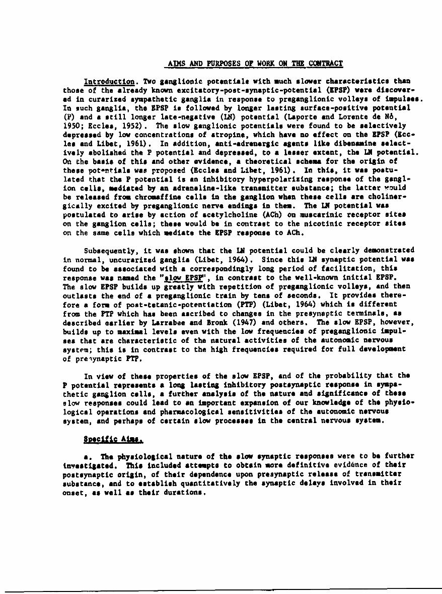
AIMS AND PURPOSES OF WORK ON THE CONTRACT
Introduction. Two ganglionic potentials with much slower characteristics thanthose of the already known excitatory-post-synaptic-potential (IPSP) were discover-ed in curarized sympathetic ganglia in response to preganglionic volleys of impulses.In such ganglia, the ZPSP is followed by longer lasting surface-positive potential(P) and a still longer late-negative (LN) potential (Laporte and Lorente de N6,1950; Eccles, 1952). The slow ganglionic potentials were found to be selectivelydepressed by low concentrations of atropine, which have no effect on the ZPSP (Ecc-lea and Libet, 1961). In addition, anti-adrenergic agents like dibenamine select-ively abolished the P potential and depressed, to a lesser extent, the LN potential.On the basis of this and other evidence, a theoretical schema for the origin ofthese pot-ntials was proposed (Eccles and Libet, 1961). In this, it was postu-lated that the P potential is an inhibitory hyperpolarizing response of the gangl-ion cells, mediated by an adrenaline-like transmitter substance; the latter wouldbe released from chromaffine cells in the ganglion when these cells are choliner-gically excited by preganglionic nerve endings in them. The IN potential waspostulated to arise by action of acetylcholine (ACh) on muscarinic receptor siteson the ganglion cells; these would be in contrast to the nicotinic receptor siteson the same cells which mediate the EPSP response to ACh.
Subsequently, it was shown that the LN potential could be clearly demonstratedin normal, uncurarized ganglia (Libet, 1964). Since this IN synaptic potential wasfound to be associated with a correspondingly long period of facilitation, thisresponse was named the "sjlow EPS1', in contrast to the well-known initial EPSP.The slow EPSP builds up greatly with repetition of preganglionic volleys, and thenoutlasts the end of a preganglionic train by tens of seconds. It provides there-fore a form of post-tetanic-potentiation (PTP) (Libet, 1964) which is differentfrom the FTP which has been ascribed to changes in the presynaptic terminals, asdescribed earlier by Larrabee and Bronk (1947) and others. The slow EPSP, however,builds up to maximal levels even with the low frequencies of preganglionic impul-ses that are characteristic of the natural activities of the autonomic nervoussystem; this is in contrast to the high frequencies required for full developmentof preiynaptic FTP.
In view of these properties of the slow LPSP, and of the probability that theP potential represents a long lasting inhibitory postsynaptic response in sympa-thetic ganglion cells, a further analysis of the nature and significance of theseslow responses could lead to an important expansion of our knowledge of the physio-logical operations and pharmacological sensitivities of the autonomic nervoussystem, and perhaps of certain slow processes in the central nervous system.
Apecjifi Aims.
a. The physiological nature of the slow synaptic responses were to be furtherinvestigated. This included attempts to obtain more definitive evidmnce of theirpostsynaptic origin, of their dependence upon presynaptic release of transmittersubstance, and to establish quantitatively the synaptic delays involved in theironset, as well as their durations.
2
b. Further evidence was to be sought bearing on the hypothesis that a cate-cholamine is involved as a transmitter in mediating the P potential. This hascentered chiefly on the testing of the effects of such compounds on the level ofthe transmembrane potential difference of ganglion cells.
c. Further testing of the sensitivities of the slow synaptic responses topharmacological agents of the ganglion-blocking type was to be carried out.
METHODS
For experiments in which recordings were made from the surface of the ganglion,the isolated preparation was mounted in a chamber, as described elsewhere in de-tail (Eccles and Libet, 1961; Eccles, 1952). The mamalian ganglion utilized wascommonly the superior cervical of the rabbit, but others, such as the cat's cooliacor stellate, were also employed. The sheath was removed from ganglion and postganglionic nerve, to reduce external shunting and increase the penetration ofchemical agents. In the frog, the sympathetic chain was utilized, with a portionof the sciatic bundle containing the postganglionic bundles of the 9th or 10thganglion in the chain. Ganglia were mounted on wire electrodes, Pt for stimu-lation of the preganglionic fibers and Ag-AgCl for recording at the ganglion andpostganglionic sites. The preparation was bathed in a Ringer-Krebs solution,which for mammalian ganglia, was maintained at 37-38oC and bubbled with 95% 02 -
5% CO2 . In recording, the preparation and electrodes could be brought into airout of the saline by tilting the chamber. Responses were recorded, with DC empli-fication throughout, between the active lead and an indifferent one generallylocated at the crushed end of the postganglionic nerve.
Intracellular recordings from single ganglion cells were made with glassmicropipettes having tip diameters of less than 0.5 microns. Successful ex-periments of this kind were confined to the frog's sympathetic ganglia, which canbe penetrated by the electrode, though not without some difficulty. The impene-trability of iaumalian ganglia, owinj to an all-pervasive tough connective tissuestructure throughout its interior, requires special development of techniques todrive the microelectrode into the ganglion. The experiences with this will bedescribed in the section below, on Experiments etc. The pro-and postSanglionictrunkm of the fro6 ganglia were drawn into glass tubes, filled with saline, whichserved as stimulating or recording leads at these points, according to the methodof Furshpan and Potter (1959). The microelectrode was advanced by an oil-filleddrive operated from another table. The Ag-AgCl wire in the micropipette fed intothe usual cathode-follower impedence-converter, and from there into DC amplifiersor meters.
UPEIMNErS, TESTS AND RSULTS
Synaptic potentials, uncontaminated by any propagated action potentials andtheir associated after-potentials, may be recorded at the surface of curarisedsympathetic ganglia. A single preganglionic volley then may elicit a series ofthree ganglionic potentials (Fig. 2; see also Iccles, 1952), representing theinitial 3PSP, a surface-positive potential (1), and a late negative one whichhas the properties of a slow ZPSP (Libet, 1964).
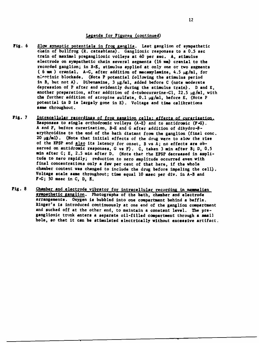
Decremental propamatlon in iostaanallonic nerve. With an electrode positionedat different points along the postganglionic nerve (see diagram, Fig. I), the
3
potentials recorded at the curarized anUmmalian ganglion may be seen to fall offrapidly with increase in distance from the end of the ganglion (Fig. 2, A-F). Forsuch an experiment all postgang'ionic nerve bundles except one were cut off, andthe remaining one was exceptionaLiy tll cleaned of adhering sheath tissue; pre-sumably the decrement with distance would be somewhat less steep under naturalconditions with a lower extracellular resistance. The change in the first 0.5 i
of the nerve bundle may not be very significant as it is impossible to avoid trap-ping small amounts of the saline solution at the junction of the ganglion withthe postganglionic bundle, when the preparation is raised into air for recording.
All three ganglionic responses, the initial EPSP, the P potential, and theslow EPSP, fall off with increasing distance from the ganglion, and do so atapproximately equal rates. It is difficult to precisely estimate the rates ofsuch decrementation from the available data, and still more so the times forachieving the maximum amplitude of each potential at each point on the postganglion-ic nerve. The times to the true maxima of the two slow responses are in any caseobscured by overlapping with the preceding synaptic responses. The rough approx-imations obtainable for the space and time constants, however, are in keeping withthe orders of magnitude expected for passive cable conduction in C fibers.
Preganalionic nerve recordings (see diagram, Fig. I), on the other hand, showno evidence of these ganglionic potentials, even when the proximal electrode isplaced less than 1 mm away from the beginning of the ganglion (Fig. 2, I0. Onlythe diphasic recordings of the action potentials in the preganglionic nerve fibersare detected in the superior cervical preparations, during the application oftrains of stimuli at up to 40 per sec for 1 sec or less.
The slow synaptic potentials are thus seen to be localized to the region ofthe ganglion cell bodies and to decrement rapidly in the postganglionic axon, inthe same way as the initial EPSP.
Effects of lowered Ca++/Mg+ ratios. The dependence of the slow potentials onpresynaptic release of ACh had already been indicated by the effect of botulinumtoxin (Eccles and Libet, 1961); this was now further examin-d by subjecting theganglia to external solutions with low Ca/Hg ratios. It has been shown that suchtreatment selectively depresies presynaptic release of transmitter subntances inresponse to presynaptic impulses (del Castillo and Katz, 1954; HuttLr and Kostial,1954).
Suitable lowering of the normal ratio of the external concentrations ofCa/Mg depressed or abolished all postsynaptic responses. In the curarized Smal-ian ganglion all three postsynaptic potentials were affected similarly; if anything,the P potential and slow EPSP were depressed wore readily than the initial EPSP.In the uncurarized ganglion, when postaynaptic discharge of iopulses was abolishedby this treatment, no postsynaptic potentials of any kind remained. These effectswere initially observed by simply raising the external MgCI 2 concentration fromImM/l to l0mM/I; subsequently raising the MgCl 2 concentration to 20 14/l pro-duced complete block. Since it Is pcssible for Mg++ concentrations as high as20 ntl/l to block conduction in some axons (Katz and Miledi, 1964) Ca/Mg ratio was also loweed by lowering Ca and raising only moderately the Mg concentration. With all CaCI 2 omittedatd MIgClt raised to only 5 nl/l in the Krebs-Ringer solution, all postsynapcic responseswere abolished; monitoring the preganglionic nerve in this case showed that the conductionof preganglionic impulses was nct significantly affected under tbsee conditions. These
4
results demonstrate that the generation of the P potential and the slow EPSP areat least as sensitive as the initial EFSP to a lowering of the external Ca&/Ms ratio.
Latency of the postsynaptic P pctential in msamalian ganglia. In order to dis-play the onset of the P potential without contamination by the initial EPSP, thelatter was completely suppressed by strong curarization. To accomplish this aconcentration of d-tubozurarine (d-TC) is required which is about 10 times thatneeded to just block the generation of a p--atganglionic propagated spike by asingle orthodromic volley. At these high concentrations of curarizing agent (about125-150 Pg/ml) the slow postaynaptic potentials, especially with single orthodromicv~lleys, are also depressed, but are still definite enough for the purpose of study-ing latency (Fig. 2, Q). Incidentally, the presence of P and slow EPSP responsesI- the absence of any initial EPSP provides some 3f the evidence for regardingthen as independent prozesses, rather than as some sort of after-effects of theEPSP (see Eccles and Libet, 1961).
That the latent period before onset ef P pctenrlal is a considerable one isevident even in the slow sweep record cf Fig.3,. When the sweep speed is in-creased to facilitate more precise measvrements of latency, however, it becomesdifficult to pinpoint the onset of P with such small amplitudes of the potential(Fig. 3D and F). Even so, the minimum latency appeared to be about 40 amec. Thisvalue was confirmed when the onset of the response was made much more distinct bypost-tetanic-potentiation (FTP) of the postsynaptic P potential. FTP of the Ppotential could be accomplished without any disturbing increase in the amplitude ofthe initial FPSP, in the sttongly curarized ganglion. Fig. 3 - , IG , and I show theresponses to single preganglionic volleys delivered at increasing times after theend of a 15 sac train of preganglionic volleys at 60/sec. The strength of stimulusin this series was sufficient to excite all che B and C fibers in the preganglionicnerve; this is much more optimal for eliciting P response than is stimulation ofB fibers alone, as In Fig. 3-D, E (see Ecclea and Libet, 1961).
The long latent period of 40 msec for the postsyaaptic P potentiai is to becompared to the shot' ore if - msec for the onset of the first (So) discharge inthe uncurarized ganglion (Fig. 3-A2 ). The latent period for the onset of the initialEPSP as measured in a lightly curarized ganglion, is approximately the same as thatof this Sa discharge. Th.ese latency %alues of -o.r~ e all Inslude conduction tiamsfrom the point of pregangliort: stimulation to the presynaptic terminals. Thelatent period of the !eczrýd ýr Sh portion of t•,e postdynaptic discharge (Fig. 3A),(also Fig. 4A) was about 15 msec lcnger than that for the Sa portion; this differ-ence can be accounted foa by the slower c.:ndurcton velocity of the pregSanglionicC fiber group as opposed ti the B titer gr(up (Fccles and Libet, 1961). Theonset of the P pQtenrial is, hvwever, initiated by the faster B impulses, whetherthese ate delivered alone (Fig. jr E) cr are foilowed by the C impulses (Fig. 3F - I).Subtracting the 5-6 msec laten:y of the Sa postmynaptic discharge thus leaves uswith a net additional delay of about 35 msic for the onset of the postsynaptic Ppotential, over and above that required for the onset of the Initial RPSP.
Latency of the slow EPS? in itamualion J1nlia. To demonstrate clearly thewer- onset of the late negative postsynaptic potential, the slow EPSP, possible inter-ad ference or masking by the P potential as well as the initial EPSP had to be elimi-
nated. This could be accomplished in the strongly curarized ganglion (Fig. 40)by the addition of a suitable concentration of diberomine (Fig. 4D -G). The latterdrug can block the P response completely while depressing the slow EPSP only
5
partially (Eccles and Libet, 1961). It can then be seen clearly (Fig. 4C -G) thatthe onset of the slow EPSP has a latent period in the range of 200-300 masec indifferent preparations. Some ganglia exhibit little or no postsynaptic P poten-tial in response to single preganglionic volley. In such preparations the onsetof the slow EPSP wty be seen distinctly even in the lightly curarized condition andwith no dibenamine added. These ciri sLances apply to the recordings shown inFig. 4 from a celiac ganglcin Df a cat. When the initial EPSP in I-A ot Fig, 5is recorded at a slower sweep speed it can be seen that only sometime after thereturn of the ganglionic potential to the resting base line does the slow EPSP be-gin to appear (arrow, Fig. 5, I-B). The onset of the slow EPSP is delayed bysomcwhat more than 300 msec from that of the in:tial EFSP in this case. Column IIin Fig. 5 shows the comparable responses after the addition of atropine (0.1 4g/ml),which can selectively block tl'e slow EPSP (Eccles and Libet, 1961). At stillslowcr sweep speeds (Fig. 5, I - CD.) the more complete course of the slow EPSPis visible.
Slow postsynaptic potentials in frog Ranglia. Concentrations of d-TC of 5to 10 pg/ml were found to block postganglicnic discqarge in response to pre-ganglionic volleys, leaving an EPSP oi appreciable size instrad. Following shorttrains (0.25 - I sec duration) cf repetitive pre,;4lionic volleys, at 10-80 persec, a distinct P potential could appear, wi-en the tetanically sumuated EPSP'ssuddenly collapse at the end cf the train (Fig. 6 . This P potential has a dur-stion of several seconds and maximum amplitudes equal to about 10% of the uncurar-ized spike amplitude.
The P synaptic potential was elicited only when the stimulating electrode onthe thoracic chain was located less then 10 m anterior to the ganglion (no. 9 or10 in the chain), as seen in Fig. 6. This means that only preganglionic fibersentering the chain within one cr two segments above the responding ganglion couldelicit this slow synaptiz potential. Stimulation of the thoracic chain more an-teriorly could still elicit a large EPSP, or large spike discharge in the uncur-arized ganglion, but could not generate any P potentiol. The preganglionic fiberswhich elicit the P synaptic potential, then, can be different and separate fromthose which elicit only an ElPSP.
Late negative synapti. potentials (slow EPSP) were ordinarily not seen in thefrog's sympathetic ganglia (eAcept in szme instances after higher frequency trains(60-80/sec) of preganglionic volleys). Thi.s is in contrast to the large ones ob-servable in mammalian ganglia (Eccles 1952) and the rather small ones reported forturtle ganglia (Laporte and Lorente de N5, 1950).
The P potential of frog garglia Lould be blocked by low concentrations ofatropine (0.1 - 0.2 pg/ml) as in mammalian ganglia. Dibensualne, however, had onlya moderate depressing action, in concentrations (3 pg/ml) which abolish P com-pletely in masmalian ganglia. The different responsiveness of the frog to certainautonomic blocking agents has already been documented by &irnstock.
Intracellular recordings in sinLle -cells of frv .an81
a) Uncurarized4 anlis. The general frrm and propurties of the dischargeof a frog ganglion cell, ortho-and antidromnically, as already described by Nishland Koketsu (1960) and Blackmsn, et al. (1963a), were confirmed by us. Ampleincidence of spontaneous miniature postsynaptic potentials was also seen, asreported by Blackman, et al (1963b).
6
The hyperpolarizing after-potential which follows discharge o' a ganglion cellwhich has been fired antidromically via its own axon, is a "true" after-potential;it should not contain any postaynaptic component such as the P potential (which isalso presumably a hyperpolarizing action). Atropine (0.1 4g/ml) was found to haveno effect on this true after-hyperpolarizing potential, in antidroiic responses.Since such concentrations of atropine completely suppress the P synaptic potential,as generated orthodromically in curarized ganglia, this proves that the P poten-tial involves a different mode of production than the true-hyperpolarization; thatis, it supports the contention that the P potential is a po3tsynaptic potentialelicited by synaptic transmitter substances, one of which can be antagonized byatropine.
b) Curarized ganglia. To demonstrate a postaynaptic hyperpolarisingresponse to preganglionic impulses, as distinguished from a true after-potentialof this kind, it is necessary to block the firing of impulses (action potentials)by the ganglion cell. This can be done by applying a curarizing agent in a con-centration sufficient to depress the EPSP below the cell's firing level. The Psynaptic potential, recordable at the surface of a ganglion, survives this kind oftreatment (Eccles, 1952" Eccles and Libet, 1961).
With a microelectrode in place inside a ganglion cell, the application ofeither d-TC, or dihydro-O-erythroidine, in concentrations just adequate for block-ing postsynaptic firing (about 5 4g/ml for either one) appeared to depress not onlythe postsynaptic response but also to depress and block transmission in the p~e-synaptic terminals as well. The evidence for the Uitter was that the EPSP eitherdisappeared suddenly (in an all-or-none fashion) shortly after the cell spike hadbeen blocked (Fig. 7C-E ), or no EPSP response could be elicited at all after thecell spike was lost. In addition, during the short time when an EPSP was stillelicited after loss of the spike, the latent period of its onset increased distinctlyover what it was before applying the curarizing agent (Fig.7A-B). Similarly, whenthe weakest blocKing concentration of these drugs was applied to a ganglion beforethe impalement of a ganglion cell by the microelectrode, subsequent penetration ofcells revealed the following. Either the cell could still respond to an ortho-dromic volley with a spike discharge, although at times somewhat delayed by a re-duction in the rate of rise of EPSP, or no response (cell spike, or BPSP) waspresent at all. In the latter case, the cell could still respond to an antidromicvolley with a normal discharge.
Depression of presynaptic transmission by TEA has also been reported by Riker(1963), with microelectrode studies of frog ganglia, although no mention of it wasmade by Blackman, et al (1963). The question arises as to why it is possible torecord an EPSP from the surface of the ganglion with the use of these drugs. It maybe that there is some slight mechanical injury or distortion of the presynapticterminations when a micropipette is being pushed through the ganglionic mass andinto a cell. This may increase the susceptibility of these terminals to a blockingaction by curarizing agents; such an increase in auceptibility by mechanical fac-tors has been observed for the neuromuscular junction (Katz and Miledi, unpublished).
Blockade of conduction in presynaptic terminals, regardless of tt specialcauses, obviously would abolish all postsynaptic responses. Consequently, a slowpostaynaptic hyperpolarizing response will have to be looked for under conditionswhich more selectively block the EPSP alone; this aspect of the study could thusnot be completed at present. Testing of other nicotinic blocking agents, which
7
might not possess the same tendency to depress presynaptic terminals as well, hashowever, been carried forward (see below).
c). Effect of epinephrine. With a ganglion cell impaled by a microelectrode,the effect of adding epinephrine on the resting transmembrane potential differencewas observed. In order to achieve a rapid entry of the substance into the vicinityof the cell, a tiny volume (0.03 al) of solution was delivered into the bath fromthe 10 ý tip of a glass pipette. The litter had been previously fixed in positionwith its tip a few -m away from the ganglion, and the solution was expressed slowly(over a 10 sec period) from the pipette by a remote micro-syringe which was con-nected to the pipette via a long polyethylene tube. Solutions in the pipette con-tained 1 m4/ml of epinephrine or more epinephrine in frog Ringer's.
Within 15 seconds after extrusi.on of the epinephrine solution, there occurreda distinct slow rise of several millivolts in the resting membrane potential, whichwas then sustained. Similar extrusions of Ringer's solution alone gave no suchchange; the change expected to be seen if there were any motion of the micro-electrode upon the extrusion of solution would be a reduction in resting potential,due to damage to the cell membrane. Such depolarizations are all too readily ob-served, if the solution is extruded too vigorously and in too large a volume. Ahyperpolariting action of epinephrine is in accord with its postulated role as amediator of the P synaptic potential (Eccles & Libet, 1961) and with the inhibit-ory effect of injected epinephrine on postganglionic discharge in response to pro-ganglionic volleys (Marazzi, 1939; Lundberg, 1952; Eccles and Libet, 1961).
Additional phsrmacololical agents for selective blockade of ZPSP. Since d-TCand dihydro-#-erythroidine both are prone to produce presynaptic block in frogganglia it was desirable to zest other blockers of nicotinic receptors which arechemically rather different from the curare group. Hexamethonium is one suchcompound, which is an excellent blocking agent in mammalian ganglia. In frogganglia, however, hexamethonium in concentrations up to 150 4/ml hardly depressedthe synaptic response. Blackman, at al. (1963) did repo t blockade by still higherconcentrations of this drug (about 225 g4/ml, i.e. 7xlO;;, but effects that re-quire such high dosage did not seen worth pursuing.
Mecanylamine, however, turned out to be a very effective blocking agent infrog ganglia. As little as 2-4 pig/ml could block orthodromically elicited post-ganglionic discharge and depress the FPSP. Under these conditions a good P po-tential could be seen to follow a short preganglionic train of impulses (Fig. 6 ).There was not time, before the termination date of this contrect, to utilize thisdrug with intracellular recording technique.
Povelogmnt of intracellu lar technique with monLAlian gangli. Mammalianganglia must of course be maintained at 37-38'C and with suitable oxygenation andstirring of the solution. A special chamber and varm-vater bath was constructedto provide this while at the same time supplying baffles against the sudden motionsof the fluid from the 02 bubbling. Photographs of this chamber are shown in Fig. S.Transmitted illumination of the ganglion is provided by reflection from a mirrormounted in the bath below the ganglion compartment.

8
The stumbling block to successful intracellular recording was not the inabilityof the microelectrode to penetrate the dense connective tissue meshwork which per-vades the interior of such ganglia. Ganglion cells at the very surface of a de-sheathed ganglion can occasionally be impaled (as reported by Eccles, 1955, 1963),but these are not suitable for the study of the slow synaptic responses; it shouldbe recalled that the latter are postulated to involve diffusion of transmitter sub-stances through intercellular spaces.
Preliminary attempts were made to facilitate penetration by treating the ganglionwith preparations of proteolytic enzymes (trypsin), or with enzymes to hydrolyseother portions of the interstitial stroma (collagenase, elastase, hyaluronidase).After action by an enzyme was sufficient to permit penetration by the microelectraie,however, there was also a considerablelossint*e postganglionic response to pregsngl-ionic volleys. Perhaps a more thorough investigation will uncover conditions inwhich a differential between the effects on penetrability and on synaptic transmissioncan be achieved. As far as tested here, such enzymatic methods appeared to be un-suitable.
Penetration into certain other tissues (e.g. retina) has been improved by someinvestigators by vertically vibrating either the whole preparation against the micro-electrode, or vibrating the microelectrode as it is lowerod into the tissue. Wehave now constructed a light-weight holder for the electrode which includes a s$ail,lectromagnetic coil for vertical vibration of the electrode (Fig. 8 ). Preliminarytests have indicated that this device may produce satisfactorily penetration, butfurther york on the optimum parameters of excursion, frequency, and pulse durationof the vibratory oscillations is still necessary.
SUMMARY OF RESULTS
Properties of the atropine-sensitive, slow synaptic responses in mamaliansympathetic garglia were further analysed. It was shown that both the surface-positive (presumably hyperpolarizing) potential (P), and the late surface-negativepotential ("slow excitatory postsynaptic potential," or slow IPSP) are localized tosoma-dendritic region, Just as is the well-known initial 1PSP (which is curare-sensitive). All these postaynaptic responses were abolished by lowering the Ca.attratio in the external sedium; i.e. they all depend upon release of some transaitcersubstancee at presynaptic teruinals. The slow responses were found to have extra-ordinarily long latent periods for their onset (about 35 maec for the P potential,and 200-300 seec fcr the slow EPSP, exclusive !- conduction time to the ganglion).
A slow P posteynaptic potential could also be denoostrated in the curarizedsympathetic ganglia of the frog, if preganglionic fibers from spinol selgxents suit-ably close to the ganglion under study were stimulated. This P potential was alsoeasily blocked by atropine, but only partially by dibnemuine.
Intracellular recordings from single ganglion cells of the frog were made.The true after-potentials which follow antidromic discharge of such cells are notaffected by doses of atropine which depress the P synaptic response. The curarizingalgents, d-tubocurarine and dihydro4-erythroidine, not only depress the EISP butalso appear to block conduction in presynaptic terminals of such preparations. Clearevidence and analysis of the curare-insensitive slow post-sInaptic responses at thesingle cell level will, therefore, have to await the use of more buitable blockingagents.
9
A catecho~amlne, epinephrine, was found to produce a hyperpolarlzation of themembrane of ganglion cells in the frog. This is in accord with its rostulated rolein the generation oi tha P synaptic potential.
Additional :_icotinic blocking agents were tested on the frog's syvpatheti'ýganglias aiexamethonium required concentrations above 150 pg/ml to produce appreci-able depression of the ?. AP, and is therefore unsuitable. Necamylamine, 2-4 PS/ml,did depress the EYSP brrongly, without blocking the slow P potential.
Development- of technique for intracellular recording in mammalian sympatheticganglia wer.z deacribed. A device for vibratory oscillation of the microelecrcodeappears to improve 'he penettatic-' oi the electrode into the ganglion.
REFERENCES
1. Blackman, J. G., Ginsborg, B. L., and Ray, C. (1963a). J. Physiol. 167, 355-373.
2. Blackman, J. G., Ginsborg, B. L. and Ray, C. (1963b). J. Physiol, 167, 389-401.
3. de Castillc, J. and Katz, B. (1954). J. Physiol. 124, 560-573.
4. Eccles, R. M. (1952). J. Physiol. 117, 181-195.
5. Eccles, R. M. (1955). J. Physiol. 130, 572-58,.
6. Eccles, R. rM. (1963). J.. hysiol. 165, 387-391.
7. Eccle., R. M, and Libet, B, (1961). J. JJ siol. 157, 484-503.
8. Furshpan, 2. J. and Potter, D. D. (1959). J. Physiol. 145, 289-325.
9. 1iutter, 0. F. and Kostial, K. (1954). J. Physiol. 124, 234-241.
10. Katz, B. end Miledi, R. (1964). J. Physiol. 171, 10-11P.
11. Lapcate, Y. and Lorente de N6, R. (1950). JS cell. comp. 1yiol. 35, Suppl. 2,61-106.
12. Lx-abeer, M. 0. and Bronk, D. W. (1947). J. Neurophysiol. 10, 139-154.
13. Libet, B. (1964). J. Physiol. 17"4, 1-25.
14. Lundberg, A. (1952). Ata physiol., scand. 26, 252-263.
15. Marazzi, A. S. (193Q). J. Pharmacol. 65, 395-404.
16. hishi, S. and Koketou, K. (1960). J. cell. comp. Physhol. 55, t5-30.
17. Riker. W. K. (1963). The Pharmacologist, 5, 242.
10
CONCLUSIONS AND RECOMMIDAT1ONS
This work has added to our understanding of the synaptic mechanisms in sym-pathetic ganglia and of the ways in which low doses of atropine say act in the body.It also supports the contention that there is an adrenergic (inhibitory) synopticmechenism operating in ganglid, in addition to the well-known cholinergic one. Itshould be noted that the fctivation of this adrenergic mechanism by preganglionicimpulses is also sensitive to blockade by atropine.
Atropine can affect brain function, and has been shown in recent years toblock spdcifically cholinergic effects at certain sites in the brain. It isrecoawiended that analyses of the effect of cholinergic agents and atropine onbrain functf.on tak* into account the possibility that these effects are transpiringat postaynaptic sites which generate slow synaptic responses, of the types seen insympaheto.c ganglia.
11
LEGENDS FOR FIGURES
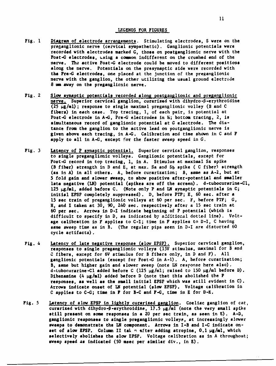
Fig. 1 Diagram of electrode arranrements. Stimulating electrodes, S were on thepreganglionic nerve (cervical sympathetic). Ganglionic potentials wererecorded with electrodes marked G, those on postganglionic nerve with thePost-C electrodes, using a common indifferent on the crushed end of thenerve. The active Post-G electrode could be moved to different positionsalong the nerve. Potentials on the presynaptic side were recorded withthe Pre-G electrodes, one placed at the junction of the preganglionicnerve with the ganglion, the other utilizing the usual ground electrode8 mm away on the preganglionic nerve.
Fig. 2 Slow snaptic gotentials recorded along postganglionic and preganglionicnerve, Superior cervical ganglion, curarized with dihydro-o-erythroidine(25 1g/ml); response to single maximal preganglionic volley (B and Cfibers) in each case. Top tracing, 1, of each pair, is potential atPost-G electrode in A-G, Pre-G electrodes in 1i; bottom tracing, 2, issimultaneous record of ganglionic potential at G electrode. The dis-tance from the ganglion to the active lead on postganglionic nerve isgiven above each tracing, in A-G. Calibration and time shown in C and Fapply to all in A-G, except .#or the faster sweep speed in G.
Fig. 3 Latency of P synaptic potential. Superior cervical ganglion, responsesto single preganglionic volleys. Ganglionic potentials, except forPost-G record in top tracing, 1, in A. Stimulus at maximal Sa spike(B fiber) strength in D and E, at max. Sa and Sb spike ( C fiber) strength(as in A) in all others. A, before curarization; B, same as A-2, but at5 fold gain and slower sweep, to show positive after-potential and smallerlate negative (LN) potential (spikes are off the screen). d-tubocurarine-Cl,125 4/1l, added before C. (Note only P and LN synaptic potentials in C;initial EPSP completely suppressed). D, before PTP; E, 60 sec. after a15 sec train of preganglionic volleys at 60 per sec. F, before PTP; G,H, and I taken at 30, 90, 240 sec. respectively after a 15 sec train at60 per sec. Arrows in D-I indicate beginning of P potential (which isdifficult to specify in D, as indicated by additional dotted line). Volt-age calibration in F applies to C-I; time in F applies to D-I, C havingsame sweep time as in B. (The regular pips seen in D-I are distorted 60cycle artifacts).
Fig. 4 Latency of late negative response (slow EPSp). Superior cervical ganglion,responses to single preganglionic volleys (15V stimulus, maximal for B andCfibers, except for 6V stimulus for B fibers only, in D and F). Allganglionic potentials (except for Post-G in A-l). A, before curarization;B, same but higher gain and slower sweep (note IN response here also),d-tubocurarine-C1 added before C (125 "/ml; raised to 150 1g/ml before D).Dibenamine (4 1g/ml) added before D (note that this abolished the Presponses, as well as the small initial EPSP which was still evident in C).Arrows indicate onset of LN potential (slow EPSP). Voltage calibration inC applies to C-G; time in F for B-C and F-C, time in E for D-E.
Fig. 5 Latency of slow EPSP in lightly curarized ganglion. Coeliac ganglion of cat,curarized with dihydro-O-erythroidine, 17.5 1g/ml (note the very small spikestill present on some responses in a 20 per sec train, as seen in E), A-D,ganglionic responses to single preganglionic volleys, at increasingly slowersweeps to demonstrate the LN component. Arrows in I-B and I-C indicate on-set of slow EPSP. Column II tak n after adding atropine, 0.1 gg/ml, whichselectively abolishes the slow EPSP. Voltage calibration as in A throughout;sweep speed as indicated (50 msec per similar div., in E).
12
Legends for Figures (continued)
Fig. 6 Slow eynaptic potentials in frog ganglia. Last ganglion of sympatheticchain of bullfrog (R. catesbiana). Ganglionic responses to a 0.5 sectrain of maximoal preganglionic volleys at 40 per sec. A, stimuluselectrode on sympathetic chain several segments (16 an) cranial to therecorded ganglion; in B-B, stimulus applied at only one or two segments( 6 am ) cranial. A-C, after addition of mecamyla-ine, 4.5 pg/ml, fornlectinic blockade. (Note P potential following the stimulus periodin B, but not A). Dibenamine, 3 jg/ml, added before C (note moderatedepression of P after and evidently during the stimulus train). D and E,another preparation, after addition of d-tubocurarine-Cl, 22.5 pg/ml, withthe further addition of atropine sulfate, 0.1 gg/ml, before E. (Note Ppotential in D is largely gone in E). Voltage and time calibrationssame throughout.
Fig. 7 Intracellular recordinrts of frog ainalion cells: effects of curarizatLon,Responses to single orthodromic volleys (A-E) and to antidromic (F-C).A and F, before curarization, B-E and G after addition of dihydro-o-erythroidine to the end of the bath distant from the ganglion (final conc.20 pg/ml). (Note that initial effects of the drug were to slow the riseof the EPSP and also its latency for onset, B vs A; no effects are ob-served on antidromic responses, C vs F). C, taken 3 min after B; D, 0.5min after C; E, 2.5 min after D, (Note that the EPSP decreased in ampli-tude to zero rapidly; reduction to zero amplitude occurred even withfinal concentrations only a few per cent of that here, if the wholechamber content was changed to include the drug before impaling the cell).Voltage scale same throughout; time equal 10 msec per div. in A-B andF-C; 50 msec in C, D, g.
Fig. 8 Chamber and electrode vibrator for intracellular recording in mammalians]ymathetic Sanalion. Photographs of the bath, chamber and electrodearrangements. Oxygen is bubbled into one compartment behind a baffle.Ringer's is introduced continuously at one end of the ganglion compartmentand sucked off at the other end, to maintain a constant level. The pre-ganglionic trunk enters a separate oil-filled compartment through a smallhole, so that it can be stimulated electrically without excessive artifact.
C,) A
'IH - - D
CDga,
-oa,
I
a,
I axn8;�
Figure 2
O.Smrn B I.* mm C 1,5mm
2 0*~mv]
2.Smm E I 3 5 F o m
0.5$cc
2
HGIPreganq, 5. 5mm
sec msecgang.
2%. I.OmV
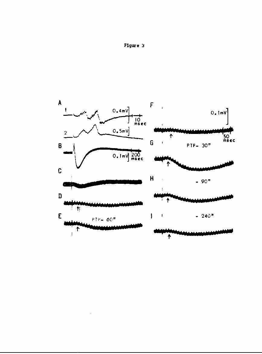
Figure 3
0.4mV 0. tmVl. %he10
2 ~ 0 5mV]-&AA&@50,
B PTP- 30" me
~ -H -90"
E TP- 6C," 1 I 240"
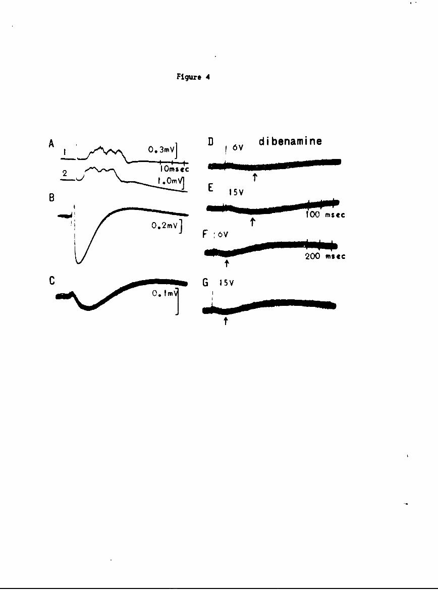
Figure 4
A 0.3VD v di benami ne
2 lmiec
B OI Em 15V
F :6v
200mse
C G isv0.m
o O.J.
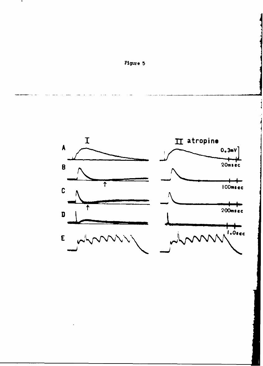
Figure 5
I fl atropineA 0. 3ev
B ~20m s £
fI IO0msec
f 200. etc
'.Osc@
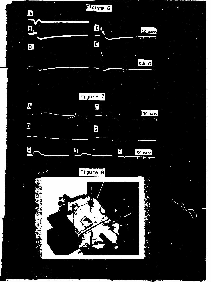
Figure 6A
B C 10 lose
DE
Figure 7A F
10 ma ec
8 G
C D E 5iwee
Figure- 8


![Attach 1 LATE [PR-PC] Development Application DA18/0685](https://img.pdfslide.net/doc/110x75/6176fbe25ca855120d27d086/attach-1-late-pr-pc-development-application-da180685-.jpg)