Embed Size (px)
Citation preview
30/04/14 Evaluation and management of supracondylar fractures in children
www.uptodate.com/contents/evaluation-and-management-of-supracondylar-fractures-in-children?topicKey=EM%2F6539&elapsedTimeMs=2&source=see… 1/36
Official reprint from UpToDate www.uptodate.com ©2014 UpToDate
AuthorLeticia Manning Ryan, MD, MPH,FAAP
Section EditorRichard G Bachur, MD
Deputy EditorJames F Wiley, II, MD, MPH
Evaluation and management of supracondylar fractures in children
All topics are updated as new evidence becomes available and our peer review process is complete.Literature review current through: Mar 2014. | This topic last updated: Feb 10, 2014.
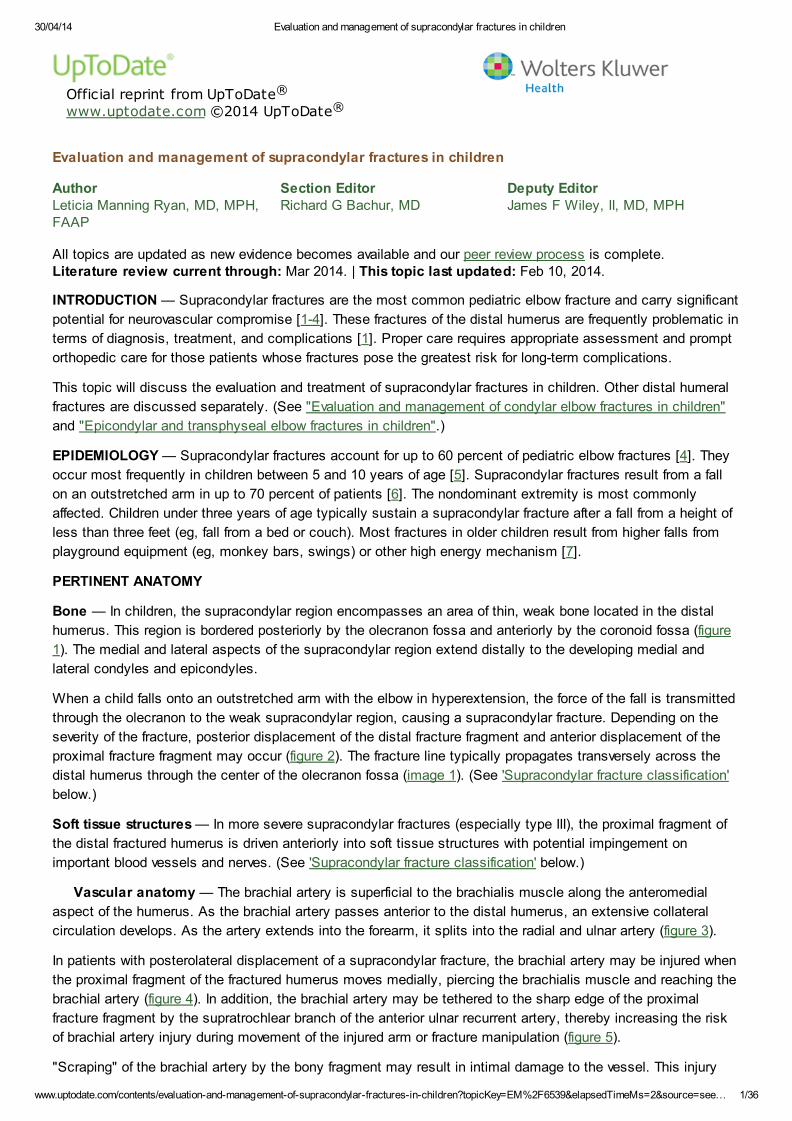
INTRODUCTION — Supracondylar fractures are the most common pediatric elbow fracture and carry significant
potential for neurovascular compromise [1-4]. These fractures of the distal humerus are frequently problematic in
terms of diagnosis, treatment, and complications [1]. Proper care requires appropriate assessment and prompt
orthopedic care for those patients whose fractures pose the greatest risk for long-term complications.
This topic will discuss the evaluation and treatment of supracondylar fractures in children. Other distal humeral
fractures are discussed separately. (See "Evaluation and management of condylar elbow fractures in children"
and "Epicondylar and transphyseal elbow fractures in children".)
EPIDEMIOLOGY — Supracondylar fractures account for up to 60 percent of pediatric elbow fractures [4]. They
occur most frequently in children between 5 and 10 years of age [5]. Supracondylar fractures result from a fall
on an outstretched arm in up to 70 percent of patients [6]. The nondominant extremity is most commonly
affected. Children under three years of age typically sustain a supracondylar fracture after a fall from a height of
less than three feet (eg, fall from a bed or couch). Most fractures in older children result from higher falls from
playground equipment (eg, monkey bars, swings) or other high energy mechanism [7].
PERTINENT ANATOMY
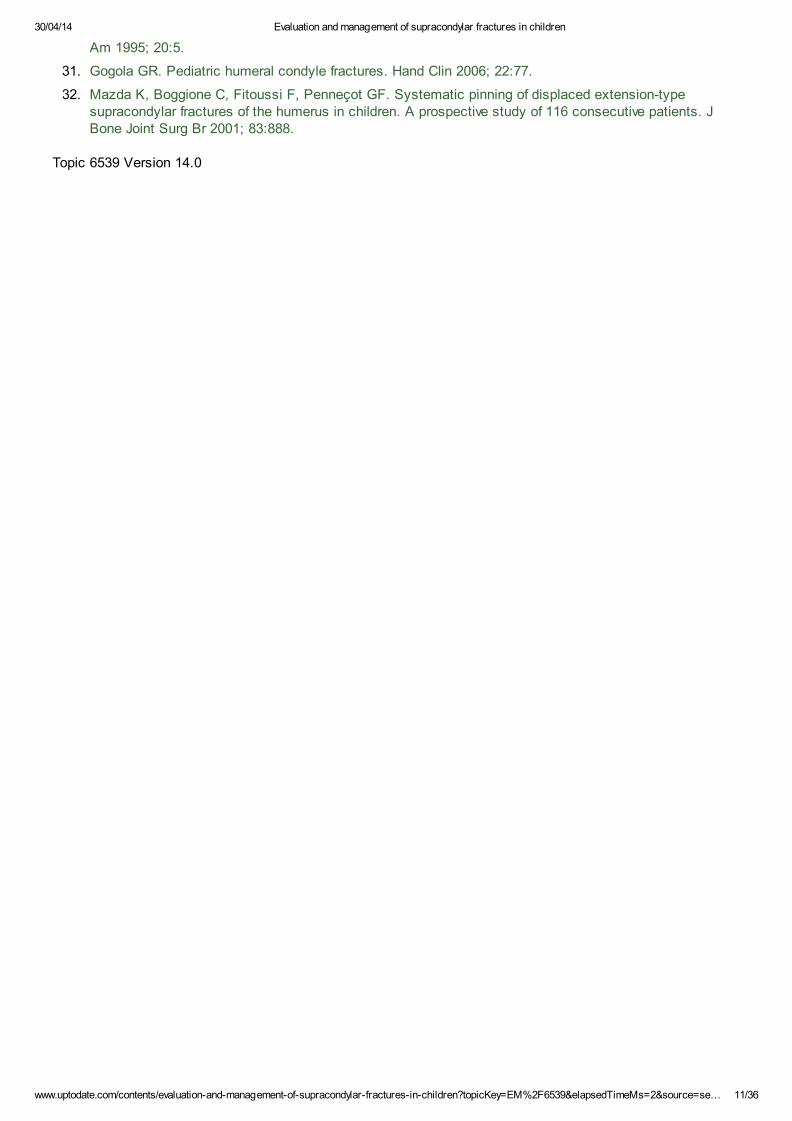
Bone — In children, the supracondylar region encompasses an area of thin, weak bone located in the distal
humerus. This region is bordered posteriorly by the olecranon fossa and anteriorly by the coronoid fossa (figure
1). The medial and lateral aspects of the supracondylar region extend distally to the developing medial and
lateral condyles and epicondyles.
When a child falls onto an outstretched arm with the elbow in hyperextension, the force of the fall is transmitted
through the olecranon to the weak supracondylar region, causing a supracondylar fracture. Depending on the
severity of the fracture, posterior displacement of the distal fracture fragment and anterior displacement of the
proximal fracture fragment may occur (figure 2). The fracture line typically propagates transversely across the
distal humerus through the center of the olecranon fossa (image 1). (See 'Supracondylar fracture classification'
below.)
Soft tissue structures — In more severe supracondylar fractures (especially type III), the proximal fragment of
the distal fractured humerus is driven anteriorly into soft tissue structures with potential impingement on
important blood vessels and nerves. (See 'Supracondylar fracture classification' below.)
Vascular anatomy — The brachial artery is superficial to the brachialis muscle along the anteromedial
aspect of the humerus. As the brachial artery passes anterior to the distal humerus, an extensive collateral
circulation develops. As the artery extends into the forearm, it splits into the radial and ulnar artery (figure 3).
In patients with posterolateral displacement of a supracondylar fracture, the brachial artery may be injured when
the proximal fragment of the fractured humerus moves medially, piercing the brachialis muscle and reaching the
brachial artery (figure 4). In addition, the brachial artery may be tethered to the sharp edge of the proximal
fracture fragment by the supratrochlear branch of the anterior ulnar recurrent artery, thereby increasing the risk
of brachial artery injury during movement of the injured arm or fracture manipulation (figure 5).
"Scraping" of the brachial artery by the bony fragment may result in intimal damage to the vessel. This injury
®
®
30/04/14 Evaluation and management of supracondylar fractures in children
www.uptodate.com/contents/evaluation-and-management-of-supracondylar-fractures-in-children?topicKey=EM%2F6539&elapsedTimeMs=2&source=see… 2/36
may subsequently lead to thrombosis and vascular insufficiency. Because of the extensive collateral circulation
present at the elbow, it is rare for ischemia of the arm to occur from complete brachial artery occlusion [8].
Median nerve injury often accompanies brachial artery impingement.
Nerve anatomy — Following a displaced supracondylar fracture, the median (including the anterior
interosseous nerve branch), radial, and ulnar nerves are at potential risk for injury (figure 3). Although nerve
injuries may rarely be associated with long term sequelae, the majority are neurapraxias, such as temporary
loss of nerve function (especially motor function) without anatomical nerve disruption [9,10].
The median nerve crosses the elbow joint with the brachial artery. Posterolateral distal fracture fragment
displacement with medial movement of the proximal fracture fragment puts the median nerve and its
anterior interosseous nerve branch at the greatest risk of injury (figure 4).
Median nerve injury results in weakness of the flexor muscles of the hand and loss of two-point sensation
on the palmar surface of the thumb and the index and middle fingers [10]. The anterior interosseous nerve
(AION) is the branch of the median nerve most commonly injured (figure 3) [9-11]. The AION lacks a
superficial sensory component [9,10]. Children and adolescents with AION syndrome initially have
proximal forearm pain followed by weakness in the hand with no sensory deficits. On physical
examination they have a weak "OK sign" and/or a lack of distal interphalangeal flexion when making an
OK sign (eg, more of a pincer grasp than an OK sign) (picture 1). (See 'Neurologic deficit' below.)
The radial nerve runs between the brachialis and brachioradialis muscles before crossing the elbow and
penetrating the supinator muscle. Posteromedial distal fracture fragment displacement increases the
chance of radial nerve impingement because of lateral proximal fracture fragment displacement (figure 4)
[12]. Injury to the radial nerve results in weakness of wrist extension, hand supination, and thumb
extension. In addition, altered sensation is found in the dorsal web space between the thumb and index
finger.
The ulnar nerve crosses the elbow posterior to the medial epicondyle and is typically not affected with
extension type supracondylar fractures. However, it is prone to injury following flexion type supracondylar
fractures (figure 6) [8]. Ulnar nerve injury causes weakness of wrist flexion and adduction, finger spread,
and flexion of the distal phalanx of the fifth digit (pinky or little finger). These motor findings are
accompanied by altered sensation of the ulnar side of the ring (fourth digit) and little fingers.
MECHANISM OF INJURY — In general, fractures of the distal humerus are most commonly due to a fall on an
outstretched hand (FOOSH) or direct trauma to the elbow [6]. (See 'Epidemiology' above.)
Extension and flexion are the two major types of supracondylar fracture and each has a characteristic
mechanism of injury [3,8].
Extension fractures account for approximately 95 percent of supracondylar fractures and typically result from a
FOOSH mechanism with the elbow hyperextended [3,9]. The distal condylar complex may then shift in either
the posterolateral or posteromedial direction (figure 4).
Supracondylar flexion fractures account for 5 percent of supracondylar fractures. In contrast to extension
injuries, these fractures result from a direct blow to the posterior aspect of the flexed elbow. In these cases, the
distal condylar complex is more likely to displace in the anterolateral direction (figure 6) [3,8,13].
PHYSICAL FINDINGS — The child with a supracondylar fracture typically has elbow pain, swelling, and very
limited to no range of motion at the elbow [8]. The clinician must rapidly assess the injury to identify degree of
fracture displacement, neurovascular compromise, and evidence of compartment syndrome.
One approach is to initially inspect the affected arm and perform a brief neurovascular examination followed by a
more complete evaluation once pain is controlled and radiographs have been obtained. The clinician should not
encourage active or passive elbow movement in a patient with a suspected supracondylar fracture until a
displaced fracture has been excluded by radiography. Patients with an obviously displaced supracondylar
fracture require initial immobilization before radiography is performed. (See 'Neurovascular examination' below
and 'Analgesia and immobilization' below.)
30/04/14 Evaluation and management of supracondylar fractures in children
www.uptodate.com/contents/evaluation-and-management-of-supracondylar-fractures-in-children?topicKey=EM%2F6539&elapsedTimeMs=2&source=see… 3/36
Inspection — The clinician should carefully inspect the anterior and posterior arm for wounds that indicate an
open fracture while limiting movement of the injured arm. In patients transferred by ambulance or from another
hospital, this task often requires careful removal of previously applied immobilization devices. Open
supracondylar fractures often manifest as a puncture wound or laceration in or just above the antecubital region.
Displaced fractures may have an "S-shaped" configuration or dimpling in the antecubital fossa associated with
marked swelling about the elbow (figure 7 and figure 8).
Ecchymosis over the anteromedial aspect of the forearm suggests brachial artery injury [8].
Nondisplaced fractures may have minimal swelling, but observation will show that the child is not using the
affected arm normally. Posterior distal humeral palpation is usually painful in these patients.
Neurovascular examination — A complete neurovascular evaluation includes an assessment of radial and
brachial pulses and the sensory and motor function of the median, radial, and ulnar nerves (table 1) [14].
Patients with a normal initial neurovascular examination warrant regular repeated evaluation, especially
immediately following splinting, fracture manipulation, or movement of the affected extremity.
Pulse — The clinician should compare the radial and brachial pulse with the opposite limb. Initially
symmetric and equal pulses with normal perfusion and skin warmth are reassuring but do not entirely exclude
vascular injury. However, patients who maintain normal pulse and perfusion over time are unlikely to have a
brachial artery injury.
If a radial or brachial pulse is not palpable, a Doppler ultrasound should be used to determine the presence of
distal perfusion. Pulse oximetry may also provide evidence of pulsatile flow as well as degree of oxygenation
distal to the injury. If the pulse oximetry provides a wave form and oxygen saturation, then it can be used to
provide continuous information on distal extremity perfusion.
Perfusion — Brisk capillary refill, normal color, and a warm hand indicate the presence of some distal
perfusion, even when distal pulses are diminished or absent. Maintenance of perfusion despite abnormal pulses
often arises from brachial artery spasm that typically resolves after urgent closed reduction and percutaneous
pinning.
However, diminished or absent pulses in association with poor distal perfusion, pallor, and a cool hand are
concerning signs of ischemia, especially in association with pain upon passive extension of the fingers. Patients
with these findings warrant emergent intervention to reestablish limb perfusion [15]. (See 'Compartment
syndrome' below and 'Absent pulse' below.)
Neurologic function — The clinician should evaluate the sensory and motor function of the median, radial,
and ulnar nerves while minimizing patient discomfort (table 1). In a patient with an extremely painful injury,
analgesia (eg, intravenous morphine 0.1 to 0.15 mg/kg, maximum single dose: 10 mg) prior to examination is
necessary to enable the child to cooperate with the examination.
The following tests establish motor function of the major nerves while minimizing extremity movements. (See
'Nerve anatomy' above.):
"OK" sign (alternatively, ask the patient to pinch your hand): anterior interosseous nerve (branch of
median nerve and radial nerve) (picture 1) [9,10]
Finger spread against resistance or holding a piece of paper firmly between the middle and ring fingers:
ulnar nerve (figure 9)
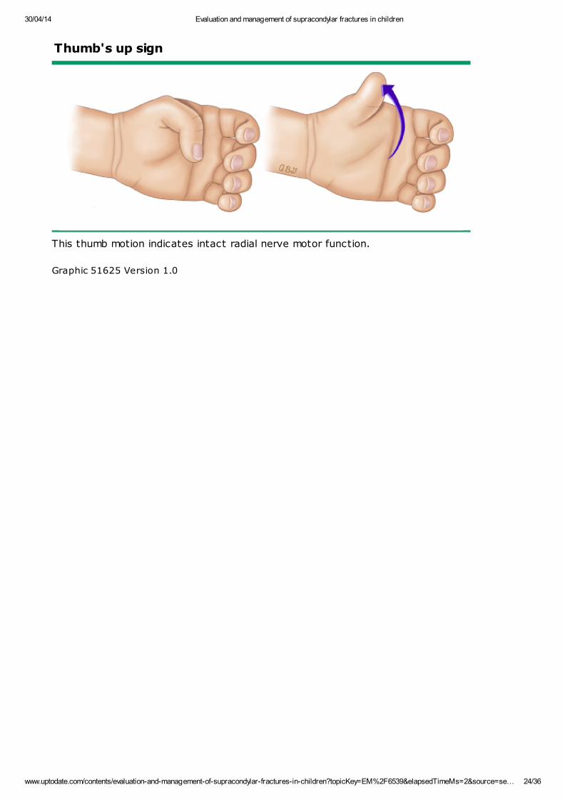
Thumb's up sign: radial nerve (figure 10)
The clinician should perform testing of two point discrimination (ie, the ability to recognize two points five
millimeters apart applied simultaneously to the skin as distinct from a single point) on the follow sites to
evaluate peripheral nerve sensation:
Dorsal web space – Innervated by the radial nerve
30/04/14 Evaluation and management of supracondylar fractures in children
www.uptodate.com/contents/evaluation-and-management-of-supracondylar-fractures-in-children?topicKey=EM%2F6539&elapsedTimeMs=2&source=see… 4/36
Palmar index (pointer) finger – Innervated by the median nerve
Pinky (or little finger) – Innervated by the ulnar nerve
Although nerve injuries may rarely be associated with long term sequelae, the majority are neurapraxias, ie,
temporary loss of nerve function (especially motor function) without anatomical nerve disruption [9,10].
Compartment syndrome — The following findings suggest a developing compartment syndrome:
Excessive swelling and ecchymosis at the elbow [3]
Increasing forearm pain
Increased pain upon passive extension of the fingers
Cold hand with poor perfusion, pallor, and diminished or absent pulse (late finding)
Compartment syndrome may occur prior to or after definitive orthopedic care but is rare (<1 percent of patients)
[7]. It is more common in children with supracondylar fracture who also have a forearm fracture. Once the
clinician suspects an acute compartment syndrome, compartment pressures must be measured to determine
whether they are elevated, or a clinical decision to perform a fasciotomy is needed. (See "Acute compartment
syndrome of the extremities", section on 'Management'.)
Associated injuries — Forearm and wrist fractures occasionally complicate supracondylar fractures in
children. Thus, the forearm and wrist warrant careful inspection for swelling and/or deformity and palpation for
bony tenderness. Radiographs of these areas should be routinely ordered in children with supracondylar
fractures. (See 'Radiographic views' below.)
In addition, shoulder and clavicle injuries may occur and any deformity, swelling, or bony tenderness in these
areas warrants radiographic investigation.
Most supracondylar fractures result from falls on an outstretched arm and are not associated with multiple
trauma. However, children who fall from a height greater than three times their standing height or 10 feet,
whichever is less, are at increased risk for multiple trauma to the head, thorax, and abdomen and may warrant,
in addition to a complete physical examination, appropriate ancillary studies. (See "Approach to the initially
stable child with blunt or penetrating injury", section on 'Approach to blunt trauma' and "Trauma management:
Approach to the unstable child", section on 'Initial approach'.)
RADIOGRAPHIC ASSESSMENT — All patients with a suspected supracondylar fracture should receive a
general assessment as well as a focused physical examination of the involved extremity. (See 'Physical
findings' above and "Procedural sedation in children outside of the operating room".)
Radiologic technique — Appropriate analgesia should be provided BEFORE extremity radiographs are
obtained. When obtaining radiographs in the child with a supracondylar fracture, the goal is to obtain the
necessary films with minimal movement of the extremity. Excessive manipulation of the extremity during
radiologic procedures may further exacerbate or precipitate neurovascular injury, especially in patients with an
unstable type II or type III supracondylar fracture.
In patients with evidence of severe fracture (S-shape configuration or pucker sign, (figure 7 and figure 8)),
splinting is advisable prior to obtaining radiographs. Alternatively, the clinician and an assistant may accompany
the patient and assist with positioning of the obviously deformed extremity while ensuring neurovascular status
does not deteriorate. (See 'Analgesia and immobilization' below.)
In patients without obvious deformity, a sling typically provides adequate support for nondisplaced fractures and
allows for radiographs to be obtained more easily.
Radiographic views — Radiographic assessment of suspected supracondylar fracture requires a true lateral of
the elbow with the humerus in anatomic position (figure 11 and image 2 and image 3).
Because of the association of supracondylar fractures with forearm fractures, the clinician should also obtain AP
30/04/14 Evaluation and management of supracondylar fractures in children
www.uptodate.com/contents/evaluation-and-management-of-supracondylar-fractures-in-children?topicKey=EM%2F6539&elapsedTimeMs=2&source=see… 5/36
and lateral radiographic views of the forearm. In many hospitals, this requirement is accomplished by including
the entire forearm in the AP and lateral views of the elbow. This approach saves time and minimizes movement
of the extremity.
Proximal humerus, clavicle, and wrist radiographs may also be necessary depending on the patient's physical
findings. (See 'Associated fractures' below.)
Ultrasound with Doppler flow should be performed in children with evidence of vascular injury (eg, decreased or
absent radial pulse) [16].
Supracondylar fracture classification — Extension type supracondylar fractures may be further classified
according to the Gartland classification system [14].
Type I – Gartland type I supracondylar fracture describes a nondisplaced fracture with radiographic
evidence of elbow effusion (anterior sail and/or posterior fat pad signs). Because the anterior and
posterior periosteum remains intact, the anterior humeral line transects the middle third of the capitellum
(image 4). (See "Elbow anatomy and radiographic diagnosis of elbow fracture in children", section on 'Fat
pads'.)
Type II – Gartland type II supracondylar fracture refers to a displaced fracture with an intact posterior
periosteum. In contrast to type I fractures, the anterior humeral line is displaced anteriorly, either hitting
the anterior third of the capitellum or missing it entirely, indicating posterior displacement of the distal
humeral fracture. In most children, the fracture line is clearly seen and "hinging" of the posterior
periosteum and posterior displacement of the distal fracture fragment may occur (image 5 and image 6).
(See "Elbow anatomy and radiographic diagnosis of elbow fracture in children", section on 'Anterior
humeral line'.)
Type III – Gartland type III supracondylar fracture is a displaced fracture with disrupted anterior and
posterior periosteum. This injury results in no continuity between the proximal and distal fracture
fragments. Type III fractures may be displaced in three directions: posteromedial (the most common
pattern), posterolateral, or anterolateral. The direction of displacement is important in determining which
neurovascular structures are at greatest risk for injury (image 7 and figure 4) [3]. (See 'Soft tissue
structures' above.)
Associated fractures — Supracondylar fractures are associated with forearm or distal radius fractures in up to
5 percent of cases. The combination of supracondylar and forearm fractures increases the possibility of
compartment syndrome [8,17]. For this reason, radiographs of the forearm should be obtained in all patients
with supracondylar fractures [14].
Proximal humerus, clavicle, and wrist fractures may also occur with a supracondylar fracture. Deformity and/or
bony tenderness during physical examination dictate appropriate radiographic assessment of these areas in
selected patients (image 8 and table 2).
TREATMENT
Absent pulse — The emergency clinician should promptly identify children with vascular insufficiency and
emergently involve an orthopedic surgeon with appropriate pediatric expertise. Rarely, these children will require
partial closed reduction in the emergency department in an attempt to restore distal circulation (figure 12) [14].
Patients who display a cold, white or cyanotic hand despite reduction attempts require operative exploration and
vascular repair.
Acute compartment syndrome — Compartment syndrome may occur prior to or after definitive orthopedic
care. It is more common in children with supracondylar fracture who also have a forearm fracture. Suspected
compartment syndrome should prompt measurement of compartment pressure and/or emergent consultation
with an orthopedic surgeon with pediatric expertise. (See 'Compartment syndrome' above and "Acute
compartment syndrome of the extremities", section on 'Measurement of compartment pressures'.)
Once confirmed by compartment pressure measurement or by clinical examination by an orthopedic surgeon,
30/04/14 Evaluation and management of supracondylar fractures in children
www.uptodate.com/contents/evaluation-and-management-of-supracondylar-fractures-in-children?topicKey=EM%2F6539&elapsedTimeMs=2&source=see… 6/36
immediate management of suspected acute compartment syndrome includes relieving all external pressure on
the compartment. Any dressing, splint, cast, or other restrictive covering should be removed. The limb should
NOT be elevated. Elevation can diminish arterial inflow and exacerbate ischemia. Analgesics should be given.
Definitive treatment consists of fasciotomy to decompress all involved compartments. (See "Acute compartment
syndrome of the extremities", section on 'Management'.)
Analgesia and immobilization — For children with adequate distal circulation and no sign of compartment
syndrome, initial therapy consists of pain management and immobilization to prevent further distraction of the
fracture [3].
Parenteral analgesia (eg, intravenous morphine 0.1 to 0.15 mg/kg, maximum single dose: 10 mg) is most
appropriate for initial pain control in patients with moderate to severe pain and should be given prior to
radiographic evaluation. Oral analgesia (eg, ibuprofen 10 mg/kg) may suffice for patients who have
suffered a nondisplaced supracondylar fracture. In most circumstances, pain relief will result in an
improved ability to assess neurovascular status in the apprehensive child.
Immobilization is particularly important prior to radiographs in children with significantly deformed
fractures. Splinting avoids additional soft tissue injury from inadvertent arm movement. In this situation,
the arm should be splinted "as it lies" (typically with elbow flexed 20 to 30 degrees) using prefabricated
splinting material or eight layers of plaster with under cast padding (eg, Webril®). A loosely applied
elastic bandage (eg, Ace® wrap) holds the splint in place. Neurovascular status should be checked
before and after splinting. If any further compromise is found after immobilization, then the splinting
material should be removed and the arm position adjusted. (See 'Absent pulse' above and 'Radiologic
technique' above.)
Orthopedic consultation — Prompt orthopedic consultation should be obtained in any of the following
circumstances:
Open supracondylar fracture
Fracture with neurovascular compromise
Type II or type III supracondylar fracture
Supracondylar fracture with evidence of compartment syndrome
DEFINITIVE CARE — Initial treatment varies for each type of supracondylar fracture:
Type I fracture — Patients with a nondisplaced Type I fracture (image 4) may be immobilized using either a
collar and cuff sling or a posterior splint and sling. If a posterior splint is placed, it should extend from the wrist
to the axilla, with the elbow at 90 degrees of flexion and the forearm in neutral position with respect to
supination and pronation [15,18].
One randomized trial of 50 children found that those immobilized initially in a posterior splint and sling returned
to normal activity sooner than those immobilized in a collar and cuff sling (median two versus seven days,
p≤0.01) [18]. The study found no difference between the two groups in daily pain scores or in resumption of
normal activity or mobility at two weeks.
Circumferential casting and extremes of flexion should be initially avoided in most cases to decrease the
incidence of compartment syndrome and vascular compromise [9,19].
The typical duration of immobilization for nondisplaced type I supracondylar fractures is three weeks [20].
Type II fracture — Type II fractures (image 5 and image 6) require orthopedic consultation for the determination
of appropriate intervention (closed versus open reduction with percutaneous pin placement). Most pediatric
orthopedists now recommend closed reduction and percutaneous pin fixation [8].
Type III fracture — Type III supracondylar fractures (image 7) require orthopedic consultation for the
determination of appropriate intervention (closed versus open reduction with percutaneous pin placement) [3].
30/04/14 Evaluation and management of supracondylar fractures in children
www.uptodate.com/contents/evaluation-and-management-of-supracondylar-fractures-in-children?topicKey=EM%2F6539&elapsedTimeMs=2&source=see… 7/36
FOLLOW-UP CARE — Children with nondisplaced fractures (eg, Gartland type I fractures) do not require
immediate orthopedic evaluation. After splinting, these patients may be referred for follow up within seven days
of the injury. The family should be instructed to return immediately for signs of unmanageable pain or
compartment syndrome. (See 'Compartment syndrome' above and 'Supracondylar fracture classification' above.)
Almost all children with displaced supracondylar fractures (eg, Gartland type II and III fractures) require open or
closed reduction and percutaneous pinning. Hospital admission for monitoring of pulses, nerve function, and
forearm compartments is also typical. After discharge, the patient is maintained in a splint or cast with
percutaneous pins. Repeat radiographs are obtained on a regular basis to assess bone healing. Pins are
removed when the distal humerus is no longer tender, usually in three to four weeks [8].
After pin and cast removal, motion exercises may be recommended as tolerated. Studies of children with
supracondylar fractures and no neurovascular deficit suggest that formal physical therapy does not appear to
improve long term mobility [21]. However, physical therapy is recommended for children with persistent
contractures after three to four months or nerve deficits.
COMPLICATIONS — Complications of supracondylar fractures include vascular insufficiency, forearm
compartment syndrome resulting in Volkmann's ischemic contracture, nerve injury, and cubitus varus deformity.
Vascular injury — Absence of the radial pulse is reported in 6 to 20 percent of all supracondylar fractures
[22,23]. Vascular injury is most common with type II and III supracondylar fractures (figure 4) [13,24]. The
brachial artery is most frequently injured in posterolaterally displaced fractures [13]. Patients without significant
improvement in pulse or Doppler pulse after orthopedic care warrant emergent vascular exploration, especially if
perfusion is not restored or if intractable pain suggestive of ischemia is present [4,16,24-26]:
A systematic review of 161 children with supracondylar fractures and a pulseless hand found that closed
reduction and percutaneous pinning resulted in return of the radial pulse in 51 percent (82 of 161) of
cases [24]. Sixty-three of 79 children with persistent pulseless hand after operative care underwent
vascular exploration. Brachial artery injury or thrombus was found in 61 patients (97 percent).
An observational study of 19 children who had a perfused but pulseless hand after operative fixation found
that the eight children who also had a median and/or anterior interosseus nerve deficit demonstrated
entrapment of both the brachial artery and the median nerve at the fracture site [25]. Among the 11
children who were treated with closed reduction followed by observation, four patients underwent
secondary exploration between two days and three weeks after injury; the brachial artery was running
through the fracture site in all patients and one developed a persistent median nerve deficit.
An observational study of 26 children who had a pink pulseless hand after initial treatment of a
supracondylar fracture found that the 23 children who presented four days to one year after injury and did
not have early release of brachial artery obstruction developed ischemic contractions of hand and/or
forearm muscles [26]. They also had 56 nerve injuries of which 41 were directly attributable to muscle
ischemia and compression. Approximately half of these nerve injuries responded to late operative
exploration.
Volkmann ischemic contracture — Vascular injury and primary swelling from the injury can lead to the
development of compartment syndrome within 12 to 24 hours [15]. If a compartment syndrome is not treated in
a timely manner, the associated ischemia and infarction may progress to Volkmann's ischemic contracture:
fixed flexion of the elbow, pronation of the forearm, flexion at the wrist, and joint extension of the metacarpal-
phalangeal joint [15].
Neurologic deficit — The frequency of neurologic deficit after supracondylar fractures is 10 to 20 percent and
increases in some series of children with type III supracondylar fractures to as high as 49 percent
[5,7,12,23,27].
Posterolateral distal fracture fragment displacement with medial movement of the proximal fracture
fragment puts the median nerve and its anterior interosseous nerve branch at the greatest risk of injury
(figure 4).
30/04/14 Evaluation and management of supracondylar fractures in children
www.uptodate.com/contents/evaluation-and-management-of-supracondylar-fractures-in-children?topicKey=EM%2F6539&elapsedTimeMs=2&source=see… 8/36
Posteromedial distal fracture fragment displacement increases the chance of radial nerve impingement
because of lateral proximal fracture fragment displacement (figure 4).
Ulnar nerve injuries are most commonly associated with flexion type supracondylar fractures (figure 6)
[5,11,27].
Although nerve injuries may be associated with long-term sequelae, the majority are neurapraxias that will
resolve within two to three months [9,27,28]. Surgical exploration should be considered for nerve deficits that
persist beyond three months [8,12,21].
Cubitus varus deformity — Angular deformity (cubitus varus or "gunstock" deformity) is a long term
complication of a supracondylar fracture (figure 13). In contrast to the proximal humeral physis, the distal
humerus physis contributes little (15 to 20 percent) to the overall longitudinal growth of the humerus [29]. Thus,
remodeling and correction of fracture angulation is limited for children with supracondylar fractures.
Modern surgical techniques (eg, closed reduction with percutaneous pinning) have reduced the frequency of
cubitus varus from 58 to approximately 3 percent of children treated for supracondylar fractures [6]. Cubitus
varus deformity is mainly cosmetic and function is usually preserved [4]. However, it is rarely associated with an
ulnar nerve palsy that may present many years after the original fracture [30].
OUTCOMES — Although immediate complications of distal humeral fractures are common in the pediatric
population, the prognosis for long term outcome and function is very good if the fracture is appropriately
diagnosed and treated. Many of the associated complications either are self limited or are amenable to
functional repair with surgical intervention.
For instance, vascular insufficiency often resolves after orthopedic alignment with percutaneous pinning.
Similarly, nerve injury typically results from neurapraxia, ie, temporary nerve dysfunction that resolves without
long term effects [9,28]. In addition, joint stiffness may also be treated surgically in most cases [3,31].
In a prospective study of 108 supracondylar fractures with complete follow up, 99 children had an excellent
outcome, 5 had a good outcome, and 4 had a poor outcome. The poor results were attributed to technical errors
in reduction resulting in an unsatisfactory cosmetic outcome but good function [32].
INFORMATION FOR PATIENTS — UpToDate offers two types of patient education materials, “The Basics” and
“Beyond the Basics.” The Basics patient education pieces are written in plain language, at the 5 to 6 grade
reading level, and they answer the four or five key questions a patient might have about a given condition. These
articles are best for patients who want a general overview and who prefer short, easy-to-read materials. Beyond
the Basics patient education pieces are longer, more sophisticated, and more detailed. These articles are
written at the 10 to 12 grade reading level and are best for patients who want in-depth information and are
comfortable with some medical jargon.
Here are the patient education articles that are relevant to this topic. We encourage you to print or e-mail these
topics to your patients. (You can also locate patient education articles on a variety of subjects by searching on
“patient info” and the keyword(s) of interest.)
Basics topics (see "Patient information: Cast and splint care (The Basics)" and "Patient information:
Elbow fracture in children (The Basics)")
Beyond the Basics topic (see "Patient information: Cast and splint care (Beyond the Basics)")
SUMMARY AND RECOMMENDATIONS
In children, the supracondylar region encompasses an area of thin, weak bone located in the distal
posterior humerus. Fracture in this region is common after a fall on an outstretched hand and may lead
to neurovascular compromise. In particular, the anterior interosseous branch of the median nerve and the
brachial artery are at risk of injury. (See 'Epidemiology' above and 'Pertinent anatomy' above.)
Clinical findings
th th
th th
30/04/14 Evaluation and management of supracondylar fractures in children
www.uptodate.com/contents/evaluation-and-management-of-supracondylar-fractures-in-children?topicKey=EM%2F6539&elapsedTimeMs=2&source=see… 9/36
The child with a supracondylar fracture typically has elbow pain, swelling, and very limited to no range of
motion at the elbow. The clinician must rapidly assess the injury to identify neurovascular compromise,
evidence of compartment syndrome, and degree of fracture displacement. (See 'Physical findings' above.)
Appropriate analgesia should be provided BEFORE extremity radiographs are obtained. Radiographic
assessment of suspected supracondylar fracture requires a true lateral of the elbow, if permitted by the
patient's clinical condition, with the humerus in anatomic position and an anteroposterior (AP) projection
of the distal humerus (table 2 and image 3 and figure 11). Because of the association of supracondylar
fractures with forearm fractures, the clinician should also obtain AP and lateral radiographic views of the
forearm. (See 'Radiographic assessment' above and "Elbow anatomy and radiographic diagnosis of elbow
fracture in children", section on 'Plain radiograph interpretation'.)
In patients with evidence of deformity or severe fracture (eg, S-shape configuration or pucker sign, (figure
7 and figure 8)), splinting is advisable prior to obtaining radiographs. (See 'Radiographic assessment'
above and "Elbow anatomy and radiographic diagnosis of elbow fracture in children", section on 'Plain
radiograph interpretation'.)
Management
The emergency clinician should promptly identify children with vascular insufficiency and emergently
involve an orthopedic surgeon with pediatric expertise. Rarely, these children will require partial closed
reduction in the emergency department in an attempt to restore distal circulation (figure 12) [14]. Patients
who display a cold, cyanotic hand despite reduction attempts require emergent operative exploration and
vascular repair. (See 'Pulse' above and 'Perfusion' above and 'Absent pulse' above.)
Suspected compartment syndrome should prompt measurement of compartment pressure and/or
emergent consultation with an orthopedic surgeon with appropriate pediatric expertise. Once confirmed
by compartment pressure measurement, immediate management of suspected acute compartment
syndrome includes relieving all external pressure on the compartment. Definitive treatment consists of
fasciotomy to decompress all involved compartments. (See 'Compartment syndrome' above and "Acute
compartment syndrome of the extremities", section on 'Measurement of compartment pressures'.)
For children with adequate distal circulation and no sign of compartment syndrome, initial therapy
consists of pain management and immobilization to prevent further displacement of the fracture. (See
'Analgesia and immobilization' above.)
Emergent orthopedic consultation is indicated for children with an open fracture, neurovascular
compromise, or acute compartment syndrome. In addition, prompt involvement of an orthopedic surgeon
is necessary for operative care of a child with a Type II or Type III supracondylar fracture. Nondisplaced
Type I supracondylar fractures are treated with a long arm splint with orthopedic follow up in one week.
(See 'Supracondylar fracture classification' above and 'Neurologic deficit' above and 'Type I fracture' above
and 'Type II fracture' above and 'Type III fracture' above.)
Use of UpToDate is subject to the Subscription and License Agreement.
REFERENCES
1. Bachman D, Santora S. Orthopedic trauma. In: Textbook of Pediatric Emergency Medicine, Fleisher GR,Ludwig S, et al. (Eds), Lippincott Williams and Wilkins, Philadelphia 2006. p.1538.
2. Della-Giustina K, Della-Giustina DA. Emergency department evaluation and treatment of pediatricorthopedic injuries. Emerg Med Clin North Am 1999; 17:895.
3. Carson S, Woolridge DP, Colletti J, Kilgore K. Pediatric upper extremity injuries. Pediatr Clin North Am2006; 53:41.
4. Lins RE, Simovitch RW, Waters PM. Pediatric elbow trauma. Orthop Clin North Am 1999; 30:119.

30/04/14 Evaluation and management of supracondylar fractures in children
www.uptodate.com/contents/evaluation-and-management-of-supracondylar-fractures-in-children?topicKey=EM%2F6539&elapsedTimeMs=2&source=se… 10/36
5. Kasser JR, Beaty JH. Supracondylar fractures of the distal humerus. In: Rockwood and Wilkins' Fracturesin Children, 5th, Beaty JH, Kasser JR. (Eds), Lippincott Williams & Wilkins, Philadelphia 2001. p.577.
6. Farnsworth CL, Silva PD, Mubarak SJ. Etiology of supracondylar humerus fractures. J Pediatr Orthop1998; 18:38.
7. Fletcher N, Schiller JR, Sumeet G, et al. Increased severity of type III supracondylar humeral fractures inthe preteen population. J Pediatr 2012; 32:567.
8. Baratz M, Micucci C, Sangimino M. Pediatric supracondylar humerus fractures. Hand Clin 2006; 22:69.
9. Villarin LA Jr, Belk KE, Freid R. Emergency department evaluation and treatment of elbow and forearminjuries. Emerg Med Clin North Am 1999; 17:843.
10. Jones ET, Louis DS. Median nerve injuries associated with supracondylar fractures of the humerus inchildren. Clin Orthop Relat Res 1980; :181.
11. Brown IC, Zinar DM. Traumatic and iatrogenic neurological complications after supracondylar humerusfractures in children. J Pediatr Orthop 1995; 15:440.
12. Campbell CC, Waters PM, Emans JB, et al. Neurovascular injury and displacement in type IIIsupracondylar humerus fractures. J Pediatr Orthop 1995; 15:47.
13. Skaggs D, Pershad J. Pediatric elbow trauma. Pediatr Emerg Care 1997; 13:425.
14. Shrader MW. Pediatric supracondylar fractures and pediatric physeal elbow fractures. Orthop Clin NorthAm 2008; 39:163.
15. Wu J, Perron AD, Miller MD, et al. Orthopedic pitfalls in the ED: pediatric supracondylar humerusfractures. Am J Emerg Med 2002; 20:544.
16. Benedetti Valentini M, Farsetti P, Martinelli O, et al. The value of ultrasonic diagnosis in the managementof vascular complications of supracondylar fractures of the humerus in children. Bone Joint J 2013; 95-B:694.
17. Roposch A, Reis M, Molina M, et al. Supracondylar fractures of the humerus associated with ipsilateralforearm fractures in children: a report of forty-seven cases. J Pediatr Orthop 2001; 21:307.
18. Oakley E, Barnett P, Babl FE. Backslab versus nonbackslab for immobilization of undisplacedsupracondylar fractures: a randomized trial. Pediatr Emerg Care 2009; 25:452.
19. McGraw JJ, Akbarnia BA, Hanel DP, et al. Neurological complications resulting from supracondylarfractures of the humerus in children. J Pediatr Orthop 1986; 6:647.
20. Green N, Van Zeeland N. Fractures and dislocations about the elbow. In: Skeletal Trauma in Children, 4thedition, Green N, Swiontowski. (Eds), Elsevier, Philadelphia 2008. p.207.
21. Keppler P, Salem K, Schwarting B, Kinzl L. The effectiveness of physiotherapy after operative treatment ofsupracondylar humeral fractures in children. J Pediatr Orthop 2005; 25:314.
22. Omid R, Choi PD, Skaggs DL. Supracondylar humeral fractures in children. J Bone Joint Surg Am 2008;90:1121.
23. Garg S, Weller A, Larson AN, et al. Clinical characteristics of severe supracondylar humerus fractures inchildren. J Pediatr Orthop 2014; 34:34.
24. Griffin KJ, Walsh SR, Markar S, et al. The pink pulseless hand: a review of the literature regardingmanagement of vascular complications of supracondylar humeral fractures in children. Eur J VascEndovasc Surg 2008; 36:697.
25. Mangat KS, Martin AG, Bache CE. The 'pulseless pink' hand after supracondylar fracture of the humerusin children: the predictive value of nerve palsy. J Bone Joint Surg Br 2009; 91:1521.
26. Blakey CM, Biant LC, Birch R. Ischaemia and the pink, pulseless hand complicating supracondylarfractures of the humerus in childhood: long-term follow-up. J Bone Joint Surg Br 2009; 91:1487.
27. Lyons ST, Quinn M, Stanitski CL. Neurovascular injuries in type III humeral supracondylar fractures inchildren. Clin Orthop Relat Res 2000; :62.
28. Jones ET, Louis DS. Median nerve injuries associated with supracondylar fractures of the humerus inchildren. Clin Orthop Relat Res 1980; :181.
29. Price C, Phillips J, Devito D. Management of fractures. In: Lovell & Winter's Pediatric Orthopaedics, 5th,Morrissey, Weinstein SL. (Eds), Lippincott Williams & Wilkins, Philadelphia 2001. p.1319.
30. Abe M, Ishizu T, Shirai H, et al. Tardy ulnar nerve palsy caused by cubitus varus deformity. J Hand Surg
30/04/14 Evaluation and management of supracondylar fractures in children
www.uptodate.com/contents/evaluation-and-management-of-supracondylar-fractures-in-children?topicKey=EM%2F6539&elapsedTimeMs=2&source=se… 11/36
Am 1995; 20:5.
31. Gogola GR. Pediatric humeral condyle fractures. Hand Clin 2006; 22:77.
32. Mazda K, Boggione C, Fitoussi F, Penneçot GF. Systematic pinning of displaced extension-typesupracondylar fractures of the humerus in children. A prospective study of 116 consecutive patients. JBone Joint Surg Br 2001; 83:888.
Topic 6539 Version 14.0
30/04/14 Evaluation and management of supracondylar fractures in children
www.uptodate.com/contents/evaluation-and-management-of-supracondylar-fractures-in-children?topicKey=EM%2F6539&elapsedTimeMs=2&source=se… 12/36
GRAPHICS
Anatomy of the distal humerus in the child
Graphic 66099 Version 1.0
30/04/14 Evaluation and management of supracondylar fractures in children
www.uptodate.com/contents/evaluation-and-management-of-supracondylar-fractures-in-children?topicKey=EM%2F6539&elapsedTimeMs=2&source=se… 13/36
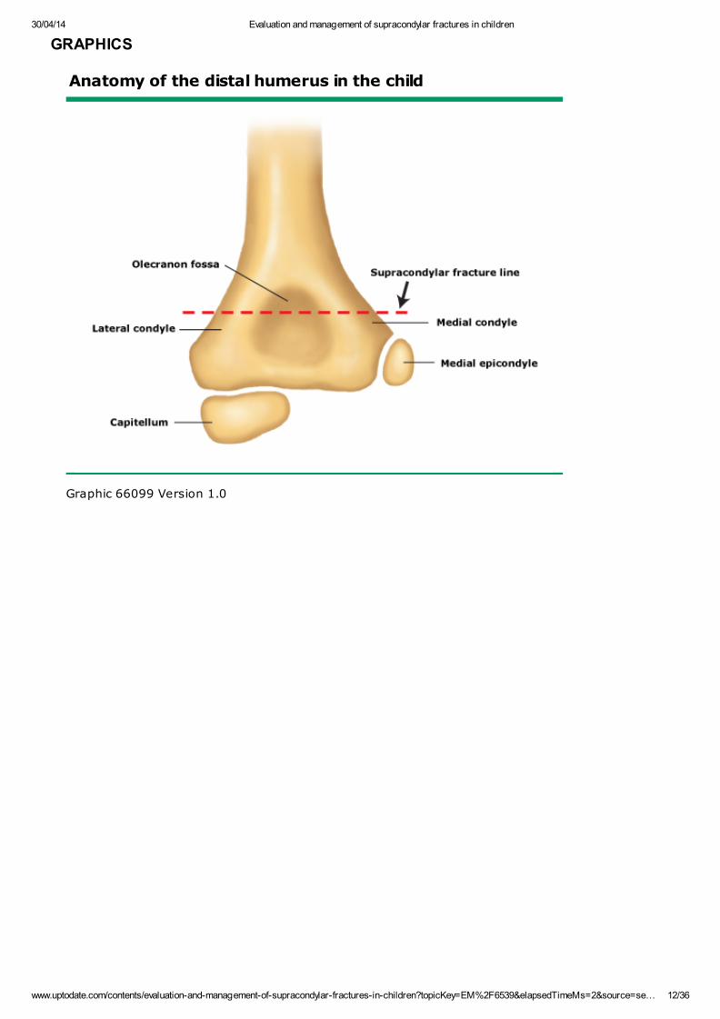
Extension supracondylar fractures
Hyperextension forces. A) Most young children attempt to break their falls with
the upper extremity extended. Because of the laxity of the ligaments, the elbow
becomes locked into hyperextension. B) This converts the linear applied force
(large arrow) to an anterior tension force. Posteriorly, the olecranon is forced
into the depths of the olecranon fossa (small arrow). C) As the bending force
continues, the distal humerus fails anteriorly in the supracondylar area. D) When
the fracture is complete, the distal fragment becomes posteriorly displaced. The
strong action of the triceps (large arrow) produces proximal displacement of the
distal fragment.
Reproduced with permission from: Kasser JR, Beaty JH. Supracondylar fractures of the
distal humerus. In: Rockwood and Wilkins' Fractures in Children, 5th ed, Beaty JH,
Kasser JR (Eds), Lippincott Williams & Wilkins 2001. Copyright © 2001 Lippincott
Williams & Wilkins.
http://www.lww.com
Graphic 54389 Version 9.0
30/04/14 Evaluation and management of supracondylar fractures in children
www.uptodate.com/contents/evaluation-and-management-of-supracondylar-fractures-in-children?topicKey=EM%2F6539&elapsedTimeMs=2&source=se… 14/36
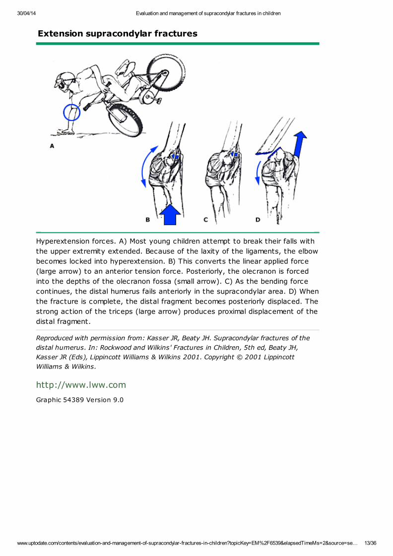
Supracondylar fracture line
Orientation of fracture lines. A) The typical transverse fracture line
originates just above the epicondyles and courses through the
supracondylar area (arrows). B) In the lateral projection, the fracture
line is usually also transverse.
Reproduced with permission from: Kasser JR, Beaty JH. Supracondylar
fractures of the distal humerus. In: Rockwood and Wilkins' Fractures in
Children, 5th ed, Beaty JH, Kasser JR (Eds), Lippincott Williams & Wilkins
2001. Copyright © 2001 Lippincott Williams & Wilkins.
http://www.lww.com
Graphic 56874 Version 9.0
30/04/14 Evaluation and management of supracondylar fractures in children
www.uptodate.com/contents/evaluation-and-management-of-supracondylar-fractures-in-children?topicKey=EM%2F6539&elapsedTimeMs=2&source=se… 15/36
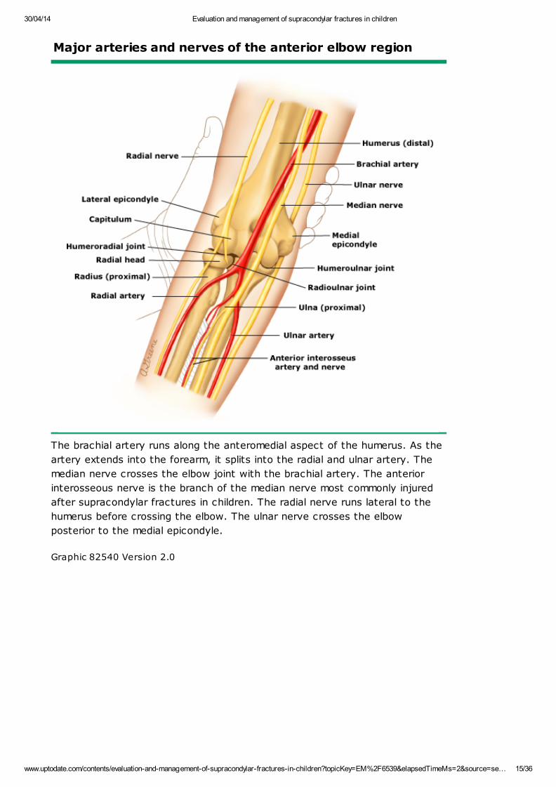
Major arteries and nerves of the anterior elbow region
The brachial artery runs along the anteromedial aspect of the humerus. As the
artery extends into the forearm, it splits into the radial and ulnar artery. The
median nerve crosses the elbow joint with the brachial artery. The anterior
interosseous nerve is the branch of the median nerve most commonly injured
after supracondylar fractures in children. The radial nerve runs lateral to the
humerus before crossing the elbow. The ulnar nerve crosses the elbow
posterior to the medial epicondyle.
Graphic 82540 Version 2.0
30/04/14 Evaluation and management of supracondylar fractures in children
www.uptodate.com/contents/evaluation-and-management-of-supracondylar-fractures-in-children?topicKey=EM%2F6539&elapsedTimeMs=2&source=se… 16/36
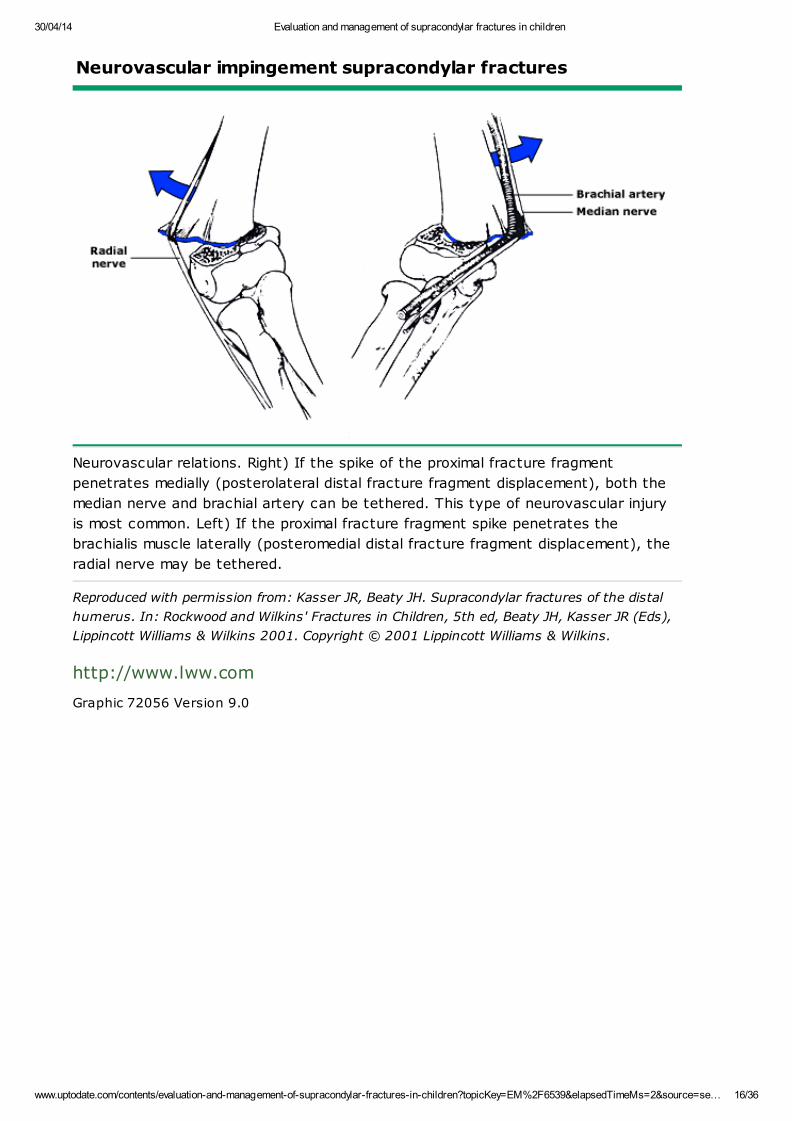
Neurovascular impingement supracondylar fractures
Neurovascular relations. Right) If the spike of the proximal fracture fragment
penetrates medially (posterolateral distal fracture fragment displacement), both the
median nerve and brachial artery can be tethered. This type of neurovascular injury
is most common. Left) If the proximal fracture fragment spike penetrates the
brachialis muscle laterally (posteromedial distal fracture fragment displacement), the
radial nerve may be tethered.
Reproduced with permission from: Kasser JR, Beaty JH. Supracondylar fractures of the distal
humerus. In: Rockwood and Wilkins' Fractures in Children, 5th ed, Beaty JH, Kasser JR (Eds),
Lippincott Williams & Wilkins 2001. Copyright © 2001 Lippincott Williams & Wilkins.
http://www.lww.com
Graphic 72056 Version 9.0
30/04/14 Evaluation and management of supracondylar fractures in children
www.uptodate.com/contents/evaluation-and-management-of-supracondylar-fractures-in-children?topicKey=EM%2F6539&elapsedTimeMs=2&source=se… 17/36
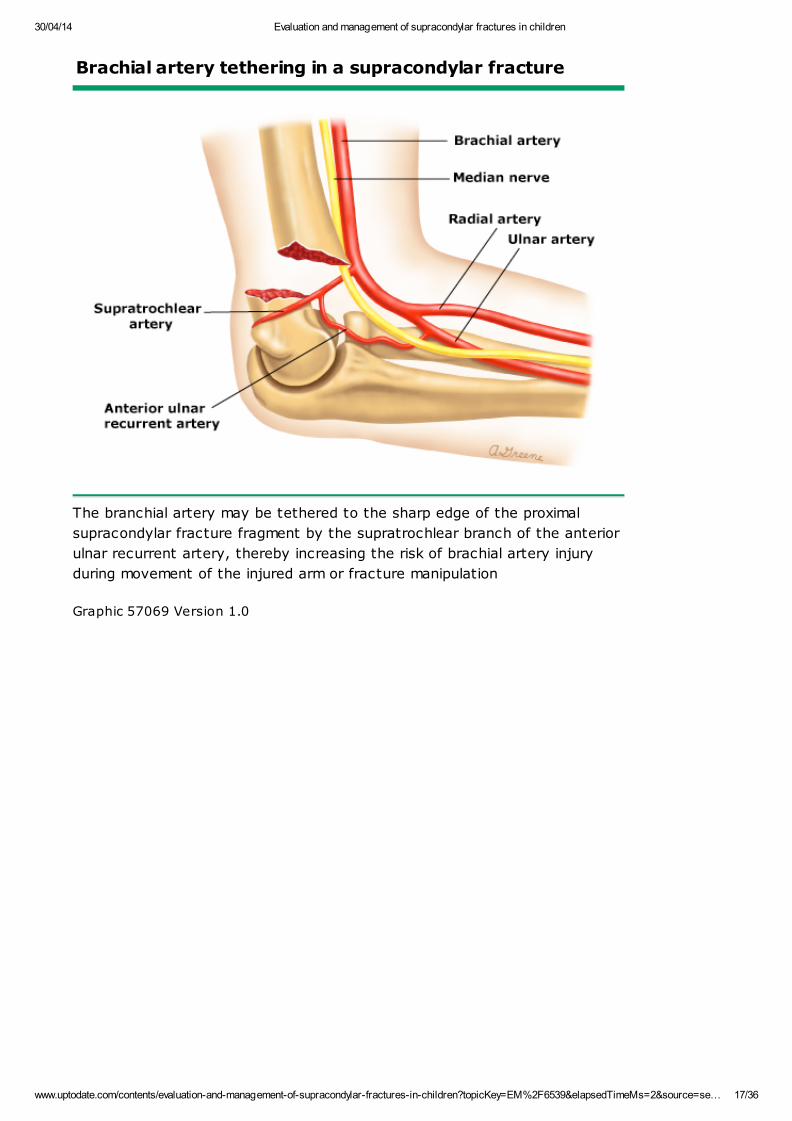
Brachial artery tethering in a supracondylar fracture
The branchial artery may be tethered to the sharp edge of the proximal
supracondylar fracture fragment by the supratrochlear branch of the anterior
ulnar recurrent artery, thereby increasing the risk of brachial artery injury
during movement of the injured arm or fracture manipulation
Graphic 57069 Version 1.0
30/04/14 Evaluation and management of supracondylar fractures in children
www.uptodate.com/contents/evaluation-and-management-of-supracondylar-fractures-in-children?topicKey=EM%2F6539&elapsedTimeMs=2&source=se… 18/36
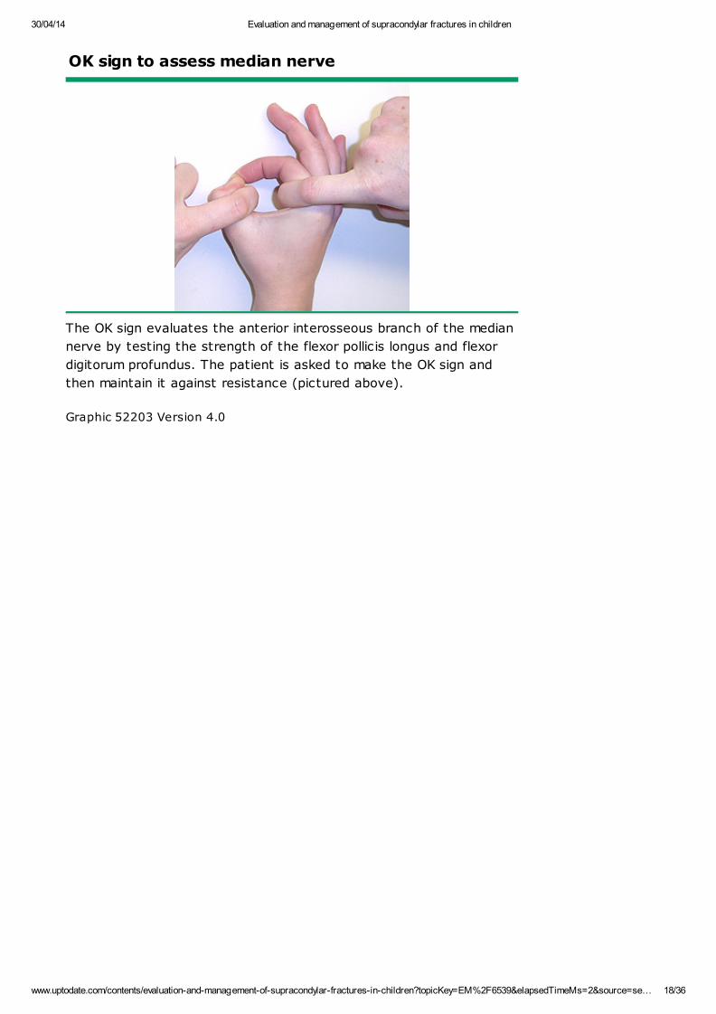
OK sign to assess median nerve
The OK sign evaluates the anterior interosseous branch of the median
nerve by testing the strength of the flexor pollicis longus and flexor
digitorum profundus. The patient is asked to make the OK sign and
then maintain it against resistance (pictured above).
Graphic 52203 Version 4.0
30/04/14 Evaluation and management of supracondylar fractures in children
www.uptodate.com/contents/evaluation-and-management-of-supracondylar-fractures-in-children?topicKey=EM%2F6539&elapsedTimeMs=2&source=se… 19/36
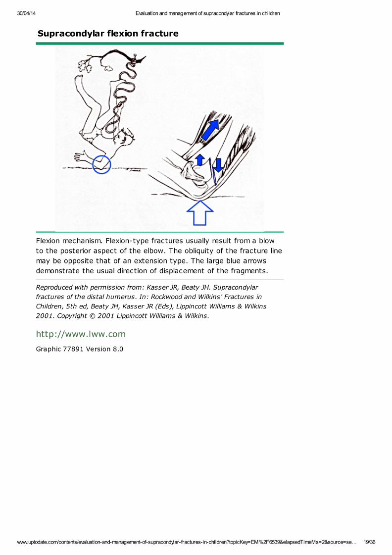
Supracondylar flexion fracture
Flexion mechanism. Flexion-type fractures usually result from a blow
to the posterior aspect of the elbow. The obliquity of the fracture line
may be opposite that of an extension type. The large blue arrows
demonstrate the usual direction of displacement of the fragments.
Reproduced with permission from: Kasser JR, Beaty JH. Supracondylar
fractures of the distal humerus. In: Rockwood and Wilkins' Fractures in
Children, 5th ed, Beaty JH, Kasser JR (Eds), Lippincott Williams & Wilkins
2001. Copyright © 2001 Lippincott Williams & Wilkins.
http://www.lww.com
Graphic 77891 Version 8.0
30/04/14 Evaluation and management of supracondylar fractures in children
www.uptodate.com/contents/evaluation-and-management-of-supracondylar-fractures-in-children?topicKey=EM%2F6539&elapsedTimeMs=2&source=se… 20/36
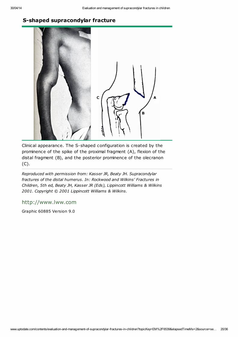
S-shaped supracondylar fracture
Clinical appearance. The S-shaped configuration is created by the
prominence of the spike of the proximal fragment (A), flexion of the
distal fragment (B), and the posterior prominence of the olecranon
(C).
Reproduced with permission from: Kasser JR, Beaty JH. Supracondylar
fractures of the distal humerus. In: Rockwood and Wilkins' Fractures in
Children, 5th ed, Beaty JH, Kasser JR (Eds), Lippincott Williams & Wilkins
2001. Copyright © 2001 Lippincott Williams & Wilkins.
http://www.lww.com
Graphic 60885 Version 9.0
30/04/14 Evaluation and management of supracondylar fractures in children
www.uptodate.com/contents/evaluation-and-management-of-supracondylar-fractures-in-children?topicKey=EM%2F6539&elapsedTimeMs=2&source=se… 21/36
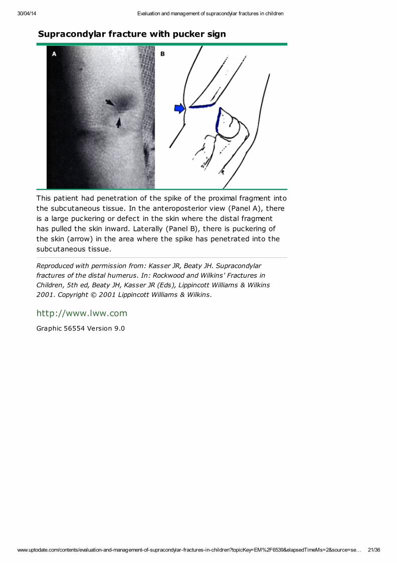
Supracondylar fracture with pucker sign
This patient had penetration of the spike of the proximal fragment into
the subcutaneous tissue. In the anteroposterior view (Panel A), there
is a large puckering or defect in the skin where the distal fragment
has pulled the skin inward. Laterally (Panel B), there is puckering of
the skin (arrow) in the area where the spike has penetrated into the
subcutaneous tissue.
Reproduced with permission from: Kasser JR, Beaty JH. Supracondylar
fractures of the distal humerus. In: Rockwood and Wilkins' Fractures in
Children, 5th ed, Beaty JH, Kasser JR (Eds), Lippincott Williams & Wilkins
2001. Copyright © 2001 Lippincott Williams & Wilkins.
http://www.lww.com
Graphic 56554 Version 9.0

30/04/14 Evaluation and management of supracondylar fractures in children
www.uptodate.com/contents/evaluation-and-management-of-supracondylar-fractures-in-children?topicKey=EM%2F6539&elapsedTimeMs=2&source=se… 22/36
Motor and sensory function of the nerves of the arm
Nerve Motor function Sensory function
Median Wrist flexion and abduction
Thumb and index finger
flexion
Thumb opposition
Palmar thumb and index finger
Ulnar Wrist flexion and adduction
Finger spread
Flexion of distal phalanx of
fifth digit
Ulnar side of fourth digit
Fifth digit
Radial Wrist extension
Supination
Thumb extension
Dorsal web space between thumb and
index finger
Musculocutaneous Elbow flexion Lateral forearm
Graphic 53902 Version 2.0
30/04/14 Evaluation and management of supracondylar fractures in children
www.uptodate.com/contents/evaluation-and-management-of-supracondylar-fractures-in-children?topicKey=EM%2F6539&elapsedTimeMs=2&source=se… 23/36
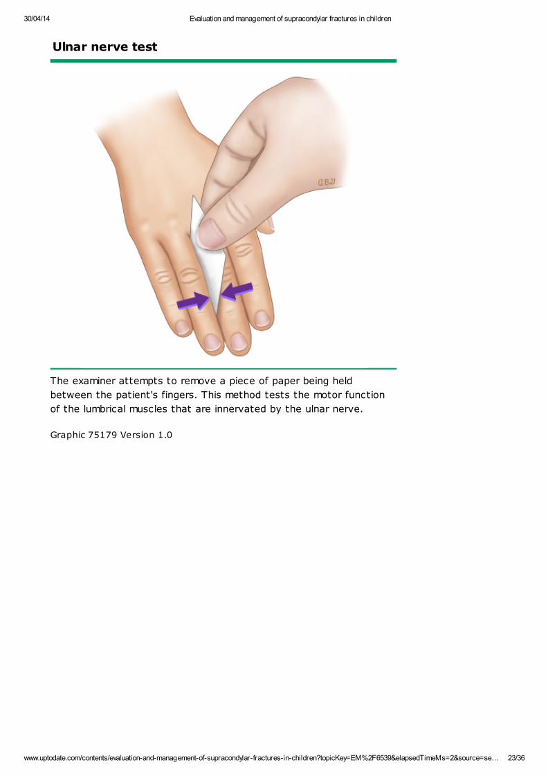
Ulnar nerve test
The examiner attempts to remove a piece of paper being held
between the patient's fingers. This method tests the motor function
of the lumbrical muscles that are innervated by the ulnar nerve.
Graphic 75179 Version 1.0
30/04/14 Evaluation and management of supracondylar fractures in children
www.uptodate.com/contents/evaluation-and-management-of-supracondylar-fractures-in-children?topicKey=EM%2F6539&elapsedTimeMs=2&source=se… 24/36
Thumb's up sign
This thumb motion indicates intact radial nerve motor function.
Graphic 51625 Version 1.0
30/04/14 Evaluation and management of supracondylar fractures in children
www.uptodate.com/contents/evaluation-and-management-of-supracondylar-fractures-in-children?topicKey=EM%2F6539&elapsedTimeMs=2&source=se… 25/36
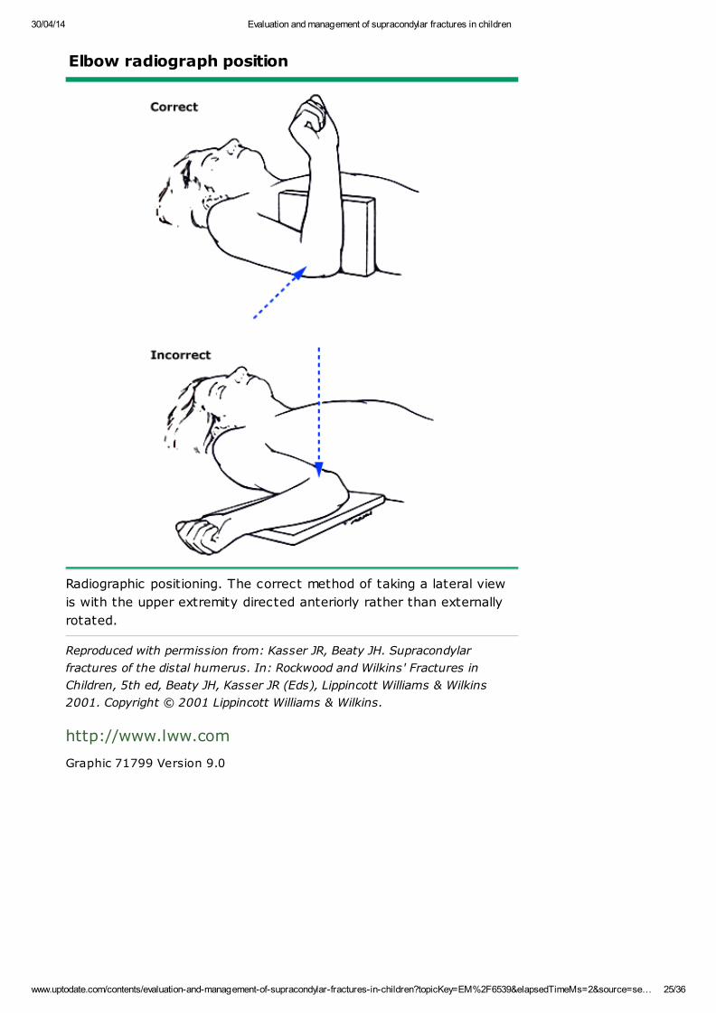
Elbow radiograph position
Radiographic positioning. The correct method of taking a lateral view
is with the upper extremity directed anteriorly rather than externally
rotated.
Reproduced with permission from: Kasser JR, Beaty JH. Supracondylar
fractures of the distal humerus. In: Rockwood and Wilkins' Fractures in
Children, 5th ed, Beaty JH, Kasser JR (Eds), Lippincott Williams & Wilkins
2001. Copyright © 2001 Lippincott Williams & Wilkins.
http://www.lww.com
Graphic 71799 Version 9.0

30/04/14 Evaluation and management of supracondylar fractures in children
www.uptodate.com/contents/evaluation-and-management-of-supracondylar-fractures-in-children?topicKey=EM%2F6539&elapsedTimeMs=2&source=se… 26/36
AP and lateral radiographs of the normal elbow and
anatomic landmarks
(A) Anteroposterior radiograph of a normal elbow of a child. (B) Normal
lateral radiograph. The radial head and neck are seen on both AP and
lateral views of the elbow. The olecranon process of the ulna is best
seen on the lateral view. On the lateral radiograph, the radial head is
superimposed over the coronoid process of the ulna.
Reproduced with permission from: Bachman D, Santora S. Orthopedic trauma.
In: Textbook of Pediatric Emergency Medicine, 5th ed, Fleisher GR, Ludwig S,
Henretig FR (Eds), Lippincott Williams & Wilkins 2006. Copyright © 2006
Lippincott Williams & Wilkins.
http://www.lww.com
Graphic 56647 Version 12.0
30/04/14 Evaluation and management of supracondylar fractures in children
www.uptodate.com/contents/evaluation-and-management-of-supracondylar-fractures-in-children?topicKey=EM%2F6539&elapsedTimeMs=2&source=se… 27/36
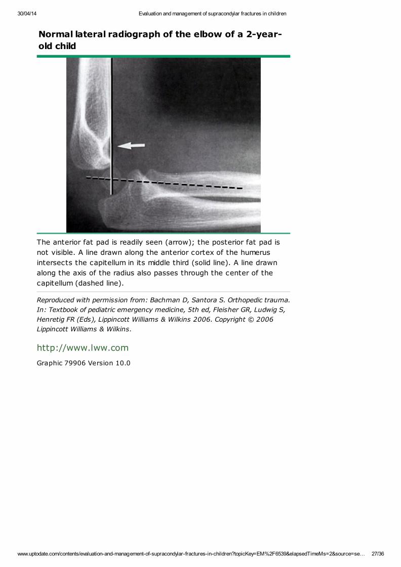
Normal lateral radiograph of the elbow of a 2-year-
old child
The anterior fat pad is readily seen (arrow); the posterior fat pad is
not visible. A line drawn along the anterior cortex of the humerus
intersects the capitellum in its middle third (solid line). A line drawn
along the axis of the radius also passes through the center of the
capitellum (dashed line).
Reproduced with permission from: Bachman D, Santora S. Orthopedic trauma.
In: Textbook of pediatric emergency medicine, 5th ed, Fleisher GR, Ludwig S,
Henretig FR (Eds), Lippincott Williams & Wilkins 2006. Copyright © 2006
Lippincott Williams & Wilkins.
http://www.lww.com
Graphic 79906 Version 10.0
30/04/14 Evaluation and management of supracondylar fractures in children
www.uptodate.com/contents/evaluation-and-management-of-supracondylar-fractures-in-children?topicKey=EM%2F6539&elapsedTimeMs=2&source=se… 28/36
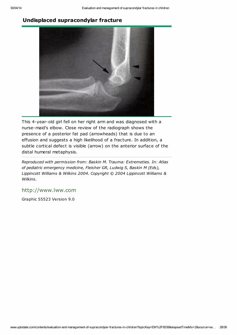
Undisplaced supracondylar fracture
This 4-year-old girl fell on her right arm and was diagnosed with a
nurse-maid's elbow. Close review of the radiograph shows the
presence of a posterior fat pad (arrowheads) that is due to an
effusion and suggests a high likelihood of a fracture. In addition, a
subtle cortical defect is visible (arrow) on the anterior surface of the
distal humeral metaphysis.
Reproduced with permission from: Baskin M. Trauma: Extremeties. In: Atlas
of pediatric emergency medicine, Fleisher GR, Ludwig S, Baskin M (Eds),
Lippincott Williams & Wilkins 2004. Copyright © 2004 Lippincott Williams &
Wilkins.
http://www.lww.com
Graphic 55523 Version 9.0
30/04/14 Evaluation and management of supracondylar fractures in children
www.uptodate.com/contents/evaluation-and-management-of-supracondylar-fractures-in-children?topicKey=EM%2F6539&elapsedTimeMs=2&source=se… 29/36
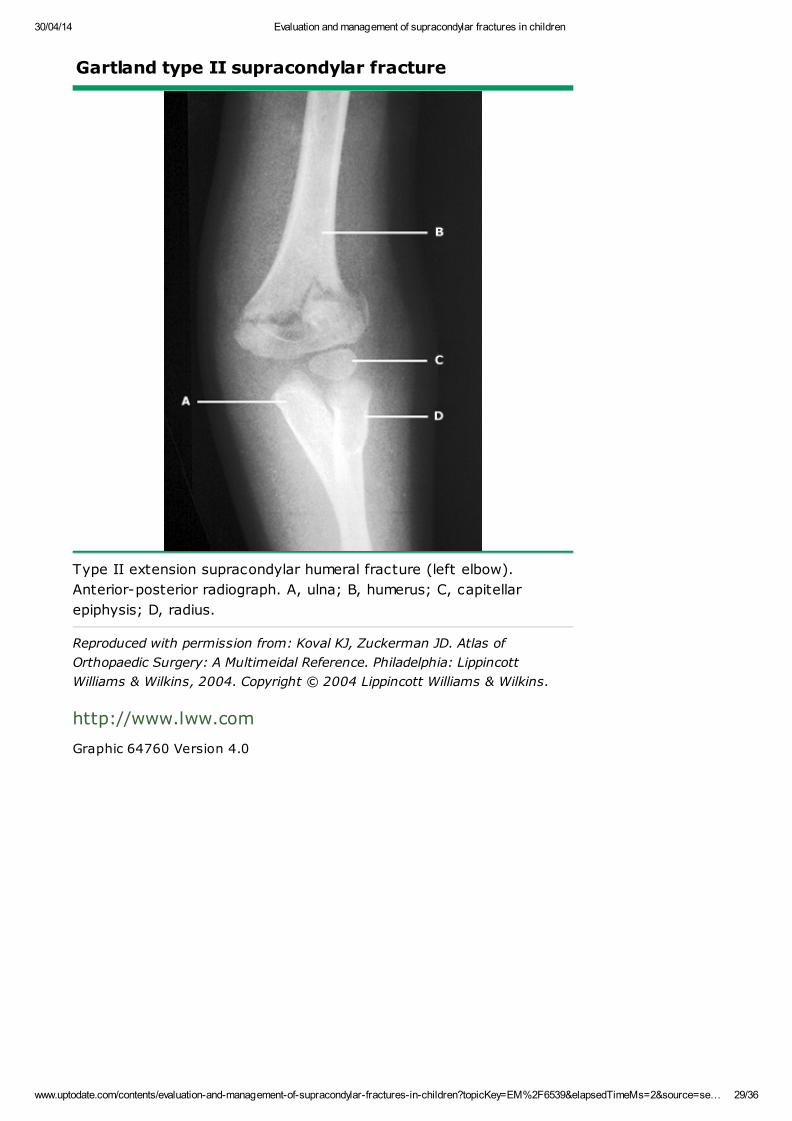
Gartland type II supracondylar fracture
Type II extension supracondylar humeral fracture (left elbow).
Anterior-posterior radiograph. A, ulna; B, humerus; C, capitellar
epiphysis; D, radius.
Reproduced with permission from: Koval KJ, Zuckerman JD. Atlas of
Orthopaedic Surgery: A Multimeidal Reference. Philadelphia: Lippincott
Williams & Wilkins, 2004. Copyright © 2004 Lippincott Williams & Wilkins.
http://www.lww.com
Graphic 64760 Version 4.0
30/04/14 Evaluation and management of supracondylar fractures in children
www.uptodate.com/contents/evaluation-and-management-of-supracondylar-fractures-in-children?topicKey=EM%2F6539&elapsedTimeMs=2&source=se… 30/36
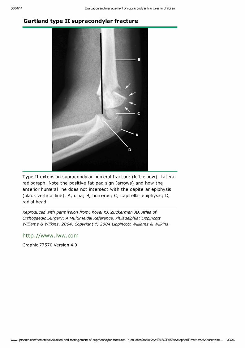
Gartland type II supracondylar fracture
Type II extension supracondylar humeral fracture (left elbow). Lateral
radiograph. Note the positive fat pad sign (arrows) and how the
anterior humeral line does not intersect with the capitellar epiphysis
(black vertical line). A, ulna; B, humerus; C, capitellar epiphysis; D,
radial head.
Reproduced with permission from: Koval KJ, Zuckerman JD. Atlas of
Orthopaedic Surgery: A Multimeidal Reference. Philadelphia: Lippincott
Williams & Wilkins, 2004. Copyright © 2004 Lippincott Williams & Wilkins.
http://www.lww.com
Graphic 77570 Version 4.0
30/04/14 Evaluation and management of supracondylar fractures in children
www.uptodate.com/contents/evaluation-and-management-of-supracondylar-fractures-in-children?topicKey=EM%2F6539&elapsedTimeMs=2&source=se… 31/36
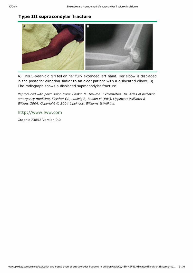
Type III supracondylar fracture
A) This 5-year-old girl fell on her fully extended left hand. Her elbow is displaced
in the posterior direction similar to an older patient with a dislocated elbow. B)
The radiograph shows a displaced supracondylar fracture.
Reproduced with permission from: Baskin M. Trauma: Extremeties. In: Atlas of pediatric
emergency medicine, Fleisher GR, Ludwig S, Baskin M (Eds), Lippincott Williams &
Wilkins 2004. Copyright © 2004 Lippincott Williams & Wilkins.
http://www.lww.com
Graphic 73852 Version 9.0
30/04/14 Evaluation and management of supracondylar fractures in children
www.uptodate.com/contents/evaluation-and-management-of-supracondylar-fractures-in-children?topicKey=EM%2F6539&elapsedTimeMs=2&source=se… 32/36
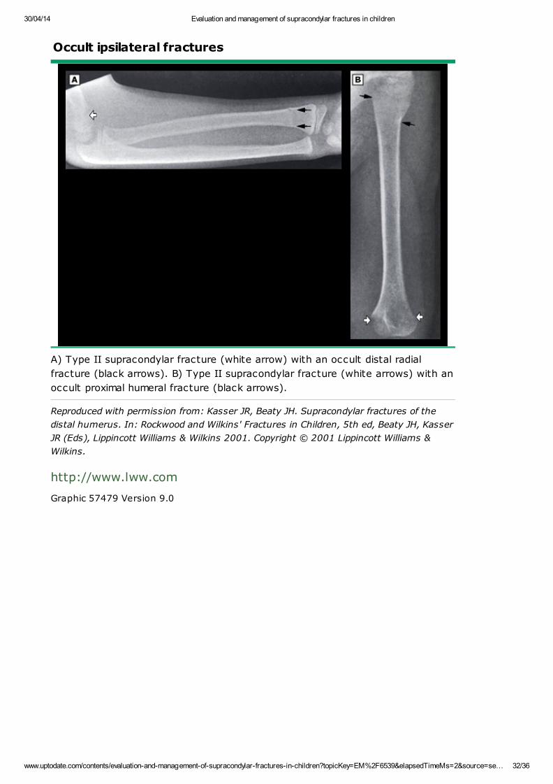
Occult ipsilateral fractures
A) Type II supracondylar fracture (white arrow) with an occult distal radial
fracture (black arrows). B) Type II supracondylar fracture (white arrows) with an
occult proximal humeral fracture (black arrows).
Reproduced with permission from: Kasser JR, Beaty JH. Supracondylar fractures of the
distal humerus. In: Rockwood and Wilkins' Fractures in Children, 5th ed, Beaty JH, Kasser
JR (Eds), Lippincott Williams & Wilkins 2001. Copyright © 2001 Lippincott Williams &
Wilkins.
http://www.lww.com
Graphic 57479 Version 9.0
30/04/14 Evaluation and management of supracondylar fractures in children
www.uptodate.com/contents/evaluation-and-management-of-supracondylar-fractures-in-children?topicKey=EM%2F6539&elapsedTimeMs=2&source=se… 33/36
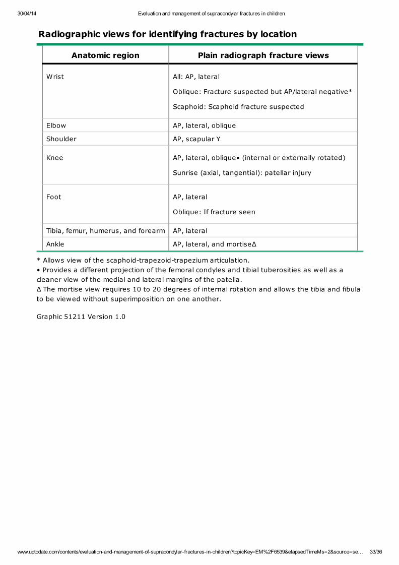
Radiographic views for identifying fractures by location
Anatomic region Plain radiograph fracture views
Wrist All: AP, lateral
Oblique: Fracture suspected but AP/lateral negative*
Scaphoid: Scaphoid fracture suspected
Elbow AP, lateral, oblique
Shoulder AP, scapular Y
Knee AP, lateral, oblique• (internal or externally rotated)
Sunrise (axial, tangential): patellar injury
Foot AP, lateral
Oblique: If fracture seen
Tibia, femur, humerus, and forearm AP, lateral
Ankle AP, lateral, and mortiseΔ
* Allows view of the scaphoid-trapezoid-trapezium articulation.
• Provides a different projection of the femoral condyles and tibial tuberosities as well as a
cleaner view of the medial and lateral margins of the patella.
Δ The mortise view requires 10 to 20 degrees of internal rotation and allows the tibia and fibula
to be viewed without superimposition on one another.
Graphic 51211 Version 1.0
30/04/14 Evaluation and management of supracondylar fractures in children
www.uptodate.com/contents/evaluation-and-management-of-supracondylar-fractures-in-children?topicKey=EM%2F6539&elapsedTimeMs=2&source=se… 34/36
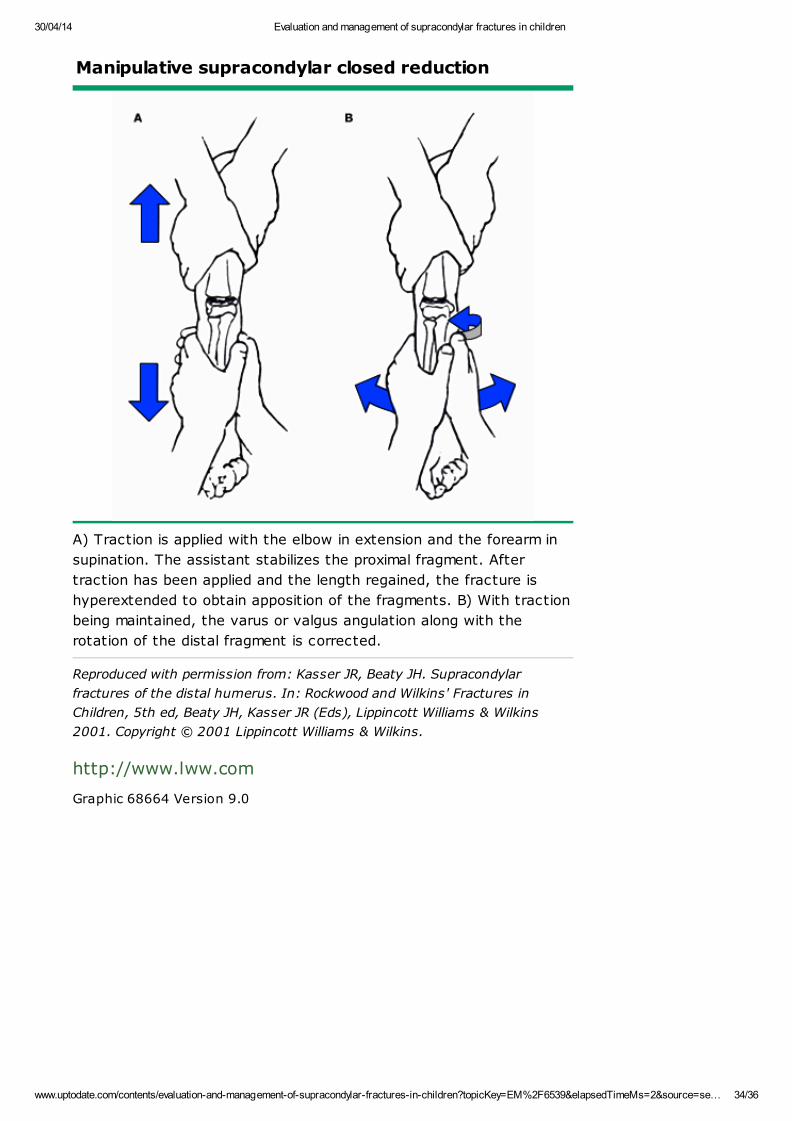
Manipulative supracondylar closed reduction
A) Traction is applied with the elbow in extension and the forearm in
supination. The assistant stabilizes the proximal fragment. After
traction has been applied and the length regained, the fracture is
hyperextended to obtain apposition of the fragments. B) With traction
being maintained, the varus or valgus angulation along with the
rotation of the distal fragment is corrected.
Reproduced with permission from: Kasser JR, Beaty JH. Supracondylar
fractures of the distal humerus. In: Rockwood and Wilkins' Fractures in
Children, 5th ed, Beaty JH, Kasser JR (Eds), Lippincott Williams & Wilkins
2001. Copyright © 2001 Lippincott Williams & Wilkins.
http://www.lww.com
Graphic 68664 Version 9.0
30/04/14 Evaluation and management of supracondylar fractures in children
www.uptodate.com/contents/evaluation-and-management-of-supracondylar-fractures-in-children?topicKey=EM%2F6539&elapsedTimeMs=2&source=se… 35/36
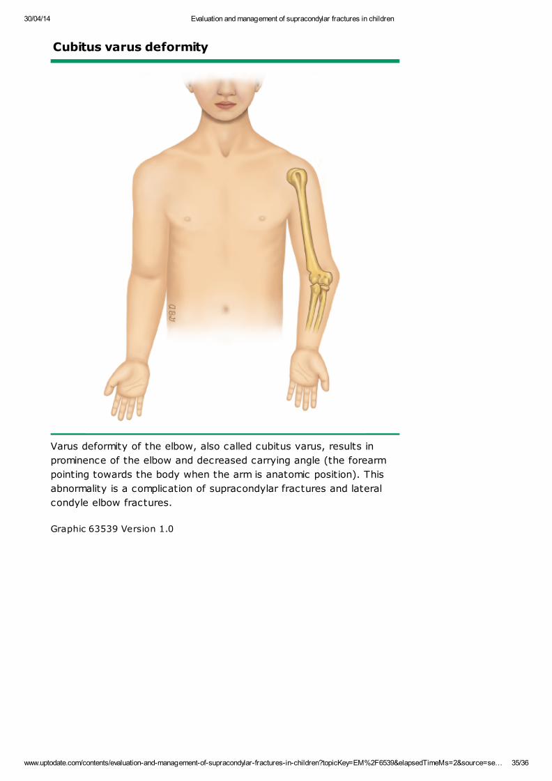
Cubitus varus deformity
Varus deformity of the elbow, also called cubitus varus, results in
prominence of the elbow and decreased carrying angle (the forearm
pointing towards the body when the arm is anatomic position). This
abnormality is a complication of supracondylar fractures and lateral
condyle elbow fractures.
Graphic 63539 Version 1.0

30/04/14 Evaluation and management of supracondylar fractures in children
www.uptodate.com/contents/evaluation-and-management-of-supracondylar-fractures-in-children?topicKey=EM%2F6539&elapsedTimeMs=2&source=se… 36/36
Disclosures: Leticia Manning Ryan, MD, MPH, FAAP Nothing to disclose. Richard G Bachur, MD Nothing to disclose. James FWiley, II, MD, MPH Employee of UpToDate, Inc.
Contributor disclosures are review ed for conflicts of interest by the editorial group. When found, these are addressed by vettingthrough a multi-level review process, and through requirements for references to be provided to support the content. Appropriatelyreferenced content is required of all authors and must conform to UpToDate standards of evidence.
Conflict of interest policy
Disclosures