Embed Size (px)
Citation preview
OLFACTORY AND THERMOSENSORY SIGNALING IN MALARIA
VECTOR MOSQUITO ANOPHELES GAMBIAE
By
Chao Liu
Dissertation
Submitted to the Faculty of the
Graduate School of Vanderbilt University
in partial fulfillment of the requirements
for the degree of
DOCTOR OF PHILOSOPHY
in
Biological Sciences
December, 2013
Nashville, Tennessee
Approved:
Dr. Terry L. Page
Dr. Patrick Abbot
Dr. Julián F. Hillyer
Dr. David M. Bader
Dr. Laurence J. Zwiebel
TABLE OF CONTENTS
Page
LIST OF TABLES ..................................................................................... vii
LIST OF FIGURES .................................................................................. viii
LIST OF ABBREVIATIONS ....................................................................... xi
CHAPTER
I. INTRODUCTION: ANOPHELES GAMBIAE IS THE PRINCIPAL
VECTOR FOR HUMAN MALARIA WHICH UTILIZE OLFACTION AND
THERMOSENSATION FOR HOST SEEKING BEHAVIOR ...................... 1
Human Malaria and Transmission ......................................................... 1
Lifecycle of Anopheles gambiae ............................................................ 4
Olfactory-mediated Host-seeking of An. gambiae .................................. 6
Larval Olfactory System……………………………………………………12
Expression of ORs in Non-chemosensory Tissue………………………14
Thermal Sensitivity in Host-seeking ..................................................... 15
Transient Receptor Potential (TRP) Channels in Thermosensation ..... 16
References ........................................................................................... 20
II. DISTINCT OLFACTORY SIGNALING MECHANISMS IN THE
MALARIA VECTOR MOSQUITO ANOPHELES GAMBIAE .................... 25
ii
Preface ................................................................................................ 25
Introduction .......................................................................................... 26
Materials and Methods ......................................................................... 29
Mosquito Rearing ............................................................................. 29
Individual Larval Behavioral Assays ................................................. 29
AgIR Identification and Expression ................................................... 33
siRNA Preparation and Injection ...................................................... 34
Real-Time PCR (qRT-PCR) ............................................................. 35
Results ................................................................................................. 37
Behavioral Responses of Individual Larva........................................ 37
AgORs Silencing Confirms a Direct Role in the DEET Response .... 42
AgIRs Mediate AgOR Independent Olfactory Responses ................ 49
Discussion............................................................................................ 63
Acknowledgments ................................................................................ 68
Author Contributions ............................................................................ 68
References ........................................................................................... 69
III. MOLECULAR CHARACTERIZATION OF LARVAL PERIPHERAL
THERMOSENSORY RESPONSES OF THE MALARIA VECTOR
MOSQUITO ANOPHELES GAMBIAE ..................................................... 72
iii
Preface ................................................................................................ 72
Introduction .......................................................................................... 73
Materials and Methods ......................................................................... 75
Mosquito rearing and larval sorting .................................................. 75
Thermo-electric control module ........................................................ 76
Fluorescent in situ hybridization and Fluorescent
immunohistochemistry on whole-mount larval antennae .................. 77
Automatic larval tracking and analysis ............................................. 78
siRNA injection and quantitative RT-PCR ........................................ 79
Results ................................................................................................. 79
Kinetic larval response to ambient temperatures .............................. 80
Thermal-induced kinesis reveals larval thermal preferences ............ 82
Plasticity of thermal-driven behavior elicited by An. gambiae
larvae triggered by the shift of cultivation temperature ..................... 84
AgTRPA1 mediates the larval sensitivity towards hot range
temperatures .................................................................................... 88
Larval behavior in the shifted hot range is also
AgTRPA1-mediated ......................................................................... 94
Discussion............................................................................................ 98
iv
Acknowledgments .............................................................................. 104
Author Contributions .......................................................................... 104
References ......................................................................................... 105
IV. Odorant Receptor-Mediated Sperm Activation in the
Malaria Mosquito, Anopheles gambiae .................................................. 109
Preface .............................................................................................. 109
Introduction ........................................................................................ 110
Materials and Methods ....................................................................... 111
Mosquito Rearing ........................................................................... 111
RNA sequencing ............................................................................ 111
Reverse transcription, Polymerase Chain Reaction ....................... 112
Localization of AgORco .................................................................. 113
Spermatozoa bioassay ................................................................... 114
Resutls ............................................................................................... 116
Non-olfactory expression of An. gambiae Odorant Receptor
Transcripts ...................................................................................... 116
Expression of AgOrco protein in male reproductive tissues ........... 119
Activation of spermatozoa .............................................................. 123
Discussion.......................................................................................... 129
v
Acknowledgments .............................................................................. 132
References ......................................................................................... 133
V. SUMMARY AND FUTURE EXPERIMENTS ..................................... 138
References ......................................................................................... 148
vi
LIST OF TABLES
Table Page
Chapter 2
1. Annotation of AgIR family members……………………………………...49
vii
LIST OF FIGURES
Figure Page
Chapter I
1. Transmission cycle of human malaria ................................................... 4
2. The lifecycle of An. gambiae.................................................................. 5
3. Sensilla types ........................................................................................ 8
4. Models of olfactory signaling cascades in insects ............................... 11
5. Model of TRP channel-mediated Drosophila phototransduction .......... 20
Chapter II
1. Operational definitions of larval movements and turns. ....................... 32
2. Larval responses in An. gambiae to yeast and DEET elicit opposite
behaviors. ............................................................................................ 38
3. Behavioral effects of yeast and DEET on An. gambiae. ...................... 39
4. Larval antennae mediate responses to yeast and DEET. .................... 41
5. Quantitative analysis demonstrates significant transcript level
reduction of AgOrco and AgOr40 after siRNA treatment. .................... 44
6. Differential sensitivity of larval responses in An. gambiae to siRNA
mediated knockdown of AgOrco is odorant dependent. ...................... 45
7. Larval behaviors after injection of non-specific small interfering RNA . 47
8. Differential sensitivity of larval responses in An. gambiae to siRNA
mediated knockdown of AgOr40 is odorant dependent. ...................... 49
viii
9. AgIR/DmIR phylogenetic tree. ............................................................. 54
10. Representative alignments of AgIR and DmIR homologs. ................. 55
11. Expression of AgIrs in larval antennae. ............................................. 57
12. Behavioral effects of 3MP and Butylamine on An. gambiae. ............. 59
13. Olfactory responses to 3-methylphenol and butylamine are
mediated by distinct signaling pathways. .......................................... 59
14. Odorant-specific differential effects of AgOr/AgIr knockdown. ........... 62
15. Quantitative mRNA analysis demonstrates significant transcript
level reduction of AgIr76b after siRNA treatment. ............................. 62
Chapter III
1. Thermal-induced mobility in WT 4th instar An. gambiae larvae. ........... 81
2. Thermal preferences of WT 4th instar An.gambiae larvae. .................. 84
3. Thermal-induced larval mobility following the shift of cultivation. ......... 86
4. Thermal preferences of WT 4th instar An. gambiae larvae
following the shift of cultivation. ........................................................... 87
5. Expression of AgTRPA1 in larval tissues. ........................................... 89
6. Larval antenna is a peripheral thermosensory organ. .......................... 91
7. Knockdown of AgTRPA1 mRNA via RNAi. .......................................... 93
8. AgTRPA1 mediates larval responses within the upper temperature
range. .................................................................................................. 96
9. AgTRPA1 mediates larval behavior within the shifted hot range. ........ 97
ix
Chapter IV
1. AgOr RNA Expression in Testes. ...................................................... 118
2. AgOr RT-PCR in Testes cDNA. ......................................................... 118
3. Orco Expression in Antennae. ........................................................... 120
4. AgORco Protein Expression in Testes. ............................................. 121
5. AgORco Expression in Spermatozoa. ............................................... 122
6. Spermatozoa Activation Assay. ......................................................... 124
7. Activation of An. gambiae Sperm by Unitary Compounds. ................ 127
x
LIST OF ABBREVIATIONS
World Health Organization WHO
Center for Disease Control and Prevention CDC
Odorant Receptor OR
Olfactory Receptor Neuron ORN
Odorant Receptor co-receptor ORco
G Protein Coupled Receptors GPCR
Open Reading Frame ORF
N, N-diethyl-meta-toluamide DEET
Transient Receptor Potential TRP
Trans-Membrane TM
Thermosensory TRP channels thermo-TRPs
Phospholipase C PLC
1, 4, 5-trisphosphate IP3
Diacylglycerol DAG
Phosphinositide-4, 5-bisphosphate PIP2
Polyunsaturated Fatty Acids PUFAs
Small interfering RNA siRNA
Anopheles gambiae Ionotropic Receptor AgIR
Double strand RNA dsRNA
Reverse Transcriptase Polymerase Chain Reaction RT-PCR
xi

Ribosomal protein S7 rps7
RNA interference RNAi
Cycle Threshold CT
Drosophila melanogaster DM
Ionotropic Glutamate Receptor iGluR
3-Methylphenol 3-MP
Butylamine BA
Whole-mount Fluorescent In-situ Hybridization WM-FISH
Fluorescent immunohistochemistry FIHC
Alkaline phosphatase AP
Horseradish peroxidase HRP
Frame per second fps
Thermotactic index TI
Standard Error of the Mean SEM
complementary DNA cDNA
4-(2-hydroxyethyl)-1-piperazineethanesulfonic acid HEPES
Activation index AI
RNA sequencing RNAseq
Reads Per Kilobase per Million RPKM
Differential interference contrast DIC
Anopheles gambiae Gustatory Receptor AgGR
Odorant Binding Protein OBP
xii

Spermatheca secretory cells SSC
Sterile Insect Technique SIT
Innovation and Discovery in Engineering And Science IDEAS
National Institutes of Health NIH
xiii
1
CHAPTER I
INTRODUCTION: ANOPHELES GAMBIAE IS THE PRINCIPAL
VECTOR FOR HUMAN MALARIA WHICH UTILIZE OLFACTION AND
THERMOSENSATION FOR HOST SEEKING BEHAVIOR
Human Malaria and Transmission
Malaria is an infectious disease inflicted upon humans and other
animals which is caused by protozoan parasites from the genus
Plasmodium. There are at least five species of Plasmodium that are
capable of infecting human populations1, among which Plasmodium
falciparum gives rise to the most severe symptoms compared to others2.
These symptoms include periodic high fever and shivering chills,
headache, fatigue, nausea and vomiting, which could lead to dehydration,
anemia, coma and even death of the individual3. In the year of 2010 alone,
the World Health Organization (WHO) has estimated that malaria has
killed between 660,000 and 1,200,000 people, most of whom are young
children. In terms of economic loss, a clear correlation has been shown
between malaria and poverty and it really contributes to a vicious cycle
considering that poverty promote malaria transmission while malaria
causes poverty by impeding economic growth4.
2
Human malaria is transmitted by Anopheline mosquitoes (family:
Culicidae; Order: Diptera) except for the special circumstances of
infections acquired trans-placentally or through blood transfusion.
Approximately 460 species of Anopheles have been identified so far while
around 70 are capable of transmitting malaria. The most efficient malaria
vectors are those which prefer human population to other animals
(anthropophily vs zoophily) and tend to feed and shelter indoors rather
than outdoors (endophily vs exophily)5. Among these, Anopheles gambiae
has been considered the most dangerous species and principal vector for
human malaria due to its high degree of anthropophily with strong
endophilic and endophagic traits. There are seven sibling species in the
complex of which An. gambiae s.s. shows the strongest anthropophilic
habit. A blood meal is needed to initiate vitellogenesis (egg development)
and many Anophelines require at least two blood meals to pass through
the pre-gravid phase6, thus making them even more efficient malaria
vectors.
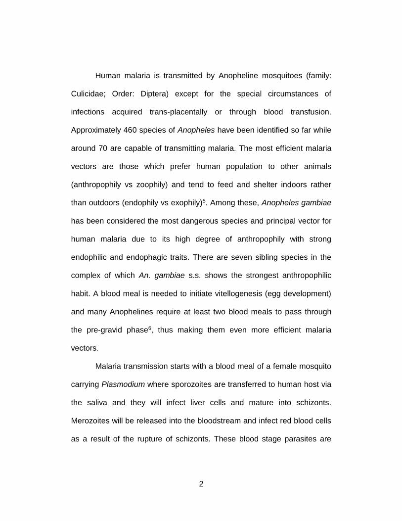
Malaria transmission starts with a blood meal of a female mosquito
carrying Plasmodium where sporozoites are transferred to human host via
the saliva and they will infect liver cells and mature into schizonts.
Merozoites will be released into the bloodstream and infect red blood cells
as a result of the rupture of schizonts. These blood stage parasites are
3
responsible for the clinical manifestations of the disease. Within the
erythrocytes, the parasites undergo asexual reproduction while a
proportion of parasites differentiate into sexual erythrocytic stages
(gametocytes). These gametocytes are ingested by a female Anopheles
during a blood meal and while in the mosquito’s stomach, gametocytes
develop into zygotes which then become motile and elongated
(ookinetes). They invade the midgut wall of the mosquito where they
develop into oocysts. The oocytes grow, rupture and release sporozoites,
which will eventually migrate to the salivary gland of the mosquito. The
sporozoites are injected into another healthy human on occasion of the
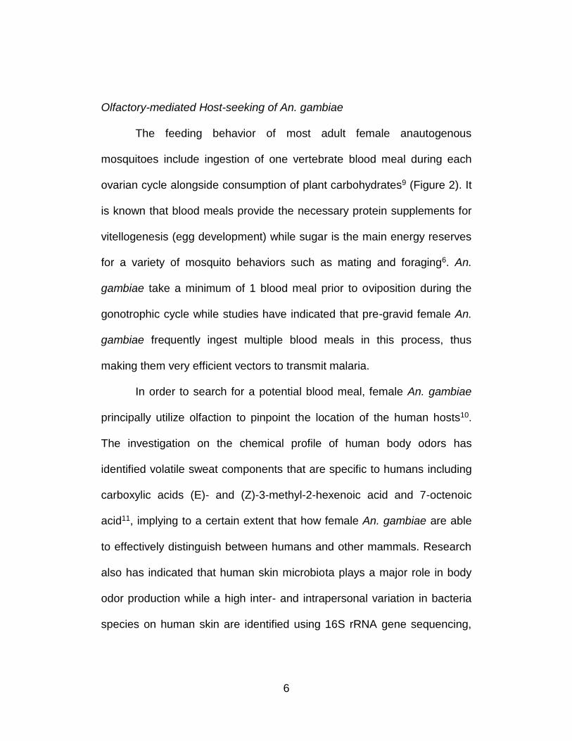
next blood feeding, therefore completing the transmission cycle (Figure 1).
4
Figure 1. Transmission cycle of human malaria. Adapted from Center for Disease Control and Prevention (CDC).
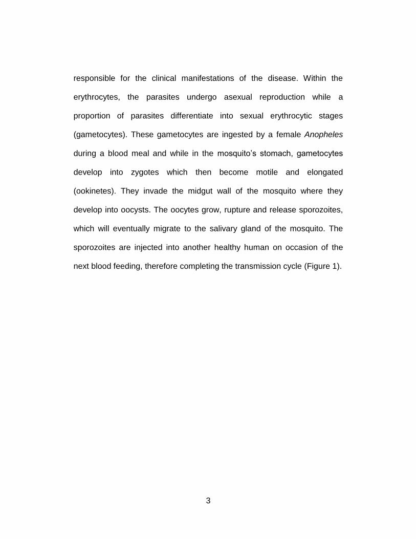
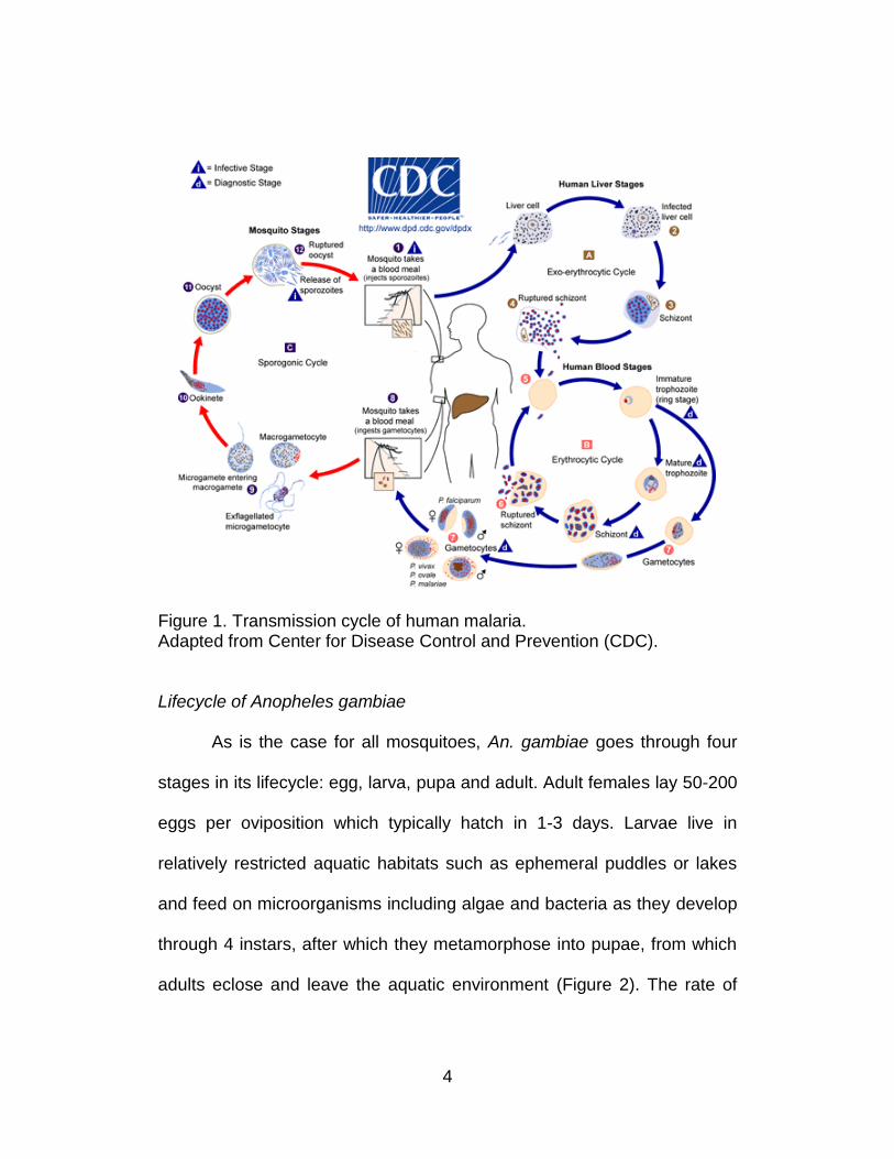
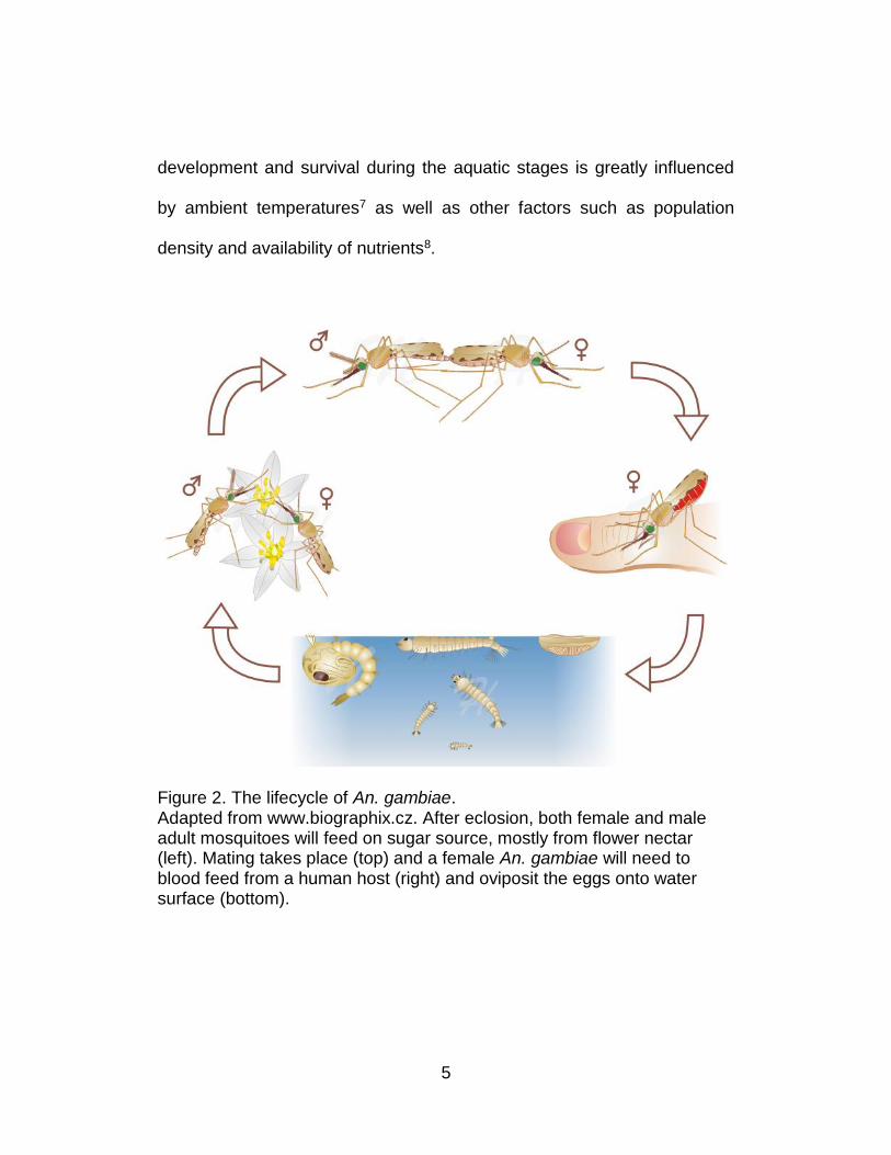
Lifecycle of Anopheles gambiae
As is the case for all mosquitoes, An. gambiae goes through four
stages in its lifecycle: egg, larva, pupa and adult. Adult females lay 50-200
eggs per oviposition which typically hatch in 1-3 days. Larvae live in
relatively restricted aquatic habitats such as ephemeral puddles or lakes
and feed on microorganisms including algae and bacteria as they develop
through 4 instars, after which they metamorphose into pupae, from which
adults eclose and leave the aquatic environment (Figure 2). The rate of
5
development and survival during the aquatic stages is greatly influenced
by ambient temperatures7 as well as other factors such as population
density and availability of nutrients8.
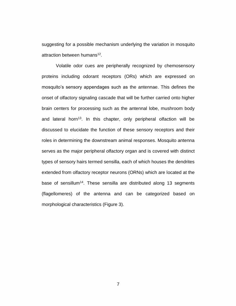
Figure 2. The lifecycle of An. gambiae. Adapted from www.biographix.cz. After eclosion, both female and male adult mosquitoes will feed on sugar source, mostly from flower nectar (left). Mating takes place (top) and a female An. gambiae will need to blood feed from a human host (right) and oviposit the eggs onto water surface (bottom).
6
Olfactory-mediated Host-seeking of An. gambiae
The feeding behavior of most adult female anautogenous
mosquitoes include ingestion of one vertebrate blood meal during each
ovarian cycle alongside consumption of plant carbohydrates9 (Figure 2). It
is known that blood meals provide the necessary protein supplements for
vitellogenesis (egg development) while sugar is the main energy reserves
for a variety of mosquito behaviors such as mating and foraging6. An.
gambiae take a minimum of 1 blood meal prior to oviposition during the
gonotrophic cycle while studies have indicated that pre-gravid female An.
gambiae frequently ingest multiple blood meals in this process, thus
making them very efficient vectors to transmit malaria.
In order to search for a potential blood meal, female An. gambiae
principally utilize olfaction to pinpoint the location of the human hosts10.
The investigation on the chemical profile of human body odors has
identified volatile sweat components that are specific to humans including
carboxylic acids (E)- and (Z)-3-methyl-2-hexenoic acid and 7-octenoic
acid11, implying to a certain extent that how female An. gambiae are able
to effectively distinguish between humans and other mammals. Research
also has indicated that human skin microbiota plays a major role in body
odor production while a high inter- and intrapersonal variation in bacteria
species on human skin are identified using 16S rRNA gene sequencing,
7
suggesting for a possible mechanism underlying the variation in mosquito
attraction between humans12.
Volatile odor cues are peripherally recognized by chemosensory
proteins including odorant receptors (ORs) which are expressed on
mosquito’s sensory appendages such as the antennae. This defines the
onset of olfactory signaling cascade that will be further carried onto higher
brain centers for processing such as the antennal lobe, mushroom body
and lateral horn13. In this chapter, only peripheral olfaction will be
discussed to elucidate the function of these sensory receptors and their
roles in determining the downstream animal responses. Mosquito antenna
serves as the major peripheral olfactory organ and is covered with distinct
types of sensory hairs termed sensilla, each of which houses the dendrites
extended from olfactory receptor neurons (ORNs) which are located at the
base of sensillum14. These sensilla are distributed along 13 segments
(flagellomeres) of the antenna and can be categorized based on
morphological characteristics (Figure 3).
8
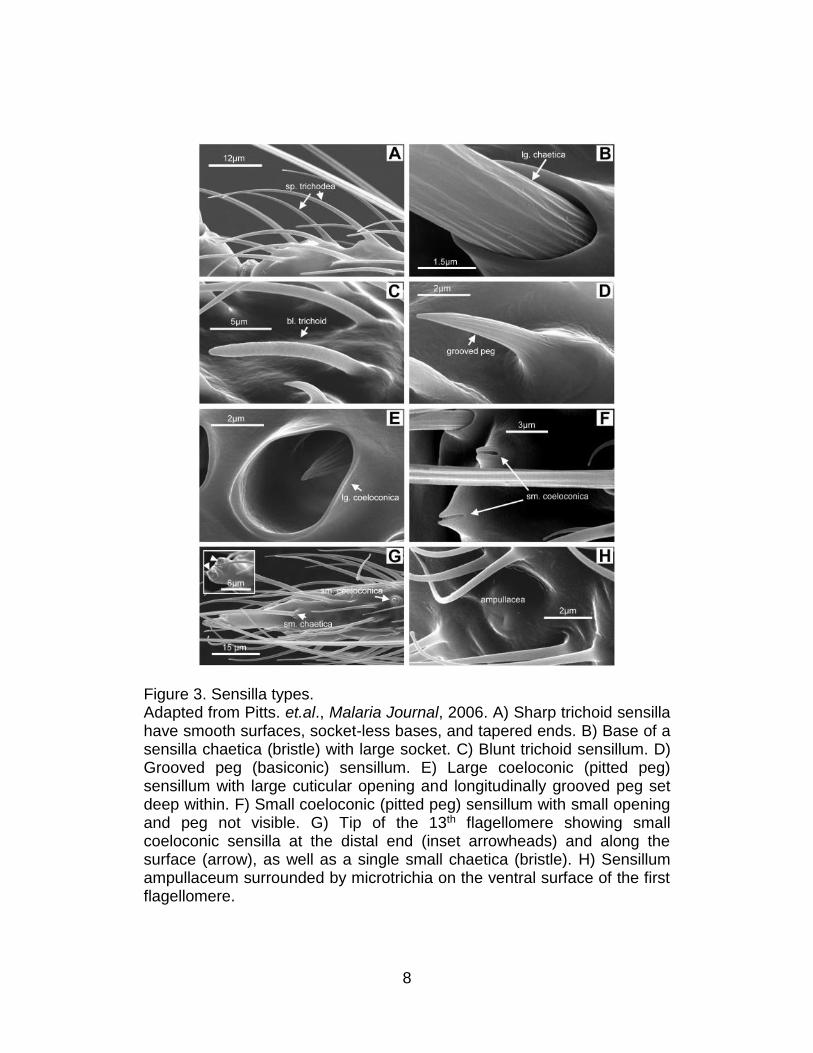
Figure 3. Sensilla types. Adapted from Pitts. et.al., Malaria Journal, 2006. A) Sharp trichoid sensilla have smooth surfaces, socket-less bases, and tapered ends. B) Base of a sensilla chaetica (bristle) with large socket. C) Blunt trichoid sensillum. D) Grooved peg (basiconic) sensillum. E) Large coeloconic (pitted peg) sensillum with large cuticular opening and longitudinally grooved peg set deep within. F) Small coeloconic (pitted peg) sensillum with small opening and peg not visible. G) Tip of the 13th flagellomere showing small coeloconic sensilla at the distal end (inset arrowheads) and along the surface (arrow), as well as a single small chaetica (bristle). H) Sensillum ampullaceum surrounded by microtrichia on the ventral surface of the first flagellomere.
9
Sensilla chaetica are sturdy bristles and occur as both large and
small subtypes which are interspersed among scales on the dorsal
surface. Sensilla trichodea are the most abundant among all sensilla types
and can be categorized into two distinct subtypes: sharp trichodea that
taper noticeably from base to tip with smooth surface as well as blunt
trichodea with a round tip that is nearly as wide as the base. Sensilla
basiconica is also known as grooved pegs and appear as thorn-shaped
hair with 10-12 grooves on the surface. Sensilla coeloconica are small,
thick-walled sensilla that houses a peg set into the bottom of a pit whose
tips often project to below the external rim of the socket. Additionally,
sensilla ampullaceal are also small, thick-walled peg set at the bottom of a
tube. However, unlike coeloconic sensilla, the pegs project perpendicular
to the tube walls. Sensilla trichodea are confirmed to be the major
olfactory chemosensilla where they respond to olfactory stimuli either by
an increase or decrease in impulse frequency as compared to the
spontaneous spiking activity. It was shown as early as 1968 that sharp
trichodea are sensitive to host odors such as those from a human hand
while the blunt trichodea are sensitive to vapors of commercial
repellents14, confirming their roles in sensing environmental odors.
Throughout four decades of research on insect chemosensilla,
extensive knowledge is known for the molecular, anatomical and
10
functional organization of the insect olfactory system, particularly in
Drosophila melanogaster15. Olfactory receptor neurons (ORNs) are
located at the base of each olfactory sensillum and project their axons
centrally to synaptic modules in the brain named glomeruli. The odorant
molecules are recognized by the ligand-binding members of the olfactory
receptor (OR) along with a highly conserved and broadly expressed co-
receptor ORco which are expressed in both the ORNs as well as the
dendrites extended from these neurons16. The insect ORs were first
identified using a novel search algorithm to probe the Drosophila genome
for predicted 7-transmembrane G-Protein Coupled Receptors (GPCRs)
with longer than 300bp Open Reading Frame (ORF) based on statistically
examining the physicochemical profile17. The search criteria was chosen
due to the fact that vertebrate ORs were confirmed to be GPCRs,
however, insect ORs show opposite topology when compared to
mammalian ORs and they share very little sequence homology and are
also evolutionarily divergent from each other as no common ancestor can
be identified using bioinformatics approaches18.
Recent research has revealed the ionic conductivity of the insect
ORs and they are now more and more believed to be ligand-gated ion
channels other than GPCRs although there is evidence that a slow-
responsive metabotropic second-messenger pathway is present additional
11
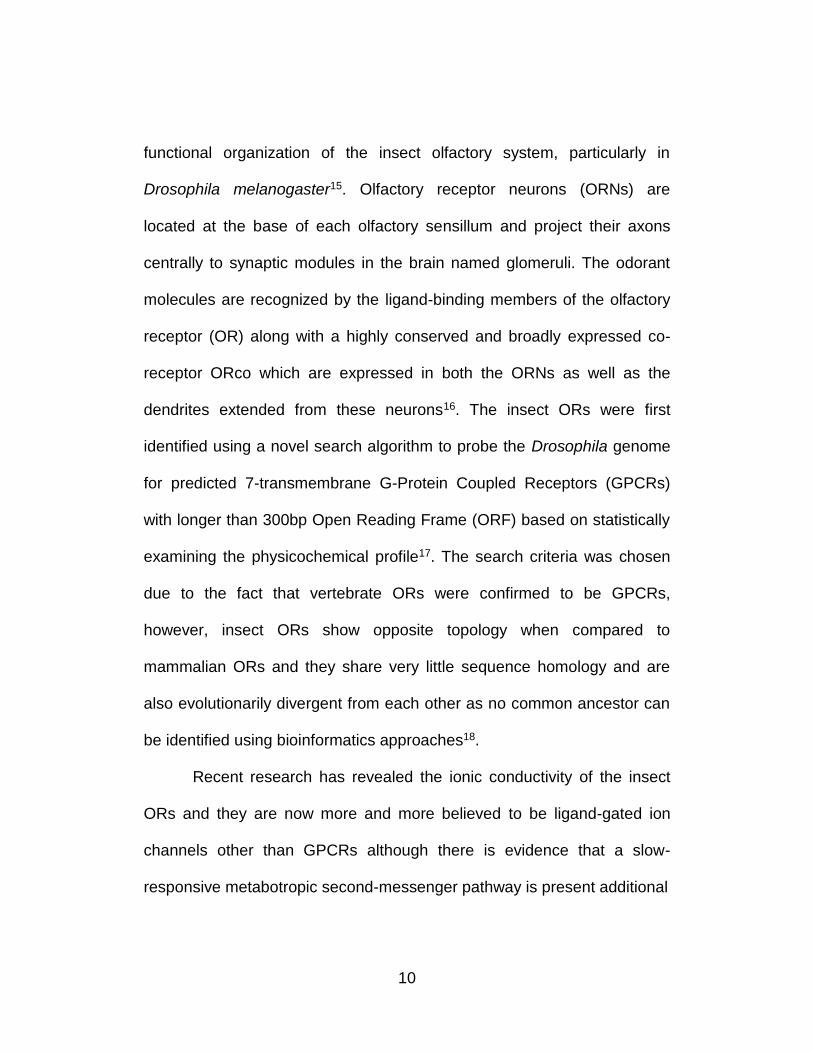
Figure 4. Models of olfactory signaling cascades in insects. Adapted from Kaupp. Nature Reviews, 2010. a) ORs from heteromeric ion channels that are directly gated by odorants. The channel complex is comprised of ligand-binding ORX as well as ORco. The channel pore of this model is not specified although current research indicates the pore region is contributed by both ORX and ORco subunits19. b) In addition to the channel model, the ORco subunit is also linked to a metabotropic pathway that is presumably slower but with amplification of signal through a second-messenger pathway the response is much stronger and prolonged. In this model it is proposed only ORco contributes to the channel pore, not ORX.
12
to the fast-responsive kinetics of ligand gating (Figure 4)20,21. The ORco
and ORX will form a heteromeric ion channel complex on the dendritic
membrane with unknown stoichiometry. Upon the binding of odorant to
ORX, the channel pore will open to allow influx of cations, which in turn,
leads to membrane depolarization and eventually an action potential16.
Thus the electric signal of the odorant can be transmitted downstream. To
date, 79 ligand-binding Ors and 1 Orco gene have been annotated in An.
gambiae genome and numerous studies have validated the essential role
of ORs in a comprehensive array of olfactory-directed behaviors in
mosquitoes including mating, nectar feeding, host seeking and oviposition,
all of which are vital for the survival of the species10. The OR-mediated
peripheral olfaction has been shown to mediate host selection as well as
sensitivity to the insect repellent N,N-diethyl-meta-toluamide (DEET) and
one of the recent studies demonstrate that the orco mutant Ae. aegypti fail
to discriminate between human and animal odors, therefore strengthening
the indispensable role of ORs in determining the host seeking behavior of
mosquitoes22.
Larval olfactory system
Previous studies have been carried out to examine the molecular
basis of olfaction in insect larvae, due to their relatively simplified neuronal
13
circuits and convenience to conduct behavioral analysis23. In An. gambiae
4th instar larvae, their major olfactory organ has been identified, which is
an aporous cone-shaped structure located at the distal tip of the antennae
named sensory cone24. As confirmed by both RT-PCR and In-situ
hybridization assays, there are 12 tuning ORs expressed on the larval
antenna. The sensory cone is innervated by dendrites extended from a
cluster of 12 ORNs with each expressing a combination of AgOrco and
AgOrX. In vitro heterologous expression of larval ORs in Xenopus oocytes
have demonstrated their efficacy to respond to a range of natural and
synthetic odors. The combinatorial odor coding for larval ORs is also
investigated by screening them against a panel of odorants. The response
spectrum of any given larval OR is discrete such that some ORs (AgOR1,
AgOR34) respond to a narrow set of odorants while others (AgOR10,
AgOR40) are much more broadly tuned. On the other hand, a single
odorant is also able to elicit responses from multiple larval ORs with
varying amplitude. For example, acetophenone, a volatile aromatic ketone
emitted by plants, which was suggested to be an insect attractant25,
generate electric responses from AgOR6, AgOR10, AgOR28 and
AgOR3724. Behaviorally, the 4th instar An. gambiae larvae exhibit dose-
dependent locomotion towards/away from several odorants which are
shown to activate larval ORs in the heterologous expression system.
14
Additionally, in chapter 2, I will provide a detailed description of a more
recent study that was carried out on late-stage An. gambiae larvae to
elucidate the in vivo role of peripherally expressed ORs in directing larval
responses towards odor stimuli as well as the characterization of a distinct
peripheral signaling pathway that is independent of ORs, but mediated by
a gene from the family of Inotropic receptors (IRs) that are distantly related
to glutamate receptors.
Expression of ORs in Non-chemosensory tissue
Although odorant receptors are, judged by the name, primarily
studied in the peripheral chemosensory organs that mediate perception of
environmental odorants, it also has been documented that the expression
of ORs is not restricted to the olfactory system. In mammals, ORs have
been found outside the olfactory system, which are suggested to function
in skeletal muscle development, regeneration, human sperm chemotaxis
etc. Meanwhile, the transcript for human ORs have been “ectopically”
identified in many internal organs, to name but a few, heart, spleen,
pancreas, placenta, lung, kidney. However, the possible roles of these
cryptic OR expression are to a large extent, mysterious26. As for insects,
very few attention has been given to the olfactory receptors in tissues
other than the chemosensory appendages. Nevertheless, an RNA-
15
sequencing-based survey of OR expression level between the whole body
of male and female An. gambiae has revealed a number of Ors, including
Orco, show male-biased expression pattern. While in samples containing
only antennae or maxillary palps, respectively, which are the major
chemosensory organs in mosquito, these ORs are undoubtedly enhanced
towards female mosquitoes27. These results have indicated that transcript
for ORs are present in tissues other than the peripheral sensory
appendages within the male An. gambiae. In chapter 4, I will expand upon
this discovery and describe the expression for a variety of ORs in the male
testis while these ORs are suggested to be functional in mediating
chemical-induced activation of flagellar movement in mature spermatozoa.
Thermal Sensitivity in Host-seeking
In addition to olfaction, female An. gambiae utilize body heat
emitted from human host as a relatively short-range guidance and it has
been shown from multiple studies that heat sensitivity is able to synergize
with olfaction to enhance the efficiency of landing behavior in
mosquitoes28. Heat cues are common to all warm-blooded hosts so it is
not likely that body heat alone will play a major role in directing human
preference in An. gambiae29, however, basic study on mosquito
thermosensation would undoubtedly shed light in the design of novel
16
control approaches that interfere with mosquito’s host seeking ability as
well as serve as a new molecular target for insecticide development as
thermal sensitivity is also vital for the survival of poikilothermic insects that
are incapable of maintaining thermal homeostasis.
Transient Receptor Potential (TRP) Channels in Thermosensation
The first TRP gene was discovered in 1969 when Cosens and
Manning characterized a Drosophila mutant that showed a transient
instead of a sustained response to bright light30. The photoreceptor cells in
the mutant flies with sustained light exposure displayed a transient, rather
than normal plateau-like receptor potential. Thus the mutant gene gained
its name trp, abbreviated for Transient Receptor Potential. Not until 20
years later that trp gene was cloned and revealed to function as a Ca2+-
permeable cation channel31. To date, research on TRP channel genes
have revealed 28 members in human32, 16 in flies33 and at least 10 in
An.gambiae (unpublished data). They can be sub-categorized into 7
subfamilies namely TRPC, TRPV, TRPM, TRPA, TRPP, TRPML and
TRPN based on sequence similarity32. The TRPC subfamily (“C” stands
for canonical) comprises of proteins with highest homology to the
Drosophila TRP protein, hence gained its name. The other subfamilies
were named after their first identified members: the TRPV subfamily after
17
the vanilloid receptor 1 (trpv1), the TRPM subfamily after the tumor
suppressor melastatin (trpm1), the TRPA subfamily after the protein
denoted ankyrin-like transmembrane domains 1 (trpa1), TRPN after the no
mechanoreceptor potential C (nompC) gene from Drosophila, TRPP after
the polycystic kidney disease-related protein 2 (trpp2), and TRPML after
mucolipin (trpml1)34.
TRP channels are found to be expressed in a broad spectrum of
organisms from worms to human and they all share 6 trans-membrane (6-
TM) domains, yet characteristic structures including ankyrin repeats,
coiled coil domain, and protein kinase domains are specific to certain
subfamilies. The TRP super-family of membrane proteins displays distinct
ion selectivity, modes of action and physiological functions among
different subfamily and even members within the same subgroup. TRP
channels were initially considered to be PLC-dependent or a Ca2+ store-
dependent cation channel until an expression-cloning method was utilized
to isolate a vanilloid receptor, which displayed high identity with
Drosophila TRP. This receptor, subsequently renamed TRPV1, was
activated not only by vanilloids such as capsaicin, but also temperatures
above 43oC35. The investigation of thermosensory TRP channels (thermo-
TRPs) has led to the characterization of no less than 6 thermo-TRPs in
mammals and an equal number in insects.
18
In Drosophila, the thermo-receptors are housed in the third
antennal segment according to ablation studies36 while in An. gambiae, a
pair of small coeloconic sensilla on the distal tip of adult female antennae
were shown to contain neurons that specifically respond to rise of
temperatures37. In contrast with limited achievements regarding peripheral
thermo-receptors in mosquitoes, comprehensive studies have been
carried out in Drosophila on thermo-TRPs in the past two decades.
painless, a member of TRPA subfamily, has heat sensitivity that is
essential for avoidance of noxious heat (above 42oC), thus uncovering its
role as a primary noxious heat detector in Drosophila38. The other gene in
Drosophila TRPA subfamily, pyrexia, is activated approximately at 40oC
and holds high potassium permeability. In addition, evidence exists that
activation of Pyrexia is essential for the prevention of paralysis during high
temperature stress39. A third member in TRPA subfamily, trpa1, is one of
the best-studied thermo-TRPs that mediates med-high temperature
sensation which is activated above 25oC and contributes to avoidance of
sub-lethal warmer temperatures in Drosophila40. TRPA1 possesses at
least two distinct functions in vivo as TRPA1 is involved in sensing
electrophiles in gustatory neurons and derives behavioral avoidance to
several tissue-damaging chemicals. It also has been shown that
19
invertebrate and vertebrate TRPA1 share a common ancestor and critical
characteristics required for electrophile detection41.
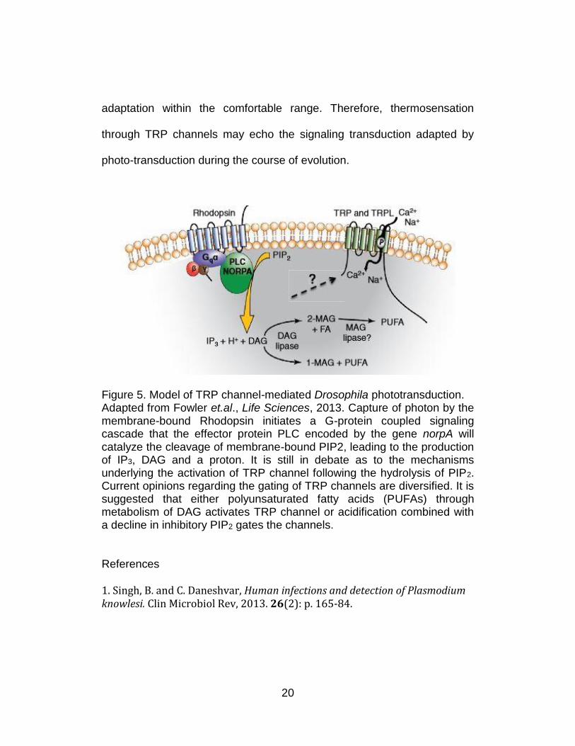
TRP channels that are involved in photo-transduction in mammals
and flies utilize a phospholipase C (PLC)-mediated signaling cascade
(Figure 5). The activation of PLC leads to the production of inositol 1,4,5-
trisphosphate (IP3) and diacylglycerol (DAG) as well as the release of Ca2+
from internal IP3-sensitive stores. They are either positively or negatively
regulated by second messengers derived from the hydrolysis of
phosphinositide-4,5-bisphosphate (PIP2) or products from these second
messengers42.
In terms of thermo-TRPs, recent behavioral studies on Drosophila
larvae regarding their thermo-preference in a thermo gradient indicated
the larvae required PLCβ to distinguish between optimal temperature
(18oC) and suboptimal temperature, thus suggesting dTRPA1 functions
through a PLC-mediated GPCR pathway43. It is more recently discovered
that Rhodopsin is involved in Drosophila thermosensation, thus
uncovering a novel role for this canonical visual G-protein as well as
strengthening the view that a second-messenger pathway is employed in
thermal sensitivity of insects44. Furthermore, the implication that activation
of TRPA1 is conjugated to a signaling cascade may promote signal
amplification of small differences in environmental temperature to facilitate
20
adaptation within the comfortable range. Therefore, thermosensation
through TRP channels may echo the signaling transduction adapted by
photo-transduction during the course of evolution.
Figure 5. Model of TRP channel-mediated Drosophila phototransduction. Adapted from Fowler et.al., Life Sciences, 2013. Capture of photon by the membrane-bound Rhodopsin initiates a G-protein coupled signaling cascade that the effector protein PLC encoded by the gene norpA will catalyze the cleavage of membrane-bound PIP2, leading to the production of IP3, DAG and a proton. It is still in debate as to the mechanisms underlying the activation of TRP channel following the hydrolysis of PIP2. Current opinions regarding the gating of TRP channels are diversified. It is suggested that either polyunsaturated fatty acids (PUFAs) through metabolism of DAG activates TRP channel or acidification combined with a decline in inhibitory PIP2 gates the channels.
References
1. Singh, B. and C. Daneshvar, Human infections and detection of Plasmodium knowlesi. Clin Microbiol Rev, 2013. 26(2): p. 165-84.
21
2. Making a difference. The World Health Report 1999. Health Millions, 1999. 25(4): p. 3-5.
3. Heineman, H.S., The clinical syndrome of malaria in the United States. A current review of diagnosis and treatment for American physicians. Arch Intern Med, 1972. 129(4): p. 607-16.
4. Sachs, J. and P. Malaney, The economic and social burden of malaria. Nature, 2002. 415(6872): p. 680-5.
5. White, G.B., Malaria vector ecology and genetics. Br Med Bull, 1982. 38(2): p. 207-12.
6. Scott, T.W. and W. Takken, Feeding strategies of anthropophilic mosquitoes result in increased risk of pathogen transmission. Trends Parasitol, 2012. 28(3): p. 114-21.
7. Bayoh, M.N. and S.W. Lindsay, Temperature-related duration of aquatic stages of the Afrotropical malaria vector mosquito Anopheles gambiae in the laboratory. Med Vet Entomol, 2004. 18(2): p. 174-9.
8. Gimnig, J.E., et al., Density-dependent development of Anopheles gambiae (Diptera: Culicidae) larvae in artificial habitats. J Med Entomol, 2002. 39(1): p. 162-72.
9. Clements, A.N., The Biology of mosquitoes. Vol. 2. 1999.
10. Takken, W. and B.G. Knols, Odor-mediated behavior of Afrotropical malaria mosquitoes. Annu Rev Entomol, 1999. 44: p. 131-57.
11. Costantini, C., et al., Electroantennogram and behavioural responses of the malaria vector Anopheles gambiae to human-specific sweat components. Med Vet Entomol, 2001. 15(3): p. 259-66.
12. Verhulst, N.O., et al., Chemical ecology of interactions between human skin microbiota and mosquitoes. FEMS Microbiol Ecol, 2010. 74(1): p. 1-9.
13. Carey, A.F. and J.R. Carlson, Insect olfaction from model systems to disease control. Proc Natl Acad Sci U S A, 2011. 108(32): p. 12987-95.
14. McIver, S.B., Sensilla mosquitoes (Diptera: Culicidae). J Med Entomol, 1982. 19(5): p. 489-535.
22
15. Couto, A., M. Alenius, and B.J. Dickson, Molecular, anatomical, and functional organization of the Drosophila olfactory system. Curr Biol, 2005. 15(17): p. 1535-47.
16. Kaupp, U.B., Olfactory signalling in vertebrates and insects: differences and commonalities. Nat Rev Neurosci, 2010. 11(3): p. 188-200.
17. Clyne, P.J., et al., A novel family of divergent seven-transmembrane proteins: candidate odorant receptors in Drosophila. Neuron, 1999. 22(2): p. 327-38.
18. Sato, K., et al., Insect olfactory receptors are heteromeric ligand-gated ion channels. Nature, 2008. 452(7190): p. 1002-6.
19. Pask, G.M., et al., Heteromeric Anopheline Odorant Receptors Exhibit Distinct Channel Properties. PLoS One, 2011. 6(12).
20. Jones, P.L., et al., Functional agonism of insect odorant receptor ion channels. Proc Natl Acad Sci U S A, 2011. 108(21): p. 8821-8825.
21. Wicher, D., et al., Drosophila odorant receptors are both ligand-gated and cyclic-nucleotide-activated cation channels. Nature, 2008. 452(7190): p. 1007-U10.
22. DeGennaro, M., et al., orco mutant mosquitoes lose strong preference for humans and are not repelled by volatile DEET. Nature, 2013. 498(7455): p. 487-91.
23. Kreher, S.A., J.Y. Kwon, and J.R. Carlson, The molecular basis of odor coding in the Drosophila larva. Neuron, 2005. 46(3): p. 445-56.
24. Xia, Y., et al., The molecular and cellular basis of olfactory-driven behavior in Anopheles gambiae larvae. Proc Natl Acad Sci U S A, 2008. 105(17): p. 6433-8.
25. Metcalf, R.L., Plant Volatiles as Insect Attractants. Crc Critical Reviews in Plant Sciences, 1987. 5(3): p. 251-301.
26. Kang, N. and J. Koo, Olfactory receptors in non-chemosensory tissues. BMB Rep, 2012. 45(11): p. 612-22.
23
27. Pitts, R.J., et al., Transcriptome profiling of chemosensory appendages in the malaria vector Anopheles gambiae reveals tissue- and sex-specific signatures of odor coding. BMC Genomics, 2011. 12.
28. Kline, D.L. and G.F. Lemire, Field evaluation of heat as an added attractant to traps baited with carbon dioxide and octenol for Aedes taeniorhynchus. J Am Mosq Control Assoc, 1995. 11(4): p. 454-456.
29. Bowen, M.F., The sensory physiology of host-seeking behavior in mosquitoes. Annu Rev Entomol, 1991. 36: p. 139-58.
30. Cosens, D.J. and A. Manning, Abnormal electroretinogram from a Drosophila mutant. Nature, 1969. 224(5216): p. 285-7.
31. Hardie, R.C. and B. Minke, The Trp Gene Is Essential for a Light-Activated Ca2+ Channel in Drosophila Photoreceptors. Neuron, 1992. 8(4): p. 643-651.
32. Voets, T., et al., Sensing with TRP channels. Nat Chem Biol, 2005. 1(2): p. 85-92.
33. Rosenzweig, M., K. Kang, and P.A. Garrity, Distinct TRP channels are required for warm and cool avoidance in Drosophila melanogaster. Proc Natl Acad Sci U S A, 2008. 105(38): p. 14668-73.
34. Vriens, J., et al., Invertebrate TRP proteins as functional models for mammalian channels. Pflugers Archiv-European Journal of Physiology, 2004. 449(3): p. 213-226.
35. Caterina, M.J., et al., The capsaicin receptor: a heat-activated ion channel in the pain pathway. Nature, 1997. 389(6653): p. 816-24.
36. Sayeed, O. and S. Benzer, Behavioral genetics of thermosensation and hygrosensation in Drosophila. Proc Natl Acad Sci U S A, 1996. 93(12): p. 6079-6084.
37. Wang, G., et al., Anopheles gambiae TRPA1 is a heat-activated channel expressed in thermosensitive sensilla of female antennae. Eur J Neurosci, 2009. 30(6): p. 967-74.
38. Sokabe, T. and M. Tominaga, A temperature-sensitive TRP ion channel, Painless, functions as a noxious heat sensor in fruit flies. Commun Integr Biol, 2009. 2(2): p. 170-3.
24
39. Lee, Y., et al., Pyrexia is a new thermal transient receptor potential channel endowing tolerance to high temperatures in Drosophila melanogaster. Nature Genetics, 2005. 37(3): p. 305-310.
40. Rosenzweig, M., et al., The Drosophila ortholog of vertebrate TRPA1 regulates thermotaxis. Genes Dev, 2005. 19(4): p. 419-24.
41. Kang, K., et al., Analysis of Drosophila TRPA1 reveals an ancient origin for human chemical nociception. Nature, 2010. 464(7288): p. 597-U155.
42. Montell, C., Drosophila TRP channels. Pflugers Archiv-European Journal of Physiology, 2005. 451(1): p. 19-28.
43. Kwon, Y., et al., Control of thermotactic behavior via coupling of a TRP channel to a phospholipase C signaling cascade. Nature Neuroscience, 2008. 11(8): p. 871-873.
44. Shen, W.L., et al., Function of Rhodopsin in Temperature Discrimination in Drosophila. Science, 2011. 331(6022): p. 1333-1336.
25
CHAPTER II
DISTINCT OLFACTORY SIGNALING MECHANISMS IN THE MALARIA
VECTOR MOSQUITO ANOPHELES GAMBIAE
Preface
This following article was published in the journal of PLoS Biology
in 2010 (Volume 8, No. 8, pii: e1000467). I was a co-first author on this
paper along with other co-authors including R. Jason Pitts (co-first),
Johnathan D. Bohbot (third author), Patrick L. Jones (fourth author),
Guirong Wang (fifth author) and Laurence J. Zwiebel (corresponding
author). In this paper, I have developed a novel single-larval bioassay for
the purpose of quantifying olfactory-driven behaviors when they are
challenged with a series of natural or synthetic chemicals which are able
to elicit larval responses in vivo. I have also implemented an RNAi-based
gene silencing protocol in this paper to specifically knockdown genes via
injection of small interfering RNA (siRNA) on 3rd instar larvae to evaluate
the effect of peripheral olfactory genes in downstream larval responses. I
took a leading role in experimental design, data acquisition, statistical
analysis as well as figure and manuscript preparation. I want to thank my
co-first author R. Jason Pitts for his contribution in the annotation of An.
26
gambiae Ionotropic Receptors (AgIRs), Johnathan D. Bohbot for figure
design, Laurence J. Zwiebel for his support and discussions on the
experimental data. I have modified the gene name for Or83b or Or7 from
the original publication to Orco to conform to the recent change of
nomenclature in the field1.
Introduction
Chemosensory cues play a central role in directing much of the
behavioral repertoire and are a significant determinant in the vectorial
capacity of female An. gambiae mosquitoes, which are responsible for the
transmission of human malaria2. Significant progress has been made in
identifying the components of olfactory pathways in An. gambiae3-7.
Nonetheless, there is a paucity of information regarding the precise
molecular mechanisms that mediate olfactory signaling in An. gambiae.
At the center of the peripheral olfactory signal transduction pathway
in An. gambiae is a family of odorant receptors (AgORs) that are
selectively expressed in olfactory receptor neurons (ORNs). Although
originally identified as candidate G-protein-coupled receptors (GPCRs)8,
several studies have disputed the GPCR nature of Anopheline and other
insect ORs9-12, which likely form ligand-gated heteromeric ion channels
that activate ORNs through ionotropic as well as perhaps metabotropic
27
mechanisms. In addition, members of a family of another set of
chemosensory receptors related to ionotropic glutamate receptors have
recently been described in Drosophila melanogaster13.
The majority of insect ORNs typically express at least two ORs that
are likely to form complexes of undetermined stoichiometry that are
composed of one highly conserved non-conventional ORco-like protein
together with a conventional OR that presumably mediates odorant
binding specificity5,9,14. In An. gambiae, 73 of the 79 AgORs originally
identified are expressed in the adult and 13 are expressed in larval
stages15. The non-conventional Anopheline ORco-like family member,
AgORco, is widely expressed in nearly all olfactory sensilla with the
notable exception of grooved-peg sensilla6, which are activated in vivo by
compounds such as ammonia, lactic acid, and other carboxylic acids that
are major components of human sweat16,17 known to evoke physiological
and/or behavioral activity in An. gambiae18,19. Indeed, recent functional
analyses of AgOR odor space reveal a paucity of responses for these
groups of odorants, suggesting Anopheline sensitivity to amines and other
variant odorants may lie outside of AgOR-based signaling.
In order to improve our understanding of mosquito olfaction, we
have continued to utilize the relative simplicity of the An. gambiae larval
olfactory system, which consists of only 12 ORNs15. In previous studies
28
utilizing behavioral and functional approaches to describe the molecular
and cellular basis for olfactory responses to a range of natural and
synthetic chemical stimuli, we identified a subset of AgORs expressed in
the larval antenna that are tuned to odorants that elicit specific behavioral
responses. Building upon those studies, we now use RNAi-based gene-
silencing approaches to validate in vivo the role of AgORs in larval
olfactory signal transduction and specifically identify the molecular
receptor that mediates the repellent activity of N, N-diethyl-m-toluamide
(DEET). In addition, we have identified and characterized a family of
chemosensory receptors that are related to inotropic glutamate receptors
(AgIRs) that underlie a novel-signaling pathway that is independent of
AgOR activity. We propose that An. gambiae expresses distinct signaling
pathways that participate in larval olfaction and are likely to also be active
in mediating adult responses to a diverse range of chemosensory stimuli.
These studies further our understanding of the molecular basis of olfaction
and olfactory-driven behaviors in An. gambiae and lay the foundation for
advancing alternatives to mosquito control strategies focused on adult life
stages.
29
Materials and Methods
Mosquito Rearing
An. gambiae sensu stricto, originated from Suakoko, Liberia, was
reared as described3. For stock propagation, 4- to 5-d-old female
mosquitoes were blood fed for 30–45 min on anesthetized mice, following
the guidelines set by Vanderbilt Institutional Animal Care and Use
Committee.
Individual Larval Behavioral Assays
Larval assays were conducted between ZT2 and ZT10 during the
standard LD12:12 rearing cycle. Here, An. gambiae 3rd or 4th instar larvae
were removed from rearing pans, rinsed carefully with distilled water to
eliminate any remaining food residue, and kept in segregated containers
with distilled water for 30 min. Odorant stocks were made by dissolving
odorant (>99% pure or of the highest grade commercially available) in pre-
heated (70°C) 2% NuSieve, GTG low-melting-temperature agarose
(Cambrex Bio Science). The assay was performed in a 10×1.5 cm Petri
dish containing 50 ml of 27°C distilled water. The odorant and larva
dropping spots were located at opposite ends along the diameter and
marked by a solid circle and a cross, respectively. The odorant/control
30
stock was placed into the dish for 1 min beforehand to equilibrate, and the
larva was gently introduced at the marked spot.
Real-time images of larval movements were obtained and
downloaded at 1 s intervals for the duration of the 5 min assay using a
custom-designed 30 frames/s video camera/computer/software system
(Model NC-70, DAGE-MTI, Michigan City, Apple PowerMac 8500/Scion
Image J v1.63, National Institutes of Health, USA). At the conclusion of
each assay, all larvae were individually stored at −80°C for molecular
analyses, as described below. The images were subsequently sorted and
analyzed using Image J (version 1.40g, NIH, USA) with its Mtrack J plug-
in (version 1.3.0). The analysis of larval responses was carried out by
tracking the motion of individual larva after marking the position of the
larva's anterior, which was easily discernable in our system. In this
manner, we were able to monitor and calculate the number of larval turns,
overall movement, resting time (s), and average velocity (mm/s) to provide
a comprehensive characterization of larval behavior patterns. Similarly, a
turn threshold was defined such that if the intersection angle between two
successive larval tracking vectors exceeded 45°, the larvae were
considered to have carried out a turn (Figure 1). Similarly, movement
thresholds were defined so as to recognize false movements and account
for the tendency of An. gambiae larvae to stochastically perform body
31
swirls that appear to lack any horizontal locomotion. In our hands, a
movement threshold was set by establishing that an individual larva turns
90° relative to an axis set at the body-length midpoint; the distance
between the previous and the current position of the larval head can be
calculated using the equation: body length/sqrt(2). By setting the
movement threshold in such a manner, we were able to compensate for
false movements that result from the tendency of An. gambiae larvae to
stochastically perform body swirls that appear to lack any horizontal
locomotion. After measurement of multiple (n>30) stage-4 larvae, we
calculated the average larval body length as ~3.25 mm in our CCD
system, thereby establishing a threshold for larval movements at ~2.3 mm,
such that any shift in larval head position exceeding this value was defined
as a single instance of larval movement (Figure 1). In addition to analyzing
tracking data for the number of movements and turns, we also measured
the average velocity (mm/s) and resting time (s) over the course of the
entire assay. Arithmetic means for each assay/treatment were analyzed
for statistical significance using single-factor ANOVA; significant results
were followed up with Tukey-Kramer post-tests to distinguish among
groups using JMP software (v. 4.0.4, SAS, Cary, NC). In the cases where
antennal and maxillary palp ablations of larvae were conducted, all
manipulations were carried out by manual dissection at 3rd instar stages,

32
after which larvae were allowed to recover for 24 h prior to behavioral
testing.
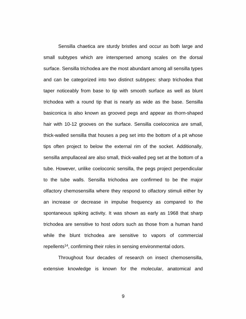
Figure 1. Operational definitions of larval movements and turns. (A) A larval body movement threshold is characterized by a larva turning its body axis by 90° and its head traveling the distance indicated. (B) A larval turn threshold is defined by a 45° angle between two successive larval tracking vectors.
33
AgIR Identification and Expression
Candidate AgIR sequences were identified in both the An. gambiae
genome using DmIR amino acid sequences as tBLASTn and BLASTp
queries, respectively. Potential exon-intron gene models were predicted
based on homology to DmIRs or AgIRs, as well as with the aid of a
Hidden Markov Model-based gene structure predictor
(www.Softberry.com). Iterative searches of all gene models were carried
out until no new candidates were identified. Conceptual translations of full
AgIR coding sequences were aligned with DmIR protein sequences using
Clustal X. Phylogenetic trees were constructed using the Neighbor-Joining
method20 with bootstrap resampling of 1,000 pseudo-replicates.
Transmembrane helices were predicted using Hidden Markov Model-
based software from the Center for Biological Sequence Analysis
(http://www.cbs.dtu.dk/services/TMHMM-2.0/). Antennae from late-instar
An. gambiae larvae were hand-dissected into RNALater-Ice solution
(Ambion, Austin, TX). Total RNA extraction and cDNA synthesis were
performed using the RNeasy Mini (Qiagen) and Transcriptor First Strand
cDNA Synthesis (Roche) kits, respectively. Antennal cDNA was used as a
template in PCR as described3. PCR primers specific for AgIrs were as
follows: AgIr8a: f5’-CCCTATGAGTGCAGAAAATT-3’ and r5’-
GGTACAGCACGTCTTCTGCG-3’; AgIr25a: f5’-
34
CAACCGACATACGCTACCAA-3’ and r5’-ACGATGAATACGCCTCCGAT-
3’; AgIr41a: f5’-ACTGGGAACTGGAGGTGGTG-3’ and r5’-
CTAAGGTGTCTCACTCCTCC-3’; AgIr41n: f5’-
ATGCACGATACATCTTGCCG-3’ and r5’-
TAAAGGACAGGAACGGTGTG-3’; AgIr76b: f5’-
CACGCTCCCAATCAACAATG-3’ and r5’-GATGGCGGCTAAACACTTCC-
3’; AgNMDAR2 f5’-AAGTTGGTGCTATGGATCAT-3’ and r5’-
ACACCATACGCATATACCCG-3’; rps7: f5’-
GGCGATCATCATCTACGTGC-3’ and r5’-
GTAGCTGCTGCAAACTTCGG-3’. cDNA amplicons were TOPO-TA
cloned into plasmid pCRII (Invitrogen) and sequenced to confirm their
identities.
siRNA Preparation and Injection
Double-stranded (ds) RNAs against a specific target gene were
prepared and purified using bidirectional in vitro transcription of full-length
cDNA templates using flanking T7 transcription initiation sites, and siRNAs
were prepared via RNAseIII digestion using Silencer siRNA Construction
reagents and protocols (Applied BioSystems/Ambion, Austin, TX).
Healthy, wild-type 3rd instar An. gambiae larvae were chosen for micro-
injection. They were pre-immobilized on 3mm filter paper on top of a 4°C
35
chill platform (BioQuip Inc, Rancho Dominquez, CA). Additional
desiccation was achieved using Kimwipes (Kimberly-Clark, Dallas TX) to
gently dry individual larva. Twin styrofoam strips were also employed as
temperature sinks to reduce distress from cold temperatures. Single barrel
borosilicate glass capillary pipettes (World Precision Instruments,
Sarasota, FL) were pulled (using a P-97 puller, Sutter Instruments,
Novato, CA) and beveled (using a Narishige EG-5 beveller, Tokyo, Japan)
to form microinjection needles. For larval microinjection, 27.6 nL of 100
nM siRNA were injected into the dorsal side of the larval thorax using a
Nanoliter 2000 system (World Precision Instruments, Sarasota, FL). Post-
injection, larvae were allowed to recover in 27°C distilled H2O with 1 ml of
larval food (as described in Mosquito Rearing section) for 48 h. Larvae
were monitored every 24 h post-injection, and non-viable individuals were
discarded.
Real-Time PCR (qRT-PCR)
Subsequent to experimental treatments and behavioral assays,
AgOrco, AgOr40 and AgIr76b transcript levels were determined by means
of quantitative RT-PCR. Each sample was comprised of 10 (AgOrco) or 30
(AgOr40, AgIr76b) larval heads that were hand-dissected from batches of
control and experimental An. gambiae larvae. RNA extraction and cDNA
36
synthesis were performed using the QIAGEN RNeasy Mini Kit and Roche
Transcriptor First Strand cDNA Synthesis Kit, respectively. All primers in
the assay were designed to span predicted introns in order to distinguish
well between genomic DNA and cDNA templates. An. gambiae ribosomal
protein S7 (rps7), which is constitutively expressed at high levels in all
tissues, was chosen as control gene to measure the relative levels of
mRNA of target genes in vivo. Primer sequences are as follows: rps7: f5′-
GGCGATCATCATCTACGTGC-3′ and r5′-GTAGCTGCTGCAAACTTCGG-
3′ (product size: 458bp cDNA); AgOrco: f5′-
ATCTTTGGCAATCGGCTCATC-3′ and r5′-GGCTCCAAGAACCGAAGC-
3′ (product size: 346 bp cDNA); AgOr40: f5′-
GACCCTCAAGAACCAGGGCT-3′ and r5′-
AATGATGGTGTAGTACGAGAAGG-3′; AgIr76b: f5′-
ATCTTCGATCCAGAGTTGCT-3′ and r5′-CCGGTCACCATGACGAAGTA-
3′. qRT-PCR was carried out using an Applied Biosystems 7300 Real-time
PCR system and SYBR green as fluorescent dye. Three experimental
repetitions were analyzed for each biological sample and the data
processed using System 7300 Sequence Detection Software (version
1.3.1). Primer efficiency was determined using a standard curve for all the
primers used. In the amplification of target genes and rps7, 8 µl and 2 µl
cDNA, respectively, from each group were used as templates. In each
37
trial, cDNA levels of target genes were quantified relative to rps7 levels
using the method of Pfaffl21.
Results
Behavioral Responses of Individual Larva
Previous studies utilized a novel paradigm to assay the behavioral
responses of large groups of An. gambiae late instar larvae to various
natural and synthetic odorants in order to characterize the molecular and
cellular elements of the larval olfactory system 15. While providing
fundamental information about the components underlying the olfactory
responses of An. gambiae larvae, these end-point studies did not provide
the precise tracking information that would allow us to distinguish between
attractive or repulsive behavioral patterns. In addition, the need for a large
number of larvae precluded its use in other experimental contexts. To
provide such information and utility, a CCD camera-based tracking system
was utilized to study the behavior of individual An. gambiae larva in
response to odorant stimuli. Visual tracking records (Figure 2) were then
analyzed to distinguish parameters associated with directional movement.
These included calculating the total number of turns, the overall number of
38
movements, the average velocity, and the resting time for each larval
behavioral assay (Figures 2 and 3).
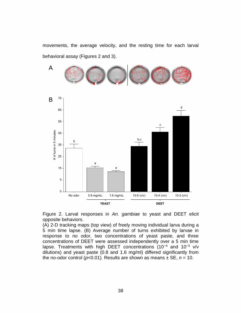
Figure 2. Larval responses in An. gambiae to yeast and DEET elicit opposite behaviors. (A) 2-D tracking maps (top view) of freely moving individual larva during a 5 min time lapse. (B) Average number of turns exhibited by larvae in response to no odor, two concentrations of yeast paste, and three concentrations of DEET were assessed independently over a 5 min time lapse. Treatments with high DEET concentrations (10−4 and 10−3 v/v dilutions) and yeast paste (0.8 and 1.6 mg/ml) differed significantly from the no-odor control (p<0.01). Results are shown as means ± SE, n = 10.
39
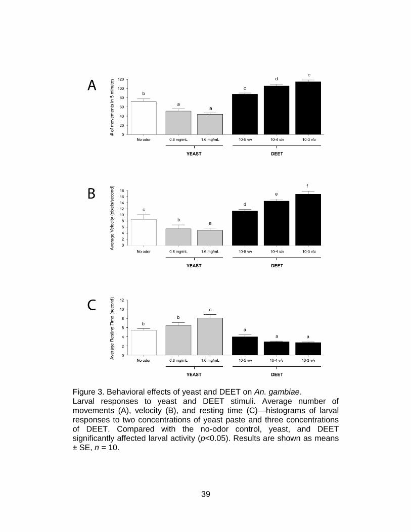
Figure 3. Behavioral effects of yeast and DEET on An. gambiae. Larval responses to yeast and DEET stimuli. Average number of movements (A), velocity (B), and resting time (C)—histograms of larval responses to two concentrations of yeast paste and three concentrations of DEET. Compared with the no-odor control, yeast, and DEET significantly affected larval activity (p<0.05). Results are shown as means ± SE, n = 10.
40
The sensitivity of this system was initially tested with two odorant
stimuli, each of which evoked a strong dose-dependent response in the
An. gambiae larvae group assay paradigm15. The first was DEET, which is
a widely used commercial insect repellent. The second was yeast paste, a
complex odorant source and a normal component of larval food. The
behavioral responses of individual An. gambiae larva to three
concentrations of DEET and two concentrations of yeast paste were
examined along with the appropriate set of parallel no-odorant controls
(Figure 2). For each assay, the four behavioral parameters described
above were quantified. In these studies, yeast paste elicited decreases in
overall larval turning (inverse klinokinesis; Figure 2) and movement
(Figure 3) as well as concomitant increases in resting time when
compared with no-odorant controls. In contrast, DEET elicited nearly the
opposite effect: An. gambiae larvae displayed a dose-dependent increase
in the turning rate (direct klinokinesis; Figure 2), number of movements,
and average velocity (direct orthokinesis; Figure 3), while the average
resting time was reduced to threshold levels at dilutions of 10−3 and 10−4.
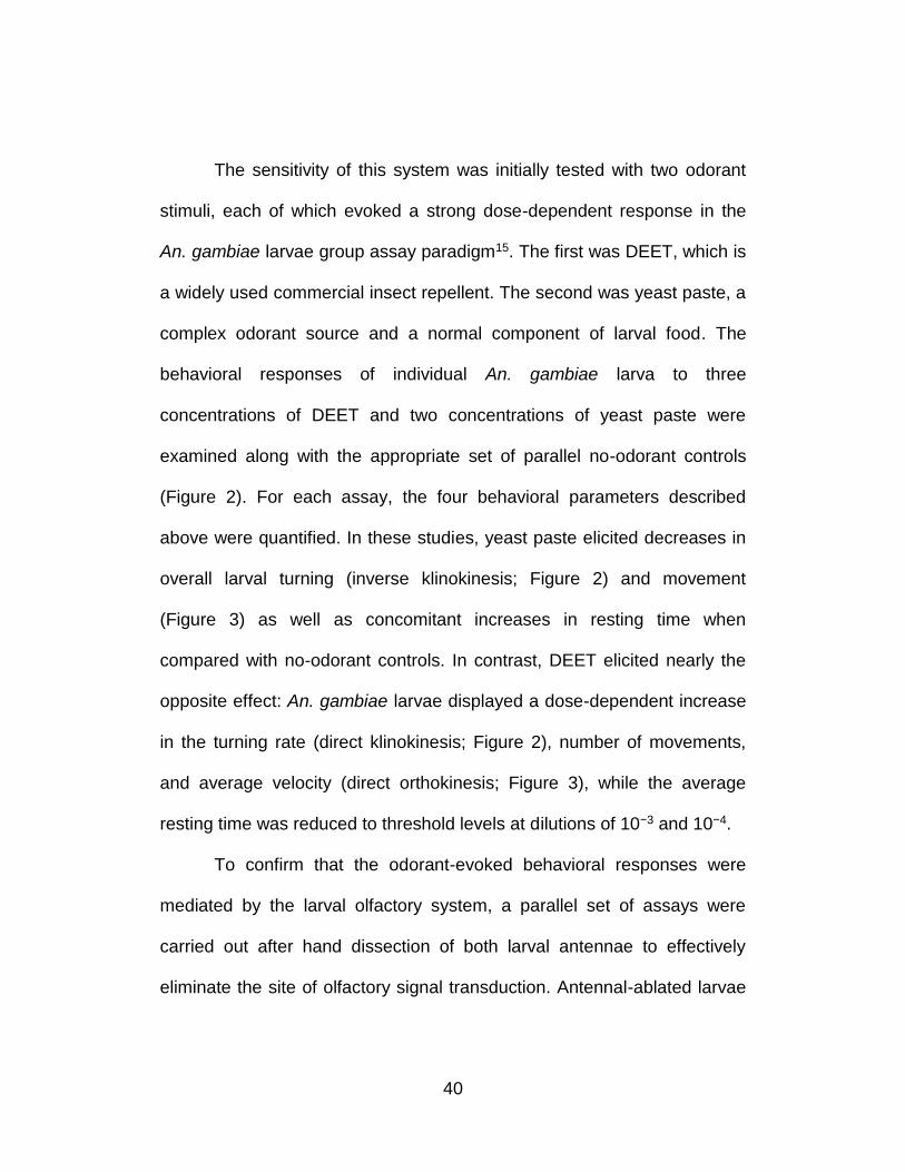
To confirm that the odorant-evoked behavioral responses were
mediated by the larval olfactory system, a parallel set of assays were
carried out after hand dissection of both larval antennae to effectively
eliminate the site of olfactory signal transduction. Antennal-ablated larvae
41
appeared to be largely indifferent to high concentrations of both DEET and
yeast, as larval responses were indistinguishable from no-odorant and
unablated controls (Figure 4). In larvae in which the antennae were left
intact but maxillary palps removed, responses to DEET and yeast paste
were similar to those in unablated controls (Figure 4). Taken together,
these data demonstrate that we have developed a robust behavioral
paradigm for examining odorant-induced responses from individual An.
gambiae larva.
Figure 4. Larval antennae mediate responses to yeast and DEET. In the presence of yeast and DEET, unablated and palp-ablated larvae responded equally to both; ablation of the antennae, however, significantly increased or decreased the number of turns (p<0.05) in response to yeast and DEET, respectively. Results are shown as means ± SE, n = 10.
42
AgORs Silencing Confirms a Direct Role in the DEET Response
To discern the molecular basis for odorant-evoked behavioral
responses of An. gambiae larvae, we initially focused on the role of
AgOrco, which is the An. gambiae ortholog of the non-conventional
Drosophila OR, DmOrco6,8, and is highly expressed in the larval
antenna15. In the absence of effective strategies to generate mutant or
transgenic strains of An. gambiae, we used RNA interference (RNAi) to
reduce AgOrco mRNA levels in individual larva, which could then be
tested for abnormal behavioral responses. Individual larval behavioral
assays followed by quantitative RNA analyses were conducted to assess
the effects of AgOrco siRNA and control siRNA microinjections on
olfactory responses and transcript levels. To account for non-specific
effects of siRNA delivery, larvae were microinjected with identical amounts
of a siRNA designed against a gene (AT5G39360) from the Arabidopsis
thaliana genome lacking significant homology to any cDNA in An.
gambiae. Furthermore, buffer-alone microinjections were carried out in
parallel to assess any potential effects of microinjection on larval behavior.
In order to assess the efficiency of siRNA-mediated knockdown of
AgOrco transcripts, a series of qRT-PCR studies were carried out on
experimental and control larvae after behavioral testing. In these assays,
cDNA was prepared from larval heads (with olfactory antennae attached)
43
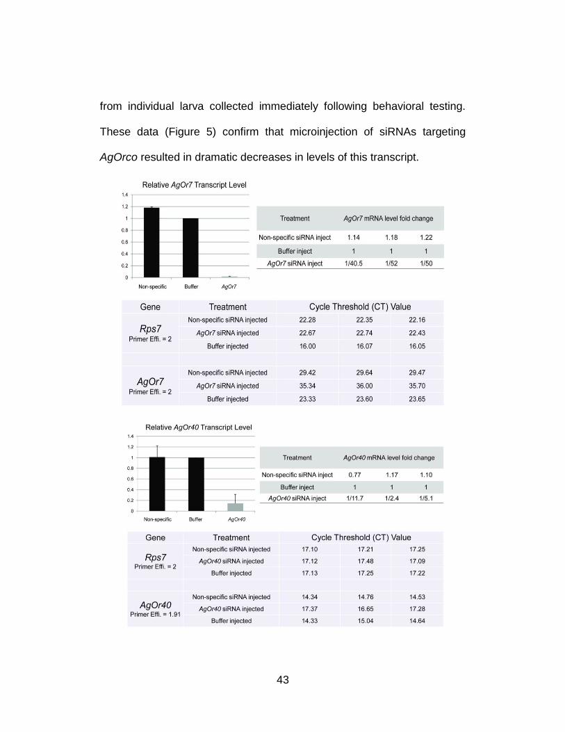
from individual larva collected immediately following behavioral testing.
These data (Figure 5) confirm that microinjection of siRNAs targeting
AgOrco resulted in dramatic decreases in levels of this transcript.
44
Figure 5. Quantitative analysis demonstrates significant transcript level reduction of AgOrco and AgOr40 after siRNA treatment. Larval cDNAs for qRT-PCR were generated using equal amounts (2 µg for AgOrco and 4 µg for AgOr40) of RNA extracted from hand-dissected larval heads from each injection treatment group, and three technical replicates were performed for each experimental group. AgOrco and AgOr40 mRNA levels were quantified as fold-changes relative to rps7 using the method of Pfaffl. AgOrco and AgOr40 levels are shown after normalization to buffer-alone controls in each of three experimental replicates. Histograms showing averaged AgOrco and AgOr40 levels normalized to buffer-alone injection controls. Standard errors were ±0.041 and ±0.029 for non-specific and AgOrco siRNA injections; ±0.127 and ±0.392 for non-specific and AgOr40 siRNA injections, respectively. Raw data from each qRT-PCR reaction indicating cycle-threshold (CT) and primer efficiency information for each technical replicate.
Although a modest microinjection effect was observed on the
average larval velocity, the overall number of turns (Figure 6) as well as
the number of movements, average velocity, and resting time (Figure 7) in
response to 1.6 mg/ml yeast paste stimuli were largely unaffected by
microinjection with AgOrco or control siRNAs. In contrast, a 1×10⁻³ (v/v)
dilution of DEET in individuals that received AgOrco siRNA showed
significant (p<0.01) reductions in turns (Figure 6), movements, and
velocity as well as a significant increase in their average resting time
relative to buffer-injected and control larvae (Figure 7). Although a modest
microinjection effect was again observed in buffer-injected larvae, these
results are consistent with the hypothesis that larval responses to DEET
45
are AgOrco-dependent whilst larval responses to yeast paste are AgOrco-
independent.
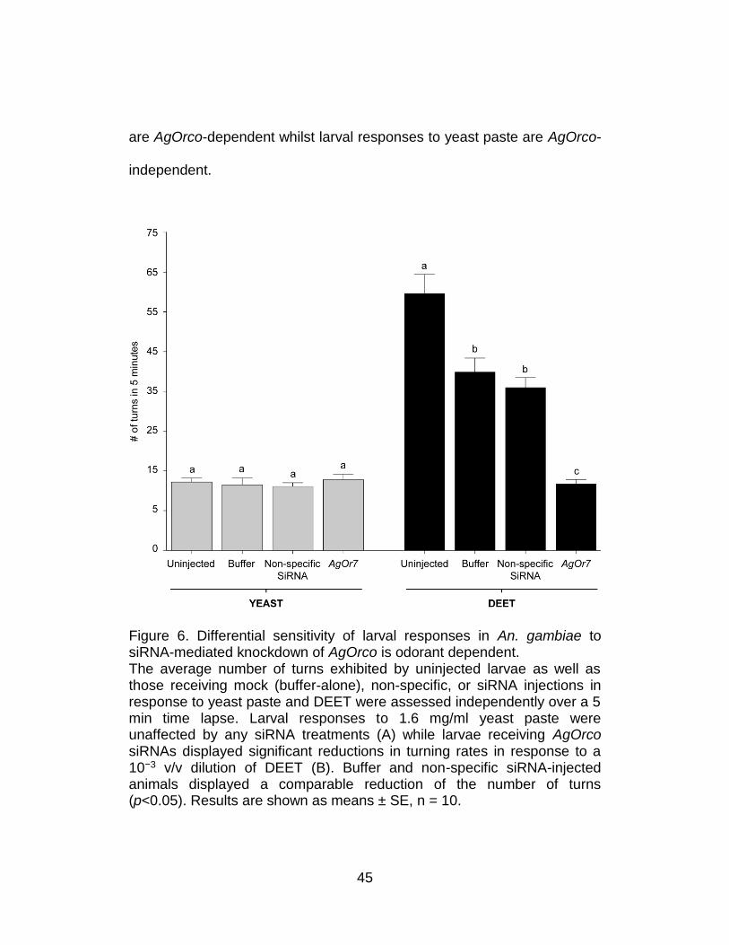
Figure 6. Differential sensitivity of larval responses in An. gambiae to siRNA-mediated knockdown of AgOrco is odorant dependent. The average number of turns exhibited by uninjected larvae as well as those receiving mock (buffer-alone), non-specific, or siRNA injections in response to yeast paste and DEET were assessed independently over a 5 min time lapse. Larval responses to 1.6 mg/ml yeast paste were unaffected by any siRNA treatments (A) while larvae receiving AgOrco siRNAs displayed significant reductions in turning rates in response to a 10−3 v/v dilution of DEET (B). Buffer and non-specific siRNA-injected animals displayed a comparable reduction of the number of turns (p<0.05). Results are shown as means ± SE, n = 10.
46
47
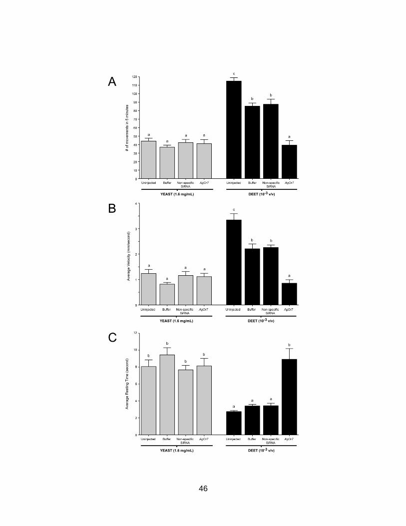
Figure 7. Larval behaviors after injection of non-specific small interfering RNA (siRNA). Averaged responses of buffer, non-specific, and AgOrco siRNA-injected larvae in the presence of 1.6 mg/ml yeast paste and a 10−3 v/v dilution of DEET. Larval movement (A), velocity (B), and resting time (C) behaviors of larvae in response to yeast paste and DEET. Knockdown of AgOrco mRNA levels has no effect on the ability of larvae to respond to yeast paste yet evokes significant behavioral alterations in larval responses to DEET (p<0.01). Results are shown as means ± SE, n = 10.
Functional studies using Xenopus oocytes15 have previously
identified AgOR40 as a conventional ligand-specific larval AgOR that
responds to DEET stimulation and, by implication, is likely to be
responsible for DEET-elicited behavioral responses in An. gambiae larvae.
Inasmuch as the molecular basis for DEET mediated behaviors remains
controversial, we tested this hypothesis by using siRNA-mediated gene
silencing to examine whether knockdown of AgOr40 transcripts would also
perturb behavioral responses to DEET and yeast paste. In these studies,
injection of siRNAs targeting AgOr40 echoed the effects of AgOrco
siRNAs and showed a significant reduction in turns and other elements of
larval behavior in response to DEET stimuli (Figure 8A) and were
unaffected in response to yeast paste (Figure 8B). As was the case for
AgOrco silencing, qRT-PCR studies were carried out on experimental and
control larvae after behavioral testing to assess the levels of AgOr40
transcripts. These data (Figure 5) confirm that microinjection of siRNAs
48
targeting AgOr40 resulted in dramatic decreases in AgOr40 transcript
levels without significantly altering AgOrco mRNA pools. Taken together,
these data directly validate the role of AgOR40 as a DEET-specific
conventional AgOR in the larval olfactory system of An. gambiae.
49
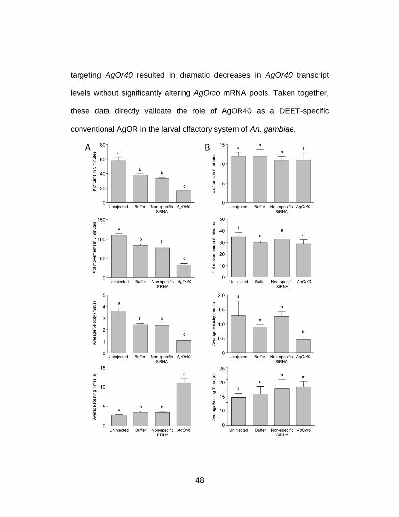
Figure 8. Differential sensitivity of larval responses in An. gambiae to siRNA-mediated knockdown of AgOr40 is odorant dependent. Larval responses exhibited by uninjected larvae as well as those receiving mock (buffer-alone), non-specific, or siRNA injections in response to DEET (A) and yeast paste (B) were assessed independently over a 5 min time lapse. Larval responses to 1.6 mg/ml yeast paste were unaffected by any siRNA treatments while larvae receiving AgOr40 siRNAs displayed significant reductions in turning rates (top panel) in response to a 10−3 v/v dilution of DEET. Buffer and non-specific siRNA-injected animals displayed a comparable reduction of the number of turns (p<0.05). Larval movement, velocity, and resting time behaviors (from top to bottom) of larvae in response to DEET (A) and yeast paste (B) where knockdown of AgOr40 mRNA levels had no effect on the ability of larvae to respond to yeast paste yet evoked significant behavioral alterations in larval responses to DEET (p<0.01). Results are shown as means ± SE, n = 10.
AgIRs Mediate AgOR Independent Olfactory Responses
Based on the AgOrco-independent response of larvae to yeast
paste, we next investigated whether AgOrco-dependent and -independent
olfactory signaling exists in An. gambiae larvae. In doing so, we
considered that AgOrco independence of the larval yeast response might,
in part, reflect that yeast paste is a complex mixture, some components of
which may activate AgOrco-independent olfactory signaling pathways. In
contrast, DEET is a unitary compound that specifically elicits AgOr-
dependent behavioral responses in An. gambiae larvae and physiological
responses in Xenopus oocyte-based AgOR functional assays15. To
examine further the possibility that distinct signaling pathways are active in
this system, we searched the An. gambiae genome for homologs of
50
variant ionotropic glutamate receptors that have recently been shown to
function as novel chemosensory proteins in D. melanogaster (DmIRs)13.
We have identified a family of 46 An. gambiae variant ionotropic glutamate
receptors, which we have named AgamGLUVIRs, and 9 homologs of
ionotropic glutamate receptors, named AgamGLURs or AgamNMDARs,
all according to the convention established by the An. gambiae genome
consortium (www.Vectorbase.org). For convenience we refer to the
AgamGLUVIR genes as AgIrs and their conceptual peptide products as
AgIRs. Another group of researchers has independently identified the
same family of genes22 and we have agreed with them on a unified
nomenclature in order to avoid confusion in future publications. A listing of
the entire gene family, their chromosome positions, and peptide
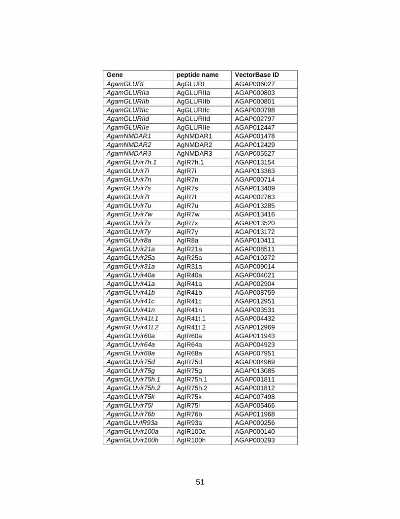
sequences is given in Table 1.
51
Gene peptide name VectorBase ID
AgamGLURI AgGLURI AGAP006027
AgamGLURIIa AgGLURIIa AGAP000803
AgamGLURIIb AgGLURIIb AGAP000801
AgamGLURIIc AgGLURIIc AGAP000798
AgamGLURIId AgGLURIId AGAP002797
AgamGLURIIe AgGLURIIe AGAP012447
AgamNMDAR1 AgNMDAR1 AGAP001478
AgamNMDAR2 AgNMDAR2 AGAP012429
AgamNMDAR3 AgNMDAR3 AGAP005527
AgamGLUvir7h.1 AgIR7h.1 AGAP013154
AgamGLUvir7i AgIR7i AGAP013363
AgamGLUvir7n AgIR7n AGAP000714
AgamGLUvir7s AgIR7s AGAP013409
AgamGLUvir7t AgIR7t AGAP002763
AgamGLUvir7u AgIR7u AGAP013285
AgamGLUvir7w AgIR7w AGAP013416
AgamGLUvir7x AgIR7x AGAP013520
AgamGLUvir7y AgIR7y AGAP013172
AgamGLUvir8a AgIR8a AGAP010411
AgamGLUvir21a AgIR21a AGAP008511
AgamGLUvir25a AgIR25a AGAP010272
AgamGLUvir31a AgIR31a AGAP009014
AgamGLUvir40a AgIR40a AGAP004021
AgamGLUvir41a AgIR41a AGAP002904
AgamGLUvir41b AgIR41b AGAP008759
AgamGLUvir41c AgIR41c AGAP012951
AgamGLUvir41n AgIR41n AGAP003531
AgamGLUvir41t.1 AgIR41t.1 AGAP004432
AgamGLUvir41t.2 AgIR41t.2 AGAP012969
AgamGLUvir60a AgIR60a AGAP011943
AgamGLUvir64a AgIR64a AGAP004923
AgamGLUvir68a AgIR68a AGAP007951
AgamGLUvir75d AgIR75d AGAP004969
AgamGLUvir75g AgIR75g AGAP013085
AgamGLUvir75h.1 AgIR75h.1 AGAP001811
AgamGLUvir75h.2 AgIR75h.2 AGAP001812
AgamGLUvir75k AgIR75k AGAP007498
AgamGLUvir75l AgIR75l AGAP005466
AgamGLUvir76b AgIR76b AGAP011968
AgamGLUvIR93a AgIR93a AGAP000256
AgamGLUvir100a AgIR100a AGAP000140
AgamGLUvir100h AgIR100h AGAP000293
52
AgamGLUvir100i AgIR100i AGAP004475
AgamGLUvir101 AgIR101 AGAP013425
AgamGLUvir133 AgIR133 AGAP005677
AgamGLUvir134 AgIR134 AGAP005678
AgamGLUvir135 AgIR135 AGAP005679
AgamGLUvir136 AgIR136 AGAP006440
AgamGLUvir137 AgIR137 -
AgamGLUvir138 AgIR138 -
AgamGLUvir139 AgIR139 AGAP006691
AgamGLUvir140.1 AgIR140.1 AGAP013242
AgamGLUvir140.2 AgIR140.2 AGAP013436
AgamGLUvir141 AgIR141 AGAP013473
AgamGLUvir142 AgIR142 AGAP006407
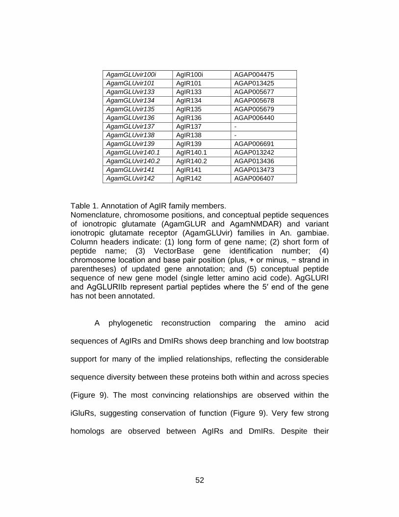
Table 1. Annotation of AgIR family members. Nomenclature, chromosome positions, and conceptual peptide sequences of ionotropic glutamate (AgamGLUR and AgamNMDAR) and variant ionotropic glutamate receptor (AgamGLUvir) families in An. gambiae. Column headers indicate: (1) long form of gene name; (2) short form of peptide name; (3) VectorBase gene identification number; (4) chromosome location and base pair position (plus, + or minus, − strand in parentheses) of updated gene annotation; and (5) conceptual peptide sequence of new gene model (single letter amino acid code). AgGLURI and AgGLURIIb represent partial peptides where the 5′ end of the gene has not been annotated.
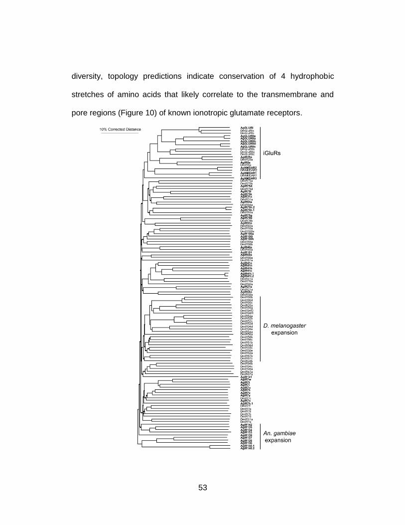
A phylogenetic reconstruction comparing the amino acid
sequences of AgIRs and DmIRs shows deep branching and low bootstrap
support for many of the implied relationships, reflecting the considerable
sequence diversity between these proteins both within and across species
(Figure 9). The most convincing relationships are observed within the
iGluRs, suggesting conservation of function (Figure 9). Very few strong
homologs are observed between AgIRs and DmIRs. Despite their
53
diversity, topology predictions indicate conservation of 4 hydrophobic
stretches of amino acids that likely correlate to the transmembrane and
pore regions (Figure 10) of known ionotropic glutamate receptors.
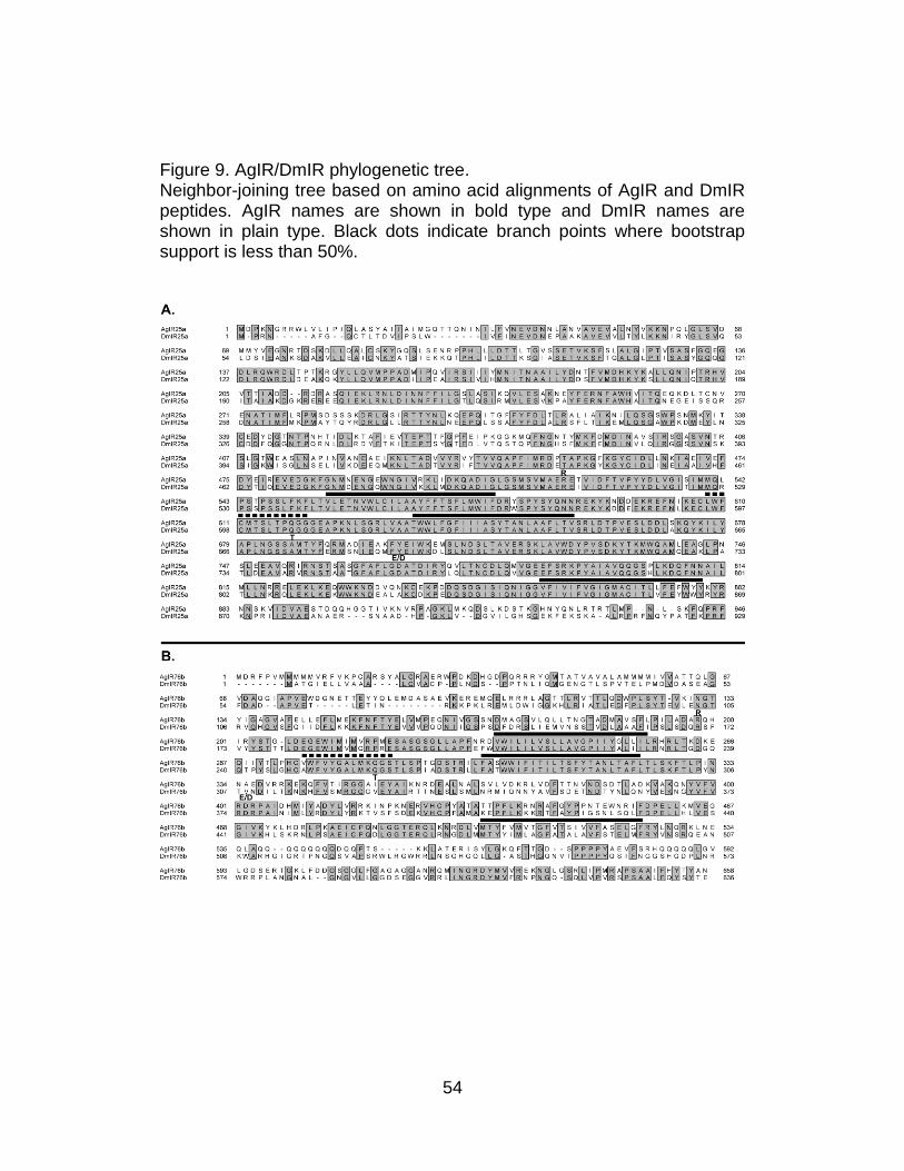
54
Figure 9. AgIR/DmIR phylogenetic tree. Neighbor-joining tree based on amino acid alignments of AgIR and DmIR peptides. AgIR names are shown in bold type and DmIR names are shown in plain type. Black dots indicate branch points where bootstrap support is less than 50%.
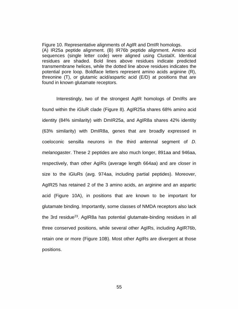
55
Figure 10. Representative alignments of AgIR and DmIR homologs. (A) IR25a peptide alignment. (B) IR76b peptide alignment. Amino acid sequences (single letter code) were aligned using ClustalX. Identical residues are shaded. Bold lines above residues indicate predicted transmembrane helices, while the dotted line above residues indicates the potential pore loop. Boldface letters represent amino acids arginine (R), threonine (T), or glutamic acid/aspartic acid (E/D) at positions that are found in known glutamate receptors.
Interestingly, two of the strongest AgIR homologs of DmIRs are
found within the iGluR clade (Figure 8). AgIR25a shares 68% amino acid
identity (84% similarity) with DmIR25a, and AgIR8a shares 42% identity
(63% similarity) with DmIR8a, genes that are broadly expressed in
coeloconic sensilla neurons in the third antennal segment of D.
melanogaster. These 2 peptides are also much longer, 891aa and 946aa,
respectively, than other AgIRs (average length 664aa) and are closer in
size to the iGluRs (avg. 974aa, including partial peptides). Moreover,
AgIR25 has retained 2 of the 3 amino acids, an arginine and an aspartic
acid (Figure 10A), in positions that are known to be important for
glutamate binding. Importantly, some classes of NMDA receptors also lack
the 3rd residue23. AgIR8a has potential glutamate-binding residues in all
three conserved positions, while several other AgIRs, including AgIR76b,
retain one or more (Figure 10B). Most other AgIRs are divergent at those
positions.
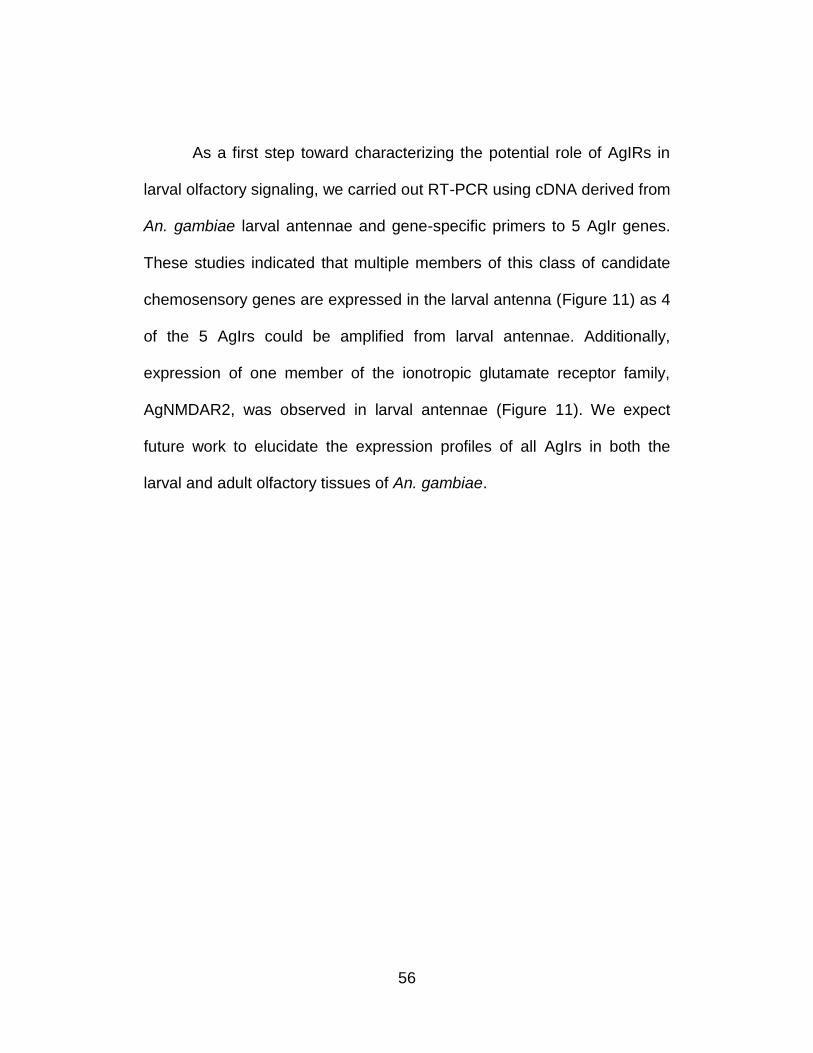
56
As a first step toward characterizing the potential role of AgIRs in
larval olfactory signaling, we carried out RT-PCR using cDNA derived from
An. gambiae larval antennae and gene-specific primers to 5 AgIr genes.
These studies indicated that multiple members of this class of candidate
chemosensory genes are expressed in the larval antenna (Figure 11) as 4
of the 5 AgIrs could be amplified from larval antennae. Additionally,
expression of one member of the ionotropic glutamate receptor family,
AgNMDAR2, was observed in larval antennae (Figure 11). We expect
future work to elucidate the expression profiles of all AgIrs in both the
larval and adult olfactory tissues of An. gambiae.
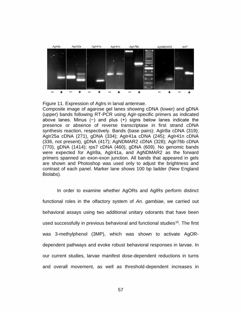
57
Figure 11. Expression of AgIrs in larval antennae. Composite image of agarose gel lanes showing cDNA (lower) and gDNA (upper) bands following RT-PCR using AgIr-specific primers as indicated above lanes. Minus (−) and plus (+) signs below lanes indicate the presence or absence of reverse transcriptase in first strand cDNA synthesis reaction, respectively. Bands (base pairs): AgIr8a cDNA (319); AgIr25a cDNA (271), gDNA (334); AgIr41a cDNA (245); AgIr41n cDNA (336, not present), gDNA (417); AgNDMAR2 cDNA (328); AgIr76b cDNA (770), gDNA (1414); rps7 cDNA (460), gDNA (609). No genomic bands were expected for AgIr8a, AgIr41a, and AgNDMAR2 as the forward primers spanned an exon-exon junction. All bands that appeared in gels are shown and Photoshop was used only to adjust the brightness and contrast of each panel. Marker lane shows 100 bp ladder (New England Biolabs).
In order to examine whether AgORs and AgIRs perform distinct
functional roles in the olfactory system of An. gambiae, we carried out
behavioral assays using two additional unitary odorants that have been
used successfully in previous behavioral and functional studies15. The first
was 3-methylphenol (3MP), which was shown to activate AgOR-
dependent pathways and evoke robust behavioral responses in larvae. In
our current studies, larvae manifest dose-dependent reductions in turns
and overall movement, as well as threshold-dependent increases in
58
average resting time (Figure 12). Furthermore, larval responses to 10−4
dilutions of 3MP were significantly altered in larvae injected with AgOrco
siRNA, whereas control or buffer-injected larval responses were
statistically equivalent to uninjected control larvae (Figures 13 and 14A).
AgOR40 is one of 3 larval AgORs with a demonstrated sensitivity to 3MP.
In that light, we also tested the ability of siRNA mediated silencing of
AgOr40 expression to alter larval responses to 3MP—in these studies a
marginal but not statistically significant effect was observed (unpublished
data) that is consistent with the role of multiple AgORs in mediating larval
sensitivity to 3MP.
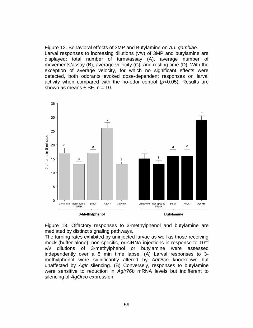
59
Figure 12. Behavioral effects of 3MP and Butylamine on An. gambiae. Larval responses to increasing dilutions (v/v) of 3MP and butylamine are displayed: total number of turns/assay (A), average number of movements/assay (B), average velocity (C), and resting time (D). With the exception of average velocity, for which no significant effects were detected, both odorants evoked dose-dependent responses on larval activity when compared with the no-odor control (p<0.05). Results are shown as means ± SE, n = 10.
Figure 13. Olfactory responses to 3-methylphenol and butylamine are mediated by distinct signaling pathways. The turning rates exhibited by uninjected larvae as well as those receiving mock (buffer-alone), non-specific, or siRNA injections in response to 10−4 v/v dilutions of 3-methylphenol or butylamine were assessed independently over a 5 min time lapse. (A) Larval responses to 3-methylphenol were significantly altered by AgOrco knockdown but unaffected by AgIr silencing. (B) Conversely, responses to butylamine were sensitive to reduction in AgIr76b mRNA levels but indifferent to silencing of AgOrco expression.
60
The next set of studies employed butylamine, a unitary odorant
which has been shown to activate grooved-peg ORNs in An. gambiae24
and Culex quinquefasciatus mosquitoes25. As was the case for 3MP,
uninjected An. gambiae larvae displayed robust dose-dependent
responses to butylamine (Figure 12). In contrast to the AgOrco-dependent
nature of larval responses to 3MP, larval responses to butylamine were
indistinguishable among animals treated with AgOrco and control siRNAs
or microinjected with buffer alone (Figures 13 and 14B).
Based on their homology to DmIRs, which have been shown to
mediate responses to amines and other odorants in Drosophila13, we
postulated that AgIRs mediate larval responses to butylamine. To test this
hypothesis, siRNA-mediated gene knockdowns were used in an attempt to
silence larval AgIRs and subsequently examine the responses of larvae to
butylamine. Of the AgIrs tested, microinjection of only one AgIr76b siRNA
displayed siRNA-specific effects on larval responses to butylamine.
Microinjection of AgIr76b siRNAs reduced AgIr76b mRNA levels (Figure
15) and led to significant alterations in larval responses to butylamine
(Figures 13 and 14B). Larval responses to butylamine were unaffected in
AgOrco knockdowns and by microinjection of non-specific siRNAs or
buffer-alone controls (Figures 13 and 14B).
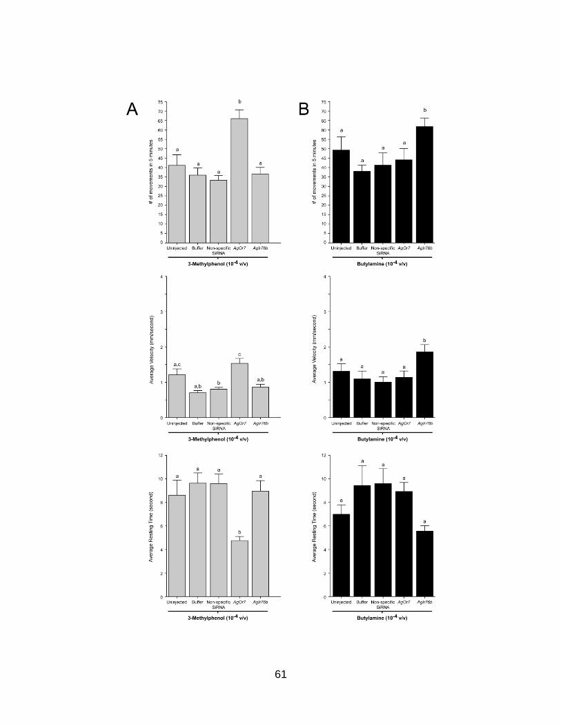
61
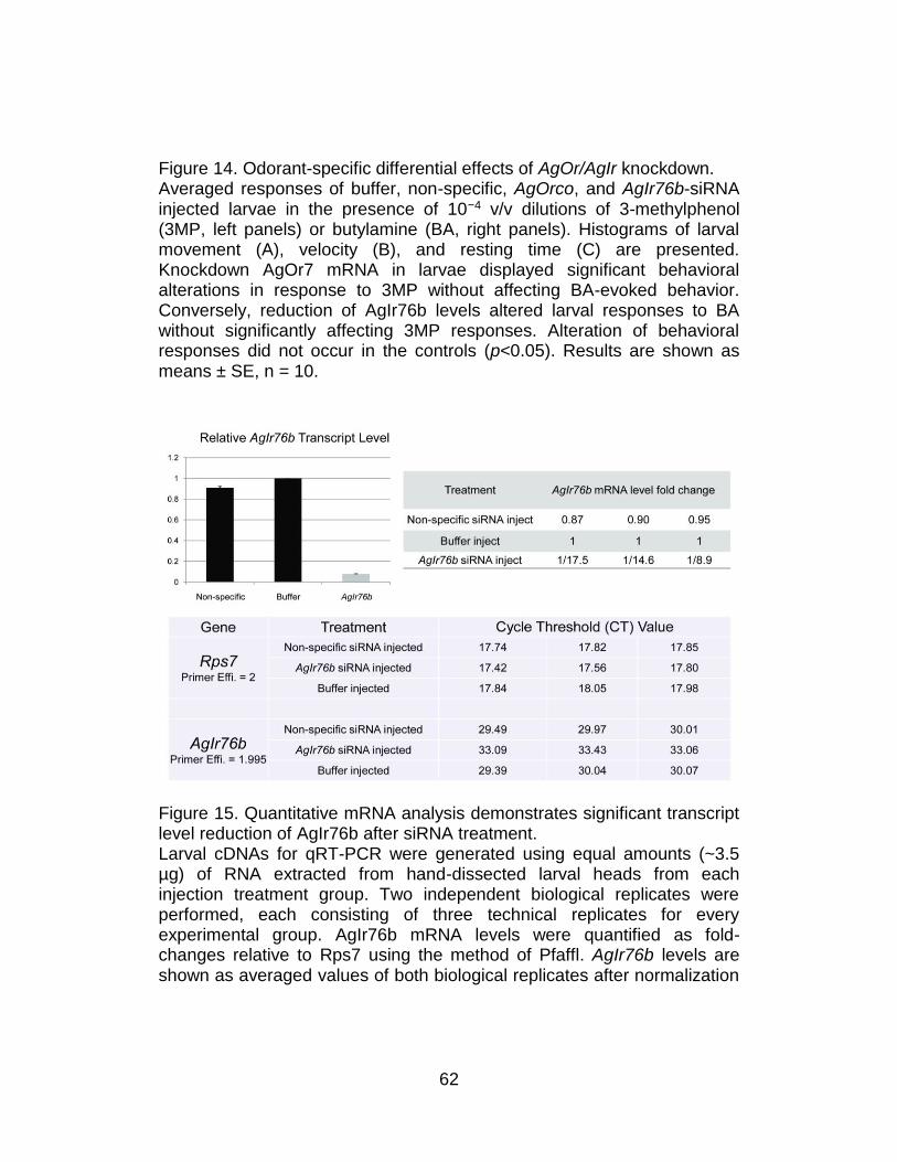
62
Figure 14. Odorant-specific differential effects of AgOr/AgIr knockdown. Averaged responses of buffer, non-specific, AgOrco, and AgIr76b-siRNA injected larvae in the presence of 10−4 v/v dilutions of 3-methylphenol (3MP, left panels) or butylamine (BA, right panels). Histograms of larval movement (A), velocity (B), and resting time (C) are presented. Knockdown AgOr7 mRNA in larvae displayed significant behavioral alterations in response to 3MP without affecting BA-evoked behavior. Conversely, reduction of AgIr76b levels altered larval responses to BA without significantly affecting 3MP responses. Alteration of behavioral responses did not occur in the controls (p<0.05). Results are shown as means ± SE, n = 10.
Figure 15. Quantitative mRNA analysis demonstrates significant transcript level reduction of AgIr76b after siRNA treatment. Larval cDNAs for qRT-PCR were generated using equal amounts (~3.5 µg) of RNA extracted from hand-dissected larval heads from each injection treatment group. Two independent biological replicates were performed, each consisting of three technical replicates for every experimental group. AgIr76b mRNA levels were quantified as fold-changes relative to Rps7 using the method of Pfaffl. AgIr76b levels are shown as averaged values of both biological replicates after normalization
63
to buffer alone controls in each of three technical replicates. Histograms showing averaged AgIr76b levels normalized to buffer alone injection controls. Standard errors were ±0.04 and ±0.003 for non-specific and AgIr76b siRNA injections, respectively. Raw data from each qRT-PCR reaction indicating cycle-threshold (CT) and primer efficiency information for each biological/technical replicate.
Discussion
In the face of a dearth of traditional genetic tools and a robust
transgenic capacity, the ability to carry out RNAi-mediated gene silencing
on individual An. gambiae larva provides an opportunity to examine the
molecular basis for olfactory driven behaviors in this disease vector.
Furthermore, the relative simplicity of the larval nervous system provides a
considerably more tractable model within a non-model system for
understanding similar processes that are presumed to underlie
chemosensory responses in adults that directly contribute to Anopheline
vectorial capacity.
In this study, we have developed a simple behavioral paradigm that
can be used to track the olfactory responses of individual An. gambiae
larva to a range of chemical stimuli. Overall, these data are consistent with
the hypothesis that when larvae are exposed to a repellent compound,
such as DEET, they exhibit an increased rate of turning and a rise in
overall movement and velocity. In contrast, an attractant such as yeast
64
paste or 3MP leads to a reduction in the number of movements, turns, and
average velocity while the average resting time is increased.
Together with gene-silencing approaches, we have employed a
novel behavioral assay to provide compelling in vivo evidence that, for the
first time, supports a direct in vivo role of AgORs in olfactory processes in
An. gambiae. Furthermore, these studies go further to address the
molecular mechanism responsible for DEET mediated repulsion of
insects. Previous studies26 suggesting that DEET's mode of action is to
inhibit the activation of a subset of insect ORs that would otherwise be
activated by attractants are in contrast to models that suggest DEET acts
via direct excitation of OR-expressing ORNs that, in turn, evoke
downstream behavioral repulsion. The excito-repellent hypothesis is
consistent with our previous study on the larval olfactory system in An.
gambiae that showed robust DEET-mediated behavioral responses that
correlated with a discrete population of larval ORNs co-expressing
AgORco/AgOR40 as well as specific DEET stimulation of Xenopus
oocytes injected with AgORco/AgOR40 cRNAs. This hypothesis is also
supported by other studies that describe DEET-mediated activation of a
subset of ORNs in Culex mosquitoes27 and more recent work in Aedes
aeqypti suggesting that DEET sensitivity is a genetically determined
characteristic affecting the functionality of discrete ORNs28. While the
65
reduction in DEET-mediated repellent responses in larvae undergoing
RNAi mediated silencing of AgOrco is consistent with a general
requirement for AgOR-based signaling, the similar effects of AgOr40
silencing specifically supports the role of both these molecular targets in
mediating DEET repellency. That these behavioral effects were manifest
by DEET alone, i.e. in the absence of any other stimuli, further validates
our earlier study and supports a direct excito-repellent mechanism for
DEET activity.
Lastly, these studies uncover the existence of at least two parallel
chemosensory transduction systems in larval-stage An. gambiae that
respond to distinct classes of odorant stimuli. One pathway, which is in
keeping with the established literature for insect olfactory signal
transduction, is based on the obligatory role of the non-conventional
Anopheline Orco family member AgOrco, which acts together with other
conventional AgORs in the formation of functional receptors. It is likely that
AgOR-dependent signaling pathways impact responses to a wide range of
odorant cues that play important roles in several aspects of Anopheline
behavior. These pathways are exemplified by the dramatic alterations in
the DEET and 3MP responses of An. gambiae larvae after RNAi-mediated
silencing of AgOrco transcripts (Figures 5, 13). The other pathway
depends on the function of the AgIr gene family, which likely recognizes
66
different odor classes than the AgOr pathway. Moreover, the similarities
between AgIRs 8a and 25a and iGluRs suggest that cellular receptors for
glutamate in the antenna could act as a neuromodulator of ORN function.
This hypothesis is consistent with the inability of AgIr25a siRNAs to alter
larval behavioral responses to odors (unpublished data).
Recent functional analyses18,19 of AgOR-based odor coding against
a diverse panel of compounds suggest that, in An. gambiae, olfactory
pathways respond to a wide range of odorant stimuli with particular affinity
for heterocyclics and aromatics that are associated with human skin
emanations16,17. These groups of odorants are thought to play essential
roles in host-seeking, oviposition, and other behaviors that are critical for
Anopheline life cycles29. Coincidently, this AgOR-based odor space is
characterized by sparse responses to the majority of acids, aldehydes,
and esters that were tested in addition to being particularly devoid of
amine-elicited responses. This raised the suggestion that sensitivity to
these classes of odorants might lie outside of AgOr-dependent olfactory
signaling pathways.
We have identified several AgIrs that are expressed in larval
olfactory tissues (Figure 11) and have used RNAi-mediated gene silencing
to demonstrate the role of one of these genes in mediating larval
responses to the AgOR-independent odorant butylamine. Critically, while
67
knockdown of AgIr76b specifically altered larval responses to butylamine,
there was no effect on responses to two other unitary odorants that were
dependent on AgOrco expression. These data are consistent with the
hypothesis that, in contrast to the AgOR-dependent sensitivity to 3MP,
DEET, and a broad range of “general” odorants, Anopheline responses to
other odorants (e.g., butylamine) are mediated through AgIr-dependent
signaling. There is reason to assume that these parallel pathways persist
through to adult An. gambiae where AgIrs are likely to be responsible for
olfactory sensitivity to important human kairomones, such as ammonia
and lactic acid that are known to activate ORNs in grooved peg sensilla30
that are devoid of AgORco. Indeed, we have observed expression of
multiple AgIrs in adult olfactory appendages, supporting the hypothesis
that this family of genes is involved in chemosensory signaling in adults
(manuscript in preparation).
Current efforts are directed toward expanding our understanding of
AgIr-based odor coding in An. gambiae. Improving our understanding of
olfactory signal transduction in An. gambiae may lead to new opportunities
to target olfactory mediated behaviors at the molecular level. In turn, this
may reduce the vectorial capacity of An. gambiae and help reduce the
transmission of malaria and other important human diseases.
68
Acknowledgments
We are grateful to Drs. P. Abbott, J.R. Carlson, H.W. Honegger,
and members of the L.J.Z. laboratory for discussions and comments on
the manuscript. We acknowledge Dr. Richard Benton and Vincent Croset
(University of Lausanne) for independently identifying the AgIR gene
family and for their cooperation in exchanging gene models and agreeing
to a unified nomenclature. We thank Z. Li and L. Sun for mosquito
rearing/technical assistance, Dr. D.E. McCauley for the use of real-time
PCR machine, and Dr. A. M. McAinsh for editorial assistance.
Author Contributions
The author(s) have made the following declarations about their
contributions: Conceived and designed the experiments: CL RJP LJZ.
Performed the experiments: CL RJP. Analyzed the data: CL RJP LJZ.
Contributed reagents/materials/analysis tools: RJP JDB PLJ GW. Wrote
the paper: CL RJP JDB PLJ LJZ.
69
References
1. Vosshall, L.B. and B.S. Hansson, A Unified Nomenclature System for the Insect Olfactory Coreceptor. Chem Senses, 2011. 36(6): p. 497-498.
2. Takken, W. and B.G. Knols, Odor-mediated behavior of Afrotropical malaria mosquitoes. Annu Rev Entomol, 1999. 44: p. 131-57.
3. Fox, A.N., et al., Candidate odorant receptors from the malaria vector mosquito Anopheles gambiae and evidence of down-regulation in response to blood feeding. Proc Natl Acad Sci U S A, 2001. 98(25): p. 14693-14697.
4. Merrill, C.E., R.J. Pitts, and L.J. Zwiebel, Molecular characterization of arrestin family members in the malaria vector mosquito, Anopheles gambiae. Insect Mol Biol, 2003. 12(6): p. 641-50.
5. Hallem, E.A., M.G. Ho, and J.R. Carlson, The molecular basis of odor coding in the Drosophila antenna. Cell, 2004. 117(7): p. 965-79.
6. Pitts, R.J., A.N. Fox, and L.J. Zwiebel, A highly conserved candidate chemoreceptor expressed in both olfactory and gustatory tissues in the malaria vector Anopheles gambiae. Proc Natl Acad Sci U S A, 2004. 101(14): p. 5058-5063.
7. Kwon, H.W., et al., Olfactory responses in a gustatory organ of the malaria vector mosquito Anopheles gambiae. Proc Natl Acad Sci U S A, 2006. 103(36): p. 13526-31.
8. Hill, C.A., et al., G protein coupled receptors in Anopheles gambiae. Science, 2002. 298(5591): p. 176-178.
9. Benton, R., et al., Atypical membrane topology and heteromeric function of Drosophila odorant receptors in vivo. PLoS Biol, 2006. 4(2): p. 240-257.
10. Lundin, C., et al., Membrane topology of the Drosophila OR83b odorant receptor. FEBS Lett, 2007. 581(29): p. 5601-5604.
11. Sato, K., et al., Insect olfactory receptors are heteromeric ligand-gated ion channels. Nature, 2008. 452(7190): p. 1002-U9.

70
12. Wicher, D., et al., Drosophila odorant receptors are both ligand-gated and cyclic-nucleotide-activated cation channels. Nature, 2008. 452(7190): p. 1007-U10.
13. Benton, R., et al., Variant Ionotropic Glutamate Receptors as Chemosensory Receptors in Drosophila. Cell, 2009. 136(1): p. 149-162.
14. Larsson, M.C., et al., Or83b encodes a broadly expressed odorant receptor essential for Drosophila olfaction. Neuron, 2004. 43(5): p. 703-14.
15. Xia, Y., et al., The molecular and cellular basis of olfactory-driven behavior in Anopheles gambiae larvae. Proc Natl Acad Sci U S A, 2008. 105(17): p. 6433-8.
16. Bernier, U.R., et al., Analysis of human skin emanations by gas chromatography/mass spectrometry. 2. Identification of volatile compounds that are candidate attractants for the yellow fever mosquito (Aedes aegypti). Anal Chem, 2000. 72(4): p. 747-56.
17. Cork, A. and K.C. Park, Identification of electrophysiologically-active compounds for the malaria mosquito, Anopheles gambiae, in human sweat extracts. Med Vet Entomol, 1996. 10(3): p. 269-76.
18. Carey, A.F., et al., Odorant reception in the malaria mosquito Anopheles gambiae. Nature, 2010. 464(7285): p. 66-71.
19. Wang, G., et al., Molecular basis of odor coding in the malaria vector mosquito Anopheles gambiae. Proc Natl Acad Sci U S A, 2010. 107(9): p. 4418-23.
20. Saitou, N. and M. Nei, The neighbor-joining method: a new method for reconstructing phylogenetic trees. Mol Biol Evol, 1987. 4(4): p. 406-25.
21. Pfaffl, M.W., A new mathematical model for relative quantification in real-time RT-PCR. Nucleic Acids Res, 2001. 29(9).
22. Croset, V., et al., Ancient protostome origin of chemosensory ionotropic glutamate receptors and the evolution of insect taste and olfaction. PLoS Genet, 2010. 6(8): p. e1001064.
23. Mayer, M.L., Glutamate receptors at atomic resolution. Nature, 2006. 440(7083): p. 456-462.
71
24. Qiu, Y.T., et al., Olfactory Coding in Antennal Neurons of the Malaria Mosquito, Anopheles gambiae. Chem Senses, 2006. 31(9): p. 845-63.
25. Syed, Z. and W.S. Leal, Acute olfactory response of Culex mosquitoes to a human- and bird-derived attractant. Proc Natl Acad Sci U S A, 2009. 106(44): p. 18803-8.
26. Ditzen, M., M. Pellegrino, and L.B. Vosshall, Insect odorant receptors are molecular targets of the insect repellent DEET. Science, 2008. 319(5871): p. 1838-42.
27. Syed, Z. and W.S. Leal, Mosquitoes smell and avoid the insect repellent DEET. Proc Natl Acad Sci U S A, 2008. 105(36): p. 13598-603.
28. Stanczyk, N.M., et al., Behavioral insensitivity to DEET in Aedes aegypti is a genetically determined trait residing in changes in sensillum function. Proc Natl Acad Sci U S A, 2010. 107(19): p. 8575-8580.
29. Meijerink, J., et al., Identification of olfactory stimulants for Anopheles gambiae from human sweat samples. J Chem Ecol, 2000. 26(6): p. 1367-1382.
30. Meijerink, J., M.A.H. Braks, and J.J.A. Van Loon, Olfactory receptors on the antennae of the malaria mosquito Anopheles gambiae are sensitive to ammonia and other sweat-borne components. J Insect Physiol, 2001. 47(4-5): p. 455-464.
72
CHAPTER III
MOLECULAR CHARACTERIZATION OF LARVAL PERIPHERAL
THERMOSENSORY RESPONSES OF THE MALARIA VECTOR
MOSQUITO ANOPHELES GAMBIAE
Preface
The following article was published was published in the journal of
PLoS One (8(8): e72595. doi:10.1371/journal.pone.0072595) on which I
was the first author along with Laurence J. Zwiebel (corresponding
author). In this paper I have developed a thermo-electric apparatus to
investigate thermosensory-driven behavior in An. gambiae larvae and
utilized Whole-mount Fluorescent In-situ Hybridization (WM-FISH) as well
as RNA interference to probe the localization and function of a
thermosensory channel protein: TRPA1, in directing larval thermal-
induced responses in vivo. This study has also broadened our view on the
role of TRPA1 in insect thermosensation by reporting for the first time that
TRPA1 is involved in thermal plasticity of An. gambiae larvae depending
on the cultivation temperature. I took part in the design of experiments,
data acquisition, statistical analysis, figure and manuscript preparation.
73
Introduction
Anopheles gambiae sensu stricto (Diptera: Culicidae) is the
principal sub-Saharan vector of human malaria that causes over a million
deaths annually1. As is true for all mosquitoes, An. gambiae goes through
pre-adult development spanning egg, larval and pupal life stages in
aqueous environments. This period typically lasts between 5 and 14 days,
depending on population density, food level and water temperatures in
larval habitats2. Although frequently overlooked, it has long been
appreciated that a significant degree of vector control is accomplished
through regulation of larval populations. Indeed, efficient regional
eradication of malaria has been achieved primarily through larvicidal
intervention3. In addition, due to their aquatic lifestyle and considerably
less complex nervous system, immature An. gambiae represents a more
tractable stage for the basic study of various physiological and sensory
processes4. Indeed, previous studies have taken advantage of both
simplicity and reproducibility of larval An. gambiae to explore the basic
principles underlying adult olfactory-driven responses, which also serve as
a foundation for further exploration of other aspects of larval sensory
biology5,6.
Mosquitoes are poikilotherms and as a result, are incapable of
maintaining thermal homeostasis7. Consequently, aquatic larvae rely on
74
their ability to sense and respond to temperature cues for several survival-
dependent behaviors in response to local temperature fluctuations. These
include the ability to navigate through rapidly changing water temperatures
in larval habitats that are alternately exposed to sunlight and darkness
during day/night cycles8. Therefore, the functional characterization of
thermal sensitivity in mosquito larvae would provide insights into these
processes as well as potentially inform our understanding of the adult
sensory system and facilitate the development of novel approaches that
are designed to modulate larval thermosensory behaviors to elicit
larvicidal activity.
While the molecular mechanisms underlying thermosensation in
An. gambiae larvae remain largely unexplored, earlier studies have
established the role of An. gambiae TRPA1 (hereafter, AgTRPA1), a
member of the Transient Receptor Potential family of sensory proteins, in
conferring sensitivity of adult peripheral thermosensory pathways to
increasing temperatures from 25 to 37°C9. This is consistent with studies
in other insects suggesting that TRPA1 represents an evolutionarily
ancient multimodal channel protein that is responsible for sensing
temperatures across the warm and/or hot range10-12. In order to continue
the exploration of peripheral thermosensation and in particular, the role of
AgTRPA1 in this context, we now focus on late-stage larvae that
75
represents a critical developmental window in establishing vectorial
capacity of An. gambiae. These studies have characterized the causal
relationships between ambient temperature and larval behavior and more
importantly, identify AgTRPA1 as a narrowly tuned peripheral high
temperature sensor in larvae that is crucial for regulating mobility as well
as thermal preference.
Materials and Methods
Mosquito rearing and larval sorting
An. gambiae sensu stricto, originated from Suakoko, Liberia, was
reared as described13 with modifications for human blood meals described
as follows: Five-day old females were allowed to feed on human blood
(purchased from Bioeclamation Inc.) for 60 minutes using a Hemotek
membrane feeding system (Discovery Workshops, UK) augmented with
CO2 and human foot odors (derived from a well-worn and unwashed
athletic sock), following the guidelines set by Vanderbilt Institutional
Animal Care and Use Committee. For behavioral and ablation studies,
early 4th or 3rd instar larvae were manually picked out from rearing pan,
respectively. Prior to analysis, larvae were rinsed gently with ddH2O on a
76
clean metal sieve to remove debris and food residuals and kept in room
temperature (24-25°C).
Thermo-electric control module
In order to generate either homogenous heating or linear
temperature gradients in behavioral arena that was composed of glass
petri dish of 150mm in diameter filled with 100ml of ddH2O, we fabricated
a design based on a similar apparatus from14. Here, a thin anodized
aluminum sheet (12 x 8 x 0.25 inch) was placed on top of two anodized
aluminum blocks whose temperatures were adjusted by using both liquid-
cooling achieved via water blocks (Custom Thermoelectric) connected to a
cycling cold-water bath as well as Peltier devices (Swiftech Inc.) coupled
with PID controllers (Oven Industry Inc.). Temperature across the
aluminum sheet was set using software (MR001 Ver. Rev B, Oven
Industry Inc.). Heating/cooling of each Peltier device was monitored in
real-time by dual-mounted thermal probes (Oven Industry Inc.) installed on
each end.
77
Fluorescent in situ hybridization and Fluorescent immunohistochemistry
on whole-mount larval antennae
Protocol for FISH studies was adapted and modified from15. Briefly,
whole larval antennae from 4th instar stage were hand-dissected into 4%
PFA in PBS with 0.1% Triton X-100. Samples were then gently transferred
into Pyrex glass dish where all subsequent treatments took place. Pre-
hybridization and hybridization were performed under 55° C for 6 and 24h,
respectively. Fast red staining was used to visualize anti-DIG antibody
linked to alkaline phosphatase (AP). Riboprobes were acquired from9 by
amplifying 900bp of AgTRPA1 coding sequence using PCR primers:
Forward: 5′-CTATTCGGCGGCTTCAATAAC-3′ as well as Reverse: 5′-
TCATTTGCCAATAGATTTGTTGAAGC-3′. RNA probes were labeled with
digoxigenin to generate sense and antisense. Anti-horseradish peroxidase
(HRP) antibody conjugated to FITC was utilized to mark neuronal axon
and dendrites. Additionally, anti-AgORco antibody raised from rabbit was
used to distinguish between AgTRPA1-expressing neurons and odorant
receptor neurons (ORNs). AgORco labeling was visualized by incubation
with Alexa Fluor goat-anti-rabbit 488 (Invitrogen). Whole antennae were
mounted in Vectashield (Vector Laboratories) and observed with an
LSM510 inverted confocal microscope (Carl Zeiss).
78
Automatic larval tracking and analysis
A digital video camera connected to Ethovision XT tracking system
(Noldus Inc.) was used to automatically capture and track locomotion of
an individual larva in the glass petri dish. For each trial, a single larva was
gently introduced at the center of the arena and given 15s to adapt prior to
the onset of recording at 10 frames per second (fps). Locomotion was
recorded for a total of 300s. For each temperature setting, a minimum of
15 trials (across an equal number of different individuals) was acquired
and parameters such as total distance travelled were calculated using
Ethovision software. For antennae as well as palp ablation studies, all
manipulations were carried out by manual dissection at 3rd instar stages,
after which larvae were allowed to recover for 24h prior to behavioral
testing. To quantify thermal preferences, we recorded the time interval that
each larva spent in either warm or cold half of a gradient that is expressed
as thermotactic index (T.I) and calculated as follows: (twarm-tcold) /
(twarm+tcold). A negative index value reflects a situation where larvae are
more inclined to stay in the cold half of the gradient (negative thermotaxis)
whereas a positive value is indicative of the opposite. For statistical
analysis, the comparison of two groups was carried out using Mann–
Whitney U tests while comparison of multiple groups was achieved using
79
Kruskal-Wallis one-way analysis of variance. p<0.05 was considered
significantly different.
siRNA injection and quantitative RT-PCR
Larval injections were carried out as previously described5. 27.6nL
of 20µM/L siRNA that target 6th and 10th exon of AgTRPA1 coding region
(UAUUGUUGAGCGGAGUGCCAGUU,
UUUUUCUCAUUCGGAUACUCGUU) (Thermo Fisher Inc.) were injected
into dorsal side of larval thorax using Nanoliter 2000 systems (World
Precision Instruments). Injected larvae were allowed to recover in 27 or
30°C with food provided for 48h. Quantitative RT-PCR was performed to
verify the quality of gene knockdown. Ribosomal protein S7 (rps7) was
chosen as internal control and primers used for these genes were: rps7:
Forward: 5’- GGTGCACCTGGATAAGAACCA-3’ Reverse:
5’- GTTCTCTGGGAATTCGAACG-3’ (Amplicon size: 112bp) and
agtrpa1: Forward: 5’-TATTCGGCGGCTTCAATAAC-3’ Reverse: 5’-
GCGTTTGAAGGATTTCCAGA-3’ (Amplicon size: 115bp). PFAFFL
method was used to quantify the relative transcript abundance.
Results
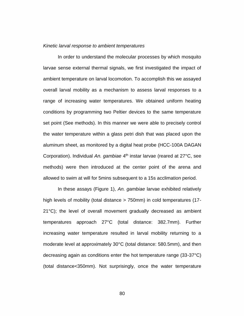
80
Kinetic larval response to ambient temperatures
In order to understand the molecular processes by which mosquito
larvae sense external thermal signals, we first investigated the impact of
ambient temperature on larval locomotion. To accomplish this we assayed
overall larval mobility as a mechanism to assess larval responses to a
range of increasing water temperatures. We obtained uniform heating
conditions by programming two Peltier devices to the same temperature
set point (See methods). In this manner we were able to precisely control
the water temperature within a glass petri dish that was placed upon the
aluminum sheet, as monitored by a digital heat probe (HCC-100A DAGAN
Corporation). Individual An. gambiae 4th instar larvae (reared at 27°C, see
methods) were then introduced at the center point of the arena and
allowed to swim at will for 5mins subsequent to a 15s acclimation period.
In these assays (Figure 1), An. gambiae larvae exhibited relatively
high levels of mobility (total distance > 750mm) in cold temperatures (17-
21°C); the level of overall movement gradually decreased as ambient
temperatures approach 27°C (total distance: 382.7mm). Further
increasing water temperature resulted in larval mobility returning to a
moderate level at approximately 30°C (total distance: 580.5mm), and then
decreasing again as conditions enter the hot temperature range (33-37°C)
(total distance<350mm). Not surprisingly, once the water temperature
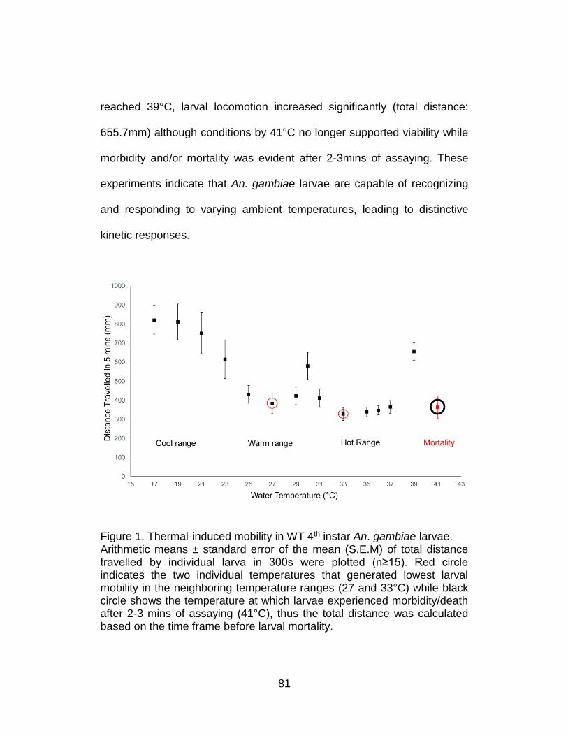
81
reached 39°C, larval locomotion increased significantly (total distance:
655.7mm) although conditions by 41°C no longer supported viability while
morbidity and/or mortality was evident after 2-3mins of assaying. These
experiments indicate that An. gambiae larvae are capable of recognizing
and responding to varying ambient temperatures, leading to distinctive
kinetic responses.
Figure 1. Thermal-induced mobility in WT 4th instar An. gambiae larvae. Arithmetic means ± standard error of the mean (S.E.M) of total distance travelled by individual larva in 300s were plotted (n≥15). Red circle indicates the two individual temperatures that generated lowest larval mobility in the neighboring temperature ranges (27 and 33°C) while black circle shows the temperature at which larvae experienced morbidity/death after 2-3 mins of assaying (41°C), thus the total distance was calculated based on the time frame before larval mortality.
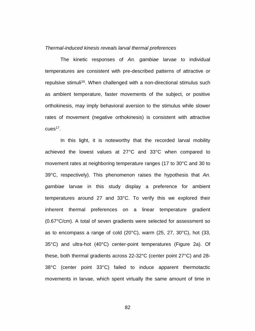
82
Thermal-induced kinesis reveals larval thermal preferences
The kinetic responses of An. gambiae larvae to individual
temperatures are consistent with pre-described patterns of attractive or
repulsive stimuli16. When challenged with a non-directional stimulus such
as ambient temperature, faster movements of the subject, or positive
orthokinesis, may imply behavioral aversion to the stimulus while slower
rates of movement (negative orthokinesis) is consistent with attractive
cues17.
In this light, it is noteworthy that the recorded larval mobility
achieved the lowest values at 27°C and 33°C when compared to
movement rates at neighboring temperature ranges (17 to 30°C and 30 to
39°C, respectively). This phenomenon raises the hypothesis that An.
gambiae larvae in this study display a preference for ambient
temperatures around 27 and 33°C. To verify this we explored their
inherent thermal preferences on a linear temperature gradient
(0.67°C/cm). A total of seven gradients were selected for assessment so
as to encompass a range of cold (20°C), warm (25, 27, 30°C), hot (33,
35°C) and ultra-hot (40°C) center-point temperatures (Figure 2a). Of
these, both thermal gradients across 22-32°C (center point 27°C) and 28-
38°C (center point 33°C) failed to induce apparent thermotactic
movements in larvae, which spent virtually the same amount of time in
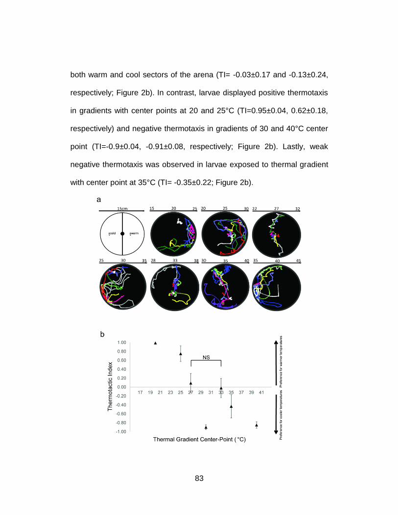
83
both warm and cool sectors of the arena (TI= -0.03±0.17 and -0.13±0.24,
respectively; Figure 2b). In contrast, larvae displayed positive thermotaxis
in gradients with center points at 20 and 25°C (TI=0.95±0.04, 0.62±0.18,
respectively) and negative thermotaxis in gradients of 30 and 40°C center
point (TI=-0.9±0.04, -0.91±0.08, respectively; Figure 2b). Lastly, weak
negative thermotaxis was observed in larvae exposed to thermal gradient
with center point at 35°C (TI= -0.35±0.22; Figure 2b).
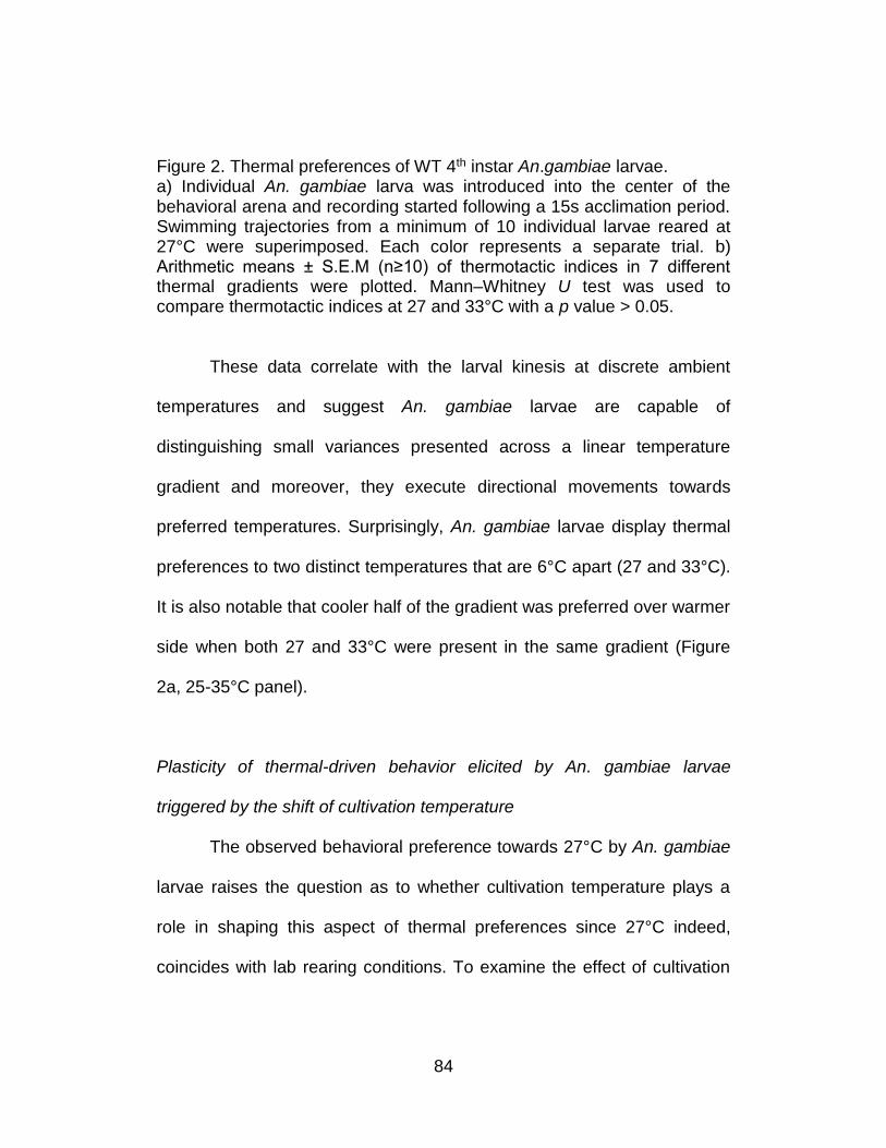
84
Figure 2. Thermal preferences of WT 4th instar An.gambiae larvae. a) Individual An. gambiae larva was introduced into the center of the behavioral arena and recording started following a 15s acclimation period. Swimming trajectories from a minimum of 10 individual larvae reared at 27°C were superimposed. Each color represents a separate trial. b) Arithmetic means ± S.E.M (n≥10) of thermotactic indices in 7 different thermal gradients were plotted. Mann–Whitney U test was used to compare thermotactic indices at 27 and 33°C with a p value > 0.05.
These data correlate with the larval kinesis at discrete ambient
temperatures and suggest An. gambiae larvae are capable of
distinguishing small variances presented across a linear temperature
gradient and moreover, they execute directional movements towards
preferred temperatures. Surprisingly, An. gambiae larvae display thermal
preferences to two distinct temperatures that are 6°C apart (27 and 33°C).
It is also notable that cooler half of the gradient was preferred over warmer
side when both 27 and 33°C were present in the same gradient (Figure
2a, 25-35°C panel).
Plasticity of thermal-driven behavior elicited by An. gambiae larvae
triggered by the shift of cultivation temperature
The observed behavioral preference towards 27°C by An. gambiae
larvae raises the question as to whether cultivation temperature plays a
role in shaping this aspect of thermal preferences since 27°C indeed,
coincides with lab rearing conditions. To examine the effect of cultivation
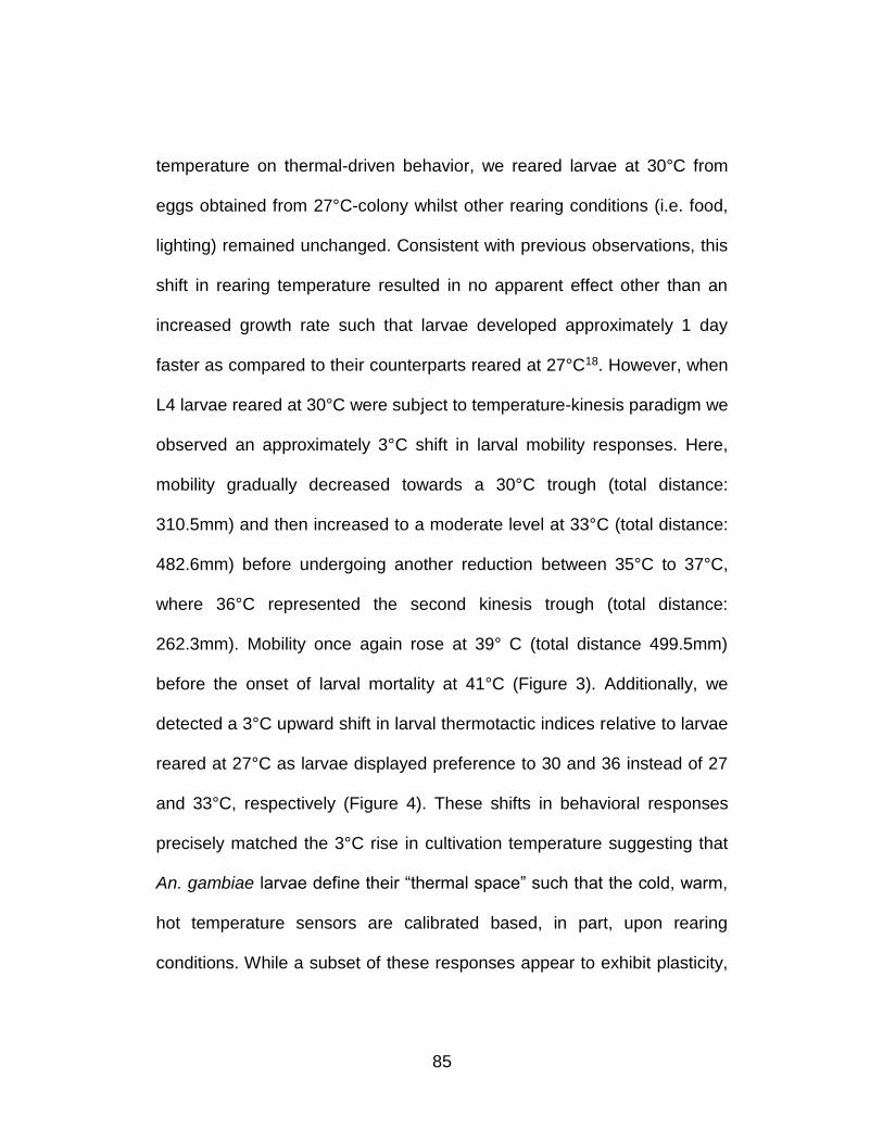
85
temperature on thermal-driven behavior, we reared larvae at 30°C from
eggs obtained from 27°C-colony whilst other rearing conditions (i.e. food,
lighting) remained unchanged. Consistent with previous observations, this
shift in rearing temperature resulted in no apparent effect other than an
increased growth rate such that larvae developed approximately 1 day
faster as compared to their counterparts reared at 27°C18. However, when
L4 larvae reared at 30°C were subject to temperature-kinesis paradigm we
observed an approximately 3°C shift in larval mobility responses. Here,
mobility gradually decreased towards a 30°C trough (total distance:
310.5mm) and then increased to a moderate level at 33°C (total distance:
482.6mm) before undergoing another reduction between 35°C to 37°C,
where 36°C represented the second kinesis trough (total distance:
262.3mm). Mobility once again rose at 39° C (total distance 499.5mm)
before the onset of larval mortality at 41°C (Figure 3). Additionally, we
detected a 3°C upward shift in larval thermotactic indices relative to larvae
reared at 27°C as larvae displayed preference to 30 and 36 instead of 27
and 33°C, respectively (Figure 4). These shifts in behavioral responses
precisely matched the 3°C rise in cultivation temperature suggesting that
An. gambiae larvae define their “thermal space” such that the cold, warm,
hot temperature sensors are calibrated based, in part, upon rearing
conditions. While a subset of these responses appear to exhibit plasticity,
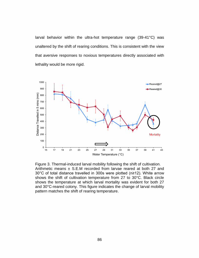
86
larval behavior within the ultra-hot temperature range (39-41°C) was
unaltered by the shift of rearing conditions. This is consistent with the view
that aversive responses to noxious temperatures directly associated with
lethality would be more rigid.
Figure 3. Thermal-induced larval mobility following the shift of cultivation. Arithmetic means ± S.E.M recorded from larvae reared at both 27 and 30°C of total distance travelled in 300s were plotted (n≥12). White arrow shows the shift of cultivation temperature from 27 to 30°C. Black circle shows the temperature at which larval mortality was evident for both 27 and 30°C-reared colony. This figure indicates the change of larval mobility pattern matches the shift of rearing temperature.
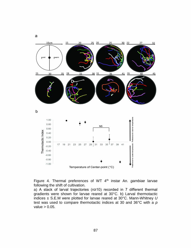
87
Figure 4. Thermal preferences of WT 4th instar An. gambiae larvae following the shift of cultivation. a) A stack of larval trajectories (n≥10) recorded in 7 different thermal gradients were shown for larvae reared at 30°C. b) Larval thermotactic indices ± S.E.M were plotted for larvae reared at 30°C. Mann-Whitney U test was used to compare thermotactic indices at 30 and 36°C with a p value > 0.05.
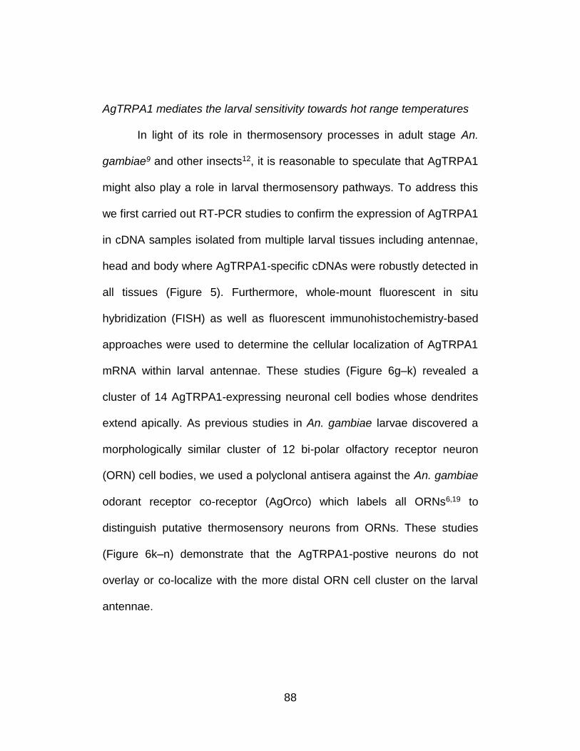
88
AgTRPA1 mediates the larval sensitivity towards hot range temperatures
In light of its role in thermosensory processes in adult stage An.
gambiae9 and other insects12, it is reasonable to speculate that AgTRPA1
might also play a role in larval thermosensory pathways. To address this
we first carried out RT-PCR studies to confirm the expression of AgTRPA1
in cDNA samples isolated from multiple larval tissues including antennae,
head and body where AgTRPA1-specific cDNAs were robustly detected in
all tissues (Figure 5). Furthermore, whole-mount fluorescent in situ
hybridization (FISH) as well as fluorescent immunohistochemistry-based
approaches were used to determine the cellular localization of AgTRPA1
mRNA within larval antennae. These studies (Figure 6g–k) revealed a
cluster of 14 AgTRPA1-expressing neuronal cell bodies whose dendrites
extend apically. As previous studies in An. gambiae larvae discovered a
morphologically similar cluster of 12 bi-polar olfactory receptor neuron
(ORN) cell bodies, we used a polyclonal antisera against the An. gambiae
odorant receptor co-receptor (AgOrco) which labels all ORNs6,19 to
distinguish putative thermosensory neurons from ORNs. These studies
(Figure 6k–n) demonstrate that the AgTRPA1-postive neurons do not
overlay or co-localize with the more distal ORN cell cluster on the larval
antennae.

89
Figure 5. Expression of AgTRPA1 in larval tissues. cDNA libraries from larval antennae, heads and bodies were generated by extracting mRNA followed by in intro reverse transcription. rps7 and agtrpa1 were amplified using gene-specific primers and run on a 2% agarose gel. “+” or “-“ indicates the presence or absence of reverse transcriptase, respectively.
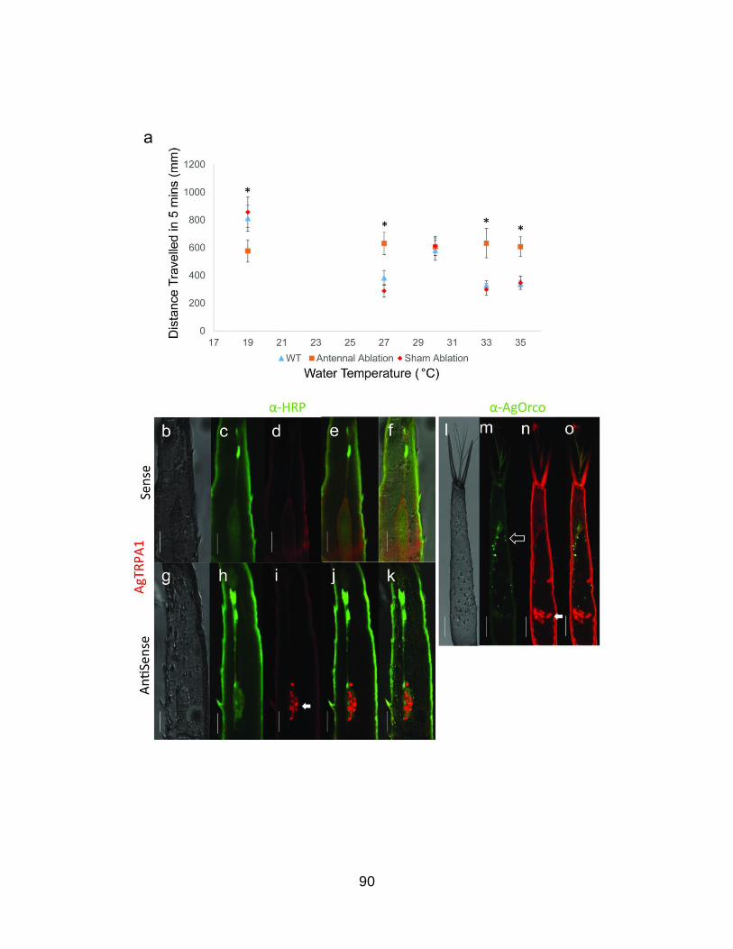
90
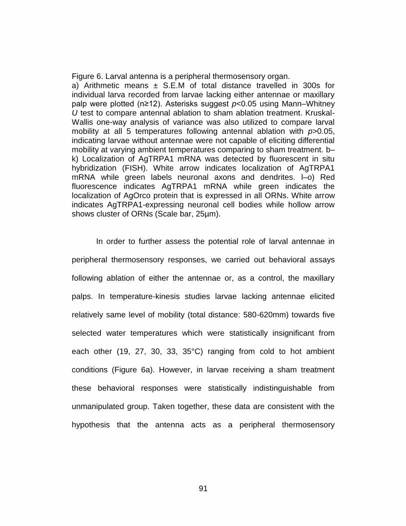
91
Figure 6. Larval antenna is a peripheral thermosensory organ. a) Arithmetic means ± S.E.M of total distance travelled in 300s for individual larva recorded from larvae lacking either antennae or maxillary palp were plotted (n≥12). Asterisks suggest p<0.05 using Mann–Whitney U test to compare antennal ablation to sham ablation treatment. Kruskal-Wallis one-way analysis of variance was also utilized to compare larval mobility at all 5 temperatures following antennal ablation with p>0.05, indicating larvae without antennae were not capable of eliciting differential mobility at varying ambient temperatures comparing to sham treatment. b–k) Localization of AgTRPA1 mRNA was detected by fluorescent in situ hybridization (FISH). White arrow indicates localization of AgTRPA1 mRNA while green labels neuronal axons and dendrites. l–o) Red fluorescence indicates AgTRPA1 mRNA while green indicates the localization of AgOrco protein that is expressed in all ORNs. White arrow indicates AgTRPA1-expressing neuronal cell bodies while hollow arrow shows cluster of ORNs (Scale bar, 25µm).
In order to further assess the potential role of larval antennae in
peripheral thermosensory responses, we carried out behavioral assays
following ablation of either the antennae or, as a control, the maxillary
palps. In temperature-kinesis studies larvae lacking antennae elicited
relatively same level of mobility (total distance: 580-620mm) towards five
selected water temperatures which were statistically insignificant from
each other (19, 27, 30, 33, 35°C) ranging from cold to hot ambient
conditions (Figure 6a). However, in larvae receiving a sham treatment
these behavioral responses were statistically indistinguishable from
unmanipulated group. Taken together, these data are consistent with the
hypothesis that the antenna acts as a peripheral thermosensory
92
appendage that is critical for thermal-induced responses in An. gambiae
larvae.
Due to the absence of available genetic mutants or a viable
methodology to generate gene-specific knockouts, we utilized RNAi-
mediated gene-silencing to reduce AgTRPA1 mRNA in order to examine
the in vivo role of AgTRPA1 in larval thermosensation. Small interfering
RNA (siRNA) oligonucleotides targeting AgTRPA1 were injected into L3
larvae along with injection of buffer-alone and a non-specific siRNA
targeting a gene (AT5G39360) from Arabidopsis thaliana that lacks
significant homology to An. gambiae genome. Knockdown of AgTRPA1
transcript was assessed using quantitative RT-PCR, which showed on
average an 80% reduction of mRNA levels (Figure 7) as compared to non-
specific siRNA treatment.
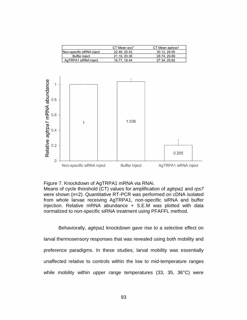
93
Figure 7. Knockdown of AgTRPA1 mRNA via RNAi. Means of cycle threshold (CT) values for amplification of agtrpa1 and rps7 were shown (n=2). Quantitative RT-PCR was performed on cDNA isolated from whole larvae receiving AgTRPA1, non-specific siRNA and buffer injection. Relative mRNA abundance + S.E.M was plotted with data normalized to non-specific siRNA treatment using PFAFFL method.
Behaviorally, agtrpa1 knockdown gave rise to a selective effect on
larval thermosensory responses that was revealed using both mobility and
preference paradigms. In these studies, larval mobility was essentially
unaffected relative to controls within the low to mid-temperature ranges
while mobility within upper range temperatures (33, 35, 36°C) were
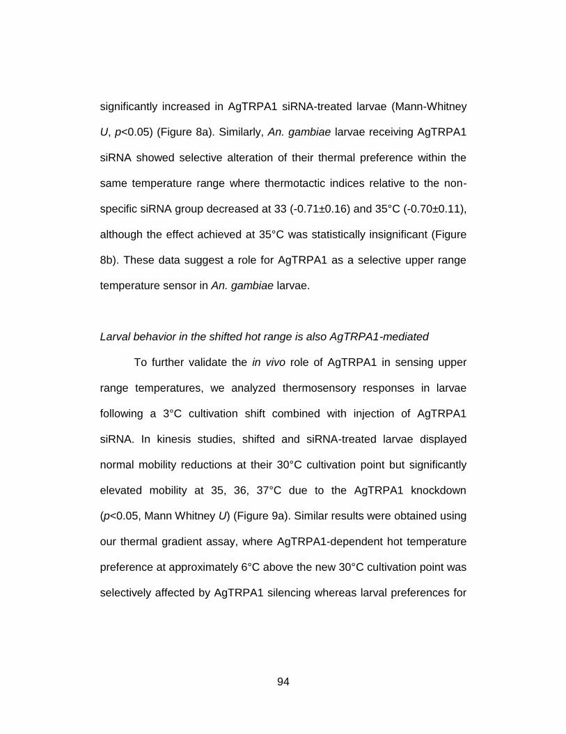
94
significantly increased in AgTRPA1 siRNA-treated larvae (Mann-Whitney
U, p<0.05) (Figure 8a). Similarly, An. gambiae larvae receiving AgTRPA1
siRNA showed selective alteration of their thermal preference within the
same temperature range where thermotactic indices relative to the non-
specific siRNA group decreased at 33 (-0.71±0.16) and 35°C (-0.70±0.11),
although the effect achieved at 35°C was statistically insignificant (Figure
8b). These data suggest a role for AgTRPA1 as a selective upper range
temperature sensor in An. gambiae larvae.
Larval behavior in the shifted hot range is also AgTRPA1-mediated
To further validate the in vivo role of AgTRPA1 in sensing upper
range temperatures, we analyzed thermosensory responses in larvae
following a 3°C cultivation shift combined with injection of AgTRPA1
siRNA. In kinesis studies, shifted and siRNA-treated larvae displayed
normal mobility reductions at their 30°C cultivation point but significantly
elevated mobility at 35, 36, 37°C due to the AgTRPA1 knockdown
(p<0.05, Mann Whitney U) (Figure 9a). Similar results were obtained using
our thermal gradient assay, where AgTRPA1-dependent hot temperature
preference at approximately 6°C above the new 30°C cultivation point was
selectively affected by AgTRPA1 silencing whereas larval preferences for
95
the newly shifted cultivation point was still unaffected by AgTRPA1
silencing (Figure 9b).
96
Figure 8. AgTRPA1 mediates larval responses within the upper temperature range. a) Arithmetic means ± S.E.M of total distance travelled in 300s for injected larvae reared at 27°C were plotted. Asterisks indicate p<0.05 comparing AgTRPA1 and Non-specific siRNA-treatment using Mann–Whitney U test. Black rectangle labels the temperature range at which larval mobility was significantly modified following the knockdown of AgTRPA1. b) A stack of larval trajectories (n≥10) recorded in 28-38°C gradient for buffer-alone and AgTRPA1, Non-specific siRNA-injected treatments were shown. Thermotactic indices ± S.E.M were plotted for injected larvae. Asterisks indicate p<0.05 comparing AgTRAP1 and Non-specific siRNA treatment (Mann–Whitney U test).
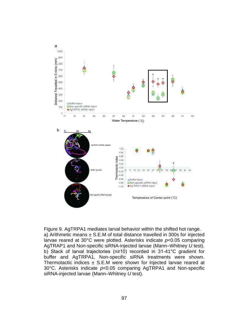
97
Figure 9. AgTRPA1 mediates larval behavior within the shifted hot range. a) Arithmetic means ± S.E.M of total distance travelled in 300s for injected larvae reared at 30°C were plotted. Asterisks indicate p<0.05 comparing AgTRAP1 and Non-specific siRNA-injected larvae (Mann–Whitney U test). b) Stack of larval trajectories (n≥10) recorded in 31-41°C gradient for buffer and AgTRPA1, Non-specific siRNA treatments were shown. Thermotactic indices ± S.E.M were shown for injected larvae reared at 30°C. Asterisks indicate p<0.05 comparing AgTRPA1 and Non-specific siRNA-injected larvae (Mann–Whitney U test).
98
Discussion
Together with chemosensory and visual modalities, thermosensory
responses of immature An. gambiae are necessary for a variety of
behaviors pertinent to robust development and survival. Environmental
temperature has a major influence on the rate of larval development and,
as a result, directly impacts vector populations and malaria transmission20.
Ambient temperature also influences the growth of algae and bacteria that
are the primary nutrients for An. gambiae larvae21. Although temperature
affects the rate of development, the relationship is not straightforward.
Indeed, the production of adult mosquitoes is not directly proportional to
the rate of larval development such that temperatures resulting in the
fastest growth produce fewer and importantly, smaller adults22. This
reflects the balance between developmental rate and the behaviors that
mediate larval survival and feeding in order to obtain adequate dietary
reserves which are associated with adult longevity, fecundity and vectorial
capacity23.
Accordingly, throughout larval life-stage, An. gambiae effectively
navigate across fluctuating water temperatures that might otherwise lead
to sub-optimal nutrition, reduced growth and death22,24. This capacity is
particularly essential for Anopheline larvae in tropical and sub-tropical
regions where water temperatures in typical larval habitats with direct sun
99
exposure (i.e. puddles and mud pit) can vary as much as 20°C through
day/night cycles8. The critical nature of larval thermosensory behaviors
underscores the rationale behind studies to characterize the underlying
cellular and molecular mechanisms that may, in turn, provide novel
opportunities for the development of cost-effective approaches to disrupt
these behaviors.
Late-stage An. gambiae larvae are capable of responding to diverse
temperatures by exhibiting differential kinesis (Figure 1). In our initial
survey of thermosensory responses we noted that larval mobility rates are
reduced on two occasions, one of which is a discrete point at 27°C
followed by a broader interval between 33–36°C that initiates
approximately 6°C higher. In light of studies undertaken in other animal
systems, these responses may reasonably be associated with behavioral
preference while high mobility rates may be correlated with avoidance.
Expanding on these observations using a temperature gradient paradigm
(Figure 2), we observed that An. gambiae larvae are indeed capable of
performing thermotactic movements when the surrounding temperature
deviates from these favored condition(s).
It is noteworthy that the robust larval preference to 27°C
corresponds to their constantly maintained rearing temperature. This is
reminiscent of similar observations in D. melanogaster where late-stage
100
larvae exhibit maximal growth rate and minimal mortality near 24°C25 and
show behavioral preference towards this temperature when placed on a
linear thermal gradient26. In order to further investigate the effect of
cultivation temperature on larval thermosensory behaviors, we shifted the
rearing conditions of a sub-population of newly oviposited An. gambiae
embryos 3°C higher to 30°C and allowed normal development to proceed
to late larval instars. Under these conditions we observed a parallel 3°C
shift in larval behavior in both kinesis and thermotaxis bioassays (Figures
3 and 4), suggesting that An. gambiae larvae utilize their cultivation
conditions to set and adjust their thermal sensors to sense ambient
temperatures. Cultivation-induced thermosensory plasticity has been
extensively investigated as a behavioral paradigm to elucidate the
mechanisms of neural plasticity and learning in the nematode
Caenorhabditis elegans. In these studies C. elegans exhibit thermotaxis
towards a new temperature following a short cultivation shift27. Similar
effects are observed in D. melanogaster although alteration of thermal
preference required a longer shift of cultivation conditions, typically several
days28. While the mechanistic basis as to how recalibration of thermal
sensors occurs remains unclear, phenotypic plasticity in thermal-driven
behavior is crucial for ectotherms where it likely enables them to better
adjust to ecological variations29.
101
In order to determine the mechanisms for An. gambiae larval
thermosensory responses, we first carried out antennal ablation on the
hypothesis that, as is the case for chemosensation6, the molecular
sensors that detect ambient temperatures to provide input for directing
downstream locomotion would likely be associated with this peripheral
appendage. This is supported by our ablation studies which demonstrate
that An. gambiae larvae lacking antennae fail to discriminate between cold
and hot ambient conditions across a range of temperatures whilst
interestingly maintaining discrete responses to 30°C (Figure 6). It is
evident that while a significant proportion of temperature sensors are
antennal, additional and as yet cryptic thermosensory signaling
pathway(s) exist.
At a molecular level, and in light of its role as a thermosensory
receptor on the adult antennae9, we focused on the role of AgTRPA1 in
these processes. In larvae, as in adults, AgTRPA1 transcripts are not
restricted to the antennae but also detected in head and body tissues
(Figure 5). Within the antennae, AgTRPA1 transcripts localize to a
discrete set of proximal neurons that are distinct from the more distal
group of AgOrco-expressing ORNs that subtend the sensory cone (Figure
6). The segregation of olfactory and thermosensory receptor neurons
within the antennae is consistent with other Diptera30.
102
Studies utilizing siRNA-directed specific gene knockdown reveal
that AgTRPA1 is required to maintain thermosensory responses to upper
temperature range (Figure 8). When heterologously expressed in Xenopus
oocytes, AgTRPA1 is detectably activated by temperatures as low as
25°C although robust currents are restricted to stimuli above 30°C9. This is
consistent with in vivo effects where AgTRPA1 knockdown results in
larvae that respond normally to cold and warm temperatures but show
altered kinesis to hot stimuli between 31 to 37°C, although statistical
significance is only achieved at 33, 35 and 36°C (Figure 8a). In addition,
AgTRPA1 is essential for larval preferences towards this range of ambient
temperatures since the silencing of AgTRPA1 decreases the thermotactic
indices at 33 and 35°C. This is similar to the thermosensory threshold of
TRPA1 in D. melanogaster larvae where dTRPA1 is also activated at
moderately elevated temperatures (≥30° C) although in fruit fly, dTRPA1 is
required for thermotactic avoidance31.
The conservation of TRPA1-dependent thermosensory
discrimination between Drosophila and Anopheles larvae in the face of
dramatic phenotypic divergence in thermal preference is most likely a
consequence of their distinctive terrestrial and aquatic ecology,
respectively. In addition, crawling D. melanogaster larva biases its forward
movements with abrupt reorientation or turns in thermotaxis32 while
103
swimming An. gambiae larvae regulate the distance travelled and latency
between repetitive “body twisting” maneuvers33. Signaling cascades may
have evolved such that thermal stimulation of TRPA1 leads to differential
effects on larval motor neurons. Furthermore, the preferred temperature
for a given ectotherm is potentially dynamic in and of itself, changing as a
function of developmental, environmental or other factors32.
Larval responses within other thermal ranges, most notably the
cultivation point, are not affected by AgTRPA1 silencing and therefore
suggest the presence of additional thermal sensors in An. gambiae. As is
the case in Drosophila34,35, it is likely that in An. gambiae multiple
molecular sensors, each of which function across a discrete temperature
range, act together to transduce thermal information that ultimately lead to
downstream behavioral responses.
Taken together, these data demonstrate that thermosensory-
mediated behavior in upper-range (“hot”) temperatures in larval stage An.
gambiae is dependent on the function of AgTRPA1. In addition to
characterizing these processes in a biologically important system, these
studies support the targeting of AgTRPA1 as a viable approach to
interfere with larval development and thereby reduce the vectorial capacity
of An. gambiae. Natural products such as mustard and horseradish that
contain allyl isothiocyanate or cinnamaldehyde, both of which act as
104
potent TRPA1 agonists36, might be used to develop novel approaches to
reduce and/or compromise larval populations of An. gambiae and, in doing
so, the transmission of human malaria.
Acknowledgments
We thank Z. Li and Dr. Eunho Suh for mosquito rearing and Dr.
Mason Klein from Harvard University for technical assistance with the
temperature setup; Dr. Ronald. J. Pitts for valuable advice on the in situ
hybridization technique as well as insightful comments on the manuscript.
We thank other members of the L.J.Z laboratory for discussions and
comments on the manuscript.
Author Contributions
Conceived and designed the experiments: CL LJZ. Performed the
experiments: CL. Analyzed the data: CL. Contributed
reagents/materials/analysis tools: CL. Wrote the manuscript: CL LJZ.
105
References
1. Zwiebel, L.J. and W. Takken, Olfactory regulation of mosquito-host interactions. Insect Biochem Mol Biol, 2004. 34(7): p. 645-652.
2. Walker, K. and M. Lynch, Contributions of Anopheles larval control to malaria suppression in tropical Africa: review of achievements and potential. Med Vet Entomol, 2007. 21(1): p. 2-21.
3. Fillinger, U. and S.W. Lindsay, Larval source management for malaria control in Africa: myths and reality. Malar J, 2011. 10.
4. Parvez, S.D. and S.S. Al-Wahaibi, Comparison of three larviciding options for malaria vector control. East Mediterr Health J, 2003. 9(4): p. 627-36.
5. Liu, C., et al., Distinct olfactory signaling mechanisms in the malaria vector mosquito Anopheles gambiae. PLoS Biol, 2010. 8(8).
6. Xia, Y., et al., The molecular and cellular basis of olfactory-driven behavior in Anopheles gambiae larvae. Proc Natl Acad Sci U S A, 2008. 105(17): p. 6433-8.
7. Fange, R., Pharmacology of poikilothermic vertebrates and invertebrates. Pharmacol Rev, 1962. 14: p. 281-316.
8. Huang, J., et al., Daily temperature profiles in and around Western Kenyan larval habitats of Anopheles gambiae as related to egg mortality. Malar J, 2006. 5.
9. Wang, G.R., et al., Anopheles gambiae TRPA1 is a heat-activated channel expressed in thermosensitive sensilla of female antennae. European Journal of Neuroscience, 2009. 30(6): p. 967-974.
10. Kang, K.J., et al., Modulation of TRPA1 thermal sensitivity enables sensory discrimination in Drosophila. Nature, 2012. 481(7379): p. 76-U82.
11. Neely, G.G., et al., TrpA1 Regulates Thermal Nociception in Drosophila. PLoS One, 2011. 6(8).
12. Shen, W.L., et al., Function of Rhodopsin in Temperature Discrimination in Drosophila. Science, 2011. 331(6022): p. 1333-1336.
106
13. Fox, A.N., et al., Candidate odorant receptors from the malaria vector mosquito Anopheles gambiae and evidence of down-regulation in response to blood feeding. Proc Natl Acad Sci U S A, 2001. 98(25): p. 14693-14697.
14. Luo, L.J., et al., Navigational Decision Making in Drosophila Thermotaxis. Journal of Neuroscience, 2010. 30(12): p. 4261-4272.
15. Lu, T., et al., Odor coding in the maxillary palp of the malaria vector mosquito Anopheles gambiae. Current Biology, 2007. 17(18): p. 1533-1544.
16. Kendeigh, S.C., Animal Ecology1961, Englewood Cliffs, N.J.,: Prentice-Hall. 468 p.
17. Gunn, D.L., The meaning of the term "klinokinesis". Anim Behav, 1975. 23(2): p. 409-12.
18. Lyimo, E.O., W. Takken, and J.C. Koella, Effect of Rearing Temperature and Larval Density on Larval Survival, Age at Pupation and Adult Size of Anopheles-Gambiae. Entomologia Experimentalis Et Applicata, 1992. 63(3): p. 265-271.
19. Pitts, R.J., A.N. Fox, and L.J. Zwiebel, A highly conserved candidate chemoreceptor expressed in both olfactory and gustatory tissues in the malaria vector Anopheles gambiae. Proc Natl Acad Sci U S A, 2004. 101(14): p. 5058-5063.
20. Koenraadt, C.J., et al., Low larval vector survival explains unstable malaria in the western Kenya highlands. Trop Med Int Health, 2006. 11(8): p. 1195-205.
21. Kaufman, M.G., et al., Importance of algal biomass to growth and development of Anopheles gambiae larvae. J Med Entomol, 2006. 43(4): p. 669-76.
22. Bayoh, M.N. and S.W. Lindsay, Effect of temperature on the development of the aquatic stages of Anopheles gambiae sensu stricto (Diptera: Culicidae). Bull Entomol Res, 2003. 93(5): p. 375-81.
23. Afrane, Y.A., et al., Effects of microclimatic changes caused by deforestation on the survivorship and reproductive fitness of Anopheles gambiae in western Kenya highlands. Am J Trop Med Hyg, 2006. 74(5): p. 772-8.
107
24. Bayoh, M.N. and S.W. Lindsay, Temperature-related duration of aquatic stages of the Afrotropical malaria vector mosquito Anopheles gambiae in the laboratory. Med Vet Entomol, 2004. 18(2): p. 174-9.
25. Siddiqui, W.H. and C.A. Barlow, Population Growth of Drosophila-Melanogaster (Diptera-Drosophilidae) at Constant and Alternating Temperatures. Annals of the Entomological Society of America, 1972. 65(5): p. 993-&.
26. Sayeed, O. and S. Benzer, Behavioral genetics of thermosensation and hygrosensation in Drosophila. Proc Natl Acad Sci U S A, 1996. 93(12): p. 6079-6084.
27. Chi, C.A., et al., Temperature and food mediate long-term thermotactic behavioral plasticity by association-independent mechanisms in C. elegans. J Exp Biol, 2007. 210(Pt 22): p. 4043-52.
28. David, J.R., et al., Phenotypic plasticity and developmental temperature in Drosophila: Analysis and significance of reaction norms of morphometrical traits. Journal of Thermal Biology, 1997. 22(6): p. 441-451.
29. Sugi, T., Y. Nishida, and I. Mori, Regulation of behavioral plasticity by systemic temperature signaling in Caenorhabditis elegans. Nature Neuroscience, 2011. 14(8): p. 984-U69.
30. Hamada, F.N., et al., An internal thermal sensor controlling temperature preference in Drosophila. Nature, 2008. 454(7201): p. 217-20.
31. Rosenzweig, M., et al., The Drosophila ortholog of vertebrate TRPA1 regulates thermotaxis. Genes Dev, 2005. 19(4): p. 419-24.
32. Garrity, P.A., et al., Running hot and cold: behavioral strategies, neural circuits, and the molecular machinery for thermotaxis in C. elegans and Drosophila. Genes Dev, 2010. 24(21): p. 2365-82.
33. Clements, A.N., The Biology of mosquitoes. Vol. 2. 1999.
34. Kwon, Y., et al., Drosophila TRPA1 Channel Is Required to Avoid the Naturally Occurring Insect Repellent Citronellal. Current Biology, 2010. 20(18): p. 1672-1678.
108
35. Rosenzweig, M., K.J. Kang, and P.A. Garrity, Distinct TRP channels are required for warm and cool avoidance in Drosophila melanogaster. Proc Natl Acad Sci U S A, 2008. 105(38): p. 14668-14673.
36. Merrill, A.W., et al., Effects of TRPA1 agonists mustard oil and cinnamaldehyde on lumbar spinal wide-dynamic range neuronal responses to innocuous and noxious cutaneous stimuli in rats. J Neurophysiol, 2008. 99(2): p. 415-425.
109
CHAPTER IV
Odorant Receptor-Mediated Sperm Activation in the Malaria
Mosquito, Anopheles gambiae
Preface
The following article was under preparation. I was a co-first author
along with my coauthors including R. Jason Pitts (co-first), Xiaofan Zhou
(co-first), Juan C. Malpartida (fourth author) and Laurence J. Zwiebel
(corresponding author). In this paper, I have developed an in vitro
bioassay for the purpose of measuring the effect of a spectrum of OR
agonists in activating flagellar movement in An. gambiae spermatozoa,
thus functionally validating the non-canonical expression of these Ors. In
order to probe the localization of ORco, I took a leading role in using RT-
PCR as well as immunohistochemistry to confirm the protein expression of
ORco along the flagellum of the mature sperm. I share equal contribution
in the design of the experiment, data acquisition, statistical analysis,
manuscript preparation with R. Jason Pitts and Xiaofan Zhou. I want to
specifically thank R. Jason Pitts for initiating this project as well as taking a
leading role in figure preparation.
110
Introduction
To date, studies of odorant receptor (OR) expression and function
in mosquitoes and other insects have been limited to adult and larval head
structures where the fundamental properties of insect chemosensation
continue to be elucidated1-6. Unlike their mammalian counterparts which
act as G-protein coupled receptors (GPCRs), insect ORs function as
heteromeric ion channel complexes of at least two subunits: one is a
highly conserved coreceptor (Orco) and the other (ORx) belongs to a
group of divergent ligand-specifying partners7-11. ORco is required for
membrane localization of OR complexes while ORx confers the odor
tuning properties12. Within this paradigm, ligands for numerous members
of the An. gambiae odorant receptor family (AgOrs) have been identified12-
14. Although AgOrs are expressed in tissues beyond adult head
appendages, studies regarding AgOr function in non-olfactory tissues
have not, until now, been conducted. While mammalian and insect ORs
operate using distinct modes of signal transduction11, these receptors play
similar functional roles in olfactory and potentially non-olfactory tissues.
One intriguing possibility, based on OR functional expression in
mammalian sperm15-17, is that AgOrs act in a similar context to mediate
An. gambiae spermatozoa responses to endogenous signaling molecules.
Indeed, several studies have suggested the existence of signaling
111
pathways in non-mammalian sperm18,19, including proteomics analyses in
Aedes aegypti and D. melanogaster, although ORs were not identified in
those studies. In a striking example of convergent evolution, we now
characterize the expression and functionality of a subset of AgOrs in
spermatozoa of An. gambiae where they act to modulate activation and
perhaps orientation, which are critical to male reproductive fitness.
Materials and Methods
Mosquito Rearing
An. gambiae sensu stricto, originally colonized from Suakoko,
Liberia was maintained as described2 with modifications for human blood
meals as follows. Five-day old females were allowed to feed on human
blood (Bioreclamation Inc.) for 60 minutes using a Hemotek membrane
feeding system (Discovery Workshops, UK), augmented with CO2 and
human foot odor from a worn, unwashed athletic sock.
RNA sequencing
Testes were dissected from sexually mature, unmated or mated
males at 4-6 days post-eclosion into Trizol reagent for subsequent total
RNA isolation. Messenger RNA was isolated and sample libraries were
112
prepared for RNA sequencing on the Illumina HiSeq platform by the
Hudson Alpha Institute for Biotechnology (Huntsville, AL). Approximately
20 million, 50bp paired-end reads were generated for each sample.
Quality filtered reads were mapped to the An. gambiae genome using the
TopHat2 short read mapper20 and quantified using GFOLD differential
expression analysis program21. Transcript abundance values were
calculated for unmated and mated samples separately.
Reverse transcription, Polymerase Chain Reaction
Testes from 4-6 day-old (d.o.) An. gambiae adult males were hand-
dissected into Trizol reagent (Life Technologies, Inc.) for subsequent RNA
isolation. Complementary DNA (cDNA) synthesis was carried out using
the Transcriptor First Strand cDNA Synthesis kit (Roche, Inc.), according
to the manufacturer’s instructions. Testes-derived cDNA was used as a
template in PCR. PCR primers specific for AgOrs were as follows:
AgOrco: Forward: TGCTGCTACACATGCTGAC and Reverse:
TAGGTGACAACGGCTCCAA; AgOr3: Forward:
CCATTTACGATAGCGAGTGG and Reverse :
GACATCTTGAGCATCTTGCC; AgOr4: Forward:
TCTAACGAACGTGGGCATCT and Reverse:
CTGCGAAAGGCTATTGGGTA; AgOr5: Forward:
113
CTCTGGTATCGCCGTTCGGT and Reverse:
GATGTTTTTGCCATATTGCC; AgOr6: Forward:
GGTGAGGATATTGTGGAATCGA and Reverse:
GGAAGCTTGCAGGATCTGACT; AgOr8: Forward:
AACAAGCTCATCGTTGCGAGCT and Reverse:
GAACGAGGTGCTTAGTATCTTC; AgOr22: Forward:
GAGTTCAGTACCGGCGTGT and Reverse:
GCAGTTTTCATAACCGCTGT; AgOr34: Forward:
TGATGTACGATGAGAGCTTGA and Reverse:
CGAGAAACATTTGCACGCTT; AgOr37: Forward:
CCATGGAAAAGTGCAACGGATG and Reverse:
CATTGCCGAAGCGACATGGT; AgOr47: Forward:
CGAACTGACTTTGAAGGGTCT and Reverse:
CTAGAAAATGTCCTTCAGCAG; AgOr70: Forward:
CGAACAAAAGATTGACGCAATG and Reverse:
GACGCTTCAACACACTCATG. PCR amplicons were cloned into plasmid
pCRIITM (Life Technologies, Inc.) and sequenced to confirm their identities.
Localization of AgORco
Cryosections of paraformaldehyde-fixed An. gambiae testes were
collected on glass slides and dried. Slides were processed according to a
114
previously published protocol2 and used as substrates for
immunohistochemistry with an ORco-specific antibody22. Primary
antibodies were diluted 1:500 in phosphate buffered saline with 0.1% triton
X-100 detergent and 5% normal goat serum (PBSTx/NGS). α-tubulin
antibody (Genetex, Inc. Cat# GTX628802) was used at a 1:500 dilution in
PBSTx/NGS. A custom synthesized Orco peptide (NHWYDGSEEAKTC;
Selleck, Inc.) was used at a concentration of 1µg/ml in a 1:250 dilution
with primary antiserum in PBSTx/NGS. A custom synthesized AgOr18
peptide was used at a concentration of 1µg/ml in a 1:250 dilution of
primary antiserum. Goat anti-rabbit, cyanine 2 conjugated secondary
antibody (Jackson Immuno Research, Inc.), was used at a 1:250 dilution
in PBSTx. The nucleic acid stain, propidium iodide (Sigma-Aldrich, Inc.
Cat# P4864 [1mg/ml]) was diluted 1:1000 in PBSTx with secondary
antibodies. A Zeiss LSM 510 (Vanderbilt Cell Imaging Shared Resource)
confocal microscope was used to document results.
Spermatozoa bioassay
We developed a bioassay to examine sperm flagellum activation in
response to a range of chemical cues. We took advantage of previous
AgOr de-orphanization studies that uncovered ligands and modulators for
both AgOrco and tuning AgOrs. Briefly, a single testis was isolated from a
115
sexually mature, 4-6 d.o., An. gambiae male and placed in 2µl assay
buffer (145 mM NaCl, 4 mM KCl, 1 mM MgCl2, 1.3 mM CaCl2, 5 mM D-
glucose, 10 mM 4-(2-hydroxyethyl)-1-piperazineethanesulfonic acid
(HEPES), pH 7.4) containing 10% (v/v) DMSO and test chemicalson a
clean glass microscope slide (24x50 mm GOLD SEAL, LOT# 121311-9)
using a pair of blunt-end forceps to prevent tissue damage. A coverslip
(22x22 mm VWR, 040912-9) was placed on top of the preparation and
gently pressed 4 times to squeeze open the testis wall and release
spermatozoa into the assay buffer (Figure 6). The slide was placed under
an inverted microscope equipped with a digital video camera (Ikegami
Digital/Zeiss Axiovert 35 at 200X magnification). Videos were recorded for
approximately 2 minutes using Ethovision software (Noldus) while the
microscope slide was slowly manipulated in the X/Y and focal planes
every 10 seconds to scan around the entire testis area (Figure 5B). Each
compound and vehicle treatment was repeated 5 to 21 times (average 9)
with spermatozoa isolated from different individuals. 8-Bromo-cAMP was
obtained from Sigma-Aldrich, Inc. (Cat# B5386). VUAA-class compounds
were prepared as previously described23. Video recorded bioassays were
arranged in randomized orders and processed using premier pro software
(Adobe Inc.) to remove unnecessary focal adjustment as well as stage
moving so that a minimum of 4 fields of view were obtained for
116
subsequent scoring. Each video clip was viewed by 4 independent
observers who were blinded to the treatment conditions and trained to
provide a general assessment on the activation level of the spermatozoa
by assigning an “activation index” (AI). The qualitative AI scale ranges
from 0, no flagella moving, to 3, nearly all flagella moving. All spermatozoa
within the field of view were considered. This assay has proven to be very
robust and allowed us to rapidly assess sperm responses to chemical
treatments. The JMP10TM statistical software package (SAS Institute, Inc.)
was used to identify statistically significant differences between mean AIs
of test compounds and vehicle, via the non-parametric Mann-Whitney U
test (p<0.01).
Resutls
Non-olfactory expression of An. gambiae Odorant Receptor Transcripts
A previous RNA sequencing (RNAseq) study in An. gambiae adults
revealed that a subset of AgOrs are enhanced in whole male bodies in
contrast to females among which AgOrco showed the highest male-biased
expression level between sexes24. One interpretation of those data is that
AgOrs are expressed in non-head tissues in males where they are utilized
in non-canonical chemosensory roles. Given the previous discovery of
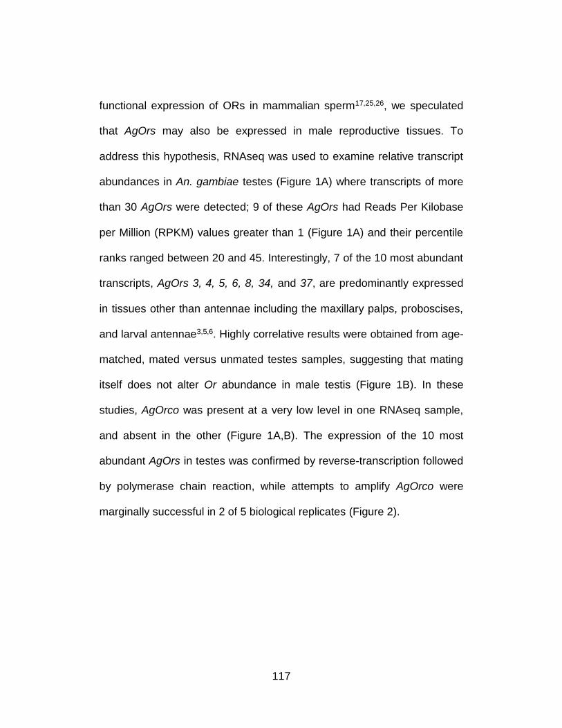
117
functional expression of ORs in mammalian sperm17,25,26, we speculated
that AgOrs may also be expressed in male reproductive tissues. To
address this hypothesis, RNAseq was used to examine relative transcript
abundances in An. gambiae testes (Figure 1A) where transcripts of more
than 30 AgOrs were detected; 9 of these AgOrs had Reads Per Kilobase
per Million (RPKM) values greater than 1 (Figure 1A) and their percentile
ranks ranged between 20 and 45. Interestingly, 7 of the 10 most abundant
transcripts, AgOrs 3, 4, 5, 6, 8, 34, and 37, are predominantly expressed
in tissues other than antennae including the maxillary palps, proboscises,
and larval antennae3,5,6. Highly correlative results were obtained from age-
matched, mated versus unmated testes samples, suggesting that mating
itself does not alter Or abundance in male testis (Figure 1B). In these
studies, AgOrco was present at a very low level in one RNAseq sample,
and absent in the other (Figure 1A,B). The expression of the 10 most
abundant AgOrs in testes was confirmed by reverse-transcription followed
by polymerase chain reaction, while attempts to amplify AgOrco were
marginally successful in 2 of 5 biological replicates (Figure 2).
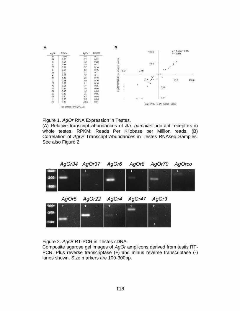
118
Figure 1. AgOr RNA Expression in Testes. (A) Relative transcript abundances of An. gambiae odorant receptors in whole testes. RPKM: Reads Per Kilobase per Million reads. (B) Correlation of AgOr Transcript Abundances in Testes RNAseq Samples. See also Figure 2.
Figure 2. AgOr RT-PCR in Testes cDNA. Composite agarose gel images of AgOr amplicons derived from testis RT-PCR. Plus reverse transcriptase (+) and minus reverse transcriptase (-) lanes shown. Size markers are 100-300bp.
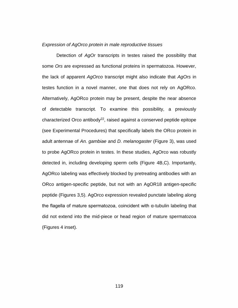
119
Expression of AgOrco protein in male reproductive tissues
Detection of AgOr transcripts in testes raised the possibility that
some Ors are expressed as functional proteins in spermatozoa. However,
the lack of apparent AgOrco transcript might also indicate that AgOrs in
testes function in a novel manner, one that does not rely on AgORco.
Alternatively, AgORco protein may be present, despite the near absence
of detectable transcript. To examine this possibility, a previously
characterized Orco antibody22, raised against a conserved peptide epitope
(see Experimental Procedures) that specifically labels the ORco protein in
adult antennae of An. gambiae and D. melanogaster (Figure 3), was used
to probe AgORco protein in testes. In these studies, AgOrco was robustly
detected in, including developing sperm cells (Figure 4B,C). Importantly,
AgORco labeling was effectively blocked by pretreating antibodies with an
ORco antigen-specific peptide, but not with an AgOR18 antigen-specific
peptide (Figures 3,5). AgOrco expression revealed punctate labeling along
the flagella of mature spermatozoa, coincident with α-tubulin labeling that
did not extend into the mid-piece or head region of mature spermatozoa
(Figures 4 inset).
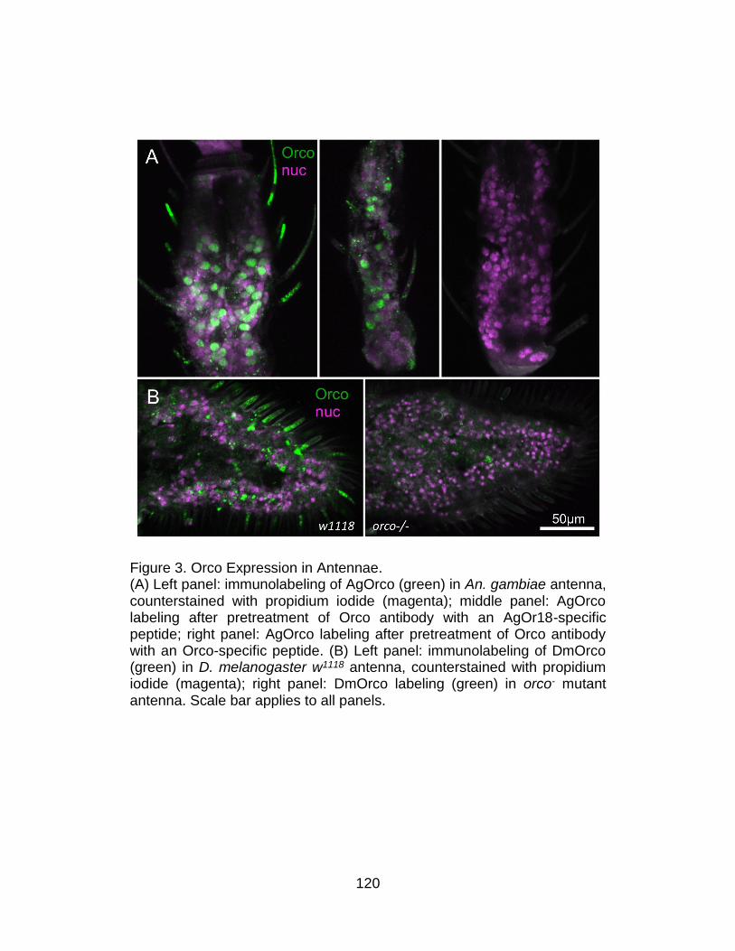
120
Figure 3. Orco Expression in Antennae. (A) Left panel: immunolabeling of AgOrco (green) in An. gambiae antenna, counterstained with propidium iodide (magenta); middle panel: AgOrco labeling after pretreatment of Orco antibody with an AgOr18-specific peptide; right panel: AgOrco labeling after pretreatment of Orco antibody with an Orco-specific peptide. (B) Left panel: immunolabeling of DmOrco (green) in D. melanogaster w1118 antenna, counterstained with propidium iodide (magenta); right panel: DmOrco labeling (green) in orco- mutant antenna. Scale bar applies to all panels.
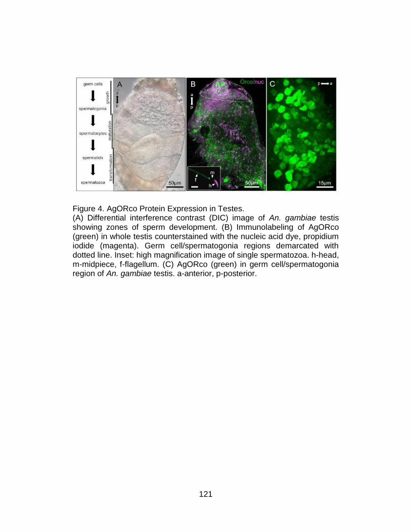
121
Figure 4. AgORco Protein Expression in Testes. (A) Differential interference contrast (DIC) image of An. gambiae testis showing zones of sperm development. (B) Immunolabeling of AgORco (green) in whole testis counterstained with the nucleic acid dye, propidium iodide (magenta). Germ cell/spermatogonia regions demarcated with dotted line. Inset: high magnification image of single spermatozoa. h-head, m-midpiece, f-flagellum. (C) AgORco (green) in germ cell/spermatogonia region of An. gambiae testis. a-anterior, p-posterior.
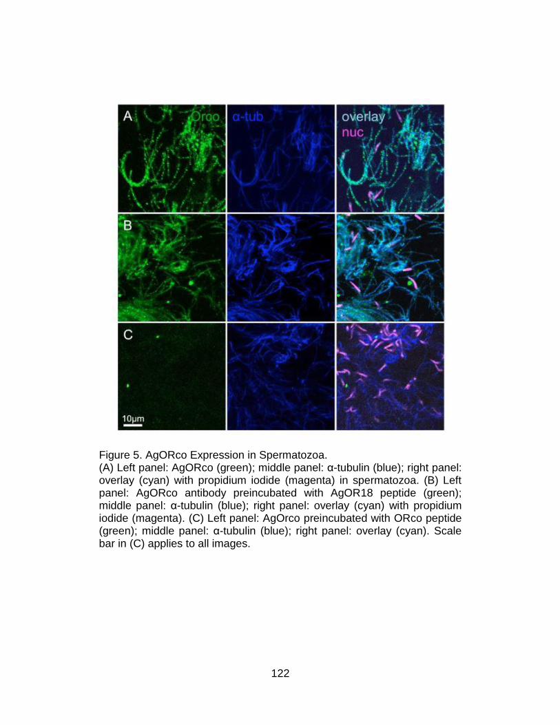
122
Figure 5. AgORco Expression in Spermatozoa. (A) Left panel: AgORco (green); middle panel: α-tubulin (blue); right panel: overlay (cyan) with propidium iodide (magenta) in spermatozoa. (B) Left panel: AgORco antibody preincubated with AgOR18 peptide (green); middle panel: α-tubulin (blue); right panel: overlay (cyan) with propidium iodide (magenta). (C) Left panel: AgOrco preincubated with ORco peptide (green); middle panel: α-tubulin (blue); right panel: overlay (cyan). Scale bar in (C) applies to all images.
123
Taken together, these results indicate that transcripts for several
tuning AgOrs and the AgORco protein are expressed in male sperm
where they may form functional ligand-gated ion channels. One
explanation for the apparent absence of AgOrco transcript in testes is that
its expression occurs prior to emergence and that the translated protein is
very stable, such that it is active throughout the adult male life. Indeed, the
apparent persistence of AgORco across multiple stages of sperm
development is an indication of this stability. These data suggest a
potentially unique role for AgOrs in An. gambiae spermatozoa where they
may function as regulators of cell motility in response to endogenous
chemical signals.
Activation of spermatozoa
In order to explore the possible biological function of AgOrs in An.
gambiae testes, and in light of the well-established chemosensory
responses of vertebrate spermatoza (reviewed in Kaupp, 2012) we have
developed a video-based bioassay (Figure 6) to examine the activation of
flagellar beating responses of spermatozoa to a range of chemical stimuli.
Because the exaggerated length of An. gambiae spermatozoa flagella
which can reach means as much as 250μm (and up to 2mm subsequent
to maturation) as compared to 50μm for human sperm27 presents a
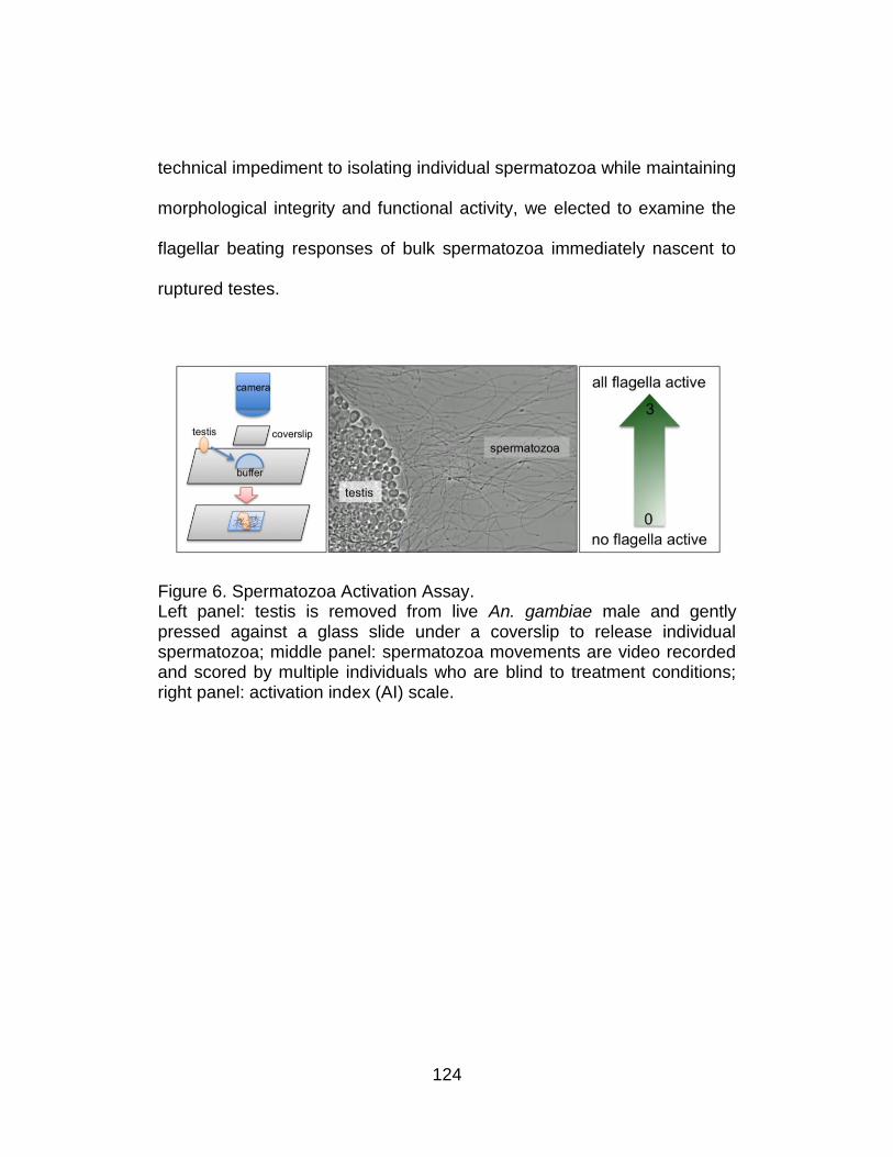
124
technical impediment to isolating individual spermatozoa while maintaining
morphological integrity and functional activity, we elected to examine the
flagellar beating responses of bulk spermatozoa immediately nascent to
ruptured testes.
Figure 6. Spermatozoa Activation Assay. Left panel: testis is removed from live An. gambiae male and gently pressed against a glass slide under a coverslip to release individual spermatozoa; middle panel: spermatozoa movements are video recorded and scored by multiple individuals who are blind to treatment conditions; right panel: activation index (AI) scale.
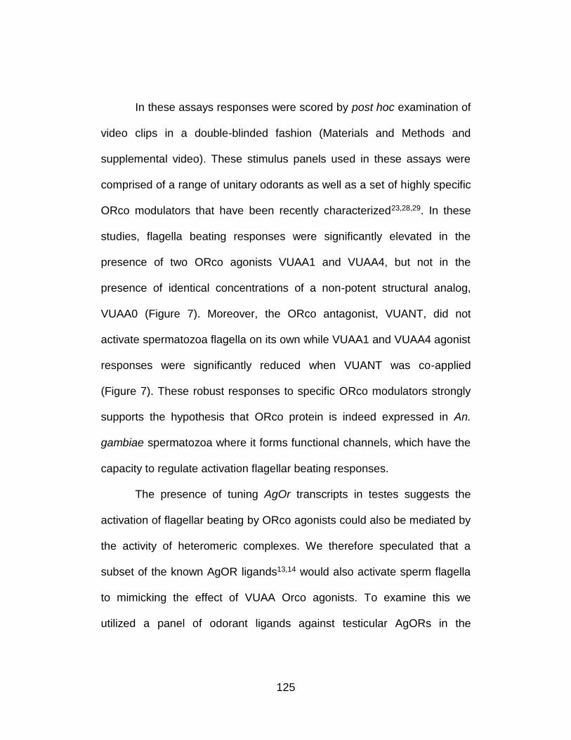
125
In these assays responses were scored by post hoc examination of
video clips in a double-blinded fashion (Materials and Methods and
supplemental video). These stimulus panels used in these assays were
comprised of a range of unitary odorants as well as a set of highly specific
ORco modulators that have been recently characterized23,28,29. In these
studies, flagella beating responses were significantly elevated in the
presence of two ORco agonists VUAA1 and VUAA4, but not in the
presence of identical concentrations of a non-potent structural analog,
VUAA0 (Figure 7). Moreover, the ORco antagonist, VUANT, did not
activate spermatozoa flagella on its own while VUAA1 and VUAA4 agonist
responses were significantly reduced when VUANT was co-applied
(Figure 7). These robust responses to specific ORco modulators strongly
supports the hypothesis that ORco protein is indeed expressed in An.
gambiae spermatozoa where it forms functional channels, which have the
capacity to regulate activation flagellar beating responses.
The presence of tuning AgOr transcripts in testes suggests the
activation of flagellar beating by ORco agonists could also be mediated by
the activity of heteromeric complexes. We therefore speculated that a
subset of the known AgOR ligands13,14 would also activate sperm flagella
to mimicking the effect of VUAA Orco agonists. To examine this we
utilized a panel of odorant ligands against testicular AgORs in the
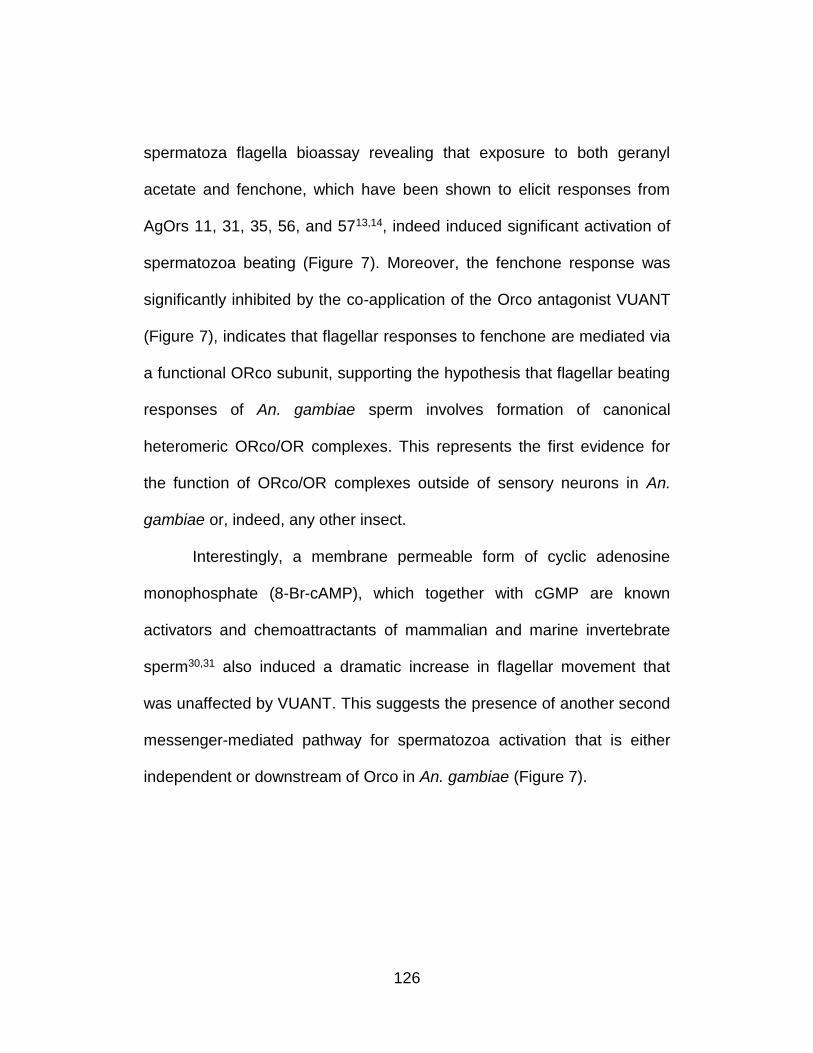
126
spermatoza flagella bioassay revealing that exposure to both geranyl
acetate and fenchone, which have been shown to elicit responses from
AgOrs 11, 31, 35, 56, and 5713,14, indeed induced significant activation of
spermatozoa beating (Figure 7). Moreover, the fenchone response was
significantly inhibited by the co-application of the Orco antagonist VUANT
(Figure 7), indicates that flagellar responses to fenchone are mediated via
a functional ORco subunit, supporting the hypothesis that flagellar beating
responses of An. gambiae sperm involves formation of canonical
heteromeric ORco/OR complexes. This represents the first evidence for
the function of ORco/OR complexes outside of sensory neurons in An.
gambiae or, indeed, any other insect.
Interestingly, a membrane permeable form of cyclic adenosine
monophosphate (8-Br-cAMP), which together with cGMP are known
activators and chemoattractants of mammalian and marine invertebrate
sperm30,31 also induced a dramatic increase in flagellar movement that
was unaffected by VUANT. This suggests the presence of another second
messenger-mediated pathway for spermatozoa activation that is either
independent or downstream of Orco in An. gambiae (Figure 7).
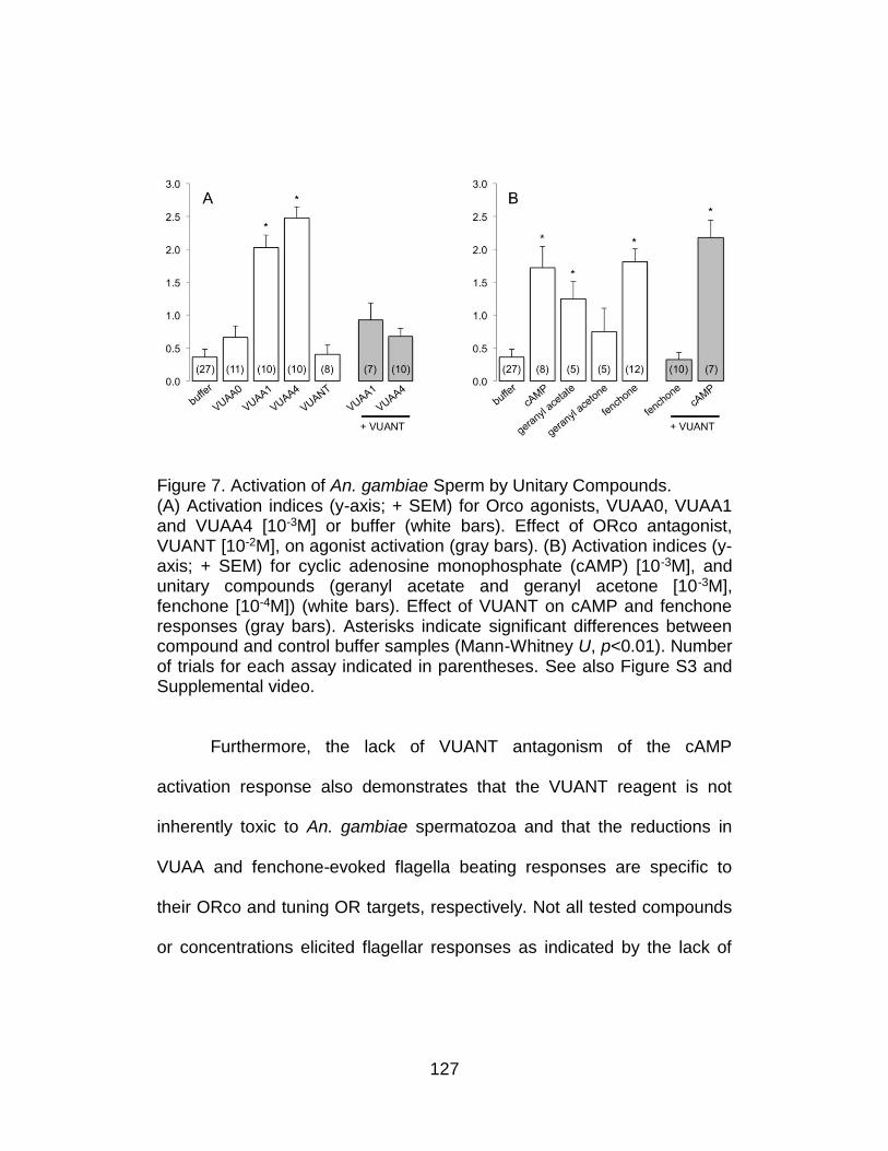
127
Figure 7. Activation of An. gambiae Sperm by Unitary Compounds. (A) Activation indices (y-axis; + SEM) for Orco agonists, VUAA0, VUAA1 and VUAA4 [10-3M] or buffer (white bars). Effect of ORco antagonist, VUANT [10-2M], on agonist activation (gray bars). (B) Activation indices (y-axis; + SEM) for cyclic adenosine monophosphate (cAMP) [10-3M], and unitary compounds (geranyl acetate and geranyl acetone [10-3M], fenchone [10-4M]) (white bars). Effect of VUANT on cAMP and fenchone responses (gray bars). Asterisks indicate significant differences between compound and control buffer samples (Mann-Whitney U, p<0.01). Number of trials for each assay indicated in parentheses. See also Figure S3 and Supplemental video.
Furthermore, the lack of VUANT antagonism of the cAMP
activation response also demonstrates that the VUANT reagent is not
inherently toxic to An. gambiae spermatozoa and that the reductions in
VUAA and fenchone-evoked flagella beating responses are specific to
their ORco and tuning OR targets, respectively. Not all tested compounds
or concentrations elicited flagellar responses as indicated by the lack of

128
significant spermatozoa activation to geranyl acetone, 10-6M 8-Br-cAMP,
10-4M geranyl acetate and 10-6M fenchone.
Taken together, these data suggest that, heteromeric complexes of
AgORs, as well as other signaling pathways, play functional roles in the
activation of spermatozoa in An. gambiae. Importantly, the role of ORs in
sperm activation is likely to be a general feature of insect reproductive
biology as immunolocalization studies reveal the presence of the highly
conserved ORco protein within spermatozoa of several other
holometabolaous insects such as the mosquito Aedes albopictus, the
fruitfly Drosophila melanogaster, and the parasitic wasp Nasonia
Vitripennis (Figure 8). We recognize that overt viability and fecundity
defects have not been reported for laboratory reared orco- mutants in D.
melanogaster22 and Ae. aegypti32, however such conditions do not
preclude the presence of a subtle, yet significant OR-based reproductive
fitness advantage in natural insect populations. Furthermore, while here
we focused on the functional expression of AgOrs due to the availability of
highly specific chemical modulators, is also likely that other ion channel
and chemosensory receptor gene families may also perform parallel
signaling functions in spermatozoa. Indeed, RNAseq-based transcriptome
profiling studies consistently revealed transcripts for multiple members of
An. gambiae variant ionotropic receptor (AgIr), gustatory receptor (AgGr),
129
and odorant-binding protein (AgObp) gene families are present in the
testes of An. gambiae males. In total, we found 14 AgGrs, 17 AgIrs, and 6
AgObps that have RPKMs greater than 1, among which 2 AgGrs, 4 AgIrs,
and 5 AgObps had transcript abundances above the median of the entire
testis transcriptome. These highly expressed chemosensory genes
include AgGr22 which encode the carbon dioxide receptor, and several
conserved AgIrs with significant antennal expression24.
Discussion
An. gambiae females are generally monandrous and remating is
rare in wild populations33,34. This necessitates the storage of sperm in the
spermatheca as well as mechanisms for their efficient use over the
reproductive life of each female. Few studies have explored the pathways
used to identify bioactive substances that elicit responses from conspecific
insect sperm and it is likely the volatile AgOR ligands utilized here do not
encompass the endogenous signals involved in An. gambiae spermatozoa
activation. Examples of directed movement of sperm have been
extensively characterized in marine invertebrates and mammals35 as well
as several insect species. For example, in the beetle, Drusilla canaliculata,
sperm migrate into the spermathecae36, while the spermathecal gland in
the boll weevil, Anthonomus grandis, is required for sperm activation,
130
storage, clearance, and fertility37. In D. melanogaster, sperm swim
backwards upon entering the female reproductive tract and genetic
ablation of the spermatheca secretory cells (SSCs) prior to mating leads to
sperm storage defects: sperm fail to migrate into spermathcae and
become inactive within the seminal receptacle38-41. Moreover, SSC-
ablated females display reduced fertility over time and ovovivipary. These
experiments suggest that substances in spermathecae, SSCs, or perhaps
other tissues are involved in the activation and chemo attraction of sperm.
Reproductive fitness is an important component in establishing and
maintaining insect populations and accordingly, the vectorial capacity of
malaria vectors. Despite ongoing efforts to characterize the role of
accessory gland proteins and sperm in the formation of the An. gambiae
mating plug and fertilization42-47, the potential signals that induce sperm
activation, spermatozoa localization, retention or fertilization within the
female reproductive tract remain unknown. An intriguing possibility is that
females produce and release chemicals that activate male sperm prior to
fertilization that also act as chemotactic cues to orient or otherwise direct
sperm motility. Importantly, the overall reproductive success of An.
gambiae males correlates positively with the presence of motile
spermatozoa in mated female spermatheca and negatively with sperm
length27,48. In this context, an enhanced understanding of An. gambiae
131
sperm activation/motility and the molecular processes that impinge upon
them will be significant in terms of both basic biology and as a potential
means to develop new control methods.
Existing vector control techniques rely heavily on insecticides;
however, insecticide resistance and altered vector behavior following
insecticide applications may erode the effectiveness of these
technologies49-51. Thus, the availability of alternative insect/vector control
methods is highly desirable and may ultimately become critical
components of integrated pest/vector management programs52,53. One
conceptual utility of these studies would be the use of chemical agents,
rather than the current reliance on radiation for the induction of male
sterility in the context of a Sterile Insect Technique (SIT) program, which
has recently been the subject of renewed interest for mosquito control54,55.
More detailed investigations of this aspect of the reproductive biology of
An. gambiae would be essential for the development of novel sterilization
or mating disruption technologies that would be expected to significantly
reduce vectorial capacity. In addition, the characterization of what is likely
to be highly conserved general principles of insect chemoreceptor function
in sperm would enhance our understanding of vector biology as well as
insect evolution and chemical ecology.
132
Our findings are reminiscent of the functional expression of ORs in
mammalian sperm where signaling machinery with predominant
expression and function in sensory tissues is also utilized in reproductive
processes. However, inasmuch as mammalian and insect ORs utilize
distinct mechanistic paradigms and do not share a common ancestry, OR
expression in sperm most likely evolved independently in the two distant
lineages, thus providing a striking example of convergent evolution.
Acknowledgments
We thank Dr. Leslie B. Vosshall (Rockefeller University) for the generous
gift of anti-Orco IC3 antisera, Ms. Zhen Li for mosquito rearing and the
Hudson Alpha Institute for Biotechnology for RNA sample preparation and
Illumina sequencing. We also thank members of the Zwiebel lab for critical
reading of the manuscript. This work was conducted in part using the
resources of the Advanced Computing Center for Research and Education
at Vanderbilt University and was supported by grants from the Innovation
and Discovery in Engineering And Science (IDEAS) program of Vanderbilt
University, the National Institutes of Health (NIAID, AI056402) and the
Foundation for the National Institutes of Health through the Grand
Challenges in Global Health Initiative (VCTR121) to LJZ. The authors
declare no competing interests.
133
References
1. Hill, C.A., et al., G protein coupled receptors in Anopheles gambiae. Science, 2002. 298(5591): p. 176-178.
2. Pitts, R.J., A.N. Fox, and L.J. Zwiebel, A highly conserved candidate chemoreceptor expressed in both olfactory and gustatory tissues in the malaria vector Anopheles gambiae. Proc Natl Acad Sci U S A, 2004. 101(14): p. 5058-5063.
3. Kwon, H.W., et al., Olfactory responses in a gustatory organ of the malaria vector mosquito Anopheles gambiae. Proc Natl Acad Sci U S A, 2006. 103(36): p. 13526-31.
4. Bohbot, J., et al., Molecular characterization of the Aedes aegypti odorant receptor gene family. Insect Mol Biol, 2007. 16(5): p. 525-537.
5. Lu, T., et al., Odor coding in the maxillary palp of the malaria vector mosquito Anopheles gambiae. Current Biology, 2007. 17(18): p. 1533-1544.
6. Xia, Y., et al., The molecular and cellular basis of olfactory-driven behavior in Anopheles gambiae larvae. Proc Natl Acad Sci U S A, 2008. 105(17): p. 6433-8.
7. Benton, R., et al., Atypical membrane topology and heteromeric function of Drosophila odorant receptors in vivo. PLoS Biol, 2006. 4(2): p. 240-257.
8. Wicher, D., et al., Drosophila odorant receptors are both ligand-gated and cyclic-nucleotide-activated cation channels. Nature, 2008. 452(7190): p. 1007-U10.
9. Sato, K., et al., Insect olfactory receptors are heteromeric ligand-gated ion channels. Nature, 2008. 452(7190): p. 1002-U9.
10. Pask, G.M., et al., Heteromeric Anopheline Odorant Receptors Exhibit Distinct Channel Properties. PLoS One, 2011. 6(12).
11. Kaupp, U.B., Olfactory signalling in vertebrates and insects: differences and commonalities. Nature Reviews Neuroscience, 2010. 11(3): p. 188-200.
12. Hallem, E.A., et al., Olfaction - Mosquito receptor for human-sweat odorant. Nature, 2004. 427(6971): p. 212-213.
134
13. Wang, G.R., et al., Molecular basis of odor coding in the malaria vector mosquito Anopheles gambiae. Proc Natl Acad Sci U S A, 2010. 107(9): p. 4418-4423.
14. Carey, A.F., et al., Odorant reception in the malaria mosquito Anopheles gambiae. Nature, 2010. 464(7285): p. 66-71.
15. Vanderhaeghen, P., et al., Specific repertoire of olfactory receptor genes in the male germ cells of several mammalian species. Genomics, 1997. 39(3): p. 239-246.
16. Goto, T., A. Salpekar, and M. Monk, Expression of a testis-specific member of the olfactory receptor gene family in human primordial germ cells. Mol Hum Reprod, 2001. 7(6): p. 553-558.
17. Spehr, M., et al., Identification of a testicular odorant receptor mediating human sperm chemotaxis. Science, 2003. 299(5615): p. 2054-2058.
18. Sirot, L.K., et al., Towards a Semen Proteome of the Dengue Vector Mosquito: Protein Identification and Potential Functions. PLoS Negl Trop Dis, 2011. 5(3).
19. Dorus, S., et al., Genomic and functional evolution of the Drosophila melanogaster sperm proteome. Nature Genetics, 2006. 38(12): p. 1440-1445.
20. Kim, D., et al., TopHat2: accurate alignment of transcriptomes in the presence of insertions, deletions and gene fusions. Genome Biology, 2013. 14(4).
21. Feng, J.X., et al., GFOLD: a generalized fold change for ranking differentially expressed genes from RNA-seq data. Bioinformatics, 2012. 28(21): p. 2782-2788.
22. Larsson, M.C., et al., Or83b encodes a broadly expressed odorant receptor essential for Drosophila olfaction. Neuron, 2004. 43(5): p. 703-14.
23. Taylor, R.W., et al., Structure-activity relationship of a broad-spectrum insect odorant receptor agonist. ACS Chem Biol, 2012. 7(10): p. 1647-52.
24. Pitts, R.J., et al., Transcriptome profiling of chemosensory appendages in the malaria vector Anopheles gambiae reveals tissue- and sex-specific signatures of odor coding. BMC Genomics, 2011. 12.
135
25. Fukuda, N., et al., Functional characterization of a mouse testicular olfactory receptor and its role in chemosensing and in regulation of sperm motility. J Cell Sci, 2004. 117(Pt 24): p. 5835-45.
26. Veitinger, T., et al., Chemosensory Ca2+ dynamics correlate with diverse behavioral phenotypes in human sperm. J Biol Chem, 2011. 286(19): p. 17311-25.
27. Voordouw, M.J., J.C. Koella, and H. Hurd, Intra-specific variation of sperm length in the malaria vector Anopheles gambiae: males with shorter sperm have higher reproductive success. Malar J, 2008. 7.
28. Jones, P.L., et al., Functional agonism of insect odorant receptor ion channels. Proc Natl Acad Sci U S A, 2011. 108(21): p. 8821-8825.
29. Jones, P.L., et al., Allosteric Antagonism of Insect Odorant Receptor Ion Channels. PLoS One, 2012. 7(1).
30. Kaupp, U.B., E. Hildebrand, and I. Weyand, Sperm chemotaxis in marine invertebrates--molecules and mechanisms. J Cell Physiol, 2006. 208(3): p. 487-94.
31. Buffone, M.G., et al., Heads or tails? Structural events and molecular mechanisms that promote mammalian sperm acrosomal exocytosis and motility. Mol Reprod Dev, 2012. 79(1): p. 4-18.
32. DeGennaro, M., et al., orco mutant mosquitoes lose strong preference for humans and are not repelled by volatile DEET. Nature, 2013. 498(7455): p. 487-91.
33. Baimai, V. and C.A. Green, Monandry (monogamy) in natural populations of anopheline mosquitoes. J Am Mosq Control Assoc, 1987. 3(3): p. 481-4.
34. Tripet, F., et al., Frequency of multiple inseminations in field-collected Anopheles gambiae females revealed by DNA analysis of transferred sperm. Am J Trop Med Hyg, 2003. 68(1): p. 1-5.
35. Kaupp, U.B., N.D. Kashikar, and I. Weyand, Mechanisms of sperm chemotaxis. Annu Rev Physiol, 2008. 70: p. 93-117.
136
36. Werner, M., et al., Queue up, please! Spermathecal filling in the rove beetle Drusilla canaliculata (Coleoptera, Staphylinidae). Naturwissenschaften, 2007. 94(10): p. 837-41.
37. Grodner, M.L. and W.L. Steffens, Evidence of a chemotactic substance in the spermathecal gland of the female boll weevil (Coleoptera: curculionidae). Trans Am Microsc Soc, 1978. 97(1): p. 116-20.
38. Yang, Y. and X. Lu, Drosophila sperm motility in the reproductive tract. Biol Reprod, 2011. 84(5): p. 1005-15.
39. Kottgen, M., et al., Drosophila sperm swim backwards in the female reproductive tract and are activated via TRPP2 ion channels. PLoS One, 2011. 6(5): p. e20031.
40. Werner, M. and L.W. Simmons, Insect sperm motility. Biol Rev Camb Philos Soc, 2008. 83(2): p. 191-208.
41. Schnakenberg, S.L., W.R. Matias, and M.L. Siegal, Sperm-storage defects and live birth in Drosophila females lacking spermathecal secretory cells. PLoS Biol, 2011. 9(11): p. e1001192.
42. Dottorini, T., et al., A genome-wide analysis in Anopheles gambiae mosquitoes reveals 46 male accessory gland genes, possible modulators of female behavior. Proc Natl Acad Sci U S A, 2007. 104(41): p. 16215-20.
43. Tripet, F., T. Thiemann, and G.C. Lanzaro, Effect of seminal fluids in mating between M and S forms of Anopheles gambiae. J Med Entomol, 2005. 42(4): p. 596-603.
44. Shutt, B., et al., Male accessory gland proteins induce female monogamy in anopheline mosquitoes. Med Vet Entomol, 2010. 24(1): p. 91-4.
45. Thailayil, J., et al., Spermless males elicit large-scale female responses to mating in the malaria mosquito Anopheles gambiae. Proc Natl Acad Sci U S A, 2011. 108(33): p. 13677-81.
46. Rogers, D.W., et al., Transglutaminase-mediated semen coagulation controls sperm storage in the malaria mosquito. PLoS Biol, 2009. 7(12): p. e1000272.
137
47. Rogers, D.W., et al., Molecular and cellular components of the mating machinery in Anopheles gambiae females. Proc Natl Acad Sci U S A, 2008. 105(49): p. 19390-5.
48. Voordouw, M.J., J.C. Koella, and H. Hurd, Comparison of male reproductive success in malaria-refractory and susceptible strains of Anopheles gambiae. Malar J, 2008. 7: p. 103.
49. Ranson, H., et al., Pyrethroid resistance in African anopheline mosquitoes: what are the implications for malaria control? Trends Parasitol, 2011. 27(2): p. 91-8.
50. Reddy, M.R., et al., Outdoor host seeking behaviour of Anopheles gambiae mosquitoes following initiation of malaria vector control on Bioko Island, Equatorial Guinea. Malar J, 2011. 10: p. 184.
51. Raghavendra, K., et al., Malaria vector control: from past to future. Parasitol Res, 2011. 108(4): p. 757-79.
52. Alonso, P.L., et al., A research agenda to underpin malaria eradication. PLoS Med, 2011. 8(1): p. e1000406.
53. mal, E.R.A.C.G.o.V.C., A research agenda for malaria eradication: vector control. PLoS Med, 2011. 8(1): p. e1000401.
54. Dame, D.A., et al., Historical applications of induced sterilisation in field populations of mosquitoes. Malar J, 2009. 8 Suppl 2: p. S2.
55. Munhenga, G., et al., Evaluating the potential of the sterile insect technique for malaria control: relative fitness and mating compatibility between laboratory colonized and a wild population of Anopheles arabiensis from the Kruger National Park, South Africa. Parasit Vectors, 2011. 4: p. 208.
138
CHAPTER V
SUMMARY AND FUTURE EXPERIMENTS
My thesis work has predominantly focused on the sensory biology
of the principal malaria vector mosquito, An. gambiae. By designing
different apparatuses that were tailored to studying one sensory modality
at a time, we have successfully broadened our view on sensory-mediated
animal behavior, which is peripherally determined by a plethora of sensory
receptor proteins. These studies not only contribute to the understanding
of basic biological processes in An. gambiae, but also provide insights in
the development of novel control strategies that target mosquito
populations. More specifically, chapter 2 and 3 study the larval OR and
TRP channel-mediated sensory system, respectively, while chapter 4
mainly deals with the reproductive system in adult stage male An.
gambiae. The central theme that is covered throughout my thesis are
odorant receptors and TRP genes, both of which are crucial channel
proteins that insects utilize to perceive the outside world in order to
engage in a spectrum of behaviors that are vital to the survival of the
organism. As for An. gambiae, their survival adds another level of medical
importance as in order for them to thrive, they have to blood feed on
139
human hosts, therefore resulting in the transmission of malaria from one
infected individual to another with high efficiency.
Numerous studies have suggested that olfaction is the most central
sensory modality that mosquito uses for host seeking, mating, nectar
feeding, oviposition etc, all of which are life-dependent behaviors1-9. If we
could elucidate the cellular and molecular mechanisms underlying
olfactory-directed behavior, we might be able to interfere/disrupt these
processes by specifically targeting the endogenous signaling pathways to
reduce malaria burden. Upon the discovery of odorant receptor gene
family in mammals which dated back to the year of 199110, efforts have
been widely taken to demonstrate that the expression of OR proteins
within ORN is essential for the animal to recognize ambient volatile odor
molecules. The binding of odorant to OR will initiate the conversion of the
chemical stimuli to electric action potentials that can be “read” and
handled by the central neural system (brain) so as to elicit downstream
effects such as learning/memory and behavioral activity
(attraction/aversion etc.). However, it is important to mention that OR
family is not the only category of receptors that is dedicated to function in
the peripheral olfactory system. It is already known that gustatory receptor
(GR) in insects is capable of mediating the responses to CO211, another
volatile “odor”, although GRs are predominantly involved in the contact-
140
based sensory modality including the sense of touch and taste12. In
addition, more-recent investigations have identified the expression of a
novel family of Ionotropic receptors (IRs) in insects that are distantly
related to glutamate receptors in mammals which are suggested to
respond to odors sharing a non-overlapping odor space with chemicals
that are sensed by ORs. These compounds include ammonia and amine
compounds13, many of which are components in human sweat. Therefore
it is indicating that an array of sensory genes from distinct gene families
operate in parallel to facilitate insect olfaction. This is also reflective of the
complexity of insect olfactory system, considering the “information flow”
generated by the enormous amount of olfactory inputs from a spectrum of
peripheral sensors.
In chapter 2, we have taken multiple approaches to examine larval
peripheral olfactory system in vivo by means of functional characterization
of both ORs and IRs. We have undertaken studies on larval stage An.
gambiae for several reasons. Briefly, in this life stage, they typically live in
small puddles or lakes and undergo more restricted lifestyle, thereby
making them an excellent target for regulation of adult populations. In the
meantime, several successes of mosquito eradication were primarily
achieved through both larviciding and adult control strategies,
demonstrating larval control can serve as a powerful alternative approach
141
in batting malaria. More importantly, due to their swimming nature, it is
much easier to carry out behavioral-related sensory studies on aquatic
larvae, whose neural system is much simplified compared to adults. For
instance, there are only 12 ORs expressed in larval ORNs while the
number in adults is 79, making them a good candidate for basic research.
Last but not least, I have successfully developed a protocol for RNAi-
mediated gene silencing in larvae, which is still fundamentally difficult to
accomplish in adult stage. All these benefits for studying larvae have
granted us the opportunity to better probe the role of sensory receptors in
leading larval responses in vivo. This “model within a non-model system”
is especially important to us when genetic manipulations are generally
lacking in adult An. gambiae. The understanding of larval sensory biology
not only will benefit the development of larval control methods, but also
has value in terms of the fundamental molecular machinery, which is
conserved to a large extent between pre-adult and adult stage
mosquitoes.
In chapter 2, we established a robust bioassay to validate the role
of OR in sensing both natural and synthetic odors. Furthermore, we
identified distinct singling pathways coupled to either OR or IR by showing
AgIr76b was necessary for the larvae to behaviorally respond to
butylamine, which was not mediated by OR as knockdown of Orco
142
expression failed to diminish this response. Additionally, we presented for
the first time that OR40 contributed to the larval responses to DEET, a
commercially available insect repellent that had been widely implemented.
Although OR40 is not expressed in the adult olfactory system, this
particular finding provides us with insights on the molecular mechanism
underlying DEET mode of action, which has been much debated in the
field. Interests were given on how DEET repels insects, and it was
previously believed DEET played an inhibitory role on insect ORNs,
therefore the attraction to human odors was diminished with application of
DEET14. Nevertheless, our study is suggestive of an alternative
explanation that instead of masking the effect of other odorants on ORNs,
DEET directly binds to a specific OR to provoke repellency. However, this
interpretation was questioned based on the broad effectively of DEET
towards various insect species. It seemed not feasible that each insect
expressed a different set of DEET receptor, which was not mutually
shared in evolution (OR family is one of the most divergent gene family
across insects). However, latest research has favored our model,
especially when a group generated Orco- mutant Aedes aegypti (yellow
fever mosquito) and showed they lacked the response to volatile DEET15
due to the loss of OR-mediated signaling. However, it is worth mentioning
that the contact repellency of DEET in the Orco- mutant mosquitoes was
143
retained, which excludes the possibility that OR is the only DEET sensor
in adult Ae. aegypti, hence additional receptor proteins are also involved.
For example, GRs are reported to be indispensable in suppressing
feeding behavior in Drosophila16. Taken together, our work has
established in vivo evidence that both OR and IR-mediated signaling are
active in larval peripheral olfactory system. OR40 is the main, if not only,
DEET receptor in An. gambiae larvae.
Following the analysis of OR functions in peripheral olfaction, in
chapter 4, we have indeed, explored a non-canonical aspect of the role
played by odorant receptor in the mosquito reproductive system. By
means of showing the functional expression of ORs in An. gambiae
spermatozoa, we put forward a novel model regarding OR function that
would renew the contemporary point of view on insect odorant receptors.
The binding of odorant molecule to OR is followed by the opening of
channel pore and influx of cations. This molecular event in OR gating is
probably also conserved in other biological processes that take place in
tissues other than the peripheral appendages in mosquitoes. For instance,
the activation of flagellar movement in a mature spermatozoa. Mobility is
the common feature for sperm cell, with the activation of sperm motion
being a prerequisite for fertilization to occur since sperm and egg are
physically separated from each other. We started this project in the hope
144
of identifying endogenous sperm attractant(s) that are produced in the
female reproductive organ and show the chemotactic movement of sperm
towards the cue(s) is dependent on ORs. However, it was very technically
challenging to conduct studies on insect sperm chemotaxis, which was
largely due to the difficulty in the isolation of individual sperm. Insect
sperm are equipped with extremely long tails (up to 2mm in length),
therefore how to overcome the entanglement of sperm tails within a
reasonable time frame (sperm need to be viable) would be a key in the
tracking/manipulation of sperm behavior. As an alternative, we took a
pharmacological approach to monitor the flagellar beating activity of An.
gambiae sperm in response to a variety of OR modulators. It is very
interesting that first, we confirmed the expression of ORs in mosquito
sperm and second, the activation of these ORs stimulate flagellar beating.
Our finding is reminiscent of the in intro studies showing OR-
mediated sperm chemotaxis in mammals. The vertebrate and invertebrate
ORs do not share common ancestry or sequence homology. Not to
mention that their modes of actions are very distinct (vertebrate ORs are
GPCRs while insect ORs are ligand-gated ion channels). Therefore this
work may suggest for a striking example of convergent evolution between
mammal and insect ORs. This also adds to the basic biology of OR
functions so that a novel target for insect control could be explored.
145
Specifically, if we can somehow modulate these sperm-expressed ORs
render them immobility, this will generate infertile males with no offspring.
As a complementary methodology to Sterile Insect Technology (SIT) that
is already implemented worldwide to eliminate agricultural pests, this
could further decrease the mosquito population. One intriguing possibility,
as suggested by our work, could be the use of VUAA-compounds, which
specifically agonize/antagonize ORco. Nonetheless, future studies are
necessary to fully explore the in vivo role of ORs in An. gambiae sperm as
well as clarify the relationship between OR and reproductive fitness, as we
also mentioned in the paper, the lab-reared Orco- mutant Ae. aegyti are
reproducible and do not seem to suffer from fertilization defects. However,
even the endogenous function of OR in reproduction is marginal, we could
still take advantage of the presence of ORs in mosquito sperm and use
them as molecular targets to influence insect fecundity.
In chapter 3, I switched the focus from chemosensory to
thermosensory system. This work was inspired by the idea that in addition
to the olfactory sensitivity, female An. gambiae utilize additional sensory
modalities in locating blood meal host. Their sensitivity to the change of
ambient temperature would be one example. The combinatorial integration
of multiple sensory abilities may increase the efficiency of host seeking
behavior. Studying thermosensory processes in mosquito will not only
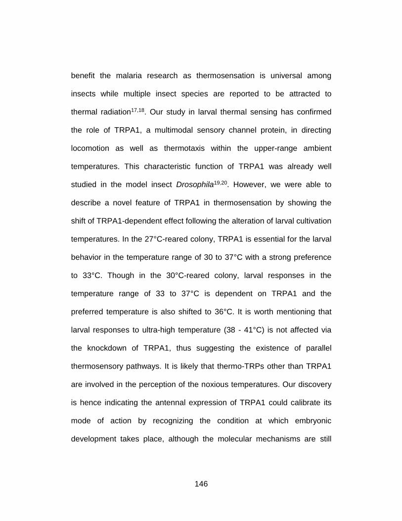
146
benefit the malaria research as thermosensation is universal among
insects while multiple insect species are reported to be attracted to
thermal radiation17,18. Our study in larval thermal sensing has confirmed
the role of TRPA1, a multimodal sensory channel protein, in directing
locomotion as well as thermotaxis within the upper-range ambient
temperatures. This characteristic function of TRPA1 was already well
studied in the model insect Drosophila19,20. However, we were able to
describe a novel feature of TRPA1 in thermosensation by showing the
shift of TRPA1-dependent effect following the alteration of larval cultivation
temperatures. In the 27°C-reared colony, TRPA1 is essential for the larval
behavior in the temperature range of 30 to 37°C with a strong preference
to 33°C. Though in the 30°C-reared colony, larval responses in the
temperature range of 33 to 37°C is dependent on TRPA1 and the
preferred temperature is also shifted to 36°C. It is worth mentioning that
larval responses to ultra-high temperature (38 - 41°C) is not affected via
the knockdown of TRPA1, thus suggesting the existence of parallel
thermosensory pathways. It is likely that thermo-TRPs other than TRPA1
are involved in the perception of the noxious temperatures. Our discovery
is hence indicating the antennal expression of TRPA1 could calibrate its
mode of action by recognizing the condition at which embryonic
development takes place, although the molecular mechanisms are still
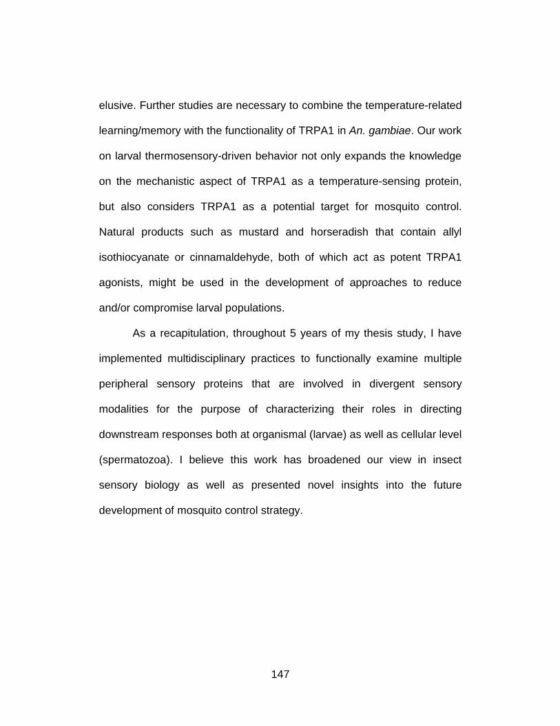
147
elusive. Further studies are necessary to combine the temperature-related
learning/memory with the functionality of TRPA1 in An. gambiae. Our work
on larval thermosensory-driven behavior not only expands the knowledge
on the mechanistic aspect of TRPA1 as a temperature-sensing protein,
but also considers TRPA1 as a potential target for mosquito control.
Natural products such as mustard and horseradish that contain allyl
isothiocyanate or cinnamaldehyde, both of which act as potent TRPA1
agonists, might be used in the development of approaches to reduce
and/or compromise larval populations.
As a recapitulation, throughout 5 years of my thesis study, I have
implemented multidisciplinary practices to functionally examine multiple
peripheral sensory proteins that are involved in divergent sensory
modalities for the purpose of characterizing their roles in directing
downstream responses both at organismal (larvae) as well as cellular level
(spermatozoa). I believe this work has broadened our view in insect
sensory biology as well as presented novel insights into the future
development of mosquito control strategy.
148
References
1. Takken, W. and B.G. Knols, Odor-mediated behavior of Afrotropical malaria mosquitoes. Annu Rev Entomol, 1999. 44: p. 131-57.
2. Hallem, E.A., et al., Olfaction - Mosquito receptor for human-sweat odorant. Nature, 2004. 427(6971): p. 212-213.
3. Zwiebel, L.J. and W. Takken, Olfactory regulation of mosquito-host interactions. Insect Biochem Mol Biol, 2004. 34(7): p. 645-652.
4. Carey, A.F. and J.R. Carlson, Insect olfaction from model systems to disease control. Proc Natl Acad Sci U S A, 2011. 108(32): p. 12987-95.
5. Pickett, J.A. and C.M. Woodcock, The role of mosquito olfaction in oviposition site location and in the avoidance of unsuitable hosts. Ciba Found Symp, 1996. 200: p. 109-19; discussion 119-23, 178-83.
6. Foster, W.A. and R.G. Hancock, Nectar-related olfactory and visual attractants for mosquitoes. J Am Mosq Control Assoc, 1994. 10(2 Pt 2): p. 288-96.
7. Mciver, S.B., Sensory Aspects of Mate-Finding Behavior in Male Mosquitos (Diptera, Culicidae). J Med Entomol, 1980. 17(1): p. 54-57.
8. Meijerink, J., M.A.H. Braks, and J.J.A. Van Loon, Olfactory receptors on the antennae of the malaria mosquito Anopheles gambiae are sensitive to ammonia and other sweat-borne components. J Insect Physiol, 2001. 47(4-5): p. 455-464.
9. Bowen, M.F., The sensory physiology of host-seeking behavior in mosquitoes. Annu Rev Entomol, 1991. 36: p. 139-58.
10. Buck, L. and R. Axel, A Novel Multigene Family May Encode Odorant Receptors - a Molecular-Basis for Odor Recognition. Cell, 1991. 65(1): p. 175-187.
11. Lu, T., et al., Odor coding in the maxillary palp of the malaria vector mosquito Anopheles gambiae. Current Biology, 2007. 17(18): p. 1533-1544.
12. Montell, C., A taste of the Drosophila gustatory receptors. Curr Opin Neurobiol, 2009. 19(4): p. 345-53.
149
13. Benton, R., et al., Variant Ionotropic Glutamate Receptors as Chemosensory Receptors in Drosophila. Cell, 2009. 136(1): p. 149-162.
14. Ditzen, M., M. Pellegrino, and L.B. Vosshall, Insect odorant receptors are molecular targets of the insect repellent DEET. Science, 2008. 319(5871): p. 1838-42.
15. DeGennaro, M., et al., orco mutant mosquitoes lose strong preference for humans and are not repelled by volatile DEET. Nature, 2013. 498(7455): p. 487-91.
16. Lee, Y., S.H. Kim, and C. Montell, Avoiding DEET through Insect Gustatory Receptors. Neuron, 2010. 67(4): p. 555-561.
17. Kleineidam, C.J., et al., Thermal radiation as a learned orientation cue in leaf-cutting ants (Atta vollenweideri). J Insect Physiol, 2007. 53(5): p. 478-487.
18. Ward, J.V. and J.A. Stanford, Thermal Responses in the Evolutionary Ecology of Aquatic Insects. Annu Rev Entomol, 1982. 27: p. 97-117.
19. Kang, K., et al., Modulation of TRPA1 thermal sensitivity enables sensory discrimination in Drosophila. Nature, 2012. 481(7379): p. 76-80.
20. Hamada, F.N., et al., An internal thermal sensor controlling temperature preference in Drosophila. Nature, 2008. 454(7201): p. 217-20.