Embed Size (px)
Citation preview

Olfactory Ensheathing Glia: an investigation of factors affecting responsiveness of these cells in
vitro and in vivo
Thalles R.B. De Mello
This thesis is presented for the degree of
Doctor of Philosophy
at The University of Western Australia
School of Anatomy & Human Biology and
School of Animal Biology
2006

ii
Abstract
Olfactory ensheathing glia (OEG) have been demonstrated to improve
functional and anatomical outcomes after injury to the nervous system and are
currently being trialled clinically. This thesis presents the investigation of two
important issues in OEG biology. The first study (Chapter 2) investigates
effects of different members of the neuregulin (NRG) family of molecules on
the proliferation of OEG, as a means of quickly obtaining large numbers of
cells for clinical or experimental use. We report that NRG-1β, but not NRG-
2α or NRG-3, has a significant proliferative effect. Furthermore, we report for
the first time that use of different mitogens (forskolin and pituitary extract)
commonly used to expand these cells in vitro, can have a significant effect on
the responsiveness of OEG to added NRG in subsequent mitogenic assays.
OEG grown initially with forskolin and pituitary extract exhibited increased
basal proliferation rates in comparison to OEG originally expanded without
these factors, and this increased rate of proliferation was sustained for at least
6 days following their withdrawal from the culture medium. We also report
for the first time the expression pattern of ErbB2, ErbB3 and ErbB4 receptors
on p75-selected OEG, and investigate their contribution to the NRG mitogenic
effect by the use of inhibitory ErbB antibodies.
Our second study (Chapter 3) seeks to clarify the role of OEG in promoting
myelination of central nervous system neurons. In this study we have

iii
investigated the myelinating ability of OEG derived from embryonic (EEG),
postnatal (PEG) and adult tissue (AEG) both in vitro and in vivo. OEG
selected by p75-immunopanning were co-cultured with dissociated cultures of
TrkA-dependant embryonic dorsal root ganglion (DRG) neurons. EEG, but
not AEG or PEG, successfully myelinated DRG neurons in the presence of
serum and/or ascorbate. AEG also failed to myelinate GDNF-dependant
embryonic DRG cultures, and growth factor-independent adult DRG cultures.
Transplantation of OEG into lysolecithin demyelinated spinal cord
demonstrated distinct ultrastructural differences between transplants of OEG
derived from animals of different ages. Furthermore, we demonstrate that
clearance of degraded myelin from the lesion site appears to be more effective
when animals are transplanted with EEG rather than AEG or Schwann cell
preparations. These results suggest that myelinating potential of OEG in vitro
and behaviour of these cells following transplantation in vivo are
developmentally regulated.
Together the two studies presented here constitute important evidence that
variations in extraction and expansion protocols can have a drastic effect on
behaviour of OEG both in vitro and in vivo, and arguably that these
differences may constitute a large source of variation between results
observed by different laboratories utilising OEG.

iv
Thesis Structure
This document is composed of four chapters.
Chapter 1 – Introduction
Chapter 1 constitutes an overview of literature covering the olfactory system,
an introduction to the use of olfactory ensheathing glia as cellular transplant
therapy to repair models of lesioned central nervous system, an overview of
myelin and the key myelin proteins investigated in this study, and an overview
of the role of neuregulins in the biology of peripheral nervous system
development.
Chapters 2 and 3
Chapters 2 and 3 describe in detail two separate studies investigating different
aspects of the biology of olfactory ensheathing glia. These chapters constitute
papers being currently prepared for submission to prominent scientific
journals (Glia and Journal of Neuroscience). As a result, they are self-
contained units detailing the studies described herein and follow a standard
paper format of a brief introduction, the materials and methods utilised, a
detailed description of the results, and a brief discussion highlighting issues of
primary importance.

v
Chapter 4 – Extended Discussion
Chapter 4 comprises an extended examination of the implications of the
results described in chapters 2 and 3 in the context of the available literature
of ensheathing cell biology. Wherever possible, the author has sought to
minimise duplication of points already covered in chapters 2 and 3. Rather,
this section attempts to expand upon those issues, and seeks to bring a sense
of context to the findings presented in this document. It is an attempt to
bridge the gap between reader and author, and details many of the thoughts,
ideas and questions arising as a result of this work, including suggestions by
the author regarding future studies that seek to investigate the biology of
olfactory ensheathing glia.

vi
Table Of Contents
1) Abstract ii
2) Thesis Structure iv
3) Table Of Contents vi
4) List Of Tables And Figures x
5) List of Abbreviations xiii
6) Acknowledgements xv
7) CHAPTER 1 – Introduction 1
i) The olfactory System 2
(a) Olfactory Ensheathing Glia 2
(b) Properties Of Olfactory Ensheathing Glia 4
ii) Attempts to repair damaged CNS 7
iii) OEG Mitogens 11
iv) The Role Of Neuregulins 12
v) The Myelin Sheath 19
(a) Protein Content Of The Myelin Sheath 19
(b) Myelination By OEG 22
vi) Summary 25
8) CHAPTER 2 – Culture Conditions Affect Proliferative
Responsiveness Of Olfactory Ensheathing Glia To Neuregulins 26
i) Abstract 27
ii) Introduction 28
iii) Methods 33
(a) Glial Cell Culture Preparation 33
(b) Cell Purity Determination 35
(c) BrdU Proliferation Assay 36
(d) Data Analysis 37
(e) Functional Blocking Of ErbB Receptors 38
(f) ErbB Receptor Immunocytochemistry 39
(g) SDS-PAGE And Western Blotting 39
(h) Detection Of ErbB Phosphorylation 40
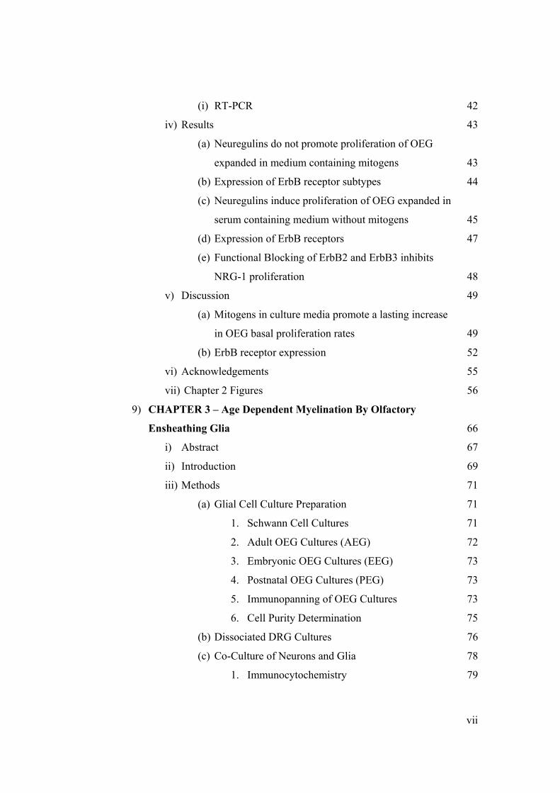
vii
(i) RT-PCR 42
iv) Results 43
(a) Neuregulins do not promote proliferation of OEG
expanded in medium containing mitogens 43
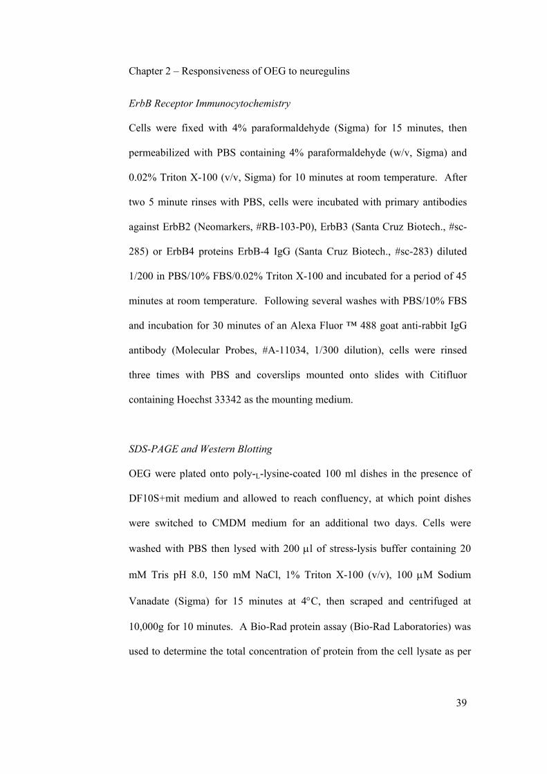
(b) Expression of ErbB receptor subtypes 44
(c) Neuregulins induce proliferation of OEG expanded in
serum containing medium without mitogens 45
(d) Expression of ErbB receptors 47
(e) Functional Blocking of ErbB2 and ErbB3 inhibits
NRG-1 proliferation 48
v) Discussion 49
(a) Mitogens in culture media promote a lasting increase
in OEG basal proliferation rates 49
(b) ErbB receptor expression 52
vi) Acknowledgements 55
vii) Chapter 2 Figures 56
9) CHAPTER 3 – Age Dependent Myelination By Olfactory
Ensheathing Glia 66
i) Abstract 67
ii) Introduction 69
iii) Methods 71
(a) Glial Cell Culture Preparation 71
1. Schwann Cell Cultures 71
2. Adult OEG Cultures (AEG) 72
3. Embryonic OEG Cultures (EEG) 73
4. Postnatal OEG Cultures (PEG) 73
5. Immunopanning of OEG Cultures 73
6. Cell Purity Determination 75
(b) Dissociated DRG Cultures 76
(c) Co-Culture of Neurons and Glia 78
1. Immunocytochemistry 79

viii
(d) Lysolecithin Demyelination of the Spinal Cord Dorsal
Funiculus 80
1. Cell Transplantation 81
2. Electron Microscopy of demyelinated
spinal cord 82
3. Toluidine Blue Staining 83
(e) Data Analysis 83
iv) Results 84
(a) Embryonic Ensheathing Glia myelinate TrkA-
dependent DRG neurons in vitro 84
(b) Adult Ensheathing Glia fail to myelinate GDNF-
dependent DRG neurons in vitro 88
(c) Adult Ensheathing Glia fail to myelinate adult DRG
neurons in vitro 89
(d) Ensheathing Glia promote remyelination of
demyelinated spinal cord 90
v) Discussion 93
(a) Myelination by OEG in vitro 94
(b) Myelination by OEG in vivo 95
vi) Acknowledgements 99
vii) Chapter 3 Figures 100
10) CHAPTER 4 – Extended Discussion 112
i) Part I 113
(a) Summary 113
(b) Influence of purification techniques on ErbB receptor
expression 113
(c) Influence of tissue age on ErbB receptor expression 117
(d) Observed Mitogenic Effect of NRG on AEG 120
ii) Part II 123
(a) Summary 123
(b) Interaction of OEG with axons 123

ix
(c) Mechanisms of action by OEG in vivo 125
(d) Influence of preparation age on promotion of axon
growth 129
(e) Importance of Neuroglial Arrangement 137
(f) Future Directions 139
(g) Concluding Remarks 140
11) Appendix A 142
12) References 159

x
List Of Tables And Figures
CHAPTER 1
• Figure 1. Binding affinities of the neuregulin isoforms utilised in
this study to the various ErbB receptor dimer combinations. 18
CHAPTER 2
• Figure 1. Effects of neuregulins on proliferation of OEG
expanded in the presence of DF10S+mit medium. 56
• Figure 2. BrdU staining of NRG-treated OEG. 57
• Figure 3. Western blotting of OEG protein lysates. 58
• Figure 4. ErbB immunocytochemistry of OEG expanded in
DF10S+mit. 59
• Figure 5. Phosphorylation of ErbB receptors. 60
• Figure 6. Proliferation dose response curve of OEG cultured in
DF10S without added mitogens and treated with NRG-1β,
NRG-2α or NRG-3. 60
• Figure 7. Proliferation dose response of OEG treated with
forskolin. 61
• Figure 8. Proliferative responses of OEG to combinations of the
mitogens. 62
• Figure 9. Western blotting of OEG purified and expanded in the
presence of DF10S medium. 63
• Figure 10. Expression of ErbB RNA in Olfactory Bulb and
cultured OEG. 63
• Figure 11. ErbB immunocytochemistry of OEG expanded in the
presence of DF10S medium. 64
• Figure 12. Functional blocking of ErbB receptors. 65

xi
CHAPTER 3
• Figure 1. Bluo Gal staining of adult OEG visualised under bright
field microscopy. 100
• Figure 2. Confirmation of myelination by Schwann cells and
unpurified EEG in a TrkA-selected DRG neuron co-culture
system. 100
• Figure 3. Immunofluorescence of glial cell/neuron co-cultures
grown in the presence of 15% (v/v) FBS. 101
• Figure 4. Co-cultures of embryonic TrkA-dependent embryonic
DRG neurons with glial cells in the presence of myelinating
factors. 102
• Figure 5. Quantitation of MBP levels detected on co-cultured
TrkA-dependent embryonic DRG neurons. 103
• Figure 6. Co-culture of glia with GDNF-selected embryonic
DRG neurons in the presence of serum. 104
• Figure 7. Co-culture of AEG with GDNF-selected embryonic
DRG neurons in the presence of serum. 105
• Figure 8. AEG cultured in the presence of 1 ng/ml GDNF. 106
• Figure 9. Co-culture of AEG with growth factor-independent
adult DRG neurons in the presence of serum. 106
• Figure 10. Toluidine Blue staining of demyelinated dorsal
funiculus at 19 days. 107
• Figure 11. Electron Micrographs of demyelinated dorsal
funiculus. 109
• Figure 12. Quantification of myelination state. 110
• Figure 13. Electron micrographs of demyelinated dorsal
funiculus. 111
xii
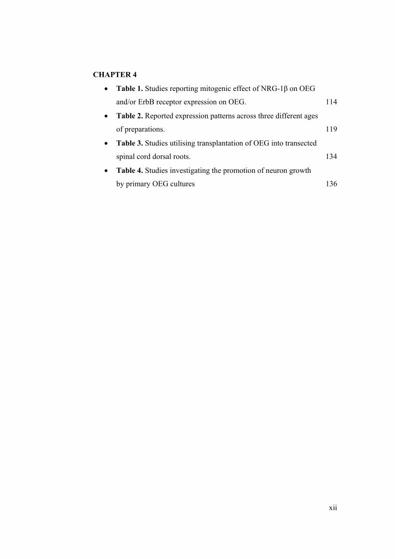
CHAPTER 4
• Table 1. Studies reporting mitogenic effect of NRG-1β on OEG
and/or ErbB receptor expression on OEG. 114
• Table 2. Reported expression patterns across three different ages
of preparations. 119
• Table 3. Studies utilising transplantation of OEG into transected
spinal cord dorsal roots. 134
• Table 4. Studies investigating the promotion of neuron growth
by primary OEG cultures 136

xiii
List of Abbreviations
AEG Adult-derived ensheathing glia
ANOVA Analysis of variance
ARIA Acetylcholine receptor inducing activity
BDNF Brain derived neurotrophic factor
CMDM Chemically defined medium
CNP 2',3'-Cyclic nucleotide 3'-Phosphodiesterase
CNS Central nervous system
CRD-NRG-1 Cysteine-rich domain containing NRG-1
DF10S Medium containing serum without added mitogens
DF10S+mit Medium containing serum and added mitogens
DMEM Dulbecco's Modified Eagle's Medium
E-N-CAM embryonic neural cell adhesion molecule
GDNF Glial cell line-derived neurotrophic factor
DRG Dorsal root ganglion
EEG Embryonically-derived ensheathing glia
EGF Epidermal growth factor
FACS Fluorescence-activated cell sorting
FBS Fetal bovine serum
FGF Fibroblast growth factor
GFAP Glial fibrillary acidic protein
GFP Green fluorescent protein
HBSS Hank's Buffered Saline Solution

xiv
HRP Horseradish peroxidase
IGF Insulin growth factor
IL Interleukin
MAG Myelin-associated glycoprotein
MBP Myelin basic protein
N-CAM Neural cell adhesion molecule
NGF Nerve growth factor
NRG Neuregulin
NT Neurotrophin
OEG Olfactory ensheathing glia
P0 Protein zero
p75 p75 low affinity neurotrophin receptor
PBS Phosphate buffered saline
PDGF Platelet-derived growth factor
PEG Postnatally-derived ensheathing glia
PBS Phosphate buffered saline
PNS Peripheral nervous system
RT-PCR Reverse transcriptase polymerase chain reaction
SDS-PAGE Sodium dodecyl sulphate polyacrylamide gel electrophoresis
SMDF Sensory and motor neuron-derived factor

xv
Acknowledgements
Firstly, I have to express my immeasurable thanks and gratitude to both my
supervisors: Dr. Giles Plant and A. Prof. Sarah Dunlop. Without their
tremendous encouragement, support, advice and friendship, this work would
simply not have been completed. They have always been there no matter how
busy they were, have always demonstrated a willingness to get personally
involved, and have never stopped believing in me. I owe everything to them.
I would also like to extend my special thanks to the following people:
Dr. Marc Ruitenberg – for invaluable assistance with the long hours of
surgery, for assistance with proofreading Chapters 2 and 3 of this thesis, and
for always being there to lighten up the mood. The thoughts of rat soup will
haunt me for the rest of my days.
Mrs Margaret Pollett – for assistance with extraction and purification of all
Schwann cells utilised in this study.
Dr. Michael Archer – for his invaluable help with the electron microscopy. I
don't think I will ever meet someone as proficient with the transmission
electron microscope. This project would have taken me another three years
were it not for him.
Mr. Guy Ben Ary – for all the assistance with my time-lapse microscopy
work, even though it never made the final cut for this thesis.
Mss. Natalie Simmons – for always taking the time to show me how to do any
little technique I required.

xvi
Mrs. Christin Christensen – for showing me the ropes during the earlier stages
of my project, and for the many philosophical conversations we shared.
Dr. William Hendricks – for taking the time and effort to generate the
lentivirus used in Chapter 3 of this study.
Dr. Stuart Hodgetts – for assistance in proofreading of Chapter 2 of this thesis.
Dr. Helen Barbour, Mss Jana Vukovic, Mss Seok Von Lee and Mss Ajanthy
Arulpragasam – for sharing this long journey with me. It was pure madness.
Dr. Alan Harvey – for assistance with proofreading Chapter 3 of this thesis,
and for being a source of inspiration at different stages of my project. The full
implications of everything he says take several days to sink in. I'm still
absorbing a lot of it.
Dr. Michael Guppy and Dr. Peter Arthur – for giving me the chance to
demonstrate undergraduate laboratory classes. Those years have been a most
memorable and enjoyable experience.
And finally, I would like to thank all of the multitude of people who have
helped me get by along the way. There are too many of you to mention here,
but you know who you are, and you know I will never forget you. Thank you.

CHAPTER 1
Introduction

Chapter 1 - Introduction
2
The Olfactory System
The olfactory mucosa is a tissue derived from the olfactory placodes of the
central nervous system (CNS), but that functions and resides in the peripheral
nervous system (PNS) (Doucette, 1989). It is composed of an olfactory
epithelium, an underlying lamina propria and a basal lamina separating the
two components (Graziadei, 1973). Olfactory receptor neuron perikarya
reside within the olfactory epithelium, with a basal axonal projection that
crosses the epithelium en route to the lamina propria (Graziadei, 1973;
Doucette, 1990). These axons join with other olfactory receptor axons
forming peripheral olfactory fascicles that cross the cribiform plate to enter
the olfactory bulb within the central nervous system (CNS) (Doucette, 1990).
Once inside the olfactory bulb, olfactory receptor axons converge onto a
number of units called glomeruli, where they synapse with mitral and tufted
cells, and periglomerular interneurons (Barber, 1981, 1982; Marin-Padilla and
Amieva, 1989; Valverde and Lopez-Mascaraque, 1991). The olfactory bulb
itself is a laminated structure comprised of the olfactory nerve fiber layer, the
glomerular layer, the external plexiform layer, the mitral cell layer, the
internal plexiform layer and the granule cell layer (Doucette, 1990; Shepherd
and Greer, 1998).

Chapter 1 - Introduction
3
Olfactory Ensheathing Glia
An important feature of the olfactory system lies in the ability of olfactory
receptor neurons to be continuously replaced throughout the lifetime in adult
mammals (Mackay-Sim and Kittel, 1991; Carr and Farbman, 1992; Graziadei
et al., 1978; Graziadei and Monti-Graziadei, 1978, 1979; Wilson and
Raisman, 1980; Murrell et al., 1996). The replacement of olfactory neurons
originates from basal cells in the olfactory neuroepithelium whose axons
elongate through the cribiform plate to reach their glomerular targets deep in
the CNS olfactory bulb (Barber and Raisman, 1978; Graziadei and Monti
Graziadei, 1979; Barber, 1981, 1982; Costanzo and Graziadei, 1983;
Doucette, 1984; Calof and Chikaraishi, 1989; Marin-Padilla & Amieva, 1989;
Mackay-Sim and Kittel, 1991). The ability of the olfactory system to
regenerate itself has been associated with the permissive environment created
by nearby olfactory ensheathing glia (OEG). OEG are unique to the olfactory
system and continuously accompany growing axons from their origin in the
olfactory neural epithelium to their targets in the olfactory glomeruli (Blanes,
1898; Doucette, 1984, 1991; Raisman, 1985; Marin-Padilla & Amieva, 1989).
The continuous accompaniment of olfactory neurons by OEG begins during
development when OEG pioneer the olfactory nerve pathway, extending
ahead of the growing neurons and facilitating both initial axon growth and
their subsequent elongation (Farbman and Squinto, 1985; Doucette, 1989;
Marin-Padilla and Amieva, 1989; Tennent and Chuah, 1996). This
ensheathment continues in the adult, where OEG completely envelop large

Chapter 1 - Introduction
4
bundles of tightly packed olfactory receptor axons, sending segregating
processes into the unmyelinated bundles and accompanying them through the
PNS-CNS transitional zone into the CNS olfactory bulb (Raisman, 1985;
Doucette, 1991; Valverde and Lopez-Mascaraque, 1991; Field et al., 2003;
Herrera et al., 2005).
Properties Of Olfactory Ensheathing Glia
At first, OEG were thought to be an intermediate glial cell type possessing
characteristics of both Schwann cells (glial cells of the PNS) and astrocytes
(glial cells of the CNS) (for review see Ramon-Cueto and Valverde, 1995).
However, unlike astrocytes which are neural tube derivatives or Schwann
cells that are derived from the neural crest, OEG are derived from the
olfactory placodes (Doucette, 1989; Chuah and Au, 1991; Norgren et al.,
1992). Further differences between OEG and Schwann cells are apparent,
including their ability to participate in the formation of the olfactory bulb glia
limitans (Doucette, 1991, 1993a), their ability to ensheathe hundreds to
thousands of unmyelinated olfactory sensory axons (Doucette, 1984; Raisman,
1985; Field et al., 2003; Herrera et al., 2005), and their ability to support
regrowth of olfactory receptor neurons both throughout life and after
extensive damage to the sensory nerves or epithelium (Barber and Raisman,
1978; Graziadei and Monti Graziadei, 1978, 1979, 1980; Williams et al.,
2004; Li et al., 2005). Further to this, recent work has demonstrated that OEG
Chapter 1 - Introduction
5
possess a transcriptional profile that is different to either Schwann cells or
astrocytes (Vincent et al., 2005; Ruitenberg et al., 2005b In Press).
Over the years, cultured OEG have been characterized from embryonic
(Doucette, 1993b), neonatal (Pixley, 1992; Barnett et al., 1993; Chuah and
Au, 1993) and adult (Ramon-Cueto and Nieto-Sampedro, 1992; Goodman et
al., 1993) rodent olfactory tissues. Despite potential developmental
differences between these various preparations (Chapter 4), a clear picture of
OEG expression profiles has emerged. They share a number of phenotypic
markers with other glial cell types, requiring that OEG identification be
performed through detection for a number of different proteins. These
include: glial fibrillary acidic protein (GFAP) (Barber and Lindsay, 1982;
Pixley, 1992; Ramon-Cueto and Nieto-Sampedro, 1992; Chuah and Au, 1993;
Doucette, 1993b; Sonigra et al., 1999), the low affinity neurotrophin receptor
p75 (Pixley, 1992; Ramon-Cueto and Nieto-Sampedro, 1992; Barnett et al.,
1993; Goodman et al., 1993; Sonigra et al., 1999), S100 (Pixley, 1992;
Doucette, 1993b; Doucette and Devon, 1995), calponin (Boyd et al., 2006),
and nestin (Sonigra et al., 1999).
A number of adhesion molecules are also produced by OEG including N-
cadherin (Chuah and Au, 1994; Sonigra et al., 1999; Lakatos et al., 2000;
Fairless et al., 2005), L1 (Miragall et al., 1988; Ramon-Cueto and Nieto-
Sampedro, 1992; Barnett et al., 1993), fibronectin (Ramon-Cueto and Nieto-
Chapter 1 - Introduction
6
Sampedro, 1992), laminin (Liesi, 1985b; Ramon-Cueto and Nieto-Sampedro,
1992; Sonigra et al., 1999), neural cell adhesion molecule (NCAM) (Chuah
and Au, 1993; Sonigra et al., 1999) and polysialic acid embryonic N-CAM (E-
N-CAM) (Franceschini and Barnett, 1996; Sonigra et al., 1999). Of these,
laminin, NCAM and E-N-CAM are of particular importance as they are
important promoters of neurite initiation, axonal elongation and growth cone
attachment and growth (Liesi, 1985a; Madison et al., 1985; Bixby et al., 1988;
Zhang et al., 1995).
Finally, a number of neurite growth promoting factors have also been found to
be expressed by OEG in vitro including nerve growth factor (NGF) (Boruch et
al., 2001; Woodhall et al., 2001; Lipson et al., 2003; Vincent et al., 2003; Liu
et al., 2005), brain derived neurotrophic factor (BDNF) (Boruch et al., 2001;
Woodhall et al., 2001; Lipson et al., 2003; Ruitenberg et al., 2003; Vincent et
al., 2003; Byrnes et al., 2005; Liu et al., 2005), neurotrophin (NT)-4/5
(Boruch et al., 2001; Vincent et al., 2003), glial cell line-derived neurotrophic
factor (GDNF) (Woodhall et al., 2001; Lipson et al., 2003), vascular
endothelial growth factor (VEGF) (Au and Roskams, 2003) and neurturin
(Woodhall et al., 2001; Lipson et al., 2003). However, some contradictions
still stand on the reported expression of ciliary neurotrophic factor (CNTF)
and NT-3 (Boruch et al., 2001; Wewetzer et al., 2001; Lipson et al., 2003;
Ruitenberg et al., 2003; Liu et al., 2005).
Chapter 1 - Introduction
7
Attempts To Repair Damaged CNS
Adult CNS neurons undergo an abortive attempt to regenerate following
injury which is likely to be due at least in part to the non-permissive nature of
the glial environment surrounding regenerating axons (Reier et al., 1983;
Fishman & Kelley, 1984; Bovolenta et al., 1992). A great number of
strategies have been utilised by researchers to try and overcome the inhibition
of the lesioned CNS environment and promote axonal regrowth. Included
amongst these are: neutralization of endogenous inhibitory environmental
signals (Schnell and Schwab, 1990; Bregman et al., 1995; Brosamle et al.,
2000; Bradbury et al., 2002; GrandPre et al., 2002; Li and Strittmatter, 2003),
boosting the intrinsic regeneration capacity of neurons by manipulation of
intracellular pathways (Cai et al., 1999; Dergham et al., 2002; Neumann et al.,
2002; Qiu et al., 2002), injection of axonal growth promoting neurotrophic
factors (Schnell et al., 1994; Kobayashi et al., 1997; Ye and Houle, 1997), the
use of extracellular matrix molecules and biopolymers as bridging structures
(Goldsmith and de la Torre, 1992; Novikova et al., 2003; Woerly et al., 2004),
peripheral nerve or embryonic tissue grafts (Richardson et al., 1980, 1982,
1984; David and Aguayo, 1981; Benfey and Aguayo, 1982; Cheng et al.,
1996; Guntinas-Lichius et al., 2002) and cellular transplantation either alone
(Wrathall et al., 1984; Kromer and Cornbrooks, 1985; Kuhlengel et al., 1990;
Li and Raisman, 1994; Martin et al., 1996; Rabchevsky and Streit, 1997; Xu
et al., 1997; Rapalino et al., 1998; McDonald et al., 1999; Pizzo et al., 2004;
Cummings et al., 2005) or in combination with other treatments (Guth et al.,

Chapter 1 - Introduction
8
1994; Xu et al., 1995; Chen et al., 1996; Bregman et al., 1997, 2002; Menei et
al., 1998; Ramon-Cueto et al., 1998; Weidner et al., 1999; Mu et al., 2000;
Bamber et al., 2001; Coumans et al., 2001; Novikova et al., 2002; Pearse et
al., 2002, 2004; Blesch and Tuszynski, 2003; Ruitenberg et al., 2003, 2005a;
Chau et al., 2004; Lu et al., 2004; Nikulina et al., 2004; Fouad et al. 2005).
There has been a great amount of data collected suggesting that OEG are
capable of promoting functional and anatomical recovery of lesioned CNS. In
vitro, OEG monolayers have been shown to promote growth of olfactory
neurites (Ramon-Cueto et al., 1993; Chuah and Au, 1994; Kafitz and Greer,
1998, 1999; Tisay and Key, 1999), retinal ganglion cells (Goodman et al.,
1993; Sonigra et al., 1999; Moreno-Flores et al., 2003; Kumar et al., 2005;
Leaver et al., 2006), embryonic sympathetic neurons and Remak's ganglia
(Lipson et al., 2003), granule cell neurons (Van Den Pol and Santarelli, 2003),
dopaminergic neurons (Denis-Donini and Estenoz, 1998; Agrawal et al.,
2004), cortical neurons (Le Roux and Reh, 1994; Chung et al., 2004) and
dorsal root ganglion neurons (Gudino-Cabrera and Nieto-Sampedro, 2000;
Gomez et al., 2003; Ruitenberg et al., 2003). These OEG growth-promoting
abilities have been associated with both membrane-bound factors and with
diffusible factors in vitro (Le Roux and Reh, 1994; Kafitz and Greer, 1998,
1999; Chung et al., 2004) and in vivo (Chuah et al., 2004), though other
studies have reported that only membrane-bound factors and not diffusible

Chapter 1 - Introduction
9
factors are at work in vitro (Chuah and Au, 1994; Sonigra et al., 1999; Lipson
et al., 2003).
In vivo, OEG have been reported to restore function and induce regrowth of
fibers in a variety of models. Transplantation of OEG into the partially
transected, crushed, photochemically lesioned or focally lesioned spinal cord
have been reported to induce sprouting, long distance regrowth of axons
through and beyond the lesion site, to promote tissue and neuronal sparing,
increase angiogenesis, decrease scar formation and/or to improve functional
recovery (Li et al., 1997, 1998, 2003a; Imaizumi et al., 2000b; Verdu et al
2001, 2003; Nash et al., 2002; Shen et al., 2002; Keyvan-Fouladi et al., 2003;
Andrews and Stelzner, 2004; Chuah et al., 2004; Garcia-Alias et al., 2004;
Lopez-Vales et al., 2004, 2006; Polentes et al., 2004; Ramer et al., 2004a;
Richter et al., 2005; Ruitenberg et al., 2003, 2005a; Sasaki et al., 2006).
Transplantation of OEG into dorsal rhizotomy models has resulted in
regrowth of fibers and restitution of spinal reflex arcs (Ramon-Cueto and
Nieto-Sampedro, 1994; Navarro et al., 1999; Taylor et al., 2001; Pascual et
al., 2002; Li et al., 2004), though other studies indicate OEG do not promote
improvement in this model (Gomez et al., 2003; Ramer et al., 2004a; Riddell
et al., 2004). Encouraging results have also been reported in the fimbria-
fornix pathway (Smale et al., 1996), the nigrostriatal dopaminergic pathway
(Agrawal et al., 2004; Johansson et al., 2005), and in the optic (Li et al.,

Chapter 1 - Introduction
10
2003b) and facial nerves (Guntinas-Lichius et al., 2001; Choi and Raisman,
2005).
Contusion models of injury provide a more complicated picture of OEG’s
ability to stimulate regeneration. Whereas some studies have reported that
OEG reduce cavity formation, promote tissue sparing, improve functional
outcomes in task-based tests, and induce sparing/regrowth of fibers across the
length of the lesion site (Plant et al., 2003; Ruitenberg et al., 2005a; Sun et al.,
2005), others have reported no significant effect by OEG on either restoration
of function or regrowth of fibers (Takami et al., 2002; Barakat et al., 2005;
Collazos-Castro et al., 2005; Resnick et al., 2003). Finally, transplantation of
OEG into completely transected spinal cord have demonstrated an increase of
motor potentials, an improvement in functional tasks and responsiveness to
proprioceptive stimuli, and an increase in ascending sensory, corticospinal,
raphespinal and/or coerulospinal fibers crossing into and through the lesion
site (Ramon-Cueto et al., 1998, 2000; Lu et al., 2001, 2002; Cao et al., 2004;
Fouad et al., 2005; Lopez-Vales et al., 2006), though at least one study has
failed to corroborate such findings (Lee et al., 2004).
OEG are also able to integrate very well with the CNS microenvironment. In
vitro, OEG intermix well with astrocytes, whereas Schwann cells form distinct
territories that do not pass over areas where astrocytes are located (Lakatos et
al., 2000; Van Den Pol and Santarelli, 2003; Fairless et al., 2005). OEG also

Chapter 1 - Introduction
11
induce a lower degree of astrocyte activation than Schwann cells in vitro and
in vivo, as measured by expression of neuronal growth inhibitory chondroitin
sulphate proteoglycans and GFAP by astrocytes (Lakatos et al., 2000; Lakatos
et al., 2003a), and are able to align themselves with the unlesioned host CNS
environment (Perez-Bouza et al., 1998).
OEG Mitogens
Several clinical trials are already underway utilising OEG to assist in the
repair of human spinal cord injury (Huang et al., 2003; Rabinovich et al.,
2003; Feron et al., 2005). The possibility of utilising autologous transplants
of OEG is especially attractive, given that OEG derived from the same patient
undergoing therapy can forgo complications related to rejection of
transplanted tissue. However, critical to the successful application of these
techniques in vivo, is the ability to quickly and efficiently proliferate large
numbers of OEG in vitro.
Several mitogens for OEG have already been identified. A strong
proliferative effect of OEG has been attributed to neuregulin (NRG)-1β
(Pollock et al., 1999; Chuah et al., 2000; Yan et al., 2001a, b),
lysophosphatidic acid (Yan et al., 2003), FGF-2 (Pollock et al., 1999; Yan et
al., 2001a, 2003), bFGF (Chuah and Teague, 1999; Au and Roskams, 2003),

Chapter 1 - Introduction
12
NT-3 (Bianco et al., 2004), hepatocyte growth factor (Yan et al., 2001b),
platelet-derived growth factor (PDGF)-BB (Pollock et al., 1999; Yan et al.,
2001a, 2003), and insulin growth factor (IGF)-1 (Yan et al., 2001a), with
small mitogenic effects by NGF (Chuah and Teague, 1999; Pollock et al.,
1999; Bianco et al., 2004) and BDNF (Bianco et al., 2004). NRG-1β is of
particular interest, given that several research groups utilise this factor to
purify and/or expand their cells in vitro (see Appendix A for summary).
Furthermore, NRG-1β is intrinsic to the development and maturation of
Schwann cells, including an important role in the synthesis of the myelin
sheath (Anderson, 1993; Shah et al., 1994; Dong et al., 1995; Shah and
Anderson, 1997; Michailov et al., 2004).
The Role Of Neuregulins
Neuregulins are a set of alternatively spliced growth factors that are
structurally related to the epidermal growth factor family of proteins
(Marchionni et al., 1993; reviewed by Ben-Baruch and Yarden, 1994;
reviewed by Lemke, 1996). The first member of this family, glial growth
factor, was identified based on its potent mitogenic effects on cultured
Schwann cells and astrocytes (Raff et al., 1978; Brockes et al., 1980a; Lemke
and Brockes, 1984; Goodearl et al., 1993; Marchionni et al., 1993).
Subsequent to this initial discovery, a number of similar molecules were found

Chapter 1 - Introduction
13
under the labels of neu differentiation factor (Peles et al., 1992; Wen et al.,
1992), heregulin (Holmes et al., 1992), acetylcholine receptor inducing
activity (ARIA) (Falls et al., 1990, 1993), sensory and motor neuron-derived
factor (SMDF) (Ho et al., 1995) and cysteine-rich domain containing NRG-1
(CRD-NRG-1 ) isoforms (Yang et al., 1998).
In order to properly label the vast number of protein isoforms controlled by
different promoters and alternative splicing (Holmes et al., 1992; Wen et al.,
1992; Falls et al., 1993; Marchionni et al., 1993; Ho et al., 1995), the entire
group was categorized under the term of neuregulins (NRG) (Marchionni et
al., 1993). Later, the neuregulins were subdivided into NRG-1 Type I (neu
differentiation factor, heregulin, acetylcholine receptor inducing activity),
NRG-1 Type II (glial growth factor) and NRG-1 Type III (SMDF, CRD-
NRG-1) (reviewed by Adlkofer and Lai, 1999). Type I NRG is the only type
expressed in early embryonic stages and has widespread tissue distribution
(Corfas et al., 1995; Burden and Yarden, 1997). Type II NRG is expressed
primarily in the spinal cord, the dorsal root ganglia and in the brain late during
embryonic development (Marchionni et al., 1993). Type III NRG are
expressed in the brain, and produced by sympathetic, motor and sensory
neurons (Ho et al., 1995; Burden and Yarden, 1997; Yang et al,. 1998;
reviewed by Garratt et al., 2000). Further variations in the bioactive
epidermal growth factor (EGF) domain of these molecules allows
categorization of neuregulins into either -α or -β types (Holmes et al., 1992;

Chapter 1 - Introduction
14
Marchionni et al., 1993; Wen et al., 1994; reviewed by Lemke, 1996). Thus
far, four different neuregulin genes have been identified, though only
neuregulin-1 has been studied in detail (reviewed by Adlkofer and Lai, 1999).
NRG-1β plays an important role during early development. Several studies
have shown that NRG-1β biases the differentiation of migrating neural crest
cells toward the Schwann cell lineage, by blocking differentiation into the
alternative neuronal lineage (Anderson, 1993; Shah et al., 1994; Shah and
Anderson, 1997). Later in development, NRG-1β promotes the survival and
differentiation of Schwann cell precursors in vitro (Dong et al., 1995), the
survival of premyelinating cells in vivo (Grinspan et al., 1996) and the
survival of mature Schwann cells following axonal transection in vivo
(Trachtenberg and Thompson, 1996; Carroll et al., 1997). NRG-1β has also
been confirmed to be a potent mitogen for mature Schwann cells in vitro, and
to be capable of promoting survival of mature Schwann cells following serum
withdrawal in vitro (Goodearl et al., 1993; Levi et al., 1995; Morrissey et al.,
1995; Rutkowski et al., 1995; Syroid, et al., 1996; Dong et al., 1997; Kim et
al., 1997). Other studies report that NRG-1β can promote Schwann cell
migration in vitro (Mahanthappa et al., 1996; reviewed by Mirsky and Jessen,
1999), with possible roles in synaptogenesis, nerve fasciculation and in both
the establishment and maintenance of neuromuscular junctions in vivo (Corfas
et al., 1995; Sandrock et al., 1997; reviewed by Burden, 1998; Morris et al.,
1999; reviewed by Garratt et al., 2000; Lin et al., 2000; Wolpowitz et al.,

Chapter 1 - Introduction
15
2000). More recently, Michailov et al., (2004) demonstrated that NRG1
expression by neuronal axons regulates thickness of the myelin sheath.
Together, these observations all imply that NRG-1 may be a direct regulator
of differentiation, myelination, proliferation, survival and migration of
Schwann cells, and thus one of the central regulatory molecules in the
neurobiology of Schwann cells.
Less is known about other members of the NRG family. NRG-2 appears to
have similar receptor-binding specificities to NRG-1, but can stimulate
different signaling pathways (Carraway III et al., 1997, Crovello et al., 1998).
NRG-2 is detected in several neural tissues in the adult rat, and is primarily
concentrated in the cerebellum, hippocampus and olfactory bulb and to a
lesser extent in the cortex, thalamic nuclei and caudate-putamen (Busfield et
al., 1997; Carraway III et al., 1997; Chang et al., 1997; Longart et al., 2004).
It is also expressed at low levels in the spinal cord (Busfield et al., 1997;
Chang et al., 1997). Generally it is expressed in areas where NRG-1 is not
expressed (Carraway III et al., 1997). NRG-2 is expressed in motor neurons,
is concentrated at synaptic sites, and may regulate synaptic differentiation
(Rimer et al., 2004).
NRG-3 has been demonstrated to be highly expressed in most regions of the
human brain, with the exception of the corpus callosum (Zhang et al., 1997;
Longart et al., 2004). It is strongly expressed in the adult spinal cord and
Chapter 1 - Introduction
16
spinal ganglia, as well as several regions of the brain including the cerebral
and piriform cortex, the mitral and glomerular layers of the olfactory bulb, the
hippocampus, hypothalamus and thalamus (Zhang et al., 1997; Longart et al.,
2004). Interestingly, it is highly expressed in the cortical plate where
differentiating cells are located, but not in the ventricular and subventricular
zones of the telencephalon where migrating and proliferating cells are found
(Zhang et al., 1997).
Both soluble and membrane bound NRG transduce their signals by means of
cell surface receptor protein tyrosine kinases (reviewed by Lemke, 1996).
The three NRG receptors are known as ErbB2, ErbB3 and ErbB4 (of
molecular weights p185, p160 and p180 respectively) (Kraus et al., 1989;
Plowman et al., 1990, 1993; reviewed by Lemke, 1996). Like many other
receptor protein-tyrosine kinases, ErbB receptors are able to
transphosphorylate each other, and to catalyse the phosphorylation and
activation of downstream signal transduction cascades such as ras and MAP
kinase pathways (Carraway and Cantley, 1994; reviewed by van der Geer et
al., 1994; Kim et al, 1995, 1997; Levi et al., 1995). Interestingly, ErbB2 does
not possess any ligand binding sites, and requires heterodimerization with
other ErbB receptors to initiate intracellular signaling (Kita et al., 1995; Levi
et al., 1995; Grinspan et al., 1996; Syroid et al., 1996; Vartanian et al., 1997).
ErbB3 possesses strong ligand affinity, but impaired tyrosine kinase activity,
thus limiting its potential to initate intracellular signalling on its own
Chapter 1 - Introduction
17
(Carraway et al., 1994; Carraway and Cantley, 1994; Guy et al., 1994; Tzahar
et al., 1994). As such, ErbB2 and ErbB3 receptors require dimerization with
other ErbB receptor to initiate intracellular signaling (Sliwkowki et al., 1994;
Carraway and Burden, 1995; Kita et al., 1995; Levi et al., 1995; Pinkas-
Kramarski et al., 1996; Syroid et al., 1996; Jones et al., 1999). ErbB4
receptors possess both catalytic and ligand binding sites, and are able to hetero
and homo-dimerize (Plowman et al., 1993; Tzahar et al., 1994). All three
ErbB receptors have various binding affinities to the different neuregulin
isoforms (summarized in Figure 1). Together, these receptors provide crucial
signals during the development of neural crest cells and Schwann cells, and
are essential for survival during embryogenesis (Meyer and Birchmeier, 1995;
Erickson et al., 1997; Riethmacher et al., 1997; Britsch et al., 1998; Morris et
al., 1999; Woldeyesus et al., 1999). The main ErbB receptors in Schwann
cells are ErbB2 and ErbB3, but low amounts of ErbB4 are also detectable
(Levi et al., 1995; Grinspan et al., 1996; Syroid et al., 1996; Carroll et al.,
1997). OEG have been reported to express ErbB2 and ErbB4, though no
consensus has been reached on the expression of ErbB3 by these cells
(Pollock et al., 1999; Thompson et al., 2000; Moreno-Flores et al., 2003).
Although much is known of the influence of neuregulins on Schwann cells,
virtually nothing is known of their possible role in OEG differentiation.
However, prior to any further studies investigating a possible role of
neuregulins on OEG differentiation, the basic effects of these molecules on
Chapter 1 - Introduction
18
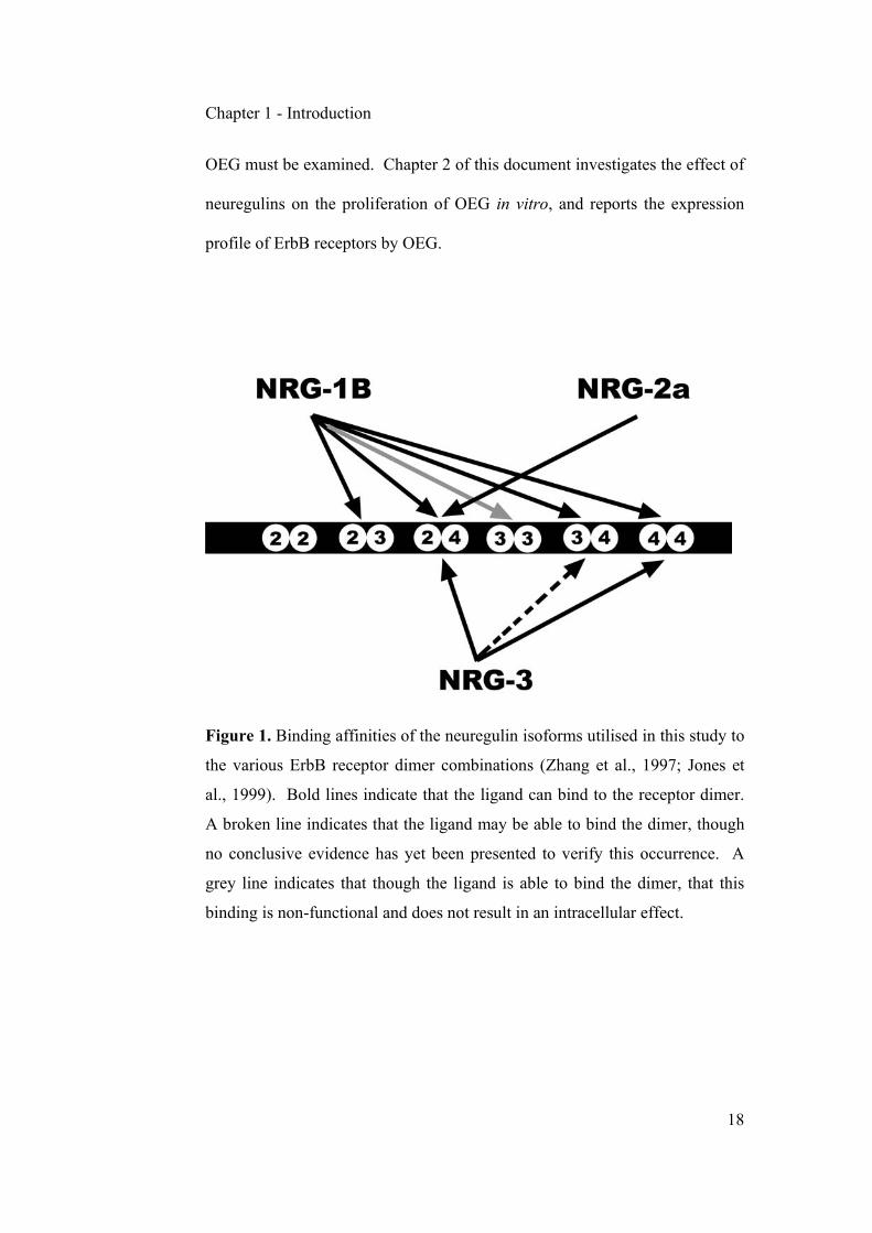
OEG must be examined. Chapter 2 of this document investigates the effect of
neuregulins on the proliferation of OEG in vitro, and reports the expression
profile of ErbB receptors by OEG.
Figure 1. Binding affinities of the neuregulin isoforms utilised in this study to
the various ErbB receptor dimer combinations (Zhang et al., 1997; Jones et
al., 1999). Bold lines indicate that the ligand can bind to the receptor dimer.
A broken line indicates that the ligand may be able to bind the dimer, though
no conclusive evidence has yet been presented to verify this occurrence. A
grey line indicates that though the ligand is able to bind the dimer, that this
binding is non-functional and does not result in an intracellular effect.

Chapter 1 - Introduction
19
The Myelin Sheath
The myelin sheath is composed of a differentiated portion of the plasma
membrane of glial cells. Schwann cells in the PNS, or oligodendrocytes in the
CNS, undergo specific changes during development that cause them to tightly
associate with nearby axons, concentrating large amounts of insulating
material around the axon and excluding as much cytosolic material from the
structure as possible. The principal role of the myelin sheath is to allow fast
saltatory conduction of nerve impulses along the axons it surrounds,
increasing the speed at which a nervous impulse is transmitted along an axon,
and effectively improving energy efficiency of conduction by a factor of 5000
fold. The correct and efficient functioning of both CNS and PNS require the
presence of compact myelin sheaths, and several diseases such as multiple
sclerosis, Charcot-Marie-Tooth disease, and Guillain-Barre syndrome are
associated with the loss of myelin (Garbay et al., 2000; Tzakos et al., 2005).
Furthermore, restoration of function following traumatic injury of the CNS is
dependant on restoration of saltatory conduction, and efficient remyelination
of demyelinated fibres.
Protein Content Of The Myelin Sheath
Proteins constitute 20-30% of the myelin sheath in the PNS. The most
important myelin proteins which pertain to this study include protein zero
(P0), myelin basic protein (MBP), myelin-associated glycoprotein (MAG) and

Chapter 1 - Introduction
20
2',3'-cyclic nucleotide 3'-phosphodiesterase (CNP). P0 is the major protein of
PNS myelin, constituting 50-70% of total myelin protein (Greenfield et al.,
1973; Wiggins et al., 1975). It is expressed by myelinating Schwann cells and
OEG in situ (Brockes et al., 1980b; Lemke, 1988; Martini et al., 1988; Lee et
al., 1997). P0 expression increases at postnatal day 5 in the PNS (Wiggins et
al., 1975; Lemke and Axel, 1985), and provides a good indicator to estimate
the onset of myelination (Peirano et al., 2000). It is also important for tight
compaction of the myelin sheath and for spacing of myelin lamellae at the
intraperiod lines (Filbin et al., 1990; Giese et al., 1992). MBP in turn
constitutes 5-15% of the PNS myelin proteins, and 30-40% of the CNS myelin
proteins (Lemke, 1988). Differential splicing of a single MBP mRNA
transcript creates a variety of MBP isoforms, whose expression is
developmentally regulated in a variety of tissues and that have a large number
of different functions in the biology of the cell (Harauz et al., 2004). Chief
amongst these functions is the involvement of MBP in the maintenance of the
major dense line of myelin and its participation with P0 in compaction of the
myelin sheath (Privat et al., 1979; Rosenbluth, 1980; Molineaux et al., 1986;
Readhead et al., 1987; Martini et al., 1995).
MAG accounts for approximately 1% of the total CNS myelin proteins, and
about 0.1% of the PNS myelin proteins (Quarles et al., 1972; Figlewicz et al.,
1981). Its function has been associated with regulation of intramembrane
spacing, signal transduction during glial cell differentiation, regulation of
Chapter 1 - Introduction
21
neurite outgrowth, in the maintenance of myelin integrity, and in the initial
stages of axonal adhesion and recognition (Johnson et al., 1989;
Mukhopadhyay et al., 1994; Fruttiger et al., 1995; Garbay et al., 2000).
Recently it has also received a lot of interest as one of the primary white
matter proteins that may be involved in inhibition of neurite growth following
CNS injury (McKerracher et al., 1994; Mukhopadhyay et al., 1994; Tang et
al., 1997; Filbin, 2003; Quarles, 2005). Finally, CNP is an early
oligodendroglial/Schwann cell marker, involved but not necessary for the
ensheathment step prior to myelin compaction (Braun et al., 1988; Reynolds
and Wilkin, 1988; Sprinkle, 1989). It constitutes less than 0.5% of the total
myelin protein in the PNS (Agrawal et al., 1990), and approximately 4% of
the total myelin protein in the CNS (Lemke , 1988). In the PNS it has been
found to be associated with the plasma membranes of cultured Schwann cells
(Yoshino et al., 1985) whereas in the CNS it is located in the cytoplasm and
paranodal loops of non-compacted oligodendrocytes (Vogel and Thompson,
1988). It has been suggested that CNP is involved in oligodendrocyte
membrane expansion and in assisting MBP to compact myelin (Yin et al.,
1997).
In the CNS, myelin genes are expressed by oligodendrocytes in a pattern that
is parallel to their differentiation state, and appear to be unaffected by the
presence or absence of neurons (Lemke, 1988). In the PNS however, axonal
contact appears to be essential for expression of myelin genes and myelination

Chapter 1 - Introduction
22
by Schwann cells (Aguayo et al., 1976; Bunge et al., 1982; Jessen and Mirsky,
1992).
Myelination By OEG
Controversy remains as to the ability of OEG to myelinate under various
experimental conditions. The first instance of OEG myelination was
demonstrated in vitro (Devon and Doucette, 1992). The researchers utilized
an unpurified preparation of embryonically-derived ensheathing glia (EEG)
(ie. a dissociated olfactory bulb culture) and co-cultured these with a
population of embryonic dorsal root ganglion neurons. Electron micrographic
and immunocytochemical evidence indicated that myelination was taking
place within this co-culture system, but only when the co-cultures were
exposed to serum in the culture medium (Devon and Doucette, 1992, 1995).
Interestingly, the authors described the myelinating cells in their system as
being indistinguishable to Schwann cells in their myelination characteristics,
raising the possibility that contaminating Schwann cells could have been at
least partially responsible for the observed results. Later, another group
repeated this experiment utilizing adult-derived ensheathing glia (AEG)
purified by immunopanning for the p75-low affinity neurotrophin receptor
(Plant et al., 2002). Unlike the original study utilizing EEG (Devon and
Doucette, 1992), these authors were unable to identify any myelination by
OEG in vitro.

Chapter 1 - Introduction
23
Numerous other studies have also investigated myelination by OEG in vivo
(Franklin et al., 1996; Li et al., 1997, 1998; Imaizumi et al., 1998, 2000a,b;
Barnett et al., 2000; Kato et al., 2000; Smith et al., 2001, 2002; Takami et al.,
2002; Lakatos et al., 2003b; Boyd et al., 2004a; Dunning et al., 2004; Radtke
et al., 2003; Sasaki et al., 2004). Most of these have reported an increased
level of myelination in the spinal cord following transplantation of OEG.
Unfortunately, very few of these studies have adequately pre-labeled the cells
prior to transplantation, making positive confirmation of OEG myelination
impossible. Recently, two studies have obtained satisfactory pre-labelling of
OEG by means of retroviral infection (Boyd et al., 2004a) or by using cells
extracted from transgenic green fluorescent protein (GFP) rats (Sasaki et al.,
2004). Boyd et al., (2004a) utilized an unpurified population of EEG, and
failed to report myelination by these cells after transplantation into crushed
spinal cord. Rather, they suggested that the observed increases in myelin
levels are due to myelination by invading Schwann cells, not OEG.
Meanwhile, Sasaki et al., (2004) have reported contrasting data – that
transplanted AEG can myelinate the lesioned spinal cord in vivo. Boyd et al.,
(2004a, b) suggest that contaminating GFP positive Schwann cells in the
preparations of Sasaki et al., (2004) are likely to account for these findings,
though they provide no explanation as to how a CNS based preparation would
become heavily contaminated with peripherally derived Schwann cells.
Sasaki et al., (2004) in turn question the efficiency of transfection and stability
of the retroviral label utilized by Boyd et al., (2004a). Both, however, agree

Chapter 1 - Introduction
24
that differences in purification procedures and the age of animal from which
the OEG preparation was extracted may account for these variations. Other
sources of variations include the type of lesion and time after transplantation,
which could also potentially account for some of these contrasting
observations.
It has been well established that Schwann cells are able to spontaneously
infiltrate the CNS and remyelinate axons after spinal cord injury (Hughes and
Brownell 1963; Blakemore, 1977; Sims and Gilmore, 1983; Beattie et al.,
1997; Brook et al., 1998, 2000). Several researchers have proposed that
transplanted OEG are able to increase recruitment of Schwann cells into the
damaged spinal cord, and that potentially most, if not all, of the reported
myelination by OEG may in fact be due to Schwann cell infiltration (Boyd et
al., 2004a, b; Ramer et al., 2004a; Richter et al., 2005). This suggestion is
supported by previous observations that Schwann cell recruitment and/or
proliferation into the injury site can be potentiated by administration of
neurotrophic factors (Namiki et al., 2000; Ruitenberg et al., 2005a), and that
OEG in turn are capable of releasing several such factors (Boruch et al., 2001;
Woodhall et al., 2001; Lipson et al., 2003; Vincent et al., 2003; Byrnes et al.,
2005; Liu et al., 2005). As such, the question on whether OEG are capable of
myelinating CNS neurons remains open for investigation.
The myelination potential of OEG extracted from animals of different ages is
examined in Chapter 3.

Chapter 1 - Introduction
25
Summary
Described in the next two chapters are two separate studies into the biology of
p75-selected OEG. The ability to proliferate OEG quickly is crucial to
successful use of these cells clinically. Though several factors that are
mitogenic for OEG have been identified, no consensus has been reached on
the optimal means of expanding these cells in vitro (Appendix A). NRG-1β
has been identified as a potent mitogen for OEG in vitro, but no studies have
investigated the effects of NRG-2 and NRG-3 on OEG proliferation.
Furthermore, very little is known about the expression and activation of ErbB
receptors in p75-selected adult OEG. In Chapter 2, we investigate the role of
NRG-1β, NRG-2 and NRG-3 on the proliferation of OEG in vitro, we
document the expression of ErbB receptors on these cells, and conduct
functional blocking studies to determine the contribution of ErbB receptor
subtypes on the proliferation of OEG.
In Chapter 3, we seek to answer some contradictory observations in the field
of OEG myelination. By utilising p75-selected OEG derived from embryonic,
postnatal and adult animals, we seek to explain why some studies report
myelination by OEG whereas others fail to do so. This study investigates the
ability of OEG to myelinate dorsal root ganglion neurons of various calibres
in vitro, and their ability to myelinate lysolecithin demyelinated spinal cord
axons in vivo.

CHAPTER 2

Chapter 2 – Responsiveness of OEG to neuregulins
27
CULTURE CONDITIONS AFFECT PROLIFERATIVE
RESPONSIVENESS OF OLFACTORY ENSHEATHING GLIA TO
NEUREGULINS
T.R. de Mello1,2; S. Busfield3; S.A. Dunlop2,3; G.W. Plant1,3 *
1. Red's Spinal Cord Research Laboratory - School of Anatomy & Human
Biology, 2. School of Animal Biology, 3. The Western Australian Institute for
Medical Research (WAIMR), The University of Western Australia, Perth,
Australia
* Corresponding author: Dr. Giles Plant (email: [email protected])
Abstract
Olfactory ensheathing glia (OEG) have been used to improve outcome after
experimental spinal cord injury and are being trialed clinically. Their rapid
proliferation in vitro is essential to optimize clinical application, with
neuregulins (NRG) being potential mitogens. We examined the effects of
NRG-1β, NRG-2α and NRG3 on proliferation of p75-immunopurified adult
OEG. OEG were grown in serum-containing medium with added bovine
pituitary extract and forskolin (added mitogens) or in serum-containing
medium (no added mitogens). Cultures were switched to chemically defined
medium (no added mitogens or serum), NRG added and OEG proliferation
assayed using BrdU. OEG grown initially with added mitogens were not

Chapter 2 – Responsiveness of OEG to neuregulins
28
responsive to added NRGs and pre-exposure to forskolin and pituitary extract
increased basal proliferation rates so that OEG no longer responded to added
NRG. However, NRG promoted proliferation if cells were initially grown in
mitogen-free medium. Primary OEG express ErbB2, ErbB3, and small levels
of ErbB4 receptors; functional blocking indicates that ErbB2 and ErbB3 are
the main NRG receptors utilized in the presence of NRG-1β. The long-term
stimulation of OEG proliferation by initial culture conditions raises the
possibility of manipulating OEG before therapeutic transplantation.
Introduction
Olfactory Ensheathing Glia (OEG) are specialized glial cells of the olfactory
pathway, a region of the CNS that is capable of supporting neuronal
replacement throughout adult life (Graziadei and Monti-Graziadei, 1978,
1979). Neuronal replacement in the olfactory system has been associated with
the permissive environment created by OEG, which chaperone newly growing
axons from their origin in the olfactory neural epithelium to their targets in the
olfactory glomeruli (Doucette, 1984, 1990; Raisman, 1985; Marin-Padilla &
Amieva, 1989). Several studies have suggested that the ability of OEG to
promote neuronal replacement lies at least in part with their similarities to
Schwann cells, the glial cells of the peripheral nervous system (Ramon-Cueto
and Nieto-Sampedro, 1992; Doucette, 1995). More recently, the therapeutic

Chapter 2 – Responsiveness of OEG to neuregulins
29
potential of OEG has come to the fore, with a number of studies reporting that
OEG transplanted into damaged areas of the CNS can improve axonal sparing,
promote regrowth of damaged fibers and most importantly improve functional
recovery (for review see Santos-Benito and Ramon-Cueto, 2003; Mackay-
Sim, 2005).
Crucial to any practical application of OEG in a clinical setting is the ability to
expand these cells rapidly and reproducibly in vitro to produce sufficient
numbers for transplantation. A number of mitogens for OEG have been
identified to date, including FGF-2, bFGF, PDGF-BB, hepatocyte growth
factor, lysophosphatidic acid, BDNF, NGF, heregulin β1, glial growth factor 2
(Chuah and Teague, 1999; Pollock et al., 1999; Chua et al., 2000; Yan et al.,
2001a,b, 2003; Alexander et al., 2002; Au and Roskams, 2003; Bianco et al.,
2004). Of these, the latter two are of special interest considering their role as
members of the neuregulin super-family.
Neuregulins (NRG) are a set of growth factors whose protein isoforms are
controlled by different promoters and alternative splicing (Holmes et al.,
1992; Wen et al., 1992; Falls et al., 1993; Marchionni et al., 1993; Ho et al.,
1995). Four different neuregulin genes have been identified, although only
neuregulin-1 has been studied in detail (reviewed by Adlkofer and Lai, 1999).
NRG-1β plays an important role during early development, shifting the
differentiation of migrating neural crest cells toward the Schwann cell lineage

Chapter 2 – Responsiveness of OEG to neuregulins
30
by blocking differentiation into the alternative neuronal lineage (Anderson,
1993; Shah et al., 1994; Shah and Anderson, 1997). Later in development,
NRG-1β promotes the survival and differentiation of Schwann cell precursors
in vitro (Dong et al., 1995) as well as the survival of premyelinating cells
(Grinspan et al., 1996) and mature Schwann cells in vivo following axonal
transection (Trachtenberg and Thompson, 1996; Carroll et al., 1997). NRG-
1β has also been confirmed to be a potent mitogen for mature Schwann cells
in vitro, and to be capable of promoting their survival following serum
withdrawal in vitro (Levi et al., 1995; Morrissey et al., 1995; Rutkowski et al.,
1995; Syroid, et al., 1996; Dong et al., 1997; Kim et al., 1997).
NRG-2 expression appears to be highest in areas of the CNS where cells are
continually replaced, namely the cerebellum, hippocampus and olfactory bulb,
and is found to a lesser extent in regions of minimal cell turnover such as the
cortex, thalamic nuclei, caudate-putamen and spinal cord (Busfield et al.,
1997; Carraway III et al., 1997; Chang et al., 1997; Longart et al., 2004).
NRG-2 is also expressed by motor neurons and terminally differentiated
Schwann cells, possibly playing a role in the regulation of synaptic
differentiation (Rimer et al., 2004). With the exception of the corpus
callosum, NRG-3 is widely expressed throughout most regions of the adult
CNS including the mitral and glomerular layers of the olfactory bulb (Zhang
et al., 1997; Longart et al., 2004). Interestingly, it is highly expressed in the
cortical plate where differentiating cells are located, but not in the ventricular

Chapter 2 – Responsiveness of OEG to neuregulins
31
and subventricular zones of the telencephalon in which migrating and
proliferating cells are found (Zhang et al., 1997).
Both soluble and membrane bound NRG transduce their signals intracellularly
by means of cell surface receptor protein tyrosine kinases known as ErbB2,
ErbB3 and ErbB4 (Kraus et al., 1989; Plowman et al., 1990, 1993; Lemke,
1996). ErbB receptors are able to transphosphorylate each other, and to
catalyse the phosphorylation and activation of downstream signal transduction
cascades such as ras and MAP kinase pathways (Carraway and Cantley, 1994;
van der Geer et al., 1994; Kim et al, 1995, 1997; Levi et al., 1995). ErbB
receptors provide crucial signals during the development of neural crest cells
and Schwann cells, and are essential for survival during embryogenesis
(Meyer and Birchmeier, 1995; Erickson et al., 1997; Riethmacher et al., 1997;
Britsch et al., 1998; Morris et al., 1999; Woldeyesus et al., 1999). The main
ErbB receptors in Schwann cells are ErbB2 and ErbB3, but low amounts of
ErbB4 are also detectable (Levi et al., 1995; Grinspan et al., 1996; Syroid et
al., 1996; Carroll et al., 1997).
Although much is known of the role of NRG in Schwann cell proliferation,
differentiation and maintenance (Garratt et al., 2000; Michailov et al., 2004),
far less is known about their influence on primary OEG in culture. NRGβ1
has been reported to act as a potent mitogen and survival factor for purified
OEG in vitro (Chuah and Teague, 1999; Pollock et al., 1999; Chuah et al.,

Chapter 2 – Responsiveness of OEG to neuregulins
32
2000; Yan et al., 2001a,b; Alexander et al., 2002), but studies have yet to
analyze the effects of NRG-2 or NRG-3 on the proliferative state and
differentiation of OEG. In addition, such analysis may also be of interest
considering that NRG-2 and NRG-3 are highly expressed in all CNS areas in
which neuronal replacement has been reported to occur (Altman and Das,
1965; Busfield et al., 1997; Chang et al., 1997; Gould et al., 1999; reviewed
by Gage, 2000).
Here we analysed the proliferation and morphology of p75-immunopurified
OEG that were extracted from the olfactory bulb nerve fibre layer of 12 week-
old (adult) F344 rats. We examined the effects of different initial culture
conditions, i.e. with and without added mitogens, on the proliferation rates of
OEG and the subsequent effects of added NRG-1β, NRG-2α and NRG-3. We
also explored in detail the long term effects of including forskolin and
pituitary extract in the initial culture medium. Furthermore, we examined the
expression of the neuregulin receptors ErbB2, ErbB3 and ErbB4 on cultured
OEG and investigated their activation upon NRG stimulation. Finally, using
selected ErbB inhibitory antibodies, we determined which receptor subtypes
are utilised by OEG during intracellular signalling. Part of this work has been
published in abstract form (de Mello et al., 2005).

Chapter 2 – Responsiveness of OEG to neuregulins
33
Methods
Glial Cell Culture Preparation
Olfactory bulbs were removed from adult Fischer F344 rats as previously
described (Ramon-Cueto et al., 1998). Blood vessels and the pia mater were
carefully removed and the ventral portion of the bulbs dissected, removing no
more than 1.5 mm of the nerve fiber and glomerular layers, and not selecting
specifically for either the outer or inner olfactory nerve layer (Au et al., 2002).
The dissected tissue, mainly olfactory nerve layer, was enzymatically digested
using 2 ml 0.25% trypsin (w/v, Worthington) and 0.25 mg/ml DNAse I
(Roche) in Hank's Buffered Saline Solution (HBSS; JRH Biosciences) for 60
minutes at 37 °C. Digestion was stopped by adding serum-containing
medium (JRH Biosciences), and the tissue mechanically dissociated using a
flame-polished pipette. The remaining suspension was centrifuged at 300g for
5 minutes and re-suspended in mitogen containing medium (DF10S+mit).
DF10S+mit medium was composed of Dulbecco's Modified Eagle's Medium
(DMEM; Invitrogen) and Hams F-12 medium (Invitrogen) at a 1:1 ratio (v/v),
10% FBS (v/v; JRH Biosciences), 2 mM L-Glutamine (Invitrogen), 50 µM
Gentamicin (Invitrogen); the mitogens were 20 µg/ml Bovine pituitary extract
(Invitrogen) and 2 µM Forskolin (Sigma, St. Louis, MO). Cells were then
plated onto poly-L-lysine (100 µg/ml; Sigma) -coated dishes and left for 4
days at 37 °C and 5 % CO2. Thereafter, cells were fed every three days with
DF10S+mit until confluency.

Chapter 2 – Responsiveness of OEG to neuregulins
34
To ensure reproducible preparation of purified OEG populations, cells were
positively selected for the p75 low affinity neurotrophin receptor via immuno-
panning. Briefly, a goat anti-mouse IgG,A,M secondary antibody (ICN
Pharmaceuticals) diluted 1/100 in 0.05 M Tris buffer (pH 9.5) was added to
100 mm non-tissue culture treated bacterial petri dishes (Corning). The
secondary antibody was left overnight at 4°C, and unbound antibody was
removed by rinsing 3 times with L-15 medium (Sigma). A monoclonal anti-
p75 antibody (clone IgG 192; gift from Dr. Patrick Wood) diluted 1/5 in L-15
was then added to each dish and allowed to bind for 2 hours at 4°C.
Unpurified OEG were trypsinised for 3 minutes with 0.05% trypsin in HBSS,
the enzyme was neutralized by addition of DF10S (DMEM/F12 50:50, 10%
FBS, 2 mM Glutamine, 50 µM Gentamicin), followed by centrifugation for
300g for 5 minutes and resuspension in L-15 medium. The unpurified cell
suspension was plated 1:2 onto the immuno-panning dishes and allowed to
bind to the p75 antibody for 30 minutes, at 4°C to minimise rates of
internalisation of the cell surface p75. Once binding was completed, cells
were vigorously washed five times with L-15 to remove any unbound or
loosely bound cells, thereby leaving only strongly adherent cells. Adherent
cells were fed with DF10S+mit and cultured at 37°C/5%CO2 (v/v) for three
days before replating onto tissue culture treated dishes (Corning) coated with
poly-L-lysine.
Chapter 2 – Responsiveness of OEG to neuregulins
35
Cell Purity Determination
Cell purities were determined on the day of use by immunostaining with a
combination of antibodies: monoclonal anti-S100 IgG (Sigma, 1/1000
dilution), rabbit anti-cow S100 IgG (1/1000 dilution, DakoCytomation),
monoclonal anti p75 IgG (Gift from Dr. Patrick Wood, 1/5 dilution), rabbit
anti glial fibrillary acidic protein (GFAP) IgG (1/500 dilution,
DakoCytomation), monoclonal anti Thy-1 IgG (Gift from Dr. Patrick Wood,
1/5 dilution), monoclonal anti O1 IgG antibody (Gift from Dr. Patrick Wood,
1/5 dilution) and monoclonal anti O4 IgG antibody (Gift from Dr. Patrick
Wood, 1/5 dilution). Briefly, cells were plated onto poly-L-lysine (100 µg/ml;
Sigma) coated 2 mm round glass coverslips at 1x10⁴ cells per coverslip in the
presence of DF10S+mit medium. The next day, cells were subjected to live
staining with primary antibodies against p75 receptor, Thy-1, O1 or O4
diluted in L-15 medium with 10% FBS (v/v) for a period of 30 minutes at
4°C. Cells were washed three times with L-15 medium and incubated with a
goat anti-mouse IgG: Cy3-conjugated antibody (1/300 dilution, Jackson
ImmunoResearch) for 30 minutes. Cells were then fixed with 4%
paraformaldehyde (w/v; Sigma) for 15 minutes and permeabilized with PBS
containing 4% paraformaldehyde (w/v, Sigma) and 0.02% Triton X-100 (v/v;
Sigma) for 10 minutes at room temperature. After rinsing two times with
PBS, cells were incubated with primary antibodies against S100 or GFAP
protein (diluted in PBS/10% FBS/0.02% Triton X-100) for 45 minutes
followed by several washes with PBS/10% FBS (v/v) and incubation for 30
Chapter 2 – Responsiveness of OEG to neuregulins
36
minutes of an Alexa Fluor ™ 488 goat anti-rabbit IgG antibody (1/600
dilution, Invitrogen). Finally, cells were rinsed three times with PBS and
coverslips mounted onto slides with Citifluor (UKC, UK) containing Hoechst
33343 (Sigma) as the mounting medium. Purity levels were calculated on the
basis of p75, S100 and GFAP staining and in all cases were determined to be
between 96-99%. Less than one percent of cells stained positively for Thy1 or
O1. The remainder were positive for GFAP but not p75, and likely to be
astrocytes (Harvey, 1994).
BrdU Proliferation Assay
OEG were beaded onto 12 mm round glass coverslips (Menzel Glaser) in the
presence of DF10S (i.e. without specific mitogens) at 1x10⁴ cells per coverslip,
and left overnight at 37°C and 5% CO2 (v/v). The following day, cell-seeded
coverslips were transferred into 24 well plates to facilitate feeding and
manipulation. Two days after plating, cells were switched to chemically
defined medium (CMDM), left for another two days, after which the medium
was switched to proliferation or control medium. CMDM medium was
composed of DMEM/F12 (50:50 v/v), 2 mM glutamine, 50 µM gentamicin, 10
µg/ml bovine insulin (Sigma), 10 µg/ml transferrin (Sigma), 200 µM
putrescine dichloride (Sigma), 30 nM sodium selenite (Sigma). Proliferating
medium consisted of CMDM with one of NRG-1β, NRG-2α or NRG-3, at
concentrations of 0.01 ng/ml, 0.1 ng/ml, 1.0 ng/ml, 10 ng/ml, 100 ng/ml or 1
µg/ml. Controls consisted of CMDM alone, DF10S medium and DF10S+mit

Chapter 2 – Responsiveness of OEG to neuregulins
37
medium. All variables were performed in triplicate, and the experiment
repeated three times.
One day after stimulation, cells were pulsed with BrdU (Roche, #1296736) at
10 µmol/L to assess proliferative effects. Twenty four hours later (i.e. 2 days
after beginning of NRG treatment), cells were fixed with 4%
paraformaldehyde for 15 minutes at room temperature, washed twice with
phosphate buffer, and incubated for 30 minutes at 37°C with 2M HCl.
Following further washes with PBS, cells were incubated overnight at 4˚C
with primary antibodies. The primary antibody mix consisted of a sheep anti-
BrdU IgG antibody (1/1600 dilution, Fitzgerald Industries, #20-BS17) and a
monoclonal anti-S100 IgG antibody (1/1000 dilution, Sigma, #S2532). The
next day, cells were washed 4 times for 5 minutes with phosphate buffer and
incubated for 45 minutes with an Alexa Fluor ™ 546 donkey anti-sheep IgG
secondary antibody (Molecular Probes, #A-21098) and an Alexa Fluor ™ 488
goat anti-mouse IgG secondary antibody (Molecular Probes, #A-11029).
Finally, cells were washed four times for 5 minutes in phosphate buffer, and
coverslips were mounted onto slides with Citifluor containing Hoechst 33342
as the mounting medium.
Data Analysis
All coverslips were imaged with an IX70 inverted microscope (Olympus,
Australia). Digital images were taken from three defined fields through a 20x

Chapter 2 – Responsiveness of OEG to neuregulins
38
objective using an Optronics 60800 camera. Cells were counted from three
defined fields within each coverslip and averaged to produce a count of BrdU-
labeled versus unlabelled cells. ANOVA showed that individual preparations
of CMDM controls did not differ from its counterparts and therefore that all
preparations represented a homogeneous population of cells. Data from all
replicates for each treatment were combined for further statistical analysis.
Dunnett’s test was used to compare differences in the mean between one
control reference population and means from all other treatments. Where
applicable the Tukey multiple comparison test was utilized to compare all
different pairs of data.
Functional Blocking of ErbB Receptors
To determine the contribution of individual ErbB receptor subtypes to
proliferation, three different ErbB inhibitory antibodies were utilized: ErbB2
Ab-1 (NeoMarkers, #RB-103-P1ABX), ErbB3 Ab-5 (NeoMarkers, #MS-303-
P1ABX) and ErbB4 Ab-3 (NeoMarkers, #MS-304-P1ABX). All ErbB
inhibitory antibodies contained no azide and were used at 1/200 dilution.
NRG-1β was added at a concentration of 5 nM one hour following the
addition of ErbB inhibitory antibodies to the cultured cells. BrdU
proliferation assays were undertaken two days following the addition of
inhibitory ErbB antibodies and NRG.
Chapter 2 – Responsiveness of OEG to neuregulins
39
ErbB Receptor Immunocytochemistry
Cells were fixed with 4% paraformaldehyde (Sigma) for 15 minutes, then
permeabilized with PBS containing 4% paraformaldehyde (w/v, Sigma) and
0.02% Triton X-100 (v/v, Sigma) for 10 minutes at room temperature. After
two 5 minute rinses with PBS, cells were incubated with primary antibodies
against ErbB2 (Neomarkers, #RB-103-P0), ErbB3 (Santa Cruz Biotech., #sc-
285) or ErbB4 proteins ErbB-4 IgG (Santa Cruz Biotech., #sc-283) diluted
1/200 in PBS/10% FBS/0.02% Triton X-100 and incubated for a period of 45
minutes at room temperature. Following several washes with PBS/10% FBS
and incubation for 30 minutes of an Alexa Fluor ™ 488 goat anti-rabbit IgG
antibody (Molecular Probes, #A-11034, 1/300 dilution), cells were rinsed
three times with PBS and coverslips mounted onto slides with Citifluor
containing Hoechst 33342 as the mounting medium.
SDS-PAGE and Western Blotting
OEG were plated onto poly-L-lysine-coated 100 ml dishes in the presence of
DF10S+mit medium and allowed to reach confluency, at which point dishes
were switched to CMDM medium for an additional two days. Cells were
washed with PBS then lysed with 200 µl of stress-lysis buffer containing 20
mM Tris pH 8.0, 150 mM NaCl, 1% Triton X-100 (v/v), 100 µM Sodium
Vanadate (Sigma) for 15 minutes at 4°C, then scraped and centrifuged at
10,000g for 10 minutes. A Bio-Rad protein assay (Bio-Rad Laboratories) was
used to determine the total concentration of protein from the cell lysate as per

Chapter 2 – Responsiveness of OEG to neuregulins
40
the manufacturer’s specifications, and 30 µg of protein was loaded onto each
well of an 8% polyacrylamide gel. Following electrophoresis, proteins were
transferred onto a nitrocellulose membrane, blocked for 1 hour in blocking
solution (20 mM Tris pH 7.4, 150 mM NaCl, 0.05% v/v Tween 20, 5% w/v
skim milk powder) and incubated for 2 hours with primary antibodies. Primary
antibodies used were: polyclonal rabbit anti ErbB-2 IgG (Neomarkers, #RB-
103-P0, 1/2000 dilution), polyclonal rabbit anti ErbB-3 IgG (Santa Cruz
Biotech., #sc-285, 1/2000 dilution) and polyclonal rabbit anti ErbB-4 IgG
(Santa Cruz Biotech., #sc-283, 1/2000 dilution). The secondary antibody used
was an anti-rabbit IgG: HRP conjugate (Promega, #W4011) at a 1/20000
dilution, incubated for 1 hour at room temperature. For some tests, a 10%
polyacrylamide Ready gel was used (Bio-Rad) with mda cells overexpressing
ErbB2/ErbB3, or ErbB4 (American Type Culture Collection, #CRL-2422) as a
positive control. In the latter case, only 1.25-1.5 µg of protein was loaded onto
the gel. Detection of bands was performed by reaction with ECL™ Western
Blotting Analysis System substrates (Amersham, #RPN2108) for two minutes,
followed by exposure to X-ray film (Kodak, XAR-5) for 5-30 minutes prior to
development of the film.
Detection of ErbB Phosphorylation
To examine ErbB receptor phosphorylation upon NRG stimulation, OEG were
grown as described above and plated onto poly-L-lysine coated 6-well plates
(Corning) in DF10S medium (i.e. no mitogens), at a density of 1.2x10⁵ cells

Chapter 2 – Responsiveness of OEG to neuregulins
41
per well. Two days later, the medium was switched to CMDM medium for a
further two days. Cells were treated with either added NRG-1β (100 ng/ml);
NRG-2α (100 ng/ml) or NRG-3 (100 ng/ml) for 15 minutes and followed by a
quick wash with PBS. Cells were treated with a cell-lysis buffer containing
150 mM NaCl, 20 mM Tris pH 8.0, 1% Triton X-100 (v/v) and 100 µM
Sodium Vanadate (Sigma). The cell lysate was centrifuged at 15,000g for 1
minute and the pellet discarded. The protein lysate was diluted to 500 µg/ml in
Tris buffer and incubated for two hours with the ErbB antibodies used
elsewhere in this study: namely ErbB-2 (Neomarkers, #RB-103-P0, 1/1000
dilution), ErbB-3 (Santa Cruz Biotech., #sc-285, 1/1000 dilution), and ErbB4
(Santa Cruz Biotech., #sc-283, 1/1000 dilution).
Following incubation with primary antibody, 20 µl of a 50% slurry of Protein
A immunobeads (Amersham, #17-6002-35) were added and incubated at 4°C
overnight. The following day, protein-bead complexes were centrifuged at
10,000g for 1 minute and washed with stress-lysis buffer five times. Beads
were loaded onto an 8% polyacrylamide gel and processed for SDS-PAGE and
Western Blotting as described above. Blots were incubated with a monoclonal
mouse anti Phospho-Tyrosine antibody (1/2000 dilution, Cell Signalling Tech.,
#9411) and an anti mouse IgG: HRP conjugate (1/20000 dilution; Amersham,
#181757).

Chapter 2 – Responsiveness of OEG to neuregulins
42
RT-PCR
Total RNA was isolated from p75-purified OEG that were grown in DF10S,
or from ventral olfactory bulb portions, using TRIzol® reagent (Invitrogen) as
per manufacturer specifications. Briefly, cells or tissue were lysed with 3 ml
TRIzol® reagent and RNA extracted with 0.6 ml chloroform prior to
centrifuging at 15,000 g for 10 minutes at 4°C. The RNA pellet was washed
once with 75% ethanol, precipitated by centrifugation, air-dried and
resuspended in 30 µl DEPC (Sigma) treated distilled water. The dried RNA
pellet was stored at -80°C until needed.
Each RNA sample was quantitated by spectrophotometry. In all cases, 2 µg
of each sample was reverse transcribed into cDNA for 50 minutes at 42°C
using 200 U of SuperScript II RNase H- reverse transcriptase (Invitrogen) and
250 ng random primers (Invitrogen) as per the manufacturers specifications.
Amplification of ErbB receptor genes was carried out with 2 µg of cDNA in a
mixture containing 40 mM Tris-HCl pH 8.4, 100 mM KCl (BDH Chemical),
0.2 mM dNTP mix (Invitrogen), 1 U of Taq DNA polymerase (Invitrogen), 10
mM MgCl2 (Invitrogen), and 0.2 µM of each specific primer. The primer
pairs (Geneworks, AUS) were designed as follows: ErbB2 forward primer,
5’- GCCTGGAGCCCTCGGAAGAA -3’; ErbB2 reverse primer, 5’-
TTAAAGGAGGCTGAGGCTGAA-3’; ErbB3 forward primer, 5’-
CTAGAGAAGGGAGAGCGGTT-3’; ErbB3 reverse primer, 5’-

Chapter 2 – Responsiveness of OEG to neuregulins
43
CCCTCTGATGACTCTGATGC-3’; ErbB4 forward primer, 5’-
TGGTCCCCCAGGCTTTCAATA-3’; ErbB4 reverse primer, 5’-
GGGCTCATTCACGTACTCATC-3’. The amplification products were
resolved by agarose gel electrophoresis and 5 µg/ml ethidium bromide at 90 V
for 1 hour and visualized under ultraviolet light.
Results
Neuregulins do not promote proliferation of OEG expanded in medium
containing mitogens
The primary goal of this study was to investigate whether NRG-1β, NRG-2α
and NRG-3 were capable of eliciting a mitogenic effect on OEG. OEG were
cultured in the presence of DF10S+mit medium (medium containing forskolin
and pituitary extract) and switched to chemically defined CMDM medium
prior to addition of the neuregulins. Proliferation rates of OEG in the different
media tested was measured using a BrdU incorporation assay. Baseline
proliferation data were obtained from cells treated with CMDM medium only.
No significant differences in proliferation were found between the CMDM
control (23.3% ± 1.7% s.e.m., n=11) and neuregulin-stimulated cells tested at
any of the concentrations used (p>0.05, Figure 1). A significant difference in
proliferation rate from the CMDM baseline control was seen only when
DF10S+mit was used as a growth medium (p<0.01; 44.9% ± 4.1% s.e.m.,

Chapter 2 – Responsiveness of OEG to neuregulins
44
n=9; data not shown). There were no visible morphological differences
between cells treated with NRG-1β, NRG-2α, NRG-3 or the DF10S+mit
medium (Figure 2).
Expression of ErbB receptors subtypes
The lack of any significant differences in the proliferation dose response
curves led us to investigate ErbB neuregulin receptor expression under
different culture conditions. First, we investigated whether the presence of
fetal bovine serum, forskolin and bovine pituitary extract affected ErbB
receptor subtype expression. Protein was extracted from confluent dishes of
OEG and analysed by SDS-PAGE and Western blotting. Two days prior to
protein extraction, one dish was treated with CMDM as a control for mitogen-
dependent ErbB receptor expression. Fetal bovine serum, forskolin and
bovine pituitary extract did not result in marked differences in ErbB receptor
expression (Fig. 3).
Immunocytochemistry of OEG confirmed the expression of ErbB2 and ErbB3
that we observed by both Western blotting and indicated a nuclear localisation
for ErbB4 (Fig. 4). The findings suggest a nuclear localisation of ErbB4 in
p75-selected OEG, but that it is not present in sufficient levels for adequate
Western blot visualisation. This suggestion was supported by our observation
that ErbB4 can be detected after immunoprecipitation from OEG lysates using

Chapter 2 – Responsiveness of OEG to neuregulins
45
Western blotting and, furthermore, that it exists in a phosphorylated state that
occurs irrespective of the type of neuregulin used to pre-treat the cells (Fig. 5).
Neuregulins induce proliferation of OEG expanded in serum containing
medium without mitogens
Given that ErbB receptor expression by OEG did not appear to be influenced
by the culture medium conditions, we revisited the culture conditions used for
OEG proliferation assays by ourselves and others (Chuah and Teague, 1999;
Pollock et al., 1999; Chua et al., 2000; Yan et al., 2001a,b; Alexander et al.,
2002). We hypothesised that mitogens already present in our cultured media
from the outset, and thus before the commencement of the proliferation study,
may have altered the basal proliferative state of the OEG resulting in long
term consequences for their proliferation. OEG were extracted as before but
neither forskolin nor bovine pituitary extract were included in the culture
media at any stage of the preparation. A dose response curve to NRG-1β
treatment (Figure 6a) confirmed previously published work (Yan et al., 2001a;
Pollock et al., 1999; Chuah et al., 2000) in demonstrating a significant dose
dependant proliferative response of OEG to added NRG-1β (CMDM control:
6.3% ± 2.1% s.e.m., n=5; 10 ng/ml NRG-1β: 18.8% ± 2.0% s.e.m., n=6,
p<0.001). A dose response curve for NRG-2α (Figure 6b) also demonstrated
a small but significant effect in proliferation at the lowest concentration tested
(0.01 ng/ml NRG-2α: 14.9% ± 2.7% s.e.m., n=6, p<0.05). No significant
effect on proliferation was observed upon addition of NRG-3 to OEG cultures

Chapter 2 – Responsiveness of OEG to neuregulins
46
though a small trend can be seen suggesting that perhaps NRG-3 may be
having a small effect (Figure 6c).
Tested in combination (Fig. 6d), NRG-1β and NRG-2α elicited a proliferative
effect in comparison to the CMDM control (19.8% ± 4.2% s.e.m., n=4,
p<0.05). Other combinations including NRG-1β were not significantly
different to the control (NRG-1β and NRG-3: 14.29% ± 3.6% s.e.m., n=5,
p>0.05; NRG-1β + NRG-2α + NRG-3: 10.8% ± 2.2% s.e.m., n=5, p>0.05).
The results indicate that NRG-3 but not NRG-2α act to inhibit the
proliferative effect of NRG-1β on OEG. This effect is very small however, as
no significance can be detected when synergistic levels are compared to the
levels of the various neuregulins on their own at the 10 ng/ml level (p>0.05).
Levels of OEG proliferation in CMDM controls were significantly different
depending on the presence or absence of forskolin and pituitary extract in the
culture medium. OEG cultured in the presence of forkolin and pituitary
extract prior to the proliferation assay demonstrated a markedly higher level
of proliferation six days later during the mitogenic assay (23.3% ± 1.7%
s.e.m., n=11) than OEG cultured in the absence of forskolin and pituitary
extract (6.3% ± 2.1% s.e.m., n=5, p=0.0002). These results support our
hypothesis that the initial presence of forkolin and pituitary extract in the

Chapter 2 – Responsiveness of OEG to neuregulins
47
culture medium can have a strong long-term effect on proliferative rates of
OEG, despite the withdrawal of these factors from the medium 6 days earlier.
Subsequent experiments showed that forskolin alone (Figure 7) acts in a dose
dependent manner to significantly increase proliferation of OEG in
comparison to CMDM controls (2 µM forskolin: 24.3% ± 5.1% s.e.m., n=9,
p<0.05). All mitogens were tested in combination and significantly enhanced
proliferation of OEG (Figure 8). Careful observation of proliferation levels
observed using 10 ng/ml NRG-1β (Figure 6a) and 2 µM forskolin individually
(Figure 7), indicates that these two factors promote the proliferation of OEG
in an additive manner (Figure 8). This additive effect is not significantly
different (p<0.05) to the proliferation observed by use of pituitary extract on
its own, or in combination with either forskolin or NRG-1β (Figure 8).
Expression of ErbB receptors
Given the marked difference in proliferation that was observed when cells
were purified and expanded in different culture medium conditions, we
repeated the ErbB receptor expression study utilising OEG that were cultured
in DF10S medium without added mitogens. Our observations indicate that,
under these conditions, OEG express ErbB2 and ErbB3, but that ErbB4 is
absent from the blots (Figure 9). Further PCR analysis confirms the
expression pattern of ErbB2 and ErbB3 in both OEG and whole olfactory

Chapter 2 – Responsiveness of OEG to neuregulins
48
bulb, but fails to demonstrate the presence of ErbB4 mRNA within cultured
OEG (Fig. 10). Subsequent immunocytochemistry reveals a pattern of
expression similar to that of OEG expanded in the presence of DF10S+mit
medium and demonstrates the presence of both ErbB2, ErbB3 and ErbB4
(Figure 11). Note that ErbB4 once again appears to be localized to the
nucleus.
Functional Blocking of ErbB2 and ErbB3 inhibits NRG-1 proliferation
To test the contribution of each ErbB receptor subtype to the proliferation of
OEG in the presence of NRG-1β, we utilised antibodies inhibitory to ErbB
receptors (without azide). OEG were extracted, purified and expanded in
DF10S medium alone to ensure a significant proliferative effect on addition of
NRG-1β. In the presence of CMDM medium alone, the ErbB receptor
inhibitors exerted no influence on OEG proliferation (Figure 12a). In the
presence of NRG-1β however (Figure 12b), the ErbB2 receptor inhibitory
antibody significantly reduced levels of proliferation from 16.4% ± 1.4%
s.e.m. (n=4) to 9.8% ± 1.2% s.e.m. (n=4, p<0.05). Similarly, a combination of
ErbB2 and ErbB3 inhibitory antibodies significantly reduced the OEG
labelling index (7.6% + 1.0% s.e.m., n=4, p<0.05). However, the ErbB4
inhibitory antibody had no effect on the action of NRG-1β (Figure 12b)
matching our observations that the ErbB4 receptor may not play a significant
role in the proliferation of cultured OEG, given its limited expression pattern
and nuclear localization.

Chapter 2 – Responsiveness of OEG to neuregulins
49
Discussion
Our findings indicate that NRG-1β, NRG-2α and NRG-3 have little or no
effect on the proliferation or morphology of p75-selected OEG when the cells
have previously been exposed in culture to forskolin and bovine pituitary
extract. However, when cultured in the absence of forskolin or bovine
pituitary extract, conditions similarly to those used by Yan et al., (2001a), a
marked dose dependent proliferative effect was observed in the presence of
NRG-1β, and small increases in proliferation in the presence of NRG-2α and
NRG-3. We also report that the proliferative effect of NRG-1β on p75-
selected OEG is mediated via the ErbB2 and ErbB3 receptors but not the
ErbB4 receptor.
Mitogens in culture media promote a lasting increase in OEG basal
proliferation rates
The present study suggests that the mitogenic response of OEG can be
influenced in the long-term by the addition of factors to the culture medium
during their phase of purification and expansion and, furthermore, that the
effects of such mitogens persist for almost a week after their withdrawal.
Given that cultured OEG still expressed high levels of ErbB2 and ErbB3
receptors irrespective of culture conditions, it would be expected that the OEG
would still be responsive to the proliferative effects of NRG-1β regardless of
pretreatment with variable culture conditions. However, we have shown that

Chapter 2 – Responsiveness of OEG to neuregulins
50
previous exposure of OEG to specific mitogens has long-term consequences
for their basal proliferative rates and responsiveness to NRG-1β stimulation.
The altered proliferative state is particularly evident when comparing baseline
chemically defined medium controls, with proliferation levels increasing from
6.3 ± 2.1% s.e.m. for OEG not pre-exposed to forskolin and pituitary extract
(Figure 6) to 23.3 ± 1.7% s.e.m. in OEG that were pre-exposed to these
factors (p=0.0002) (Figure 1). This elevation in the base proliferative rate of
OEG in turn masks any proliferation effects of added NRG, and yields a non-
significant result for any of the NRG concentrations used on cells previously
expanded with mitogens in the culture medium.
A possible explanation for the ability of pituitary extract to alter base
proliferative states of OEG is the presence of high levels of neuregulins in
bovine pituitary extract. In fact, neuregulin itself was first identified from the
proliferative effects of bovine pituitary extract on Schwann cells (Raff et al.,
1978; Brockes et al., 1980a). Forskolin in turn significantly upregulates levels
of both ErbB2 and ErbB3 protein in Schwann cells, and accentuates the
proliferative response of Schwann cells to the addition of neuregulins
(Goodearl et al., 1993; Fregien et al., 2004). The possibility exists that
expansion of OEG in forskolin-containing medium also upregulates
expression of the ErbB receptors, and that these receptors are constantly
activated, internalised and recycled by the action of neuregulins present in
added bovine pituitary extract. Such events would explain the greatly

Chapter 2 – Responsiveness of OEG to neuregulins
51
increased proliferative rates of OEG in the presence of these two factors
during the expansion stages in vitro. It also is therefore reasonable to suggest
that the lack of a proliferative effect of NRG-1β on OEG pre-treated with
pituitary extract and forskolin is, at least in part, due to desensitization or
saturation of intracellular pathways involved in proliferation. Indeed, our
immunocytochemical and functional blocking analyses suggest that ErbB2
and ErbB3 receptors are present on the surface of OEG, and that these
receptors are available for the binding of exogenously applied NRG-1β. It has
yet to be determined whether other signaling systems within OEG could be
influenced by high levels of ErbB activation prior to the mitogenic assay.
Our report that culture conditions can affect base proliferative rates of OEG
and their responsiveness to neuregulins is not without precedent in other cell
types. Dong et al. (1997) demonstrated that pre-exposure of Schwann cells to
serum-containing or serum-free medium could radically alter the outcome of a
subsequent mitogen assay carried out in serum free conditions. However, we
have yet to determine whether the observed increase in basal proliferative
rates is indicative of more drastic changes to the phenotype of our cultured
cells throughout the growth and expansion periods in vitro.

Chapter 2 – Responsiveness of OEG to neuregulins
52
ErbB receptor expression
We report the expression of ErbB receptors in p75-selected adult OEG for the
first time, finding that both ErbB2 and ErbB3 are expressed regardless of the
presence of serum or added mitogens to the culture media. Analysis by
Western blotting and PCR indicates a lack of ErbB4 in our cultured cells, but
immunocytochemical and immunoprecipitation studies both indicate the
presence of low levels of ErbB4 receptor. Our observation that low levels of
ErbB4 receptor may be located in the nucleus is supported by previous studies
indicating that ErbB4 can be proteolytically cleaved and translocated to the
nucleus very quickly upon heregulin stimulation (Ni et al., 2001; Carpenter,
2003). We suggest that the low levels and possible nuclear localization of
ErbB4 in OEG isolated using our culture paradigm precludes ErbB4 from
having a significant effect on OEG proliferation. Indeed, our receptor
blocking studies show that ErbB2, but not Erb4, inhibitory antibodies
significantly reduce the proliferative effect of NRG-1β on OEG. These
observations are further supported by the work of Jones et al., (2006), who
reported that ErbB2 has the ability to recruit and activate a greater number of
intracellular signaling molecules than previously thought, and that ErbB4 may
serve a more specialized and less physiologically relevant function than the
other ErbB receptors.
Unaccounted by our hypothesis, however, is the observed trend of increasing
proliferation levels with increasing NRG-3 concentration, and the observed

Chapter 2 – Responsiveness of OEG to neuregulins
53
effect on proliferation by NRG-2α at the lowest concentration tested in OEG
cultured in the absence of forkolin and pituitary extract. NRG-3 has
detectable binding affinities only to ErbB2/ErbB4 heterodimer complexes or
to ErbB4/ErbB4 homodimer complexes, whereas NRG-2α binds only to
ErbB2/ErbB4 heterodimers (Zhang et al., 1997; Jones et al., 1999). Further
experiments will have to be performed to conclusively identify if OEG
constitutively express very low levels of surface ErbB4 receptors that remain
undetectable by the assays employed in this study.
Our reported expression profile for ErbB receptors is confirmed by previous
work on unpurified populations of OEG derived from young P21 rats
(Moreno-Flores et al., 2003). However, our findings differ from that
previously reported in OEG derived from P2-P7 postnatal rats (Thompson et
al., 2000; Pollock et al., 1999). These researchers have reported that cultured
OEG express ErbB2 and ErbB4 mRNA and protein, but that OEG do not
express ErbB3 mRNA or protein (Thompson et al., 2000; Pollock et al.,
1999). Our results clearly demonstrate a strong expression of ErbB3 in p75-
purified adult OEG. We propose two factors that may account for these
differences. The first is a variation in the purification protocol utilized by
these different groups. Whereas our findings are confirmed in the study
utilizing unpurified OEG populations (Moreno-Flores et al., 2003), the studies
providing results contradictory to our own have purified their preparations by
FACS with the O4 antibody (Thompson et al., 2000; Pollock et al., 1999). It

Chapter 2 – Responsiveness of OEG to neuregulins
54
is possible that selection for the O4 antigen yields a distinct population of
OEG from those selected by the p75 low affinity neurotrophin receptor
(Kumar et al., 2005; Wewetzer et al., 2005) and would serve to explain why
unpurified populations of OEG (Moreno-Flores et al., 2003) would support
some of our observed results. Another possibility for the discrepancy in
published results is that differences in the age of animals used for OEG
preparation could be a factor. Cells extracted from P1-P7 rat pups by
Thompson et al., (2000) and Pollock et al., (1999) are presumably
representative of an earlier phenotypic OEG population compared to the OEG
we used here. This second hypothesis is supported by the agreement of our
results with those of Moreno-Flores et al., (2003), who utilized young P21
animals in their preparations. Further work is being carried out by our
laboratory to attempt to confirm these two hypotheses.
In conclusion, our data demonstrate that NRG-1β but not NRG-2α or NRG-3
have significant dose-dependent mitogenic effects on OEG, and demonstrate
for the first time the expression and activation patterns of ErbB receptor
subpopulations in p75-selected OEG in vitro. We also show that culture
conditions during the purification and expansion phase of p75-selected OEG
can affect the outcome of subsequent mitogenic assays. This last observation
is of particular importance for transplantation work utilizing these cells, and
raises the question of whether such effects will influence the behavior of such
cells once transplanted in vivo. The implication that culture conditions during

Chapter 2 – Responsiveness of OEG to neuregulins
55
the expansion phase may alter the long-term responsiveness of OEG to
different neurotrophic factors provides an as of yet unexplored means of
manipulating the potential plasticity of OEG in vitro to provide quantifiable
and reproducible regenerative responses after transplantation in vivo.
Acknowledgements
This work was supported by an NHMRC Project Grant (ID# 9935975), The
Neurotrauma Research Program of Western Australia and the Ramaciotti
Foundation. Dr. Giles Plant is an NHMRC RD Wright Research Fellow (ID#
303265) and A. Prof. Sarah Dunlop is an NH&MRC Senior Research Fellow
(ID# 254670). Special thanks to Dr. Patrick Wood for providing us with
several of the antibodies utilized in this study. Also we would like to
acknowledge the contributions of Dr. Henglin Yan, Dr. J.A. Plunkett and
Linda White for the help with early preliminary data, and Dr. Marc
Ruitenberg for assistance in editing the manuscript.

Proliferation of ensheathing glia in culture
56
Figure 1. Effects of neuregulins on proliferation of OEG expanded in the
presence of DF10S+mit medium. Proliferation dose response curves
demonstrate a lack of proliferative effects of NRG-1β, NRG-2α and NRG-3
(A, B, C). There was no significant difference between all tested
concentrations of the neuregulins and the CMDM baseline control. No
significant synergistic effects on OEG proliferation were observed when the
neuregulins were tested in combination at 10ng/ml (D). Error bars indicate ±
s.e.m.

Proliferation of ensheathing glia in culture
57
Figure 2. BrdU staining of NRG-treated OEG. Immunostaining of
incorporated BrdU is indicated in red. All cell nuclei are labeled with
Hoechst 3444 (blue). A,B: OEG grown for two days in mitogen containing
DF10S+mit medium. C,D: OEG cultured in the presence of 10 ng/ml NRG-
1β. E, F: OEG cultured in the presence of 10 ng/ml NRG-2α. G, H: OEG
cultured in the presence of 10 ng/ml NRG-3. No observable differences in
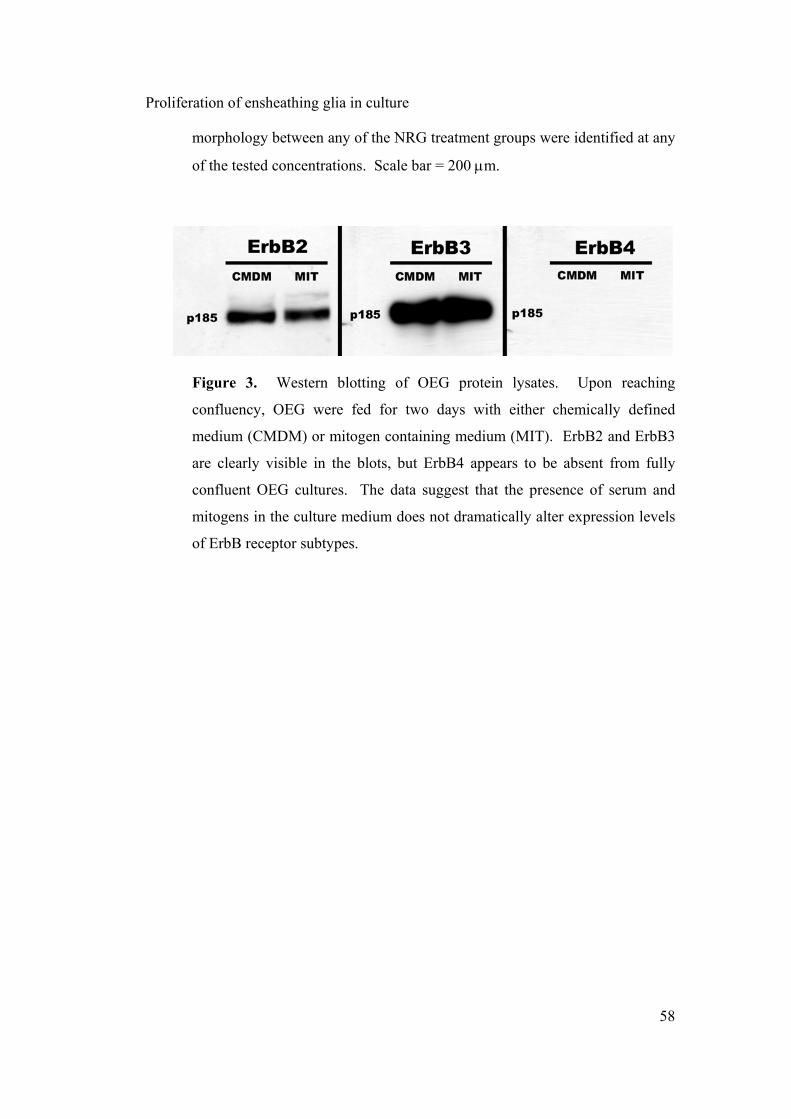
Proliferation of ensheathing glia in culture
58
morphology between any of the NRG treatment groups were identified at any
of the tested concentrations. Scale bar = 200 µm.
Figure 3. Western blotting of OEG protein lysates. Upon reaching
confluency, OEG were fed for two days with either chemically defined
medium (CMDM) or mitogen containing medium (MIT). ErbB2 and ErbB3
are clearly visible in the blots, but ErbB4 appears to be absent from fully
confluent OEG cultures. The data suggest that the presence of serum and
mitogens in the culture medium does not dramatically alter expression levels
of ErbB receptor subtypes.

Proliferation of ensheathing glia in culture
59
Figure 4. ErbB immunocytochemistry of OEG expanded in DF10S+mit.
The fields indicate ErbB2 (A), ErbB3 (B), and ErbB4 staining (C). Field (D)
shows mda cells overexpressing ErbB4 and stained with the anti-ErbB4
antibody. Note that ErbB4 appears to specifically localized to the nucleus.
Scale bar = 100 µm.

Proliferation of ensheathing glia in culture
60
Figure 5. Phosphorylation of ErbB receptors. Cultured OEG were treated
with NRG-1β, NRG-2α or NRG-3, then immunoprecipitated and
electrophoresed. Membranes were stained with an anti-phosphotyrosine
antibody. ErbB2, ErbB3 and ErbB4 are present in a phosphorylated state
irrespective of the neuregulin type used as a pre-treatment.
Figure 6. Proliferation dose response of OEG cultured in DF10S without
added mitogens and treated with NRG-1β, NRG-2α or NRG-3. A clear
proliferative effect of NRG-1β can be seen when the treated cells are not
previously extracted and purified in the presence of forskolin or bovine
pituitary extract (A). NRG-2α exhibits a small proliferative effect at low
concentrations (B), whereas NRG-3 shows no significant effect on

Proliferation of ensheathing glia in culture
61
proliferation (C). No significant synergistic effects on OEG proliferation
were observed when the neuregulins were tested in combination at 10ng/ml
(D). Error bars indicate ± s.e.m. (n=6). An * indicates a significant
difference compared to the CMDM control (p<0.05).
Figure 7. Proliferation dose response of OEG treated with forskolin. OEG
cultured in the absence forskolin and bovine pituitary extract in the culture
media exhibit a strong dose dependant proliferation response to forskolin.
Error bars indicate ± s.e.m. (n=9). An * indicates a significant difference
compared to the CMDM control (p<0.05).

Proliferation of ensheathing glia in culture
62
Figure 8. Proliferative responses of OEG to combinations of the mitogens.
Bovine pituitary extract (BPE) induces proliferation to a level comparable to
both NRG-1β and forskolin (FSK) together. Concentrations were: NRG-1β:
10 nM, forskolin: 2 µM and bovine pituitary extract: 20 µg/ml. Error bars
indicate ± s.e.m. (n=9). All results were significantly different to the CMDM
control (p<0.05).

Proliferation of ensheathing glia in culture
63
Figure 9. Western blotting of OEG purified and expanded in the presence of
DF10S medium. Membranes were labeled with antibodies against ErbB-2,
ErbB-3 and ErbB-4. ErbB-2 and ErbB-3 are expressed in OEG and by the
control cells. Mda cells overexpressing ErbB2 and ErbB3 are shown as a
positive control for those receptors. Note that ErbB4 also appears to be
absent from the blots.
Figure 10. Expression of ErbB RNA in Olfactory Bulb and cultured OEG.
RT-PCR detects the presence of ErbB2 and ErbB3 in both olfactory bulbs
and in cultured OEG. ErbB4 RNA also appears to be present in the olfactory
bulb, but is absent from cultured OEG.

Proliferation of ensheathing glia in culture
64
Figure 11. ErbB immunocytochemistry of OEG expanded in the presence of
DF10S medium. The fields indicate ErbB2 (A,B), ErbB3 (C, D), and ErbB4
staining (E, F). Note that ErbB4 appears to specifically localized to the
nucleus. Scale bars = 200 µm.

Proliferation of ensheathing glia in culture
65
Figure 12. Functional blocking of ErbB receptors. OEG grown in DF10S
medium were assayed for proliferation in the presence of CMDM or CMDM
and 5 nM NRG-1β. Antibody inhibitors of ErbB receptors were then added
to either of these two treatment groups. A: No inhibitory effect on
proliferation of OEG treated with CMDM medium alone is evident upon
addition of the ErbB inhibitory antibodies. B: In the group containing 5 nM
NRG-1β a marked drop in proliferation is evident upon the addition of
antibodies inhibitory to ErbB2 and ErbB2 + ErbB3 receptors, but no
significant drop in proliferation can be seen when ErbB4 inhibitory antibody
is added on its own. In all cases n=4, and * indicates p < 0.05.

CHAPTER 3

Chapter 3 – Age-dependent myelination by OEG
67
AGE-DEPENDENT MYELINATION BY OLFACTORY
ENSHEATHING GLIA
T.R. de Mello1,2; M.J. Ruitenberg1, W.T. Hendriks4, S.V. Lee1, J.
Verhaagen3,4, S.A. Dunlop2,3, G.W. Plant1,3
1 Red's Spinal Cord Research Laboratory, School of Anatomy & Human
Biology and 2School of Animal Biology, 3Western Australian Institute for
Medical Research, The University of Western Australia, Crawley, Perth,
Western Australia, Australia and 4Graduate School for Neurosciences
Amsterdam, Neuroregeneration Laboratory, Netherlands Institute for Brain
Research, Amsterdam, The Netherlands
Correspondence to: Dr Giles W. Plant, Red's Spinal Cord Research
Laboratory, School of Anatomy and Human Biology, Mail Bag Delivery Point
M309, The University of Western Australia, 35 Stirling Highway, Crawley,
Perth, WA 6099, Australia
Email: [email protected]
Abstract
Olfactory ensheathing glia (OEG) have been demonstrated to improve
functional and anatomical outcomes after injury to the nervous system.
However, contradictory observations have left open the question as to their
ability to myelinate central nervous system neurons. In this study we have

Chapter 3 – Age-dependent myelination by OEG
68
compared the myelinating ability of OEG derived from embryonic (EEG),
postnatal (PEG) and adult tissue (AEG) both in vitro and in vivo. OEG were
purified by p75-immunopanning, expanded in the presence of medium
containing pituitary extract and forskolin, then co-cultured with dissociated
cultures of TrkA-dependant embryonic dorsal root ganglion (DRG) neurons.
EEG, but not AEG or PEG, successfully myelinated DRG neurons in the
presence of serum and/or ascorbate. AEG also failed to myelinate GDNF-
dependant embryonic DRG cultures, and growth factor-independent adult
DRG cultures. Transplantation of OEG into lysolecithin demyelinated spinal
cord demonstrated distinct ultrastructural differences between transplants of
OEG from animals of different ages. AEG and Schwann cells exhibited a
similar proportion of axons that were unmyelinated, surrounded by intact
myelin or surrounded by loose uncompacted myelin. EEG displayed an
increased number of unmyelinated axons, and a proportionally smaller
number of axons surrounded by loose uncompacted myelin. These results
suggest that myelinating potential of OEG in vitro and behavior of these cells
following transplantation in vivo are developmentally regulated.

Chapter 3 – Age-dependent myelination by OEG
69
Introduction
Olfactory Ensheathing Glia (OEG) are specialized glial cells of the primary
olfactory pathway, a region of the CNS that is capable of supporting neuronal
replacement throughout adult life (Graziadei and Monti-Graziadei, 1978,
1979). Neuronal replacement in the olfactory system has been associated with
the permissive environment created by OEG, which chaperone newly growing
axons from their origin in the olfactory neural epithelium to their targets in the
olfactory glomeruli (Doucette, 1984, 1990; Raisman, 1985; Marin-Padilla &
Amieva, 1989). Several studies have suggested that the ability of OEG to
promote axonal growth lies at least in part with their similarities to Schwann
cells, glial cells within the peripheral nervous system (Ramon-Cueto and
Nieto-Sampedro, 1992; Doucette, 1995). More recently, the therapeutic
potential of OEG has come to the fore, with studies reporting that OEG
transplanted into areas of CNS damage can improve axonal sparing, promote
regrowth of damaged fibers and most importantly improve functional
recovery (reviewed by Santos-Benito and Ramon-Cueto, 2003; Mackay-Sim,
2005).
Closely related to improvement of function by OEG is their reported ability to
remyelinate demyelinated or compromised CNS axons. The first instance of
OEG myelination was demonstrated in vitro (Devon and Doucette, 1992).
Since then numerous studies have investigated myelination by OEG both in

Chapter 3 – Age-dependent myelination by OEG
70
vitro (Devon and Doucette, 1995; Plant et al., 2002) and in vivo (Franklin et
al., 1996; Li et al., 1997, 1998; Imaizumi et al., 1998, 2000a, b; Barnett et al.,
2000; Kato et al., 2000; Smith et al., 2001, 2002; Takami et al., 2002; Lakatos
et al., 2003b; Boyd et al., 2004a; Dunning et al., 2004; Radtke et al., 2004;
Sasaki et al., 2004), though results are often contradictory. For example,
unpurified embryonic-derived OEG (EEG), ie. dissociated olfactory bulb
cultures, have been reported to myelinate dorsal root ganglion (DRG) fibers in
vitro in the presence of serum (Devon and Doucette, 1992; Devon and
Doucette, 1995). On the other hand, adult-derived OEG (AEG) purified by
selection for the p75 low affinity neurotrophin receptor do not myelinate DRG
axons under similar culture conditions (Plant et al., 2002). In addition, two
studies which have successfully labeled OEG in vivo post-transplantation have
yielded contradictory results regarding OEG myelination. The contradictory
results may be due to the use of unstable or leaky markers when pre-labeling
OEG prior to cell transplantation and/or variation in purification techniques
and/or the age of the animal from which the cells were extracted (Boyd et al.,
2004a; Sasaki et al., 2004).
Here we have for the first time compared the myelination potential of p75-
selected OEG across the three most commonly used ages of donor animals:
embryonic (EEG), postnatal (PEG) and adult (AEG). Immunocytochemical
techniques were used to compare the capacity of these cells to myelinate
dissociated embryonic DRG cultures under unique combinations of various

Chapter 3 – Age-dependent myelination by OEG
71
conditions previously reported to induce myelination by either Schwann cells
or OEG (Eldridge et al., 1987; Devon and Doucette, 1995; Koenig et al.,
1995). We have also investigated the influence of axonal caliber and age on
the capacity of OEG to myelinate in vitro, and undertaken a comparative
study in vivo contrasting the ability of OEG preparations derived from animals
of different ages to remyelinate the demyelinated spinal cord. Part of this
work has been published in abstract form (de Mello et al., 2003).
Methods
Glial Cell Culture Preparation
Schwann Cell Cultures
Purified Schwann cells were prepared as previously described (Morrissey et
al., 1991; Plant et al., 2002). Briefly, the sciatic nerve was extracted 8 week
old F344 rats, cut into 1 mm pieces and plated onto a 35 mm dish containing
700 µl of Dulbecco's Modified Eagle's Medium (DMEM; Invitrogen,
Melbourne, Australia) with 10% FBS (v/v, JRH Biosciences, Lenexa, KS).
After 1 week, explants were transferred into fresh medium and thereafter once
a week until the third week. Tissue pieces were then enzymatically and
mechanically dissociated, and transferred to 100 mm poly-L-lysine (100
µg/ml; Sigma, St. Louis, MO) coated dishes with DMEM/10% FBS medium

Chapter 3 – Age-dependent myelination by OEG
72
containing 20 µg/ml Bovine pituitary extract (Invitrogen) and 2 µM forskolin
(Sigma).
Adult OEG Cultures (AEG)
Olfactory bulbs were removed from adult Fischer F344 rats as previously
described (Ramon-Cueto et al., 1998; Plant et al., 2002). Blood vessels and
the pia mater were carefully removed and the ventral portion of the bulbs
dissected, removing no more than 1.5 mm of the nerve fiber and glomerular
layers, and not selecting specifically for either the outer or inner olfactory
nerve layer (Au et al., 2002). The dissected tissue, mainly olfactory nerve
layer, was enzymatically digested using 2 ml 0.25% trypsin (w/v,
Worthington, Lakewood, NJ) and 0.25 mg/ml DNAse I (Roche, Castle Hill,
Australia) in Hank's Buffered Saline Solution (HBSS; JRH Biosciences) for
60 minutes at 37 °C. Digestion was stopped by adding serum-containing
medium (JRH Biosciences), and the tissue mechanically dissociated using a
flame-polished pipette. The remaining suspension was centrifuged at 300g for
5 minutes and re-suspended in mitogen containing medium (DF10S+mit).
DF10S+mit medium was composed of DMEM (Invitrogen) and Hams F-12
medium (Invitrogen) at a 1:1 ratio (v/v), 10% FBS (v/v; JRH Biosciences), 2
mM L-Glutamine (Invitrogen), 50 µM Gentamicin (Invitrogen); the mitogens
were 20 µg/ml Bovine pituitary extract (Invitrogen) and 2 µM Forskolin
(Sigma). Cells were then plated onto poly-L-lysine (100 µg/ml; Sigma) -
coated dishes and left for 4 days at 37 °C and 5 % CO2. Thereafter, cells were
Chapter 3 – Age-dependent myelination by OEG
73
fed every three days with DF10S+mit until confluency. The cells did not
undergo any further replating prior to purification by immunopanning.
Embryonic OEG Cultures (EEG)
Embryos were removed from E19 timed-pregnant Fischer F344 rats, and
olfactory bulbs extracted as previously described by Devon and Doucette
(1992). The entire olfactory bulbs were enzymatically digested by transferral
to a dish containing 2 ml of 0.1% trypsin (w/v) and 50 µl DNAse I in HBSS
for 25 minutes at 37 °C. Thereafter the cells were mechanically dissociated
and cultured similarly to the method described for adult ensheathing glia.
Postnatal OEG Cultures (PEG)
Olfactory bulbs were removed from postnatal day 7 Fischer F344 rats in a
modification of the method previously described by Barnett et al. (1993). The
blood vessels and pia mater were carefully taken away from the bulbs, and the
entire cleaned olfactory bulbs enzymatically digested by transferral to a dish
containing 2 ml of 0.1% trypsin (w/v) and 50 µl DNAse I in HBSS for 30
minutes at 37 °C. Thereafter the cells were mechanically dissociated and
cultured similarly to the method described for adult ensheathing glia.
Immunopanning of OEG Cultures
To ensure reproducible preparation of purified cell populations, primary OEG
cell cultures were positively selected for p75 via immuno-panning. Briefly, a

Chapter 3 – Age-dependent myelination by OEG
74
goat anti-mouse IgG,A,M secondary antibody (#55486, MP Biomedicals,
Irvine, CA) diluted 1:100 in 0.05 M Tris buffer (ph 9.5) was added to 100 mm
non-tissue culture treated bacterial petri dishes (Corning, Acton, MA). The
secondary antibody was left overnight at 4°C, and unbound antibody was
removed by rinsing 3 times with Leibovitz’s L-15 medium (L-15; Sigma). A
monoclonal anti-p75 antibody (clone IgG 192; gift from Dr. Patrick Wood,
University of Miami School of Medicine) diluted 1:4 in L-15 was then added
to each dish and allowed to bind for 2 hours at 4°C. Unpurified OEG were
trypsinised for 3 minutes with 0.05% trypsin in HBSS, the enzyme was
neutralized by addition of DF10S, followed by centrifugation for 300g for 5
minutes and resuspension in L-15 medium. The unpurified cell suspension
was plated 1:2 onto the immuno-panning dishes and allowed to bind to the
p75 antibody for 30 minutes, at 4°C to minimise rates of internalisation of the
cell surface p75 receptor. Once binding was completed, cells were vigorously
washed 5-7 times with L-15 to remove any unbound or loosely bound cells,
thereby leaving only strongly adherent cells. Adherent cells were fed with
DF10S+mit and cultured at 37°C/5%CO2 (v/v) for three days before replating
onto tissue culture treated dishes (Corning) coated with poly-L-lysine.
Thereafter, AEG typically attained confluency after 7 days, PEG after 7 days,
and EEG after 9 days. All cells were used within a period of 7-18 days after
immunopanning (DIV 1-2).

Chapter 3 – Age-dependent myelination by OEG
75
Cell Purity Determination
Cell purities were determined on the day of use by immunostaining with a
combination of antibodies: monoclonal anti-S100 IgG (Sigma, 1/1000
dilution), rabbit anti-cow S100 IgG (1/1000 dilution, DakoCytomation,
Glostrup, Denmark), monoclonal anti p75 IgG (Gift from Dr. Patrick Wood,
1/5 dilution), rabbit anti glial fibrillary acidic protein (GFAP) IgG (1/500
dilution, DakoCytomation), monoclonal anti Thy-1 IgG (Gift from Dr. Patrick
Wood, 1/5 dilution), monoclonal anti O1 IgG antibody (Gift from Dr. Patrick
Wood, 1/5 dilution) and monoclonal anti O4 IgG antibody (Gift from Dr.
Patrick Wood, 1/5 dilution). Briefly, cells were plated onto poly-L-lysine (100
µg/ml; Sigma) coated 2 mm round glass coverslips at 1x10⁴ cells per coverslip
in the presence of DF10S+mit medium. The next day, cells were subjected to
live staining with primary antibodies against p75 receptor, Thy-1, O1 or O4
diluted in L-15 medium with 10% FBS (v/v) for a period of 30 minutes at
4°C. Cells were washed three times with L-15 medium and incubated with a
goat anti-mouse IgG: Cy3-conjugated antibody (1/300 dilution, Jackson
ImmunoResearch, West Grove, PA) for 30 minutes. Cells were then fixed
with 4% paraformaldehyde (w/v; Sigma) for 15 minutes and permeabilized
with PBS containing 4% paraformaldehyde (w/v, Sigma) and 0.02% Triton X-
100 (v/v; Sigma) for 10 minutes at room temperature. After rinsing two times
with PBS, cells were incubated with primary antibodies against S100 or
GFAP protein (diluted in PBS/10% FBS/0.02% Triton X-100) for 45 minutes
followed by several washes with PBS/10% FBS (v/v) and incubation for 30

Chapter 3 – Age-dependent myelination by OEG
76
minutes of an Alexa Fluor ™ 488 goat anti-rabbit IgG antibody (1/600
dilution, Invitrogen). Finally, cells were rinsed three times with PBS and
coverslips mounted onto slides with Citifluor (UKC, UK) containing Hoechst
33343 (Sigma) as the mounting medium. Purity levels were calculated on the
basis of p75, S100 and GFAP staining and in all cases were determined to be
between 96-99% (AEG and PEG) or between 90-95% (EEG). Less than one
percent of cells stained positively for Thy1 or O1. The remainder were
positive for GFAP but not p75, and likely to be astrocytes (Harvey, 1994).
Dissociated DRG Cultures
Embryonic DRG were extracted from day 15 embryos as previously described
(Kleitman et al., 1998; Plant et al., 2002). Briefly, E15 timed-pregnant
Sprague Dawley rats were killed by injection of 0.2 ml Pentobarbitone sodium
(325 mg/ml). The embryos were removed and placed in L-15 medium.
Heads and ventral portions of the embryos were removed under an AIS-
OPTICAL dissecting microscope (Rowe Scientific, Australia), the viscera
removed, vertebrae carefully clipped with fine forceps and the entire spinal
cord of each embryo dissected free. Ganglia were removed from the spinal
cord with the aid of fine forceps. Dissociation of ganglia was performed by
incubation in 0.25% trypsin (w/v) in HBSS for 45 min at 37°C. Following
enzymatic treatment, trypsin digestion was stopped by addition of L-15 cell
medium containing 20% FBS (v/v). The dissociated cells were centrifuged at
300g for 5 minutes, the pellet resuspended in 2ml L-15 medium containing

Chapter 3 – Age-dependent myelination by OEG
77
10% FBS (v/v) and DRG were mechanically triturated using a flame polished
pipette. Cells were centrifuged once more at 300g for 5 minutes. Finally, the
pellet was resuspended in sufficient NLA medium to ensure that a dilution of
1.5 ganglia per 110 µl was achieved. NLA medium comprised 100 ml
Neurobasal cell medium (Invitrogen), 10% B27 supplement (v/v, Invitrogen),
2 mM Glutamine (Invitrogen) and 50 µM Gentamicin (Invitrogen). A 55 µl
suspension was plated onto the centre of each collagen coated aclar hat
(Kleitman et al., 1998; Plant et al., 2002) and the cells transferred to a
37°C/5% CO2 incubator.
The following day, aclar hats were flooded with 0.5 ml of NLA-f medium.
NLA-f medium consisted of NLA medium with added anti-mitotics to
eliminate contaminating cells from the neuronal culture: 5 mM Uridine
(Sigma) and 5 mM Fluorouridine (Sigma). Thereafter cells were fed every
two days with NLA medium. Every third feed for the next three weeks was
performed with NLA-f medium to ensure the absence of contaminating non-
neuronal cells. Selection of TrkA-dependent neurons was achieved by
inclusion of nerve growth factor (NGF; 100 ng/ml, kindly provided by Dr.
Patrick Wood and Dr. Bob Rush) at all stages during the experiment.
Selection of GDNF-dependent neurons was achieved by inclusion of 1 ng/ml
glial cell line-derived neurotrophic factor (GDNF; PeproTech, Rocky Hill,
NJ) instead of NGF at all stages during the experiment.
Chapter 3 – Age-dependent myelination by OEG
78
Adult DRG cultures were extracted from 3 month old rats as previously
described (Purves-Tyson and Keast, 2004). After extraction ganglia were
incubated for one hour with 1.3 mg/ml (w/v) collagenase in HBSS at 37°C,
followed by one hour in a solution containing 1.3 mg/ml (w/v) collagenase
and 0.25% (w/v) trypsin in HBSS at 37°C prior to dissociation and plating of
cells. Neurobasal-A medium (Invitrogen) was used instead of Neurobasal
medium, and no NGF or GDNF was included in the culture medium at any
stage.
Co-Culture of Neurons and Glia
One week following the last pulse with NLA-f medium, DRG neurons were
co-cultured with either Schwann cells, adult OEG, postnatal OEG or
embryonic OEG. Glial cells were counted with a haemocytometer, suspended
in NLA medium, and 50,000 cells in a 0.5 ml volume were added to each
neuronal culture. Co-cultures were fed every two days for two weeks with
NLA medium. After two weeks the cell medium was switched to include
factors reported to promote myelination by glial cells. These included
ascorbate: a reported trigger for myelination by Schwann cells (Eldridge et al.,
1987), FBS: a reported trigger for myelination by OEG (Devon and Doucette,
1995), and progesterone: reported to enhance Schwann cell myelination in the
presence of ascorbate and to stimulate production of myelin proteins in both
PNS and CNS glia (Jung-Testas et al., 1994; Koenig et al., 1995; Baulieu and
Schumacher, 2000; Ghoumari et al., 2003; Melcangi et al., 2003). Different

Chapter 3 – Age-dependent myelination by OEG
79
groups included those containing 50 µg/ml ascorbate, 15% FBS (v/v), NLA
medium alone, and a group containing 50 µg/ml ascorbate (Sigma), 15% FBS
(v/v) and 20 mM progesterone (Sigma) together. Cells were fed every two
days for two weeks with these different factors, whereupon they were fixed
and processed for either immunocytochemistry or electron microscopy.
Immunocytochemistry
Neuronal-glia cocultures were fixed with 4% paraformaldehyde (w/v, Sigma)
for 15 minutes, then permeabilized with 4% paraformaldehyde containing
0.02% Triton X-100 (v/v, Sigma) for 10 minutes at room temperature. To
permeabilize myelin sheaths for myelin basic protein (MBP) antibody
binding, cultures were first treated with ice-cold 50% acetone (Biolab,
Mulgrave, Australia) for two minutes, then ice-cold 100% acetone for two
minutes and finally ice-cold 50% acetone for two minutes. After rinsing with
PBS (2x 5 minutes), cells were incubated with primary antibodies for 45
minutes. Several different primary antibodies were used in combination
throughout this study. These included: monoclonal anti-MBP (#SMI 99 and
#SMI 94) IgG antibodies (1/1000 dilution, Sternberger, Berkeley, CA), rabbit
anti-cow S100 IgG polyclonal antibody (1/400 dilution, #Z0311,
DakoCytomation), monoclonal anti-neurofilament (clone RT97, 1/10 dilution,
Developmental Hybridoma Bank), rabbit anti-neuronal class III β-tubulin IgG
(1/2000 dilution, #PRB-435P-100, Covance, Princeton, NJ), monoclonal anti
myelin-associated glycoprotein IgG (MAG, 1/500 dilution, #MAB1567,

Chapter 3 – Age-dependent myelination by OEG
80
Chemicon, Boronia, Australia), monoclonal anti-2’, 3’- cyclic nucleotide 3’-
phosphodiesterase (CNP) IgG (1/500 dilution, #SMI 91, Sternberger,
Berkeley, CA), rabbit anti-protein zero IgG (P0, 1/500 dilution, Gift of Dr.
Bruce Trapp). The next day, cells were washed with phosphate buffer (4x 5
minutes) and incubated for 30 minutes with an Alexa Fluor ™ 546 donkey
anti-sheep IgG secondary antibody (#A-21098, Invitrogen) and an Alexa
Fluor ™ 488 goat anti-mouse IgG secondary antibody (#A-11029,
Invitrogen). Finally, cells were washed four times for 5 minutes in phosphate
buffer, and coverslips mounted onto slides with Citifluor containing Hoechst
33343 as the mounting medium.
Lysolecithin Demyelination of the Spinal Cord Dorsal Funiculus
Surgical anaesthesia was induced by intramuscular injection of ketamil (100
mg/kg of body weight) and xylazil (10 mg/kg of body weight). Following a
laminectomy of the T10 vertebrae, the animal's vertebral column was
stabilised on a sterotaxic microinjector. Focal areas of demyelination were
created by injection of 1 µl of a 1% (w/v) L-α lysophosphatidyl choline
solution (Sigma) diluted in saline as previously described by Woodruff and
Franklin (1999). Lysophosphatidylcholine disrupts membranes, including that
of myelin, by inserting into lipid bilayers to form micelles (Weltzien, 1979;
Gregson, 1989). Injections were performed with the aid of a 60 µm tip glass
pipette attached to a 5 µl Hamilton syringe at 0.5 mm depth into the dorsal
funiculus of the spinal cord. The volume was delivered over a period of 10

Chapter 3 – Age-dependent myelination by OEG
81
minutes with a Harvard syringe pump (Harvard Apparatus, Holliston, MA),
and the needle left in place for 5 minutes prior to suturing of the wound.
Animals received daily subcutaneous injection of pain killers (buprenorphine,
20 µg/kg) for three days after the surgery.
Cell Transplantation
Purified AEG, EEG and Schwann cells were pre-labelled with a lacZ
transgene via a lentivirus vector (Lv-LacZ; Ruitenberg et al., 2002). Briefly,
Lv-LacZ was added to the culture medium at a multiplicity of infection of
100, and incubated with the cells for 24 hours prior to replacement of the
media. Approximately 90% of cells were labelled two days later as
determined by visualisation of Bluo-Gal reaction product under bright field
microscopy utilising a high magnification objective (Figure 1). Three days
following initial transduction, cells were resuspended in DF10S medium at a
density of 50,000 cells per µl. This cell suspension (2 µl) was injected into
the demyelination site four days after initiation of demyelination. Viability of
transplanted Schwann cells, AEG and EEG was determined to be 85-90% by
trypan blue staining immediately prior to transplantation. A control group
received injections of DF10S medium alone. Following surgery, the wound
was sutured and animals allowed to recover for a further 14 days prior to
sacrifice and perfusion. Treatment with buprenorphine (20 µg/kg) was
continued for one week following cellular transplantation. Eight animals were
utilised for each group, for a total of 36 animals.
Chapter 3 – Age-dependent myelination by OEG
82
Electron Microscopy of demyelinated spinal cord
All animals were euthanased by intraperitoneal injection of 0.2 ml
Pentobarbitone sodium (325 mg/ml), transcardially perfused with 200 ml of
PBS and 1000 I.U./L Heparin Sodium (Mayne Pharma, Melbourne,
Australia), followed by 200 ml of 2% glutaraldehyde in PBS. Following
perfusion, spinal cords were removed, cut into 1 mm segments, transferred
into glass vials and incubated with Bluo-Gal reaction buffer for 8 hours at
37˚C. Bluo-Gal reaction buffer consisted of 0.1 M Phosphate buffer (pH 7.4)
with 2 mM MgCl2, 5 mM EDTA, 240 µM Na Deoxycholic acid (Sigma), 200
mg/L Nonidet NP 40 (Sigma), 2 mM Bluo-gal (Sigma), 20 mM potassium
ferrocyanide (Sigma), and 20 mM potassium ferricyanide (Sigma). Following
Bluo-Gal staining, fixed sections were washed three times with phosphate
buffer and post-fixed in 2% glutaraldehyde overnight at 4°C. The next day,
sections were washed three times for 5 minutes with 0.15 M PBS, post-fixed
for a further two hours with 1% osmium tetroxide (v/v, ProSciTech,
Thuringowa, Australia) in 0.1 M PBS, pH 7.4, followed by three washes with
PBS, before dehydration through graded alcohols. Tissue pieces were rinsed
twice for five minutes in propylene oxide, and left overnight at room
temperature in a 1:1 mixture of propylene oxide and araldite resin to ensure
penetration. Araldite resin consisted of 46% Araldite 502 (v/v, Probing &
Structure), 28.4% DDSA (v/v, ProSciTech, Thuringowa, Australia), 24.1%
NMA (v/v, ProSciTech, Thuringowa, Australia) and 1.5% DMP-30 (v/v,
ProSciTech, Thuringowa, Australia). On the following day, the araldite resin
Chapter 3 – Age-dependent myelination by OEG
83
was replaced with freshly made resin, and allowed to polymerise overnight at
64°C. Ultrathin sections were cut on an ultramicrotome (LKD, Ultranova),
collected onto copper grids, stained with uranyl acetate (5% in 15% acetic
acid) and lead citrate (Reynolds, 1963) for 2 minutes, and viewed under an
electron microscope (Philips 410).
Toluidine Blue Staining
Sections were mounted onto gelatin coated slides and dried at room
temperature overnight. The dry slides were then placed into toluidine blue
solution for a period of 30-45 seconds. Toluidine blue solution consisted of
Toluidine Blue (0.5% w/v) and Sodium Tetraborate (0.5% w/v) dissolved in
water. The stained sections were rinsed in deionised water briefly followed
by dehydration through graded alcohols. Finally, slides were moved into
toluene and coverslipped using DPX (Chem-Supply, Gilman, Australia) as the
mounting medium.
Data Analysis
All co-cultures were imaged with an Olympus IX70 inverted microscope.
Images were taken from nine defined fields through a 20x objective using an
Optronics 60800 camera (Figure 5a). Together, the analysed fields in each
coverslip accounted for 2.41 ± 0.15 % (s.e.m.) of the total area of each
coverslip. Three lines were drawn horizontally across each imaged field in
defined positions (Figure 5b), and the number of myelinated profiles (MBP

Chapter 3 – Age-dependent myelination by OEG
84
positive) crossing each line was counted to obtain a total number of profiles
per field. Counts from all nine defined fields were averaged to produce a final
comparative value for the level of myelin in each coverslip. The final number
of myelinated profiles was expressed in the text as x units. Five coverslips
were analysed for each variable of the experiment, and a Tukey test was used
for all statistical analyses. Myelination state in vivo was quantified by
counting of three random electron microscope images (x1600 magnification)
from the lesion site of each group. Each axon was allocated a rating for its
myelination state (intact myelin, degraded or loose uncompacted myelin,
unmyelinated) and expressed as a percentage of the total number of axons
counted for each group (Figure 12).
Results
Embryonic Ensheathing Glia myelinate TrkA dependent DRG neurons in vitro
We first investigated the ability of all OEG ages to myelinate TrkA-dependent
neurons in vitro. Embryonic DRG neurons were cleared of contaminating
cells and selected for TrkA-dependent neurons over four weeks. p75-selected
OEG derived from embryonic, postnatal or adult animals were co-cultured
with the embryonic DRG neurons and stimulated to myelinate with either
ascorbate or FBS. All co-cultures from these groups were processed for
immunocytochemistry and stained with antibodies against MBP and S100, as
well as the nuclear dye Hoechst 33343. MBP was chosen as a marker of

Chapter 3 – Age-dependent myelination by OEG
85
mature myelin formation due to its late expression during the myelination
process in the PNS (Hahn et al., 1987), and its pivotal role in myelin
formation in the CNS (Readhead et al., 1987; Katsuki et al., 1988; Lemke,
1988 for review). Confirmation that MBP could be used as a marker for
myelination was performed by feeding a co-culture of TrkA selected DRG
neurons and unpurified EEG or Schwann cells with 15% (v/v) FBS or 50
µg/ml ascorbate respectively. The results confirm earlier findings of Devon
and Doucette (1992), showing the presence of mature myelin in those cultures
(Figure 2a and 2d). The distribution of Hoechst 33343-labelled nuclei in both
co-cultures also indicates a close association of the glial cells with neuronal
axons (Figure 2c and 2f), as would be expected with a myelinating phenotype.
Note that the MBP marker is not normally expressed in either purified or
unpurified populations of EEG, and is not present in the glial cells co-cultured
with neurons prior to the addition of serum to the culture medium (data not
shown).
Having confirmed the functionality of our in vitro myelination assay, we co-
cultured DRG neuronal cultures with p75-selected OEG derived from three
different aged animals, namely embryonic day E19 OEG (EEG), postnatal day
7 OEG (PEG), and adult OEG (AEG) derived from 8 week old rats. Figure 3
illustrates Schwann cells and our three OEG preparations when co-cultured in
the presence of 15% (v/v) FBS without ascorbate. Schwann cells, PEG and
AEG co-cultures do not demonstrate any MBP immunoreactivity (Figure 3a,

Chapter 3 – Age-dependent myelination by OEG
86
3c and 3d respectively), but p75-selected EEG show strong MBP
immunoreactivity under these culture conditions (Figure 3b). Of particular
interest is the distribution of cells in these culture systems as illustrated by
Hoechst 33343 staining. Schwann cells and purified EEG aggregate with
axons (Figure 3i and 3j respectively). Purified PEG and AEG show no
preference for either the neuronal axons or the collagen substrate (Figure 3k
and 3l respectively). To further illustrate the relationship of glial cells to DRG
axons, we compared the different co-cultures under phase microscopy (Figure
4). In these images, the characteristic tubular structures of myelin can be seen
in Schwann cells co-cultured with DRG neurons in the presence of 50 µg/ml
ascorbate (Figure 4a), in purified EEG co-cultured with DRG neurons in the
presence of 15% (v/v) FBS (Figure 4b), and in unpurified EEG co-cultured
with DRG neurons in the presence of 15% (v/v) FBS (Figure 4c). Purified
PEG or AEG do not appear to show any preference for neuronal axons (Figure
4d and 4e respectively). This lack of preference of purified AEG for either
collagen substrate or DRG axons is more clearly seen in Figure 4f, which
shows an AEG co-culture stained with Hoechst 33343 (blue), S100 (green)
and the RT97 antibody (red) with specific staining for the phosphorylated
form of high molecular weight neuronal filaments (Wood and Anderton, 1981,
Anderton et al., 1982).
Levels of MBP in co-cultures under different medium conditions was
quantified for cultures containing DRG neurons without additional glial cells,

Chapter 3 – Age-dependent myelination by OEG
87
or co-cultures of DRG neurons with either Schwann cells, purified AEG,
purified PEG or purified EEG (Figure 5a-c). Each group was treated with
either medium alone, medium containing 50 µg/ml ascorbate or medium
containing 15% (v/v) FBS. Most cultures contained no detectable levels of
MBP, with positive results seen only in Schwann cell co-cultures in the
presence of 50 µg/ml ascorbate (6.30 ± 1.47 units s.e.m., n=4), and in purified
EEG co-cultures in the presence of either 50 µg/ml ascorbate (6.55 ± 1.38
units s.e.m., n=4) or 15% (v/v) FBS (13.35 ± 1.97 units s.e.m., n=5). A
student t-test failed to show a significant difference between MBP levels in
EEG co-cultures treated with either 50 µg/ml ascorbate or 15% (v/v) FBS
(p>0.1).
In an attempt to provoke a positive myelinating response from our purified
AEG and PEG cultures, we combined 50 µg/ml ascorbate, 15% (v/v) FBS,
and 20 mM progesterone in cultures containing AEG, PEG or EEG. Despite
the addition of all three factors together, MBP levels remained undetectable in
cultures containing DRG alone, AEG or PEG. The only positive response was
seen in co-cultures containing purified EEG (Figure 5d). Levels of MBP
staining in purified EEG cultures (20.81 ± 1.45 units s.e.m., n=6) in the
presence of all three factors was not significantly different to either ascorbate
or serum alone EEG groups (p>0.1).

Chapter 3 – Age-dependent myelination by OEG
88
Adult Ensheathing Glia fail to myelinate GDNF-dependent DRG neurons in
vitro
We next investigated the myelination potential of AEG in a GDNF-dependant
embryonic DRG neuron co-culture situation. Although OEG do not myelinate
the small calibre axons (0.1-0.4 µm) of the olfactory nerves (Graziadei, 1973;
Doucette, 1991), we reasoned that OEG might myelinate larger calibre axons
in vitro since large GDNF-dependent axons are myelinated by Schwann cells
in the presence of ascorbate (Hoke et al., 2003). Co-cultures of AEG and
DRG neurons were prepared as elsewhere in this study, except that GDNF
was included in the culture medium at all stages of preparation instead of
NGF. This culture regimen has been demonstrated to select embryonic DRG
cultures for large diameter growth factor-dependent axons (Gavazzi et al.,
1999). However, AEG failed to demonstrate positive immunostaining for
MBP in the presence of serum (Figure 6) or ascorbate (data not shown).
Interestingly, AEG demonstrated a markedly different interaction with axons
to that exhibited by Schwann cells. Schwann cells formed a close association
with neuronal axons throughout the culture (Fig. 6a). AEG preferred to
associate into clusters of cells towards the edges of the neuronal culture (Fig.
6b, 6c, 6d), but the positioning of the clusters demonstrated no preferred
association with either axons or the collagen substrate. In the centre of the co-
cultures, where axons were present at higher densities, AEG demonstrated no
preference for either collagen substrate or neuronal axons, and did not form
clusters (Fig. 7). Subsequent analysis for MAG immunoreactivity (Fig. 7d),
Chapter 3 – Age-dependent myelination by OEG
89
as well as CNP immunoreactivity (data not shown) failed to reveal detectable
levels of any of these myelin proteins in co-cultures containing AEG.
Finally, to address the possible argument that GDNF itself could have a direct
effect on the myelination potential of AEG, we analysed the expression of
myelin proteins on AEG cultured without neurons. Our results demonstrate
that GDNF has no effect on the expression of p75, S100 or GFAP, with
almost 100% of cells positively expressing these markers (Fig. 8a, 8b). The
myelin markers CNP, MAG, and MBP were not expressed by any of our
cultured cells (data not shown). P0 expression on the other hand, was found to
be present in GDNF-treated AEG cultures (Fig. 8c). Previously in our
laboratory we have demonstrated that P0 is expressed at very low levels in
99% of AEG under both serum containing and serum free conditions, and that
all P0 expressing cells also display p75 immunoreactivity (unpublished
observations). Together, these results indicate that GDNF itself does not have
an effect on the phenotypic characteristics of AEG.
Adult Ensheathing Glia fail to myelinate adult DRG neurons in vitro
We repeated our experiment on AEG co-cultures utilising DRG neurons
derived from adult animals. Adult DRG neurons are growth factor-receptor
independent, and contain a mixture of axonal calibres in culture (Lindsay,
1988). Again, all co-cultures of neurons and AEG failed to reveal a detectable
level of either MBP, CNP, or MAG immunoreactivity (data not shown). AEG

Chapter 3 – Age-dependent myelination by OEG
90
expressed basal levels of P0 when co-cultured with adult DRG neurons in the
presence of serum (Fig. 9), though this result is not unexpected given that P0 is
constitutively expressed by AEG even before co-culture (Seok Voon Lee,
unpublished observations). Again, we did not observe any preference for
AEG with either adult DRG axons or the collagen substrate (Fig. 9), and no
tubular structures characteristic of myelin was seen in any culture.
Ensheathing Glia promote remyelination of demyelinated spinal cord
Following our results in vitro, we investigated the remyelination potential of
OEG in vivo. We utilised a simple chemical demyelination model using L-α
lysophosphatidyl choline injection into the dorsal funiculus of adult rat spinal
cord. Glial cells were labelled with a LacZ lentivirus vector and transplanted
into the lesion site four days later to minimise cell death due to possible
inflammatory responses of the host animal (Ousman and David, 2000).
Groups included: 1) a medium only control, 2) transplanted Schwann cells, 3)
AEG and 4) EEG. PEG were not used in this part of the study given our
inability to induce these cells to myelinate in vitro, and their unlikely clinical
use. Analysis of the lesion sites was performed 15 days after transplantation
to ensure that spontaneous remyelination by endogenous cells would not be
completed (Gilson and Blakemore, 1993; Pavelko et al., 1998; Woodruff and
Franklin, 1999).
Chapter 3 – Age-dependent myelination by OEG
91
Semi-thin sections of the lesion site 19 days after initial dorsal funiculus
demyelination revealed substantial differences between the experimental
groups (Figure 10). In all groups, large numbers of fibroblasts, inflammatory
cells and macrophages in the process of phagocytosing myelin debris were
present throughout the transplant site. Intact densely packed axons with
varying amounts of myelin were present in both medium alone and Schwann
cell groups. However in the AEG group, axons appeared to be separated by
large quantities of extracellular matrix and cytoplasm (Figure 10 e-f). By
contrast, in the EEG group, axonal distribution was more ordered (Figure 10
g-h). The differences were further emphasised by electron micrographs of the
transplant sites which contained degraded myelin profiles, peripheral
(Schwann cell-like) myelin profiles, central (oligodendrocyte) myelin profiles,
and unmyelinated axons (Figure 11). The EEG transplant group (Figure 11d)
showed marked differences compared to the AEG transplant group (Figure
11c). In the EEG group, intact axons are mostly unmyelinated and are
fasciculated by large process bearing cells, an almost identical arrangement to
that previously reported using unpurified EEG preparations (Boyd et al.,
2004a). This arrangement is not seen in the AEG group, where many axons
appear to be surrounded by either intact or loose, uncompacted myelin (Figure
11c).
Quantification of myelination state (Figure 12) indicated that the medium
control group possessed the lowest amount of intact myelin (6.7% ± 1.1%

Chapter 3 – Age-dependent myelination by OEG
92
s.e.m., n=3), with all glial transplant groups exhibiting a significantly higher
percentage of myelinated profiles (Schwann cells: 29.7% ± 4.8%, AEG:
30.6% ± 4.0%, EEG: 29.6% ± 4.8% s.e.m., for all p<0.05, n = 3). No
significant difference in levels of intact myelin was observed between any of
the transplanted groups (p>0.05). However, degraded myelin profiles were
highest for the medium control group (81.7% ± 3.5% s.e.m., n=3), and
significantly lower for both Schwann cell (55.2% ± 6.1% s.e.m., n=3, p<0.05)
and AEG groups (49.6% ± 5.2% s.e.m., n=3, p<0.01). Interestingly, levels of
degraded myelin present in the EEG transplant group (11.2% ± 1.3% s.e.m.,
n=3) were far below those exhibited by either medium alone (p<0.001),
Schwann cell (p<0.001) or EEG groups (p<0.01). Correspondingly, the
proportion of unmyelinated axons in the EEG group (59.2% ± 4.0%, s.e.m.,
n=3) was significantly higher to that seen in any other group (medium: 11.5%
± 2.4%, Schwann cells: 15.1% ± 4.3%, AEG: 19.8% ± 5.5% s.e.m., n=3,
p<0.001). No significant difference in the percentage of unmyelinated axons
was seen between medium alone, Schwann cell or AEG groups (p>0.05).
Despite the differences in appearance and myelination state between the
various groups, no significant difference was detected between total numbers
of axons present in each group (p>0.05).
Closer inspection of the lesion site in the AEG and EEG transplant groups
revealed that in all cases where peripheral myelin is seen, the myelinating
glial cell is present in a 1:1 relationship with the axon (Figure 13). These cells

Chapter 3 – Age-dependent myelination by OEG
93
possess a basal lamina (Figure 13d) and resemble Schwann cells.
Unfortunately, none of our transplanted Schwann cells or AEG demonstrated
any visible bluo-gal reaction product. Transplanted EEG demonstrated faint
electron dense precipitate (Figure 13c) characteristic of LacZ reaction
(Franklin and Barnett, 1991; Weis et al., 1991; Sekerkova et al., 1997; Boyd et
al., 2004a) but this was deemed too pale for accurate counts.
Ultrastructurally, unmyelinating AEG and EEG were recognisable by their
large amount of cytoplasm and numerous processes surrounding nearby axons
(Figure 13 b-c). Interestingly, the EEG transplant group always exhibited
EEG enveloping large clusters of myelinating cells that resembled Schwann
cells in appearance, but the EEG never contacted axons directly (Figure 13c).
Our observations support the data reported by Boyd et al. (2004a), who
described that unpurified EEG transplants form channels around central cores
of axons myelinated in a 1:1 relationship by glia resembling Schwann cells.
Discussion
We have shown that OEG derived from embryonic, but not adult or postnatal
animals, are capable of myelinating TrkA-dependent DRG neurons in vitro.
Furthermore, we have shown that the inability of AEG to myelinate DRG
axons in vitro is not related to axonal calibre. We have also demonstrated that
both AEG and EEG behave differently when transplanted into demyelinated

Chapter 3 – Age-dependent myelination by OEG
94
spinal cord in vivo. These results indicate a developmental regulation of OEG
behaviour and responses to factors both in vitro and in vivo.
Myelination by OEG in vitro
We have shown that adult and postnatally derived OEG cannot be induced to
myelinate TrkA-dependant DRG neurons under culture conditions containing
serum, ascorbate and progesterone. This finding agrees with that previously
reported by Plant et al. (2002), who failed to induce myelination of AEG
under conditions containing serum and ascorbate. Here we extend the
findings and report that whereas both AEG and PEG do not myelinate in the
presence of serum, ascorbate and progesterone in vitro, that embryonically
derived OEG do form myelin. Previously, others have shown that unpurified
populations of EEG can myelinate TrkA-dependent cultures under medium
conditions containing serum (Devon and Doucette, 1995). Here we report for
the first time that EEG preparations are also able to myelinate DRG neurites
under conditions containing ascorbate alone. Although we cannot
categorically rule out the possibility that contaminating Schwann cells in the
DRG preparation are responsible, we were unable to observe myelination in
the medium alone, AEG and PEG groups. We also cannot rule out the
possibility that Schwann cell precursors are responsible for the myelination
observed in the ascorbate-treated EEG group, though myelination in the
presence of serum alone (without ascorbate) would indicate that Schwann cell
precursors are not completely responsible for our observations. The data

Chapter 3 – Age-dependent myelination by OEG
95
suggests that if EEG are responsible for myelinating DRG neurites, then the
ability may be associated with the age of the animal and may be indicative of
a developmentally regulated variation in the extracted cells.
Our subsequent studies further support this hypothesis and demonstrate that
AEG appear incapable of myelination in vitro. AEG demonstrated no
preference for either axons or the collagen substrate in GDNF-dependent
DRG cultures, or in growth factor-independent adult DRG cultures. AEG also
demonstrated no visible staining for MAG, CNP or MBP in either GDNF or
growth factor-independent neuronal co-culture systems, either in the presence
of ascorbate or serum. Interestingly, AEG cultures constitutively express P0
protein both before and after co-culture with neurons, thus indicating that at
least in part they still possess intrinsic features that indicate they may be able
to myelinate neurons. It remains to be seen if AEG will in future be able to be
induced to myelinate if cultured under different medium conditions, or if the
basal expression of P0 by AEG is a developmental remnant from a stage
where EEG had the capacity to myelinate.
Myelination by OEG in vivo
We have performed for the first time a comparative in vivo study of the
myelination ability of OEG derived from animals of two different ages.
Lysolecithin-induced demyelinated areas of the dorsal funiculus bear a
distinctly varied ultrastructural appearance in all treatment groups examined at

Chapter 3 – Age-dependent myelination by OEG
96
both light and electron microscopic levels. Medium only controls exhibit
large number of axons surrounded by degraded myelin profiles, and very few
axons that are unmyelinated or enveloped by mature myelin sheaths. Our
Schwann cell controls demonstrate a marked increase in levels of intact
mature myelin, a finding that is supported by previous studies of Schwann
cells in demyelinating lesions of the CNS (Honmou et al.., 1996). The AEG
transplant group displays a similar proportion of myelinated profiles to the
Schwann cell transplant group, though a distinct difference is observed in the
ultrastructural appearance of the lesion site itself. Many previous in vivo
studies have argued that OEG (both purified and unpurified preparations) are
able to myelinate demyelinated neurons, and have consistently indicated that
OEG myelin is indistinguishable from Schwann cell myelin in terms of
conduction velocity, immunoreactivity and morphology (Franklin et al., 1996;
Barnett et al., 2000; Imaizumi et al., 2000a, b; Kato et al., 2000).
The peripheral myelin observed in our AEG transplant group is
ultrastructurally indistinguishable from Schwann cell myelin (Friede and
Samorajski, 1968), and several researchers have proposed that much of the
observed myelination may be due to increased recruitment of endogenous
Schwann cells into the lesion site (Boyd et al., 2004a, b; Ramer et al., 2004a;
Richter et al., 2005). Furthermore, it is well established that lysolecithin
induced lesions cause short term permeability to the blood brain barrier,
allowing for invasion of immune response cells into the lesion site (Ousman

Chapter 3 – Age-dependent myelination by OEG
97
and David, 2000). Although the timeframe in our experiments was designed
to avoid this phase, it is possible that transplantation was performed at a time
when such effects had not completely subsided, thus facilitating movement of
Schwann cells from the periphery into the site of AEG transplanted cells.
However, given the failure of our Lv-LacZ label, we cannot accurately
confirm that purified AEG are not responsible for myelination in our
lysolecithin induced lesions. Nevertheless, GFP-labelled AEG have been
shown to be responsible for the majority of peripheral-like myelin following
transplantation into spinal cord lesions (Sasaki et al., 2004) although
contamination by Schwann cells during OEG isolation remains to be
excluded.
The finding that spinal cords transplanted with purified AEG exhibit a random
distribution of myelinated and unmyelinated axons throughout the lesion site
is of interest. By contrast, two other groups have previously demonstrated
that animals transplanted with AEG form clusters of myelinating cells that are
sometimes surrounded by unmyelinating AEG (Li et al., 1997, 1998, 2003b;
Sasaki et al., 2004), though another group has failed to report these
observations (Takami et al., 2002). However, the studies used spinal cord
transection rather than lysolecithin demyelination as described here. It is
possible that the cluster arrangement is only seen in lesions where active
neuronal degeneration is taking place, rather than simple remyelination as in
our system. Most importantly however, is the fact that such clusters of

Chapter 3 – Age-dependent myelination by OEG
98
myelinated axons have been consistently observed in experimental models
utilising cells derived from younger animals. For example, clusters or
channels of myelinating axons are often seen in transplants of PEG into
chronically demyelinated spinal cord (Franklin et al., 1996; Smith et al., 2001;
Lakatos et al., 2003b), and transected spinal cord (Imaizumi et al., 2000b).
Indeed, the work reported here on EEG confirms the work of Boyd et al.,
(2004a) showing that transplanted EEG do not contact axons, but rather
surround clusters of myelinating cells resembling Schwann cells.
The age-dependent differences in behaviour of OEG transplants are further
emphasised by our observations on the myelinated status of axons within the
lesion site. Our results demonstrate that axons in the EEG and AEG
transplant groups possess a similar proportion of intact myelin to the Schwann
cell group. However, the proportion of axons surrounded by loose
uncompacted myelin is significantly less in EEG groups, and the proportion of
axons that are completely unmyelinated is correspondingly higher. The
observations indicate that OEG transplants behave in a developmentally-
dependant manner that can not only dramatically modify the ultrastructural
appearance of the lesion site, but also the extent of repair to the damaged
myelin. These findings are also suggestive of a developmental shift in the
properties of OEG populations, and suggest that age-related differences may
yet be found in the bulb in vivo despite recent findings (Magavi et al., 2005).
Chapter 3 – Age-dependent myelination by OEG
99
Acknowledgements
This work was supported by the UWA Small Grant, The Neurotrauma
Research Program of Western Australia and the Ramaciotti Foundation. Dr.
Giles Plant is an NHMRC RD Wright Research Fellow (ID# 303265) and A.
Prof. Sarah Dunlop is an NH&MRC Senior Research Fellow (ID# 254670).
Special thanks to Dr. Patrick Wood (The Miami Project to Cure Paralysis,
University of Miami School of Medicine, Miami, Florida), and to Dr. Bruce
Trapp (Department of Neurosciences, Lerner Research Institute, Cleveland,
Ohio) for providing us with several of the antibodies utilized in this study.
The RT97 monoclonal antibody, developed by Dr. John Wood, was obtained
from the Developmental Studies Hybridoma Bank developed under the
auspices of the NICHD and maintained by the Sandoz Institute for Medical
Research, London, UK. We would also like to thank A. Prof. Janet Keast
(University of New South Wales, Sydney, Australia) for providing us with
instruction in the culturing of adult DRG neurons. We also thank Michael
Archer, School of Animal Biology, The University of Western Australia for
assistance with the ultrastructural work.

100
Figure 1. Bluo Gal staining of adult OEG visualised under bright field microscopy. The
arrowhead indicates a cluster of densely labeled cells. The small arrow indicates an
unlabelled OEG. This image has been contrast enhanced for clear visualization of the
cells. Scale bar = 50 µm.
Figure 2. Confirmation of myelination by Schwann cells and unpurified EEG in a TrkA
selected DRG neuron co-culture system. Unpurified EEG (A, B, C) co-cultured with
dissociated neurons express MBP in the presence of 15% (v/v) FBS (A). Schwann cells
(D, E, F) also express MBP when co-cultured in the presence of 50 µg/ml ascorbate (D).
Red staining indicates presence of S100 protein in unpurified EEG co-cultures (B) and
Schwann cell co-cultures (E). The polyclonal S100 antibody used binds to both neuronal
and glial forms of S100, and provides a picture of all the cells within this system.
Hoechst 33343 staining is indicated for unpurified EEG (C) and Schwann cell (F) co-

101
cultures. A close association of glial cells with neuronal axons can be seen in both of
these cultures (arrows). Scale bar = 200 µm.
Figure 3. Immunofluorescence of glial cell/neuron co-cultures grown in the presence of
15% (v/v) FBS. Green denotes MBP staining, red denotes S100 staining and blue
denotes Hoechst 33343 staining of all cell nuclei. Shown are Schwann cells (A, E, I),
EEG (B,F,J), PEG (C,G,K) and AEG (D,H,L) co-cultures. Note that MBP staining is
only present in co-cultures containing purified EEG (B), but is absent from cultures
containing Schwann cells, PEG or AEG (A, C, D respectively). Also note that Hoechst
33343 staining appears to be tightly localised to neuronal axons in cultures containing
Schwann cells (I) and EEG (J) (arrows), but not in cultures containing PEG (K) or AEG
(L). Scale bar = 200 µm.

102
Figure 4. Co-cultures of TrkA-dependent embryonic DRG neurons with glial cells in the
presence of myelinating factors. Shown are phase images of co-cultures containing
Schwann cells in the presence of 50 µg/ml ascorbate (A), and p75-selected EEG (B),
unpurified EEG (C), p75 selected PEG (D) and p75 selected AEG (E) all cultured in the
presence of 15% (v/v) FBS. Shown in (F) is a purified AEG co-culture stained with
RT97 anti-neurofilament antibody (red) and a polyclonal S100 antibody (green) with
Hoechst 33343 (blue). Tubular structures characteristic of myelin sheaths are visible in
A, B and C. PEG and AEG (D and E respectively) (arrows) appear to show no
preference for neurons within the co-culture. This is more clearly evidenced by the
immunofluorescent image of an AEG co-culture shown in (F). Note that axons in (F)
appear orange-yellow due to the presence of both RT97 and S100 in the filaments. Scale
bar = 200 µm.

103
Figure 5. Quantitation of MBP levels detected on co-cultured TrkA-dependent
embryonic DRG neurons. (A) Nine defined fields were analysed in each DRG culture.
The illustrated culture was stained with Sudan Black and is provided courtesy of Dr.
Giles Plant. (B) Three lines were drawn within each defined field, and the number of
MBP positive axons crossing these lines were counted. The counted myelin segments per
field were averaged to obtain a measure of the amount of myelin in each culture. (C)

104
Indicated are levels of myelin present in cultures without glial cells, and in neuronal
cultures containing either Schwann cells, EEG, PEG or AEG. The different treatment
groups indicated include groups of either medium alone (-A), medium containing 50
µg/ml ascorbate (+A) or medium containing 15% (v/v) FBS (+S). Detectable levels of
myelin were only observed in co-cultures containing Schwann cells in the presence of
ascorbate, and in co-cultures containing EEG in the presence of either serum or
ascorbate. (D) Indicated are levels of myelin present in co-cultures containing 50 µg/ml
ascorbate, 15% (v/v) FBS, and 20 mM progesterone. The addition of progesterone does
not stimulate either PEG or AEG to myelinate, and does not significantly increase EEG
myelination (p>0.1). In all groups n=4-6. Error bars indicate ± s.e.m.
Figure 6. Co-culture of glia with GDNF-selected embryonic DRG neurons in the
presence of serum. Shown are Schwann cell co-cultures (A), and AEG co-cultures (B, C,
D). Images C and D illustrate the same field at different magnifications. Green denotes
MBP staining (not detectable), red denotes S100 staining and blue denotes Hoechst
33343 staining of all cell nuclei (colour composite appears as pink in the image). All
four images were taken from the edges of the culture where axons were present at lower
densities. AEG appear to associate into clusters, though no preference of these clusters
for either axons or collagen substrate was observed. No MBP immunoreactivity was
observed under these culture conditions. Scale bar = 200 µm.

105
Figure 7. Co-culture of AEG with GDNF-selected embryonic DRG neurons in the
presence of serum. AEG do not preferentially associate with neurons at the densely
populated centre of the neuronal cultures (A, C). Image B demonstrates the same field
depicted in A, and was immunostained with antibodies against β-III tubulin (red), MBP
(green, not detectable) and treated with Hoechst 33343 (blue) which stains all cell nuclei.
Field D illustrates the same field depicted in C, and was immunostained with antibodies
aganst β-III tubulin (red), MAG (green, not detectable) and treated with Hoechst 33343
(blue). Neither MBP nor MAG immunoreactivity is visible, and no preferrential
association of AEG with either neurons or substrate is observed. Scale bar = 200 µm.

106
Figure 8. AEG cultured in the presence of 1 ng/ml GDNF. Field A was immunostained
with antibodies against p75 (green) and S100 (red). Nearly 100% of all cells were
positive for these two markers. Field B was immunostained with antibodies against p75
(green) and GFAP (red), and once again nearly 100% of cells analysed in our purified
AEG cultures expressed both of these proteins. Field C was immunostained with
antibodies against P0 (red) and p75 (green). Nearly 100% of AEG express basal levels of
P0. Scale bar = 100 µm.
Figure 9. Co-culture of AEG with growth factor-independent adult DRG neurons in the
presence of serum. This field was immunostained with antibodies against P0 (red) and
RT97 neurofilament (green), and treated with Hoechst 33343 (blue) which stains all cell
nuclei. AEG express basal levels of P0, but demonstrate no preferential association with
either neurons or substrate. Scale bar = 200 µm.

107
Figure 10. Toluidine Blue staining of demyelinated dorsal funiculus at 19 days
(continued next page).
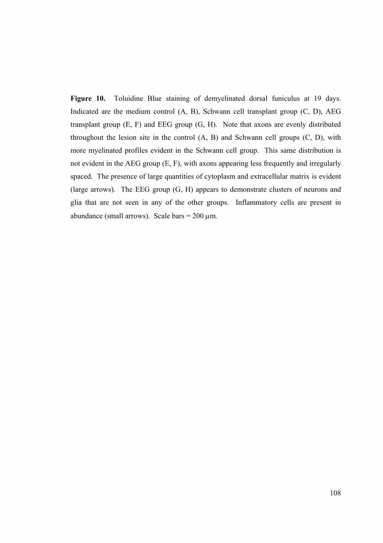
108
Figure 10. Toluidine Blue staining of demyelinated dorsal funiculus at 19 days.
Indicated are the medium control (A, B), Schwann cell transplant group (C, D), AEG
transplant group (E, F) and EEG group (G, H). Note that axons are evenly distributed
throughout the lesion site in the control (A, B) and Schwann cell groups (C, D), with
more myelinated profiles evident in the Schwann cell group. This same distribution is
not evident in the AEG group (E, F), with axons appearing less frequently and irregularly
spaced. The presence of large quantities of cytoplasm and extracellular matrix is evident
(large arrows). The EEG group (G, H) appears to demonstrate clusters of neurons and
glia that are not seen in any of the other groups. Inflammatory cells are present in
abundance (small arrows). Scale bars = 200 µm.

109
Figure 11. Electron Micrographs of demyelinated dorsal funiculus. Shown are
representative images of the medium control group (A), Schwann cell transplant group
(B), AEG transplant group (C) and EEG group (D). Axons are densely distributed
throughout the lesion in both medium control (A) and Schwann cell groups (B), with
degraded myelin profiles abundantly present in the medium control group. Large
quantities of cytoplasm and extracellular matrix separate individual axons throughout the
lesion site in the AEG transplant group (C). Axons in the EEG group are distinctly
fasciculated (arrows), and very small amounts of degraded myelin profiles are present
(D). Scale bar = 10 µm.

110
Figure 12. Quantification of myelination state. Shown are percentage counts of axons
from within the lesion site of the four different transplant groups. Axons possessing
intact myelin, degraded myelin or an unmyelinated state are represented. All transplanted
glial cell types significantly decrease the amount of degraded myelin present, and
increase levels of intact myelin. EEG in particular demonstrate a large decrease in
degraded myelin profiles and a corresponding increase in both unmyelinated and intact
myelin profiles. In all cases n=3 counted images at x1600 magnification. * indicates p <
0.05, ** indicates p < 0.01, *** indicates p < 0.001. All indicated statistical comparisons
were made against the corresponding medium only control groups. The table indicates
raw counts.

111
Figure 13. Electron micrographs of demyelinated dorsal funiculus. Depicted are AEG
transplant groups (A, B) and EEG transplant groups (C, D). Myelinated profiles are
abundant in the AEG group (A), with peripheral type myelin seen in a 1:1 relationship
with axons (black arrows). (B) AEG (white arrows) are present and discerned by their
ability to extend processes (small black arrows) around several axons (*). (C) EEG
(large arrow) are never seen to directly myelinate axons, but rather to engulf cells that
maintain a 1:1 relationship with axons (white arrowheads). (D) The myelinating cells
maintain a 1:1 relationship with axons (*) and possess a basal lamina (small arrows), a
characteristic feature of Schwann cells. Scale bars for A and C = 8 µm. Scale bars for B
and D = 2 µm.

CHAPTER 4
Extended Discussion
Chapter 4 – Extended Discussion
113
Part I
Summary
We have presented here two studies analysing different aspects of OEG
biology. In the first study (Chapter 2) we reported the effects of added
neuregulin (NRG) isoforms on the proliferation of OEG in vitro. We
demonstrated that culture conditions during the purification and expansion
phases, prior to performing a mitogenic assay, are crucial determinants of the
responsiveness of adult olfactory ensheathing glia (AEG) to added NRG.
Added mitogens to the culture medium such as forskolin and pituitary extract
can mask the responsiveness of AEG to NRG, and increase the base
proliferation rate of AEG for at least six days subsequent to their removal.
We have also reported the expression and activation patterns of ErbB
receptor subpopulations in purified p75-selected AEG, investigated their
functional role to AEG proliferation by use of ErbB antibody inhibitors, and
have related our data to previously published results in the literature. This
comparison illustrates that perceived differences in the published expression
of ErbB receptors may be attributable to variations in age of preparation and
purification techniques employed in other laboratories.
Influence of purification techniques on ErbB receptor expression
One indication that purification techniques may have an important role in
observations of OEG biology stems from our reported ErbB expression
profile utilising p75-purified AEG (Chapter 2). Here we reported that

Chapter 4 – Extended Discussion
114
ErbB2, ErbB3 and ErbB4 are expressed by AEG. These results are contrary
to previously published observations utilising postnatal olfactory ensheathing
glia (PEG; P2-P7) purified by selection for the O4 antigen and expanded
under similar culture conditions (Pollock et al., 1999; Thompson et al., 2000)
(summarised in Table 1). The discrepancy between the expression profiles of
AEG and PEG is not altogether unexpected in light of recently published
studies.
Table 1. Studies reporting mitogenic effect of NRG-1β on OEG and/or ErbB receptor
expression on OEG. Listed are the tissue source of cells utilised, the purification
techniques, culture conditions under which cells were expanded, and a summary of the
observations. Although several studies have reported increased proliferation of OEG in
the presence of NRG-1β, only a few of those studies performed concentration dose
response curves (DRC) for OEG proliferation.
Study Cells used Purification Culture Observations Chuah et al., 2000 OB-PEG AraC BPE weak proliferative DRC Pollock et al., 1999 OB-PEG O4 FACS proliferative DRC express ErbB2 + ErbB4 not ErbB3 Alexander OB-PEG O4 FACS proliferation et al., 2002 Thompson OB-PEG O4 FACS ErbB4 mRNA et al., 2000 no ErbB3 mRNA Moreno-Flores OB-P21 unpurified BPE express ErbB2 strongly, et al., 2003 forskolin ErbB3 + ErbB4 weakly Yan et al., 2001a OB-AEG p75 IP proliferative DRC Yan et al., 2001b OB-AEG p75 IP proliferation Key: OB-xEG = OEG derived from the olfactory bulb. OB-P21 = OEG derived from the olfactory bulb of young P21 animals. IP = immunopanning. FACS = fluorescence activated cell sorting. BPE = bovine pituitary extract. AraC = cytosine arabinoside. DRC = NRG-1β dose response curve.

Chapter 4 – Extended Discussion
115
Previous studies have already shown that different populations of OEG are
likely to be present within the olfactory nerve layer of the olfactory bulb (Au
et al., 2002). According to Kumar et al., (2005), only 21.3% of cells
extracted from the adult olfactory bulb are O4-positive, and of these only up
to 5% co-express p75. They also report that less than 10% of O4-positive
cells express GFAP in culture whereas 70-80% of p75 positive cells express
this marker, indicating that O4-positive and p75-positive cells may constitute
distinct populations from within the olfactory bulb. Wewetzer et al., (2005)
has also suggested that selection of PEG using O4 antigen does not occur by
means of a marker that is specific to PEG, but rather by selection of PEG that
have attached fragments of olfactory receptor neurons on their cell surface.
Although the authors also report that O4-selected PEG internalise these
axonal fragments and begin to express p75 after several days in culture, they
did not carry out comparative stains with other OEG markers such as GFAP
or S100 (Wewetzer et al., 2005). As such, the possibility remains that
selection of OEG for the O4 antigen may select a mixed population of OEG
and other cell types with O4 positive fragments of their cell surface, such as
astrocytes, fibroblasts, non-myelinating Schwann cells or oligodendrocyte
precursors.
Although the study of Moreno-Flores et al., (2003) appears to agree with our
reported expression of ErbB receptors (Table 1), one must be careful in
interpreting their study as confirmation of AEG receptor expression. In their

Chapter 4 – Extended Discussion
116
study, Moreno-Flores et al., (2003) utilised an unpurified population of
young P21 OEG. These unpurified cells were expanded in the presence of
pituitary extract and forskolin prior to analysis. Although it is possible that
an unpurified population would contain a higher proportion of OEG that are
p75-positive prior to extraction from the olfactory bulb, this population also
includes a large number of olfactory fibroblasts or meningeal cells (Barber
and Lindsay, 1982).
The question as to proliferation of contaminants is particularly important
even in purified preparations of OEG. Several different approaches have
been devised for purification of OEG. These include removal of rapidly
proliferating cells by treatment with anti-mitotics (Vincent et al., 2003),
treatment with neurotrophins (Bianco et al., 2004), positive immunoselection
for cells that express the p75 low affinity neurotrophin receptor (Ramon-
Cueto and Nieto-Sampedro, 1994; Gudino-Cabrera and Nieto-Sampedro,
1996; Ramon-Cueto et al., 1998) or the O4 antigen (Barnett et al., 1993),
negative selection of contaminating cells by means such as sorting for the
Thy-1.1 fibroblast marker (Chuah and Au, 1993; Gudino-Cabrera and Nieto-
Sampedro, 1996), differential attachment of astrocytes, macrophages and
microglia (Nash et al., 2001; Wang et al., 2005), and removal of
contaminants by a short period of trypsinisation (Ramon-Cueto and Nieto-
Sampedro, 1992). Despite the different purification techniques employed, all
of these studies still report the presence of contaminating cells in their
Chapter 4 – Extended Discussion
117
preparations: 8% for treatment with cytosine arabinoside (Vincent et al.,
2003), 20% for unpurified populations treated with neurotrophins (Bianco et
al., 2004), 3% for p75 magnetic activated cell sorting (MACS) (Gudino-
Cabrera and Nieto-Sampedro, 1996), 5-15% for p75 immunopanning
(Ramon-Cueto and Nieto-Sampedro, 1994), 3-30% for O4 selection (Barnett
et al., 1993; Franceschini and Barnett, 1996; Riddell et al., 2004), 7-20% for
differential attachment (Nash et al., 2001; Lipson et al., 2003), and 5-28%
with methods utilising removal of fibroblasts as the primary means of
purification (Chuah and Au, 1993; Gudino-Cabrera and Nieto-Sampedro,
1996). It is currently unknown if these contaminating cells express ErbB
receptors, or if these cells are able to proliferate at a rate comparable to that
of OEG once mitogens such as pituitary extract and forskolin are included in
the culture media.
Influence of tissue age on ErbB receptor expression
The age of animals from which OEG preparations are derived is another
possible variable that may account for contradictory reports on ErbB
expression. Studies utilising PEG report the expression of ErbB2 and ErbB4,
but not ErbB3 mRNA and protein (Pollock et al., 1999; Thompson et al.,
2000). Our results indicate that ErbB2 and ErbB3 mRNA and protein are
strongly expressed, and that ErbB4 protein but not mRNA is detectable in
AEG cultures (Table 2). We cannot disregard the notion that variations in
the age of preparation may play a role in this observed difference between

Chapter 4 – Extended Discussion
118
ours and other published results. This hypothesis is supported in part by the
study of Moreno-Flores et al., (2003), who utilized young P21 animals. The
authors report that ErbB2, ErbB3 and ErbB4 proteins are expressed, in
agreement with our findings and differing from studies utilising cells derived
from younger animals (Pollock et al., 1999; Thompson et al., 2000).
Other circumstantial evidence supports the hypothesis that cultured OEG
may retain developmentally regulated expression patterns (Table 2). For
example, PEG have been reported to express TrkB, but not TrkA or TrkC
receptor protein and mRNA (Woodhall et al., 2001; Vincent et al., 2003).
Meanwhile, AEG have been reported to express both protein and mRNA for
TrkA, TrkB and TrkC receptors (Bianco et al., 2004). Nevertheless, given
that these cells have been purified with different techniques, these observed
correlations do not in any way constitute proof that age of preparation may
have an effect on the expression profile of OEG in vitro. Detailed
comparative studies utilising standardised culture and purification protocols
will be nescessary to ascertain this hypothesis. However, further evidence
for potential developmentally regulated differences between OEG
populations is discussed in Part II of this chapter.
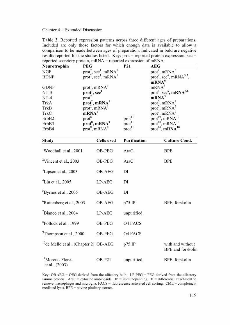
Chapter 4 – Extended Discussion
119
Table 2. Reported expression patterns across three different ages of preparations. Included are only those factors for which enough data is available to allow a comparison to be made between ages of preparation. Indicated in bold are negative results reported for the studies listed. Key: prot = reported protein expression, sec = reported secretory protein, mRNA = reported expression of mRNA. Neurotrophin PEG P21 AEG NGF prot2, sec1, mRNA1 prot4, mRNA3 BDNF prot2, sec1, mRNA1 prot4, sec6, mRNA3,5,
mRNA6 GDNF prot1, mRNA1 mRNA3
NT-3 prot1, sec1 prot4, sec6, mRNA3,6 NT-4 prot2 mRNA3 TrkA prot2, mRNA1 prot7, mRNA7
TrkB prot2, mRNA1 prot7, mRNA7 TrkC mRNA1 prot7, mRNA7 ErbB2 prot8 prot11 prot10, mRNA10 ErbB3 prot8, mRNA9 prot11 prot10, mRNA10 ErbB4 prot8, mRNA9 prot11 prot10, mRNA10
Study Cells used Purification Culture Cond. 1Woodhall et al., 2001 OB-PEG AraC BPE 2Vincent et al., 2003 OB-PEG AraC BPE 3Lipson et al., 2003 OB-AEG DI 4Liu et al., 2005 LP-AEG DI 5Byrnes et al., 2005 OB-AEG DI 6Ruitenberg et al., 2003 OB-AEG p75 IP BPE, forskolin 7Bianco et al., 2004 LP-AEG unpurified 8Pollock et al., 1999 OB-PEG O4 FACS 9Thompson et al., 2000 OB-PEG O4 FACS 10de Mello et al., (Chapter 2) OB-AEG p75 IP with and without
BPE and forskolin
11Moreno-Flores OB-P21 unpurified BPE, forskolin et al., (2003) Key: OB-xEG = OEG derived from the olfactory bulb. LP-PEG = PEG derived from the olfactory lamina propria. AraC = cytosine arabinoside. IP = immunopanning, DI = differential attachment to remove macrophages and microglia. FACS = fluorescence activated cell sorting. CML = complement mediated lysis. BPE = bovine pituitary extract.

Chapter 4 – Extended Discussion
120
Observed Mitogenic Effect of NRG on AEG
Several studies have previously reported that NRG-1β is a potent mitogen for
OEG (Pollock et al., 1999; Chuah et al., 2000; Yan et al., 2001a, b;
Alexander et al., 2002). However, only three performed dose response
curves to NRG-1β (Table 1), and the only study that is directly comparable to
our experiments in terms of age of preparation, purification methodology,
and culture conditions is that of Yan et al., (2001a). Not surprisingly, we
obtain comparable results to theirs for proliferation of AEG, with similar
dose response curves and similar levels of proliferation prior to addition of
NRG to the culture medium. Our observations indicate that peak
proliferation in the presence of NRG-1β was approximately 19%, 19% in the
presence of 2 µM forskolin, 38% in the presence of bovine pituitary extract,
45% in the presence of DF10S+mit medium (contains serum, forskolin and
pituitary extract), 45% in the presence of both NRG-1β and pituitary extract,
42% in the presence of both NRG-1β and forskolin, and 42% in the presence
of foskolin and pituitary extract. From these results we see that there is no
significant difference on AEG proliferation between any of the groups where
growth factors were tested in combination (Chapter 2). Furthermore, there is
no added benefit of including bovine pituitary extract together with NRG-1β
in the culture medium. This is likely due to the presence of NRG-1β in
pituitary extract mixtures (Raff et al., 1978; Brockes et al., 1980).
Interestingly, NRG-1β and forskolin act together to produce an additive
effect on proliferative rates, though their combined rate of proliferation is

Chapter 4 – Extended Discussion
121
once again not significantly different to that observed with pituitary extract
alone. The significance of these findings lies in the applicability of our
results to the rapid enhancement of AEG proliferation in vitro.
We suggest here that the utilisation of bovine pituitary extract in culture
media may be superfluous for the short-term proliferation of AEG. The
proliferative effect produced by a combination of NRG-1β and forskolin is
comparable to that of DF10S+mit medium. Furthermore, it has been
previously demonstrated that addition of a forskolin/NRG-1β combination to
cultured PEG is able to remove the proliferation arrest induced by astrocyte
conditioned medium, and that this proliferative effect is maintained over long
periods in culture (Alexander et al., 2002). Removal of pituitary extract from
AEG expansion protocols would eliminate another potential source of
variability found in experimental procedures, as the exact contents of
pituitary extracts vary depending on individual stock numbers and
manufacturers. Furthermore, pituitary extract contains other AEG
proliferative factors including FGF and PDGF, whose effect on other aspects
of AEG biology are still unknown (Gospodarowics et al., 1983; Ueno et al.,
1986; Halper et al., 1992; Chuah and Teague, 1999; Pollock et al., 1999; Au
and Roskams, 2003; Yan et al., 2001a, 2003).
We do however hesitate to recommend that the combination of NRG-1β and
forskolin be used to rapidly proliferate OEG preparations in vitro. Firstly,
Chapter 4 – Extended Discussion
122
we do not yet know whether p75-selected preparations from younger animals
(EEG or PEG) express the various ErbB receptor subtypes, or if NRG-1β is
capable of exerting a mitogenic effect upon those cells. Secondly, we do not
at this stage fully understand the implications of utilising these factors to
proliferate OEG in vitro, and the subsequent impact on proliferation and axon
interactions after transplantation in vivo. For example, application of cAMP
analogues to astrocytes in vitro have been found to induce measurable effects
post-transplantation (Chu et al., 1999). The converse is also true, with
injections of cAMP analogues in vivo demonstrating distinct effects several
days later on the extracted cells in vitro (Neumann et al., 2002). Vincent et
al., (2003) have also demonstrated that alterations of intracellular cAMP
levels can provoke drastic morphologic shifts in cultured PEG, and
hypothesised that OEG may be incredibly plastic cells capable of adjusting to
new environments by adopting a variety of different phenotypes. Given that
forskolin is a cAMP-inducing agent (Seamon et al., 1981; Fradkin et al.,
1982), we would strongly recommend that further studies be performed into
the effects of forskolin on cultured OEG in vitro and after transplantation in
vivo.

Chapter 4 – Extended Discussion
123
Part II
Summary
Our second study (Chapter 3) investigated the ability of OEG derived from
three different ages of animals to myelinate DRG neurons in vitro, and to
remyelinate the demyelinated dorsal funiculus of rats in vivo. We
demonstrated that age of preparation is a possible determinant of myelination
potential of OEG, and that this potential is unaffected by either axonal calibre
or the addition of factors known to enhance Schwann cell myelination in
vitro. That is, in our hands, EEG are able to produce myelin in vitro but PEG
and AEG are not. Furthermore, our results show that preparations derived
from embryonic animals interact very differently with the demyelinated
spinal cord environment, and that clearance of degraded myelin appears to be
more effective when animals are transplanted with EEG rather than AEG or
Schwann cell preparations.
Interaction of OEG with axons
The failure of our Lv-LacZ label to provide a strong consistent signal was
unfortunate in that we were consequently not able to answer the question as
to whether OEG can directly myelinate axons in vivo. Though faint label
could be visualised within non-myelinating cells of the EEG transplant
groups, no label was seen in any of the myelinating cells. Furthermore, no
label was visualised in electron micrographs of Schwann cell and AEG-

Chapter 4 – Extended Discussion
124
transplanted groups. This brings to light the question of whether the
lentivirus label is being downregulated in cells closely associated with axons,
and whether AEG do indeed have the capacity to myelinate as suggested by
several researchers (Sasaki et al., 2004; Li et al., 1997, 1998, 2003b).
All the data to date indicate that AEG appear to lack the ability to myelinate
axons in vitro under all culture conditions tested so far (Plant et al., 2002; de
Mello et al., Chapter 3). Here we provide, to our knowledge for the first
time, that the ability of OEG to myelinate DRG axons in vitro may be
dependant primarily on the age of the animal from which the glial cells were
extracted. Furthermore, we have demonstrated that there is an age-dependent
variation in the behaviour of OEG following transplantation into a
demyelinating lesion of the adult rat spinal cord, and that OEG derived from
embryonic animals appear to be significantly more effective at inducing
clearance of degraded myelin from chemically demyelinated dorsal
funiculus. Still unadressed by our study however, is the underlying
mechanism as to why EEG appear to be significantly better at preserving the
integrity of the lesion site following a demyelinating lesion, and which
mechanisms are responsible for the increased clearance of degraded myelin
profiles observed within our EEG group. Our data suggest that the answer to
this question appears to lie in the ability of the different transplant groups to
induce phagocytosis of degraded myelin within the lesion site.

Chapter 4 – Extended Discussion
125
Mechanisms of action by OEG in vivo
Though our study did not investigate the effect on recruitment of phagocytic
cells into the demyelinated area, it nevertheless remains likely that the
observed increase in clearance of degraded myelin must be, at least in part,
due to the effect of EEG on recruited cells. In a normal lysolecithin
demyelination model, the inflammatory response is quickly activated leading
to invasion of T cells, neutrophils and monocytes within the first 6-12 hours
(Kume et al., 1992; Ousman and David, 2000). Though many of the
invading cells remain present only transiently, activated macrophages will
continue to clear degraded myelin from the target area up to 4 weeks post-
lesion (Gilson and Blakemore, 1993; Pavelko et al., 1998). It is possible that
EEG are chemotactic for these inflammatory cells, may induce other CNS
cells to increase production of such chemotactic factors, or may prolong the
initial period of monocyte recruitment and activation.
No studies to date have focused on the ability of OEG to produce factors that
may be chemotactic for macrophages, a mechanism that appears to be of
critical importance given the suggestion by some researchers that a robust
macrophage response may be associated with efficient remyelination and
clearance of the lesion site (Graca and Blakemore, 1986; Perry et al., 1987;
David et al., 1990; George and Griffin, 1994; Morell et al., 1998; Rapalino et
al., 1998). A number of molecules such as tumour necrosis factor (TNF)-α,
interleukin (IL)-1, and IL-6 have been shown to increase recruitment and

Chapter 4 – Extended Discussion
126
activation of monocytes and microglia in the spinal cord (Giulian et al., 1989;
Schnell et al., 1999b; Klusman and Schwab, 1997; Smith et al., 1998). Other
candidate molecules that have been implicated in chemoattraction of
monocytes or in mediation of myelin phagocytosis include IP-10 (Luster and
Ravetch, 1987), MAC-2 (Reichert et al., 1994), monocyte chemoattractant
protein (MCP)-1α (Toews et al., 1998), IL-8, IL-10, growth-related oncogene
(GRO)-α, macrophage inflammatory protein (MIP)-1α, and granulocyte
macrophage-colony stimulating factor (GM-CSF) (Bartholdi and Schwab,
1997; Von Zahn et al, 1997; McTigue et al., 1998; Smith et al., 1998;
Ousman and David, 2001; Ma et al, 2002). Further studies will be necessary
to examine the ability of OEG either to directly express these or other unique
molecules, or to induce their expression at the site of OEG transplantation.
Furthermore, extracellular molecules such as VCAM-1, ICAM-1,
fibronectin, laminin and collagen type I have been associated with increased
adhesion of monocytes and/or increased functional activity of macrophages
and microglia (Newman and Tucci, 1990; Chamak and Mallat, 1991; Carlos
et al., 1991; Ley, 1996). Though several studies have thus far indicated that
AEG are capable of depositing large quantities of laminin and fibronectin in
vitro (Ramon-Cueto and Nieto-Sampedro, 1992; Sonigra et al., 1999; Au and
Roskams, 2003) and in vivo (Ramer et al., 2004a, b), the expression profiles
of other extracellular molecules after transplantation of OEG into lesioned
spinal cord remains unknown. To date, no comparison has been made

Chapter 4 – Extended Discussion
127
between EEG and older OEG preparations regarding the production of these
molecules, nor have any studies broadened their scope to include the other
molecules mentioned above.
Another possibility that has yet to be addressed is the effect of OEG
preparations on the integrity of the blood-brain barrier and its subsequent
effect on infiltration of endogenous cells into the lesion site (Andersson et al.,
1992; Riva-Depaty et al., 1994), or the simple fact that increased myelin
clearance may be attributable at least in part to direct phagocytosis by
recruited Schwann cells (Holtzman and Novikoff, 1965; Stoll et al., 1989;
Reichert et al., 1994; Fernandez-Valle et al., 1995; Liu et al., 1995b; Hirata et
al., 1999). Direct phagocytosis by OEG is another factor that must be
considered, given their ability to internalise degraded axonal material in vitro
(Wewetzer et al., 2005) and in injury models of the olfactory bulb in vivo
(Chuah et al., 1995; Susuki et al., 1996; Li et al., 2005). Though none of
these possibilities have been addressed in our study, they remain open for
investigation in future studies of OEG transplantation in the spinal cord.
Finally, the effect of OEG transplantation on the expression of molecules
directly associated with myelin destruction and phagocytosis during nerve
degeneration remains completely unknown. Apolipoprotein E (ApoE) for
example, has been implicated with recycling of myelin lipids for axonal
regeneration (reviewed by Vance et al., 2000). ApoE is produced by

Chapter 4 – Extended Discussion
128
macrophages and fibroblasts in response to nerve injury (Snipes et al., 1986;
Boyles et al., 1989; Saada et al., 1995), at which time Schwann cells
proximate to damaged neurites upregulate expression of ApoE low density
lipoprotein receptors (Rothe and Muller, 1991; Vance et al., 2000).
Unfortunately, no information is currently available on the expression or
induction of ApoE by OEG, though such studies are urgently needed. For
example, ApoE production by olfactory fibroblasts would go a long way
towards explaining why Li et al., (1997, 1998, 2003b) and other groups
(Appendix A) insist that unpurified (ie. fibroblast containing) populations of
OEG have a greater potential to promote regrowth and repair of lesioned
spinal cord than purified preparations. Conversely, a detailed analysis of low
density lipoprotein receptor expression by OEG would provide valuable
clues as to the underlying mechanisms by which these cells promote
improved clearance of myelin.
In any case, it stands to reason that the ability of OEG to clear degraded
myelin profiles quickly and efficiently may be one of the central aspects
behind their ability to promote axonal regeneration in the lesioned spinal
cord. It is well known that in the PNS regeneration can only proceed after
injured tissue components have been cleared via Wallerian degeneration
(Hirata and Kawabuchi, 2002). Furthermore, several studies have
demonstrated that macrophages can alter the nonpermissive adult CNS to a
state that permits axonal growth after injury (David et al., 1990; Lazarov-
Chapter 4 – Extended Discussion
129
Spiegler et al., 1996, 1998; Prewitt et al., 1997; Rapalino et al., 1998), a
situation reflected by the lack of secondary damage during macrophage
recruitment in lysolecithin demyelination models (Hall, 1972; Gregson,
1989; Jeffery and Blakemore, 1995). Future studies exploring this possibility
are encouraged, with special attention paid to the differences of the CNS
macrophage response upon transplantation of OEG derived from various
stages of development. However, any such future studies must pay special
attention to the type of injury used, as macrophage responses in the CNS
have been reported to vary depending on the type and location of injury
(Hirata et al., 1999; Schnell et al., 1999a).
Influence of preparation age on promotion of axon growth
Unfortunately, no clear picture has emerged from the published literature
relating the age from which OEG were extracted and the extent of
functional/axonal regeneration observed. One reason for this lies with the
paucity of published studies that have investigated the use of
purified/unpurified EEG transplants into the lesioned CNS (Smale et al.,
1996; Boyd et al., 2004a). Furthermore, no clear interpretation of the
literature can be made between studies utilising PEG and AEG. This is due
to the large number of other variables present between the various studies
undertaken to date, including type and extent of lesion, location of lesion,
method of purification and expansion of the transplanted cells, time after

Chapter 4 – Extended Discussion
130
injury at which the transplant was carried out, and parameters utilised to
determine functional and anatomical recovery.
For example, several studies have utilised dorsal funiculus transection or
crush injury to analyse the effects of OEG transplantation (Li et al., 1997,
1998, 2003a; Imaizumi et al., 2000b; Nash et al., 2002; Shen et al., 2002;
Keyvan-Fouladi et al., 2003; Andrews and Stelzner, 2004; Polentes et al.,
2004; Sasaki et al., 2004, 2006; Ramer et al., 2004a; Richter et al., 2005;
Ruitenberg et al., 2005a). Of these, none have utilised EEG transplantation
and only three have utilised PEG (Imaizumi et al., 2000b; Ramer et al.,
2004a; Richter et al., 2005), the remainder using AEG (Li et al., 1997, 1998,
2003a; Nash et al., 2002; Shen et al., 2002; Keyvan-Fouladi et al., 2003;
Andrews and Stelzner, 2004; Polentes et al., 2004; Sasaki et al., 2004, 2006;
Ruitenberg et al., 2005a). Two of the studies utilising PEG transplantation
used cells derived from the olfactory bulb of rats (Imaizumi et al., 2000b;
Richter et al., 2005), whereas the third study utilised mouse PEG derived
from the lamina propria (Ramer et al., 2004a). Finally, the three studies on
PEG utilised different purification and/or expansion techniques. Imaizumi et
al., (2000b) performed a purification method similar to that used by Chuah
and Au (1993), where PEG are treated with cytosine arabinoside to remove
some contaminating cells, immunoadsorbed with antiserum to Thy-1.1 to
remove fibroblasts, and expanded rapidly by the addition of bovine pituitary
extract to the culture medium. Ramer et al., (2004a) utilised PEG, purified
Chapter 4 – Extended Discussion
131
by Thy-1.1 complement mediated lysis and expanded without added
mitogens in the culture medium. Richter et al., (2005) utilised PEG purified
by p75 immunopanning and expansion of cells in medium without mitogens
added. The type and location of lesion performed in either study was also
different, with Imaizumi et al., (2000b) performing transverse cuts along the
dorsal aspect of the lumbar spinal cord, whereas Ramer et al., (2004a) and
Richter et al., (2005) performed a dorsolateral funiculus crush of the cervical
spinal cord. Finally, the means by which anatomical and functional recovery
are assessed also varied greatly between each different study. The large
number of variables is also reflected in studies utilising transplantation of
AEG into the damaged dorsal funiculus, making interpretation of the
literature superficial at best.
The only model that can be compared and contrasted effectively is the
transplantation of OEG of different ages into the lesioned dorsal roots of the
spinal cord. Several studies have investigated the ability of OEG to restore
functional and anatomical connections in this lesion system (Ramon-Cueto
and Nieto-Sampedro, 1994; Navarro et al., 1999; Taylor et al., 2001; Pascual
et al., 2002; Gomez et al., 2003; Li et al., 2004; Ramer et al., 2004b; Riddell
et al., 2004). Suspensions of AEG were utilised in some of these studies
(Ramon-Cueto and Nieto-Sampedro, 1994; Navarro et al., 1999; Taylor et
al., 2001; Pascual et al., 2002; Gomez et al., 2003; Li et al., 2004) whereas
PEG were used in others (Ramer et al., 2004b; Riddell et al., 2004). The

Chapter 4 – Extended Discussion
132
results of these studies are summarised in Table 3. Briefly, restoration of
function and regrowth of ascending sensory axons past the dorsal root entry
zone and into the spinal cord was only observed in groups transplanted with
p75-selected AEG (Ramon-Cueto and Nieto-Sampedro, 1994; Navarro et al.,
1999; Taylor et al., 2001; Pascual et al., 2002) and unpurified AEG (Li et al.,
2004), but not AEG that were purified by removal of fastly adhering cells
and negative selection of fibroblasts (Gomez et al., 2003). Groups that did
not utilize AEG but used PEG instead (Ramer et al., 2004b; Riddell et al.,
2004) did not observe any significant regrowth of axons into the spinal cord
or restoration of function.
Gomez et al., (2003) suggested some of the differences observed between
these studies could be accounted for by variations in the extent of rhizomy in
nearby roots that could have allowed spared axons to sprout into the lesioned
area. This explanation is feasible, explaining why regrowth of fibers is
observed in studies utilising rhizotomy of 1-3 roots (Ramon-Cueto and
Nieto-Sampedro, 1994; Navarro et al, 1999; Pascual et al., 2002; Li et al.,
2004), but fails to account for the observed regrowth in studies where 7
dorsal roots were transected (Taylor et al., 2001), or the lack of regrowth in
other studies where only one dorsal root was severed (Riddell et al., 2004).
Another possibility not addressed by the authors is that the age animal from
which the cells are derived, or the purification techniques utilised, may be an

Chapter 4 – Extended Discussion
133
important factor accounting for the differences observed between these
studies. It is noteworthy that functional and anatomical regeneration is
observed only in studies utilising p75-selected AEG, or unpurified AEG
(Table 3). Groups utilising PEG (Ramer et al., 2004b; Riddell et al., 2004)
have failed to observe significant functional or anatomical recovery, as has
one study utilising AEG purified by negative immunoselection of fibroblasts
(Gomez et al., 2003). Two of the studies reporting negative results utlised
methods to eliminate contaminating fibroblasts from their cultures (Gomez et
al., 2003; Ramer et al., 2004b) whereas the third study utlised selection for
the O4 antigen as the primary means of purification (Riddell et al., 2004).
Selection for the O4 antigen may select for a subpopulation of OEG that do
not possess the full regenerative potential of p75-selected OEG (Kumar et al.,
2005). Our laboratory has also recently demonstrated that cultured
preparations of olfactory bulb cells possess different subpopulations of
fibroblasts, and that not all of these populations may express Thy-1.1 (Sophie
Callender, unpublished observations). This is of particular importance as any
contaminating cells in the preparation may proliferate once in contact with
damaged spinal cord tissue (Woodhouse et al., 2005) and become an
unknown factor during the repair stage in vivo. These observations highlight
the importance of future studies in defining and standardising the basic
methodology surrounding the extraction and purification of cells for
transplantation, an aspect that has often been neglected in OEG studies to
date.

Chapter 4 – Extended Discussion
134
Table 3. Studies utilising transplantation of OEG into transected spinal cord dorsal roots.
Study Cells used Purification Observations Ramon-Cueto and OB-AEG p75 IP Regeneration and ingrowth of Nieto-Sampedro, 1994 ascending sensory fibers into
contralateral dorsal horn Navarro et al., 1999 OB-AEG p75 MACS Restoration of spinal reflex
arcs. Regenerating ascending axons crossed DREZ
Taylor et al., 2001 OB-AEG p75 IP restoration of biceps reflex
activity and sensory input Pascual et al., 2002 OB-AEG p75 MACS restoration of sensory stimuli
implying regrowth of sensory axons through DREZ
Li et al., 2004 OB-AEG unpurified regrowth through DREZ, into
grey matter of dorsal horn and ascending dorsal columns
Gomez et al., 2003 OB-AEG DI, Thy1.1 No significant increase in
–ve MACS sensory afferent regrowth Riddell et al., 2004 OB-PEG O4 FACS No increase in ascending
fiber ingrowth. No detectable post-synaptic activity
Ramer et al., 2004b LP-PEG Thy1.1 CML No increase in sensory
afferent regrowth. Key: OB-xEG = OEG derived from the olfactory bulb. LP-PEG = PEG derived from the olfactory lamina propria. IP = immunopanning, MACS = magnetic activate cell sorting. FACS = fluorescence activated cell sorting. CML = complement mediated lysis. DREZ = dorsal root entry zone.
Despite these interesting correlations in published studies in vivo, there is no
evidence to suggest that age of preparation is a crucial factor influencing the
growth promoting (as opposed to myelinating) abilities of OEG in vitro.
Promotion of axonal growth by OEG has been reported in embryonic (Denis-
Donini and Estenoz, 1988; Kafitz and Greer; 1998, 1999), postnatal (Chuah
and Au, 1994; Le Roux and Reh, 1994; Tisay and Key, 1999; Van Den Pol

Chapter 4 – Extended Discussion
135
and Santarelli, 2003), young P21 (Moreno-Flores et al., 2003) and adult
preparations (Ramon-Cueto et al., 1993; Sonigra et al., 1999; Gudino-
Cabrera and Nieto-Sampedro, 2000; Gomez et al., 2003; Lipson et al., 2003;
Agrawal et al., 2004; Kumar et al., 2005; Leaver et al., 2006) (Table 4).
Furthermore, a comparative study by Goodman et al., (1993) has found that
selected immortalised cell lines of both PEG and AEG are able to promote
growth of embryonic chick retinal ganglion cells similarly, and concluded
that OEG appear to retain their ability to promote growth throughout
development. It would however be interesting to undertake a comparative
study between primary cultures of AEG, PEG and EEG.
Three studies to date have reported that diffusible factors released by OEG
are able to promote neurite growth in vitro (Le Roux and Reh, 1994; Kafitz
and Greer, 1998, 1999; Chung et al., 2004). One group reported a strong
growth promoting effect by unpurified EEG conditioned medium (Kafitz and
Greer, 1998, 1999), whereas only a weak growth promoting effect was
observed by groups utilising unpurified PEG (Le Roux and Reh, 1994;
Chung et al., 2004). One further study has reported that PEG are capable of
releasing axonal growth-promoting neurotrophic factors in vivo (Chuah et al.,
2004). Meanwhile, studies investigating AEG populations in vitro (Sonigra
et al., 1999; Lipson et al., 2003; Leaver et al., 2006) or PEG cultures
eliminated from fibroblasts (Chuah and Au, 1994) have failed to detect an
effect of OEG diffusible factors on neurite growth. To date, no study has

Chapter 4 – Extended Discussion
136
contrasted the effect of preparation age on the extent of neurite growth
promotion, nor has a detailed study been performed contrasting age of
preparation on release of diffusible factors by OEG.
Table 4. Studies investigating the promotion of neuron growth by primary OEG cultures.
OEG are cultured in DMEM supplemented with 10% FCS in all studies presented.
Additional factors present in the culture medium are listed where appropriate.
Study Cells used Purification Culture Growth Agrawal et al., 2004 OB-AEG unpurified Yes Kumar et al., 2005 OB-AEG p75 IP forskolin Yes ONR-AEG and GGF2 Gomez et al., 2003 OB-AEG DI, Thy1.1 Yes
–ve MACS Gudino-Cabrera and OB-AEG unpurified Yes Nieto-Sampedro, 2000 Ramon-Cueto et al., 1993 OB-AEG unpurified Yes Leaver et al., 2006 OB-AEG p75 IP forskolin Yes. Contact BPE mediated only. Lipson et al., 2003 OB-AEG DI Yes. Contact mediated only Sonigra et al., 1999 OB-AEG unpurified Yes. Contact mediated only Moreno-Flores et al., 2003 OB-P21 unpurified forskolin Yes BPE Chuah et al., 2004 (in vivo) OB-PEG AraC BPE Yes. Diffusible
factors. Chung et al., 2004 OB-PEG and AraC BPE Yes. Diffusible OM-PEG factors Chuah and Au, 1994 OB-PEG AraC and BPE Yes. Contact
Thy1.1 IA mediated only Le Roux and Reh, 1994 OB-PEG unpurified Yes. Diffusible factors
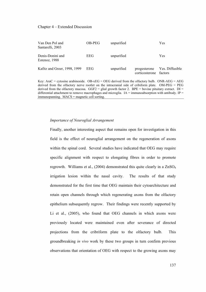
Chapter 4 – Extended Discussion
137
Van Den Pol and OB-PEG unpurified Yes Santarelli, 2003 Denis-Donini and EEG unpurified Yes Estenoz, 1988 Kafitz and Greer, 1998, 1999 EEG unpurified progesterone Yes. Diffusible corticosterone factors Key: AraC = cytosine arabinoside. OB-xEG = OEG derived from the olfactory bulb. ONR-AEG = AEG derived from the olfactory nerve rootlet on the intracranial side of cribiform plate. OM-PEG = PEG derived from the olfactory mucosa. GGF2 = glial growth factor 2. BPE = bovine pituitary extract. DI = differential attachment to remove macrophages and microglia. IA = immunoabsorption with antibody. IP = immunopanning. MACS = magnetic cell sorting.
Importance of Neuroglial Arrangement
Finally, another interesting aspect that remains open for investigation in this
field is the effect of neuroglial arrangement on the regeneration of axons
within the spinal cord. Several studies have indicated that OEG may require
specific alignment with respect to elongating fibres in order to promote
regrowth. Williams et al., (2004) demonstrated this quite clearly in a ZnSO4
irrigation lesion within the nasal cavity. The results of that study
demonstrated for the first time that OEG maintain their cytoarchitecture and
retain open channels through which regenerating axons from the olfactory
epithelium subsequently regrow. Their findings were recently supported by
Li et al., (2005), who found that OEG channels in which axons were
previously located were maintained even after severance of directed
projections from the cribriform plate to the olfactory bulb. This
groundbreaking in vivo work by these two groups in turn confirm previous
observations that orientation of OEG with respect to the growing axons may

Chapter 4 – Extended Discussion
138
be a critical factor in neuronal guidance (Sonigra et al., 1999; Van Den Pol
and Santarelli, 2003). Though significant progress has been made to date
utilising suspensions of isolated cells transplanted into lesions of the CNS,
this recent work brings to light another possible means of improving
functional outcomes, provided a means can be found to reproduce these
specific neuroglial arrangements and open channels prior to transplantation
of tissue bridges across the damaged area.
Furthermore, questions arise as to how such organised OEG structures
respond when the age of the preparation is varied. Is the reason why we
observe clear channels of EEG in our lysolecithin demyelinated animals due
to use of cells that are still more responsive to axonal cues and thus able to
rearrange themselves easily into growth promoting clusters/channels around
the axons of the spinal cord? Can AEG be induced to form such
arrangements in cultured matrices prior to transplantation into the cord, and if
so do these arrangements encourage directed regrowth of fibres as the work
of Williams et al., (2004) and Li et al., (2005) suggests they would? Taking
a step backwards, what would we have observed in our demyelinated model
had we also transected the cord? Would the transplanted EEG in our model
maintain their neuroglial arrangements following spinal transection and
subsequent degeneration of axons similarly to OEG located in the olfactory
system? Further studies into transplantation of olfactory nerve layer or
lamina propria into the lesioned spinal cord would provide a logical

Chapter 4 – Extended Discussion
139
framework for further studies, utilising olfactory tissues that have been
previously cleared of axons but that still retain an AEG glial arrangement that
may be conducive to growth. Another simpler approach has already been
initiated by De Mello and colleagues utilising time-lapse video microscopy to
answer the basic question of whether OEG from different age preparations
align themselves with growing axons in vitro, or whether growing axons
merely follow OEG paths already set prior to axonal contact.
Future Directions
Future studies analysing the regenerative potential of OEG derived from
animals of different ages will have to be performed with careful
consideration of all of the above. Several well designed studies have already
compared the benefits of acute vs delayed transplantation of AEG into the
spinal cord (Plant et al., 2003; Lopez-Vales et al., 2006), the benefits of
injecting the OEG directly into the lesion site or at points located more
distally to the injury (Andrews and Stelzner, 2004; Ramer et al., 2004b;
Richter et al., 2005), and the benefits of utilising PEG derived from the
olfactory bulb compared to PEG derived from the lamina propria of the
olfactory mucosa (Richter et al., 2005). Whereas all of these studies have
been controlled sufficiently to allow comparative analyses of the treatments
in question, there are still many factors that will need to be systematically
addressed by future reserch:
Chapter 4 – Extended Discussion
140
1) Does the age animal from which the OEG preparation is derived have a
significant effect on tissue sparing, cavity formation, axonal regeneration and
sprouting of spinal cord axons in a partial transection model, a contusion
model, a dorsal rhizotomy model and/or in a complete transection model?
2) Do culture conditions prior to transplantation affect the observed
functional/anatomical effects of the transplanted cells?
3) Does the location of the olfactory pathway (olfactory mucosa vs olfactory
bulb) from which the cells are derived have an effect on regeneration or
sparing in the aforementioned models? How are these responses affected by
the age of preparation?
4) Does the neuroglial arrangement of the transplanted cells affect the observed
functional/anatomical recovery?
5) Does acute vs delayed transplantation affect regenerative responses? How
does the site of injection affect the observed responses?
Concluding Remarks
We have demonstrated here in two studies that OEG populations are cells
possessing remarkable plasticity. The manner in which these cells respond
both in vitro and in vivo appears to be primarily dependant upon the age of
the animal from which they were extracted. Further investigation of the
literature seems to indicate that these age-dependant variations in behaviour
are not restricted to myelination potential and integration within the

Chapter 4 – Extended Discussion
141
demyelinated spinal cord, but that they are potentially a large source of
variation between results by different laboratories in other models of CNS
injury. We have also demonstrated in Chapter 2 that the conditions in which
the cells are grown following extraction can influence their responsiveness to
growth factors in vitro, and that these effects are persistent for a minimum of
six days after withdrawal of the impingent culture conditions. Every
laboratory working with OEG utilises their own individual means of
expanding these cells post-extraction. In this manner, the same cell type
extracted with similar protocols in two different laboratories may in fact also
behave significantly differently in subsequent experiments.
This wide variation in different aspects of in vitro and in vivo experiments
employing OEG should be discussed in appropriate forums if we are to
reduce variability of results in this field and increase cross applicability of
collected results. Another problem that remains unaddressed by our two
studies presented here is the issue of OEG purification techniques employed
in different laboratories. Though more studies have recently begun to
address these issues (Kumar et al., 2005), much work remains to be done to
contrast the effects of different purification techniques on the regenerative
potential of OEG. In the past 15 years giant strides forward have been made
into the study of OEG biology, but much remains to be done before clinical
transplantation of these cells can be performed with support of the existing
body of knowledge fully behind it.

APPENDIX A
Appendix A
143
Included in this appendix is a table summarising the various methods utilised
to culture OEG. Understanding the impact of variations such as source of the
cells, age of animal from which they are extracted, purification methods, and
mitogenic factors added to the tissue culture medium during the expansion
phases in vitro are vital to understanding the impact of each of these studies in
the field of OEG biology. Included in this table are all studies located by the
author that have utilised OEG derived from rodent tissue. Not included here
are studies utilising immortalised cell lines, cells derived from sources other
than the rodent, and studies that have utilised biopsies of olfactory tissue.
This table provides a quick reference guide for comparison of results obtained
from different laboratories utilising transplanted or cultured OEG.
Appendix A
144
Abbreviation Key Cells used If multiple cell types are listed, the authors have utilised multiple preparations as part of their study.
OE-xEG = OEG derived from the olfactory neuroepithelium ONR-xEG = OEG derived from olfactory nerve rootlets OM-xEG = OEG derived from the olfactory mucosa LP-xEG = OEG derived from the lamina propria of the olfactory
mucosa Purification If multiple purification methods are listed then the authors have compared use of the various treatments. Where used, the term 'and' indicates that those purification methods were used sequentially in the preparation.
AraC = treatment with cytosine arabinoside CML = complement mediated lysis DI = purification by differential attachment rates of
macrophages and fibroblasts FACS = Fluorescence activated cell sorting IA = immunoadsorption IP = immunopanning MACS = magnetic cell sorting NT-3 = purification was performed by the addition of
neurotrophin-3 to the culture medium BDNF = purification was performed by the addition of BDNF
to the culture medium. ST = removal of contaminants by a short period of
trypsinisation Culture conditions Culture medium in all cases is comprised of DMEM or DMEM/F12 (50:50 v/v) supplemented with 5%-10% FCS. Listed here are additional growth factors added to the culture medium to which the author would like to draw special attention. Factors such as L-Glutamine, glucose, gentamycin, etc, are not included here.
ACM = astrocyte conditioned medium BPE = bovine pituitary extract FGF2 = fibroblast growth factor 2 GGF2 = glial growth factor 2

Appendix A
145
Observations A brief summary of the major findings of the study, with emphasis on cultured and transplanted results. 5HT = serotonergic axons bFGF = basic fibroblast growth factor
BDNF = brain derived neurotrophic factor BPE = bovine pituitary extract CN = cortical neurons CNP = 2',3'-Cyclic nucleotide 3'-Phosphodiesterase CGRP = calcitonin gene-related peptide, axons positive for CSPG = chondroitin sulfate proteoglycans CST = corticospinal tract DRG = dorsal root ganglion ECM = extracellular matrix GFAP = glial fibrillary acidic protein IGF-1 = insulin growth factor-1 LPA = lysophosphatidic acid OM = olfactory mucosa MBP = myelin basic protein MEP = motor evoked potential MRI = magnetic resonance imaging
NOR = noradrenergic axons NT-3 = neurotrophin-3 PDGF-BB = platelet-derived growth factor BB
RGC = retinal ganglion cell RST = rubrospinal tract SC = Schwann cells SSEP = somatosensory evoked potential
TH = tyrosine hydroxilase X-EB = X irradiation and Ethidium Bromide

Study Cells used Purification Culture Observations Agrawal et al., 2004 OB-AEG unpurified in vivo: improvement in SLA increase TH +ve fiber
density in 6-OHDA lesion of nigrostriatal pathway. In vitro: improved growth of TH +ve neurons.
Alexander et al., 2002 OB-PEG O4 FACS in vitro: investigated long-term proliferation effects of
different combinations of mitogens to 22 days. Andrews and OB-AEG DI in vivo: promotion of growth in bilateral crush of cord is Stelzner, 2004 additive with promotion by sciatic nerve conditioning
lesion. Au and Roskams, 2002 OM-PEG Thy-1.1 CML in vitro: describe culturing technique for mouse PEG. Au and Roskams, 2003 LP-PEG Thy-1.1 CML in vitro: characterised expression profile differs to OB-
xEG. bFGF is mitogenic but not in long-term. Barakat et al., 2005 OB-AEG p75 IP forskolin, BPE in vivo: poor survival, promotion of growth, and
restoration of function in contused cord. Barber and Lindsay, 1982 OB-AEG, OB-PEG unpurified in vitro: characterise expression and morphology of OM-AEG, OM-PEG cultures from bulb and mucosa, from adult and neonate. Barnett et al., 1993 OB-PEG O4 FACS in vitro: describe culturing technique and cell
characteristics.
Bianco et al., 2004 LP-AEG unpurified in vitro: NT-3 promotes proliferation of GFAP +ve NT-3 olfactory cells. Expression profile analysis performed. BDNF
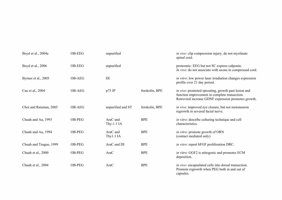
Boyd et al., 2004a OB-EEG unpurified in vivo: clip compression injury, do not myelinate spinal cord. Boyd et al., 2006 OB-EEG unpurified proteomic: EEG but not SC express calponin. In vivo: do not associate with axons in compressed cord. Byrnes et al., 2005 OB-AEG DI in vitro: low power laser irradiation changes expression
profile over 21 day period. Cao et al., 2004 OB-AEG p75 IP forskolin, BPE in vivo: promoted sprouting, growth past lesion and
function improvement in complete transection. Retroviral increase GDNF expression promotes growth.
Choi and Raisman, 2005 OB-AEG unpurified and ST forskolin, BPE in vivo: improved eye closure, but not motoneuron
regrowth in severed facial nerve. Chuah and Au, 1993 OB-PEG AraC and BPE in vitro: describe culturing technique and cell Thy-1.1 IA characteristics. Chuah and Au, 1994 OB-PEG AraC and BPE in vitro: promote growth of ORN
Thy1.1 IA (contact mediated only). Chuah and Teague, 1999 OB-PEG AraC and DI BPE in vitro: report bFGF proliferation DRC. Chuah et al., 2000 OB-PEG AraC BPE in vitro: GGF2 is mitogenic and promotes ECM
deposition. Chuah et al., 2004 OB-PEG AraC BPE in vivo: encapsulated cells into dorsal transection.
Promote regrowth when PEG both in and out of capsules.

Chung et al., 2004 OB-PEG and AraC BPE in vitro: promote growth of CN (mediated by contact OM-PEG and diffusible factors) Collazos-Castro et al., 2005 OB-AEG DI and in vivo: no regrowth of CST axons and no improvement -ve Thy-1.1 MACS of function in contused cord. Deni-Donini and OB-EEG unpurified in vitro: promote growth of dopaminergic neurons from Estenoz, 1988 substantia nigra. Devon and Doucette, 1992 OB-EEG unpurified in vitro: myelinate DRG neurons. Devon and Doucette, 1995 OB-EEG unpurified in vitro: myelinate DRG neurons without ascorbate. Doucette, 1993b OB-EEG unpurified in vitro: describe phenotypic features and effect of ACM
or cAMP analogues. Doucette and Devon, 1994 OB-PEG unpurified in vitro: attempt to promote expression of myelinating
phenotype (neuron-free cultures). MBP not expressed under any condition.
Doucette and Devon, 1995 OB-EEG unpurified in vitro: attempt to promote expression of myelinating
phenotype (neuron-free cultures). MBP not expressed under any condition.
Dunning et al., 2004 OB-PEG AraC and ST forskolin, in vivo: tracked cells via MRI. Report remyelination of and p75 IP heregulin X-EB cord. Fairless et al., 2005 OB-PEG O4 FACS FGF2, ACM, in vitro: move easily over astrocyte cultures, SC do not. forskolin, BPE N-cadherin important for SC but not PEG movement.

Fouad et al., 2005 OB-AEG p75 IP forskolin, BPE in vivo: combined therapy with SC and chondroitinase in complete cord transection, increased functional outcomes and growth of 5HT fibers.
Franceschini and OB-PEG O4 FACS ACM in vivo: p75 and E-N-CAM define two populations of Barnett, 1996 OEG in bulb. In vitro: Majority of cells p75-ve but gain
p75 over time in culture. Garcia-Alias et al., 2004 OB-AEG p75 MACS in vivo: improve tissue preservation, restoration of
behavioral skills and physiological outcome in photochemical lesion of cord.
Gomez et al., 2003 OB-AEG DI and Thy1.1 in vivo: no regrowth in dorsal rhizotomy model.
–ve MACS in vitro: promote growth of DRG neurons. Gudino-Cabrera and OB-AEG p75 MACS in vivo: tranplanted cells migrate long distances in Nieto-Samepedro, 1996 Thy-1.1 –ve MACS unlesioned hippocampus. In vitro: viability of cells
maintained after freezing. Gudino-Cabrera and OB-AEG unpurified in vitro: report tanycytes and pituicytes express similar Nieto-Sampedro, 2000 markers to AEG. All three glial types promote growth
of DRG neurons. Guntinas-Lichius et al., 2001 OB-PEG AraC forskolin in vivo: increased number of motoneurons but no
improvement of whisking behaviour in facial nerve axotomy model.
Hayat et al., 2003a OB-AEG unpurified forskolin, in vitro: investigated intracellular Ca2+ handling in cells
GGF2 resting or in contact with RGC. Blocking Ca2+ influx decreases axon growth.

Hayat et al., 2003b OB-AEG unpurified forskolin, in vitro: G proteins regulate calcium signalling GGF2 and thus RGC neurite growth.
Imaizumi et al., 1998 OB-PEG unpurified in vivo: remyelinate X-EB cord, improve conduction
velocity. Imaizumi et al., 2000b OB-PEG AraC and BPE in vivo: regrow and remyelinate transected dorsal Thy-1.1 IA column, improve conduction velocity. Jani and Raisman, 2004 OB-AEG unpurified in vitro: investigated proliferative rates of cells to 21 OM-AEG days. p75+ve cells from OM maintained high
proliferation rates. Johansson et al., 2005 OB-AEG DI in vivo: combined with mesocenphalic embryonic tissue
in 6-OHDA lesion of nigrostriatal pathway. Improved function, survival and regrowth of dopaminergic fibers.
Kafitz and Greer, 1998 OB-EEG unpurified progesterone, in vitro: promote growth of ORN (mediated by contact corticosterone and diffusible factors) Kafitz and Greer, 1999 EEG unpurified progesterone, in vitro: promote growth of ORN (mediated by contact corticosterone and diffusible factors) Keyvan-Fouladi et al., 2003 OB-AEG unpurified in vivo: improved forepaw reaching, angiogenesis and
CST axon regrowth in electrolytic lesion of dorsal CST. Kumar et al., 2005 OB-AEG p75 IP forskolin, in vitro: p75 selected AEG better at promoting RGC
ONR-AEG p75 –ve IP GGF2 growth, no difference in regrowth by p75+ve OB-AEG vs ONR-AEG.

Lakatos et al., 2000 OB-PEG O4 FACS in vitro: PEG but not SC intermix well with astrocytes. SC but not PEG induce CSPG expression in astrocytes.
Lakatos et al., 2003a OB-PEG O4 FACS forskolin, in vitro: lesser astrocytic response than SC in unlesioned heregulin spinal cord. Lakatos et al., 2003b OB-PEG • unpurified forskolin, in vivo: remyelinate X-EB cord, myelination improves if • AraC and ST heregulin meningeal cells included.
and p75 IP Leaver et al., 2006 OB-AEG p75 IP forskolin, BPE in vitro: promote RGC growth (contact mediated only) Lee et al., 2004 OB-AEG unpurified in vivo: tracked cells via MRI, no function improvement
after complete transection of cord. Le Roux and Reh, 1994 OB-PEG unpurified in vitro: promote growth of CN (mediated by contact and diffusible factors) Li et al., 1997 OB-AEG unpurified in vivo: improve forepaw reaching, myelinate and
regrow axons after focal CST lesion. Li et al., 1998 OB-AEG unpurified in vivo: myelinate and regrow axons after focal CST
lesion, highly angiogenic. Li et al., 2003a OB-AEG unpurified in vivo: improve supraspinal control, breathing and
climbing in dorsal hemisection of cord. Li et al., 2003b OB-AEG unpurified in vivo: axons regrow through transected optic nerve. Li et al., 2004 OB-AEG unpurified in vivo: axons regrow into dorsal horn and columns in
dorsal rhizotomy model.
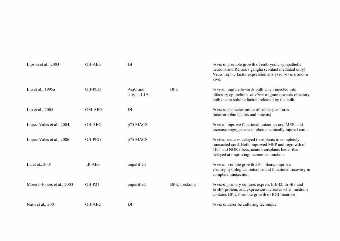
Lipson et al., 2003 OB-AEG DI in vitro: promote growth of embryonic sympathetic
neurons and Remak's ganglia (contact mediated only). Neurotrophic factor expression analysed in vitro and in vivo.
Liu et al., 1995a OB-PEG AraC and BPE in vivo: migrate towards bulb when injected into Thy-1.1 IA olfactory epithelium. In vitro: migrate towards olfactory
bulb due to soluble factors released by the bulb. Liu et al., 2005 OM-AEG DI in vitro: characterisation of primary cultures
(neurotrophic factors and mitosis) Lopez-Vales et al., 2004 OB-AEG p75 MACS in vivo: improve functional outcomes and MEP, and
increase angiogenesis in photochemically injured cord. Lopez-Vales et al., 2006 OB-PEG p75 MACS in vivo: acute vs delayed transplants in completely
transected cord. Both improved MEP and regrowth of 5HT and NOR fibers, acute transplants better than delayed at improving locomotor function.
Lu et al., 2001 LP-AEG unpurified in vivo: promote growth 5HT fibers, improve
electrophysiological outcome and functional recovery in complete transection.
Moreno-Flores et al., 2003 OB-P21 unpurified BPE, forskolin in vitro: primary cultures express ErbB2, ErbB3 and
ErbB4 protein, and expression increases when medium contains BPE. Promote growth of RGC neurons.
Nash et al., 2001 OB-AEG DI in vitro: describe culturing technique.

Nash et al., 2002 OB-AEG DI in vivo: improved forepaw reaching and axon regrowth in lesion of CST.
Navarro et al., 1999 OB-AEG p75 MACS in vivo: restoration of H response and withdrawal reflex,
and CGRP fiber regrowth into dorsal horn in dorsal rhizotomy model.
Pascual et al., 2002 OB-AEG p75 MACS in vivo: promote axon regeneration and bladder activity
in dorsal rhizotomy model. Pearse et al., 2004 OB-AEG p75 IP forskolin, BPE in vivo: combination with SC promoted tissue sparing
and 5HT fiber growth in contused cord. Perez-Bouza et al., 1998 OB-AEG unpurified GGF2 in vivo: align with host environment (unlesioned thalamus) and induce growth of fibers into regions not normally innervated. Pixley et al., 1992 OB-PEG unpurified in vitro: characterized expression patterns of cultures. Pixley, 1996 OB-PEG unpurified in vitro: characterised cell populations, no CNP
reactivity found. Plant et al., 2002 OB-AEG p75 IP forskolin, BPE in vitro: do not myelinate DRG neurons. Plant et al., 2003 OB-AEG p75 IP forskolin, BPE in vivo: reduce cavity formation, promote tissue and
supraspinal sparing, only delayed transplant improves function.
Polentes et al., 2004 OB-AEG Thy-1.1 –ve MACS in vivo: restore nervous phrenic and diaphragm muscular
activity after unilateral cord hemisection.

Pollock et al., 1999 OB-PEG O4 FACS in vitro: neuregulin is strong mitogen and survival factor. Express ErbB2 and ErbB4 but not ErbB3.
Ramer et al., 2004a LP-PEG Thy-1.1 CML in vivo: decrease cavity and scar formation, increased
growth of 5HT and TH +ve axons, increased angiogenesis and increased recruitment of SC in dorsolateral crush of cord.
Ramer et al., 2004b LP-PEG Thy1.1 CML . in vivo: no increased sensory afferent ingrowth in
dorsal rhizotomy model. Ramon-Cueto and OB-AEG unpurified and ST in vitro: characterisation of primary cultures. Nieto-Sampedro, 1992 Ramon-Cueto and OB-AEG p75 IP in vivo: ingrowth of ascending sensory fibers into Nieto-Sampedro, 1994 contralateral dorsal horn in dorsal rhizotomy model. Ramon-Cueto et al., 1993 OB-AEG unpurified in vitro: characterisation of primary cultures, promote
growth of ORN.
Ramon-Cueto et al., 1998 OB-AEG p75 IP forskolin, BPE in vivo: combined therapy with SC in completely transected cord, CGRP and 5HT axons regrow.
Ramon-Cueto et al., 2000 OB-AEG p75 IP forskolin, BPE in vivo: improve function and proprioceptive response,
and induce regrowth of NOR and 5HT axons in completely transected cord.
Resnick et al., 2003 OB-AEG p75 IP forskolin, BPE in vivo: no restoration of function in contusion injury.
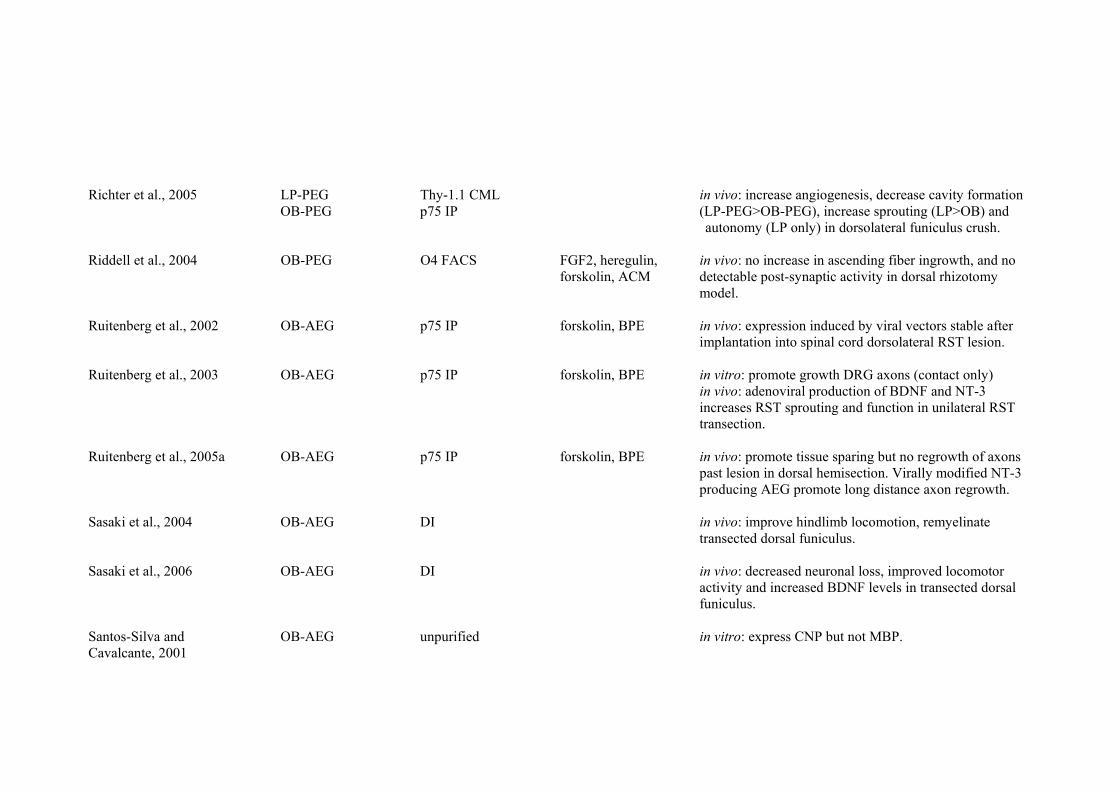
Richter et al., 2005 LP-PEG Thy-1.1 CML in vivo: increase angiogenesis, decrease cavity formation OB-PEG p75 IP (LP-PEG>OB-PEG), increase sprouting (LP>OB) and
autonomy (LP only) in dorsolateral funiculus crush. Riddell et al., 2004 OB-PEG O4 FACS FGF2, heregulin, in vivo: no increase in ascending fiber ingrowth, and no forskolin, ACM detectable post-synaptic activity in dorsal rhizotomy
model. Ruitenberg et al., 2002 OB-AEG p75 IP forskolin, BPE in vivo: expression induced by viral vectors stable after
implantation into spinal cord dorsolateral RST lesion. Ruitenberg et al., 2003 OB-AEG p75 IP forskolin, BPE in vitro: promote growth DRG axons (contact only) in vivo: adenoviral production of BDNF and NT-3
increases RST sprouting and function in unilateral RST transection.
Ruitenberg et al., 2005a OB-AEG p75 IP forskolin, BPE in vivo: promote tissue sparing but no regrowth of axons
past lesion in dorsal hemisection. Virally modified NT-3 producing AEG promote long distance axon regrowth.
Sasaki et al., 2004 OB-AEG DI in vivo: improve hindlimb locomotion, remyelinate
transected dorsal funiculus. Sasaki et al., 2006 OB-AEG DI in vivo: decreased neuronal loss, improved locomotor
activity and increased BDNF levels in transected dorsal funiculus.
Santos-Silva and OB-AEG unpurified in vitro: express CNP but not MBP. Cavalcante, 2001
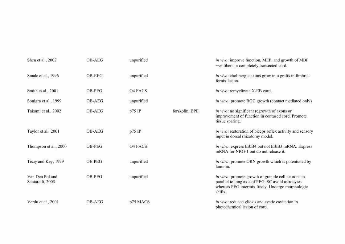
Shen et al., 2002 OB-AEG unpurified in vivo: improve function, MEP, and growth of MBP +ve fibers in completely transected cord.
Smale et al., 1996 OB-EEG unpurified in vivo: cholinergic axons grow into grafts in fimbria-
fornix lesion. Smith et al., 2001 OB-PEG O4 FACS in vivo: remyelinate X-EB cord. Sonigra et al., 1999 OB-AEG unpurified in vitro: promote RGC growth (contact mediated only) Takami et al., 2002 OB-AEG p75 IP forskolin, BPE in vivo: no significant regrowth of axons or
improvement of function in contused cord. Promote tissue sparing.
Taylor et al., 2001 OB-AEG p75 IP in vivo: restoration of biceps reflex activity and sensory
input in dorsal rhizotomy model. Thompson et al., 2000 OB-PEG O4 FACS in vitro: express ErbB4 but not ErbB3 mRNA. Express
mRNA for NRG-1 but do not release it.
Tisay and Key, 1999 OE-PEG unpurified in vitro: promote ORN growth which is potentiated by laminin.
Van Den Pol and OB-PEG unpurified in vitro: promote growth of granule cell neurons in Santarelli, 2003 parallel to long axis of PEG. SC avoid astrocytes
whereas PEG intermix freely. Undergo morphologic shifts.
Verdu et al., 2001 OB-AEG p75 MACS in vivo: reduced gliosis and cystic cavitation in
photochemical lesion of cord.

Verdu et al., 2003 OB-AEG p75 MACS in vivo: prevent loss of parenchyma, no improvement of function, increased nociceptive w/drawal, and increased MEP and SSEP in photochemical lesion of cord.
Vincent et al., 2003 OB-PEG AraC BPE in vitro: morphology changes can be controlled by
cAMP and endothelin-1. Neurotrophin profile unchanged by culture conditions.
Vincent et al., 2005 OB-PEG AraC BPE in vitro: PEG are more closely related to SC than to
astrocytes according to microarray analysis.
Wang et al., 2005 OB-AEG DI in vitro: describe culturing technique and cell characteristics.
Wewetzer et al., 2001 OB-PEG AraC forskolin in vitro: express CNTF mRNA and its receptor.
Forskolin increases CNTF mRNA and decreases receptor mRNA. CNTF not mitogenic.
Wewetzer et al., 2005 OB-PEG O4 MACS in vitro: O4 reactivity due to axonal fragments on p75 MACS surface of cell. PEG phagocytose these fragments in
vivo and in vitro. p75 expression increases as O4 decreases.
Woodhall et al., 2001 OB-PEG AraC BPE in vitro: express GDNF, BDNF and NGF proteins,
secrete BDNF and NGF, express TrkB mRNA. Woodhall et al., 2003 OB-PEG AraC BPE in vivo: report mRNA changes following
implantation into dorsal transection of cord

Woodhouse et al., 2005 OB-PEG AraC BPE in vitro: cultured with stabbed or contused cord. Contaminant proliferation high in presence of cord tissue. High apoptosis of PEG with acutely injured cord.
Xia et al., 2005 OB-PEG AraC and DI BPE in vitro: galanin inhibits proliferation, report mRNA
expression of galanin receptors. Yan et al., 2001a OB-AEG p75 IP in vitro: proliferation DRC to heregulin, FGF2, PDGF-
BB and IGF-1. All are mitogenic in presence of serum, only heregulin and FGF2 in absence of serum.
Yan et al., 2001b OB-AEG p75 IP in vitro: hepatocyte growth factor promotes dose-
dependant proliferation, heregulin promotes proliferation.
Yan et al., 2003 OB-PEG p75 IP in vitro: proliferation DRC to LPA, tested in
combination with other factors. LPA promotes migration/proliferation.

References
159
REFERENCES Adlkofer K, Lai C (1999) Role of neuregulins in glial development. Glia 29:104-111. Aguayo AJ, Charron L, Bray GM (1976) Potential of Schwann cells from unmyelinated nerves to produce myelin: a quantitative ultrastructural and autoradiographic study. J Neurocytol 5:565-573. Agrawal HC, Sprinkle TJ, Agrawal D (1990) 2’-3’ cyclic nucleotide-3’-phosphodiesterase in peripheral nerve myelin is phosphorylated by a phorbol ester-sensitive protein kinase. Biochem Biophys Res Comm 170:817-823. Agrawal AK, Shukla S, Chaturvedi RK, Seth K, Srivastava N, Ahmad A, Seth PK (2004) Olfactory ensheathing cell transplantation restores functional deficits in rat model of Parkinson's disease: a cotransplantation approach with fetal ventral mesencephalic cells. Neurobiol Dis 16:516-526. Alexander CL, Fitzgerald UF, Barnett SC (2002) Identification of growth factors that promote long-term proliferation of olfactory ensheathing cells and modulate their antigenic phenotype. Glia 37:349-364. Altman J, Das GD (1965) Autoradiographic and histological evidence of postnatal hippocampal neurogenesis in rats. J Comp Neurol 124:319-335. Anderson DJ (1993) Cell and molecular biology of neural crest cell lineage diversification. Curr Opin Neurobiol 3:8-13. Andersson PB, Perry VH, Gordon S (1992) The acute inflammatory response to lipopolysaccharide in CNS parenchyma differs from that in other body tissues. Neurosci 48:169-186. Anderton BH, Breinburg D, Downes MJ, Green PJ, Tomlinson BE, Ulrich J, Wood JN, Kahn J (1982) Monoclonal antibodies show that neurofibrillary tangles and neurofilaments share antigenic determinants. Nature 298:84-86. Andrews MR, Stelzner DJ (2004) Modification of the regenerative response of dorsal column axons by olfactory ensheathing cells or peripheral axotomy in adult rat. Exp Neurol 190:311-327. Au E, Roskams AJ (2002) Culturing olfactory ensheathing glia from the mouse olfactory epithelium. Methods Mol Biol 198:49-54. Au E, Roskams AJ (2003) Olfactory ensheathing cells of the lamina propria in vivo and in vitro. Glia 41:224-236.

References
160
Au WW, Treloar HB, Greer CA (2002) Sublaminar organisation of the mouse olfactory bulb nerve layer. J Comp Neurol 446:68-80. Bamber NI, Li H, Lu X, Oudega M, Aebischer P, Xu XM (2001) Neurotrophins BDNF and NT-3 promote axonal re-entry into the distal host spinal cord through Schwann cell-seeded min-channels. Eur J Neurosci 13:257-268. Barber PC (1981) Regeneration of vomeronasal nerves into the main olfactory bulb in the mouse. Brain Res 216:239-251. Barber PC (1982) Neurogenesis and regeneration in the primary olfactory pathway of mammals. Bibl Anat 23:12-25. Barber PC, Lindsay RM (1982) Schwann cells of the olfactory nerves contain glial fibrillary acidic protein and resemble astrocytes. Neurosci 7:3077-3090. Barber PC, Raisman G (1978) Replacement of receptor neurones after section of the vomeronasal nerves in the adult mouse. Brain Res 147:297-313. Barnett SC, Roskams AJ (2002) Olfactory ensheathing cells. Isolation and culture from the rat olfactory bulb. Methods Mol Biol 198:41-48. Barnett SC, Alexander CL, Iwashita Y, Gilson JM, Crowther J, Clark L, Dunn LT, Papanastassiou V, Kennedy PGE, Franklin RJM (2000) Identification of a human olfactory ensheathing cell that can effect transplant-mediated remyelination of demyelinated CNS axons. Brain 123:1581-1588. Barnett SC, Hutchins A-M, Noble M (1993) Purification of olfactory nerve ensheathing cells from the olfactory bulb. Dev Biol 155:337-350. Barakat DJ, Gaglani SM, Neravetla SR, Sanchez AR, Andrade CM, Pressman Y, Puzis R, Garg MS, Bunge MB, Pearse DD (2005) Survival, integration, and axon growth support of glia transplanted into the chronically contused spinal cord. Cell Transplant 14:225-240. Bartholdi D, Schwab ME (1997) Expression of pro-inflammatory cytokine and chemokine mRNA upon experimental spinal cord injury in mouse: an in situ hybridization study. Eur J Neurosci 9:1422-1438. Baulieu E-E, Schumacher M (2000) Progesterone as a neuroactive neurosteroid, with special reference to the effect of progesterone on myelination. Steroids 65:605-612.

References
161
Beattie M, Bresnahan JC, Komon J, Tovar CA, Van Meter M, Anderson DK, Faden AI, Hsu CY, Noble LJ, Salzman S, Young W (1997) Endogenous repair after spinal cord contusion injuries in the rat. Exp Neurol 148:453-463. Ben-Baruch N, Yarden Y (1994) Neu differentiation factors: a family of alternatively spliced neuronal and mesenchymal factors. Proc Soc Exp Biol Med 208:221-227. Benfey M, Aguayo AJ (1982) Extensive elongation of axons from rat brain into peripheral nerve grafts 296:150-152. Bianco JI, Perry C, Harkin DG, Mackay-Sim A, Feron F (2004) Neurotrophin 3 promotes purification and proliferation of olfactory ensheathing cells from human nose. Glia 45:111-123. Bixby JL, Lilien J, Reichardt LF (1988) Identification of the major proteins that promote neuronal process outgrowth on Schwann cells in vitro. J Cell Biol 107:353-361. Blakemore WF (1977) Remyelination in the spinal cord of the cat following intraspinal injections of lysolecithin. J Neurol Sci 33:31-43. Blanes T (1898) Sobre algunos puntos dudosos de la estructura del bulbo olfatorio. Rev Trim Microg 3:99-127. Blesch A, Tuszynski MH (2003) Cellular GDNF delivery promotes growth of motor and dorsal column sensory axons after partial and complete spinal cord transections and induces remyelination. J Comp Neurol 467:403-417. Boruch AV, Conners JJ, Pipitone M, Deadwyler G, Storer PD, Devries GH, Jones KJ (2001) Neurotrophic and migratory properties of an olfactory ensheathing cell line. Glia 33:225-229. Bovolenta P, Wandosell F, Nieto-Sampedro M (1992) CNS glial scar tissue: a source of molecules which inhibit central neurite outgrowth. Prog Brain Res 94:367-379. Boyd JG, Lee J, Skihar V, Doucette R, Kawaja MD (2004a) LacZ-expressing olfactory ensheathing cells do not associate with myelinated axons after implantation into the compressed spinal cord. PNAS 101:2162-2166. Boyd JG, Doucette R, Kawaja MD (2004b) Defining the role of olfactory ensheathing cells in facilitating axon remyelination following damage to the spinal cord. FASEB Journal 19:694-703.
References
162
Boyd JG, Jahed A, McDonald TG, Krol KM, Van Evk JE, Doucette R, Kawaja MD (2006) Proteomic evaluation reveals that olfactory ensheathing cells but not Schwann cells express calponin. Glia 53:434-440. Boyles JK, Zoellner CD, Anderson LJ, Kosik LM, Pitas RE, Wesgraber KH, Hull DY, Mahley RW, Gebicke-Haerter PJ, Ignatius MJ, Shooter EM (1989) A role for apolipoprotein E, apolipoprotein A-I, and low density lipoprotein receptors in cholesterol transport during regeneration and remyelination of the rat sciatic nerve. J Clin Invest 83:1015-31. Bradbury EJ, Moon LD, Popat RJ, King VR, Bennet GS, Patel PN, Fawcett JW, McMahon SB (2002) Chondroitinase ABC promotes functional recovery after spinal cord injury. Nature 416:636-640. Braun PE, Sandillon F, Edwards A, Matthieu JM, Privat A (1988) Immunocytochemical localization by electron microscopy of 2'3'-cyclic nucleotide 3'-phosphodiesterase in developing oligodendrocytes of normal and mutant brain. J Neurosci 8:3057-3066. Bregman BS, Coumans JV, Dai HN, Kuhn PL, Lynskey J, mcAtee M, Sandhu F (2002) Transplants and neurotrophic factors increase regeneration and recovery of function after spinal cord injury. Prog Brain Res 137:257-273. Bregman BS, Kunkel-Bagden E, Schnell L, Dai HN, Gao D, Schwab ME (1995) Recovery from spinal cord injury mediated by antibodies to neurite growth inhibitors. Nature 378:498-501. Bregman BS, McAtee M, Dai HN, Kuhn PL (1997) Neurotrophic factors increase axonal growth after spinal cord injury and transplantation in the adult rat. Exp Neurol 148:475-494. Britsch S, Li L, Kirchhoff S, Theuring F, Brinkmann V, Birchmeier C, Riethmacher D (1998) The ErbB2 and ErbB3 receptors and their ligand, neuregulin-1, are essential for development of the sympathetic nervous system. Genes Dev 12:1825-1836. Brockes JP, Lemke GE, Balzer DR Jr (1980a) Purification and preliminary characterization of a glial growth factor from the bovine pituitary. J Biol Chem 255:8374-8377. Brockes JP, Raff MC, Nishiguchi DJ, Winter J (1980b) Studies on cultured rat Schwann cells. III. Assays for peripheral myelin proteins. J Neurocytol 9:67-77.

References
163
Brosamle C, Huber AB, Fiedler M, Skerra A, Schwab ME (2000) Regeneration of lesioned corticospinal tract fibers in the adult rat induced by a recombinant, humanized IN-1 antibody fragment. J Neurosci 20:8061-8068. Brook GA, Plate D, Franzen R, Martin D, Moonen G, Schoenen J, Schmitt AB, Noth J, Nacimiento W (1998) Spontaneous longitudinally orientated axonal regeneration is associated with the Schwann cell framework within the lesion site following spinal cord compression injury of the rat. J Neurosci Res 53:51-65. Bunge MB, Williams AK, Wood PM (1982) Neuron-Schwann cell interaction in basal lamina formation. Dev Biol 92:449-460. Burden SJ (1998) The formation of neuromuscular synapses. Genes Dev 12:133-148. Burden S, Yarden Y (1997) Neuregulins and their receptors: a versatile signaling molecule in organogenesis and oncogenesis. Neuron 18:847-855. Busfield SJ, Michnick DA, Chickering TW, Revett TL, Ma J, Woolf EA, Comrack CA, Dussault BJ, Woolf J, Goodearl ADJ, Gearing DP (1997) Characterization of a neuregulin-related gene, Don-1, that is highly expressed in restricted regions of the cerebellum and hippocampus. Mol Cell Biol 17:4007-4014. Byrnes KR, Wu X, Waynant RW, Ilev IK, Anders JJ (2005) Low power laser irradiation alters gene expression of olfactory ensheathing cells in vitro. Lasers Surg Med 37:161-171. Cai D, Shen Y, De Bellard M, Tang S, Filbin MT (1999) Prior exposure to neurotrophins blocks inhibition of axonal regenration by MAG and myelin via a CAMP-dependent mechanism. Neuron 22:89-101. Calof AL, Chikaraishi DM (1989) Analysis of neurogenesis in a mammalian neuroepithelium: proliferation and differentiation of an olfactory neuron precursor in vitro. Neuron 3:115-127. Cao L, Liu L, Chen Z-Y, Wang L-M, Ye J-L, Qiu H-Y, Lu C-L, He C (2004) Olfactory ensheathing cells genetically modified to secrete GDNF to promote spinal cord repair. Brain 127:535-549. Carlos T, Kovach N, Schwartz B, Rosa M, Newman B, Wayne E, Benjamin C, Osborn L, Lobb R, Harlan J (1991) Human monocyte bind to two cytokine-induced adhesion ligands on cultured human endothelial cells: Endothelial-leukocyte adhesion molecule-1 and vascular cell adhesion molecule-1. Blood 77:2266-2271.

References
164
Carpenter G (2003) Nuclear localization and possible functions of receptor tyrosine kinases. Curr Opin Cell Biol 15:143-148. Carr VM, Farbman AI (1992) Ablation of the olfactory bulb up-regulates the rate of neurogenesis and induces precocious cell death in olfactory epithelium. Exp Neurol 115:55-59. Carraway KL III, Burden SJ (1995) Neuregulins and their receptors. Curr Opin Neurobiol 5:606-612. Carraway KL III, Cantley LC (1994) A neu acquaintance for erbB3 and erbB4: a role for receptor heterodimerization in growth signalling. Cell 78:5-8. Carraway KL III, Sliwkowski MX, Akita R, Platko JV, Guy PM, Nuijens A, Diamonti AJ, Vandlen RL, Cantley LC, Cerione RA (1994) The erbB3 gene product is a receptor for heregulin. J Biol Chem 269:14303-14306. Carraway KL III, Weber JL, Unger MJ, Ledesma J, Yu N, Gassmann M, Lai C (1997) Neuregulin-2, a new ligand of ErbB3/ErbB4-receptor tyrosine kinases. Nature 387:512-516. Carroll SL, Miller ML, Frohnert PW, Kim SS, Corbett JA (1997) Expression of neuregulins and their putative receptors, ErbB2 and ErbB3, is induced during Wallerian degeneration. J Neurosci 17:1642-1659. Chamak B, Mallat M (1991) Fibronectin and laminin regulate the in vitro differentiation of microglial cells. Neurosci 45:513-527. Chang H, Riese DJ 2nd, Gilbert W, Stern DF, McMahan UJ (1997) Ligands for ErbB-family receoptors encoded by a neuregulin-like gene. Nature 387:509-512. Chau CH, Shum DK, Li H, Pei J, Lui YY, Wirthlin L, Chan YS, Xu XM (2004) Chondroitinase ABC enhances axonal regrowth through Schwann cell-seeded guidance channels after spinal cord injury. FASEB J 19:194-196. Chen A, Xu XM, Kleitman N, Bunge MB (1996) Methylprednisolone administration improves axonal regeneration into Schwann cell grafts in transected adult rat thoracic spinal cord. Exp Neurol 138:261-276. Cheng H, Cao Y, Olson L (1996) Spinal cord repair in adult paraplegic rats: partial restoration of hind limb function. Science 273:510-513.
References
165
Choi D, Raisman G (2005) Disorganization of the facial nucleus after nerve lesioning and regeneration in the rat: effects of transplanting candidate reparative cells to the site of injury. Neurosurg 56:1093-1100. Chu J, Hatton J, Su H (1999) Effects of epidermal growth factor and dibutyryl cyclic adenosine monophosphate on the migration pattern of astrocytes grafted into adult rat brain. Neurosurg 45:859-866. Chuah MI, Au C (1991) Olfactory Schwann cells are derived from precursor cells in the olfactory epithelium. J Neurosci Res 29:172-180. Chuah MI, Au C (1993) Cultures of ensheathing cells from neonatal rat olfactory bulbs. Brain Res 601:213-220. Chuah MI, Au C (1994) Olfactory cell cultures on ensheathing cell monolayers. Chem Senses 19:25-34. Chuah MI, Teague R (1999) Basic fibroblast growth factor in the primary olfactory pathway: mitogenic effect on ensheathing cells. Neurosci 88:1043-1050. Chuah MI, Tennent R, Jacobs I (1995) Response of olfactory Schwann cells to intranasal zinc sulfate irrigation. J Neurosci Res 42:470-478. Chuah MI, Cossins J-M, Woodhall E, Tennent R, Nash G, West AK (2000) Glial growth factor 2 induces proliferation and structural changes in ensheathing cells. Brain Res 857:265-274. Chuah MI, Choi-Lundberg D, Weston S, Vincent AJ, Chung RS, Vickers JC, West AK (2004) Olfactory ensheathing cells promote collateral axonal branching in the injured adult rat spinal cord. Exp Neurol 185:15-25. Chung RS, Woodhouse A, Fung S, Dickson TC, West AK, Vickers JC, Chuah MI (2004) Olfactory ensheathing cells promote neurite sprouting of injured axons in vitro by direct cellular contact and secretion of soluble factors. CMLS 61:1238-1245. Collazos-Castro JE, Muneton-Gomez VC, Nieto-Sampedro M (2005) Olfactory glia transplantation into cervical spinal cord contusion injuries. J Neurosurg Spine 3:308-317. Corfas G, Rosen KM, Aratake H, Krauss R, Fischbach GD (1995) Differential expression of ARIA isoforms in the rat brain. Neuron 14:103-115. Costanzo RM, Graziadei PP (1983) A quantitative analysis of changes in the olfactory epithelium following bulbectomy in hamster. 215:370-381.

References
166
Coumans JV, Lin TT, Dai HN, MacArthur L, McAtee M, Nash C, Bregman BS (2001) Axonal regeneration and functional recovery after complete spinal cord transection in rats by delayed treatment with transplants and neurotrophins. J Neurosci 21:9334-9344. Crovello CS, Lai C, Cantley LC, Carraway KL III (1998) Differential signaling by the epidermal growth factor-like growth factors neuregulin-1 and neuregulin-2. J Biol Chem 273:26954-26961. Cummings BJ, Uchida N, Tamaki SJ, Salazar DL, Hooshmand M, Summers R, Gage FH, Anderson AJ (2005) Human neural stem cells differentiate and promote locomotor recovery in spinal cord-injured mice. Proc Natl Acad Sci USA 102:14069-14074. David S, Aguayo AJ (1981) Axonal elongation into peripheral nervous system "bridges" after central nervous system injury in adult rats. Science 214:931-933. David S, Bouchard C, Tsatas O, Giftochristos N (1990) Macrophages can modify the nonpermissive nature of the adult mammalian central nervous system. Neuron 5:463-469. De Mello TR, Dunlop S, Plant GW (2003) Myelination Potential of Olfactory Ensheathing Glia. Society for Neuroscience Meeting, New Orleans, USA. De Mello TR, Busfield S, Dunlop S, Plant GW (2005) Proliferative effects of neuregulins on olfactory ensheathing glia. Australian Neuroscience Society, Perth, Western Australia. Denis-Donini S, Estenoz M (1988) Interneurons versus efferent neurons: heterogeneity in their neurite outgrowth response to glia from several brain regions. Dev Biol 130:237-249. Dergham P, Ellezam B, Essagian C, Avedissian H, Lubell WD, McKerracher L (2002) Rhosignalling pathway targeted to promote spinal cord repair. J Neurosci 22:6570-6577. Devon R, Doucette R (1992) Olfactory ensheathing cells myelinate dorsal root ganglion neurites. Brain Res 589:175-179. Devon R, Doucette R (1995) Olfactory ensheathing cells do not require L-ascorbic acid in vitro to assemble a basal lamina or to myelinate dorsal root ganglion neurites. Brain Res 688:223-229.
References
167
Dong Z, Brennan A, Liu N, Yarden Y, Lefkowitz G, Mirsky R, Jessen KR (1995) neu differentiation factor is a neuron-glia signal and regulates survival, proliferation and maturation of rat Schwann cell precursors. Neuron 15:585-596. Dong Z, Dean C, Walters JE, Mirsky R, Jessen KR (1997) Response of Schwann cells to mitogens in vitro is determined by pre-exposure to serum, time in vitro and developmental stage. Glia 20:219-230. Doucette JR (1984) The glial cells in the nerve fiber layer of the rat olfactory bulb. Anat Rec 210:385-391. Doucette R (1989) Development of the nerve fiber layer in the olfactory bulb of mouse embryos. J Comp Neurol 285:514-527. Doucette R (1990) Glial influences on axonal growth in the primary olfactory system. Glia 3:433-449. Doucette R (1991) PNS-CNS transitional zone of the first cranial nerve. J Comp Neurol 312:451-466. Doucette R (1993a) Glial cells in the nerve fiber layer of the main olfactory bulb of embryonic and adult animals. Microsc Res Tech 24:113-130. Doucette R (1993b) Glial progenitor cells of the nerve fiber layer of the olfactory bulb: effect of astrocyte growth media. J Neurosci Res 35:274-287. Doucette R (1995) Olfactory ensheathing cells: potential for glial cell transplantation into areas of CNS injury. Histol Histopathol 10:503-507. Doucette R, Devon R (1994) Media that support the growth and differentiation of oligodendrocytes do not induce olfactory ensheathing cells to express a myelinating phenotype. Glia 10:296-310. Doucette R, Devon R (1995) Elevated intracellular levels of cAMP induce olfactory ensheathing cells to express Gal-C and GFAP but not MBP. Glia 13:130-140. Dunning MD, Lakatos A, Loizou L, Kettunen M, ffrench-Constant C, Brindle KM, Franklin RJM (2004) Superparamagnetic iron oxide-labeled Schwann cells and olfactory ensheathing cells can be traced in vivo by magnetic resonance imaging and retain functional properties after transplantation into the CNS. J Neurosci 24:9799-9810.

References
168
Eldridge CF, Bunge MB, Bunge RP, Wood PM (1987) Differentiation of axon-related Schwann cells in vitro. I. Ascorbic acid regulates basal lamina assembly and myelin formation. J Cell Biol 105:1023-1034. Erickson SL, O’Shea KS, Ghaboosin N, Loverro L, Frantz G, Bauer M, Lu LH, Moore MW (1997) ErbB3 is required for normal cerebellar and cardiac development: A comparison with erbB2 and heregulin deficient mice. Development 124:4999-5011. Falls DL, Harris DA, Johnson FA, Morgan MM, Corfas G, Fischbach GD (1990) Mr 42,000 ARIA: A protein that may regulate the accumulation of acetylcholine receptors at developing chick neuromuscular junctions. CSH Symp Quant Biol 55:397-406. Falls DL, Rosen KM, Corfas G, Lane WS, Fischbach GD (1993) ARIA, a protein that stimulates acetylcholine receptor synthesis, is a member of the neu ligand family. Cell 72:801-815. Farbman AI, Squinto LM (1985) Early development of olfactory receptor cell axons. Brain Res 351:205-213. Fairless R, Frame MC, Barnett SC (2005) n-Cadherin differentially determines Schwann cell and olfactory ensheathing cell adhesion and migration responses upon contact with astrocytes. Mol & Cell Neurosci 28:253-263. Fernandez-Valle C, Bunge RP, Bunge MB (1995) Schwann cells degrade myelin and proliferate in the absence of macrophages: evidence from in vitro studies of Wallerian degeneration. J Neurocytol 24:667-679. Feron F, Perry C, Cochrane J, Licina P, Nowitzke A, Urquhart S, Geraghty T, Mackay-Sim A (2005) Autologous olfactory ensheathing cell transplantation in human spinal cord injury. Brain 128:2951-2960. Field PM, Li Y, Raisman G (2003) Ensheathment of the olfactory nerves in the adult rat. J Neurocytol 32:317-324. Figlewicz DA, Quarles RH, Johnson D, Barbarash GR, Sternberger NH (1981) Biochemical demonstration of the myelin-associated glycoprotein in the peripheral nervous system. J Neurochem 37:749-758. Filbin MT, Walsh FS, Trapp BD, Pizzey JA, Tennekoon GI (1990) Role of myelin P0 protein as a homophilic adhesion molecule. Nature 344:871-872. Filbin MT (2003) Myelin-associated inhibitors of axonal regeneration in the adult mammalian CNS. Nat Rev Neurosci 4:703-713.

References
169
Fishman PS, Kelley JP (1984) The fate of severed corticospinal axons. Neurology 34:1161-1167. Fouad K, Schnell L, Bunge, MB, Schwab ME, Liebscher T, Pearse DD (2005) Combining Schwann cell bridges and olfactory-ensheathing glia grafts with chondroitinase promotes locomotor recovery after complete transection of the spinal cord. J Neurosci 25:1169-1178. Fradkin JE, Cook GH, Kilhoffer MC, Wolff J (1982) Forskolin stimulation of thyroid adenylate cyclase and cyclic 3’,5’-adenosine monophosphate accumulation. Endocrinology 111:849-856. Franceschini IA, Barnett, SC (1996) Low-Affinity NGF-receptor and E-N-CAM expression define two types of olfactory nerve ensheathing cells that share a common lineage. Dev Biol 173:327-343. Franklin RJM, Barnett SC (1991) The electron microscopic appearance of the β-galactosidase reaction product. Acta Neuropathol 81:686-687. Franklin RJ, Gilson JM, Franceschini IA, Barnett SC (1996) Schwann cell-like myelination following transplantation of an olfactory bulb-ensheathing cell line into areas of demyelination in the adult CNS. Glia 17:217-224. Fregien NL, White LA, Bunge MA, Wood PA (2004) Forskolin increases neuregulin receptors in human Schwann cells without increasing receptor mRNA. Glia 49:24-35. Friede RL, Samorjski T (1968) Myelin formation in the sciatic nerve of the rat: a quantitative electron microscopic, histochemical and radioautographic study. J Neuropathol Exp Neurol 27:546-570. Fruttiger M, Montag D, Schachner M, Martini R (1995) Crucial role for the myelin-associated glycoprotein in the maintenance of axon-myelin integrity. Eur J Neurosci 7:511-515. Gage FH (2000) Mammalian neural stem cells. Science 287:1433-1438. Garbay B, Heape AM, Sargueil F, Cassagne C (2000) Myelin synthesis in the peripheral nervous system. Prog Neurobiol 61:267-304. Garcia-Alias G, Lopez-Vales R, Fores J, Navarro X, Verdu E (2004) Acute transplantation of olfactory ensheathing cells or Schwnann cells promotes recovery after spinal cord injury in the rat. J Neurosci Res 75:632-641.

References
170
Garratt AN, Britsch S, Birchmeier C (2000) Neuregulin, a factor with many functions in the life of a Schwann cell. BioEssays 22:987-996. Gavazzi I, Kumar RDC, McMahon SB, Cohen J (1999) Growth responses of different subpopulations of adult sensory neurons to neurotrphic factors in vitro. Eur J Neurosci 11:3405-3414. George R, Griffin JW (1994) Delayed macrophage responses and myelin clearance during Wallerian degeneration in the central nervous system: the dorsal radiculotomy model. Exp Neurol 129:225-236. Ghoumari AM, Ibanez C, El-Etr M, Leclerc P, Eychenne B, O'Malley BW, Baulieu EE, Schumacher M (2003) Progesterone and its metabolites increase myelin basic protein expression in organotypic slice cultures of rat cerebellum. J Neurochem 86:848-859. Giese KP, Martini R, Lemke G, Soriano P, Schachner M (1992) Mouse P0 gene disruption leads to hypomyelination, abnormal expression of recognition molecules, and degeneration of myelin and axons. Cell 71:565-576. Gilson J, Blakemore WF (1993) Failure of remyelination in areas of demyelination produced in the spinal cord of old rats. Neurpathol Appl Neurobiol 19:173-181. Giulian D, Chen J, Ingeman JE, George JK, Noponen M (1989) The role of mononuclear phagocytes in wound healing after traumatic injury to adult mammalian brain. J Neurosci 8:4416-4429. Goldsmith HS, de la Torre JC (1992) Axonal regeneration after spinal cord transection and reconstruction. Brain Res 589:217-224. Gomez VM, Averill S, King V, Yang Q, Perez ED, Chacon SC, Ward R, Nieto-Sampedro M, Priestley J, Taylor J (2003) Transplantation of olfactory ensheathing cells fails to promote significant axonal regeneration from dorsal roots into the rat cervical cord. J Neurocytol 32:53-70. Goodman MN, Silver J, Jacobberger JW (1993) Establishment and neurite outgrowth properties of neonatal and adult rat olfactory bulb glial cell lines. Brain Res 619:199-213. Goodearl AD, Davis JB, Mistry K, Minghetti L, Otsu M, Waterfield MD, Stroobant P (1993) Purification of multiple forms of glial growth factor. J Biol. Chem 268:18095-18102. Gospodarowicz D, Cheng J, Lirette M (1983) Bovine brain and pituitary fibroblast growth factors: comparison of their abilities to support the

References
171
proliferation of human and bovine vascular endothelial cells. J Cell Biol 97:1677-1685. Gould E, Reeves AJ, Graziano MSA, Gross CG (1999) Neurogenesis in the neocortex of adult primates. Science 286:548-552. Graca DL, Blakemore WF (1986) Delayed remyelination in rat spinal cord following ethidium bromide injection. Neuropathol Appl Neurobiol 12:593-605. GrandPre T, Li S, Strittmatter SM (2002) Nogo-66 receptor antagonist peptide promotes axonal regeneration. Nature 417:547-551. Graziadei PPC (1973) Vertebrates olfactory mucosa. In: The ultrastructure of sensory organs (Friedman I ed), pp269-305. North-Holland Publishing Co: London. Graziadei PPC, Monti Graziadei GA (1978) Continuous nerve cell renewal in the olfactory system. In: Handbook of sensory physiology, Vol. 9. (Jacobson M, ed.), pp55-82. Springer Verlag: Berlin. Graziadei PP, Monti Graziadei GA (1979) Neurogenesis and neuron regeneration in the olfactory system of mammals. I. Morphological aspects of differentiation and structural organization of the olfactory sensory neurons. J Neurocytol 8:1-8. Graziadei PPC, Monti Graziadei GA (1980) Neurogenesis and neuron regeneration in the olfacotory system of mammals. III. Deafferentation and reinnervation of the olfactory bulb following section of the fila olfactoria in rat. J Neurocytol 9:145-162. Graziadei PPC, Levine RR, Monti Graziadei GA (1978) Regeneration of olfactory axons and synapse formation in the forebrain after bulbectomy in neonatal mice. Proc Natl Acad Sci USA 75:5230-5234. Greenfield S, Brostoff S, Eylar EH, Morell P (1973) Protein composition of myelin of the peripheral nervous system. J Neurochem 20:1207-1216. Gregson NA (1989) Lysolipids and membrane damage: lysolecithin and its interaction with myelin. Biochem Soc Trans 17:280-283. Grinspan JB, Marchionni MA, Reeves M, Coulaloglou M, Scherer SS (1996) Axonal interactions regulate Schwann cell apoptosis in developing peripheral nerve: neuregulin receptors and the role of neuregulins. J Neurosci 16:6107-6118.

References
172
Gudino-Cabrera G, Nieto-Sampedro M (1996) Ensheathing cells: large scale purification from adult olfactory bulb, freeze-preservation and migration of transplanted cells in adult brain. Rest Neurol Neurosci 10:25-34. Gudino-Cabrera G, Nieto-Sampedro M (2000) Schwann-like macroglia in adult rat brain. Glia 30:49-63. Guntinas-Lichius O, Angelov DN, Tomov TL, Dramiga J, Neiss WF, Wewetzer K (2001) Transplantation of olfactory ensheathing cells stimulates the collateral sprouting from axotomized adult rat facial motoneurons. Exp Neurol 172:70-80. Guntinas-Lichius O, Wewetzer K, Tomov TL, Azzolin N, Kazemi, S, Streppel M, Neiss WF, Angelov DN (2002) Transplantation of olfactory mucosa minimizes axonal branching and promotes the recovery of vibrissae motor performance after facial nerve repair in rats. J Neurosci 15:7121-7131. Guth L, Zhang Z, Roberts E (1994) Key role for pregnenolone in combination therapy that promotes recovery after spinal cord injury. Proc Natl Acad Sci USA 91:12308-12312. Guy PM, Platko JV, Cantley LC, Cerione RA, Carraway KL III (1994) Insect cell-expressed p180erbB3 possesses an impaired tyrosine kinase activity. Proc Natl Acad Sci USA 91:8132-8136. Hahn AF, Whitaker JN, Kachar B, Webster HdeF (1987) P2, P1 and P0 myelin protein expression in developing rat sixth nerve: a quantitative immunocytochemical study. J Comp Neurol 260:501-512. Hall SM (1972) The Effect of injections of lysophosphatidylcholine into white matter of the adult mouse spinal cord. J Cell Sci 10:535-546. Halper J, Parnell PG, Carter BJ, Ren P, Scheithauer BW (1992) Presence of growth factors in human pituitary. Lab Invest 66:639-645. Harauz G, Ishiyama N, Hill CMD, Bates IR, Libich DS, Fares C (2004) Myelin basic protein – diverse conformational states of an intrinsically unstructured protein and its roles in myelin assembly and multiple sclerosis. Micron 35:503-542. Harvey AR (1994) Expression of low affinity NGF (p75) receptors in rat superior colliculu: studies in vivo, in vitro, and in fetal tectal grafts. Exp Neurol 130:237-249.

References
173
Hayat S, Wigley CB, Robbins J (2003a) Intracellular calcium handling in rat olfactory ensheathing cells and its role in axonal regeneration. Neurosci 259-270. Hayat S, Thomas A, Afshar F, Sonigra R, Wigley CB (2003b) Manipulation of olfactory ensheathing cell signalling mechanisms: effects on their support for neurite regrowth from adult CNS neurons in culture. Glia 44:232-241. Herrera LP, Casas CE, Bates ML, Guest JD (2005) Ultrastructural study of the primary olfactory pathway in Macaca fascicularis. J Comp Neurol 488:427-441. Hirata K, Kawabuchi M (2002) Myelin phagocytosis by macrophages and nonmacrophages during Wallerian degeneration. Microsc Res Tech 57:541-547. Hirata K, Mitoma H, Ueno N, He J, Kawabuchi M (1999) Differential response of macrophage subpopulations to myelin degradation in the injured rat sciatic nerve. J Neurocytol 28:685-695. Ho WH, Armanini MP, Nuijens A, Phillips HS, Osheroff PL (1995) Sensory and motor neuron-derived factor. A novel heregulin variant highly expressed in sensory and motot neurons. J Biol Chem 270:14523-14532. Hoke A, Ho T, Crawford TO, LeBel C, Hilt D, Griffin JW (2003) Glial cell line-derived neurotrophic factor alters axon Schwann cell units and promotes myelination in unmyelinated nerve fibers. J Neurosci 23:561-567. Holmes WE, Sliwkowski MX, Akita RW, Henzel WJ, Lee J, Park JW, Yansura D, Abadi N, Raab H, Lewis GD, Shepard HM, Kuang W-J, Wood WI, Goeddel DV, Vandlen RL (1992) Identification of heregulin, a specific activation of p185erbB2. Science 256:1205-1210. Holtzman E, Novikoff AB (1965) Lysosomes in the rat sciatic nerve following crush. J Cell Biol 27:651-669. Honmou O, Felts PA, Waxman SG, Kocsis JD (1996) Restoration of normal conduction properties in demyelinated spinal cord axons in the adult rat by transplantation of exogenous Schwann cells. J Neurosci 16:3199-3208. Huang H, Chen L, Hongmei W, Bo X, Bingehen L, Rui W, Jian Z, Feng Z, Zheng C, Ying L, Yinglun S, Wei H, Shuyi P, Junzhao S (2003) Influence of patients age on functional recovery after transplantation of olfactory ensheathing cells into injured spinal cord injury. Chin Med J 116:1488-1491.

References
174
Hughes JT, Brownell B (1963) Aberrant nerve fibres within the spinal cord. J neurol Neurosurg Psychiat 26:528-534. Imaizumi T, Lankford KL, Wazman SG, Greer CA, Kocsis JD (1998) Transplanted olfactory ensheathing cells remyelinate and enhance axonal conduction in the demyelinated dorsal columns of rat spinal cord. J Neurosci 18:6176-6185. Imaizumi T, Lankford KL, Burton WV, Fodor WL, Kocsis JD (2000a) Xenotransplantation of transgenic pig olfactory ensheathing cells promotes axonal regenration in rat spinal cord. Nature Biotech 18:949-953. Imaizumi T, Lankford KL, Kocsis JD (2000b) Transplantation of olfactory ensheathing cells or Schwann cells restores rapid and secure conduction across the transected spinal cord. Brain Res 854:70-78. Jani HR, Raisman G (2004) Ensheathing cell cultures from the olfactory bulb and mucosa. Glia 47:130-137. Jeffery ND, Blakemore WF (1995) Remyelination of mouse spinal cord axon demyelinated by local injection of lysolecithin. J Neurocytol 24:775-781. Jessen KR, Mirsky R (1992) Schwann cells: early lineage, regulation of proliferation and control of myelin formation. Curr Opin Neurobiol 2:575-581. Johansson S, Lee I-H, Olson L, Spenger C (2005) Olfactory ensheathing glial co-grafts improve functional recovery in rats with 6-OHDA lesions. Brain 128:2961-2976. Johnson PW, Abramow-Newerly W, Seilheimer B, Sadoul R, Tropak MB, Arquint M, Dunn RJ, Schachner M, Roder JC (1989) Recombinant myelin-associated glycoprotein confers neural adhesion and neurite outgrowth function. Neuron 3:377-385. Jones JT, Akita RW, Sliwkowski MX (1999) Binding specificities and affinities of egf domains for ErbB receptors. FEBS Letters 447:227-231. Jones RB, Gordus A, Krall JA, MacBeath G (2006) A quantitative protein interaction network for the ErbB receptors using protein microarrays. Nature 439:168-174. Jung-Testas I, Schumacher M, Robel P, Baulieu EE (1994) Actions of steroid hormones on glial cells of the central and peripheral nervous system. J Steroid Biochem 48:145-154.

References
175
Kafitz KW, Greer CA (1998) The influence of ensheathing cells on olfactory receptor cell neurite outgrowth in vitro. Ann NY Acad Sci 855:266-269. Kafitz KW, Greer CA (1999) Olfactory ensheathing cells promote neurite extension from embryonic olfactory receptor cells in vitro. Glia 25:99-110. Kato T, Honmou O, Uede T, Hashi K, Kocsis JD (2000) Transplantation of human olfactory ensheathing cells elicits remyelination of demyelinated rat spinal cord. Glia 30:209-218. Katsuki M, Sato M, Kimura M, Yokoyama M, Kobayashi K, Nomura T (1988) Conversion of normal behavior of shiverer by myelin basic protein antisense cDNA in transgenic mice. Science 241:593-595. Keyvan-Fouladi N, Raisman G, Li Y (2003) Functional repair of the corticospinal tract by delayed transplantation of olfactory ensheathing cells in adult rats. J Neurosci 23:9428-9434. Kim HA, Rosenbaum T, Marchionni MA, Ratner N, DeClue JE (1995) Schwann cells from neurofibromin deficient mice exhibit activation of p21ras, inhibition of cell proliferation and morphological changes. Oncogene 11:325-335. Kim HA, DeClue JE, Ratner N (1997) cAMP-dependent protein kinase A is required for Schwann cell growth: interactions between the cAMP and neuregulin/tyrosine kinase pathways. J Neurosci Res 49:236-247. Kita Y, Mayer J, Zamborelli T, Hara S, Rohde M, Watson E, Koski R, Ratzkin B, Nicolson M (1995) Boactive synthetic peptide of NDF/Heregulin. Biochem Biophys Res Comm 210:441-451. Kleitman N, Wood PM, Bunge RP (1998) Tissue culture methods for the study of myelination. In: Culturing nerve cells (Banker G, Goslin K, ed), pp545-594. Cambridge: London MIT Press. Klusman I, Schwab ME (1997) Effects of pro-inflammatory cytokins in experimental spinal cord injury. Brain Res 762:173-184. Kobayashi NR, Fan DP, Giehl KM, Bedard AM, Wiegand SJ, Tetzlaff W (1997) BDNF and NT-4/5 prevent atrophy of rat rubrospinal neurons after cervical axotomy, stimulate GAP-43 and Talpha1-tubulin mRNA expression, and promote axonal regeneration. J Neurosci 17:9583-9595. Koenig HL, Schumacher M, Ferzaz B, Do Thi AN, Ressouches A, Guennoun R, Jung-Testas I, Robel P, Akwa Y, Baulieu E-E (1995) Progesterone synthesis and myelin formation by Schwann cells. Science 268:1500-1503.

References
176
Kraus MH, Issing W, Miki T, Popescu NC, Aaronson SA (1989) Isolation and characterization of ERBB3, a third member of the ERBB/ epidermal growth factor receptor family: Evidence for overexpression in a subset of human mammary tumors. Proc Natl Acad Sci USA 86:9193-9197. Kromer LF, Cornbrooks CJ (1985) Transplants of Schwann cell cultures promote axonal regeneration in the adult mammalian brain. Proc Natl Acad Sci USA 82:6330-6334. Kuhlengel KR, Bunge MB, Bunge RP, Burton H (1990) Implantation of cultured sensory neurons and Schwann cells into lesioned neonatal rat spinal cord. II. Implant characteristics and examination of corticospinal tract growth. J Comp Neurol 293:74-91. Kumar R, Hayat S, Felts P, Bunting S, Wigley C (2005) Functional differences and interactions between phenotypic subpopulations of olfactory ensheathing cells in promoting CNS axonal regeneration. Glia 50:12-20. Kume N, Cybulsky MI, Gimbrone MA Jr (1992) Lysophosphatidylcholine, a component of atherogenic lipoproteins, induces mononuclear leukocyte adhesion molecules in cultured human and rabbit arterial endothelial cells. J Clin Invest 90:1138-1144. Lakatos A, Franklin RJ, Barnett SC (2000) Olfactory ensheathing cells and Schwann cells differ in their in vitro interactions with astrocytes. Glia 32:214-25. Lakatos A, Barnett SC, Franklin JM (2003a) Olfactory ensheathing cells induce less host astrocyte response and chondroitin sulphate proteoglycan expression than Schwann cells following transplantation into adult CNS white matter. Exp Neurol 184:237-246. Lakatos A, Smith PM, Barnett SC, Franklin RJM (2003b) Meningeal cells enhance limited CNS remyelination by transplanted olfactory ensheathing cells. Brain 126:598-609. Lazarov-Spiegler O, Solomon AS, Zeev-Brann AB, Hirschberg DL, Lavie V, Schwartz M (1996) Transplantation of activated macrophages overcomes central nervous system regrowth failure. FASEB J 10:1296-12302. Lazarov-Spiegler O, Solomon AS, Schwartz M (1998) Peripheral nerve-stimulated macrophages simulate a peripheral nerve-like regenerative response in rat transected optic nerve. Glia 24:329-337.

References
177
Leaver SG, Harvey AR, Plant GW (2006) Adult olfactory ensheathing glia promote the long-distance growth of adult retinal ganglion cell neurites in vitro. Glia In press. Lee I-H, Bulte JWM, Schweinhardt P, Douglas T, Trifunovski A, Hofstetter C, Olson L, Spenger C (2004) In vivo magnetic resonance tracking of olfactory ensheathing glia grafted into the rat spinal cord. Exp Neurol 187:509-516. Lee MJ, Brennan A, Blanchard A, Zoidl G, Dong Z, Tabernero A, Zoidl C, Dent MAR, Jessen KR, Mirsky R (1997) P0 is constitutively expressed in the rat neural crest and embryonic nerves and is negatively and positively regulated by axons to generate non-myelin-forming and myelin-forming Schwann cells respectively. Mol Cell Neurosci 8:336-350. Lemke G (1988) Unwrapping the genes of myelin. Neuron 1:535-543. Lemke G (1996) Neuregulins in Development. Mol Cell Neurosci 7:247-262. Lemke G, Axel R (1985) Isolation and sequence of a cDNA encoding the major structural protein of peripheral myelin. Cell 40:501-508. Lemke GE, Brockes JP (1984) Identification and purification of glial growth factor. J Neurosci 4:75-83. Le Roux PD, Reh TA (1994) Regional differences in glial-derived factors that promote dendritic outgrowth from mouse cortical neurons in vitro. J Neurosci 14:4639-4655. Levi ADO, Bunge RP, Lofgren JA, Meima L, Hefti F, Nikolics K, Sliwkowski MX (1995) The influence of heregulins on human Schwann cell proliferation. J Neurosci 15:1329-1340. Ley K (1996) Molecular mechanisms of leukocyte recruitment in the inflammatory process. Cardiovasc Res 32:733-742. Li Y, Raisman G (1994) Schwann cells induce sprouting in motor and sensory axons in the adult rat spinal cord. J Neurosci 14:4050-4063. Li S, Strittmatter SM (2003) Delayed systemic Nogo-66 receptor antagonist promotes recovery from spinal cord injury. J Neurosci 23:4219-4227. Li Y, Field PM, Raisman G (1997) Repair of adult rat corticospinal tract by transplants of olfactory ensheathing cells. Science 277:2000-2002.

References
178
Li Y, Field PM, Raisman G (1998) Regeneration of adult rat corticospinal axons induced by transplanted olfactory ensheathing cells. J Neurosci 18:10514-10524. Li Y, Decerchi P, Raisman G (2003a) Transplantation of olfactory ensheathing cells into spinal cord lesions restores breathing and climbing. J Neurosci 23:727-731. Li Y, Sauve Y, Li D, Lund RD, Raisman G (2003b) Transplanted olfactory ensheathing cells promote regeneration of cut adult rat optic nerve axons. J Neurosci 27:7783-7788. Li Y, Carlstedt T, Berthold, C-H, Raisman G (2004) Interaction of transplanted olfactory-ensheathing cells and host astrocytic processes provides a bridge for axons to regenerate axons across the dorsal root entry zone. Exp Neurol 188:300-308. Li Y, Field PM, Raisman G (2005) Olfactory ensheathing cells and olfactory nerve fibroblasts maintain continuous open channels for regrowth of olfactory nerve fibres. Glia 52:245-251. Liesi P (1985a) Do neurons in the vertebrate CNS migrate on laminin? EMBO J 4:1163-1170. Liesi P (1985b) Laminin-immunoreactive glia distinguish regenerative adult CNS systems from non-regenerative ones. EMBO J 4:2505-2511. Lin WC, Sanchez HB, Deerlinck T, Morris JK, Ellisman M, Lee KF (2000) Aberrant development of motor axons and neuromuscular synapses in erbB2-deficient mice. Proc Natl Acad Sci USA 97:1299-1304. Lindsay RM (1988) Nerve growth factors (NGF, BDNF) enhance axonal regeneration but are not required for survival of adult sensory neurons. J Neurosci 8:2394-2405. Lipson AC, Widenfalk J, Lindqvist E, Ebendal T, Olson L (2003) Neurotrophic properties of olfactory ensheathing glia. Exp Neurol 180:167-171. Liu KL, Chuah MI, Lee KKH (1995a) Soluble factors from the olfactory bulb attract olfactory Schwann cells. J Neurosci 15:990-1000. Liu HM, Yan LH, Yang YJ (1995b) Schwann cell properties: 3. C-fos expression, bFGF production, phagocytosis and proliferation during Wallerian degeneration. J Neuropathol Exp Neurol 54:487-496.

References
179
Liu JB, Tang TS, Gong AH, Sheng WH, Yang JC (2005) The mitosis and immunocytochemistry of olfactory ensheathing cells from nasal olfactory mucosa. Clin J Traumatol 8:306-310. Longart M, Liu Y, Karavanova I, Buonanno A (2004) Neuregulin-2 is developmentally regulated and targeted to dendrites of central neurons. J Comp Neurol 472:156-172. Lopez-Vales R, Garcia-Alias G, Fores J, Navarro X, Verdu E (2004) Increased expression of cyco-oxygensase 2 and vascular endothelial growth factor in lesioned spinal cord by translanted olfactory ensheathing glia. J Neurotrauma 21:1031-1043. Lopez-Vales R, Fores J, Verdu E, Navarro X (2006) Acute and delayed transplantation of olfactory ensheathing cells promote partial recovery after complete transection of the spinal cord. Neurobiol Dis 21:57-68. Lu J, Feron F, Ho SM, Mackay-Sim A, Waite PM (2001) Transplantation of nasal olfactory tissue promotes partial recovery in paraplegic adult rats. Brain Res 889:344-357. Lu J, Feron F, Mackay-Sim A, Waite PME (2002) Olfactory ensheathing cells promote locomotor recovery after delayed transplantation into transected spinal cord. Brain 125:14-21. Lu P, Yang H, Jones LL, Filbin MT, Tuszynski MH (2004) Combinatorial therapy with neurotrophins and cAMP promotes axonal regeneration beyond sites of spinal cord injury. J Neurosci 24:6402-6409. Luster AD, Ravetch JV (1987) Genomic characterization of a gamma-interferon-inducible gene (IP-10) and identification of an interferon-inducible hypersensitive site. Mol Cell Biol 7:3723-3731. Ma M, Wei T, Boring L, Charo IF, Ransohoff RM, Jakeman LB (2002) Monocyte recruitment and myelin removal are delayed following spinal cord injury in mice with CCR2 chemokine receptor deletion. J Neurosci Res 68:691-702. Mackay-Sim A (2005) Olfactory ensheathing cells and spinal cord repair. J Med 54:8-14. Mackay-Sim A, Kittel P (1991) Cell dynamics in the adult mouse olfactory epithelium: a quantitative autoradiographic study. J Neurosci 11:979-984.
References
180
Madison R, da Silva CF, Dikkes P, Chiu TH, Sidman RL (1985) Increased rate of peripheral nerve regeneration using bioresorbable nerve guides and a laminin-containing gel. Exp Neurol 88:767-772. Magavi SSP, Mitchell BD, Szentirmai O, Carter BS, Macklis JD (2005) Adult-born and preexisting olfactory granule neurons undergo distinct experience-dependent modification of their olfactory responses in vivo. J Neurosci 25:10729-10739. Mahanthappa NK, Anton ES, Matthew WD (1996) Glial growth factor 2, a soluble neuregulin, directly increases Schwann cell motility and indirectly promotes neurite outgrowth. J Neurosci 16:4673-4683. Marchionni MA, Goodearl AD, Chen MS, Bermingham-McDonogh O, Kirk C, Hendricks M, Danehy F, Misumi D, Sudhalter J, Kobayashi K, Wroblewski D, Lynch C, Baldassare M, Hiles I, Davis JB, Hsuan JJ, Totty NF, Otsu M, BcBurney RN, Waterfield MD, Stoobant P, Gwynne D (1993) Glial growth factors are alternatively spliced erbB2 ligands expressed in the nervous system. Nature 362:312-318. Marin-Padilla M, Amieva MR (1989) Early neurogenesis of the mouse olfactory nerve: Golgi and electron microscopic studies. J Comp Neurol 288:339-352. Martin D, Robe P, Franzen R, Delree P, Schoenen J, Stevenaert A, Moonen G (1996) Effects of Schwann cell transplantation in a contusion model of rat spinal cord injury. J Neurosci Res 45:588-597. Martini R, Bollensen E, Schachner M (1988) Immunocytological localization of the major peripheral nervous system glycoprotein P0 and the L2/HNK-1 and L3 carbohydrate structures in developing and adult mouse sciatic nerve. Dev Biol 129:330-338. Martini R, Mohajeri MH, Kasper S, Giese KP, Schachner M (1995) Mice doubly deficient in the genes for P0 and myelin basic protein show that both proteins contribute to the formation of the major dense line in peripheral nerve myelin. J Neurosci 15:4488-4495. McDonald JW, Liu XZ, Qu Y, Liu S, Mickey SK, Turetsky D, Gottlieb DI, Choi DW (1999) Transplanted embryonic stem cells survive, differentiate and promote recovery in injured rat spinal cord. Nat Med 5:1410-1412. McKerracher L, David S, Jackson DL, Kottis V, Dunn RJ, Braun PE (1994) Identification of myelin-associated glycoprotein as a major myelin-derived inhibitor of neurite growth. Neuron 13:805-811.
References
181
McTigue DM, Tani M, Krivacic K, Chernosky A, Kelner GS, Macicjewski D, Maki R, Ransohoff RM, Stokes BT (1998) Selective chemokine mRNA accumulation in the rat spinal cord after contusion injury. J Neurosci Res 53:368-376. Melcangi RC, Ballabio M, Cavarretta I, Gonzalex LC, Leonelli E, Veiga S, Martini L, Magnaghi V (2003) Effects of neuroactive steroids on myelin of peripheral nervous system. J Steroid Biochm & Mol Biol 85:323-327. Menei P, Montero-Menei C, Whittemore SR, Bunge RP, Bunge MB (1998) Schwann cells genetically modified to secrete human BDNF promote enhanced axonal regrowth across transected adult rat spinal cord. Eur J Neurosci 10:607-621. Meyer D, Birchmeier C (1995) Multiple essential functions of neuregulin in development. Nature 378:386-390. Michailov G, Sereda MW, Brinkmann BG, Fischer TM, Haug B, Birchmeier C, Role L, Lai C, Schwab MH, Nave K-A (2004) Axonal neuregulin-1 regulates myelin sheath thickness. Science 304:700-3. Miragall F, Kadmon G, Husmann M, Schachner M (1988) Expression of cell adhesion molecules in the olfactory system of the adult mouse: presence of the embryonic form of N-CAM. Dev Biol 129:516-531. Mirsky R, Jessen KR (1999) The neurobiology of Schwann cells. Brain Path 9:293-311. Molineaux SM, Engh H, De Ferra F, Hudson L, Lazzarini RA (1986) Recombination within the myelin basic protein gene created the dysmyelinating shiverer mouse mutation. PNAS 83:7542-7546. Morell P, Barrett CV, Mason JL, Toews AD, Hostettler, JD, Knapp GW, Matsushima GK (1998) Gene expression in brain during cuprizone-induced demyelination and remyelination. Mol Cell Neurosci 12:220-227. Moreno-Flores MT, Lim F, Martin-Bermejo J, Diaz-Nido J, Avila J, Wandosell F (2003) Immortalized olfactory ensheathing glia promote axonal regeneration of rat retinal ganglion neurons. J Neurochem 85:861-871. Morris JK, Lin W, Hauser C, Marchuk Y, Getman D, Lee KF (1999) Rescue of the cardiac defect in erbB2 mutant mice reveals essential roles of erbB2 in peripheral nervous system development. Neuron 23:273-283.

References
182
Morrissey TK, Kleitman N, Bunge RP (1991) Isolation and functional characterisation of Schwann cells derived from adult peripheral nerve. J Neurosci 11:2433-2442. Morrissey TK, Levi AD, Nuijens A, Sliwkowski MX, Bunge RP (1995) Axon-induced mitogenesis of human Schwann cells involves heregulin and p185erbB2. Proc Natl Acad Sci USA 92:1431-1435. Mu X, Azbill RD, Springer JE (2000) Riluzole and methylprednisolone combined treatment improves functional recovery in traumatic spinal cord injury. J Neurotrauma 17:773-780. Mukhopadhyay G, Doherty P, Wash FS, Crocker PR, Filbin MT (1994) A novel role for myelin-associated clycoprotein as an inhibitor of axonal regeneration. Neuron 13:757-767. Murrell W, Bushell GR, Livesey J, McGrath J, MacDonald KP, Bates PR, Mackay-Sim A (1996) Neurogenesis in adult human. Neuroreport 76:1189-1194. Nash HH, Borke RC, Anders JJ (2001) New method of purification for establishing primary cultures of ensheathing cells from the adult olfactory bulb. Glia 34:81-87. Nash HH, Borke RC, Anders JJ (2002) Ensheathing cells and methylprednisolone promote axonal regeneration and functional recovery in the lesioned adult rat spinal cord. J Neurosci 22:7111-7120. Namiki J, Kojima A, Tator CH (2000) Effect of brain-derived neurotrophic factor, nerve growth factor, and neurotrophin-3 on functional recovery and regeneration after spinal cord injury in adult rats. J Neurotrauma 17:1219-1231. Navarro X, Valero A, Gudino G, Fores J, Rodriguez FJ, Verdu E, Pascual R, Cuadras J, Nieto-Sampedro M (1999) Ensheathing glia transplants promote dorsal root regeneration and spinal reflex restitution after multiple lumbar rhizotomy. Annals Neurol 45:207-215. Neumann S, Bradke F, Tessier-Lavigne M, Basbaum AI (2002) Regeneration of sensory axons within the injured spinal cord induced by intraganglionic cAMP elevation. 34:885-893. Newman SL, Tucci, MA (1990) Regulation of human monocyte/macrophage function by extracellular matrix. Adherence of monocytes to collagen matrices enhances phagocytosis of opsonised bacteria by activation of complement receptors and enhancement of Fc receptor function. J Clin Invest 86:703-714.

References
183
Ni C-Y, Murphy MP, Golde TE, Carpenter G (2001) γ-Secretase cleavage and nuclear localization of ErbB-4 receptor tyrosine kinase. Science 294:2179-2181. Nikulina E, Tidwell JL, Dai HN, Bregman BS, Filbin MT (2004) The phosphodiesterase inhibitor rolipram delivered after a spinal cord lesion promotes axonal regeneration and functional recovery. Proc Natl Acad Sci USA 101:8786-8790. Norgren RB Jr, Ratner N, Brackenbury R (1992) Development of olfactory nerve glia defined by a monoclonal antibody specific for Schwann cells. Dev Dynamics 194:231-238. Novikova LN, Novikov LN, Kellerth JO (2002) Differential effects of neurotrphins on neuronal survival and axonal regeneration after spinal cord injury in adult rats. J Comp Neurol 452:255-263. Novikova LN, Novikov LN, Kellerth JO (2003) Biopolymers and biodegradable smart implants for tissue regeneration after spinal cord injury. Curr Opin Neurol 16:711-715. Ousman SS, David S (2000) Lysophosphatidylcholine induces rapid recruitment and activation of macrophages in the adult mouse spinal cord. Glia 30:92-104. Ousman SS, David S (2001) MIP-1α, MCP-1, GM-CSF, and TNF-α control the immune cell response that mediates rapid phagocytosis of myelin from the adult mouse spinal cord. J Neurosci 21:4649-4656. Pascual JI, Gudino-Cabrera G, Insausti R, Nieto-Sampedro M (2002) Spinal implants of olfactory ensheathing cells promote axon regeneration and bladder activity after bilateral lumbosacral dorsal rhizotomy in the adult rat. J Urol 167:1522-1526. Pavelko KD, van Engelen BGM, Rodriguez M (1998) Acceleration in the rate of CNS remyelination in lysolecithin-induced demyelination. J Neurosci 18:2498-2505. Pearse DD, Pereira FC, Marcillo AE, Bates ML, Berrocal YA, Filbin MT, Bunge MA (2002) cAMP and Schwann cells promote axonal growth and functional recovery after spinal cord injury. Nat Medicine 10:610-616. Pearse DD, Marcillo AE, Oudega M, Lynch MP, Wood PM, Bunge MB (2004) Transplantation of Schwann cells and olfactory ensehathing glia after
References
184
spinal cord injury: does pretreatment with methylprednisolone and interleukin-10 enhance recovery? J Neurotrauma 21:1223-1239. Peirano RI, Goerich DE, Riethmacher D, Wegner M (2000) Protein zero gene expression is regulated by the glial transcription factor Sox10. Mol Cell Biol 20:3198-3209. Peles E, Bacus SS, Koski RA, Lu HS, Wen D, Ogden SG, Levy RB, Yarden Y (1992) Isolation of the neu/HER-2 stimulatory ligand: A 44 kd glycoprotein that induces differentiation of mammary tumor cells. Cell 69:205-216. Perez-Bouza A, Wigley CB, Nacimiento W, Noth J, Brook GA (1998) Spontaneous orientation of transplanted olfactory gloia influences axonal regeneration. Neuroreport 9:2971-2975. Perry VH, Brown MC, Gordon S (1987) The macrophage response to central and peripheral nerve injury. A possible role for macrophages in regeneration. J Exp med 164:1218-1223. Pinkas-Kramarski R, Soussan L, Waterman H, Levkowitz G, Alroy I, Klapper L, Lavi S, Seger R, Ratzkiin BJ, Sela M, Yarden Y (1996) Diversification of Neu differentiation factor and epidermal growth factor signalling by combinatorial receptor interactions. EMBO J 15:2452-2467. Pixley SK (1992) The olfactory nerve contains two populations of glia, identified both in vivo and in vitro. Glia 5:269-284. Pixley SK (1996) Characterization of olfactory receptor neurons and other cell types in dissociated rat olfactory cell cultures. Int J Devl Neurosci 14:823-839. Pizzo DP, Paban V, Coufal NG, Gage FH, Thal LJ (2004) Long-term production of choline acetyltransferase in the CNS after transplantation of fibroblasts modified with a regulatable factor. Brain Res Mol Brain Res 126:1-13. Plant GW, Currier PF, Cuervo EP, Bates ML, Pressman Y, Bunge MB, Wood PM (2002) Purified adult ensheathing glia fail to myelinate axons under culture conditions that enable Schwann cells to form myelin. J Neurosci 22:6083-6091. Plant GW, Christensen CL, Oudega M, Bunge MB (2003) Delayed transplantation of olfactory ensheathing glia promotes sparing/regeneration of supraspinal axons in the contused adult rat spinal cord. J Neurotrauma 20:1-16.

References
185
Plowman GD, Whitney GS, Neubauer MG, Green JM, McDonald VL, Todaro GJ, Shoyab M (1990) Molecular cloning and expression of an additional epidermal growth factor receptor-related gene. Proc Natl Acad Sci USA 87:4905-4909. Plowman GD, Culouscou JM, Whitney GS, Green JM, Carlton GW, Foy L, Neubauer MG, Shoyab M (1993) Ligand-specific activation of HER4/p180erbB4, a fourth member of the epidermal growth factor receptor family. Proc Natl Acad Sci USA 90:1746-1750. Polentes J, Stamegna JC, Nieto-Sampedro M, Gauthier P (2004) Phrenic rehabilitation and diaphragm recovery after cervical injury and transplantation of olfactory ensheathing cells. Neurobiol Dis 16:638-653. Pollock GS, Franceschini IA, Graham G, Marchionni MA, Barnett SC (1999) Neuregulin is a mitogen and survival factor for olfactory bulb ensheathing cells and an isoform is produced by astrocytes. Eur. J. Neurosci 11:761-768. Prewitt CM, Niesman IR, Kane CJ, Houle JD (1997) Activated macrophage/microglial cells can promote the regeneration of sensory axons into the injured spinal cord. Exp Neurol 148:433-443. Privat A, Jacque C, Bourre JM, Dupouey P, Baumann N (1979) Absence of the major dense line in myelin of the mutant mouse “shiverer”. Neurosci Lett 12:107-112. Purves-Tyson TD, Keast JR (2004) Rapid actions of estradiol on cyclic amp response-element binding protein phosphorylation in dorsal root ganglion neurons. Neurosci 129:629-637. Quarles RH (2005) Comparison of CNS and PNS myelin proteins in the pathology of myelin disorders. J Neurol Sci 228:187-189. Quarles RH, Everly JL, Brady RO (1972) Demonstration of a glycoprotein which is associated with a purified myelin fraction from rat brain. Biochem Biophys Res Commun 47:491-497. Qiu J, Cai D, Dai H, McAtee M, Hoffman PN, Bregman BS, Filbin MT (2002) Spinal axon regeneration induced by elevation of cyclic AMP. Neuron 34:895-903. Rabchevsky AG, Streit WJ (1997) Grafting of cultured microglial cells into the lesioned spinal cord of adult rats enhances neurite outgrowth. J Neurosci Res 47:34-48.

References
186
Rabinovich SS, Seledtsov VI, Poveschenko OV, Senuykov VV, Taraban VY, Yarochno VI, Kolosov NG, Savchenko SA, Kozlov VA (2003) Transplantation treatment of spinal cord injury patients. Biomed Pharmacother 57:428-433. Radtke C, Akiyama A, Brokaw J, Lankford KL, Wewetzer K, Fodor WL, Kocsis JD (2003) Remyelination of the nonhuman primate spinal cord by tranplantation of H-transferase transgenic adult pig olfactory ensheathing cells. FASEB J 18:335-7 Raff MC, Abney E, Brockes JP, Hornby-Smith A (1978) Schwann cell growth factors. Cell 15:813-822. Raisman G (1985) Specialized neuroglial arrangement may explain the capacity of vomeronasal axons to reinnervate central neurons. Neuroscience 14:237-254. Ramer LM, Au E, Richter MW, Liu J, Tetzlaff W, Roskams AJ (2004a) Peripheral olfactory ensheathing cells reduce scar and cavity formation and promote regeneration after spinal cord injury. J Comp Neurol 473:1-15. Ramer LM, Richter MW, Roskams AJ, Tetzlaff W, Ramer MS (2004b) Peripherally-derived olfactory ensheathing cells do not promote primary afferent regeneration following dorsal root injury. Glia 47:189-206. Ramon-Cueto A, Nieto-Sampedro M (1992) Glial cells from adult rat olfactory bulb: immunocytochemical properties of pure cultures of ensheathing cells. Neurosci 47:213-220. Ramon-Cueto A, Nieto-Sampedro M (1994) Regeneration into the spinal cord of transected dorsal root axons is promoted by ensheathing glia transplants. Exp Neurol 127:232-244. Ramon-Cueto A, Valvede F (1996) Olfactory bulb ensheathing glia: a unique cell type with axonal growth-promoting properties. Glia 14:163-173. Ramon-Cueto A, Perez J, Nieto-Sampedro M (1993) In vitro enfolding of olfactory neurites by p75 NGF receptor positive ensheathing cells from adult rat olfactory bulb. Eur J Neurosci 5:1172-1180. Ramon-Cueto A, Plant GW, Avila J, Bunge MB (1998) Long-distance axonal regeneration in the transected adult rat spinal cord is promoted by olfactory ensheathing glia transplants. J Neurosci 18:3803-3815.

References
187
Ramon-Cueto A, Cordero MI, Santos-Benito FF, Avila J (2000) Functional recovery of paraplegic rats and motor axon regeneration in their spinal cords by olfactory ensheathing glia. Neuron 25:425-435. Rapalino O, Lazarov-Spiegler O, Agranov E, Velan GJ, Yoles E, Fraidakis M, Solomon A, Gepstein R, Katz A, Belkin M, Hadani M, Schwartz M (1998) Implantation of homologous macrophages results in partial recovery of paraplegic rats. Nature Med 4:814-821. Readhead C, Popko B, Takahashi N, Shine HD, Saavedra RA, Sidman RL, Hood L (1987) Expression of a myelin basic protein gene in transgenic shiverer mice: correction of the dysmyelinating phenotype. Cell 48:703-712. Reichert F, Saada A, Rotshenker S (1994) Peripheral nerve injury induces Schwann cells to express two macrophage phenotypes: Phagocytosis and the galactose-specific lectin MAC-2. J Neurosci 14:3231-3245. Reier PJ, Perlow MJ, Guth L (1983) Development of embryonic spinal cord transplants in the rat. Brain Res 312:201-219. Resnick DK, Cechvala CF, Yan Y, Witwer BP, Sun D, Zhang S (2003) Adult olfactory ensheathing cell transplantation for acute spinal cord injury. J Neurotrauma 20:279-285. Reynolds ES (1963) The use of lead citrate at high pH as an electron-opaque stain in electron microscopy. J Cell Biol 17:208-212. Reynolds R, Wilkin GP (1988) Development of macroglial cells in rat cerebellum. II. An in situ immunohistochemical study of oligodendroglial lineage from precursor to mature myelinating cell. Development 102:409-425. Richardson PM, McGuinness UM, Aguayo AJ (1980) Axons from CNS neurons regenerate into PNS grafts. Nature 284:264-265. Richardson PM, McGuinness UM, Aguayo AJ (1982) Peripheral nerve autografts to the rat spinal cord: studies with axonal tracing methods. Brain Res 237:147-162. Richardson PM, Issa VM, Aguayo AJ (1984) Regeneration of long spinal axons in the rat. J Neurocytol 13:165-182. Richter MW, Fletcher PA, Liu J, Tetzlaff W, Roskams AJ (2005) Lamina propria and olfactory bulb ensheathing cells exhibit differential integration and migration and promote differential axon sprouting in the lesioned spinal cord. J Neurosci 25:10700-10711.

References
188
Riddell JS, Enriquez-Denton M, Toft A, Fairless R, Barnett SC (2004) Olfactory ensheathing cell grafts have minimal influence on regeneration at the dorsal root entry zone following rhizotomy. Glia 150-167. Riethmacher D, Sonnenberg-Riethmacher E, Brinkmann V, Yamaai T, Lewin GR, Birchmeier C (1997) Severe neuropathies in mice with targeted mutations in the ErbB3 receptor. Nature 725-730. Rimer M, Prieto AL, Weber JL, Colasante C, Ponomareva O, Fromm L, Schwab MH, Lai C, Burden SJ (2004) Neuregulin-2 is synthesized by motor neurons and terminal Schwann cells and activates acetylcholine receptor transcription in muscle cells expressing ErbB4. Mol Cell Neurosci 26:271-281. Riva-Depaty I, Fardeau C, Mariani J, Bouchaud C, Delhaye-Bouchaud, N (1994) Contribution of peripheral macrophages and microglia to the cellular reaction after mechanical or neurotoxin-induced lesions of the rat brain. Exp Neurol 128:77-87. Rosenbluth J (1980) Central myelin in the mouse mutant shiverer. J Comp Neurol 194:639-648. Rothe T, Muller HW (1991) Uptake of endoneurial lipoprotein into Schwann cells and sensory neurons is mediated by low density lipoprotein receptors and stimulated after axonal injury. J Neurochem 57:2016-2025. Ruitenberg MJ, Plant GW, Christensen CL, Blits B, Niclou SP, Harvey AR, Boer GJ, Verhaagen J (2002) Viral vector-mediated gene expression in olfactory ensheathing glia implants in the lesioned rat spinal cord. Gene Ther 9:135-146. Ruitenberg MJ, Plant GW, Hamers FPT, Wortel J, Blits B, Dijkhuizen PA, Gispen WH, Boer GJ, Verhaagen J (2003) Ex vivo adenoviral vector-mediated neurotrophin gene transfer to olfactory ensheathing glia: effects on rubrospinal tract regeneration, lesion size, and functional recovery after implantation in the injured rat spinal cord. J Neurosci 23:7045-7058. Ruitenberg MJ, Levison DB, Lee SV, Verhaagen J, Harvey AR, Plant GW (2005a) NT-3 expression from engineered olfactory ensheathing glia promotes spinal sparing and regeneration. Brain 128:839-853. Ruitenberg MJ, Vukovic, Sarich J, Busfield SJ, and Plant GW (2005b) Olfactory ensheathing cells: characteristics, genetic engineering and therapeutic potential. J Neurotrauma (In press).

References
189
Rutkowski JL, Kirk CJ, Lerner MA, Tennekoon GI (1995) Purification and expansion of human Schwann cells in vitro. Nature Med 1:80-83. Saada A, Dunaevsky-Hutt A, Aamar A, Reichert F, Rotschenker S (1995) Fibroblasts that reside in mouse and frog injured peripheral nerves produce apolipoproteins. J Neurochem 64:1996-2003. Sandrock Jr AW, Dryer SE, Rosen KM, Gozani SN, KMramer R, Theill LE, Fischbach GD (1997) Maintenance of acetylcholine receptor number by neuregulins at the neuromuscular junction in vivo. Science 276:599-603. Santos-Benito FF, Ramon-Cueto A (2003) Olfactory ensheathing glia transplantation: a therapy to promote repair in the mammalian central nervous system. Anat Rec 271B:77-85. Santos-Silva A, Cavalcante LA (2001) Expression of the non-compact myelin protein 2',3'-cyclic nucleotide 3'-phosphodiesterase (CNPase) in olfactory bulb ensheathing glia from explant cultures. Neurosci Res 40:189-193. Sasaki M, Lankford KL, Zemedkun M, Kocsis JD (2004) Identified olfactory ensheathing cells transplanted into the transected dorsal funiculus bridge the lesion and form myelin. J Neurosci 24:485-8493. Sasaki M, Hains BC, Lankford KL, Waxman, SG, Kocsis JD (2006) Protection of corticospinal tract neurons after dorsal spinal cord transection and engraftment of olfactory ensheathing cells. Glia 53:352-359. Schnell L, Schwab ME (1990) Axonal regeneration in the rat spinal cord produced by an antibody against myelin-associated neurite growth inhibitors. Nature 343:269-272. Schnell L, Schneider R, Kolbeck R, Barde YA, Schwab ME (1994) Neurotrophin-3 enhances sprouting of corticospinal tract during development and after adult spinal cord lesion. Nature 367:170-173. Schnell L, Fearn S, Klassen H, Schwab ME, Perry VH (1999a) Acute inflammatory responses to mechanical lesions in the CNS: differences between brain and spinal cord. Eur J Neurosci 11:3648-3658. Schnell L, Fearn S, Schwab ME, Perry VH, Anthony DC (1999b) Cytokine-induced acute inflammation in the brain and spinal cord. J Neuropathol Exp Neurol 58:245-254. Seamon KB, Padgett W, Daly JW (1981) Forskolin: unique diterpene activator of adenylate cyclase in membranes and in intact cells. Proc Natl Acad Sci USA 78:3363-3367.

References
190
Sekerkova G, Katarova Z, Joo F, Wolff JR, Prodan S, Szabo G (1997) Visualisation of β-galactosidase by enzyme and immunohistochemistry in the olfactory bulb of transgenic mice carrying the LacZ transgene. J Histochem & Cytochem 45:1147-1155. Shah NM, Anderson DJ (1997) Integration of multiple instructive cues by neural crest stem cells reveals cell-intrinsic biases in relative growth factor responsiveness. Proc Natl Acad Sci USA 94:11369-11374. Shah NM, Marchionni MA, Isaacs I, Stroobant P, Anderson DJ (1994) Glial growth factor restricts mammalian neural crest stem cells to a glial fate. Cell 77:349-360. Shen H, Tang Y, Wu Y, Chen Y, Cheng Z (2002) Influences of olfactory ensheathing cells transplantation on axonal regeneration in spinal cord of adult rats. Chin J Traumatol 5:136-141. Shepherd GM, Greer CA (1998) Olfactory bulb. In: The synaptic organisation of the brain (Shepherd GM ed), pp159-203. Oxford University Press: New York Sims TJ, Gilmore SA (1983) Interactions between intraspinal Schwann cells and the cellular constituents normally occurring in the spinal cord: an ultrastructural study in the irradiated rat. Brain Res 276:17-30. Sliwkowski MX, Schaefer G, Akita RW, Lofgren JA, Fitzpatrick VD, Nuijens A, Fendly BM, Cerione RA, Vandlen RL, Carraway KL III (1994) Coexpression of erbB2 and erbB3 proteins reconstitutes a high affinity receptor for heregulin. J Biol Chem 269:14661-14665. Smale KA, Doucette R, Kawaja MD (1996) Implantation of olfactory ensheathing cells in the adult rat brain following fimbria-fornix transection. Exp Neurol 137:225-233. Smith ME, van der Maesen K, Somera FP (1998) Macrophage and microglia responses to cytokines in vitro: phagocytic activity, proteolytic enzyme release, and free radical production. J Neurosci Res 54:68-78. Smith PM, Sim FJ, Barnett SC, Franklin RJ (2001) SCIP/Oct-6, Krox-20, and desert hedgehog mRNA expression during CNS remyelination by tranplanted olfactory ensheathing cells. Glia 36:342-353. Smith PM, Lakatos A, Barnett SC, Jeffery ND, Franklin RJM (2002) Cryopreserved cells isolated from the adult canine olfactory bulb are capable

References
191
of extensive remyelination following transplantation into the adult rat CNS. Exp Neurol 176:402-406. Snipes GJ, McGuire CB, Norden JJ, Freeman JA (1986) Nerve injury stimulates the secretion of apolipoprotein E by nonneuronal cells. Proc Natl Acad Sci USA 83:1130-1134. Sonigra RJ, Brighton PC, Jacoby J, Hall S, Wigley CB (1999) Adult rat olfactory nerve ensheathing cells are effective promoters of adult central nervous system neurite outgrowth in coculture. Glia 25:256-269. Sprinkle TJ (1989) 2',3'-cyclic nucleotide 3'-phosphodiesterase, an oligodendrocyte-Schwann cell and myelin-associated enzyme of the nervous system. Crit Rev Neurobiol 4:235-301. Stoll G, Griffin JW, Li CY, Trapp BD (1989) Wallerian degeneration in the peripheral nervous system: participation of both Schwann cells and macrophages in myelin degradation. J Neuroctyol 18:671-683. Sun TS, Ren JX, Shi JG (2005) Repair of acute spinal cord injury promoted by transplantation of olfactory ensheathing glia. Zhongguo Yi Xue ke Xue Yuan Xue Bao 27:143-147. Susuki Y, Takeda M, Farbman AI (1996) Supporting cells as phagocytes in the olfactory epithelium after bulbectomy. J Comp Neurol 376:509-517. Syroid DE, Maycox PR, Burrola PG, Liu N, Wen D, Lee K-F, Lemke G, Kilpatrick TJ (1996) Cell death in the Schwann cell lineage and its regulation by neuregulin. Proc Natl Acad Sci USA 93:9229-9234. Takami T, Oudega M, Bates ML, Wood PM, Kleitman N, Bunge MB (2002) Schwann cell but not olfactory ensheathing glia transplants improve hindlimb locomotor performance in the moderately contused adult rat thoracic spinal cord. J Neurosci 22:6670-6681. Tang S, Woodhall RW, Shen YJ, deBellard ME, Saffell JL, Doherty P, Walsh FS, Filbin MT (1997) Soluble myelin-associated glycoprotein (MAG) found in vivo inhibits axonal regeneration. Mol Cell Neurosci 9:333-346. Taylor JS, Muneton-Gomez VC, Eguia-Recuero R, Nieto-Sampedro M (2001) Transplants of olfactory ensheathing cells promote functional repair of multiple dorsal rhizotomy. Prog Brain Res 132:641-654. Tennent R, Chuah MI (1996) Ultrastructural study of ensheathing cells in early development of olfactory axons. Dev Brain Res 95:135-139.

References
192
Thompson RJ, Roberts B, Alexander CL, Williams SK, Barnett SC (2000) Comparison of neuregulin-1 expression in olfactory ensheathing cells, Schwann cells and astrocytes. J Neurosci Res 61:171-85. Tisay KT, Key B (1999) The extracellular matrix modulates olfactory neurite outgrowth on ensheathing cells. J Neurosci 19:9890-9899. Toews AD, Barrett C, Morell P (1998) Monocyte chemoattractant protein 1 is responsible for macrophage recruitment following injury to sciatic nerve. J Neurosci Res 53:260-267. Trachtenberg JT, Thompson WJ (1996) Schwann cell apoptosis at developing neuromuscular junctions is regulated by glial growth factor. Nature 379:174-177. Tzahar E, Levkowitz G, Karunagaran D, Yi L, Peles E, Lavi S, Chang D, Liu N, Yayon A, Wen D, Yarden Y (1994) ErbB-3 and erbB-4 function as the respective low and high affinity receptor of all neu differentiation factor/heregulin isoforms. J Biol Chem 269:25226-25233. Tzakos AG, Troganis A, Theodorou V, Tselios T, Svarnas C, Matsoukas J, Apostopoulos V, Georthanassis IP (2005) Structure and function of the myelin proteins: current status and perspectives in relation to multiple sclerosis. Curr Med Chem 12:1569-1587. Ueno N, Baird A, Esch F, Shimasaki S, Ling N, Guillemin R (1986) Prufication and partial characterization of a mitogenic factor from bovine liver: structureal homology with basic fibroblast growth factor. Regul Pept 16:135-145. Valverde F, Lopez-Mascaraque L (1991) Neuroglial arrangements in the olfactory glomeruli of the hedgehog. J Comp Neurol 307:658-674. Van Den Pol AN, Santarelli JG (2003) Olfactory ensheathing cells: time lapse imaging of cellular interactions, axonal support, rapid morphologic shifts, and mitosis. J Comp Neurol 458:175-194. van der Geer P, Hunter T, Lindberg RA (1994) Receptor protein-tyrosine kinases and their signal transduction pathways. Annu Rev Cell Biol 10:251-337. Vance JE, Campenot RB, Vance DE (2000) The synthesis and transport of lipids for axonal growth and nerve regeneration. Biochim Biophys Acta 1486:84-96.

References
193
Vartanian T, Goodearl A, Viehover A, Fischbach G (1997) Axonal neuregulin signals cells of the oligodendrocyte lineage through activation of HER4 and Schwann cells through HER2 and HER3. J Cell Biol 137:211-220. Verdu E, Garcia-Alias G, Fores J, Gudino-Cabrera G, Muneton VC, Nieto-Sampedro M, Navarro X (2001) Effects of ensheathing cells transplanted into photochemically damaged spinal cord. Neuroreport 12:2303-2309. Verdu E, Garcia-Alias G, Fores J, Lopez-Vales R, Navarro X (2003) Olfactory ensheathing cells transplanted in lesioned spinal cord prevent loss of spinal cord parenchyma and promote functional recovery. Glia 42:275-286. Vincent AJ, West AK, Chuah MI (2003) Morphological plasticity of olfactory ensheathng cells is regulated by cAMP and endothelin-1. Glia 41:393-403. Vincent AJ, Taylor JM, Choi-Lundberg DL, West AK, Chuah MI (2005) Genetic expression profile of olfactory ensheathing cells is distinct from that of Schwann cells and astrocytes. Glia 51:132-147. Vogel US, Thompson RJ (1988) Molecular structure, localization and possible functions of the myelin-associated enzyme 2’,3’-cyclic nucleotide 3’-phosphodiesterase. J Neurochem 50:1667-1677. von Zahn J, Moller T, Kettenmann H, Nolte C (1997) Microglial phagocytosis is modulated by pro- and anti-inflammatory cytokines. Neuroimmunol 8:3851-3856. Wang CT, H SH, Yang H, Zhang P, Fei LL, You SW, Ju G (2005) Culture, purification and biological characters of olfactory ensheathing cells from adult transgenic mice. Shi Yan Sheng Wu Xue Bao 38:340-346. Weidner N, Blesch A, Grill RJ, Tuszynski MH (1999) Nerve growth factor-hypersecreting Schwann cell grafts augment and guide spinal cord axonal growth and remyelinate central nervous system axons in a phenotypically appropriate manner that correlates with expression of L1. J Comp Neurol 413:495-506. Weis J, Fine SM, David C, Savarirayan S, Sanes JR (1991) Integration site-dependent expression of a transgene reveals specialized features of cells associated with neuromuscular junction. J Cell Biol 113:1385-1397. Weltzien HU (1979) Cytolytic and membrane-perturbing properties of lysophosphatidylcholine. Biochim Biophys Acta 559:259-287. Wen D, Peles E, Cupples R, Suggs SV, Bacus SS, Luo Y, Trail G, Hu S, Silbiger SM, Levy RB, Koski RA, Lu HS, Yarden Y (1992) Neu

References
194
differentiation factor: a transmembrane glycoprotein containing an EGF domain and an immunoglobulin homology unit. Cell 69:559-572. Wen D, Suggs SV, Karunagaran D, Liu N, Cupples RL, Luo Y, Janssen AM, Ben-Baruch N, Trollinger DB, Jacobsen VL, Meng S-Y, Lu HS, Hu S, Chang D, Yang W, Yanigahara D, Koski RA, Yarden Y (1994) Structural and functional aspects of the multiplicity of Neu differentiation factors. Mol Cell Biol 14:1909-1919. Wewetzer K, Grothe C, Claus P (2001) In vitro expression and regulation of ciliary neurotrophic factor and its α receptor subunit in neonatal rat olfactory ensheathing cells. Neurosci Lett 306:165-168. Wewetzer K, Kern N, Ebel C, Radtke C, Brandes G (2005) Phagocytosis of O4+ axonal fragments in vitro by p75- neonatal rat olfactory ensheathing cells. Glia 49:577-587. Wiggins RC, Benjamins JA, Morell P (1975) Appearance of myelin proteins in rat sciatic nerve during development. 89:99-106. Williams SK, Franklin RJM, Barnett SC (2004) Response of olfactory ensheathing cells to the degeneration and regeneration of the peripheral olfactory system and the involvement of neuregulins. J Comp Neurol 470:50-62. Wilson KCP, Raisman G (1980) Age-related changes in the neurosensory epithelium of the mouse vomeronasal organ: extended period of post-natal growth in size and evidence for rapid cell turnover in the adult. Brain Res 185:103-113. Woerly S, Doan VD, Sosa N, de Vellis J, Espinosa-Jeffrey A (2004) Prevention of gliotic scar formation by NeuroGel allows partial endogenous repair of transected cat spinal cord. J Neurosci Res 75:262-272. Woldeyesus MT, Britsch S, Riethmacher D, Xu L, Sonnenberg-Riethmacher E, Abou-Rebyeh F, Harvey R, Caroni P, Birchmeier C (1999) Genes Dev 2538-2548. Wolpowitz D, Mason TBA, Dietrich P, Mendelsohn MA, Scherer SS (2000) Cysteine-rich domain isoforms of the neuregulin-1 gene are required for maintenance of peripheral synapses. Neuron 25:79-91. Wood JN, Anderton BH (1981) Monoclonal antibodies to mammalian neurofilaments. Bioscience Reports 1:263-268.

References
195
Woodhall E, West AK, Chuah MI (2001) Cultured olfactory ensheathing cells express nerve growth factor, brain-derived neurotrophic factor, glia cell line-derived neurotrophic factor and their receptors. Mol Brain Res 88:203-213. Woodhall E, West AK, Vickers JC, Chuah MI (2003) Olfactory ensheathing cell phenotype following implantation in the lesioned spinal cord. CMLS 60:2241-2253. Woodhouse A, Vincent AJ, Kozel MA, Chung RS, Waite PM, Vickers JC, West AK, Chuah MI (2005) Spinal cord tissue affects ensheathing cell proliferation and apoptosis. Neuroreport 16:737-740. Woodruff RH, Franklin JM (1999) The expression of myelin protein mRNAs during remyelination of lysolecithin-induced demyelination. Neuropathol Appl Neurobiol 25:226-235. Wrathall JR, Kapoor V, Kao CC (1984) Observation of cultured peripheral non-neuronal cells implanted into the transected spinal cord. Acta Neuropathol Berl 64:203-212. Xia C-Y, Yuan C-X, Yuan C-G (2005) Galanin inhibits the proliferation of glial olfactory ensheathing cells. Neuropeptides 39:453-459. Xu XM, Guenard V, Kleitman N, Aebischer P, Bunge MB (1995) A combination of BDNF and NT-3 promotes supraspinal axonal regeneration into Schwann cell grafts in adult rat thoracic spinal cord. Exp Neurol 134:261-272. Xu XM, Chen A, Guenard V, Kleitman N, Bunge MB (1997) Bridging Schwann cell transplants promote axonal regeneration from both the rostral and caudal stumps of transected adult rat spinal cord. J Neurocytol 26:1-16. Yan H, Bunge MB, Wood PM, Plant GW (2001a) Mitogenic response of adult rat olfactory ensheathing glia to four growth factors. Glia 33:334-342. Yan H, Nie X, Kocsis JD (2001b) Hepatocyte growth factor is a mitogen for olfactory ensheathing cells. J Neurosci Res 66:698-704. Yan H, Lu D, Rivkees SA (2003) Lysophosphatidic acid regulates the proliferation and migration of olfactory ensheathing cells in vitro. Glia 44:26-36. Yang X, Kuo Y, Devay P, Yu C, Role L (1998) A cysteine-rich isoform of neuregulin controls the level of expression of neuronal nicotinic receptor channels during synaptogenesis. Neuron 20:255-270.
References
196
Ye JH, Houle JD (1997) Treatment of the chronically injured spinal cord with neurotrophic factors can promote axonal regeneration from supraspinal neurons. Exp Neurol 143:70-81. Yin X, Peterson J, Gravel M, Braun PE, Trapp BD (1997) CNP overexpression induces aberrant oligodendrocyte membranes and inhibits MBP accumulation and myelin compaction. J Neurosci Res 50:238-247. Yoshino JE, Dinneen MP, Sprinkly TJ, DeVries GH (1985) Localization of 2’,3’-cyclic nucleotide 3’-phosphodiesterase on cultured Schwann cells. Brain Res 325:199-203. Zhang D, Sliwkowski MX, Mark M, Frantz G, Akita R, Sun Y, Hillan K, Crowley C, Brush J, Godowski PJ (1997) Neuregulin-3 (NRG3): a novel neural tissue-enriched protein that binds and activates ErbB4. Proc Natl Acad Sci USA 94:9562-9567. Zhang Y, Campbell G, Anderson PN, Martini R, Schachner M, Lieberman AR (1995) Molecular basis of interactions between regenerating adult rat thalamic axons and Schwann cells in peripheral nerve grafts I. Neural cell adhesion molecules. J Comp Neurol 361:193-209.