Embed Size (px)
Citation preview
Acta neurol. scandinav. 61, 42-48, 1980
Department of Neurology and Neurosurgery, School of Medicine, University of Oporto, and Hospital de S. JoPo, Oporto, Portugal
Oligoclonal gamma-globulin of cerebrospinal fluid in neurobrucellosis
CARLOS ALBERTO SILVA, MARIA EDITE RIO, ANT~NIO MAIA-GON~ALVES,
CELSCI CRUZ SiLVIA PEREIRA, MANUELA PALMEIRA, MARIA REGINA BRITO A N D
The clinical manifestations and laboratory findings of two cases of n e u r e brucellosis are described. The cerebrospinal fluid (CSF) protein electre phoresis showed increased gamma-globulins and decreased relative values of pre-albumin and albumin, associated to high values of total CSF pro- tein. The evidence of oligoclonal morpho'logy of CSF gamma-globulins suggests that part of them had an intrathecal origin, in addition to an in- creased crossing of the damaged blood-CSF barrier. Similar oligoclonal appearance of CSF gamma-globulins was observed in two additional cases of neurobrucellosis previously reported.
Key words: Agarose CSF electrophoresis - CSF protein - neurabrucel- losis - oligoclonal gamma-globulin.
Neurological complications of brucellosis are well known and in some cases, neurological symptoms were the first manifestation of the disease ( A bram- sky 1977, Fincham et al. 1963, Larbrisseau et al. 1978, Lecour et al. 1978, Sahadevan et al. 1968). Increased total protein has been found, as it is usual in inflammatory disorders of the nervous system, principally if men- ingeal involvement is present. However, references to electrophoretic changes of cerebrospinal fluid (CSF) protein in neurobrucellosis are surprisingly rare, as pointed out by Larbrisseau et al. (1978) who noted increased gamma- globulin in three cases. We had occasion to perform the CSF protein electro- phoresis in two cases recently reported in Portugal (Lecour et al. 1978). In this paper we describe the clinical features and CSF electrophoretic findings in two new cases of brucellosis with neurological manifestations, and em- phasize the presence of oligoclonal gamma-globulin in CSF.
METHODS Total protein concentration has been determined in CSF by the method described by Lowry, as modified by Papadopoulos et at. (1959) and in serum by the technique of Sob (1949). Protein concentration of 45 mgl108 ml was considered the upper normal value in lumbar CSF.
0001-6314/80/01~42-07$0.2.50/0 @ 1980 Munksgaard, Copenhagen

Table 1. CSF and serum protein values and CSF cells in neurobrucellosis cases
CSF Serum
Cell Total Pre- Globulin Oligo- Total protein ~-gl~~* Albumin
% % count protein alb. a1 a2 p 7 y clonal % % % % % bands (d10Occ) (X 10V1) (mg/100 cc) %
Case 1 13/12/77 42 460 1.1 47.1 3.4 6.7 10.4 4.0 27.2 + 10.0 17.1 10/01/78 5 43 3.5 49.7 3.7 7.8 11.5 7.1 16.9 + 9.0 ND 11/05/78 11 39 3.3 59.3 3.9 5.5 11.3 6.1 10.8 -
27/11/78 2 139 1.8 42.6 2.0 7.0 8.7 5.2 32.6 + 7.0 22.2 10/01/79 5 66 3.5 56.8 3.2 6.3 9.0 3.5 17.7 + 6.4 28.7 21/02/79 ND 38 2.6 70.5 2.2 5.6 8.3 2.0 9.1 - 6.4 15.5
Case 3 13 46 4.1 53.2 3.3 9.9 13.9 5.3 10.2 + 7.5 18.2 Case 4 60. 59 3.1 53.4 2.5 6.7 13.4 5.7 15.5 + 7.7 28.7
Normal values <3 < 45 3.1 63.4 3.4 7.6 11.3 4.0 6.5 - 7.1 13.4 SD 1.5 5.0 1.2 1.8 2.2 1.2 1.1 1.3 2.3
ND = Not determined; SD = Standard deviation.
7.8 21.6 P u Case 2
44
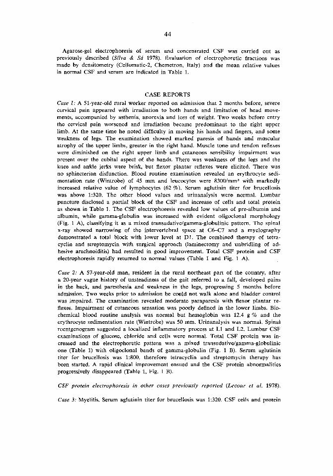
Agarose-gel electrophoresis of serum and concentrated CSF was carried out as previously described (Silva & Sa' 1978). Evaluation of electropho'retic fractions was made by densitometry (Cellomatic-2, Chemetron, Italy) and the mean relative values in normal CSF and serum are indicated in Table 1.
CASE REPORTS Case 1: A 51-year-old rural worker reported on admission that 2 months before, severe cervical pain appeared with irradiation to both hands and limitation of head move- ments, accompanied by asthenia, anorexia and loas of weight. Two weeks before entry the cervical pain worsened and irradiation became predominant to the right upper limb. At the same time he noted difficulty in moving his hands and fingers, and some weakness of legs. The examination showed marked paresis of hands and muscular atrophy of the upper limbs, greater in the right hand. Muscle tone and tendon reflexes were diminished on the right upper limb and cutaneous sensibility impairment was present over the cubital aspect of the hands. There was weakness of the legs and the knee and ankle jerks were brisk, but flexor plantar reflexes were elicited. There was no sphincterian disfunction. Blood routine examination revealed an erythrocyte sedi- mentation rate (Wintrobe) of 45 mm and leucocytes were 830a/mm3 with markedly increased relative value of lymphocytes (62 %). Serum aglutinin titer for brucellosis was above 1:320. The olther blood values and urinanalysis were normal. Lumbar puncture disclosed a partial block of the CSF and increase of cells and total protein as shown in Table 1. The CSF electrophoresis revealed low values of pre-albumin and albumin, while gamma-globulin was increased with evident oligoclonal morphology (Fig. 1 A), classifying it as a mixed transudative/gamma-globulinic pattern. The spinal x-ray showed narrowing of the intervertebral space at C6-C7 and a myelography demonstrated a total block with lower level at D1. The combined therapy of tetra- cyclin and streptomycin with surgical approach (laminectomy and unbridling of ad- hesive arachnoiditis) had resulted in good improvement. Total CSF pro4ein and CSF electrophoresis rapidly returned to normal values (Table 1 and Fig. 1 A).
Case 2: A 57-year-old man, resident in the rural northeast part of the country, after a 20-year vague history of unsteadiness of the gait referred to a fall, developed pains in the back, and paresthesia and weakness in the legs, progressing 5 months before admission. Two weeks prior to admission he could not walk alone and bladder control was impaired. The examination revealed moderate paraparesis with flexor plantar re- flexes. Impairment of cutaneous sensation was poorly defined in the lower limbs. Bio- chemical blood routine analysis was normal but hemoglobin was 12.4 g % and the erythrocyte sedimentation rate (Wintrobe) was 50 mm. Urinanalysis was normal. Spinal roentgenogram suggested a localized inflammatory process at L1 and L2. Lumbar CSF examinations of glucose, chloride and cells were normal. Total CSF protein was in- creased and the electrophoretic pattern was a mixed transudativeJgamma-globulinic one (Table I) with oligoclmal bands of gamma-globulin (Fig. 1 B). Serum aglutinin titer for brucellosis was 1:800, therefore tetracyclin and streptomycin therapy has been started. A rapid clinical improvement ensued and the CSF protein abnormalities progressively disappeared (Table 1, Fig. 1 B).
CSF protein electrophoresis in other cases previously reported (Lecour et al. 1978).
Case 3: Myelitis. Serum aglutinin titer for brucellosis was 1:320. CSF cells and protein

A
45
B
Figure 1. CSF protein electrophoresis in neurobrucettosis. Evolution of CSF electro- phoretic pattern in Case 1 ( A ) and Case 2 (B) . Oligoclonal gamma-globulin in Cases 3
and 4 (C and D, respectively).
electrophoretic values are shown in Table 1. Oligoclonal bands of the CSF gamma- globulin were evident (Fig. 1 C). Case 4: Subacute meningitis. Serum aglutinin titer for brucellosis was 1:320. Cells and protein values of CSF are presented in Table 1. Oligoclonal gamma-globulin was ob- served in CSF electrophoresis (Fig. 1 D).
.
DISCUSSION
Diagnosis of brucellosis was supported by positive serum agglutination tests, and there were typical manifestations of radiculitis secondary to arachnoidi- tis with minor spinal cord involvement. Bone abnormalities revealed by spinal X-ray in Case 2 are suggestive of a primary spondylitic process. Ob- viously, cells and total protein in CSF were increased. Larbrisseau et al. (1978) were probably the first ones to notice a marked increase of gamma- globulins they found in CSF of three patients with brucellar meningoence- phalitis. Such a finding is usual in several subacute and chronic inflamma- tory diseases of the nervous system, which is thought to reflect a humoral response within the central nervous system (CNS) against an infectious antigen (Johnson et al. 1977, Laterre 1965, Laterre et al. 1970, Link & Miiller 1971, Tourtellotte 1970, Vandvik & Skrede 1973).
On a previous study in the rabbit about immunological response in the CNS (Silva et al. 1977), we suggested that for formation of antibodies in the CSF, a secondary intrathecal stimulation after previous sensitization of

46
immunological centers is necessary. For that reason, formation of anti- bodies and increasing of gamma-globulins in the CSF are accomplished only 2-3 weeks after the primary contact with the antigen, since for sec- ondary intrathecal stimulation to occur, the antigen has to remain that time in the CNS or its envelopments. So, only in subacute or chronic infections of the CNS may increased relative values of CSF gamma-globulin be in- duced by immunological mechanisms.
The immune response occurs regardless of the nature of the agent, and may be observed in bacterian, parasitary, fungical and viral diseases of the CNS (Johnson et al. 1977, Laterre 1965, Laterre et al. 1970, Link & Miiller 1971, Link et al. 1973, Vandvik & Skrede 1973, Tourtellotte 1970). But in these diseases the CSF electrophoretic patterns are quite different be- cause variable degrees of breakdown of blood-brain or blood-CSF barriers may result as a consequence of the inflammatory process. While in subacute sclerosing panencephalitis, the barrier damage is absent or minimal and a selective rise of the CSF gamma-globulins occurs, in other subacute diseases such as tuberculous meningitis, toxoplasmosis or cysticercosis (Johnson et al. 1977, Laterre 1965, Laterre et al. 1970, Link & Miiller 1971, Vandvik & Skrede 1973), a marked barrier breakdown is associated, resulting then CSF electrophoretic abnormaIities described as transudative by Laterre (1965) which makes less clear the underlying immunological response. In such a case, quotients as the ratio between the relative values of gamma- globulin of CSF and serum (Schinko & Tshabitscher 1957, Peter et al. 1974) or the IgG-index (Delpech & Lichtblau 1972, Christensen et al. 1978) among others, may be helpful to demonstrate local production of immuno- globulins in the CNS in spite of increased CSF total protein. This mixed transudative/gamma-globulinic pattern seems to be frequent in neurobrucel- losis, as the first electrophoresis of Cases 1 and 2 documents and is sug- gested by the results of Larbrisseau et al. (1978). However, in those two cases, added to the transudative features is a very elevated and markedly oligoclonal gamma-globulin. Oligoclonal morphology of the CSF gamma- globulins was also observed in Cases 3 and 4. The significance of the oligo- clonal gamma-globulin has been extensively studied in multiple sclerosis and subacute sclerosing panencephalitis and there is no doubt about its close relationship to intrathecal synthesis of immunoglobulins (Cutler et al. 1968, Johnson et al. 1977, Laterre et al. 1970, Link 1967, Link & Miiller 1971, Link et al. 1973, Vandvik & Skrede 1973). The subsequent CSF electro- phoresis in Cases 1 and 2 makes more evident the underlying immuno- logical process, since oligoclonal and increased gamma-globulins persist after the remission of the inflammatory process traduced by the disappear- ance of transudative features on the electrophoretic pattern. The import- ance of the oligoclonal morphology of the gamma-globulin is once more

47
demonstrated, since it allows us to suggest intrathecal synthesis of im- munoglobulins also in neurobrucellosis.
ACKNOWLEDGMENTS We are very grateful to Dr. Hennque Lecour and co-workers for permission to refer details of Cases 3 and 4.
REFERENCES Abramsky, 0. (1977): Neurological features as presenting manifestations of brucellosis.
Eur. Neurol. 15, 281-284. Christensen, O., J. Clausen & T. Fog (1978): Relationships between abnormal IgG index,
oligoclonal bands, acute phase reactants and some clinical data in multiple sclc- rosis. J. Neurol. 218, 237-244.
Cutler, R. W. P., E. Merler & J. P. Hammerstad (1968): Production of antibody by the central nervous system in subacute sclerosing panencephalitis. Neurol. 18 (Part 2),
Delpech, B. & E. Lichtblau (1972): etude quantitative des immunoglobulines G et de l'albumine du liquide cephalorachidien. Clin. Chim. Acta 37, 15-23.
Fincham, R. W., A. L. Sahs & B. J. Joynt (1963): Protean manifestations of nervous system brucellosis. Case histories of a wide variety of clinical forms. J. Am. Med.
Johnson, K. P., S. C. Arrigo, B. J. Nelson & A. Ginsberg (1977): Agarose electrophoresis of cerebrospinal fluid in multiple sclerosis. A simplified method for demonstrating cerebrospinal fluid oligoclonal immunoglobulin bands. Neurol. 27, 273-277.
Larbrisseau, A., E. Maravi, F. Aguilera & J. M. Martinez-Lage (1978): The neurological complications of brucellosis. J. Canad. Sci. Neurol. 5, 369-376.
Laterre, E. C. (1965): Les Protdines du Liquide Cdphalorachidien B 1'6tat Normal et Pathologique. Arscia, Bruxelles.
Laterre, E. C., A. Callewaert, J. F. Heremans & Z. Sfaello (1970): Electrophoretic mor- phology of gamma-globulin in cerebrospinal fluid of multiple sclerosis and other diseases of the nervous system. Neurol. 20, 982-990.
Lecour, H., P. Barata-Feyo, F. Cerqueira-Magro & C. Cruz (1978): Neurobrucelose. J. Mddico 96, 417-419.
Link, H. (1967): Immunoglobulin G and low molecular weight proteins in human cere- brospinal fluid: chemical and immunological characterizatioa with special refer- ence to multiple sclerosis. Acta Neurol. Scand. 43 (Suppl. 28), 1-136.
Link, H. & R. Miiller (1971): Immunoglobulins in multiple sclerosis and infections of the nervous system. Arch. Neurol. 25, 326-344.
Link, H., M. Panelius & A. A. Sami (1973): Immunoglobulins and measles antibodies in subacute sclerosing panencephalitis (Demonstration of synthesis of oligoclonal IgG with measles antibody activity within the central nervous system). Arch. Neurol. 28, 23-30.
Peter, A., A. Lowenthal and I. Juvancz (1974): Changes of y-globulins in serum and cerebrospinal fluid of patients with subacute sclerosing panencephalitis. J. Neurol.
Papadopoulos, N. M., W. C. Hess, D. O'Doherty & D. E. McLane (1959): A procedure for the determination of cerebrospinal fluid total protein and gamma-globulin in neurologic disorders. Clin. Chem. 5, 569-574.
129-132.
ASSOC. 184, 269-275.
207, 85-92.

48
Sahadevan, M. G., M. Singh, P. P. Joseph & R. S. Hoon (1968): Meningomyelitis due to brucellosis. Br. Med. J. 4, 432-433.
Schinko, H. & H. Tshabitscher (1957): Der 11-Quocient als Ausdruck der Relation Liquor zu Serum y-Globulin. Wien. Klin. Wschr. 69, 705-713.
Silva, C. A., M. J. SB & C. Cruz (1977): Tetanus antibody production in serum and cere- brospinal fluid in the rabbit and correlated histopathological features of the cen- tral nervous system. J. Neurol. Sci. 33, 213-227.
Silva, C. A. & M. J. SB (1978): Electrophoretic pattern of cerebrospinal fluid proteins in non-neoplastic infantile hydrocephalus. Acta Neurol. Scand. 57, 317-328.
Sols, A. (1949): Valoration de las proteinas del suero por referencia a la media nor- mal. Rev. Esp. Fisiol. 5, 239-250.
Tourtellotte, W. (1970): On cerebrospinal fluid immunoglobulin-G (IgG) quotients in multiple sclerosis and other diseases. A review and a new formula to estimate the amount of IgG synthesized per day by the central nervous system. J. Neurol. Sci.
Vandvik, B. & S. Skrede (1973): Electrophoretic examination of cerebrospinal fluid pro- teins in multiplo sclerosis and other neurological diseases. Eur. Neurol. 9, 224-241.
10, 279-304.
Received August 8, accepted October 16, 1979
Carlos Albert0 Silva, M.D. ServiGo de Neurologia e Neurocirurgia Faculdade de Medicina Port0 Portugal


![Thymoglobulin (anti-thymocyte globulin [rabbit]) · 2020. 12. 14. · DESCRIPTION . Thymoglobulin® (Anti-thymocyte globulin [rabbit]) is a purified, pasteurized, gamma immune globulin](https://img.pdfslide.net/doc/110x75/60c2dece3812e518472963b9/thymoglobulin-anti-thymocyte-globulin-rabbit-2020-12-14-description-thymoglobulin.jpg)
![Thymoglobulin (anti-thymocyte globulin [rabbit]) - Sanofiproducts.sanofi.ca/en/thymoglobulin.pdf · Thymoglobulin® (Anti-thymocyte Globulin [Rabbit]) ... (ATG) products, as protein](https://img.pdfslide.net/doc/110x75/5aa587a17f8b9ab4788d4753/thymoglobulin-anti-thymocyte-globulin-rabbit-anti-thymocyte-globulin-rabbit.jpg)