Embed Size (px)
Citation preview
FROM GREEN TO YELLOW
A LEAF STORY
Olivier Keech
Akademisk avhandling
Som med vederbörligt tillstånd av rektorsämbetet vid Umeå universitet för avläggande av
filosofie doktorsexamen i ämnet växtfysiologi.
Framläggs till offentligt försvar i KB3A9, KBC-huset,
Fredagen den 12 oktober 2007, kl. 10.00
Avhandlingen kommer att försvaras på engelska.
Fakultetsopponent: Dr. Lee Sweetlove, University Lecturer in Plant Science,
Department of Plant Sciences, University of Oxford, Oxford, United Kingdom
Umeå Plant Science Centre, Department of Plant Physiology
Umeå University, Umeå, Sweden
2007
From Green to Yellow A Leaf Story
Olivier Keech, Umeå Plant Science Centre, Department of Plant Physiology, Umeå University,
SE- 901 87, Umeå, Sweden, October 2007
Doctoral dissertation ISBN 978-91-7264-400-7
Abstract When switching from green to yellow, a leaf undergoes both morphological and metabolic changes. This process is known as senescence and improved understanding of its mechanisms is important both from a fundamental scientific perspective but also for biotechnological applications. The present thesis reports on several important aspects regarding the cellular and metabolic mechanisms occurring during leaf senescence with an emphasis on the mitochondrial contribution to this process. As a first step, we developed methods to isolate either highly functional crude mitochondria or highly purified mitochondria from leaves of Arabidopsis thaliana. These methods were further used to study mitochondrial contributions to cellular redox homeostasis and to estimate the mitochondrial capacities in leaves undergoing senescence. In particular, we compared the induction of senescence by different dark treatments in Arabidopsis. The comparison between individually darkened leaves and leaves from whole darkened plants revealed different metabolic strategies in response to darkness. Integrating data from measurements of photosynthesis, respiration and confocal laser microscopy with transcriptomics and metabolomics profiling, we suggested that metabolism in leaves of the whole darkened plants enter a “stand-by mode” with low mitochondrial activity in order to maintain the photosynthetic machinery for as long as possible. In contrast, mitochondria from individually darkened leaves are more active and may provide energy and carbon skeletons for the degradation of cell constituents, facilitating the retrieval of nutrients. We also investigated the dynamic of the microtubular cytoskeleton during dark-induced senescence. Mitochondrial mobility was affected by an early disruption of the microtubules in individually darkened leaves but not in whole darkened plants. In addition, several microtubules associated proteins (MAPs) seemed to be involved in the bundling of the microtubules around the chloroplasts. Altogether, the work presented in this thesis highlights several important steps regarding the metabolic adjustments and the cellular mechanisms in Arabidopsis leaves submitted to prolonged darkness. In particular, we suggest the mitochondria to fulfill specific and important functions during leaf senescence since the role of mitochondria in leaves experiencing prolonged darkness appears very dependant on the whole metabolic status of the plant. Keywords: Arabidopsis thaliana, chloroplasts, cytoskeleton, darkness, metabolism, microscopy, mitochondria, microtubules, senescence, system redox.
FROM GREEN TO YELLOW
A LEAF STORY
Olivier Keech
Umeå Plant Science Centre, Department of Plant Physiology
Umeå University, Umeå, Sweden
2007
A vous qui m’avez soutenu et cru en moi…
Il n’est point de bonheur sans liberté, ni de liberté sans
courage ! [Périclès]
Copyright © Olivier Keech, 2007
Umeå Plant Science Centre
Department of Plant Physiology
Umeå University
SE-901 87 Umeå
Sweden
Cover illustration: Jan Eklöf (Merci man!)
ISBN 978-91-7264-400-7
Printed by Solfjäden Offset AB, Umeå, 2007
Table of contents
List of papers
Preface
A PhD journey
INTRODUCTION 1
1. Senescence 1
1.1 Introduction to senescence
1.2 What is leaf senescence?
1.3 What can trigger leaf senescence?
1.4 Ozone: an example of stress leading to leaf senescence
1.4.1 What is Ozone?
1.4.2 What are the effects of Ozone on plants?
1.4.3 Ozone and Senescence
1.5 Signalling during senescence
1.5.1 Sugars signalling
1.5.2 Hormones signalling
1.6 Genes regulation during leaf senescence
2. Mitochondria 13
2.1 Introduction
2.2 The origins of the plant mitochondria
2.3 Basic functions of the plant mitochondria
2.3.1 The mitochondrial electron transport chain
2.3.2 The tricarboxylic acid cycle
2.3.3 The photorespiration
3. Aim: Why Senescence and Mitochondria? 25
3.1 The mitochondrial contribution to programmed cell death
3.2 Senescence / programmed cell death: where are the boundaries?
3.3 Economical perspectives
3.4 Aim and hypothesis
RESULTS AND DISCUSSION 28
4. Isolation of mitochondria: a useful tool 28
4.1 Why Arabidopsis?
4.2 Where are the difficulties?
4.3 Comments about the two methods
4.4 A tool for further studies
5. Dark-induced senescence as a model 32
6. Regulation of the metabolism 35
6.1 A cytological approach
6.2 Photosynthesis
6.3 Mitochondrial respiration
6.4 Metabolomics
6.5 Conclusion
REMARKS 42
FRÅN GRÖNT TILL GULT - EN BLADHISTORIA 43
AKNOWLEDGEMENTS 44
REFERENCES 46
List of papers
The following work is based on the here below papers, which will be referred to by their Roman
numerals.
I. Keech O, Dizengremel P, Gardestrom P (2005). Preparation of leaf mitochondria
from Arabidopsis thaliana. Physiol. Plant. 124: 403-409.
II. Rouhier N, Gelhaye E, Villarejo A, Srivastava M, Keech O, Droux M, Finkemeier I, Samuelsson G, Dietz KF, Jacquot JP, Wingsle G (2005). Identification of plant glutaredoxin targets. Antioxid. Redox Sign. 7: 919-929.
III. Gama F*, Keech O*, Eymery F, Finkemeier I, Gelhaye E, Gardestrom P, Dietz KJ,
Rey P, Jacquot J-P, Rouhier N (2007) The mitochondrial type II peroxiredoxin from poplar. Physiol. Plant. 129: 196-206.
IV. Keech O, Pesquet E, Ahad A, Askne A, Nordvall D, Vodnala SM, Tuominen T, Hurry
V, Dizengremel P and Gardeström P (2007). The different fate of mitochondria and chloroplasts during dark-induced senescence in Arabidopsis leaves. Plant Cell Environ. (doi: 10.1111/j.1365-3040.2007.01724.x)
V. Ahad A, Keech O, Sjödin A, Stenlund H, Moritz T, Jansson S, and Gardeström P
(2007). Leaf metabolism during dark induced senescence in arabidopsis integrating metabolomics and transcriptomics. (Manuscript)
VI. Keech O*, Pesquet E*, Sjödin A, Jansson S, Tuominen H, Ahad A, and Gardeström
P (2007). Early disruption of the microtubules during dark-induced senescence. (Manuscript)
* Authors contributed equally to this work Articles I, III and IV are reprinted with the kind permission of Blackwell Publishing Inc. Article II is reprinted with the kind permission of Mary Ann Liebert, Inc.
Preface
In this preface I would like to say a few words about being a PhD-student. The following thesis
reports the results of my research during my PhD time but it does not show how my thoughts
have matured during these years. Beyond the work and the experiments, successful or not, I
learnt a lot about the others, about myself, about my strengths and my weaknesses. This cannot
be written in any manuscript or article, but it appears to be essential in the process of becoming a
doctor. Becoming a researcher does not only include being able to run experiments or to write
articles. It is also to be able to interact with human beings from first grade students to professors
and that also includes persons from different professional horizons. We need to be able to present
our work to the public, to make it as clear and as simple to understand as we can. We need to
work on our skill to insufflate our knowledge and passion for sciences to others. Furthermore,
failures and disillusions forged us along the time with courage, patience, endurance, modesty and
humility. Finally, and maybe the most important thing, I have learnt from “you”, from every
single person irrespective of their social belongings I have interacted with during these years.
The evolution appears to be personal but cannot be achieved without the others. Always
remember: we own our fate!
Olivier K.
A PhD journey…
12th of July 2007. Well well well, here I am, starting to write my thesis to get the grade of PhD in
Plant Sciences. I have about a month to summarize by this text my work as PhD student. I guess
there are several ways to start writing a thesis and I don’t really know which one is best or even
if there is one better than another one…However, in order to summarize my work during the last
four and a half years and also to help the reader to understand this PhD time, I think I need to let
my mind going back in the past and tell you what was going on for me at that time! I started my
PhD with a project in collaboration between the Umeå Plant Science Centre (Umeå, Sweden)
under the supervision of the professor Per Gardeström and the department of Ecology and Forest
Ecophysiology (Nancy, France) under the supervision of the Professor Pierre Dizengremel. The
subject was basically defined as 2 proteomic studies: one was about a comparison of the
mitochondrial proteomes during cold stress in Arabidopsis leaves and the other one was about
the modifications of the mitochondrial proteome during ozone stress in Arabidopsis leaves. As
mentioned here above, the 2 projects dealt with mitochondrial proteomes from Arabidopsis
leaves which first implicated to be able to get these mitochondrial proteomes. Back in the
beginning of 2003, there were a couple of articles available purposing methods for isolation of
mitochondria from Arabidopsis (Cf paper I and references therein). However, none of them truly
dealt with Arabidopsis leaves. The authors clearly explained that their preparation was not pure
enough for suitable proteomic analysis and consequently, they decided to work with dark grown
cell-suspension cultures. However, our aim was clear and we wanted to work with leaves
(mitochondrial metabolism is tightly coupled to the photosynthetic machinery which makes cell
metabolism so interesting!). So, I started to work on establishing a protocol to isolate pure and
functional mitochondria from Arabidopsis leaves. After 6 months of work (spending my days in
the cold room…One told me Umeå was cold!) and going through the literature, I came to the
point saying that we should change my subject and focus on leaf senescence and ozone instead of
cold and ozone. Both Per and Pierre agreed and I must admit now that I am very thankful to them
for the freedom they let me during all those years! After a year and a half, I finally established 2
suitable protocols yielding either highly functional crude mitochondria or highly purified
mitochondria, see paper I (Keech et al., 2005). Through collaborations with the lab of Jean-
Pierre Jacquot and Nicolas Rouhier (Nancy, France), we further used these protocols to first
determine potential targets of plant glutaredoxins, see paper II (Rouhier et al., 2005) and then to
characterize the mitochondrial peroxiredoxin IIf in poplar, see paper III (Gama et al., 2007).
However, in order to study the potential modifications of the proteome during leaf senescence,
we first needed a system where we could get senescing leaves at different stages. We got 2
choices: 1/ use leaves from natural aging Arabidopsis plants 2/ use a system where we can
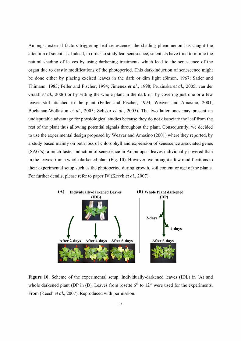
induce leaf senescence. We opted for the second choice. In 2001, Weaver and Amasino (2001)
described, mainly based on a molecular biology approach, a faster induction of senescence in
individually covered Arabidopsis leaves in comparison with the leaves from a whole darkened
plant. We thought this system to be extremely interesting because i) the system is very easy to
handle ii) we can discriminate the effects of starvation and/or darkness from the effects of
senescence iii) we can easily get an important amount of fresh material to isolate mitochondria.
However, before running any proteomic studies, we needed to describe in details these two
experimental setups. Based on a detailed quantification of organelles by confocal laser
microscopy, photosynthetic and respiratory measurements, we showed that these two
experimental setups exhibited different metabolic adjustments in response to darkness, see paper
IV (Keech et al., 2007). At the same time, Dr. Abdul Ahad, postdoc in Per Gardeström’s group
at that time, started a project focusing on metabolic and transcriptomic comparisons of
individually darkened leaves with leaves from whole darkened plants. The results of this work
are presented in paper V (Ahad et al. manuscript). Finally, since we suspected the cytoskeleton
to be involved in the cellular rearrangement observed and described in paper IV, we present in
paper VI (Keech et al. manuscript) the results of our up-to-date investigations.
Consequently, and before I let the reader going though the following thesis, I need to clarify 2
things about my work and my primary objectives: First, no data on comparison of mitochondrial
proteomes are presented in the present thesis and second, even if I did some preliminary work on
ozone stress, no data are included in this thesis in order to make a clearer story.
INTRODUCTION
1. Senescence
1.1 Introduction to senescence
Through the plant kingdom, there is a large range of lifespans. Certain tree species can live for
several centuries whereas other plants, such as Arabidopsis, can complete their life cycle in a few
weeks. Beyond this first remark, we also want to notice that individual organs of a plant such as
leaves, flowers and vessels have lifespans that can substantially differ from that of the whole
organism. For example, in a tree, a trunk consists of wood, which is the result of dead vascular
tissue. So, it is important to keep in mind that there is a high rate of cell death occurring
throughout the life of the plant (Guarente et al., 1998). When contemplating the longevity and
ultimate fate of plant tissues, laws of thermodynamics must be kept in mind. A plant cell requires
energy input for its creation and maintenance and in theory an individual cell could be
maintained alive forever if enough energy was provided. Yet, somatic tissues in plants have
limited life spans (Bleecker and Patterson, 1997). Interestingly, the lifespan of leaves for
example may vary from few days to as long as 20 to 30 years in species of Araucaria
(Woolhouse, 1967).
When considering an individual leaf, its development primary requires nutrients from the rest of
the plant. But as soon as the photosynthetic apparatus is achieved and functional, the leaf
converts from a nutrient sink to a nutrient source and the payoff can be established. It is
advantageous to maintain leaves only for as long as they contribute to the survival of the plant.
However, when the leaf gets mature, its life spans truly depends on environmental factors, both
abiotic and biotic such as extreme temperatures, drought, shading, ozone and pathogen infection
for instance (Woolhouse, 1967; Smart, 1994; Gan and Amasino, 1997; Thompson et al., 2004).
In temperate region of the world for example, the shortening days and colder temperatures of the
approaching winter limit the leaf productivity which leads the leaves from trees and other
perennial plants to turn to magnificent yellow, orange and red colors. Finally, a massive
1
programmed cell death occurs and leads to death and loss of the leaves. This phenomenon is
often referred to in the plant biology literature as leaf senescence. Specifically, the foliar
senescence relates to the process by which nutrients are mobilized from the dying leaf to other
parts of the plant to support their growth. Nutrient availability, particularly nitrogen, has been a
major limit to growth and reproductive success throughout plant evolution (Guarente et al.,
1998). Moreover, plants are fixed in a particular location in the soil and deplete their local
environment. Thus, it becomes very easy to consider that plants have evolved mechanisms for
dealing with obsolete organ systems, especially when we know that photosynthetic organs are
very rich in valuable nutrients for the plants.
1.2 What is leaf senescence?
Foliar senescence is often regarded as a particular type of programmed cell death (PCD)
(Nooden et al., 1997; Pennell and Lamb, 1997; Delorme et al., 2000; Lim et al., 2003; Thomas et
al., 2003; Van Doorn, 2005) and its main purpose in plants is for nutrients mobilization and
recycling. Throughout the scientific literature, common features are stated to describe the
evolution of this regulated process within an organ such as a leaf. Chloroplasts are the first
affected by the cellular degeneration process. Proteins and RNA’s are degraded, causing the
photosynthetic machinery to be rapidly impaired. Nutrients such as nitrogen, phosphorous,
sulphur, minerals and metals ions are drawn back from the senescing leaves (source) to be
recycled in other parts of the plant (sink) (For reviews see: (Smart, 1994; Buchanan-Wollaston,
1997; Nooden et al., 1997; Himelblau and Amasino, 2001; Hortensteiner and Feller, 2002;
Buchanan-Wollaston et al., 2003). The loss of chlorophyll, one of the main biological markers
for leaf senescence, begins from the outer parts of the leaf and proceeds inwards giving to the
leaf this characteristic yellowish color. The tissues near the vascular system are the last ones to
senesce, since they are needed for nutrient allocation (Quirino et al., 2000). Nowadays, the
understanding of senescence mechanisms is part of both fundamental scientific questions and an
economical challenges to increase the yield of the crops by prolonging the photosynthetic
activity and to minimize the post-harvest quality loss in vegetables (Gan and Amasino, 1997).
Leaf cells ongoing senescence undergo many biochemical and structural changes controlled by
an important programming of gene expression. However, even though the catabolism leading
2
chloroplasts to become gerontoplasts is under direct nuclear control (Feller and Fischer, 1994;
Gan and Amasino, 1997; Van Doorn, 2005), mitochondria are known to remain intact until
rather late during the process most probably in order to supply energy mainly for reallocation of
nutrients (Feller and Fischer, 1994; Smart, 1994; Collier and Thibodeau, 1995; Bhalerao et al.,
2003; Lim et al., 2003; Keskitalo et al., 2005). Nevertheless, no clear evidences are given about
the respective roles of these two organelles during the leaf senescence. For instance, it has been
recently suggested that chloroplasts could regulate leaf senescence by increasing the reduction
level of electrons transporters and by generating reactive oxygen species (ROS), which are
further thought to damage cell structures and functions (Zapata et al., 2005).
1.3 What can trigger leaf senescence?
There are many factors that can initiate senescence in leaves and it is obvious that there are many
different pathways involved in controlling the process (Smart, 1994; Gan and Amasino, 1997).
As examples, we would like to mention that leaf senescence can be induced by a number of
different environmental stresses such as pathogen infection (Quirino et al., 1999; Beers and
McDowell, 2001), nutrient deficiency (Feller and Fischer, 1994), drought (Pastori and Trippi,
1993; Pic et al., 2002; Munné-Bosch and Alegre, 2004), ozone (Paakkonen et al., 1997; Puffett
et al., 1997; Ojanpera et al., 1998; Miller et al., 1999; Pell et al., 1999) or even UV-B (Pradhan
et al., 2006). However, a recent study from Costa et al. (2006) showed that UV-C delay
postharvest senescence in broccoli florets. Overall, it seems that many signalling pathways
controlling gene expression in response to different stresses are also involved in leaf senescence
(Buchanan-Wollaston et al., 2003). For instance, and taking the HR (HR: hypersensitive
response, a disease- resistance response that results from incompatible pathogen interactions) as
example, one could think that a senescing leaf is more susceptible to pathogen infections and
consequently the defense-related genes induced during leaf senescence would be a normal
preventive response to potential pathogenesis accompanying the senescence process. However,
Quirino et al. (1999) demonstrated that defense-related genes are still induced during leaf
senescence of Arabidopsis plants grown in sterile conditions which in turn indicates that the
expression of defense-related genes (and most likely many other stress-related genes in other
stress cases) might be an integral part of the senescence program. These considerations are in
3
line with an interesting study based on the Arabidopsis Genechip system (Affymetrix) where
Chen et al. (2002) reported that 2/3 of the transcription factors genes induced by stress treatment
were also induced during leaf senescence which suggests extensive overlapping in the responses
to these stress treatments. However, depending on the nature and the intensity of the stress apply
we are still unable to explain clearly whether stress causes the onset of senescence or whether the
senescence itself induces stress responses.
1.4 Ozone as an example of stress leading to leaf senescence
1.4.1 What is ozone?
Ozone (O3), also called “super-oxygen”, is the tri-atomic form of oxygen and is naturally formed
by the UV (ultra-violet) rays of the sun (photochemical reaction) and by lightning (bioelectrical
reaction). Ozone may also be formed synthetically by passing air or oxygen over an UV lamp
(photochemical reaction) and through a high-voltage field (bioelectrical reaction). Some of the
oxygen molecules are split into two separate oxygen atoms. These singlet atoms then form semi-
stable bonds with the oxygen molecules. These polyatomic oxygen molecules are highly reactive
because the third oxygen atom, also known as a "Hungry Atom", is very willing to break away
from this semi-stable bond and gives hydroxyl radical. The hydroxyl radical serves as the main
scavenger in the atmosphere, reacting with a variety of compounds such as hydrocarbons,
hydrogen sulfide, and carbon monoxide that would otherwise accumulate and would poison most
of breathing organisms. Due to its reactivity, ozone has a very short life span (about 20 min.).
In the nature, two kinds of “Ozone” can be distinguished; the stratospheric ozone and the
tropospheric ozone. Most of ozone resides in the stratosphere (layer of the atmosphere between
10 and 40 km above the earth’s surface) and is essential to protect life on the earth by acting as a
shield against solar UV-B irradiation (280nm-320nm) (Zinser et al., 2000). Tropospheric ozone
which appears in the atmospheric layer from the surface up to about 10 km is mainly the result of
human activity (the burning of fossil fuels commonly gathered under the name of petroleum
increases the production of carbon compounds that reacts in sunlight with oxygen to form ozone
in the troposphere (Buchanan et al., 2000)). Interestingly, troposheric ozone may be essential for
4
survival of many organisms by playing a key role in enhancing human health and well being
since O3 is involved in chemical reactions that clean the troposphere of some pollutants but is at
the same time drastically harmful to life because it reacts with any oxidizable compound (organic
or inorganic) (http://earthobservatory.nasa.gov/Library/Ozone/). In plants, the discovery of the
phytotoxicity of O3 during the mid 1950’s (Haagen-Smit et al., 1952) prompted widespread
studies on the effects of O3 on their growth and development (Ashmore, 2005).
1.4.2 What are the effects of ozone on plants?
The biochemical and molecular mechanisms underlying O3 phytotoxicity have only begun to be
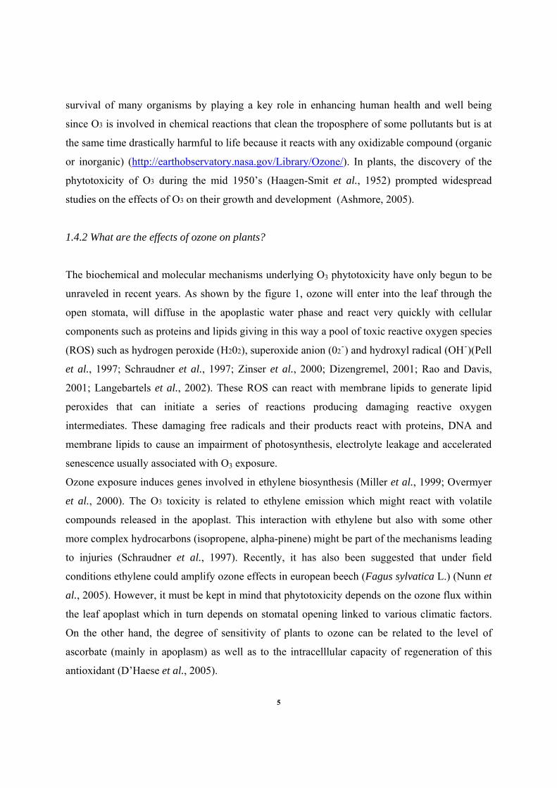
unraveled in recent years. As shown by the figure 1, ozone will enter into the leaf through the
open stomata, will diffuse in the apoplastic water phase and react very quickly with cellular
components such as proteins and lipids giving in this way a pool of toxic reactive oxygen species
(ROS) such as hydrogen peroxide (H202), superoxide anion (02ֿ) and hydroxyl radical (OHֿ)(Pell
et al., 1997; Schraudner et al., 1997; Zinser et al., 2000; Dizengremel, 2001; Rao and Davis,
2001; Langebartels et al., 2002). These ROS can react with membrane lipids to generate lipid
peroxides that can initiate a series of reactions producing damaging reactive oxygen
intermediates. These damaging free radicals and their products react with proteins, DNA and
membrane lipids to cause an impairment of photosynthesis, electrolyte leakage and accelerated
senescence usually associated with O3 exposure.
Ozone exposure induces genes involved in ethylene biosynthesis (Miller et al., 1999; Overmyer
et al., 2000). The O3 toxicity is related to ethylene emission which might react with volatile
compounds released in the apoplast. This interaction with ethylene but also with some other
more complex hydrocarbons (isopropene, alpha-pinene) might be part of the mechanisms leading
to injuries (Schraudner et al., 1997). Recently, it has also been suggested that under field
conditions ethylene could amplify ozone effects in european beech (Fagus sylvatica L.) (Nunn et
al., 2005). However, it must be kept in mind that phytotoxicity depends on the ozone flux within
the leaf apoplast which in turn depends on stomatal opening linked to various climatic factors.
On the other hand, the degree of sensitivity of plants to ozone can be related to the level of
ascorbate (mainly in apoplasm) as well as to the intracelllular capacity of regeneration of this
antioxidant (D’Haese et al., 2005).
5
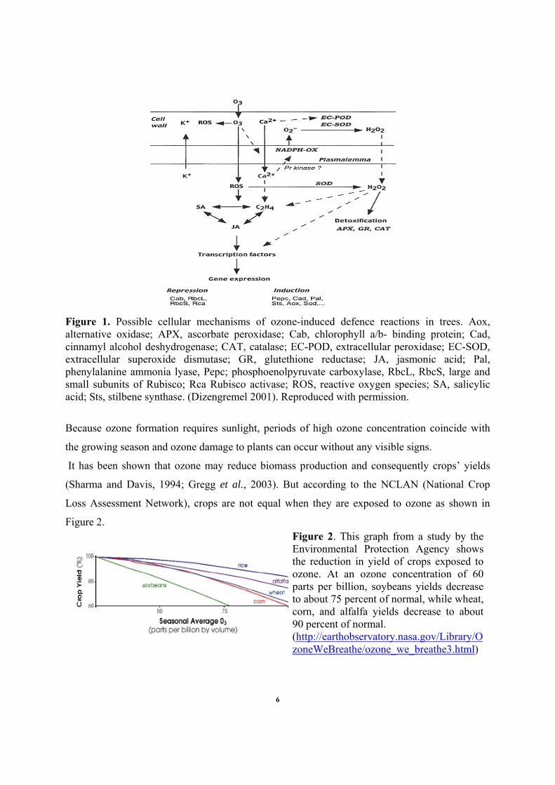
Figure 1. Possible cellular mechanisms of ozone-induced defence reactions in trees. Aox, alternative oxidase; APX, ascorbate peroxidase; Cab, chlorophyll a/b- binding protein; Cad, cinnamyl alcohol deshydrogenase; CAT, catalase; EC-POD, extracellular peroxidase; EC-SOD, extracellular superoxide dismutase; GR, glutethione reductase; JA, jasmonic acid; Pal, phenylalanine ammonia lyase, Pepc; phosphoenolpyruvate carboxylase, RbcL, RbcS, large and small subunits of Rubisco; Rca Rubisco activase; ROS, reactive oxygen species; SA, salicylic acid; Sts, stilbene synthase. (Dizengremel 2001). Reproduced with permission.
Because ozone formation requires sunlight, periods of high ozone concentration coincide with
the growing season and ozone damage to plants can occur without any visible signs.
It has been shown that ozone may reduce biomass production and consequently crops’ yields
(Sharma and Davis, 1994; Gregg et al., 2003). But according to the NCLAN (National Crop
Loss Assessment Network), crops are not equal when they are exposed to ozone as shown in
Figure 2.
Figure 2. This graph from a study by the Environmental Protection Agency shows the reduction in yield of crops exposed to ozone. At an ozone concentration of 60 parts per billion, soybeans yields decrease to about 75 percent of normal, while wheat, corn, and alfalfa yields decrease to about 90 percent of normal. (http://earthobservatory.nasa.gov/Library/OzoneWeBreathe/ozone_we_breathe3.html)
6
1.4.3 Ozone and senescence
Many efforts are made today to determine precisely the mode of action of ozone and since many
years, scientists are trying to correlate physiological and biochemical events occuring in the
plants with the intensity of ozone stress (Schraudner et al., 1997). Phytotoxicity of ozone can be
divided into acute and chronic exposures (Sandermann, 1996; Pell et al., 1997). The former one
leads most of the time to a quick cell death whereas the latter one may cause the acceleration of
the normal rate of foliar senescence especially by a chlorophyll and protein loss within the foliar
cells (Brendley and Pell, 1998; Rao and Davis, 2001; Rao et al., 2002).
In many plant species, an accelerated foliar senescence has been reported as one of the harmful
effects of O3 (Pell et al., 1997; Brendley and Pell, 1998; Bielenberg et al., 2002; Langebartels et
al., 2002). This was also recently observed for several aspen stands at the free-air (CO2+ O3)
exposure site Aspen FACE (in northern Wisconsin) (Karnosky et al., 2005). A study from 1999
revealed that Arabidopsis O3-induced senescence involves many (although not all) of the genes
associated with natural leaf senescence (Miller et al., 1999). The findings of Miller et al. (1999)
have been strengthened by two recent studies. D’Haese et al. (2006) observed in Arabidopsis the
simultaneous induction of salicylic acid synthesis and genes involved in programmed cell death
and senescence. Recently, Gupta et al. (2005) showed higher expression of senescence-
associated genes (SAGs) and genes involved in the flavanoid pathway after long-term exposure
of Populus tremuloides to elevated tropospheric O3 in the Aspen FACE facility. Moreover they
showed that many signaling and defense-related genes were also up-regulated and a lower
expression of several photosynthesis and energy-related genes was observed under O3, in normal
or elevated CO2 treatment.
Consequently, and as we have previously mentioned, ozone exposure leads to a reduction of the
photosynthetic capacity by chlorophyll degradation and by an accelerated loss of both the
activity and the quantity of Rubisco and Rubisco activase (Glick et al., 1995; Brendley and Pell,
1998; Fontaine et al., 1999; Dizengremel, 2001; Pelloux et al., 2001). At the same time, the
catabolic pathways (glycolysis, pentose phosphate pathway) and the mitochondrial respiration
are increased (Dizengremel, 2001). However, the role of increased mitochondrial respiration
7
from photosynthetic tissues during the plant response to O3 remains insufficiently defined. It
seems then essential to investigate further their metabolic role and involvement into the
biochemical events and regulatory pathways facing ozone stress.
1.5 Signalling during senescence
1.5.1 Sugars signalling
Leaves are specialized organs for photosynthetic activity and their lifespan truly depends on their
photoassimilates production. Interestingly, in green leaves and under light conditions, low sugar
levels enhance photosynthesis, and the accumulation of glucose and sucrose represses the
transcription of photosynthetic genes (Rolland et al., 2002). During leaf senescence, one of the
most early and common event occurring is the rapid impairment of photosynthesis. It has even
been proposed several times that the rapid decline of photosynthetic activity could be a signal for
the induction of leaf senescence (Smart, 1994; Bleecker and Patterson, 1997). In addition, the
sugar starvation resulting from a reduction in the photosynthetic capacity is also thought to be
linked with the induction of leaf senescence (Hensel et al., 1993). This is further supported by
the observation that the dark induced expression of many SAGs (senescence associated gene) is
repressed in the presence of sucrose (Chung et al., 1997; Fujiki et al., 2001). In line with the
theory that decreased sugar concentration would trigger senescence, it has been shown that
transgenic tomato plants that overexpress the hexokinase (which catalyzes hexose
phosphorylation) exhibit an accelerated senescence although their actual sugar content were
lower than those from wild type plants (Dai et al., 1999). Despite these findings, the role of
sugars in the induction of leaf senescence under natural conditions is still unclear and remains
rather controversial.
There is an extensive data set on a wide range of species showing that the soluble sugar
concentration often goes up at the onset of leaf senescence (Nooden et al., 1997; Keskitalo et al.,
2005). Similarly, Wingler et al. (2006) found that glucose and fructose accumulated in
Arabidopsis leaves until late senescence. Moreover, a recent study from Pourtau et al. (2006)
confirmed that Arabidopsis leaf senescence was induced rather than repressed by sugars. It must
be kept in mind that the sink–source balances may affect the partitioning of sugar within a plant
8
and can consequently induce leaf senescence. Young leaves are sink organs until their complete
maturation, whereas old leaves remain source organs by providing sugars to the rest of the plant
and especially to the expanding leaves. When young leaves of sunflower and bean plants have
fully developed their own photosynthetic machinery their demands for sugars begin to decrease.
Such limited demands may lead to the accumulation of carbohydrates in the old leaves and their
induction of senescence. However, when young leaves are shaded and therefore cannot operate
photosynthesis and produce sugars, the older leaves of the same plant do not accumulate sugars
and their senescence becomes retarded (Ono et al., 2001). Taken altogether, these results indicate
that senescence may be induced by carbohydrate accumulation and not by starvation. However,
this seems highly dependant on a combination of environmental factors. Finally, sugar control of
senescence is influenced by many other, environmental factors that affect leaf senescence, such
as light conditions, CO2 concentrations, nitrogen supply, stress and pathogen and it is highly
possible that environmental signals are integrated by sugar signaling. For a very good review on
the subject, we redirect the reader to Wingler et al. (2006).
1.5.2 Hormones signalling
Cytokinin and ethylene represent maybe the two best examples for hormonal involvement in the
control of senescence. The signal that initiates the onset of developmental senescence appears to
involve cytokinin. It has been known for many years that cytokinin levels decline in senescing
leaves and that treatment with cytokinin can delay leaf senescence. To deliver cytokinin
specifically to leaves at the onset of senescence, Gan and Amasino (1995) used the promoter of
one of the senescence-associated genes (SAG12, coding for a cystein protease uniquely active
during senescence) to drive expression of the gene coding for an enzyme involved in the
cytokinin biosynthesis (the Agrobacterium ipt gene). The transgenic tobacco plants were shown
to remain green and non-senescent for an extended period of time and a clear improvement of
several traits important in agronomy, including a 50% increase in both seed yield and total
biomass was observed. However, it is important to note that cytokinin alone may not be
sufficient to delay all of the symptoms associated with leaf senescence (Oh et al., 1996).
9
As we mentioned above, the other phytohormone commonly involved in senescence signalling is
the ethylene. It seems that ethylene was discovered to induce senescence firstly by looking at the
trees growing near by the streetlamps. Before incandescent streetlamps, gas lighting was
employed. The earliest lamps required that a lamplighter toured the town at dusk, lighting each
of the lamps by striking the flame when the gas supply was activated. However, it was noticed
that leaves from trees growing nearby those streetlamps showed an accelerated-senescence. It
was later understood that during gas combustion in the lamps ethylene was produced. Ethylene is
essential for the ripening of many fruits, and plants exposed to ethylene show premature
senescence. Grbic and Bleeker (1995) showed that the leaves of an ethylene insensitive mutant
of Arabidopsis (Etr1) were delayed in their onset of senescence. In addition, certain Arabidopsis
mutant lines that have been identified as showing delayed senescence turn out to have defects in
genes in the ethylene signalling pathway (Oh et al., 1997). However, in all these cases
senescence occurs normally once the process has begun. Hence, it has been concluded that
ethylene is a modulator of leaf senescence; its presence will speed up the senescence process but
it is not essential for senescence to occur. Leaves have to be a certain age to be ready for the
ethylene signal, young leaves treated with ethylene do not senesce (Buchanan-Wollaston et al.,
2003).
Furthermore, other hormones such as abscisic (ABA), salicylic acid (SA), jasmonic acid (JA)
and giberellic acids (GA) are also reported to be more or less involved in the signalling pathways
linked with senescence. The plant hormones ABA, SA, JA are known to promote senescence,
whereas GA is known to inhibit senescence and promote flowering. It is becoming increasingly
clear that various hormone-mediated signalling pathways form an interactive network. However,
although the reports related to ethylene and cytokinins were fairly consistent and were pointing
at the same direction, the complex interconnecting pathways of these phytohormones during
senescence makes the whole conception rather unclear. This is partially due to a sum of
divergent reports and observations about the implication of these phytohormones in signalling
pathways during senescence. Finally, some evidences for a link between these plant hormones,
senescence- associated processes, and ascorbic acid are also rising. Due to its essential function
as a co-factor for the biosynthesis of giberellic acids and abscisic acid, ascorbate appears to
influence not only the endogenous level but also signalling of these phytohormones during
10
senescence. Barth et al.(2006) hypothesized that low levels of ascorbate cause accelerated
flowering and senescence under long-day conditions and delayed flowering and senescence
under short-day conditions through alterations in phytohormone levels that are at least partially
dependent on photoperiod. However, the role of ascorbate in regulating the final stages of plant
development and its involvement during senescence remains to be elucidated.
1.6 Genes regulation during leaf senescence
More than 20 years ago, Watanabe and Imaseki (1982) highlighted important changes in mRNA
translation during leaf senescence. Since the past decade, numerous studies about gene
expression analysis during leaf senescence have become available and have revealed that leaf
senescence is also characterized by substantial changes in gene expression. For example, the
abundance of transcripts encoding proteins involved in photosynthesis decreases sharply during
senescence. In contrast, a group of genes, often called senescence associated genes (SAGs) and
mainly encoding degradative enzymes such RNases, proteinases or lipases are specifically up-
regulated during leaf senescence (Gan and Amasino, 1997; Buchanan-Wollaston et al., 2003). In
addition, several genes involved in nutrient mobilization and reallocation processes are known to
be up-regulated during leaf senescence (Brugiere et al., 2000; Masclaux-Daubresse et al., 2005;
Pageau et al., 2006).
In 2001, He et al. screened 1300 Arabidopsis enhancer trap lines and identified 147 lines
expressing GUS specifically in leaves ongoing senescence. Using these lines, they analyzed the
effects of 6 senescence promoting factors: ethylene, JA, ABA, brassinosteroids, dehydration and
darkness. Interestingly, they noticed that none of these factors could induce up-regulation of all
the lines (He et al., 2001). This thus reinforces the idea of some overlap between age-dependant
senescence and senescence induced by other factors, although distinct sets of SAGs are induced
under each induction condition. In a more recent study, Guo et al. (2004) have identified almost
2500 ESTs representing a collection of genes specifically expressed during Arabidopsis leaf
senescence. Among them, the authors found more than 130 transcriptional regulators and 182
genes whose products are components of signal transduction pathways in senescent leaves. One
hundred and sixteen of these genes are predicted to be involved in protein turnover.
11
Although Arabidopsis is likely the most used species for plant biology nowadays; it remains a
model of senescence for annual species. Further studies then dealt with perennial species. For
example, Bhalerao et al. (2003) and Andersson et al. (2004) reported transcriptomes of Aspen
leaves (Populus tremula) during natural autumn senescence. Nevertheless, they observed that
transcripts during autumn senescence had very much in common with the leaf transcriptomes
from annual plants ongoing senescence.
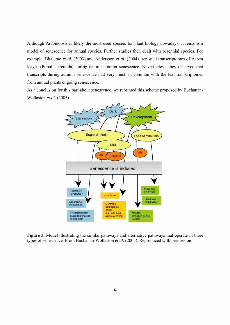
As a conclusion for this part about senescence, we reprinted this scheme proposed by Buchanan-
Wollaston et al. (2005).
Figure 3. Model illustrating the similar pathways and alternative pathways that operate in three types of senescence. From Buchanan-Wollaston et al. (2005). Reproduced with permission.
12
2. Mitochondria
2.1 Introduction
Since when do we talk about mitochondria?
Mitochondria were described during the 19th century where early studies from
cytologists reported the presence of subcellular granules similar in size and shape to bacteria in a
variety of different cell types. Kolliker described conspicuous "granules" aligned between the
striated myofibrils of muscle and was the first to isolate mitochondria from insect muscles.
Fleming also observed "filaments" in the cytoplasm of other cell types. In 1890, Robert Altman
discovered a method of staining these structures with fuchsin that made it possible to
demonstrate their occurrence in nearly all types of cells. Consequently, he postulated that these
granules were the basic units of cellular activity. Accordingly to their size and shape similar to
bacteria, he named them "bioblasts". In 1898, the term mitochondrion was coined by the german
microbiologist Carl Benda; in Greek "mitos" means "thread" and "khondros" "granule". Benda
made valuable observations on their form and distribution in preparations stained with alizarin
and crystal violet (Scott and Logan, 2007). In 1900, L. Michaelis selectively stained
mitochondria with the dye Janus Green B. Since this staining method is based on redox
properties (the dye must be oxidized to become colored), Michaelis proposed that mitochondria
were cellular oxidizing agents. During the period 1900–1930, most cytologists recognized the
mitochondrion as a well-defined and ubiquitous organelle, although at that time there was no real
agreement about its function. Separation of mitochondria by differential centrifugation of cell
homogenates was first attempted with some success by Bensley and Hoerr in 1934. The method
was further perfected by Claude in the early 1940’s and by Hogeboom, Schneider, and Palade in
1948. It was only during the late 1940’s that mitochondria were identified as the site of oxidative
energy metabolism (Logan, 2003). The development of improved methods of fixation and thin
sectioning for electron microscopy enabled Palade and Sjostrand to describe independently in
1953 the basic structural plan of the internal membranes of the mitochondria. Since then,
mitochondria have become subject of intensive researches. They have been shown to be involved
in many human deceases and the understanding of their integration and regulation within the cell
metabolism appears crucial for further treatments and cures. Concerning plant tissues, Millerd et
13
al. (1951) were the first to isolate mitochondria, allowing a long series of work leading to better
understand the specific functional characteristics of these organelles when comparing to animal
counterparts (Douce, 1985). However and with no regard to their phyla, roles of mitochondria in
cell metabolism remain under intensive investigations.
2.2 The origins of mitochondria
Two theories for the origins of mitochondria have been proposed through the years. The first
theory is based on a non-symbiotic hypothesis where mitochondria would have evolved from
compartmentalization of existing genetic material within the ancestral proto-eukaryote.
However, complete genome sequences for many mitochondria, as well as for some bacteria,
together with the nuclear genome sequence of yeast have provided a coherent view of the origin
of mitochondria. In particular, conventional phylogenetic reconstructions with genes coding for
proteins active in energy metabolism and translation have confirmed the another theory: the
endosymbiosis hypothesis (Andersson and Kurland, 1999). Interestingly, more than a century
ago, Altman had already speculated that bioblasts were capable of an independent existence, yet
formed a colonial association with the cytoplasm of a host cell, and that it was through this
association that the host cell acquired the properties of life (Tzagoloff, 1982). The endosymbiosis
theory postulates the capture, about 2 billion years ago, of an α-proteobacterial endosymbiont by
a nucleus-containing eukaryotic host cell (Gray, 1999; Gray et al., 1999; Lang et al., 1999; Gray
et al., 2001). Possibly, the mitochondria of animals, fungi and plants have originated from a
single symbiosis preceding the divergences of these kingdoms (Fig. 4).
14
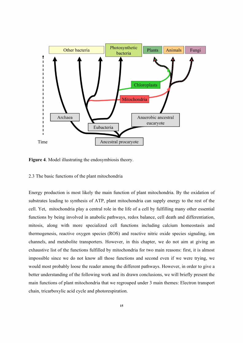
Figure 4. Model illustrating the endosymbiosis theory.
2.3 The basic functions of the plant mitochondria
Energy production is most likely the main function of plant mitochondria. By the oxidation of
substrates leading to synthesis of ATP, plant mitochondria can supply energy to the rest of the
cell. Yet, mitochondria play a central role in the life of a cell by fulfilling many other essential
functions by being involved in anabolic pathways, redox balance, cell death and differentiation,
mitosis, along with more specialized cell functions including calcium homeostasis and
thermogenesis, reactive oxygen species (ROS) and reactive nitric oxide species signaling, ion
channels, and metabolite transporters. However, in this chapter, we do not aim at giving an
exhaustive list of the functions fulfilled by mitochondria for two main reasons: first, it is almost
impossible since we do not know all those functions and second even if we were trying, we
would most probably loose the reader among the different pathways. However, in order to give a
better understanding of the following work and its drawn conclusions, we will briefly present the
main functions of plant mitochondria that we regrouped under 3 main themes: Electron transport
chain, tricarboxylic acid cycle and photorespiration.
15
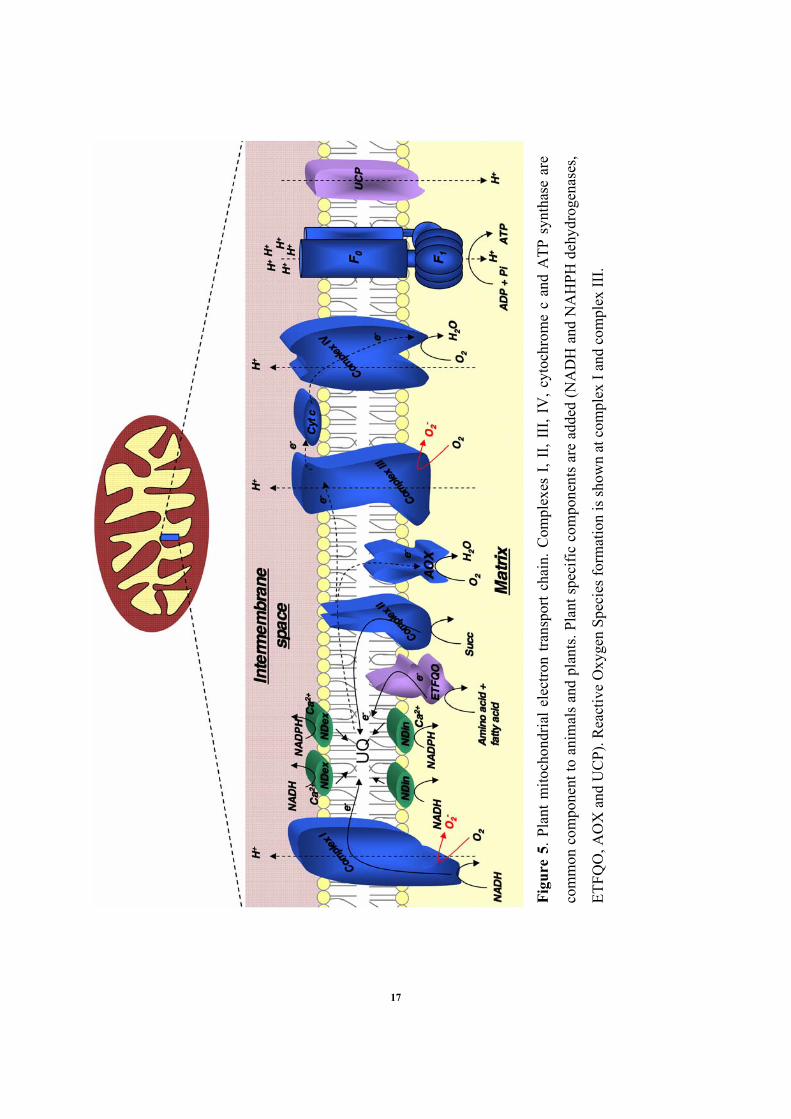
2.3.1 The mitochondrial electron transport chain
As we just mentioned, one of the primary functions of plant mitochondria is to produce and
supply energy to the rest of the cell. The production ATP (Adenosine triphosphate) is the result
of the mitochondrial electron transport chain activity which takes place in the inner membrane of
plant mitochondria. During this process also called oxidative phosphorylation, electrons resulting
from oxidation of donors (NADH or FADH2) are carried through the electron transport chain via
redox reactions and achieve their “journey” by finally reducing O2 into H2O. The transport of
electron is coupled with proton pumping from matrix to the inter-membrane space generating a
transmembrane electrochemical proton gradient. The backflow of protons is further used for
ATP synthesis i.e. phosphorylation of ADP into ATP.
In the mitochondrial electron transport chain (mETC), electrons are carried by proteins such as
cytochromes (proteins with iron containing heme group), iron-sulfur proteins (with either 2Fe/2S
or 4Fe/4S iron-sulfur center), and the small molecule ubiquinone (also called coenzyme Q).
Major components of the electron transport chain are grouped in four multisubunit complexes:
the NADH dehydrogenase complex (complex I), the succinate dehydrogenase (complex II), the
cytochrome c reductase (complex III) and the chrome c oxidase (complex IV). In addition, the
carrier lipid ubiquinone mediates transfer of electrons from the dehydrogenases to complex III
and the monomeric protein cytochrome c shuffles electron between complex III and complex IV.
Complex I: The NADH dehydrogenase complex has a mass of about 1 MDa and is composed of
over 40-45 distinct proteins. At least 10 cofactors are attached to this complex (one flavin
mononucleotide and nine Fe-S clusters) (Heinemeyer et al., 2007). Proton pumping occurs at the
dehydrogenase complex when electrons from the oxidation of NADH,H+ are transferred to
ubiquinone which becomes reduced in to ubiquinol. Four protons are pumped from the matrix
side to the inter-membrane space of each pair of electrons passing through the complex I.
Complex I can be inhibited by rotenone.
16
Figu
re 5
. Pla
nt m
itoch
ondr
ial e
lect
ron
trans
port
chai
n. C
ompl
exes
I, I
I, II
I, IV
, cyt
ochr
ome
c an
d A
TP s
ynth
ase
are
com
mon
com
pone
nt to
ani
mal
s an
d pl
ants
. Pla
nt s
peci
fic c
ompo
nent
s ar
e ad
ded
(NA
DH
and
NA
HPH
deh
ydro
gena
ses,
ETFQ
O, A
OX
and
UC
P). R
eact
ive
Oxy
gen
Spec
ies f
orm
atio
n is
show
n at
com
plex
I an
d co
mpl
ex II
I.
17
Complex II: The succinate dehydrogenase is the smallest of all four complexes and it does not
pump any protons. It includes four types of subunits and five cofactors (one flavin adenine
dinucleotide and three Fe-S clusters and one heme b) (Heinemeyer et al., 2007). The complex II,
to date the only membrane bound enzyme of the tricarboxylic acid cycle, oxidizes succinate into
fumarate at an active site containing FAD (flavin adenine dinucleotide). Further, electrons reduce
ubiquinone into ubiquinol.
Complex III: The ubiquinol-cytochrome c reductase is a functional dimer of about 500 kDa. Each
monomer is composed of 10-11 proteins and 4 cofactors (three hemes and one Fe-S cluster). The
complex III oxidizes ubiquinol and reduces cytochrome c (which can only accept one electron at
a time). Four protons are pumped for each pair of electrons passing through the complex.
Interestingly, the two largest subunits (also named core proteins) of the complex III are
suspected to have a peptidase activity in plants. Isolated complex III from plants was shown to
efficiently remove presequences from mitochondrial precursor proteins (Heinemeyer et al.,
2007). Complex III can be inhibited by antimycin A.
Complex IV: The cytochrome c oxidase has a mass of about 210 kDa and is made up of 13
subunits and four cofactors are attached to the complex (two heme a and two Cu2+). Complex IV
oxidizes cytochrome c and finally reduces oxygen into water which is the end product of the
electron transport chain. Four electrons are necessary to reduce O2 into H2O. The complex IV
can be inhibited by cyanide, azide and carbon monoxide.
ATP synthase: Sometimes also called the complex V, ATP synthase has a molecular mass of
about 600 kDa. It is composed of two domains F0 and F1. F1, the part situated in the
mitochondrial matrix, is composed of 5 different subunits (α, β, γ, δ and ε) and the F0 part,
located in the inner mitochondrial membrane, is composed of ten subunits (a, b, c and several
additional small subunits which are designated differentially in different organisms).
In addition to the four complexes described here above, plant mitochondria posses a few
additional enzymes that play an important role in the mETC either as electron donors or
acceptors.
18
-The uncoupling proteins (UCP’S): Described in brown adipose tissues for the first time by
Nicholls and Locke (1984) more than 2 decades ago, UCP’s have later been evidenced in plants
by Laloi et al. (1997). UCP’s have a mass of about 32 kDa and they catalyze dissipation of the
electrochemical gradient of protons on a fatty-acid dependant manner. In addition, they are
activated by superoxide and aldehyde products of lipid peroxidation (Considine et al., 2003;
Smith et al., 2004).
-The NAD(P)H dehydrogenases: Both the inner and the outer surface of the inner mitochondrial
membrane has 2 additional dehydrogenases: one NADH and one NADPH types. They are not
inhibited by rotenone and they transfer electrons to the pool of ubiquinone bypassing the
complex I.
-The alternative oxidase: This enzyme has a molecular mass of about 35 kDa (32 to 37 kDa
according to species). It is plant specific and works as a dimer. Alternative oxidase can accept
electrons before the complex III and will reduce O2 into H2O like the complex IV. However,
this pathway is non-phosphorylating. The alternative oxidase can be inhibited by
salicylhydroxamic (SHAM) acid and propylgallate (PG).
-The electron-transfer flavoprotein:ubiquinone oxidoreductase (ETFQO): Recently, Ishizaki et
al. (2005; 2006) evidenced in the inner mitochondrial membrane of Arabidopsis a new protein
involved in amino acid degradation and maybe also having a role in chlorophyll degradation.
The electron-transfer flavoprotein:ubiquinone oxidoreductase (ETFQO) receives electrons from
the matrix enzyme: the electron-tranfer flavoprotein.
-The glycerol-3-phosphate dehydrogenase (FAD-G3PDH): This enzyme was identified by Shen
et al. (2003) in Arabidopsis. It is a flavoprotein located on the outer surface of the inner
mitochondrial membrane and it oxidizes glycerol-3-phosphate to dihydroxyacetone phosphate
19
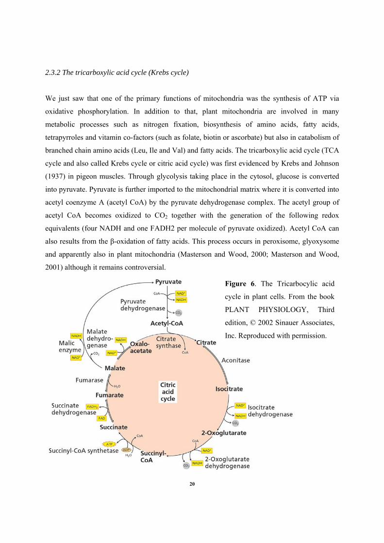
2.3.2 The tricarboxylic acid cycle (Krebs cycle)
We just saw that one of the primary functions of mitochondria was the synthesis of ATP via
oxidative phosphorylation. In addition to that, plant mitochondria are involved in many
metabolic processes such as nitrogen fixation, biosynthesis of amino acids, fatty acids,
tetrapyrroles and vitamin co-factors (such as folate, biotin or ascorbate) but also in catabolism of
branched chain amino acids (Leu, Ile and Val) and fatty acids. The tricarboxylic acid cycle (TCA
cycle and also called Krebs cycle or citric acid cycle) was first evidenced by Krebs and Johnson
(1937) in pigeon muscles. Through glycolysis taking place in the cytosol, glucose is converted
into pyruvate. Pyruvate is further imported to the mitochondrial matrix where it is converted into
acetyl coenzyme A (acetyl CoA) by the pyruvate dehydrogenase complex. The acetyl group of
acetyl CoA becomes oxidized to CO2 together with the generation of the following redox
equivalents (four NADH and one FADH2 per molecule of pyruvate oxidized). Acetyl CoA can
also results from the β-oxidation of fatty acids. This process occurs in peroxisome, glyoxysome
and apparently also in plant mitochondria (Masterson and Wood, 2000; Masterson and Wood,
2001) although it remains controversial.
Figure 6. The Tricarbocylic acid
cycle in plant cells. From the book
PLANT PHYSIOLOGY, Third
edition, © 2002 Sinauer Associates,
Inc. Reproduced with permission.
20
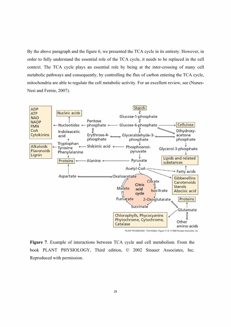
By the above paragraph and the figure 6, we presented the TCA cycle in its entirety. However, in
order to fully understand the essential role of the TCA cycle, it needs to be replaced in the cell
context. The TCA cycle plays an essential role by being at the inter-crossing of many cell
metabolic pathways and consequently, by controlling the flux of carbon entering the TCA cycle,
mitochondria are able to regulate the cell metabolic activity. For an excellent review, see (Nunes-
Nesi and Fernie, 2007).
Figure 7. Example of interactions between TCA cycle and cell metabolism. From the
book PLANT PHYSIOLOGY, Third edition, © 2002 Sinauer Associates, Inc.
Reproduced with permission.
21
On the figure 7, we can notice the general involvements of the TCA cycle in several biosynthesis
pathways. For example, the biosynthesis of amino acids is mainly localized in plastids. However,
by the TCA cycle, mitochondria can supply α-keto-glutarate and oxaloacetate which once
respectively converted to glutamate and aspartate will represent the backbone or carbon skeleton
for amino acids synthesis.
2.3.3 The photorespiration
As early as 1920, Otto Warburg made the observation that O2 inhibits photosynthesis. This
phenomenon, originally known as the ‘‘Warburg effect’’ was later recognized as the light-
dependent release of CO2 by photosynthetic organisms, or photorespiration (Eckardt, 2005). The
mechanistic basis of photorespiration was found in the dual nature of the RUBISCO (ribulose 1,5
bisphosphate carboxylase/oxygenase). Rubisco is a bifunctional enzyme that catalyzes both the
carboxylation and oxygenation of Ribulose 1,5 biphosphate (RuBP). The photosynthetic
carboxylation refers to CO2 fixation and yields two molecules of 3-phosphoglycerate (3PGA)
(i.e. the Calvin cycle) whereas the oxygenation process leads to the photorespiration (Fig. 8).
Oxygenation of RuBP leads to the production of one molecule of 3-PGA and one molecule of the
2-carbon compound: the 2-phosphoglycolate. Since the two substrates (O2 and CO2) are
competitive with regard to RUBISCO, an increase in CO2 concentration leads to inhibition of
oxygenase activity and vice versa. Temperature is an external factor enhancing the
photorespiration. For example, on hot and dry days, stomata are closed and the O2 concentration
in the leaf exceeds that of CO2, thereby increasing the competition for the common active site of
the RUBISCO. Furthermore, the solubility of CO2 diminishes more rapidly than that of O2 as
temperature increases.
22
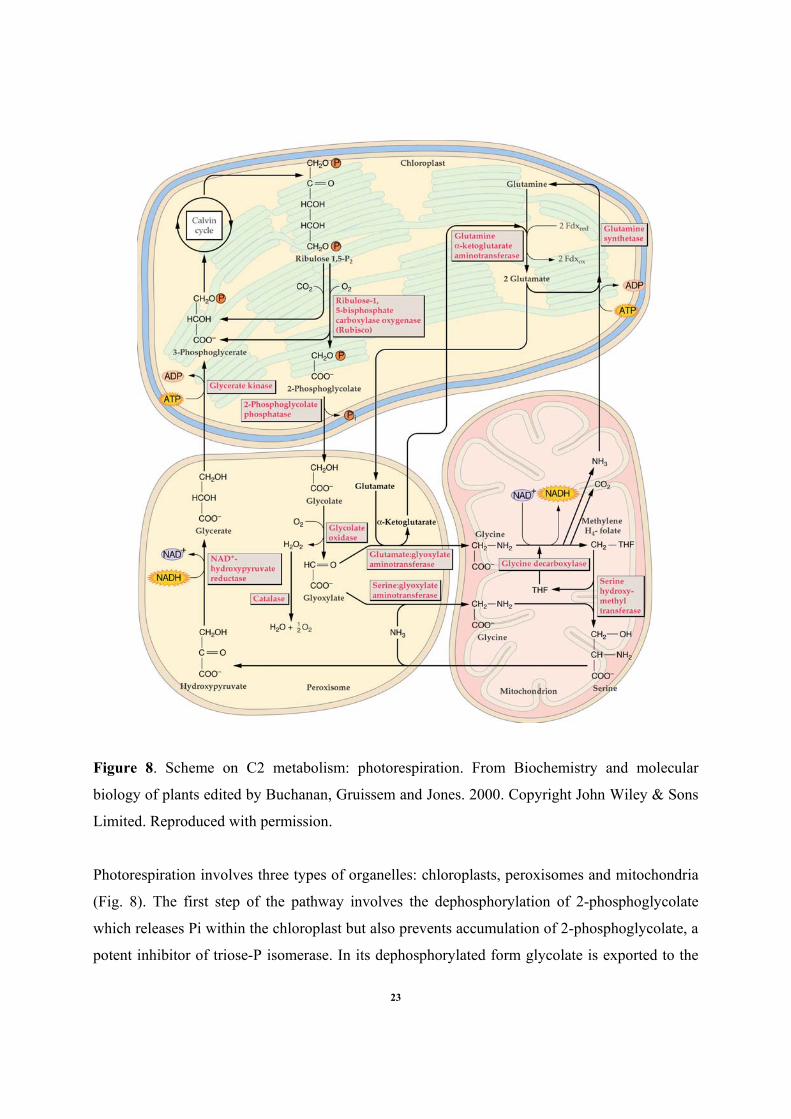
Figure 8. Scheme on C2 metabolism: photorespiration. From Biochemistry and molecular
biology of plants edited by Buchanan, Gruissem and Jones. 2000. Copyright John Wiley & Sons
Limited. Reproduced with permission.
Photorespiration involves three types of organelles: chloroplasts, peroxisomes and mitochondria
(Fig. 8). The first step of the pathway involves the dephosphorylation of 2-phosphoglycolate
which releases Pi within the chloroplast but also prevents accumulation of 2-phosphoglycolate, a
potent inhibitor of triose-P isomerase. In its dephosphorylated form glycolate is exported to the
23
peroxisome where it is converted to glyoxylate. The hydrogen peroxide generated is detoxified
by catalase. Glyoxylate is then converted into glycine by two different enzymes:
serine:glyoxylate aminotransferase and glutamate:glyoxylate aminotransferase After its transfer
to the mitochondrion, glycine gets converted into serine by the glycine decarboxylase complex.
Glycine decarboxylase complex has four different subunit (P, H, T and L), which catalyze the
transfer of a methylene group from glycine to tetrahydrofolate with the concomitant release of
NH3 and CO2, and reduction of NAD+ to NADH (we can remark that NH3 is used by glutamine
synthetase in the chloroplast). The serine hydroxylmetyltransferase catalyses then the transfer of
the methylene group to another glycine molecule to form serine. Exported back to peroxisome,
serine is converted to hydroxypyruvate via serine:glyoxylate aminotransferase. The last of the
peroxisome steps consists in the reduction of hydroxypyruvate into glycerate by an NADH-
dependent hydroxypyruvate reductase. Finally, glycerate is exported to the chloroplast where it is
phosphorylated to 3-phosphoglycerate and reenters the Calvin cycle.
Despite the net loss of carbon (up to 25% of the carbon that is fixed during photosynthetic
carbon assimilation), the photorespiration allows a partial recycling of carbon. The
photorespiratory cycle is essential for plant growth, as demonstrated by photorespiration mutants
that are non-viable in normal air (around 0.04% CO2) and grow only in elevated CO2 (1% to 2%
CO2), conditions under which RuBP oxygenation is suppressed (Somerville, 2001). However,
the outcome of photorespiration remains a loss of CO2 and energy in photosynthetic cells. The
biological function of photorespiration is not clear. One possibility is that photorespiratory cycle
is necessary under conditions of high light intensity and low CO2 concentration (i.e. when
stomata is closed under water stress) to dissipate excess ATP and reducing power from the
photosynthesis light reactions, thus to prevent damage to the photosynthetic apparatus
(Somerville, 2001). Finally, photorespiration is also seen as an evolutionary adaptation. Due to
the increasing amount of O2 in the biosphere, plants had to find a way to salvage the loss of
carbon which seems to be an inevitable consequence of the RUBISCO reaction mechanisms.
24
3. Aim: Why senescence and mitochondria?
After reading the two previous chapters, one obvious question that directly comes to the reader
is: Why senescence and mitochondria? Through this new chapter, I will try to lead the reader
towards our aim and hypothesis about the here presented thesis. In order to facilitate their
understanding, I will first address 3 points:
3.1 The mitochondrial contribution to programmed cell death
In animal programmed cell death (PCD), also called apoptosis, mitochondria are shown to play a
crucial role by generating ROS via the mitochondrial electron transport chain and by integrating
several signals such as oxidants and Ca2+ overload which further leads to PCD by the release of
cell-death mediators such as cytochrome c, AIF (Apoptosis Inducing Factors) and the
endonuclease G (Green and Reed, 1998; Lam et al., 2001; Li et al., 2001; Arnoult et al., 2002).
Yet, in plants, the situation concerning the role of mitochondria during PCD becomes more
obscure and still remains under important investigations.
3.2 Senescence / programmed cell death: where are the boundaries?
Over the last decade, scientists have tried to define the boundaries between senescence and PCD
but this issue is still a matter of debate (Thomas et al., 2003; van Doorn and Woltering, 2004;
Van Doorn, 2005). The death of an organ is generally called “senescence” whereas the death of a
cell often obviously refers to the programmed cell death process (Of course, the death of an
organ results from the combined and rather synchronized death of cells). Nevertheless, as
Thomas et al. (2003) argued, reversibility might serve to distinguish senescence fundamentally
from programmed cell death (PCD), as does the fact that viability is essential for the initiation
and progress of cell senescence. This seems to be in line with the idea raised by Delorme et al.
(2000) after their study on a metalloproteinase from cucumber. They suggested that either PCD
could occur only at the culmination of the senescence program or that the processes are distinct
with PCD being triggered at the end of senescence. This consequently would mean that PCD and
25
senescence should be clearly dissociated by their fundamental mechanisms but associated in a
common fate of a cell or an organ ongoing death process.
3.3 Economical perspectives
As we previously mentioned, the understanding of leaf respiration during aging and senescence
is part of very important economical challenges. We can take the recent use of Modified
Atmosphere Packaging (MAP) as an illustration.
In order to preserve fruits and vegetables and to increase their shelf-life time, plant respiration is
studied to design adequate packaging for shipping and conservation. By correctly manipulating
the amount of O2 and CO2 within the packaging, MAP enables the produce to live longer by
delaying respiration, ripening and ethylene production. This in turn reduces browning, retards
textural softening, preserves vitamins and extends the overall freshness of the packaged produce
(http://www.convex.co.nz/prod_refresh_technical.html). This demonstrates the importance of a
correct understanding of the cellular and metabolic mechanisms occurring during maturation,
aging, senescence and death.
3.4 Aim and Hypothesis
We just mentioned that mitochondria are known to be involved in animal apoptosis but their role
during plant PCD is still enigmatic and rather controversial. Furthermore, even though PCD and
senescence are both assimilated to the death process, the mitochondrial contribution to the
process of leaf senescence is unknown (despite a few sparse reports) and surprisingly remains
rather aside from current researches. However, during leaf senescence, chloroplasts are primary
targets of catabolism, which leads to the rapid impairment of photosynthesis. Consequently, we
assumed that such loss of photosynthetic capacity might lead to important perturbations and
reprogramming in the plant cell metabolism. Since chloroplasts loose their functionalities during
leaf senescence, we further hypothesized that mitochondria could have a crucial role by
supplying energy and the adequate carbon skeletons to the rest of the cell in response to the
potential demand due to catabolism and nutrient recycling. Therefore, the following study will
26

present the result of our investigations in our attempt to clarify the rearrangements of the cell
metabolism and the potential role played by mitochondria during the leaf senescence.
27
RESULTS AND DISCUSSION
4. Isolation of mitochondria: a useful tool
Leaves are the main photosynthetic organs of the plant, but due to their photosynthetic activity,
their metabolism has to adjust constantly. As a result, studies of isolated cell organelles are
essential for the understanding of the processes at the cellular and subcellular levels in
photosynthetic cells. Leaf cell metabolism represents a very complex network of inter-crossing
metabolic pathways between organelles. However, in order to obtain clear results, we often have
to work with isolated organelles. These isolated organelles have to be sufficiently pure and intact
to adequately represent their function in the intact cell. Since our interest was on mitochondria,
we decided to first develop a protocol for isolation of mitochondria from Arabidopsis leaves
suitable for further studies.
4.1 Why Arabidopsis?
During the last decade, Arabidopsis thaliana Heynh., a small Brassicaceae, became the model
plant for intensive research worldwide. Its small, diploid genome, fully sequenced (Initiative,
2000), has provided invaluable tools for genomic and proteomic studies. The information
compiled in databases makes Arabidopsis a model plant for many scientists, which offers
opportunities for detailed studies of cellular functions that is not possible with other species.
Several reports had already described the isolation of mitochondrial Arabidopsis leaf fractions
for specific studies (Somerville and Ogren, 1982; Turano et al., 1997; Berkemeyer et al., 1998;
Yasuno and Wada, 1998; Fan et al., 1999; Johansson et al., 2004; Taira et al., 2004). However,
most of these reports lack detailed information on the purity and intactness of the preparations
obtained, and they do not consider respiratory properties, an important aspect of the functionality
of isolated mitochondria.
28
4.2 Where are the difficulties?
At first, a practical problem associated with Arabidopsis is that the plant is small, and thus, it is
difficult to obtain large quantities of its leaves. It was suggested that a hydroponic cultivation
system would be useful for increasing the leaf mass (Norén et al., 2004). This might improve the
situation somewhat, but the yield of leaves from Arabidopsis will always be limited.
In leaves, photosynthetically active cells (i.e. mesophyll cells and guard cells of stomata) contain
chloroplasts. During extraction process, the grinding of the leaves often leads to considerable
contamination by fragments of broken chloroplasts, which causes problems for the isolation and
study of other cell fractions from leaves. In addition to thylakoids, peroxisomes that have a rather
similar density as mitochondria generally contaminate the mitochondrial fraction. Therefore, an
alternative method, adopted by several scientific groups (Davy de Virville et al., 1994; Kruft et
al., 2001; Millar et al., 2001; Werhahn et al., 2001), has been to work with cell-suspension
cultures of non-green tissues in order to obtain preparations with good respiratory properties
(Douce and Packer, 1987) and, thus, greatly reduce peroxisome and chloroplast (especially
thylakoids) contamination; the two main contaminants of the mitochondrial fraction from green
leaves, irrespective of the plant species.
Finally, isolation of functionally intact leaf mitochondria can be a very difficult task. First, one
needs to establish the right composition of extraction solutions as well as their correct
combination for the extraction, to track the mitochondria during the isolation procedure, to run
purity and integrity tests. Moreover, mitochondria have important dynamics which can influence
their isolation.
Even though isolation procedures yielding highly purified and functional mitochondria have
been described for many species (for review see (Douce, 1985)), we have not been able to obtain
good preparation of leaf mitochondria from Arabidopsis by most of the techniques and protocols
cited in this book and other articles. Consequently, we present in paper I (Keech et al., 2005) two
different procedures for isolating mitochondria from Arabidopsis leaves. Those two procedures
are also briefly described in Figure 9. Method A produces highly functional mitochondria with
respect to respiratory properties using a small quantity (about 5 g) of leaf material. However, the
resulting preparations are contaminated by chloroplast material. The second protocol, method B,
29
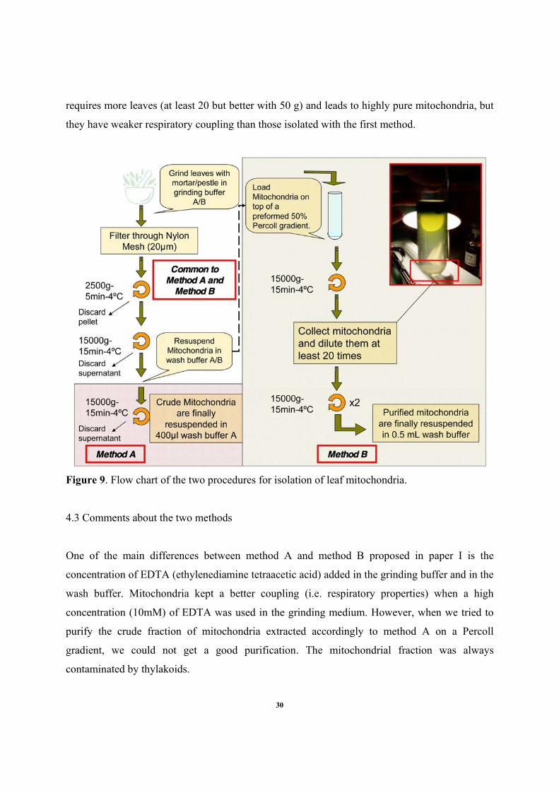
requires more leaves (at least 20 but better with 50 g) and leads to highly pure mitochondria, but
they have weaker respiratory coupling than those isolated with the first method.
Figure 9. Flow chart of the two procedures for isolation of leaf mitochondria.
4.3 Comments about the two methods
One of the main differences between method A and method B proposed in paper I is the
concentration of EDTA (ethylenediamine tetraacetic acid) added in the grinding buffer and in the
wash buffer. Mitochondria kept a better coupling (i.e. respiratory properties) when a high
concentration (10mM) of EDTA was used in the grinding medium. However, when we tried to
purify the crude fraction of mitochondria extracted accordingly to method A on a Percoll
gradient, we could not get a good purification. The mitochondrial fraction was always
contaminated by thylakoids.
30
Even though we do not have a clear explanation for this, we suggest a high concentration of
EDTA to limit damages caused by the lipases released during the extraction process. This would
explain the better coupling of mitochondria extracted by method A. Arabidopsis is known to
have several lipases, notably the 2 families of phospholipases C and D (Dhonukshe et al., 2003)
which need Ca2+ as cofactor (Wang, 2005). The EDTA can chelate both Ca2+ and Mg2+. The
chelation of Ca2+ would consequently reduce the activity of the phospholipases limiting the
release of free fatty acids and free-head groups (Wang, 2005) destabilizing the biological
membranes. However, since EDTA also chelates Mg2+, it might be possible to get a separation of
grana stacks into series of single thylakoids by the lack of magnesium (Popovic et al., 1979).
This would result in a huge set of thylakoid clusters from many different sizes and densities and
would then explain the presence of thylakoids all over the Percoll gradient and our troubles to get
good purification of mitochondria with method A.
Another point we would like to mention about the composition of the buffers used in the two
methods described in paper I is about the Percoll gradient. To our knowledge, there is no other
protocol to isolate mitochondria where a 50 % Percoll gradient has been used. Here, we do not
want to highlight the “modest novelty” of using a 50% Percoll buffer but more the fact that the
combination of a preformed gradient and a 50% Percoll buffer can make the trick! However, we
come to question about the mitochondria that are able to migrate to such densities. For
information, during centrifugation for purification of mitochondria, the auto-generated Percoll
gradient is often between 25 and 32 percents Percoll. Consequently, we came to think that
several subpopulation of mitochondria coexisted in leaves and we have basically been able to
isolate one of these subpopulations with mitochondria having higher density. This is purely
speculative but however in line with remarks from Dr D.C. Logan (personal communication)
about the dynamics of plant mitochondria. Mitochondrial population can vary in number and in
mass according to the phase of the cell cycle and the physiological status of the cell (Scott and
Logan, 2007).
31
4.4 A tool for further studies
It took more than a year to establish a successful protocol. However, as we previously mentioned
here above, we also tried several procedure previously described to isolate mitochondria from
Arabidopsis leaves. One of those consisted on a combination of differential centrifugations and
phase partitioning (Bergman et al., 1980). By using this technique, adapted for Arabidopsis
though, we also obtained rather good results to get purified mitochondria from Arabidopsis
leaves. We actually used this technique to identify plant glutaredoxin (Grx) targets (paper II)
through a collaborative work with N Rouhier and JP Jacquot. However, as one can see in paper
II, few targets of Grx found in the Arabidopsis mitochondrial extract were actually localized in
peroxisomes or in chloroplasts. It is known that up to date, none of the protocol for extraction of
mitochondria from leaves, irrespective to the species, leads to perfectly pure fraction. Even a
very recent protocol developed by Dr. H. Eubel (personal communication) and based on free
flow electrophoresis cannot yield to a totally pure fraction of leaf mitochondria.
As one can see through papers III, IV and VI, the protocol developed in paper I has been widely
used for different purposes. In Paper III (Gama et al., 2007), in order to characterize the PrxIIf (a
miotochondrial peroxiredoxin), we used the method A to obtain fraction enriched with
mitochondria from poplar leaves. Method A was also used in paper IV (Keech et al., 2007) to
evaluate the mitochondrial respiration during dark-induced senescence in Arabidopsis leaves.
Finally, isolation of highly purified mitochondria (method B) has been used in several other
experiments related to different projects, including a few preliminary experiments in the
proteomic field. However, since none of them has been finalized yet, the results are not presented
in this thesis. So, to conclude, the establishment of such protocol was very laborious and time
consuming for a PhD project but after all it appears to be of importance for many of the studies
we led and are still leading.
5. Dark induced senescence as a model
The experimental setup
32
Amongst external factors triggering leaf senescence, the shading phenomenon has caught the
attention of scientists. Indeed, in order to study leaf senescence, scientists have tried to mimic the
natural shading of leaves by using darkening treatments which lead to the senescence of the
organ due to drastic modifications of the photoperiod. This dark-induction of senescence might
be done either by placing excised leaves in the dark or dim light (Simon, 1967; Satler and
Thimann, 1983; Feller and Fischer, 1994; Jimenez et al., 1998; Pruzinska et al., 2005; van der
Graaff et al., 2006) or by setting the whole plant in the dark or by covering just one or a few
leaves still attached to the plant (Feller and Fischer, 1994; Weaver and Amasino, 2001;
Buchanan-Wollaston et al., 2005; Zelisko et al., 2005). The two latter ones may present an
undisputable advantage for physiological studies because they do not dissociate the leaf from the
rest of the plant thus allowing potential signals throughout the plant. Consequently, we decided
to use the experimental design proposed by Weaver and Amasino (2001) where they reported, by
a study based mainly on both loss of chlorophyll and expression of senescence associated genes
(SAG’s), a much faster induction of senescence in Arabidopsis leaves individually covered than
in the leaves from a whole darkened plant (Fig. 10). However, we brought a few modifications to
their experimental setup such as the photoperiod during growth, soil content or age of the plants.
For further details, please refer to paper IV (Keech et al., 2007).
Figure 10. Scheme of the experimental setup. Individually-darkened leaves (IDL) in (A) and
whole darkened plant (DP in (B). Leaves from rosette 6th to 12th were used for the experiments.
From (Keech et al., 2007). Reproduced with permission.
33
As shown in Figure 10, individually-covered leaves exhibited a noticeable yellowing and also
tended to become a bit thinner after already 4 days of darkness (A). In contrast, leaves from
plants darkened during 6 days did not exhibit an observable difference in their color in
comparison to the plant which stayed in light (B). However, leaves tended to rise to the top and
young leaves had longer petioles than their homologues from plants in light, two typical
characteristics of the “shade-avoidance response”.
As a model for leaf senescence, this experimental setup presents both advantages and
disadvantages that we will try to summarize now.
Experimental setups with individually-darkened leaves have already been used before
(references mentioned here above). As a matter of fact, it also facilitates the comparison with
data from other groups and consequently enhances the scientific relevance of the presented
results. Moreover, this system seems to present a biological relevance (see here below).
Practically, this system is very simple to handle, synchronization of leaf senescence is fairly
robust and both young and mature leaves do react to this drastic modification of the photoperiod.
Note though that young leaves will have a delayed senescence in comparison with mature leaves.
Nevertheless, the two types of leaves will undergo leaf senescence. However, a few parameters
remain difficult to control. We noticed a slight increase of temperature and of humidity inside the
plastic “mittens”. In addition, the gas exchanges remain uncontrolled inside the mittens.
However, in a recent study about transcriptomics between leaves senescing from natural aging
and leaves ongoing dark-induced senescence, van der Graaff et al. (2006) showed that more than
75% of the genes significantly up- or down-regulated in individually darkened leaves showed the
same response during natural, age dependent, senescence. They concluded that developmental
senescence and dark-induced senescence programs share many common pathways. In addition,
in order to address whether the senescence was in fact induced by the darkness itself, and not by
some artifact of the experimental design, Weaver and Amasino (2001) described a few important
controls that we can summarize as follow:
-When leaves were covered with petroleum jelly on both adaxial and abaxial sides of the leaves
(reducing the stomatal conductance), no yellowing was evidenced suggesting that reduced
transpiration does not induce senescence in this experimental system.
-When leaves were covered with small boxes made of either black or clear x-ray film, the leaves
covered with black boxes were induced to yellow, whereas the leaves covered with clear boxes
34
were not. Therefore, since the only difference between the two treatments was most probably the
light received, it strongly supports the fact that a drastic change in the light intensity induces
senescence.
As for individually-covered leaves, experimental setups where the whole plant is darkened have
been widely used to induce senescence ((Weaver and Amasino, 2001; Buchanan-Wollaston et
al., 2005; Ishizaki et al., 2005; Ishizaki et al., 2006)). It is extremely simple to handle but the
biological relevance can easily be questioned. Moreover, since absolutely no light is perceived,
the plant undergoes severe starvation which might not be the case during natural leaf senescence.
To conclude, the biological relevance seems higher for individually-covered leaves than for the
whole darkened plant. Indeed, during the development of Arabidopsis (notably under short days
photoperiod), older leaves are progressively covered or shaded by new leaves due to the
phyllotaxy of Arabidopsis. Whereas, the biological relevance of a plant experiencing prolonged
darkness (several days) is far from being obvious (except maybe during a sunny day in Kiruna in
beginning of January!). Consequently, even if a few parameters need to be improved and better
controlled, we think that individually-covered leaf represents a good way to mimic leaf
senescence and can be an interesting alternative to a whole plant switch to darkness. However,
the comparison between the two treatments remains beneficial for our understanding about the
specific regulations and adjustments of the metabolism during senescence.
6. Regulation of the metabolism
In this study, we decided to compare the regulation of metabolism (with an emphasis on
photosynthesis and respiration) in both individually-darkened leaves (IDL) and in leaves from
whole darkened plants (DP). We brought several modifications to the experimental system
proposed by Weaver and Amasino (2001). Nevertheless, we validated our modifications by
coming to the same and obvious point: senescence is faster in individually-darkened leaves than
in whole darkened plant. However, in the next paragraph, we present several evidences showing
that regulation of the metabolism in the two sorts of leaves (from IDL and from DP) will actually
be the result of different adaptive strategies to dark treatment.
35
6.1 A cytological approach
In French, we often say “voir c’est croire!” which literally means “to see is to believe!”. We
carried out a rather detailed study by confocal laser microscopy in order to visualize the
organelles content in leaves during the two darkening treatments. One of our primary ideas was
to normalize mitochondrial respiration values and western blottings by an estimation of the
number of mitochondria in the leaf. Interestingly, we observed a drastic reduction (around 65 %)
of the number of mitochondria in IDL whereas the mitochondrial population was rather
conserved in all cell types (epidermis cells, guard cells of stomata and mesophyll cells) of leaves
from darkened plants (paper IV). We also noticed that in IDL, mitochondria were rounder and
slightly bigger than mitochondria from both DP and light treatments. Chloroplasts were also
influenced by the darkening treatments. After 6 days, we estimated a decrease in chloroplasts
number which became small “gerontoplasts” to be around 60% in IDL whereas only a loss of 30
% with no change in size was noticed in DP. In addition to this quantification but only in IDL,
we noticed an important cytological rearrangement in mesophyll cells where chloroplasts and
mitochondria were clustered together. Similar aggregations of organelles had also been reported
in protoplast from senescing leaves of Pisum sativum (Simeonova et al., 2004) and in PCD of
mesophyll cells from cucumber (Cucumis sativus L.) (Selga et al., 2005). Curious to understand
how such cytological rearrangement might be possible, we investigated the dynamic of the
microtubular cytoskeleton during dark-induced senescence (Paper VI). We noticed an early
degradation of the microtubular network in individually-darkened leaves. Using the microarrays
data set from paper IV, we focused on a few specific proteins, the microtubules associated
proteins (MAP’s) that could have a role in the rearrangement of microtubules under certain
conditions and especially could be involved in a kind of bundling of the microtubules.
6.2 Photosynthesis
In order to follow the photosynthetic capacity of chloroplasts, we measured several parameters
such as Fv/Fm, ETR and ΦPSII from leaves undergoing the different treatments. All results
showed the same trend (paper IV). After 6 days of treatment, the photosynthetic capacity was
conserved in leaves from whole-darkened plant whereas it was progressively lost in individually-
36
darkened leaves. In addition, photosynthetic pigments were quantified by HPLC analysis. In
individually darkened leaves, chlorophylls, xanthophylls and carotenoids remained constant
during the first two - three days in darkness but when kept in the dark for longer periods, all
pigment classes declined to 30-40% of control levels after 6 days (Paper IV). In contrast, leaves
from whole darkened plants maintained pigment contents similar to light controls during the
same period with only a small decline at the end of the period studied. None of the treatments
exhibited modification of the chlorophyll a/b (Paper IV). Furthermore, we could also notice that
for both whole darkened plant and individually darkened leaves, the HPLC analysis did not
reveal new peaks as by-products from pigments degradation as seen in autumn senescence of
poplar leaves (Keskitalo et al., 2005).
6.3 Mitochondrial Respiration
In vivo – We followed dark respiration in leaves individually-covered and from whole darkened
plant. When expressed on a leaf area basis, the respiration dropped of 50% after 2 days for the
two treatments and remained at this rate during the six days of the experiment. When expressed
on a protein basis, dark respiration in leaves from whole darkened plant dropped the same way as
when expressed on the leaf area basis whereas the dark respiration in individually darkened
leaves increased significantly during the senescing process (Paper IV). Since a significant
difference was noticeable between IDL and DP, we decided to investigate the in vitro
mitochondrial capacity for respiration.
In vitro - For mitochondria from normally illuminated leaves, respiratory rates (expressed on a
mitochondrial protein basis) were stable with all substrates during the 6 days experimental
period. With mitochondria isolated from leaves of DP, respiratory rates were significantly lower
with all the substrates tested after both 4 and 6 days of dark treatment. Interestingly,
mitochondria isolated from IDL showed high respiratory rates throughout the dark treatment
with all the substrates tested. In addition, no significant differences in respiratory rates were
observed between mitochondria from normally illuminated leaves and mitochondria from IDL
along the 6 days period of treatment (Paper IV).
37
The RCR provides an estimation of the coupling between electron transport and oxidative
phosphorylation and it depends on the intactness of the inner mitochondrial membrane. For all
treatments the general trend was that malate + glutamate and glycine gave higher RCR than
NADH and succinate. The RCR values were very similar for mitochondria extracted from
normally illuminated leaves throughout the experiment (paper IV). However, in mitochondria
from both DP and IDL the RCR tended to decrease relative to normally illuminated leaves. For
mitochondria from IDL, the RCR decreased throughout the 6 days whereas for mitochondria
from DP, RCR dropped significantly for all substrates at day 2 but stayed fairly stable during the
subsequent 4 days of the experiment.
6.4 Metabolomics
To get a better understanding of the metabolic changes during the respective treatments, we
looked at the changes in content of specific metabolites in leaves and petioles (Paper V).
In leaves - Carbohydrates were, as expected much higher in light than in IDL and DP.
Furthermore, in some cases the content in IDL was somewhat higher than in DP. A few specific
amino acids, Gln and Gly in particular, were much higher in L as compared to dark treatments.
However, during prolonged darkness in DP, many other amino acids and other nitrogen
containing compounds increased. The N-metabolite progressively increased during dark
treatment in DP whereas they remained low in IDL and in light (Phe, Asn, 3-cyanoalanine,
ornitine, urea, 2,5-diaminovalerolactam). In addition, several common organic acids were high in
light, intermediate in IDL and low in DP (succinate, malate, fumarate) whereas the citrate level
was rather stable between the treatments.
In petioles - EDTA exudates from leaf petioles were also analysed by GC/ToF-MS. After one
day in darkness the relative content of amino acids was low in petioles of both DP and IDL.
After three days, several amino acids increased in both DP and IDL and to a large extent this
increase mirrored to changes in the leaves. After six days in darkness the content of amino acids
continued to increase in IDL but decreased in DP as compared to 3 days in darkness.
38
This supports the suggestion that amino acids are exported away from IDL thereby alleviating
the problem of N-accumulation from degraded amino acids and contributing to the retrieval of
nitrogen during senescence. In DP on the other hand the export does not seem to occur. During
this condition the metabolism also enters a stand-by mode with decreased rate of respiration
(Keech et al., 2007) which would be appropriate in order to maintain viability as long as
possible. During this condition of severe starvation amino acids accumulate as metabolic end
products giving a way to store nitrogen in non-toxic compounds.
6.5 Conclusion
Altogether, data obtained from whole darkened plant (transcriptomics, metabolomics,
physiological measurements and microscopy) suggest that under prolonged darkness Arabidopsis
leaf metabolism could enter a “stand-by” mode to preserve photosynthetic capacities by slowing
down the respiration to limit the consumption of substrates which most likely are in short supply
during such long period of starvation. Let’s remark, as we previously mentioned, that prolonged
darkness is obviously not a “natural physiological stress” for a plant. Ishizaki et al. (2006)
recently estimated the loss of soluble carbohydrate content from whole darkened plants to be
around 80% after only 3 days of darkening. Consequently, it seems that the plant reacts by
slowly degrading the cell wall as source of carbon since no sugars are available. This is
consistent with early results from Journet et al. (1986) where they demonstrated that in
carbohydrate starved sycamore cells, i.e. after the soluble carbohydrates and starch were
depleted, the cells started to degrade cell components to obtain substrates for respiration. This
was associated with a decreased rate of mitochondrial respiration that may be a direct
consequence of substrate limitation (Aubert et al., 1996). Moreover, Ishizaki et al. (2006)
proposed that the mitochondrial complex ETF/ETFQO may play an essential role during
carbohydrate deprivation by supplying alternative substrates, notably branch chain amino acids
to the respiratory chain. However, the consequences of activity of this pathway for mitochondrial
respiration remain unknown. Therefore, it seems likely that the differences in mitochondrial
respiratory capacity we observed were linked to the depletion of “normal” reserves and a switch
to other substrates affecting mitochondrial respiratory capacity. As a consequence, whole
39
darkened plant slowly reduces the amount of organelles but keep them functional most likely to
be able to restart their photosynthetic activity as soon as the light comes back.
In contrast to DP, an accelerated senescence was observed in IDL. The photosynthetic apparatus
was quickly degraded but the mitochondrial respiratory capacity remained high. The release of
cytochrome c is a common hallmark of apoptosis in animals and has also been observed in few
cases of plants PCD (Balk et al., 1999; Sun et al., 1999; Balk and Leaver, 2001) but not in all
(Yu et al., 2002). Interestingly, since the mitochondrial capacity to oxidize substrates is
maintained during the accelerated senescence observed in IDL, it seems unlikely for cytochrome
c to be released during dark-induced senescence. The reduction of the number of mitochondria
in IDL after 6 days suggests that two populations of mitochondria could coexist: one population
active and one population undergoing degradation. In consequence, two mitochondrial
populations with different membrane permeability, one with retained coupling and one with low
(or absent) coupling, may explain the decrease in RCR observed in IDL. In support of such a
suggestion, Simeonova et al. (2004) observed that mitochondria from protoplasts isolated from
senescing leaves of Pisum satisum were divided into two populations, one with mitochondria
keeping a high membrane potential along the senescing process and another one with
mitochondria loosing their membrane potential during the process. A possible mechanism to
explain the existence of these two populations may be related to production of reactive oxygen
species (ROS). In mammalian cells undergoing PCD, production of ROS is supposed to trigger a
mitochondrial permeability transition (MPT) which in turn dissipates the proton motive force
causing an uncoupling of oxidative phosphorylation reviewed by Skulachev (1996). In plants, it
has been commonly postulated that plant mitochondria could generate ROS and loose their
membrane potential during PCD (Jones, 2000; Tiwari et al., 2002; Yao et al., 2002; Yao et al.,
2004). We thus speculate that ROS production could alter mitochondrial permeability during
dark-induced senescence which could progressively lead to the impairment between oxidation
and phosphorylation as observed in our study. The implication of ROS would also be reinforced
by two facts: 1/ in paper III, we showed by western blotting that the amount of the prx-2f does
not change in IDL whereas more than 60% of leaf mitochondria disappeared and 2/ the recent
work of Logan and co-workers (Logan, 2006) reported that a large number of mitochondria
swelled to at least double their volume after ROS treatment of Arabidopsis mesophyll
40
protoplasts. The latter point is in line with the bigger and rounder mitochondria we reported in
paper IV. However, in paper VI, we also demonstrated that even very high concentration of
hydrogen peroxide (one of the main ROS produced in a cell) did not induce the similar alteration
of the microtubular cytoskeleton as seen during dark-induced senescence. So, we need to lead
further investigations to understand properly the role played by reactive oxygen species during
dark-induced senescence. In conclusion, we think that the remaining mitochondria in
individually-darkened leaves could play an active role both to maintain sufficient ATP and also
to provide proper carbon skeletons needed for degradations and reallocations occurring during
the leaf senescence.
41
REMARKS
This thesis appears as some pioneer work for our group. We developed several “tools” to lead
further studies and our preliminary investigations on adjustments of cell metabolism have
highlighted the metabolic changes between an individually darkened leaf and a leaf from a whole
darkened plant. These changes also included a divergent role of the mitochondrial metabolism
between the two treatments. Research about mitochondria during leaf senescence has been rather
superficial during the last decade whereas the involvements of mitochondria during programmed
cell death were under deeper investigations. Nevertheless, we highlighted the fact that
mitochondria could have a different role and behavior during leaf senescence than the ones
commonly accepted during PCD which in turn questions once again about the boundaries
between senescence and programmed cell death. In addition to this study, we, especially the new
members of the group, are now dealing with questions related to the C/N fluxes, to the
importance of Glycine decarboxylase complex and to the light dependant regulatory pathways
during leaf senescence. So to conclude, I am glad to see that finally this thesis work is a basis for
future investigations. Of course, things could have been done differently and/or maybe in a better
way but nevertheless, this work have found its place in international conferences and especially
have led us to deepen our understandings about the role of leaf mitochondria.
Finally, a few complementary remarks:
-Several times in this report, we mentioned the possibility to have different subpopulation of
mitochondria in a leaf. Although no data are clearly supporting this idea, the point remains
interesting to consider and I hope I will be able in the near future to investigate this idea.
- We also often mentioned the probabilities for ROS to be involved during dark induced
senescence. However, we have not led any further studies yet.
-One of our strongest beliefs when we started this project was to see the glycine decarboxylase
complex disappear during dark induced senescence. However, it was not the case which might
suggest some other roles for the GDC than only its involvement in photorespiration.
- When working with senescence, one has to keep in mind that the complexity of leaf senescence
is also suggested by the fact that it can be induced by numerous factors, biotic and abiotic.
42
FRÅN GRÖNT TILL GULT – EN BLADHISTORIA
När ett blad gulnar genomgår det både morfologiska och metaboliska förändringar. Denna process benämns senescence och en förbättrad förståelse av dess mekanismer är viktiga både ur ett grundvetenskapligt perspektiv och för potentiella bioteknologiska applikationer. Denna avhandling rapporterar om flera viktiga aspekter relaterade till de cellulära och metaboliska mekanismer som sker under senescencen med tonvikt på mitokondriernas bidrag till denna process. I ett första steg utvecklade vi metoder för att isolera antingen mycket funktionella mitokondrier eller mycket rena mitokondrier från blad av Arabidopsis thaliana. Dessa metoder användes sedan till för att studera mitokondriella bidrag till cellens redox balans och att uppskatta mitokondriernas kapacitet under senescence-processen. Framför allt jämfördes induktionen av senescencen berodende på olika mörkerbehandlingar av Arabidopsis. Jämförelse mellan individuellt mörklagda blad med hela mörklagda växter visade en betydande skillnad i metabolisk strategi mellan de två mörkerbehandlingarna. Genom att integrera data från mätningar av fotosyntes, respiration och konfokal mikroskopi med transcriptomics- och metabolomics-profiler föreslår vi att metabolismen hos blad från helt mörklagda växter antar ett ”stand-by läge” för att kunna bibehålla fotosynteskapaciteten så länge som möjligt. I kontrast till detta visar mitokondrier från individuellt mörklagda blad en hög aktivitet och kan därmed producera energi och kolskelett för degraderingen av cellkomponenter, vilket möjliggör återvinning av näringsämnen. Vi har även studerat dynamiken av det mikrotubulibaserade cytoskelettet under mörkerindicerad senescence. Mitokondriernas rörlighet påverkades av en tidig nedbrytning av mikrotubuli hos individuellt mörklagda blad men inte hos blad där hela växten mörkerbehandlats. Dessutom verkade ett flertal mikrotubuliassocierade proteiner (MAP’s) att vara involverade i buntningen av mikrotubuli runt kloroplasterna. Sammanfattningsvis belyser det arbete som presenteras i denna avhandling ett flertal viktiga steg med avseende på metabolisk anpassning och andra cellulär mekanismer i Arabidopsisblad som utsätts för långvarig mörkerbehendling. Specifikt föreslår vi att mitokondrierna bidrar med speciella och viktiga funktioner under bladens senescence eftersom mitokondriernas roll under långvarig mörkerbehandling av blad verkar bero på den totala statusen av metabolismen hos växten.
43
AKNOWLEDGEMENTS
Well, this is maybe trickier than the writing of the previous pages…
But first, I think I need to thank my opponent, the “Rapporteurs”, the “Examinateurs” and the
members of the jury for giving their time to consider my work. Thank you!
-Now of course, Per G.! Well, Per, you are a special person, most probably the nicest boss that
one can have…Sometimes, for a “hot blood” like me, it has been amazingly difficult to
understand one of the calmest man on earth…But, anyway, it worked out! In addition to the rest,
I also want to thank you for teaching me how to get calmer and less stressed both in science and
in life…Thanks for the road Per!
-To my other supervisor, Pierre D. Thank you very much for believing in me, for your kind and
motivating support during these years. I hope now you will have time to relax a bit and maybe,
we will talk about “mito” a few more times in the future.
-Another person whom has counted very much during that thesis is Slim. Man, was great to have
you around! Thanks for all the scientific, philosophical and “women-ical” talks late in the
evenings when I felt alone at work.
-Vaughan. At first, I thought we would have got troubles together…And finally, you are a super
cool guy. Thanks mate for all your precious help!
-Now, extremely important! My two + one office mates…Arf, nothing, along the years would
have been as enjoyable as it has been without my two dears Gunilla and Jan. You are heroes!
Able to survive, everyday‘s life in the same office as me! Incredible! You have been so great for
so many things (Jan always backing me up when my computer skills were reaching their limits
and Gunilla cheering me up when things got dark). Thank you so, so much for your calm, your
happiness, bearing my mood and especially my B&B’s stories!! Of course, the office would not
be complete without Linda. You arrived later but still, it was a pleasure to share office and good
moments with you (and a few B&B’S stories as well ;-)...you, curious girl!!!).
-A huge “thank” to Siv, Monica, Inger and the cleaning staff for their constant help in the
background (sort to say). We can never get something well built without good foundations! We
all need to remember that!
44
-To the French Mafia, allias Charleen, Joule, Rozenn, Pepito, Delphine, Yohann, Melanie,
Laurent, Catherine, Mathieu, Gaia. Thanks for sharing great “French moments”!
-Of course, I shared different things with different persons at UPSC but I want to thank you all,
PhD-students, post docs, coworkers from UPSC and other departments. It has been great
meeting you! Special acknowledgements though for Lazlo B., Sandra J., Arsenio V., Junko T.,
Marja H., Eva S., Francesco G., Jean-Pierre J., Andreas S., Leszek K., Maribel G., Meng
M., Marie-Charlotte N., Stefan B. for both the personal and the scientific interactions!
-I also want to thank a bunch of Swedes who made my life in Sweden a marvelous experience:
Johanna, Kxöl, Bengson, Rez, Sussie, Terese(s) X5 (at least), Julia, Anna-Maria, Nina,
Thomas!
-To the two “gros” from Nancy, allias tonton Nico and tonton Matt, thanks a lot guys, hope to
be with you soon again!
-Marco! Friend, coworker, “personal sport trainer”…Well, it would not have been the same
without you man! Wherever, whenever again! Take care bro!
-Edouard and Delphine, thanks for your friendship and your professional support!
-Steph, for your sweetness during the last months of this work…
-To the “bras cassés team”, thanks guys...For your encouragements and for being my dear
friends for all these years…
My apologies if in the hurry to finalize this work I have forgotten names…
And finally, I want to dedicate all my work to my beloved ones! P’Pa, M’man, Lou, Dan,
Caro, Fred, Will, Cat, JC, M’mie, Lolo.
Tout spécialement:
P’pa, merci d’avoir toujours été un exemple de droiture et de courage. Je suis fier d’être ton fils!
M’man, merci pour toujours avoir cru en moi!
Je vous aime…
45
REFERENCES
Andersson, A., Keskitalo, J., Sjodin, A., Bhalerao, R., Sterky, F., Wissel, K., Tandre, K., Aspeborg, H., Moyle, R., Ohmiya, Y., Bhalerao, R., Brunner, A., Gustafsson, P., Karlsson, J., Lundeberg, J., Nilsson, O., Sandberg, G., Strauss, S., Sundberg, B., Uhlen, M., Jansson, S., and Nilsson, P. (2004). A transcriptional timetable of autumn senescence. Genome Biology 5, R24.
Andersson, S.G.E., and Kurland, C.G. (1999). Origins of mitochondria and hydrogenosomes. Current Opinion in Microbiology 2, 535-541.
Arnoult, D., Parone, P., Martinou, J-C., Antonsson, B., Estaquier, J., and Ameisen, J.C. (2002). Mitochondrial release of apoptosis-inducing factor occurs downstream of cytochrome c release in response to several proapoptotic stimuli. Journal of Cell Biology 159, 923-929.
Ashmore, M.R. (2005). Assessing the future global impacts of ozone on vegetation. Plant Cell and Environmnent 28, 949-964.
Aubert, S., Gout, E., Bligny, R., Marty-Mazars, D., Barrieu, F., Alabouvette, J., Marty, F., and Douce, R. (1996). Ultrastructural and Biochemical Characterization of Autophagy in Higher Plant Cells Subjected to Carbon Deprivation: Control by the Supply of Mitochondria with Respiratory Substrates. Journal of Cell Biology 133, 1251-1263.
Balk, J., and Leaver, C.J. (2001). The PET1-CMS Mitochondrial Mutation in Sunflower Is Associated with Premature Programmed Cell Death and Cytochrome c Release. Plant Cell 13, 1803-1818.
Balk, J., Leaver, C.J., and McCabe, P.F. (1999). Translocation of cytochrome c from the mitochondria to the cytosol occurs during heat-induced programmed cell death in cucumber plants. Febs Letters 463, 151-154.
Barth, C., De Tullio, M., and Conklin, P.L. (2006). The role of ascorbic acid in the control of flowering time and the onset of senescence. Journal of Experiemental Botany 57, 1657-1665.
Beers, E.P., and McDowell, J.M. (2001). Regulation and execution of programmed cell death in response to pathogens, stress and developmental cues. Current Opinion in Plant Biology 4, 561-567.
Bergman, A., Gardeström, P., and Ericson, I. (1980). Method to Obtain a Chlorophyll-free Preparation of Intact Mitochondria from Spinach Leaves. Plant Physiology 66, 442-445.
Berkemeyer, M., Scheibe, R., and Ocheretina, O. (1998). A Novel, Non-redox-regulated NAD-dependent Malate Dehydrogenase from Chloroplasts of Arabidopsis thaliana L. Journal of Biological Chemistry 273, 27927-27933.
Bhalerao, R., Keskitalo, J., Sterky, F., Erlandsson, R., Bjorkbacka, H., Birve, S.J., Karlsson, J., Gardestrom, P., Gustafsson, P., Lundeberg, J., and Jansson, S. (2003). Gene expression in autumn leaves. Plant Physiology 131, 430-442.
Bielenberg, D.G., Lynch, J.P., and Pell, E.J. (2002). Nitrogen dynamics during O3-induced accelerated senescence in hybrid poplar. Plant Cell and Environment 25, 501-512.
Bleecker, A.B., and Patterson, S.E. (1997). Last exit: Senescence, abscission, and meristem arrest in Arabidopsis. Plant Cell 9, 1169-1179.
46
Brendley, B.W., and Pell, E.J. (1998). Ozone-induced changes in biosynthesis of Rubisco and associated compensation to stress in foliage of hybrid poplar. Tree Physiology 18, 81-90.
Brugiere, N., Dubois, F., Masclaux, C., Sangwan, R.S., and Hirel, B. (2000). Immunolocalization of glutamine synthetase in senescing tobacco (Nicotiana tabacum L.) leaves suggests that ammonia assimilation is progressively shifted to the mesophyll cytosol. Planta 211, 519-527.
Buchanan-Wollaston, V. (1997). Review article. The molecular biology of leaf senescence. Journal of Experiemental Botany 48, 181-199.
Buchanan-Wollaston, V., Earl, S., Harrison, E., Mathas, E., Navabpour, S., Page, T., and Pink, D. (2003). The molecular analysis of leaf senescence - a genomics approach. Plant Biotechnology Journal 1, 3-22.
Buchanan-Wollaston, V., Page, T., Harrison, E., Breeze, E., Lim, P., Nam, H., Lin, J., Wu, S., Swidzinski, J., Ishizaki, K., and Leaver, C. (2005). Comparative transcriptome analysis reveals significant differences in gene expression and signalling pathways between developmental and dark/starvation-induced senescence in Arabidopsis. Plant Journal 42, 567-585.
Buchanan, B.B., Gruissem, W., and Jones, R.L. (2000). Biochemistry and Molecular Biology of Plants. (Rockville: American Society of Plant Physiologists).
Chen, W., Provart, N.J., Glazebrook, J., Katagiri, F., Chang, H.-S., Eulgem, T., Mauch, F., Luan, S., Zou, G., Whitham, S.A., Budworth, P.R., Tao, Y., Xie, Z., Chen, X., Lam, S., Kreps, J.A., Harper, J.F., Si-Ammour, A., Mauch-Mani, B., Heinlein, M., Kobayashi, K., Hohn, T., Dangl, J.L., Wang, X., and Zhu, T. (2002). Expression Profile Matrix of Arabidopsis Transcription Factor Genes Suggests Their Putative Functions in Response to Environmental Stresses. Plant Cell 14, 559-574.
Chung, B.C., Lee, S.Y., Oh, S.A., Rhew, T.H., Nam, H.G., and Lee, C.H. (1997). The promoter activity of sen1, a senescence-associated gene of Arabidopsis, is repressed by sugars. Journal of Plant Physiology 151, 339-345.
Collier, D., and Thibodeau, B. (1995). Changes in respiration and chemical content during autumnal senescence of Populus tremuloides and Quercus rubra leaves. Tree Physiology 15, 759-764.
Considine, M.J., Goodman, M., Echtay, K.S., Laloi, M., Whelan, J., Brand, M.D., and Sweetlove, L.J. (2003). Superoxide stimulates a proton leak in potato mitochondria that is related to the activity of uncoupling protein. Journal of Biological Chemistry 278, 22298-22302.
Costa, L., Vicente, A.R., Civello, P.M., Chaves, A.R., and Martinez, G.A. (2006). UV-C treatment delays postharvest senescence in broccoli florets. Postharvest Biology and Technology 39, 204-210.
D'Haese, D., Horemans, N., De Coen, W., and Guisez, Y. (2006). Identification of late O3-responsive genes in Arabidopsis thaliana by cDNA microarray analysis. Physiologia Plantarum 128, 70-79.
D’Haese, D., Vandermeiren, K., Asard, H., and Horemans, N. (2005). Other factors than apoplastic ascorbate contribute to the differential ozone tolerance of two clones of Trifolium repens L. Plant Cell and Environmnent 28, 623-632.
Dai, N., Schaffer, A., Petreikov, M., Shahak, Y., Giller, Y., Ratner, K., Levine, A., and Granot, D. (1999). Overexpression of Arabidopsis hexokinase in tomato plants inhibits growth, reduces photosynthesis, and induces rapid senescence. Plant Cell 11, 1253-1266.
47
Davy de Virville, J., Aaron, I., Alin, M.-F., and Moreau, F. (1994). Isolation and properties of mitochondria from Arabidopsis thaliana cell suspension cultures. Plant Physiology and Biochemistry 32, 159-166.
Delorme, V.G.R., McCabe, P.F., Kim, D.-J., and Leaver, C.J. (2000). A Matrix Metalloproteinase Gene Is Expressed at the Boundary of Senescence and Programmed Cell Death in Cucumber. Plant Physiology 123, 917-928.
Dhonukshe, P., Laxalt, A.M., Goedhart, J., Gadella, T.W.J., and Munnik, T. (2003). Phospholipase D activation correlates with microtubule reorganization in living plant cells. Plant Cell 15, 2666-2679.
Dizengremel, P. (2001). Effects of ozone on the carbon metabolism of forest trees. Plant Physiology and Biochemistry 39, 729-742.
Douce, R. (1985). Mitochondria in Higher Plants: structure, function and biogenesis. (Orlando: Academic Press, Inc.).
Douce, R., and Packer, L. (1987). Plant Cell Membranes. (London: Academic Press, Inc.). Eckardt, N. (2005). Photorespiration revisited. Plant Cell 17, 2139-2141. Fan, L., Zheng, S., Cui, D., and Wang, X. (1999). Subcellular Distribution and Tissue
Expression of Phospholipase Dalpha , Dbeta , and Dgamma in Arabidopsis. Plant Physiology 119, 1371-1378.
Feller, U., and Fischer, A.M. (1994). Nitrogen Metabolism in Senescing Leaves. Critical Reviews in Plant Sciences 13, 241-273.
Fontaine, V., Pelloux, J., Podor, M., Afif, D., Gerant, D., Grieu, P., and Dizengremel, P. (1999). Carbon fixation in Pinus halepensis submitted to ozone. Opposite response of ribulose-1,5-bisphosphate carboxylase/oxygenase and phosphoenolpyruvate carboxylase. Physiologia Plantarum 105, 187-192.
Fujiki, Y., Yoshikawa, Y., Sato, T., Inada, N., Ito, M., Nishida, I., and Watanabe, A. (2001). Dark-inducible genes from Arabidopsis thaliana are associated with leaf senescence and repressed by sugars. Physiologia Plantarum 111, 345-352.
Gama, F., Keech, O., Eymery, F., Finkemeier, I., Gelhaye, E., Gardestrom, P., Dietz, K.J., Rey, P., Jacquot, J.-P., and Rouhier, N. (2007). The mitochondrial type II peroxiredoxin from poplar. Physiologia Plantarum 129, 196-206.
Gan, S., and Amasino, R.M. (1995). Inhibition of leaf senescence by autoregulated production of cytokinin. Science 270, 1986-1988.
Gan, S., and Amasino, R.M. (1997). Making Sense of Senescence (Molecular Genetic Regulation and Manipulation of Leaf Senescence). Plant Physiology 113, 313-319.
Glick, R.E., Schlagnhaufer, C.D., Arteca, R.N., and Pell, E.J. (1995). Ozone-Induced Ethylene Emission Accelerates the Loss of Ribulose-1,5-Bisphosphate Carboxylase Oxygenase and Nuclear-Encoded Messenger-Rnas in Senescing Potato Leaves. Plant Physiology 109, 891-898.
Gray, M.W. (1999). Evolution of organellar genomes. Current Opinion in Genetics and Development 9, 678-687.
Gray, M.W., Burger, G., and Lang, B.F. (1999). Mitochondrial Evolution. Science 283, 1476-1481.
Gray, M.W., Burger, G., and Lang, F.B. (2001). The origin and early evolution of mitochondria. Genome Biology 2, 1018.1011-1018.1015.
Grbic, V., and Bleecker, A.B. (1995). Ethylene regulates the Timing of Leaf Senescence in Arabidopsis. Plant Journal 8, 595-602.
48
Green, D., and Reed, J. (1998). Mitochondria and Apoptosis. Science 281, 1309-1312. Gregg, J.W., Jones, C.G., and Dawson, T.E. (2003). Urbanization effects on the tree growth in
the vicinity of New York City. Nature 424, 183-187. Guarente, L., Ruvkun, G., and Amasino, R. (1998). Aging, life span, and senescence. PNAS
95, 11034-11036. Gupta, P., Duplessis, S., White, H., Karnosky, D.F., Martin, F., and Podila, G.K. (2005).
Gene expression patterns of trembling aspen trees following long-term exposure to interacting elevated CO2 and tropospheric O3. New Phytologist. 167, 129-142.
Haagen-Smit, A.J., Darley, E.F., Zaitlin, M., Hull, H., and Noble, W. (1952). Investigation on injury to plants from air pollution in the Los Angeles area. Plant Physiology 27, 18-34.
He, Y., Tang, W., Swain, J.D., Green, A.L., Jack, T.P., and Gan, S. (2001). Networking Senescence-Regulating Pathways by Using Arabidopsis Enhancer Trap Lines. Plant Physiology 126, 707-716.
Heinemeyer, J., Dudkina, N., Boekema, E., and Braun, H. (2007). Supramolecular structure of the oxidative phosphorylation system in plants. In Plant mitochondria, D.C. Logan, ed (Blackwell publishing Ltd), pp. 171-184.
Hensel, L.L., Grbic, V., Baumgarten, D.A., and Bleecker, A.B. (1993). Developmental and Age-Related Processes That Influence the Longevity and Senescence of Photosynthetic Tissues in Arabidopsis. Plant Cell 5, 553-564.
Himelblau, E., and Amasino, R.M. (2001). Nutrients mobilized from leaves of Arabidopsis thaliana during leaf senescence. Journal of Plant Physiology 158, 1317-1323.
Hortensteiner, S., and Feller, U. (2002). Nitrogen metabolism and remobilization during senescence. Journal of Experimental Botany 53, 927-937.
Initiative, T.A.G. (2000). Analysis of the genome sequence of the flowering plant Arabidopsis thaliana. Nature 408, 796-815.
Ishizaki, K., Larson, T.R., Schauer, N., Fernie, A.R., Graham, I.A., and Leaver, C.J. (2005). The Critical Role of Arabidopsis Electron-Transfer Flavoprotein:Ubiquinone Oxidoreductase during Dark-Induced Starvation. Plant Cell 17, 2587-2600.
Ishizaki, K., Schauer, N., Larson, T.R., Graham, I.A., Fernie, A.R., and Leaver, C.J. (2006). The mitochondrial electron transfer flavoprotein complex is essential for survival of Arabidopsis in extended darkness. Plant Journal 47, 751-760.
Jimenez, A., Hernandez, J.A., Pastori, G., del Rio, L.A., and Sevilla, F. (1998). Role of the Ascorbate-Glutathione Cycle of Mitochondria and Peroxisomes in the Senescence of Pea Leaves. Plant Physiology 118, 1327-1335.
Johansson, E., Olsson, O., and Nystrom, T. (2004). Progression and specificity of protein oxidation in the life cycle of Arabidopsis thaliana. Journal of Biological Chemistry 279, 22204-22208.
Jones, A. (2000). Does the plant mitochondrion integrate cellular stress and regulate programmed cell death? Trends in Plant Science 5, 225-230.
Journet, E., Bligny, R., and Douce, R. (1986). Biochemical changes during sucrose deprivation in higher plant cells. Journal of Biological Chemistry 261, 3193-3199.
Karnosky, D.F., Pregitzer, K.S., Zak, D.R., Kubiske, M.E., Hendrey, G.R., Weinstein, D., Nosal, M., and Percy, K.E. (2005). Scaling ozone responses of forest trees to the ecosystem level in a changing climate. Plant Cell and Environment. 28, 965-981.
Keech, O., Dizengremel, P., and Gardestrom, P. (2005). Preparation of leaf mitochondria from Arabidopsis thaliana. Physiologia Plantarum 124, 403-409.
49
Keech, O., Pesquet, E., Ahad, A., Askne, A., Nordvall, D.A.G., Vodnala, S.M., Tuominen, H., Hurry, V., Dizengremel, P., and Gardestrom, P. (2007). The different fates of mitochondria and chloroplasts during dark-induced senescence in Arabidopsis leaves. Plant, Cell & Environment. doi: 10.1111/j.1365-3040.2007.01724.x
Keskitalo, J., Bergquist, G., Gardestrom, P., and Jansson, S. (2005). A Cellular Timetable of Autumn Senescence. Plant Physiology 139, 1635-1648.
Krebs, H., and Johnson, W. (1937). The role of citric acid cycle in intermediary metabolism in animal tissues. Enzymologia 4, 148-156.
Kruft, V., Eubel, H., Jänsch, L., Werhahn, W., and Braun, H.-P. (2001). Proteomic Approach to Identify Novel Mitochondrial Proteins in Arabidopsis. Plant Physiology 127, 1694-1710.
Laloi, M., Klein, M., Riesmeier, J.W., Muller-Rober, B., Fleury, C., Bouillaud, F., and Ricquier, D. (1997). A plant cold-induced uncoupling protein. Nature 389, 135-136.
Lam, E., Kato, N., and Lawton, M. (2001). Programmed cell death, mitochondria and the plant hypersensitive response. Nature 411, 848-853.
Lang, B.F., Gray, M.W., and Burger, G. (1999). Mitochondrial genome evolution and the origin of eukaryotes. Annual Review of Genetics 33, 351-397.
Langebartels, C., Wohlgemuth, H., Kschieschan, S., Grun, S., and Sandermann, H. (2002). Oxidative burst and cell death in ozone-exposed plants. Plant Physiology and Biochemistry 40, 567-575.
Li, L.Y., Luo, X., and Wang, X. (2001). Endonuclease G is an apoptotic DNase when released from mitochondria. Nature 412, 95-99.
Lim, P.O., Woo, H.R., and Nam, H.G. (2003). Molecular genetics of leaf senescence in Arabidopsis. Trends in Plant Science 8, 272-278.
Logan, D.C. (2003). Mitochondrial dynamics. New Phytologist 160, 463-478. Logan, D.C. (2006). The mitochondrial compartment. Journal of Experimental Botany 57, 1225-
1243. Masclaux-Daubresse, C., Carayol, E., and Valadier, M.-H. (2005). The two nitrogen
mobilisation- and senescence-associated GS1 and GDH genes are controlled by C and N metabolites. Planta 221, 580-588.
Masterson, C., and Wood, C. (2000). Mitochondrial B-oxidation of fatty acids in higher plants. Physiologia Plantarum 109, 217-224.
Masterson, C., and Wood, C. (2001). Mitochondrial and peroxisomal beta-oxidation capacities of organs from a non-oilseed plant. Proceedings of the Royal Society of London Series B-Biological Sciences 268, 1949-1953.
Millar, H.A., Sweetlove, L.J., Giege, P., and Leaver, C.J. (2001). Analysis of the arabidopsis Mitochondrial proteome. Plant Physiology 127, 1711-1727.
Miller, J.D., Arteca, R.N., and Pell, E.J. (1999). Senescence-associated gene expression during ozone-induced leaf senescence in Arabidopsis. Plant Physiology 120, 1015-1023.
Millerd, A., Bonner, J., Axelrod, B., and Bandurski, P. (1951). Oxidative and phosphorylative activity of plant mitochondria. PNAS 37, 855-862.
Munné-Bosch, S., and Alegre, L. (2004). Die and let live: leaf senescence contributes to plant survival under drought stress. Functional Plant Biology 31, 203-216.
Nicholls, D.G., and Locke, R. (1984). Thermogenic mechanisms in brown fat. Physiological Reviews 64, 1-64.
50
Nooden, L.D., Guiamet, J.J., and John, I. (1997). Senescence mechanisms. Physiologia Plantarum 101, 746-753.
Norén, H., Svensson, P., and Andersson, B. (2004). A convenient and versatile hydroponic cultivation system for Arabidopsis thaliana. Physiologia Plantarum 121, 343-348.
Nunes-Nesi, A., and Fernie, A.R. (2007). Mitochondrial metabolism. In Plant mitochondria, D.C. Logan, ed (Blackwell publishing Ltd), pp. 212-277.
Nunn, A.J., Anegg, S., Betz, G., Simons, S., Kalisch, G., Seidlitz, H.K., Grams, T.E.E., Haberle, K.-H., Matyssek, R., Bahnweg, G., Sandermann, H., and Langebartels, C. (2005). Role of ethylene in the regulation of cell death and leaf loss in ozone-exposed European beech. Plant Cell and Environment 137, 886-897.
Oh, S.A., Lee, S.Y., Chung, I.K., Lee, C.-H., and Nam, H.G. (1996). A senescence-associated gene of Arabidopsis thaliana is distinctively regulated during natural and artificially induced leaf senescence. Plant Molecula Biology 30, 739-754.
Oh, S.A., Park, J.H., Lee, G.I., Paek, K.H., Park, S.K., and Nam, H.G. (1997). Identification of three genetic loci controlling leaf senescence in Arabidopsis thaliana. Plant Journal 12, 527-535.
Ojanpera, K., Patsikka, E., and Ylaranta, T. (1998). Effects of low ozone exposure of spring wheat on net CO2 uptake, Rubisco, leaf senescence and grain filling. New Phytologist 138, 451-460.
Ono, K., Nishi, Y., Watanabe, A., and Terashima, I. (2001). Possible mechanisms of adaptive leaf senescence. Plant Biology 3, 234-243.
Overmyer, K., Tuominen, H., Kettunen, R., Betz, C., Langebartels, C., Sandermann, H., Jr., and Kangasjarvi, J. (2000). Ozone-Sensitive Arabidopsis rcd1 Mutant Reveals Opposite Roles for Ethylene and Jasmonate Signaling Pathways in Regulating Superoxide-Dependent Cell Death. Plant Cell 12,1849-1862.
Paakkonen, E., Holopainen, T., and Karenlampi, L. (1997). Differences in growth, leaf senescence and injury, and stomatal density in birch (Betula pendula Roth) in relation to ambient levels of ozone in Finland. Environmnental Pollution 96, 117-127.
Pageau, K., Reisdorf-Cren, M., Morot-Gaudry, J.-F., and Masclaux-Daubresse, C. (2006). The two senescence-related markers, GS1 (cytosolic glutamine synthetase) and GDH (glutamate dehydrogenase), involved in nitrogen mobilization, are differentially regulated during pathogen attack and by stress hormones and reactive oxygen species in Nicotiana tabacum L. leaves. Journal of Experimental Botany 57, 547-557.
Pastori, G.M., and Trippi, V.S. (1993). Antioxidative Protection in a Drought-Resistant Maize Strain During Leaf Senescence. Physiologia Plantarum 87, 227-231.
Pell, E.J., Schlagnhaufer, C.D., and Arteca, R.N. (1997). Ozone-induced oxidative stress: Mechanisms of action and reaction. Physiologia Plantarum 100, 264-273.
Pell, E.J., Sinn, J.P., Brendley, B.W., Samuelson, L., Vinten-Johansen, C., Tien, M., and Skillman, J. (1999). Differential response of four tree species to ozone-induced acceleration of foliar senescence. Plant Cell and Environmnent 22, 779-790.
Pelloux, J., Jolivet, Y., Fontaine, V., Banvoy, J., and Dizengremel, P. (2001). Changes in Rubisco and Rubisco activase gene expression and polypeptide content in Pinus halepensis M. subjected to ozone and drought. Plant Cell and Environnment 24, 123-131.
Pennell, R.I., and Lamb, C. (1997). Programmed cell death in plants. Plant Cell 9, 1157-1168.
51
Pic, E., de la Serve, B.T., Tardieu, F., and Turc, O. (2002). Leaf Senescence Induced by Mild Water Deficit Follows the Same Sequence of Macroscopic, Biochemical, and Molecular Events as Monocarpic Senescence in Pea. Plant Physiology 128, 236-246.
Popovic, R.B., Kyle, D.J., Cohen, A.S., and Zalik, S. (1979). Stabilization of Thylakoid Membranes by Spermine during Stress-induced Senescence of Barley Leaf Discs. Plant Physiology 64, 721-726.
Pourtau, N., Jennings, R., Pelzer, E., Pallas, J., and Wingler, A. (2006). Effect of sugar-induced senescence on gene expression and implications for the regulation of senescence in Arabidopsis. Planta 224, 556-568.
Pradhan, M., Joshi, P., Nair, J., Ramaswamy, N., Iyer, R., Biswal, B., and Biswal, U. (2006). UV-B exposure enhances senescence of wheat leaves: modulation by photosynthetically active radiation. Radiation and Environmental Biophysics 45, 221-229.
Pruzinska, A., Tanner, G., Aubry, S., Anders, I., Moser, S., Muller, T., Ongania, K.-H., Krautler, B., Youn, J.-Y., Liljegren, S.J., and Hortensteiner, S. (2005). Chlorophyll Breakdown in Senescent Arabidopsis Leaves. Characterization of Chlorophyll Catabolites and of Chlorophyll Catabolic Enzymes Involved in the Degreening Reaction. Plant Physiology 139, 52-63.
Puffett, J.D., Arteca, R.N., and Pell, E.J. (1997). Expression of senescence associated genes during ozone stress. Plant Physiology 114, 425-425.
Quirino, B.F., Normanly, J., and Amasino, R.M. (1999). Diverse range of gene activity during Arabidopsis thaliana leaf senescence includes pathogen-independent induction of defense-related genes. Plant Molecular Biology 40, 267-278.
Quirino, B.F., Noh, Y.-S., Himelblau, E., and Amasino, R.M. (2000). Molecular aspects of leaf senescence. Trends in Plant Science 5, 278-282.
Rao, M.V., and Davis, K.R. (2001). The physiology of ozone induced cell death. Planta 213, 682-690.
Rao, M.V., Lee, H., and Davis, K.R. (2002). Ozone-induced ethylene production is dependent on salicylic acid, and both salicylic acid and ethylene act in concert to regulate ozone-induced cell death. Plant Journal 32, 447-456.
Rolland, F., Moore, B., and Sheen, J. (2002). Sugar sensing and signaling in plants. Plant Cell 14, S185-S205.
Rouhier, N., Villarejo, A., Srivastava, M., Gelhaye, E., Keech, O., Droux, M., Finkemeier, I., Samuelsson, G., Dietz, K.J., Jacquot, J.P., and Wingsle, G. (2005). Identification of plant glutaredoxin targets. Antioxidants & Redox Signaling 7, 919-929.
Sandermann, H. (1996). Ozone and plant health. Annual Review of Phytopathology 34, 347-366.
Satler, S., and Thimann, K. (1983). Relation between Respiration and Senescence in Oat Leaves. Plant Physiology 72, 540-546.
Schraudner, M., Langebartels, C., and Sandermann, H. (1997). Changes in the biochemical status of plant cells induced by the environmental pollutant ozone. Physiologia Plantarum 100, 274-280.
Scott, I., and Logan, D.C. (2007). Mitochondrial dynamics: the control of mitochondrial shape, size, number, mobility, and cellular inheritance. In Plant Mitochondria, D.C. Logan, ed (Blackwell Publishing Ltd), pp. 1-28.
52
Selga, T., Selga, M., and Pavila, V. (2005). Death of mitochondria during programmed cell death of leaf mesophyll cells. Cell Biology International 29, 1050-1056.
Sharma, Y.K., and Davis, K.R. (1994). Ozone-Induced Expression of Stress-Related Genes in Arabidopsis-Thaliana. Plant Physiology 105, 1089-1096.
Shen, W.Y., Wei, Y.D., Dauk, M., Zheng, Z.F., and Zou, J.T. (2003). Identification of a mitochondrial glycerol-3-phosphate dehydrogenase from Arabidopsis thaliana: evidence for a mitochondrial glycerol-3-phosphate shuttle in plants. Febs Letters 536, 92-96.
Simeonova, E., Garstka, M., Koziol-Lipinska, J., and Mostowska, A. (2004). Monitoring the mitochondrial transmembrane potential with the JC-1 fluorochrome in programmed cell death during mesophyll leaf senescence. Protoplasma 223, 143-153.
Simon, E. (1967). Types of Leaf Senescence. In Aspects of the Biology of Ageing, H. Woolhouse, ed (Cambridge: Oxford University Press), pp. 215-230.
Skulachev, V.P. (1996). Why are mitochondria involved in apoptosis? Permeability transition pores and apoptosis as selective mechanisms to eliminate superoxide-producing mitochondria and cell. Febs Letters 397, 7-10.
Smart, C. (1994). Gene expression during leaf senescence. New Phytologist 126, 419-448. Smith, A.M.O., Ratcliffe, R.G., and Sweetlove, L.J. (2004). Activation and Function of
Mitochondrial Uncoupling Protein in Plants. Journal of Biological Chemistry 279, 51944-51952.
Somerville, C.R. (2001). An early Arabidopsis demonstration. Resolving a few issues concerning photorespiration. Plant Physiology 125, 20-24.
Somerville, C.R., and Ogren, W.L. (1982). Mutants of the cruciferous plant Arabidopsis thaliana lacking glycine decarboxylase activity. Biochemistry Journal 202, 373-380.
Sun, Y.-L., Zhao, Y., Hong, X., and Zhai, Z.-H. (1999). Cytochrome c release and caspase activation during menadione-induced apoptosis in plants. Febs Letters 462, 317-321.
Taira, M., Valtersson, U., Burkhardt, B., and Ludwig, R.A. (2004). Arabidopsis thaliana GLN2-encoded glutamine synthetase is dual targeted to leaf mitochondria and chloroplasts. Plant Cell 16, 2048-2058.
Thomas, H., Ougham, H.J., Wagstaff, C., and Stead, A.D. (2003). Defining senescence and death. Journal of Experimental Botany 54, 1127-1132.
Thompson, J.E., Hopkins, M.T., Taylor, C., and Wang, T.-W. (2004). Regulation of senescence by eukaryotic translation initiation factor 5A: implications for plant growth and development. Trends in Plant Science 9, 174-179.
Tiwari, B.S., Belenghi, B., and Levine, A. (2002). Oxidative Stress Increased Respiration and Generation of Reactive Oxygen Species, Resulting in ATP Depletion, Opening of Mitochondrial Permeability Transition, and Programmed Cell Death. Plant Physiology 128, 1271-1281.
Turano, F.J., Thakkar, S.S., Fang, T., and Weisemann, J.M. (1997). Characterization and Expression of NAD(H)-Dependent Glutamate Dehydrogenase Genes in Arabidopsis. Plant Physiology 113, 1329-1341.
Tzagoloff, A. (1982). Mitochondria. (New York, NY: Plenum Press). van der Graaff, E., Schwacke, R., Schneider, A., Desimone, M., Flugge, U.-I., and Kunze,
R. (2006). Transcription Analysis of Arabidopsis Membrane Transporters and Hormone Pathways during Developmental and Induced Leaf Senescence. Plant Physiology 141, 776-792.
53
Van Doorn, W. (2005). Plant programmed cell death and the point of no return. Trends in Plant Science 10, 478-483.
van Doorn, W.G., and Woltering, E.J. (2004). Senescence and programmed cell death: substance or semantics? Journal of Experimental Botany 55, 2147-2153.
Wang, X.M. (2005). Regulatory functions of phospholipase D and phosphatidic acid in plant growth, development, and stress responses. Plant Physiology 139, 566-573.
Watanabe, A., and Imaseki, H. (1982). Changes in translatable mRNA in senescing wheat leaves. Plant and Cell Physiology 23, 489-497.
Weaver, L.M., and Amasino, R.M. (2001). Senescence Is Induced in Individually Darkened Arabidopsis Leaves, but Inhibited in Whole Darkened Plants. Plant Physiology 127, 876-886.
Werhahn, W., Niemeyer, A., Jänsch, L., Kruft, V., Schmitz, U.K., and Braun, H.-P. (2001). Purification and Characterization of the Preprotein Translocase of the Outer Mitochondrial Membrane from Arabidopsis. Identification of Multiple forms of TOM20. Plant Physiology 125, 943-954.
Wingler, A., Purdy, S., MacLean, J.A., and Pourtau, N. (2006). The role of sugars in integrating environmental signals during the regulation of leaf senescence. Journal of Experimental Botany 57, 391-399.
Woolhouse, H. (1967). The Nature of Senescence in Plants. In Aspects of the Biology of Aging, H. Woolhouse, ed (Cambridge: Oxford University Press), pp. 179-213.
Yao, N., Eisfelder, B.J., Marvin, J., and Greenberg, J.T. (2004). The mitochondrion - an organelle commonly involved in programmed cell death in Arabidopsis thaliana. Plant Journal 40, 596-610.
Yao, N., Tada, Y., Sakamoto, M., Nakayashiki, H., Park, P., Tosa, Y., and Mayama, S. (2002). Mitochondrial oxidative burst involved in apoptotic response in oats. Plant Journal 30, 567-579.
Yasuno, R., and Wada, H. (1998). Biosynthesis of Lipoic Acid in Arabidopsis: Cloning and Characterization of the cDNA for Lipoic Acid Synthase. Plant Physiology 118, 935-943.
Yu, X., Perdue, T., Heimerb, Y., and Jones, A. (2002). Mitochondrial involvement in tracheary element programmed cell death. Cell Death and Differentiation 9, 189-198.
Zapata, J.M., Guera, A., Esteban-Carrasco, A., Martin, M., and Sabater, B. (2005). Chloroplasts regulate leaf senescence: delayed senescence in transgenic ndhF-defective tobacco. Cell Death and Differentiation 12, 1277-1284.
Zelisko, A., Garcia-Lorenzo, M., Jackowski, G., Jansson, S., and Funk, C. (2005). AtFtsH6 is involved in the degradation of the light-harvesting complex II during high-light acclimation and senescence. PNAS 102, 13699-13704.
Zinser, C., Jungblut, T., Heller, W., Seidlitz, H.K., Schnitzler, J.P., Ernst, D., and Sandermann, H. (2000). The effect of ozone in Scots pine (Pinus sylvestris L.): gene expression, biochemical changes and interactions with UV-B radiation. Plant Cell and Environmnent 23, 975-982.
54