Embed Size (px)
DESCRIPTION
FOURIER DOMAIN OPTICAL COHERENCE TOMOGRAPHY IN THE MANAGEMENT OF POSTERIOR SEGMENT DISORDERS: A REVEALING CASE SERIES. OLUFEMI ODERINLO FRCSEd FWACS DRCOphth Consultant Ophthalmologist and Vitreoretina Surgeon EYE FOUNDATION HOSPITAL GROUP. INTRODUCTION. - PowerPoint PPT Presentation
Citation preview
FOURIER DOMAIN OPTICAL COHERENCE TOMOGRAPHY IN THE MANAGEMENT OF POSTERIOR
SEGMENT DISORDERS: A REVEALING CASE SERIES
OLUFEMI ODERINLO FRCSEd FWACS DRCOphthConsultant Ophthalmologist and Vitreoretina
Surgeon EYE FOUNDATION HOSPITAL GROUP
INTRODUCTION
• There are two principles of image acquisition and data processing in OCT:
• Time domain and Fourier domain algorithms.• In time domain OCT, there is a mechanical
moving part that performs the A-scan, and the information along the longitudinal direction is accumulated over the course of the longitudinal scan time. Thus, the rate of the scan is limited by the movement of the part.
• In Fourier domain OCT, the information in an entire A-scan is acquired by a charge-coupled device (CCD) camera simultaneously.
• The A-scan acquisition rate is limited by the CCD camera frame transfer rate and the computer calculation time to perform the Fourier transform of the CCD acquired raw data into A scan information.
• As there is no mechanical movement, the scan time in Fourier domain OCT is faster. This is an important advancement because faster acquisition time means lesser variability in the result due to the patient’s eye movements. This device has a higher resolution than time domain OCT.
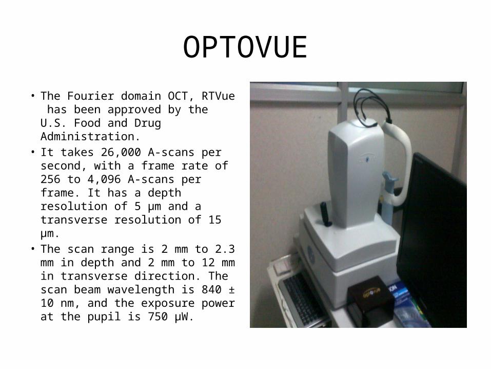
OPTOVUE• The Fourier domain OCT, RTVue
has been approved by the U.S. Food and Drug Administration.
• It takes 26,000 A-scans per second, with a frame rate of 256 to 4,096 A-scans per frame. It has a depth resolution of 5 μm and a transverse resolution of 15 μm.
• The scan range is 2 mm to 2.3 mm in depth and 2 mm to 12 mm in transverse direction. The scan beam wavelength is 840 ± 10 nm, and the exposure power at the pupil is 750 μW.

• Decision making in macula hole
CASE 1
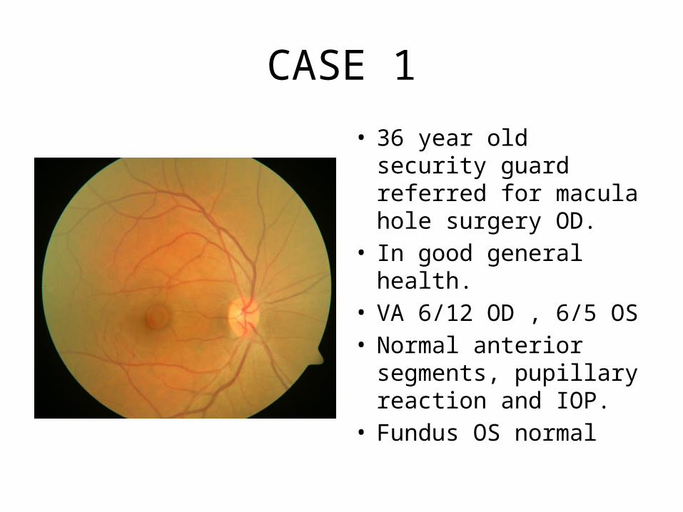
• 36 year old security guard referred for macula hole surgery OD.
• In good general health.• VA 6/12 OD , 6/5 OS• Normal anterior
segments, pupillary reaction and IOP.
• Fundus OS normal
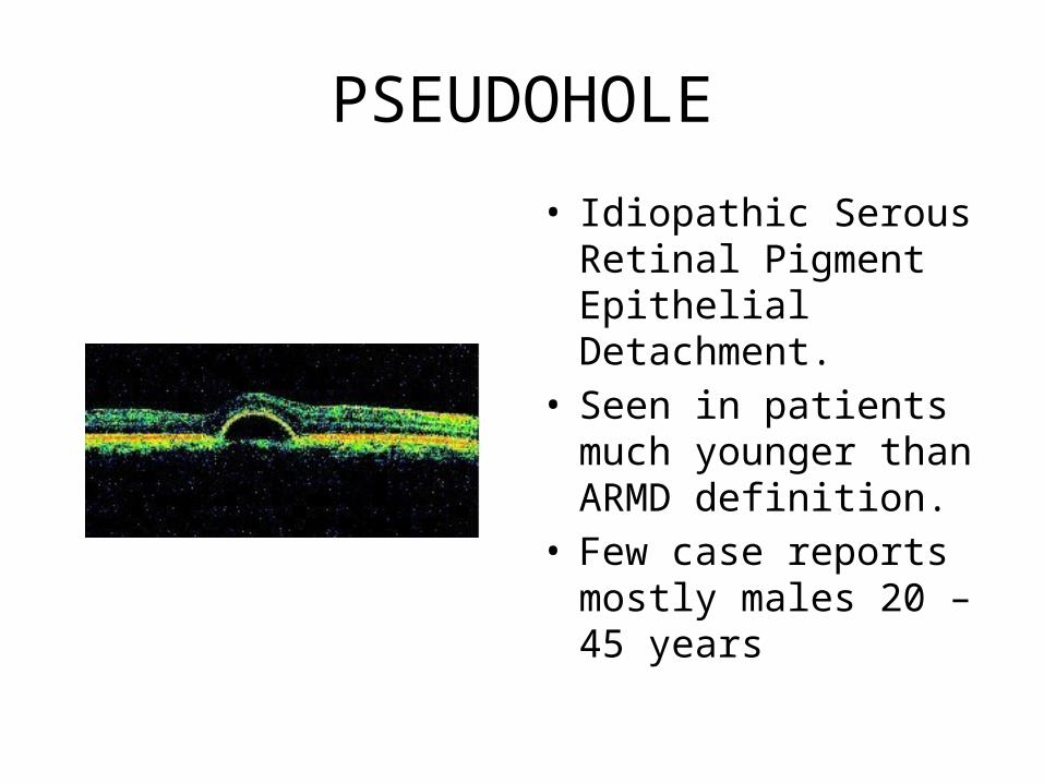
PSEUDOHOLE
• Idiopathic Serous Retinal Pigment Epithelial Detachment.
• Seen in patients much younger than ARMD definition.
• Few case reports mostly males 20 – 45 years
Pathophysiology• a non-specific anatomical alteration that may result from any
number of choroidal disorders that disrupt the normal junction between the basement membrane of the RPE and the inner collagenous layer of Bruch’s membrane.
• Idiopathic cases are sometimes associated with ICSC; some believe these two conditions to represent a continuum of a similar underlying pathology.
• Uncomplicated idiopathic serous detachments of the RPE often resolve spontaneously, however, those associated with more generalized damage to the choriocapillaris may be complicated by hemorrhage, choroidal neovascular membrane formation, and disciform scarring.
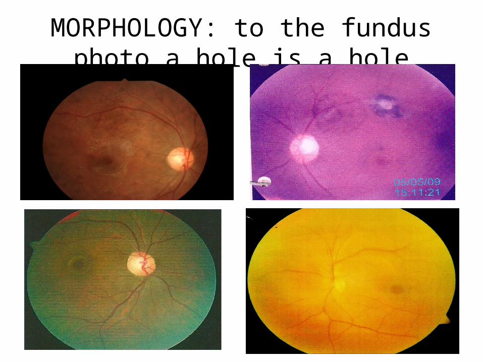
MORPHOLOGY: to the fundus photo a hole is a hole
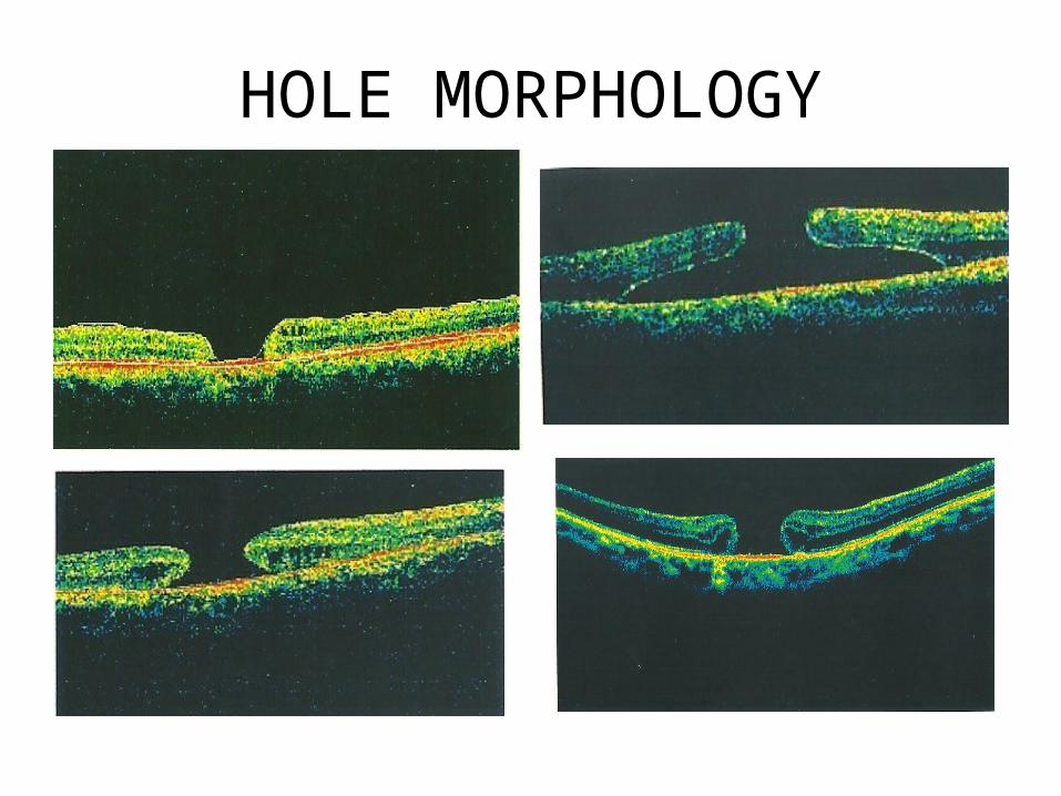
HOLE MORPHOLOGY
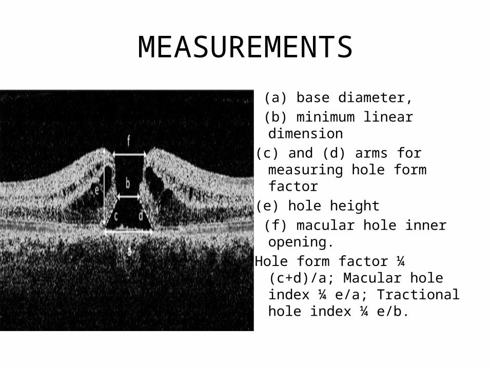
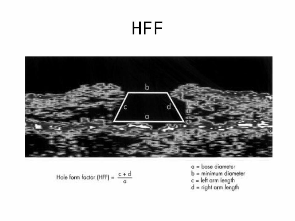
MEASUREMENTS
(a) base diameter, (b) minimum linear dimension(c) and (d) arms for measuring
hole form factor(e) hole height (f) macular hole inner
opening. Hole form factor ¼ (c+d)/a;
Macular hole index ¼ e/a; Tractional hole index ¼ e/b.
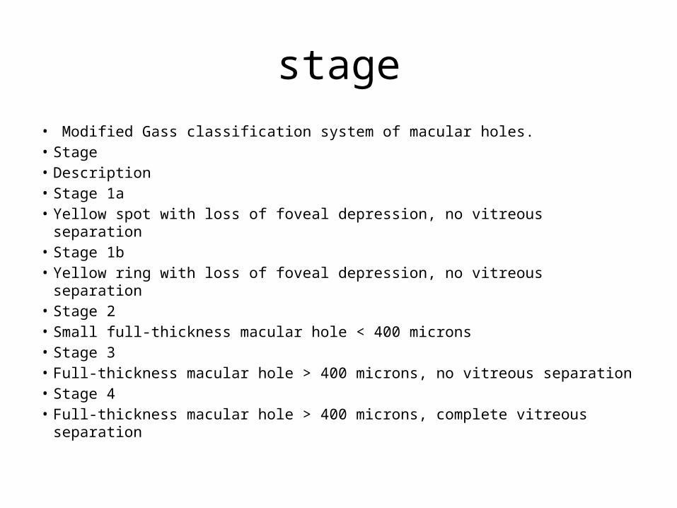
stage• Modified Gass classification system of macular holes.• Stage• Description• Stage 1a• Yellow spot with loss of foveal depression, no vitreous separation • Stage 1b• Yellow ring with loss of foveal depression, no vitreous separation• Stage 2• Small full-thickness macular hole < 400 microns• Stage 3• Full-thickness macular hole > 400 microns, no vitreous separation• Stage 4• Full-thickness macular hole > 400 microns, complete vitreous separation
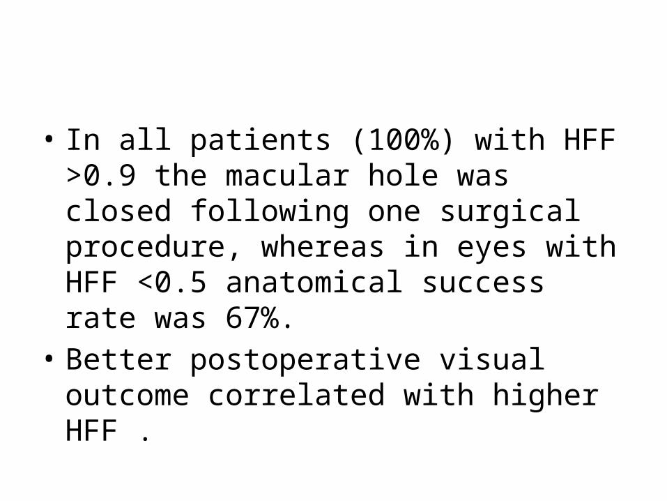
• In all patients (100%) with HFF >0.9 the macular hole was closed following one surgical procedure, whereas in eyes with HFF <0.5 anatomical success rate was 67%.
• Better postoperative visual outcome correlated with higher HFF .
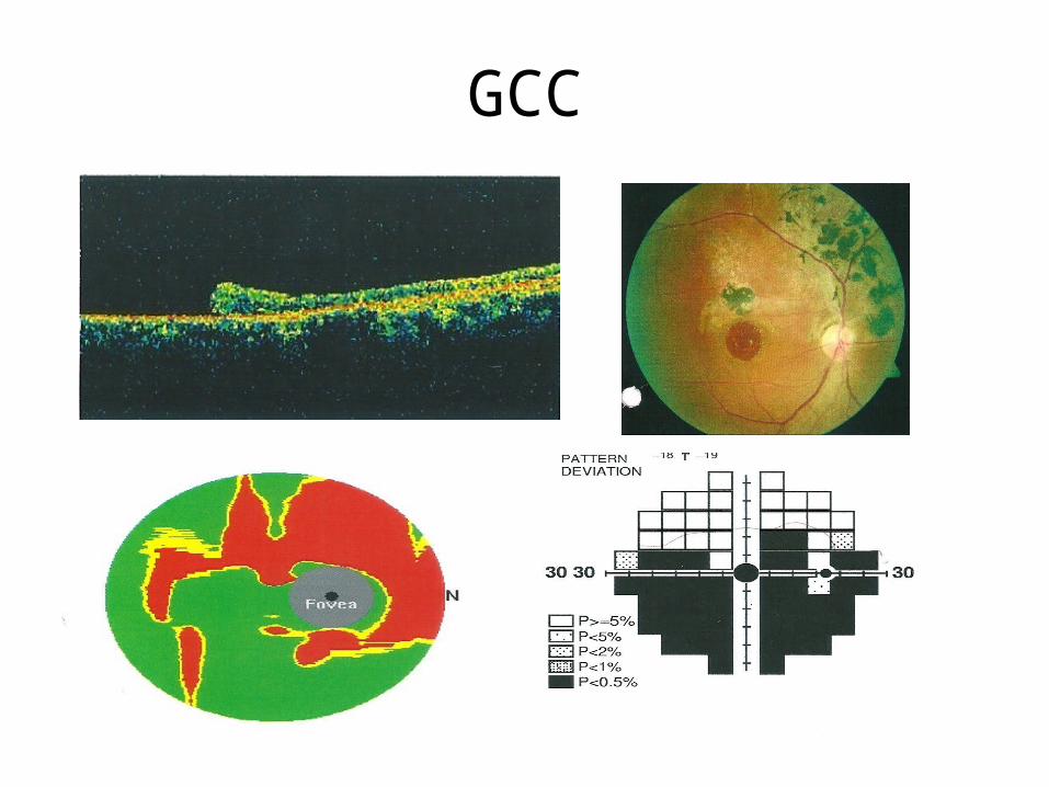
GCC
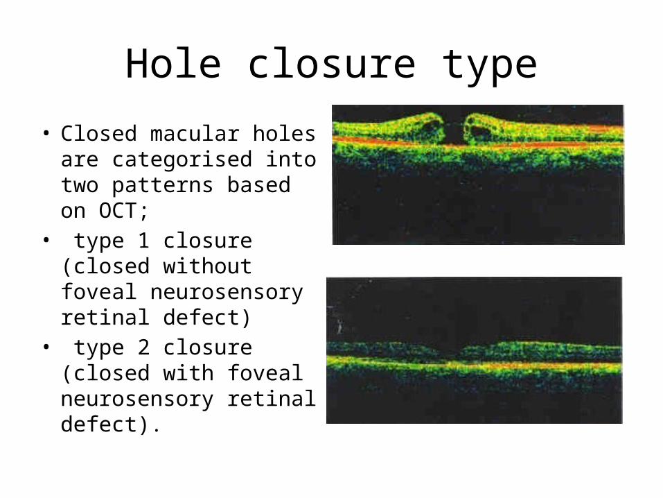
Hole closure type
• Closed macular holes are categorised into two patterns based on OCT;
• type 1 closure (closed without foveal neurosensory retinal defect)
• type 2 closure (closed with foveal neurosensory retinal defect).
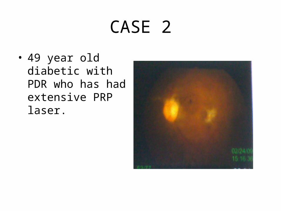
CASE 2
• 49 year old diabetic with PDR who has had extensive PRP laser.
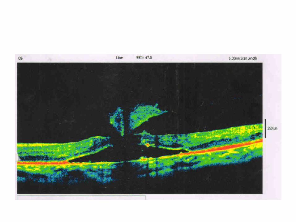
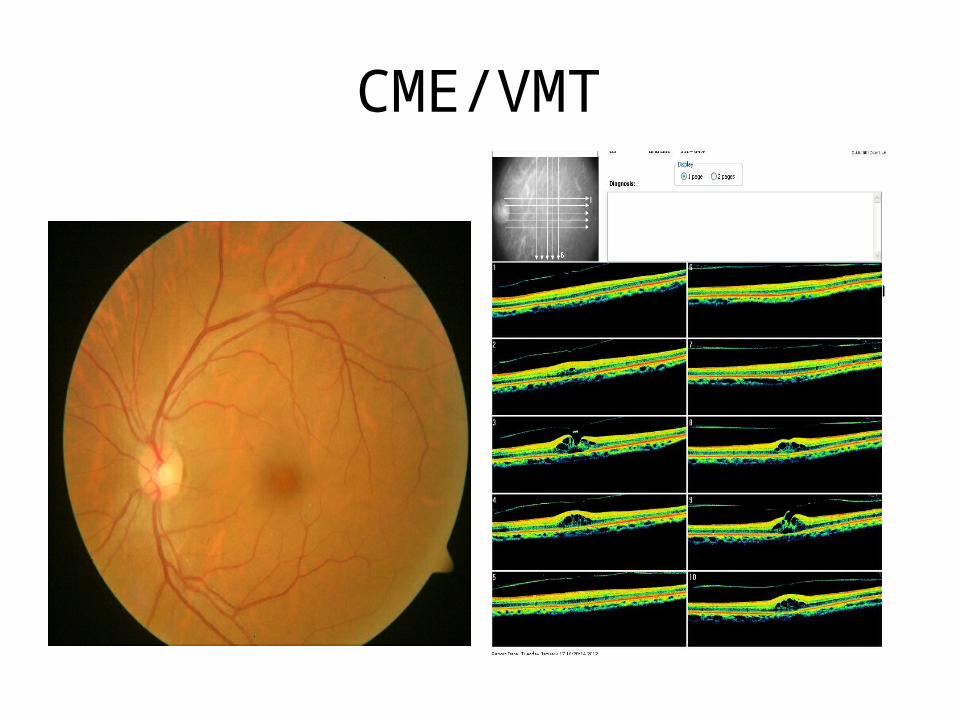
CME/VMT

• Reason for poor vision in heriditary disorders
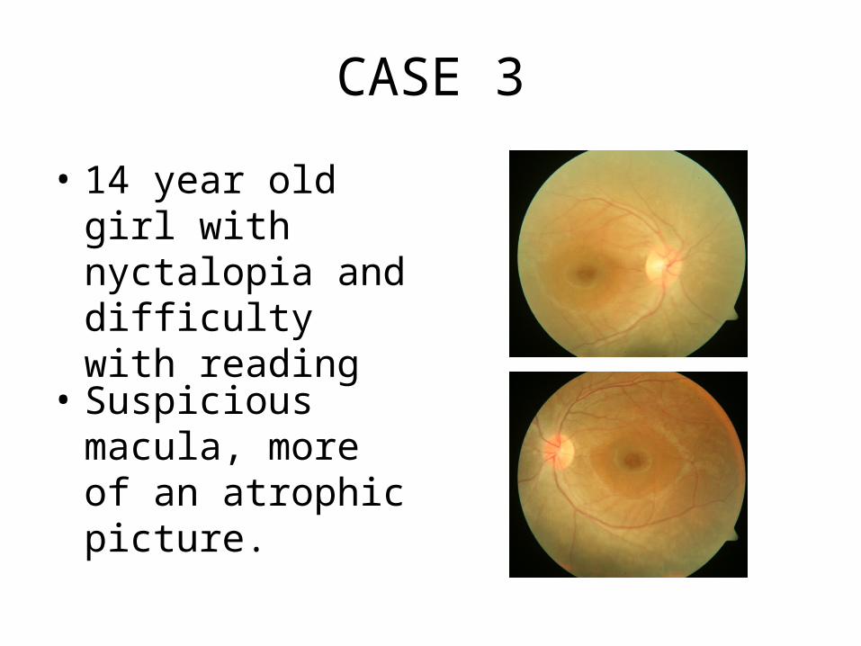
CASE 3
• 14 year old girl with nyctalopia and difficulty with reading
• Suspicious macula, more of an atrophic picture.
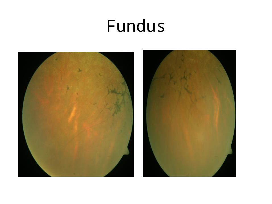
Fundus
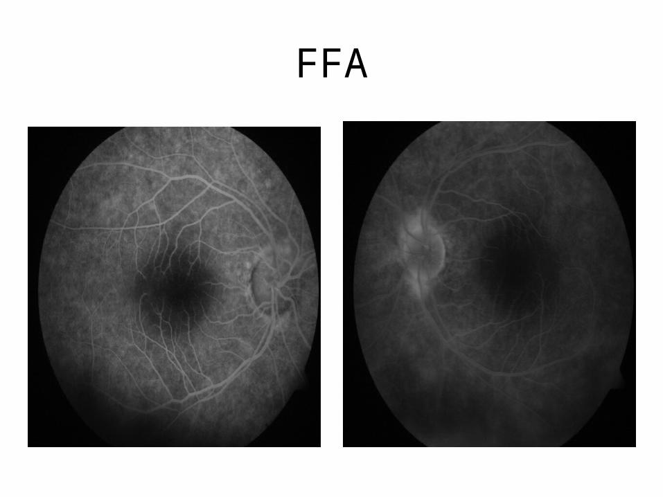
FFA
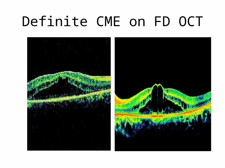
Definite CME on FD OCT
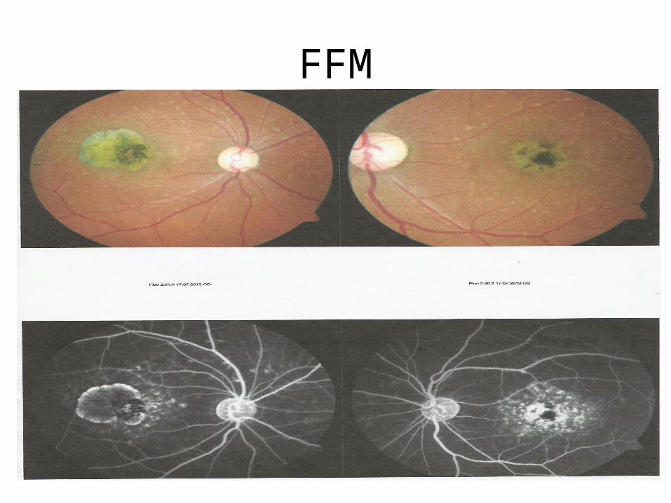
FFM
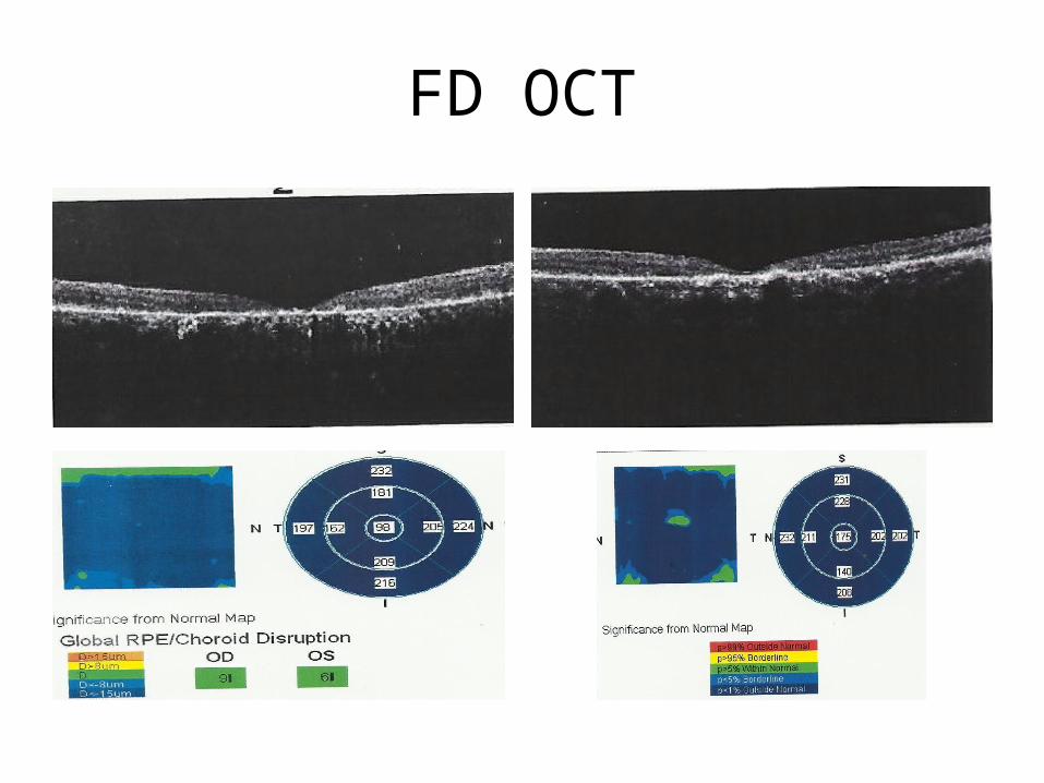
FD OCT

• To evaluate unsatisfactory vision after surgery.
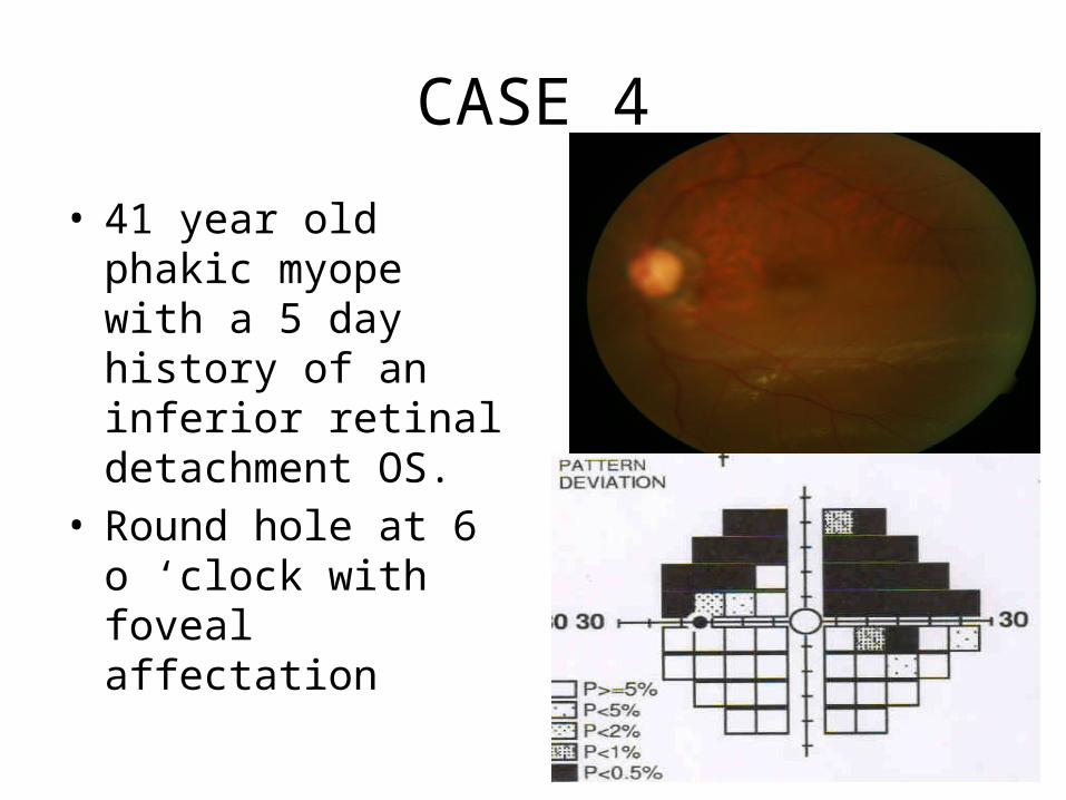
CASE 4
• 41 year old phakic myope with a 5 day history of an inferior retinal detachment OS.
• Round hole at 6 o ‘clock with foveal affectation
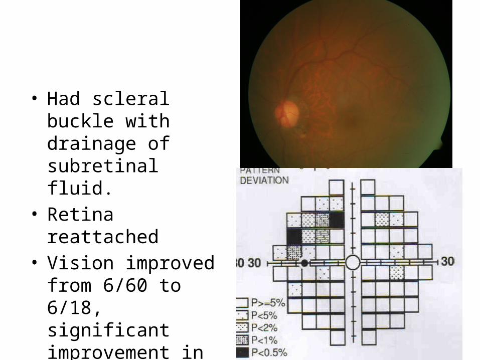
• Had scleral buckle with drainage of subretinal fluid.
• Retina reattached• Vision improved from
6/60 to 6/18, significant improvement in CVF
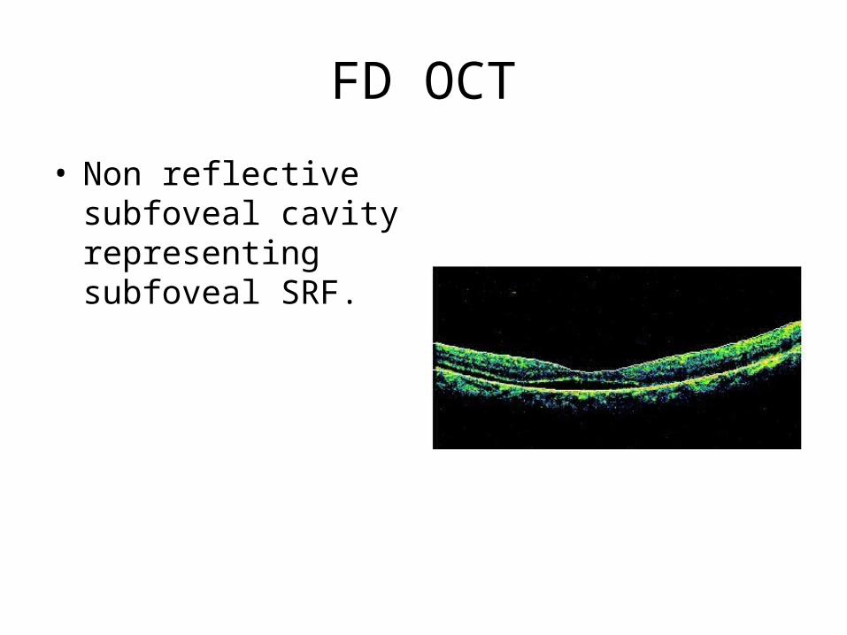
FD OCT
• Non reflective subfoveal cavity representing subfoveal SRF.
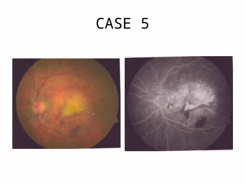
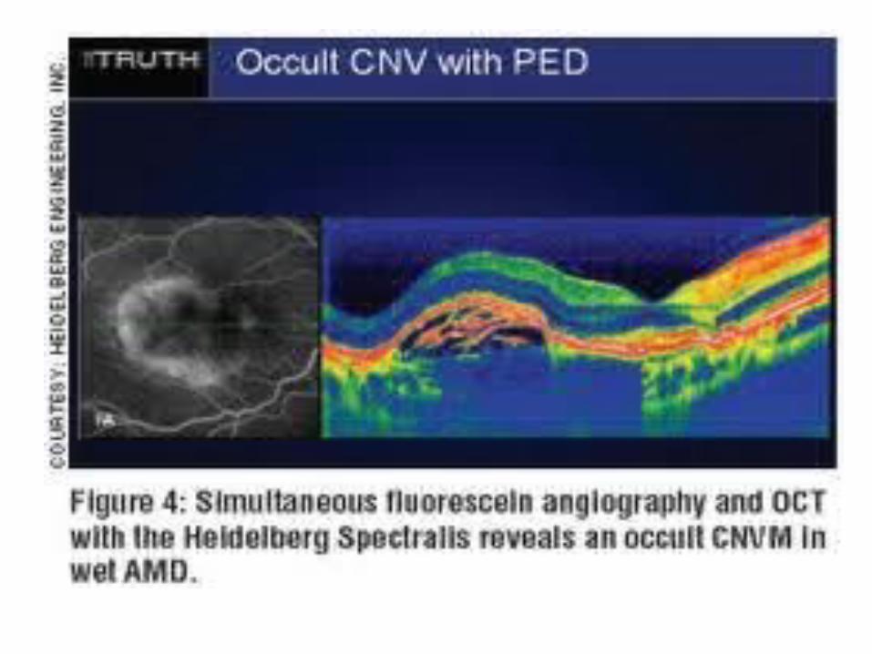
CASE 5
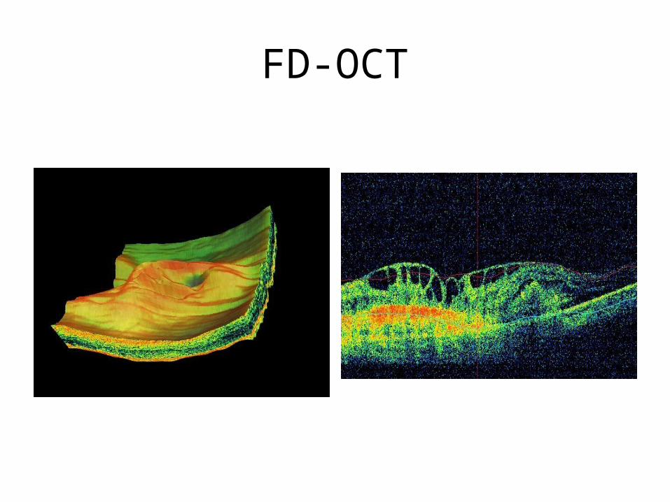
FD-OCT
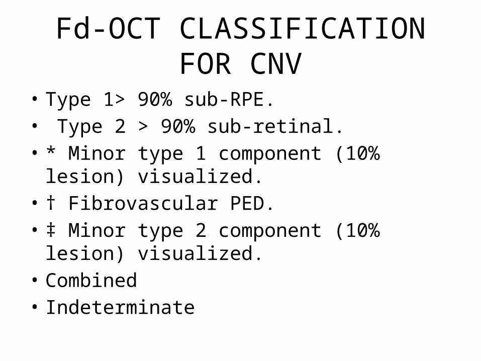
Fd-OCT CLASSIFICATION FOR CNV
• Type 1> 90% sub-RPE.• Type 2 > 90% sub-retinal.• * Minor type 1 component (10% lesion)
visualized.• † Fibrovascular PED.• ‡ Minor type 2 component (10% lesion)
visualized.• Combined• Indeterminate
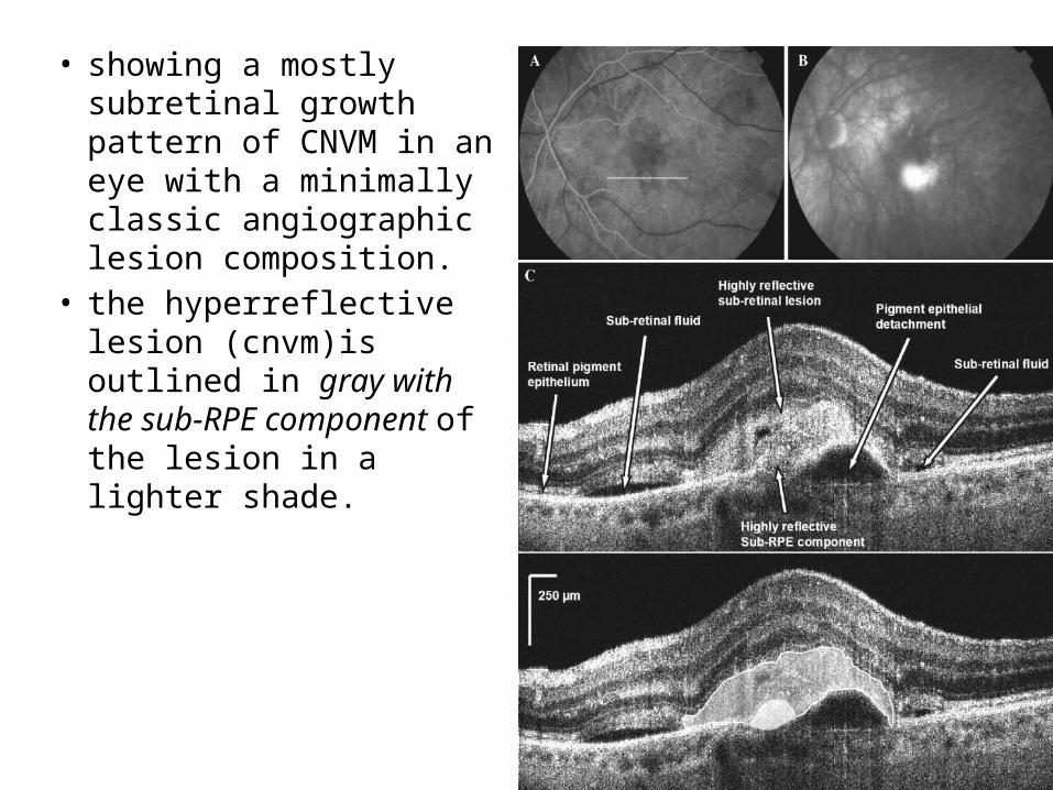
• showing a mostly subretinal growth pattern of CNVM in an eye with a minimally classic angiographic lesion composition.
• the hyperreflective lesion (cnvm)is outlined in gray with the sub-RPE component of the lesion in a lighter shade.

• WITH FD OCT FOR MACULA LESIONS I CAN SEE CLEARLY NOW.

THANK YOU
REFERENCES
• Desai VN, Hee MR, Puliafito CA. Optical coherence tomography of macular holes. In: Madreperla SA, McCuen BW, eds. Macular hole: pathogenesis, diagnosis and treatment. Oxford: Butterworth-Heinemann 1999:37–47