Embed Size (px)
Citation preview
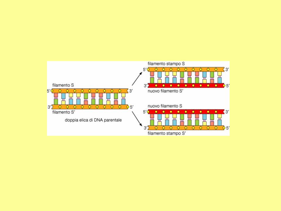
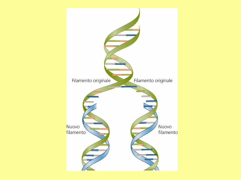
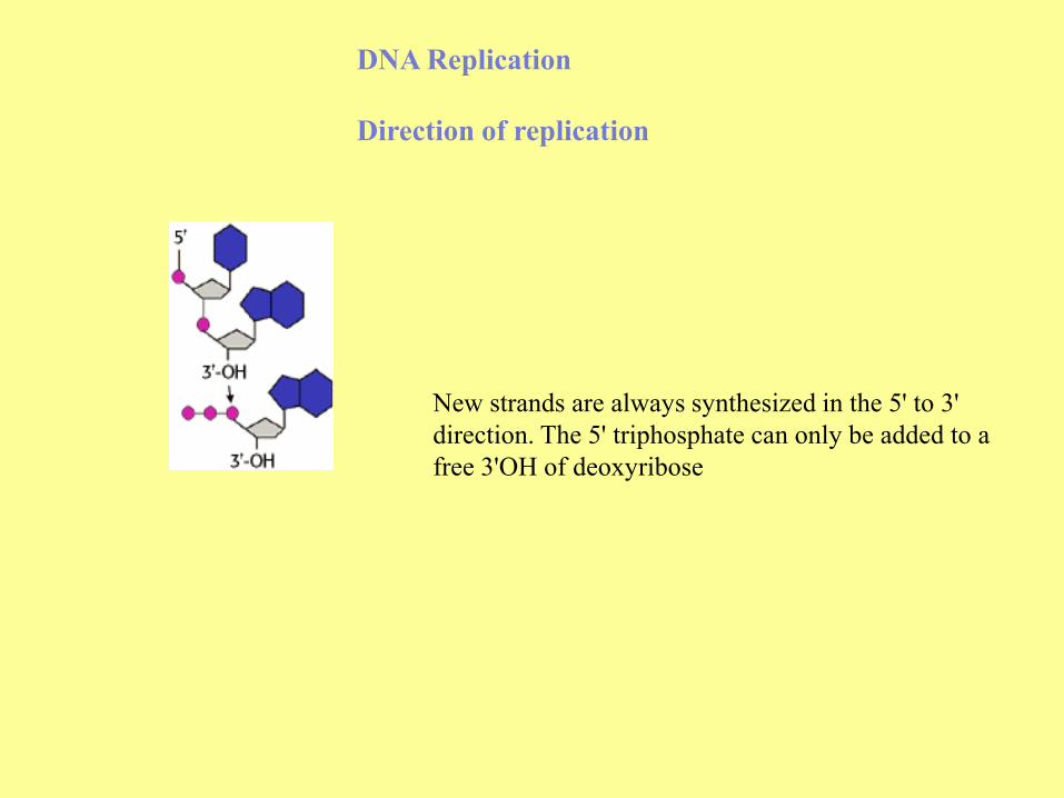
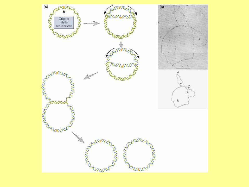
DNA Replication
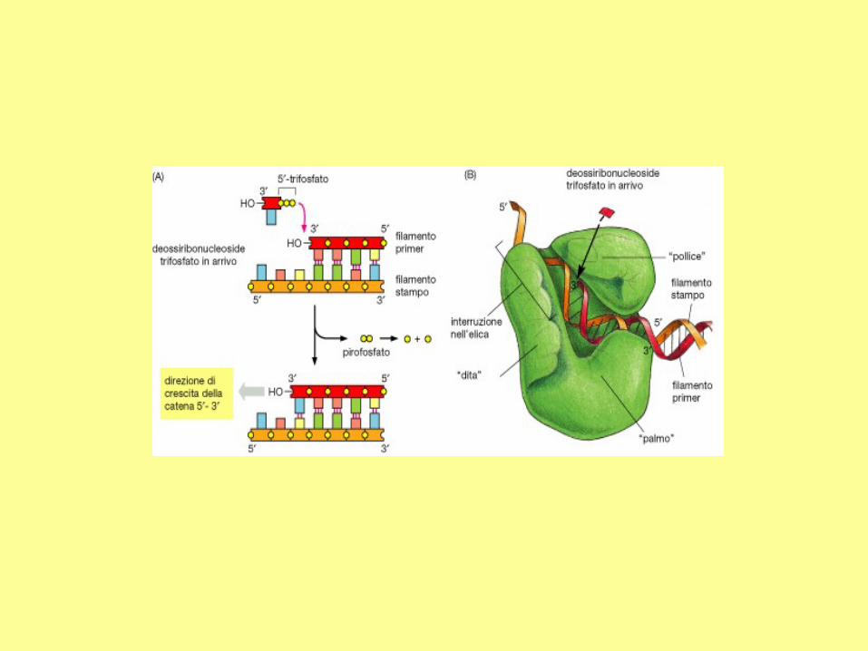
Direction of replication
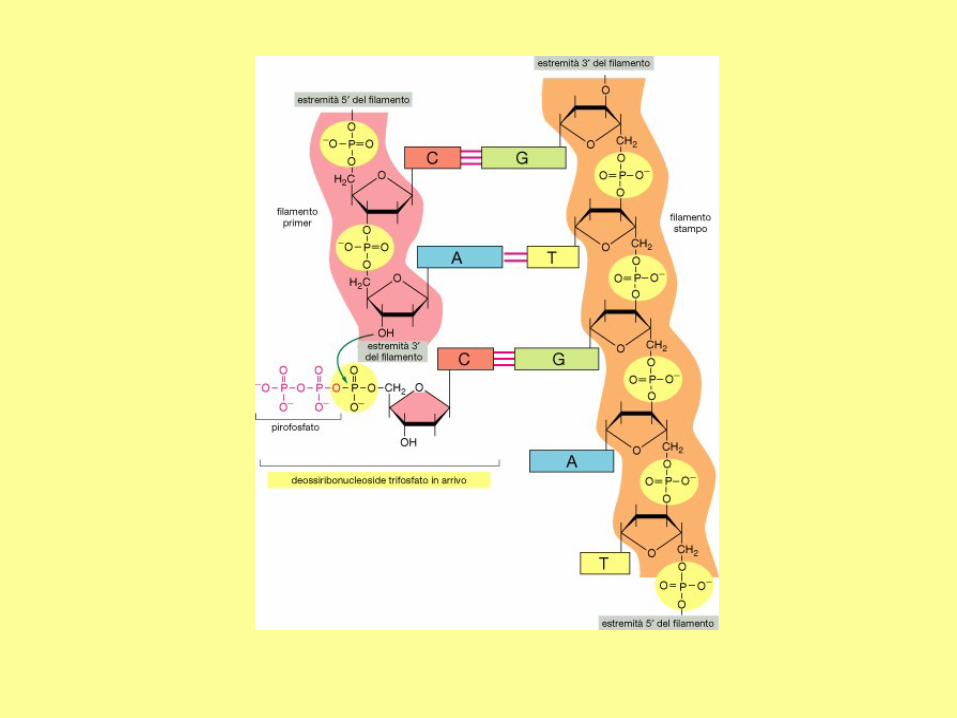
New strands are always synthesized in the 5' to 3'
direction. The 5' triphosphate can only be added to a
free 3'OH of deoxyribose
There is a major difference between DNA polymerase and RNA polymerase: the RNA polymerase can
synthesize a new strand whereas the DNA polymerase can only extend an existing strand. Therefore, to
synthesize a DNA molecule, a short RNA molecule (~ 5 - 12 nucleotides) should be synthesize first by a
specific enzyme. The initiating RNA molecule is known as the primer, and the enzyme is called primase.
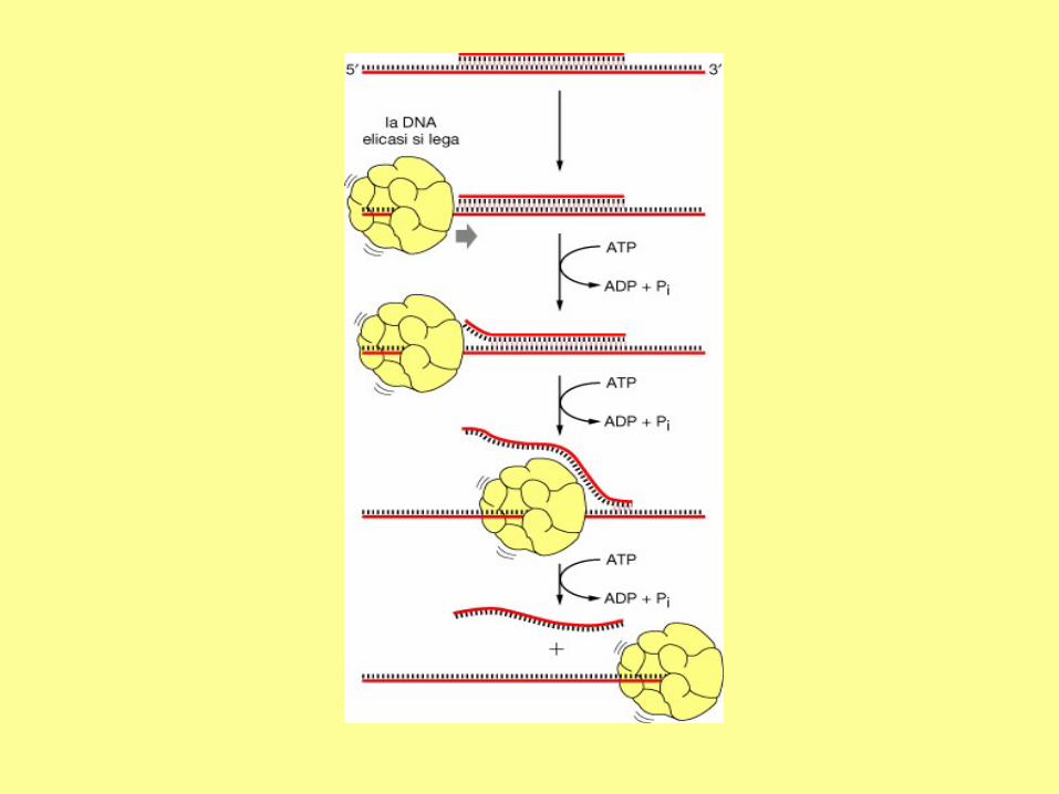
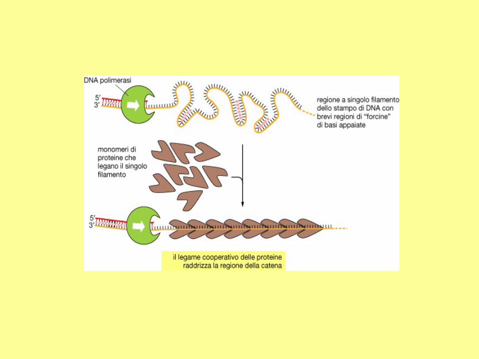
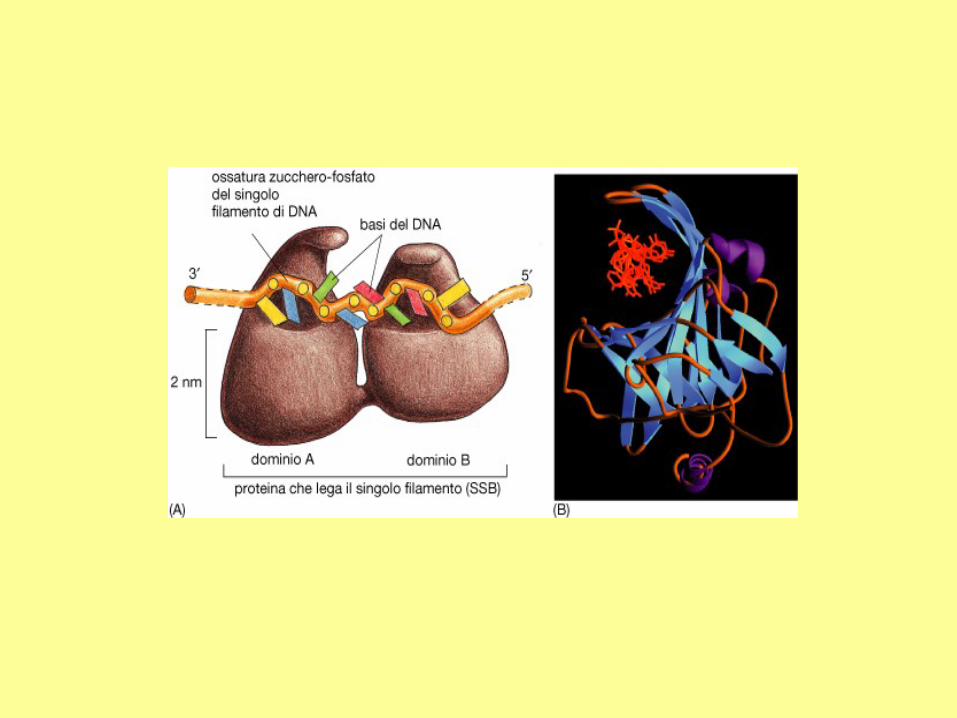
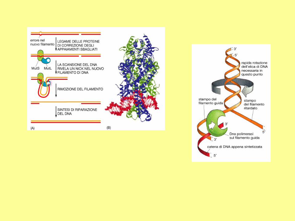
In addition to DNA polymerase and primase, DNA replication requires helicase and single strand binding
protein (SSB protein). The role of helicase is to unwind the duplex DNA. SSB proteins can bind to both
separated strands, preventing them from annealing (reconstitution of double-stranded DNA from single
strands).
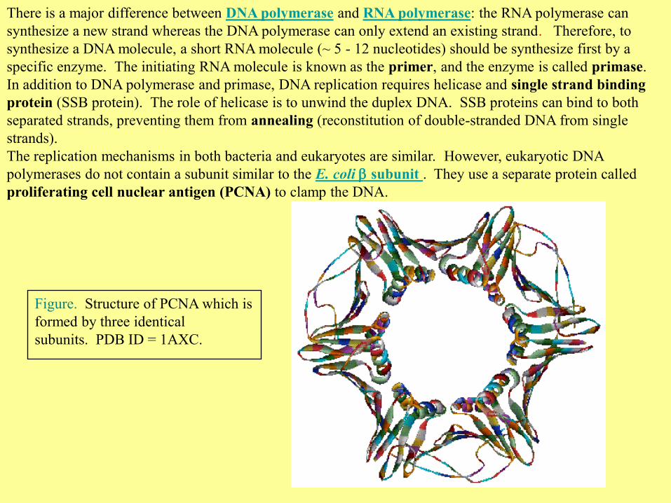
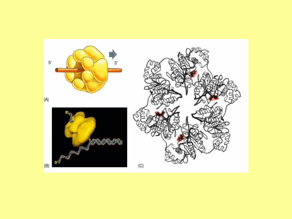
The replication mechanisms in both bacteria and eukaryotes are similar. However, eukaryotic DNA
polymerases do not contain a subunit similar to the E. coli b subunit . They use a separate protein called
proliferating cell nuclear antigen (PCNA) to clamp the DNA.
Figure. Structure of PCNA which is
formed by three identical
subunits. PDB ID = 1AXC.
DNA polymerases
E. Coli
Three types of DNA polymerases exist in E. coli: I, II and III. The DNA polymerase I is
used to fill the gap between DNA fragments of the lagging strand. It is also the major
enzyme for gap filling during DNA repair. The DNA polymerase II is encoded by the PolB
gene, which is involved in the SOS response to DNA damage. DNA replication is mainly
carried out by the DNA polymerase III.
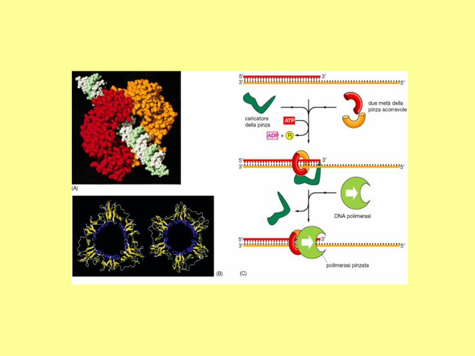
The DNA polymerase III consists of several subunits, with a total molecular weight
exceeding 600 kD. Among them, , , and subunits constitute the core polymerase. The
major role of other subunits is to keep the enzyme from falling off the template strand. As
shown in Figure 7-B-1, two b subunits can form a donut-shaped structure to clamp a DNA
molecule in its center, and slide with the core polymerase along the DNA molecule. This
allows continuous polymerization of up to 5 x 10⁵ nucleotides. In the absence of b
subunits, the core polymerase would fall off the template strand after synthesizing 10-50
nucleotides.
Mammals
There are five types of DNA polymerases in mammalian cells: , b, , , and . The
subunit is located in the mitochondria, responsible for the replication of mtDNA. Other
subunits are located in the nucleus. Their major roles are given below:
: synthesis of lagging strand.
b: DNA repair.
: synthesis of leading strand.
: DNA repair.
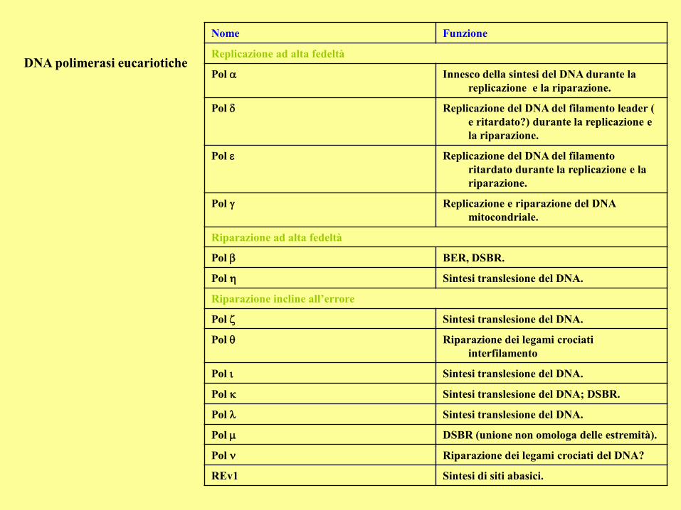
DNA polimerasi eucariotiche
Nome Funzione
Replicazione ad alta fedeltà
Pol Innesco della sintesi del DNA durante la
replicazione e la riparazione.
Pol Replicazione del DNA del filamento leader (
e ritardato?) durante la replicazione e
la riparazione.
Pol Replicazione del DNA del filamento
ritardato durante la replicazione e la
riparazione.
Pol Replicazione e riparazione del DNA
mitocondriale.
Riparazione ad alta fedeltà
Pol b BER, DSBR.
Pol Sintesi translesione del DNA.
Riparazione incline all’errore
Pol Sintesi translesione del DNA.
Pol Riparazione dei legami crociati
interfilamento
Pol Sintesi translesione del DNA.
Pol Sintesi translesione del DNA; DSBR.
Pol Sintesi translesione del DNA.
Pol DSBR (unione non omologa delle estremità).
Pol Riparazione dei legami crociati del DNA?
REv1 Sintesi di siti abasici.
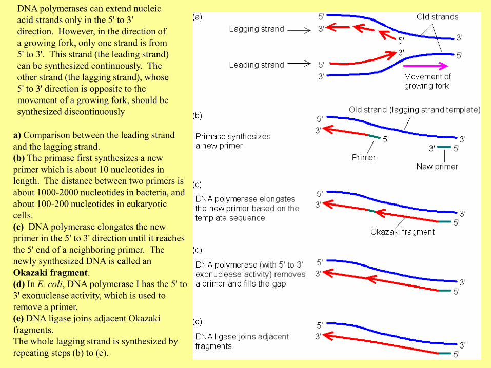
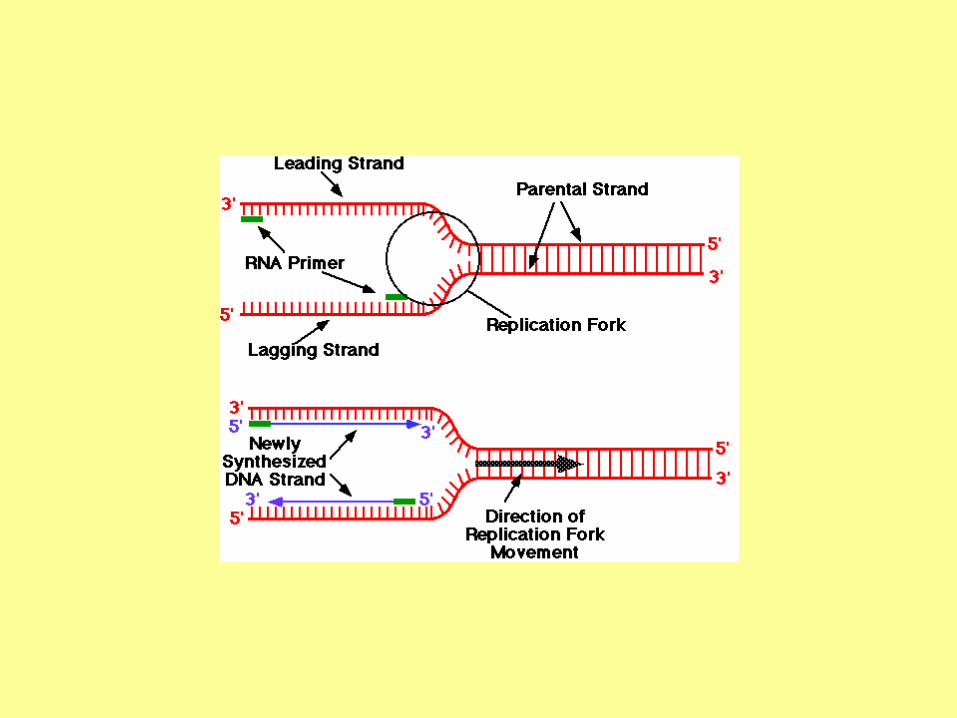
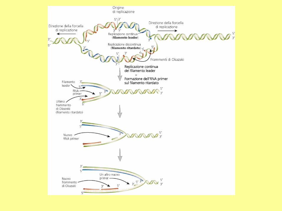
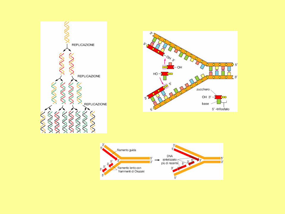
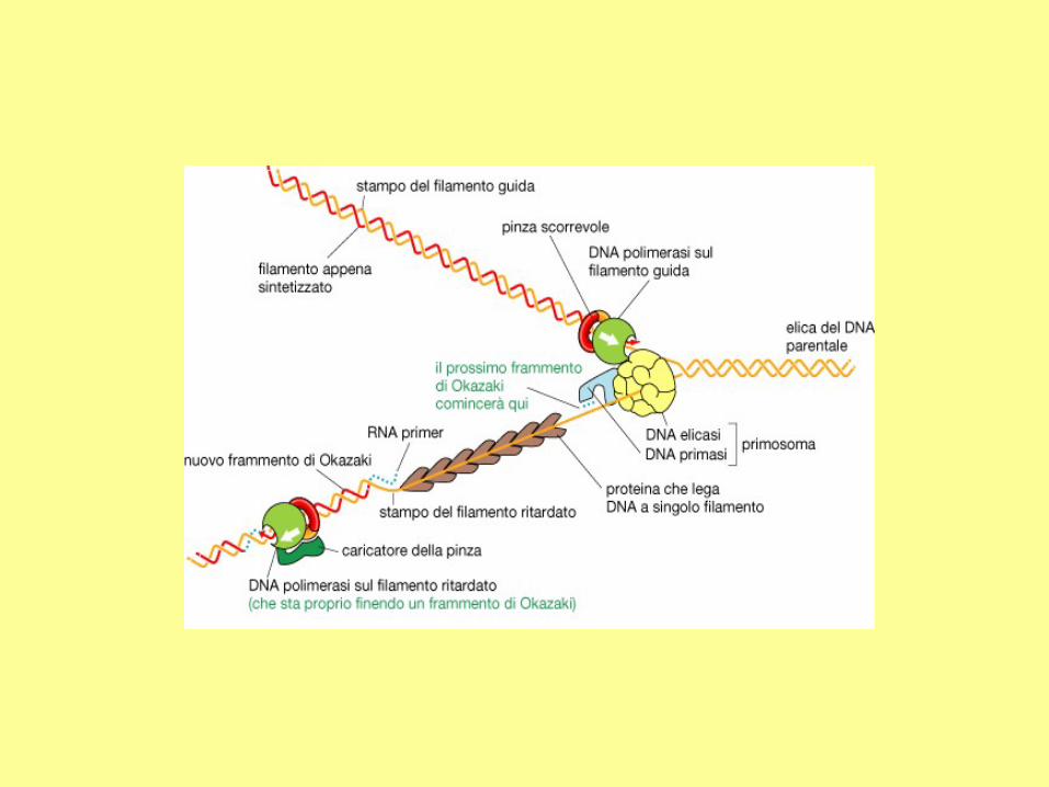
DNA polymerases can extend nucleic
acid strands only in the 5' to 3'
direction. However, in the direction of
a growing fork, only one strand is from
5' to 3'. This strand (the leading strand)
can be synthesized continuously. The
other strand (the lagging strand), whose
5' to 3' direction is opposite to the
movement of a growing fork, should be
synthesized discontinuously
a) Comparison between the leading strand
and the lagging strand.
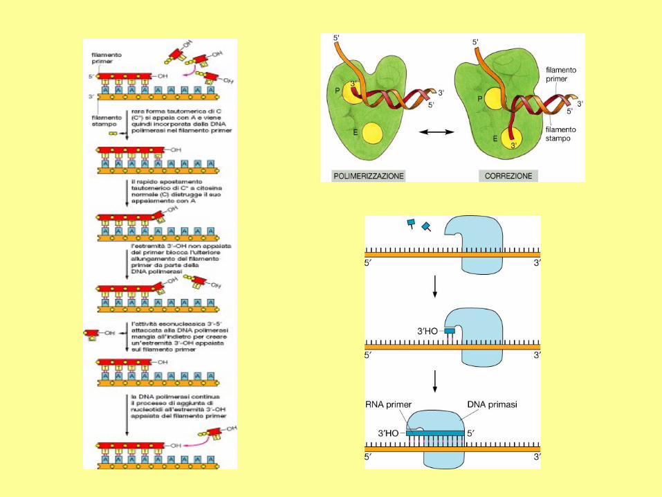
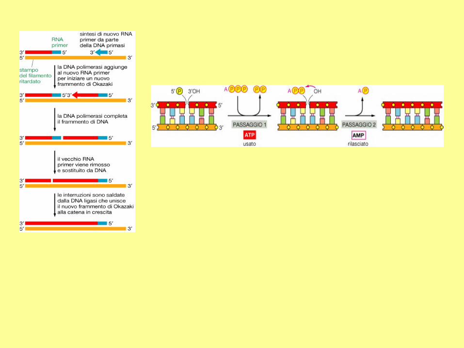
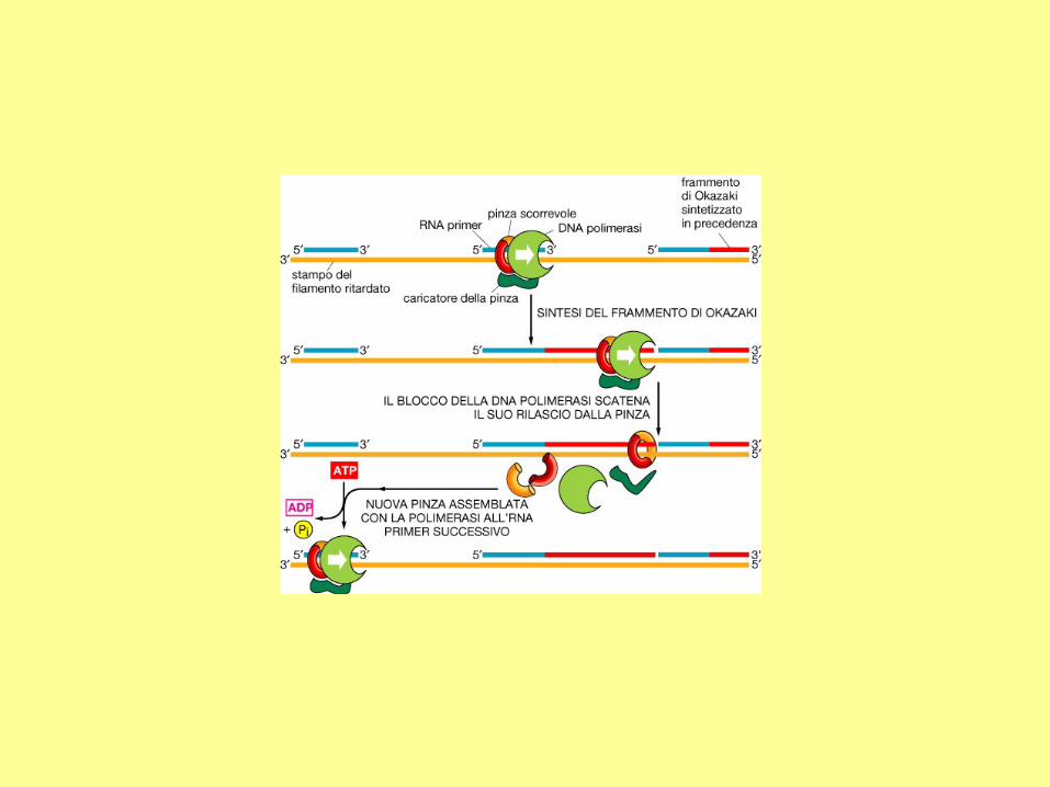
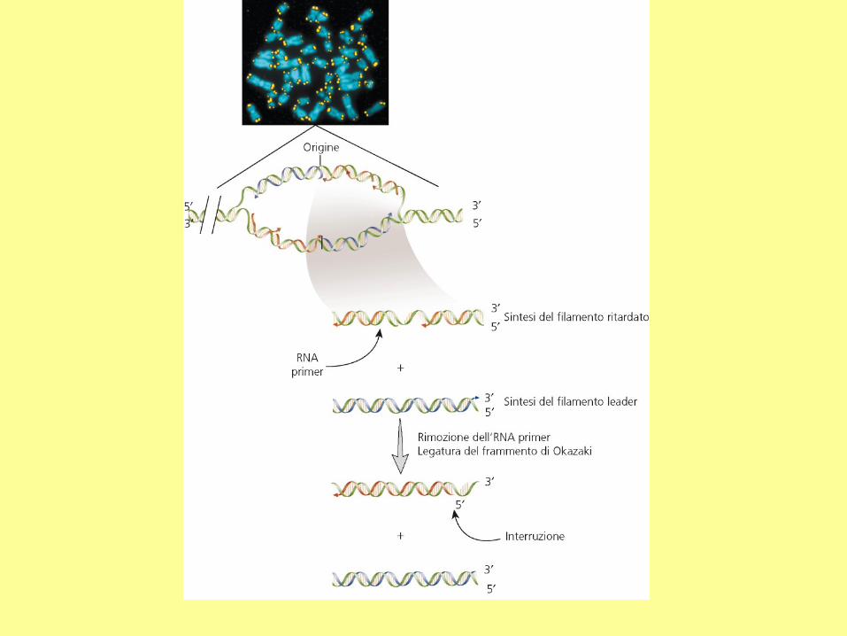
(b) The primase first synthesizes a new
primer which is about 10 nucleotides in
length. The distance between two primers is
about 1000-2000 nucleotides in bacteria, and
about 100-200 nucleotides in eukaryotic
cells.
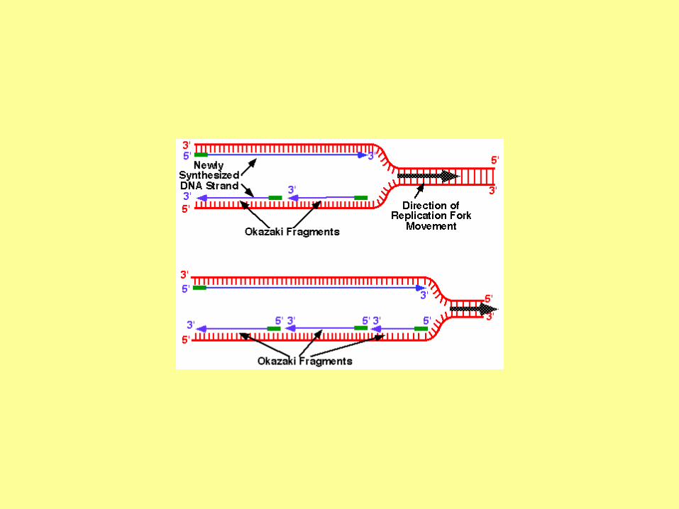
(c) DNA polymerase elongates the new
primer in the 5' to 3' direction until it reaches
the 5' end of a neighboring primer. The
newly synthesized DNA is called an
Okazaki fragment.
(d) In E. coli, DNA polymerase I has the 5' to
3' exonuclease activity, which is used to
remove a primer.
(e) DNA ligase joins adjacent Okazaki
fragments.
The whole lagging strand is synthesized by
repeating steps (b) to (e).
DNA Replication
RNA primers
The initiation of replication always starts with a short RNA piece. The DNA
replicating enzymes will only add nucleotides to the 3' end of DNA or RNA,
and thus an RNA primase must start replication. The DNA polymerases must
correct errors or "proof read", a function that is not compatible with initiation.
Meselson-Stahl DNA replication experiment
In the Meselson-Stahl DNA replication experiment, if the cells were first grown for
many generations in N-15 containing media, and then switched to N-14 containing
media, what percent of the DNA had 1 light strand and 1 heavy strand after 2
generations of growth in N-14 growth media?
Meselson and Stahl's Experiment
Meselson and Stahl in 1957 gave experimental evidence that each DNA strand served
as a template for new synthesis, a process called semi-conservative replication
E. coli grown in 15N nitrogen (heavy isotope).
Switch to 14N nitrogen (light) and after one, two, or three generations take samples of
DNA.
Mix with cesium chloride and separate heavy and light DNA.
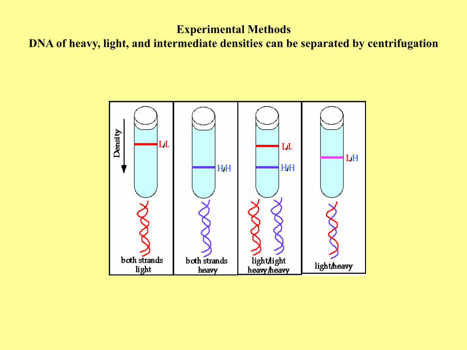
Experimental Methods
DNA of heavy, light, and intermediate densities can be separated by centrifugation
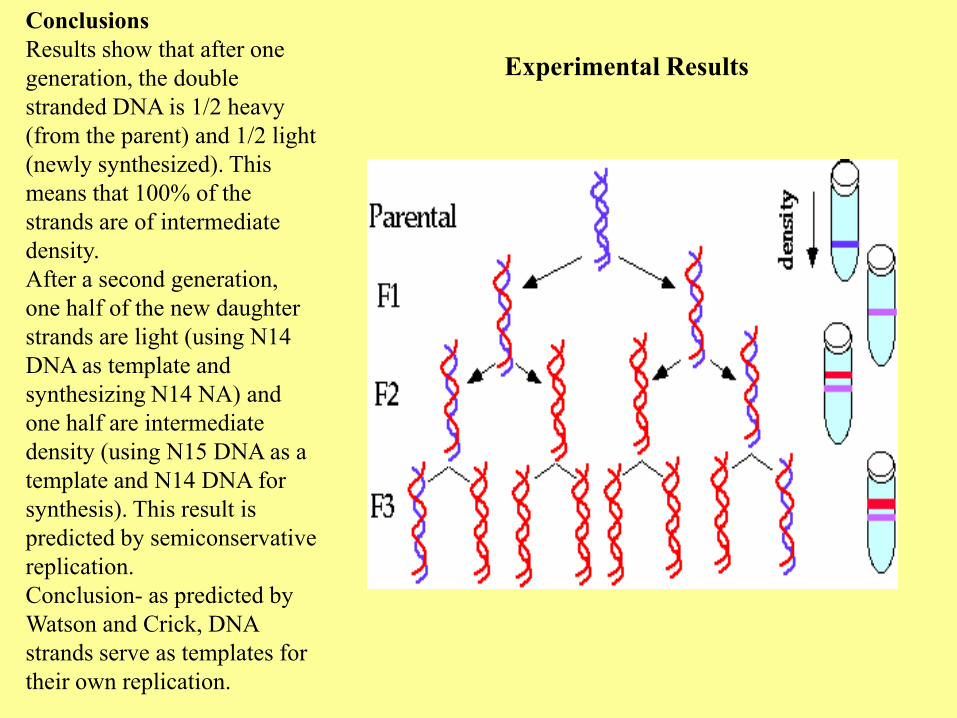
Experimental Results
Conclusions
Results show that after one
generation, the double
stranded DNA is 1/2 heavy
(from the parent) and 1/2 light
(newly synthesized). This
means that 100% of the
strands are of intermediate
density.
After a second generation,
one half of the new daughter
strands are light (using N14
DNA as template and
synthesizing N14 NA) and
one half are intermediate
density (using N15 DNA as a
template and N14 DNA for
synthesis). This result is
predicted by semiconservative
replication.
Conclusion- as predicted by
Watson and Crick, DNA
strands serve as templates for
their own replication.


Enzimi e fattori che intervengono nella replicazione del DNA
Funzione E. coli Uomo
Elicasi DnaB Mcm2-7
Elicasi di caricamento/primasi DnaC Mcm2-7
Mantenimento del singolo
filamento
SSB RPA
Innesco DnaG (primasi) Pol /primasi
Pinza scorrevole b PCNA
Caricamento della pinza (ATPasi) Complesso RFC
Allungamento del filamento Pol III Pol /Pol
Rimozione dell’RNA primer Pol I FEN-1, Rnasi H1
Legatura dei frammenti di
Okazaki
Ligasi Ligasi 1
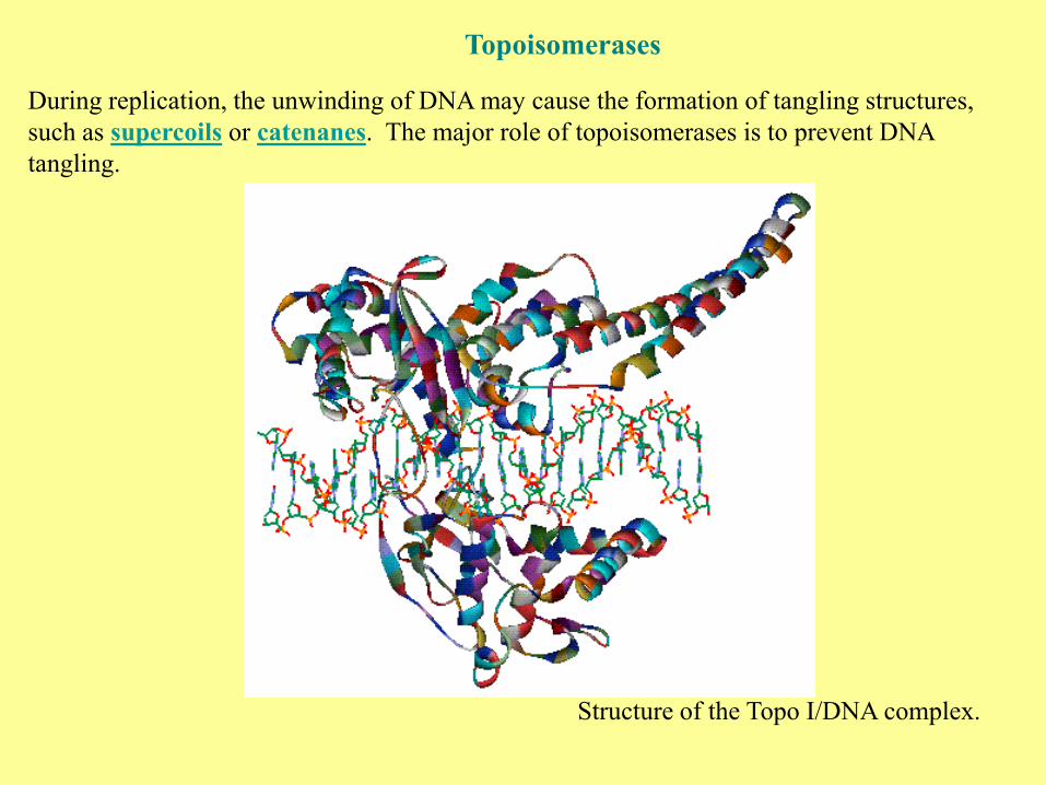
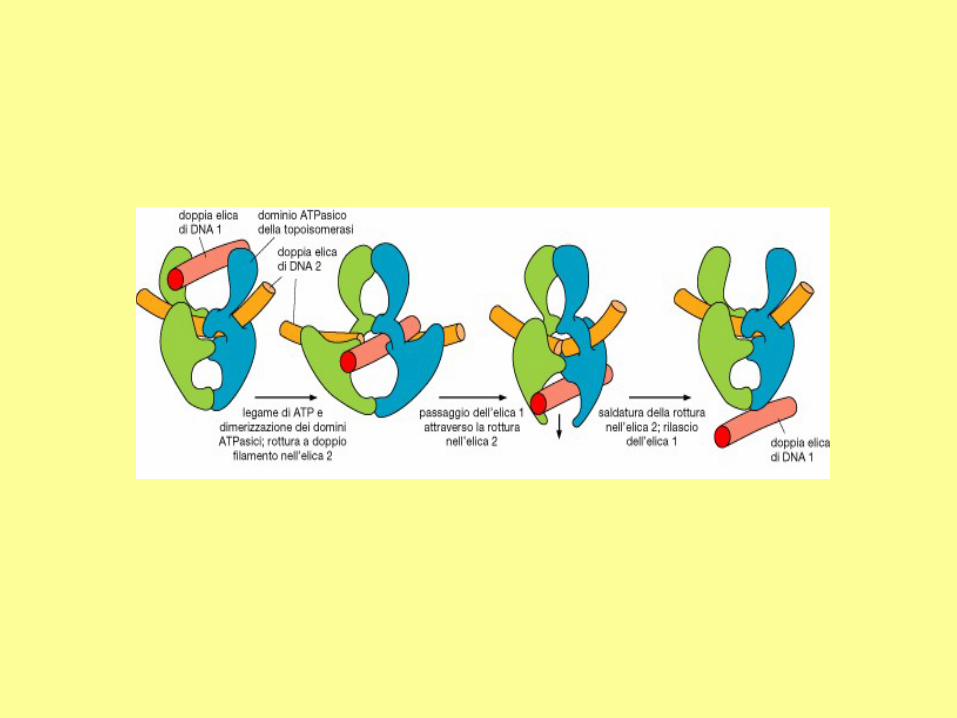
Topoisomerases
During replication, the unwinding of DNA may cause the formation of tangling structures,
such as supercoils or catenanes. The major role of topoisomerases is to prevent DNA
tangling.
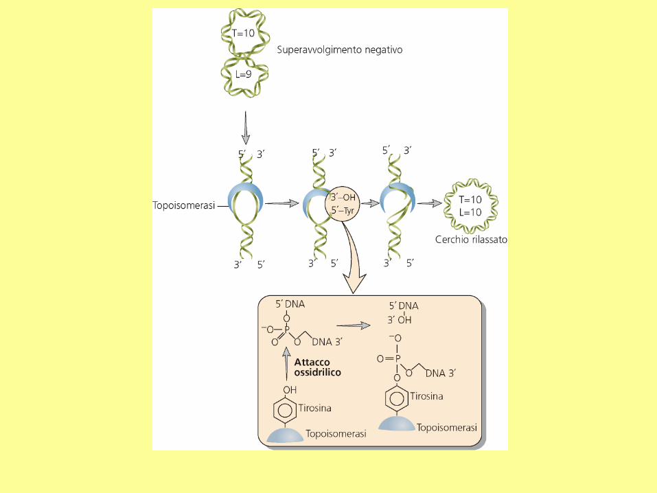
Structure of the Topo I/DNA complex.
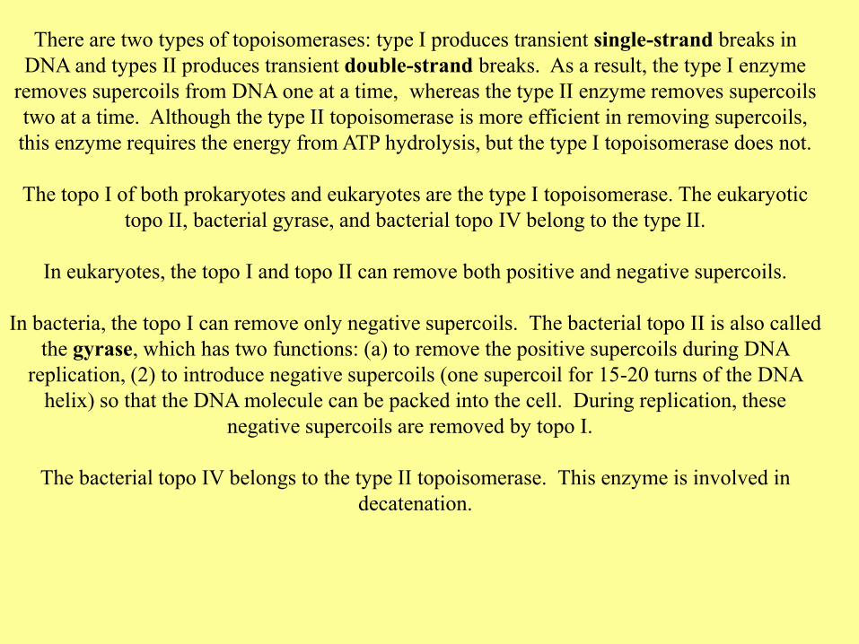
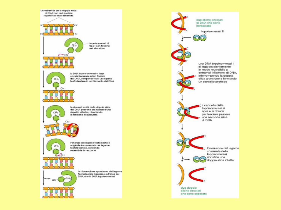
There are two types of topoisomerases: type I produces transient single-strand breaks in
DNA and types II produces transient double-strand breaks. As a result, the type I enzyme
removes supercoils from DNA one at a time, whereas the type II enzyme removes supercoils
two at a time. Although the type II topoisomerase is more efficient in removing supercoils,
this enzyme requires the energy from ATP hydrolysis, but the type I topoisomerase does not.
The topo I of both prokaryotes and eukaryotes are the type I topoisomerase. The eukaryotic
topo II, bacterial gyrase, and bacterial topo IV belong to the type II.
In eukaryotes, the topo I and topo II can remove both positive and negative supercoils.
In bacteria, the topo I can remove only negative supercoils. The bacterial topo II is also called
the gyrase, which has two functions: (a) to remove the positive supercoils during DNA
replication, (2) to introduce negative supercoils (one supercoil for 15-20 turns of the DNA
helix) so that the DNA molecule can be packed into the cell. During replication, these
negative supercoils are removed by topo I.
The bacterial topo IV belongs to the type II topoisomerase. This enzyme is involved in
decatenation.
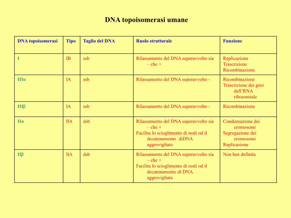
DNA topoisomerasi umane
DNA topoisomerasi Tipo Taglio del DNA Ruolo strutturale Funzione
I IB ssb Rilassamento del DNA superavvolto sia
– che +
Replicazione
Trascrizione
Ricombinazione
IIIα IA ssb Rilassamento del DNA superavvolto - Ricombinazione
Trascrizione dei geni
dell’RNA
ribosomiale
IIIb IA ssb Rilassamento del DNA superavvolto - Ricombinazione
IIα IIA dsb Rilassamento del DNA superavvolto sia
– che +
Facilita lo scioglimento di nodi od il
decatenamento diDNA
aggrovigliato
Condensazione dei
cromosomi
Segregazione dei
cromosomi
Replicazione
IIb IIA dsb Rilassamento del DNA superavvolto sia
– che +
Facilita lo scioglimento di nodi od il
decatenamento di DNA
aggrovigliato
Non ben definita
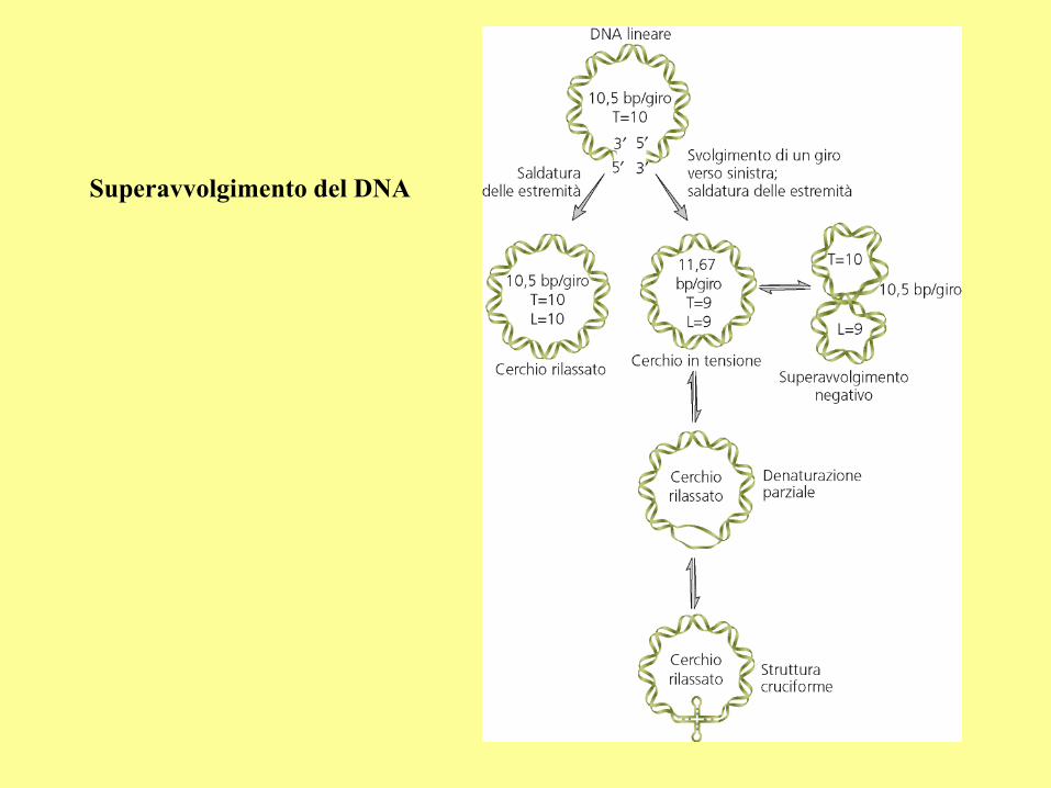
Superavvolgimento del DNA
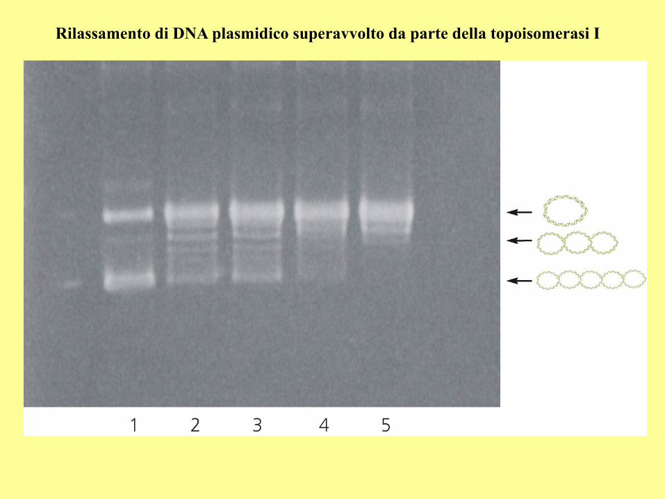
Rilassamento di DNA plasmidico superavvolto da parte della topoisomerasi I
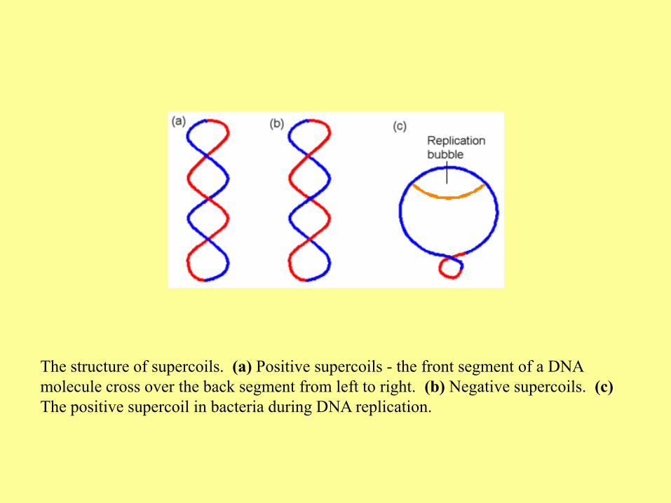
The structure of supercoils. (a) Positive supercoils - the front segment of a DNA
molecule cross over the back segment from left to right. (b) Negative supercoils. (c)
The positive supercoil in bacteria during DNA replication.
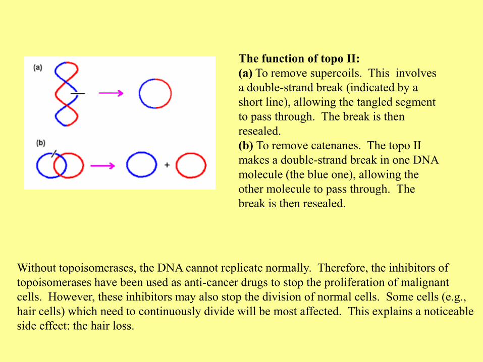
Without topoisomerases, the DNA cannot replicate normally. Therefore, the inhibitors of
topoisomerases have been used as anti-cancer drugs to stop the proliferation of malignant
cells. However, these inhibitors may also stop the division of normal cells. Some cells (e.g.,
hair cells) which need to continuously divide will be most affected. This explains a noticeable
side effect: the hair loss.
The function of topo II:
(a) To remove supercoils. This involves
a double-strand break (indicated by a
short line), allowing the tangled segment
to pass through. The break is then
resealed.
(b) To remove catenanes. The topo II
makes a double-strand break in one DNA
molecule (the blue one), allowing the
other molecule to pass through. The
break is then resealed.
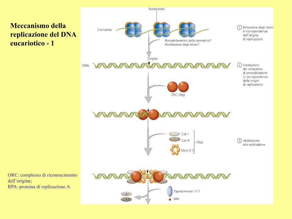
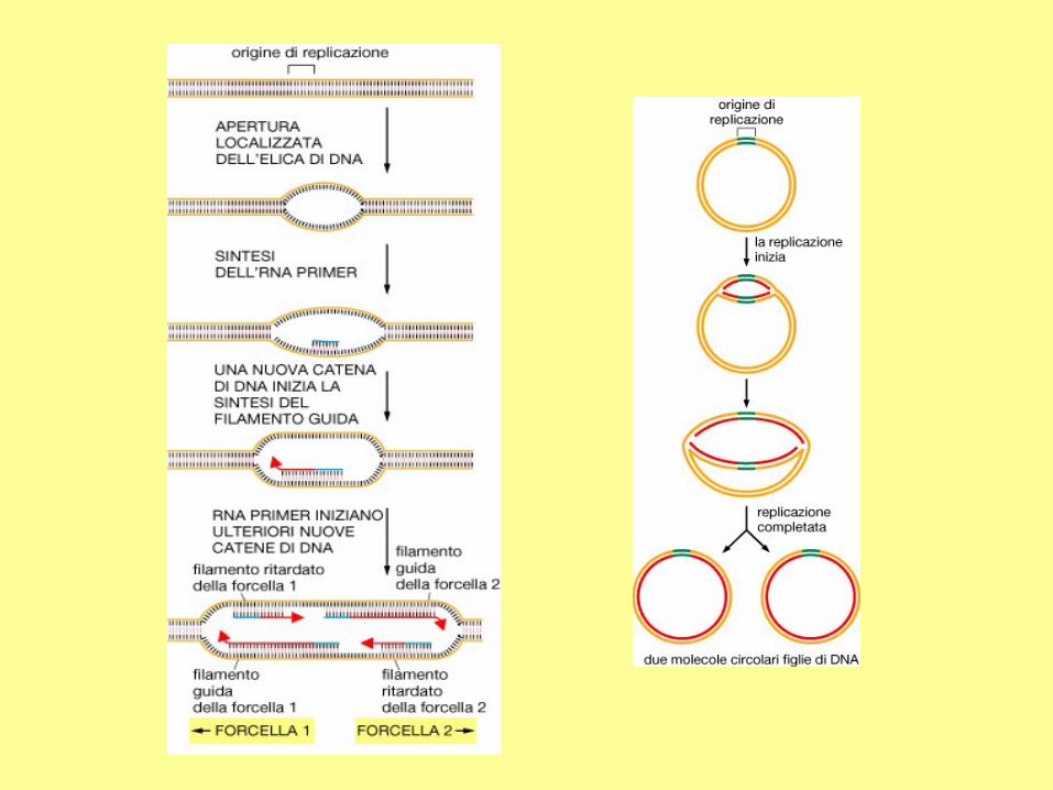
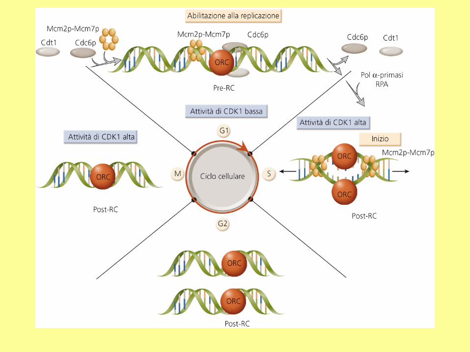
Meccanismo della
replicazione del DNA
eucariotico - 1
ORC: complesso di riconoscimento
dell’origine;
RPA: proteina di replicazione A.
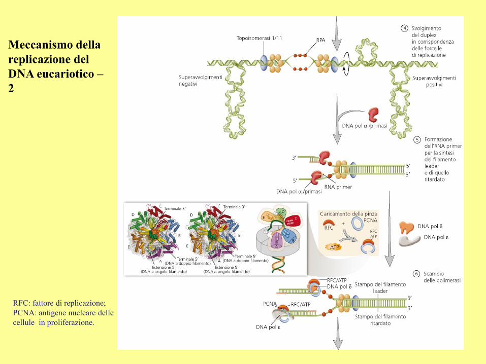
Meccanismo della
replicazione del
DNA eucariotico –
2
RFC: fattore di replicazione;
PCNA: antigene nucleare delle
cellule in proliferazione.
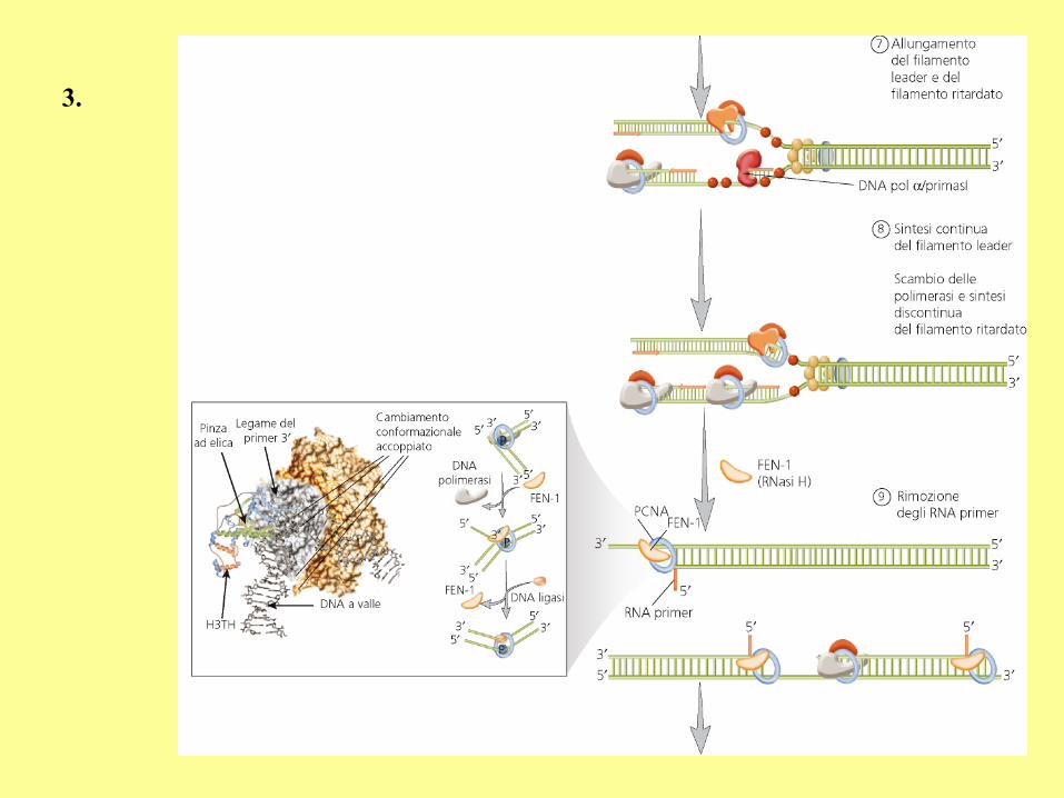
3.
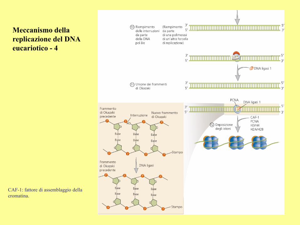
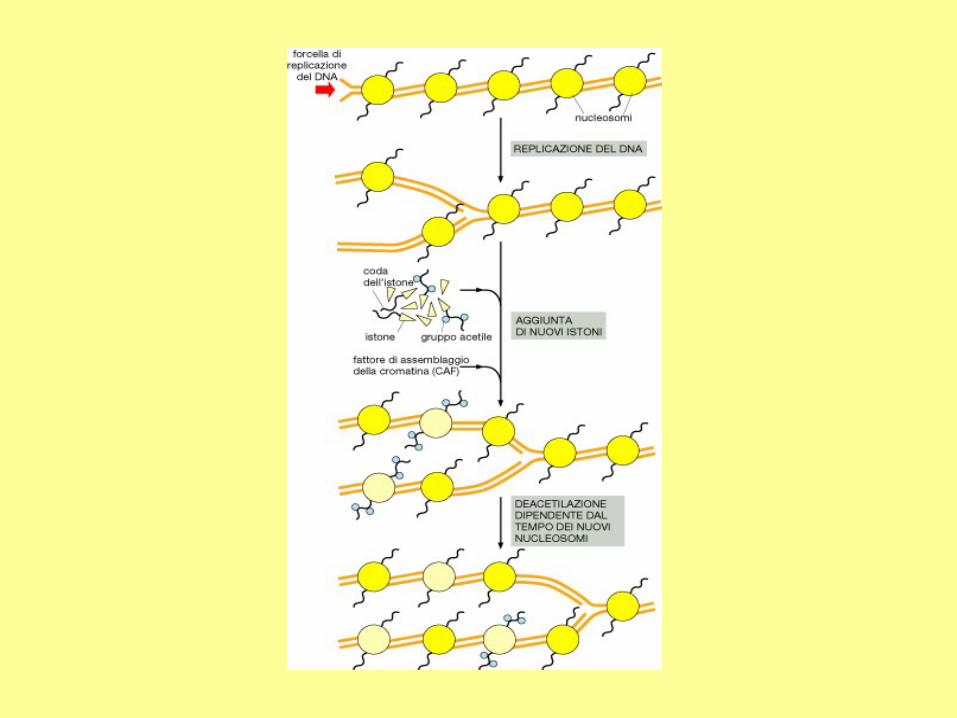
Meccanismo della
replicazione del DNA
eucariotico - 4
CAF-1: fattore di assemblaggio della
cromatina.

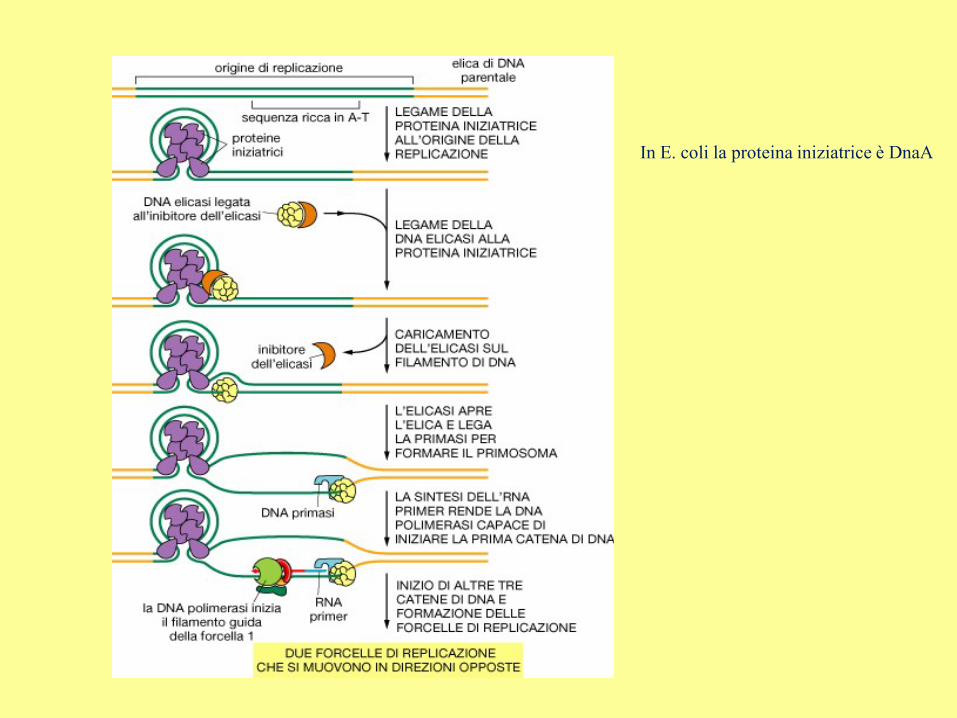
In E. coli la proteina iniziatrice è DnaA
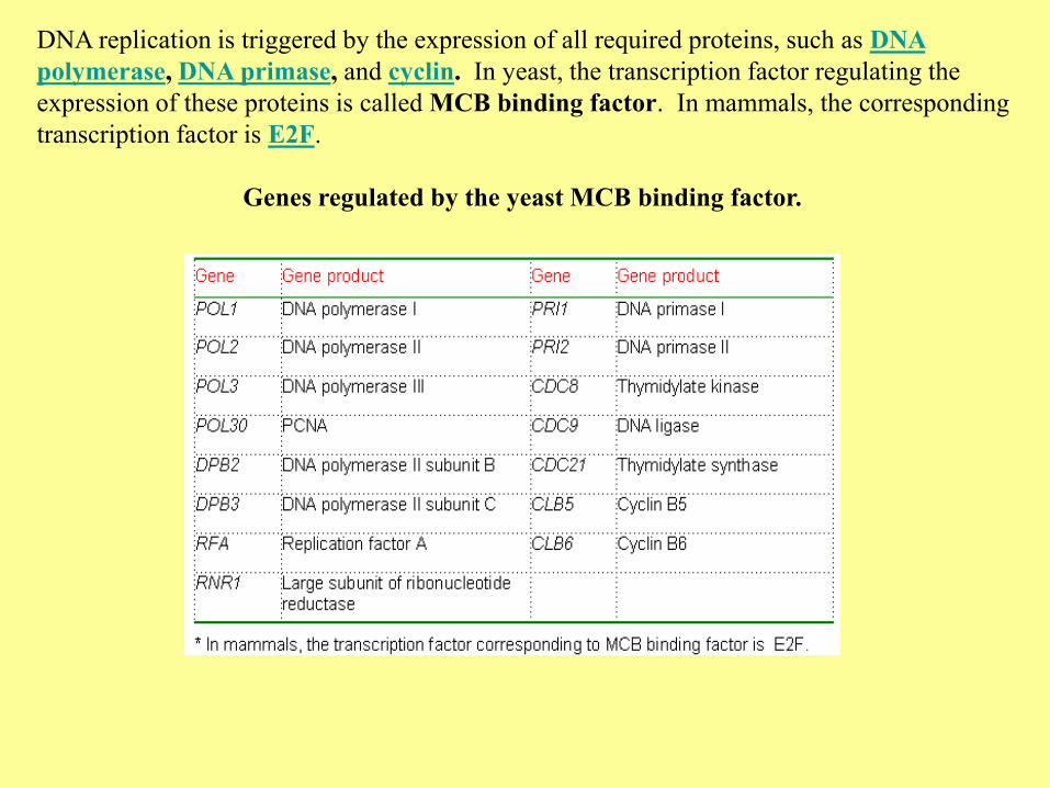
DNA replication is triggered by the expression of all required proteins, such as DNA
polymerase, DNA primase, and cyclin. In yeast, the transcription factor regulating the
expression of these proteins is called MCB binding factor. In mammals, the corresponding
transcription factor is E2F.
Genes regulated by the yeast MCB binding factor.
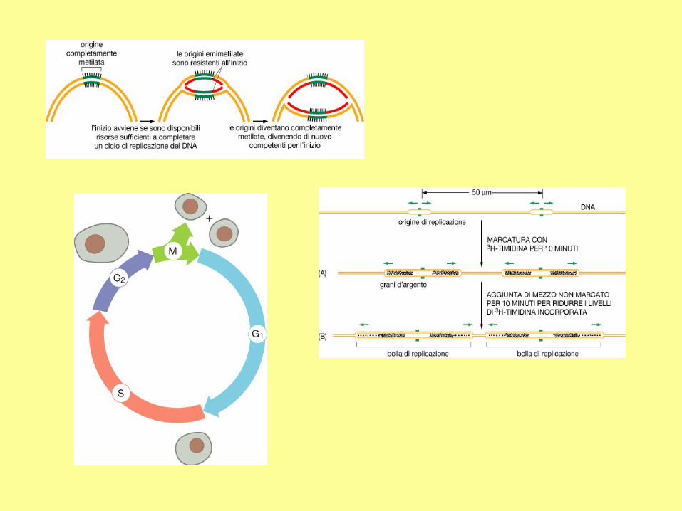
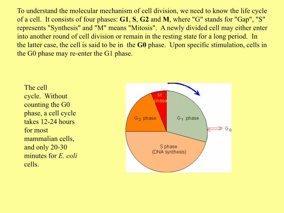
To understand the molecular mechanism of cell division, we need to know the life cycle
of a cell. It consists of four phases: G1, S, G2 and M, where "G" stands for "Gap", "S"
represents "Synthesis" and "M" means "Mitosis". A newly divided cell may either enter
into another round of cell division or remain in the resting state for a long period. In
the latter case, the cell is said to be in the G0 phase. Upon specific stimulation, cells in
the G0 phase may re-enter the G1 phase.
The cell
cycle. Without
counting the G0
phase, a cell cycle
takes 12-24 hours
for most
mammalian cells,
and only 20-30
minutes for E. coli
cells.
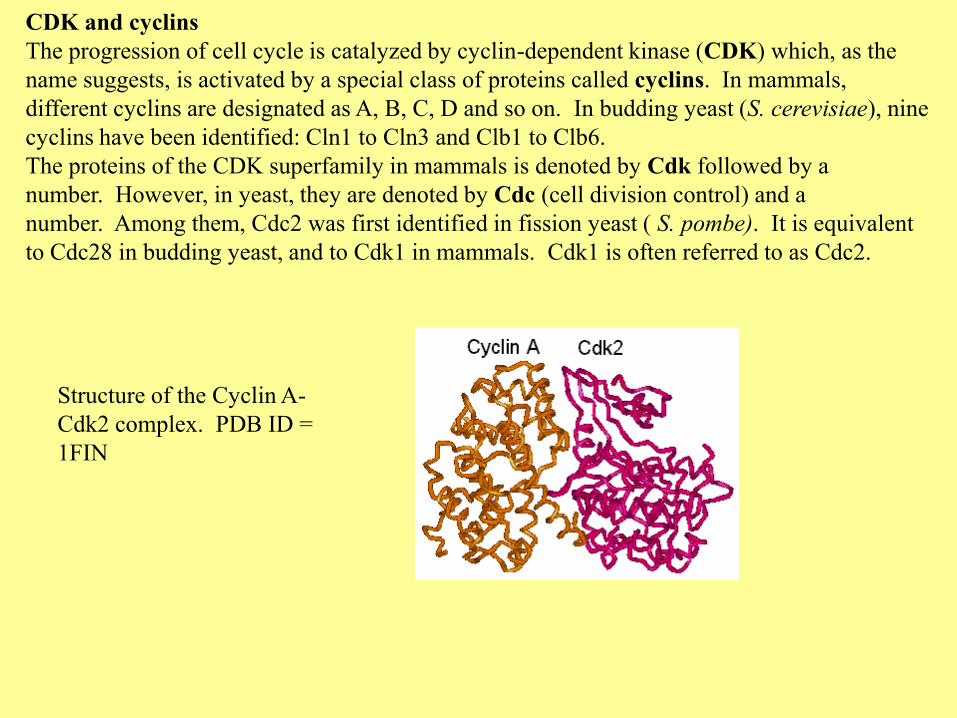
CDK and cyclins
The progression of cell cycle is catalyzed by cyclin-dependent kinase (CDK) which, as the
name suggests, is activated by a special class of proteins called cyclins. In mammals,
different cyclins are designated as A, B, C, D and so on. In budding yeast (S. cerevisiae), nine
cyclins have been identified: Cln1 to Cln3 and Clb1 to Clb6.
The proteins of the CDK superfamily in mammals is denoted by Cdk followed by a
number. However, in yeast, they are denoted by Cdc (cell division control) and a
number. Among them, Cdc2 was first identified in fission yeast ( S. pombe). It is equivalent
to Cdc28 in budding yeast, and to Cdk1 in mammals. Cdk1 is often referred to as Cdc2.
Structure of the Cyclin A-
Cdk2 complex. PDB ID =
1FIN
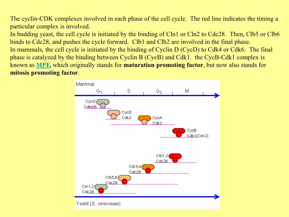
The cyclin-CDK complexes involved in each phase of the cell cycle. The red line indicates the timing a
particular complex is involved.
In budding yeast, the cell cycle is initiated by the binding of Cln1 or Cln2 to Cdc28. Then, Clb5 or Clb6
binds to Cdc28, and pushes the cycle forward. Clb1 and Clb2 are involved in the final phase.
In mammals, the cell cycle is initiated by the binding of Cyclin D (CycD) to Cdk4 or Cdk6. The final
phase is catalyzed by the binding between Cyclin B (CycB) and Cdk1. the CycB-Cdk1 complex is
known as MPF, which originally stands for maturation promoting factor, but now also stands for
mitosis promoting factor.
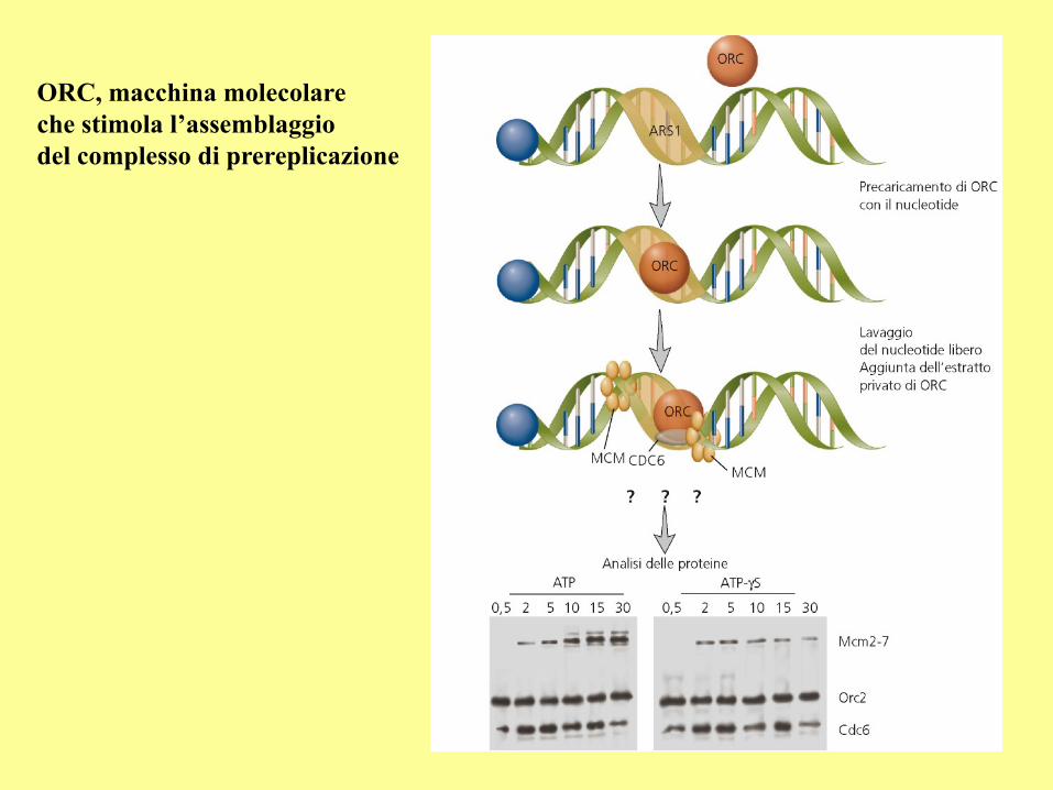
ORC, macchina molecolare
che stimola l’assemblaggio
del complesso di prereplicazione
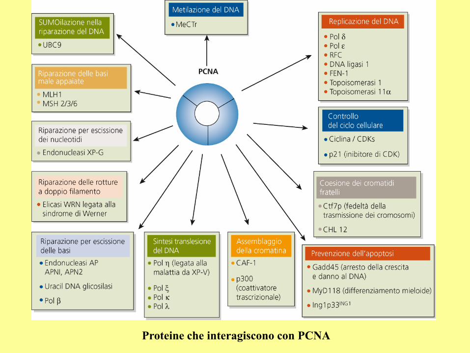
Proteine che interagiscono con PCNA
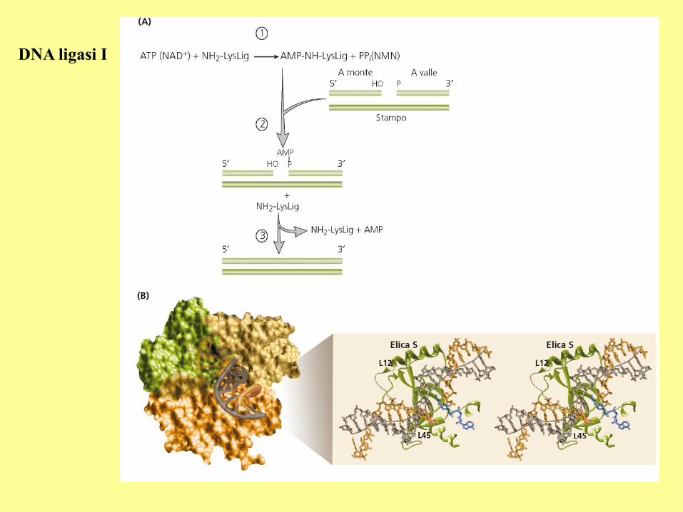
DNA ligasi I
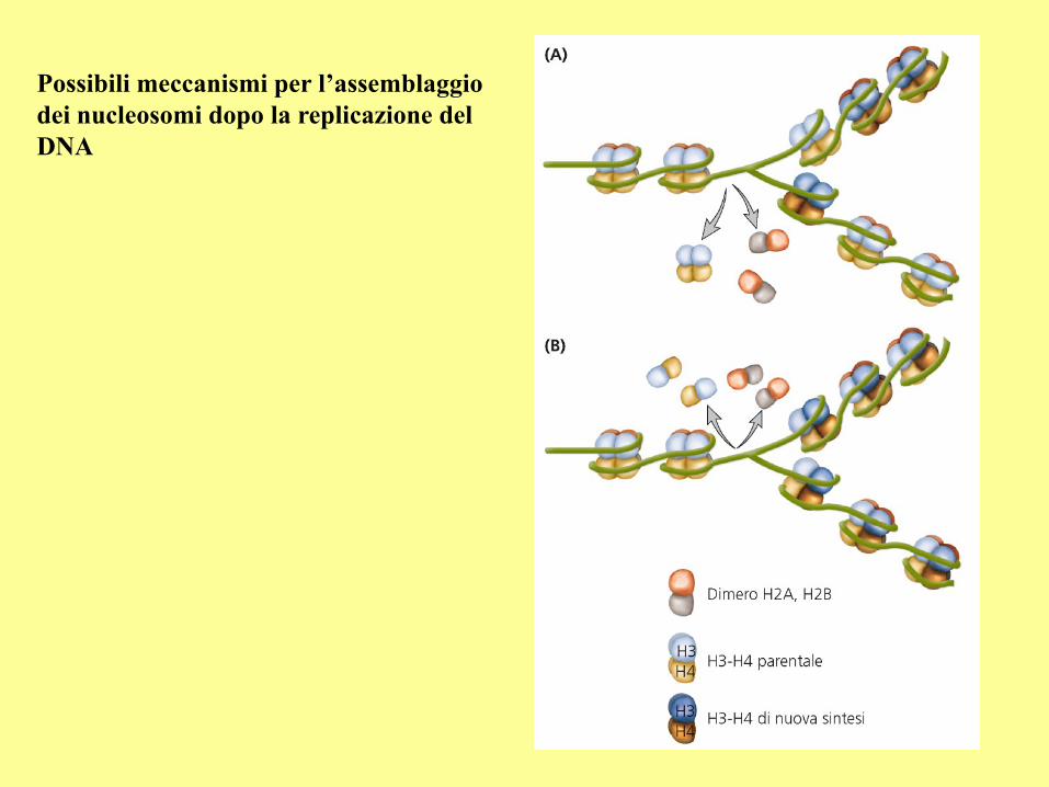
Possibili meccanismi per l’assemblaggio
dei nucleosomi dopo la replicazione del
DNA
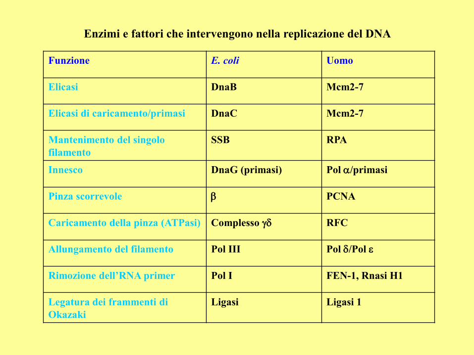
Enzimi e fattori che intervengono nella replicazione del DNA
Funzione E. coli Uomo
Elicasi DnaB Mcm2-7
Elicasi di caricamento/primasi DnaC Mcm2-7
Mantenimento del singolo
filamento
SSB RPA
Innesco DnaG (primasi) Pol /primasi
Pinza scorrevole b PCNA
Caricamento della pinza (ATPasi) Complesso RFC
Allungamento del filamento Pol III Pol /Pol
Rimozione dell’RNA primer Pol I FEN-1, Rnasi H1
Legatura dei frammenti di
Okazaki
Ligasi Ligasi 1
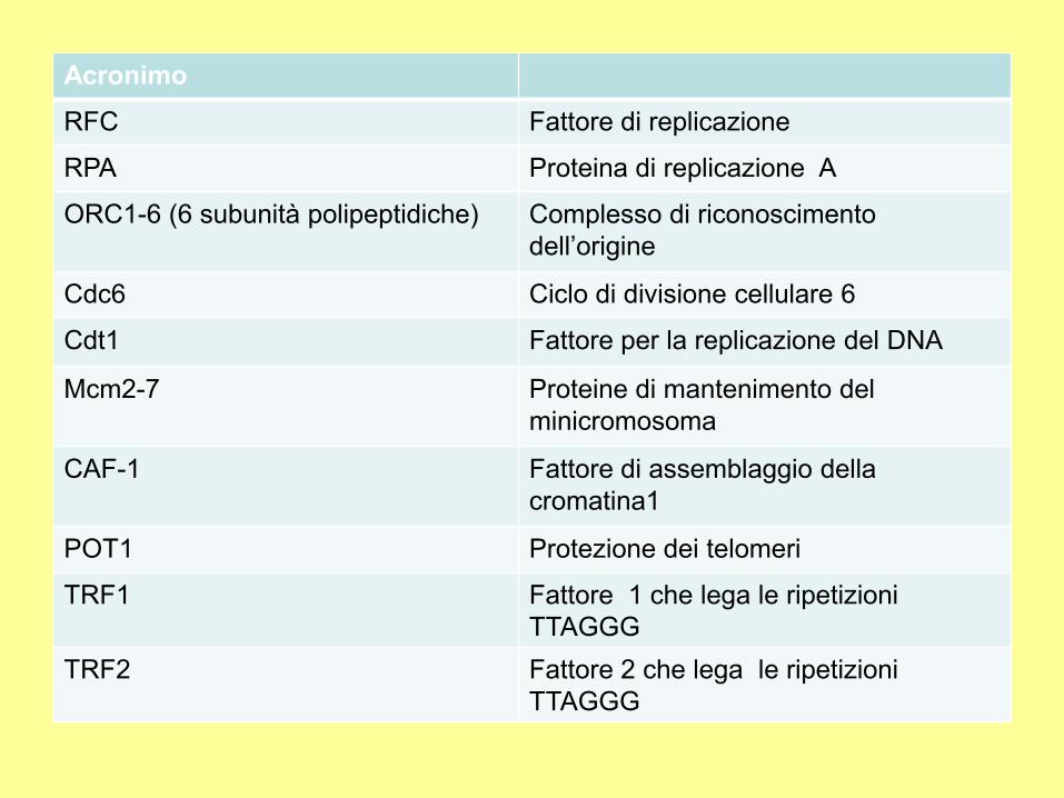
Acronimo
RFC Fattore di replicazione
RPA Proteina di replicazione A
ORC1-6 (6 subunità polipeptidiche) Complesso di riconoscimento
dell’origine
Cdc6 Ciclo di divisione cellulare 6
Cdt1 Fattore per la replicazione del DNA
Mcm2-7 Proteine di mantenimento del
minicromosoma
CAF-1 Fattore di assemblaggio della
cromatina1
POT1 Protezione dei telomeri
TRF1 Fattore 1 che lega le ripetizioni
TTAGGG
TRF2 Fattore 2 che lega le ripetizioni
TTAGGG
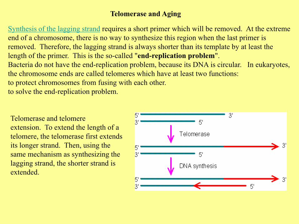
Telomerase and Aging
Synthesis of the lagging strand requires a short primer which will be removed. At the extreme
end of a chromosome, there is no way to synthesize this region when the last primer is
removed. Therefore, the lagging strand is always shorter than its template by at least the
length of the primer. This is the so-called "end-replication problem".
Bacteria do not have the end-replication problem, because its DNA is circular. In eukaryotes,
the chromosome ends are called telomeres which have at least two functions:
to protect chromosomes from fusing with each other.
to solve the end-replication problem.
Telomerase and telomere
extension. To extend the length of a
telomere, the telomerase first extends
its longer strand. Then, using the
same mechanism as synthesizing the
lagging strand, the shorter strand is
extended.
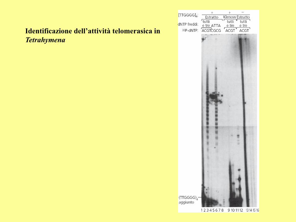
Identificazione dell’attività telomerasica in
Tetrahymena
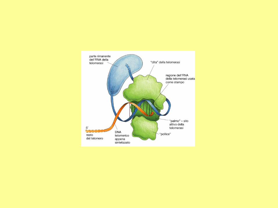
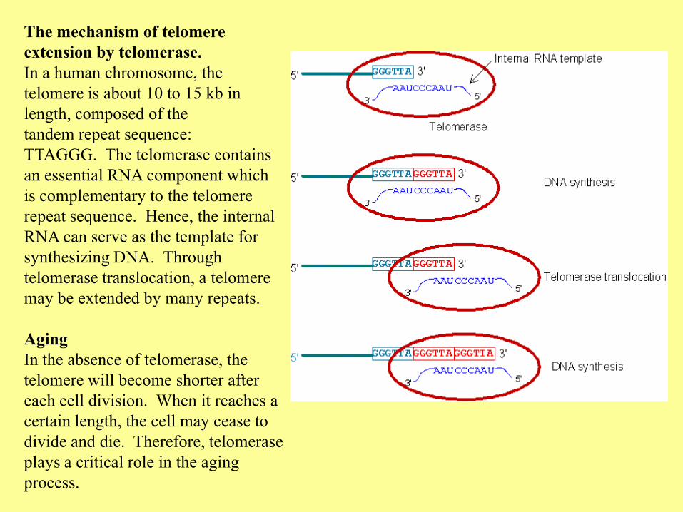
The mechanism of telomere
extension by telomerase.
In a human chromosome, the
telomere is about 10 to 15 kb in
length, composed of the
tandem repeat sequence:
TTAGGG. The telomerase contains
an essential RNA component which
is complementary to the telomere
repeat sequence. Hence, the internal
RNA can serve as the template for
synthesizing DNA. Through
telomerase translocation, a telomere
may be extended by many repeats.
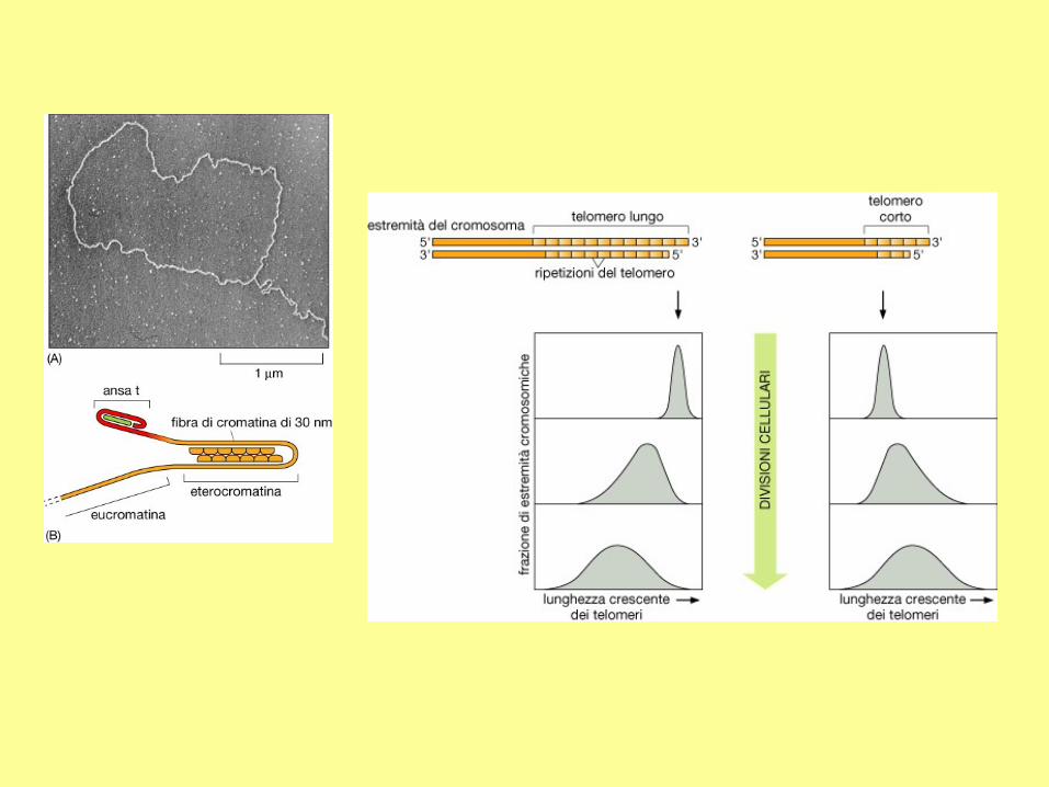
Aging
In the absence of telomerase, the
telomere will become shorter after
each cell division. When it reaches a
certain length, the cell may cease to
divide and die. Therefore, telomerase
plays a critical role in the aging
process.
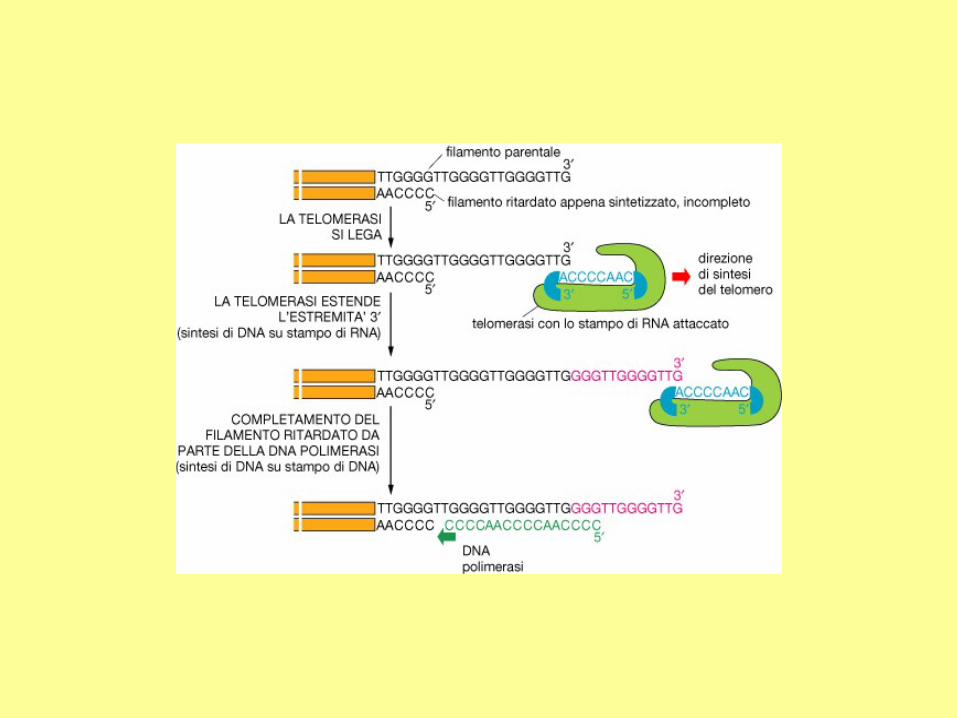

Sintesi del DNA telomerico da
parte della telomerasi umana
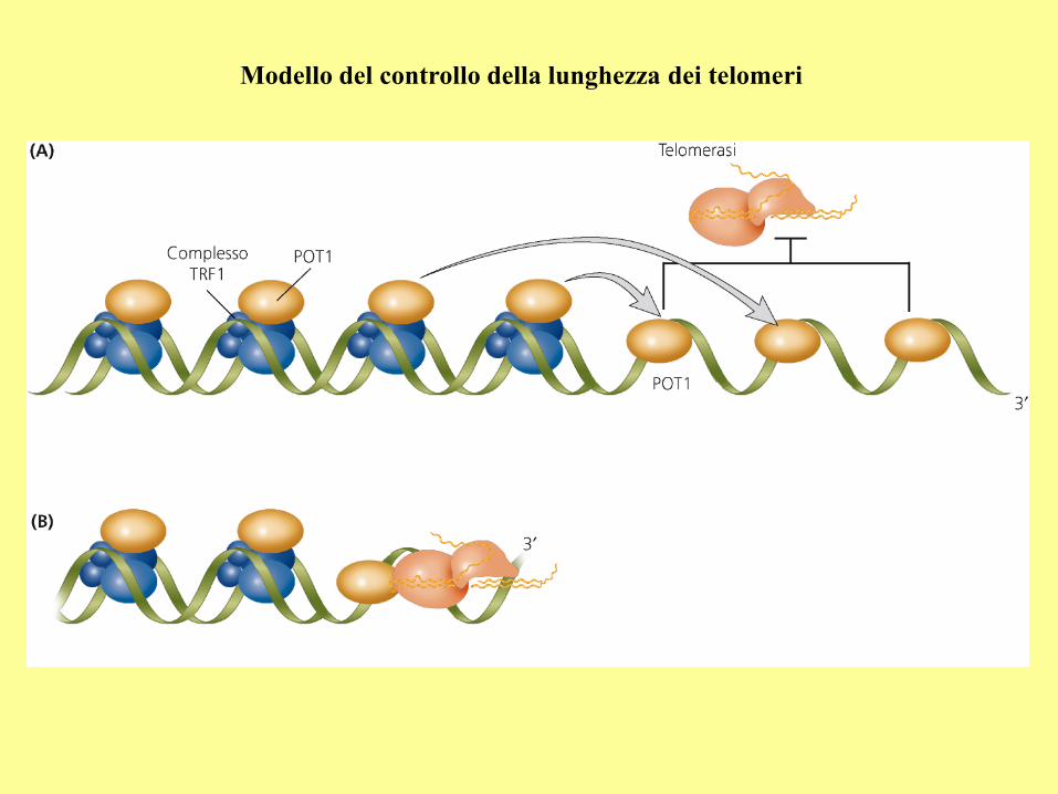
Modello del controllo della lunghezza dei telomeri
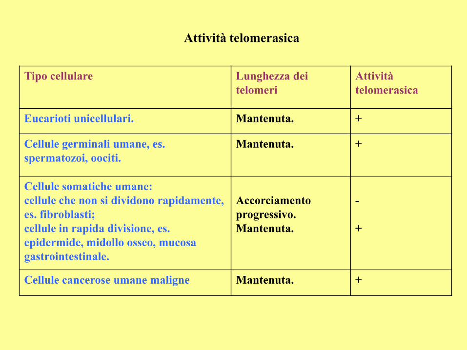
Attività telomerasica
Tipo cellulare Lunghezza dei
telomeri
Attività
telomerasica
Eucarioti unicellulari. Mantenuta. +
Cellule germinali umane, es.
spermatozoi, oociti.
Mantenuta. +
Cellule somatiche umane:
cellule che non si dividono rapidamente,
es. fibroblasti;
cellule in rapida divisione, es.
epidermide, midollo osseo, mucosa
gastrointestinale.
Accorciamento
progressivo.
Mantenuta.
-
+
Cellule cancerose umane maligne Mantenuta. +
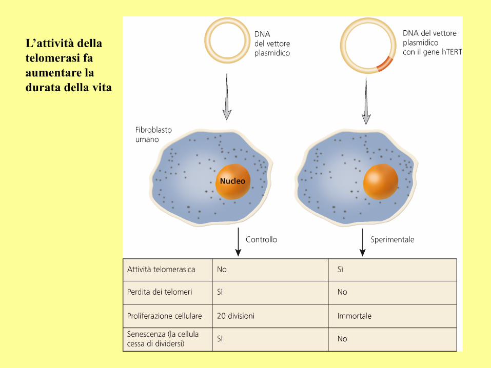
L’attività della
telomerasi fa
aumentare la
durata della vita
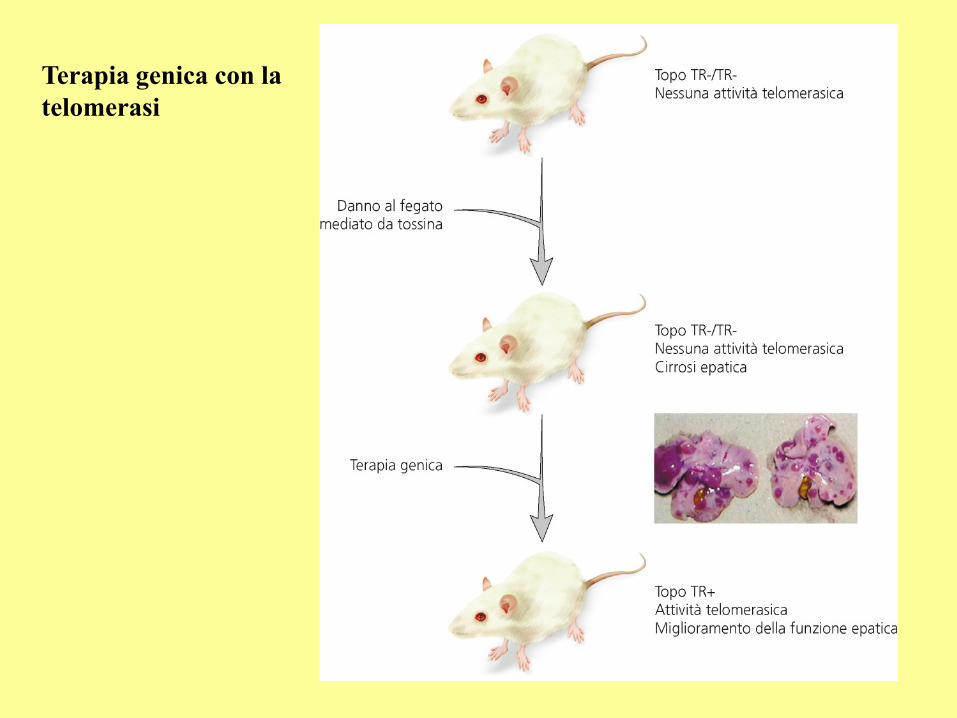
Terapia genica con la
telomerasi
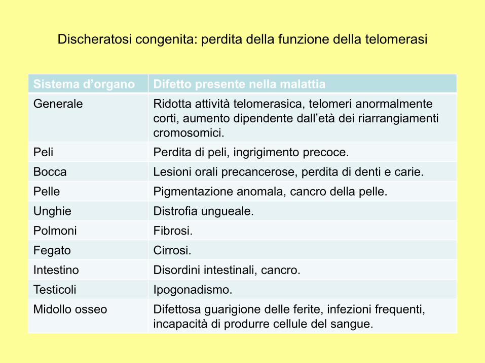
Sistema d’organo Difetto presente nella malattia
Generale Ridotta attività telomerasica, telomeri anormalmente
corti, aumento dipendente dall’età dei riarrangiamenti
cromosomici.
Peli Perdita di peli, ingrigimento precoce.
Bocca Lesioni orali precancerose, perdita di denti e carie.
Pelle Pigmentazione anomala, cancro della pelle.
Unghie Distrofia ungueale.
Polmoni Fibrosi.
Fegato Cirrosi.
Intestino Disordini intestinali, cancro.
Testicoli Ipogonadismo.
Midollo osseo Difettosa guarigione delle ferite, infezioni frequenti,
incapacità di produrre cellule del sangue.
Discheratosi congenita: perdita della funzione della telomerasi