Embed Size (px)
Citation preview
Full Paper
On-Off PVC Membrane Based Potentiometric Immunosensor forLabel-Free Detection of Alpha-Fetoprotein
Lu Zhou, Ruo Yuan,* Yaqin Chai
Chongqing Key Laboratory of Analytical Chemistry, College of Chemistry and Chemical Engineering, Southwest University,Chongqing, 400715, P. R. China*e-mail: [email protected]
Received: December 6, 2006Accepted: February 26, 2007
AbstractA poly(vinylchloride) (PVC) membrane based potentiometric immunosensor for the direct detection of alpha-fetoprotein (AFP) has been developed. First, Au colloid particle was chemisorbed upon amino groups of o-phenylenediamine, which were dissolved in plasticized PVC membrane. Then alpha-fetoprotein antibody (anti-AFP)was immobilized upon the surface of the Au colloid particle to prepare a potentiometric AFP immunosensor. The Aucolloid particle modified PVC membrane was characterized by digital photo and transmission electron microscope(TEM). The immunosensor exhibited fast potentiometric response (�4 min) and showed specific response to AFP inthe range of 4.9 to 158.5 ng/mL with a correlation coefficient of 0.9971 and a detection limit of 1.6 ng/mL. The factorsinfluencing the performance of the immunosensor were also studied in detail. Moreover, the proposed method iseconomical and efficient as well as potentially attractive for clinical immunoassays.
Keywords: Potentiometric immunosensor, Alpha-fetoprotein, o-Phenylenediamine, Poly(vinylchloride), Au colloidparticle
DOI: 10.1002/elan.200603836
1. Introduction
Alpha-fetoprotein (AFP) is a glycoprotein molecule [1],which is serological marker for hepatocellular carcinoma(HCC) [2]. The detection of AFP is important for earlydiagnosis and monitoring of tumor aggressiveness, treat-ment responsiveness, recurrence and survival. Total AFPhas the sensitivity of 60% and specificity of 90% for thedetection ofHCC.Conventionalmethods for determinationof AFP such as immunoradiometric assay (IRMA), which isextremely sensitive, have obvious disadvantages of beingexpensive and of short shelf life (I-labeled antibody); andenzyme-linked immunosorbent assay (ELISA) is less sensi-tive and flexible in terms of design and application. Thus, it isimportant to develop simple, sensitive and cheapmethod forthe determination of AFP.Janata [3] proposed a method to construct a label free
potentiometric immunosensor for the determination ofYeast mannan, in which the instrument used was reductionin size, cost and analysis time. However, the later studiesabout ISE-based immunosensor [4], enzyme modified ISE-based immunosensor [5] and ion-sensitive field effecttransistor (ISFET) [6] showed that the potentiometricanalysis exhibited some drawbacks, such as unsatisfiedsensitivity and specificity. And several reviews [7, 8] alsopointedout this kindof immunosensor suffered fromseveralproblems: low signal-to-noise, poor reliability, poor selec-tivity, and expensive fabrication. These disadvantages
obstruct the clinical application of the potentiometricimmunosensor.Recently, some novel potentiometric immunosensors
such as human immunoglobulin G (IgG) immunosensor[9] based on covalent immobilization of anti-IgG on silverelectrode, hepatitis B surface antigen (HBsAg) immuno-sensor [10] based on polypyrrole modified electrode andherbicide simazine immunosensor [11] based on peroxidaselabel and competitive immunoreaction were reported. Inour research laboratory, we employed self-assembly tech-nique, nanotechnology and electrochemical polymerizationtechnique to construct potentiometric immunosensors [12 –15] for the detectionof hepatitisB surface antigen (HBsAg),cancer embryo antigen (CEA) and diphtheria antigen(Diph). However, the fabrication of these immunosensorsis complex and the cost is expensive. So a simple and cheappotentiometric immunosensor with acceptable assay capa-bility is necessary.In this present work, we proposed a disposable poly(vinyl
chloride) (PVC) membrane based immunosensor for thedirect determination of AFP, which provide a new platform(PVCmembrane modified with Au colloid particle) insteadof immunosensor base on gold electrode, platinum elec-trode and carbon electrode. Firstly, PVC membrane con-taining o-phenylenediamine was prepared. And then Aucolloid particle was adsorbed on the surface of the PVCmembrane via amino groups of o-phenylenediamine. Fi-nally, anti-AFP was adsorbed on the surface of Au colloid
1131
Electroanalysis 19, 2007, No. 11, 1131 – 1138 H 2007 WILEY-VCH Verlag GmbH&Co. KGaA, Weinheim

particle topreparepotentiometricAFP immunosensor.Whenthe immunosensor was immersed in the solution containingAFP, the interaction of AFP and anti-AFP leads the decreaseof the relative density of electron charges on membrane andinduces potentiometric shift directly to proportional to theconcentration of AFP. The results of the experiment demon-strated that the sensor has high selectivity and sensitivity.Andthis kind of immunosensor is easy to prepare with low cost. Inaddition, the factors influencing the performance of theimmunosensor have been studied in detail.
2. Experimental
2.1. Chemicals
Alpha-fetoprotein antibody (anti-AFP), alpha-fetoprotein(AFP) and AFP ELISA kits were purchased from BiocellCompany (Zhengzhou,China).Human serum sampleswereobtained from the affiliated hospital of Southwest Univer-sity. (Caution! The sample is infectant andmust be carefullyhandled under protective equipment.) Poly(vinyl chloride)(PVC), di(n-octyl)phthalate (DOP), tetrahydrofuran(THF), and o-phenylenediamine were purchased fromShanghai Chemical Reagent Co. (Shanghai, China). Cancerembryo antigen (CEA) and hepatitis B surface antigen(HBsAg) were purchased fromZhengzhouBiocell Institute(Zhengzhou, China). Human immunoglobulin G (IgG) wasprovided by Chengdu Institute of Biological Products(China). Bovine serum albumin (BSA) was obtained fromSigma (USA). All other chemicals used were of analyticalgrade and were used as received. TheAFPwas stored in thefrozen state, and its standard solution were prepared dailywith doubly distilled water as in use. Phosphate bufferedsaline solutions (PBS, 0.1 M) at various pHvalueswere usedin the experiments. Doubly distilled water was usedthroughout the experiments. All of the electrochemicalexperiments in this paper were performed at normaltemperature (25� 0.5 8C).
2.2. Apparatus
All potentiometric measurements were carried out with adigital ion analyzer (Model PHS-3C, Dazhong InstrumentsFactory, Shanghai, China). CS501-SP super digital constanttemperature instrument (Chongqing Sida Instrument Fac-tory, China) was used for the incubation of the immuno-sensor. All experiments were carried out with a conven-tional two-electrode system with the immunosensor asworking electrode and saturated calomel electrode (SCE) asreference electrode. pH measurements were made with apH meter (MP 230, Mettler-Toledo, Switzerland). Photo-graphofPVCmembranewas carried outwith digital camera(FinePix S7000, Fujifilm, Japan). The size of Au colloidparticle and the Au colloid particle modified PVC mem-brane were estimated by transmission electron microscopy(TEM) (H600, Hitachi Instrument, Japan).
2.3. Preparation of Au Colloid Particle
All glassware used in the following procedures was cleanedin a bath of freshly prepared K2Cr2O7-H2SO4, rinsedthoroughly with double distilled water, and dried prior touse. The Au colloid particle was prepared according to theliterature [16]. The solution color was claret. Solution wasstored in a refrigerator with a dark colored glass bottlebefore use. The mean size of the prepared Au colloidparticle was about 16 nm, which was confirmed by TEM(Fig. 1. A).
2.4. Preparation of Immunosensor
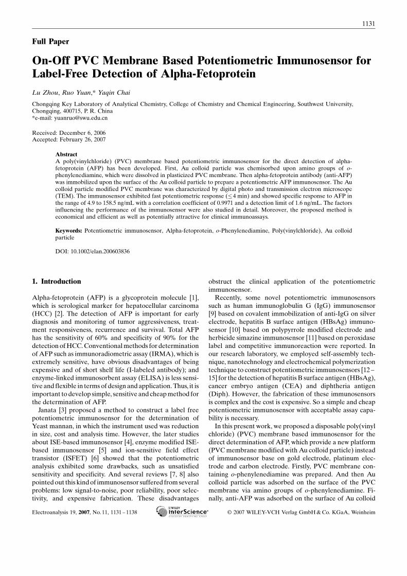
0.1 g PVC, 0.01 g o-phenylenediamine, and 0.2215 g di-n-octyl-phthalate were mixed and thoroughly dissolved inTHF (2 mL). The mixture was transferred into a glass dishwith a diameter of 2 cm. The solvent was slowly evaporateduntil a concentrated mixture was obtained. The PVCmembrane was activated by immerging into 0.05 M HClfor 1 h and rinsed with double distilled water to removephysically adsorbed HCl. The obtained membrane wassoaked in the solution of the Au colloid particle for 4 h toadsorb Au colloid particle. At last, anti-AFP was adsorbedon the surface of Au colloid particle. After rinsed by doubledistilled water, the roundness dense membrane was adhib-ited onto the PVC tube.And then 0.1 MKClwas added intothe PVC tube. The siliver-siliver chloride inner electrodewas inserted in the PVC tube. The prepared modifiedimmunosensor was incubated in 0.25 wt% BSA for 60 minat 37 8C to block unspecific sites and stored at 4 8Cwhen notin use. The schematic diagram of the PVC membrane andthe structure of the immunosensor were shown in Figure 2.
2.5. Measurement Procedure
The potentiometric response for anti-AFP andAFP derivesfrom potentiometer.When the electrode was immersed in a5 mL PBS (pH 6.5), the steady-state potentiometric value(E1 vs. SCE) was recorded, and after an appropriate volumeof the standard positive or negative serum was added intothe PBS, the steady-state potentiometric value (E2 vs. SCE)was obtained. Either antibodies or antigens in aqueoussolution have a net electrical charge polarity, which is tocorrelate the isoelectric points of the species and the ioniccomposition of the solution. As the isoelectric point of anti-AFP andAFP is less than 6.0, they are negatively charged inPBS (pH 6.5). Therefore, the potentiometric responsesdecrease after the antibody – antigen reaction. The poten-tiometric response of the immunosensor towards AFP isevaluated following the equation:
E¼E2�E1
All electrochemical measurements were done in an un-stirred 10 mL electrochemical cell at 25� 0.5 8C.
1132 L. Zhou et al.
Electroanalysis 19, 2007, No. 11, 1131 – 1138 www.electroanalysis.wiley-vch.de H 2007 WILEY-VCH Verlag GmbH&Co. KGaA, Weinheim

ELISA is based on a solid phase sandwich immunoassay.AFP standards or specimens were processed by AFPELISA kits with standard method.
2.6. Statistical Analysis
Datawere expressed asmean� standard error ofmean. Therepeatability, selectivity, and the difference between thedetection result of ELISA and that of the proposed
immunosensor were analyzed with a c2 test. P< 0.05 wasconsidered to be statistically significant.
3. Results and Discussion
3.1. Characterization of PVC Membrane Modified withNanogold
The digital photograph of PVCmembranemodified with orwithout Au colloid particle was shown in Figure 1 C. As it
Fig. 1. A) The TEM images of Au colloid particles. The size of the Au colloid particles is about 16 nm. B) TEM image of the Au colloidparticle modified PVC membrane. C) The photo of PVC membrane modified with Au colloid particle (a) and without Au colloidparticle (b).
Fig. 2. Diagram of the immunosensor: A) The stepwise fabrication process of PVC membrane. B) Structure of the immunosensor.
1133Label-Free Detection of Alpha-Fetoprotein
Electroanalysis 19, 2007, No. 11, 1131 – 1138 www.electroanalysis.wiley-vch.de H 2007 WILEY-VCH Verlag GmbH&Co. KGaA, Weinheim

can be seen, the claret PVCmembranewas shown in picturea.Comparedwith picture b,we found that the redAu colloidparticle was adsorbed onto the surface of the PVCmembrane. Meanwhile, the PVC membrane without o-phenylenediamine was treated with Au colloid particle. Thecolor of the membrane obtained did not change. Moreover,TEM investigation showed that no Au colloid particle wasadsorbed on the PVC membrane without o-phenylenedi-amine. According to the results of the experiment, weconcluded that the adsorption of Au colloidal particle onPVC membrane containing o-phenylenediamine occurredvia amino groups of o-phenylenediamine.The morphology of the Au colloid particle modified PVC
membrane was imaged by TEM as is shown in Figure 1. B.Structural characterizations showed that the Au colloidparticle was chemisorbed upon amino groups of o-phenyl-enediamine dissolved in plasticized PVC membrane. TheTEM morphology of Au colloid particle modified PVCmembrane agree with the photograph.
3.2. Optimization of Experimental Conditions
3.2.1. Components of Membrane
The membrane was constructed by combining variouscomponents. The nature and amount of o-phenylenedi-amine, nature of plasticizer, plasticizer/PVC ratio, areknown to significantly influence the sensitivity and linearrange [17]. In order to obtain the best performance of theimmunosensor, varying amount of the DOP and o-phenyl-enediamine were dissolved with PVC in 2 mLTHF (Weightof the whole mixture and PVC were constant). The generalprocedure to prepare the PVC membrane was to mixthoroughly 100 mg PVC (30.2%), 221.5 mg of plasticizerDOP (66.8%), 10 mg of o-phenylenediamine (3.0%) in aglass dish of 2 cm diameter. The mixture was completelydissolved in 2 mL THF. The resulting mixture was evapo-rated slowly until a concentratedmixture was obtained. Theresulting membrane was assembled and tested. The perfor-mance was listed in Table 1. The ratio of membraneingredients was optimized and the ingredients of electrodeB was chosen for the fabrication of the membrane in thisexperiment.
3.2.2. Effect of Temperature
The effect of temperature on the immunoreaction wasexamined at the range from 10 to 50 8C (Fig. 3.A). Accord-ing to the study of the temperature influence, we found thatan increase of temperature had a favorable effect on theimmunoreaction using constant concentration of AFP. It iswell known that an optimal temperature of immunoreactionwould be 37 8C. However, a high temperature mightdecrease the activity of antigen or antibody, leading to thedeterioration of response signals and lifetime. Consideringlifetime and sensitivity, we choose normal temperature(25 8C) as measure temperature in our study.
3.2.3. pH of the Working Buffer
The effect of pH on the sensor response lies in two mainaspects: one is the effect on the affinity (i.e. degree offirmness of immunocomplex, an important affect factor ofreaction balance); and the other one is the effect on theactivity of the antibody and antigen. Unsuitable pH maycause protein denaturalization. The effect of pH on theimmunosensors was studied in the pH range 5.5 – 8.0 inpresence of 56 ng/mL AFP solution (Fig. 3. B). Theoptimum pH was 6.5. Thus, pH 6.5 was chosen for the test.
3.2.4. Response Time of Incubation
The influence of the immunochemical incubation time (i.e.when the antigen – antibody reaction occurs) on responsesignals was studied. When the analyte antigens reach theantibodies modified on the electrode surface, it would takesome time for the contacting species to form immunocom-plex. The immunosensor was incubated for 1 – 10 min in the0.1 M PBS pH 6.5 containing AFP 20 ng/mL. When theinteraction time was over 4 min, the potentiometric re-sponses of the immunosensor kept constant. This indicatedthat the interaction reached themaximumvalue after 4 min.Thus, 4 min was selected as the reaction time for all thesubsequent assays.
3.3. Detection of AFP
When the antigen was bound on the antibody immobilizedon the electrode, there would be an additional layer, which
Table 1. The parameters of PVC membrane and the analytical parameters of the immunosensor.
Electrode wt% of various components Slope [a] (mV) Linear range (ng/mL) Detection limit [a] (ng/mL)
PVC DOP o-Phenylenediamine
A 30.2 68.3 1.5 7.5� 0.02 16.7 – 25 6.0� 0.07B 30.2 66.8 3.0 48.7� 0.06 4.9 – 158.5 1.6� 0.01C 30.2 66.2 3.6 42.3� 0.03 6.0 – 136.0 2.0� 0.05D 30.2 65.3 4.5 41.5� 0.01 10.0 – 126.5 3.5� 0.01E 30.2 63.8 6.0 42.2� 0.03 7.2 – 128.5 2.4� 0.02
[a] Mean� SD of three measurements
1134 L. Zhou et al.
Electroanalysis 19, 2007, No. 11, 1131 – 1138 www.electroanalysis.wiley-vch.de H 2007 WILEY-VCH Verlag GmbH&Co. KGaA, Weinheim

changed the potentiometric response. The calibration curveof the potentiometric immunosensor was obtained usingAFP standard solution under optimal experimental con-dition.A good linear relationshipwas observed between thechange of potential and the logarithm ofAFP concentrationand the linear range was from 4.9 to 158.5 ng/mL with adetection limit of 1.6 ng/mL (estimated to be 3 times the
standard deviation of zero-dose response). The linearregression equation is DE¼� 49.2 log CAFPþ 36.7 with acorrelation coefficient of 0.9971.In addition, the potentiometric response of the potentio-
metric immunosensor during the steps of a complete assay(association and dissociation of the complex) were shown inTable 2. The potentiometric response of the immunoreac-tion is much larger than the potentiometric shifts of theadsorbing of nanoparticle, anti-AFP and BSA.
3.4. Reproducibility and Stability
The reproducibility of the potentiometric response of thesame electrode was examined at an AFP concentration of20 ng/mL, and the relative standard deviations (RSD) were2.8% (n¼ 5)(After completing each assay, the immunosen-sor was immersed in 5 mol/L urea for 10 min to dissociatethe antigen – antibody complex.). the transient curves of thereproducibility of the measurements was shown Figure 4.The fabrication reproducibility was estimated from theresponse for 20 ng/mLAFP at eight different electrodes, theRSD were calculated to be 8.1%.The storage stability of the electrode was tested over a 40-
day period, when the AFP electrode was stored in therefrigerator at 4 8C and measured intermittently (every 3days), no apparent change of potentiometric response wasfound over 36 days, a RSD of 3.7% was acquired. And itretained 97.2% of its initial potentiometric value over 40days.
3.5. Selectivity Against Interferences
Four interfering proteins were used to evaluate the selec-tivity of the electrode. We chose tumor marker (CEA)(400 ng/mL), common human antigen (IgG) (1200 ng/mL),familiar pathogen (HBsAg) (1320 ng/mL) and a usualsubstance for blocking non-special site (BSA) (100 ng/mL)in this study. The potential shift was calculated from thepotential shift of the immunosensor in assay solutioncontaining 20 ng/mL AFP and different concentrationpossible interfering substance (respectively and together)compared with the potential signals of the immunosensor inthe same assay solution containing only 20 ng/mLAFP. Theresult (DE< 1.5 mV) shows that these potential interfering
Fig. 3. A) Potentiometric response of the antibody modifiedelectrode in 0.1 M PBS containing 40 ng/mL AFP at the differenttemperature. B) Potentiometric response of the antibody modifiedelectrode in 0.1 M PBS containing 56 ng/mL AFP at the differentpH.
Table 2. Potential-time response of the immunosensor during the steps of complete assay.
PVC membrane Nano Au particle[b] (16 nm)
Anti-AFP [c] BSA [d](0.25 wt%)
AFP [e](20 ng/mL)
Urea [f](5 mol/L)
Potential shift [a] (mV) �9.1� 0.17 �26.9� 0.21 15.6� 0.3 �8.8� 0.03 �39.9� 0.11 �12.8� 0.10
[a] Mean� SD of three measurements[b] After 7 minutes the potential shift is stabile[c] After 6 minutes the potential shift is stabile and the nano-Au particle was adsorbed for 4 hours[d] After 8 minutes the potential shift is stabile and the immunosensor was incubated for 1 hours.[e] After 2 minutes the potential shift is stabile[f] After 30 seconds the potential shift is stabile.
1135Label-Free Detection of Alpha-Fetoprotein
Electroanalysis 19, 2007, No. 11, 1131 – 1138 www.electroanalysis.wiley-vch.de H 2007 WILEY-VCH Verlag GmbH&Co. KGaA, Weinheim

proteins do not interfere with the potentiometric responseof the sensor. The effect of possible components in humanblood was also studied. 20 ng/mL AFP and blank buffersolution were analyzed by being added with interferingspecies. It showed upper limit concentration of Kþ, Naþ,Ca2þ, glucose and l-asparagive did not interfere. Theperformance was listed in Table 3.
3.6. Regeneration of the Immunosensor
Regeneration of immunosensor is crucial in immunoassayand there are two options. One is the repeated use of thesame immunosensor, the other is the immunosensor isdisposable. Regeneration of the immunosensor can signifi-cantly reduce cost of analysis compared to disposableimmunosensors. However, the immobilized immunore-agent or immunocomplex could also suffer from the func-tional damage of immunoactivity because the regenerationreagent provide drastic conditions (e.g., in alkaline or acidicsolutions or with chaotropic agents) [18, 19]. In this experi-ment, the disposable immunosensor was employed becauseof its low cost and easy fabrication.
3.7. Preliminary Applications
In order to evaluate the analytical applicability of the newtechnique, 26 human serum specimens, whichwere obtainedfrom our university hospital, were examined by the devel-oped immunoassay and the ELISA method. The compar-ison of results is shown in Figure 5 A. It described thecorrelation between the results obtained by the PVCmembrane based immunosensor and by theELISAmethod.The regression equation obtained is y¼ 0.9982xþ 0.3923with a correlation coefficient of 0.9995 (P> 0.05).Comparison of AFP measured by ELISA and the
developed immunoassay with Bland –Altman method [20]was shown in Figure 5 B. For the data s¼0.71. The standarderror of the mean difference is thus 0.14, For the 95%confidence interval we have 25 degrees of freedom andt¼1.96. Hence the 95% confidence interval for the bias is1.4� (1.96� 0.14) to 1.4þ (1.96� 0.14), giving 1.33 to1.67 ng/mL. The standard error of the limit Mean differ-ence-2s is 0.24 ng/mL. The 95% confidence interval for thelower limit of agreement is �0.02� (1.96� 0.24) to�0.02þ (1.96� 0.24), giving �0.45 to �0.49 ng/mL. Forthe upper limit of agreement the 95% confidence interval is2.35 to 3.29 ng/mL. It shows that there is no significantlydifference between the results given by two methods. Thus,the developed immunoassay may provide a feasible alter-
Fig. 4. Transient curves of the same immunosensor for 5 measurements.
Table 3. Potentiometric response of the immunosensor towards different ions.
Potential response [a] (mV) Naþ (3.3 g/L) Kþ (196 mg/L) Ca2þ (104 mg/L) l-Asparaginase (55 mg/L) Glucose (1.1 g/L)
Before AFP treatment 2.3� 0.01 3.0� 0.15 3.5� 0.21 3.6� 0.06 5.0� 0.10After AFP treatment �0.16� 0.010 0.5� 0.026 0.9� 0.016 1.4� 0.02 1.7� 0.015
[a] Mean� SD of three measurements
1136 L. Zhou et al.
Electroanalysis 19, 2007, No. 11, 1131 – 1138 www.electroanalysis.wiley-vch.de H 2007 WILEY-VCH Verlag GmbH&Co. KGaA, Weinheim

native tool for determining AFP in human serum in clinicallaboratory.
4. Conclusions
In this paper, we have described a new PVC membranebased potentiometric immunosensor for the determinationof AFP. Compared with traditional immunosensor based ongold, silver and carbon electrode, this kind of immunosensorprovide a novel transducer and sensitive membrane whichdo not need base electrode. The potentiometric PVCmembrane immunosensor is very easy to prepare and is oflow cost for fabrication. The result of experiment demon-strated that the sensor has high selectivity and sensitivity,
fast response time and low detect limit. The immunosensorcould be applied to determination of concentration of AFPin human serum. This method also may be used toimmobilize some other biomolecules to develop biosensorsand bioreactors.
5. Acknowledgements
Financial support of this work was provided by the NaturalScience Foundation of China (20675064), the ChineseEducation Ministry Foundation for excellent young teach-ers, the Natural Science Foundation of Chongqing city(CSTC-2004BB4149, 2005BB4100) and High TechnologyProject Foundation of Southwest University (XSGX02).
Fig. 5. A) Comparison of the titer results of determining samples between the ELISA method and the PVC membrane basedimmunosensor. B) Bland –Altman plot for the AFP ELISA monitor and AFP the developed immunoassay monitor.
1137Label-Free Detection of Alpha-Fetoprotein
Electroanalysis 19, 2007, No. 11, 1131 – 1138 www.electroanalysis.wiley-vch.de H 2007 WILEY-VCH Verlag GmbH&Co. KGaA, Weinheim

6. References
[1] D. Bader, A. Riskin, O. Vafsi, A. Tamir, B. Peskin, N. Israel,R. Merksamer, H. Dar, M. David, Clin. Chim. Acta. 2004,349, 15.
[2] A. Cucchetti, M. Vivarelli, F. Piscaglia, B. Nardo, R.Montalti, G. L. Grazi, M. Ravaioli, G. L. Barba, A. Cavallari,L. Bolondi, A. D. Pinna, J. Hepatology 2005, 43, 310.
[3] J. Janata, J. Am. Chem. Soc. 1975, 97, 2914.[4] D. Brown, M. Meyerhoff, Biosens. Bioelectron. 1991, 6, 615.[5] J. Caras, J. Janata, Anal. Chem. 1980, 52, 1935.[6] P. P. Christopher, Clin. Chem. 1998, 44, 2071.[7] L. M. Claire, J. N. David, P. P. Christoppher, Clin. Chem.
1996, 42, 193.[8] M. Mehrab, M. Abdi, Anal. Sci. 2004, 20, 1113.[9] C. L. Feng, Y. H. Xu, L. M. Song, Sens. Actuators B. 2000, 66,
190.[10] D. Purvis, O. Leonardova, D. Farmakovsky, V. Cherkasov,
Biosens. Bioelectron. 2003, 18, 1385.
[11] M. F. Yulaev, R. A. Sitdikov, N. M. Dmitrieva, E. V. Yazyni-na, A. V. Zherdev, B. B. Dzantiev, Sens. Actuators B 2001, 75,129.
[12] R. Yuan, D. P. Tang, Y. Q. Chai, X. Zhong, Y. Liu, J. Y. Dai,Langmuir 2004, 17, 7240.
[13] L. Y. Zhang, R. Yuan, X. Q. Huang, Y. Q. Chai, S. R. Cao,Electrochem. Commun. 2004, 6, 1222.
[14] D. P. Tang, R. Yuan, Y. Q. Chai, X. Zhong, Y. Liu, J. Y. Dai,Clin. Biochem. 2006, 39, 309.
[15] Q. Zhu, R. Yuan, Y. Q. Chai, N. Wang, Y. Zhuo, Y. Zhang,X. L. Li, Electrochim. Acta. 2006, 51, 3763.
[16] G. Frens, Nat. Phys. Sci. (London) 1973, 241, 20.[17] T. Katsu, K. Ido, K. Takaishi, H. Yokosu, Sens. Actuators B.
2002, 87, 331.[18] F. V. Bright, T. A. Betts, K. S. Litwiler, Anal. Chem. 1990, 62,
1065.[19] J. L. Boitieux, R. Groshemy, D. Thomas, Anal. Chim. Acta.
1987, 197, 229.[20] J. M. Bland, D. G. Altman, Lancet 1986, 327, 307.
1138 L. Zhou et al.
Electroanalysis 19, 2007, No. 11, 1131 – 1138 www.electroanalysis.wiley-vch.de H 2007 WILEY-VCH Verlag GmbH&Co. KGaA, Weinheim