Embed Size (px)
Citation preview

European Polymer Journal 53 (2014) 37–49
Contents lists available at ScienceDirect
European Polymer Journal
journal homepage: www.elsevier .com/locate /europol j
On row-structures in sheared polypropyleneand a propylene–ethylene copolymer
0014-3057/$ - see front matter � 2014 Elsevier Ltd. All rights reserved.http://dx.doi.org/10.1016/j.eurpolymj.2014.01.010
⇑ Corresponding author. Address: Electron Microscopy Laboratory, J.J.Thomson Building, University of Reading, Whiteknights, Reading RG66AF, UK. Tel.: +44 (0)118 378 4616; fax: +44 (0) 118 378 4606.
E-mail address: [email protected] (R.H. Olley).
Robert H. Olley a,⇑, Geoffrey R. Mitchell a,b, Yasmin Moghaddam a
a Department of Physics, University of Reading, Whiteknights, Reading RG6 6AF, UKb Centre for Rapid and Sustainable Product Development, Polytechnic Institute of Leiria, Rua de Portugal – Zona Industrial, 2430-028 Marinha Grande, Portugal
a r t i c l e i n f o
Article history:Received 5 August 2013Received in revised form 20 December 2013Accepted 14 January 2014Available online 20 January 2014
Keywords:PolypropyleneCopolymerNucleationSEM (scanning electron microscopy)WAXS (wide-angle X-ray scattering)SAXS (small-angle X-ray scattering)
a b s t r a c t
The crystallisation of polymers such as polyethylene or polypropylene from the melt isgreatly influenced by the flow-thermal history prior to the crystallisation. We explorethe influence of the chemical configuration of polypropylene based chains on the formationof row structures on crystallisation. We use a combination of in situ time resolved small-angle X-ray scattering, ex situ wide angle X-ray scattering with optical and scanning elec-tron microscopy to show that row nuclei are formed in random copolymers of propylenewith a limited amount of ethylene subjected to modest shear flow fields. We contrastobservations performed using two homopolymers of isotactic polypropylene and one ran-dom copolymer of propylene and ethylene. We propose that it is not strictly necessary toargue that the row nuclei are already crystalline nor to invoke epitaxial crystallisation asthe mechanism for the nucleation of lamellae, as similar structures can be formed on car-bon nanotubes and fibrils of dibenzylidene sorbitol. The combination of microscopy andscattering provides a powerful approach to investigating these phenomena, especially ascompared to either technique used in isolation.
� 2014 Elsevier Ltd. All rights reserved.
1. Introduction
The crystallisation of polymers from stressed melts is ofwidespread interest both from a science viewpoint and froma technological processing perspective. The subject ofcrystallisation from stressed melts has been reviewed byKumaraswamy [1] and more recently by Janeschitz-Kriegl[2], and one frequent occurrence in the time sequence ofcrystallisation from a stressed melt is the formation of rownuclei which give rise to structures known as ‘‘shish-kebabs’’. In polypropylene such oriented structures may alsogrow on heterogeneous linear nuclei such as fibres by trans-crystallisation [3], for example in the case of in situ grownnanofibrils [4] or through the addition of nanoparticulatessuch as carbon nanotubes [5], whereas the presence of
materials such as graphene nanosheets [6] may enhancethe development of homogeneous row nuclei.
Polypropylene is the polymer best known for its remark-able propensity for forming row structures, but they are alsofound in polyethylene [7–9] as well as isotactic polystyrene[10], poly(phenylene sulfide) [11] and polylactide [12].
In polyethylene [7–9] and in polypropylene [13] the nu-clei may be formed from a particular fraction of the poly-mer melt and in such cases it is well established that thegeneration of row nuclei is very dependent on the presenceof a high molecular weight fraction or ‘tail’ in the polymers.Various models have been proposed, some depending onthe coil-stretch transition [14,15], though alternative mod-els exist such as [16]. Seki et al. have argued that rownuclei occur when there is sufficient of this HMW fractionto allow long-chain overlap [17]. Regardless of the mecha-nism of formation of the shish, it is understood that thekebab formation is a case of lamellar crystallisation on pre-crystallised fibres (shish) that serve as nucleation sites forthe kebabs [18].

38 R.H. Olley et al. / European Polymer Journal 53 (2014) 37–49
Isotactic polypropylene, like most semi-crystallinepolymers, crystallises from a quiescent melt in the formof spherulites but it readily assumes other geometries,crystallising from homogeneous row nuclei to form rowstructures or cylindrites, or from heterogeneous surfacesto give transcrystalline layers [19]. Row structures or cylin-drites are easily generated by first shearing the melt, andcan form in a range of number densities, ranging from iso-lated ones whose development can be followed easily un-der the optical microscope to very dense parallel arrayswhere the individual structures cannot be discerned opti-cally. In polypropylene, a high tacticity index has beenfound favourable to the development of shish-kebabs[17]. This raises questions as to the polymer attributes re-quired to form row nuclei. Some work has been performedon blends of medium molecular weight branched polyeth-ylene with a minority component of high or ultra-highmolecular weight linear polyethylene (ethylene homopoly-mer). There the high molecular weight linear componenthas been observed to template the growth of the branchedpolyethylene [8,15]. However, we are not aware of anywork where a copolymer by itself has been shown to formrow structures, whether this be a majority ethylene or amajority propylene copolymer. This work compares thebehaviour of two propylene homopolymers with that of apropylene–ethylene copolymer to explore the effects ofthe copolymerisation on the formation of row nuclei andsubsequent templated lamellar growth.
2. Materials
Three polypropylene based materials were employed inthis study, all supplied by Borealis, Finland and the charac-teristics are detailed in Table 1. As a ‘reference’ material weused a high-crystallinity homopolymer polypropyleneHCPP with high tacticity. We compared this referencematerial with a second homo-polymer (ZNPP) preparedusing Ziegler–Natta technology and which exhibits a lowertacticity and weight average molecular weight. The thirdmaterial is a random copolymer (RACO) based on propyl-ene with 5.1 wt% ethylene as comonomer. In terms of themelt flow index HCPP and RACO are very similar whilstthe ZNPP system is substantially higher.
3. Experimental methods
3.1. Small-angle X-ray scattering
Time-resolving small-angle X-ray scattering (SAXS)measurements were made using the intense flux available
Table 1Properties of the polypropylene based polymers used in this work.
HCPP ZNPP RACO
Comonomer ethene wt% 0 0 5.1MFR 2.16 kg; g/10 min 6.5 19 7.9Tacticity; mmmm tetrads 97.8 93.0 –Tm�C 165.8 164.3 137.2Mn Daltons 65,650 75,100 77,650Mw Daltons 310,000 208,000 266,500Mw/Mn 4.7 2.8 3.4
on the fixed wavelength beamline 16.1 at the DaresburySynchrotron Radiation source. Scattering data were col-lected using the Rapid Area Detector System with a time-cycle of 10 s. The scattering geometry was calibrated usinga wet collagen fibre. The in situ small-angle X-ray scatter-ing measurements employed a parallel plate shear cellequipped with mica windows, specially designed to facili-tate in situ time-resolving X-ray scattering measurements[20]. A schematic of the shear cell and scattering geometryis shown in Fig. 1. The rotating plate consists of a stainlesssteel tri-spoke arrangement covered with a thin mica disc(0.03 mm in thickness), which allows the incident beam topass unhindered for 85% of each revolution. An upstreamsynchronised rotating mask, fabricated from lead, mini-mised the background intensity for the period of a revolu-tion when a spoke would have intercepted the incidentbeam. The fixed plate consists of a stainless steel plate witha single chamfered hole covered with a thin mica disc. Theshear cell was equipped with electrical heating and cryo-genic gas cooling system which allowed the sample tem-perature to be controlled and varied at set rates. Thegeometry of the cell was such that the incident beamwas normal to the flow direction and parallel to the veloc-ity gradient.
In this work, each sample was subjected to a definedtemperature/shear flow cycle and an example is shownin Fig. 2. The design of the shear cell enabled X-ray scatter-ing data to be obtained throughout the cycle. The specificcycle shown here contained the following stages: (a) heat-ing from room temperature to 192.4 �C at a rate of 20�/min; (b) held at 192.4 �C for 5 min; (c) cooled to room tem-perature at a rate of 10 �C/min. When the sample reached aselected temperature, Ts, a shear rate of 20 s�1 was appliedfor 25 s, giving a total of 500 shear units. During the shear-ing period, the sample continued to cool and over theshearing period the temperature dropped by 4.2 �C. Aftercessation of shear, cooling continued to below room tem-perature, after which the sample was removed from theshear cell. The 10 s data accumulation cycle used through-out the heating cycle gave a temperature variation of�1.7 �C for each recorded scattering pattern. Samples foruse in the shear cell were pre-moulded into discs 1 mmin thickness and 19 mm in diameter in the melt using asimple metal mould and hydraulic press.
3.2. Ex situ wide-angle X-ray scattering
Wide-angle X-ray scattering (WAXS) data were ob-tained for processed samples at room temperature usingtwo different approaches. The first used a flat plate Riga-ku/MSC Saturn 92 CCD camera with a Rigaku FR-D rotatinganode generator.
In the second method, intensity data were recorded as afunction of a at a fixed value of |Q| range using a symmet-rical transmission diffractometer equipped with a graphitemonochromator and pinhole collimation and a Cu Ka X-raysource where a is the angle between the symmetry axis ofthe sample and the scattering vector Q and |Q| = 4psinh/k,where k is the incident wavelength and 2h is the scatteringangle. This instrument allowed a map of the undistortedreciprocal space in contrast to the limitations of a flat

Fig. 1. Schematic of the shear flow cell and the scattering geometry used in this work.
Fig. 2. A plot of the recorded temperature against time for the samples of HCCP sheared at 138.5�. The time of the shear pulse is also shown.
R.H. Olley et al. / European Polymer Journal 53 (2014) 37–49 39
detector fixed geometry system. Samples were mountedon the diffractometer so that the WAXS data were col-lected from a volume at the same radius of the disc asthe SAXS data. As a consequence both sets of data relateto the same shear rate during the shear phase.
3.3. Microscopy
For microscopy, processed samples were etched for 1 husing the permanganic based mixture consisting of a 1%(w/v) solution of potassium permanganate in an acid mix-ture of 10 vols concentrated sulfuric acid, 4 vols ortho-phosphoric acid (85%) and 1 vol water. The etchingprocedures described in [21] were employed. Specimenswere sputter coated with gold for examination by the fol-lowing two microscopic techniques.
The first technique was based on a reflecting micro-scope setup for differential interference contrast, orNomarski microscopy [22]. This enables a view of largeareas which can only be observed piecemeal under theSEM. Moreover, it gives strong contrast for small variationsin surface height, of the order of hundreds of nanometres,helped by the gold coating. The second technique involvedthe use of scanning electron microscopy operating in highvacuum mode at 20 kV using a Cambridge Stereoscan 360SEM with a tilt angle of 35�.
4. Results
4.1. Small-angle X-ray scattering
In this section, SAXS patterns are displayed for the threematerials studied, namely HCPP (Figs. 3 and 4), RACO(Figs. 5 and 6), and ZNPP (Figs. 7 and 8). The first figurein each pair contains a sequence of patterns as they devel-op from a single shearing temperature, the second figureshows the pattern from the end product of a range ofshearing temperatures. Starting with HCPP, Fig. 3 showsa selection of SAXS patterns obtained during a particulartime-resolved experiment of the type shown in Fig. 2.These are displayed in conjunction with the calculatedinvariant X and its derivative dX/dT.
In each experiment, there are two important parame-ters, Ts the temperature at which shearing was initiatedand T the temperature of the measurement. In this partic-ular experiment Ts = 138.5 �C. The sequence of data shownin Fig. 3 starts at a point after the application of the shearflow pulse. As the temperature drops at a constant rate of10 �C min�1, intense scattering appears as spots or lobesalong the meridional axis which is parallel to what wasthe flow axis. The intensity of these lobes initially growsbut after reaching a maximum at �107 �C, it falls continu-ously although it is always above the value shown in the

Fig. 3. A plot of the invariant, X and the derivative dX/dT against the experimental temperature T, derived from SAXS data for a sample of HCPP subjectedto the temperature–shear–time profile described in the text with Ts = 138.5 �C.
Fig. 4. SAXS patterns of HCCP1 recorded at T = 89.5 �C which have beensubjected to a similar shear–temperature–time profile shown in Fig. 2with the shear flow temperature Ts indicated in the figure.
40 R.H. Olley et al. / European Polymer Journal 53 (2014) 37–49
melt. This type of anisotropic pattern is typical of a semi-crystalline polymer in which the scattering arises fromstacks of crystalline lamellae separated by regions of amor-phous material. The position of the lobe in terms of |Q| pro-vides information of the so-called long period, which is thethickness of the lamellar plus the amorphous layer. Thelocalisation of the scattering along the meridional axis re-veals that these lamellae are preferentially arranged nor-mal to the flow axis. The arrangement arises as aconsequence of the formation during the flow phase ofrow nuclei which are aligned parallel to the flow axis andtemplate or direct the growth of the chain folded lamellaeperpendicular to the row nuclei. At commencement ofshearing, at T � 137 �C (first pattern), the pattern has not
changed noticeably from the original melt at 190 �C. Thesmall spikes around the beam stop arise from the shear cellitself and do not relate to the sample. A significant amountof oriented scattering has become visible at T � 130 �C(second pattern) in the form of a vertical streak with a hor-izontal FWHM of�0.002 �1. On reaching a temperature of118.9 �C, the oriented growth has become considerablymore pronounced, and there is some lateral growth interms of broadening the central streak. As the specimencools still further, a ring arising from isotropic, spheruliticmaterial develops, reaching maximum intensity near102.6 �C.
We extracted a number of quantitative parametersfrom each of the small-angle X-ray scattering patterns.We have monitored the fraction of crystals by calculatingthe invariant X given by [23]
X ¼Z p=2
0
Z Qmax
Q¼0jQ j2IðjQ j;aÞ sin adQda
which is directly related to the average of the square of theelectron density differences; if the density difference be-tween the crystals and amorphous is constant this is pro-portional to the volume fraction of crystals. We havecalculated the derivative dX/dT by fitting quartic functionsto successive sets of points on the function X(T) as shownin Fig. 3 and taking the analytical derivative of thatfunction.
Fig. 3 also shows the value of the invariant calculatedfrom each SAXS pattern obtained in this experiment as afunction of the experimental temperature T. Shearingstarted at 148.3 �C during continuous cooling from192.4 �C at 10 K/min and the shear flow ceased at144.1 �C. The invariant is constant until the temperaturereaches�136 �C. Under these conditions the crystallisationproceeds to develop a high level of crystal orientation. Themain features in the invariant are a rise to maximum as thespecimen crystallises, then as the specimen cools further,

Fig. 5. A plot of the invariant, X and the derivative dX/dT against the experimental temperature T, derived from SAXS data for a sample of RACO subjectedto the temperature–shear–time profile described in the text with Ts = 123.8 �C.
Fig. 6. SAXS patterns of RACO recorded at T = 89.5 �C which have beensubjected to a similar shear–temperature–time profile shown in Fig. 2with the shear flow temperature Ts indicated in the figure.
R.H. Olley et al. / European Polymer Journal 53 (2014) 37–49 41
all the scattering intensities decrease in a roughly expo-nential way, a phenomenon already reported for polyethyl-ene [24]. We attribute this to the fact that the difference indensities of crystalline and amorphous PP is reducing de-spite the increasing level of crystallinity, so giving a lowerscattering intensity [25]. We have also plotted the deriva-tive dX/dT which is a measure of the rate of crystal growth.The derivative of the invariant shows two peaks, at 122.5�and 110.5 �C (Table 2), these peaks are attributed to themaximum growth rate of the primary radial lamellae andof secondary, mostly crosshatched, lamellae.
Fig. 4 shows the small angle X-ray scattering patternsfor samples prepared with different shear temperaturesTs. These patterns were obtained after cooling to a giventemperature, in the case here 89.5 �C where all specimens
will have effectively crystallised fully. The advantage ofusing the patterns taken at this temperature is that the dif-ference between densities of the crystalline and amor-phous components is greater than at room temperatureand hence the contrast is higher making observations morestraightforward. At first sight, the specimens from thehighest shear temperature appears to be isotropic, whilefor Ts = 153.2 �C there is a very slight azimuthal variationin the intensity. As the shear temperature decreases we ob-serve a pair of lobes in the scattering pattern on the verti-cal axis, which we attribute to arrays of parallel lamellaegrowing from row structures. The mounting of the shearcell is such that any row nuclei generated should be verti-cally oriented. Lamellae growing from these would havetheir plane horizontal, but the stack of lamellae would beone on top of another along a vertical axis, giving rise tovertically oriented scattering. These lobes become increas-ingly pronounced as the shearing temperature is reducedto 138.5 �C in approximately 5� steps.
The increase in orientation with lower shear tempera-ture is as expected, since the row nuclei formed duringshearing would be more stable at the lower temperatures,and also have less time to melt out before reaching thetemperature of crystallisation onset. However, the patternfrom lowest shearing temperature 128.7 �C is again lesspronounced, and reasons for this will be discussed accord-ing to evidence from microscopy.
For the specimen prepared with Ts = 158.1 �C, thepattern takes the form of an isotropic ring which doesnot reveal any preferred orientation of the lamellarcrystals. Nevertheless, SAXS patterns recorded duringcooling for this specimen did display some orientation. AtT = 110.7 �C the pattern contained both isotropic andanisotropic scattering features which arise from differentnucleation processes, and at that temperature they ap-peared equally weighted. At 102.3 �C, the non-orientedgrowth had greatly increased and come to completelydominate the pattern. This suggests that growth from
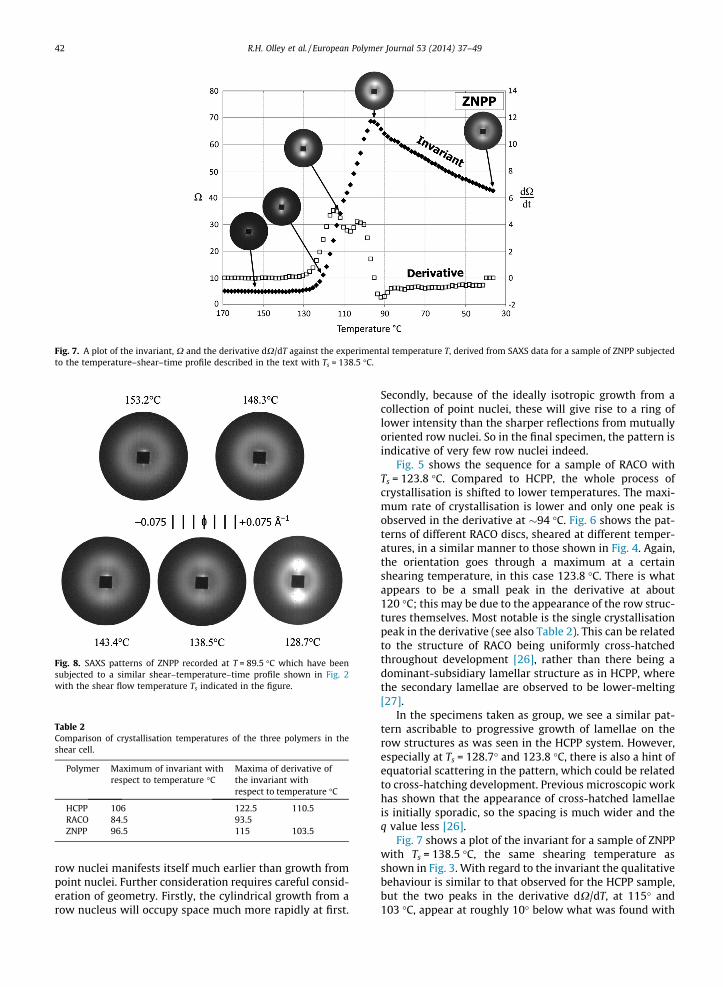
Fig. 7. A plot of the invariant, X and the derivative dX/dT against the experimental temperature T, derived from SAXS data for a sample of ZNPP subjectedto the temperature–shear–time profile described in the text with Ts = 138.5 �C.
Fig. 8. SAXS patterns of ZNPP recorded at T = 89.5 �C which have beensubjected to a similar shear–temperature–time profile shown in Fig. 2with the shear flow temperature Ts indicated in the figure.
Table 2Comparison of crystallisation temperatures of the three polymers in theshear cell.
Polymer Maximum of invariant withrespect to temperature �C
Maxima of derivative ofthe invariant withrespect to temperature �C
HCPP 106 122.5 110.5RACO 84.5 93.5ZNPP 96.5 115 103.5
42 R.H. Olley et al. / European Polymer Journal 53 (2014) 37–49
row nuclei manifests itself much earlier than growth frompoint nuclei. Further consideration requires careful consid-eration of geometry. Firstly, the cylindrical growth from arow nucleus will occupy space much more rapidly at first.
Secondly, because of the ideally isotropic growth from acollection of point nuclei, these will give rise to a ring oflower intensity than the sharper reflections from mutuallyoriented row nuclei. So in the final specimen, the pattern isindicative of very few row nuclei indeed.
Fig. 5 shows the sequence for a sample of RACO withTs = 123.8 �C. Compared to HCPP, the whole process ofcrystallisation is shifted to lower temperatures. The maxi-mum rate of crystallisation is lower and only one peak isobserved in the derivative at �94 �C. Fig. 6 shows the pat-terns of different RACO discs, sheared at different temper-atures, in a similar manner to those shown in Fig. 4. Again,the orientation goes through a maximum at a certainshearing temperature, in this case 123.8 �C. There is whatappears to be a small peak in the derivative at about120 �C; this may be due to the appearance of the row struc-tures themselves. Most notable is the single crystallisationpeak in the derivative (see also Table 2). This can be relatedto the structure of RACO being uniformly cross-hatchedthroughout development [26], rather than there being adominant-subsidiary lamellar structure as in HCPP, wherethe secondary lamellae are observed to be lower-melting[27].
In the specimens taken as group, we see a similar pat-tern ascribable to progressive growth of lamellae on therow structures as was seen in the HCPP system. However,especially at Ts = 128.7� and 123.8 �C, there is also a hint ofequatorial scattering in the pattern, which could be relatedto cross-hatching development. Previous microscopic workhas shown that the appearance of cross-hatched lamellaeis initially sporadic, so the spacing is much wider and theq value less [26].
Fig. 7 shows a plot of the invariant for a sample of ZNPPwith Ts = 138.5 �C, the same shearing temperature asshown in Fig. 3. With regard to the invariant the qualitativebehaviour is similar to that observed for the HCPP sample,but the two peaks in the derivative dX/dT, at 115� and103 �C, appear at roughly 10� below what was found with

Fig. 9. WAXS patterns recorded for (a) sample of compacted iPP fibreswith the fibre axis vertical on the page (b) sample of HCCP1 prepared withTs = 138.5 �C with the flow axis vertical.
R.H. Olley et al. / European Polymer Journal 53 (2014) 37–49 43
HCCP; the maximum in the invariant X also occurs about10� lower (Table 2). Fig. 8 shows the SAXS patterns re-corded for ZNPP discs with different values of Ts; these ap-pear to correspond most closely to the HCPP sample withTs roughly 10� higher, and somewhat less oriented.
Looking at all three materials, as crystallisation pro-ceeds through cooling, the SAXS pattern moves to a largerQ value as the density of lamellae forming on the rownuclei increasing with time. At the highest temperaturesof shearing, the SAXS patterns at T = 89.5 �C show anisotropic distribution of the scattering. In other words,the shearing does not result in the formation of row nuclei,or else these row nuclei are either redissolved or becomerandomly aligned.
4.2. Ex situ wide-angle X-ray
We have used ex situ wide-angle X-ray scattering toevaluate the distribution of crystal orientation, or moreprecisely the distribution of the orientation of crystalplanes in the final room temperature samples. The crystalstructure of the three polymers is dominated by thea-form, and the reflections due to this are chosen to assessthe level of preferred orientation of the crystal planes.Microscopic observation (below) does reveal the presenceof some b-form, but it is not present in sufficient propor-tion to contribute significantly to the diffraction patterns.As a reference to the a-form, Fig. 9a shows the WAXS pat-tern for a compaction made of highly aligned fibres of iPP[28]. This shows the classic fibre diffraction pattern foriPP with the fibres mounted in the vertical direction. Theoriginal fibres would show very high orientation indeed,and only equatorial 110 reflections. However, in the com-paction process a small amount of material has meltedand recrystallised in the crosshatched form, so extra 110arcs at roughly 80�, almost meridional, are seen, but atmuch lower intensity than is seen for spherulitic PP. The040 reflection relates to the common b-axis, and so onearc is common to both the main and crosshatched lamel-lae. Fig. 9b shows the WAXS pattern for the HCPP specimensheared at 138.5 �C, mounted so that what was the flowaxis is vertical and the volume of material examined wasthe same as in the earlier SAXS experiment. It is immedi-ately clear that the orientation distribution of the crystalplanes is broader than in the fibre pattern as is evidencedby the azimuthal arcing of the reflections. The meridional110 reflections are much stronger relative to the equatorialin the sheared disc. These reflections are widely known toarise from the cross-hatched component of iPP spheruliticmorphology [29]. In the fibre specimen these very weak in-deed, but here they are much stronger. Unfortunately theflat detector technique does not allow the meridionalreflections to be observed completely without tilting thespecimen.
Fig. 10 shows the azimuthal profiles for the 110 (left)and 040 (right) reflections measured using the 3-circle dif-fractometer, for HCPP (a), RACO (b) and ZNPP (c) treated ata series of values of Ts. The general curve shape for the 110reflections is the same for each material, with four peaks,the two equatorial ones (90� and 270�) being weaker thanthose close to the meridian (0� and 180�), which matches
what is observed in Fig. 9b. The 040 reflections only showmaxima at the equatorial position. In each column, thebasic form of the curves is similar but as the value of Ts
increases, the level of anisotropy drops.Since the SAXS patterns in the previous figures show
predominant peaks for lamellae emerging equatoriallyfrom vertical row nuclei, corresponding to the originaldominant primary lamellae, it appears surprising that thesecondary lamellae formed by cross-hatching feature soprominently in the WAXS patterns. Recent calculations[30] show that oriented cross-hatched daughter lamellaegive rise to much stronger 110 reflections (relative to the040) than parent lamellae. We suggest that with these, agreater proportion of the lamellar material is set for thespecific diffraction conditions, than is the case with theparent lamellae.
4.3. Microscopy
The X-ray scattering results so far have all been takenfrom one particular distance from the centre of each spec-imen, which corresponds to a specific shear rate. However,with microscopy one can, by working along the radius, alsostudy the variation of morphology resulting from differingshear rates, which increase linearly from the centre of aspecimen. We used both optical and electron microscopyto explore this spatial variation.
Fig. 11 shows images taken with the interference micro-scope of the specimen sheared at T = 148.3 �C. Fig. 11(a)

Fig. 10. Plots of I(a) with values of |Q| corresponding to the peak of the 110 and 040 reflections for samples of HCCP1, RACO and ZNPP prepared with thevalues of Ts shown on the curves.
44 R.H. Olley et al. / European Polymer Journal 53 (2014) 37–49
shows an area �6 mm from the centre with what was theflow axis vertical on the page. Starting from the outside(right of picture), one sees the densely packed row mor-phology which extends to the edge of the disc, to an
optically brighter band in the middle. This shows rows afew lm apart, but particularly significant about the bright-ness is that it suggests a greater uniformity of organisationin this region, especially in regard to the a⁄ outward

Fig. 11. Optical micrographs of the etched surface of a sample of HCCP1 Ts = 148.3 �C, coated with gold and imaged using Nomarski contrast. (a) 6 mm fromthe centre of the disc and (b) 3 cm from the centre. The flow axis is vertical on the page. White bar = 0.1 mm.
Fig. 12. Scanning electron micrographs of etched surfaces of shear cell specimens of HCCP coated with gold prepared at different shearing temperatures. (a)138.5 �C, (b) 138.5 �C, (c) 158.1 �C, and (d) 128.7 �C.
R.H. Olley et al. / European Polymer Journal 53 (2014) 37–49 45
growth direction. At left of picture, towards the centre,well-separated row structures are observed in a largelyspherulitic matrix. What is significant here is that as theshear rate and therefore the shear strain varies linearlyacross the radius, it reveals that there is a critical valueof shear parameters (rate or overall strain) that leads to amuch greater development of row structures, as observedpreviously in polyethylene [7]. Fig. 11b shows a region3 mm from the centre, with isolated row structures in aspherulitic matrix. The SAXS data were recorded from avolume position further to the right of the image shownin Fig. 11a.
In Fig. 12, observations of similar HCPP specimens un-der the SEM, taken from about 2 mm in from the edge,roughly equivalent to the position of the X-ray beam, cor-respond to the optical pictures and deductions from the
SAXS data. Fig. 12a shows the most highly oriented speci-men (Ts = 138.5 �C) shows densely packed row structures.In Fig. 12b there are some features standing out, whichwe know from experience with row structures generatedfrom molten pellets and in injection moulded specimens,as well as from work of other authors [31,32] are what looklike lamellar stacks of the b-form, though strictly speakingthey are not stacks of individual lamellae but hedrites ob-served perpendicular to their axes [33]. Although in manyZiegler–Natta PPs b-spherulites form from the unperturbedmelt, HCPP hardly ever develops the b-form. The presenceof this form is generally much more pronounced with rowstructures: for example in what appears to be a very simi-lar system using the same shear cell, where transcrystalli-sation occurs on oriented fibrils of dibenzylidene sorbitol[4], this form has not been observed. This distinction was

Fig. 13. Scanning electron micrographs of etched surfaces of shear cell specimens of RACO coated with gold prepared at different shearing temperatures. (a)163.0 �C, (b) 123.8 �C, (c) 114 �C, and (d) 104.2 �C.
46 R.H. Olley et al. / European Polymer Journal 53 (2014) 37–49
most pronounced in a reported work, where only thea-form was observed growing on a quiescent fibre, whileif the fibre was gently pulled in the melt, the b-form grewfrom row structures generated next to the fibre surface [3].Fig. 12c shows at sufficiently high shearing temperatures(158.1 �C and above) row structures either do not form orare sufficiently transient not to survive cooling to a practi-cal crystallisation temperature. Fig. 12d shows that at atemperature roughly 10� below the optimum for rowdevelopment (128.7 �C) there is considerable developmentof spherulites again appear, suggesting that they werenucleated before the shearing started. This is in agreementwith the somewhat lower orientation of this specimenobserved by SAXS and WAXS.
Fig. 13 shows SEM micrographs of RACO. Sheared athigh temperature, 163 �C (Fig. 13a), the specimen looksrather like an unsheared one, being indistinguishable fromthe morphology of such specimens as intensively studiedunder TEM [26]. It shows at high contrast thin objects ofabout 5 lm in length, which are observed both isolatedand in groups in what appears to be a rather featurelessmatrix. However, these TEM studies have revealed thatboth appearances are due to the same kind of object,namely the early growth stages of crystalline PP calledquadrites, consisting of practically equal amounts of lamel-lae in either of two fast growth orientations generated bycross-hatching, and sharing a common b-axis, and consid-erably thinner in the dimension along that axis. How theyare revealed by etching depends on the orientation of thecommon b-axis – if it lies close to the plane of the etchedsurface, then the quadrites are revealed edge-on, and insharp contrast. If a quadrite is seen looking down theb-axis then the surface rugosity is much less, and in the
case of PP copolymers it requires TEM [26] or AFM [34]to clearly reveal the surface structure. Because of the largenumber of specimens being studied, we did not undertakeTEM studies with any of them. The specimen sheared at123.8 �C (Fig. 13b) shows some row structures but theseare still largely surrounded by the typical unsheared mor-phology. No b-crystalline material is observed: it has beenreported that even in the presence of a b-nucleating agentthe incorporation of comonomer units (roughly half thequantity in RACO) considerably reduces the tendency tob-crystallisation, and that, unlike in the homopolymer,there is no temperature range in which the b growth ratedoes not exceed that in the a-form [35]. A specimensheared at 114.0 �C (Fig. 13c) is most densely packed withrow structures. The lowest shear temperature applied tothis specimen was 104 �C (Fig. 13d), and here there aremuch fewer row structures, while there are many clustersof isolated quadrites seen among the row structures. Herewe have competition between generation of row struc-tures in shear, growth of these row structures, and growthof quadrites which may have started to nucleate while oreven before shear was applied.
Fig. 14 shows the morphology of ZNPP specimens.Sheared at the highest temperature, 167.9 �C (Fig. 14a)the morphology is entirely spherulitic. In the view of thespecimen sheared at 128.7 �C (Fig. 14b) two well devel-oped row structures are seen, surrounded by a spheruliticmatrix. The two fan-shaped objects in bright contrast areb-crystalline hedrites which as described above oftenarises in row structures. The low nucleation density bothof rows and spherulites allows these to develop a moretypical ‘axialitic’ fan structure due to the usual branchingmechanism of spherulites [36], which the b-material can

Fig. 14. Scanning electron micrographs of etched surfaces of shear cell specimens of ZNPP coated with gold prepared at different shearing temperatures. (a)167.9 �C, (b) 128.7 �C, (c) 118.9 �C, and (d) 118.9 �C.
R.H. Olley et al. / European Polymer Journal 53 (2014) 37–49 47
do here because at this temperature it has a faster growthrate than the majority of a-spherulitic material and cangrow ahead and colonise areas of uncrystallised melt[31]. Two views (Fig. 13c and d) of a specimen sheared at118.9� show much development of row structures, with amuch more extensive development of the b-form than inHCPP. Both views show a few isolated spherulites whichmust have started to develop before the onset of shear.
5. Discussion
5.1. Basic behaviour of the polymers
An undisturbed, relaxed polypropylene melt, not con-taining an added nucleating agent, will generally formspherulites on cooling from the melt. The presence ofrow structures is an indication that the melt is not totallyrelaxed, but at the time of crystallisation retained somememory of flow. The most commonly observed presenceis in injection moulded articles, where they tend to formnear the surface close to where shearing of the melt is atmaximum.
They are also potentially present in commercial pelletsbefore moulding. However, they do not manifest them-selves until the pellet is melted at not too high a tempera-ture, typically 170–200 �C, and then allowed torecrystallise. From such experiments one can determinethat whatever gives rise to them decays with time in themelt, typically over minutes, and more quickly at highertemperatures. Pellets are prepared from an extruded melt,and their row structure precursors are probably producedunder extensional flow, even the gentlest of which is ableto produce row structures [13].
The presence of row nuclei will cause the material tocrystallise at a higher temperature than from an equivalentmelt without them. What has been observed optically byVarga [31], ourselves and others is how in a thin film in themicroscope hot stage, the field rapidly fills with long bire-fringent rows, while at the same temperature, if the row nu-clei are completely melted out beforehand, the field fillsmuch more slowly with spherulites grown from isolated nu-clei. In rows formed from remelted pellets, this can requireremelting tens of degrees above any observable crystallisa-tion temperature, but in the present work, rows generatedby shear are formed at somewhat lower temperatures, atwhich crystallisation might be observed but only after wait-ing for some hours. This behaviour is related to the self seed-ing process, where similar enhancement of crystallisationrate can be followed by thermal analysis [37].
If row nuclei are very densely packed, then row struc-tures are seen to have filled space before any significantamount of spherulitic growth could start. However, at thehigher shearing temperatures observed here, competitionis seem to take place between the two forms of growth.Comparing row nuclei with a given distance apart and het-erogeneous point nuclei with the same spacing, the rowstructures with their cylindrical form will initially generatemore crystal than the array of small spherulites, but thedifference in rate will decrease with time until space isfilled. However, at higher shearing temperatures wherethe row structures are more widely spaced, the compara-tively closer spacing of heterogeneous nuclei which re-mains unaffected will ensure that spherulitic growthdominates.
As observed during their development in the hot stageoptical microscope, PP row structures do indeed appear

48 R.H. Olley et al. / European Polymer Journal 53 (2014) 37–49
very cylindrical. One might thus consider that the row nu-clei, while the molecular chains are aligned giving rise to aunique c-axis, might approximate to an idealised cylindri-cal symmetry. However, close electron microscopic obser-vation of row structures in a similar PP homopolymer,formed and crystallised at 140 �C [13], shows that thereis a continuity of crystal orientation, over quite consider-able length, of the lamellae which grow from the nuclei.Even so, the row structures do display a considerableapproximation to cylindrical growth, attributable to local-ised discontinuities in the crystallography of the row nu-cleus itself, as well as occasional incoherent secondarynucleation which allows space-filling growth to start atnon-crystallographic angles.
It was observed also [13] that in a low-tacticity fractionof homopolymer, which might behave similarly to thecopolymer, the rows did not display incoherent secondarynucleation to such an extent, resulting in row structureswith angular gaps in their outward growth profile. Never-theless, the main feature has that has been observed isthat, without question, copolymers of this type form rowstructures just as readily as homopolymers.
Differences between the homopolymer HCPP and thecopolymer RACO have already been characterised forspherulitic growth. In the homopolymer [27] the nucleusfirst develops into an object in which cross-hatching devel-ops equally in both directions, but as growth proceeds theoutward-growing lamellae with become increasingly dom-inant with distance from the nucleus, and the populationof cross-hatched material increasingly smaller in propor-tion. In the copolymer likewise the initial object is cross-hatched, but the dominant lamellae must remain spacedout, the intervening space being filled by molecules withtoo high an ethylene content to crystallise at the chosentemperature [26].
In homopolymer row nuclei, the development of cross-hatching takes a different course from that in spherulites.The initial growth is made up of densely packed parallelstacks of lamellae, with no room for cross-hatching. Asdevelopment proceeds, deviations of a few degrees fromthe initial direction of growth lead to increasing gaps be-tween the primary lamellae, which are filled with cross-hatched material. However, this will lead to a limiteddevelopment of cross-hatching, similar to that which is ap-proached in the mature development of spherulites, butfrom the opposite direction. A similar development is ob-served in different types of polyethylene nucleated on ul-tra-high molecular polyethylene fibres, where lamellaeare seen to be thinner and more densely packed close tothe fibre itself, while further out fuller lamellar develop-ment is found, similar to that near the growth front of largespherulites [38]. The cited work also draws out contrast tospherulitic growth, where close to the nucleus lamellaehave greater space for development and thickening byannealing compared with further out, where an array ofdominant nuclei has established itself. With the copoly-mer, the initial lamellae will not be able to fill the space[39], so that cross-hatching can develop close to thenucleus.
In the homopolymer, very similar row structure arraysare produced in injection moulding, and their development
has been time-monitored and observed under the electronmicroscope using a stopped-flow technique [40]. Althoughthis is referred to as ‘‘shear-enhanced’’ nucleation, it isthought that the actual mechanism is one of extensionalflow, since more than the minimum amount of shear willof itself introduce an extensional strain in any given ele-ment of the liquid. Indeed, using the gentlest of techniques,a small extensional deformation on the melt produces rowstructures suitable for electron microscopic observation[13]. So in the present work, extensional deformation ofindividual fluid elements is most likely to be the sourceof row nucleation, with the high molecular weight compo-nents of the materials, consisting of the molecules with thelonger relaxation times [41] partly compensating for theweak extensional component of the flow. In a reportedcomparison of two materials, an increase of around 60%in polydispersity and weight average molecular weightleads to an order of magnitude increase in the viscosity-averaged relaxation time [42].
Especially in view of the fact that the copolymer alsoeasily forms row structures, we attribute the observationthat ZNPP forms a given density of row structures at tem-peratures roughly 10� lower than the equivalent in HCPP tothe lower Mw and presumably short high molecular weighttail of the ZNPP, rather than its somewhat lower tacticity.
Thus far, we have discussed the crystalline structurethat forms from the row nuclei, or in the more colloquialdescription of ‘‘shish-kebab’’ growth, the structure of the‘‘kebab’’. This, however, is a different matter from thestructure of the ‘‘shish’’ itself.
In the homopolymer, it is quite easy to envisage the rownuclei as aggregates of oriented molecules which havecome side-to-side and formed a crystalline core. In poly-propylene, this requires a specific orientation of helixdirection with regard to the crystallography of the mate-rial. One slight difference from lamellar crystallisation isthat in lamellae, helix direction can be selected on re-entryfrom a chain fold, whereas in the crystallisation of a bundleof oriented chains, each chain will, so to speak, have towait until a suitable niche is found. The structure formed,however, does not necessarily have to display typical crys-tal facets. While the c-axis will naturally extend along therow length, observations [13] perpendicular to the rowaxis show that several different orientations of the a⁄ andb-axes are found in close proximity, so the row structureis roughly cylindrical in section, rather than lath-like aswould obtain if strict crystallographic continuity weremaintained. Indeed, the material in the row nucleus couldbe quite disordered as regards the a⁄ and b-axes, and exhi-bit only a crude form of crystallinity, with nucleationoccurring by graphoepitaxy [43].
In the copolymer, there is the added difficulty of whatto do with the comonomer units. In lamellar crystallisa-tion, these are rejected from the crystal lattice and are con-centrated in the amorphous material. However, this is notpossible if an entire chain has to be contained in a crystal-line core. It is likely the ethylene units can be incorporatedrelatively easily, since they will not force an expansion ofthe crystal lattice, but give rise to vacancies where themissing methyl units would be. While thermodynamicallyunfavourable, this would be no more so than stems folding

R.H. Olley et al. / European Polymer Journal 53 (2014) 37–49 49
at a lamellae interface, and so a depression of crystallisa-tion temperature, consistent with what is observed here,should be sufficient for chain to crystallise and form a nu-cleus. Such might not necessarily be the case with butyl orhigher comonomers, which would significantly expand thecrystal lattice. Regarding comonomer distributions, ananalysis of a series of RACO polymers has shown that thecomonomer content is remarkably uniform over themolecular weight distribution, and so need not be takeninto account here [44]. Recent work [30] indicates thatthe presence of butene-1 comonomer leads to the produc-tion of c-phase, which transforms to a on mechanicalstretching of the fully crystallised material.
6. Conclusions
(1) This present study confirms the trend, previouslyreported by other workers, that the degree of forma-tion of row structures in isotactic polypropylenehomopolymer is related to the presence of highmolecular weight fraction in the molecular weightdistribution.
(2) The precursors to row structures are formed in asheared melt, and can persist with relaxation timesof several minutes. The phenomenon cannot simplybe described as ‘‘shear-induced crystallisation’’, butis a distinct precursor to the crystallisation process.
(3) Row nuclei are also formed in random copolymers ofpropylene with a limited amount of ethylene. There-fore, if they crystallise before nucleating the lamel-lae that form on them, they will be quite defectivein their crystalline lattice, in contrast to the so-called‘‘extended chain’’ crystallisation of polyethylene athigh pressure.
(4) It is, however, not necessary to invoke crystallisation ofthe row nuclei as necessary for the nucleation of lamel-lae, because transcrystallisation can occur on carbonnanotubes and fibrils of dibenzylidene sorbitol.
(5) The lamellar spacings of polypropylene in row struc-tures are characteristic of whether the material is ahomopolymer or copolymer, such as a characteristicof mature spherulitic growth. This feature is similarto that observed in crystallisation of different typesof polyethylene as observed on ultra-high molecularpolyethylene fibres, where the growth rapidlybecomes characteristic of the material itself, ratherthan being dictated by the fibre.
(6) The combination of microscopy and scattering pro-vides a powerful approach to investigating thesephenomena, especially as compared to either tech-nique used in isolation.
Acknowledgements
The synchrotron based studies were performed at theSTFC Daresbury Synchrotron on beam-line 16.1 and wethank Anthony Gleeson for his help with the beam-line.The polymers were provided by Borealis Ltd. The electronmicroscopy was performed in the Centre for AdvancedMicroscopy at the University of Reading.
References
[1] Kumaraswamy G. J Macromol Sci C:Polym Rev 2005;45:375–97.[2] Janeschitz-Kriegl H. Crystallisation modalities in polymer melt
processing. Fundamental aspects of structure formation. Flowinduced processes causing oriented crystallisation. Wien, NewYork: Springer; 2010 [Chapter 3].
[3] Varga J, Karger-Kocsis J. J. Mater Sci Lett 1994;13:1069–71.[4] Nogales A, Mitchell GR, Vaughan AS. Macromolecules
2003;36:4898–906.[5] Chen YH, Zhong GJ, Lei J, Li ZM, Hsiao BS. Macromolecules
2011;44:8080–92.[6] Xu JZ, Chen C, Wang Y, Tang H, Li ZM, Hsiao BS. Macromolecules
2011;44:2808–18.[7] Pople JA, Mitchell GR, Sutton SJ, Vaughan AS, Chai C. Polymer
1999;40:2769–77.[8] An Y, Holt JJ, Mitchell GR, Vaughan AS. Polymer 2006;47:5643–56.[9] Kanaya T, Matsuba G, Ogino Y, Nishida K, Shimizu HM, Shinohara T,
et al. Macromolecules 2007;40:3650–4.[10] Azzurri F, Alfonso GC. Macromolecules 2008;41:1377–83.[11] Zhang R, Min M, Gao Y, Lu A, Yu X, Huang Y, et al. Mater Lett
2008;62:1414–7.[12] Li XJ, Li ZM, Zhong GJ, Li LB. J Macromol Sci – Phys.
2008;B47:511–22.[13] White HM, Bassett DC. Polymer 1998;39:3211–9.[14] Zhu PW, Tung J, Edward G. Polymer 2005;46:10960–9.[15] Hsiao BS, Yang L, Somani RH, Avila-Orta CA, Zhu LU. Phys Rev Lett
2005;94:117802.[16] Janeschitz-Kriegl H. Macromolecules 2006;39:4448–54.[17] Seki M, Thurman DW, Oberhauser JP, Kornfield JA. Macromolecules
2002;35:2583–94.[18] Dukovski I, Muthukumar M. J Chem Phys 2003;118:6648–55.[19] White HM, Bassett DC. Polymer 1997;38:5515–20.[20] Nogales A, Thornley SA, Mitchell GR. J Macromol Sci – Phys
2004;B43:1161–70.[21] Shahin MM, Olley RH, Blissett MJ. J Polym Sci Polym Phys
1999;37:2279–86.[22] Sections 5.4.1 to 5.7 in applied polymer light microscopy. Hemsley
DA, editor. London: Elsevier Applied Science; 1989.[23] Alexander LE. X-Ray diffraction methods in polymer science. New
York: Wiley; 1979 [Chapter 5].[24] Schouterden P, Vandermarliere M, Riekel C, Koch MHJ, Groeninckx G,
Reynaers H. Macromolecules 1989;22:237–44.[25] Fischer EW, Kloos F, Lieser G. J Polym Sci Lett 1969;7:845–50.[26] Weng J, Olley RH, Bassett DC, Jääskeläinen P. J Polym Sci B: Polym
Phys 2004;42:3318–32.[27] Weng J, Olley RH, Bassett DC, Jääskeläinen P. J Polym Sci Polym Phys
2003;41:2342–54.[28] Abo El Maaty MI, Bassett DC, Olley RH, Hine PJ, Ward IM. J Mater Sci
1996;31:1157–63.[29] Padden FJ, Keith HD. J Appl Phys 1959;30:1479–84.[30] Mao YM, Li X, Burger C, Hsiao BS, Tsou AH. Polymer
2013;54:1432–9.[31] Varga J. J Mater Sci 1992;27:2557–79.[32] Filipe S, Knogler B, Buchmann K, Obadal M. J Therm Anal Calorim
2009;98:667–74.[33] Trifonova-van Haering D, Varga J, Ehrenstein GW, Vancso GJ. J Polym
Sci Polym Phys 2000;38:672–81.[34] Hosier IL, Alamo RG, Lin JS. Polymer 2004;45:3441–55.[35] Varga J, Schulek-Tóth F. J Therm Anal 1996;47:941–55.[36] Bassett DC, Vaughan AS. Polymer 1985;26:717–25.[37] Fillon B, Wittmann JC, Lotz B, Thierry A. J Polym Sci B – Polym Phys
1993;31:1383–93.[38] el Maaty MIA, Bassett DC. Polymer 2005;20:8682–8.[39] Zhao Y, Vaughan AS, Sutton SJ, Swingler SG. Polymer
2001;42:6599–608.[40] Kumaraswamy G, Verma RK, Issaian AM, Wang P, Kornfield JA, Yeh F,
et al. Polymer 2000;41:8931–40.[41] Mykhaylyk OO, Chambon P, Impradice C, Fairclough JPA, Terrill NJ,
Ryan AJ. Macromolecules 2010;43:2389–405.[42] Pantani R, Balzano L, Peters GWM. Macromol Mater Eng
2012;297:60–7.[43] Yan S, Bonnet M, Petermann J. Polymer 2000;41:1139–45.[44] Gahleitner M, Jääskeläinen P, Ratajski E, Paulik C, Reussner J,
Wolfschwenger J, et al. J Appl Polym Sci 2005;95:1073–81.