Embed Size (px)
Citation preview
http://jhc.sagepub.com/Journal of Histochemistry & Cytochemistry
http://jhc.sagepub.com/content/17/2/110The online version of this article can be found at:
DOI: 10.1177/17.2.110
1969 17: 110J Histochem CytochemHOLDE PUCHTLER, SUSAN N. MELOAN and MARY S. TERRY
ON THE HISTORY AND MECHANISM OF ALIZARIN AND ALIZARIN RED S STAINS FOR CALCIUM
Published by:
http://www.sagepublications.com
On behalf of:
Official Journal of The Histochemical Society
can be found at:Journal of Histochemistry & CytochemistryAdditional services and information for
http://jhc.sagepub.com/cgi/alertsEmail Alerts:
http://jhc.sagepub.com/subscriptionsSubscriptions:
http://www.sagepub.com/journalsReprints.navReprints:
http://www.sagepub.com/journalsPermissions.navPermissions:
What is This?
- Feb 1, 1969Version of Record >>
at CHRISTIAN UNIV on December 18, 2013jhc.sagepub.comDownloaded from at CHRISTIAN UNIV on December 18, 2013jhc.sagepub.comDownloaded from
110
THE JOURNAL OF HI8TOCHEMISTRY AND CYTOCHEMI8TRY
Copyright © 1969 by The Histochemical Society, Inc.
Vol. 17, No. 2
Printed in U.S.A.
ON THE HISTORY AND MECHANISM OF ALIZARIN AND ALIZARIN
RED S STAINS FOR CALCIUM”2
HOLDE PUCHTLER, SUSAN N. MELOAN AND MARY S. TERRY
Department of Pathology, Medical College of Georgia, Eugene Talmadge Memorial Hospital,
Augusta, Georgia
Received for publication October 18, 1968
Alizarin (madder) has been used in textile dyeing since early antiquityc In histology cal-
cium-alizarin or calcium-alizarin red S compounds are often referred to as “lake” or “com-plex.” Chemical and infrared spectroscopic data showed that these compounds are salts, notchelates. In dye chemistry the term lake denotes a poorly soluble or insoluble salt of awater-soluble dye. Salt formation between calcium deposits in tissues and alizarin or aliza-rn red S is indicated by the sensitivity of these compounds toward dilute acetic acid. Aciddyes for lakes, which do not contain chelating groups, also stained calcium deposits selec-tively. Alizarin stained calcium deposits intensely only around pH 12. Alizarin red S coloredcalcium deposits selectively around pH 9; neutral and acid dye solutions produced severediffusion artifacts. Chemical data indicate that alizarin red S can react with calcium via itssulfonic acid and/or its OH groups.
In histology alizarin red S and von Kossa’s
technique are commonly used for demonstration
of calcium salts. Von Kossa’s procedure is
believed to visualize phosphate and carbonate
anions, whereas alizarin red S reacts with calcium
and other cations. Both methods have been used
for diagnosis of calcium deposits. However,
during investigations of early arteriosclerotic
lesions we observed striking discrepancies be-
tween the amounts of “calcium” demonstrated
by alizarin red S and by von Kossa’s technique.
For example, in some arteries von Kossa’s
procedure colored segments of the internal
elastic membrane black, yet adjacent sections
utterly refused to bind alizarin red 5, and vice
versa. Obviously, for an understanding of the
processes occurring in these arteriosclerotic
lesions, it was essential to obtain information
concerning the histochemical significance of these
procedures. A preliminary perusal of the literature
showed differences between chemical and histo-
logic concepts; e.g. chemists classify the calcium-
alizarin or alizarin red S compounds as a salt,
whereas in histology and histoehemistry it has
‘Dedicated with admiration to Dr. R. D. Lillie,who by his far ranging knowledge of the historyand chemistry of dyes, elevated staining from acraft to a branch of histochemistry.
2 This investigation was supported by a grant-in-aid from the Georgia Heart Association andUnited States Public Health Service ResearchGrant HE 12147 from the National Heart Insti-tute.
been referred to as a lake or complex. Further-
more, since the publication of the reviews by
Harms (22) and McGee-Russell (32), new
chemical and infrared spectroscopic data on
calcium-alizarin-alizarin red S and related com-
pounds have become available. It was therefore
deemed of interest to reinvestigate the mechanism
of alizarin and alizarin red S stains for calciumand to search for a method which would avoid
diffusion artifacts. Since this project was part of
a study of early arteriosclerotic lesions, investiga-
tions were limited to calcifications in soft tissues;
bone and teeth were excluded. These studies and
a review of pertinent literature are presented in
this report. Observations on the mechanism of
von Kossa’s procedure will be published sepa-
rately.
MATERIALS AND METHODS
Experiments were carried out on human autopsymaterial. Blocks of tissues were fixed in absolute
alcohol, Carnoy’s fluid (absolute alcohol-chloro-form-glacial acetic acid, 6:3:1), 10% unbuffered
formalin or Zenker-formol (Spuler, Maximow)and embedded in Paraplast by Autotechnicon.Sections were cut at 5 p.
One batch of alizarin siccum (Chroma, C.I.58000), four batches of the biologic stain alizarinred S (Chroma, C.I. 58005), one sample of thecorresponding textile dye Diamond red W (VeronaDyestuffs) and one sample of alizarin blue S
(Chroma, C.I. 67415) were used. Solutions, 0.1and 0.5%, of the sulfonated dyes in distilled water
at CHRISTIAN UNIV on December 18, 2013jhc.sagepub.comDownloaded from
ALIZARIN AND ALIZARIN RED S STAINS FOR CALCIUM 111
and in 0.5% aqueous solutions of Brook’s buffers,pH 4.8, 7.2 and 9.0 were prepared. To determine
possible effects of the buffer salts on the stainingreaction, Sorensen’s phosphate buffer and barbital
buffer, pH 7.2 and 9, were substituted for Brook’sbuffer. Owing to the poor solubility of alizarin,
only saturated solutions of this dye in pH 7.2 and9 buffers and in 0.3 and 1% aqueous NaOH were
tested. Sections were stained for 5 mm or 1 hr,rinsed in buffer solution for about 5 sec, dehy-drated in three changes of absolute alcohol,cleared in xylene and mounted in Permount. Other
series were stained with alizarin red S as recom-mended by McGee-Russell (32).
To obtain information concerning the selec-
tivity of alizarin and alizarin red S for calcium,the reactivity of various salts was studied in
model experiments. To avoid lengthy repetition,
the compounds used are itemized only in Table I.Approximately 500 mg of the substance (A.C.S.reagent grade) to be studied were placed near
one end of a slide. Two to 3 drops of the dye solu-tion were added; another drop was placed at the
other end of the slide to serve as a color standard.The interaction between test substance and dye
solution was observed between parallel andcrossed polaroids for 2-5 mm or until no furtherchanges occurred. This procedure is a modification
of the technic recommended by McGee-Russell(32) for staining of calcium in tissue sections.
In another series the salts were placed on slides,
mixed with gelatin, air-dried, fixed in formalde-hyde vapor or Carnoy’s solution and stained as
described above for tissue sections.To investigate the role of the sulfonic acid
group of alizarin red S in the staining of calciumdeposits in tissues, the staining patterns of sal-fonated dyes without chelating groups recom-
mended by Pratt (38) or the Colour Index (10)for preparation of barium or calcium lakes were
studied. The following dyes were employed:Bordeaux red B (C.I. 16180), brilliant black (C.I.27260), croceine orange Y (C.I. 15970), fast light
rubine BL (C.I. 17065), fast red S (C.I. 15620) and
Ponceau 2R (C.I. 16150). To determine whether
or not high affinity for earth alkalis is a peculiarityof certain acid dyes, a series of sulfonated dyes(without chelating groups) was selected for com-parison, namely Acilan cosine E (C.I. 14710),Acilan red S2B (C.I. 23910), amaranth (C.I.16185), azophloxin GA and fast crimson GR (C.I.
18050), azorubin S (0.1. 14720), Benzo fast scarlet4BS (C.I. 29160), Benzo fast scarlet 4GS (C.I.29185), Biebrich scarlet WS (0.1. 26905), brilliant
scarlet 6R (C.I. 16255), croceine scarlet 6R (C.I.16255), croceine scarlet MOOP (0.1. 27290), di-phenyl green GPD (C.I. 30295), durol black 2B
(0.1. 26370), fast wool red GL (C.I. 17045), levanol
fast scarlet FGN (0.1. 18020), levanol yellow 6G(C.I. 23900), Niagara sky blue 6B (0.1. 24410),
orange G and wool orange 2G (0.1. 16230), Siriussupra red 4BLA (0.1. 29065), Sirius supra tur-quoise LG (0.1. 74180), Solophenyl orange TGL
(0.1. 40215 and 40220) and tartrazine 0 (0.1.19140). Sections were stained in 0.5% solutions
of these dyes for 5 mm or 1 hr.The dye names given above are the trade names
used by the manufacturers. Previous paper chro-matographic studies showed significant discrep-ancies between the composition of some dye
samples carrying the same Colour Index numberobtained from different manufacturers (44). Itwas therefore deemed expedient to use the trade
names of the dyes employed rather than syno-nyms. Other brands of these dyes may or may notyield similar staining patterns.
Calcium and related salts were removed fromsections by treatment with 5% aqueous acetic
acid for 5 or 10 mm. Sections were then washedin distilled water and stained as described above.Since alizarin-metal chelates are insoluble inacetic acid, whereas salts of alizarin are readily
soluble (22), stained sections were treated with5% aqueous acetic acid for 5 mm to obtain in-
formation concerning the nature of the bondbetween dyes and calcium deposits in tissues.
Paper chromatograms of the four batches of
alizarin red S and of Diamond red W were pre-
pared according to the method described byRosenthal, Puchtler and Sweat (44).
A Reichert Zetopan microscope equipped with
a twin lamp unit (tungsten and mercury vapor
lamp), bright field and dark field condensers, was
used for comparison of staining and fluorescencemicroscopic patterns. The combination of ultra-violet blue pass filter BG 12/3 mm and ultravioletblue barrier filter Sp. 3 (GG 9/1 mm + OG 1/1.5mm) was employed. Dyes which were not fluores-cent under these conditions were studied also
with ultraviolet pass filter UG 1/1.5 mm andbarrier filter Sp. 2 (GG 13/1 + 3 mm + Wratten
foil 2B).The term “calcium deposits” is used in a de-
scriptive sense without strict chemical denota-tions; these areas may or may not contain othercations in addition to calcium.
RESULTS
Effect of fixatives: Comparison of sections
from the same organ fixed in the solutions listedabove and stained under identical conditionsshowed striking differences. Coloration of calciumdeposits was most intense in alcohol- and Carnoy-fixed sections; no difference was observed between
these two fixatives. In formalin- and Zenker-
at CHRISTIAN UNIV on December 18, 2013jhc.sagepub.comDownloaded from
112 PUCHTLER, MELOAN AND TERRY
fixed material calcium deposits were weakly to
moderately colored. Staining of calcium deposits
decreased with the duration of storage in formalin
and was abolished after 2-3 weeks. The stainingproperties of material fixed in Zenker-formol
varied significantly and seemed to depend on theduration of fixation and subsequent washing.Therefore, unless otherwise stated, all descriptionsand comments hereafter refer only to alcohol- and
Carnoy-fixed tissues.Binding of alizarin: Alizarin was practically
insoluble in buffers of pH 7.2 and did not staincalcium deposits. Saturated solutions of alizarin
in buffers of pH 9 or in 0.3% aqueous NaOHcolored most calcium deposits moderately; very
small deposits were only faintly stained. Veryintense bluish red to purple coloration of all
calcium deposits within 5 mm was obtained with
saturated solutions of alizarin in 1% aqueous
NaOH; other tissue structures remained un-stained. Small intracellular deposits were clearlydelimited and there were no diffusion artifacts.Sections stained with this procedure were used as
standards in assessing the specificity and intensityof coloration obtained with other staining tech-niques described below. Unfortunately, in this
strongly alkaline solution sections tend to becomedetached from slides and the method is thereforeinconvenient for general use in hospital pathology.
Attempts to substitute 1% NaOH in 70% ethanol
as a dye solvent were unsuccessful; under theseconditions calcium deposits were only weakly to
moderately colored, even when staining wasprolonged up to 1 hr.
Aqueous unbuffered solutions of alizarinred S: The staining properties of 0.1% solutions of
alizarin red S varied widely from batch to batch.One batch, which had been purchased severalyears before, yielded deep orange-red coloration ofcalcium deposits within 5 mm; other tissue
structures were stained light pink. When stainingtime was extended to 1 hr, the intensity of colora-tion of calcium deposits was strikingly decreased
and other tissue structures were moderately
stained. This loss of contrast made it difficult toidentify small foci of calcification. Practically
identical staining patterns were obtained with thecorresponding textile dye Diamond red W. A
sample of alizarin red S bought in the course ofthis study stained calcium deposits yellowishorange; all other tissue structures were coloredbright yellow (staining time 5 mm). Two further
samples from different batches of alizarin red Syielded similar staining patterns. No trace of thered dye could be found in sections stained for 1hr in the three dye samples containing yellowimpurities; tissues were colored uniformly yellow.
With all dye samples tested, diffusion artifacts
around calcium deposits were already severe in
sections stained for 5 mm. Individual granules,
e.g., in muscle fibers or renal epithelial cells, were
not recognizable; the cells were colored uniformlyorange-red.
Alizarin red S in buffer solutions, pH 4.8:
The staining patterns obtained with these solu-tions were very similar to those produced byunbuffered aqueous solutions.
Paper chromatograms of the old batch of alizarinred S and of Diamond red W were very similar.
The dye moved in a well defined band which wasframed by traces of bluish pink material. Theother three dye samples contained less alizarinred S and showed significant amounts of yellow
impurities which moved more slowly than thedye; the dye band was not homogeneous butincluded a brownish substance. The pH values of0.1% aqueous solutions of these dyes were: old
batch of alizarin red S, 3.5; Diamond red W, 3.7;other batches of alizarin red 5, 2.7, 2.9 and 2.9;
thus the impurities were apparently more acidthan alizarin red S.
McGee-Russell’s procedure: Under the condi-tions of McGee-Russell’s procedure (32) all dye
samples stained calcium deposits deep orange.Intensity and hue of background coloration varied
with the amount of impurities in the dye samples,as described above for 0.1% aqueous solutions ofthese dyes. When staining time was limited to 1
mm, diffusion of the orange substance was moder-ate. However, small intra- or extracellular gran-
ules, which stood out clearly in adjacent sectionsstained with alkaline solutions of alizarin, alizarinred S or alizarin blue S (see below), could not beidentified; such areas were colored uniformly
orange.Alizarin red S in buffer solutions, pH 7.2:
Calcium deposits were colored orange-red. Sec-tions stained for 5 mm showed moderate diffusion;granules of calcified material in cells and muscle
fibers were not distinguishable. All other tissue
structures were stained faintly pink; binding ofthe yellow impurities in three dye batches was
abolished. However, some yellow colorationpersisted in and around calcium deposits, particu-larly between closely spaced lesions. In sections
stained for 1 hr the intensity of coloration ofcalcium deposits and background staining weremoderately increased, and diffusion aroundcalcified areas was more marked.
Alizarin red S in buffer solutions, pH 9: In
sections stained for 5 mm calcium deposits werestained red without a yellow tinge; all other
structures were unstained or were colored faintlypink. Even small granules of calcified material
stood out clearly against the practically colorlessbackground. No trace of yellow could be found in
at CHRISTIAN UNIV on December 18, 2013jhc.sagepub.comDownloaded from
ALIZARIN AND ALIZARIN RED S STAINS FOR CALCIUM 113
these sections; the staining patterns obtainedwith the different dye batches were identical.Intensity of coloration of calcium deposits in-creased with time of staining up to 1 hr. No halosor other signs of diffusion were found in this
series; even in sections stained for 1 hr calciumdeposits were sharply delineated, e.g., in renalepithelial cells or cardiac muscle fibers containingnumerous granules.
Alizarin blue S: Solutions of this dye indistilled water or in pH 7.2 buffers stained fairlylarge calcium deposits weakly; small deposits were
barely recognizable. Calcium deposits were
moderately colored after staining for 5 mm in a
0.5% solution of alizarin blue S in pH 9 buffers;
optimal coloration of calcium deposits wereobtained by staining for 1 hr. Under this conditioncalcium deposits were colored deep blue to reddish
blue and were sharply delimited. All other tissuestructures remained unstained.
Acid dyes for lakes: In sections stained for 5mm, the sulfonated azo dyes Bordeaux red B,brilliant black, croceine orange Y, fast lightrubine BL and Ponceau 2R stained calcium de-
posits selectively. Intensity of coloration wasincreased when staining time was prolonged to 1hr. Diffusion did not occur; calcium deposits were
sharply delineated. Excellent contrast betweenintensely colored calcium deposits and practically
unstained tissues was obtained with Bordeaux
red B, brilliant black and fast light rubine BL.The deep yellow coloration imparted by croceineorange Y and the yellowish red color of Ponceau2R were found less convenient for studies of
fine granules in early stages of calcification, e.g.,
in arteriosclerosis and in renal tubular epithelial
tells. The pH value of the 0.5% aqueous solutionsof these dyes ranged from 7.0 (brilliant black) to
9.7 (Bordeaux red B). Solutions of these dyes
buffered to pH 4.8-5 stained all tissues stronglyand identification of calcium deposits became
difficult or impossible.Aqueous 0.5% solutions of azorubin S (pH 6.5),
Biebrich scarlet WS (pH 6.2), fast red S (pH 6.2)and croceine scarlet MOOP (pH 9) stained calciumdeposits intensely; other tissue structures wereweakly or moderately colored. The stainingpatterns produced by these dyes closely resembled
those obtained with aqueous solutions of alizarinred S at pH 5 or below. Contrast between calcium
deposits and tissues decreased with prolongationof the staining time beyond 5 mm, owing toincreasing background staining. The other sal-
fonated dyes tested colored tissues and calciumdeposits more or less uniformly within 5 mm.
Selectivity of sulfonated dyes for calcium depositswas independent of the pH of the dye solution;the pH values of 0.5% aqueous solutions of non-
selective dyes varied from 6.2 to 9.5 and werecomparable to those of acid dyes for lakes.
In sections stained with Ponceau 2R the strongred fluorescence of calcium deposits contrastedwell with the faint greenish gray primary fluores-cence of other tissue structures. Calcium depositsstained with Bordeaux red B were weakly tomoderately fluorescent. Sections treated withbrilliant black, croceine orange Y or fast lightrubine BL did not show secondary fluorescence.Dyes which produced background staining were
unsuitable for fluorescence microscopy. For
example, azorubine 5, which produced slight tomoderate background staining, rendered calciumdeposits intensely fluorescent, but other tissue
structures showed strong secondary fluorescence.Treatment of sections with acetic acid: No
calcium deposits could be found in sections pre-
treated with 5% aqueous acetic acid for 5 or 10mm and stained with alizarin, alizarin red S oracid dyes for lakes; other tissue structures werefaintly colored or remained unstained. When
stained sections, which showed intensely colored
calcium deposits, were treated with 5% aqueousacetic acid for 5 mm, this coloration was removed.Occasionally a few large calcified areas remainedfaintly stained.
Model experiments: Dispersions of various
calcium and other salts in gelatin were foundunsuitable. When such model slides were stainedin acid dye solutions, the fairly acidophilic gelatinalso bound the dye and evaluation of the reactions
of embedded salts became difficult, if not im-possible. Alkaline dye solutions (pH 9-12) causedswelling of the gelatin, and membranes of gelatintended to become detached for the slides. These
series were therefore regarded as unsuitable forcritical evaluation.
In contrast, when the dye solutions were addedto salts on a glass slide under microscopic control,
changes of color were easily observable. Theresults of this series are summarized in Table I.
Obviously, alizarin and alizarin red S were notspecific for calcium, nor for earth alkalis, butreacted with a wide variety of cations. Further-
more, the colors of the dye salts were also notspecific for certain cations. The formation ofcolored dye salts depended, at least in part, on
the solubility of the compounds tested. Forexample, the practically insoluble BaSO4 did notreact with the dye solutions; in contrast, thesoluble BaCh produced significant color changes.
The color changes of the dyes commenced at thesurface of the crystals. As the crystals dissolved,
the metal-dye compounds diffused into the sur-rounding solution. A strongly colored layer wasformed on the surface of poorly soluble crystals,
but the interior of the crystals remained unstained
at CHRISTIAN UNIV on December 18, 2013jhc.sagepub.comDownloaded from
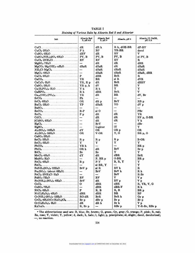
TABLE I
Staining of Various Salts by Alizarin Red S and Alizarina
SaltAlizarin Red
S,pH4.8Alizarin Red
S,pH9Alizarin 1% NaOH,
pHll.8
CuCl dR dR h R h, dOll-BR dP-RVCuCl2�2H2O P h BP YR-BR decolCuSO4�5H2O dRV dR RV VCuSO4(NH4)zSO4�6H:O PV, B PV, B PV, B sl PV, BCuCl2�2NH4Cl RV RV RV RMgSO4�7H2O - dR dR dBR
MgCO3.Mg(OH),.nH,O dRsR dIlsR dR dRsRNH4Cl�MgCl2 - dRaR dRsR dR.sR
MgCl2�6H2O - dRsR dRsR dRaR, dBRCaCl,�2H2O P dBR BrR RCaCO, YR BR sl R dVR
CaC2O4�H2O YR, R p dR BrR dBRVCaSO4�2H2O YR p, h dV BR VCa(H,P04)2.H,O Y h R h Y YCaHPO4 Rh dBR BrR VCaio(OH)2(P04)6 YR dR BR dV, BrSrCO3 Ph - - -
SrCl2�6H,O OR dR p BrY RB pBaCl2�2H,O YP dRsR YO dV pBaSO4 - - - -
ZnCO, R-P si 0 - PBrZnC1, P p dR dR YR
CdC1, - dR dR RV p, 0-BR
3CdSO4�8H2O - dR dR V h
HgCl: - BrY - dBrHgSO4 - Lighter 1Y Y
Al,(SO4),.18H20 dY OR OR p ORAl2(S04),.18H,O OR V-OR Y, 0 BR p, 0CaSO4�2H20SnCI,#{149}2H,0 R p R p R p B-ORSnCl4�5H20 Y Y Y YPbCO1 YRh - - BRpPbC12 OR h dR BrV Br pBiCl, Br Br Y YMnCl2�4H2O dY dR dBR BrRMnSO4.H20 - P, BR p 0-BR BR pFeCl2�4H20 B p P-Y B, R, Y YFeCl, - slBR,Y - YFeNH4(SO4)2� 12H2O BrV p si R RV h RFe,(S04), (about 6H20) - BrV BrV h R hFeCl,�2NH4Cl�H,O - - BrV R-BrFeSO4.7H20 RV RV V V p
Fe(NH4)2(S04)2�6HzO BrV dR RV p V pCoOl, 0 dBR d.BR R, YEt, V, GCoSOg�7H,0 - dBR dBR-V R hNiCl,�6H,O P R, B R, B BR pNi(C,H,O,),.4H,O dBR dBR BR BPCr(NH4)(S04)2.12H,O RO-dR BrR BrR h Gy pCrCls�6H,O[Cr(H,O)6JCl, Br p dBr p Br p Br pCr(C,H,0,),.H,0 dR dR h Br h V pK,Cr,O7 R, Br p - RBr p Y-R-Br, RBr p
#{149}The abbreviations used are: B, blue; Br, brown; G, green; Gy, gray; 0, orange; P, pink; R, red;
Ra, rose; V, violet; Y, yellow; d, dark; h, halo; 1, light; p, precipitate; sl, slight; decol, decolorized;
-, no reaction.
114
at CHRISTIAN UNIV on December 18, 2013jhc.sagepub.comDownloaded from
ALIZARIN AND ALIZARIN RED S STAINS FOR CALCIUM 115
or retained the original color. Thus the dye solu-tions apparently did not penetrate the crystals,
but reacted only with the superficial layers.
Frequently, such salts resembled some calcium
deposits in tissues which also showed an intenselycolored periphery and weakly colored or unstained
center. The progressive breakdown of crystal
structure and simultaneous formation of coloredcompounds could easily be followed by alternate
observation between crossed and parallel polar-
oids. A few compounds did not dissolve duringthe period of observation; only a thin colored
layer was formed on their surface. This group
included CaC,04, SrCO, and PbCO,.A few compounds, e.g., MgCl,, did not react
with alizarmn red S at pH 4.8, but produced color
changes with the other dye solutions tested.
Ca(H,P04), reacted slightly with alizarin red S
in pH 9 buffer solution but not with alizarin redS in pH 4.8 buffer solution or with alizarin. Sinceit appeared possible that this phenomenon might
be due to a lowering of the pH of the dye solutionby the dissolving salt (see “Discussion” below),2-3 drops of the dye solvents were added to crys-tals of Ca(H,,P04), and the approximate pH of
the resulting saturated solution was determined
by indicator papers. The pH values were 2-2.5.
DISCUSSION
The literature on alizarin and alizarin red S
stains for calcium deposits in tissues has been
reviewed comprehensively by Harms (22) and
McGee-Russell (32). These histologic procedures
were originally based on ancient traditions in
textile dyeing with madder (18). The term “lake”
for the alizarin-calcium compound was ap-parently derived from half-forgotten early chemi-
cal concepts. It therefore seems appropriate to
review briefly the history of dyeing with madder
and alizarin. Explanations of the chemical
mechanism of ali.zarin and alizarin red S stains,
whether the term lake implies complex or salt
formation, are scanty. This discussion therefore
is limited mainly to chemical aspects of theinteraction of these dyes with calcium and other
cations.
History of madder dyeing: Madder was
used for dyeing of textiles in early antiquity.
According to Forbes (14), the oldest samples are
pieces of cotton from Mohenjo-Daro dating
back to the third mifiennium B.C.; in Egypt,
textiles dyed with madder were found in tombs
of the period of the XVIII-XXth Dynasties.
The oldest extant samples of recipe books for
dyeing are the Leyden Papyrus X and the
Stockholm Papyrus, which are believed to have
been written about the end of the 3rd century
A.D. (6). However, some of these recipes refer
to originals of 300 B.C. at the latest and probably
much earlier (14). The Stockholm Papyrus (7)
contains numerous mordanting procedures and
recipes for dyeing with madder and other natural
dyes. Lime water and alum dissolved in vinegar
(aluminum acetate?) are frequently recom-
mended as mordants. For dyeing a rose color,
wool was smeared with ashes, washed in liquid
from potter’s clay and mordanted; after being
rinsed in salt water, the wool was dyed in a
solution of madder, bean meal and white oil in
rain water, and aftertreated with alum. To
obtain a purple coloration, textiles were treated
first with a blue dye and then mordanted and
dyed with madder as described above, except
that the concentration of madder was doubled.
However, it was already known that the shade
of madder dyeings could be varied by the use of
different mordants (14). Postmordanting with
alum was recommended to prevent fading; this
procedure seems to be a precursor of the after-
chroming or coppering procedures of modern
textile industry to improve fastness properties.
According to Caley (7), the methods described
in these papyri were essentially the ones used
during the next 1500 years until the advent of
synthetic dyes.
Parenthetically, the dyeing of wool on live
animals, commented upon so disapprovingly by
Pliny, is mentioned in the Stockholm Papyrus,
which gives “Book 3 of Africanus” as reference.
The animals were washed and areas to be dyed
were mordanted with a solution of alum in
vinegar. According to Pliny (23), the dyes were
applied “by the means of certain barks of a foot
and a half long dipped in these colours, and so
imprinted and set upon their fleeces as if riotous
wantonness and superfluitie should force Nature’sworke, and make wool grow of the colour.”
However, it seems much more probable that
these decorations were simply proprietary im-
prints; dyes are still used for this purpose in the
Basque region when the sheep are taken to the
mountains for summer grazing. Since alizarin on
an aluminum mordant has light fastness 7-8 (9),
it seems well suited to withstand fading on
animals exposed to the strong sunlight of Egypt.
In contrast to branding, this ancient procedure
at CHRISTIAN UNIV on December 18, 2013jhc.sagepub.comDownloaded from
116 PUCHTLER, MELOAN AND TERRY
did not interfere with later conversion of the
skins to leather.
Madder was widely distributed in the Old
World and cultivated near Rome in classical
antiquity (46). The history of madder dyeing in
Europe has been reviewed by H#{252}bner (23) and
Hailer (21). According to H#{252}bner (23), the role
of aluminum in madder dyeing was recognized by
Petty in 1667, who concluded “that the use of
allum is to be a Vinculum between the Cloth and
the Colour.” During the following centuries
madder dyeing apparently came under the
influence of alchemy. To “animalize” textiles,
repeated treatments with cow, sheep or goat
dung prior to mordanting and dyeing and an
aftertreatment with dung were considered es-
sential by Bancroft in 1813 (23). These alchemical
practices were challenged less than a century ago
by Schlieper and Baum, who recommended
mordanting wite aluminum and calcium salts and
dyeing in a solution of alizarin and calcium salts
(21), a procedure similar to the methods de-
scribed in the Stockholm papyrus. The im-
portance of calcium salts in dyeing with madder
was already recognized by Hausmann in 1791
(21), but the chemical mechanism of this calcium
effect on the dyeing of alum-mordanted material
with madder or alizarin was finally clarified only
in the 20th century.
Early chemical studies of alizarin-calcium
compounds: The history of alizarin was re-
viewed by Graebe and Liebermann (20). Alizarin
and purpurin were first isolated from madder
roots by Colin and Robiquet in 1826. Incidentally,
the name alizarin was derived from alizari, the
term for roots of Rubia tinctorum imported from
the Orient. The composition of alizarin was
determined independently by Strecker and by
Graebe and Liebermann in 1868 (20). The
calcium, barium and lead salts of alizarin were
clearly described by Graebe and Liebermann (20).
Perkin (35) published a detailed study of the Na,
Na,, K, K, and Ca salts of alizarin and of the
role of the 1- and 2-OH groups in salt formation;
he was apparently the first to suggest that in
alizarin-aluminum-calcium compounds calcium
“neutralized” the 2-OH group. These early
authors distinguished clearly between calcium
salts of alizarin and the lakes (chelates) of this
dye with aluminum.
The interactions of alizarin and other anthra-
quinones with metals and bases were studied in
detail by Pfeiffer (36, 37). Weak bases formed
TABLE II
Dissociation Constants of 1- and i-OH
Groups of Anthraquinones (p4)
2-Hydroxyanthraquinone 24 X 10-’Alizarin, first constant 6.6 X 10’
1-Hydroxyanthraquinone 3.2 X 10�’
Alizarin, second constant 1.1 X 10⁻¹²
salts only with the 2-OH group; alkali and earth
alkali hydroxides reacted first with the 2-OH and
then with the 1-OH group of ali.zarin. Only the
1-OH group participated in complex (chelate)
formation; the 2-OH group remained free and
could form salts. On the basis of these observa-
tions Pfeiffer (37) suggested that in dyeing with
madder or alizarin aluminum, iron, chromium
and similar metals form a complex with the
1-OH and 9-CO group; calcium and other earth
alkalis form a salt with the 2-OH group. The
physical chemistry of alizarin and other anthra-
quinone derivatives was studied by Huttig (24).
Comparison of the dissociation constants of
alizarin with those of 1- and 2-hydroxyanthra-
quinone indicated that the 2-OH group is more
strongly acid than the 1-OH group (Table II), as
already suggested by Pfeiffer (37). Correlating the
physical-chemical data with the experiences of
textile dyers, Huttig (24) suggested that salt
formation of a free hydroxy group with calcium
plays a major role in the formation of the
calcium-aluminum-alizarin compound. This con-
clusion was confirmed by Attree and Perkin (2),
Venkataraman (48), Harms (22), Gerstner (16)
and Kiel and Heertjes (25, 26).
Early histologic uses of madder and
alizarin: The literature from 1567 to the early
20th century on coloration of bones and/or
teeth of animals fed madder has been reviewed
by Cameron (8). The recognition of the relation
between coloration of bone by madder and
staining of calcium has been ascribed to Gottlieb
in 1914 (8). However, according to Haller (21),
Bancroft noticed this relation in 1818 and tried
to use calcium salts-without alum-as a
mordant in madder dyeing. The affinity of
madder for calcium salts of bones was known also
to 19th century histologists. In a discussion of
Lieberkuhn’s paper (1874, original not available),
Gierke (17) stated explicitly that after pigeons
had been fed madder the dye combined with the
calcium salts of bones, but not with the organic
matrix; the latter could be removed by boiling
at CHRISTIAN UNIV on December 18, 2013jhc.sagepub.comDownloaded from
00/\
00
0
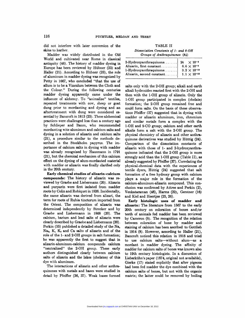
ALIZARIN AND ALIZARIN RED S STAINS FOR CALCIUM 117
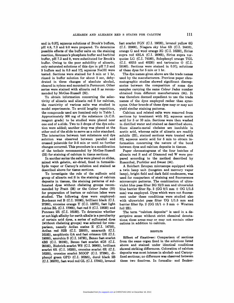
Fzo. 1. Formula of the calcium-aluminurn-alizarin compound suggested by Rutishauser (22,45).
in NaOH without loss of coloration (“Nach
Futterung von Tauben mit Krapp verband sich
der Farbstoff mit den Kalksalzen der Knochen
und nicht mit der organischen Grundsubstanz
derselben. Man kann diese daher durch Kochen
in Natronlosung entfernen, ohne dass die Farbung
leidet.”).
Alizarin was apparently first used as a histo-
chemical reagent by LieberkUhn in 1874; he
injected a 5% neutral solution of alizarin and
reported reaction of the dye with calcium phos-
phate but not with calcium carbonate (17).
Despite these observations, Gierke (17) recom-
mended alizarin only for staining of spinal cord.
In the chapter on calcium salts in the Enzyklopa-
die der mikroskopischen Technik, Magnus (30)
discussed the role of calcium salts in textile
dyeing with alizarin and stated explicity that
calcium forms a salt with alizarin, yet, in the
chapter on alizarin, Magnus (30) remarked that
this dye was of little use in histologic technique;
alizarin red S was mentioned as a stain for
neuroglia, mitochondria and spinal cord. How-
ever, Roehl (43) obtained intense violet coloration
of calcium deposits in kidneys with alizarin in a
solution of NaOH; for staining with alizarin red S
he recommended addition of NaOH, ammonia or
lithium carbonate to the dye solution. These
early authors apparently regarded the calcium-
alizarin compound as a salt. The term lake for
the alizarin-calcium compound in tissue sections
seems to have been derived from Pfeiffer’s (37)
term “mixed lakes” (gemischte Lacke) for calcium-
aluminum-alizarin and similar compounds (5).
However, Pfeiffer’s (37) sharp distinction be-
tween calcium salt and aluminum, iron or
chromium chelate in these compounds became
blurred in the histologic literature, as shown by
Becher’s (5) complaints that in histology the
terms mordant and lake were not based on a
rational definition and were used much more
loosely and vaguely than in the textile industry.
Becher (5) tried to introduce the distinction
between alizarin-calcium salts and chelates with
aluminum and other metals into histology, but
apparently with little success. In the more
recent histologic literature available to us, this
difference is stressed only by Harms (22).
Recent chemical and infrared spectro-
scopic studies of alizarin: A rather complicated
structural formula of the calcium-aluminum-
alizarin compound (Fig. 1) was suggested
by Rutishauser in 1940 (22, 45). Parenthet-
ically, a similar structure has been suggested
for carmin, a calcium-aluminum compound of
carminic acid (22). However, in this structure
(Fig. 1) there would be a large strain in the
central bridge between the two aluminum atoms
(25). Kiel and Heertjes (25-28) therefore re-
investigated the composition and structure of
compounds formed by alizarin and its 3-deriva-
tives with calcium, aluminum and various other
0
�i�Lo-c0-�o
o�. ,o ___
H2OmAI 0-Ca---’O A
H20
Ca-0’
0
1”H20
at CHRISTIAN UNIV on December 18, 2013jhc.sagepub.comDownloaded from
Ca++‘‘H20
H20
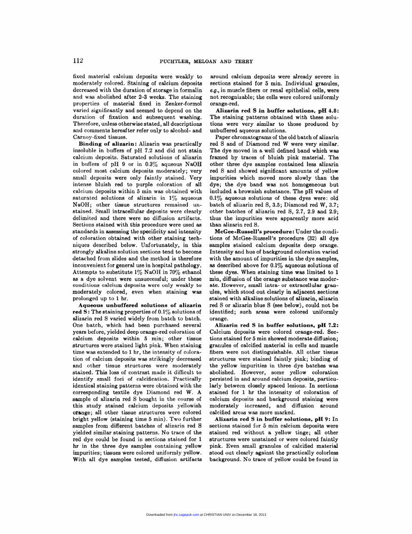
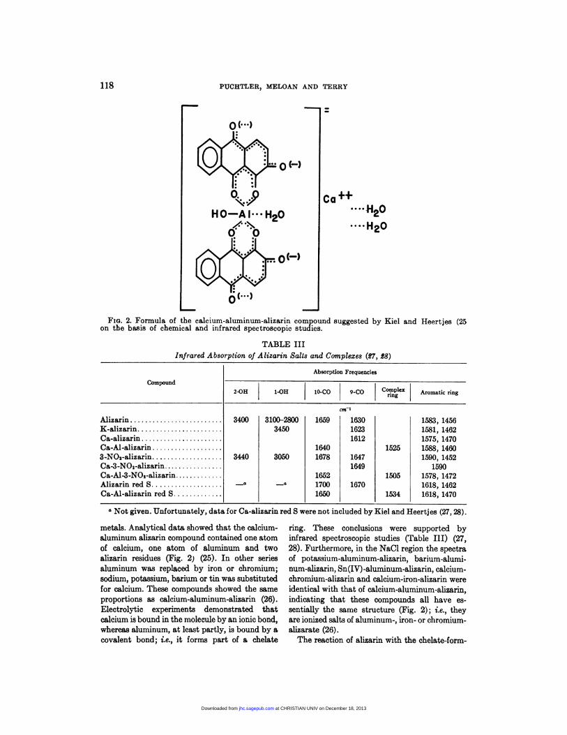
Fia. 2. Formula of the calcium-aluminum-alizarin compound suggested by Kiel and Heertjes (25on the basis of chemical and infrared spectroscopic studies.
TABLE III
infrared Absorption of Alizarin Salts and Complexes (p7, �8)
118 PUCHTLER, MELOAN AND TERRY
H0-AI#{149}” H20,,�%
�IIIIIIX:r
Compound
Absorption Frequencies
2-OH 1-OH 10-CO 9-CO Complex Mo�tic �
AlizarinK-alizarin
Ca-alizarinCa-Al-alizarin
3-NO,-alizarinCa-3-NO,-alizarinCa-Al-3-NO,-alizarin
Alizarin red SCa-Al-alizarin red S
3400
3440
-�
3100-2800
3450
3050
-#{176}
cm’
1659 1630
1623
16121640
1678 16471649
16521700 16701650
1525
1505
1534
1583, 14561581, 14621575, 14701588, 1460
1590, 1452
15901578, 14721618, 1462
1618, 1470
a Not given. Unfortunately, data for Ca-alizarin red S were not included by Kiel and Heertjes (27,28).
metals. Analytical data showed that the calcium-
aluminum alizarin compound contained one atom
of calcium, one atom of aluminum and two
alizarin residues (Fig. 2) (25). In other series
aluminum was replaced by iron or chromium;
sodium, potassium, barium or tin was substituted
for calcium. These compounds showed the same
proportions as calcium-aluminum-alizarin (26).
Electrolytic experiments demonstrated that
calcium isbound in the molecule by an ionic bond,
whereas aluminum, at least partly, is bound by a
covalent bond; i.e., it forms part of a chelate
ring. These conclusions were supported by
infrared spectroscopic studies (Table Ill) (27,
28). Furthermore, in the NaCl region the spectra
of potassium-aluminum-alizarin, barium-alumi-
num-alizarin, Sn(IV) -aluminum-ali.zarin, calcium-
chromium-alizarin and calcium-iron-alizarin were
identical with that of calcium-alurninum-alizarin,
indicating that these compounds all have es-
sentially the same structure (Fig. 2); i.e., they
are ionized salts of aluminum-, iron- or chromium-
alizarate (26).
The reaction of alizarin with the chelate-form-
at CHRISTIAN UNIV on December 18, 2013jhc.sagepub.comDownloaded from
Ca� +
0 OH
0
+H20
ALIZARIN AND ALIZARIN RED S STAINS FOR CALCIUM 119
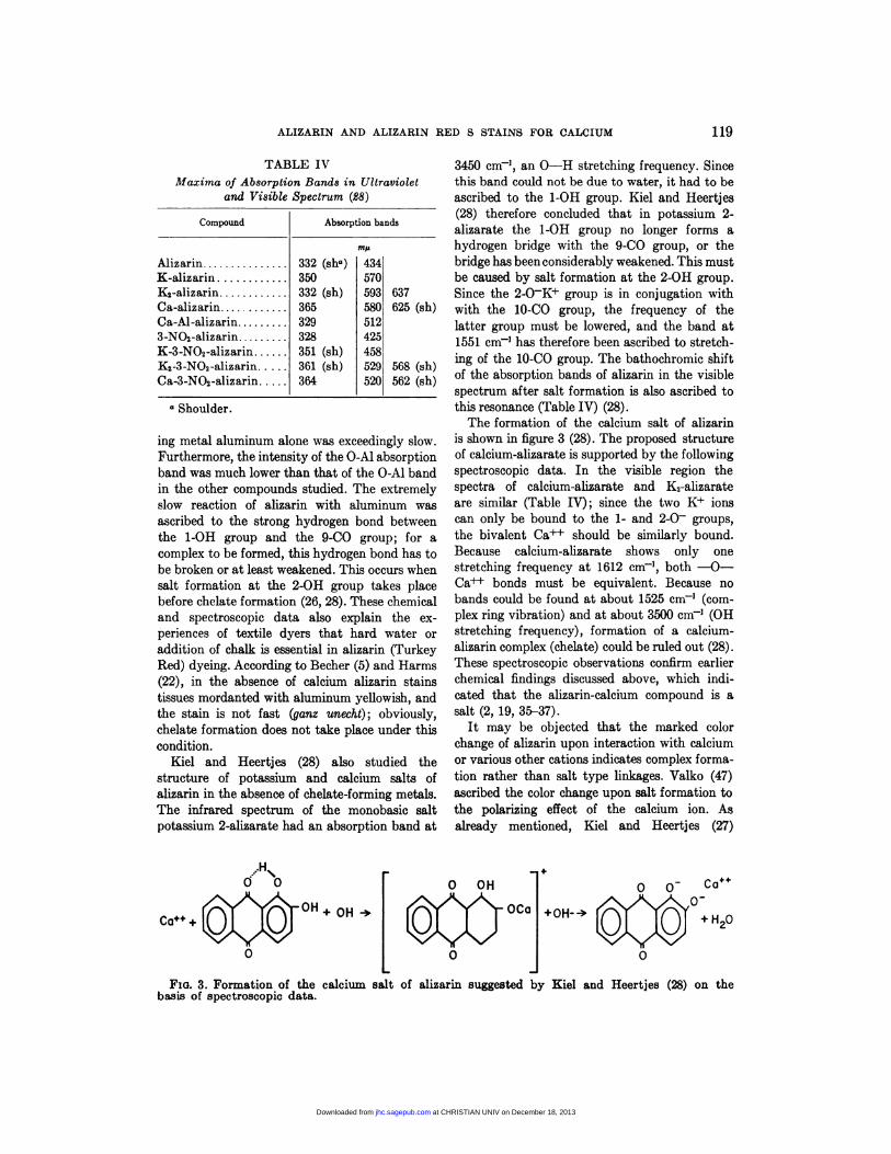
Fio. 3. Formation of the calcium salt of alizarin suggested by Kiel and Heertjes (28) on thebasis of spectroscopic data.
TABLE IV
Maxima of Absorption Bands in Ultraviolet
and Visible Spectrum (p8)
Compound Absorption bands
‘nih
AlizarinK-alizarin
332
350
(she) 434
570K,-alizarin
Ca-alizarin
Ca-Al-alizarin
332365
329
(sh) 593580
512
637625 (sh)
3-N02-alizarin 328 425K-3-NO2-alizarin
K,-3-NO2-alizarin
Ca-3-NO2-alizarin
351
361
364
(sh)(sh)
458529
520
568
562
(sh)
(sh)
a Shoulder.
ing metal aluminum alone was exceedingly slow.
Furthermore, the intensity of the 0-Al absorption
band was much lower than that of the 0-Al band
in the other compounds studied. The extremely
slow reaction of alizarin with aluminum was
ascribed to the strong hydrogen bond between
the 1-OH group and the 9-CO group; for a
complex to be formed, this hydrogen bond has to
be broken or at least weakened. This occurs when
salt formation at the 2-OH group takes place
before chelate formation (26, 28). These chemical
and spectroscopic data also explain the ex-
periences of textile dyers that hard water or
addition of chalk is essential in alizarin (Turkey
Red) dyeing. According to Becher (5) and Harms
(22), in the absence of calcium alizarin stains
tissues mordanted with aluminum yellowish, and
the stain is not fast (ganz unecht); obviously,
chelate formation does not take place under this
condition.
Kid and Heertjes (28) also studied the
structure of potassium and calcium salts of
alizarin in the absence of chelate-forming metals.
The infrared spectrum of the monobasic salt
potassium 2-alizarate had an absorption band at
3450 cm’, an 0-H stretching frequency. Since
this band could not be due to water, it had to be
ascribed to the 1-OH group. Kiel and Heertjes
(28) therefore concluded that in potassium 2-
alizarate the 1-OH group no longer forms a
hydrogen bridge with the 9-CO group, or the
bridge has been considerably weakened. This must
be caused by salt formation at the 2-OH group.
Since the 2-OK� group is in conjugation with
with the 10-CO group, the frequency of the
latter group must be lowered, and the band at
1551 cm’ has therefore been ascribed to stretch-
ing of the 10-CO group. The bathochromic shift
of the absorption bands of alizarin in the visible
spectrum after salt formation is also ascribed to
this resonance (Table IV) (28).
The formation of the calcium salt of alizarin
is shown in figure 3 (28). The proposed structure
of calcium-alizarate is supported by the following
spectroscopic data. In the visible region the
spectra of calcium-alizarate and K2-alizarate
are similar (Table IV); since the two K� ions
can only be bound to the 1- and 2-0- groups,
the bivalent Ca� should be similarly bound.
Because calcium-alizarate shows only one
stretching frequency at 1612 cm’, both -0-
Ca� bonds must be equivalent. Because no
bands could be found at about 1525 cnc’ (com-
plex ring vibration) and at about 3500 cm� (OH
stretching frequency), formation of a calcium-
alizarin complex (chelate) could be ruled out (28).
These spectroscopic observations confirm earlier
chemical findings discussed above, which indi-
cated that the alizarin-calcium compound is a
salt (2, 19, 35-37).
It may be objected that the marked color
change of alizarin upon interaction with calcium
or various other cations indicates complex forma-
tion rather than salt type linkages. Valko (47)
ascribed the color change upon salt formation to
the polarizing effect of the calcium ion. As
already mentioned, Kiel and Heertjes (27)
at CHRISTIAN UNIV on December 18, 2013jhc.sagepub.comDownloaded from
120 PUCHTLER, MELOAN AND TERRY
demonstrated that such bathochronic shifts are
due to alterations of the resonance system of
alizarin during salt formation (28) . Moreover,
alizarin and alizarin red S are polygenetic dyes;
i.e., the color of the dye salt varies with the
cation (31, 38, 40, 48). Color differences among
various metal compounds of alizarin and alizarin
red S were observed also in the model experi-
ments described above (Table I), but these color
changes do not permit conclusions whether
certain metals are bound by salt type linkage or
by chelate formation.
Correlation of chemical data and staining
properties of alizarin: As shown in Table II,
the dissociation constant of the 2-OH group of
alizarin is 6.6 x 10-’ and that of the 1-OH group
is 1.1 x 10” (24); the corresponding pK values
are 8.18 and 11.96 respectively. Therefore,
alizarin in aqueous solutions of pH 7 or below is
only slightly dissociated and shows little tend-
ency to form calcium salts. This also explains
the faint coloration of bones in in vivo experiments
observed by Richter (42). Differences between in
vivo staining with alizarin and madder are
discussed below. In solutions of alizarin adjusted
to pH 9 about 50% of the 2-OH groups should be
dissociated; the red coloration of calcium de-
posits by such solutions indicates formation of themonoalizarate. Around pH 12 nearly all 2-OH
and about half of the 1-OH groups are dissociated
and the blue dializarate is formed. The reddish
blue coloration of calcium deposits suggests
that the red monoalizarate and the blue di-
alizarate may occur side by side. That the
calcium-alizarin compound in tissue sections is a
salt, and not a chelate, is indicated by its sensi-
tivity toward dilute acetic acid. As pointed out
by Becher (5), Cameron (8) and Harms (22),
calcium salts of alizarin dissociate readily in cold
acetic acid, whereas alizarin-metal chelates
disintegrate only after prolonged boiling in
H2SO4 or other mineral acids.
Unfortunately, alizarin is not specific for
calcium but can react also with other metals
(Table I). In “blind” tests the color differences
among various alizarin-metal compounds were
found insufficient for identification of the cations.
Identification is complicated also by the fact
that the anion of the salt can affect the color of
the alizarin-metal compound as shown by the
calcium and iron salts in Table I. This effect
seems to be due, at least in part, to alterations of
the pH of the dye solution by the anion. For
example, Ca(H2PO4)2 did not form colored
compounds with alizarin in pH 9 buffer solutions
or in 1 % NaOH (Table I). As already mentioned,
saturated solutions of Ca(H2PO4)2 in these
solvents had pH values of 2-2.5. Obviously, the
hydroxyl groups of alizarin are not dissociated
under these conditions and salt formation cannot
take place. If a calcium deposit should consist
entirely of Ca(H2PO4),, it may remain unstained.
Hence, alizarin cannot be depended on to visualize
primary calcium phosphate.
Madder versus alizarin: Differences between
madder and synthetic alizarin in vital staining
of bone have been stressed by Richter (42).
According to the Colour Index (9), madder
contains six dyes in the form of their glycosides,
namely alizarin (rubierythric acid, CI. 75330),
xanthopurpurin (1 ,3-dthydroxyanthraquinone,
C.I. 75340), xanthopurpurin-3-carboxylic acid
(C.I. 75370), rubiadin (1 ,3-hydroxy-3-methyl-
anthraquinone, C.I. 75350), purpurin (1,2,4-
hydroxyanthraquinone, C.I. 75410) and pseudo-
purpurin (purpurin-3-carboxylic acid, C.I. 75420).
Comparative in vivo studies with alizarin and
purpurin-3-carboxylic acid showed that only the
latter gave the bright carmine red color typical
of madder-stained bones; alizarin tinted bones
slightly pale bluish pink; extraction of madder-
stained bones confirmed that coloring of bone
was mainly due to purpurin-3-carboxylic acid
(42). In in vitro experiments, purpurin-3-carbox-
ylic acid formed colored calcium salts in the acid,
neutral and alkaline range (42). Unfortunately,
xanthopurpurmn-3-carboxylic acid and purpurmn-3..
carboxylic acid were not available for this study.
However, the data provided by Richter (42)
indicate that the staining properties of purpurin-
3-carboxylic acid are similar to those of alizarin-3-
sulfonic acid. Thus, staining patterns obtained
with madder should be compared with those
produced by alizarin red S rather than by
alizarin.
Interaction of alizarin red S and other
sulfonated dyes with calcium: The 1- and
2-OH groups of alizarin red S show the same
behavior toward calcium and various other
metals as those of the parent dye alizarin (27, 28).
Differences between frequencies of certain bands
in the visible and infrared spectrum of salts of
alizarin and its 3-substituted derivative have
been ascribed to effects of the electronegative
nitro or sulfonic acid group (27). However, the
interaction of alizarin red S with calcium is not
at CHRISTIAN UNIV on December 18, 2013jhc.sagepub.comDownloaded from
ALIZARIN AND ALIZARIN RED S STAINS FOR CALCIUM 121
limited to the 1- and 2-OH groups; the sulfonic
acid group also forms a salt with calcium. One
calcium atom can react with two sulfonic acid
groups; i.e., it can bind two dye molecules of
alizarin red S according to the general formula:
2 dye-SO3Na + CaC12 -� (dye-SO,)2Ca + 2
NaCl (4, 40). Salt formation between alizarin
red S and calcium, strantium, barium, zinc,
magnesium, aluminum and other metals later
than group II in acid solutions was described
by Atack (1), who emphasized that these com-
pounds are salts, not complexes.
Formation of insoluble or poorly soluble
calcium or barium salts of sulfonated dyes is
widely used in industry in the preparation of
pigments; these dye salts are referred to as lakes
or toners. In the dye and pigment industry
the term lake denotes a poorly soluble or insolu-
ble salt of a water-soluble acid or basic dye (31,
38, 41, 48). Sulfonated dyes suitable for this
purpose are classified as “acid dyes for lakes”
(10). In the precise terminology of dye chemistry
the designation “acid dye” signifies that these
dyes do not contain chelating groups. Dyes
capable of chelate formation with metals are
classified as mordant dyes. Of course, certain
mordant dyes can also form lakes, i.e. the readily
soluble sodium or potassium salts can be con-
verted into poorly soluble calcium or barium
salts. Barium salts (lakes) of sulfonated anthra-
quinone dyes have been described by Mattiello
(31). Alizarin red S is now mainly used in the
preparation of lakes; in textile dyeing it has been
largely replaced by other dyes (4).
The effect of pH on lake formation by acid
dyes has been discussed by Pratt (38). In these
chemical studies an excess of metal was used.
At pH 5.5 and below, all dye molecules reacted
with the metal. Lake (salt) formation decreased
around pH �; in alkaline solutions only a mod-
erate amount of dye was bound by the metal.
This pH effect is very inconvenient for histology.
At pH 5.5 and below, basic groups of tissues also
react with sulfonic acid groups of dyes; this
diffuse coloration of sections made identification
of small calcium deposits very difficult. The
choice of acid dyes for lakes is therefore limited
to those dyes which produce strong selective
coloration of calcium deposits in the nonoptimal
pH range 7-9.
In contrast to acid dyes for lakes, hydroxy-
anthraquinone dyes containing acid groups can
readily form salts over a wide p11 range. In
acid and neutral media only the sulfonic or
carboxylic groups take part in salt formation
(42). Acid and neutral solutions of alizarin red
S are ochre yellow; the salts (lakes) formed with
calcium in model experiments and in calcifiedtissues are colored orange or yellowish red.
In alkaline solutions the hydroxyl groups partici-
pate in salt formation (42); the calcium salts
formed under these conditions are deep red to
bluish red. The bluish tinge becomes more
noticeable with increasing PH; i.e., the salts of
alizarin red S formed in strongly alkaline solu-
tions resemble those of alizarin. In the weakly
alkaline region (around pH 9) the two types of
reactions probably overlap. This assumption
is supported by observations that at pH 9
calcium deposits are stained selectively by acid
dyes for lakes and by alizarin and alizarin blue
S. In analogy to the formula for the calcium-
aluminum compound of alizarin red S given by
Baumann and Hensel (4), the calcium salt
formed under alkaline conditions would be (Ca-
ali.zarin-3-SO3)2Ca. As in the case of alizarin,
the color change of alizarin red S cannot be
regarded as evidence for complex (chelate)
formation. Striking color changes upon salt
formation with calcium or barium have also
been observed with acid dyes for lakes which
do not contain chelating groups (31).
The use of the term lake as a synonym for
chelates apparently caused some ambiguity
even in the chemical literature. For example,
compounds of alizarin red S with hafnium and
other cations of the titan group have been in-
terpreted as chelates (innere Kompl&xsalze) (12),
yet Venkataraman (48) cited spectroscopic
evidence that the hafniurn-alizarin compound
is a salt, not a chelate. Hence, reports concerning
complex (chelate) formation of alizarin and its
derivatives with various cations should be
interpreted with caution. The investigations
by Kiel and Heertjes (25-28) indicate that
infrared spectroscopy is at present the most
reliable method for distinction between salts
and chelates of alizarin and its derivatives.
Alizarin red S is as nonspecific for calcium as
the parent dye alizarin. These observations artin agreement with findings by Dahl (11). Im
addition to the compounds listed in Table I�
alizarin red S is also very sensitive towart
the ions of uranium, titanium, bismuth ank
thallium (15), and toward zirconium, hafnium
and thorium (12). The differences in color
at CHRISTIAN UNIV on December 18, 2013jhc.sagepub.comDownloaded from
122 PUCHTLER, MELOAN AND TERRY
among various alizarin red S-metal compounds
tested were insufficient for identification of
the cations in a blind study. As already men-
tioned, the same difficulty was encountered with
alizarin.Binding of acid dyes for lakes by calcium
deposits in tissues: The selective coloration
of calcium deposits by several acid dyes for
lakes shows that salt formation between sul-
fonic acid groups and calcium or other cations
occurs under the conditions of histologic tech-
nique. The abolition of staining by pretreatment
of sections with dilute acetic acid substantiates
the conclusion that these dyes react with cal-
cium or other metallic cations and not with
basic groups of proteins. Preliminary compara-
tive studies of acid dyes for lakes and other
acid dyes indicate relations between the configura-
tion of dye molecules and selectivity for calcium
deposits. Even dyes which differ only in the
position of one sulfonic acid group can show
strikingly different staining properties. For
example, the monoazo dye Bordeaux red B
(1-naphthylamine -‘ 2-naphthol-3 , 6-disulfonic
acid) colored calcium deposits strongly and
selectively, but Ponceau 6R (1-naphthylamine
2-naphthol-6 ,8-disulfonic acid) showed no affinity
for calcium deposits. Further investigations of
the relations between dye structure and affinity
for calcium are in progress. Previous studies
showed relations between fluorescence and
structure of azo dyes (39). These relations were
found to apply also to the calcium lakes. Of the
dyes tested, Ponceau 2R was found most suitable
for fluorescence microscopic studies of calcium
deposits.
The effects of different fixatives and of varia-
tions in fixation and embedding procedures on the
binding of acid dyes for lakes and the specificity
of these dyes for various cations have not yet
been determined. These dyes were included in this
study for the sole purpose of obtaining informa-
tion concerning the role of the sulfonic acid group
of alizarin red S in the staining of calcium deposits
in alcohol- or Carnoy-fixed tissues. Therefore,
until further data become available, these dyes
should not be regarded as substitutes for alizarin
and its derivatives.
Binding of alizarin red S by tissue sections:
The recommended pH values for solutions of
alizarin red S vary from 4.1-4.2 (32) to 6.3-6.5
(11). Our experiences indicate that the staining
patterns obtained with alizarin red S in acid
solutions differ significantly from batch to batch
and apparently depend on the impurities in the
dye samples. Of the five samples tested in this
study, only two produced passable selective
staining of calcium deposits at pH values below
7. The yellow impurities of the other three dye
samples competed effectively with alizarin red
S for binding sites in calcium deposits. Thus the
discrepancies between recommended pH values
and reported difficulties in reproducing the work
of others can readily be explained by differences
in the dye samples used by various workers. The
low pH values of aqueous solutions of the impure
dyes and the staining properties of the con-
taminants indicate that these compounds are
strongly acid. Since many commercial samples of
azo dyes contain intermediates (44), it seemedconceivable that batches of alizarin red S may
also be contaminated with sulfonated by-
products of the manufacturing process. According
to Fierz-David and Blangey (13) and Venkatara-
man (48), 2-anthraquinonesulfonic acid is used as
starting material in commercial production of
alizarin. A concentrated solution of 2-anthra-
quinonesulfonic acid, NaOH and NaC1O3 or
NaNO3 is heated at 185#{176}Cand 5-6 atm pressurefor 48-72 hr. The 2-anthraquinonesulfonic acid
is converted into 1-hydroxyantb.raquinone-2-
sulfonic acid; in a second step the disodium salt of
alizarin is formed. Completion of the reaction is
indicated by loss of fluorescence; 2-anthra-
quinonesulfonic acid and 1-hydroxyanthra-
quinone-2-sulfonic acid are strongly fluorescent,
but alizarin is nonfluorescent (13). Alizarin red S
is prepared by sulfonation of alizarin. When
tissue sections stained with impure samples of
alizarin red S were viewed in ultraviolet blue
light, the structures colored by the yellow
contaminants showed moderate to strong yellow-
ish red fluorescence. Calcium deposits stained red
by alizarin red S were nonfluorescent. Theseobservations suggest that the yellow impurities
consist, at least in part, of intermediates.
Further disadvantages of acid solutions of
alizarin red S are diffusion artifacts and loss of
calcium. Gomori (19) recommended buffer solu-
tions of pH 4.5-5 for removal of calcifications in
tissues. It is therefore not surprising that solutions
of alizarin red S at pH 4.8 or below removed
significant amounts of calcium, and diffusion
artifacts became evident within 1 mm. Hence,
acid solutions of alizarin red S are unsuitable for
studies of the localization of calcium deposits at
at CHRISTIAN UNIV on December 18, 2013jhc.sagepub.comDownloaded from
ALIZARIN AND ALIZARIN RED S STAINS FOR CALCIUM 123
the intracellular level, e.g., in muscle fibers or
renal epithelial cells, and for semiquantitative
estimates. In the discussion of methods for
enzymes, Gomori (19) stated explicitly that
calcium salts cannot be used effectively below pH
8.5 because of their solubility and consequent
diffusion artifacts. Such diffusion artifacts were
very obvious in sections stained with alizarin red
S at pH 7.2, but were absent in sections stained
with alizarin red S at pH 9, the pH value recom-
mended by Gomori (19) for procedures employing
calcium salts. Parenthetically, alkaline solutions
of alizarin red S were already in use around the
turn of the century. Roehl (43) recommended
differentiation in alkaline media or addition of
NaOH, Li2CO3 or NH3 to aqueous solutions of
alizarin red S. Differentiation is unacceptable in
histochemistry (34) and impractical for general
hospital pathology. In this study, 0.5% solutions
of alizarin red S buffered to approximately pH 9
yielded optimal coloration of calcium deposits. As
already mentioned, at this pH the yellow im-
purities present in three dye batches were no
longer bound by tissue sections; the staining
patterns obtained with the five dye batches
tested were indistinguishable. Higher p1-1 values
were found impractical because the sections
tended to become detached from the slides.
Because solutions of alizarin red S buffered to
pH 9 do not cause noticeable diffusion artifacts
or loss of calcium salts, staining can be prolonged
to 1 hr. Intensity of coloration of calcium,
aluminum or other salts by alizarin red S in-
creases with the time allowed for lake (salt)
formation (49). Since replacement of anions in
calcium deposits by the dye progresses gradually
from the periphery, staining for 30-60 mm
improved coloration of crystalline areas more
than 10 � in diameter. The slight grayish pink
background stain did not interfere with the study
of small intracellular calcium deposits and
actually served as a counterstain. As pointed out
by Barka and Anderson (3), counterstaining is
undesirable from a histochemical standpoint.
Because calcium and other salts of alizarin red S
are soluble in slightly acid media (33), acid dyes
are difinitely unsuitable as counterstains. Basic
dyes tend to react with calcium deposits and are
therefore not recommended (29).
Effect of fixatives: The solubility of calcium
deposits in solutions of pH 4.5-5 (19) readily
explains the deleterious effect of unbuffered 10%
formalin. Treatment of alcohol- or Carnov-fixed
sections with 10% unbuffered formaliti for 15 hr
removed all calcium deposits. Similar treatment
�vith Zenker-formol (Spuler, �Iaxiiuow) was
equally effective. Ilence, the major loss of
calcium seems to occur (luring fixation and not
at later stages of the embeddmg procedure.
According to 1)ahl (11), solutions of formaliti
buffered to I)11 7.2 or 8.35-8.7 also caused
significant loss of calcium salts and were clearly
inferior to al)solute alcohol. Obviously, these
fixatives are unsuitable for histochemical studies
of calcium deposits. That (‘arnoy’s fluid, which
contaim 10% acetic acid, preserves calciuni
deposits as wellas absolute alcohol is probably due
to the very low dissociation of acetic acid and pOOr
solubility of calcium salts in absolute alcohol and
chloroform.
REFEHENCES
I. Atack, F. W.: A new reagent for the detectionand colorimetric estimation of aluminum.
J. Soc. Chem. md. 34: 936, 1915.2. Attree, G. F. and Perkin, A. G.: Reduction
products of the hydroxyanthraquinones.J. Chem. Soc. 134: 144, 1931.
3. Barka, T. and Anderson, P. J.: Histochemistry:Theory, Practice and Bibliography. HoeberMedical Division, Harper and Row, Pub-ushers, Inc., New York, 1963.
4. Baumann, 11. and ilensel, II. H.: Neue Metal!-komplexfarbstoffe, Struktur und farberischeEigenschaften. For(seh r. Chc?n. Forsch. 7:
643, 1967.5. Becher, S.: Untersuchungen liber Echtfarbung
der Zeilkerne mit k#{252}n.stlichen Beizenfarb-stoffen und die Theorie des hislologischenFarbeprozesses mit gelosten Lacken. Verlagvon Gebruder Borntraeger, Berlin, 1921.
6. Caley, E. II.: The Leyden Papyrus X. AnEnglish translatioii with brief notes. .1.Chem. Ed. 3: 1149, 1926.
7. Caley, E. R.: The Stockholm Papyrus. AnEnglish translation with brief notes. .1.Chem. Ed. 4: 979, 1927.
8. Cameron, U. H.: The staining of calcium.J. Path. Bact. 33: 931, 1930.
9. Colour In(iex, Ed. 2, Vol. 1. Society of 1)yers
and Colourists and American Associationof Textile Chemists and Colorists, Lowell,Mass., 1956.
10. Colour Index, Ed. 2, Vol. 2. Society of 1)yersand Colourists and American Associationof Textile Chemists and Colorists, Lowell,Mass., 1957.
11. 1)ahl, L. K.: A simple and sensitive histo-chemical method for calcium. Proc. Soc.Exp. Biol. 80: 474, 1952.
12. l)orta-Schaeppi, Y., ilurzeler, H. arid Tread-well, W. 1).: Uber die St#{246}chiometrie undExtinktion geloster Alizarinlacke von Ka-tionen der Titanreihe. Helv. Chim. �tcta34: 797, 1951.
13. Fierz-David, H. E. and Blangey, L.: Grund-legende Operationen der Farbenehemie, Ed.8. Springer \Tcrlag, Wien, 1952.
at CHRISTIAN UNIV on December 18, 2013jhc.sagepub.comDownloaded from
124 PUCHTLEII, MELOAN AND TERRY
14. F’orhes, H. J. : Chemical, culinary and cosmeticarts. In .1 History of Technology, editedby C. Singer, E. J. Holmyard and A. ii.hail, vol. 1. Oxford University Press, New\ork, 1954, i. 238.
15. (;erii�uth, F. (1. and �1itchell, C. : I)etectioii
itII(l identification of specific cations withso(liun�-alizarili-sulfonate. .1 iiier. J. Pharm.101: 46, 1929.
16. Gerstner, ii. : Die (‘heinie (icr .1pplikation vonKorn plcxfarbstoffen. Akademie Verlag, Ber-liii, 1959.
17. Gierke, 11.: Fiirberei zu mikroskopischenZwecken. Z. Wiss. Miki. 1: 62, 497, 1884.
18. Cierke, II.: F#{228}rberei zu mikroskopischeii
Zwecken. Z. Wis.s. Mikr. 2: 13, 1885.19. Gomori, (4.: Microscopic Histochemistry.
University of Chicago Press, Chicago, 1952.20. (lraebe, C. and Liebermaiiii, C.: Ueber
Anthracen mid Alizarin. A nn. Chem.Pharm., �‘�P1�1. 7, 257, 1870.
21. llaller, H.: Vorn T#{252}rkischrot zum Alizarinrot.Melliand Textilhei. 19: 448, 504, 595, 731,796, 1938.
22. harms, II.: Handbuch dci Farbstoffe liii dieMikroskopie, Teil II, 1. Liefg. StaufenVerlag, Kamp-Lintfort, 1957.
23. ll#{252}bner, J.: A contribution to the history of
dyeing. J. Soc. Dyers Go!. 29: 344, 1913.24. llQttig, U. F.: Physikalisch-chemische Unter-
suchungen (ler ()xy - mid 1) ioxyanthra-chinone ni it besonderer Berucksichtigungihres Beizvermbgens. Z. Phy.s. C/rein. 87:129, 1914.
25. Kiel, E. U. and Heertjes, P. M.: Metal com-plexes of alizarin. I. The structure of thecalcium-aluminum lake of alizarin. J. Soc.Dyers Go!. 79: 21, 1963.
26. KieI, E. U. and lleertjes, P. M.: Metal corn-plexes of alizarin. II. The structure of somemetal complexes of alizarin other thanTurkey red. J. Soc. Dyers Go!. 79: 61, 1963.
27. Kid, E. U. and lleertjes, P. M.: Metal corn-plexes of alizarin. III. The structure ofmetal complexes of sonic 3-derivatives of
alizarin. J. Soc. Dyers Go!. 79: 186, 1963.28. Kiel, E. U. and Heertjes, P. M.: Metal com-
plexes of alizarin. IV. The structure of thepotassium and calcium salts of alizarinand 3-nitroalizariti. J. Soc. I)�jeis Col. 79:363, 1963.
29. Lillie, R. I).: Histopathologic Technic andPractical Histocheinistry, Ed. 3. The Blakis-ton 1)ivision, McUraw-H ill Book Company,New York, 1965.
30. Magiius, \V.: Alizariti, Alizaritie, Calciumace-tat. In Enzyklopadie (icr MikroskopischenTechnik, edited by P. Ehrlich, H. Krause.M. Mosse, H. Hosin and K. Weigert, Ed. 2,Vol. 1. Urban and Schwarzeiiherg, Berlin,1910, pp. 14 and 161.
:31. Mattiello, J. J.: Cheiiiistrv of tie synthetic
organic �)igmeIlts. In Protective and Decora-live Goatings, edited by J. J. Mattiello, Vol.2. John Wiley and Sons, New York, 1942.
32. McGee-Russell, S. M. : Histochemical methodsfor calcium. J. Histochem. Cytochem. 6: 22,1958.
33. Parker, C. A. and Goddard, A. P. : The reac-tion of aluminum ions with alizarin-3-sulfonate with particular reference to theeffect of addition of calcium ions. Anal.Chin,. Acta 4: 517, 1950.
34. Pearse, A. G. E.: Histochemistry: Theoreticaland Applied, Ed. 2. Little, Brown andCompany, Boston, 1961.
35. Perkin, A. U.: A reaction of some phenoliccolouring matters. J. C/rein. Soc. 75: 433,1899.
36. Pfeiffer, P.: Zur Kenntnis der Farblacke. I.Ber. I)tsch. C/rem. Ges. 44: 2653, 1911.
37. Pfeiffer, P.: Zur Theorie der Farblacke. II.Ann. Chem. 398: 137, 1913.
38. Pratt, L. S.: The Chemistry and Physics of
Organic Pigments. John Wiley and Sons,New York, 1947.
:39. Puchtler, H., Sweat, F. and Uropp, S.: Aninvestigation into the relation betweenstructure and fluorescence of azo dyes. J.Roy. .1! icr. Soc. 87: 309, 1967.
40. Hath, 11.: Lehrbuch der Textiichemie, Ed. 2.Springer-Verlag, Berlin, 1963.
41. Remington, J. S. and Francis, W.: Pigments:Their Manufacture, Properties and Use.Leonard Hill, Ltd., London, 1954.
42. Richter, 1).: Vital staining of bones withmadder. Biochem. J. 31: 591, 1937.
43. Hoehl, W.: Uber Kalkablagerung und-Ausscheidung in der Niere. Beitr. Path.Anat., suppl. 7, 456, 1905.
44. Rosenthal, S. I., Puchtler, H. and Sweat, F.:Paper chromatography of dyes: method toinvestigate vagaries in histological staining.Arch. Path. (Chicago) 80: 190, 1965.
45. Schweizer, H. H.: Kiinstliche organischeFarbstoffe und ihre Zwischenprodukte.Springer-Verlag, Berlin, 1964.
46. Taylor, F. S. and Singer, C.: Pre-scientificindustrial chemistry. In A History of Tech-nology, edited by C. Singer, E. J. Holmyard,A. R. Hall and T. I. Williams, Vol. 2. OxfordUniversity Press, New York, 1956.
47. Valk#{243},E.: Kolloidchen,ische Grundlagen derTextilveredlung. Springer-Verlag, Berlin,1937.
48. Venkatararnan, K.: The Chemistry of Syn-thetic Dyes, Vol. 2. Academic Press, NewYork, 1952.
49. \oe, J. H. and Hill, W. L.: An investigationof the reaction of sodiurn alizarin mono-sulfoiiate with aluminum under differentexperimental conditions with reference toits use in colorimetrv. J. Amer. Chem. Soc.50: 748, 1928.
at CHRISTIAN UNIV on December 18, 2013jhc.sagepub.comDownloaded from

![The Garvagh Madonna Illuminating the Photochemistry of ...posters/Examples...Rafaello Santi (Raphael): The Garvagh Madonna Madder Lake [Alizarin + Puprurin] Alizarin Purpurin Madder](https://img.pdfslide.net/doc/110x75/60e7340affeae4294037f4d9/the-garvagh-madonna-illuminating-the-photochemistry-of-postersexamples-rafaello.jpg)