Embed Size (px)
Citation preview
ZOOLOGIA 29 (5): 459–466, October, 2012doi: 10.1590/S1984-46702012000500010
© 2012 Sociedade Brasileira de Zoologia | www.sbzoologia.org.br | All rights reserved.
The Neotropical leafhopper genus Scoposcartula Young,1977 comprises 15 species, including one from SE. Brazil thatremains undescribed because it is known only from femalespecimens. This genus is distributed from Costa Rica to Argen-tina, with records in Panama, Colombia, Venezuela, Guyana,Brazil, Bolivia, and Paraguay. A comprehensive study on thetaxonomy and phylogeny of Scoposcartula was published byLEAL et al. (2009), who pointed out that the presence of a dis-tinct sclerotized line on the female pygofer is a synapomorphyof the genus. In addition to this synapomorphy, the genus canalso be recognized by the position of the ocelli, which inScoposcartula are usually located well behind the anterior eyeangles, an uncommon condition among the NeotropicalCicadellini. The reader should consult LEAL et al. (2009), YOUNG
(1977), and RODRIGUES et al. (2010) for additional data on thetaxonomy, phylogeny, and distribution of Scoposcartula and itsspecies.
GERMAR (1821) described Tettigonia frontalis based onmaterial from Brazil. METCALF (1955) recognized that the spe-cific epithet proposed by Germar was preoccupied and, thus,proposed a new name, frontaliana. METCALF (1955) transferredthis species to the Neotropical genus Amblyscarta Stål, 1869.This placement was followed by YOUNG (1977) in his compre-hensive monograph of the New World Cicadellini. NeitherMetcalf nor Young studied syntypes of A. frontaliana, whichare housed in the Museum für Naturkunde der Humboldt-
Universität in Berlin. During a study of the digital images ofAmblyscarta species on the internet site Sharpshooter Leafhop-pers of the World (WILSON et al. 2009), we noticed a remarkablesimilarity in the shape, size, and color pattern between thephotographs of two syntypes of A. frontaliana (from the stateof Bahia, NE. Brazil) and a species of Scoposcartula, S. talitaeLeal, Mejdalani & Cavichioli, 2005 (from the state of EspíritoSanto, SE. Brazil). After borrowing and analyzing the syntypesof A. frontaliana, we were able to confirm our suspicion that A.frontaliana belongs in Scoposcartula. We also confirmed our ini-tial observation that A. frontaliana is externally very similar toS. talitae. The two species, however, can be easily distinguishedby characters of the male genitalia.
In the present paper, we transfer A. frontaliana toScoposcartula, redescribe this species based on the type series,and select a male syntype to be the lectotype. We also describethe previously unknown female of S. talitae. The most relevantcharacters for distinguishing between the two species are dis-cussed.
MATERIAL AND METHODS
Techniques for preparation of male and female genitalstructures follow OMAN (1949) and MEJDALANI (1998), respec-tively. The dissected parts are stored in microvials with glyc-erin and attached below the specimens, as suggested by YOUNG
On the identification of two Brazilian leafhoppers: redescription ofScoposcartula frontaliana comb. nov. and description of the female of S.
talitae (Hemiptera: Cicadellidae: Cicadellini)
Gabriel Mejdalani1,2 & Rachel Alexandre Carvalho1
1 Departamento de Entomologia, Museu Nacional, Universidade Federal do Rio de Janeiro. Quinta da Boa Vista,São Cristóvão, 20940-040 Rio de Janeiro, RJ, Brasil. E-mail: [email protected]; [email protected] Corresponding author.
ABSTRACT. This paper deals with two species of Scoposcartula Young, 1977 that are very similar to each other externally
(form and color pattern): S. frontaliana (Metcalf, 1955) comb. nov. (from the state of Bahia, NE. Brazil) and S. talitae
Leal, Mejdalani & Cavichioli, 2005 (from the state of Espírito Santo, SE. Brazil). The former species, previously positioned
in Amblyscarta Stål, 1869, is redescribed and a male specimen is selected as the lectotype. The previously unknown
female of the latter species is described for the first time. Scoposcartula frontaliana can be distinguished from S. talitae by
features of the male and female genitalia. The most remarkable difference is in the male pygofer, which has a conspicu-
ously concave posterior margin in S. frontaliana, whereas it is convex in S. talitae. The posterior margin of the female
sternite VII is convex in S. frontaliana, whereas in S. talitae it has a pair of shallow concavities and a median dentiform
projection.
KEY WORDS. Auchenorrhyncha; Brazil; female genitalia; lectotype; male genitalia; morphology; sharpshooter.
460 G. Mejdalani & R. A. Carvalho
ZOOLOGIA 29 (5): 459–466, October, 2012
& BEIRNE (1958). The morphological terminology follows YOUNG
(1977), except for the facial areas of the head (HAMILTON 1981,MEJDALANI 1998) and the female genitalia (NIELSON 1965, HILL
1970, LEAL et al. 2009). Use of the term gonoplac (= third ovi-positor valvula) and the names for the processes of the dorsaland ventral sculptured areas of the first ovipositor valvula fol-low MEJDALANI (1998). The photographs of the second oviposi-tor valvula were taken with a digital camera attached to anoptical microscope.
The specimens studied herein belong to the Museum fürNaturkunde der Humboldt-Universität (ZMHB; Berlin), MuseuNacional, Universidade Federal do Rio de Janeiro (MNRJ; Riode Janeiro), and Departamento de Zoologia, Universidade Fed-eral do Paraná (DZUP; Curitiba). Label data are given insidequotation marks with a reversed virgule (\) separating lines onthe labels and a semicolon separating different labels of a speci-men.
TAXONOMY
Scoposcartula frontaliana (Metcalf) comb. nov.Figs 1-7
Tettigonia frontalis Germar, 1821: 64 [preoccupied]Amblyscarta frontaliana Metcalf, 1955: 264 [nom. nov. pro
Tettigonia frontalis Germar, 1821, nec Tettigonia frontalisDonovan, 1798].
This species was catalogued by METCALF (1965) and MCKAMEY
(2007).
Description. Male lectotype. Length, 12.4-12.5 mm (n =2, lectotype and additional specimen).
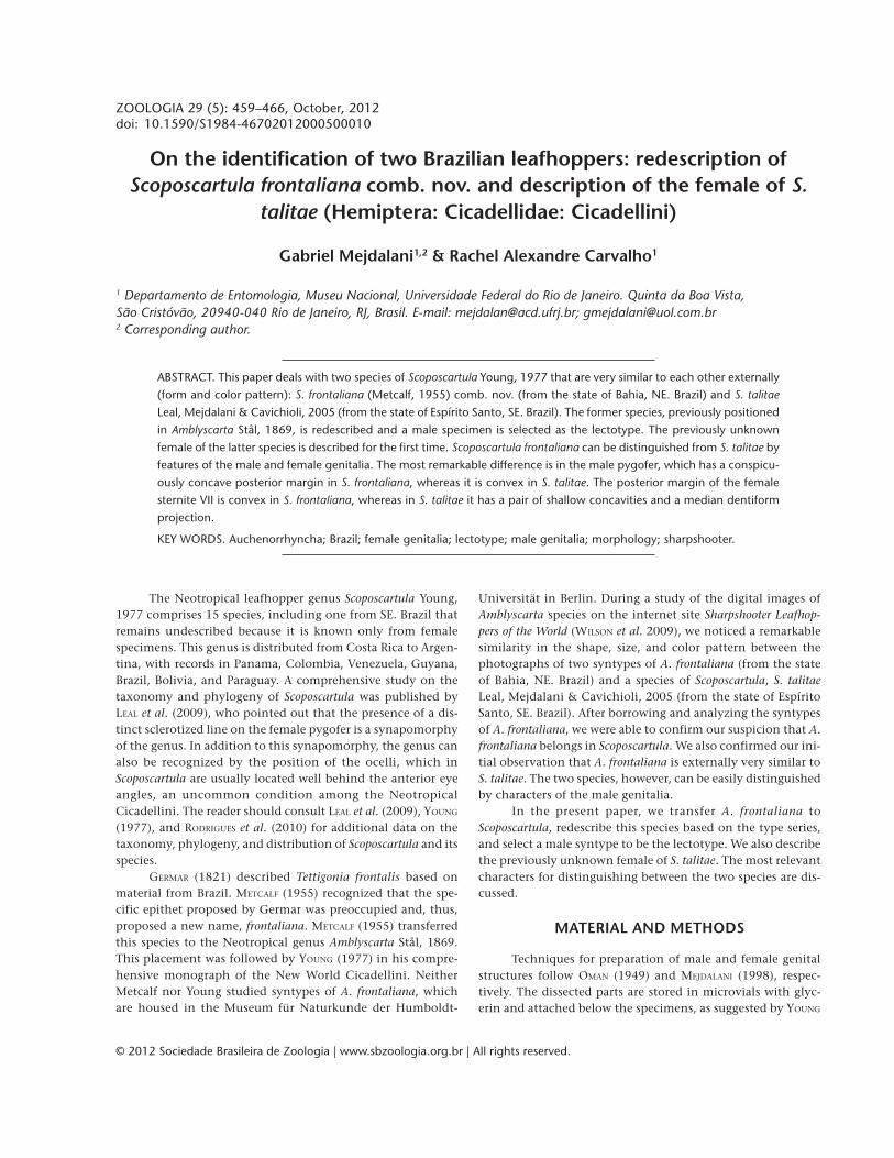
Head (Figs 1 and 2) moderately produced anteriorly;median length of crown about one-third interocular width andone-fourth transocular width; anterior margin broadly roundedin dorsal view; without carina at transition from crown to face;ocelli located behind line between anterior eye angles, eachocellus distinctly closer to adjacent eye angle than to medianline of crown; crown convex with depression on lateral por-tions between eye and ocellus; without median fovea and with-out sculpturing or setae; frontogenal sutures extending ontocrown and attaining ocelli; antennal ledges not protuberant,in lateral view with anterior margin oblique and convex; fronsconvex, not flattened medially, with distinct muscle impres-sions; epistomal suture complete; clypeus with upper half con-tinuing profile of frons, forming distinct angle with nearlyhorizontal lower half, apical margin convex.
Thorax (Figs 1 and 2) with pronotal width approximatelyequal to transocular distance; lateral pronotal margins slightlyconvergent anteriorly, posterior margin almost rectilinear; pos-terior 2/3 of pronotal disc distinctly striate; dorsopleural cari-nae complete (lectotype) or incomplete (additional specimen),declivous anteriorly. Mesonotum with scutellum not striate.Forewings (Fig. 1) with veins distinct; with four apical cells,
base of fourth more proximal than base of third; with threeclosed anteapical cells; membrane distinct, including apicalcells, apical portions of brachial and anteapical cells, and dis-tal costal area. Hindwings with vein R2+3 incomplete. Hindlegs(lectotype) with femoral setal formula 2:1:1 (2:1:1:1 on the rightside of additional specimen); length of first tarsomere slightlygreater than combined length of two succeeding tarsomeres;first tarsomere with two parallel rows of small setae on plantarsurface.
Color (Figs 1 and 2). Ground color of crown, pronotum,and mesonotum dark brown to black; lateral portions ofpronotum (lectotype) with two brownish-white, transverselyaligned maculae, outer macula larger than inner one (or withsingle pair of small maculae in additional specimen). Forew-ings dark red with three transverse, elongate white maculae:basalmost macula extending from basal half of clavus to nearcostal margin, median macula extending from distal half ofclavus to near costal margin, distalmost macula extending fromapex of brachial cell to outer margin of outer anteapical cell;forewing membrane amber. Face and lateral portions of thoraxmostly dark brown to black; large macula on median superiorportion of frons, large macula on lateral pronotal lobe, andmost of mesepimeron, brownish-white; rostrum and legs yel-lowish-brown to brown. Abdomen mostly red; genitalia darkbrown to black with ventral margin of pygofer red.
Genitalia. Pygofer (Fig. 3), in lateral view, well producedposteriorly, subrectangular, just slightly narrowed toward apex;posterior margin distinctly concave; macrosetae distributedmostly on posterior third of disc, extending slightly anteriorlyon ventral portion; without pygofer processes. Subgenital plates(Fig. 4), in ventral view, triangular, distinctly narrowed onmedian third, not fused to each other basally, not extending asfar posteriorly as pygofer apex; with uniseriate macrosetae alongouter margin. Connective (Fig. 5), in dorsal view, T-shaped;stalk distinctly longer than arms, extending farther posteriorlythan styles, directed dorsally and rightward (asymmetrical, notlocated on median sagittal plane). Styles (Fig. 5), in dorsal view,with outer median lobe; apical portion curved, digitiform,slightly narrowed apically; apex obtuse. Aedeagus (Fig. 6) sym-metrical, compressed laterally, without processes; shaft, in lat-eral view, simple, spatulate, with convex dorsal margin; withinconspicuous sclerotized projection on distal half of dorsalmargin; gonopore located apically. Paraphysis (Fig. 5) articu-lated to apex of connective, extremely elongate, extendingmuch farther posteriorly than apex of subgenital plates andslightly beyond pygofer apex ventrally, distinctly curved onbasal half, asymmetrical, not located on median sagittal plane;formed by single ramus (without smaller branch); apex acute.
Female. Length, 12.4 mm (n = 1).Head, thorax, and color similar to those of the above-
described male. Mesonotum with scutellum slightly striate.Fore- and hindwings, at rest position, extending well beyondovipositor apex. One specimen with single pair of small macu-
461On the identification of two Brazilian leafhoppers
ZOOLOGIA 29 (5): 459–466, October, 2012
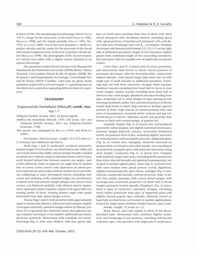
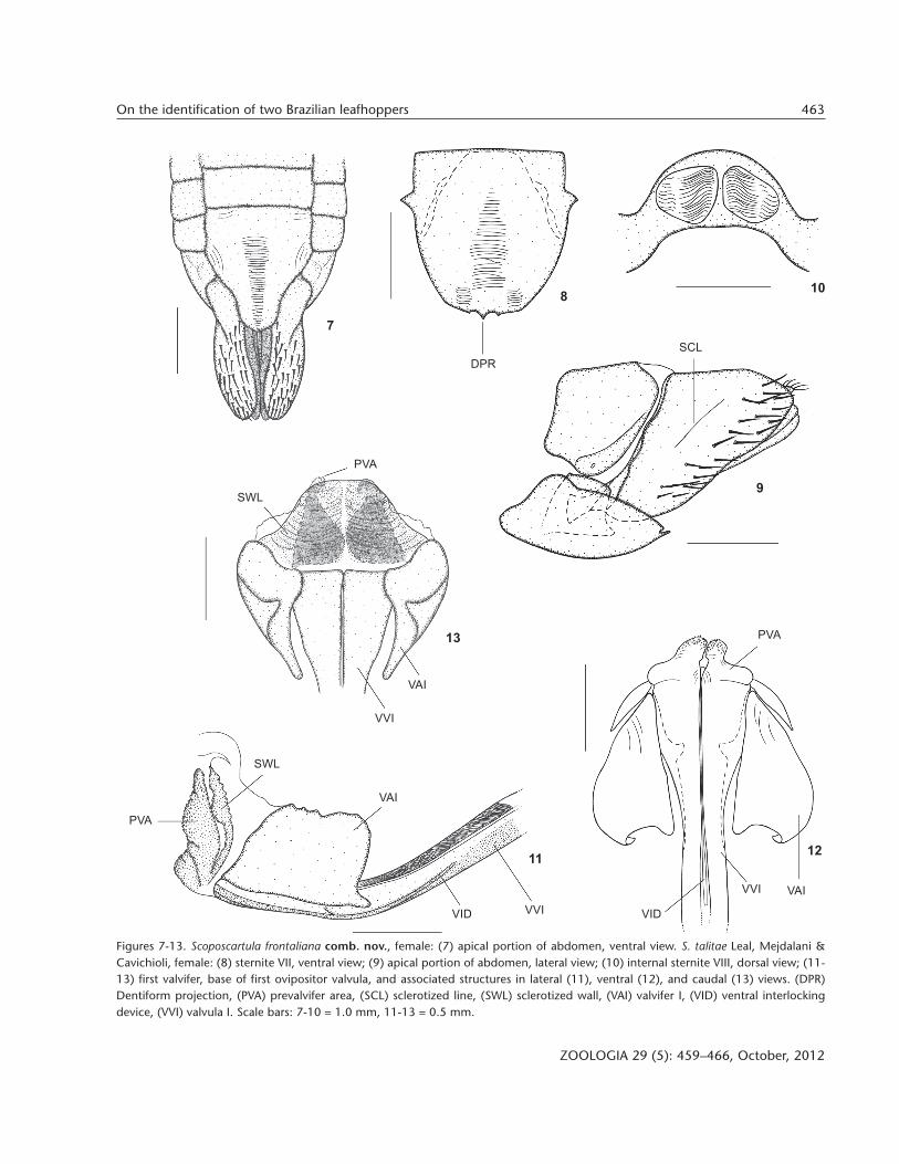
lae on lateral pronotal margins. Abdominal sternite VII (Fig. 7)well produced posteriorly; distal third narrowed, forming dis-tinct median lobe; posterior margin convex, round, withoutmedian dentiform projection.
Material examined. Northeastern Brazil, Bahia. Two males,one female: “Bahia\Freire [collector]\Nr. 6614”; “Syn-Typus ?”;“Zool. Mus.\Berlin” (ZMHB). One female, “frontalis\Gm.\Sign.
[SIGNORET 1853: 329] pl. 8 fig. 7”; “6614”; “? Type ?\Tettigonia\frontalis\Germar, 1821”; “[illegible word]\Bahia Freir.”;“Amblyscarta\frontaliana n. nov.\Metcalf, 1955”; “Zool.Mus.\Berlin” (ZMHB). One of the males, which was dissected, ishere designated as the lectotype (see justification in the discus-sion below); it received the following label: “LECTOTYPEM\Amblyscarta\frontaliana\Metcalf, 1955”. (An additional fe-
Figures 1-6. Scoposcartula frontaliana comb. nov., male lectotype (position of the pin on the mesonotum is indicated in figures 1 and 2):(1) body, lateral view; (2) head, pronotum, and mesonotum, dorsal view; (3-6) genitalia: (3) pygofer, valve, and subgenital plate, lateralview; (4) valve and subgenital plates, ventral view; (5) connective, style, and paraphysis, dorsal view; (6) ejaculatory reservoir, aedeagus,and anal tube, lateral view. Scale bars: 1 = 5.0 mm, 2-6 = 1.0 mm.
1
2
3
54
6
462 G. Mejdalani & R. A. Carvalho
ZOOLOGIA 29 (5): 459–466, October, 2012
male type with label “frontalis m.\Brasil.” is housed in the col-lection of the Ivan Franko National University, Lviv, Ukraine;HOLOVACHOV 2008 and Daniela Takiya, personal communication.).
Scoposcartula talitaeLeal, Mejdalani & Cavichioli, 2005
Figs 8-17
Scoposcartula talitae Leal, Mejdalani & Cavichioli, 2005: 2.
Description. Female. Length, 11.5 mm (n = 1).Head, thorax, and color similar to those of the above-
described male of S. frontaliana. Mesonotum with scutellumslightly striate. Fore- and hindwings, at rest position, extend-ing well beyond ovipositor apex. Lateroinferior portions of fronsand adjacent area of genae with white maculae; with singlepair of large white maculae on lateral pronotal portions;distalmost white macula of forewings extending from apex ofbrachial cell to near costal margin. Hindlegs with femoral setalformula 2:1:1 (left side) or 2:1:1:1 (right side).
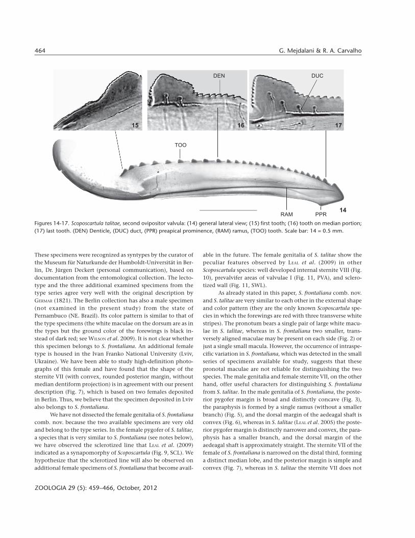
Genitalia. Abdominal sternite VII (Figs 8 and 9) well pro-duced posteriorly; distal third not forming distinct median lobe;posterior margin with pair of shallow concavities (emargin-ations) and median dentiform projection (Fig. 8, DPR); outer(ventral) surface striated on median portion; inner (dorsal) sur-face with large ridge with approximate form of inverted “V”(convex anteriorly, deeply concave posteriorly), arms of “V”forming expanded area posteriorly. Internal sternite VIII (Fig.10), in dorsal view, formed by pair of striated plates locatedabove inverted “V”. Pygofer (Fig. 9), in lateral view, moder-ately produced posteriorly; posterior margin narrowly rounded;dorsomedian portion of pygofer disc with faint sclerotized line(Fig. 9, SCL); macrosetae distributed on posterior third andextending anteriorly along ventral margin. Valvifers I (Figs 11-13, VAI), in lateral view, of somewhat trapezoidal form, withlobed projection on posteroventral portion. Valvulae I (Fig. 12,VVI), in ventral view, expanded basally (this expanded por-tion, in dorsal view, with lobe directed medially); prevalviferareas (Figs 11 and 12, PVA), in ventral view, with distinct lobeon inner portion of anterior margin and, in lateral view, form-ing conspicuous process directed dorsally; surface of prevalviferareas with tiny spiniform tegumentary processes; valvulae Iblade, in lateral view, with apical dentiform projection slightlyserrated dorsally; dorsal sculptured area (mostly scale-like pro-cesses arranged in oblique lines) extending from portion justbehind basal curvature to apical portion; ventral sculpturedarea (scale-like processes) restricted to apical portion; ventralinterlocking device (Figs 11 and 12, VID) distinct, elongate,restricted to basal half of blade, located along ventral blademargin but with distal portion directed dorsally. Sclerotizedwall (Fig. 11, SWL) located just behind prevalvifer areas, con-nected to the latter and to valvifers I, with corrugated surface.Valvulae II (Fig. 14), in lateral view, expanded beyond basalcurvature; dorsal margin moderately convex with approxi-
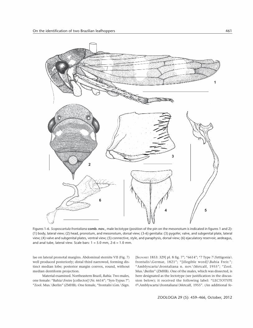
mately 25 continuous teeth (Fig. 14, TOO); most teeth formedby anterior elevated portion and posterior low flat portion (Figs16 and 17); first tooth (Fig. 15) distinct from remaining ones,its anterior portion lower than posterior one; denticles distrib-uted on teeth (Fig. 16, DEN) and on dorsal and ventral apicalportions (except on apex) of blade (dentate ventroapical por-tion longer than dorsoapical one); preapical prominence (Fig.14, PPR) distinct; apex obtuse; valvula with ducts (Fig. 17, DUC)extending into teeth or approaching them, as well as extend-ing toward apical portion. Gonoplacs, in lateral view, abruptlyexpanded on median portion; expanded portion slightly nar-rowed apically; apex rounded; surface with tiny spiniform tegu-mentary processes located mostly on posterior portion andextending anteriorly along ventral margin; some scattered smallsetae also present.
Material examined. Southeastern Brazil, Espírito Santo.One female: “BR/ES, Santa Teresa\Est. [Estação] Bio. [Biológica]Santa Lúcia\19-23/VIII/2009\R. Carvalho & M. Lopes” (MNRJ).One male (holotype): “Santa Teresa/ES\Reserva do Museu\16/X/2003\T. T. Mauro col.”; “Scoposcartula\talitae M\Leal et al.,2005\A. H. Leal det. 2005”; “HOLÓTIPO [holotype]” (MNRJ).One male (paratype): “Parque Sooretama\LINHARES Esp.Santo\Brasil V-1953\P A. Teles Col.”; “PARÁTIPO [paratype]”;“Scoposcartula\talitae M\Leal et al., 2005\A. H. Leal det. 2005”(DZUP).
DISCUSSION
The male genital features clearly indicate that Amblyscartafrontaliana belongs in Scoposcartula. For instance, the connec-tive has the shape of a transverse bar (i.e., it does not bear amedian stalk) in Amblyscarta (YOUNG 1977, MEJDALANI & NESSIMIAN
1991), whereas in Scoposcartula it has a distinct stalk, so that itcan be Y- or T-shaped (LEAL et al. 2009). The connective of S.frontaliana comb. nov. is clearly T-shaped (Fig. 5). In addition,paraphyses or paraphysis are never present in Amblyscarta(YOUNG 1977), whereas such structures are always well devel-oped in Scoposcartula (LEAL et al. 2009). The genitalia of S.frontaliana bear an extremely elongate paraphysis (Fig. 5). Fur-thermore, the examined specimens fit the detailed descriptionof Scoposcartula provided by YOUNG (1977). The unavailabilityof males prevented YOUNG (1977) from establishing the propergeneric assignment of this species. Since the taxonomy of theCicadellinae at the generic and specific levels is based prima-rily on features of the male genitalia, we believe that the selec-tion of a male to be the lectotype of S. frontaliana is advisable(see YOUNG 1958 and YOUNG & LAUTERER 1966 for relevant com-ments on lectotype proposals, the 1966 paper dealingespecifically with the Cicadellinae). Unfortunately, the origi-nal description (GERMAR 1821) did not mention the number ofspecimens examined or their sexes. The type locality (Brazil)given in the original description (“habitat in Brasilia”) is inagreement with the label data (“Bahia”) of our four specimens.
463On the identification of two Brazilian leafhoppers
ZOOLOGIA 29 (5): 459–466, October, 2012
Figures 7-13. Scoposcartula frontaliana comb. nov., female: (7) apical portion of abdomen, ventral view. S. talitae Leal, Mejdalani &Cavichioli, female: (8) sternite VII, ventral view; (9) apical portion of abdomen, lateral view; (10) internal sternite VIII, dorsal view; (11-13) first valvifer, base of first ovipositor valvula, and associated structures in lateral (11), ventral (12), and caudal (13) views. (DPR)Dentiform projection, (PVA) prevalvifer area, (SCL) sclerotized line, (SWL) sclerotized wall, (VAI) valvifer I, (VID) ventral interlockingdevice, (VVI) valvula I. Scale bars: 7-10 = 1.0 mm, 11-13 = 0.5 mm.
7
8 10
13
9
1211
DPRSCL
VAI
PVA
SWL
PVA
VVI
SWL
VAI
PVA
VVI VAI
VIDVVIVID
464 G. Mejdalani & R. A. Carvalho
ZOOLOGIA 29 (5): 459–466, October, 2012
These specimens were recognized as syntypes by the curator ofthe Museum für Naturkunde der Humboldt-Universität in Ber-lin, Dr. Jürgen Deckert (personal communication), based ondocumentation from the entomological collection. The lecto-type and the three additional examined specimens from thetype series agree very well with the original description byGERMAR (1821). The Berlin collection has also a male specimen(not examined in the present study) from the state ofPernambuco (NE. Brazil). Its color pattern is similar to that ofthe type specimens (the white maculae on the dorsum are as inthe types but the ground color of the forewings is black in-stead of dark red; see WILSON et al. 2009). It is not clear whetherthis specimen belongs to S. frontaliana. An additional femaletype is housed in the Ivan Franko National University (Lviv,Ukraine). We have been able to study high-definition photo-graphs of this female and have found that the shape of thesternite VII (with convex, rounded posterior margin, withoutmedian dentiform projection) is in agreement with our presentdescription (Fig. 7), which is based on two females depositedin Berlin. Thus, we believe that the specimen deposited in Lvivalso belongs to S. frontaliana.
We have not dissected the female genitalia of S. frontalianacomb. nov. because the two available specimens are very oldand belong to the type series. In the female pygofer of S. talitae,a species that is very similar to S. frontaliana (see notes below),we have observed the sclerotized line that LEAL et al. (2009)indicated as a synapomorphy of Scoposcartula (Fig. 9, SCL). Wehypothesize that the sclerotized line will also be observed onadditional female specimens of S. frontaliana that become avail-
able in the future. The female genitalia of S. talitae show thepeculiar features observed by LEAL et al. (2009) in otherScoposcartula species: well developed internal sternite VIII (Fig.10), prevalvifer areas of valvulae I (Fig. 11, PVA), and sclero-tized wall (Fig. 11, SWL).
As already stated in this paper, S. frontaliana comb. nov.and S. talitae are very similar to each other in the external shapeand color pattern (they are the only known Scoposcartula spe-cies in which the forewings are red with three transverse whitestripes). The pronotum bears a single pair of large white macu-lae in S. talitae, whereas in S. frontaliana two smaller, trans-versely aligned maculae may be present on each side (Fig. 2) orjust a single small macula. However, the occurrence of intraspe-cific variation in S. frontaliana, which was detected in the smallseries of specimens available for study, suggests that thesepronotal maculae are not reliable for distinguishing the twospecies. The male genitalia and female sternite VII, on the otherhand, offer useful characters for distinguishing S. frontalianafrom S. talitae. In the male genitalia of S. frontaliana, the poste-rior pygofer margin is broad and distinctly concave (Fig. 3),the paraphysis is formed by a single ramus (without a smallerbranch) (Fig. 5), and the dorsal margin of the aedeagal shaft isconvex (Fig. 6), whereas in S. talitae (LEAL et al. 2005) the poste-rior pygofer margin is distinctly narrower and convex, the para-physis has a smaller branch, and the dorsal margin of theaedeagal shaft is approximately straight. The sternite VII of thefemale of S. frontaliana is narrowed on the distal third, forminga distinct median lobe, and the posterior margin is simple andconvex (Fig. 7), whereas in S. talitae the sternite VII does not
Figures 14-17. Scoposcartula talitae, second ovipositor valvula: (14) general lateral view; (15) first tooth; (16) tooth on median portion;(17) last tooth. (DEN) Denticle, (DUC) duct, (PPR) preapical prominence, (RAM) ramus, (TOO) tooth. Scale bar: 14 = 0.5 mm.
15
DEN DUC
TOO
RAM PPR
16 17
14
465On the identification of two Brazilian leafhoppers
ZOOLOGIA 29 (5): 459–466, October, 2012
form a median lobe on the distal third and its posterior marginhas a pair of shallow concavities and a median dentiform pro-jection (Fig. 8, DPR).
The female specimen of S. talitae herein described wascollected in the type locality (Estação Biológica de Santa Lúcia),a 440 ha reserve of Atlantic Forest (dense ombrofilous forest;550-950 m a.s.l.) in the state of Espírito Santo (EBSL 2011).Unfortunately, we have no precise data on the distribution ofS. frontaliana comb. nov. in the state of Bahia. The otherScoposcartula species recorded from this state are S. concinna(Perty, 1833), S. furcifera Leal, Mejdalani & Cavichioli, 2005,and S. oculata (Signoret, 1853) (LEAL et al. 2009). Habitat dataare also not available for the latter three species. Brief habitatnotes have been published only for S. tobiasi Cavichioli &Mejdalani, 1996 and S. flavovittata Mejdalani, 1992. Accordingto CAVICHIOLI & MEJDALANI (1996), S. tobiasi is a common speciesin the Mantiqueira mountain range (SE. Brazil), occurring atItatiaia National Park from the inferior altitudinal forest (about1,500 m a.s.l.) to areas of transition between the superior alti-tudinal forest and the alpine field (campo de altitude; about 2,100m a.s.l.). Scoposcartula flavovittata, on the other hand, is appar-ently very rare (only two specimens are known). This species isrecorded only from its type locality, a very small spot of forestsurrounded by a restinga environment (sand-dune vegetation)at the state of Rio de Janeiro, SE. Brazil (MEJDALANI 1992,CAVICHIOLI & MEJDALANI 1996).
ACKNOWLEDGMENTS
We are greatly indebted to Jürgen Deckert (Museum fürNaturkunde der Humboldt-Universität), who has kindly sentus on loan the types of Scoposcartula frontaliana (Metcalf) comb.nov. and provided invaluable information on the Germar col-lection. Daniela Takiya (Universidade Federal do Rio de Janeiro)has also provided useful information on the Germar collec-tion. Ihor Shydlovskyy (Ivan Franko National University) haskindly sent us high-definition photographs of a female type ofS. frontaliana. Márcia Couri (Museu Nacional, UFRJ) allowed usto use the photographic equipment of her laboratory. A fel-lowship from Conselho Nacional de DesenvolvimentoCientífico e Tecnológico (CNPq; process number 301391/2011-4) to GM is acknowledged. The manuscript benefited from theuseful comments of two anonymous reviewers.
LITERATURE CITED
CAVICHIOLI, R.R. & G. MEJDALANI. 1996. Scoposcartula Young: des-crições de uma espécie nova e da fêmea de S. flavovittataMejdalani, e redescrição de S. concinna (Perty) comb. nov.(Homoptera, Cicadellidae, Cicadellini). Revista Brasileirade Zoologia 13: 963-971.
EBSL. 2011. Estação Biológica de Santa Lúcia. Diagnósticoambiental. Available online at: http://acd.ufrj.br/~araujo/
ebsl/biologica_diagnosticoambiental.htm [Accessed: 29/X/2011].
GERMAR, E. F. 1821. Bemerkungen über einige Gattungen derCicadarien. Magazin der Entomologie 4: 1-106.
HAMILTON, K.G.A. 1981. Morphology and evolution of the rhyn-chotan head (Insecta: Hemiptera, Homoptera). CanadianEntomologist 113: 953-974.
HILL, B.G. 1970. Comparative morphological study of selectedhigher categories of leafhoppers (Homoptera: Cicadellidae).Ann Arbor, University Microfilms, XI+187p.
HOLOVACHOV, O. 2008. Insects of E.-F. Germar in the collectionsof the Zoological Museum. Available online at: http://bioweb.lnu.edu.ua/zoo/mus/pages/germar_collection.htm[Accessed: 5/XI/2011].
LEAL, A.H.; G. MEJDALANI; & R.R. CAVICHIOLI. 2005. Two new speciesof the Neotropical leafhopper genus Scoposcartula (Insecta:Hemiptera: Cicadellidae: Cicadellini). Zootaxa 848: 1-9.
LEAL, A.H.; G. MEJDALANI; R.R. CAVICHIOLI & R.A. CARVALHO. 2009.Taxonomy and phylogeny of the leafhopper genusScoposcartula (Insecta: Hemiptera: Cicadellidae). Systematics& Biodiversity 7: 215-233.
MCKAMEY, S.H. 2007. Taxonomic catalogue of the leafhoppers(Membracoidea). Part 1. Cicadellinae. Memoirs of theAmerican Entomological Institute 78: 1-394.
MEJDALANI, G. 1992. Uma nova espécie de Scoposcartula Young,1977 do sudeste do Brasil (Homoptera, Cicadellidae,Cicadellinae). Revista Brasileira de Biologia 52: 231-234.
MEJDALANI, G. 1998. Morfologia externa dos Cicadellinae(Homoptera, Cicadellidae): comparação entre Versigonaliaruficauda (Walker) (Cicadellini) e Tretogonia cribrata Melichar(Proconiini), com notas sobre outras espécies e análise daterminologia. Revista Brasileira de Zoologia 15: 451-544.
MEJDALANI, G. & J.L. NESSIMIAN. 1991. Descrição do macho deAmblyscarta stillifera (Stal, 1862) (Homoptera, Cicadellidae,Cicadellinae). Revista Brasileira de Entomologia 35: 307-309.
METCALF, Z.P. 1955. New names in the Homoptera. Journal ofthe Washington Academy of Sciences 45: 262-267.
METCALF, Z.P. 1965. General catalogue of the Homoptera.Fascicle VI, Cicadelloidea. Part 1, Tettigellidae. Washing-ton, D.C., Agricultural Research Service, United StatesDepartment of Agriculture, 730p.
NIELSON, M.W. 1965. A revision of the genus Cuerna (Homoptera,Cicadellidae). Technical Bulletin of the United StatesDepartment of Agriculture 1318: 1-48.
OMAN, P.W. 1949. The Nearctic leafhoppers (Homoptera: Cica-dellidae). A generic classification and check list. Memoirsof the Entomological Society of Washington 3: 1-253.
RODRIGUES, L.G.N.; G. MEJDALANI & R.A. CARVALHO. 2010. A newspecies of Scoposcartula (Hemiptera: Cicadellidae: Cicadellini)with phylogenetic and biogeographic comments on thegenus. Zootaxa 2511: 59-68.
SIGNORET, V. 1853. Revue iconographique des Tettigonides. Annalesde la Société Entomologique de France 1: 323-374.
466 G. Mejdalani & R. A. Carvalho
ZOOLOGIA 29 (5): 459–466, October, 2012
WILSON, M.R.; J.A. TURNER & S.H. MCKAMEY. 2009. Sharpshooterleafhoppers of the world (Hemiptera: Cicadellidae subfamilyCicadellinae). Available online at: http://naturalhistory.museumwales.ac.uk/sharpshooters/home.php [Accessed:19/V/2010].
YOUNG, D.A. 1958. On lectotype proposals. Systematic Zoology7: 120-122.
YOUNG, D.A. 1977. Taxonomic study of the Cicadellinae(Homoptera: Cicadellidae), Part 2. New World Cicadellini
Submitted: 08.V.2012; Accepted: 05.VII.2012.Editorial responsibility: Walter A.P. Boeger
and the genus Cicadella. Technical Bulletin of the NorthCarolina Agricultural Experiment Station 239: 1-1135.
YOUNG, D.A. & B.P. BEIRNE. 1958. A taxonomic revision of theleafhopper genus Flexamia and a new related genus(Homoptera: Cicadellidae). Technical Bulletin of theUnited States Department of Agriculture 1173: 1-53.
YOUNG, D.A. & P. LAUTERER. 1966. Types of Cicadellinae(Homoptera, Cicadellidae) in the Moravian Museum. ActaMusei Moraviae 51: 261-270.