Embed Size (px)
DESCRIPTION
The bi-monthly review of oncology publications. Oncology News is a unique publication for cancer professionals, which provides reviews of oncology and related journals. Cancer care professionals will value the magazine’s practical and accessible approach becoming the most popular way to keep up-to-date on the latest news.
Citation preview

OncologyISSN 1751-4975
Volume 9 Issue 1 • March/April 2014 • www.oncologynews.biz
news
Wear A Hat DayTurn to page 37
ECHNO Special Editionvisit
Oncology News stand 16
Head & Neck Cancer – Use of Transoral Laser Microsurgery for Treatment of Hypopharyngeal Cancer
Neuro-oncology – Axl as a Therapeutic Target in Merlin-Deficient Tumours
Cancer Image Analysis – Image Based Tissue Segmentation: Towards the Automation of MammographicRisk Assessment
Lung Cancer – Raising Awareness Without Stigmatising
GI Cancer – Borderline Resectable Pancreatic Head Cancer: Neoadjuvant Chemotherapy and Portal
Breast Cancer – Understanding Breast Cancer Survival with Epidemiology
Medics & IT specialistscombine for uniquetraining App– Page 11
* Intermediate risk patient defi ned as having a Nottingham Prognostic Index (NPI) above 3.4 or being at intermediate risk by other decision making tools or protocols
1. NICE: Diagnostics guidance, DG10. Sept 2013
This piece is intended to educate physicians on the clinical utility of the Oncotype DX Breast Cancer Assay, and should not be provided to patients.Genomic Health and Oncotype DX are registered trademarks of Genomic Health, Inc. © 2014 Genomic Health, Inc. All rights reserved. GHI10290_1113_EN_UK
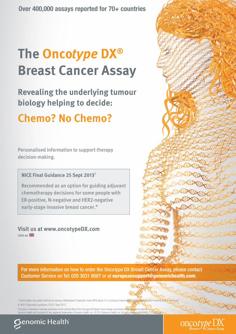
Over 400,000 assays reported for 70+ countries
The Oncotype DX® Breast Cancer AssayRevealing the underlying tumour biology helping to decide:
Chemo? No Chemo?
Visit us at www.oncotypeDX.comclick on
Personalised information to support therapydecision-making.
For more information on how to order the Oncotype DX Breast Cancer Assay, please contact Customer Service on Tel: 020 3031 8087 or at [email protected].
NICE Final Guidance 25 Sept 20131
Recommended as an option for guiding adjuvant chemotherapy decisions for some people with ER-positive, N-negative and HER2-negative early-stage invasive breast cancer.*
Volume 9 Issue 1 • March/April 2014 3
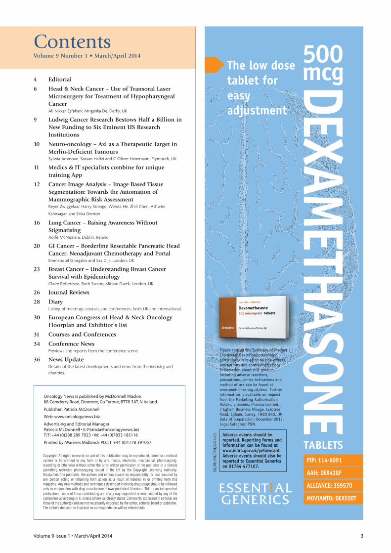
ContentsVolume 9 Number 1 • March/April 2014
4 Editorial
6 Head & Neck Cancer – Use of Transoral LaserMicrosurgery for Treatment of HypopharyngealCancer Ali Nikkar-Esfahani, Mriganka De, Derby, UK
9 Ludwig Cancer Research Bestows Half a Billion inNew Funding to Six Eminent US ResearchInstitutions
10 Neuro-oncology – Axl as a Therapeutic Target inMerlin-Deficient TumoursSylwia Ammoun, Sassan Hafizi and C Oliver Hanemann, Plymouth, UK
11 Medics & IT specialists combine for uniquetraining App
12 Cancer Image Analysis – Image Based TissueSegmentation: Towards the Automation ofMammographic Risk AssessmentReyer Zwiggelaar, Harry Strange, Wenda He, Zhili Chen, Ashwini
Kshirsagar, and Erika Denton
16 Lung Cancer – Raising Awareness WithoutStigmatisingAoife McNamara, Dublin, Ireland
20 GI Cancer – Borderline Resectable Pancreatic HeadCancer: Neoadjuvant Chemotherapy and PortalEmmanouil Giorgakis and Sas Dijk, London, UK
23 Breast Cancer – Understanding Breast CancerSurvival with EpidemiologyClaire Robertson, Ruth Swann, Miriam Dwek, London, UK
26 Journal Reviews
28 DiaryListing of meetings, courses and conferences, both UK and international.
30 European Congress of Head & Neck OncologyFloorplan and Exhibitor’s list
31 Courses and Conferences
34 Conference NewsPreviews and reports from the conference scene.
36 News UpdateDetails of the latest developments and news from the industry andcharities.
The low dosetablet foreasyadjustment
PIP: 114-6091
AAH: DEX418F
ALLIANCE: 359570
MOVIANTO: DEX500T
EG/D
E/ON
/JAN
/201
4/05
Adverse events should be reported. Reporting forms and information can be found at www.mhra.gov.uk/yellowcard. Adverse events should also be reported to Essential Generics on 01784 477167.
Please consult the Summary of Product Characteristics before prescribing, particularly in relation to side-effects, precautions and contra-indications. Information about this product, including adverse reactions, precautions, contra-indications and method of use can be found at www.medicines.org.uk/emc Further information is available on request from the Marketing Authorisation Holder: Chemidex Pharma Limited, 7 Egham Business Village, Crabtree Road, Egham, Surrey, TW20 8RB, UK. Date of preparation: December 2013. Legal Category: POM.
TABLETS
500DEXAMETHASONE mcg
Oncology News is published by McDonnell Mackie, 88 Camderry Road, Dromore, Co Tyrone, BT78 3AT, N Ireland.
Publisher: Patricia McDonnell
Web: www.oncologynews.biz
Advertising and Editorial Manager: Patricia McDonnell • E: [email protected]/F: +44 (0)288 289 7023 • M: +44 (0)7833 185116
Printed by: Warners Midlands PLC, T: +44 (0)1778 391057
Copyright: All rights reserved; no part of this publication may be reproduced, stored in a retrievalsystem or transmitted in any form or by any means, electronic, mechanical, photocopying,recording or otherwise without either the prior written permission of the publisher or a licensepermitting restricted photocopying issued in the UK by the Copyright Licensing Authority.Disclaimer: The publisher, the authors and editors accept no responsibility for loss incurred byany person acting or refraining from action as a result of material in or omitted from thismagazine. Any new methods and techniques described involving drug usage should be followedonly in conjunction with drug manufacturers' own published literature. This is an independentpublication - none of those contributing are in any way supported or remunerated by any of thecompanies advertising in it, unless otherwise clearly stated. Comments expressed in editorial arethose of the author(s) and are not necessarily endorsed by the editor, editorial board or publisher.The editor's decision is final and no correspondence will be entered into.
4 Volume 9 Issue 1 • March/April 2014
FROM THE EDITOR
An Enemy of the People –“War”on CancerEnemies and WarsCancer has been with us (mankind and other livingthings) throughout evolution. It is an insidious andunwarranted manifestation in which cells behave inan uncontrolled manner, putting an organism at riskof an earlier death than by “natural causes”. We seeit as an aberration – an enemy of the people – tobe attacked, and ultimately and ideally preventedor cured. Richard Nixon’s declaration of war oncancer in the ‘70s was based on encouragingadvances regarding RNA viruses and cancer, leadingto the notion that if enough money is thrown atthe problem, it could be conquered in a decade ortwo – truly wishful thinking. Forty years on, we hear the rallying call again
for attack in the guise of battle-zones and war-zones [1]. A consensus reached among its thought-leaders was that “for most forms of cancer,enduring disease-free responses are rare, and cureseven rarer.” Curiously, however, this sort of warcapitulation comes attached to the equallycategorical non-sequitur – “despite extraordinaryprogress in our understanding of diseasepathogenesis…” In the context in which cancerresearch and therapeutics operate, these twostatements dominating the narrative may becharacterised as a “lack of fit” or paradoxical.Generals (some of the thought-leaders”?) areneeded to take charge of the “war”, cooperatinginternationally and using all possible tactics fromevery possible angle. Progress in the last fourdecades saw many leaps forward, but somecommentators suggest the advances overall havenot been significant [2-4]. Assuming our semi-demoralised community of cancer researchers canregroup, redeploy their forces more effectivelyand adopt better strategies [1,2], would areassessment 40 years on be any better? Wagingoutright war would undoubtedly advance us a fewmore steps, but complete prevention and cure areuntenable goals. Cancer is a perennial problem,not one that can be defeated within a given time-frame. For this reason, Hanahan’s analogy [1] seemsfarfetched and overstated (e.g. his penultimatesentence refers to a “multidimensional cancerbattlespace vision” – but read on!). The problemswith a holistic war are how it can beimplemented, co-ordinated and financed. Wars areextraordinarily expensive; the announcement(page 9) that the Ludwig Foundation will spendhalf a billion US dollars over five years may bringdividends, but will probably be seen in due courseas a drop in the ocean.
External versus internal factorsA top priority must be prevention, for whichintimate knowledge of external risk factors relatedto carcinogenesis is required; thanks to thecontributions of epidemiologists, there is a quitesophisticated list of those risks. It is more difficultto increase the chances of prevention wheregenetic and internal factors predisposing people tocancer are concerned, especially in asymptomaticcancers. Cancer might also be due to a misplacedcell in the society of cells – the original (stem?) cellidea called an embryonic rest by Cohnheim-Ribbert[5] – or as stressed in Smithers “attack oncytologism” (6).
Control and Quality of Life (QOL) Curing cancer remains a pipe-dream; the disorderwill arise as long as our species exists, withincreased longevity now exacerbating the problem.We must think in terms of control being a morepractical goal, which requires rational interventioncoupled with excellent management “on all fronts.”Management has to be customised since alltumours are unique and constantly changing, andthe teams involved nowadays include professionsfrom surgeons to chaplains. The cost, especiallywith ageing populations, might soon run away withall health budgets. The focus should also remainfirmly on QOL, however short life-expectancymight be, since this often brings the greatestbenefit to the patient.
Guerrilla tactics – metastasis We have to understand how tumour cells invadeother tissues near and far; without dissemination,cancer becomes a much more manageableproblem. “Guerrilla tactics” are extremely difficultto combat, especially when the enemy keepschanging its characteristics and tactics. Thischanging behaviour leads to resistance, an issuethat should also have high priority. To take ouranalogies further, my impression is that cancers aremore like terrorist activities that have manydifferent origins, and that need to be prevented inthe first place, otherwise contained and eliminatedas best possible in each case, which has littlesemblance to outright war.Our struggle against cancer is an enduring
activity, not a battle or war with a victory in sight.The notion that this is achievable by all-out war ispoorly founded and insensitive in that it continuesto give false hope to many cancer sufferers. ●
REFERENCES
1. Hanahan D. Series 2.Rethinking the war on cancer.The Lancet 2014;383:558-83.
2. Bagley CG and Ellis LM.Raise standards for preclinicalcancer research. Nature,2012;483:531-3.
3. Kolata G. As Other DeathRates Fall, Cancer’s ScarcelyMoves. The New York Times,April 24, 2009.
4. Wheatley D. Progress inCancer – a disappointingperformance, but a reasonableprospect? (Editorial)Oncology News, 7, June2013. See also: The difficultyof delivering promises: a fewcomments. Ibid, Sep 2013.
5. See Morton LT, Moore R J. Achronology of medicine andrelated sciences, Scolar Press,1997, 475, which discussesthe Cohnheim-Ribberttheory of cancer.
6. Smithers DW. Cancer – anAttack on Cytologism. TheLancet. 1962;1(7228):493-9.
Denys WheatleyEditor

Setofilm 4mg and 8mg orodispersible films. Abbreviated Prescribing InformationREFER TO THE SUMMARY OF PRODUCT CHARACTERISTICS (SmPC) BEFORE PRESCRIBING. Presentation: Indication:
Dosage and administration:
Table 1: BSA and Weight based dosing for Chemotherapy
BSA Day 1a,b Day 2-6b
<0.6m
0.6mWeight Day 1a,b Day 2-6b
ab
Contra-indications:
Warnings and precautions for use:
Interactions:
Pregnancy and lactation: Use Side effects:
Licensing and Legal Category:
For further information contact:
®
Date of preparation/revision:
Adverse events should be reported. Reporting forms and information can be found at www.mhra.gov.uk/yellowcard. Adverse events should also be reported to
Medical Information at Norgine Pharmaceuticals Ltd on 01895 826606.
References: 1. ®
2. SE/3650/JUL/13.
A new form of ondansetron that dissolves in seconds.1
SETOFILM®
offers a
20%cost saving...2 ...versus ODT o
nd
ansetron
6 Volume 9 Issue 1 • March/April 2014
Squamous cell carcinoma of thehypopharynx is relatively rare comparedto other major sites of the head and neck,and accounts for approximately 3% to 5%
of all head and neck squamous cell carcinomas.Despite this low incidence, hypopharyngealcarcinomas show the worst survival rates withinthe head and neck region. Advanced stage ofdisease at the time of diagnosis seems to bemainly responsible for the poor prognosis.Interestingly, oncologic results for hypopharyngealcarcinomas have not significantly improved duringrecent decades regardless of the chosenmanagement scheme [1]. Traditionally, total laryngopharyngectomy
followed by postoperative radiation has been thepreferred treatment in many centres. In the lastdecade, with the emergence of organ preservationprotocols, a tendency towards chemo-radiotherapy has reduced the percentage ofprimary surgery in head and neck carcinomas.However, the success of organ preservationprotocols relies not only on favourable survivaland preservation rates, but also on adequatefunction of the remaining organ, together with thefeasibility of adequate salvage surgery for caseswith local and regional failure. Long-term toxicityin patients treated with concurrent chemo-radiotherapy, with the subsequent loss of functionof many preserved organs and inability to benefitfrom radiation in the future has made chemo-radiotherapy a suboptimal choice of treatment [2].This is especially relevant in piriform fossa
because the dose administered to the pharyngealconstrictor muscles cannot be reduced due tothese structures being the primary target and thefeasibility of salvage surgery is low in comparisonto laryngeal carcinoma .Takes et al analyses thecurrent trends in the initial management ofhypopharyngeal carcinoma and concludes that, inearly stages, both surgery and radiotherapy areconsidered good organ-preserving treatmentoptions. For advanced disease, most patients aretreated with total laryngopharyngectomyfollowed by chemo-radiotherapy or up-frontchemo-radiotherapy. However, the authors
propose that the TNM classification may be abetter tool to guide physicians in treatmentdecisions involving organ preservation strategiesthan overall stage classification [3]. In a further review, Gourin and Johnson reveal
that despite the increasing popularity of organpreservation protocols, primary surgical therapiescontinue to play an important role. The authorssuggest that primary surgery is indicated inselected hypopharyngeal carcinomas when thesurgical approach offers an alternative to radiationor the possibility to reduce the intensity ofadjuvant therapy or when the extent of theprimary tumour mandates a surgical approach tooptimise survival and function [4].
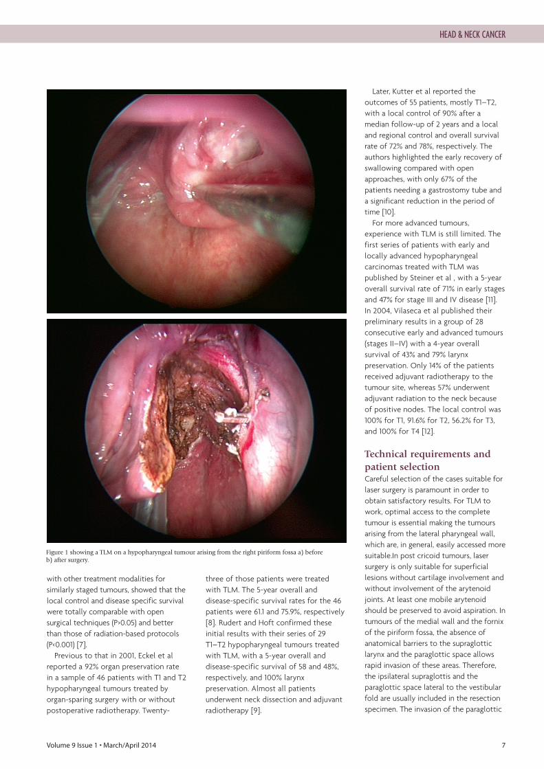
Transoral Laser Microsurgery (TLM)for hypopharyngeal carcinomaUse of CO2 laser for oncological purposes in theupper aerodigestive tract was first introduced bySteiner in the late 1980s; His initial results weregiven no credit by many head and neck surgeons,but encouraged by others [5]. In the early 1990s,Zeitels et al reported a case series ofsupraglottic and hypopharyngeal carcinomastreated with laser only or with laser plusradiotherapy, The lesions were highly selected forsmall volume and endoscopic accessibility. Theauthors concluded that endoscopic resectionswere less morbid and more cost-effective thanopen surgery or radiotherapy [6]. While use ofTLM in early laryngeal carcinoma has becomeincreasingly used, its use in treatment ofhypopharyngeal carcinomas remains to be theless established (Figure 1).In 2010, Karatzanis et al evaluated 119 patients
with T1 and T2 hypopharyngeal carcinomasprimarily managed with laser surgery. Localcontrol and 5-year disease-specific survival were90 and 77.8% for T1, and 83.1 and 70% for T2. 2.5%of Patients received permanent trachesotomiesdue to chronic post operative aspirations and 2.5% were reported to require permanentgastrostomy tubes due to impaired swallowing.More recently, A retrospective comparison at thesame institution, comparing the outcomes of TLM
Use of Transoral LaserMicrosurgery for Treatment ofHypopharyngeal Cancer
Ali Nikkar-EsfahaniDepartment of Otolaryngology-Head and Neck Surgery, Royal Derby Hospital, Derby, UK.
Mriganka DeDepartment of Otolaryngology-Head and Neck Surgery, Royal Derby Hospital, Derby, UK.
Correspondence address:Ali Nikkar-Esfahani,Department of Otolaryngology, Royal Derby Hospital. E: [email protected]
HEAD & NECK CANCER
Volume 9 Issue 1 • March/April 2014 7
with other treatment modalities forsimilarly staged tumours, showed that thelocal control and disease specific survivalwere totally comparable with opensurgical techniques (P>0.05) and betterthan those of radiation-based protocols(P<0.001) [7]. Previous to that in 2001, Eckel et al
reported a 92% organ preservation ratein a sample of 46 patients with T1 and T2hypopharyngeal tumours treated byorgan-sparing surgery with or withoutpostoperative radiotherapy. Twenty-
three of those patients were treatedwith TLM. The 5-year overall anddisease-specific survival rates for the 46patients were 61.1 and 75.9%, respectively[8]. Rudert and Hoft confirmed theseinitial results with their series of 29T1–T2 hypopharyngeal tumours treatedwith TLM, with a 5-year overall anddisease-specific survival of 58 and 48%,respectively, and 100% larynxpreservation. Almost all patientsunderwent neck dissection and adjuvantradiotherapy [9].
Later, Kutter et al reported theoutcomes of 55 patients, mostly T1–T2,with a local control of 90% after amedian follow-up of 2 years and a localand regional control and overall survivalrate of 72% and 78%, respectively. Theauthors highlighted the early recovery ofswallowing compared with openapproaches, with only 67% of thepatients needing a gastrostomy tube anda significant reduction in the period oftime [10].For more advanced tumours,
experience with TLM is still limited. Thefirst series of patients with early andlocally advanced hypopharyngealcarcinomas treated with TLM waspublished by Steiner et al , with a 5-yearoverall survival rate of 71% in early stagesand 47% for stage III and IV disease [11].In 2004, Vilaseca et al published theirpreliminary results in a group of 28consecutive early and advanced tumours(stages II–IV) with a 4-year overallsurvival of 43% and 79% larynxpreservation. Only 14% of the patientsreceived adjuvant radiotherapy to thetumour site, whereas 57% underwentadjuvant radiation to the neck becauseof positive nodes. The local control was100% for T1, 91.6% for T2, 56.2% for T3,and 100% for T4 [12].
Technical requirements andpatient selectionCareful selection of the cases suitable forlaser surgery is paramount in order toobtain satisfactory results. For TLM towork, optimal access to the completetumour is essential making the tumoursarising from the lateral pharyngeal wall,which are, in general, easily accessed moresuitable.In post cricoid tumours, lasersurgery is only suitable for superficiallesions without cartilage involvement andwithout involvement of the arytenoidjoints. At least one mobile arytenoidshould be preserved to avoid aspiration. Intumours of the medial wall and the fornixof the piriform fossa, the absence ofanatomical barriers to the supraglotticlarynx and the paraglottic space allowsrapid invasion of these areas. Therefore,the ipsilateral supraglottis and theparaglottic space lateral to the vestibularfold are usually included in the resectionspecimen. The invasion of the paraglottic
Figure 1 showing a TLM on a hypopharyngeal tumour arising from the right piriform fossa a) before b) after surgery.
HEAD & NECK CANCER
8 Volume 9 Issue 1 • March/April 2014
space lateral to the true vocal cordusually precludes the indication of TLM.These limitations reduce the percentageof patients with hypopharyngealcarcinoma that are suitable for TLM atpresentation [2].
Advantages of transoral lasermicrosurgeryIn contrast to radical surgical procedures,TLM allows minimisation of the loss ofhealthy tissue and thus avoiding extensivereconstruction procedures. In most cases,tracheotomies are not required and theneed for postoperative gastrostomy tubesis lower when compared to otherconservation regimen or to open surgery.The preservation of pharyngeal sensorynerve function results in betterpostoperative swallowing and furtherreduces postoperative morbidity such asaspiration pneumonia. TLM can beseamlessly integrated into any therapeuticregimen while maintaining all salvagetreatment options. Furthermore, the minimally invasive
nature of TLM increases indications in theelderly whenever the general
performance status is adequate to allowsurgery. Compared with organpreservation protocols, one of theadvantages of TLM is the possibility toobtain prognostic information from thesurgical specimen. Precise data on tumourcharacteristics, nodal status, or thepresence of perineural or vascularinvasion will allow rational administrationof adjuvant treatment preventingovertreatment.
Disadvantages of transorallaser microsurgery One of the limitations of TLM is thereduced percentage of patients suitablefor this technique at presentation.Although few authors have publishedgood results in moderately advancedcases, the best functional and oncologicresults are obtained in T1–T2 which makeonly 20% of hypopharyngeal carcinomas. An advanced learning curve in the field
of TLM is required to approach and treatmoderately advanced hypopharyngealcarcinomas with a favourable success rate.According to the low percentage of earlycases at initial presentation, it could be
difficult for most of the surgeons toachieve such a learning curve, but, withthe increasing use of the laser and theemerging field of transoral roboticsurgery, an increase in experience is to beexpected in the future.
ConclusionIn experienced hands, TLM is a realalternative to any other surgical ornonsurgical therapeutic regimen in thetreatment of early hypopharyngealcarcinoma. High rates of organ andfunction preservation can be achievedwithout compromising the oncologicoutcome and with relatively lowmorbidity. A careful selection of suitablepatients is mandatory for this kind ofsurgery. Randomised studies comparingprimary surgical approaches and organ-preservation protocols are necessary toclarify the role of primary surgery in thetreatment of hypopharyngeal carcinomas.The inclusion of outcomes such as survivalrates, organ and function preservationrates, cost of the procedures and qualityof life should be mandatory. ●
1. Martin A, Jackel M, Christiansen H, Mahmoodzada M, Kron M, Steiner W. OrganPreserving Transoral Laser Microsurgery for Cancer of the Hypopharynx. Laryngoscope,2008;118:398-402.
2. Vilaseca I, Blanch JL, Bernal-Sprekelsen M. Transoral laser surgery for hypopharyngealcarcinomas. Curr Opin Otolaryngol Head NeckSurg. 2012;Apr;20(2):97-102.
3. Takes RP, Strojan P, Silver CE. Current trends in initial management ofhypopharyngeal cancer: the declining use of open surgery. Head Neck,2012;Feb;34(2):270-81.
4. Gourin CG, Johnson JT. A contemporary review of indications for primary surgical careof patients with squamous cell carcinoma of the head and neck. Laryngoscope2009;119:2124-34.
5. Steiner W. Experience in endoscopic laser surgery of malignant tumors of theupperaerodigestive tract. Adv Otorhinolaryngol 1988;39:135-44.
6. Zeitels SM, Koufman JA, Davis RK, Vaughan CW. Endoscopic treatment ofsupraglottis and hypopharynx cancer. Laryngoscope 1994;104:71-8.
7. Karatzanis AD, Psychogios G, Waldfahrer F. T1 and T2 hypopharyngeal cancertreatment with laser microsurgery. J Surg Oncol 2010;102:27-33.
8. Eckel HE, Staar S, Volling P, et al. Surgical treatment for hypopharynx carcinoma:feasibility, mortality, and results. Otolaryngol Head Neck Surg, 2001;124:561-9.
9. Rudert HH, Hoft S. Transoral carbon-dioxide laser resection of hypopharyngealcarcinoma. Eur Arch Otorhinolaryngol 2003;260:198-206.
10. Kutter J, Lang F, Monnier P, Pasche P. Transoral laser surgery for pharyngeal andpharyngolaryngeal carcinomas. Arch Otolaryngol Head Neck Surg 2007;133:139-44.
11. Steiner W, Ambrosch P, Hess CF, Kron M. Organ preservation by transoral lasermicrosurgery in piriform sinus carcinoma. Otolaryngol Head Neck Surg 2001;124:58-67.
12. Vilaseca-González I, Bernal-Sprekelsen M, Blanch-Alejandro JL, Moragas-Lluis M.Complications in transoral CO2 laser surgery for carcinoma of the larynx andhypopharynx. Head Neck 2003;25:382-8.
REFERENCES
HEAD & NECK CANCER
Further information from: Ivor Smith, ScheBo • Biotech UK Ltd, PO Box 6359, Basingstoke, RG22 4WETel: 01256 477259 Fax: 01256 327889 E-mail: [email protected] www.schebo.co.uk
®
• Bringing sensitivity to bowel cancer screening
• Diagnosis and monitoring of various cancers
®
•
•®
M2-pyruvate kinase has a key role in controlling tumour glucose metabolism
Volume 9 Issue 1 • March/April 2014 9
Ludwig Cancer Research Bestows Half aBillion in New Funding to Six EminentUS Research Institutions
In January 2014 Cancer research in
the US got a critical boost todayas the six Ludwig Centers atJohns Hopkins University, Harvard
University, the Massachusetts Instituteof Technology, Memorial Sloan-Kettering Cancer Center, StanfordUniversity and the University ofChicago received a total of $540million as part of a gift from LudwigCancer Research, on behalf of itsfounder, Daniel K Ludwig. This newfunding ranks among the largestprivate philanthropic gifts to cancerresearch.This gift adds to the endowments
established in 2006 to create theLudwig Centers at each institution,bringing the Ludwig total funding atthese institutions to $900 million.Ludwig’s global contribution toadvancing cancer research is now $2.5billion.“Never before has the cancer
community had the knowledge andtools to probe so deeply intounderstanding cancer and discoveringnew ways to defeat it,” said EdMcDermott, Ludwig trustee andpresident and CEO of the LudwigInstitute for Cancer Research. “Moremust be done in terms of funding toensure continued progress in an era ofshrinking global resources for research.Providing reliable, long-term supportto scientists fosters high impact,innovative research and must remain apriority for the cancer community.”Initial funding to the six US-based
Ludwig Centers has already yieldedgroundbreaking discoveries. It haspaved the way for the firstcomprehensive maps of the genomiclandscapes of cancers, transformative“smart drugs” and immunotherapytreatments, and fast-tracked researchto bring new treatments for varioustypes of metastatic and rare cancers.
“The additional funding receivedtoday will allow the Ludwig Centers toexpand and amplify their efforts inperpetuity. Sustained support enablesthe Centers to continue training thebest and the brightest of the nextgeneration of scientists,” said BertVogelstein, MD, co-director, LudwigCenter at Johns Hopkins. “Ludwig putsgreat faith in its scientists by providingongoing investment that allows themto expedite research and take risks –the only way to make trulybreakthrough discoveries.”This gift complements the late
American businessman Daniel KLudwig’s global plan for financingcancer research. The new funding wasrealised by the sale of New York realestate investments held by Mr Ludwig.Ludwig’s first contribution to cancerresearch was made in 1971 when heestablished the Ludwig Institute forCancer Research – a not-for-profitthat supports more than 600 cancerresearchers at dedicated labs aroundthe world. Ludwig Cancer Researchcomprises the Ludwig Institute, the sixUS-based Ludwig Centers and selectaffiliated scientists across the globe.“With independent, flexible, and
long-range funding we can now take anidea based on the best scientific andmedical insights, and pursue it furtherregardless of how long it may take orthe size of the eventual patientpopulation it may benefit,” said GeorgeD Demetri, MD, co-director, LudwigCenter at Harvard. “We also have thefreedom to collaborate with leadingscientists around the globe, which canlead to new innovations to help cancerpatients.”
Visit: www.ludwigcancerresearch.org
10 Volume 9 Issue 1 • March/April 2014
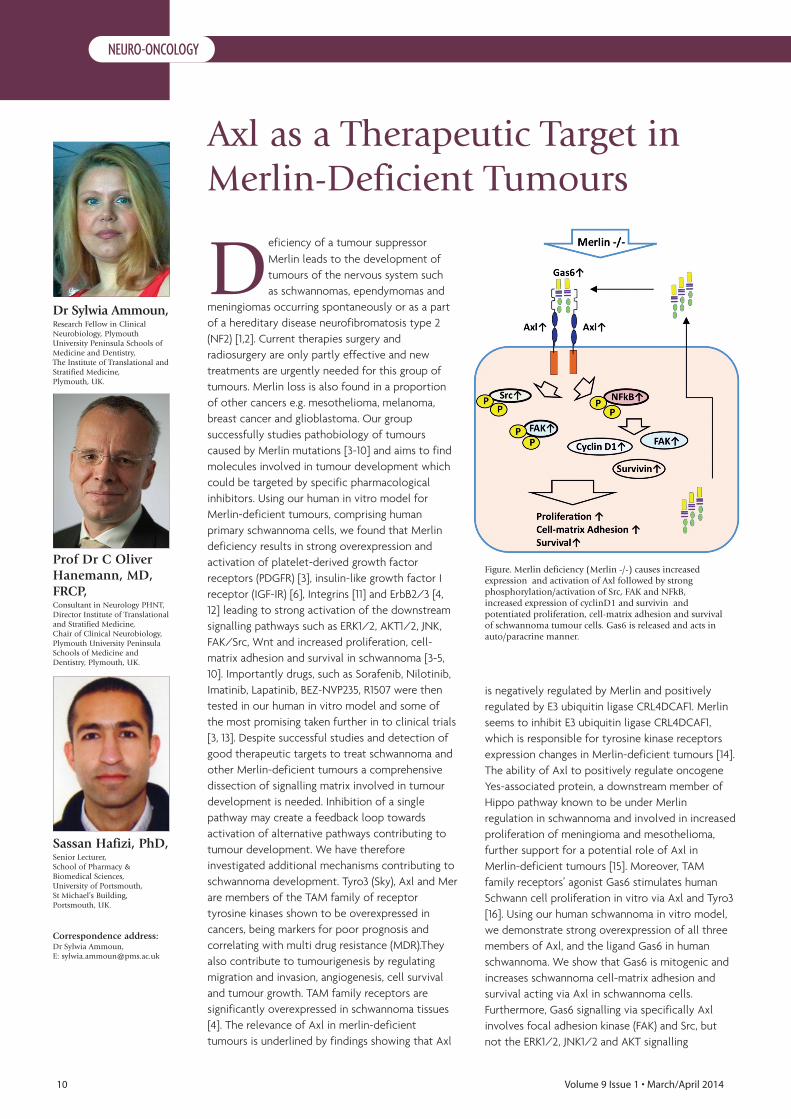
Deficiency of a tumour suppressorMerlin leads to the development oftumours of the nervous system suchas schwannomas, ependymomas and
meningiomas occurring spontaneously or as a partof a hereditary disease neurofibromatosis type 2(NF2) [1,2]. Current therapies surgery andradiosurgery are only partly effective and newtreatments are urgently needed for this group oftumours. Merlin loss is also found in a proportionof other cancers e.g. mesothelioma, melanoma,breast cancer and glioblastoma. Our groupsuccessfully studies pathobiology of tumourscaused by Merlin mutations [3-10] and aims to findmolecules involved in tumour development whichcould be targeted by specific pharmacologicalinhibitors. Using our human in vitro model forMerlin-deficient tumours, comprising humanprimary schwannoma cells, we found that Merlindeficiency results in strong overexpression andactivation of platelet-derived growth factorreceptors (PDGFR) [3], insulin-like growth factor Ireceptor (IGF-IR) [6], Integrins [11] and ErbB2/3 [4,12] leading to strong activation of the downstreamsignalling pathways such as ERK1/2, AKT1/2, JNK,FAK/Src, Wnt and increased proliferation, cell-matrix adhesion and survival in schwannoma [3-5,10]. Importantly drugs, such as Sorafenib, Nilotinib,Imatinib, Lapatinib, BEZ-NVP235, R1507 were thentested in our human in vitro model and some ofthe most promising taken further in to clinical trials[3, 13]. Despite successful studies and detection ofgood therapeutic targets to treat schwannoma andother Merlin-deficient tumours a comprehensivedissection of signalling matrix involved in tumourdevelopment is needed. Inhibition of a singlepathway may create a feedback loop towardsactivation of alternative pathways contributing totumour development. We have thereforeinvestigated additional mechanisms contributing toschwannoma development. Tyro3 (Sky), Axl and Merare members of the TAM family of receptortyrosine kinases shown to be overexpressed incancers, being markers for poor prognosis andcorrelating with multi drug resistance (MDR).Theyalso contribute to tumourigenesis by regulatingmigration and invasion, angiogenesis, cell survivaland tumour growth. TAM family receptors aresignificantly overexpressed in schwannoma tissues[4]. The relevance of Axl in merlin-deficienttumours is underlined by findings showing that Axl
is negatively regulated by Merlin and positivelyregulated by E3 ubiquitin ligase CRL4DCAF1. Merlinseems to inhibit E3 ubiquitin ligase CRL4DCAF1,which is responsible for tyrosine kinase receptorsexpression changes in Merlin-deficient tumours [14].The ability of Axl to positively regulate oncogeneYes-associated protein, a downstream member ofHippo pathway known to be under Merlinregulation in schwannoma and involved in increasedproliferation of meningioma and mesothelioma,further support for a potential role of Axl inMerlin-deficient tumours [15]. Moreover, TAMfamily receptors’ agonist Gas6 stimulates humanSchwann cell proliferation in vitro via Axl and Tyro3[16]. Using our human schwannoma in vitro model,we demonstrate strong overexpression of all threemembers of Axl, and the ligand Gas6 in humanschwannoma. We show that Gas6 is mitogenic andincreases schwannoma cell-matrix adhesion andsurvival acting via Axl in schwannoma cells.Furthermore, Gas6 signalling via specifically Axlinvolves focal adhesion kinase (FAK) and Src, butnot the ERK1/2, JNK1/2 and AKT signalling
Axl as a Therapeutic Target inMerlin-Deficient Tumours
Dr Sylwia Ammoun,Research Fellow in ClinicalNeurobiology, PlymouthUniversity Peninsula Schools ofMedicine and Dentistry, The Institute of Translational andStratified Medicine, Plymouth, UK.
Prof Dr C OliverHanemann, MD,FRCP,Consultant in Neurology PHNT,Director Institute of Translationaland Stratified Medicine,Chair of Clinical Neurobiology,Plymouth University PeninsulaSchools of Medicine andDentistry, Plymouth, UK.
Sassan Hafizi, PhD,Senior Lecturer,School of Pharmacy &Biomedical Sciences,University of Portsmouth,St Michael’s Building,Portsmouth, UK.
Correspondence address:Dr Sylwia Ammoun,E: [email protected]
Figure. Merlin deficiency (Merlin -/-) causes increasedexpression and activation of Axl followed by strongphosphorylation/activation of Src, FAK and NFkB,increased expression of cyclinD1 and survivin andpotentiated proliferation, cell-matrix adhesion and survivalof schwannoma tumour cells. Gas6 is released and acts inauto/paracrine manner.
NEURO-ONCOLOGY
pathways. We also demonstrate the role ofNFkB, which regulates Gas6/Axl mediatedoverexpression of survivin, cyclin D1 and FAKleading to enhanced survival, cell-matrixadhesion and proliferation of schwannoma.NFkB expression was found to be Merlindependent and its activity depended on Axl.We thus suggest Axl as a promising therapeutictarget for schwannoma and other merlin-deficient tumours. ●
1. Hanemann CO. Magic but treatable? Tumours due to loss ofmerlin. Brain 2008;131:606-615.
2. Louis DN, Ramesh V, Gusella JF. Neuropathology andmolecular genetics of neurofibromatosis 2 and related tumors.Brain Pathology 1995;5:163-72.
3. Ammoun S, Flaiz C, Ristic N, Schuldt J, Hanemann CO.Dissecting and targeting the growth factor-dependent andgrowth factor-independent extracellular signal-regulated kinasepathway in human schwannoma. Cancer Res 2008;68:5236-45.
4. Ammoun S, Cunliffe CH, Allen JC, Chiriboga L, GiancottiFG, Zagzag D, Hanemann CO, Karajannis MA. Erbb/herreceptor activation and preclinical efficacy of lapatinib investibular schwannoma. Neuro Oncol 2010;12:834-43.
5. Ammoun S, Schmid MC, Zhou L, Ristic N, Ercolano E,Hilton DA, Perks CM, Hanemann CO. Insulin-like growthfactor-binding protein-1 (igfbp-1) regulates humanschwannoma proliferation, adhesion and survival. Oncogene 2011
6. Ammoun S, Schmid MC, Ristic N, Zhou L, Hilton D,Ercolano E, Carroll C, Hanemann CO. The role of insulin-like growth factors signaling in merlin-deficient humanschwannomas. GLIA 2012;60:1721-33.
7. Flaiz C, Chernoff J, Ammoun S, Peterson JR, HanemannCO. Pak kinase regulates rac gtpase and is a potential target inhuman schwannomas. ExpNeurol 2009;218:137-44.
8. Hanemann CO, Bartelt-Kirbach B, Diebold R, KampchenK, Langmesser S, Utermark T. Differential gene expressionbetween human schwannoma and control schwann cells.NeuropatholApplNeurobiol 2006;32:605-14.
9. Kaempchen K, Mielke K, Utermark T, Langmesser S,Hanemann CO. Upregulation of the rac1/jnk signalingpathway in primary human schwannoma cells. HumMolGenet2003;12:1211-21.
10. Zhou L, Ercolano E, Ammoun S, Schmid MC, Barczyk MA,Hanemann CO. Merlin-deficient human tumors show loss ofcontact inhibition and activation of wnt/beta-catenin signalinglinked to the pdgfr/src and rac/pak pathways. Neoplasia2011;13:1101-12.
11. Utermark T, Kaempchen K, Hanemann CO. Pathologicaladhesion of primary human schwannoma cells is dependent onaltered expression of integrins. Brain Pathol 2003;13:352-63.
12. Wickremesekera A, Hovens CM, Kaye AH. Expression oferbb-1 and 2 in vestibular schwannomas. JClinNeurosci2007;14:1199-206.
13. Ammoun S, Schmid MC, Triner J, Manley P, HanemannCO. Nilotinib alone or in combination with selumetinib is adrug candidate for neurofibromatosis type 2. Neuro Oncol2011;13:759-66.
14. Li W, You L, Cooper J, Schiavon G, Pepe-Caprio A, Zhou L,Ishii R, Giovannini M, Hanemann CO, Long SB,Erdjument-Bromage H, Zhou P, Tempst P, Giancotti FG.Merlin/nf2 suppresses tumorigenesis by inhibiting the e3ubiquitin ligase crl4(dcaf1) in the nucleus. Cell2010;140:477-90.
15. Yi C, Kissil JL. Merlin in organ size control and tumorigenesis:Hippo versus egfr? Genes Dev 2010;24:1673-9.
16. Li R, Chen J, Hammonds G, Phillips H, Armanini M,Wood P, Bunge R, Godowski PJ, Sliwkowski MX, MatherJP. Identification of gas6 as a growth factor for human schwanncells. J Neurosci 1996;16:2012-19.
REFERENCES
NEURO-ONCOLOGYMedics & IT specialists combine for unique training App
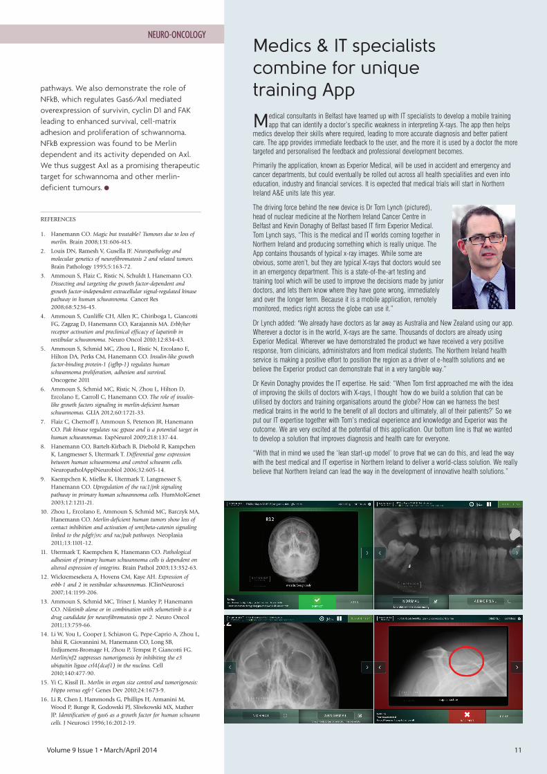
Medical consultants in Belfast have teamed up with IT specialists to develop a mobile trainingapp that can identify a doctor’s specific weakness in interpreting X-rays. The app then helps
medics develop their skills where required, leading to more accurate diagnosis and better patientcare. The app provides immediate feedback to the user, and the more it is used by a doctor the moretargeted and personalised the feedback and professional development becomes.
Primarily the application, known as Experior Medical, will be used in accident and emergency andcancer departments, but could eventually be rolled out across all health specialities and even intoeducation, industry and financial services. It is expected that medical trials will start in NorthernIreland A&E units late this year.
The driving force behind the new device is Dr Tom Lynch (pictured),head of nuclear medicine at the Northern Ireland Cancer Centre inBelfast and Kevin Donaghy of Belfast based IT firm Experior Medical.Tom Lynch says, “This is the medical and IT worlds coming together inNorthern Ireland and producing something which is really unique. TheApp contains thousands of typical x-ray images. While some areobvious, some aren’t, but they are typical X-rays that doctors would seein an emergency department. This is a state-of-the-art testing andtraining tool which will be used to improve the decisions made by juniordoctors, and lets them know where they have gone wrong, immediatelyand over the longer term. Because it is a mobile application, remotelymonitored, medics right across the globe can use it.”
Dr Lynch added: "We already have doctors as far away as Australia and New Zealand using our app.Wherever a doctor is in the world, X-rays are the same. Thousands of doctors are already usingExperior Medical. Wherever we have demonstrated the product we have received a very positiveresponse, from clinicians, administrators and from medical students. The Northern Ireland healthservice is making a positive effort to position the region as a driver of e-health solutions and webelieve the Experior product can demonstrate that in a very tangible way.”
Dr Kevin Donaghy provides the IT expertise. He said: “When Tom first approached me with the ideaof improving the skills of doctors with X-rays, I thought ‘how do we build a solution that can beutilised by doctors and training organisations around the globe? How can we harness the bestmedical brains in the world to the benefit of all doctors and ultimately, all of their patients?’ So weput our IT expertise together with Tom’s medical experience and knowledge and Experior was theoutcome. We are very excited at the potential of this application. Our bottom line is that we wantedto develop a solution that improves diagnosis and health care for everyone.
“With that in mind we used the ‘lean start-up model’ to prove that we can do this, and lead the waywith the best medical and IT expertise in Northern Ireland to deliver a world-class solution. We reallybelieve that Northern Ireland can lead the way in the development of innovative health solutions.”
Volume 9 Issue 1 • March/April 2014 11
12 Volume 9 Issue 1 • March/April 2014
Historically, mammographic riskassessment, i.e. estimating theprobability of the development ofbreast cancer, has been based on an
individual's personal and family background. It hasbeen shown that the amount of fibroglandulartissue as well as its distribution of anatomicaltissue in mammographic images is stronglycorrelated with the probability to develop breastcancer. However, manual assessment shows inter-and intra-observer variability and automation ofthis process has therefore been considereddesirable. Such automated methods cover fattyversus dense tissue segmentation, more advancedsegmentation approaches and feature spaceclassification. We provide an overview of variousapproaches to mammographic risk assessmentand how this might be used in future computeraided diagnosis (CAD) systems.
Mammographic risk assessmentOver the past decades, a number of links havebeen investigated between mammographic riskassessment and patient-specific andenvironmental aspects, covering family history,diet and genetic markers. Such aspects arecurrently captured in a number of associatedmodels, e.g. the Gail model [1] and the Tyrer-Cuzick model [2].However, it should be noted that the above-
mentioned mammographic risk models are basedon non-image based information and how tointegrate image-based information into such riskmodels is an area of current research [3]. In thelate 1960’s and mid 1970’s, Wolfe [4-6] started toinvestigate the links between mammographicimage information and mammographic riskassessment and found that based on his four riskclasses there was a significant difference in riskbetween the lowest and the highest classes (by afactor of up to twenty in specific studies [5]).Wolfe’s classes include aspects of bothparenchymal patterns and intensity variations inthe mammographic images. This work wasfollowed up by Boyd [7], who established amodel based on the percentage dense tissue.Further to this, Tabár and Dean [8] extended thework of both Wolfe and Boyd by describingnormal mammographic tissue by four specific
building blocks: radiolucent (fatty), homogeneous,nodular and linear tissue, and linked thedistribution of these to mammographic riskassessment. Closely related to Boyd's work, the four Breast
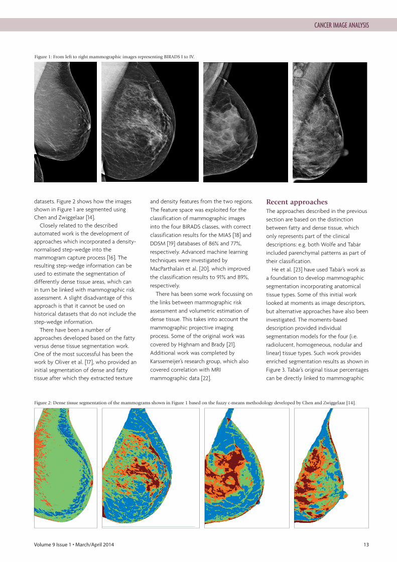
Imaging-Reporting and Data System (BIRADS)classes as defined by American College ofRadiology BIRADS lexicon are: BIRADS I) thebreast is almost entirely fatty, BIRADS II) there arescattered areas of fibroglandular density, BIRADSIII) the breasts are heterogeneously dense, whichmay obscure small masses, and BIRADS IV) thebreasts are extremely dense, which lowers thesensitivity of mammography. A set of examplemammographic images can be found in Figure 1,which shows BIRADS I to IV cases. Work byMuhimmah et al. [11] has shown that there is clearcorrelation between the various image basedmammographic risk assessment models. Variousbreast screening and breast cancer detectionprogrammes have adapted mammographic riskassessment [9,10], but it should be noted thatnone of these currently incorporate theautomatic analysis of mammographic imageinformation.
Dense/fatty tissueFor early research involving automated analysisthe methods were closely related to the work ofBoyd et al. [7], with a strong emphasis onsegmentation and estimation of dense tissuewithin the breast (the fatty tissue is simply theremaining breast tissue). This work was furtherdeveloped into “Cumulus”, which is an interactivesoftware that has been used as a standard withinthe field [12]. Since the development of Cumulus, there have
been a number of approaches that proposed afully automated method for estimation of densemammographic tissue. A typical example of this isthe recent work by Nickson et al. [13] whichprovides breast density segmentation based onhistogram statistics and boundary gradientsinformation. In contrast, Chen and Zwiggelaar [14]developed an automated density segmentationapproach based on fuzzy c-means [15], whichincorporates local spatial and intensityinformation. In both cases, the robustness of thedeveloped approaches was evaluated on large
Image Based TissueSegmentation: Towards the Automation of Mammographic RiskAssessmentReyer Zwiggelaar,
Professor, Department of ComputerScience, Aberystwyth University,E: [email protected]
Harry Strange,Postdoctral Research Associate, Aberystwyth University.
Wenda He,Postdoctral Research Associate, Aberystwyth University.
Zhili Chen,Lecturer, Shenyang Jianzhu University.
Ashwini Kshirsagar,Sr Principal Scientist and Manager,Clinical Research, Hologic Inc.
Erika Denton,Honorary Professor of Radiology,University of East Anglia and Norfolk &Norwich University Hospital.
CANCER IMAGE ANALYSIS
Volume 9 Issue 1 • March/April 2014 13
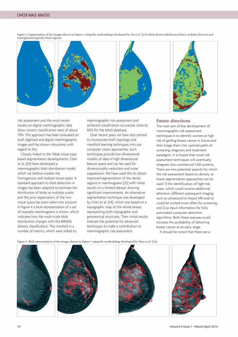
datasets. Figure 2 shows how the imagesshown in Figure 1 are segmented usingChen and Zwiggelaar [14].Closely related to the described
automated work is the development ofapproaches which incorporated a density-normalised step-wedge into themammogram capture process [16]. Theresulting step-wedge information can beused to estimate the segmentation ofdifferently dense tissue areas, which canin turn be linked with mammographic riskassessment. A slight disadvantage of thisapproach is that it cannot be used onhistorical datasets that do not include thestep-wedge information.There have been a number of
approaches developed based on the fattyversus dense tissue segmentation work.One of the most successful has been thework by Oliver et al. [17], who provided aninitial segmentation of dense and fattytissue after which they extracted texture
and density features from the two regions.The feature space was exploited for theclassification of mammographic imagesinto the four BIRADS classes, with correctclassification results for the MIAS [18] andDDSM [19] databases of 86% and 77%,respectively. Advanced machine learningtechniques were investigated byMacParthalain et al. [20], which improvedthe classification results to 91% and 89%,respectively.There has been some work focussing on
the links between mammographic riskassessment and volumetric estimation ofdense tissue. This takes into account themammographic projective imagingprocess. Some of the original work wascovered by Highnam and Brady [21].Additional work was completed byKarssemeijer’s research group, which alsocovered correlation with MRImammographic data [22].
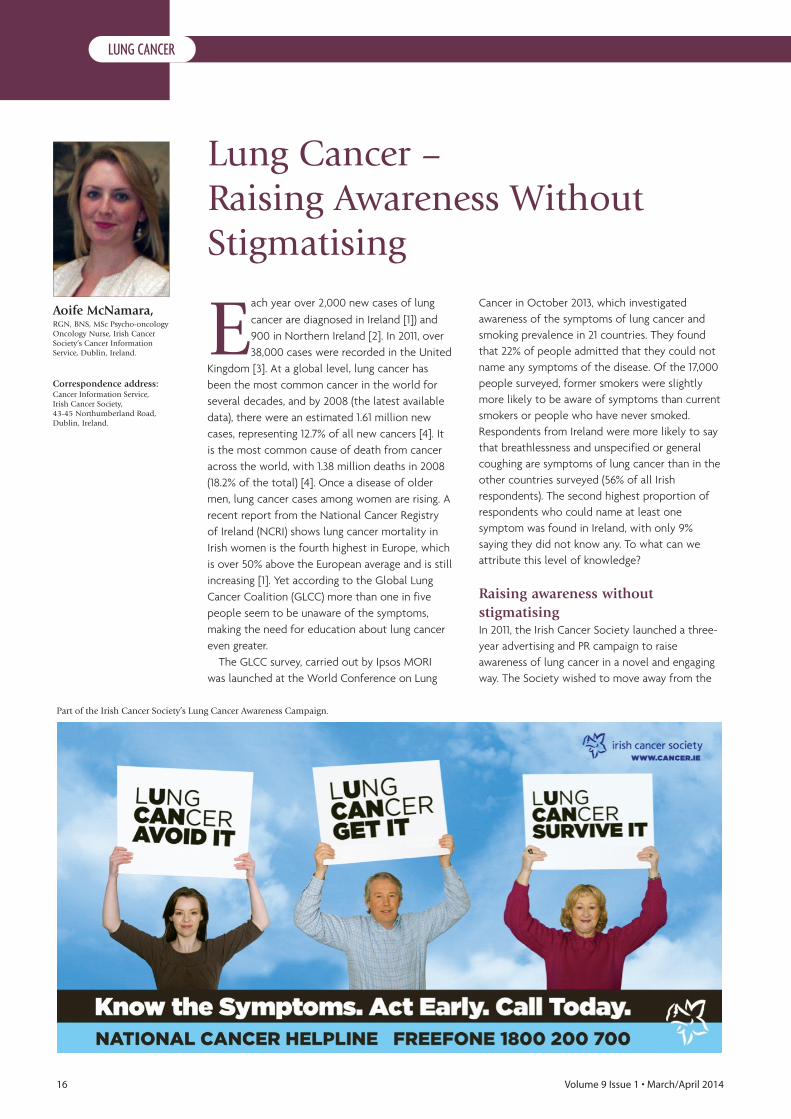
Recent approaches The approaches described in the previoussection are based on the distinctionbetween fatty and dense tissue, whichonly represents part of the clinicaldescriptions: e.g. both Wolfe and Tabárincluded parenchymal patterns as part oftheir classification.He et al. [23] have used Tabár’s work as
a foundation to develop mammographicsegmentation incorporating anatomicaltissue types. Some of this initial worklooked at moments as image descriptors,but alternative approaches have also beeninvestigated. The moments-baseddescription provided individualsegmentation models for the four (i.e.radiolucent, homogeneous, nodular andlinear) tissue types. Such work providesenriched segmentation results as shown inFigure 3. Tabár’s original tissue percentagescan be directly linked to mammographic
CANCER IMAGE ANALYSIS
Figure 1: From left to right mammographic images representing BIRADS I to IV.
Figure 2: Dense tissue segmentation of the mammograms shown in Figure 1 based on the fuzzy c-means methodology developed by Chen and Zwiggelaar [14].
14 Volume 9 Issue 1 • March/April 2014
risk assessment and the most recentresults on digital mammographic datashow correct classification rates of about79%. This approach has been evaluated onboth digitised and digital mammographicimages and has shown robustness withregard to this. Closely linked to the Tabár tissue type
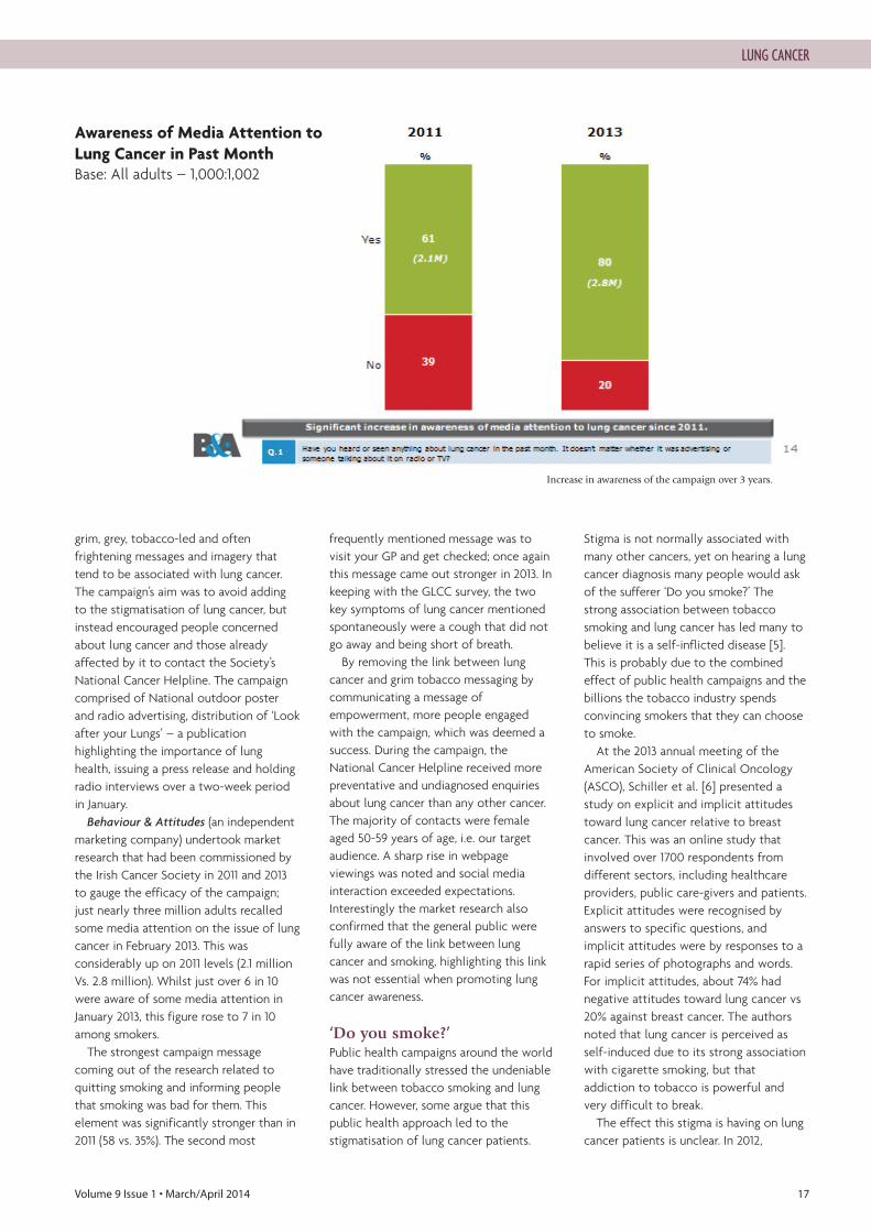
based segmentation developments, Chenet al. [24] have developed amammographic blob distribution model,which we believe models thehomogenous and nodular tissue types. Astandard approach to blob detection inimages has been adapted to estimate thedistribution of blobs at multiple scalesand the prior expectation of the twotissue types has been taken into account.In Figure 4 a blob representation of a setof example mammograms is shown, whichindicates how the multi-scale blobdistribution changes with the BIRADSdensity classification. This resulted in anumber of metrics, which were linked to
mammographic risk assessment andachieved classification accuracies close to80% for the MIAS database.Over recent years we have also started
to incorporate both topology andmanifold learning techniques into ourcomputer vision approaches. Suchtechniques provide low dimensionalmodels of data in high dimensionalfeature space and can be used fordimensionality reduction and noisesuppression. We have used this to obtainimproved segmentation of the denseregions in mammograms [25] with initialresults on a limited dataset showingsignificant improvements. An alternativesegmentation technique was developedby Chen et al. [24], which was based on atopographic map of the whole breast,representing both topographic andgeometrical structures. Their initial resultsindicate the potential for advancedtechniques to make a contribution tomammographic risk assessment.
Future directionsThe main aim of the development ofmammographic risk assessmenttechniques is to identify women at highrisk of getting breast cancer in future andthen triage them into optimal paths ofscreening, diagnosis and treatmentparadigms. It is hoped that novel riskassessment techniques will eventuallyintegrate into commercial CAD systems.There are two potential aspects for whichthe risk assessment based on density ortissue segmentation approaches can beused: 1) the identification of high-riskcases, which could receive additionalattention, different subsequent imagingsuch as ultrasound or breast MR and/orcould be invited more often for screening,and 2) as input information for fullyautomated computer detectionalgorithms. Both these avenues couldincrease the probability of detectingbreast cancer at an early stage.It should be noted that there are a
CANCER IMAGE ANALYSIS
Figure 4. Blob representation of the images shown in Figure 1 using the methodology developed by Chen et al. [24].
Figure 3: Segmentation of the images shown in Figure 1 using the methodology developed by He et al. [23] which shows radiolucent (blue), nodular (brown) andhomogeneous (green) tissue regions.

Volume 9 Issue 1 • March/April 2014 15
CANCER IMAGE ANALYSIS
number of commercial systems available,which are currently aimed at estimatingvolumetric breast density and/or areabreast density for Full-Field DigitalMammography (FFDM) images. Suchsystems are used to assist radiologists inthe assessment of breast tissuecomposition and provides a density score,which can be linked to BIRADS breastcomposition categories. These systemsinclude Quantra (Hologic Inc.), Volpara
(Volpara Solutions), and MicroDose SI(Philips Healthcare).The systems described above are aimed
at helping mammography practicesachieve a reader independent objectivebreast density assessment. After theadoption of tomosynthesis images, it isexpected that conventional FFDM imageswill phase out and, there will be a need totranslate the currently developedapproaches of breast density estimation
using FFDM to the new modality oftomosynthesis. This would possiblyprovide a close to volumetricdensity/tissue segmentation, which maylead to a more reliable mammographicrisk assessment.It should also be noted that another
aspect for further development of theTabár-based work is the temporal analysisof not just changes in density, but also inparenchymal patterns. ●
1. Gail MH, Brinton LA, Byar DP, Corle DK, Green SB, Schairer C, Mulvihill JJ. Projectingindividualized probabilities of developing breast cancer for white females who are beingexamined annually. J Natl Cancer Inst 1989;81(24):1879–86.
2. Tyrer J, Duffy SW, Cuzick J. A breast cancer prediction model incorporating familial andpersonal risk factors. Stat Med 2004;23,1111-30.
3. Eriksson L, Hall P, Czene K, Dos Santos SI, McCormack V, Bergh J, Bjohle J, Ploner A.Mammographic density and molecular subtypes of breast cancer. Br J Cancer2012;107,18–23.
4. Wolfe JN. A study of breast parenchyma by mammography in the normal woman andthose with benign and malignant disease. Radiology 1967;9,201-205.
5. Wolfe JN. Breast patterns as an index of risk for developing breast cancer. AmericanJournal of Roentgenology 1976a;126,1130-7.
6. Wolfe JN. Risk for breast cancer development determined by mammographic parenchymalpattern. Cancer 1976b;37(5), 2486-92.
7. Boyd NF, O’Sullivan B, Campbell JE, Fishell E, Simor I, Cooe G, Germanson T.Mammographic signs as risk factor for breast cancer. Br J Cancer 1982;45, 185-93.
8. Tabár L, Dean PB. Mammographic parenchymal patterns and risk of breast cancer? J AmMed Assoc. 1982;247,185-98.
9. BIRADS (1995) American College of Radiology. Breast Imaging Reporting and DataSystem (BI-RADS) 2nd edition. Reston: American College of Radiology.
10. Roche NA, Given-Wilson RM, Thomas VA, Sacks NP. Assessment of a scoring system forbreast imaging. Br J Surgery 1998;85,669-72.
11. Muhimmah I, Oliver A, Denton ERE, Pont J, Pérez E, Zwiggelaar R. Comparisonbetween Wolfe, Boyd, BI-RADS and Tabár based mammographic risk assessment. LNCS2006;4046, 407-415.
12. Yaffe MJ. Measurement of mammographic density. Breast Cancer Research 2008;10(209).
13. Nickson C, Arzhaeva Y, Aitken Z, Elgindy T, Buckley M, Li M, English DR, KavanaghAM. AutoDensity: an automated method to measure mammographic breast density thatpredicts breast cancer risk and screening outcomes. Breast Cancer Research 2013;15,R80.
14. Chen Z, Zwiggelaar R. A modified fuzzy c-means algorithm for breast tissue densitysegmentation in mammograms. 10th IEEE International Conference on InformationTechnology and Applications in Biomedicine 2010.
15. Dunn JC. A fuzzy relative of the ISODATA process and its use in detecting compact well-separated clusters. Journal of Cybernetics 1973;3,32-57.
16. Diffey J, Hufton A, Astley SM. A new step-wedge for the volumetric measurement ofmammographic density. Lecture Notes in Computer Science 2006;4046,1-9.
17. Oliver A, Freixenet J, Marti R, Pont, J, Pérez E, Denton ERE, Zwiggelaar R. A novelbreast tissue density classification methodology. IEEE Transactions on InformationTechnology in Biomedicine 2008;12(1),55-65.
18. Suckling J, Partner J, Dance DR, Astley SM, Hutt I, Boggis CRM, Ricketts I, StamatakisE, Cerneaz N, Kok SL, Taylor P, Betal D, Savage J. The Mammographic Image AnalysisSociety digital mammogram database. International Workshop on DigitalMammography 1994;211-21.
19. Heath M, Bowyer K, Kopans D, Moore R, Kegelmeyer PJ. The Digital Database forScreening Mammography. Proceedings of the International Workshop on DigitalMammography 2000;212–8.
20. MacParthalain N, Jensen R, Shen Q, Zwiggelaar R. Fuzzy-rough approaches formammographic risk analysis. Intell Data Anal 2010;14 (2),225-44.
21. Hartman K, Highnam R, Warren R, Jackson V. Volumetric assessment of breast tissuecomposition from FFDM images. LNCS 2008;5116,33‐9.
22. van Engeland S, Snoeren PR, Huisman H, Boetes C, Karssemeijer N. Volumetric breastdensity estimation from full-field digital mammograms. IEEE Transactions on MedicalImaging 2006;25(3),273-82.
23. He W, Denton ERE, Stafford K, Zwiggelaar R. Mammographic image segmentation andrisk classification based on mammographic parenchymal patterns and geometric moments.Biomedical Signal Processing and Control 2011;6(3),321-9.
24. Chen Z, Denton ERE, Zwiggelaar R. Topographic representation based breast densitysegmentation for mammographic risk assessment. International Conference on ImageProcessing 2012;1993-6.
25. Strange H, Denton ERE, Kibiro M, Zwiggelaar R. Manifold learning for densitysegmentation in high risk mammograms. Lecture Notes in Computer Science2013;7887,245-52.
26. Chen Z, Wang L, Denton E, Zwiggelaar R. A multiscale blob representation ofmammographic parenchymal patterns and mammographic risk assessment. Lecture Notesin Computer Science 2013;8048,346-53.
REFERENCES
Oncology Tools for Results
E: [email protected] • T: +44 (0) 1638 782600 • F: +44 (0) 1638 782606
Key ProductsAntibodies Assays Biochemicals
Proteins Reagents Vectors
Over 350,000 Products Online!Stratech supports your specialist product needs byproviding a cost effective, convenient & reliable sourceof life science products. Browse our Oncology range at:
www.stratech.co.uk/cancer
16 Volume 9 Issue 1 • March/April 2014
Each year over 2,000 new cases of lungcancer are diagnosed in Ireland [1]) and900 in Northern Ireland [2]. In 2011, over38,000 cases were recorded in the United
Kingdom [3]. At a global level, lung cancer hasbeen the most common cancer in the world forseveral decades, and by 2008 (the latest availabledata), there were an estimated 1.61 million newcases, representing 12.7% of all new cancers [4]. Itis the most common cause of death from canceracross the world, with 1.38 million deaths in 2008(18.2% of the total) [4]. Once a disease of oldermen, lung cancer cases among women are rising. Arecent report from the National Cancer Registryof Ireland (NCRI) shows lung cancer mortality inIrish women is the fourth highest in Europe, whichis over 50% above the European average and is stillincreasing [1]. Yet according to the Global LungCancer Coalition (GLCC) more than one in fivepeople seem to be unaware of the symptoms,making the need for education about lung cancereven greater. The GLCC survey, carried out by Ipsos MORI
was launched at the World Conference on Lung
Cancer in October 2013, which investigatedawareness of the symptoms of lung cancer andsmoking prevalence in 21 countries. They foundthat 22% of people admitted that they could notname any symptoms of the disease. Of the 17,000people surveyed, former smokers were slightlymore likely to be aware of symptoms than currentsmokers or people who have never smoked.Respondents from Ireland were more likely to saythat breathlessness and unspecified or generalcoughing are symptoms of lung cancer than in theother countries surveyed (56% of all Irishrespondents). The second highest proportion ofrespondents who could name at least onesymptom was found in Ireland, with only 9%saying they did not know any. To what can weattribute this level of knowledge?
Raising awareness withoutstigmatisingIn 2011, the Irish Cancer Society launched a three-year advertising and PR campaign to raiseawareness of lung cancer in a novel and engagingway. The Society wished to move away from the
Lung Cancer –Raising Awareness WithoutStigmatising
Aoife McNamara,RGN, BNS, MSc Psycho-oncologyOncology Nurse, Irish CancerSociety’s Cancer InformationService, Dublin, Ireland.
Correspondence address:Cancer Information Service, Irish Cancer Society, 43-45 Northumberland Road,Dublin, Ireland.
Part of the Irish Cancer Society’s Lung Cancer Awareness Campaign.
LUNG CANCER
Volume 9 Issue 1 • March/April 2014 17
grim, grey, tobacco-led and oftenfrightening messages and imagery thattend to be associated with lung cancer.The campaign’s aim was to avoid addingto the stigmatisation of lung cancer, butinstead encouraged people concernedabout lung cancer and those alreadyaffected by it to contact the Society’sNational Cancer Helpline. The campaigncomprised of National outdoor posterand radio advertising, distribution of ‘Lookafter your Lungs’ – a publicationhighlighting the importance of lunghealth, issuing a press release and holdingradio interviews over a two-week periodin January.
Behaviour & Attitudes (an independentmarketing company) undertook marketresearch that had been commissioned bythe Irish Cancer Society in 2011 and 2013to gauge the efficacy of the campaign;just nearly three million adults recalledsome media attention on the issue of lungcancer in February 2013. This wasconsiderably up on 2011 levels (2.1 millionVs. 2.8 million). Whilst just over 6 in 10were aware of some media attention inJanuary 2013, this figure rose to 7 in 10among smokers. The strongest campaign message
coming out of the research related toquitting smoking and informing peoplethat smoking was bad for them. Thiselement was significantly stronger than in2011 (58 vs. 35%). The second most
frequently mentioned message was tovisit your GP and get checked; once againthis message came out stronger in 2013. Inkeeping with the GLCC survey, the twokey symptoms of lung cancer mentionedspontaneously were a cough that did notgo away and being short of breath.By removing the link between lung
cancer and grim tobacco messaging bycommunicating a message ofempowerment, more people engagedwith the campaign, which was deemed asuccess. During the campaign, theNational Cancer Helpline received morepreventative and undiagnosed enquiriesabout lung cancer than any other cancer.The majority of contacts were femaleaged 50-59 years of age, i.e. our targetaudience. A sharp rise in webpageviewings was noted and social mediainteraction exceeded expectations.Interestingly the market research alsoconfirmed that the general public werefully aware of the link between lungcancer and smoking, highlighting this linkwas not essential when promoting lungcancer awareness.
‘Do you smoke?’Public health campaigns around the worldhave traditionally stressed the undeniablelink between tobacco smoking and lungcancer. However, some argue that thispublic health approach led to thestigmatisation of lung cancer patients.
Stigma is not normally associated withmany other cancers, yet on hearing a lungcancer diagnosis many people would askof the sufferer ‘Do you smoke?’ Thestrong association between tobaccosmoking and lung cancer has led many tobelieve it is a self-inflicted disease [5].This is probably due to the combinedeffect of public health campaigns and thebillions the tobacco industry spendsconvincing smokers that they can chooseto smoke. At the 2013 annual meeting of the
American Society of Clinical Oncology(ASCO), Schiller et al. [6] presented astudy on explicit and implicit attitudestoward lung cancer relative to breastcancer. This was an online study thatinvolved over 1700 respondents fromdifferent sectors, including healthcareproviders, public care-givers and patients.Explicit attitudes were recognised byanswers to specific questions, andimplicit attitudes were by responses to arapid series of photographs and words.For implicit attitudes, about 74% hadnegative attitudes toward lung cancer vs20% against breast cancer. The authorsnoted that lung cancer is perceived asself-induced due to its strong associationwith cigarette smoking, but thataddiction to tobacco is powerful andvery difficult to break. The effect this stigma is having on lung
cancer patients is unclear. In 2012,
Increase in awareness of the campaign over 3 years.
LUNG CANCER
Awareness of Media Attention toLung Cancer in Past MonthBase: All adults – 1,000:1,002
18 Volume 9 Issue 1 • March/April 2014
Chambers et al. [8] reviewed reports onlung cancer stigma and suggested that it isreasonable to assume it has a negativeeffect on the psychosocial well-being ofpeople affected by lung cancer, bothpatients and their loved ones. At thesame time Cataldo et al. [7] published astudy of almost 200 lung cancer patientsin which strong associations were foundbetween lung cancer stigma anddepression and quality of life. Nosignificant difference was found betweennever and ever smokers [9]. These authorsrecently developed an instrument tomeasure the stigma perceived by lungcancer patients – the Cataldo LungCancer Stigma Scale (CLCSS). The CLCSScan be used to identify not only thepresence of stigma, but the effect it hason the lung cancer patient. It is hopedthat the introduction of this tool willeventually lead to effective interventionsthat can be used to combat the negativeeffects of stigma [7].
‘An emotional rollercoaster’Research clearly suggests that stigma is avery real issue for lung cancer patients.This burden is in addition to knowing thatthey have been diagnosed with thebiggest cancer killer worldwide. Themajority of patients are diagnosed at alate stage with a poor prognosis; theyoften experience debilitating symptoms,making it unsurprising that lung cancerpatients can become distressed. Theimpact of a cancer diagnosis can evoke a
range of emotions, anything from fear,anxiety, and anger to denial. There is noright or wrong way to feel, no right timeto experience any one particular emotion.Patients often describe this as being like‘an emotional rollercoaster.’ This may be anormal reaction to a cancer diagnosis;however 25-30% of all newly diagnosedcancer patients experience elevated levelsof emotional distress [1] and struggle tocope with the impact of their disease.The National Comprehensive CancerNetwork (NCCN) defines distress as:
‘A multifactorial unpleasantemotional experience of apsychological (cognitive, behavioural,emotional), social, and / or spiritualnature that may interfere with theability to cope effectively withcancer, its physical symptoms and itstreatment.’ [11]
A well-recognised study by Zabora et al.[10] in 2001 examined the prevalence ofpsychological distress by cancer site andfound considerable variation. Of thepatients surveyed, 43% of lung cancerpatients experienced increased levels ofdistress in comparison to 32% of breastcancer patients, 31% of bowel cancerpatients and 30% of prostate cancerpatients. Similarly Joyce et al. [11] foundlung cancer patients scored high ondistress levels due to poor prognosis andself-attribution due to smoking. Theauthors noted that low survival ratesassociated with lung cancer allows the
patient little time to adjust to theirdiagnosis and prognosis, contributingfurther to their distress levels (12).
‘No one in the world deserveslung cancer’ – Global LungCancer CoalitionNo one in the world deserves lung cancer.It is the role of organisations like the IrishCancer Society, Cancer Focus NorthernIreland and the Roy Castle LungFoundation to increase awareness of thedisease and provide support to thosealready affected by lung cancer. Raisingawareness is vital, but we must be mindfulto do so without stigmatising and addingto the burden of lung cancer. ●
1. National Cancer Registry of Ireland (2013) Cancer in Ireland 2013 – Annual reportof the National Cancer Registry. Available at:http://www.ncri.ie/sites/ncri/files/pubs/ann_report2013_V08_wcover_woback.pdf(Accessed 15 August 2013)
2. Public Health Agency (2013) Available at:http://www.publichealth.hscni.net/news/get-lung-cancer-aware-november-0(Accessed 25 November 2013)
3. Health and Social Care Information Centre (2012) National Lung Cancer AuditReport. Available at: http://www.hqip.org.uk/national-lung-cancer-audit-report-2012/ (Accessed 28 November 2013)
4. Globocan (2010) Available at: http://globocan.iarc.fr/factsheet.asp (Accessed 15 August 2013)
5. Chapple A, Ziebland S & McPherson A. Stigma, shame and blame experienced bypatients with lung cancer: qualitative study. BMJ, 2004;328(7454):1470.
6. Schiller JH, Bowden CJ, Mills J et al. (2013) Explicit and implicit attitudes towardlung cancer (LC) relative to breast cancer (BC). Program and abstracts of the 2013Annual Meeting of the American Society of Clinical Oncology; May 31-June 4,2013; Chicago, Illinois. Abstract 8017.
7. Cataldo JK, Slaughter R, Jahan TM, Pongguan VL & Hwang WJ. Measuring Stigmain People with Lung Cancer: Psychometric Testing of the Cataldo Lung Cancer StigmaScale. Oncology Nursing Forum 2011;January 1; 38(1): E46-E54 doi:10.1188/11.ONF.E46-E54
8. Chambers SK, Dunn J, Occhipinti S, Hughes S, Baade P, Sinclair S, Aitkin J, Youl P& O’Connell DL. A systematic review of the impact of stigma and nihilism on lungcancer outcomes. BMC Cancer 2012;12:184 doi: 10.1186/1471-2407-12-184.
9. Cataldo JK, Jahan TM & Pongguan VL. Lung cancer stigma, depression, and quality oflife among ever and never smokers. European Journal of Oncology Nursing.2012;July;16(3):264-9 doi: 10.1016/j.ejon.2011.06.008
10. Zabora et al. The prevalence of Psychological Distress by cancer site. Psycho-Oncology 2001;10:19-28.
11. National Comprehensive Cancer Network (2009) NCCN Clinical PracticeGuidelines in Oncology: Distress Management. Available at: http://www.nccn.org(Accessed 15 August 2013)
12. Joyce M, Schwartz S & Huhmann M. Supportive care in Lung cancer. Seminars inoncology nursing 2008;Vol 24 No. 1(February):57-67.
REFERENCES
USEFUL WEBSITES:
Irish Cancer Society: http://www.cancer.ie
Global Lung Cancer Coalition: http://www.lungcancercoalition.org
National Comprehensive CancerNetwork: http://www.nccn.org
Cancer Focus Northern Ireland: http://cancerfocusni.org
Roy Castle Lung Foundation: http://www.roycastle.org
LUNG CANCER
m
You know how good Macmillan palliative care nurses are. But did you know most of the people we’re helping today come to us for other support? And that it’s provided by a wider Macmillan team who will listen to and support your patients with the sensitivity you’d expect.
Our Support Line team can help your patients with so many things. Questions about tests. Feelings that go with diagnosis. More information about treatment options or managing side effects. Money worries connected to cancer. Even the emotional and practical issues on the way back to health – like returning to work.
Tell your patients and their families about the free Macmillan Support Line,
0808 808 00 00Monday to Friday, 9am-8pm
If most Of the pEo�l�
we hElp tOdAy don’t ne�d pAlliative care,
wh�t do they nEed?
They often need someone to listen calmly and with empathy to their feelings and fears.And I can help with that.’MargaretMacmillan Support Line Officer
‘
Macmillan Cancer Support, registered charity in England and Wales (261017), Scotland (SC039907) and the Isle of Man (604).
Togeth�r we can h�lp mOre �eoPle af��Cted by can�er
20 Volume 9 Issue 1 • March/April 2014
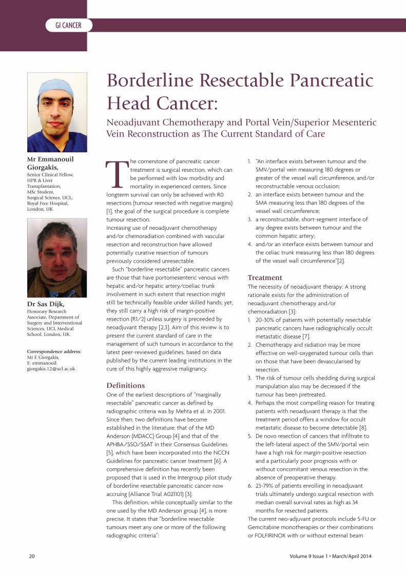
The cornerstone of pancreatic cancertreatment is surgical resection, which canbe performed with low morbidity andmortality in experienced centers. Since
longterm survival can only be achieved with R0resections (tumour resected with negative margins)[1], the goal of the surgical procedure is completetumour resection.Increasing use of neoadjuvant chemotherapyand/or chemoradiation combined with vascularresection and reconstruction have allowedpotentially curative resection of tumourspreviously considered unresectable. Such “borderline resectable” pancreatic cancers
are those that have portomesenteric venous withhepatic and/or hepatic artery/coeliac trunkinvolvement in such extent that resection mightstill be technically feasible under skilled hands; yet,they still carry a high risk of margin-positiveresection (R1/2) unless surgery is preceeded byneoadjuvant therapy [2,3]. Aim of this review is topresent the current standard of care in themanagement of such tumours in accordance to thelatest peer-reviewed guidelines, based on datapublished by the current leading institutions in thecure of this highly aggressive malignancy.
DefinitionsOne of the earliest descriptions of “marginallyresectable” pancreatic cancer as defined byradiographic criteria was by Mehta et al. in 2001.Since then, two definitions have becomeestablished in the literature: that of the MDAnderson (MDACC) Group [4] and that of theAPHBA/SSO/SSAT in their Consensus Guidelines[5], which have been incorporated into the NCCNGuidelines for pancreatic cancer treatment [6]. Acomprehensive definition has recently beenproposed that is used in the Intergroup pilot studyof borderline resectable pancreatic cancer nowaccruing (Alliance Trial A021101) [3]. This definition, while conceptually similar to the
one used by the MD Anderson group [4], is moreprecise. It states that “borderline resectabletumours meet any one or more of the followingradiographic criteria”:
1. “An interface exists between tumour and theSMV/portal vein measuring 180 degrees orgreater of the vessel wall circumference, and/orreconstructable venous occlusion;
2. an interface exists between tumour and theSMA measuring less than 180 degrees of thevessel wall circumference;
3. a reconstructable, short-segment interface ofany degree exists between tumour and thecommon hepatic artery;
4. and/or an interface exists between tumour andthe celiac trunk measuring less than 180 degreesof the vessel wall circumference”[2].
TreatmentThe necessity of neoadjuvant therapy: A strongrationale exists for the administration ofneoadjuvant chemotherapy and/orchemoradiation [3]:1. 20-30% of patients with potentially resectablepancreatic cancers have radiographically occultmetastatic disease [7].
2. Chemotherapy and radiation may be moreeffective on well-oxygenated tumour cells thanon those that have been devascularised byresection.
3. The risk of tumour cells shedding during surgicalmanipulation also may be decreased if thetumour has been pretreated.
4. Perhaps the most compelling reason for treatingpatients with neoadjuvant therapy is that thetreatment period offers a window for occultmetastatic disease to become detectable [8].
5. De novo resection of cancers that infiltrate tothe left-lateral aspect of the SMV/portal veinhave a high risk for margin-positive resectionand a particularly poor prognosis with orwithout concomitant venous resection in theabsence of preoperative therapy.
6. 23-79% of patients enrolling in neoadjuvanttrials ultimately undergo surgical resection withmedian overall survival rates as high as 34months for resected patients.
The current neo-adjuvant protocols include 5-FU orGemcitabine monotherapies or their combinationsor FOLFIRINOX with or without external beam
Borderline Resectable PancreaticHead Cancer: Neoadjuvant Chemotherapy and Portal Vein/Superior MesentericVein Reconstruction as The Current Standard of Care
Mr EmmanouilGiorgakis,Senior Clinical Fellow, HPB & LiverTransplantation,MSc Student, Surgical Science, UCL,Royal Free Hospital,London, UK.
Dr Sas Dijk,Honorary ResearchAssociate, Department ofSurgery and InterventionalSciences, UCL MedicalSchool, London, UK.
Correspondence address:Mr E Giorgakis,E: [email protected]
GI CANCER
Volume 9 Issue 1 • March/April 2014 21
GI CANCER
radiation. Objective radiographicresponses are rare and downstaging toresectable tumours is exceedingly rare [3].Nonetheless, a single retrospective studyshowed that, up to 66% of patients mayundergo tumoour resection with a 95%rate of R0 resection [9].
Staging LaparoscopyIt should always precede pancreatectomyif CA 19–9 level is ≥150 U/ml and tumoursize ≥3 cm, since it may reveal occultmetastatic disease in 31% of patients.
Surgical resection Unfortunately, only 15%–20% of patientsare suitable candidates for surgery [15],either due to locally advanced disease orbecause of synchronous distantmetastases. Pancreatic tumour frequentlyextends directly into the retroperitonealspaces and involves the superiormesenteric vein–portal vein (SMV-PV)[15]. In an effort to improve lifeexpectancy, in many centres a moreaggressive approach has been employed,involving SMV-PV resection (VR) in viewto increase the curability of pancreaticcancer [15]. Venous resection and reconstruction
should be performed for borderlineresectable tumours involving theSMV/portal vein as long as “reasonablevenous inflow and outflow is present andthe surgeon feels that an R0 or R1resection likely can be accomplished”[10,15]. When such operations areperformed at high-volume institutions,survival rates become similar to those forpatients undergoingpancreaticoduodenectomy withoutvenous resection [11]. Despite apparentintraoperative tumour adherence to thevein, only 60- 70% of specimens willshow histopathologic evidence of venousinvasion11. Those that do, however, havepoorer prognosis [12].Hepatic arterial resection and
reconstruction duringpancreaticoduodenectomy for borderlineresectable pancreatic cancer may bereasonable in highly selected patientstreated at specialty centers experiencedin vascular reconstruction [6,15].Resection of the SMA as part of apancreaticoduodenectomy, however, isassociated with high morbidity rates and
is thus not recommended [13,15].Adequate regional lymphadenectomy
means at least 15 nodes harvested [6,15].Lymphadenectomy should routinelyinclude periduodenal and peripancreaticnodes as well as those to the right of thehepatoduodenal ligament and thesuperior mesenteric artery (SMA) and theanterior and posteriorpancreaticoduodenal lymph nodes.Prospective, randomised trials haveshown that extended lymphadenectomyoffers no survival benefit and may resultin lower quality of life than standardlymphadenectomy [14].The surgical margin most likely to be
positive is the SMA margin (theretroperitoneal margin) [10]. NCCNguidelines recommend “skeletonisation ofthe SMA down to its adventitiaanteriorly, laterally, and posteriorly tominimise the risk of a positive margin inthis location”[6].
Pancreatoduodenectomy withportal vein/superiormesenteric veinreconstruction (PD-VR): The first case of pancreatectomy with VRwas reported by Moore et al. in 1951. Inrecent years, VR can be safely performed[15]. However, arterial resection inpancreatectomy remains a challengingprocedure with significantly highmorbidity and mortality rates [13,15]. Although duration of Whipple’s
procedure along with vascularreconstruction is longer and operativeblood loss is greater in such cases,mortality and morbidity rates arecomparable between the two groups(3.3% and 41.9% respectively) [15]. It isreasonable to assume that as a surgeon’sexperience increases, operative time andblood loss will likely decrease [6]. Resultsfrom two prospective randomised studiesshowed that the surgery group hadsignificantly better survival than thepalliative gastrobiliary bypass group orradiochemotherapy group [15,16].Furthermore, the overall survival did notdiffer between operation with VR andwithout [15]. As quoted by Tempero et al.“this is consistent with the hypothesisthat tumour with portal vein adherenceor invasion may represent a function oftumour location, and possibly tumour
size, rather than an indicator of aggressivetumour biology” [6]. Moreover, asLygidakis et al. have marked, “tumourextension to the SMV-PV does notnecessarily indicate tumour invasion” [16].Perhaps remarkably, on the most recentmeta-analysis on SMV-PV reconstructionfor borderline pancreatic cancer [14],histopathology evaluations revealed that“a considerable percentage of patients(43.1%) who underwent VR for pancreaticcancer were found to have inflammatoryadhesions without cancer invasion”.The depth of SMV-PV wall invasion is
an indicator of poor outcome after PD-VR, since mesenteric vessels resected enbloc with the lesion on macroscopicgrounds carry documented worseprognosis if the vessel walls prov to beinfiltrated by malignancy rather thanhaving been thickened by the thepericancerous fibrosis. However, it isdifficult to differentiate malignant frominflammatory adherence of the SMV-PVpre-operatively since detecting theprecise site of tumour infiltration isonly possible by histopathologicalanalysis [16].
Principles of surgicaltechniqueA thorough description of the currentsurgical approach advocated by theAmerican College of Surgeons is the oneeloquently described by Weitz et.al, whocomment [18]: “In an effort to achievecomplete resection of a pancreatic headcancer, resection of the portal vein, thesuperior mesenteric vein, or both, needsto be performed if there is suspectedinvasion of the venous wall. To adhere tothe principles of oncologic surgery, thisresection should be performed withoutviolating the integrity of the tumour. Thekey components of the technique areearly identification of the superiormesenteric artery, which serves as a guidethroughout the resection, completemobilisation of the right hemicolon andmesenteric root if necessary, and controlof all vascular structures, with resectionof the portal vein, pancreatic head, andtumour as the last part of the resectionphase” [18]. A trial dissection along theportal vein or shaving the tumour off theportal vein during resection, withseparate resection and reconstruction of
22 Volume 9 Issue 1 • March/April 2014
the involved part of the portal vein,should be avoided at all costs; transectingacross tumour may result in a higher rateof tumour recurrence” [18], Weitz et alwarned.
Management in high-volumecentresAs Evans et. Al [10] quoted: “the tide ofliterature is moving in a directionsupporting better outcomes in higher-volume centers for patients withpancreatic cancer” [10]. That being said,specifically addressing the issue ofvascular resection and reconstruction, itremains to be proved if management insuch centres will ensure optimised results:“venous involvement remains difficult toascertain in many patients, thus, if thesurgeon is unprepared for venousinvolvement or is unfamiliar with thetechniques necessary to adequately treatthese patients, then substandard care willresult”. “In this situation, one of twooutcomes may occur: either theresectable patient fails to get apotentially therapeutic procedurealtogether or a technically inadequateoperation occurs, resulting in a grossly
positive margin or marked morbidity”.
ConclusionsPancreatic tumour resection remains themainstay of therapy for physiologically andsurgically eligible patients [10]. “Coupledwith improvements in imaging, moreconsistent classification schemes and theuse of nonsurgical adjuvant therapies”, themajor achievements of our era in thebattle against pancreatic cancer have been“extension of potentially curative resectionto those previously considered ineligible”[10]. Evolution in therapy may haveimproved survival by allowing treatment ofpreviously untreatable patients or bydownstaging patients with more advanceddisease such that their outcomes aresimilar to disease at an earlier stage.Several reports demonstrate equivalent
morbidity and mortality for patientsundergoing PD with or without vascularresection [10]. It is important to note thatreports of vascular resection generallycome from larger or high-volume centers.“Unlike extended lymphadenectomywhere no survival benefit exists, vascularresection allows complete tumourclearance (R0 resection) when this would
otherwise be precluded” [10]. In terms of surgical technique, “the key
to portal vein resection for advancedpancreatic cancer is the superiormesenteric artery” [18]. “Dissection on theright and posterior aspect of this arteryguides the surgeon through the criticalpart of the procedure and helps toachieve a complete tumour resectionwithout violating the integrity of thetumour” [18]. Based on all data available, PV/SMV
resection and reconstruction is thecurrent standard of care during apancreatoduodenectomy when there is areasonable expectation for an R0resection. PD with portal venousreconstruction is justified since it mayresult in R0 resection and long-termsurvival comparable to that obtained withstandard resection. All these remarkable achievements in
surgical approach and chemoradiation putaside, overall improvements in survival haveremained elusive, unfortunately onlystrengthening the notion that tumourbiology continues to be the mostsignificant factor influencing these patients'outcomes. ●
REFERENCES
1. Wagner M, Redaelli C, Lietz M, et al. Curative resection is the single most importantfactor determining outcome in patients with pancreatic adenocarcinoma. Br J Surg2004;91:586-94.
2. Katz MH, Marsch R, Herman JM, Shi Q, Collison E, Venook AP, et al. Borderlineresectable pancreatic cancer: need for standardization and methods for optimal clinicaltrial design. Ann Surg Oncol 2013;20(8):2787-95.
3. Cooper A, Tzeng CW, Katz M. Treatment of Borderline Resectable Pancreatic Cancer.Current Treatment Options in Oncology. 2013;14:293-310.
4. Varadhachary GR, Tamm EP, Abbruzzese JL, Xiong HQ, Crane CH, Wang H, etal. Borderline resectable pancreatic cancer: definitions, management, and the role ofpreoperative therapy. Ann Surg Oncol. 2006;13:1035-46.
5. Callery MP, Chang KJ, Fishman EK, Talamonti MS, William Traverso L, LinehanDC. Pretreatment as- sessment of resectable and borderline resectable pancreaticcancer: expert consensus statement. Ann Surg Oncol. 2009;16:1727–33.
6. Tempero MA, Arnoletti JP, Behrman SW, Ben-Josef E, Benson 3rd AB, Casper ES,et al. Pancreatic adenocarcinoma, version 2.2012: featured updates to the NCCNguidelines. J Natl Compr Canc Netw. 2012;10:703-713.
7. Contreras CM, Stanelle EJ, Mansour J, Hinshaw JL, Rikkers LF, Rettammel R, etal. Staging laparoscopy enhances the detection of occult metastases in patients withpancreatic adenocarcinoma. J Surg Oncol. 2009;100:663-9.
8. Glant JA, Waters JA, House MG, Zyromski NJ, Nakeeb A, Pitt HA, et al. Does theinterval from im- aging to operation affect the rate of unanticipated metastasisencountered during operation for pancre- atic adenocarcinoma? Surgery.2011;150:607–616.
9. Katz MH, Fleming JB, Bhosale P, Varadhachary G, Lee JE, Wolff R, et al. Responseof borderine resectable pancreatic cancer to neoadjuvant therapy is not reflected byradiographic indicators. Cancer. 2012;118:5749-56.
10. Evans DB, Farnell MB, Lillemoe KD, Vollmer Jr C, Strasberg SM, Schulick RD.Surgical treatment of resectable and borderline resectable pancreas cancer: expertconsensus statement. Ann Surg Oncol. 2009;16:1736–44.
11. Yekebas EF, Bogoevski D, Cataldegirmen G, Kunze C, Marx A, Vashist YK, et al.En bloc vascular resection for locally advanced pancretic malignancies infiltratingmajor blood vessels: perioperative outcome and long-term survival in 136 patients.Ann Surg. 2008;247:3000-3009.
12. Wang J, Estrella JS, Peng L, Rashid A, Varadhachary GR, Wang H, et al. Histologictumor involvement of superior mesenteric vein/portal vein predicts poor prognosis inpatients with stage II pancreatic adeno- carcinoma treated with neoadjuvantchemoradiation. Cancer. 2012;118:3801–3811.
13. Amano H, Miura F, Toyota N, Wada K, Katoh K, Hayano K, et al. Ispancreatectomy with arterial re- construction a safe and useful procedure for locallyadvanced pancreatic cancer? J Hepatobiliary Pancreat Surg. 2009;16:850–7.
14. Nimura Y, Nagino M, Takao S, Takada T, Miyazaki K, Kawarada Y, et al. Standardversus extended lymphadenectomy in radical pancreatoduodenectomy for ductaladenocarcinoma of the head of the pancreas: long-term results of a Japanesemulticenter randomized controlled trial. J Hepatobiliary Pancreat Sci.2012;19:230–41.
15. Zhou Y, Z Zhang, Y Liu, B Li, D Xu. Pancreatectomy Combined with SuperiorMesenteric Vein–Portal Vein Resection for Pancreatic Cancer: A Meta-analysis. WorldJ Surg. 2012; 36: 884-91.
16. Lygidakis NJ, Singh G, Bardaxoglou E et al. Mono-bloc total spleno-pancreaticoduodenectomy for pancreatic head carcinoma with portal-mesenteric venousinvasion. A prospective randomized study. Hepatogastroenterology.2004;51:427–33.
17. Bachellier P, Rosso E, Lucescu I et al. Is the need for an arterial resection acontraindication to tpancreatic resection for locally advanced pancreaticadenocarcinoma? A case-matched controlled study. J Surg Oncol 103:75-84.
18. J Weitz, P Kienle, J Schmidt, H Friess, MW Büchler. Portal Vein Resection forAdvanced Pancreatic Head Cancer. J Am Coll Surg. 2007;204(4):712-6.
GI CANCER
Volume 9 Issue 1 • March/April 2014 23
There is convincing evidence to suggestthat lifestyle factors are important inboth the development and progressionof breast cancer, and a significant number
of cases may be prevented if diet, activity andweight modifications were adopted by appropriategroups of individuals [1]. The American Institute forCancer Research [2] estimates that approximately38% of breast cancers may be preventable, yetconcurrently 49,564 and 397 cases of female andmale breast cancers respectively were reported in2010 and almost 11,600 patients succumbed to thedisease in the UK that same year [3]. Considerableefforts to understand these statistics and todevelop Public Health measures targetingdecreasing incidence and mortality rates are on-going, yet recent evidence suggests that there isstill much work to be done [4].
The role of epidemiology inunderstanding how diet and lifestyleaffect breast cancer outcomesIn vitro and in vivo studies have been key to ourunderstanding of cancer pathology and prognosis.Controlled laboratory investigations clarify howcancer-causing substances (for example: tar,benzene, arsenic and nitrosamines) cause DNAdamage and subsequently affect cellular behaviour.Molecular approaches such as those concernedwith genome wide analysis, epigenetic,transcriptomic and proteomic changes havestarted to explain some of these discrepancies, butthe role of environment remains a particularlycomplex area of research. Prospectiveobservational studies of heterogeneouspopulations in free-living settings have shown thatbiological adaptations observed in lab-basedstudies explain only a fraction of cases (forexample up to 30% of lung cancer; [5]). Whereeffects are convincing (and large), the importanceof human risk assessment is clear; but the path to
discovery of cause and effect where effect sizesare smaller, and perhaps less consistent, ischallenging.Utopia for risk factor epidemiology in breast
cancer research would be formulation ofprevention and management strategies integratingthe best available evidence and linked tomeasureable health outcome improvements.Patients are however variably exposed (andreactive) to many diverse factors prior to andfollowing a diagnosis of breast cancer.Determining their individual and collectiveinfluences on disease-related risk constitutes “amuch duller scalpel” [6] than consideration ofresults obtained from randomised controlled trials.We must acknowledge however that biomedicalresearch is unlikely to address the challenges ofeither genetic or biological susceptibility tocancer, unless careful consideration of socio-demographic and behavioural factors (amongothers) is made [7]. Estimates of the population risk of developing
cancer must be considered in the context ofdiverse biological and environmental information.It is clear that exposure to environmental factorsmust precede the disease outcome. Therefore, thechallenge for research teams aiming to elucidatethe effects of exposures – both singly orcombined (in the so-called ‘eposome’) – is howbest to control for errors including (but not limitedto) incomparable diagnostic methods, mis-reporting errors (a particularly relevant issue whenquantifying dietary intake estimates and reports oflifestyle habits), biological plausibility, specificity,and consistency. Epidemiology is thought to holdthe key to formulation of dietary and lifestylerecommendations, yet interpretation of our work– by all that use it – requires rigour. It is acknowledged that exposure to risk factors
does not remain static: most, if not all, vary withinthe general population. Action on Smoking and
Understanding Breast CancerSurvival with Epidemiology
Dr Claire Robertson,PhD, RNutr, Honorary Secretary,Association for Nutrition, Senior Lecturer in Nutrition and Public Health, University of Westminster.
Dr Ruth Swann PhD, Research Associate, Against Breast Cancer ResearchUnit, University of Westminster.
Correspondence address:Dr Claire E Robertson, PhD, R.Nutr., MA HE, fHEA),Course Leader,MSc Public Health Nutrition,Honorary Secretary, Association for Nutrition,Faculty of Science & Technology,Dept Human and HealthSciences,University of Westminster,115 New Cavendish Street,London, W1W 6UWT: +44 (0)20 7911 5000 ext 64127.
Acknowledgements:The DietCompLyf study wasestablished and is maintainedwith funding from Against BreastCancer (Registered CharityNumber 1121258). Researchnurses in the Study Centres havebeen supported by the NCRN.Dr Ruth Swan acknowledgesreceipt of funding from AgainstBreast Cancer (Registered CharityNumber 1121258).
BREAST CANCER
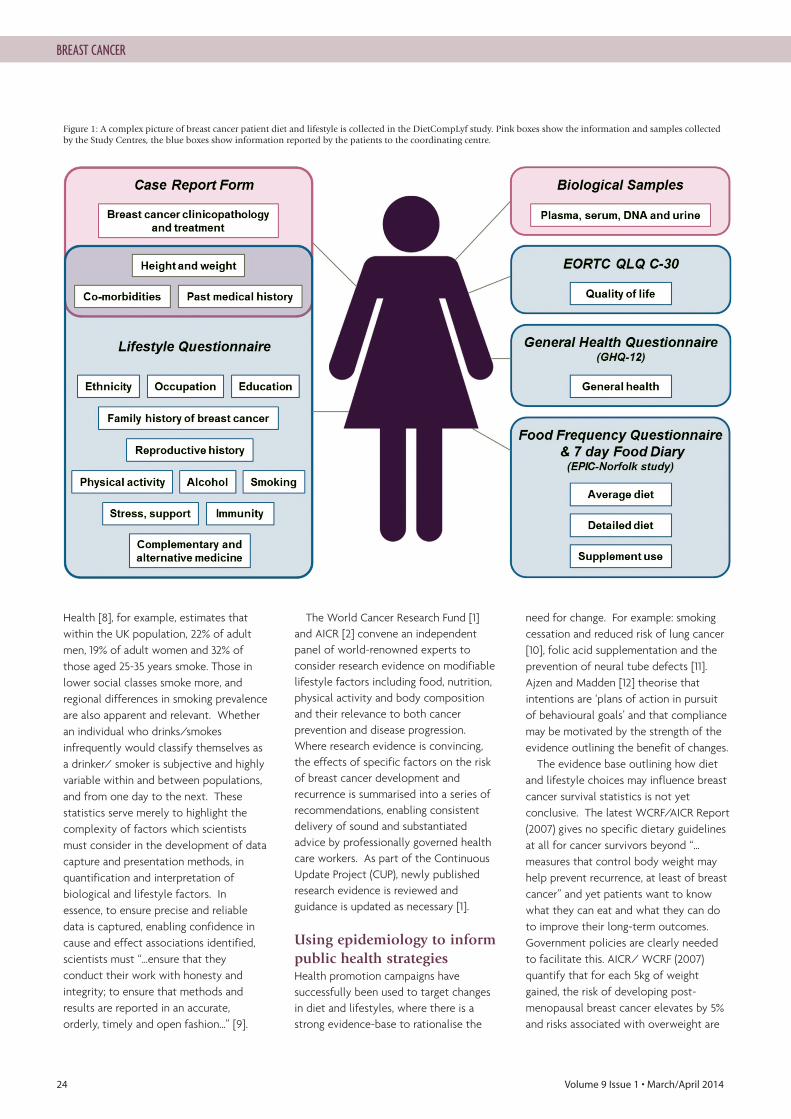
24 Volume 9 Issue 1 • March/April 2014
Health [8], for example, estimates thatwithin the UK population, 22% of adultmen, 19% of adult women and 32% ofthose aged 25-35 years smoke. Those inlower social classes smoke more, andregional differences in smoking prevalenceare also apparent and relevant. Whetheran individual who drinks/smokesinfrequently would classify themselves asa drinker/ smoker is subjective and highlyvariable within and between populations,and from one day to the next. Thesestatistics serve merely to highlight thecomplexity of factors which scientistsmust consider in the development of datacapture and presentation methods, inquantification and interpretation ofbiological and lifestyle factors. Inessence, to ensure precise and reliabledata is captured, enabling confidence incause and effect associations identified,scientists must “…ensure that theyconduct their work with honesty andintegrity; to ensure that methods andresults are reported in an accurate,orderly, timely and open fashion…” [9].
The World Cancer Research Fund [1]and AICR [2] convene an independentpanel of world-renowned experts toconsider research evidence on modifiablelifestyle factors including food, nutrition,physical activity and body compositionand their relevance to both cancerprevention and disease progression.Where research evidence is convincing,the effects of specific factors on the riskof breast cancer development andrecurrence is summarised into a series ofrecommendations, enabling consistentdelivery of sound and substantiatedadvice by professionally governed healthcare workers. As part of the ContinuousUpdate Project (CUP), newly publishedresearch evidence is reviewed andguidance is updated as necessary [1].
Using epidemiology to informpublic health strategies Health promotion campaigns havesuccessfully been used to target changesin diet and lifestyles, where there is astrong evidence-base to rationalise the
need for change. For example: smokingcessation and reduced risk of lung cancer[10], folic acid supplementation and theprevention of neural tube defects [11].Ajzen and Madden [12] theorise thatintentions are ‘plans of action in pursuitof behavioural goals’ and that compliancemay be motivated by the strength of theevidence outlining the benefit of changes. The evidence base outlining how diet
and lifestyle choices may influence breastcancer survival statistics is not yetconclusive. The latest WCRF/AICR Report(2007) gives no specific dietary guidelinesat all for cancer survivors beyond “…measures that control body weight mayhelp prevent recurrence, at least of breastcancer” and yet patients want to knowwhat they can eat and what they can doto improve their long-term outcomes.Government policies are clearly neededto facilitate this. AICR/ WCRF (2007)quantify that for each 5kg of weightgained, the risk of developing post-menopausal breast cancer elevates by 5%and risks associated with overweight are
Figure 1: A complex picture of breast cancer patient diet and lifestyle is collected in the DietCompLyf study. Pink boxes show the information and samples collectedby the Study Centres, the blue boxes show information reported by the patients to the coordinating centre.
BREAST CANCER
Volume 9 Issue 1 • March/April 2014 25
BREAST CANCER
1. World Cancer Research Fund (WCRF) / American Institute for Cancer Research(AICR) (2007) Food, Nutrition, Physical Activity, and the Prevention of Cancer: aGlobal Perspective. Washington DC: AICR.
2. American Institute for Cancer Research (AICR) Get Pink. Pink on Purpose.Accessed from: http://www.aicr.org/how-you-can-help/get-involved/pink-on-purpose/pop_index.html (24th January, 2014)
3. Cancer Research UK: Breast cancer incidence statistics. Accessed from:http://www.cancerresearchuk.org/cancerinfo/cancerstats/types/breast/incidence/uk-breast-cancer-incidence-statistics#age (7th September, 2013).
4. Ioannidis JPA. Implausible results in human nutrition research. Definitive solutionswon’t come from another million observational papers or small randomized trials. BritishMedical Journal 2013;347: f6698.
5. Doll R & Peto R. The causes of cancer: quantitative estimates of avoidable risks ofcancer in the United States today. Journal of the National Cancer Institute1981;66(6): 1191-308.
6. Taubes G. Epidemiology faces its limits. Science, New Series 1995;269(5221):164-9.
7. Wiseman M and Jackson A. A response to: Ioannidis JPA. Implausible results inhuman nutrition research. Definitive solutions won’t come from another millionobservational papers or small randomized trials. British Medical Journal 2013;347: f6698.
8. Action on Smoking and Health (November 2013). Facts at a glance: Smokingstatistics. Accessed from: http://www.ash.org.uk/files/documents/ASH_93.pdf (6thJanuary, 2014).
9. International Council for Science (2008) Freedom, Responsibility and Universalityof Science. Accessed from: http://www.icsu.org/publications/cfrs/freedom-responsibility-booklet/ICSU-CFRS-booklet.pdf (20th January, 2014).
10. Doll R & Hill BA. Lung cancer and other causes of death in relation to smoking: Asecond report on the mortality of British Doctors. British Medical Journal 1956;(Nov10th):1072-81.
11. MRC (Medical Research Council) Vitamin Study Research Group. Prevention ofneural tube defects: results of the Medical Research Council Vitamin Study. The Lancet1991;338:131–7.
12. Ajzen I & Madden TJ. Prediction of goal-directed behaviour: attitudes, intentions, andperceived behavioural control. Journal of Experimental Social Psychology1986;22:453–74.
13. Department of Health (2011) Healthy lives, healthy people: A call to action onobesity in England. Norwich, Her Majesty’s Stationary Office. Available from:https://www.gov.uk/government/uploads/system/uploads/attachment_data/file/213720/dh_130487.pdf
14. Department of Health (2006) Our health, our care, our say: a new direction forcommunity services. Norwich, Her Majesty’s Stationary Office. Available from:http://www.official-documents.gov.uk/document/cm67/6737/6737.pdf
15. Chambers AF. Influence of diet on metastasis and tumor dormancy. Clinical andExperimental Metastasis 2009;26:61-6.
16. Swann R, Perkins A, Velentzis LS, Ciria C, Dutton SJ, Mulligan AA, Woodside JV,Cantwell MM, Leatham AJ, Robertson CE and Dwek MV. The DietCompLyf study: aprospective cohort study of breast cancer survival and phytoestrogen consumption.Maturitas 2013;75(3):232-40.
17. Kushi LH, Kwan ML, Lee MM and Ambrosone CB. Lifestyle factors ad survival inwomen with breast cancer. The Journal of Nutrition 2007;137: 236S-242S.
REFERENCES
not limited to cancer. The Healthy Lives,Healthy PeopleWhite Paper [13] suggestedthat an obese woman (BMI ≥30 kg/m2) isalmost 13 times more likely to developdiabetes, more than four times morelikely to develop high blood pressure andmore than three times more likely to havea heart attack than a non-obese woman(BMI <30 kg/m2). Worryingly, datapublished by the Health and Social CareInformation Centre (2013) highlighted that65% of UK adults were classified asoverweight or obese (BMI >25 kg/m2) andonly 34% of a normal weight in 2011. If theresults from the Government ‘Our health,your care, your say’ report [14] arerepresentative, patients want to modifytheir behaviour to prevent the recurrenceof cancer and increase their overall healthto and to avoid co-diagnosis with othernon-communicable diseases (e.g., type 2diabetes, hypertension), but there is along way to go before this will beachieved. We must acknowledge that neither a
cancer cell nor the environment in whichit is located is a static entity. Drugs usedin cancer treatments, the hormonal statusof the patients, their diets and nutritionare features which could change theoutcomes following a breast cancerdiagnosis [15]. Ioannidis [4] highlights anapparent inability for promising results
observed in randomised controlled trialsto be translated into large scale,population studies and acknowledges thata “quick fix” answer cannot be expected.Randomised clinical trials are prohibitivelyslow and expensive for the investigationrisk factors for cancer, and ‘controlled’environments are not realistic for theinvestigation of long-term outcomes. Wemust therefore rely on the careful designof case control or cohort studies of largepopulations. Such projects must also relyon existing evidence of influencingfactors, both to ensure collection ofrelevant information, and to enableappropriate statistical analysis of theeffect sizes. Recruiting a fully representative sample
of a complete patient group is notwithout difficulty, however, and socio-economic, ethnic and cultural variabilitymay affect the utility of results obtained.Confounding biases and measurementerrors must be carefully considered wheninterpreting results from epidemiologicalstudies and biological plausibility must bethe cornerstone. DietCompLyf, an observational study of
diet, lifestyle and breast cancer outcome,considered the issues outlined abovewithin its design. The heterogeneity ofbreast cancer diagnoses alongside thevariable influences of patient
demographics, clinical decision making,molecular and phenotypic features of thecancer are being considered in a cohortof 3,157 UK women with primary invasive(non-metastatic) breast cancer [16].Patients were recruited onto the study 12months (±3 months) post-diagnosis andare (or have been) being actively followedup for 5-years, enabling a plethora of datato be captured and quality controlchecked prior to exploration of whetherdiet and lifestyle modifications caninfluence long-term survival rates forbreast cancer patients. ‘Diet’ is beingevaluated comprehensively, using patternsof consumption as well as estimation ofindividual nutrient intakes calculated fromrecords of food only and from ‘food anddietary supplement’ use. DietCompLyf isone of the largest studies of lifestylebehaviours and breast cancer survivalworldwide [17], and the only such study inthe UK. While our cohort does notperhaps contain the 100,000 individuals or50 years of follow up desired by Ioannidis(2013), the integrity of this – like severalother – research group is to utiliseepidemiological methods carefully, aimingto advance understanding throughobservation and exploration of ourpatients’ experiences enabling translationof knowledge into influentialrecommendations for patients. ●
26 Volume 9 Issue 1 • March/April 2014
The LancetRisk of venous thromboembolism associatedwith peripherally inserted central catheters: asystemic review and meta-analysisChopra V, Anand S, Hickner A, Buist M, Rogers MAM, Saint S and Flanders
SA. The Lancet 2013;382(9889):311-25.
The relative risk of the development of venous thromboembolismfrom peripherally inserted central catheters (PICC) and other centralvenous catheters (CVC) is unknown. More evidence should aidappropriate selection of the device and informed consent for aspecific patient according to his or her need and preference. Asystemic review and meta-analysis of the risk of venous throm-boembolism associated with PICCs compared with CVCs has beenundertaken. Several databases, including Medline, Embase, Biosis,Cochrane Central Register of Controlled Trials, Conference PapersIndex and Scopus were searched. Other studies were identifiedthrough manual searches of bibliographies, the internet, and directcontacts (to obtain unpublished data). All human studies publishedin full text, abstract, or poster form were eligible for inclusion.These were of adult patients of 18+ years who had had a PICCinserted. They were assessed with the Newcastle-Ottawa risk ofbias scale. Where there was no comparison group, the pooledfrequency of venous thromboembolism was calculated for patientsreceiving PICCs. In studies comparing PICCs with other CVCs,summary odds ratios (ORs) were calculated with a random effectsmeta-analysis. Of the 533 identified citations, 64 (12 with acomparison group and 52 without) including 29 503 patients metthe eligibility criteria. In the non-comparison studies, the weightedfrequency of PICC-related deep vein thrombosis was highest inpatients who were critically ill (13·9%, 95% CI 7·7-20·1) and thosewith cancer (6·7%, 4·7-8·6). Meta-analysis of 11 studies comparingthe risk of deep vein thrombosis related to PICCs with that relatedto CVCs showed that PICCs had an increased risk of deep veinthrombosis (OR 2·55, 1·544·2, p<0·0001), but not pulmonaryembolism. With the baseline PICC-related deep vein thrombosisrate of 2·7% and a pooled OR of 2·55, the number needed toharm relative to CVCs was 26 (95% CI 13-71). We conclude thatPICCs are associated with a higher risk of deep vein thrombosisthan CVCs, especially in patients who are critically ill or those witha malignancy. The decision to insert PICCs should be guided byweighing the risk of thrombosis against their benefit.
Reviewer’s opinion: The meta-analysis results and comprehensiveoverview on the subject of “intravenous catheter use and the riskof associated complications” presented by the authors is plausible.Use of these devices has increased many folds in oncology over thelast decade, particularly PICC, being easier to insert. After rigorousstructured training, the procedure is mostly done by chemotherapynurses and relatively junior medical staff. Identification of a higherincidence of PICC-associated deep vein thrombosis compared tocentral venous catheters identified in the meta-analysis could bedue to these catheters being longer, increasing venous endothelialtrauma, but the risk factors and safety measures need more assess-ment. Despite the inclusion of several unpublished data in thisarticle, the authors should be congratulated because it is highlyunlikely that a randomised controlled prospective study on thesubject will be conducted. The evidence presented is compellingand consistent across all the studies included within their meta-analysis. Unfortunately, currently available pharmacologicalmeasures do not provide reasonable protection against thrombosis.
BioMedES LtdProducing the very best definitive
versions of your biomedical reports andpapers www.biomedes.co.uk
• BioMedES reworks sound scientific papers, technical reports,and other (biomedical) documents, putting them into themost idiomatic English and passes the most stringent peerreview and quality control assessments
• It copy edits for a number of big biomedical publishinghouses
• Runs four journals in the life sciences
• It provides the Secretariat for an international organisationfor cell biology (www.ifcbiol.org) and an enterprise company
• It prepares and publishes e-books in biomedicine
• It designs logos for biomedical and many other organisations
• It collates and prepares abstracts for scientific and othermeetings
• The company is involved in arranging both national andinternational conferences
• It also runs courses on scientific and medical writing, and onelectronic publishing at home and abroad
Why not contact us at:BioMedES Ltd
Leggat House, Keithhall, Inverurie, AB51 0LX, UK.Tel: +44-1467-6702809, Fax: +44-1467-629123,
Email: [email protected]
Never heard of us?Not surprising, since our operations are mostly “behind the scenes”. But we may be able to help you withyour publication problems, from technical notes to e-books.
What does BioMedES do?
JOURNAL REVIEWS
PANEL OF JOURNAL REVIEWERS
Dr Qian An, PhD MD, Senior Research Fellow, Portsmouth University, UK.
Mr Mriganka De, FRCS (ORL-HNS), Consultant ENT Head & Neck/ThyroidSurgeon, Derby Royal Hospital, UK.
Ms Helen Evans, Senior Lecturer in Cancer Nursing, Institute of Nursing andMidwifery, University of Brighton, UK.
Dr Simon Grumett, PhD FRCP, Consultant & Honorary Senior Lecturer inMedical Oncology, Royal Wolverhampton Hospitals NHS Trust & University ofBirmingham, UK.
Mr Tasadooq Hussain, BA(Edu.) (MD) MRCS a Clinical Research Fellow BreastSurgery at Castle Hill Hospital, Hull and Eat Yorkshire Hospitals NHS, UK.
Richard Novell, MChir FRCS, Consultant Coloproctologist, The Royal Free Hospital,London, UK.
Dr Sunil Upadhyay, Consultant Clinical Oncologist, Queen’s Centre for Oncology,Castle Hill Hospital, Hull, UK.
Volume 9 Issue 1 • March/April 2014 27
Risk assessment, fully informed consent and optimum precautions,like avoidance of misplacement of the tip of the catheter, must beundertaken when these devices have to be inserted. – SU
New England Journal ofMedicinePazopanib versus Sunitinib in Metastatic Renal-Cell Carcinoma
Motzer R J, Hutson T E, Cella D et al. New England Journal of Medicine
2013;369(8):722-31.
Pazopanib and sunitinib provide a progression-free survival (PFS)benefit compared with placebo or interferon in patients withmetastatic renal-cell carcinoma. This randomised trial comparedhead-to-head, the efficacy and safety of pazopanib and sunitinibas first-line therapy. In this multi-centre, phase III study, 1110patients with clear-cell metastatic renal-cell carcinoma wererandomised in a 1:1 ratio to receive a continuous dose ofpazopanib (800 mg once daily; 557 patients) or sunitinib in 6-week cycles (50 mg once daily for 4 weeks, followed by 2 weekswithout treatment; 553 patients). The primary end-point was PFS,the study being designed to show that pazopanib was not inferiorto sunitinib. Secondary end-points included overall survival, safetyand quality of life.Pazopanib was not inferior to sunitinib with respect to PFS (HR
for PFS or death from any cause, 1.05; 95% confidence interval[CI], 0.90 to 1.22), meeting the predefined non-inferiority margin.Overall survival was similar (HR for death with pazopanib, 0.91;95% CI, 0.76 to 1.08). Patients treated with sunitinib were more (i)fatigue (63 vs. 55%), hand-foot syndrome (50 vs. 29%) andthrombocytopenia (78 vs. 41%); and those treated with pazopanibmore often had increased ALT (60 vs. 43%), weight loss, alopeciaand change of hair colour. The mean change from baseline in 11of 14 health-related quality-of-life domains, particularly thoserelated to fatigue or soreness in the mouth, throat, hands, or feetduring the first 6 months of treatment, favoured pazopanib(P<0.05 for all 11 comparisons). Thus, pazopanib and sunitinibhave similar efficacy, but the safety, quality-of-life and patient satis-faction with treatment profiles favoured pazopanib.
Reviewer’s opinion: Side effects and its impact on quality of life(QOL) are important considerations for both patients and theirclinicians in the management of advanced cancers. It (QOL) takesprecedence when different therapies have similar efficacies(response rate, PFS and OS), but significant differences in their sideeffects for some of the patients. The results of a COMPARZ trialclearly establish the superiority of pazopanib over sunitinib, thereference standard on this front. Both drugs are multi-targeted TKIwith similar efficacy, which is reassuring for patients and their clini-cians. It allows them to choose the most appropriate agent.However, non-inferiority is not synonymous with equally efficacy.One of the most pertinent points is that pazopanib was superioron 11 out of 14 measures of QOL. Pazopanib has already beenrecommended by the NICE as first-line treatment for patients inthe UK with advanced kidney cancer, since GSK officials hadagreed to a 12.5% discount on the list price and possibly a secondrebate following the outcome of COMPARZ. Lower medicalresources need, e.g. fewer phone calls to clinics and visits to hospi-tals due to better tolerance, will favourably influence when cost-benefit issue have been reconsidered by the authorities. – SU
@Paxmancoldcap
paxman-coolers.com
The leadingglobal expertin scalp cooling.
Registered charity number 1153487 (England and W
ales), Com
pany limited by guarantee num
ber 08570737 (England and Wales)
#WearAHatDay
HELPUS RAISE
£1 MILLIONFOR A NEW
RESEARCHCENTRE
WEAR AHAT DAYWEAR AHAT DAYWEAR AWEAR AWEAR AWEAR AWEAR AWEAR AWEAR AWEAR AWEAR AHOLD AN EVENT & RAISE MONEY !
2014
More childrenand adults
under 40 dieof a brain tumour
than from anyother cancer
Please mention Oncology News when responding
JOIN US& FUND THE FIGHT!
www.wearahatday.orgwearahatday@
braintumourresearch.org01296 733011
TO REGISTER& DONATE!
JOURNAL REVIEWS
28 Volume 9 Issue 1 • March/April 2014
MarchVascular Access & PICC Training10 March 2014; Manchester, UKW: www.christie.nhs.uk/school-of-oncology/education-eventsT: +44 (0)161 446 3773 or E: [email protected]
10th Palliative Care Congress 12-14 March 2014; Harrogate, UKT: +44 (0)1489 565 475E: [email protected] W: www.pccongress.org.uk
SSO 2014 Annual Cancer Symposium13-15 March, 2014; Arizona, USAW: www.surgonc.org
National Breast Oncoplastic Study Day17 March 2014; London, UK W: www.royalmarsden.nhs.uk
The Management, Treatment & FuturePerspectives of Cancer of Unknown Primary21-22 March 2014; London, UKW: www.cupfoundjo.org
Specialist Palliative Care24 March 2014; Manchester, UKW: www.christie.nhs.uk/school-of-oncology/education-eventsT: +44 (0)161 446 3773 or E: [email protected]
Bladder cancer25 March 2014; London, UKW: www.rcr.ac.uk/oncologyevents E: [email protected]
NEWAnnual Marie Curie Palliative Care ResearchConference28 March 2014; London, UKW: www.rsm.ac.uk/palliative
Pancreatic, Hepatobiliary & NeuroendocrineTumours31 March 2014; Manchester, UKW: www.christie.nhs.uk/school-of-oncology/education-eventsT: +44 (0)161 446 3773 or E: [email protected]
April
Survivorship: Changing Prospects for Cancer4 April 2014; Manchester, UKW: www.christie.nhs.uk/school-of-oncology/education-eventsT: +44(0)161 446 3773 or E: [email protected]
105th AACR Annual Meeting 5-9 April, 2014; San Diego, California W: www.aacr.org
Intensive Revision Course for MCEM Part A Exam 12 April 2014; Manchester, UKW: www.christie.nhs.uk/school-of-oncology/education-eventsT: +44 (0)161 446 3773 or E: [email protected]
The Edinburgh MRCS Exam Preparation Course12-13 April 2014; Manchester, UKW: www.christie.nhs.uk/school-of-oncology/education-eventsT: +44(0)161 446 3773 or E: [email protected]
6th European Congress on Head and NeckOncology 24-26 April 2014; Liverpool, UKW: http://echno2014.com/
Gastrointestinal Masterclass28-29 April 2014; Leeds, UKW: www.rcr.ac.uk/oncologyevents E: [email protected]
May
Skin Cancer of the Head & Neck1 May 2014; Utrecht, the NetherlandsW: reconstruction-skin-cancer.com
NEWCurrent Issues in Palliative Care 2014, 9th nationalconference8-9 May 2014; London, UKW: www.mahealthcareevents.co.uk
NEWRCN Women’s health conference and exhibition:Women's health and gynaecology - What's new?9 May 2014; London, UKLaura BenfieldE: [email protected]: +44 (0)20 7647 3591W: www.rcn.org.uk/WH14
Biological Basis of Cancer Therapy: MedicalPhysics12-16 May 2014; Manchester, UKW: www.christie.nhs.uk/school-of-oncology/education-eventsT: +44 (0)161 446 3773 E: [email protected]
Nutritional Care of Cancer Patients13 May 2014; London, UK W: www.royalmarsden.nhs.uk
NEWAdvanced Leadership19 May 2014; Manchester, UKW: www.christie.nhs.uk/school-of-oncology/education-eventsT: +44 (0)161 446 3773 E: [email protected]
NEWAssociation of Breast Surgery Conference & AGM19-20 May 2014; Liverpool, UKW: www.associationofbreastsurgery.org.uk
Upper GI 20 May 2014; Manchester, UKW: www.christie.nhs.uk/school-of-oncology/education-eventsT: +44 (0)161 446 3773 orE: [email protected]
Practical Image Guided GynaecologicalBrachytherapy22 May 2014; Manchester, UKW: www.christie.nhs.uk/school-of-oncology/education-eventsT: +44(0)161 446 3773 or E: [email protected]
NEW2nd National Oncology Update for Palliative CarePhysicians22-23 May 2014; Preston, UKW: www.stcatherines.co.uk/education-and-train-ing/study-sessions/the-second-national-oncology-update-for-palliative-care-physiciaE: [email protected]
NEW2014 ASCO Annual Meeting30 May-3 June 2014; Chicago, Illinois, USAW: http://am.asco.org
June
Targeted Treatments for Urological Cancers5 June 2014; Manchester, UKW: www.christie.nhs.uk/school-of-oncology/education-eventsT: +44 (0)161 446 3773 or E: [email protected]
Improving clinical outcomes through qualityimprovement activity: a focus on lung, bladderand oesophageal cancers Clinical Oncology AuditConference6 June 2014; London, UKW: www.rcr.ac.uk/oncologyevents E: [email protected]
Everything you ever wanted to know about LungCancer Imaging7 June 2014; London, UK W: www.royalmarsden.nhs.uk
International Symposium on Pediatric Neuro-oncology8 June – 2 July 2014; SingaporeW: ispo2014.com
NEWLate Effects in Cancer Survivors - 5th biennialSheffield meeting12-13 June 2014; Sheffield, UKE: [email protected]
Targeted Treatments for Haematological Cancers16 June 2014; London, UK W: www.royalmarsden.nhs.uk
Camstrand Research Conference17-18 June 2014; Manchester, UKW: www.christie.nhs.uk/school-of-oncology/education-eventsT: +44(0)161 446 3773 or E: [email protected]
NEWABS Trainees Meeting19-20 June 2014; Glasgow, UKW: www.associationofbreastsurgery.org.uk
Haematology Nurse Study Day (Stem CellTransplantation)23 June 2014; Manchester, UKW: www.christie.nhs.uk/school-of-oncology/education-eventsT: +44 (0)161 446 3773 or E: [email protected]
Pain in the Cancer Patient23-24 June 2014; London, UK W: www.royalmarsden,nhs.uk/paincancerpatient
To have your event listed in the Oncology News diary, E: [email protected] by April 5th 2014
COURSES AND CONFERENCES
Volume 9 Issue 1 • March/April 2014 29
Robert A. Weinberg,
The Biology of Cancer,The Biology of Cancer is a
“The Biology of Cancer
GENETICS AND GENOMICS IN MEDICINE
THE BIOLOGY OF CANCER,
Tom Strachan, Judith Goodship and Patrick Chinnery,
www.garlandscience.com
NEW
May 2014 • Paperback • 500pp • 270 illus PB ISBN: 978-0-8153-4480-3 • £46.00
July 2013 • 960pp • 584 illus • Includes a poster and DVD-ROMHB ISBN: 978-0-8153-4219-9 • £109.00 • PB ISBN: 978-0-8153-4220-5 • £60.00
Save 20% using promotional code CBN35 at
checkout when placing orders
at www.crcpress.com Expires 30/06/2014
FREE Worldwide Delivery
Communicating with Cancer Patients
John F. Smyth October 2013 | 978-1-4822-2678-2
100pp | £18.99 £15.19
Targeted Therapies in Oncology Second Edition
Giuseppe Giaccone & Jean-Charles Soria
October 2013 | 978-1-84214-545-6512pp | £100.00 £80.00
V iew our en t i re Onco logy co l l ec t ion he re : h t tp : / /b i t . l y /Onco logyCRC
NEWOncology titles from CRC Press
14:35:58
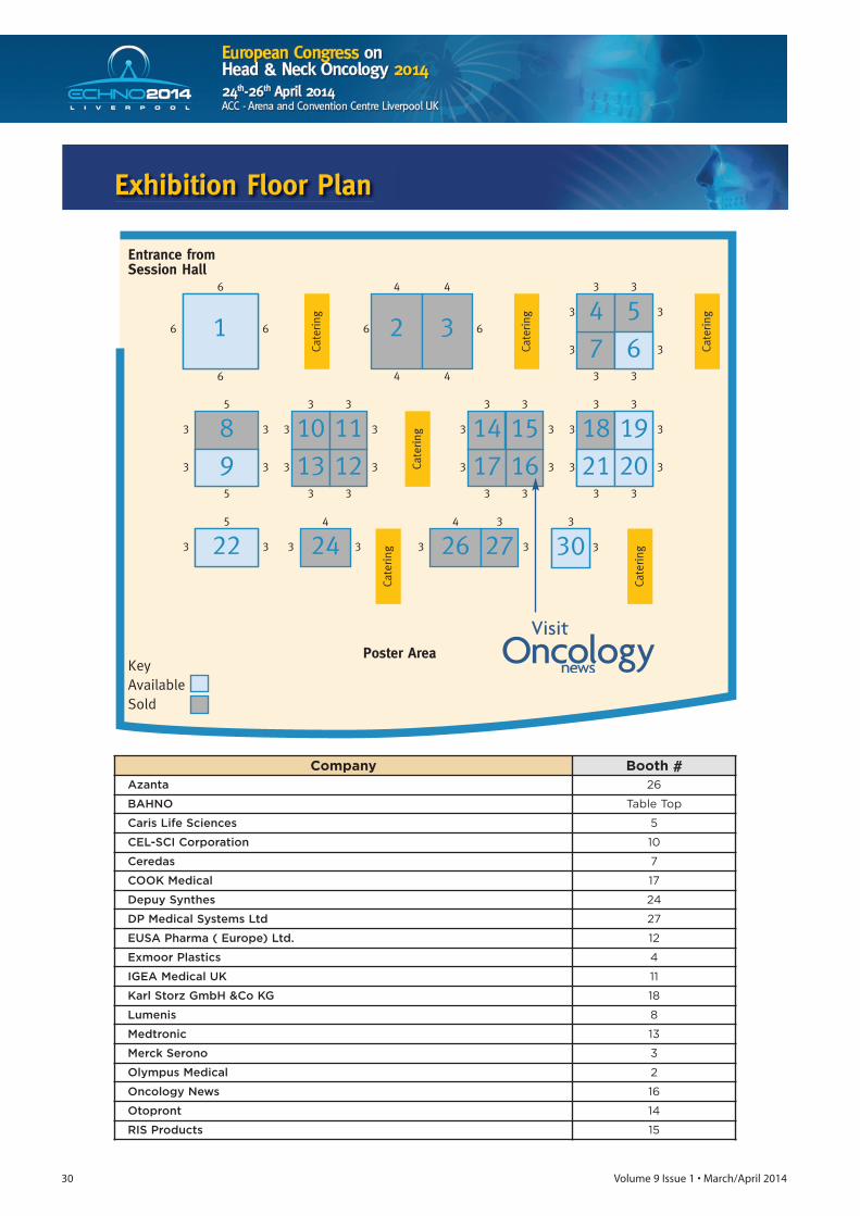
30 Volume 9 Issue 1 • March/April 2014
Exhibition Floor Plan
Company Booth #Azanta 26
BAHNO Table Top
Caris Life Sciences 5
CEL-SCI Corporation 10
Ceredas 7
COOK Medical 17
Depuy Synthes 24
DP Medical Systems Ltd 27
EUSA Pharma ( Europe) Ltd. 12
Exmoor Plastics 4
IGEA Medical UK 11
Karl Storz GmbH &Co KG 18
Lumenis 8
Medtronic 13
Merck Serono 3
Olympus Medical 2
Oncology News 16
Otopront 14
RIS Products 15
Visit
impakt.org
IMPORTANT DEADLINES:26 February 2014 Early registration and training course application
09 April 2014 Late registration and pre-registration closure
Organisers Partners
BREAST CANCER TRAINING COURSE
Focus on Translational Research
7-8 MAY 2014Brussels, Belgium
THE PRE-IMPAKT EARLY CAREER TRAINING COURSE IS DESIGNED FOR EARLY TO MID-CAREER ONCOLOGISTS AND RESEARCH SCIENTISTS.IMPAKT 2014 CHAIRS:
Scientifi c Chair: Giuseppe Curigliano, IT
Executive Chair: Alastair M. Thompson, UK
Co-Founders: Martine Piccart, BE and Jose Baselga, US.
32 Volume 9 Issue 1 • March/April 2014
Symposia:BNOS Main Meeting: Contemporary Approaches to: – Clinical Management– Molecular Characterisation – In vivo and in vitro models– Molecular Neuropathology– Radiology In Adult and Paediatric Brain Tumours
Nursing/AHP Symposium:Contemporary approaches to clinical care of adult and paediatricpatients with brain tumours
• Abstract submission closes 21st March• Registration opens 1st April• Registration with accommodation – CLOSES – 31st MAY 2014
The British Neuro-oncology Society is the only UK multidisciplinary society dedicated to advances in research and the treatment of adult and paediatric braintumours. Our annual meeting attracts delegates from all clinical neuro-oncology specialties (including representatives from neurosurgery, neurology,neuroradiology, neuro-oncology and neuropathology), clinical nurse specialists, charities and basic scientists leading the field in neuro-oncology research.
For programme and meeting details see www.bnos.org.uk.
BNOS 2014 Liverpool
Contemporary Approaches to Paediatric and Adult BrainTumoursWednesday 9th – Friday 11th July 2014
Venue: LJMU The John Lennon Art & Design Building
Accommodation: Vine Court, University of Liverpoolor Hotels
Key dates
31 March – 4 JuneAbstract submission
7 April – 31 JulyEarlybird registration
Submit your abstract
by 4 June 2014
2–5 November 2014BT Convention Centre, Liverpool, UK2014
Join us at the 10th NCRI Cancer ConferenceThe largest cancer research meeting in the UK
Celebrating the 10th NCRI Cancer Conference
conference.ncri.org.uk
Cancer cell and model systemsDiagnosis and therapy
Epidemiology and preventionHealth service research
Information, patients and the publicSurvivorship and end of life care
Submit your research on:
COURSES AND CONFERENCES
Volume 9 Issue 1 • March/April 2014 33
The Changing Prospects for Cancer New models of aftercare for those living
with and beyond cancerFriday 4th April 2014
The Lowry, Media City, Pier 8, Salford Quays, M50 3AZ
Suitable For: Everybody welcome, including clinicians in NHS services including cancer, primary and community based services, commissioners, and managers involved in service delivery and improvement
Study Day Fee: £150 or £125 Early Bird (If booked before Monday 3rd March 2014)Further Info: Visit www.christie.nhs.uk/school-of-oncology
or e-mail [email protected]
IntroductionAs the National Cancer Survivorship Initiative (NCSI) vision for cancer survivorship begins to become a reality for patients, professionals and commissioners, this conference will explore some of the fundamental issues and controversies that are beginning to emerge.The conference will bring delegates together to debate possible solutions.
COURSES AND CONFERENCES
Are you organising an annual meeting or conference which you would like to tell our readers about? Or would you like to write a report on a meeting or conference of particular interest?If so, contact Patricia McDonnell at Oncology News on T/F: +44 (0)288 289 7023, E: [email protected]
T he Association of Breast SurgeryConference & AGM will be held onthe 19th & 20th May 2014 at the ACC
Liverpool. The ABS was founded in 2010and is continuing to grow and enhance itsreputation as the voice of Breast Surgery inthe UK, representing healthcare professionalstreating malignant and benign breast diseasefor the benefit of their patients.The ABS Conference & AGM is the
Association’s main meeting of the year andin 2013 attracted 850 delegates and 40exhibitors. The meeting is multidisciplinaryand is attended by surgeons, nurses,oncologists, radiologists, QA and audit staff.
Guest Lecturers for 2014 include:• Prof Kelly Hunt, MD Anderson Cancer
Center, Houston, Texas Local-regionaltherapy in the setting of neoadjuvantchemotherapy
• Prof Emiel Rutgers, Netherlands CancerInstitute, AmsterdamIs there an increasing role for PET-CTscanning in the management of breastcancer?
Symposia include:• Management of breast cancer in the
older patient: Age is just a number• Moving forward with familial breast
cancer
• Addressing overtreatment• Oncoplastic breast surgery• Biological factors in breast cancer behaviour and
treatment • Update on axillary management• The reality behind information giving and treatment
choice• New technologies and techniques• Clinical research and professional development• Survivorship• Nursing hot topics• Putting learning into nursing practice• Commissioning, tariffs and coding explainedThere will also be the annual presentation of the NHS BSP &ABS Audit of Screen Detected Breast Cancers in a sessionfocusing on breast cancer audits and individual surgicalperformance.Abstract submission for the conference has closed with
over 300 abstracts being submitted. There will besubmitted paper sessions, including a BJS Prize session, andover 180 poster presentations.The Annual Dinner will be held at St George’s Hall on
Monday 19th May, allowing members and non members tosocialise with colleagues from across the UK and elsewhere. Adrinks reception will be held at the Bluecoat on the evening ofSunday 18th May for delegates travelling to Liverpool thenight before the conference.
Visit: www.associationofbreastsurgery.org.uk or email Jackie Spencer-Smith [email protected]
Association of Breast Surgery Conference & AGM Date: 19-20 May 2014. Venue: Liverpool, UK.
CONFERENCE NEWS
WIN 2014 Symposium Date: 23-24 June 2014. Venue: Paris, France.
T he next annual WIN Symposium promises to become an exceptionalglobal event entirely dedicated to breakthrough biomarkerinvestigations and combination therapies for cancer. Major
developments in precision cancer medicine will be addressed in sessionsfeaturing basic and clinical cancer research on: • Molecular analysis of immune cells and immunotherapy, with keynote
lecturer Guido Kroemer, (France)• What can we learn from hemato-oncology?, with keynote lecturer Bob
Löwenberg, (Netherlands)• Innovative therapeutic initiatives and models of cooperation: Denis
Lacombe (Brussels); Jean-Charles Soria (France)
• Tumour cell plasticity and drugable targets: special keynote lecture byRobert Weinberg (USA)
• Combination of targeted therapies, with keynote lecturers: Hans Clevers(Netherlands); René Bernards, (Netherlands)
• New findings in fundamental mechanisms in pediatric solid and liquidcancers, with keynote lecturer Stefan Pfister (Germany)
• Blood and body fluids – non-invasive investigations in oncology, withkeynote lecturer Leroy Hood (USA)
Scott P. Serota, President and CEO of Blue Cross and Blue Shield Association(BCBSA), will deliver the opening address on June 23. Mr. Serota has servedBCBSA as president and CEO since 2000.
34 Volume 9 Issue 1 • March/April 2014
Preview
Preview
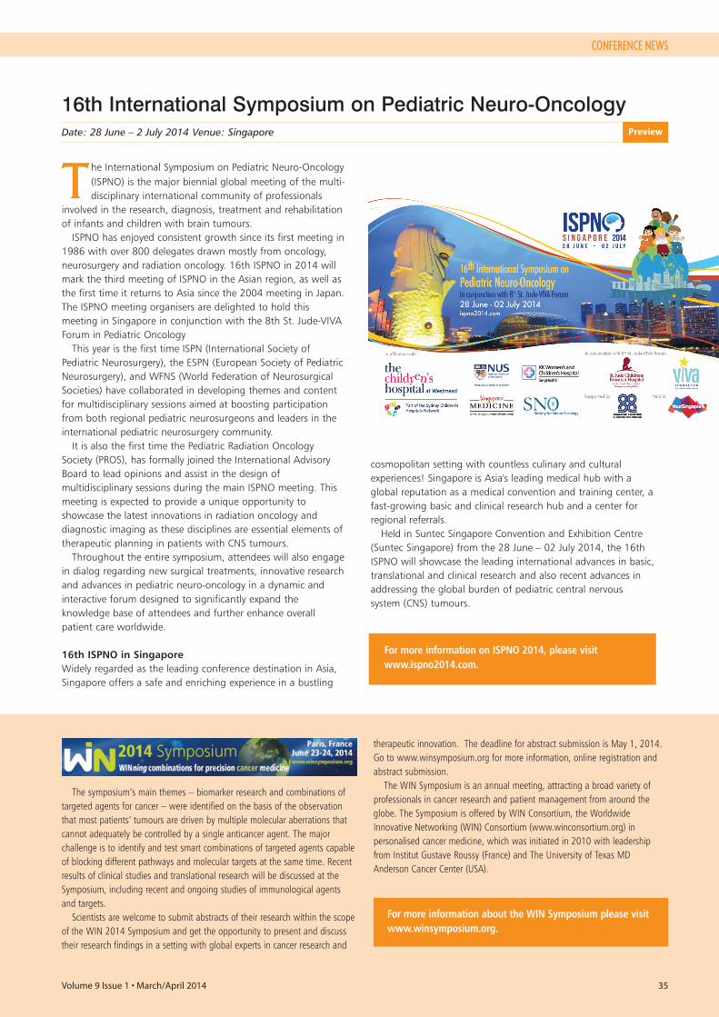
T he International Symposium on Pediatric Neuro-Oncology(ISPNO) is the major biennial global meeting of the multi-disciplinary international community of professionals
involved in the research, diagnosis, treatment and rehabilitationof infants and children with brain tumours. ISPNO has enjoyed consistent growth since its first meeting in
1986 with over 800 delegates drawn mostly from oncology,neurosurgery and radiation oncology. 16th ISPNO in 2014 willmark the third meeting of ISPNO in the Asian region, as well asthe first time it returns to Asia since the 2004 meeting in Japan.The ISPNO meeting organisers are delighted to hold thismeeting in Singapore in conjunction with the 8th St. Jude-VIVAForum in Pediatric OncologyThis year is the first time ISPN (International Society of
Pediatric Neurosurgery), the ESPN (European Society of PediatricNeurosurgery), and WFNS (World Federation of NeurosurgicalSocieties) have collaborated in developing themes and contentfor multidisciplinary sessions aimed at boosting participationfrom both regional pediatric neurosurgeons and leaders in theinternational pediatric neurosurgery community.It is also the first time the Pediatric Radiation Oncology
Society (PROS), has formally joined the International AdvisoryBoard to lead opinions and assist in the design ofmultidisciplinary sessions during the main ISPNO meeting. Thismeeting is expected to provide a unique opportunity toshowcase the latest innovations in radiation oncology anddiagnostic imaging as these disciplines are essential elements oftherapeutic planning in patients with CNS tumours.Throughout the entire symposium, attendees will also engage
in dialog regarding new surgical treatments, innovative researchand advances in pediatric neuro‐oncology in a dynamic andinteractive forum designed to significantly expand theknowledge base of attendees and further enhance overallpatient care worldwide.
16th ISPNO in SingaporeWidely regarded as the leading conference destination in Asia,Singapore offers a safe and enriching experience in a bustling
cosmopolitan setting with countless culinary and culturalexperiences! Singapore is Asia's leading medical hub with aglobal reputation as a medical convention and training center, afast-growing basic and clinical research hub and a center forregional referrals.Held in Suntec Singapore Convention and Exhibition Centre
(Suntec Singapore) from the 28 June – 02 July 2014, the 16thISPNO will showcase the leading international advances in basic,translational and clinical research and also recent advances inaddressing the global burden of pediatric central nervoussystem (CNS) tumours.
For more information on ISPNO 2014, please visitwww.ispno2014.com.
16th International Symposium on Pediatric Neuro-Oncology Date: 28 June – 2 July 2014 Venue: Singapore
CONFERENCE NEWS
The symposium’s main themes – biomarker research and combinations oftargeted agents for cancer – were identified on the basis of the observationthat most patients’ tumours are driven by multiple molecular aberrations thatcannot adequately be controlled by a single anticancer agent. The majorchallenge is to identify and test smart combinations of targeted agents capableof blocking different pathways and molecular targets at the same time. Recentresults of clinical studies and translational research will be discussed at theSymposium, including recent and ongoing studies of immunological agentsand targets.
Scientists are welcome to submit abstracts of their research within the scopeof the WIN 2014 Symposium and get the opportunity to present and discusstheir research findings in a setting with global experts in cancer research and
therapeutic innovation. The deadline for abstract submission is May 1, 2014.Go to www.winsymposium.org for more information, online registration andabstract submission.
The WIN Symposium is an annual meeting, attracting a broad variety ofprofessionals in cancer research and patient management from around theglobe. The Symposium is offered by WIN Consortium, the WorldwideInnovative Networking (WIN) Consortium (www.winconsortium.org) inpersonalised cancer medicine, which was initiated in 2010 with leadershipfrom Institut Gustave Roussy (France) and The University of Texas MDAnderson Cancer Center (USA).
For more information about the WIN Symposium please visitwww.winsymposium.org.
Volume 9 Issue 1 • March/April 2014 35
Preview
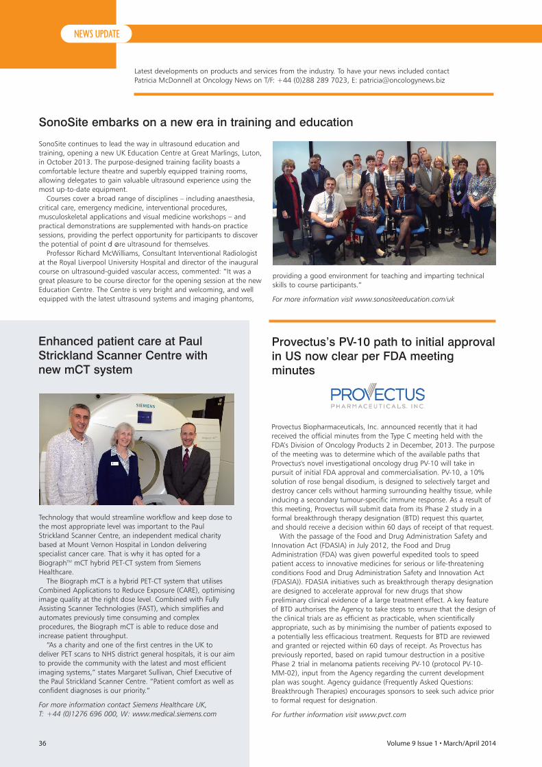
36 Volume 9 Issue 1 • March/April 2014
Provectus Biopharmaceuticals, Inc. announced recently that it hadreceived the official minutes from the Type C meeting held with theFDA’s Division of Oncology Products 2 in December, 2013. The purposeof the meeting was to determine which of the available paths thatProvectus's novel investigational oncology drug PV-10 will take inpursuit of initial FDA approval and commercialisation. PV-10, a 10%solution of rose bengal disodium, is designed to selectively target anddestroy cancer cells without harming surrounding healthy tissue, whileinducing a secondary tumour-specific immune response. As a result ofthis meeting, Provectus will submit data from its Phase 2 study in aformal breakthrough therapy designation (BTD) request this quarter,and should receive a decision within 60 days of receipt of that request. With the passage of the Food and Drug Administration Safety and
Innovation Act (FDASIA) in July 2012, the Food and DrugAdministration (FDA) was given powerful expedited tools to speedpatient access to innovative medicines for serious or life-threateningconditions Food and Drug Administration Safety and Innovation Act(FDASIA)). FDASIA initiatives such as breakthrough therapy designationare designed to accelerate approval for new drugs that showpreliminary clinical evidence of a large treatment effect. A key featureof BTD authorises the Agency to take steps to ensure that the design ofthe clinical trials are as efficient as practicable, when scientificallyappropriate, such as by minimising the number of patients exposed toa potentially less efficacious treatment. Requests for BTD are reviewedand granted or rejected within 60 days of receipt. As Provectus haspreviously reported, based on rapid tumour destruction in a positivePhase 2 trial in melanoma patients receiving PV-10 (protocol PV-10-MM-02), input from the Agency regarding the current developmentplan was sought. Agency guidance (Frequently Asked Questions:Breakthrough Therapies) encourages sponsors to seek such advice priorto formal request for designation.
For further information visit www.pvct.com
SonoSite embarks on a new era in training and education
SonoSite continues to lead the way in ultrasound education andtraining, opening a new UK Education Centre at Great Marlings, Luton,in October 2013. The purpose-designed training facility boasts acomfortable lecture theatre and superbly equipped training rooms,allowing delegates to gain valuable ultrasound experience using themost up-to-date equipment. Courses cover a broad range of disciplines – including anaesthesia,
critical care, emergency medicine, interventional procedures,musculoskeletal applications and visual medicine workshops – andpractical demonstrations are supplemented with hands-on practicesessions, providing the perfect opportunity for participants to discoverthe potential of point of care ultrasound for themselves. Professor Richard McWilliams, Consultant Interventional Radiologist
at the Royal Liverpool University Hospital and director of the inauguralcourse on ultrasound-guided vascular access, commented: “It was agreat pleasure to be course director for the opening session at the newEducation Centre. The Centre is very bright and welcoming, and wellequipped with the latest ultrasound systems and imaging phantoms,
providing a good environment for teaching and imparting technicalskills to course participants.”
For more information visit www.sonositeeducation.com/uk
Latest developments on products and services from the industry. To have your news included contact Patricia McDonnell at Oncology News on T/F: +44 (0)288 289 7023, E: [email protected]
NEWS UPDATE
Enhanced patient care at PaulStrickland Scanner Centre withnew mCT system
Technology that would streamline workflow and keep dose tothe most appropriate level was important to the PaulStrickland Scanner Centre, an independent medical charitybased at Mount Vernon Hospital in London deliveringspecialist cancer care. That is why it has opted for aBiographTM mCT hybrid PET-CT system from SiemensHealthcare. The Biograph mCT is a hybrid PET-CT system that utilises
Combined Applications to Reduce Exposure (CARE), optimisingimage quality at the right dose level. Combined with FullyAssisting Scanner Technologies (FAST), which simplifies andautomates previously time consuming and complexprocedures, the Biograph mCT is able to reduce dose andincrease patient throughput. “As a charity and one of the first centres in the UK to
deliver PET scans to NHS district general hospitals, it is our aimto provide the community with the latest and most efficientimaging systems,” states Margaret Sullivan, Chief Executive ofthe Paul Strickland Scanner Centre. “Patient comfort as well asconfident diagnoses is our priority.”
For more information contact Siemens Healthcare UK,T: +44 (0)1276 696 000, W: www.medical.siemens.com
Provectus’s PV-10 path to initial approvalin US now clear per FDA meeting minutes

Volume 9 Issue 1 • March/April 2014 37
NEWS UPDATE
Varian Medical Systems, a leader in radiationoncology equipment and software, hassigned an agreement with the UK’s NationalHealth Service (NHS) to acquire an NHS-developed cancer care planning tool calledthe Radiation oncology Planning OnlineResource Tool (R-PORT). Varian intends tooffer R-PORT to oncology departmentsglobally. R-PORT has been used in the NHS in
recent years to help control costs andmanage electronic data. Under the terms ofthe agreement, Varian Medical Systems willadd this tool to its extensive oncologyservices portfolio and develop and enhanceit for worldwide use. “R-PORT has helped oncology providers
within the NHS network to model changemanagement before incurring major costs,”says Steve Laws, Varian’s EMEA regionaldirector of software sales. “It also measuresthe results and costs of changemanagement for reporting purposes. It ishighly synergistic with Varian’s existingsoftware portfolio and builds on Varian’smission to be a ‘partner for life’ –supporting our customers to deliver worldclass cancer care.”“At a time of rising costs, uncertainty
concerning reimbursement and increasingnumbers of cancer patients, healthcareproviders worldwide are seeking solutions tohelp them deliver better patient care,” saysScott Brouse, Varian’s vice president ofWorldwide Site Solutions. “This is a tool thatcan help Varian support our customers inthe fight against cancer.”
For further information contactNeil Madle, Varian Medical Systems, T: +44 7786 526068, E: [email protected] W: www.varian.com
First Gamma KnifeCentre in South Westopens in Bristol
Wear A Hat Day – get your hatson, hold an event, and raisemoney for brain tumour research
Calling all hospitals, clinics, and othermedical centres, we’re asking you to holda Wear A Hat Day event on Friday 28thMarch and raise funds for vital scientificresearch into brain tumours.Brain tumours are responsible for more
loss of life in children than leukaemia, inwomen under 35 than breast cancer andin men under 45 than prostate cancer.Despite a decade of campaigning, braintumours still receive less than 1% of thenational spend on cancer research. With your help we can change this.
Wear A Hat Day is a great way to showyour support and help raise the crucialfunds and awareness needed. Full detailsare available from www.wearahatday.org,which provides an overview of thecampaign, tells you how to register, andcarries links to various resources that canbe used to help promote and maximise theimpact of your Wear A Hat Day event.
For further information contactT: +44(0)1296 733011 or E: [email protected]
More patients at Leeds Teaching Hospitals NHSTrust receiving advanced radiotherapy
A few months after Elekta unveiled itsground-breaking Versa HD™ system –which unites cutting edge beam shapingand High Dose Rate mode technologies –Leeds Teaching Hospitals installed two ofthe units, the first entering clinical servicein July 2013, and the second in November2013. Already, the first Versa HD has hada significant impact on clinical workflow.Up to 60 patients per day are scheduledfor treatment on the new machine.Treatment slots are booked at 10-minuteintervals, so that as many as six patientscan be treated per hour. “Initially, patients undergoing radical
prostate radiotherapy have been prioritisedfor treatment, all with High Dose Ratemode VMAT,” says Vivian Cosgrove, PhD,head of radiotherapy physics at Leeds. “Inaddition, the process to move all SBRT
treatments to High Dose Rate mode VMAThas begun. Patients have their SBRTtreatment scheduled to a 20-minute timeslot on the Versa HD. In September 2013, over 25 percent of
all radical treatments were delivered in theLeeds Cancer Centre using IMRT andVMAT – equivalent to about 90 newpatients starting advanced radiotherapytreatment every month. In addition, 20-25new patients also start an SBRT courseeach month. “This volume growth has been greatly
enhanced by opportunities to develop thetechniques, delivery equipment andworkflow on the research accelerators,side-by-side with the clinical service,” DrCosgrove added.
For further information contactE: [email protected]

38 Volume 9 Issue 1 • March/April 2014
NEWS UPDATE
Cook Medical introducesEchoTip® ProCore™EndobronchialUltrasound Needle
Cook Medical is introducing the firstendobronchial ultrasound (EBUS) needlein Europe that can acquire histologicalsamples. The EchoTip® ProCore™Endobronchial Ultrasound Needle givesphysicians the ability to retrieve both celland tissue samples from lymph nodes ortumours in the pulmonary area. The EchoTip ProCore EBUS needle is a
single-use needle that collects cell andtissue samples in order for a physician todiagnose lung cancer and variousmediastinal diseases. The needle can beused for fine-needle biopsy of lesions ofsubmucosal and extramural lesionswithin or adjacent to thetracheobronchial tree or gastrointestinaltract. Prior to the availability of the EchoTip
ProCore EBUS needle, tissue samplesfrom the pulmonary area could beobtained only by surgical methods. TheEchoTip ProCore EBUS needle is designedwith a core-trap technology that allowsphysicians to collect tissue samplesthrough a minimally invasive procedure. “Cook’s ProCore technology has
already proven itself in the GI endoscopicultrasound space,” said Barry Slowey,global leader of Cook Medical’sEndoscopy division. “We hope that thistechnology will aid in quick and accuratediagnosis for patients.”
For further information visitwww.cookmedical.com
Paxman Coolers Ltd appoints a distributor forAustralia & New Zealand
Paxman Coolers Ltd, the leadingglobal expert in scalp cooling, hasrecently announced a distributionagreement for Australia and NewZealand. Paxman Coolers Ltd hassigned an exclusive agreement withRegional Health Care Group (RHCG), aleading distributor of medicalproducts in Australia and NewZealand. Paxman Coolers Ltd isdedicated to preventingchemotherapy-induced hair loss andthe technology has been successful forthousands of patients throughout theworld.Under the terms of the agreement
with RHCG, the Paxman devices willbe available to Chemotherapy centresthroughout Australia and NewZealand exclusively through RHCG. “This will be an important addition to
our portfolio. Within Australia & NewZealand there are over 15,000 peoplediagnosed with Breast Cancer alone.Scalp cooling is widely available in the UKand it should become the standard ofcare for ANZ patients,” stated NeilSpence, National Marketing Manager of
Regional Health Care Group (RHCG). “Welook forward to combining the Paxmantechnology with RHCG’s extensivedistribution network to give patients andcare provider’s access to the life-changing benefits possible with thePaxman device.”
For further information visitwww.paxman-coolers.com
Provectus announces PV-10’s assessment fordrug-drug interaction potential is subject of articlepublished by Xenobiotica
Provectus Biopharmaceuticals, Inc., adevelopment-stage oncology anddermatology biopharmaceuticalcompany, and XenoTech, a preclinicalCRO and pioneer in collaborative researchsurrounding in vitro drug metabolismand pharmacokinetics services,announced that an article describing astudy to determine the potential of rosebengal disodium to cause drug-druginteractions has been published byXenobiotica, a peer-reviewed scientificjournal that publishes comprehensiveresearch papers on pharmacokinetics (thestudy of distribution, metabolism,disposition and excretion of drugs). Thepublished research indicated that the riskof PV-10 causing clinically relevant drug-drug interactions is likely minimal. PV-10,a 10% solution of rose bengal that iscurrently under clinical investigation as anovel cancer therapeutic, is designed to
selectively target and destroy cancer cellswithout harming surrounding healthytissue, minimising the potential forsystemic side effects. The study was undertaken prior to
initiation of the now ongoing testing ofPV-10 plus sorafenib (cohort 2) in aclinical trial of PV-10 intralesionalinjection in hepatocellular carcinomapatients taking a stable dose of sorafenib(ClinicalTrials.gov Identifier:NCT00986661). Sorafenib is acompetitive inhibitor of cytochrome P450(CYP) drug metabolism enzymes and isreliant on the UDP-glucuronosyltransferase (UGT) pathwayfor efficient clearance. CYP and UGTenzymes help to biotransform smalllipophilic drugs like sorafenib into water-soluble excretable metabolites.
For further information visitwww.pvct.com
Volume 9 Issue 1 • March/April 2014 39
NEWS UPDATE
Innovative partnerships pave the way for massivenew funding programme for brain tumour research
Leading campaign charity Brain TumourResearch has announced ground-breaking collaborative partnerships withthree new Brain Tumour ResearchCentres of Excellence. The leading institutions embarking on
innovative funding partnerships with thecharity are:• Queen Mary University of London in
collaboration with UCL Institute ofNeurology, researching glioblastomamultiforme and identifying effectivedrug treatments.
• Imperial College Healthcare NHS Trustinvestigating tumour metabolisms anddeveloping innovative 3D real-timesurgical imaging.
• Plymouth University Peninsula Schoolsof Medicine and Dentistry conductingworld-leading research into low-gradebrain tumours.
This collaboration will lead to a £20minvestment in brain tumour research overthe next five years and will establish anetwork of research centres with secure
long-term funding covering the keysalaried positions. The charity will shape research teams
and priorities, ensuring all 120+ types ofbrain tumours are being researched, thatthe causes of brain tumours arediscovered, that complex brain tumourbehaviour is understood, and bettertreatments for all brain tumour patientsare developed.
For further information please contactKatie Abbotts, T: 07810 504380 or E: [email protected]
Elekta highlight their product range at ESTRO33
Elekta is a human care company thatpioneers significant innovations and clinicalsolutions for treating cancer and braindisorders. The company develops state-of-the-art tools and treatment planningsystems for radiation therapy, radiosurgeryand brachytherapy, as well as workflowenhancing software systems across thespectrum of cancer care. At ESTRO33 Elekta (booth #6200) will
demonstrate a comprehensive array ofadvanced solutions for cancer management.These products include the new Versa HD™treatment system, Leksell Gamma Knife®
Perfexion™ stereotactic radiosurgery system,Monaco® 5 treatment planning system –featuring a new graphical user interface andsystem architecture – Clarity® Autoscan forreal-time soft tissue motion tracking,Identify™ for enhanced patient safety, andthe company’s advanced solutions forbrachytherapy, including the recentlylaunched Esteya® electronic brachytherapysystem for treating skin cancer. Elekta’stheme for ESTRO33 is “Power of Care”which emphasises the synergy of technologyand people working together to supportpositive patient outcomes.
For further information contactE: [email protected]
Varian spotlights full range of products atArab Health 2014 in Dubai
Varian Medical Systems recentlydemonstrated its full range ofradiotherapy delivery systems andsoftware at the Arab HealthCongress in January 2014. TheVarian booth (Sheikh Saeed Hall 1,booth No. S1 – C40) featured thecompany’s technology andproducts for radiotherapy,radiosurgery, brachytherapy, andproton therapy. At the show, Varian announced
a collaboration with MediclinicInternational to equip the newMediclinic City Hospital NorthWing radiotherapy department inDubai, which is expected to start clinicaltreatments in late 2015.
Full Range of ProductsThe latest version of Varian’s TrueBeam™platform for fast and precise radiotherapyand radiosurgery were on display,showcasing new image-guidance andprecise patient positioning capabilitiessuch as the PerfectPitch™ six-degrees-of-freedom couch, giving clinicians moreangles from which to deliver treatments. Visitors to the Varian booth learned
more about the company’s new EdgeRadiosurgery™ treatment system, Varian’sfirst dedicated, fully integrated end-to-end
solution for planning and deliveringadvanced radiosurgery treatments usingnew real-time tumour tracking technologyand motion management capabilities.Varian also unveiled RapidPlan™ for
Middle East customers, offeringknowledge-based treatment planningcapabilities aimed at helping to createcomplex RapidArc® volumetric modulatedarc therapy and intensity-modulatedradiotherapy (IMRT) treatment plans.
For further information contactNeil Madle, Varian Medical Systems, T: +44 7786 526068, E: [email protected] W: www.varian.com
http://is.gd/oncologyfacebook
Sophisticated target conformance without compromised treatment times
Deliver Complex Head and Neck Plans 42% Faster
One Solution. Unlimited Possiblities.
www.VersaHD.com4513
371
123
0 11
:13
As compared to previous generation Elekta digital linear accelerators. Stieler F, Steil V, Wenz F, Lohr F, Department of Radiation Oncology, University Medical Center Mannheim, University of Heidelberg, Germany. Versa HD is not available for sale or distribution in all markets. Please contact your Elekta representative for details.