Embed Size (px)
Citation preview

Submitted 19 April 2020, Accepted 10 April 2021, Published 24 May 2021
Corresponding Author: M. Niranjan – e-mail – [email protected] 171
One new species and two new records of Xylarialean fungi from
Andaman Islands, India
Niranjan M1,2*, Anupam D1,3, Parida P1 and Sarma VV1
1Department of Biotechnology, Pondicherry University, Kalapet, Pondicherry – 605014, India 2Department of Botany, Rajiv Gandhi University, Rono Hills, Doimukh, Itanagar, Arunachal Pradesh – 791112, India 3Synapse Biology Lab, Department of Molecular and Cellular Neuroscience, National Brain Research Center (NBRC),
NH-08, Manesar, Gurgaon, Haryana – 122052, India
Niranjan M, Anupam D, Parida P, Sarma VV 2021 – One new species and two new records of
Xylarialean fungi from Andaman Islands, India. Current Research in Environmental and Applied
Mycology (Journal of Fungal Biology) 11(1), 171–184, Doi 10.5943/cream/11/1/13
Abstract
Information on the molecular diversity of the Xylarialean fungi from the Andaman and
Nicobar Islands is scarce. Xylarialean fungi are widely distributed in India, and studies revealed
that two new records Hypomontagnella spongiphila and Nemania bipapillata, one new species
Neoanthostomella samachedabeejae. In the phylogenetic tree generated by the ITS sequence,
results were showing that three species good bootstrap support. In addition, the new species is
compared with existing species in detail in the table. Microscopic and molecular studies support all
these species.
Key words – Ascomycota – Hypoxylaceae – Taxonomy – Xylariaceae
Introduction
The fungi had poorly investigated from the Andaman and Nicobar Islands (A and N), India,
until 2014 (Singh 1969, 1970, 1973, Upreti & Singh 1987, Upreti 1990, 2014, Bhat 1993,
Chinnaraj 1993, Jagadeesh Ram 2014, Jagadeesh Ram & Sinha 2016). Niranjan & Sarma (2018a)
have reported the complete check list of fungi from the Islands and compiled 446 fungi. Among
446, most of the ascomycetes are inhabit in terrestrial plant leaves and marine fungi (Chinnaraj
1993, Hosagoudar 2013, Jagadeesh Ram & Sinha 2016, Niranjan & Sarma 2018a, b). The
examination of dead and decomposing twigs on the forest floor has resulted in discovering new
species and records of Xylariales.
Xylariaceae has been one of the dominant family found in the Indian subcontinent for which
many taxa have been described (Thind & Waraitch 1969, 1976, Karun & Sridhar 2015, Dargan
2016, Nandan Patel & Krishnappa 2017, Debnath et al. 2018). Most of the exploration of
Xylariaceae species has been compiled from Southern India by Pande (2008) fallowed by Karun &
Sridhar (2015) reported 24 Xylaria species, similarly in northern part covered by Koyani et al.
(2016) reported over 19 species of Xylariaceae from Gujarat state, India. From the Andaman
Islands, 8 species have been reported, including two new Rosellinia species (Niranjan & Sarma
2018a, c) and remaining new records. The recent treatments on Xylariaceae have been published
recently (Senanayake et al. 2015, Daranagama et al. 2018). Xylariaceae consists of more than 85
genera and 1300 species Koyani et al. (2016) and revised to 87 genera (Maharachchikumbura et al.
Current Research in Environmental & Applied Mycology (Journal of Fungal Biology)
11(1): 171–184 (2021) ISSN 2229-2225
www.creamjournal.org Article
Doi 10.5943/cream/11/1/13

172
2016). Later on phylogenetic intervention deliminate to describe the new combinations leads to
reduce 37 genera (Daranagama et al. 2018 and Wendt et al. 2018).
The recent revision of Xylariales order (Hyde et al. 2020) recognized 14 families (including
Myelospermataceae under Xylariales incertae sedis) and the dominant families Xylariaceae and
Hypoxylaceae consists of 32 and 19 genera respectively. The recent studies eructed new genera
Neoxylaria (Konta et al. 2020) added and detailed notes on Xylariaceae.
Materials & Methods
The morphological studies performed based on Niranjan & Sarma (2018b). Morphological
identification was performed by referring to various monographs and individual publications,
including Pande (2008), Maharachchikumbura et al. (2016) and Hyde et al. (2020). Herbarium
samples have been deposited at the Agharkar Research Institute (ARI) of the Ajrekar Mycological
Herbarium (AMH), Pune, India. Cultures are maintained in Fungal Biotechnology Laboratory,
Department of Biotechnology, Pondicherry University. GenBank accession numbers are available
at https://submit.ncbi.nlm.nih.gov/subs/. The individual ITS sequences obtained were submitted to
the NCBI Blast search tool to reveal closely related matches on GenBank. Multiple sequence
alignments were performed in online software (http://mafft.cbrc.jp/alignment/server/index.html,
Katoh & Standley 2013). All the phylogenetic data sets used in this study are mentioned in Table 1.
DNA extraction, amplification and sequencing
The isolation of individual spores was performed as described by Choi et al. (1999). Three
pure axenic cultures in malt extract agar (MEA) were grown for a week at 28°C, DNA extraction
performed by manufacturer protocol (Thermo Scientific, USA). The internal transcribed spacer
(ITS) was chosen for phylogenetic analysis. This region was amplified by PCR using the pair of
primers ITS1 and ITS4. The polymerase chain reaction (PCR) was performed with a total volume
of 25μL, 12 μL of Taq DNA Polymerase 2x Master Mix RED 1.5 mM MgCl2, 1 μL of each primer
(10 μM), 1 μL (10–50 ng) genomic DNA and remaining volume makeup with nuclease free water.
PCR amplification conditions were set as follows, an initial denaturation at 95°C for 5 min,
followed by 35 cycles of denaturation at 95°C for 90 seconds, primer annealing 52°C for ITS,
primer extension at 72°C for 1 min and a final extension step at 72°C for 10 min. PCR
amplification was checked on 0.8% Agarose gel stained with ethidium bromide. The PCR products
were purified by using a Gene JET PCR purification kit (Thermofisher, Lithuania) by following the
manufacturer’s protocol. Then the PCR products were sent outsourcing (Agri Genome, Kochi,
India) for the sequencing.
Phylogenetic analysis
The phylogeny was constructed using aligned sequence data performed using maximum
likelihood (ML), maximum parsimony (MP) and Bayesian criteria (MB). The maximum likelihood
was represented by using the randomized accelerated maximum likelihood (RAxML). The
RAxML-HPC2 in XSEDE (version 8.2.8) (Stamatakis 2014) on the CIPRES Science Gateway
platform (Miller et al. 2010) using the GTR + I + G evolution model. The phylograms were
visualized with the FigTree v. 1.4.0 program (Rambaut 2012) and were reorganized in Microsoft
Power Point (v. 2007) and Adobe Photoshop (v. 7.0, Adobe®, San José, CA).
MP was achieved with PAUP v. 4.0b10 (Swofford 2002), with the following parameters, as
disordered characters of equal weight, random addition of taxa, branch exchange with the bisection
tree reconnection algorithm (TBR), the branch reduce if the maximum branch length was zero.
Alignment gaps were treated as missing characters in the combined dataset analysis, where they
occurred in relatively conserved regions. The trees were deduced using the heuristic search option
with 1000 additions of random sequences, with a maximum of trees set to 1000. Descriptive tree
statistics for parsimony. The length of the trees (TL), the consistency index (CI), the retention index
(RI), the relative consistency index (RC) and the homoplasy index (HI) were calculated for the

173
trees generated according to different optimization criteria. Kishino-Hasegawa tests (Kishino &
Hasegawa 1989) were performed to determine if the trees were significantly different.
MB analysis was performed with MrBayes v.3.1.2 (Huelsenbeck & Ronquist 2001) to assess
the subsequent Bayesian probabilities (BYPP) (Rannala & Yang 1996, Zhaxybayeva & Gogarten
2002) by sampling Markov Chain Monte Carlo (BMCMC). GTR + I + G was used in the
command. Six simultaneous Markov chains were run over 5000000 generations, and tree samples
were taken every 1000 generations. The distribution of recording probability scores was examined
to determine the stationary phase for each search and to decide whether additional trials were
needed to reach convergence, using the Tracer 1.5 program (Rambaut & Drummond 2007). The
first 20% of the trees generated were discarded, and the remaining 80% was used to calculate the
subsequent probabilities of the majority rule consensus tree. A BYPP greater than 0.60 is indicated
above each node. We consider bootstrap support >75 as strong support, between 50 and 75 as
moderate support and less than 50 as minimum support.
Results
Molecular phylogenetic analyses
In the phylogeny of Hypomontagnella spongiphila (NFCC-4378) constructed using closely
related taxa Annulohypoxylon, Hypomontagnella and Daldinia addition to Xylaria hypoxylon (CBS
122620) selected as the outgroup. RAxML analysis yielded a minimum scoring tree with a final
ML optimization likelihood value of -12664.883372. The matrix had 427 distinct alignment
patterns with 9.62% of indeterminate characters or gaps. The estimated base frequencies were as
follows A = 0.251363, C = 0.248286, G = 0.231114, T = 0.2692238, substitution rate AC =
1.310818, AG = 2.3338671, AT = 1.597524, CG = 0.974316, CT = 3.212431, GT = 1.000000.
Proportion of invariable sites I = 0.298569, gamma distribution shape parameter α = 0.693555. The
maximum parsimonious dataset is 686 characters, including 273 constants, 297 informative
parsimony and 116 uninformative parsimony. The parsimonious analysis of the data matrix resulted
in a thousand equally parsimonious trees with a length of 2939 steps (CI = 0.269, RI = 0.397, RC =
0.107, HI = 0.731) in the first tree. The phylogenetic trees result from ML, a distinct complex of
Hypomontagnella was observed within the Hypoxylon sequences similar and consistent with
previous studies (Wibberg et al. 2020). Phylogenetic analysis has shown that our H. spongiphila
NFCC4378_MT644605 nested with H. spongiphila CCL_KY744359 with a 64% strong bootstrap
support in MP.
In the Nemania bipapillata NFCC-4519 phylogenetic analysis, presumed by morphology
assumption it closely related to Nemania, therefore we selected the Nemania and allied genera
sequences were selected in addition, Botryosphaeria dothidea BD080705002 as outgroup. RAxML
analysis yielded a minimum scoring tree with a final ML optimization likelihood value of -
4780.691359. The matrix had 288 distinct alignment patterns with 10.53% of undetermined
characters or gaps. Estimated base frequencies were as follows, A = 0.242739, C = 0.255614, G =
0.239944, T = 0.261703, substitution rates AC = 1.301212, AG = 2.460070, AT = 1.203682, CG =
0.597153, CT = 3.829878, GT = 1.000000. Proportion of invariable sites I= 0.258137, gamma
distribution shape parameter α = 0.532472. The maximum parsimonious (MP) dataset consists of
509 characters, of which 249 were constant, 203 parsimony–informative and 57 parsimony–
uninformative. The parsimony analysis of the data matrix resulted in one thousand equally
parsimonious trees with a length of 949 steps (CI = 0.496, RI = 0.755, RC = 0.375, HI = 0.504) in
the first tree. The overall topology of the phylogenetic trees resulted from ML, MP, similar and
incongruent in with earlier studies (Hyde et al. 2020). The phylogenetic analysis showed that our
Nemania bipapillata NFCC-4519 has sister-cladding with Nemania bipapillata JQ862661 with
strong bootstrap support 100% in MP.
The phylogenetic tree of Neoanthostomella samachedabeejae NFCC-4517, constructed by
ITS sequence taxa from Xylariaceae while Sordaria tomentoalba_CBS56972_MH860578 served
as the outgroup taxon. RAxML analysis yielded a minimum scoring tree with a final ML

174
optimization likelihood value of -5572.802237. The matrix had 238 distinct alignment patterns with
1.47% of indeterminate characters or gaps. The estimated base frequencies were as follows A =
0.254423, C = 0.248620, G = 0.223137, T = 0.273819, substitution rate AC = 2.151608, AG =
2.985945, AT = 2.416015, CG = 1.070409, CT = 6.021074, GT = 1.000000. Proportion of
invariable sites I = 0.242763, gamma distribution shape parameter α = 0.566179. The maximum
economical (MP) data set consists of 397 characters, of which 169 were constant, 186 parsimony -
informative and 42 parsimony - non-informative. The parsimony analysis of the data matrix
showed a thousand equally parsimonious trees with a length of 1148 steps in the first tree (CI =
0.349, RI = 0.642, RC = 0.224, HI = 0.651). The phylogenetic analysis N. samachedabeejae
NFCC4517_MT644606 has a branch cladding with N. viticola MFLUCC160243_NR16551 with
low bootstrap support of 61% in MP. The phylogenetic tree similar results Daranagama et al.
(2016) and one new combination Pseudoanthostomella thailandica (= Anthostomella thailandica)
confirmed.
Taxonomy
Hypomontagnella spongiphila Kuhnert 2020 Figs 1, 4
Saprobic on un-identified twig. Teleomorph – Stromata superficial, black thick woody to
carbonaceous, covered the ascomata except in apical papilla. Ascomata perithecial, grouped in
stromata, globose, bipartite, semi-immersed in stromata up to 1/3 portion, papilla below the
stromata, grey purple colour in 10% KOH. Peridium consists of outer dark brown to inner hyaline
cell layers. Hamathecium paraphyses, numerous, septate, branched, longer than asci uneven width
1–5 μm. Asci 71–105 × 5–5.5 μm (x̅ = 82 × 5, n = 25), unitunicate, cylindrical, rounded end with
apical chamber without apical ring, long pedicel, smooth-walled. Ascospores 9.5–11 × 5–5.5 μm (x̅
= 11 × 5, n = 25), 8-spored, hyaline to purple to brown at maturity, rarely overlapping, aseptate,
straight germ slit with central globose guttulate, ellipsoid, inequilateral, obtuse end. Anamorph –
Undetermined.
Known distribution – French Polynesia and India.
Material examined – INDIA, Andaman and Nicobar Islands, South Andaman, Chidiya Tapu,
View Point Area (11o29’22”N 92o42’36.6”E). Isolated on un-identified twig, 8 January 2017, M.
Niranjan and V.V. Sarma (PUFNI 17449). Submitted in Ajrekar Mycological Herbarium-AMH
(AMH-9985) and Ex-type living culture (NFCC-4378).
Notes – Hypomontagnella was described by Lambert et al. (2019) based on morphology,
chemotaxonomy and molecular phylogeny. Hypomontagnella consists of perispores and not find in
Annulohypoxylon and Jackrogersella similarly distinct from Hypoxylon in having annulate disc
with papillate ostioles and lack of stromatal granules. Presently Hypomontagnella dwell six species
H. austrobahiensis, H. barbarensis, H. monticulosa, H. rubigineoareolata, H. spongiphila, H.
submonticulosa. The present collection of Hypomontagnella spongiphila shares morphological
similarities with the genus. The holotype (H. spongiphila) was collected from the marine source
(Wibberg et al. 2020) that lack of teleomorph and anamorph, whereas the current specimen was
collected from the terrestrial habitat but it is sharing sequence similarities (Leman-Loubiere et al.
2017, Lambert et al. 2019. Therefore, H. spongiphila can be survived in marine, and terrestrial
conditions and our collection also can improve the species knowledge by adding the morphological
data. Herein, our collection is provided as a new record in India.
Nemania bipapillata (Berk. and M.A. Curtis) Pouzar, Ceská Mykologie 39 (1): 24 (1985)
Figs 2, 5
Saprobic on an unidentified twig. Teleomorph – Ascostromata, superficial, carbonaceous,
central ostiolated, dark brown papilla, surface wave like circles, thick at base and thin towards to
apex, perithecial ascomata, mostly single, rarely 2-3 covered by stromata. Peridium composed of
two layers, outer thick brown and inner pale brown textura prismatica cells. Hamathecium
paraphyses upto 7 μm wide, filamentous, unbranched, guttulate, longer than the asci, wide at base

175
and arrow towards apex. Asci 160–215 × 10–13 μm (x̅ = 185 × 12, n = 25), spore bearing part 65–
89 length (x̅ = 74) unitunicate, 8–spored, cylindrical, apical ring 2.3–2.8 × 2–2.2 μm (x̅ = 2.5, n =
25), J+ in Lougals solution, long pedicel, persistent. Ascospores 11–13 × 4.6–7.8 μm (x̅ = 12 × 5, n
= 25), hyaline to pale brown, becoming dark brown at maturity, in-equilateral ends, straight germ
slit, smooth-walled. Anamorph – Undetermined.
Fig. 1 – Phylogram generated from maximum parsimony by using the various sequences belongs to
the Xylariaceae. The best scores generated using the maximum likelihood (ML) and parsimony

176
(MP) as 84/64 bootstrap values are given at the above nodes. Newly generated sequences of
Hypomontagnella spongiphila in bold. The tree was rooted with Xylaria hypoxylon as outgroup.
Fig. 2 – Phylogram of the taxon Nemania generated by maximum parsimony based on ITS dataset.
Newly generated sequences of Nemania bipapillata is in bold. The tree was rooted with
Botryosphaeria dothidea as an outgroup.
Known distribution – Australia, Belgium, Brazil, Bhutan, Brunei Darussalam, Canada, Congo,
Costa Rica, Colombia, Cuba, Denmark, French Guiana, Gabon, Guyana, Hong Kong, Japan, India,

177
Mexico, New Zealand, Thailand, Taiwan, Trinidad and Tobago, Panama, Pakistan, Philippines,
Paraguay, South Africa, Sierra Leone, Switzerland, UK, USA.
Material examined – INDIA, Andaman and Nicobar Islands, South Andaman, Chidiya Tapu,
Near Viewpoint (11o30’44”N 92o42’34”E), Recorded on Paraishia insignis decaying twig, 20 May
2018, M. Niranjan and V.V. Sarma (PUFNI 18745) AMH 10065. Ex-type living culture NFCC-
4519.
Fig. 3 – Phylogram of Neoanthostomella samachedabeejae generated by maximum parsimony with
Sordaria tomentoalba as outgroup. A newly generated sequence is in red bold.

178
Fig. 4 – Hypomontagnella spongiphila (NFCC-4378). a, b Stromata. c Stromata grey purple in 10%
KOH. d Paraphyses. e-g Asci. h Peridium. i-n Ascospores. Scale bars: h = 50 µm, d-g = 20 µm. i-n
= 10 µm.
Notes – Nemania was introduced by Pouzar (1985) to fill distinct species from the genus
Hypoxylon, are found as saprophytic and endophytic (Medina et al. 2019). The present record of N.
bipapillata sharing the similarities with the original description in having the hemispherical
stromata, pale brown flattened cells of peridium, cylindrical asci, and brown ascospores. The
present taxon distinct in having the superficial ascostromata, textura prismatica and textura
angularis of peridium, whereas previous one does not. The ascospores of current specimen having
slightly smaller ascospores (11–13 × 4.6–7.8 vs. 10–15 × 5–6.5), lack of sheath and pod like
appendages compared to original description (Smith & Hyde 2001, Ju & Rogers 2002).
Neoanthostomella samachedabeejae M. Niranjan and V.V. Sarma sp. nov. Figs 3, 6
Index Fungorum number: IF558023
Etymology – The ascospores have a straight longitudinal germ slit (sama = straight, cheda =
slit, bija = spores).

179
Saprobic on an unknown decaying twig. Teleomorph – Pseudostromata aggregated rarely
clustered, brown, lignicolous. Ascomata 260–290 × 170–250 μm, perithecial, globose, scattered,
immersed, coriaceous, individual central ostiolated, erumpent neck, central empty canal. Peridium
17.2 μm wide, consist of brown to hyaline textural subglobosa and textura angularis cell layers.
Hamathecium paraphyses 2–2.5 μm wide, septate, unbranched, guttulate, thin towards apex. Asci
58–94 × 6–7.5 μm (x̅ = 76 × 7, n=25), unitunicate, cylindrical, without apical ring but it consists of
apical thickening, rounded apex, medium pedicel, smooth-walled. Ascospores 9–12 × 4–6 μm (x̅ =
11 × 5, n=25), ovoid to oblong, initially hyaline becoming brown at maturity, arranged in an
overlapping uniseriate, straight germ slit, smaller than the spore length, obtuse ends, smooth
walled. Anamorph – Undetermined.
Fig. 5 – Nemania bipapillata (NFCC-4519). a, b Ascostromata. c Vertical section of ascostroma.

180
d, e Paraphyses. f-h Asci. i textura porrecta. j textura prismatica. k-m Ascospores. n, o Pure culture
on malt extract agar. Scale bars: d-h = 50 μm, i-m = 10 μm.
Fig. 6 – Neoanthostomella samachedabeejae (NFCC-4517, holotype). a, b Ascomata. c, d Vertical
section of ascomata. e-g Asci h Peridium. h-k Asci. l Periphyses. i, j Pure culture on malt extract
agar. k-m Ascospores. Scale bars: c = 200 μm, d, h = 50 μm, e-g, k-m = 10 μm.
Known distribution – INDIA (Present study).
Material examined – India, Andaman and Nicobar Islands, South Andaman, Chidiya Tapu,
View Point (11o30’49”N 92o42’38”E), an unknown decaying twig, 20 May 2018, M. Niranjan and
V.V. Sarma (PUFNI 18753). The herbarium AMH 10058, Ex-type living culture NFCC-4517.
Notes – New records and species of Anthostomella and allied genera have been reported
recently (Lu et al. 1999, Rappaz 1995, Lu & Hyde 2000, Crous et al. 2015, Daranagama et al. 2016,
Voglmayr et al. 2018. Mostly they are collected in forests as saprobic and endophytic forms.
Molecular distinct Anthostomella species were transferred into the newly described genera such as
Alloanthostomella, Anthostomelloides, Neoanthostomella and Pseudoanthostomella (Daranagama
et al. 2016) along with a key provided to Anthostomella-like genera. The genus Neoanthostomella
was described by Dai et al. (2017) with N. pseudostromatica type species. Later one more species
were added N. viticola (Daranagama et al. 2016). The morphological description pseudostromata
solitary to gregarious. Ascomata gregarious, growing together in a single pseudostroma,
periphysate, ostiolate carbonaceous neck. Peridium brown to hyaline cells of textura angularis
181
layers. Ascospores ellipsoid, aseptate, dark brown, guttulate, full length germ slit.
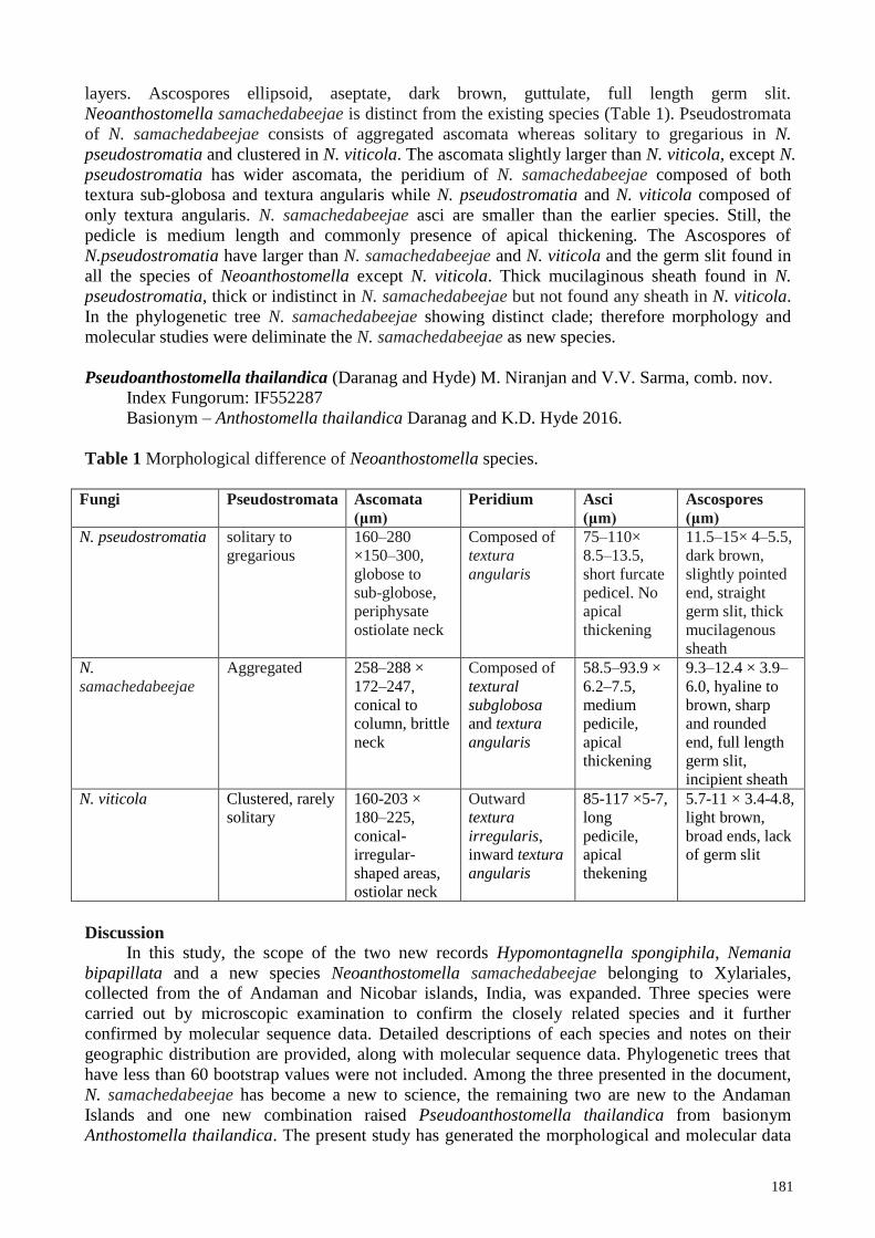
Neoanthostomella samachedabeejae is distinct from the existing species (Table 1). Pseudostromata
of N. samachedabeejae consists of aggregated ascomata whereas solitary to gregarious in N.
pseudostromatia and clustered in N. viticola. The ascomata slightly larger than N. viticola, except N.
pseudostromatia has wider ascomata, the peridium of N. samachedabeejae composed of both
textura sub-globosa and textura angularis while N. pseudostromatia and N. viticola composed of
only textura angularis. N. samachedabeejae asci are smaller than the earlier species. Still, the
pedicle is medium length and commonly presence of apical thickening. The Ascospores of
N.pseudostromatia have larger than N. samachedabeejae and N. viticola and the germ slit found in
all the species of Neoanthostomella except N. viticola. Thick mucilaginous sheath found in N.
pseudostromatia, thick or indistinct in N. samachedabeejae but not found any sheath in N. viticola.
In the phylogenetic tree N. samachedabeejae showing distinct clade; therefore morphology and
molecular studies were deliminate the N. samachedabeejae as new species.
Pseudoanthostomella thailandica (Daranag and Hyde) M. Niranjan and V.V. Sarma, comb. nov.
Index Fungorum: IF552287
Basionym – Anthostomella thailandica Daranag and K.D. Hyde 2016.
Table 1 Morphological difference of Neoanthostomella species.
Fungi Pseudostromata Ascomata
(μm)
Peridium Asci
(μm)
Ascospores
(μm)
N. pseudostromatia solitary to
gregarious
160–280
×150–300,
globose to
sub-globose,
periphysate
ostiolate neck
Composed of
textura
angularis
75–110×
8.5–13.5,
short furcate
pedicel. No
apical
thickening
11.5–15× 4–5.5,
dark brown,
slightly pointed
end, straight
germ slit, thick
mucilagenous
sheath
N.
samachedabeejae
Aggregated 258–288 ×
172–247,
conical to
column, brittle
neck
Composed of
textural
subglobosa
and textura
angularis
58.5–93.9 ×
6.2–7.5,
medium
pedicile,
apical
thickening
9.3–12.4 × 3.9–
6.0, hyaline to
brown, sharp
and rounded
end, full length
germ slit,
incipient sheath
N. viticola Clustered, rarely
solitary
160-203 ×
180–225,
conical-
irregular-
shaped areas,
ostiolar neck
Outward
textura
irregularis,
inward textura
angularis
85-117 ×5-7,
long
pedicile,
apical
thekening
5.7-11 × 3.4-4.8,
light brown,
broad ends, lack
of germ slit
Discussion
In this study, the scope of the two new records Hypomontagnella spongiphila, Nemania
bipapillata and a new species Neoanthostomella samachedabeejae belonging to Xylariales,
collected from the of Andaman and Nicobar islands, India, was expanded. Three species were
carried out by microscopic examination to confirm the closely related species and it further
confirmed by molecular sequence data. Detailed descriptions of each species and notes on their
geographic distribution are provided, along with molecular sequence data. Phylogenetic trees that
have less than 60 bootstrap values were not included. Among the three presented in the document,
N. samachedabeejae has become a new to science, the remaining two are new to the Andaman
Islands and one new combination raised Pseudoanthostomella thailandica from basionym
Anthostomella thailandica. The present study has generated the morphological and molecular data
182
of ITS for isolates from the Andaman Islands and provides an update of the molecular sequence
data. These results emit light in subsequent studies of Xylariaceae fungi on the remaining islands.
Acknowledgements
We would like to thank the SERB, Ministry of Science and Technology of the Indian
government, for funding the project (SERB/SB/SO/PS/18/2014 dt.19.5.2015). We also thank the
District Forestry Officials of the South, Central, North Andaman Districts and the Forestry Service
of the (PCCF) A and N Islands, India, for permission to collect samples from each district. UGC-
SAP and DST-FIST programs are thanked for infrastructural support. We thank the Department of
Biotechnology Pondicherry University for providing the facilities. Niranjan would like to thank the
SERB, the Indian government, for providing a grant.
References
Bhat DJ. 1993 – Twenty-Five New Conidial Fungi from the Western Ghats and the Andaman
Islands (India). Mycotaxon 49, 19–90.
Chinnaraj S. 1993 – Higher marine fungi from mangroves of Andaman and Nicobar Islands.
Sydowia 45, 109–115.
Choi YW, Hyde KD, Ho WH. 1999 – Single spore isolation of fungi. Fungal Diversity 3, 29–38.
Crous PW, Wingfield MJ, Guarro J, Hernández-Restrepo M et al. 2015 – Fungal Planet description
sheets: 320–370. Persoonia: Molecular Phylogeny and Evolution of Fungi 34, 167.
Dai DQ, Phookamsak R, Wijayawardene NN, Li WJ et al. 2017 – Bambusicolous fungi. Fungal
Diversity 82, 1–5.
Daranagama DA, Camporesi E, Jeewon R, Liu X et al. 2016 – Taxonomic rearrangement of
Anthostomella (Xylariaceae) based on a multigene phylogeny and morphology. Cryptogamie,
Mycologie 37, 509–538.
Daranagama DA, Hyde KD, Sir EB, Thambugala KM et al. 2018 – Towards a natural classification
and backbone tree for Graphostromataceae, Hypoxylaceae, Lopadostomataceae and
Xylariaceae. Fungal diversity 88, 1–65.
Dargan JS. 2016 – My dates with Perithecial Fungi. Kavaka 47, 27–34.
Debnath S, Majumdar K, Das P, Saha AK. 2018 – New Distribution Record of Five Species of
Xylaria from Tripura, Northeast India. Research and Reviews: A Journal of Life Sciences 5,
1–10.
Huelsenbeck JP, Ronquist F. 2001 – MRBAYES: Bayesian inference of phylogenetic trees.
Bioinformatics 17, 754–755.
Hosagoudar VB. 2013 – Meliolales of India-Volume III. Journal of Threatened Taxa 8, 3993–4068.
Hyde KD, Norphanphoun C, Maharachchikumbura SSN, Bhat DJ et al. 2020 – Refined families of
Sordariomycetes. Mycosphere 11, 305–1059.
Jagadeesh Ram TAM. 2014 – The genus Herpothallon (Arthoniaceae) in the Andaman Islands,
India. The Lichenologist 46, 39–49.
Jagadeesh Ram TAM, Sinha GP. 2016 – A world key to Cryptothecia and Myriostigma
Arthoniaceae, with new species and new records from the Andaman and Nicobar Islands,
India. Phytotaxa 266, 103–114.
Ju YM, Rogers JD. 2002 – The genus Nemania (Xylariaceae). Nova Hedw 74, 75–120
Karun NC, Sridhar KR. 2015 – Xylaria complex in the South Western India. Plant Pathology and
Quarantine 5, 83–96.
Katoh K, Standley DM. 2013 – MAFFT multiple sequence alignment software version 7:
improvements in performance and usability. Molecular Biology and Evolution 30, 772–780.
Konta S, Hyde KD, Phookamsak R, Xu JC et al. 2020 – Polyphyletic genera in Xylariaceae
(Xylariales): Neoxylaria gen. nov. and Stilbohypoxylon. Mycosphere 11, 2629–2651.
Nandan Patel KJ, Krishnappa M. 2017 – Diversity of Members in SagaraTaluk, Karnataka, India.
Journal of Mycology and Plant Pathology 47, 447–452.

183
Kishino H, Hasegawa M. 1989 – Evaluation of the maximum likelihood estimate of the
evolutionary tree topologies from DNA sequence data, and the branching order in
Hominoidea. Journal of molecular evolution 29, 170–179.
Koyani RD, Patel HR, Vasava AM, Rajput KS. 2016 – Xylariaceae: Overview and addition to
fungal diversity of Gujarat state. Studies in Fungi 1, 69–79.
Lambert C, Wendt L, Hladki AI, Stadler M, Sir EB. 2019 – Hypomontagnella (Hypoxylaceae): a
new genus segregated from Hypoxylon by a polyphasic taxonomic approach. Mycological
progress 18, 187–201.
Leman-Loubière C, Le Goff G, Debitus C, Ouazzani J. 2017 – Sporochartines A–E, a new family
of natural products from the marine fungus Hypoxylon monticulosum isolated from a
Sphaerocladina sponge. Frontiers in Marine Science 4, 1–9.
Lu BS, Hyde KD. 2000 – Species of Anthostomella from Brunei, including A. oblongata sp. nov.
Mycoscience 41, 223–226.
Lu BS, Hyde KD, Yuan ZQ. 1999 – The genus Anthostomella in Australia. Fungal Diversity 3, 99–
106.
Maharachchikumbura SSN, Hyde KD, Jones EBG, McKenzie EHC et al. 2016 – Families of
Sordariomycetes. Fungal Diversity 79, 1–317.
Medina RP, Araujo AR, Batista JM, Cardoso CL et al. 2019 – Botryane terpenoids produced by
Nemania bipapillata, an endophytic fungus isolated from red alga Asparagopsis taxiformis-
Falkenbergia stage. Scientific reports 9, 1–11.
Niranjan M, Sarma VV. 2018a – A check-list of fungi from Andaman and Nicobar Islands, India.
Phytotaxa 347, 101–126.
Niranjan M, Sarma VV. 2018b – New records of Ascomycetous fungi from Andaman Islands,
India and their molecular sequence data. Current Research in Environmental and Applied
Mycology 8, 331–350.
Niranjan M, Sarma VV. 2018c – Twelve new species of Ascomycetous fungi from Andaman
Islands, India. Kavaka 50, 84–97.
Pande A. 2008 – Ascomycetes of peninsular India. Scientific Publishers.
Pouzar Z. 1985 – Reassessment of the Hypoxylon serpens-complex 1.CeskaMykologie 39, 15–25.
Rambaut A. 2012 – FigTree version 1.4.0.
Rambaut A, Drummond AJ. 2007 – Tracer v. 1.4. Available from: http://beast.bio.ed.ac.uk/Tracer.
Rannala B, Yang Z. 1996 – Probability distribution of molecular evolutionary trees: a new method
of phylogenetic inference. Journal of Molecular Evolution 43, 304–311.
Rappaz F. 1995 – Anthostomella and related Xylariaceous fungi on hard wood from Europe North
America. Mycologia Helvetica 7, 99–168.
Senanayake IC, Maharachchikumbura SS, Hyde KD, Bhat JD et al. 2015 – Towards unraveling
relationships in Xylariomycetidae (Sordariomycetes). Fungal Diversity 73, 73–144.
Singh A. 1969 – On some Foliicolous Lichens from Andaman. Plant Science 1, 97–100.
Singh A. 1970 – Strigula and Raciborskiella species from the Andaman Islands, India. Bryologist
73, 719–722.
Singh A. 1973 – Some Foliicolous members of Lecideaceae new to Andaman Islands, India. Revue
Bryologique et Lichenologique. Caen. N. S. 39, 479–489.
Smith GJ, Hyde KD. 2001 – Fungi from palms. XLIX. Astrocystis, Biscogniauxia, Cyanopulvis,
Hypoxylon, Nemania, Guestia, Rosellinia and Stilbohypoxylon. FungaI Diversity 7, 89–127.
Stamatakis A. 2014 – RAxML version 8: a tool for phylogenetic analysis and post–analysis of large
phylogenies. Bioinformatics 30, 1312–1313.
Swofford DL. 2002 – PAUP: phylogenetic analysis using parsimony, version 4.0 b10. Sinauer
associates.
Thind KS, Waraitch KS. 1969 – Xylariaceae of India I. – Proceedings: Plant Sciences 70, 131–138.
Thind KS. Waraitch KS. 1976 – Xylariaceae of India part 3. Indian Journal of Mycology and Plant
Pathology 6, 113–120.

184
Upreti, DK. 1990 – Lichen genus Pyrenula in India: I Pyrenula subducta spore type. Journal-
Hattori Botanical Laboratory 68, 269–278.
Upreti, DK. 2014 – Lichen genus Pyrenula from India: The species with spores of Pyrenula
brunnea type. Bulletin de la Société Botanique de France, Lettres Botaniques 138, 241–247.
Upreti, DK, Singh, A. 1987 – A new species of Porina from Andaman Islands, India. Biological
Journal of the Linnean Society 94, 399–402.
Voglmayr H, Friebes G, Gardiennet A, Jaklitsch WM. 2018 – Barrmaelia and Entosordaria in
Barrmaeliaceae (fam. nov., Xylariales) and critical notes on Anthostomella-like genera based
on multigene phylogenies. Mycological progress 7, 155–177.
Wibberg D, Stadler M, Lambert C, Bunk B et al. 2020 – High quality genome sequences of thirteen
Hypoxylaceae (Ascomycota) strengthen the phylogenetic family backbone and enable the
discovery of new taxa. Fungal Diversity 1–22.
Wendt L, Sir EB, Kuhnert E, Heitkämper S et al. 2018 – Resurrection and emendation of the
Hypoxylaceae, recognised from a multigene phylogeny of the Xylariales. Mycological
Progress 17, 115–154.
Zhaxybayeva O, Gogarten JP. 2002 – Bootstrap, Bayesian probability and maximum likelihood
mapping: exploring new tools for comparative genome analyses. BMC Genomics 3, 4.