Embed Size (px)
Citation preview

World Journal of Stomatology2013 Bound Volume 2 Issue 1-4: 1-107
ISSN 2218-6263 (online)
World Journal of StomatologyWorld J Stomatol 2013 February 20; 2(1): 1-29
ISSN 2218-6263 (online)
www.wjgnet.comwww.wjgnet.com
World Journal of StomatologyWorld J Stomatol 2013 May 20; 2(2): 30-34
ISSN 2218-6263 (online)
www.wjgnet.comwww.wjgnet.com
World Journal of StomatologyWorld J Stomatol 2013 August 20; 2(3): 35-66
ISSN 2218-6263 (online)
www.wjgnet.comwww.wjgnet.com
World Journal of StomatologyWorld J Stomatol 2013 November 20; 2(4): 67-107
ISSN 2218-6263 (online)
www.wjgnet.com
Volume End
www.wjgnet.com

World Journal of StomatologyW J S
EDITOR-IN-CHIEFPeter E Murray, Fort Lauderdale
GUEST EDITORIAL BOARD MEMBERSDa-Tian Bau, TaichungKuo-Wei Chang, TaipeiMu-Kuan Chen, ChanghuaShih-Shun Chen, TaichungShu-Ching Chen, TaoyuanWei-Fan Chiang, TainanJiiang-Huei Jeng, TaipeiSang-Heng Kok, TaipeiIebin Lian, ChanghuaChun-Pin Lin, TaipeiChi-Cheng Tsai, Taichung
MEMBERS OF THE EDITORIAL BOARD
AustraliaJaafar Abduo, CrawleyAnut Itthagarun, SouthportArash Nikgoo, ProspectSarbin Ranjitkar, AdelaideQingsong Adam Ye, Cairns
AustriaKurt Alexander Schicho, ViennaGerlig Widmann, Innsbruck
BelgiumJimoh Olubawo Agbaje, LeuvenHugo De Bruyn, Ghent
Sven Saussez, Mons
BrazilMiguel G Setubal Andrade, Cabula M de Oliveira Barceleiro, Nova FriburgoRicardo Carneiro Borra, Sao PauloBernardo Brasileiro, AracajuFernanda Brito, Rio de JaneiroMaximiliano S Cenci, PelotasFabio Andre dos Santos, Ponta GrossaAnderson J Ferreira, Belo HorizonteCM da Silva Figueredo, Rio de JaneiroMariana Fampa Fogacci, Rio de JaneiroAna Lúcia Franco, AraraquaraDaniela AG Gonçalves, AraraquaraPersonal History, TaubateMarinella Holzhausen, São PauloMartinho C Rebello Horta, Minas GeraisCaio Cesar de Souza Loureiro, São PauloBeatriz Silva Câmara Mattos, São PauloMichel R Messora, Ribeirão PretoArthur Belem Novaes Jr, Ribeirao PretoLucinei Roberto Oliveira, Minas GeraisAna Carolina Prado Ribeiro, PiracicabaAdalberto Luiz Rosa, Ribeirao PretoPaulo Sergio da Silva Santos, BauruFW Garcia de Paula e Silva, Ribeirao Preto
BulgariaAngel Georgiev Bakardjiev, Sofia
CanadaReginaldo Bruno Gonçalves, QuébecDaniel Grenier, Laval
Anuradha Prakki, TorontoMahmoud Rouabhia, Québec
ChileEmma Marcela Hernandez Rios, Santiago
ChinaWei-Liang Chen, GuangzhouShiu-Yin Cho, Hong KongDeng-Hui Duan, BeijingTao Hu, ChengduGang Li, BeijingMing-Yu Li, ShanghaiHe-Ming Lu, NanningSheng-Hua Wei, HarbinRicky Wing Kit Wong, Hong KongHao Yu, FuzhouRong-Sheng Zeng, GuangzhouJia-Wei Zheng, ShanghaiLai-Ping Zhong, Shanghai
ColombiaCarlos Martin Ardila, Medellín
CroatiaKristina Gorseta, Zagreb
DenmarkRodrigo López, AarhusFrances M Andreasen, Copenhagen
Editorial Board2011-2015
The World Journal of Stomatology Editorial Board consists of 345 members, representing a team of worldwide experts in stomatology. They are from 48 countries, including Australia (5), Austria (2), Belgium (3), Brazil (24), Bulgaria (1), Canada (4), Chile (1), China (24), Colombia (1), Croatia (1), Denmark (2), Egypt (6), Finland (3), France (4), Germany (7), Greece (8), Hungary (1), India (28), Iran (5), Israel (12), Italy (28), Japan (18), Jordan (7), Malaysia (5), Mexico (4), Myanmar (1), Netherlands (1), New Zealand (2), Nigeria (6), Norway (1), Poland (1), Portugal (3), Saudi Arabia (4), Serbia (1), Singapore (1), South Africa (1), South Korea (4), Spain (3), Sri Lanka (2), Sudan (1), Sweden (8), Switzerland (4), Tanzania (1), Thailand (8), Turkey (29), United Arab Emirates (2), United Kingdom (7), and United States (50).
IWJS|www.wjgnet.com February 20, 2013
Egypt
Mohamed Farag Ayad, TantaAhmed Samir Bakry, AlexandriaFarid S El-Askary, CairoAhmed Abdel Rahman Hashem, CairoMostafa Ibrahim Mostafa, CairoWeam Ahmad Maher Rashwan, Cairo
Finland
Hadi Ghasemi, HelsinkiYrjö Tapio Konttinen, BiomedicumArzu Tezvergil-Mutluay, Turku
France
Laurent Dupoirieux, ParisMichel Goldberg, ParisFrancis Mora, ParisJacques-Olivier Pers, Brest Cedex
Germany
Bilal Al-Nawas, MainzChristel Herold-Mende, HeidelbergAnahita Jablonski-Momeni, MarburgAdrian Kasaj, MainzChristian Morsczeck, RegensburgUrs Müller-Richter, WürzburgAfshin Teymoortash, Marburg
Greece
Kyrgidis Athanassios, ThessalonikiKoliniotou-K Eugenia, ThessalonikiPetros Koidis, ThessalonikiSotirios Kotsovilis, AthensKonstantinos X Michalakis, ThessalonikiMoschos A Papadopoulos, ThessalonikiChristos Ν Yapijakis, AthensSpiros Zinelis, Athens
Hungary
Zsuzsanna Suba, Üllői út
India
Ashish Aggarwal, BareillyVivek Aggarwal, New DelhiPunnya V Angadi, BelgaumDeepika Bablani, New DelhiN Vasudev Ballal, ManipalSaurab Bither, SirhindRevant H Chole, BhopalRamesh Chowdhary, BangaloreSatya N Das, New DelhiGingu Koshy George, KeralaRajshekhar Halli, PuneJojo Kottoor, KochiThilla Sekar Vinoth Kumar, ChennaiAjay Mahajan, Shimla
Ravi Mehrotra, AllahabadPrasanna Neelakantan, Tamil NaduAnand Chidanand Patil, BelgaumPravinkumar G Patil, NagpurVidya Rattan, ChandigarhGaurav Sharma, New DelhiSaumyendra Vikram Singh, LucknowGokul Sridharan, NavimumbaiShobha Tandon, KarnatakaNitesh Tewari, LucknowManuel Sebastian Thomas, MangaloreShaji Thomas, BhopalMilind M Vaidya, Navi MumbaiPrapulla Venkataramaiah, Bangalore
Iran
Marzieh Alikhasi, TehranHamid Jafarzadeh, MashhadMohammad H Kalantar Motamedi, TehranDonia Sadri, TehranShahriar Shahi, Tabriz
Israel
Dror Aizenbud, HaifaImad Abu El-Naaj, NofitIris Slutzky Goldberg, JerusalemYoav Leiser, HaifaLiran Levin, HaifaSaul Lin, HaifaJoseph Nissan, Tel-AvivMicha Peled, HaifaDevorah Schwartz-Arad, Ramat HasharonHaim Tal, Tel AvivYehuda Zadik, JerusalemUri Lucian Zilberman, Ashkelon
Italy
Roberto Abundo, TorinoFabio D Amico, CataniaScribante Andrea, PaviaClaudio Arcuri, RomeGiovanni N Berta, TorinoPaolo Boffano, TurinPaolo Boscolo-Rizzo, TrevisoGaetano Calesini, RomeGiuseppina Campisi, PalermoGuglielmo Giuseppe Campus, SassariFrancesco Carinci, FerraraEnrico Conserva, AlbengaClaudia Dellavia, MilanAlfio Ferlito, UdineAndrea Ferri, ParmaPierfrancesco Rossi Iommetti, RomeGiuseppe Isgro, BarcellonaGiovanni Lorenzo Lodi, MilanoLorenzo Lo Muzio, FoggiaGiuseppina Nocca, RomeGiovanna Orsini, AnconaGianluca Plotino, RomeLuigi Fabrizio Rodella, BresciaGianrico Spagnuolo, NapoliGiorgio Tabanella, RomeSimona Tecco, PescaraCorrado Toro, RagusaMario Veltri, Siena
Japan
Junichi Asaumi, Okayama cityMiyuki Azuma, TokyoKazuyoshi Baba, TokyoYoshitaka Fujii, TokyoSaburo Hidaka, FukuokaMasaki Honda, TokyoMasato Hotta, Mizuho-cityAtsushi Kameyama, ChibaHiroyuki Kanzaki, Miyagi-prefTakeshi Kikuchi, AichiKatsuaki Mishima, UbeTakuro Sanuki, OsakaHidenobu Senpuku, TokyoHidetoshi Shimauchi, SendaiHiroshi Sugiya, FujisawaTomoki Sumida, EhimeTakaaki Tomofuji, OkayamaAkihiro Yoshida, Kitakyushu
Jordan
Taiseer H Al-Khateeb, IrbidFidaa Almomani, IrbedLama Awawdeh, IrbidNajla Dar-Odeh, AmmanAhmad A Salam Ahmad Hamdan, AmmanMohammad Hammad, AmmanMa’amon A Rawashdeh, Irbid
Malaysia
Shani Ann Mani, Kuala LumpurWei Cheong Ngeow, Kuala LumpurAbhishek Parolia, Kuala LumpurWihaskoro Sosroseno, Kedah Darul AmanMaen Zreaqat, Kota Bharu
Mexico
Ronell Bologna-Molina, DurangoCarlo Eduardo Medina Solis, HidalgoJorge Paredes Vieyra, TijuanaRogelio José Scougall Vilchis, Toluca
Myanmar
Myat Nyan, Yangon
Netherlands
Yijin Ren, Groningen
New Zealand
Alan Graham Thomas Payne, WhangareiDonald Royden Schwass, Dunedin
Nigeria
Wasiu Lanre Adeyemo, LagosAdekoya S Comfort Ayodele, Osun State
IIWJS|www.wjgnet.com February 20, 2013

Chima Oji, EnuguHector Oladapo Olasoji, MaiduguriChristopher Ikeokwu Udoye, EnuguVincent Ifechukwukwu Ugboko, Ile-Ife
Norway
Vaska Vandevska-Radunovic, Oslo
Poland
Katarzyna Emerich, Gdansk
Portugal
Eunice Palmeirão Carrilho, CoimbraManuel Marques Ferreira, CoimbraRui Amaral Mendes, Porto
Saudi Arabia
Solaiman M Al-Hadlaq, RiyadhMohammad S Al-Zahrani, JeddahAnil Sukumaran, RiyadhSanthosh Kumar Tadakamadla, Jazan
Serbia
Ivana Radovic, Beograd
Singapore
Goh Bee Tin, Singapore
South Africa
Johannes Petrus Reyneke, Morningside
South Korea
Dong Kuk Ahn, DeaguSung-Dae Cho, JeonjuJong-Ho Lee, SeoulHyo-Sang Park, Daegu
Spain
Guillermo Quindos Andres, BilbaoPía López-Jornet, MurciaMiguel A Iglesia Puig, Zaragoza
Sri Lanka
Thiraviam Sabesan, BadullaWM Tilakaratne, Peradeniya
Sudan
Neamat Hassan Abu-bakr, Khartoum
Sweden
Majid Ebrahimi, UmeåJorgen Ekstrom, GothenburgLars Eliasson, StrömstadKarl-Erik Kahnberg, GothenburgTomas Magnusson, JonkopingKerstin Elisabeth Schander, GothenburgYoung-Taeg Sul, GothenburgInger Margareta Wårdh, Huddinge
Switzerland
Marco Aglietta, BernHeinz-Theo Lübbers, ZurichMutlu Özcan, ZurichTobias T Tauböck, Zurich
Tanzania
Febronia K Kahabuka, Dar es salaam
Thailand
Orapin Ajcharanukul, BangkokKittipong Dhanuthai, ChulalongkornBoonlert Kukiattrakoon, SongkhlaRangsini Mahanonda, BangkokWipawee Nittayananta, SongkhlaPrisana Pripatnanont, SongkhlaSuwimol Taweechaisupapong, Khon KaenViroj Wiwaintkit, Bangkok
Turkey
Hasan Ayberk Altug, AnkaraHatice Altundal, IstanbulTaner Arabaci, ErzurumVolkan Arisan, IstanbulFunda Bayindir, ErzurumMehmet Emre Benlidayi, AdanaGiray Bolayir, SivasIsil Cekic-Nagas, AnkaraCetin Celenk, SamsunAyhan Comert, AnkaraCandan Efeoglu, IzmirUgur Erdemir, IstanbulOnur Geckili, IstanbulOsman Gokay, AnkaraNurhan Guler, IstanbulSema S Hakki, KonyaKivanc Kamburoglu, AnkaraBurcak Kaya, AnkaraGuven Kayaoglu, AnkaraYonca Korkmaz, AnkaraBurcu Bal Kucuk, IstanbulHüsamettin Oktay, IstanbulZeynep Ökte, Ankaraİrfan Özyazgan, KayseriIlkay Peker, AnkaraGürel Pekkan, KutahyaTolga Fikret Tözüm, AnkaraAslihan Usumez, İstanbulHasan Güney Yilmaz, Mersin
United Arab Emirates
Natheer Hashim Al-Rawi, SharjahVellore Kannan Gopinath, Sharjah
United Kingdom
Vyomesh Bhatt, BirminghamLeandro Chambrone, CochraneMarcus Mau, LondonMuzzammil A Nusrath, NewcastleSalvatore Sauro, LondonMohammad Owaise Sharif, ManchesterMuy-Teck Teh, London
United States
Sercan Akyalcin, HoustonBen Balevi, VancouverIndraneel Bhattacharyya, GainesvilleNabil F Bissada, ClevelandJames L Borke, AugustaGerard Byrne, LincolnJohn H Campbell, BuffaloJack Caton, RochesterShuo Chen, San AntonioDiane Cummins, PiscatawayLawrence Gettleman, LouisvilleViolet Ibolya Haraszthy, BuffaloRichard Tsu-hsun Kao, San FranciscoJoseph Katz, GainesvilleToshihisa Kawai, CambridgeRobert B Kerstein, MedfordKing Kim, RockledgeTae Kim, Los AngelesGary D Klasser, GlenviewJens Kreth, OklahomaAnn W Kummer, CincinnatiDaniel M Laskin, RichmondJaebum Lee, AugustaRenata Serricchio Leite, CharlestonLouis M Lin, New YorkZi-Jun Liu, SeattleCheen Y Loo, BrightonWilliam James Maloney, New YorkGeorge A Mandelaris, Park RidgeAnwar T Merchant, ColumbiaIvar Andreas Mjör, GainesvilleFatemeh Momen-Heravi, BostonAna Nemec, DavisCornelis H Pameijer, SimsburyPauline Chu Pan, Morris PlainsJae Hyun Park, MesaLilliam Marie Pinzón, San FranciscoCharles Brian Preston, East AmherstTerry Dalton Rees, DallasFouad S Salama, OmahaNachum Raphael Samet, BostonJoel Lawrence Schwartz, ChicagoOthman Shibly, BuffaloG Dave Singh, BeavertonAlexandre Rezende Vieira, PittsburghAlessandro Villa, BostonAlvin G Wee, OmahaWilliam Andrew Yeudall, RichmondBurak Yilmaz, Columbus
IIIWJS|www.wjgnet.com February 20, 2013
World Journal of StomatologyWorld J Stomatol 2013 February 20; 2(1): 1-29
ISSN 2218-6263 (online)
www.wjgnet.comwww.wjgnet.com

World Journal of StomatologyW J S
1 Riskaspectsofdentalrestoratives:Fromamalgamtotooth-coloredmaterials
Frankenberger R, Garcia-Godoy F, Murray PE, Feilzer AJ, Krämer N
12 Effectsoflowintensitylaserirradiationphototherapyondentalpulp
constructs
Elnaghy AM, Murray PE, Bradley P, Marchesan M, Namerow KN, Badr AE, El-Hawary YM,
Badria FA
18 OzoneactiononStreptococcusmutans andLactobacillusfermentum:Apilot
study
Marques J, Paula A, Gonçalves T, Ferreira M, Carrilho E
24 MMP-8analysisingingivalcrevicularfluidusingELISAandnovelchair-side
test
Akbari G, Prabhuji MLV, Karthikeyan BV, Chorghade SG
Contents
FRONTIER
Quarterly Volume 2 Number 1 February 20, 2013
IWJS|www.wjgnet.com February 20, 2013|Volume 2|Issue 1|
BRIEF ARTICLE
ContentsWorld Journal of Stomatology
Volume 2 Number 1 February 20, 2013
EDITORS FOR THIS ISSUE
Responsible Assistant Editor: Shuai Ma Responsible Science Editor: Ling-Ling WenResponsible Electronic Editor: Xiao-Mei Zheng Proofing Editor-in-Chief: Lian-Sheng Ma
World Journal of StomatologyRoom 903, Building D, Ocean International Center, No. 62 Dongsihuan Zhonglu, Chaoyang District, Beijing 100025, ChinaTelephone: +86-10-85381891Fax: +86-10-85381893E-mail: [email protected]://www.wjgnet.com
PUBLISHERBaishideng Publishing Group Co., LimitedFlat C, 23/F., Lucky Plaza, 315-321 Lockhart Road, Wan Chai, Hong Kong, ChinaFax: +852-6555-7188Telephone: +852-3177-9906E-mail: [email protected]://www.wjgnet.com
PUBLICATIONDATEFebruary 20, 2013
COPYRIGHT© 2013 Baishideng. Articles published by this Open-Access journal are distributed under the terms of the Creative Commons Attribution Non-commercial License, which permits use, distribution, and repro-duction in any medium, provided the original work is properly cited, the use is non commercial and is other-wise in compliance with the license.
SPECIALSTATEMENTAll articles published in this journal represent the viewpoints of the authors except where indicated oth-erwise.
INSTRUCTIONSTOAUTHORSFull instructions are available online at http://www.wjgnet.com/2218-6263/g_info_20100722180909.htm
ONLINESUBMISSIONhttp://www.wjgnet.com/esps/
IIWJS|www.wjgnet.com
APPENDIX
ABOUT COVER
AIM AND SCOPE
INDEXINg/ABSTRACTINg
February 20, 2013|Volume 2|Issue 1|
NAMEOFJOURNALWorld Journal of Stomatology
ISSNISSN 2218-6263 (online)
LAUNCHDATEDecember 31, 2011
FREQUENCYQuarterly
EDITOR-IN-CHIEFPeter E Murray, BSc (Hons), PhD, Professor, Pathol-ogist, Department of Endodontics, College of Dental Medicine, Nova Southeastern University, 3200 South University Drive, Fort Lauderdale, FL 33328-2018, United States
EDITORIALOFFICEJin-Lei Wang, DirectorXiu-Xia Song, Vice Director
I-V Instructionstoauthors
EditorialBoardMemberofWorldJournalofStomatology ,MichelRMessora,DDS,PhD,DepartmentofSurgeryandBucco-MaxillofacialTraumatologyandPeriodontology,RibeiraoPretoSchoolofDentistry,UniversityofSaoPaulo-USP,Av.Cafés/n,14040-904RibeirãoPreto,SP,Brazil
World Journal of Stomatology (World J Stomatol, WJS, online ISSN 2218-6263, DOI: 10.5321) is a peer-reviewed open access academic journal that aims to guide clinical practice and improve diagnostic and therapeutic skills of clinicians.
WJS covers topics concerning oral and craniofacial sciences, oral and craniofacial development/growth, dental tissue regeneration, craniofacial bone and cartilage research, oral and maxillofacial genetic diseases, developmental abnormalities and soft tissue defects, pulpal and periapical diseases, periodontal diseases and oral mucosal diseases, salivary gland diseases, oral and maxillofacial vascular/nervous diseases, jaw bone diseases, taste abnormalities, oral and maxillofacial pain, occlusion and temporomandibular diseases, repair and treatment of tooth defects, loss and dento-maxillofacial deformities, oral and maxillofacial biomechanics and biomaterials, new techniques for diagnosis/treatment of oral and maxillofacial diseases; and stomatology-related evidence-based medicine, epidemiology and nursing. Priority publication will be given to articles concerning diagnosis and treatment of stomatologic diseases. The following aspects are covered: Clinical diagnosis, laboratory diagnosis, differential diagnosis, imaging tests, pathological diagnosis, molecular biological diagnosis, immunological diagnosis, genetic diagnosis, functional diagnostics, and physical diagnosis; and comprehensive therapy, drug therapy, surgical therapy, interventional treatment, minimally invasive therapy, and robot-assisted therapy.
We encourage authors to submit their manuscripts to WJS. We will give priority to manuscripts that are supported by major national and international foundations and those that are of great basic and clinical significance.
World Journal of Stomatology is now indexed in Digital Object Identifier.
I-III EditorialBoardFLYLEAF

World Journal of StomatologyW J S
Online Submissions: http://www.wjgnet.com/esps/[email protected]:10.5321/wjs.v2.i1.1
World J Stomatol 2013 February 20; 2(1): 1-11ISSN 2218-6263 (online)
© 2013 Baishideng. All rights reserved.
Risk aspects of dental restoratives: From amalgam to tooth-colored materials
Roland Frankenberger, Franklin Garcia-Godoy, Peter E Murray, Albert J Feilzer, Norbert Krämer
Roland Frankenberger, Department of Operative Dentistry and Endodontics, Dental School, University of Marburg and Univer-sity Medical Center Giessen and Marburg, Campus Marburg, D-35039 Marburg, GermanyFranklin Garcia-Godoy, College of Dentistry, University of Ten-nessee, Knoxville, TN 38163, United StatesPeter E Murray, Department of Endodontics, College of Dental Medicine, Nova Southeastern University, Fort Lauderdale, FL 33328-2018, United StatesAlbert J Feilzer, Department of Dental Materials Science, Aca-demic Centre for Dentistry Amsterdam (ACTA), University of Amsterdam, NL-1066 EA Amsterdam, The NetherlandsNorbert Krämer, Department of Pediatric Dentistry, Dental School, University of Giessen and University Medical Center Gies-sen and Marburg, Campus Giessen, D-35392 Giessen, GermanyAuthor contributions: All the authors contributed to this article.Correspondence to: Roland Frankenberger, DMD, PhD, FICD, FADM, Professor and Chair, Department of Operative Dentistry and Endodontics, Dental School, University of Marburg and University Medical Center Giessen and Marburg, Campus Marburg, Georg-Voigt-Strasse 3, D-35039 Marburg, Germany. [email protected]: +49-6421-5863240 Fax: +49-6421-5863745Received: August 22, 2012 Revised: January 28, 2013Accepted: February 5, 2013Published online: February 20, 2013
AbstractDental materials’ choice of patients has considerably changed. Whereas cast gold and amalgam have been the predominant biomaterials for decades, today tooth-colored materials like resin-based composites and ce-ramics are more and more successful. However, are we going to replace a good but biologically question-able material (amalgam) with an equal material (resin composite) being more esthetic but also biologically questionable? For amalgam, long-term clinical stud-ies reported some significant hints that in single cases amalgam may be a health hazard for patients, finally Norway banned amalgam completely. The main ad-vantage of a resin-based composite over amalgam is
its tooth-like appearance and more or less absence of extensive preparation rules. For many years it was be-lieved that resin-based composites may cause pulpal injury. However, pulpal injury associated with the use of resin-based composites is not correlated with their cytotoxic properties. Nevertheless, resin-based compos-ites and other dental materials require rigorous safety evaluation and continuous monitoring to prevent ad-verse events similar like with amalgam. Because of non-biocompatible pulp responses to resin-based compos-ites and amalgam, they should not be placed in direct contact with the dental pulp. The less dentin remaining in the floor of preparations between resin-based com-posites or other dental materials is more likely to cause pulpitis. Percentage of patients and dental practitioners who display allergic reactions is between 0.7% and 2%. The release of cytotoxic monomers from resin-based materials is highest after polymerization and much lower after 1 wk. Substances released from resin-based composites have been shown to be toxic in cytotoxic-ity tests. Nevertheless, in vitro cytotoxicity assays have shown that amalgam has greater toxic effects than resin-based composites, sometime 100-700-fold higher. Altogether, the risk of side-effects is low, but not zero, especially for dental personnel.
© 2013 Baishideng. All rights reserved.
Key words: Exposures; Restoratives; Amalgam; Resin-based composites; Adhesives
Frankenberger R, Garcia-Godoy F, Murray PE, Feilzer AJ, Krämer N. Risk aspects of dental restoratives: From amalgam to tooth-colored materials. World J Stomatol 2013; 2(1): 1-11 Available from: URL: http://www.wjgnet.com/2218-6263/full/v2/i1/1.htm DOI: http://dx.doi.org/10.5321/wjs.v2.i1.1
INTRODUCTIONThe choice of dental materials has considerably changed
FRONTIER
1 February 20, 2013|Volume 2|Issue 1|WJS|www.wjgnet.com
during the last 20 years[1-3]. In former times, cast gold and amalgam have been the materials of choice for decades[4]. However, after amalgam was alleged to be inacceptably toxic and simultaneously esthetic demands of patients were growing, tooth-colored materials like resin-based composites and ceramics took more and more parts of this huge market[5].
In terms of biocompatibility and exposure, cast gold may be still the best restorative material, however, it is non-esthetic when it is used in visible areas such as pre-molars and also here some health concerns in terms of gold allergies are present[6]. Furthermore, the gold prize considerably increased from < $ 200 to > $ 1000 per ounce during the last decade which is consequently also transferred to restoration costs and therefore being det-rimental for cost effectiveness as well. Other highly bio-compatible materials like phosphate or glass ionomer ce-ments are too brittle and therefore not able to withstand intraoral occlusal forces in deciduous and permanent teeth over time[1,2].
Today’s restorative trend clearly answers the question “black or white?” by more and more moving from metal-lic amalgam to resin-based composites[5]. The same is true for bonded all-ceramic restorations such as ceramic inlays and onlays, because they have to be adhesively luted with the same adhesives and resin-based composite luting ce-ments. So we face the interesting question whether we are replacing a clinically good but biologically questionable material (amalgam) with an equal material being more es-thetic (resin composite) but also (or even more?) biologi-cally questionable.
AMALGAMAmalgam is one of the by far most successful dental restoratives which has been used all over the world since more than 150 years[7-10]. Long-term data are sufficient and long-term costs due to repair, refurbishment and tooth hard tissue loss during replacement are favour-able[1-3,11-15]. Disadvantages are a compromised esthetic ap-pearance due to an argentic to black color and especially biocompatibility concerns[7,16-24]. Dental silver amalgam consists of 50% mercury (in a complex mixture of cop-per, tin, silver, and zinc) and therefore this material was always suspected to be a considerable hazard for both patient and environment[25-35].
In the literature of the field, two opposing groups are identified: Primarily toxicologists are arguing against the health risks of mercury vapor being released from amalgam restorations and potentially threatening health of both patients and dentists and moreover polluting the environment by dental mixing and application pro-cesses[25-50]. On the other hand many authors with clini-cal dental background repeatedly state that amalgam per se is one of the most successful restorative materi-als[3,5,8,11,13,14,23,24,51-65]. So what is the real threat with amal-gam? It is common knowledge that high-dose exposure to elemental mercury vapor cause several diseases like
emotional dysfunction[53]. However, it is not fully under-stood to the date whether smaller amounts like being released from amalgam restorations are a considerable health hazard as well[51-53,66,67].
In a retrospective cohort study involving 20 000 par-ticipants over 20 years (1977-1997) in the New Zealand defense force[28]. The cohort was linked with morbidity records by use of a time-varying exposure unit of 100 amalgam surface-years. Multiple sclerosis had an adjusted hazard ratio of 1.24, but there was no association with chronic fatigue syndrome (0.98), or kidney diseases[28]. Also Aminzadeh et al[25] reported some hints for a pos-sible correlation of amalgam restorations and multiple sclerosis, however, also stating that more clinical studies are needed.
One of the most intensive clinical trials so far was the New England’s Children Amalgam Trial (NECAT) giving clinical result. 534 children (6-10 years old) with carious primary molars received either amalgam or resin compos-ite restorations. Evaluated parameters were neuropsycho-logical outcome (Full-Scale IQ score, General Memory Index, Visual-Motor Composite of the Wide Range As-sessment of Visual Motor Abilities) and renal glomerular function with no statistical differences between resin com-posite and amalgam groups in any of the investigated cri-teria[51,52,68]. Furthermore, parent-completed child behavior checklists and children’s self-reports were collected. Children’s psychosocial status was evaluated in relation to three indices of mercury exposure: treatment assignment, surface-years of amalgam, and urinary mercury excretion. Again, there was no evidence that exposure to mercury from dental amalgam was associated with adverse psy-chosocial outcomes[53]. In another part of NECAT, longi-tudinal amalgam exposure data in children randomized to amalgam restorations were analyzed. Amalgam and U-Hg were moderately correlated with the total of amalgam surfaces having been a good predictor of current U-Hg and posterior occlusal surface-years for cumulative U-Hg. One additional amalgam surface caused a 9% increase in current U-Hg, and one posterior occlusal surface-year re-sulted in a 3% increase in cumulative U-Hg excretion[61]. Finally it could be shown that daily chewing gum use re-sulted in higher urinary Hg levels[69].
Halbach et al[56] measured internal exposure to amal-gam-related mercury in plasma and erythrocytes after amalgam removal and estimated the amalgam-related ab-sorbed dose in 82 patients. Post-removal steady-state Hg concentrations were taken for 18 mo for three groups: Removal of the fillings/removal and non-specific detoxi-fication/health promotion program without removal. After amalgam removal, inorganic Hg was decreased, leveling at 27% of pre-removal levels after 60 d. Organic Hg in plasma did not change. Organic Hg in red cells of group A was lower in the early post-removal phase and higher in the late post-removal phase, being higher than the pre-removal control. A protracted increase in organic Hg was also found in red cells of group B after 60 d. In all groups, time profiles of urinary concentra-
2 February 20, 2013|Volume 2|Issue 1|WJS|www.wjgnet.com
Frankenberger R et al . Risk of dental restoratives
tion and excretion of total-Hg were similar to those of inorganic-Hg levels in plasma. It was estimated that the amalgam-related inhalation and ingestion of Hg species were within the limits proposed by the World Health Organization (WHO), Agency for Toxic Substances and Desease Registry, and Environmental Protection Agency. The integrated daily Hg dose absorbed from amalgam was estimated < 3 μg for an average number of fillings and 7.4 μg for high amalgam load, with 30 μg being the tolerable dose according to the WHO[56].
On the other hand, there is no doubt that amalgam restorations release small amounts of mercury during clinical service which is absorbed by several body tissues in human subjects[32-35,64,70-75]. The daily dose is found to be 14% of the threshold above which observable adverse neurological symptoms are expected[75]. It has reported that methyl mercury and inorganic mercury levels in blood and cortex of autopsy bodies with a significant correlation between methyl mercury in blood and oc-cipital cortex. Inorganic mercury in blood and occipital cortex, as well as total-Hg in pituitary and thyroid were strongly associated with the number of dental amalgam surfaces at the time of death[46,67]. Mutter et al[33] repeated-ly stated that some of the clinical studies reporting low to no risk connected with dental amalgam may be methodi-cally flawed which may lead to inadequate conclusions about the safety of dental amalgam. He also identified mercury vapor as potential reason for autism or Kawa-saki’s disease[34,35,76]. It is also controversially discussed whether carbamide peroxide tooth bleaching agents lead to an increased release of mercury[77,78].
Another important point in the amalgam issue is oc-cupational exposure for dentists and dental nurses[26,31,79-87]. It was found that a correlation between total Hg-U and duration of dental practice exists[87]. However, a cyto-genetic damage in oral health professions dealing with amalgam was not reported[26,87]. Farahat et al[82] showed that dental staff have significant exposure to mercury va-por, furthermore indicating a negative impact of mercury on thymus gland functions[82,87]. Jones et al[84] investigated possible residual adverse effects from occupational mer-cury exposure in dentistry in 115 graduates of a dental nurse school from 1968-1971 because 30 years ago, dental nurses worked with amalgam without protective gloves or a ventilation system, resulting in chronic mercury expo-sure. Significant differences were found in current health experience and reproductive health, especially early hyster-ectomy experience. Reporting of Occupational Overuse Syndrome was strongly positively correlated with years of work.
Finally, also environmental aspects of mercury pol-lution by amalgam waste of dental practices and clin-ics have to be considered. Mercury occurs in nature as sulfides and in some minerals. All over the world every year 20 000-30 000 tons of mercury are discharged into the environment. Less than 50% of freshly triturated amalgam is inserted in cavities, more than 50% is waste. Extracted teeth with preexisting amalgams, amalgam-
contaminated capsules and cotton rolls are discharged with the solid waste. However, dental mercury con-tamination makes only 3%-4% of global mercury being insignificant compared with industrial pollution[30,80,88,89]. With proper amalgam separators it could be even more reduced[30,80,88,89].
Despite all hints towards side-effects caused by mer-cury vapor of dental amalgam restorations, unpropor-tionally many patients suffer amalgam incompatibility. Gottwald et al[55] conducted an interdisciplinary case-control study with special focus on toxicological, aller-gic, psychological and psychiatric aspects. Patients with amalgam-associated complaints (n = 40) were compared to amalgam bearers without complaints (n = 40) regard-ing quantity, surface area and quality of amalgam fillings, mercury load in blood and urine, allergy examination, and psychometric assessment with questionnaires noting coping strategies, interpersonal problems and self-con-sciousness. Patients and controls did not reveal different mercury concentrations in body fluids with patients hav-ing higher levels of psychic distress, higher incidence of depression and somatization disorders as well as different styles of coping with anxiety compared to controls. So the theory of amalgam-related complaints as an expression of underlying psychic problems was confirmed. A socio-economically important issue is that a ban of dental amal-gam would also have some economic impact. Beazoglou et al[90] calculated the economic costs of an amalgam ban in the United States with total expenditures for restora-tions increasing from $ 46.2 billion to $ 49.7 billion and with consequently 15 444 021 fewer restorations inserted per year. An estimated first-year impact of an amalgam ban means an increase in expenditures of $ 8.2 billion.
Altogether it can be summarized that long-term clini-cal studies primarily demonstrated that amalgam can be safely used for patients, dental staff, and environment. However, there are some significant hints that in single cases amalgam may be a health hazard for patients. From 2008, Norway banned amalgam completely which is an-other hint[58]. So, amalgam remains an excellent restorative material with centuries of clinical success and decades of significant problems in biocompatibility.
RESIN-BASED COMPOSITESA remarkable change in restorative dentistry has been the dramatic drop in the use of amalgam to restore teeth[91]. Patient and practitioner demand for a tooth-colored ma-terial as an alternative to amalgam was addressed in 1955 by Dr. Buonocore who described the use of a plastic material to restore teeth[92]. Later in 1950s, the first tooth-colored direct restorative material called Sevitron was produced by L.D. Caulk[93]. In the 1960s, several resin-based composite dental restorative materials (resin-based composites) were introduced[94]. The main advantage of a resin-based composite over amalgam is that it can be made in a wide range of tooth colors allowing the almost invisible restoration of teeth. However, the benefits of
3 February 20, 2013|Volume 2|Issue 1|WJS|www.wjgnet.com
Frankenberger R et al . Risk of dental restoratives

resin-based composites in comparison with amalgam and other dental materials have proved to be controversial. Normally resin-based composites can be used to restore teeth and repair or replace failing restorations with less removal of vital tooth structure in comparison with amalgam[95].
Unlike amalgam, resin-based composites must be bonded to teeth using an adhesive, which makes them more expensive and more technique-sensitive. Without meticulous placement, resin-based composite restorations can fail quickly. Nevertheless, even with the most meticu-lous placement, the longevity of resin-based composite restorations placed in posterior teeth has been shown to be significantly less than amalgam restorations[96]. The main reasons for the inferior clinical longevity of com-posite restorations in comparison with amalgam are mar-ginal discoloration and a loss of adhesion[97].
Resin-based composites shrink by approximately 5% upon light-curing, which can create gaps for bacterial microleakage along the cavity margins[98]. These examples indicate that many of the problems patients have suf-fered with resin-based composites does not appear to be directly caused by the chemicals within the formulation of the material, but because of the shortcomings of the material when it is used to restore teeth. The short-comings of resin-based composites, particularly their po-lymerization shrinkage, are an active area of research and new lower shrinkage materials are under development to help improve their clinical performance similar to amal-gam restorations.
The earliest resin-based composites had the worst longevity because they were prone to breakage and leak-age due to their weak compressive strength[99]. The initial techniques to etch enamel to bond dental restorations were also not very successful, so many restorations suf-fered a loss of adhesion and were lost[100]. Many clinicians were initially reluctant to bond to dentin because they feared the high acid content of the etchant would cause a necrosis of underlying pulp tissue[101]. Subsequently, it was discovered that the buffering capacity of dentin, along with an improved quality of sealing to reduce microleakage, reduced the pulp irritation beneath resin-based composite restorations[102]. As research progressed, the concept of the “hybrid layer” was created to explain the physical and chemical interactions of the adhesive, resin-based composite, and tooth structure[103]. The “hy-brid layer” concept has proved to be useful to develop research strategies to increase the quality of sealing and bonding of resin-based composites to tooth structure[104]. Improvements to the process of accomplishing resin-based composite bonding to tooth structure progressed through a number of “generations”. Each new genera-tion of resin-based composite materials have had im-proved bonding and physical properties which are ben-eficial to patients through their increased longevity[105]. The current, 7th generation of resin-based composite adhesives can accomplish very high bond strengths to tooth structure[106]. The newest generations of “one-step”
resin-based composite materials are generally easier for practitioners to use, and help reduce the exposure of pa-tients to failed restorations.
For many years it was believed that the toxicity of the chemicals in the resin-based composite materials was responsible for pulpal injury. However, pulpal injury as-sociated with the use of resin-based composites could not be correlated with their cytotoxic properties[107]. The discovery of the effect of bacterial contamination on the vitality of the tooth pulp, was a major milestone in dental research. In general, resin-based composites and other dental materials do not provide a hermetic seal with the tooth structure. Bacterial leakage may subsequently occur. The presence of bacteria and their toxic products can evoke an inflammatory response in the underlying pulp. Suh et al[108] demonstrated that the growth of bacteria in cavity restorations was directly correlated with pulpal in-flammatory responses in the adjacent pulp tissue. As yet no permanent filling material has shown to consistently provide a perfect marginal seal, so leakage and bacterial contamination are always a threat to the integrity of the pulp. Therefore, the antibacterial properties of restor-ative materials are of considerable importance, and this explains the clinical success of some cytotoxic restorative materials, such as zinc oxide eugenol[109]. Despite these findings, it must be acknowledged that generally it is preferable to use dental materials which have the least potential to be toxic to patients and dental professionals. Similar to amalgam, resin-based composites and other dental materials require rigorous safety evaluation and continuous monitoring[110] to prevent adverse events.
Dentin and enamel have different physical properties and elemental compositions which have complicated the resin-based composite bonding to tooth structure[111]. It was discovered that the inclusion of hydrophobic mono-mers in adhesives could not penetrate the aqueous envi-ronment of demineralized dentin. Thus, methacrylate-based priming agents were used to create a permeable interface for the formation of a hybrid layer[112] which can increase micromechanical retention of the resin-based composite[113]. Thus, the need for “wet bonding” arose, and techniques for preparing the interface for increasingly hydrophobic monomers were developed[114]. Wet bonding systems have been successful[115]. However, they require the handling of multiple components which must be used in multiple steps. To facilitate the ease and speed with which bonding can be accomplished, the lat-est generation of “one-step adhesive systems” have been introduced which don’t have a separate acid etching step. Instead, acrylic resin monomers themselves provide the acidity needed for demineralization and simultaneously penetrate exposed and uplifted collagen fibrils[116]. A den-tal composite typically consists of a resin-based oligomer matrix, such as a bisphenol A-glycidyl methacrylate (Bis-GMA) or urethane dimethacrylate (UDMA), and an in-organic filler such as silicon dioxide silica. Compositions vary widely, with proprietary mixes of resins forming the matrix, as well as engineered filler glasses and glass
� February 20, 2013|Volume 2|Issue 1|WJS|www.wjgnet.com
Frankenberger R et al . Risk of dental restoratives

ceramics. The filler gives the composite wear resistance and translucency. A coupling agent such as silane is used to enhance the bond between these two components. An initiator package (such as: Camphorquinone, Phenylpro-panedion or Lucirin) begins the polymerization reaction of the resins. A catalyst is added in varying concentra-tions to control the speed of polymerization[117]. Resin-based composite materials are all capable of causing moderate to severe cytotoxicity when placed in contact with in vitro cell lines[118]. Resin-based composite materi-als may also cause severe pulp necrosis when used for direct-pulp capping[119]. The migration of adhesive and resin-based composite particles into pulp tissue can stimulate inflammatory responses[120]. Because of these non-biocompatible pulp responses to resin-based com-posites and amalgam, they should not be placed in direct contact with the dental pulp. A biocompatible liner such as Ca(OH)2 or preferably; mineral trioxide aggregate (MTA) must be used as a liner to help prevent unfavor-able responses to direct pulp capping with resin-based composite[121] or amalgam. An MTA or Ca(OH)2 liner is not needed in shallow indirect pulp capping restorations because the buffering effect of dentin can prevent the diffusion of chemicals from resin-based composites and amalgam from entering the pulp tissue, particularly when the dentin thickness is above 0.5 mm[122]. The less dentin remaining in the floor of preparations between resin-based composites or other dental materials is more likely to cause pulpitis[122].
A number of local and systemic reactions to resin-based composite materials have been reported. The inci-dence of patients and dental practitioners who display al-lergic reactions is between 0.7% and 2%[123-126]. The main source of cellular and molecular cytotoxic injury from resin-based materials is claimed to be the leaching of un-polymerized monomers from the restoration during and after polymerization[127] which can reduce pulp vitality and cause a retraction of the gingival margin[128,129]. The release of cytotoxic monomers from resin-based materi-als is highest after polymerization and much lower after 1 wk[130]. Which may suggest the health risks to patients and practitioners are highest when in contact with newly polymerized resin-based composite materials, and the health risk diminishes over time.
Erosion and saliva degradation of resin-based com-posites may cause the release of leachable substances. Human-saliva derived esterases can biodegrade resin-based composites, causing the release of (Bis-GMA) mono-mers and (UDMA-type) comonomer[131]. The substances released from resin-based composites, particularly the (Bis-GMA) monomers have been shown to be toxic in cytotoxicity tests[132]. The presence of leached compounds is dependent on the formulation of resin-based com-posite[133]. The more flowable resin-based composites are more toxic than the traditional resin-based composites[134]. The relative in vitro cytotoxicity of resin-based composite monomers measured using a bromodeoxyuridine assay discovered that the Hg2+ amalgam component was four-
fold more toxic than Bis-GMA to human gingival fibro-blasts[135]. Almost all the in vitro cytotoxicity assays have shown that amalgam has greater toxic effects than resin-based composites, sometime 100-700-fold higher[136]. A problem is the general lack of resin-based composite biocompatibility data in comparison with amalgam. The results from systemic toxicity tests of resin-based composites do not indicate any unacceptable risk to the patient’s general health[137]. The in vitro screening of some components of resin-based composites are mutagenic[138]. Due to the limitations of the in vitro genotoxicity test sys-tems and the comparatively high concentrations needed to elicit the reactions, no unacceptable risk can yet be derived from those data for the patient[139]. Most of the available data suggests that amalgam is relatively more hazardous to patients and dental professionals, than resin-based composites.
Skin and mucosa which come into contact with resin-based composites and bonding agents can become slightly inflamed which is commonly observed as a reddening of the affected area. However, if a patient or dental profes-sional is allergic to a compound within the resin-based composite their reactions may be more severe and allergic irritant contact dermatitis can be observed. Contact urti-caria, pigmentary changes, and photoallergic contact der-matitis may occasionally occur. Rarely other health effects, such as respiratory and neurologic signs and symptoms have been reported, but none have been linked to dental resin-based composites[140]. The concentrations are prob-ably too minute to cause systemic reactions[137]. The most common resin-based composites to cause contact derma-titis, are (meth)acrylics, polyurethanes, phenol-formalde-hydes, polyesters, amino resins (melamine-formaldehydes, urea-formaldehydes), polyvinyls, polystyrenes, polyolefins, polyamides and polycarbonates[140]. Contact dermatitis usually presents on the hands, fingers, and forearms, while facial, eyelid, and neck involvement may occur through indirect contact, e.g., via the hands, or from airborne expo-sure[140]. Patch testing with commercially available materi-als is important for a diagnosis of an allergy[141]. In some countries, occupational dermatoses are relatively common among dental staff, sometimes entailing occupational disability and re-schooling[142]. The risk of occupational dermatoses can be reduced by the development of new bonding techniques and careful risk-benefit assessments in the formulation of new dental composites. To protect pa-tients from potential hazards of light-cured monomers re-leased from resin-based composites it is important to use an effective curing unit and to applying the light-curing for the recommended length of time[142]. To protect dental professionals from the potential hazards of monomers re-leased from resin-based composites, gloves should always been worn to prevent direct skin contact.
THE RISK ASPECT IN RESTORATIVE DENTISTRYDental restorative materials represent the most frequent
5 February 20, 2013|Volume 2|Issue 1|WJS|www.wjgnet.com
Frankenberger R et al . Risk of dental restoratives

replacement materials in the human body[143]. Despite that fact, biocompatibility issues regarding dental ma-terials (especially amalgam) have not been scientifically evaluated until the early 1980s[141]. During the last two de-cades, however, amalgam lost its unique feature because adhesively bonded resin composites got suitable even for stress-bearing posterior restorations[144]. The paradigm shift towards minimally invasive restorations additionally supported this trend[145]. However, in many cases there is almost no patient or dental staff knowledge of hazards by the use of dental restoratives[146]. Furthermore it is of significant interest whether recently used dental materials changed the use-risk ratio.
Fundamental judgement tool of dental materials is a risk analysis. Schmalz et al[145] defined the term “risk” concerning biocompatibility of dental restoratives as “the probability of a side effect and the severity of that side effect”. Risk analysis implies the description of indication ranges of a medical product, analysis of tissue exposure, and potential hazard[147]. So risk analyses try to determine the probability and severity of side effects for human health by exact knowledge of their composition. The consecutive risk assessment clarifies under estimation of usefulness and risk, whether a medical product may enter the market. Here it is decisive to compare the advantages of the material with the frequency and severity of side-effects[148]. In restorative dentistry, primarily a potential hazard by release of ingredients is discussed. Dental biomaterials are medical products with medium hazard potential. This means that clinical investigations are not mandatory in Europe, manufacturers just have to meet minimum requirements[147]. Especially in the post-amal-gam era in the middle of the 1990s, some restoratives diminished from the market because minimum require-ments were not achieved (Figure 1)[149].
Systematic epidemiological studies concerning fre-quency of side-effects with dental biomaterials are miss-ing. Mjör[147] reported possible side-effects with different materials with 13 325 sessions (done by 137 dentists) and 24 cases of subjective discomfort, 7 cases of acute na-ture, and 15 cases of long-standing effects. In eight cases, amalgam was the reason for patients’ complaints[150]. So altogether the risk of side-effects is low, but not zero[148].
Dental personnel is much more under risk than pa-tients. Geukens et al[148] observed 13 000 patients with contact dermatitis. In 31 patients (meth) acrylates were responsible for the complaints, and almost 50% of these group was working as dentist or dental nurse or dental technician[151]. Unfortunately, latex or vinyl gloves do not guarantee for safety due to their permeability at least after some minutes. This should be one of the reasons that dental personnel reveal increased rates of contact derma-titis of the fingers or allergic reactions following contact with monomers[152-154]. Thus, it is clearly recommended to completely avoid contact with unpolymerized resins[147].
Side-effects of dental materials are primarily of a local nature (e.g., gingivitis, mucosal alterations, pulpitis, etc.) or allergic (type Ⅰ: immediate reaction or type Ⅳ: delayed reaction). Contact allergies have been observed for nickel
sulfate, potassium dichromate, cobalt chloride, palladium chloride and gold sodium thiosulfate in patients with presence of metal allergy[155].
Other systemic effects (e.g., mutagenic, cancerogenic or teratogenic) are more of a theoretical nature[147]. Al-though there is a proven amount of substance release, this does not automatically mean an inacceptable health hazard[137]. Bacsik et al[156] could show estrogenic effects by bisphenol-A in mice, however, it is a matter of clini-cal relevance when substances are directly injected into the stomach of the animals instead of investigating true release from restorations. So also here clinical studies remain the ultimate instrument for risk assessment[157]. In the course of prospective clinical studies with dental
6 February 20, 2013|Volume 2|Issue 1|WJS|www.wjgnet.com
Figure 1 Some restoratives diminished from the market. A: Ariston restora-tion in lower second premolar at baseline; B: Due to a 2% linear expansion, 18 mo of clinical service were enough to disrupt the lingual cusp.
B
A
Figure 2 Fracture of a ceramic inlay in a upper first premolar.
Frankenberger R et al . Risk of dental restoratives
� February 20, 2013|Volume 2|Issue 1|WJS|www.wjgnet.com
biomaterials, the risk aspect plays a minor role. Main fo-cus here are longevity aspects such as marginal integrity, restoration integrity, hypersensitivities, and recurrent car-ies. Moreover, patient numbers in dentistry e.g., for stud-ies with posterior restorations are normally in the range of 30 patients[158] which may not be of sufficient power to describe side-effects. This is clearly reflected by evalu-ations of side-effects with local anesthesia. Despite 0.5 million local anesthetic injections which are administered in the United States daily, the actual risks of toxicity from these local anesthetic injections remain more or less un-known[159]. Therefore, prospective clinical studies mainly concentrate on local risks such as pulp reactions, compat-ibility with gingiva/periodontium, irritation of the oral mucosa, or biofilm accumulation[157].
The benefit of dental biomaterials is still related to longevity. Kaplan-Meier survival curves and the associ-ated nonparametric log rank test statistic are methods of choice for estimation of survival and therefore also fail-ure risk[159]. This risk is appropriately reflected by annual failure rates[160,161]. Quality assessment of dental restora-tions is carried out according to modified USPHS critera with clinical examinations and analysis of replicas[162]. Main failure reasons are related to crucial criteria “mar-ginal quality”, “restoration integrity”, “tooth integrity”, and “hypersensitivities” (Figure 2)[162]. Concerning clinical success, ADA criteria of 1996 are still valid. Failure rates < 10% after 4 years are defined as acceptable.
REFERENCES1 Hickel R, Kaaden C, Paschos E, Buerkle V, García-Godoy F,
Manhart J. Longevity of occlusally-stressed restorations in posterior primary teeth. Am J Dent 2005; 18: 198-211 [PMID: 16158813]
2 Manhart J, Chen H, Hamm G, Hickel R. Buonocore Memo-rial Lecture. Review of the clinical survival of direct and indirect restorations in posterior teeth of the permanent den-tition. Oper Dent 200�; 29: �81-508 [PMID: 15��08�1]
3 Mjör IA, Moorhead JE, Dahl JE. Selection of restorative materials in permanent teeth in general dental practice. Acta Odontol Scand 1999; 57: 25�-262 [PMID: 1061�902 DOI: 10.1080/000163599�28661]
� Erpenstein H, Kerschbaum T, Halfin T. Long-term survival of cast-gold inlays in a specialized dental practice. Clin Oral Investig 2001; 5: 162-166 [PMID: 116�2560 DOI: 10.100�/s00�8�0100119]
5 De Moor R, Delmé K. [Black or white--Which choice for the molars? Part 2. Which does one choose for the restoration of posterior teeth: amalgam or composite?]. Rev Belge Med Dent (198�) 2008; 63: 135-1�6 [PMID: 1922�68�]
6 Ahnlide I, Ahlgren C, Björkner B, Bruze M, Lundh T, Möller H, Nilner K, Schütz A. Gold concentration in blood in rela-tion to the number of gold restorations and contact allergy to gold. Acta Odontol Scand 2002; 60: 301-305 [PMID: 12�18�21 DOI: 10.1080/000163502602�8283]
� Osborne JW. Safety of dental amalgam. J Esthet Restor Dent 200�; 16: 3��-388 [PMID: 158013�3 DOI: 10.1111/j.1�08-82�0.200�.tb000�2.x]
8 Smith D. Mercury pollution: fact or fiction? J Okla Dent Assoc 200�; 95: 5 [PMID: 155089�5]
9 Kostyniak PJ. Mercury and dentistry. Alpha Omegan 2003; 96: 53-56 [PMID: 1�983�31]
10 Soler JI, Ellacuria J, Triana R, Guinea E, Osborne JW. A his-
tory of dental amalgam. J Hist Dent 2002; 50: 109-116 [PMID: 12�1315�]
11 Fuks AB. Status of amalgams in pediatric dentistry: pros and cons. Alpha Omegan 2005; 98: 26-32 [PMID: 16381��0]
12 Maserejian NN , Tavares MA, Hayes C, Soncini JA, Trachtenberg FL. Prospective study of 5-year caries incre-ment among children receiving comprehensive dental care in the New England children‘s amalgam trial. Community Dent Oral Epidemiol 2009; 37: 9-18 [PMID: 18�82333 DOI: 10.1111/j.1600-0528.2008.00�3�.x]
13 Sjögren P, Halling A. Survival time of Class II molar restora-tions in relation to patient and dental health insurance costs for treatment. Swed Dent J 2002; 26: 59-66 [PMID: 12�628�3]
1� Sjögren P, Halling A. Long-term cost of direct Class II molar restorations. Swed Dent J 2002; 26: 10�-11� [PMID: 12�2522�]
15 Trachtenberg F, Maserejian NN, Tavares M, Soncini JA, Hayes C. Extent of tooth decay in the mouth and increased need for replacement of dental restorations: the New England Children’s Amalgam Trial. Pediatr Dent 2008; 30: 388-392 [PMID: 189�259�]
16 Nur Ozdabak H, Karaoğlanoğlu S, Akgül N, Polat F, Seven N. The effects of amalgam restorations on plasma mercury levels and total antioxidant activity. Arch Oral Biol 2008; 53: 1101-1106 [PMID: 18�90��3 DOI: 10.1016/j.archoralbio.2008.05.012]
1� Osborne JW. Dental amalgam is 50% mercury ... or is it? Oper Dent 2005; 30: 2�� [PMID: 159869��]
18 Puriene A, Janulyte V, Musteikyte M, Bendinskaite R. Gen-eral health of dentists. Literature review. Stomatologija 200�; 9: 10-20 [PMID: 1���99�3]
19 St John KR. Biocompatibility of dental materials. Dent Clin North Am 200�; 51: ���-�60, viii [PMID: 1�58615� DOI: 10.1016/j.cden.200�.03.003]
20 Sweeney M, Creanor SL, Smith RA, Foye RH. The release of mercury from dental amalgam and potential neurotoxico-logical effects. J Dent 2002; 30: 2�3-250 [PMID: 12�50�15 DOI: 10.1016/S0300-5�12(02)000�0-�]
21 Udoye C, Aguwa E. Amalgam safety and dentists’ attitude: a survey among a Subpopulation of Nigerian dentists. Oper Dent 2008; 33: �6�-��1 [PMID: 1866650� DOI: 10.23�1/0�-123]
22 van Zyl I. Mercury amalgam safety: a review. J Mich Dent Assoc 1999; 81: �0-�8, 50, 52 [PMID: 10686928]
23 Wahl MJ. Amalgam--Resurrection and redemption. Part 1: the clinical and legal mythology of anti-amalgam. Quintes-sence Int 2001; 32: 525-535 [PMID: 11�95565]
2� Yip HK, Li DK, Yau DC. Dental amalgam and human health. Int Dent J 2003; 53: �6�-�68 [PMID: 1��253�� DOI: 10.1002/j.18�5-595X.2003.tb00888.x]
25 Aminzadeh KK, Etminan M. Dental amalgam and multiple sclerosis: a systematic review and meta-analysis. J Public Health Dent 200�; 67: 6�-66 [PMID: 1��36982 DOI: 10.1111/j.1�52-�325.200�.00011.x]
26 Atesagaoglu A, Omurlu H, Ozcagli E, Sardas S, Ertas N. Mercury exposure in dental practice. Oper Dent 2006; 31: 666-669 [PMID: 1�1539�� DOI: 10.23�1/05-128]
2� Balevi B. Are dental amalgams toxic to children? Comment on 2 recently published randomized controlled trials. J Can Dent Assoc 200�; 73: 51-5� [PMID: 1�2959��]
28 Bates MN, Fawcett J, Garrett N, Cutress T, Kjellstrom T. Health effects of dental amalgam exposure: a retrospec-tive cohort study. Int J Epidemiol 200�; 33: 89�-902 [PMID: 15155698 DOI: 10.1093/ije/dyh16�]
29 Bates MN. Mercury amalgam dental fillings: an epidemio-logic assessment. Int J Hyg Environ Health 2006; 209: 309-316 [PMID: 16��88�8 DOI: 10.1016/j.ijheh.2005.11.006]
30 Hiltz M. The environmental impact of dentistry. J Can Dent Assoc 200�; 73: 59-62 [PMID: 1�2959�6]
31 Hörsted-Bindslev P. Amalgam toxicity--environmental and occupational hazards. J Dent 200�; 32: 359-365 [PMID: 15193�83 DOI: 10.1016/j.jdent.200�.02.002]
Frankenberger R et al . Risk of dental restoratives
8 February 20, 2013|Volume 2|Issue 1|WJS|www.wjgnet.com
32 Mutter J, Naumann J. Mercury and the risk of myo-cardial infarction. N Engl J Med 2003; 348: 2151-215�; author reply 2151-215� [PMID: 12�65162 DOI: 10.1056/NEJM200305223�82119]
33 Mutter J, Naumann J, Sadaghiani C, Walach H, Drasch G. Amalgam studies: disregarding basic principles of mercury toxicity. Int J Hyg Environ Health 200�; 207: 391-39� [PMID: 15��110� DOI: 10.10�8/1�38-�639-00305]
3� Mutter J, Naumann J, Schneider R, Walach H, Haley B. Mer-cury and autism: accelerating evidence? Neuro Endocrinol Lett 2005; 26: �39-��6 [PMID: 1626��12]
35 Mutter J, Yeter D. Kawasaki’s disease, acrodynia, and mer-cury. Curr Med Chem 2008; 15: 3000-3010 [PMID: 190�56�8 DOI: 10.21��/092986�08�868�8�12]
36 af Geijersstam E, Sandborgh-Englund G, Jonsson F, Ekstrand J. Mercury uptake and kinetics after ingestion of dental amalgam. J Dent Res 2001; 80: 1�93-1�96 [PMID: 11926235]
3� Barregard L. Mercury from dental amalgam: looking beyond the average. Occup Environ Med 2005; 62: 352-353 [PMID: 159018�9 DOI: 10.1136/oem.200�.018911]
38 Barregard L. Exposure to inorganic mercury: from dental amalgam to artisanal gold mining. Environ Res 2008; 107: �-5 [PMID: 1838��68 DOI: 10.1016/j.envres.2008.02.006]
39 Barregard L, Trachtenberg F, McKinlay S. Renal effects of dental amalgam in children: the New England children’s amalgam trial. Environ Health Perspect 2008; 116: 39�-399 [PMID: 18335109 DOI: 10.1289/ehp.1050�]
�0 Clarkson TW, Magos L. The toxicology of mercury and its chemical compounds. Crit Rev Toxicol 2006; 36: 609-662 [PMID: 169�3��5 DOI: 10.1080/10�08��06008�5619]
�1 Clarkson TW, Vyas JB, Ballatori N. Mechanisms of mer-cury disposition in the body. Am J Ind Med 200�; 50: �5�-�6� [PMID: 1����36� DOI: 10.1002/ajim.20��6]
�2 Edlich RF, Greene JA, Cochran AA, Kelley AR, Gubler KD, Olson BM, Hudson MA, Woode DR, Long WB, McGregor W, Yoder C, Hopkins DB, Saepoff JP. Need for informed con-sent for dentists who use mercury amalgam restorative ma-terial as well as technical considerations in removal of dental amalgam restorations. J Environ Pathol Toxicol Oncol 200�; 26: 305-322 [PMID: 1819�828 DOI: 10.1615/JEnvironPatholToxi-colOncol.v26.i�.�0]
�3 Edlich RF, Cross CL, Dahlstrom JJ, Long WB, Newkirk AT. Implementation of revolutionary legislation for informed consent for dental patients receiving amalgam restorations. J Environ Pathol Toxicol Oncol 2008; 27: 1-3 [PMID: 18551891 DOI: 10.1615/JEnvironPatholToxicolOncol.v2�.i1.10]
�� Edlich RF, Cochran AA, Cross CL, Wack CA, Long WB, Newkirk AT. Legislation and informed consent brochures for dental patients receiving amalgam restorations. Int J Toxicol 2008; 27: 313-316 [PMID: 1882139� DOI: 10.1080/10915810802366851]
�5 Guzzi G, Minoia C, Pigatto PD, Severi G. Methylmercury, amalgams, and children’s health. Environ Health Perspect 2006; 114: A1�9; author reply A1�9-A150 [PMID: 1650���3 DOI: 10.1289/ehp.11�-a1�9a]
�6 Guzzi G, Grandi M, Cattaneo C, Calza S, Minoia C, Ronchi A, Gatti A, Severi G. Dental amalgam and mercury levels in autopsy tissues: food for thought. Am J Forensic Med Pathol 2006; 27: �2-�5 [PMID: 165013�� DOI: 10.109�/01.paf.00002011��.62921.c8]
�� Guzzi G, Pigatto PD. Occupational exposure to mercury from amalgams during pregnancy. Occup Environ Med 200�; 64: �15-�16; discussion �15-�16 [PMID: 1�881��3 DOI: 10.1136/oem.200�.032�89]
�8 Guzzi G, Minoia C. Biological detoxification and mercury dental amalgam. J Dent Res 2008; 87: 800 [PMID: 18�1920� DOI: 10.11��/15��05910808�00912]
�9 Guzzi G, Fogazzi GB, Cantù M, Minoia C, Ronchi A, Pi-gatto PD, Severi G. Dental amalgam, mercury toxicity, and
renal autoimmunity. J Environ Pathol Toxicol Oncol 2008; 27: 1��-155 [PMID: 185�0850 DOI: 10.1615/JEnvironPatholToxi-colOncol.v2�.i2.�0]
50 Guzzi G, Pigatto PD. Urinary mercury levels in children with amalgam fillings. Environ Health Perspect 2008; 116: A286-A28� [PMID: 18629336 DOI: 10.1289/ehp.11235]
51 Bellinger DC, Trachtenberg F, Daniel D, Zhang A, Tavares MA, McKinlay S. A dose-effect analysis of children’s expo-sure to dental amalgam and neuropsychological function: the New England Children’s Amalgam Trial. J Am Dent As-soc 200�; 138: 1210-1216 [PMID: 1��85386]
52 Bellinger DC, Daniel D, Trachtenberg F, Tavares M, McKin-lay S. Dental amalgam restorations and children’s neuropsy-chological function: the New England Children’s Amalgam Trial. Environ Health Perspect 200�; 115: ��0-��6 [PMID: 1��31�96 DOI: 10.1289/ehp.9�9�]
53 Bellinger DC, Trachtenberg F, Zhang A, Tavares M, Daniel D, McKinlay S. Dental amalgam and psychosocial status: the New England Children’s Amalgam Trial. J Dent Res 2008; 87: ��0-��� [PMID: 18�3�5�9 DOI: 10.11��/15��05910808�0050�]
5� Giangrego E. Amalgam: has junk science caused dentists to pull it? CDS Rev 2006; 99: 10-13 [PMID: 16903�98]
55 Gottwald B, Kupfer J, Traenckner I, Ganss C, Gieler U. Psy-chological, allergic, and toxicological aspects of patients with amalgam-related complaints. Psychother Psychosom 2002; 71: 223-232 [PMID: 1209��88 DOI: 10.1159/0000636�8]
56 Halbach S, Vogt S, Köhler W, Felgenhauer N, Welzl G, Kremers L, Zilker T, Melchart D. Blood and urine mer-cury levels in adult amalgam patients of a randomized controlled trial: interaction of Hg species in erythrocytes. Environ Res 2008; 107: 69-�8 [PMID: 1��6�92� DOI: 10.1016/j.envres.200�.0�.005]
5� Hyson JM. Amalgam: Its history and perils. J Calif Dent As-soc 2006; 34: 215-229 [PMID: 168950�8]
58 Jones DW. A Scandinavian tragedy. Br Dent J 2008; 204: 233-23� [PMID: 1832�185 DOI: 10.1038/bdj.2008.151]
59 Jones DW. Has dental amalgam been torpedoed and sunk? J Dent Res 2008; 87: 101-102 [PMID: 18218833 DOI: 10.11��/15��05910808�00203]
60 Martin MD, Woods JS. The safety of dental amalgam in chil-dren. Expert Opin Drug Saf 2006; 5: ��3-�81 [PMID: 1�0��80� DOI: 10.151�/1���0338.5.6.��3]
61 Maserejian NN, Trachtenberg FL, Assmann SF, Barregard L. Dental amalgam exposure and urinary mercury levels in children: the New England Children’s Amalgam Trial. Envi-ron Health Perspect 2008; 116: 256-262 [PMID: 1828832� DOI: 10.1289/ehp.10��0]
62 Maserejian NN , Tavares MA, Hayes C, Soncini JA, Trachtenberg FL. Rural and urban disparities in caries preva-lence in children with unmet dental needs: the New England Children‘s Amalgam Trial. J Public Health Dent 2008; 68: �-13 [PMID: 181�9�69 DOI: 10.1111/j.1�52-�325.200�.0005�.x]
63 Mitchell RJ, Koike M, Okabe T. Posterior amalgam resto-rations--usage, regulation, and longevity. Dent Clin North Am 200�; 51: 5�3-589, v [PMID: 1�5861�� DOI: 10.1016/j.cden.200�.0�.00�]
6� Needleman HL. Mercury in dental amalgam--a neurotoxic risk? JAMA 2006; 295: 1835-1836 [PMID: 166221�6 DOI: 10.1001/jama.295.15.1835]
65 Wahl MJ. Amalgam--resurrection and redemption. Part 2: The medical mythology of anti-amalgam. Quintessence Int 2001; 32: 696-�10 [PMID: 11695138]
66 Berglund A. Release of mercury vapor from dental amal-gam. Swed Dent J Suppl 1992; 85: 1-52 [PMID: 1��5�5�]
6� Björkman L, Lundekvam BF, Laegreid T, Bertelsen BI, Morild I, Lilleng P, Lind B, Palm B, Vahter M. Mercury in human brain, blood, muscle and toenails in relation to ex-posure: an autopsy study. Environ Health 200�; 6: 30 [PMID: 1�931�23 DOI: 10.1186/1��6-069X-6-30]
68 Bellinger DC, Trachtenberg F, Barregard L, Tavares M, Cer-
Frankenberger R et al . Risk of dental restoratives
9 February 20, 2013|Volume 2|Issue 1|WJS|www.wjgnet.com
nichiari E, Daniel D, McKinlay S. Neuropsychological and renal effects of dental amalgam in children: a randomized clinical trial. JAMA 2006; 295: 1��5-1�83 [PMID: 16622139 DOI: 10.1001/jama.295.15.1��5]
69 Dunn JE, Trachtenberg FL, Barregard L, Bellinger D, McKin-lay S. Scalp hair and urine mercury content of children in the Northeast United States: the New England Children’s Amal-gam Trial. Environ Res 2008; 107: �9-88 [PMID: 1�9615�1 DOI: 10.1016/j.envres.200�.08.015]
�0 Larose P, Basciano M. Dental mercury and Norway. J Dent Res 2008; 87: �13; author reply �13 [PMID: 18�3�5�0 DOI: 10.11��/15��05910808�00512]
�1 Leistevuo J, Leistevuo T, Helenius H, Pyy L, Osterblad M, Huovinen P, Tenovuo J. Dental amalgam fillings and the amount of organic mercury in human saliva. Caries Res 2001; 35: 163-166 [PMID: 1138519� DOI: 10.1159/0000���50]
�2 Lindbohm ML, Ylöstalo P, Sallmén M, Henriks-Eckerman ML, Nurminen T, Forss H, Taskinen H. Occupational expo-sure in dentistry and miscarriage. Occup Environ Med 200�; 64: 12�-133 [PMID: 1�053021 DOI: 10.1136/oem.2005.026039]
�3 Luglie PF, Campus G, Chessa G, Spano G, Capobianco G, Fadda GM, Dessole S. Effect of amalgam fillings on the mer-cury concentration in human amniotic fluid. Arch Gynecol Obstet 2005; 271: 138-1�2 [PMID: 1�689312 DOI: 10.100�/s00�0�-003-05�8-6]
�� Magos L, Clarkson TW. Overview of the clinical toxic-ity of mercury. Ann Clin Biochem 2006; 43: 25�-268 [PMID: 1682�2�5 DOI: 10.1258/000�56306���69565�]
�5 Mitchell RJ, Osborne PB, Haubenreich JE. Dental amalgam restorations: daily mercury dose and biocompatibility. J Long Term Eff Med Implants 2005; 15: �09-�21 [PMID: 1639313� DOI: 10.1615/JLongTermEffMedImplants.v15.i6.120]
�6 Geier DA, Kern JK, Geier MR. A prospective study of pre-natal mercury exposure from maternal dental amalgams and autism severity. Acta Neurobiol Exp (Wars) 2009; 69: 189-19� [PMID: 19593333]
�� Al-Salehi SK, Hatton PV, Miller CA, Mcleod C, Joiner A. The effect of carbamide peroxide treatment on metal ion release from dental amalgam. Dent Mater 2006; 22: 9�8-953 [PMID: 163�5959 DOI: 10.1016/j.dental.2005.10.006]
�8 Rotstein I, Dogan H, Avron Y, Shemesh H, Steinberg D. Mercury release from dental amalgam after treatment with 10% carbamide peroxide in vitro. Oral Surg Oral Med Oral Pathol Oral Radiol Endod 2000; 89: 216-219 [PMID: 106�3659 DOI: 10.106�/moe.2000.102160]
�9 Burk JW. The impact of mercury on the environment. J Calif Dent Assoc 200�; 32: 885; discussion 885 [PMID: 15651�6�]
80 Chin G, Chong J, Kluczewska A, Lau A, Gorjy S, Tennant M. The environmental effects of dental amalgam. Aust Dent J 2000; 45: 2�6-2�9 [PMID: 11225525 DOI: 10.1111/j.183�-�819.2000.tb00258.x]
81 Costa RD, Cossich ES, Tavares CR. Influence of the tempera-ture, volume and type of solution in the mercury vaporiza-tion of dental amalgam residue. Sci Total Environ 2008; 407: 1-6 [PMID: 1893�962 DOI: 10.1016/j.scitotenv.2008.09.013]
82 Farahat SA, Rashed LA, Zawilla NH, Farouk SM. Effect of occupational exposure to elemental mercury in the amalgam on thymulin hormone production among dental staff. Toxicol Ind Health 2009; 25: 159-16� [PMID: 19�82909 DOI: 10.11��/0��8233�091052�0]
83 Fasunloro A, Owotade FJ. Occupational hazards among clinical dental staff. J Contemp Dent Pract 200�; 5: 13�-152 [PMID: 151506�1]
8� Jones L, Bunnell J, Stillman J. A 30-year follow-up of residual effects on New Zealand School Dental Nurses, from occupa-tional mercury exposure. Hum Exp Toxicol 200�; 26: 36�-3�� [PMID: 1�615119 DOI: 10.11��/096032�10�0�682�]
85 Joshi A, Douglass CW, Kim HD, Joshipura KJ, Park MC, Rimm EB, Carino MJ, Garcia RI, Morris JS, Willett WC. The relationship between amalgam restorations and mercury
levels in male dentists and nondental health professionals. J Public Health Dent 2003; 63: 52-60 [PMID: 1259�586 DOI: 10.1111/j.1�52-�325.2003.tb03���.x]
86 Paksoy CS, Görgün S, Nalçaci R, Yagbasan A. Assessment of blood mercury levels in practicing Turkish clinicians, dental students, and dental nurses. Quintessence Int 2008; 39: e1�3-e1�8 [PMID: 1908189�]
8� Trzcinka-Ochocka M, Gazewski A, Brodzka R. Exposure to mercury vapors in dental workers in Poland. Int J Occup Med Environ Health 200�; 20: 1��-153 [PMID: 1�638681 DOI: 10.2��8/v10001-00�-001�-1]
88 Kao RT, Dault S, Pichay T. Understanding the mercury reduction issue: the impact of mercury on the environment and human health. J Calif Dent Assoc 200�; 32: 5��-5�9 [PMID: 15�68538]
89 Lubick N. Dental offices contribute to methylmercury bur-den. Environ Sci Technol 2008; 42: 2�12 [PMID: 18�9�108 DOI: 10.1021/es08�083k]
90 Beazoglou T, Eklund S, Heffley D, Meiers J, Brown LJ, Bai-lit H. Economic impact of regulating the use of amalgam restorations. Public Health Rep 200�; 122: 65�-663 [PMID: 1�8��313]
91 Hickel R. Trends in materials science from the point of view of a practicing dentist. J Eur Ceram Soc 2009; 29: 1283-1289 [DOI: 10.1016/j.jeurceramsoc.2008.08.01�]
92 BUONOCORE MG. A simple method of increasing the ad-hesion of acrylic filling materials to enamel surfaces. J Dent Res 1955; 34: 8�9-853 [PMID: 132�1655 DOI: 10.11��/002203�55503�0060801]
93 Buonocore MG. Retrospections on bonding. Dent Clin North Am 1981; 25: 2�1-255 [PMID: �018935]
9� Suh BI. All-Bond--fourth generation dentin bonding system. J Esthet Dent 1991; 3: 139-1�� [PMID: 181�583 DOI: 10.1111/j.1�08-82�0.1991.tb00986.x]
95 Bernardo M, Luis H, Martin MD, Leroux BG, Rue T, Leitão J, DeRouen TA. Survival and reasons for failure of amalgam versus composite posterior restorations placed in a random-ized clinical trial. J Am Dent Assoc 200�; 138: ��5-�83 [PMID: 1�5�5266]
96 Kugel G, Ferrari M. The science of bonding: from first to sixth generation. J Am Dent Assoc 2000; 131 Suppl: 20S-25S [PMID: 108603�1]
9� Gwinnett AJ. Acid etching for composite resins. Dent Clin North Am 1981; 25: 2�1-289 [PMID: �01893�]
98 Cox CF, Suzuki S. Re-evaluating pulp protection: calcium hydroxide liners vs. cohesive hybridization. J Am Dent Assoc 199�; 125: 823-831 [PMID: 80�0533]
99 Perdigão J, Lopes M. Dentin bonding--state of the art 1999. Compend Contin Educ Dent 1999; 20: 1151-1158, 1160-1162; quiz 116� [PMID: 1085026�]
100 Nakabayashi N, Pashley DH. Hybrid layer formation. In: Nakabayashi N, Pashley DH, editors. Hybridization of hard dental tissues. Tokyo: Quintessence, 1998: 8-9
101 Kanca J. Improving bond strength through acid etching of dentin and bonding to wet dentin surfaces. J Am Dent Assoc 1992; 123: 35-�3 [PMID: 151�516]
102 Perdigão J, Lopes M. Dentin bonding--questions for the new millennium. J Adhes Dent 1999; 1: 191-209 [PMID: 11�25668]
103 Kallenos TN, Al-Badawi E, White GE. An in vitro evalua-tion of microleakage in class I preparations using 5th, 6th and �th generation composite bonding agents. J Clin Pediatr Dent 2005; 29: 323-328 [PMID: 16161398]
10� Watts A, Paterson RC. Cellular responses in the dental pulp: a review. Int Endod J 1981; 14: 10-19 [PMID: �02�136 DOI: 10.1111/j.1365-2591.1981.tb0105�.x]
105 Brännström M, Vojinovic O, Nordenvall KJ. Bacteria and pulpal reactions under silicate cement restorations. J Prosthet Dent 19�9; 41: 290-295 [PMID: 283229 DOI: 10.1016/0022-3913(�9)90009-X]
106 Wright KJ, Barbosa SV, Araki K, Spångberg LS. In vitro anti-
Frankenberger R et al . Risk of dental restoratives
10 February 20, 2013|Volume 2|Issue 1|WJS|www.wjgnet.com
microbial and cytotoxic effects of Kri 1 paste and zinc oxide-eugenol used in primary tooth pulpectomies. Pediatr Dent 199�; 16: 102-106 [PMID: 80159�9]
10� Food and Drug Administration, HHS. Dental devices: classification of dental amalgam, reclassification of dental mercury, designation of special controls for dental amalgam, mercury, and amalgam alloy. Final rule. Fed Regist 2009; 74: 38685-38�1� [PMID: 19655�69]
108 Suh BI, Cincione FA. All-bond 2: The fourth generation bonding system. Esthet Dent Update 1992; 3: 61-66
109 Kenshima S, Francci C, Reis A, Loguercio AD, Filho LE. Conditioning effect on dentin, resin tags and hybrid layer of different acidity self-etch adhesives applied to thick and thin smear layer. J Dent 2006; 34: ��5-�83 [PMID: 16621219 DOI: 10.1016/j.jdent.2006.03.001]
110 Kwong SM, Cheung GS, Kei LH, Itthagarun A, Smales RJ, Tay FR, Pashley DH. Micro-tensile bond strengths to sclerot-ic dentin using a self-etching and a total-etching technique. Dent Mater 2002; 18: 359-369 [PMID: 121�55�� DOI: 10.1016/S0109-56�1(01)00051-3]
111 White KC, Cox CF, Kanka J, Dixon DL, Farmer JB, Snuggs HM. Pulpal response to adhesive resin systems applied to acid-etched vital dentin: damp versus dry primer applica-tion. Quintessence Int 199�; 25: 259-268 [PMID: 8058899]
112 Ferracane JL. Current trends in dental composites. Crit Rev Oral Biol Med 1995; 6: 302-318 [PMID: 866��21 DOI: 10.11��/10�5��119500600�0301]
113 Suh BI. A �th generation universal bonding system. Asian J Aesthet Dent 199�; 2: 19-25 [PMID: 9063110]
11� Griggs JA, Shen C, Anusavice KJ. Sensitivity of catalyst/base ratio on curing of resin luting agents: polymerization exotherm analysis. Dent Mater 199�; 10: 31�-318 [PMID: ��98592 DOI: 10.1016/0109-56�1(9�)90039-6]
115 Geurtsen W, Lehmann F, Spahl W, Leyhausen G. Cytotox-icity of 35 dental resin composite monomers/additives in permanent 3T3 and three human primary fibroblast cultures. J Biomed Mater Res 1998; 41: ���-�80 [PMID: 9659618]
116 Pameijer CH, Stanley HR. The disastrous effects of the “total etch” technique in vital pulp capping in primates. Am J Dent 1998; 11 Spec No: S�5-S5� [PMID: 9�60880]
11� Kitasako Y, Shibata S, Tagami J. Migration and particle clearance from hard-setting Ca(OH)2 and self-etching adhe-sive resin following direct pulp capping. Am J Dent 2006; 19: 3�0-3�5 [PMID: 1�212080]
118 Bogen G, Kim JS, Bakland LK. Direct pulp capping with mineral trioxide aggregate: an observational study. J Am Dent Assoc 2008; 139: 305-315; quiz 305-315 [PMID: 18310�35]
119 Murray PE, Lumley PJ, Smith AJ. Preserving the vital pulp in operative dentistry: 3. Thickness of remaining cavity den-tine as a key mediator of pulpal injury and repair responses. Dent Update 2002; 29: 1�2-1�8 [PMID: 12050883]
120 Carmichael AJ, Gibson JJ, Walls AW. Allergic contact der-matitis to bisphenol-A-glycidyldimethacrylate (BIS-GMA) dental resin associated with sensitivity to epoxy resin. Br Dent J 199�; 183: 29�-298 [PMID: 93�5��� DOI: 10.1038/sj.bdj.�809�99]
121 Hensten-Pettersen A. Skin and mucosal reactions associated with dental materials. Eur J Oral Sci 1998; 106: �0�-�12 [PMID: 958�90�]
122 Munksgaard EC, Hansen EK, Engen T, Holm U. Self-reported occupational dermatological reactions among Dan-ish dentists. Eur J Oral Sci 1996; 104: 396-�02 [PMID: 8930589 DOI: 10.1111/j.1600-0�22.1996.tb00098.x]
123 Ortengren U, Andreasson H, Karlsson S, Meding B, Bar-regård L. Prevalence of self-reported hand eczema and skin symptoms associated with dental materials among Swedish dentists. Eur J Oral Sci 1999; 107: �96-505 [PMID: 10625110 DOI: 10.10�6/j.0909-8836.1999.eos10�612.x]
12� Goldberg M. In vitro and in vivo studies on the toxicity of dental resin components: a review. Clin Oral Investig 2008;
12: 1-8 [PMID: 180�0�29 DOI: 10.100�/s00�8�-00�-0162-8]125 Geurtsen- W, Leyhausen G. Chemical-Biological Interac-
tions of the resin monomer triethyleneglycol-dimethacrylate (TEGDMA). J Dent Res 2001; 80: 20�6-2050 [PMID: 11808�59 DOI: 10.11��/002203�5010800120�01]
126 Peumans M, Van Meerbeek B, Lambrechts P, Vanherle G, Quirynen M. The influence of direct composite additions for the correction of tooth form and/or position on periodontal health. A retrospective study. J Periodontol 1998; 69: �22-�2� [PMID: 96093�1 DOI: 10.1902/jop.1998.69.�.�22]
12� Franz A, König F, Lucas T, Watts DC, Schedle A. Cytotoxic effects of dental bonding substances as a function of degree of conversion. Dent Mater 2009; 25: 232-239 [PMID: 18���602 DOI: 10.1016/j.dental.2008.0�.003]
128 Tabatabaee MH, Mahdavi H, Zandi S, Kharrazi MJ. HPLC analysis of eluted monomers from two composite resins cured with LED and halogen curing lights. J Biomed Mater Res B Appl Biomater 2009; 88: 191-196 [PMID: 18618�6� DOI: 10.1002/jbm.b.3116�]
129 Darmani H, Al-Hiyasat AS, Milhem MM. Cytotoxicity of dental composites and their leached components. Quintes-sence Int 200�; 38: �89-�95 [PMID: 1�8�3986]
130 Müller H, Olsson S, Söderholm KJ. The effect of comono-mer composition, silane heating, and filler type on aqueous TEGDMA leachability in model resin composites. Eur J Oral Sci 199�; 105: 362-368 [PMID: 9298369 DOI: 10.1111/j.1600-0�22.199�.tb00253.x]
131 Al-Hiyasat AS, Darmani H, Milhem MM. Cytotoxicity eval-uation of dental resin composites and their flowable deriva-tives. Clin Oral Investig 2005; 9: 21-25 [PMID: 15635��� DOI: 10.100�/s00�8�-00�-0293-0]
132 Reichl FX, Simon S, Esters M, Seiss M, Kehe K, Kleinsasser N, Hickel R. Cytotoxicity of dental composite (co)monomers and the amalgam component Hg(2+) in human gingival fibroblasts. Arch Toxicol 2006; 80: �65-��2 [PMID: 16���958 DOI: 10.100�/s0020�-006-00�3-5]
133 Reichl FX, Walther UI, Durner J, Kehe K, Hickel R, Kun-zelmann KH, Spahl W, Hume WR, Benschop H, Forth W. Cytotoxicity of dental composite components and mercury compounds in lung cells. Dent Mater 2001; 17: 95-101 [PMID: 111633�� DOI: 10.1016/S0109-56�1(00)00029-�]
13� Schmalz G. The biocompatibility of non-amalgam dental filling materials. Eur J Oral Sci 1998; 106: 696-�06 [PMID: 958�903 DOI: 10.10�6/j.0909-8836.1998.eos10602ii05.x]
135 Schweikl H, Hiller KA, Bolay C, Kreissl M, Kreismann W, Nusser A, Steinhauser S, Wieczorek J, Vasold R, Schmalz G. Cytotoxic and mutagenic effects of dental composite materi-als. Biomaterials 2005; 26: 1�13-1�19 [PMID: 155�61�5 DOI: 10.1016/j.biomaterials.200�.05.025]
136 Schweikl H, Spagnuolo G, Schmalz G. Genetic and cellular toxicology of dental resin monomers. J Dent Res 2006; 85: 8�0-8�� [PMID: 1699812� DOI: 10.11��/15��05910608501001]
13� Cao LY, Sood A, Taylor JS. Hand/face/neck localized pat-tern: sticky problems--resins. Dermatol Clin 2009; 27: 22�-2�9, v [PMID: 19580919 DOI: 10.1016/j.det.2009.05.012]
138 Schedle A, Ortengren U, Eidler N, Gabauer M, Hensten A. Do adverse effects of dental materials exist? What are the consequences, and how can they be diagnosed and treated? Clin Oral Implants Res 200�; 18 Suppl 3: 232-256 [PMID: 1�59�385 DOI: 10.1111/j.1600-0501.200�.01�81.x]
139 Tang AT, Björkman L, Ekstrand J. New filling materials--an occupational health hazard. Ann R Australas Coll Dent Surg 2000; 15: 102-105 [PMID: 11�09913]
1�0 Knezevic A, Zeljezic D, Kopjar N, Tarle Z. Influence of cur-ing mode intensities on cell culture cytotoxicity/genotoxic-ity. Am J Dent 2009; 22: �3-�8 [PMID: 19281112]
1�1 Tillberg A, Järvholm B, Berglund A. Risks with dental ma-terials. Dent Mater 2008; 24: 9�0-9�3 [PMID: 1816�381 DOI: 10.1016/j.dental.200�.11.009]
1�2 Statement on posterior resin-based composites. ADA Coun-
Frankenberger R et al . Risk of dental restoratives

11 February 20, 2013|Volume 2|Issue 1|WJS|www.wjgnet.com
cil on Scientific Affairs; ADA Council on Dental Benefit Pro-grams. J Am Dent Assoc 1998; 129: 162�-1628 [PMID: 9818585]
1�3 Murdoch-Kinch CA, McLean ME. Minimally invasive den-tistry. J Am Dent Assoc 2003; 134: 8�-95 [PMID: 12555961]
1�� Schmalz G, Arenholt-Bindslev D. Biocompatibility of dental materials. Heidelberg: Springer, 2009
1�5 Schmalz G, Geurtsen W. Unwanted biological side effects. In: Kapppert HF, Eichner K, editors. Dental Biomaterials and their application. II: Clinical Aspects. Stuttgart: Thieme, 2008: 2-31
1�6 Krämer N, García-Godoy F, Frankenberger R. Evaluation of resin composite materials. Part II: in vivo investigations. Am J Dent 2005; 18: �5-81 [PMID: 159�3822]
1�� Mjör IA. Biological side effects to materials used in den-tistry. J R Coll Surg Edinb 1999; 44: 1�6-1�9 [PMID: 103�2�81]
1�8 Geukens S, Goossens A. Occupational contact allergy to (meth)acrylates. Contact Dermatitis 2001; 44: 153-159 [PMID: 1121�98� DOI: 10.103�/j.1600-0536.2001.0��003153.x]
1�9 Afsahi SP, Sydiskis RJ, Davidson WM. Protection by latex or vinyl gloves against cytotoxicity of direct bonding adhe-sives. Am J Orthod Dentofacial Orthop 1988; 93: ��-50 [PMID: 2962�8� DOI: 10.1016/0889-5�06(88)90192-8]
150 Aalto-Korte K, Alanko K, Kuuliala O, Jolanki R. Meth-acrylate and acrylate allergy in dental personnel. Contact Dermatitis 200�; 57: 32�-330 [PMID: 1�93���8 DOI: 10.1111/j.1600-0536.200�.0123�.x]
151 Kieć-Swierczyńska M, Krecisz B. Allergic contact dermatitis in a dental nurse induced by methacrylates. Int J Occup Med Environ Health 2003; 16: �3-�� [PMID: 12�05�21]
152 Marcusson JA. Contact allergies to nickel sulfate, gold sodium thiosulfate and palladium chloride in patients claiming side-effects from dental alloy components. Contact Dermatitis 1996; 34: 320-323 [PMID: 880�223 DOI: 10.1111/
j.1600-0536.1996.tb02215.x]153 Al-Hiyasat AS, Darmani H, Elbetieha AM. Leached com-
ponents from dental composites and their effects on fertil-ity of female mice. Eur J Oral Sci 200�; 112: 26�-2�2 [PMID: 1515�926 DOI: 10.1111/j.1600-0�22.200�.00136.x]
15� Geurtsen W. Biocompatibility of resin-modified filling materials. Crit Rev Oral Biol Med 2000; 11: 333-355 [PMID: 1102163� DOI: 10.11��/10�5��11000110030�01]
155 Posterior composite resins. Council on Dental Materials, Instruments, and Equipment. J Am Dent Assoc 1986; 112: �0�-�09 [PMID: 3�58�8�]
156 Bacsik CJ, Swift JQ, Hargreaves KM. Toxic systemic reac-tions of bupivacaine and etidocaine. Oral Surg Oral Med Oral Pathol Oral Radiol Endod 1995; 79: 18-23 [PMID: �61�153 DOI: 10.1016/S10�9-210�(05)8006�-8]
15� Cole SR, Hernán MA. Adjusted survival curves with inverse probability weights. Comput Methods Programs Biomed 200�; 75: �5-�9 [PMID: 151580�6 DOI: 10.1016/j.cmpb.2003.10.00�]
158 Hickel R, Manhart J. Longevity of restorations in posterior teeth and reasons for failure. J Adhes Dent 2001; 3: �5-6� [PMID: 1131�38�]
159 Ryge G, Snyder M. Evaluating the clinical quality of restora-tions. J Am Dent Assoc 19�3; 87: 369-3�� [PMID: �515696]
160 Hujoel PP. Design and analysis issues in split mouth clinical trials. Community Dent Oral Epidemiol 1998; 26: 85-86 [PMID: 96�5�00 DOI: 10.1111/j.1600-0528.1998.tb01932.x]
161 Krämer N, Frankenberger R, Pelka M, Petschelt A. IPS Em-press inlays and onlays after four years--a clinical study. J Dent 1999; 27: 325-331 [PMID: 103��606 DOI: 10.1016/S0300-5�12(98)00059-1]
162 Eichmiller FC, Marjenhoff WA. Posterior restorative ma-terials research. J Calif Dent Assoc 1996; 24: �3-�6 [PMID: 9120616]
P-Reviewer Eugenia KK S- Editor Huang XZ L- Editor A E- Editor Zheng XM
Frankenberger R et al . Risk of dental restoratives

World Journal of StomatologyW J S
Online Submissions: http://www.wjgnet.com/esps/[email protected]:10.5321/wjs.v2.i1.12
World J Stomatol 2013 February 20; 2(1): 12-17ISSN 2218-6263 (online)
© 2013 Baishideng. All rights reserved.
Effects of low intensity laser irradiation phototherapy on dental pulp constructs
Amr M Elnaghy, Peter E Murray, Paul Bradley, Melissa Marchesan, Kenneth N Namerow, Amany E Badr, Youssry M El-Hawary, Farid A Badria
Amr M Elnaghy, Conservative Dentistry and Endodontics De-partment, Faculty of Dentistry, Mansoura University, Mansoura 35516, EgyptPeter E Murray, Melissa Marchesan, Kenneth N Namerow, Department of Endodontics, College of Dental Medicine, Nova Southeastern University, Fort Lauderdale, FL 33328-2018, United StatesPaul Bradley, Department of Orofacial Pain, College of Dental Medicine, Nova Southeastern University, Fort Lauderdale, FL 33328-2018, United StatesAmany E Badr, Youssry M El-Hawary, Department of Oral Biology, Faculty of Dentistry, Mansoura University, Mansoura 35516, EgyptFarid A Badria, Department of Pharmacognosy, Faculty of Phar-macy, Mansoura University, Mansoura 35516, EgyptAuthor contributions: All authors contributed equally to this work.Correspondence to: Dr. Peter E Murray, PhD, Professor, Department of Endodontics, College of Dental Medicine, Nova Southeastern University, 3200 South University Drive, Fort Lau-derdale, FL 33328-2018, United States. [email protected]: +1-954-2621743 Fax: +1-954-2621743Received: August 25, 2012 Revised: January 4, 2013Accepted: January 17, 2013Published online: February 20, 2013
AbstractAIM: To investigate low intensity laser irradiation pho-totherapy (LILIP) on the proliferation, mineralization and degradation of dental pulp constructs.
METHODS: Stem cells from human exfoliated de-ciduous teeth (SHED) were grown to confluence and seeded on collagen scaffolds to create dental pulp constructs. LILIP was delivered to the dental pulp constructs using an 830 nm GaAIAs laser at an output power of 20 mW. The LILIP energy density was 0.4, 0.8, 1.2, and 2.4 J/cm2. After 8 d, the cell proliferation and degradation within the dental pulp constructs were measured using histologic criteria. After 28 d, the effect
of LILIP on SHED mineralization was assessed by von Kossa staining.
RESULTS: SHED proliferation within the dental pulp constructs varied after exposure to the 0.4, 0.8, 1.2, and 2.4 J/cm2 LILIP energy densities (P < 0.05). The maximum proliferation of SHED in nutrient deficient media was 218% after exposure to a 1.2 J/cm2 LILIP energy density. SHED grown in nutrient deficient me-dia after exposure to a 0.4, 0.8, and 1.2 J/cm2 LILIP energy density, proliferated by 167-218% compared to the untreated (non-LILIP) control group (P < 0.05). SHED exposed to a 0.4, 0.8, and 1.2 J/cm2 LILIP en-ergy density, and grown in optimal nutritional condi-tions and proliferated by 147%-164% compared to the untreated (non-LILIP) control group (P < 0.05). The exposure of SHED to the highest LILIP energy density (2.4 J/cm2) caused a reduction of the cell proliferation of up to 73% of the untreated (non-LILIP) control (P < 0.05). The amount of mineral produced by SHED in-creased over time up to 28 d (P < 0.05). The 0.8 and 1.2 J/cm2 LILIP energy densities were the most effective at stimulating the increased the mineralization of the SHED from 150%-700% compared to untreated (non-LILIP) control over 28 d (P < 0.05). The degradation of dental pulp constructs was affected by LILIP (P < 0.05). The dental pulp constructs grown in optimal nu-tritional conditions exposed to a 0.8 J/cm2 or 1.2 J/cm2 LILIP energy density had 13% to 16% more degrada-tion than the untreated (non-LILIP) control groups (P < 0.05). The other LILIP energy densities caused a 1% degradation of dental pulp constructs in optimal nutri-tional conditions (P > 0.05).
CONCLUSION: LILIP can enhance or reduce SHED proliferation, degradation and mineralization within dental pulp constructs. LILIP could promote the healing and regeneration of dental tissues.
© 2013 Baishideng. All rights reserved.
BRIEF ARTICLE
12 February 20, 2013|Volume 2|Issue 1|WJS|www.wjgnet.com

Elnaghy AM et al . Laser irradiation as part of endodontic regeneration
Key words: Dental pulp cells; Proliferation; Low inten-sity laser; Low intensity laser irradiation phototherapy; Stem cells from human exfoliated deciduous teeth
Elnaghy AM, Murray PE, Bradley P, Marchesan M, Namerow KN, Badr AE, El-Hawary YM, Badria FA. Effects of low intensi-ty laser irradiation phototherapy on dental pulp constructs. World J Stomatol 2013; 2(1): 12-17 Available from: URL: http://www.wjgnet.com/2218-6263/full/v2/i1/12.htm DOI: http://dx.doi.org/10.5321/wjs.v2.i1.12
INTRODUCTIONRegenerative endodontics procedures are biologically based procedures that are used to replace the damaged dentin and root structures of teeth as well as cells of the pulp-dentin complex[1]. Regenerative endodontic pro-cedures are: root canal revascularization, apexogenesis, apexification, partial pulpotomy, direct pulp capping, stem cell therapy and dental pulp constructs[1]. Endodontic regenerative procedures are widely expected to become more common in coming decades[2]. The increased usage of regenerative therapies is likely because of the discov-ery of dental stem cells, the use of improved treatment protocols, and the availability of new technologies[1]. The success of regenerative endodontic procedures is depen-dent on stimulating the proliferation and mineralization activity of stem cells from human exfoliated deciduous teeth (SHED) and other dental stem cells[3]. Previous research has demonstrated pulp healing and regeneration by adding growth factors to increase dental pulp stem cells (DPSCs) activity[4]. No previous research has inves-tigated the possibility of using lasers to increase SHED proliferation, mineralization or degradation of scaffolds.
Lasers are beneficial for some dental treatments, such as oral surgery[5], endodontics[6], periodontology[7],
and restorative dentistry[8]. Low intensity laser irradiation phototherapy (LILIP) can change cell activity[9]. LILIP has been used in the treatment of dentin hypersensitivity, gingivitis, periodontitis, and to heal oral ulcers[10,11]. In re-sponse to LILIP, fibroblast cells can increase their rate of proliferation by 300% to 600%[12]. In response to LILIP, epithelial cells cultured in a nutritionally deficient state can dramatically increase their rate of proliferation[13].
LILIP can be effective in stimulating the proliferation and mineralization activity of osteoblasts and fibroblasts[12-14].
LILIP can increase the proliferation of DPSCs, as indi-cated by measuring their cell mitochondrial activity using the 3-(4,5-dimethylthiazol-2-yl)-2,5-diphenyltetrazolium bromide (MTT) assay[15]. Regenerative endodontic pro-cedures require SHED proliferation, mineralization and degradation of scaffolds if they are delivered into teeth as a dental construct[16,17] to attempt to regenerate teeth. However, the SHED responses to LILIP have not been evaluated. Consequently, there is a need to investigate the effects of LILIP on SHED proliferation, mineralization, and scaffold degradation, to identify its optimal and in-
jurious effects, prior to its potential use as part of future regenerative endodontic procedures. The aim of this research was to investigate the effects of LILIP on the proliferation and mineralization SHED, and the degrada-tion of dental pulp constructs.
MATERIALS AND METHODSCell culturesThe SHED was donated under a material transfer agree-ment with the National Institutes of Dental and Cranio-facial Research (Bethesda, MD). Rat fibroblast L929 cells (ATTC, Manassas, VA) were used as a control treatment group cell line. The SHED were cultured in Dulbecco’s modified Eagles medium (DMEM; BD Biosciences, Franklin Lakes, NJ) supplemented with 10% or 2.5% fetal bovine serum (FBS) (HyClone, Logan, UT) and 1% gentamycin and amphotericin antibiotic supplement. Cell cultures were maintained at 37 ℃ in a humidified atmosphere of 5% CO2 with the culture media being re-plenished every second day. Confluent cultures of SHED were collected by trypsinization (0.25% trypsin/EDTA; Mediatech, Inc., Herndon, VA).
Preparation of three-dimensional scaffolds for cell cultureThree-dimensional collagen scaffolds (Collacote; Zimmer Dental, Carlsbad, CA) were cut into 2 mm × 2 mm sheets. Each scaffold was soaked in Hanks’ balanced salt solution (HBSS; Cellgro, Herndon, VA) and stored at 4 ℃. Before use, the HBSS was replaced by culture medium. The scaf-folds were incubated in DMEM at 37 ℃ for 30 min be-fore application of the cells to equalize culture conditions and temperature between the scaffolds and cells.
Creation of dental pulp constructsThe SHED were added to the scaffolds in fresh aliquots; each scaffold was seeded with a million (× 106) cells to create dental pulp constructs[16,17]. The L929 (× 106) cells were also applied to the scaffolds as a control treatment. The scaffolds were maintained in 6-well culture plates (BD Biosciences, Franklin Lakes, NY) containing 5 mL of cul-ture media. The DMEM culture media was removed and replenished every 2 d. After 8 d of cell culture, the dental pulp constructs were transferred to 96-well plates.
Laser irradiation of dental pulp constructsLaser irradiation was delivered with Gallium-Aluminum-Arsenide (GaAlAs) laser (Asah Medico Uni-Laser, Hvi-dovre, Denmark). The irradiations were performed in contact with the plate base using the punctual irradiation mode in a 0.252 cm2 area[15]. The 830 nm laser was ap-plied with output power setting of 20 mW. The laser device was calibrated using laser power meter (Model OPM-572; Sanwa, Tokyo, Japan). The clear base of a well from 96-well plate was separated and the laser power output was measured after the laser passed through the base to determine the exact energy density on the cells. This measurement was repeated three times and the aver-
13 February 20, 2013|Volume 2|Issue 1|WJS|www.wjgnet.com

age was calculated. The energy density was applied using 6, 12, 18, and 36 s of irradiation time. The energy density was calculated using the following formula: energy den-sity (J/cm2) = [power (W) × time (s)]/area (cm2).
Vital staining of SHED in dental pulp constructsDuring the cell culture of the dental pulp constructs, 0.0016% neutral red dye (JT Baker, Phillipsburg, NJ) cell viability marker was added to the DMEM in order to stain the vital cells dark red[1]. The SHED and L929s were cultured for 8 d on the collagen scaffolds, with 10 culture replicates for each of the constructs treatments. For each cell type, the experimental groups were: lased 6 s with 0.4 J/cm2, lased 12 s with 0.8 J/cm2, lased 18 s with 1.2 J/cm2, and lased 36 s with 2.4 J/cm2. The control for this investigation was including constructs without irra-diation.
Histology of dental pulp constructsThe dental pulp constructs were removed from cell cul-ture and fixed by submerging them in a 10% neutral-buffered formalin (BDH Chemicals, Poole, United King-dom) solution for 24 h. All the tissue constructs were then dehydrated in a graded series of alcohols from 70% to 100%. The constructs were then embedded in paraffin wax blocks and cut into serial histologic sections of 5-μm thickness using a microtome. The histology sections were then mounted onto glass slides and covered with a cover slip using adhesive.
Pathohistometric analysis of tissue constructsThe numbers of stained neutral red SHED were counted as the number of vital metabolically active cells within each of the dental pulp constructs[16]. The cells were counted using pathohistometric analysis, the numbers of cells per microscope field with 5 random microscope fields being counted per specimen using a light micro-scope (Vista vision, VWR Scientific, West Chester, PA) at × 200 magnifications with a reticule[18]. Construct degra-dation was measured as an area of scaffold that no longer existed using a light microscope at × 200 magnifications with a reticule.
Assessment of mineralization capacity of SHEDSHED and L929s were cultured and treated with LILIP using the same energy density and 6, 12, 18, and 36 s of ir-radiation time which was described previously. The SHED and L929s cells were incubated with DMEM mineraliza-tion induction media, supplemented with 10 mmol/L so-dium β-glycerophosphate (Sigma, St. Louis, MO, United States), for 28 d. The mineralization was assessed by a von Kossa staining (Diagnostic BioSystems, Pleasanton, CA)[19]. The mineralized cultures were fixed with 10% buffered formalin for 30 min. Subsequently, they were washed and stained with von Kossa silver and exposed to ultraviolet light for 30 min. Then cells were treated with 5% sodium thiosulfate for 2 min and washed again. The mineralization capacity of each cell line was determined
and compared by measuring the density of mineral nodules formed in each cell type using the tagged image format (tif) image for manipulation in Adobe Photoshop (Adobe Systems, San Jose, CA). Mineralization was mea-sured in three random areas of each specimen.
Statistical analysisThe data were analyzed using an analysis of variance statistical test, followed by Scheffe’s multiple comparison tests between treatment groups (Statview, SAS Institute Inc., Cary, NC). A P value of P < 0.05 was considered statistically significant.
RESULTSLILIP output power and energy densityThe output power setting of 20 mW of the GaAlAs laser was measured by the laser energy meter as 16.83 mW reaching the SHED through the plastic base of the 6 well plates. After applying the LILIP energy density for 6, 12, 18, and 36 s, the energy density was calculated using a formula to be 0.4, 0.8, 1.2, and 2.4 J/cm2.
Nutritionally deficient FBS concentration for SHEDPrior to experimentation a pilot study of the effects FBS concentrations (10%-1%) within the DMEM culture media found that a 2.5% FBS concentration, was the minimal concentration of FBS necessary to avoid SHED death and reduced cell proliferation. The 2.5% FBS con-centration met the criteria[12,13,15] to be the conditions of SHED nutritional deficit, and the 10% FBS concentra-tion was the optimal nutritional condition.
SHED proliferation following LILIPA pilot study revealed that the maximum SHED re-sponses were seen 8 d or more following exposure to LILIP, consequently the SHED responses in this pres-ent study were measured 8 d after exposure to LILIP. SHED proliferation within the dental pulp constructs varied after exposure to the 0.4, 0.8, 1.2, and 2.4 J/cm2 LILIP energy densities (P < 0.05). The maximum pro-liferation of SHED in nutrient deficient FBS media was 218% after exposure to a 1.2 J/cm2 LILIP energy density. SHED grown in nutritional deficient media after expo-sure to a 0.4, 0.8, and 1.2 J/cm2 LILIP energy density, proliferated by 167%-218% compared to the untreated (non-LILIP) control group (P < 0.05). SHED exposed to a 0.4, 0.8, and 1.2 J/cm2 LILIP energy density, and grown in optimal nutritional conditions and proliferated by 147%-164% compared to the untreated (non-LILIP) control group (P < 0.05). The exposure of SHED to the highest LILIP energy density (2.4 J/cm2) caused a reduc-tion of the cell proliferation of up to 73% of the untreat-ed (non-LILIP) control (P < 0.05). The nutrient deficient (2.5%) FBS culture media and optimal (10%) FBS culture media had little effect on the loss of SHED proliferation following exposure to the highest (2.4 J/cm2) LILIP en-ergy density (P > 0.05). The loss of proliferation (62%)
14 February 20, 2013|Volume 2|Issue 1|WJS|www.wjgnet.com
Elnaghy AM et al . Laser irradiation as part of endodontic regeneration

of L929 was less than the loss of proliferation (73%) of SHED after exposure to the highest (2.4 J/cm2) LILIP energy density (Figure 1).
SHED mineralization following LILIPThe amount of mineral produced by SHED varied 28 d after exposure to 0.4, 0.8, 1.2, and 2.4 J/cm2 LILIP en-ergy densities (P < 0.05). SHED produced (106%-255%) more mineral than L929 cells (P < 0.05). The amount of mineral produced by SHED increased over time up to 28 d (P < 0.05). The 0.8 and 1.2 J/cm2 LILIP energy den-sities were the most effective at stimulating the SHED to produce minerals over 28 d (P < 0.05) (Figure 2). The 0.8 and 1.2 J/cm2 LILIP energy densities increased the min-eralization of the SHED from 150%-700% compared to untreated (non-LILIP) control SHED.
SHED degradation of constructs following LILIPThe degradation of dental pulp constructs was affected by LILIP (P < 0.05). The dental pulp constructs grown in optimal nutritional conditions exposed to a 0.8 J/cm2
or 1.2 J/cm2 LILIP energy density had 13% to 16% more degradation than the untreated (non-LILIP) control groups (P < 0.05). The other LILIP energy densities were less effective at causing the degradation of dental pulp constructs (Figure 3).
DISCUSSIONRegenerative endodontic procedures are successful be-cause SHED and other dental stem cells can regener-ate dentin and dental-pulp tissues[20]. The stimulation of SHED to increase proliferation, mineralization and scaffold degradation can be important to ensure that endodontic healing is quick and effective. This is the first investigation of using LILIP to control SHED prolifera-tion, mineralization and construct degradation. This is also the first investigation to determine the optimal and injurious ranges of LILIP energy densities to enhance
and inhibit the activity of a dental stem cell line. The GaAlAs laser was used with an output power
setting of 20 mW, but the laser energy meter measured only 16.83 mW reaching the SHED through the plastic base of the 96 well plates. The loss of 15.9% of the laser energy was factored into the energy densities (0.4, 0.8, 1.2, and 2.4 J/cm2) of this present study. A limiting fac-tor in some previous laser studies[15,21-23] is that the energy densities reaching the cells were not measured by a meter, and so it is not clear what actual energy density was used. In this study, the LILIP parameters (wavelength, power output, irradiated area), were kept constant, except the ir-radiation times (6, 12, 18, and 36 s, and their correspond-ing energy densities (0.4, 0.8, 1.2, and 2.4 J/cm2). The GaAlAs laser wavelength was 830 nm, which is ideal for LILIP[22,23], but low energy compared to other laser types.
The effect of LILIP has been studied on several cell types[12-14]. A previous study found that LILIP can increase the proliferation of DPSCs, by measuring their cell mitochondrial activity using the MTT assay[15]. How-ever, it is not clear what the precise change in the rate of DPSCs proliferation was, or if the 20 mW and 6 s of LILIP energy density[15] was the most optimal, since no other energy densities were investigated, or if DPSCs and SHED used in this present study, share similar responses to LILIP.
The present study discovered that the LILIP energy density could enhance or reduce SHED proliferation and degradation within dental pulp constructs. SHED proliferation increased following exposure to 0.4, 0.8, and 1.2 J/cm2 LILIP energy densities following culture in both the optimum and nutrient deficient FBS conditions. After prolonged exposure to 2.4 J/cm2 LILIP energy density, the proliferation of SHED was inhibited. The results indicate that a 1.2 J/cm2 LILIP energy density is optimal to enhance SHED proliferation. This is consis-tent with previous research demonstrating that LILIP can stimulate cell proliferation in a narrow energy range, and with low energy density[24]. Excessive LILIP energy densities can inhibit cell proliferation[12,23]. The range of energy densities in this current study which could en-hance or reduce SHED activity is in accordance with the Arndt-Shultz Law[25]. The Arndt-Shultz Law predicts that a small amount of laser or other source of energy will increase physiological activity, and that a larger amount of the energy will kill cells[12,23]. In this study, the energy stimulation range was between 0.4-1.2 J/cm2 and the en-ergy inhibition occurred at a 2.4 J/cm2 energy density.
SHED are beneficial for regenerative endodontics be-cause they can differentiate into mineralizing cells which can regenerate teeth[26]. In the von Kossa staining part of the present study, the SHED treated with mineraliza-tion induction media, we observed to deposit substantial amounts of mineral nodules (black color). The formation of mineral nodules suggests that the SHED differenti-ated into an odontoblast-like type of cell[27]. The SHED had a greater capacity for mineralization than the control L929 cultures. All the cell cultures showed ascending
15 February 20, 2013|Volume 2|Issue 1|WJS|www.wjgnet.com
300
250
200
150
100
50
0
Prol
ifera
tion
perc
enta
ge (
%)
P < 0.05P < 0.05
Stressed Optimal
FBS concentration
Kev for LILIP energy
0.4 J/cm2
0.8 J/cm2
1.2 J/cm2
2.4 J/cm2
Figure 1 Bar chart of the stem cells from human exfoliated deciduous teeth proliferation within the dental pulp constructs with different low intensity laser irradiation phototherapy energy densities and different fetal bovine serum concentrations. LILIP: Low intensity laser irradiation pho-totherapy; FBS: Fetal bovine serum.
Elnaghy AM et al . Laser irradiation as part of endodontic regeneration

mineralization percentages from 14-28 d regardless the cell line or the FBS concentrations. The cultures supple-mented with optimal FBS concentration showed higher percentages of mineralization than the stressed cultures. This indicates that nutrient deficient SHED has a lower capacity for mineralization, suggesting that a nutrient deficit can reduce cell mineralization activity.
The molecular mechanism whereby LILIP can in-crease cell activity is reported to be its ability to increase the concentration of calcium in the cytoplasm from the mitochondria[9,28,29]. Consequently, the calcium transport-ed into the cytoplasm can increase the rate of cell mitosis and improve cell proliferation. Further research is needed to identify how the molecular mechanisms of cells can be targeted to cause them to proliferate, differentiate and mineralize, as an alternative to the traditional use of growth factors for this purpose[4].
In conclusion, a 1.2 J/cm2 energy density of LILIP enhances SHED proliferation, dental pulp construct deg-radation, and mineralization. These results are significant because SHED and other dental cell proliferation, dental pulp construct degradation, and mineralization are needed
to make regenerative endodontics quick and effective. Future clinical research is needed to more completely identify the regeneration benefits of using LILIP, such as following the accidental exposure of the dental pulp, Cvek pulpotomy, tooth revascularization and regeneration.
COMMENTSBackgroundThis is the first article to describe using low intensity laser irradiation photo-therapy (LILIP) to control stem cells from human exfoliated deciduous teeth (SHED) proliferation, mineralization and construct degradation. This is also the first article to determine the optimal and injurious ranges of LILIP energy densi-ties to enhance and inhibit the activity of a dental stem cell line. Research frontiersA hotspot of dental research is to develop new therapies which can promote dental tissue healing and regeneration. LILIP could be used to activate SHED and potentially other stem cells to regenerate missing dental tissues.Innovations and breakthroughsAt the current time, lasers are most often used to cut soft dental tissues. LILIP is a new type of laser therapy. LILIP could be used more frequently in future dental practice to regenerate missing tissues for patients. ApplicationsFuture clinical research is needed to more completely identify the applications of using LILIP, such as to promote the healing of the exposed dental pulp and in conjunction with Cvek pulpotomy, tooth revascularization and regeneration procedures.Peer reviewThe reviewers found the article to be innovative and interesting. The article is significant because it is the first article to evaluate the effects of LILIP on SHED within dental pulp constructs.
REFERENCES1 Murray PE, Garcia-Godoy F, Hargreaves KM. Regenerative
endodontics: a review of current status and a call for action. J Endod 2007; 33: 377-390 [PMID: 17368324 DOI: 10.1016/j.joen.2006.09.013]
2 Epelman I, Murray PE, Garcia-Godoy F, Kuttler S, Namerow KN. A practitioner survey of opinions toward regenerative endodontics. J Endod 2009; 35: 1204-1210 [PMID: 19720217 DOI: 10.1016/j.joen.2009.04.059]
3 Miura M, Gronthos S, Zhao M, Lu B, Fisher LW, Robey PG, Shi S. SHED: stem cells from human exfoliated deciduous teeth. Proc Natl Acad Sci USA 2003; 100: 5807-5812 [PMID:
16 February 20, 2013|Volume 2|Issue 1|WJS|www.wjgnet.com
Stressed Optimal
FBS concentration
Kev for LILIP energy
Control0.4 J/cm2
0.8 J/cm2
1.2 J/cm2
2.4 J/cm2
12
10
8
6
4
2
0
Min
eral
izat
ion
perc
enta
ge (
%)
P < 0.05
14
P < 0.05
21
P < 0.05
28
P < 0.05
14
P < 0.05
21
P < 0.05
28
Figure 2 Bar chart of the mineralization percentages of stem cells from human exfoliated deciduous teeth cultures with different low intensity laser ir-radiation phototherapy energy densities and different fetal bovine serum concentrations. LILIP: Low intensity laser irradiation phototherapy; FBS: Fetal bovine serum.
Deg
rada
tion
perc
enta
ge (
%)
P < 0.05
P < 0.05
Stressed Optimal
FBS concentration
Kev for LILIP energy
0.4 J/cm2
0.8 J/cm2
1.2 J/cm2
2.4 J/cm2
Figure 3 Bar chart of the degradation within the dental pulp constructs with different low intensity laser irradiation phototherapy energy densities and different fetal bovine serum concentrations. LILIP: Low intensity laser irradiation phototherapy; FBS: Fetal bovine serum.
20
18
16
14
12
10
8
6
4
2
0
P < 0.05
COMMENTS
Elnaghy AM et al . Laser irradiation as part of endodontic regeneration
12716973 DOI: 10.1073/pnas.0937635100]4 Kikuchi N, Kitamura C, Morotomi T, Inuyama Y, Ishimatsu
H, Tabata Y, Nishihara T, Terashita M. Formation of dentin-like particles in dentin defects above exposed pulp by controlled release of fibroblast growth factor 2 from gelatin hydrogels. J Endod 2007; 33: 1198-1202 [PMID: 17889689 DOI: 10.1016/j.joen.2007.07.025]
5 Kucerová H, Dostálová T, Himmlova L, Bártová J, Mazánek J. Low-level laser therapy after molar extraction. J Clin Laser Med Surg 2000; 18: 309-315 [PMID: 11572225]
6 Kimura Y, Yamazaki R, Goya C, Tomita Y, Yokoyama K, Matsumoto K. A comparative study on the effects of three types of laser irradiation at the apical stop and apical leak-age after obturation. J Clin Laser Med Surg 1999; 17: 261-266 [PMID: 11800098]
7 Yilmaz S, Kuru B, Kuru L, Noyan U, Argun D, Kadir T. Ef-fect of gallium arsenide diode laser on human periodontal disease: a microbiological and clinical study. Lasers Surg Med 2002; 30: 60-66 [PMID: 11857606 DOI: 10.1002/lsm.10010]
8 Ceballos L, Toledano M, Osorio R, García-Godoy F, Flaitz C, Hicks J. ER-YAG laser pretreatment effect on in vitro sec-ondary caries formation around composite restorations. Am J Dent 2001; 14: 46-49 [PMID: 11806480]
9 Friedmann H, Lubart R, Laulicht I, Rochkind S. A possible explanation of laser-induced stimulation and damage of cell cultures. J Photochem Photobiol B 1991; 11: 87-91 [PMID: 1791497 DOI: 10.1016/1011-1344(91)80271-I]
10 Mercer C. Lasers in dentistry: a review. Part 1. Dent Update 1996; 23: 74-80 [PMID: 8948198]
11 Walsh LJ. The current status of low level laser therapy in dentistry. Part 2. Hard tissue applications. Aust Dent J 1997; 42: 302-306 [PMID: 9409045 DOI: 10.1111/j.1834-7819.1997.tb00134.x]
12 Pereira AN, Eduardo Cde P, Matson E, Marques MM. Effect of low-power laser irradiation on cell growth and procol-lagen synthesis of cultured fibroblasts. Lasers Surg Med 2002; 31: 263-267 [PMID: 12355572 DOI: 10.1002/lsm.10107]
13 Eduardo FP, Mehnert DU, Monezi TA, Zezell DM, Schubert MM, Eduardo CP, Marques MM. Cultured epithelial cells response to phototherapy with low intensity laser. Lasers Surg Med 2007; 39: 365-372 [PMID: 17457843 DOI: 10.1002/lsm.20481]
14 Karu T. Laser biostimulation: a photobiological phenom-enon. J Photochem Photobiol B 1989; 3: 638-640 [PMID: 2507763 DOI: 10.1016/1011-1344(89)80088-0]
15 Eduardo Fde P, Bueno DF, de Freitas PM, Marques MM, Passos-Bueno MR, Eduardo Cde P, Zatz M. Stem cell prolif-eration under low intensity laser irradiation: a preliminary study. Lasers Surg Med 2008; 40: 433-438 [PMID: 18649378 DOI: 10.1002/lsm.20646]
16 Gebhardt M, Murray PE, Namerow KN, Kuttler S, Garcia-Godoy F. Cell survival within pulp and periodontal constructs. J Endod 2009; 35: 63-66 [PMID: 19084127 DOI: 10.1016/j.joen.2008.09.020]
17 Gotlieb EL, Murray PE, Namerow KN, Kuttler S, Garcia-Go-
doy F. An ultrastructural investigation of tissue-engineered pulp constructs implanted within endodontically treated teeth. J Am Dent Assoc 2008; 139: 457-465 [PMID: 18385030]
18 Murray PE, Smith AJ, Garcia-Godoy F, Lumley PJ. Compari-son of operative procedure variables on pulpal viability in an ex vivo model. Int Endod J 2008; 41: 389-400 [PMID: 18298576 DOI: 10.1111/j.1365-2591.2007.01364.x]
19 Liu L, Ling J, Wei X, Wu L, Xiao Y. Stem cell regulatory gene expression in human adult dental pulp and periodontal ligament cells undergoing odontogenic/osteogenic differ-entiation. J Endod 2009; 35: 1368-1376 [PMID: 19801232 DOI: 10.1016/j.joen.2009.07.005]
20 Cordeiro MM, Dong Z, Kaneko T, Zhang Z, Miyazawa M, Shi S, Smith AJ, Nör JE. Dental pulp tissue engineering with stem cells from exfoliated deciduous teeth. J Endod 2008; 34: 962-969 [PMID: 18634928 DOI: 10.1016/j.joen.2008.04.009]
21 Tuby H, Maltz L, Oron U. Low-level laser irradiation (LLLI) promotes proliferation of mesenchymal and cardiac stem cells in culture. Lasers Surg Med 2007; 39: 373-378 [PMID: 17457844 DOI: 10.1002/lsm.20492]
22 Renno AC, McDonnell PA, Parizotto NA, Laakso EL. The effects of laser irradiation on osteoblast and osteosarcoma cell proliferation and differentiation in vitro. Photomed La-ser Surg 2007; 25: 275-280 [PMID: 17803384 DOI: 10.1089/pho.2007.2055]
23 Renno AC, McDonnell PA, Crovace MC, Zanotto ED, Laakso L. Effect of 830 nm laser phototherapy on osteoblasts grown in vitro on Biosilicate scaffolds. Photomed Laser Surg 2010; 28: 131-133 [PMID: 19814702 DOI: 10.1089/pho.2009.2487]
24 Loevschall H, Arenholt-Bindslev D. Effect of low level di-ode laser irradiation of human oral mucosa fibroblasts in vitro. Lasers Surg Med 1994; 14: 347-354 [PMID: 8078384 DOI: 10.1002/lsm.1900140407]
25 Tuner J, Hode L. Laser therapy. Grangesberg: Prima Books, 2002
26 Dominici M, Le Blanc K, Mueller I, Slaper-Cortenbach I, Marini F, Krause D, Deans R, Keating A, Prockop Dj, Hor-witz E. Minimal criteria for defining multipotent mesen-chymal stromal cells. The International Society for Cellular Therapy position statement. Cytotherapy 2006; 8: 315-317 [PMID: 16923606 DOI: 10.1080/14653240600855905]
27 Yu J, Wang Y, Deng Z, Tang L, Li Y, Shi J, Jin Y. Odontogen-ic capability: bone marrow stromal stem cells versus dental pulp stem cells. Biol Cell 2007; 99: 465-474 [PMID: 17371295 DOI: 10.1042/BC20070013]
28 Manteifel V, Bakeeva L, Karu T. Ultrastructural changes in chondriome of human lymphocytes after irradiation with He-Ne laser: appearance of giant mitochondria. J Photochem Photobiol B 1997; 38: 25-30 [PMID: 9134752 DOI: 10.1016/S1011-1344(96)07426-X]
29 Marques MM, Pereira AN, Fujihara NA, Nogueira FN, Eduardo CP. Effect of low-power laser irradiation on protein synthesis and ultrastructure of human gingival fibroblasts. Lasers Surg Med 2004; 34: 260-265 [PMID: 15022254 DOI: 10.1002/lsm.20008]
P-Reviewer Spagnuolo G S- Editor Wen LL L- Editor A E- Editor Zheng XM
17 February 20, 2013|Volume 2|Issue 1|WJS|www.wjgnet.com
Elnaghy AM et al . Laser irradiation as part of endodontic regeneration
World Journal of StomatologyW J S
Online Submissions: http://www.wjgnet.com/esps/[email protected]:10.5321/wjs.v2.i1.18
World J Stomatol 2013 February 20; 2(1): 18-23ISSN 2218-6263 (online)
© 2013 Baishideng. All rights reserved.
Ozone action on Streptococcus mutans and Lactobacillus fermentum: A pilot study
Joana Marques, Anabela Paula, Teresa Gonçalves, Manuel Ferreira, Eunice Carrilho
Joana Marques, Anabela Paula, Teresa Gonçalves, Manuel Ferreira, Eunice Carrilho, Faculty of Medicine, the University of Coimbra, Area of Dentistry, Av Bissaya Barreto, Blocos de Celas, 3000-075 Coimbra, PortugalAuthor contributions: Marques J, Paula A, Gonçalves T, Ferreira M and Carrilho E designed the research; Marques J, Paula A, Gonçalves T and Carrilho E performed the research; Marques J, Gonçalves T, Ferreira M and Carrilho E analyzed the data; Marques J, Ferreira M and Carrilho E wrote the paper.Supported by GAPI, Cabinet Support for Research Projects of Faculty of Medicine of the University of Coimbra, process No. 19Correspondence to: Dr. Joana Marques, Faculty of Medi-cine, the University of Coimbra, Area of Dentistry, Av Bissaya Barreto, Blocos de Celas, 3000-075 Coimbra, Portugal. [email protected]: +351-927977880 Fax: +351-239402910Received: September 19, 2012 Revised: December 26, 2012Accepted: January 11, 2013Published online: February 20, 2013
AbstractAIM: To study the effectiveness of ozone in the elimi-nation of cariogenic bacteria, followed with fluoride supplements.
METHODS: Sixty extracted teeth free of caries were used, and five groups were constituted. In Group Ⅰ, the teeth were immersed in artificial saliva. In Group Ⅱ, the teeth were inoculated with Streptococcus mutans (S. mutans) and immersed in artificial saliva. In Group Ⅲ the teeth were inoculated with Lactobaccilus fermen-tum (L. fermentum) and immersed in artificial saliva. In Group Ⅳ the teeth were inoculated with S. mutans and L. fermentum and immersed in artificial saliva and the teeth in Group Ⅴ were inoculated with S. mutans and L. fermentum, and were subjected to the applica-tion of ozone and to the action of a fluoride mineralizing gel. DIAGNOdent was used to evaluate the caries of the
teeth 3 wk after inoculation of bacteria and after that the teeth of Group Ⅴ were subjected to the application of ozone during 60 s, by HealOzone. After the application of ozone, products of the remineralization kit supplied by the manufacturer were applied daily, during 30 d. At the end samples were collected for analysis and evaluation of bacterial activity by polymerase chain reaction.
RESULTS: Regarding the value of caries, obtained via DIAGNOdent, in the initial measurement the groups are homogeneous (P = 0.730). There was an increase in DIAGNOdent values, presenting statistical signifi-cant difference regarding the initial measurement in all groups (P ˂ 0.001), except in group Ⅰ - only artificial saliva - which shows that the artificial carie model was effective. Comparing the initial and final measurements for each of the 60 teeth, it can be observed that in 9 teeth (15.0%) there was a decrease in values between the two measurements, one (1.7%) retained the same values in the two measurements and in the remaining 50 cases (83.3%) there was increase in values be-tween the initial and final measurements. It should also be noted that in the teeth inoculated with S. mutans + L. fermentum, there was an increase of the values in 100% of cases, and in all groups except the group with artificial saliva, there is a more frequent increase in the values. In group Ⅴ, subject to the application of ozone, bacterial DNA was not detected, in group Ⅳ, bacterial DNA was detected.
CONCLUSION: Ozone was effective in the elimination of the study bacteria.
© 2013 Baishideng. All rights reserved.
Key words: Dental caries; Cariogenic agents; Lactobac-cilus fermentum; Streptococcus mutans ; Ozone
Marques J, Paula A, Gonçalves T, Ferreira M, Carrilho E. Ozone action on Streptococcus mutans and Lactobacillus fermentum: A pilot study. World J Stomatol 2013; 2(1): 18-23 Available from:
BRIEF ARTICLE
18 February 20, 2013|Volume 2|Issue 1|WJS|www.wjgnet.com
URL: http://www.wjgnet.com/2218-6263/full/v2/i1/18.htm DOI: http://dx.doi.org/10.5321/wjs.v2.i1.18
INTRODUCTIONCurrently dental caries can be defined as a multifactorial and infectious disease, characterized by a localized hard tissue demineralization of tooth, resulting from acidic products, through bacterial fermentation of carbohy-drates[1,2].
Clarke[3] (1924) identified Streptococcus mutans (S. mutans) in carious lesions and 36 years later Fitzgerald and Keyes found that these bacteria were capable of inducing caries in hamsters. A number of studies in humans have shown that caries are a confirmation of bacterial infection, pri-marily mediated by S. mutans, which is transmited through the saliva within the family unit. Lactobacillus fermentum (L. fermentum) is also associated with the caries process, but is not responsible for it, since it does not have the ca-pacity for the adherence like S. mutans[3].
Until today no single factor was found as a caries inducer, instead several factors are proposed such as: mi-croorganisms, diet, teeth and saliva, resulting in the meta-bolic activity of bacteria that will ferment diet carbohy-drates to produce lactic acid[1,2,4]. Metabolic activity leads to changes of pH on the interface of the tooth surface and bacterial deposits, producing an imbalance between the enamel and the fluid of the plaque, causing the loss of tooth mineral when pH decreases, or the mineral gain when the pH increases[1]. The cumulative result of these processes can be demineralization or remineralization, with mineral loss, leading to dissolution of the dental tis-sues and the formation of caries[1].
In strategies for management of caries, emphasis has been given to preventive treatments and remineralizing procedures in the early detected stages[1,5]. In the presence of caries we have various treatment options. The deci-sion in cavitated teeth becomes simpler, since the treat-ment choice is conventional drilling and filling[5]. In the lesions that are not cavitated, the decisions become more complex, two types of approach can be selected: invasive, assuming that the lesion is active and will progress; or a conservative approach, in order to stop the progression of caries. The latter approach is where emphasis has been placed, leading to more research in this area involv-ing fluoride products and the remineralization process[6]. These approaches can be divided into selective invasive interventions and non-invasive interventions, in which ozone is included[7]. The non-invasive interventions focus on caries prevention and the preservation of demineral-ized enamel and dentin, but without cavitation. Under these conditions “restitutio ad integrum” is thought to be possible, by removing bacteria and products of their metabolism and allowing the remineralizing process using fluoride[7].
The antimicrobial effects of ozone have been known for many years[5]. The direct application of this gas on
coronary or root surfaces seems to have a sterilization effect[8]. It is mentioned that it is able to slow, stop, or reverse the carious process[7]. It is believed that the ozone is also useful in reducing the microbial flora in cavitated lesions, before proceeding to filling[7]. Although ozone is not a radical, it is the third most potent oxidant[9]. Due to its oxidative power, it induces the destruction of the cell walls and membranes, and cytoplasm of bacteria, fungi and certain viruses[10,11]. The use of ozone in dentistry has been advocated for the sterilization of cavities, root ca-nal, periodontal pocket and herpetic lesions[9]. However, its major indication is in the treatment of caries[4,5]. The ozone procedure involves the application of ozone gas, the use of remineralizing agents and an oral hygiene kit to give to the patient, in order to promote the process of remineralization.
The present work aims to assess the effectiveness of ozone in the elimination of cariogenic bacteria.
MATERIALS AND METHODSTeeth preparationSixty extracted premolars and molars without caries were selected and stored in a saline solution. The extracted teeth were set in acrylic blocks and numbered. Before using the DIAGNOdent® pen 2190 (KAVO GmbH, Biberach, Germany) to evaluate the caries, the teeth were cleaned, dried and selected according the value obtained with DI-AGNOdent. An occlusal recess of small size (0.08 mm wide, 1 mm deep and 3 mm in length) was carried out, with a 008 cylindrical diamond drill, placed in turbine with cooling air/water.
Artificial salivaArtificial saliva solution was used and prepared based on a modification of the basal medium mucin (BMM) me-dium with modified Shellis artificial saliva, whose compo-sition is described in the Table 1. The bacteria used were S. mutans (ATCC reference: 25 175) and L. fermentum (ATCC reference: 14 932). These bacteria were grown and main-tained according to the instructions in the ATCC (Ameri-can Type, Culture and Collection). For inoculation of the samples, bacteria were prepared in phosphate buffered solution (PBS) suspensions. This was subsequently dilut-ed to the final value of 105 colony forming units for each bacteria of saliva used for this study. Artificial saliva was supplemented with 2% glucose (20 g/L).
Constitution of groupsFive groups were formed: Group Ⅰ - 10 teeth, immersed in artificial saliva; Group Ⅱ - 10 teeth, inoculated with L. fermentum with a bacterial load of 105 and immersed in ar-tificial saliva; Group Ⅲ - 10 teeth, inoculated with S. mutans with a bacterial load of 105 and immersed in artificial sa-liva; Group Ⅳ- 10 teeth, inoculated with L. fermentum and S. mutans with a total bacterial load of 2 × 10⁵ and im-mersed in artificial saliva; Group Ⅴ - 20 teeth inoculated with L. fermentum and S. mutans with a total bacterial load
19 February 20, 2013|Volume 2|Issue 1|WJS|www.wjgnet.com
Marques J et al . Ozone action in cariogenic agents

of 2 × 10⁵, subject to the application of ozone followed by fluoride remineralizing gel (supplied by the manufac-turer of HealOzone, with sodium fluoride 0, 24% w/w) and immersed in artificial saliva.
All teeth during the experimental protocol were kept in an incubator at 37 ℃ with permanent stirring at 150 r/min. All procedures performed during the experimental protocol were performed in a laminar flow chamber. The teeth were immersed in artificial saliva and inoculated in accordance with the specifications for each experimental group. DIAGNOdent was used to evaluate the teeth 3 wk after inoculation, as well as the pH, using a pH mea-suring tape (pH 1-10 Universalindikator Merck).
Ozone applicationThe application of Ozone was made with HealOzone® in group Ⅴ (KAVO GmbH, Biberach, Germany) 3 wk after bacterial inoculation. Before ozone application, the teeth were removed from the medium and washed with a sterile saline solution, and ozone was applied for 60 s. For ozone application a silicone dome was used, which was selected according to the size of each tooth, and changed between each tooth. After application of the ozone a reducing solution was applied, supplied by the manufacturer, for 60 s. Then the teeth were immersed in fresh medium again. Glucose at 0.05% was added to the new medium, also composed of artificial saliva. The bottles were flushed with sterile saline solution before the new medium was inserted. 30 d later, the teeth were brushed with a tooth brush and tooth paste provided by the manufacturer of HealOzone. At the end of brushing a fluoride spray was applied, also supplied by the manu-facturer, as a remineralization protocol once a day.
Sample preparationSeven weeks after inoculation, a collection of Groups Ⅳ and Ⅴ was made, prepared by excavation of cavities, with a dentin sterile excavator. The samples were stored in PBS and 25% glycerol. The extraction of total DNA from bacteria was performed by the method of protein-ase K. Extraction was performed using the protocol of bacteria Lysis Buffer from Roche. After the extraction, the DNA was quantified.
Statistical analysisStatistical analysis of results shows the mean values and
standard deviation. Comparison between groups was performed using Kruskal-Wallis test with multiple com-parisons and adjusted in each group, by the Wilcoxon test. Comparisons between the initial and final measure-ments in each tooth and their relation with group was performed using a χ2 test with simulation by the Monte Carlo method (P < 0.001). All data analysis was per-formed by SPSS, version 19, at a significance level of 5%.
RESULTSRegarding the value of caries, obtained with DIAGNO-dent, in the initial measurement groups are homogeneous (P = 0.730) but the second measuring statistical differ-ence exist between groups, the differences being detected between groups: group Ⅰ and group Ⅴ (P < 0.001), group Ⅰ and group Ⅳ (P = 0.014), group Ⅳ and group Ⅴ (P = 0.036).
There was an increase in DIAGNOdent values, pre-senting statistical significant difference regarding the initial measurement in all groups, except in the group Ⅰ, only artificial saliva, see Table 2.
Comparing the initial and final measurements for each of the 60 teeth, it can be observed that in 9 teeth (15.0%) there was a decrease in values between the two measurements, one (1.7%) retained the same values in the two measurements and in the remaining 50 cases (83.3%) there was an increase in values between the initial and fi-nal measurements. Although changes are associated with the group, a decrease of the values can be expected only in the group which used artificial saliva and an increase in teeth with S. mutans or S. mutans + L. fermentum. The maintenance of values between measurements occurred in the L. fermentum group. It should also be noted that in the teeth inoculated with S. mutans + L. fermentum there was an increase of the values in 100% of cases, and in all groups except the group with artificial saliva there is a more frequent increase in the values (Table 3).
In group Ⅴ, subject to the application of ozone, bacte-rial DNA was not detected. In group Ⅳ, bacterial DNA was detected, polymerase chain reaction amplification curves, for some teeth of group Ⅳ are presented in Figure 1.
DISCUSSIONAt the beginning of the experimental work, all teeth were free from caries. The highest values accepted, quanti-fied by DIAGNOdent® pen 2190, were 12. According to the recommendations of KaVo, this corresponds to the threshold for considering the tooth tissue normal and healthy. The diagnosis of caries lesions is made after visual and tactile examination of tooth surfaces, however it may be necessary the use auxiliary diagnostic tests[1]. A tool developed for the diagnosis and quantification of caries is DIAGNOdent, which is based on tissue fluo-rescence induced by laser light. In interpreting the values obtained, it is necessary to take into consideration that there may be false positives. These can occur in the fol-
20 February 20, 2013|Volume 2|Issue 1|WJS|www.wjgnet.com
Table 1 Composition of artificial saliva, modification of basal medium mucin Medium with modified artificial Shellis saliva
g/L
Yeast extract 5Proteose peptone 10Potassium chloride (KCl; 74.5 g/mol) 1.2Sodium hydrogen carbonate (NaHCO3; 84 g/L) 0.6Potassium thiocyanate (KSCN; 97.18 g/mol) 0.23Sodium Di-hydrogen phosphate (Na2HPO 4.12 H2O; 358 g/mol) 0.9
Marques J et al . Ozone action in cariogenic agents

lowing situations: teeth with plaque; fillings with fluores-cent composites; proximity of the pulp; food waste; pro-phylactic pastes; remineralized cavities; increased natural fluorescence and patients exposed to radiation.
Three weeks after bacterial inoculation, the presence of caries was evaluated in all groups, using DIAGNO-dent® pen 2190. Some authors claim that the diagnostic technologies such ECM (Electric Caries Meter) and DIAGNOdent, perform better in the early detection of caries in occlusal surfaces, compared with visual examina-tion[12]. Goel et al[13] in 2009 conducted an in vivo study that compares the efficacy of DIAGNOdent with conven-tional methods (visual, tactile and bitewings radiographs). The authors concluded that DIAGNOdent showed high precision compared to conventional methods for the detection of enamel caries. However, when the cavities reach dentine, although precision is high, it is similar to other diagnostic methods. The DIAGNOdent is the di-agnostic method chosen for this work since it allows the quantification of dental caries, has good accuracy, repro-ducibility, sensitivity and validity[12,13].
Plaque in humans is a complex biofilm compris-ing hundreds of microbiological species[14]. The culture plaque using the BMM medium, exhibits similar growth rates behaviour to the natural. Oral fluids are the major source of nutrients to bacterial plaque[14]. Several stud-
ies simulating these oral fluids in vitro contained as main components mucin, yeast extract and/or peptones. One of these formulations is the BMM medium, which has been widely used for in vitro studies of oral bacteria[14]. The presence of yeast extract and peptones contributes to the presence of a variable concentration of peptides, vitamins and ions in the medium[14]. Shellis in 1978 de-veloped an artificial saliva, chemically defined, containing various ions, amino acids, vitamins, growth factors and bovine origin mucin[14]. This has as its major components potassium chloride, sodium chloride, sodium hydrogen-carbonate and potassium thiocyanate[14].
The artificial saliva used in this experimental study consisted of a modification of BMM medium, with modi-fied Shellis saliva, which since the system used was a sim-ple one, only comprises two bacteria. To allow a greater bacterial growth prior to inoculation of bacteria on teeth, the artificial saliva was supplemented with 2% glucose, to allowing the formation of lactic acid by fermentation.
After the appearance of the tooth caries and applica-tion of ozone, the teeth were immersed in new medium, the same as used earlier, but without glucose supple-mentation, since the carious lesion was established and the concentration of glucose in yeast extract must cor-respond to 0.05% and the concentration of glucose in saliva in an adult is 0.01%[15].
21 February 20, 2013|Volume 2|Issue 1|WJS|www.wjgnet.com
Table 2 Results of DIAGNOdent, expressed as mean ± SE, quartiles and range of variation
Material Med n Mean Structural equation modeling
Min Max P25 P50 P75 Med1 vs Med2
Artificial Saliva 1 10 6.10 0.795 2 11 4.75 6.00 7.50 0.7592 10 8.20 4.54 0 45 0.00 1.00 14.75
L. fermentum 1 10 5.40 0.792 2 10 3.00 5.50 7.25 0.0112 10 19.40 6.31 2 63 7.00 9.50 27.50
S. mutans 1 10 5.60 0.859 2 9 2.00 6.50 8.00 0.0112 10 30.00 10.33 0 99 9.25 15.50 47.00
S. mutans + L. fermentum 1 10 6.00 0.978 2 12 4.50 5.00 8.50 0.0052 10 31.90 3.29 16 47 25.00 29.50 43.75
S. mutans + L. fermentum ozone + remin 1 20 5.15 0.782 1 12 2.25 4.00 8.75 < 0.0012 20 45.95 5.90 20 99 24.50 37.00 68.50
Comparisons between 5 groups 1 P = 0.7302 P < 0.001
S. mutans: Streptococcus mutans; L. fermentum: Lactobacillus fermentum.
Table 3 Measurements difference for each of 60 teeth
Group Total
Artificial saliva L. fermentum S. mutans S. mutans + L. fermentum
S. mutans + L. fermentum + ozone
Difference Decreased n (%) 7 1 1 0 0 9Residue 5.5 -0.5 -0.5 -1.5 -3.0
Maintenance n (%) 0 1 0 0 0 1Residue -0.2 0.8 -0.2 -0.2 -0.3
Increased n (%) 3 8 9 10 20 50Residue -5.3 -0.3 0.7 1.7 3.3
Total 10 10 10 10 20 60
S. mutans: Streptococcus mutans; L. fermentum: Lactobacillus fermentum.
Marques J et al . Ozone action in cariogenic agents

In order to promote bacterial retention on site and to allow faster development of the caries process, we per-formed a cavity of small dimensions on the occlusal sur-face as the occlusal surfaces are particularly susceptible to caries[1].
The antimicrobial effect of ozone has been shown by several studies[11,16]. However there are studies that show conflicting results, such as those done by Baysan et al[17] in 2007, which demonstrated that the application of ozone in carious lesions of dentin did not significantly reduce the number of bacteria detected in infected dentin in non-cavitated caries. Castillo et al[11] in 2008 conducted an in vitro study, which used 41 strains of S. mutans includ-ing native strains obtained from the saliva of 27 children, which after being cultured, were placed in eppendorf. The ozone was applied to the eppendorf and the results show that after application of ozone for 40 s, no bacteria were viable. Polydorou et al[16] in 2006, compared the an-tibacterial effect of ozone with two dentin adhesive sys-tems. The adhesive systems showed significant antibacte-rial activity. However, application of ozone has proved a promising in eliminating residual microorganisms in deep cavities and potentially increases the clinical success of fillings. Knight et al[18] in 2008 conducted an in vitro study to determine the effects of ozone application in dentin before the formation of a biofilm. The study showed that the ozone application prevents the formation of a biofilm of S. mutans and Lactobacillus acidophilus for a
period of 4 wk.Studies conducted to evaluate the efficacy of ozone
in carious lesions in pits and fissures and roots already have a good level of scientific evidence, since they are randomized. However, there are authors who raise ques-tions about the evaluation method, which is in most cases performed by DIAGNOdent, and as to whether the participants were aware of the treatment adminis-tered to them (only the study by Holmes is double-blind) and that many of the studies were conducted by the team members from the main precursor of this therapy, Edward Lynch[10]. Systematic reviews on the subject are unanimous about the fact that more scientific evidence is needed that this therapy can be accepted as an alternative therapeutic approach to early detected caries[5,10].
From the study it can be concluded that the caries induction protocol employed in this study was effective in the development of caries lesions. The application of ozone through HealOzone for 60 s, at a concentration of 2100 ppm was effective in eliminating caries bacteria. No bacterial DNA was detected after the application of ozone, followed by the daily application of the reminer-alizing products for 30 d. The treatment with ozone is advantageous in minimally invasive dentistry, it maintains the healthy tissue, and does not require anaesthesia, and with a simple and non-time-consuming process presents satisfactory results. More evidence is still needed before the cost-benefit ratio can be assessed.
22 February 20, 2013|Volume 2|Issue 1|WJS|www.wjgnet.com
Amplification curves
Fluo
resc
ence
(53
0)
2.5
2.3
2.1
1.9
1.7
1.5
1.3
1.1
0.9
07
0.5
0.3
0.1
2 6 10 14 18 22 26 30 34 38 42Cycles
Analysis notes
Figure 1 Polymerase chain reaction amplification curves of real-time for the group Ⅳ, inoculated with Streptococcus mutans and Lactobacillus fermentum.
Marques J et al . Ozone action in cariogenic agents

ACKNOWLEDGMENTSTo Paulo Ferreira e Nuno Brito for their help, during the experimental work.
COMMENTSBackgroundCarious lesions occur due to acid dissolution of the enamel and/or dentin as a result of the metabolism of specific microorganisms, the main ones being Strep-tococcus mutans (S. mutans) and Lactobacillus fermentum (L. fermentum). Ozone is a powerful oxidant that has the ability to eliminate 99% of bacteria, fungi and viruses. Once the bacteria is eliminated, the remineralization may oc-curs in the treated area.Research frontiersThe bacterias S. mutans and L. fermentum are effective in producing tooth de-cay immersed in artificial saliva, and ozone was able to eliminate them. In this study the authors shows the capability of ozone in their elimination.Innovations and breakthroughsThis is the first study to demonstrated the capability of bacteria elimination by the ozone, by polymerase chain reaction. Demonstrating that the ozone is highly effective. This study by demonstrated the bacteria elimination, shows that the ozone provides conditions for the remineralization.ApplicationsOzone therapy is advantageous in minimally invasive dentistry, because it maintains healthy tissue, does not require anesthesia and with a simple and non-time-consuming procedure gives satisfactory results.TerminologyThe carie occurs due to acid dissolution of the enamel and/or dentin as a re-sult of the metabolism of specific microorganisms, the main are S. mutans and L. fermentum. Ozone is oxidant that has the ability to eliminate bacteria, fungi and viruses.Peer reviewThe manuscript is good to add some information on the antibacterial effect of ozone in dental caries.
REFERENCES1 Fejerskov O, Kidd E. Cárie Dentária: A Doença e seu Trata-
mento Clínico. São Paulo: Livraria Santos Editora, 20052 Selwitz RH, Ismail AI, Pitts NB. Dental caries. Lancet 2007;
369: 51-59 [PMID: 17208642]3 Anderson MH, Bales DJ, Omnell KA. Modern management
of dental caries: the cutting edge is not the dental bur. J Am Dent Assoc 1993; 124: 36-44 [PMID: 8505449]
4 Johansson E, Claesson R, van Dijken JW. Antibacterial ef-fect of ozone on cariogenic bacterial species. J Dent 2009; 37: 449-453 [PMID: 19342147 DOI: 10.1016/j.jdent.2009.02.004]
5 Lindtjørn B. Disaster epidemiology. Lancet 1991; 337: 116-117
[PMID: 1670703]6 Doméjean-Orliaguet S, Léger S, Auclair C, Gerbaud L,
Tubert-Jeannin S. Caries management decision: influence of dentist and patient factors in the provision of dental services. J Dent 2009; 37: 827-834 [PMID: 19628326 DOI: 10.1016/j.jdent.2009.06.012]
7 Lynch E. Ozone: the revolution in dentistry. London: Quin-tessence Publishing Co. Ltd., 2004
8 Millar BJ, Hodson N. Assessment of the safety of two ozone delivery devices. J Dent 2007; 35: 195-200 [PMID: 17030396 DOI: 10.1016/j.jdent.2006.07.010]
9 Nogales CG, Ferrari PH, Kantorovich EO, Lage-Marques JL. Ozone therapy in medicine and dentistry. J Contemp Dent Pract 2008; 9: 75-84 [PMID: 18473030]
10 Azarpazhooh A, Limeback H. The application of ozone in dentistry: a systematic review of literature. J Dent 2008; 36: 104-116 [PMID: 18166260 DOI: 10.1016/j.jdent.2007.11.008]
11 Castillo A, Galindo-Moreno P, Avila G, Valderrama M, Liébana J, Baca P. In vitro reduction of mutans streptococci by means of ozone gas application. Quintessence Int 2008; 39: 827-831 [PMID: 19093059]
12 Pereira AC, Eggertsson H, Martinez-Mier EA, Mialhe FL, Eckert GJ, Zero DT. Validity of caries detection on occlusal surfaces and treatment decisions based on results from mul-tiple caries-detection methods. Eur J Oral Sci 2009; 117: 51-57 [PMID: 19196318 DOI: 10.1111/j.1600-0722.2008.00586.x]
13 Goel A, Chawla HS, Gauba K, Goyal A. Comparison of valid-ity of DIAGNOdent with conventional methods for detection of occlusal caries in primary molars using the histological gold standard: an in vivo study. J Indian Soc Pedod Prev Dent 2009; 27: 227-234 [PMID: 19915274 DOI: 10.4103/0970-4388.57658]
14 Wong L, Sissons C. A comparison of human dental plaque microcosm biofilms grown in an undefined medium and a chemically defined artificial saliva. Arch Oral Biol 2001; 46: 477-486 [PMID: 11311195]
15 Di Gioia ML, Leggio A, Le Pera A, Liguori A, Napoli A, Siciliano C, Sindona G. Quantitative analysis of human sali-vary glucose by gas chromatography-mass spectrometry. J Chromatogr B Analyt Technol Biomed Life Sci 2004; 801: 355-358 [PMID: 14751806]
16 Polydorou O, Pelz K, Hahn P. Antibacterial effect of an ozone device and its comparison with two dentin-bonding systems. Eur J Oral Sci 2006; 114: 349-353 [PMID: 16911107]
17 Baysan A, Beighton D. Assessment of the ozone-mediated killing of bacteria in infected dentine associated with non-cavitated occlusal carious lesions. Caries Res 2007; 41: 337-341 [PMID: 17713332 DOI: 10.1159/000104790]
18 Knight GM, McIntyre JM, Craig GG, Mulyani PS. The inabil-ity of Streptococcus mutans and Lactobacillus acidophilus to form a biofilm in vitro on dentine pretreated with ozone. Aust Dent J 2008; 53: 349-353 [PMID: 19133951 DOI: 10.1111/j.1834-7819.2008.00077.x]
P-Reviewers Messora M, Ardila M, Pekkan G, Zhong LP, Abu El-Naaj I, Aggarwal V S- Editor Huang XZ
L- Editor A E- Editor Zheng XM
23 February 20, 2013|Volume 2|Issue 1|WJS|www.wjgnet.com
COMMENTS
Marques J et al . Ozone action in cariogenic agents
World Journal of StomatologyW J S
Online Submissions: http://www.wjgnet.com/esps/[email protected]:10.5321/wjs.v2.i1.24
World J Stomatol 2013 February 20; 2(1): 24-29ISSN 2218-6263 (online)
© 2013 Baishideng. All rights reserved.
MMP-8 analysis in gingival crevicular fluid using ELISA and novel chair-side test
Ghousia Akbari, MLV Prabhuji, Bangalore Varadhan Karthikeyan, Sandip G Chorghade
Ghousia Akbari, Department of Periodontics, M.S. Ramaiah Dental College and Hospital, Bengaluru 560054, IndiaMLV Prabhuji, Bangalore Varadhan Karthikeyan, Depart-ment of Periodontics, Krishnadevaraya College of Dental Sci-ences, Bengaluru 562157, IndiaSandip G Chorghade, Department of Microbiology and Cell Biology, Indian Institute of Science, Bengaluru 560054, IndiaAuthor contributions: All the authors have substantially con-tributed to the design of the study, acquisition of data and drafting and revising the article.Correspondence to: Ghousia Akbari, MDS, Department of Periodontics, M.S. Ramaiah Dental College and Hospital, MSR Nagar, MSRIT post, Bengaluru 560054, India. [email protected]: +91-87-92141535 Fax: +91-80-23601825Received: November 3, 2012 Revised: January 8, 2013Accepted: January 11, 2013Published online: February 20, 2013
AbstractAIM: To validate accuracy of a novel chair-side test for matrix metalloproteinase (MMP)-8 as compared to enzyme-linked immunosorbent assay (ELISA) in Peri-odontal health and disease.
METHODS: Gingival crevicular fluid was collected from 150 subjects, Group 1 (healthy) - 50 subjects, Group 2 (gingivitis) - 50 subjects and Group 3 (chronic peri-odontitis) - 50 subjects. A chair-side test strip was indig-enously prepared using polyclonal antibodies (principle of immunochromatography) to detect the MMP-8 levels. The detection accuracy (sensitivity and specificity) of the MMP-8 levels by chair-side test kit were compared with ELISA at baseline and 3 mo after scaling and root planing among the study population.
RESULTS: The novel chair side test detected MMP-8 levels in accordance with ELISA which at baseline were higher in Group 2 and Group 3 as compared to controls (P < 0.05), and these enzyme levels decreased
after therapy (P < 0.05). The chair-side test could differentiate healthy, gingivitis and periodontitis. The detection accuracy of the chair-side test strip were on par with ELISA (sensitivity 92.9% and specificity of 100%) which were statistically significant (P < 0.05). A desire to arouse interest about periodontal health and maintenance in the Indian population provided a strong rationale for us to develop our chair-side test strips to suit our economy. Moreover, this was the first ever ef-fort to develop and validate a chair-side test strip to detect MMP-8 levels in the Indian population. This test can be used on a large scale in private dental practice for the early detection of disease, tapping the sites at risk for disease, alongside helps in patient education and motivation for maintenance.
CONCLUSION: This study shows that the novel chair side test kit detects MMP-8 levels a biomarker of peri-odontal disease progression accurately making it a good chair side diagnostic tool. Further, it is cost ef-fective and time saving which can make it applicable in private dental practice on a large scale for the early detection of periodontal disease.
© 2013 Baishideng. All rights reserved.
Key words: Chair-side test; Chronic periodontitis; Gin-gival crevicular fluid; Matrix metalloproteinase-8; Peri-odontal health
Akbari G, Prabhuji MLV, Karthikeyan BV, Chorghade SG. MMP-8 analysis in gingival crevicular fluid using ELISA and novel chair-side test. World J Stomatol 2013; 2(1): 24-29 Available from: URL: http://www.wjgnet.com/2218-6263/full/v2/i1/24.htm DOI: http://dx.doi.org/10.5321/wjs.v2.i1.24
INTRODUCTIONPeriodontal diseases are chronic inflammatory diseases of
BRIEF ARTICLE
24 February 20, 2013|Volume 2|Issue 1|WJS|www.wjgnet.com
the supporting structures of the teeth. Though periodon-titis is often triggered by periodontopathogens, its clinical outcome is highly influenced by the host local immune response[1]. In view of the irreversible nature of progres-sive periodontitis, an early diagnosis and treatment of this disease is important. An early diagnosis will help prevent further irreversible loss of connective tissue attachment of teeth and adjacent alveolar bone associated with peri-odontal disease[2,3]. It is a well-established fact that the host immune products in periodontitis are synthesized locally and appear within the gingival crevicular fluid (GCF). This makes GCF ideal for obtaining diagnostic information of periodontal health or disease status[4]. The markers thus identified include cytokines, prostaglandins, bacterial- as well as host-derived enzymes, and connective tissue-degradation products, alongside bone matrix com-ponents that are primarily isolated in the GCF[5].
Matrix metalloproteinases (MMPs) are one such Group of enzymes which play a key role in the mediation of tissue destruction in periodontitis. MMP-8, a collagenase synthesized by neutrophils, is the major metalloprotein-ase implicated in the degradation and remodeling of the extracellular matrix. MMP-8 has a strong affinity towards type Ⅰ collagen, which is present in abundance in the periodontal tissues[6]. There is growing evidence which indicate that a predominant association exists between in-creased GCF collagenase activity and disease progression, as it is extensively distributed in diseased periodontal tis-sues. A significant decrease in the GCF MMP-8 activity has been demonstrated following successful non-surgical periodontal therapy[7,8]. The potentiality of MMP-8 as a biomarker of periodontal disease progression is evident from literature.
Keeping this in view, the first chair-side, point-of-care dip-stick test based on the principle of immunochro-matography utilizing monoclonal antibodies was devel-oped and tested successfully to detect MMP 8 levels in GCF[9]. However, it was not popularized to be used on a large scale in private dental practice, probably because there were no further studies reported in literature evalu-ating and validating its use on a large sample size; Sec-ondly, cost of the test kit could have escalated with the use of monoclonal antibody.
This fact provides a strong clinical rationale to indig-enously develop a point-of-care test utilizing polyclonal antibody which is cost effective compared to monoclonal based test kit for MMP-8 detection with a diagnostic ac-curacy on par with enzyme-linked immunosorbent assay (ELISA). To overcome this limitation we indigenously developed a novel chair side point of care dip stick test based on the principle of immunochromatography utilizing polyclonal antibody (cost effective) instead of monoclonal antibody to detect MMP-8. The objective of the current study is to validate diagnostic accuracy (sensitivity and specificity) of our indigenously developed a novel chair-side test kit compared to that of ELISA in periodontal health and disease. Further to check the cost-
effectiveness to suit the economies of developing coun-tries like India.
MATERIALS AND METHODSStudy populationThe study population consisted of 187 subjects (77 males and 90 females), 30-39 years of age, who were screened from the outpatient section of Department of Periodon-tics, Krishnadevaraya College of Dental Sciences and Hospital, Bangalore and 150 subjects (75 males and 75 females) were recruited for the study that was conducted during April to May 2010.
The following criteria prevented the patients from being included in the study Groups: medically compro-mised patients requiring prophylactic antibiotics, patients on antibiotic therapy within the last 6 mo, patients who had received any form of periodontal therapy surgical or non-surgical within 6 mo of baseline examination, smok-ers, pregnant patients, patients with recent orthodontic treatment, pulpal or periapical involvement on the quali-fying teeth.
Each subject underwent a full mouth periodontal probing and charting, the subjects were categorized into three Groups based on clinical examination of gingival index (GI) (Loe and Silness, 1963), plaque index (PI) (Turesky Gilmore Glickman modification of the Quigley and Hein plaque index, 1970) and probing pocket depth (PPD) using a UNC15 probe. Fifty subjects with clini-cally healthy periodontium, mean PI ≤ 1, mean GI ≤ 1, no PPD, were included in Group 1. Group 2 (gingivitis Group) consisted of Fifty subjects with gingival inflam-mation as indicated by the mean PI ≥ 2, GI ≥ 2 and the absence of PPD. Group 3 (chronic periodontitis) con-sisted of fifty subjects with a mean PI ≥ 2, GI ≥ 2 and PPD ≥ 5 mm falling in the category of severe periodon-titis as per the classification. Therefore of the patients recruited in the study, Group 1 had individuals with a healthy periodontium, Group 2 included individuals with gingivitis and no attachment loss and those in Group 3 were diagnosed to have severe chronic periodontitis. Groups 2 and 3 were treated with non surgical approach, scaling and root planning was done using area-specific Gracey currets (Hu friedy) and ultrasonic scalers.
Subjects satisfying the above criteria for enrolment were selected consecutively, and ethical clearance for the study was obtained from the institutional ethical review board, Rajiv Gandhi University of Health Sciences, in ac-cordance with the guidelines of Indian council of Medi-cal research. Written informed consent as per the dec-laration of Helnski 2008 was obtained from those who agreed to participate in the study.
Site selection and collection of GCFThe study was a triple blind prospective cross-sectional study in which clinical examination, Group allocation and sample site selection was performed by one examiner, the
25 February 20, 2013|Volume 2|Issue 1|WJS|www.wjgnet.com
Akbari G et al . Novel test to screen MMP-8
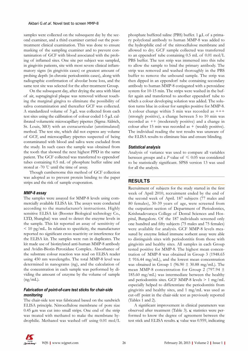
samples were collected on the subsequent day by the sec-ond examiner, and a third examiner carried out the post-treatment clinical examination. This was done to ensure masking of the sampling examiner and to prevent con-tamination of GCF with blood associated with the prob-ing of inflamed sites. One site per subject was sampled, in gingivitis patients, site with most severe clinical inflam-matory signs (in gingivitis cases) or greatest amount of probing depth (in chronic periodontitis cases), along with radiographic confirmation of alveolar bone loss, and the same test site was selected for the after-treatment Group.
On the subsequent day, after drying the area with blast of air, supragingival plaque was removed without touch-ing the marginal gingiva to eliminate the possibility of saliva contamination and thereafter GCF was collected. A standardized volume of 3 μL was collected from each test sites using the calibration of colour coded 1-5 μL cal-ibrated volumetric microcapillary pipettes (Sigma Aldrich, St. Louis, MO) with an extracurricular (unstimulated) method. The test site, which did not express any volume of GCF, and microcapillary pipettes suspected of being contaminated with blood and saliva were excluded from the study. In such cases the sample was obtained from the tooth that showed the next highest PPD in the same patient. The GCF collected was transferred to eppendorf tubes containing 0.5 mL of phosphate buffer saline and stored at -70 ℃ until the time of assay.
Though cumbersome this method of GCF collection was adopted as to prevent protein binding to the paper strips and the risk of sample evaporation.
MMP-8 assayThe samples were assayed for MMP-8 levels using com-mercially available ELISA kit. The assays were conducted according to the manufacturer’s instructions. Highly sensitive ELISA kit (Booster Biological technology Co., LTD, Shanghai) was used to detect the enzyme levels in the sample. This kit reported an assay of sensitivity of < 10 pg/mL. In relation to specificity, the manufacturer reported no significant cross reactivity or interference for the ELISA kit. The samples were run in duplicates. The kit made use of biotinylated anti-human MMP-8 antibody and Avidin-Biotin-Peroxidase Complex. Absorbance of the substrate colour reaction was read on ELISA reader using 450 nm wavelengths. The total MMP-8 level was determined in nanograms (ng), and the calculation of the concentration in each sample was performed by di-viding the amount of enzyme by the volume of sample (ng/mL).
Fabrication of point-of-care test sticks for chair-side monitoringThe chair-side test was fabricated based on the sandwich ELISA principle. Nitrocellulose membrane of pore size 0.45 μm was cut into small strips. One end of the strip was treated with methanol to make the membrane hy-drophilic. Methanol was washed off using 0.01 mol/L
phosphate buffered saline (PBS) buffer. 1 μL of a prima-ry polyclonal antibody to human MMP-8 was added to the hydrophilic end of the nitrocellulose membrane and allowed to dry. GCF sample collected was transferred to an eppendorf tube containing 0.5 mL of 0.01 mol/L PBS buffer. The test strip was immersed into this tube to allow the sample to bind the primary antibody. The strip was removed and washed thoroughly in the PBS buffer to remove the unbound sample. The strip was then dipped in an eppendorf tube containing secondary antibody to human MMP-8 conjugated with a peroxidase system for 10-15 min. The strips were washed in the buf-fer again and transferred to another eppendorf tube to which a colour developing solution was added. The solu-tion turns blue in colour for samples positive for MMP-8. A colour change within 5 min was recorded as +++ (strongly positive), a change between 5 to 10 min was recorded as ++ (moderately positive) and a change in colour after 15 min was recorded as + (weakly positive). The individual reading the test results was unaware of the ELISA results to eliminate bias and ensure blinding.
Statistical analysisAnalysis of variance was used to compare all variables between groups and a P value of ≤ 0.05 was considered to be statistically significant. SPSS version 13 was used for all the analysis.
RESULTSRecruitment of subjects for the study started in the first week of April 2010, recruitment ended by the end of the second week of April. 187 subjects (77 males and 80 females), 30-39 years of age, were screened from the outpatient section of Department of Periodontics, Krishnadevaraya College of Dental Sciences and Hos-pital, Bangalore. Of the 187 individuals screened only one hundred and fifty subjects (75 males and 75 females) were available for analysis. GCF MMP-8 levels mea-sured by enzyme linked immune sorbent assay were able to distinguish sites with periodontitis from those with gingivitis and healthy sites. All samples in each Group tested positive for MMP-8. The highest mean concen-tration of MMP-8 was obtained in Group 3 (1948.65 ± 916.44 mg/mL), and the lowest mean concentration was obtained in Group 1 (96.90 ± 30.88 mg/mL). The mean MMP-8 concentration for Group 2 (797.94 ± 185.60 mg/mL) was intermediate between the healthy and periodontitis sites. GCF MMP-8 levels > 1 mg/mL especially helped to differentiate the periodontitis from gingivitis and healthy sites, and 1 mg/mL was used as cut-off point in the chair-side test as previously reported (Tables 1 and 2).
A significant improvement in clinical parameters was observed after treatment (Table 3). κ statistics were per-formed to know the degree of agreement between the test stick and ELISA results. κ value was 0.959, indicating
26 February 20, 2013|Volume 2|Issue 1|WJS|www.wjgnet.com
Akbari G et al . Novel test to screen MMP-8
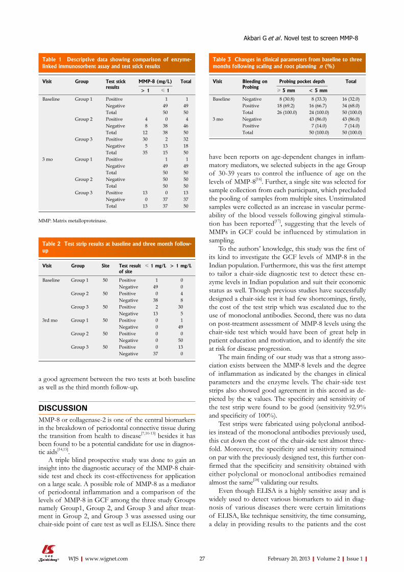
a good agreement between the two tests at both baseline as well as the third month follow-up.
DISCUSSIONMMP-8 or collagenase-2 is one of the central biomarkers in the breakdown of periodontal connective tissue during the transition from health to disease[7,10-13] besides it has been found to be a potential candidate for use in diagnos-tic aids[14,15].
A triple blind prospective study was done to gain an insight into the diagnostic accuracy of the MMP-8 chair-side test and check its cost-effectiveness for application on a large scale. A possible role of MMP-8 as a mediator of periodontal inflammation and a comparison of the levels of MMP-8 in GCF among the three study Groups namely Group1, Group 2, and Group 3 and after treat-ment in Group 2, and Group 3 was assessed using our chair-side point of care test as well as ELISA. Since there
have been reports on age-dependent changes in inflam-matory mediators, we selected subjects in the age Group of 30-39 years to control the influence of age on the levels of MMP-8[16]. Further, a single site was selected for sample collection from each participant, which precluded the pooling of samples from multiple sites. Unstimulated samples were collected as an increase in vascular perme-ability of the blood vessels following gingival stimula-tion has been reported[17], suggesting that the levels of MMPs in GCF could be influenced by stimulation in sampling.
To the authors’ knowledge, this study was the first of its kind to investigate the GCF levels of MMP-8 in the Indian population. Furthermore, this was the first attempt to tailor a chair-side diagnostic test to detect these en-zyme levels in Indian population and suit their economic status as well. Though previous studies have successfully designed a chair-side test it had few shortcomings, firstly, the cost of the test strip which was escalated due to the use of monoclonal antibodies. Second, there was no data on post-treatment assessment of MMP-8 levels using the chair-side test which would have been of great help in patient education and motivation, and to identify the site at risk for disease progression.
The main finding of our study was that a strong asso-ciation exists between the MMP-8 levels and the degree of inflammation as indicated by the changes in clinical parameters and the enzyme levels. The chair-side test strips also showed good agreement in this accord as de-picted by the κ values. The specificity and sensitivity of the test strip were found to be good (sensitivity 92.9% and specificity of 100%).
Test strips were fabricated using polyclonal antibod-ies instead of the monoclonal antibodies previously used, this cut down the cost of the chair-side test almost three-fold. Moreover, the specificity and sensitivity remained on par with the previously designed test, this further con-firmed that the specificity and sensitivity obtained with either polyclonal or monoclonal antibodies remained almost the same[18] validating our results.
Even though ELISA is a highly sensitive assay and is widely used to detect various biomarkers to aid in diag-nosis of various diseases there were certain limitations of ELISA, like technique sensitivity, the time consuming, a delay in providing results to the patients and the cost
27 February 20, 2013|Volume 2|Issue 1|WJS|www.wjgnet.com
Table 1 Descriptive data showing comparison of enzyme-linked immunosorbent assay and test stick results
Visit Group Test stick results
MMP-8 (mg/L) Total
> 1 ≤ 1
Baseline Group 1 Positive 1 1Negative 49 49Total 50 50
Group 2 Positive 4 0 4Negative 8 38 46Total 12 38 50
Group 3 Positive 30 2 32Negative 5 13 18Total 35 15 50
3 mo Group 1 Positive 1 1Negative 49 49Total 50 50
Group 2 Negative 50 50Total 50 50
Group 3 Positive 13 0 13Negative 0 37 37Total 13 37 50
MMP: Matrix metalloproteinase.
Table 2 Test strip results at baseline and three month follow-up
Visit Group Site Test result of site
≤ 1 mg/L > 1 mg/L
Baseline Group 1 50 Positive 1 0Negative 49 0
Group 2 50 Positive 0 4Negative 38 8
Group 3 50 Positive 2 30Negative 13 5
3rd mo Group 1 50 Positive 0 1Negative 0 49
Group 2 50 Positive 0 0Negative 0 50
Group 3 50 Positive 0 13Negative 37 0
Table 3 Changes in clinical parameters from baseline to three months following scaling and root planning n (%)
Visit Bleeding on Probing
Probing pocket depth Total
≥ 5 mm < 5 mm
Baseline Negative 8 (30.8) 8 (33.3) 16 (32.0)Positive 18 (69.2) 16 (66.7) 34 (68.0)Total 26 (100.0) 24 (100.0) 50 (100.0)
3 mo Negative 43 (86.0) 43 (86.0)Positive 7 (14.0) 7 (14.0)Total 50 (100.0) 50 (100.0)
Akbari G et al . Novel test to screen MMP-8
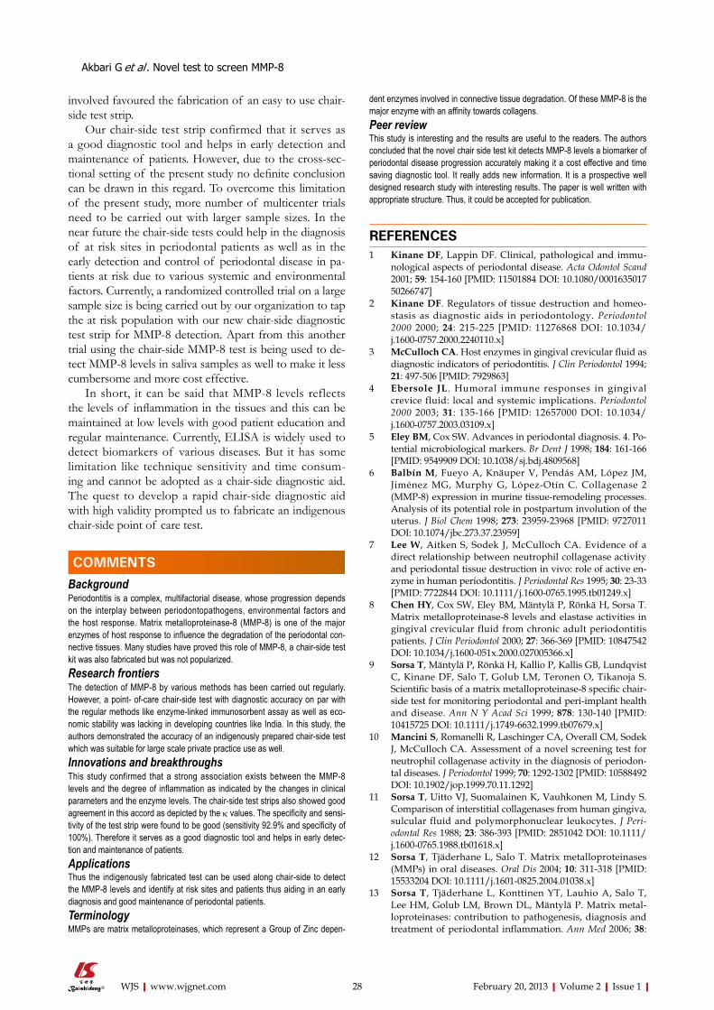
involved favoured the fabrication of an easy to use chair-side test strip.
Our chair-side test strip confirmed that it serves as a good diagnostic tool and helps in early detection and maintenance of patients. However, due to the cross-sec-tional setting of the present study no definite conclusion can be drawn in this regard. To overcome this limitation of the present study, more number of multicenter trials need to be carried out with larger sample sizes. In the near future the chair-side tests could help in the diagnosis of at risk sites in periodontal patients as well as in the early detection and control of periodontal disease in pa-tients at risk due to various systemic and environmental factors. Currently, a randomized controlled trial on a large sample size is being carried out by our organization to tap the at risk population with our new chair-side diagnostic test strip for MMP-8 detection. Apart from this another trial using the chair-side MMP-8 test is being used to de-tect MMP-8 levels in saliva samples as well to make it less cumbersome and more cost effective.
In short, it can be said that MMP-8 levels reflects the levels of inflammation in the tissues and this can be maintained at low levels with good patient education and regular maintenance. Currently, ELISA is widely used to detect biomarkers of various diseases. But it has some limitation like technique sensitivity and time consum-ing and cannot be adopted as a chair-side diagnostic aid. The quest to develop a rapid chair-side diagnostic aid with high validity prompted us to fabricate an indigenous chair-side point of care test.
COMMENTSBackgroundPeriodontitis is a complex, multifactorial disease, whose progression depends on the interplay between periodontopathogens, environmental factors and the host response. Matrix metalloproteinase-8 (MMP-8) is one of the major enzymes of host response to influence the degradation of the periodontal con-nective tissues. Many studies have proved this role of MMP-8, a chair-side test kit was also fabricated but was not popularized.Research frontiersThe detection of MMP-8 by various methods has been carried out regularly. However, a point- of-care chair-side test with diagnostic accuracy on par with the regular methods like enzyme-linked immunosorbent assay as well as eco-nomic stability was lacking in developing countries like India. In this study, the authors demonstrated the accuracy of an indigenously prepared chair-side test which was suitable for large scale private practice use as well.Innovations and breakthroughsThis study confirmed that a strong association exists between the MMP-8 levels and the degree of inflammation as indicated by the changes in clinical parameters and the enzyme levels. The chair-side test strips also showed good agreement in this accord as depicted by the κ values. The specificity and sensi-tivity of the test strip were found to be good (sensitivity 92.9% and specificity of 100%). Therefore it serves as a good diagnostic tool and helps in early detec-tion and maintenance of patients.ApplicationsThus the indigenously fabricated test can be used along chair-side to detect the MMP-8 levels and identify at risk sites and patients thus aiding in an early diagnosis and good maintenance of periodontal patients.TerminologyMMPs are matrix metalloproteinases, which represent a Group of Zinc depen-
dent enzymes involved in connective tissue degradation. Of these MMP-8 is the major enzyme with an affinity towards collagens.Peer reviewThis study is interesting and the results are useful to the readers. The authors concluded that the novel chair side test kit detects MMP-8 levels a biomarker of periodontal disease progression accurately making it a cost effective and time saving diagnostic tool. It really adds new information. It is a prospective well designed research study with interesting results. The paper is well written with appropriate structure. Thus, it could be accepted for publication.
REFERENCES1 Kinane DF, Lappin DF. Clinical, pathological and immu-
nological aspects of periodontal disease. Acta Odontol Scand 2001; 59: 154-160 [PMID: 11501884 DOI: 10.1080/000163501750266747]
2 Kinane DF. Regulators of tissue destruction and homeo-stasis as diagnostic aids in periodontology. Periodontol 2000 2000; 24: 215-225 [PMID: 11276868 DOI: 10.1034/j.1600-0757.2000.2240110.x]
3 McCulloch CA. Host enzymes in gingival crevicular fluid as diagnostic indicators of periodontitis. J Clin Periodontol 1994; 21: 497-506 [PMID: 7929863]
4 Ebersole JL. Humoral immune responses in gingival crevice fluid: local and systemic implications. Periodontol 2000 2003; 31: 135-166 [PMID: 12657000 DOI: 10.1034/j.1600-0757.2003.03109.x]
5 Eley BM, Cox SW. Advances in periodontal diagnosis. 4. Po-tential microbiological markers. Br Dent J 1998; 184: 161-166 [PMID: 9549909 DOI: 10.1038/sj.bdj.4809568]
6 Balbín M, Fueyo A, Knäuper V, Pendás AM, López JM, Jiménez MG, Murphy G, López-Otín C. Collagenase 2 (MMP-8) expression in murine tissue-remodeling processes. Analysis of its potential role in postpartum involution of the uterus. J Biol Chem 1998; 273: 23959-23968 [PMID: 9727011 DOI: 10.1074/jbc.273.37.23959]
7 Lee W, Aitken S, Sodek J, McCulloch CA. Evidence of a direct relationship between neutrophil collagenase activity and periodontal tissue destruction in vivo: role of active en-zyme in human periodontitis. J Periodontal Res 1995; 30: 23-33 [PMID: 7722844 DOI: 10.1111/j.1600-0765.1995.tb01249.x]
8 Chen HY, Cox SW, Eley BM, Mäntylä P, Rönkä H, Sorsa T. Matrix metalloproteinase-8 levels and elastase activities in gingival crevicular fluid from chronic adult periodontitis patients. J Clin Periodontol 2000; 27: 366-369 [PMID: 10847542 DOI: 10.1034/j.1600-051x.2000.027005366.x]
9 Sorsa T, Mäntylä P, Rönkä H, Kallio P, Kallis GB, Lundqvist C, Kinane DF, Salo T, Golub LM, Teronen O, Tikanoja S. Scientific basis of a matrix metalloproteinase-8 specific chair-side test for monitoring periodontal and peri-implant health and disease. Ann N Y Acad Sci 1999; 878: 130-140 [PMID: 10415725 DOI: 10.1111/j.1749-6632.1999.tb07679.x]
10 Mancini S, Romanelli R, Laschinger CA, Overall CM, Sodek J, McCulloch CA. Assessment of a novel screening test for neutrophil collagenase activity in the diagnosis of periodon-tal diseases. J Periodontol 1999; 70: 1292-1302 [PMID: 10588492 DOI: 10.1902/jop.1999.70.11.1292]
11 Sorsa T, Uitto VJ, Suomalainen K, Vauhkonen M, Lindy S. Comparison of interstitial collagenases from human gingiva, sulcular fluid and polymorphonuclear leukocytes. J Peri-odontal Res 1988; 23: 386-393 [PMID: 2851042 DOI: 10.1111/j.1600-0765.1988.tb01618.x]
12 Sorsa T, Tjäderhane L, Salo T. Matrix metalloproteinases (MMPs) in oral diseases. Oral Dis 2004; 10: 311-318 [PMID: 15533204 DOI: 10.1111/j.1601-0825.2004.01038.x]
13 Sorsa T, Tjäderhane L, Konttinen YT, Lauhio A, Salo T, Lee HM, Golub LM, Brown DL, Mäntylä P. Matrix metal-loproteinases: contribution to pathogenesis, diagnosis and treatment of periodontal inflammation. Ann Med 2006; 38:
28 February 20, 2013|Volume 2|Issue 1|WJS|www.wjgnet.com
COMMENTS
Akbari G et al . Novel test to screen MMP-8
306-321 [PMID: 16938801 DOI: 10.1080/07853890600800103]14 Mäntylä P, Stenman M, Kinane DF, Tikanoja S, Luoto H,
Salo T, Sorsa T. Gingival crevicular fluid collagenase-2 (MMP-8) test stick for chair-side monitoring of periodonti-tis. J Periodontal Res 2003; 38: 436-439 [PMID: 12828663 DOI: 10.1034/j.1600-0765.2003.00677.x]
15 Mäntylä P, Stenman M, Kinane D, Salo T, Suomalainen K, Tikanoja S, Sorsa T. Monitoring periodontal disease status in smokers and nonsmokers using a gingival crevicular fluid matrix metalloproteinase-8-specific chair-side test. J Peri-odontal Res 2006; 41: 503-512 [PMID: 17076774 DOI: 10.1111/j.1600-0765.2006.00897]
16 Chakraborti T, Mandal M, Das S, Chakraborti S. Age-de-
pendent change in arachidonic acid metabolic capacity in rat alveolar macrophages. Biochem Mol Biol Int 1999; 47: 501-507 [PMID: 10204087 DOI: 10.1080/15216549900201533]
17 Sueda T, Bang J, Cimasoni G. Collection of gingival fluid for quantitative analysis. J Dent Res 1969; 48: 159 [PMID: 5252093 DOI: 10.1177/00220345690480011501]
18 Lauhio A, Salo T, Ding Y, Konttinen YT, Nordström D, Tsch-esche H, Lähdevirta J, Golub LM, Sorsa T. In vivo inhibition of human neutrophil collagenase (MMP-8) activity during long-term combination therapy of doxycycline and non-steroidal anti-inflammatory drugs (NSAID) in acute reactive arthritis. Clin Exp Immunol 1994; 98: 21-28 [PMID: 7923879 DOI: 10.1111/j.1365-2249.1994.tb06601.x]
P-Reviewers Pavlidis TE, Chen MK S- Editor Huang XZ L- Editor A E- Editor Zheng XM
29 February 20, 2013|Volume 2|Issue 1|WJS|www.wjgnet.com
Akbari G et al . Novel test to screen MMP-8
World Journal of StomatologyW J S
GENERAL INFORMATIONWorld Journal of Stomatology (World J Stomatol, WJS, online ISSN 2218-6263, DOI: 10.5321) is a peer-reviewed open access (OA) academic journal that aims to guide clinical practice and improve diagnostic and therapeutic skills of clinicians.
Aim and scopeWJS covers topics concerning oral and craniofacial sciences, oral and craniofacial development/growth, dental tissue regeneration, craniofacial bone and cartilage research, oral and maxillofacial ge-netic diseases, developmental abnormalities and soft tissue defects, pulpal and periapical diseases, periodontal diseases and oral mucosal diseases, salivary gland diseases, oral and maxillofacial vascular/ner-vous diseases, jaw bone diseases, taste abnormalities, oral and maxil-lofacial pain, occlusion and temporomandibular diseases, repair and treatment of tooth defects, loss and dento-maxillofacial deformities, oral and maxillofacial biomechanics and biomaterials, new tech-niques for diagnosis/treatment of oral and maxillofacial diseases; and stomatology-related evidence-based medicine, epidemiology and nursing. The current columns of WJS include editorial, frontier, diagnostic advances, therapeutics advances, field of vision, mini-reviews, review, topic highlight, medical ethics, original articles, case report, clinical case conference (Clinicopathological conference), and autobiography. Priority publication will be given to articles con-cerning diagnosis and treatment of stomatologic diseases. The fol-lowing aspects are covered: Clinical diagnosis, laboratory diagnosis, differential diagnosis, imaging tests, pathological diagnosis, molecu-lar biological diagnosis, immunological diagnosis, genetic diagnosis, functional diagnostics, and physical diagnosis; and comprehensive therapy, drug therapy, surgical therapy, interventional treatment, minimally invasive therapy, and robot-assisted therapy.
We encourage authors to submit their manuscripts to WJS. We will give priority to manuscripts that are supported by major national and international foundations and those that are of great basic and clinical significance.
WJS is edited and published by Baishideng Publishing Group (BPG). BPG has a strong professional editorial team composed of science editors, language editors and electronic editors. BPG currently publishes 42 OA clinical medical journals, including 41 in English, has a total of 15 471 editorial borad members or peer reivewers, and is a world first-class publisher.
ColumnsThe columns in the issues of WJS will include: (1) Editorial: The editorial board members are invited to make comments on an im-portant topic in their field in terms of its current research status and future directions to lead the development of this discipline; (2) Frontier: The editorial board members are invited to select a highly cited cutting-edge original paper of his/her own to summarize ma-jor findings, the problems that have been resolved and remain to be resolved, and future research directions to help readers understand his/her important academic point of view and future research dir-ections in the field; (3) Diagnostic Advances: The editorial board members are invited to write high-quality diagnostic advances in their field to improve the diagnostic skills of readers. The topic covers general clinical diagnosis, differential diagnosis, pathological diagnosis, laboratory diagnosis, imaging diagnosis, endoscopic diag-nosis, biotechnological diagnosis, functional diagnosis, and physical diagnosis; (4) Therapeutics Advances: The editorial board members
are invited to write high-quality therapeutic advances in their field to help improve the therapeutic skills of readers. The topic covers medication therapy, psychotherapy, physical therapy, replacement therapy, interventional therapy, minimally invasive therapy, endo-scopic therapy, transplantation therapy, and surgical therapy; (5) Field of Vision: The editorial board members are invited to write commentaries on classic articles, hot topic articles, or latest articles to keep readers at the forefront of research and increase their levels of clinical research. Classic articles refer to papers that are included in Web of Knowledge and have received a large number of citations (ranking in the top 1%) after being published for more than years, reflecting the quality and impact of papers. Hot topic articles refer to papers that are included in Web of Knowledge and have received a large number of citations after being published for no more than 2 years, reflecting cutting-edge trends in scientific research. Latest articles refer to the latest published high-quality papers that are included in PubMed, reflecting the latest research trends. These commentary articles should focus on the status quo of research, the most important research topics, the problems that have now been resolved and remain to be resolved, and future research directions. Basic information about the article to be commented (including authors, article title, journal name, year, volume, and inclusive page numbers; (6) Minireviews: The editorial board members are invited to write short reviews on recent advances and trends in research of molecular biology, genomics, and related cutting-edge technologies to provide readers with the latest knowledge and help improve their diagnostic and therapeutic skills; (7) Review: To make a systematic review to focus on the status quo of research, the most import-ant research topics, the problems that have now been resolved and remain to be resolved, and future research directions; (8) Topic Highlight: The editorial board members are invited to write a series of articles (7-10 articles) to comment and discuss a hot topic to help improve the diagnostic and therapeutic skills of readers; (9) Medical Ethics: The editorial board members are invited to write articles about medical ethics to increase readers’ knowledge of medical eth-ics. The topic covers international ethics guidelines, animal studies, clinical trials, organ transplantation, etc.; (10) Clinical Case Confer-ence or Clinicopathological Conference: The editorial board mem-bers are invited to contribute high-quality clinical case conference; (11) Original Articles: To report innovative and original findings in stomatology; (12) Brief Articles: To briefly report the novel and in-novative findings in stomatology; (13) Meta-Analysis: To evaluate the clinical effectiveness in stomatology by using data from two or more randomised control trials; (14) Case Report: To report a rare or typical case; (15) Letters to the Editor: To discuss and make reply to the contributions published in WJS, or to introduce and com-ment on a controversial issue of general interest; (16) Book Reviews: To introduce and comment on quality monographs of stomatology; and (17) Autobiography: The editorial board members are invited to write their autobiography to provide readers with stories of success or failure in their scientific research career. The topic covers their basic personal information and information about when they started doing research work, where and how they did research work, what they have achieved, and their lessons from success or failure.
Name of journalWorld Journal of Stomatology
ISSNISSN 2218-6263 (online)
Online Submissions: http://www.wjgnet.com/esps/[email protected]
World J Stomatol 2013 February 20; 2(1): �-V�SSN 2218-6263 (online)
© 2013 Baishideng. All rights reserved.
INSTRUCTIONS TO AUTHORS
� February 20, 2013|Volume 2|�ssue 1|WJS|www.wjgnet.com

Instructions to authors
Launch dateNovember 31, 2011
FrequencyQuarterly
Editor-in-ChiefPeter E Murray, BSc (Hons), PhD, Professor, Pathologist, Department of Endodontics, College of Dental Medicine, Nova Southeastern University, 3200 South University Drive, Fort Lauder-dale, FL 33328-2018, United States
Editorial officeJin-Lei Wang, DirectorXiu-Xia Song, Vice DirectorWorld Journal of StomatologyRoom 903, Building D, Ocean International Center,No. 62 Dongsihuan Zhonglu, Chaoyang District,Beijing 100025, ChinaTelephone: +86-10-85381891Fax: +86-10-85381893E-mail: [email protected]://www.wjgnet.com
PublisherBaishideng Publishing Group Co., LimitedFlat C, 23/F., Lucky Plaza, 315-321 Lockhart Road, Wan Chai, Hong Kong, ChinaTelephone: +852-58042046Fax: +852-31158812E-mail: [email protected]://www.wjgnet.com
Production centerBeijing Baishideng BioMed Scientific Co., LimitedRoom 903, Building D, Ocean International Center,No. 62 Dongsihuan Zhonglu, Chaoyang District,Beijing 100025, ChinaTelephone: +86-10-85381892Fax: +86-10-85381893
Representative officeUSA Office8226 Regency Drive, Pleasanton, CA 94588-3144, United States
Instructions to authorsFull instructions are available online at http://www.wjgnet.com/ 2218-6263/g_info_20100722180909.htm.
Indexed and Abstracted inDigital Object Identifier.
SPECIAL STATEMENTAll articles published in this journal represent the viewpoints of the authors except where indicated otherwise.
Biostatistical editingStatistical review is performed after peer review. We invite an ex-pert in Biomedical Statistics to evaluate the statistical method used in the paper, including t-test (group or paired comparisons), chi-squared test, Ridit, probit, logit, regression (linear, curvilinear, or stepwise), correlation, analysis of variance, analysis of covariance, etc. The reviewing points include: (1) Statistical methods should be described when they are used to verify the results; (2) Whether the statistical techniques are suitable or correct; (3) Only homogeneous data can be averaged. Standard deviations are preferred to standard errors. Give the number of observations and subjects (n). Losses in observations, such as drop-outs from the study should be re-ported; (4) Values such as ED50, LD50, IC50 should have their
95% confidence limits calculated and compared by weighted probit analysis (Bliss and Finney); and (5) The word ‘significantly’ should be replaced by its synonyms (if it indicates extent) or the P value (if it indicates statistical significance).
Conflict-of-interest statementIn the interests of transparency and to help reviewers assess any po-tential bias, WJS requires authors of all papers to declare any compet-ing commercial, personal, political, intellectual, or religious interests in relation to the submitted work. Referees are also asked to indi-cate any potential conflict they might have reviewing a particular paper. Before submitting, authors are suggested to read “Uniform Requirements for Manuscripts Submitted to Biomedical Journals: Ethical Considerations in the Conduct and Reporting of Research: Conflicts of Interest” from International Committee of Medical Journal Editors (ICMJE), which is available at: http://www.icmje.org/ethical_4conflicts.html.
Sample wording: [Name of individual] has received fees for serv-ing as a speaker, a consultant and an advisory board member for [names of organizations], and has received research funding from [names of organization]. [Name of individual] is an employee of [name of or-ganization]. [Name of individual] owns stocks and shares in [name of organization]. [Name of individual] owns patent [patent identification and brief description].
Statement of informed consentManuscripts should contain a statement to the effect that all human studies have been reviewed by the appropriate ethics committee or it should be stated clearly in the text that all persons gave their informed consent prior to their inclusion in the study. Details that might disclose the identity of the subjects under study should be omitted. Authors should also draw attention to the Code of Ethics of the World Med-ical Association (Declaration of Helsinki, 1964, as revised in 2004).
Statement of human and animal rightsWhen reporting the results from experiments, authors should follow the highest standards and the trial should conform to Good Clinical Practice (for example, US Food and Drug Administration Good Clinical Practice in FDA-Regulated Clinical Trials; UK Medicines Research Council Guidelines for Good Clinical Practice in Clinical Trials) and/or the World Medical Association Declaration of Hel-sinki. Generally, we suggest authors follow the lead investigator’s na-tional standard. If doubt exists whether the research was conducted in accordance with the above standards, the authors must explain the rationale for their approach and demonstrate that the institutional review body explicitly approved the doubtful aspects of the study.
Before submitting, authors should make their study approved by the relevant research ethics committee or institutional review board. If human participants were involved, manuscripts must be accom-panied by a statement that the experiments were undertaken with the understanding and appropriate informed consent of each. Any per-sonal item or information will not be published without explicit con-sents from the involved patients. If experimental animals were used, the materials and methods (experimental procedures) section must clearly indicate that appropriate measures were taken to minimize pain or discomfort, and details of animal care should be provided.
SUBMISSION OF MANUSCRIPTSManuscripts should be typed in 1.5 line spacing and 12 pt. Book Antiqua with ample margins. Number all pages consecutively, and start each of the following sections on a new page: Title Page, Abstract, Introduction, Materials and Methods, Results, Discus-sion, Acknowledgements, References, Tables, Figures, and Figure Legends. Neither the editors nor the publisher are responsible for the opinions expressed by contributors. Manuscripts formally ac-cepted for publication become the permanent property of Baish-ideng Publishing Group Co., Limited, and may not be reproduced by any means, in whole or in part, without the written permission of both the authors and the publisher. We reserve the right to copy-edit and put onto our website accepted manuscripts. Authors should follow the relevant guidelines for the care and use of laboratory ani-
�� February 20, 2013|Volume 2|�ssue 1|WJS|www.wjgnet.com
Instructions to authors
mals of their institution or national animal welfare committee. For the sake of transparency in regard to the performance and report-ing of clinical trials, we endorse the policy of the ICMJE to refuse to publish papers on clinical trial results if the trial was not recorded in a publicly-accessible registry at its outset. The only register now available, to our knowledge, is http://www.clinicaltrials.gov spon-sored by the United States National Library of Medicine and we en-courage all potential contributors to register with it. However, in the case that other registers become available you will be duly notified. A letter of recommendation from each author’s organization should be provided with the contributed article to ensure the privacy and secrecy of research is protected.
Authors should retain one copy of the text, tables, photo-graphs and illustrations because rejected manuscripts will not be returned to the author(s) and the editors will not be responsible for loss or damage to photographs and illustrations sustained dur-ing mailing.
Online submissionsManuscripts should be submitted through the Online Submission System at: http://www.wjgnet.com/esps/. Authors are highly recom-mended to consult the ONLINE INSTRUCTIONS TO AUTHORS (http://www.wjgnet.com/2218-6263/g_info_20100722180909.htm) before attempting to submit online. For assistance, authors encoun-tering problems with the Online Submission System may send an email describing the problem to [email protected], or by telephone: +86-10-85381892. If you submit your manuscript online, do not make a postal contribution. Repeated online submission for the same manuscript is strictly prohibited.
MANUSCRIPT PREPARATIONAll contributions should be written in English. All articles must be submitted using word-processing software. All submissions must be typed in 1.5 line spacing and 12 pt. Book Antiqua with ample mar-gins. Style should conform to our house format. Required informa-tion for each of the manuscript sections is as follows:
Title pageTitle: Title should be less than 12 words.
Running title: A short running title of less than 6 words should be provided.
Authorship: Authorship credit should be in accordance with the standard proposed by ICMJE, based on (1) substantial contribu-tions to conception and design, acquisition of data, or analysis and interpretation of data; (2) drafting the article or revising it critically for important intellectual content; and (3) final approval of the ver-sion to be published. Authors should meet conditions 1, 2, and 3.
Institution: Author names should be given first, then the complete name of institution, city, province and postcode. For example, Xu-Chen Zhang, Li-Xin Mei, Department of Pathology, Chengde Med-ical College, Chengde 067000, Hebei Province, China. One author may be represented from two institutions, for example, George Sgourakis, Department of General, Visceral, and Transplantation Surgery, Essen 45122, Germany; George Sgourakis, 2nd Surgical Department, Korgialenio-Benakio Red Cross Hospital, Athens 15451, Greece
Author contributions: The format of this section should be: Author contributions: Wang CL and Liang L contributed equally to this work; Wang CL, Liang L, Fu JF, Zou CC, Hong F and Wu XM designed the research; Wang CL, Zou CC, Hong F and Wu XM performed the research; Xue JZ and Lu JR contributed new reagents/analytic tools; Wang CL, Liang L and Fu JF analyzed the data; and Wang CL, Liang L and Fu JF wrote the paper.
Supportive foundations: The complete name and number of sup-
portive foundations should be provided, e.g., Supported by National Natural Science Foundation of China, No. 30224801
Correspondence to: Only one corresponding address should be provided. Author names should be given first, then author title, af-filiation, the complete name of institution, city, postcode, province, country, and email. All the letters in the email should be in lower case. A space interval should be inserted between country name and email address. For example, Montgomery Bissell, MD, Professor of Medicine, Chief, Liver Center, Gastroenterology Division, Uni-versity of California, Box 0538, San Francisco, CA 94143, United States. [email protected]
Telephone and fax: Telephone and fax should consist of +, coun-try number, district number and telephone or fax number, e.g., Tele-phone: +86-10-85381892 Fax: +86-10-85381893
Peer reviewers: All articles received are subject to peer review. Normally, three experts are invited for each article. Decision on acceptance is made only when at least two experts recommend publication of an article. All peer-reviewers are acknowledged on Express Submission and Peer-review System website.
AbstractThere are unstructured abstracts (no less than 200 words) and struc-tured abstracts. The specific requirements for structured abstracts are as follows:
An informative, structured abstract should accompany each manuscript. Abstracts of original contributions should be struc-tured into the following sections: AIM (no more than 20 words; Only the purpose of the study should be included. Please write the Aim in the form of “To investigate/study/…”), METHODS (no less than 140 words for Original Articles; and no less than 80 words for Brief Articles), RESULTS (no less than 150 words for Original Articles and no less than 120 words for Brief Articles; You should present P values where appropriate and must provide relevant data to illustrate how they were obtained, e.g., 6.92 ± 3.86 vs 3.61 ± 1.67, P < 0.001), and CONCLUSION (no more than 26 words).
Key wordsPlease list 5-10 key words, selected mainly from Index Medicus, which reflect the content of the study.
Core tip Please write a summary of less than 100 words to outline the most innovative and important arguments and core contents in your paper to attract readers.
TextFor articles of these sections, original articles and brief articles, the main text should be structured into the following sections: INTRO-DUCTION, MATERIALS AND METHODS, RESULTS and DISCUSSION, and should include appropriate Figures and Tables. Data should be presented in the main text or in Figures and Tables, but not in both.
IllustrationsFigures should be numbered as 1, 2, 3, etc., and mentioned clearly in the main text. Provide a brief title for each figure on a separate page. Detailed legends should not be provided under the figures. This part should be added into the text where the figures are applicable. Keep-ing all elements compiled is necessary in line-art image. Scale bars should be used rather than magnification factors, with the length of the bar defined in the legend rather than on the bar itself. File names should identify the figure and panel. Avoid layering type directly over shaded or textured areas. Please use uniform legends for the same subjects. For example: Figure 1 Pathological changes in atrophic gas-tritis after treatment. A: ...; B: ...; C: ...; D: ...; E: ...; F: ...; G: …etc. It is our principle to publish high resolution-figures for the E-versions.
��� February 20, 2013|Volume 2|�ssue 1|WJS|www.wjgnet.com

TablesThree-line tables should be numbered 1, 2, 3, etc., and mentioned clearly in the main text. Provide a brief title for each table. Detailed legends should not be included under tables, but rather added into the text where applicable. The information should complement, but not duplicate the text. Use one horizontal line under the title, a second under column heads, and a third below the Table, above any footnotes. Vertical and italic lines should be omitted.
Notes in tables and illustrationsData that are not statistically significant should not be noted. aP < 0.05, bP < 0.01 should be noted (P > 0.05 should not be noted). If there are other series of P values, cP < 0.05 and dP < 0.01 are used. A third series of P values can be expressed as eP < 0.05 and fP < 0.01. Other notes in tables or under illustrations should be expressed as 1F, 2F, 3F; or sometimes as other symbols with a superscript (Arabic numerals) in the upper left corner. In a multi-curve illustration, each curve should be labeled with ●, ○, ■, □, ▲, △, etc., in a cer-tain sequence.
AcknowledgmentsBrief acknowledgments of persons who have made genuine con-tributions to the manuscript and who endorse the data and conclu-sions should be included. Authors are responsible for obtaining written permission to use any copyrighted text and/or illustrations.
REFERENCESCoding systemThe author should number the references in Arabic numerals ac-cording to the citation order in the text. Put reference numbers in square brackets in superscript at the end of citation content or after the cited author’s name. For citation content which is part of the narration, the coding number and square brackets should be typeset normally. For example, “Crohn’s disease (CD) is associated with increased intestinal permeability[1,2]”. If references are cited directly in the text, they should be put together within the text, for example, “From references[19,22-24], we know that...”
When the authors write the references, please ensure that the order in text is the same as in the references section, and also ensure the spelling accuracy of the first author’s name. Do not list the same citation twice.
PMID and DOIPleased provide PubMed citation numbers to the reference list, e.g., PMID and DOI, which can be found at http://www.ncbi.nlm.nih.gov/sites/entrez?db=pubmed and http://www.crossref.org/Sim-pleTextQuery/, respectively. The numbers will be used in E-version of this journal.
Style for journal referencesAuthors: the name of the first author should be typed in bold-faced letters. The family name of all authors should be typed with the in-itial letter capitalized, followed by their abbreviated first and middle initials. (For example, Lian-Sheng Ma is abbreviated as Ma LS, Bo-Rong Pan as Pan BR). The title of the cited article and italicized journal title (journal title should be in its abbreviated form as shown in PubMed), publication date, volume number (in black), start page, and end page [PMID: 11819634 DOI: 10.3748/wjg.13.5396].
Style for book referencesAuthors: the name of the first author should be typed in bold-faced letters. The surname of all authors should be typed with the initial letter capitalized, followed by their abbreviated middle and first initials. (For example, Lian-Sheng Ma is abbreviated as Ma LS, Bo-Rong Pan as Pan BR) Book title. Publication number. Publication place: Publication press, Year: start page and end page.
FormatJournals English journal article (list all authors and include the PMID where applicable)1 Jung EM, Clevert DA, Schreyer AG, Schmitt S, Rennert J,
Kubale R, Feuerbach S, Jung F. Evaluation of quantitative con-trast harmonic imaging to assess malignancy of liver tumors: A prospective controlled two-center study. World J Gastroenterol 2007; 13: 6356-6364 [PMID: 18081224 DOI: 10.3748/wjg.13. 6356]
Chinese journal article (list all authors and include the PMID where applicable)2 Lin GZ, Wang XZ, Wang P, Lin J, Yang FD. Immunologic
effect of Jianpi Yishen decoction in treatment of Pixu-diar-rhoea. Shijie Huaren Xiaohua Zazhi 1999; 7: 285-287
In press3 Tian D, Araki H, Stahl E, Bergelson J, Kreitman M. Signature
of balancing selection in Arabidopsis. Proc Natl Acad Sci USA 2006; In press
Organization as author4 Diabetes Prevention Program Research Group. Hyperten-
sion, insulin, and proinsulin in participants with impaired glu-cose tolerance. Hypertension 2002; 40: 679-686 [PMID: 12411462 PMCID:2516377 DOI:10.1161/01.HYP.0000035706.28494. 09]
Both personal authors and an organization as author 5 Vallancien G, Emberton M, Harving N, van Moorselaar RJ;
Alf-One Study Group. Sexual dysfunction in 1, 274 European men suffering from lower urinary tract symptoms. J Urol 2003; 169: 2257-2261 [PMID: 12771764 DOI:10.1097/01.ju. 0000067940.76090.73]
No author given6 21st century heart solution may have a sting in the tail. BMJ
2002; 325: 184 [PMID: 12142303 DOI:10.1136/bmj.325. 7357.184]
Volume with supplement7 Geraud G, Spierings EL, Keywood C. Tolerability and safety
of frovatriptan with short- and long-term use for treatment of migraine and in comparison with sumatriptan. Headache 2002; 42 Suppl 2: S93-99 [PMID: 12028325 DOI:10.1046/j.1526-4610.42.s2.7.x]
Issue with no volume8 Banit DM, Kaufer H, Hartford JM. Intraoperative frozen
section analysis in revision total joint arthroplasty. Clin Orthop Relat Res 2002; (401): 230-238 [PMID: 12151900 DOI:10.1097/00003086-200208000-00026]
No volume or issue9 Outreach: Bringing HIV-positive individuals into care. HRSA
Careaction 2002; 1-6 [PMID: 12154804]
BooksPersonal author(s)10 Sherlock S, Dooley J. Diseases of the liver and billiary system.
9th ed. Oxford: Blackwell Sci Pub, 1993: 258-296Chapter in a book (list all authors)11 Lam SK. Academic investigator’s perspectives of medical
treatment for peptic ulcer. In: Swabb EA, Azabo S. Ulcer disease: investigation and basis for therapy. New York: Marcel Dekker, 1991: 431-450
Author(s) and editor(s)12 Breedlove GK, Schorfheide AM. Adolescent pregnancy.
2nd ed. Wieczorek RR, editor. White Plains (NY): March of Dimes Education Services, 2001: 20-34
Conference proceedings13 Harnden P, Joffe JK, Jones WG, editors. Germ cell tumours V.
Proceedings of the 5th Germ cell tumours Conference; 2001 Sep 13-15; Leeds, UK. New York: Springer, 2002: 30-56
Conference paper14 Christensen S, Oppacher F. An analysis of Koza's computa-
tional effort statistic for genetic programming. In: Foster JA, Lutton E, Miller J, Ryan C, Tettamanzi AG, editors. Genetic programming. EuroGP 2002: Proceedings of the 5th Euro-pean Conference on Genetic Programming; 2002 Apr 3-5; Kinsdale, Ireland. Berlin: Springer, 2002: 182-191
Electronic journal (list all authors)15 Morse SS. Factors in the emergence of infectious diseases.
Instructions to authors
�V February 20, 2013|Volume 2|�ssue 1|WJS|www.wjgnet.com

Instructions to authors
Emerg Infect Dis serial online, 1995-01-03, cited 1996-06-05; 1(1): 24 screens. Available from: URL: http://www.cdc.gov/ncidod/eid/index.htm
Patent (list all authors)16 Pagedas AC, inventor; Ancel Surgical R&D Inc., assignee.
Flexible endoscopic grasping and cutting device and position-ing tool assembly. United States patent US 20020103498. 2002 Aug 1
Statistical dataWrite as mean ± SD or mean ± SE.
Statistical expressionExpress t test as t (in italics), F test as F (in italics), chi square test as χ2 (in Greek), related coefficient as r (in italics), degree of freedom as υ (in Greek), sample number as n (in italics), and probability as P (in italics).
UnitsUse SI units. For example: body mass, m (B) = 78 kg; blood pres-sure, p (B) = 16.2/12.3 kPa; incubation time, t (incubation) = 96 h, blood glucose concentration, c (glucose) 6.4 ± 2.1 mmol/L; blood CEA mass concentration, p (CEA) = 8.6 24.5 mg/L; CO2 volume fraction, 50 mL/L CO2, not 5% CO2; likewise for 40 g/L formal-dehyde, not 10% formalin; and mass fraction, 8 ng/g, etc. Arabic numerals such as 23, 243, 641 should be read 23 243 641.
The format for how to accurately write common units and quantums can be found at: http://www.wjgnet.com/2218-6263/g_info_20100725073806.htm.
AbbreviationsStandard abbreviations should be defined in the abstract and on first mention in the text. In general, terms should not be abbrevi-ated unless they are used repeatedly and the abbreviation is helpful to the reader. Permissible abbreviations are listed in Units, Symbols and Abbreviations: A Guide for Biological and Medical Editors and Authors (Ed. Baron DN, 1988) published by The Royal Society of Medicine, London. Certain commonly used abbreviations, such as DNA, RNA, HIV, LD50, PCR, HBV, ECG, WBC, RBC, CT, ESR, CSF, IgG, ELISA, PBS, ATP, EDTA, mAb, can be used directly without further explanation.
ItalicsQuantities: t time or temperature, c concentration, A area, l length, m mass, V volume.Genotypes: gyrA, arg 1, c myc, c fos, etc.Restriction enzymes: EcoRI, HindI, BamHI, Kbo I, Kpn I, etc.Biology: H. pylori, E coli, etc.
Examples for paper writingAll types of articles’ writing style and requirement will be found in the link: http://www.wjgnet.com/esps/NavigationInfo.aspx?id=15
RESUBMISSION OF THE REVISED MANUSCRIPTSAuthors must revise their manuscript carefully according to the revision policies of Baishideng Publishing Group Co., Limited. The revised version, along with the signed copyright transfer agreement, responses to the reviewers, and English language Grade A certifi-cate (for non-native speakers of English), should be submitted to the online system via the link contained in the e-mail sent by the edi-tor. If you have any questions about the revision, please send e-mail to [email protected].
Language evaluation The language of a manuscript will be graded before it is sent for revision. (1) Grade A: priority publishing; (2) Grade B: minor lan-guage polishing; (3) Grade C: a great deal of language polishing needed; and (4) Grade D: rejected. Revised articles should reach Grade A.
Copyright assignment formPlease download a Copyright assignment form from http://www.wjgnet.com/2218-6263/g_info_20100725073726.htm.
Responses to reviewersPlease revise your article according to the comments/suggestions provided by the reviewers. The format for responses to the reviewers’ comments can be found at: http://www.wjgnet.com/2218-6263/g_info_20100725073445.htm.
Proof of financial supportFor papers supported by a foundation, authors should provide a copy of the approval document and serial number of the foundation.
Links to documents related to the manuscript WJS will be initiating a platform to promote dynamic interactions be-tween the editors, peer reviewers, readers and authors. After a manu-script is published online, links to the PDF version of the submitted manuscript, the peer-reviewers’ report and the revised manuscript will be put on-line. Readers can make comments on the peer reviewer’s report, authors’ responses to peer reviewers, and the revised manu-script. We hope that authors will benefit from this feedback and be able to revise the manuscript accordingly in a timely manner.
Publication feeWJS is an international, peer-reviewed, OA online journal. Articles published by this journal are distributed under the terms of the Cre-ative Commons Attribution Non-commercial License, which permits use, distribution, and reproduction in any medium and format, pro-vided the original work is properly cited. The use is non-commercial and is otherwise in compliance with the license. Authors of accepted articles must pay a publication fee. Publication fee: 600 USD per article. All invited articles are published free of charge.
V February 20, 2013|Volume 2|�ssue 1|WJS|www.wjgnet.com

World Journal of StomatologyWorld J Stomatol 2013 May 20; 2(2): 30-34
ISSN 2218-6263 (online)
www.wjgnet.comwww.wjgnet.com

World Journal of StomatologyW J S
30 QuantitativescintigraphicanalysisoftheapicalsealinThermafil/Topsealand
RealSeal1/Realsealfilledrootcanals
Ferreira MM, Abrantes M, Carrilho EV, Botelho MF
Contents
BRIEF ARTICLE
Quarterly Volume 2 Number 2 May 20, 2013
IWJS|www.wjgnet.com May 20, 2013|Volume 2|Issue 2|

ContentsWorld Journal of Stomatology
Volume 2 Number 2 May 20, 2013
EDITORS FOR THIS ISSUE
Responsible Assistant Editor: Shuai Ma Responsible Science Editor: Ling-Ling WenResponsible Electronic Editor: Xiao-Mei Zheng Proofing Editor-in-Chief: Lian-Sheng Ma
World Journal of StomatologyRoom 903, Building D, Ocean International Center, No. 62 Dongsihuan Zhonglu, Chaoyang District, Beijing 100025, ChinaTelephone: +86-10-85381891Fax: +86-10-85381893E-mail: [email protected]://www.wjgnet.com
PUBLISHERBaishideng Publishing Group Co., LimitedFlat C, 23/F., Lucky Plaza, 315-321 Lockhart Road, Wan Chai, Hong Kong, ChinaFax: +852-6555-7188Telephone: +852-3177-9906E-mail: [email protected]://www.wjgnet.com
PUBLICATIONDATEMay 20, 2013
COPYRIGHT© 2013 Baishideng. Articles published by this Open-Access journal are distributed under the terms of the Creative Commons Attribution Non-commercial License, which permits use, distribution, and repro-duction in any medium, provided the original work is properly cited, the use is non commercial and is other-wise in compliance with the license.
SPECIALSTATEMENTAll articles published in this journal represent the viewpoints of the authors except where indicated oth-erwise.
INSTRUCTIONSTOAUTHORSFull instructions are available online at http://www.wjgnet.com/2218-6263/g_info_20100722180909.htm
ONLINESUBMISSIONhttp://www.wjgnet.com/esps/
IIWJS|www.wjgnet.com
APPENDIX
ABOUT COVER
AIM AND SCOPE
INDEXINg/ABSTRACTINg
May 20, 2013|Volume 2|Issue 2|
NAMEOFJOURNALWorld Journal of Stomatology
ISSNISSN 2218-6263 (online)
LAUNCHDATEDecember 31, 2011
FREQUENCYQuarterly
EDITOR-IN-CHIEFPeter E Murray, BSc (Hons), PhD, Professor, Pathol-ogist, Department of Endodontics, College of Dental Medicine, Nova Southeastern University, 3200 South University Drive, Fort Lauderdale, FL 33328-2018, United States
EDITORIALOFFICEJin-Lei Wang, DirectorXiu-Xia Song, Vice Director
I-V Instructionstoauthors
EditorialBoardMemberofWorldJournalofStomatology ,Sang-HengKok,Professor,DepartmentofDentistry,NationalTaiwanUniversityHospital,No1Chang-TeStreet,10016,Taipei,Taiwan
World Journal of Stomatology (World J Stomatol, WJS, online ISSN 2218-6263, DOI: 10.5321) is a peer-reviewed open access academic journal that aims to guide clinical practice and improve diagnostic and therapeutic skills of clinicians.
WJS covers topics concerning oral and craniofacial sciences, oral and craniofacial development/growth, dental tissue regeneration, craniofacial bone and cartilage research, oral and maxillofacial genetic diseases, developmental abnormalities and soft tissue defects, pulpal and periapical diseases, periodontal diseases and oral mucosal diseases, salivary gland diseases, oral and maxillofacial vascular/nervous diseases, jaw bone diseases, taste abnormalities, oral and maxillofacial pain, occlusion and temporomandibular diseases, repair and treatment of tooth defects, loss and dento-maxillofacial deformities, oral and maxillofacial biomechanics and biomaterials, new techniques for diagnosis/treatment of oral and maxillofacial diseases; and stomatology-related evidence-based medicine, epidemiology and nursing. Priority publication will be given to articles concerning diagnosis and treatment of stomatologic diseases. The following aspects are covered: Clinical diagnosis, laboratory diagnosis, differential diagnosis, imaging tests, pathological diagnosis, molecular biological diagnosis, immunological diagnosis, genetic diagnosis, functional diagnostics, and physical diagnosis; and comprehensive therapy, drug therapy, surgical therapy, interventional treatment, minimally invasive therapy, and robot-assisted therapy.
We encourage authors to submit their manuscripts to WJS. We will give priority to manuscripts that are supported by major national and international foundations and those that are of great basic and clinical significance.
World Journal of Stomatology is now indexed in Digital Object Identifier.
I-III EditorialBoardFLYLEAF

World Journal of StomatologyW J S
Online Submissions: http://www.wjgnet.com/esps/[email protected]:10.5321/wjs.v2.i2.30
World J Stomatol 2013 May 20; 2(2): 30-34ISSN 2218-6263 (online)
© 2013 Baishideng. All rights reserved.
Quantitative scintigraphic analysis of the apical seal in Thermafil/Topseal and RealSeal 1/Realseal filled root canals
Manuel Marques Ferreira, Margarida Abrantes, Eunice Virgínia Carrilho, Maria Filomena Botelho
Manuel Marques Ferreira, Eunice Virgínia Carrilho, Depart-ment of Dentistry, Faculty of Medicine, University of Coimbra, 3000 Coimbra, PortugalMargarida Abrantes, Maria Filomena Botelho, Department of Biophysics and Biomathematics, IBILI-Faculty of Medicine, University of Coimbra, 3000 Coimbra, PortugalAuthor contributions: All authors contributed equally to this work.Correspondence to: Dr. Manuel Marques Ferreira, Depart-ment of Dentistry, Faculty of Medicine, University of Coimbra, Av. Bissaya Barreto, Blocos de Celas, 3000 Coimbra, Portugal. [email protected]: +351-239484183 Fax: +351-239834616Received: September 24, 2012 Revised: December 27, 2012Accepted: January 5, 2013Published online: May 20, 2013
AbstractAIM: To investigate the microleakage of two different root canal obturation systems, using the nuclear medi-cine approach.
METHODS: Twenty-six single-rooted extracted teeth were selected. The crowns were sectioned to obtain 15-mm long root segments and each tooth was pre-pared using rotary ProFile® instruments. The roots were divided into 2 experimental groups using RealSeal 1 and RealSeal sealer or Thermafil and TopSeal sealer as well as two control groups. On the 7th and the 28th day the apices were submersed in a solution of 99mTc-Pertechnetate during 3 h. The radioactivity was count-ed using a γ camera.
RESULTS: The present study showed that none of the root canal-filled teeth was leakage free. The statistical analyses were made using Kruskal-Wallis and statisti-cal significance was assessed using α = 0.05. Although apical leakage measured in counts per minute (cpm) in the Thermafil/TopSeal group was lower than in the RealSeal/RealSeal group (363 916 ± 180 707.7 cpm vs
533 427 ± 414 020.6 cpm) on 7th day and (1 678 200 ± 567 217.4 cpm vs 2 240 518 ± 383 356.7 cpm) on 28th day, there was no statistical difference (P > 0.05). In the Thermafil/TopSeal group and RealSeal 1/RealSeal group it was found that over time, the number of counts increased between 7 d and 28 d (363 916 ± 180 707.7 cpm vs 1 678 200 ± 567 217.4 cpm) and (533 427 ± 414 020.6 cpm vs 2 240 518 ± 383 356.7 cpm), respec-tively, with statistically significant differences (Therma-fil/TopSeal group, P = 0.015 and RealSeal 1/RealSeal group, P = 0.036).
CONCLUSION: Both carrier-based Realseal 1 and Thermafil techniques showed a similar sealing effect, but none of the materials was leakage free.
© 2013 Baishideng. All rights reserved.
Key words: Microleakage; Nuclear medicine; Obtura-tion; Thermafil; Realseal
Ferreira MM, Abrantes M, Carrilho EV, Botelho MF. Quan-titative scintigraphic analysis of the apical seal in Thermafil/ Topseal and RealSeal 1/Realseal filled root canals. World J Stomatol 2013; 2(2): 30-34 Available from: URL: http://www.wjgnet.com/2218-6263/full/v2/i2/30.htm DOI: http://dx.doi.org/10.5321/wjs.v2.i2.30
INTRODUCTIONIt has been established that apical periodontitis is caused by bacteria derived from the root canal[1,2]. The first stage of root canal therapy is microbial control, followed by root canal filling. Microbial control includes removal of the protein degradation products, toxins, and mainly bacteria[3]. The root canal filling must seal the canal space both apically and coronally and the most commonly used material for root canal obturation is gutta-percha com-bined with a sealer. Gutta-percha is considered an imper-
BRIEF ARTICLE
30 May 20, 2013|Volume 2|Issue 2|WJS|www.wjgnet.com
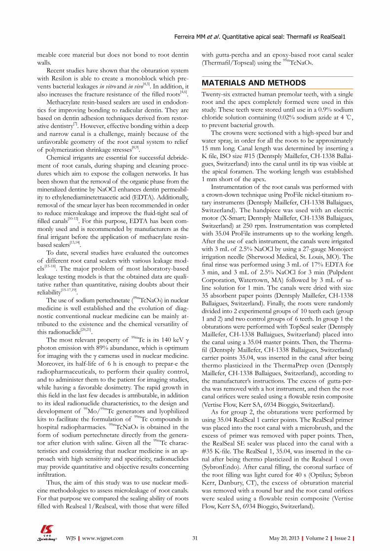
meable core material but does not bond to root dentin walls.
Recent studies have shown that the obturation system with Resilon is able to create a monoblock which pre-vents bacterial leakages in vitro and in vivo[4,5]. In addition, it also increases the fracture resistance of the filled roots[4,6].
Methacrylate resin-based sealers are used in endodon-tics for improving bonding to radicular dentin. They are based on dentin adhesion techniques derived from restor-ative dentistry[7]. However, effective bonding within a deep and narrow canal is a challenge, mainly because of the unfavorable geometry of the root canal system to relief of polymerization shrinkage stresses[8,9].
Chemical irrigants are essential for successful debride-ment of root canals, during shaping and cleaning proce-dures which aim to expose the collagen networks. It has been shown that the removal of the organic phase from the mineralized dentine by NaOCl enhances dentin permeabil-ity to ethylenediaminetetraacetic acid (EDTA). Additionally, removal of the smear layer has been recommended in order to reduce microleakage and improve the fluid-tight seal of filled canals[10-12]. For this purpose, EDTA has been com-monly used and is recommended by manufacturers as the final irrigant before the application of methacrylate resin-based sealers[13,14].
To date, several studies have evaluated the outcomes of different root canal sealers with various leakage mod-els[15-18]. The major problem of most laboratory-based leakage testing models is that the obtained data are quali-tative rather than quantitative, raising doubts about their reliability[15-17,19].
The use of sodium pertechnetate (99mTcNaO4) in nuclear medicine is well established and the evolution of diag-nostic conventional nuclear medicine can be mainly at-tributed to the existence and the chemical versatility of this radionuclide[20,21].
The most relevant property of 99mTc is its 140 keV γ photon emission with 89% abundance, which is optimum for imaging with the γ cameras used in nuclear medicine. Moreover, its half-life of 6 h is enough to prepar-e the radiopharmaceuticals, to perform their quality control, and to administer them to the patient for imaging studies, while having a favorable dosimetry. The rapid growth in this field in the last few decades is attributable, in addition to its ideal radionuclide characteristics, to the design and development of 99Mo/99mTc generators and lyophilized kits to facilitate the formulation of 99mTc compounds in hospital radiopharmacies. 99mTcNaO4 is obtained in the form of sodium pertechnetate directly from the genera-tor after elution with saline. Given all the 99mTc charac-teristics and considering that nuclear medicine is an ap-proach with high sensitivity and specificity, radionuclides may provide quantitative and objective results concerning infiltration.
Thus, the aim of this study was to use nuclear medi-cine methodologies to assess microleakage of root canals. For that purpose we compared the sealing ability of roots filled with Realseal 1/Realseal, with those that were filled
with gutta-percha and an epoxy-based root canal sealer (Thermafil/Topseal) using the 99mTcNaO4.
MATERIALS AND METHODSTwenty-six extracted human premolar teeth, with a single root and the apex completely formed were used in this study. These teeth were stored until use in a 0.9% sodium chloride solution containing 0.02% sodium azide at 4 ℃, to prevent bacterial growth.
The crowns were sectioned with a high-speed bur and water spray, in order for all the roots to be approximately 15 mm long. Canal length was determined by inserting a K file, ISO size #15 (Dentsply Maillefer, CH-1338 Ballai-gues, Switzerland) into the canal until its tip was visible at the apical foramen. The working length was established 1 mm short of the apex.
Instrumentation of the root canals was performed with a crown-down technique using ProFile nickel-titanium ro-tary instruments (Dentsply Maillefer, CH-1338 Ballaigues, Switzerland). The handpiece was used with an electric motor (X-Smart; Dentsply Maillefer, CH-1338 Ballaigues, Switzerland) at 250 rpm. Instrumentation was completed with 35.04 ProFile instruments up to the working length. After the use of each instrument, the canals were irrigated with 3 mL of 2.5% NaOCl by using a 27-gauge Monoject irrigation needle (Sherwood Medical, St. Louis, MO). The final rinse was performed using 3 mL of 17% EDTA for 3 min, and 3 mL of 2.5% NaOCl for 3 min (Pulpdent Corporation, Watertown, MA) followed by 3 mL of sa-line solution for 1 min. The canals were dried with size 35 absorbent paper points (Dentsply Maillefer, CH-1338 Ballaigues, Switzerland). Finally, the roots were randomly divided into 2 experimental groups of 10 teeth each (group 1 and 2) and two control groups of 6 teeth. In group 1 the obturations were performed with TopSeal sealer (Dentsply Maillefer, CH-1338 Ballaigues, Switzerland) placed into the canal using a 35.04 master points. Then, the Therma-fil (Dentsply Maillefer, CH-1338 Ballaigues, Switzerland) carrier points 35.04, was inserted in the canal after being thermo plasticized in the ThermaPrep oven (Dentsply Maillefer, CH-1338 Ballaigues, Switzerland), according to the manufacturer’s instructions. The excess of gutta-per-cha was removed with a hot instrument, and then the root canal orifices were sealed using a flowable resin composite (Vertise Flow, Kerr SA, 6934 Bioggio, Switzerland).
As for group 2, the obturations were performed by using 35.04 RealSeal 1 carrier points. The RealSeal primer was placed into the root canal with a microbrush, and the excess of primer was removed with paper points. Then, the RealSeal SE sealer was placed into the canal with a #35 K-file. The RealSeal 1, 35.04, was inserted in the ca-nal after being thermo plasticized in the Realseal 1 oven (SybronEndo). After canal filling, the coronal surface of the root filling was light cured for 40 s (Optilux; Sybron Kerr, Danbury, CT), the excess of obturation material was removed with a round bur and the root canal orifices were sealed using a flowable resin composite (Vertise Flow, Kerr SA, 6934 Bioggio, Switzerland).
31 May 20, 2013|Volume 2|Issue 2|WJS|www.wjgnet.com
Ferreira MM et al . Quantitative apical seal: Thermafil vs RealSeal1

All experimental procedures were carried out by the same endodontist.
After the filling procedures, two radiographs were taken in orthoradial and proximal views, to analyse the quality of the canal filling.
The filled root segments were stored for 1 wk at 37 ℃ and 100% relative humidity to allow the sealers to set completely, before leakage evaluation using a nuclear medicine approach.
For the control group (n = 6), procedures for selection and instrumentation were the same as those described for the experimental groups, except that the prepared root ca-nal space was not obturated in the positive control group.
Evaluation of leakageFor leakage determination, the roots of the experimen-tal groups and positive control group were covered by two layers of nail varnish, except for 2 mm of the apical foramen. In the negative control group, the entire root surface, including the apical foramen was covered by the nail varnish. The teeth were suspended in Eppendorf tubes, containing 99mTcNaO4 remaining in contact with the solution for 3 h. After this period the roots were dried on absorbent paper and the varnish was then removed. Scintigraphic images were made for each tooth using a γ camera (GE 400 AC, Milwaukee, United States). For each tooth a static image was acquired for 3 min at a 512 × 512 matrix size (Figure 1). Regions of interest (ROIs) in each image were drawn over each tooth, to obtain the total counts and the counts per minute (cpm). All the nuclear medicine procedures were performed by a single nuclear medicine specialist, in a blinded fashion. The procedure was repeated at 7 and 28 d.
Statistical analysisThe statistical analyses were cariied out using the Krus-kal-Wallis method and statistical significance was assessed using α = 0.05, using statistics software (SPSS version 19, Japan Inc., Tokyo, Japan).
RESULTSThe means and standard deviations of ROI cpm from the specimens at 7 and 28 d after obturation are given in Table 1. In group 1 (Thermafil/TopSeal, Figure 1A), it was found that over time, the number of counts in-creased between 7 and 28 d: 363 916 and 1 678 200 cpm, respectively. These differences are statistically significant (P = 0.015, Table 1). In group 2 (RealSeal1/RealSeal, Figure 1B) statistically significant differences were found between the counts obtained at 7 and 28 d: 533 427 and 2 240 518 cpm, respectively (P = 0.036, Table 1). Com-paring the core/sealers, it was found that although the Thermafil system had less leakage than RealSeal 1, there was no statistically significant difference.
DISCUSSIONAnalysis of the sealing ability of new root canal obtura-tion systems is important, in relation to both the coronal and apical leakage, because they have been cited as a sig-nificant cause of post treatment disease[10,11].
Although a bacterial leakage model may appear to be more clinically relevant than a fluid infiltration model, the latter technique was used here[22].
Dye penetration is one of the most commonly used methods for the investigation of apical leakage because of the simplicity of both the laboratory procedures and the final reading of the results.
Although the fluid transport model/bacterial penetra-tion has been widely used to determine leakage around coronal restorations and endodontic retrograde fillings[23] and has been proven to be more sensitive than conven-tional dye penetration, radionuclide methods are not usu-ally used.
The principal advantage of using a radioactive probe with 99mTcNaO4 is that this radionuclide method is quan-titative and nondestructive, enabling measurement of microleakage from the same specimens at intervals over extended periods, without destroyed the sample. This procedure is important for the study of interfaces. With other methods, artifacts may occur at the surface level due to the section cutting process, but this does not oc-
32 May 20, 2013|Volume 2|Issue 2|WJS|www.wjgnet.com
Table 1 Mean values and SD of counts per minute and scores for each root for each time
Day Group n Mean (cpm) StDev (cpm)
D7 Ctr + 3 1 072 039 561 126.9Ctr - 3 95 881.0 117 789.4
Group 1a 10 363 916 180 707.7Group 2b 10 533 427 414 020.6
D28 Ctr + 3 2 328 358 112 295.3Ctr - 3 352 275.7 340 167.6
Group 1a 10 1 678 200 567 217.4Group 2b 10 2 240 518 383 356.7
Group 1: Thermafil/TopSeal; Group 2: RealSeal 1/RealSeal; Ctr +: Positive control; Ctr - : Negative control. aP = 0.015, bP = 0.036.Figure 1 In each image regions of interest were drawn over each tooth,
to obtain the counts/min. A: Nuclear Medicine static image obtained 3 h after 99mTcNaO4 infiltration of filled roots with Thermafil/TopSeal; B: Nuclear Medicine static image obtained 3 h after 99mTcNaO4 infiltration of filled roots with RealSeal 1/RealSeal.
BA
Ferreira MM et al . Quantitative apical seal: Thermafil vs RealSeal1
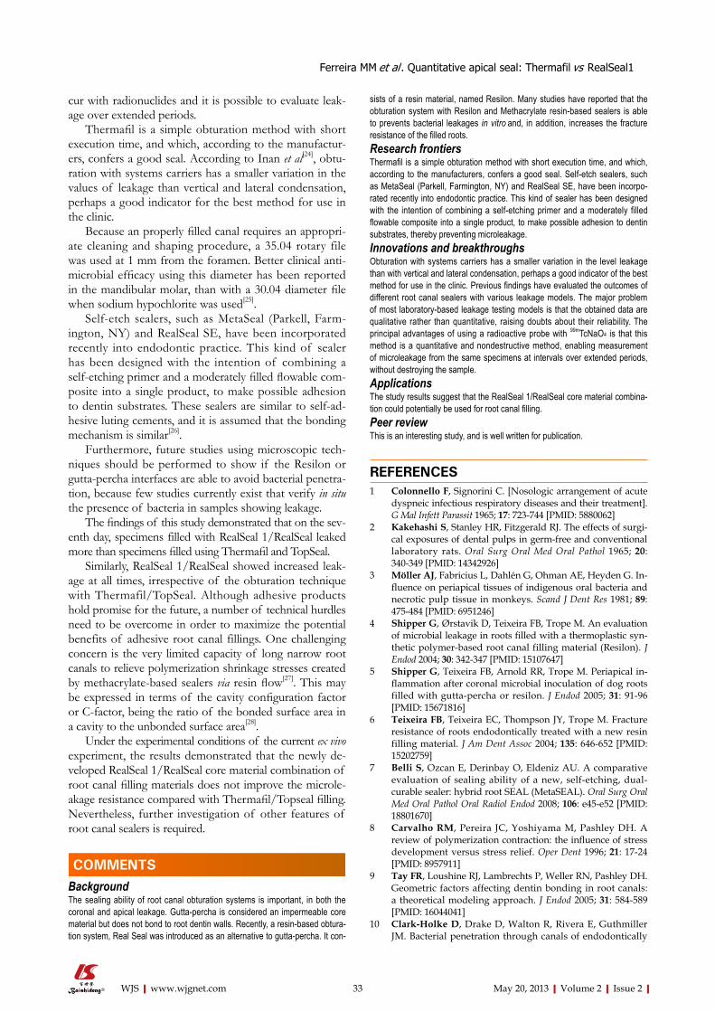
cur with radionuclides and it is possible to evaluate leak-age over extended periods.
Thermafil is a simple obturation method with short execution time, and which, according to the manufactur-ers, confers a good seal. According to Inan et al[24], obtu-ration with systems carriers has a smaller variation in the values of leakage than vertical and lateral condensation, perhaps a good indicator for the best method for use in the clinic.
Because an properly filled canal requires an appropri-ate cleaning and shaping procedure, a 35.04 rotary file was used at 1 mm from the foramen. Better clinical anti-microbial efficacy using this diameter has been reported in the mandibular molar, than with a 30.04 diameter file when sodium hypochlorite was used[25].
Self-etch sealers, such as MetaSeal (Parkell, Farm-ington, NY) and RealSeal SE, have been incorporated recently into endodontic practice. This kind of sealer has been designed with the intention of combining a self-etching primer and a moderately filled flowable com-posite into a single product, to make possible adhesion to dentin substrates. These sealers are similar to self-ad-hesive luting cements, and it is assumed that the bonding mechanism is similar[26].
Furthermore, future studies using microscopic tech-niques should be performed to show if the Resilon or gutta-percha interfaces are able to avoid bacterial penetra-tion, because few studies currently exist that verify in situ the presence of bacteria in samples showing leakage.
The findings of this study demonstrated that on the sev-enth day, specimens filled with RealSeal 1/RealSeal leaked more than specimens filled using Thermafil and TopSeal.
Similarly, RealSeal 1/RealSeal showed increased leak-age at all times, irrespective of the obturation technique with Thermafil/TopSeal. Although adhesive products hold promise for the future, a number of technical hurdles need to be overcome in order to maximize the potential benefits of adhesive root canal fillings. One challenging concern is the very limited capacity of long narrow root canals to relieve polymerization shrinkage stresses created by methacrylate-based sealers via resin flow[27]. This may be expressed in terms of the cavity configuration factor or C-factor, being the ratio of the bonded surface area in a cavity to the unbonded surface area[28].
Under the experimental conditions of the current ex vivo experiment, the results demonstrated that the newly de-veloped RealSeal 1/RealSeal core material combination of root canal filling materials does not improve the microle-akage resistance compared with Thermafil/Topseal filling. Nevertheless, further investigation of other features of root canal sealers is required.
COMMENTSBackgroundThe sealing ability of root canal obturation systems is important, in both the coronal and apical leakage. Gutta-percha is considered an impermeable core material but does not bond to root dentin walls. Recently, a resin-based obtura-tion system, Real Seal was introduced as an alternative to gutta-percha. It con-
sists of a resin material, named Resilon. Many studies have reported that the obturation system with Resilon and Methacrylate resin-based sealers is able to prevents bacterial leakages in vitro and, in addition, increases the fracture resistance of the filled roots. Research frontiersThermafil is a simple obturation method with short execution time, and which, according to the manufacturers, confers a good seal. Self-etch sealers, such as MetaSeal (Parkell, Farmington, NY) and RealSeal SE, have been incorpo-rated recently into endodontic practice. This kind of sealer has been designed with the intention of combining a self-etching primer and a moderately filled flowable composite into a single product, to make possible adhesion to dentin substrates, thereby preventing microleakage.Innovations and breakthroughsObturation with systems carriers has a smaller variation in the level leakage than with vertical and lateral condensation, perhaps a good indicator of the best method for use in the clinic. Previous findings have evaluated the outcomes of different root canal sealers with various leakage models. The major problem of most laboratory-based leakage testing models is that the obtained data are qualitative rather than quantitative, raising doubts about their reliability. The principal advantages of using a radioactive probe with 99mTcNaO4 is that this method is a quantitative and nondestructive method, enabling measurement of microleakage from the same specimens at intervals over extended periods, without destroying the sample. ApplicationsThe study results suggest that the RealSeal 1/RealSeal core material combina-tion could potentially be used for root canal filling. Peer reviewThis is an interesting study, and is well written for publication.
REFERENCES1 Colonnello F, Signorini C. [Nosologic arrangement of acute
dyspneic infectious respiratory diseases and their treatment]. G Mal Infett Parassit 1965; 17: 723-744 [PMID: 5880062]
2 Kakehashi S, Stanley HR, Fitzgerald RJ. The effects of surgi-cal exposures of dental pulps in germ-free and conventional laboratory rats. Oral Surg Oral Med Oral Pathol 1965; 20: 340-349 [PMID: 14342926]
3 Möller AJ, Fabricius L, Dahlén G, Ohman AE, Heyden G. In-fluence on periapical tissues of indigenous oral bacteria and necrotic pulp tissue in monkeys. Scand J Dent Res 1981; 89: 475-484 [PMID: 6951246]
4 Shipper G, Ørstavik D, Teixeira FB, Trope M. An evaluation of microbial leakage in roots filled with a thermoplastic syn-thetic polymer-based root canal filling material (Resilon). J Endod 2004; 30: 342-347 [PMID: 15107647]
5 Shipper G, Teixeira FB, Arnold RR, Trope M. Periapical in-flammation after coronal microbial inoculation of dog roots filled with gutta-percha or resilon. J Endod 2005; 31: 91-96 [PMID: 15671816]
6 Teixeira FB, Teixeira EC, Thompson JY, Trope M. Fracture resistance of roots endodontically treated with a new resin filling material. J Am Dent Assoc 2004; 135: 646-652 [PMID: 15202759]
7 Belli S, Ozcan E, Derinbay O, Eldeniz AU. A comparative evaluation of sealing ability of a new, self-etching, dual-curable sealer: hybrid root SEAL (MetaSEAL). Oral Surg Oral Med Oral Pathol Oral Radiol Endod 2008; 106: e45-e52 [PMID: 18801670]
8 Carvalho RM, Pereira JC, Yoshiyama M, Pashley DH. A review of polymerization contraction: the influence of stress development versus stress relief. Oper Dent 1996; 21: 17-24 [PMID: 8957911]
9 Tay FR, Loushine RJ, Lambrechts P, Weller RN, Pashley DH. Geometric factors affecting dentin bonding in root canals: a theoretical modeling approach. J Endod 2005; 31: 584-589 [PMID: 16044041]
10 Clark-Holke D, Drake D, Walton R, Rivera E, Guthmiller JM. Bacterial penetration through canals of endodontically
33 May 20, 2013|Volume 2|Issue 2|WJS|www.wjgnet.com
COMMENTS
Ferreira MM et al . Quantitative apical seal: Thermafil vs RealSeal1
treated teeth in the presence or absence of the smear layer. J Dent 2003; 31: 275-281 [PMID: 12735922]
11 Economides N, Kokorikos I, Kolokouris I, Panagiotis B, Go-gos C. Comparative study of apical sealing ability of a new resin-based root canal sealer. J Endod 2004; 30: 403-405 [PMID: 15167466]
12 Shahravan A, Haghdoost AA, Adl A, Rahimi H, Shadifar F. Effect of smear layer on sealing ability of canal obturation: a systematic review and meta-analysis. J Endod 2007; 33: 96-105 [PMID: 17258623]
13 Khedmat S, Shokouhinejad N. Comparison of the efficacy of three chelating agents in smear layer removal. J Endod 2008; 34: 599-602 [PMID: 18436043]
14 Carvalho AS, Camargo CH, Valera MC, Camargo SE, Manci-ni MN. Smear layer removal by auxiliary chemical substances in biomechanical preparation: a scanning electron microscope study. J Endod 2008; 34: 1396-1400 [PMID: 18928856]
15 Camps J, Pashley D. Reliability of the dye penetration stud-ies. J Endod 2003; 29: 592-594 [PMID: 14503834]
16 Delivanis PD, Chapman KA. Comparison and reliability of techniques for measuring leakage and marginal penetra-tion. Oral Surg Oral Med Oral Pathol 1982; 53: 410-416 [PMID: 7043354]
17 Barthel CR, Moshonov J, Shuping G, Orstavik D. Bacterial leakage versus dye leakage in obturated root canals. Int En-dod J 1999; 32: 370-375 [PMID: 10551110]
18 Kim YK, Grandini S, Ames JM, Gu LS, Kim SK, Pashley DH, Gutmann JL, Tay FR. Critical review on methacrylate resin-based root canal sealers. J Endod 2010; 36: 383-399 [PMID: 20171352]
19 Goldman M, Simmonds S, Rush R. The usefulness of dye-penetration studies reexamined. Oral Surg Oral Med Oral Pathol 1989; 67: 327-332 [PMID: 2927929]
20 Pogrel MA, Kopf J, Dodson TB, Hattner R, Kaban LB. A com-
parison of single-photon emission computed tomography and planar imaging for quantitative skeletal scintigraphy of the mandibular condyle. Oral Surg Oral Med Oral Pathol Oral Radiol Endod 1995; 80: 226-231 [PMID: 7552889 DOI: 10.1016/S1079-2104(05)80206-9]
21 Banerjee S, Pillai MR, Ramamoorthy N. Evolution of Tc-99m in diagnostic radiopharmaceuticals. Semin Nucl Med 2001; 31: 260-277 [PMID: 11710769]
22 Michaïlesco PM, Valcarcel J, Grieve AR, Levallois B, Lerner D. Bacterial leakage in endodontics: an improved method for quantification. J Endod 1996; 22: 535-539 [PMID: 9198441]
23 Wu MK, De Gee AJ, Wesselink PR, Moorer WR. Fluid trans-port and bacterial penetration along root canal fillings. Int Endod J 1993; 26: 203-208 [PMID: 8225638]
24 Inan U, Aydemir H, Taşdemir T. Leakage evaluation of three different root canal obturation techniques using electrochemi-cal evaluation and dye penetration evaluation methods. Aust Endod J 2007; 33: 18-22 [PMID: 17461836]
25 Shuping GB, Orstavik D, Sigurdsson A, Trope M. Reduction of intracanal bacteria using nickel-titanium rotary instru-mentation and various medications. J Endod 2000; 26: 751-755 [PMID: 11471648]
26 Resende LM, Rached-Junior FJ, Versiani MA, Souza-Gabriel AE, Miranda CE, Silva-Sousa YT, Sousa Neto MD. A com-parative study of physicochemical properties of AH Plus, Epiphany, and Epiphany SE root canal sealers. Int Endod J 2009; 42: 785-793 [PMID: 19548934]
27 Alster D, Feilzer AJ, de Gee AJ, Davidson CL. Polymerization contraction stress in thin resin composite layers as a func-tion of layer thickness. Dent Mater 1997; 13: 146-150 [PMID: 9758966]
28 Feilzer AJ, De Gee AJ, Davidson CL. Setting stress in com-posite resin in relation to configuration of the restoration. J Dent Res 1987; 66: 1636-1639 [PMID: 10872397]
P- Reviewer El-Askary F S- Editor Wen LL L- Editor Hughes D E- Editor Zheng XM
34 May 20, 2013|Volume 2|Issue 2|WJS|www.wjgnet.com
Ferreira MM et al . Quantitative apical seal: Thermafil vs RealSeal1

World Journal of StomatologyWorld J Stomatol 2013 August 20; 2(3): 35-66
ISSN 2218-6263 (online)
www.wjgnet.comwww.wjgnet.com

World Journal of StomatologyW J S
35 Dentalstemcells:Progressandperspectives
Dimitrova-Nakov S, Harichane Y, Goldberg M, Kellermann O
40 Basicpropertiesandtypesofzirconia:Anoverview
Saridag S, Tak O, Alniacik G
48 Clinicalevaluationofimplantsinpatientswithmaxillofacialdefects
Atalay B, Bilhan H, Geckili O, Bilmenoglu C, Meric U
56 Evaluationoffactorsaffectingthesuccessrateoforthodonticmini-implants
bysurvivalanalysis
Baik UB, Bayome M, Han KH, Park JH, Jung MH, Kook YA
62 Managementofmissileinjuriestothemaxillofacialregion:Acaseseries
Ebrahimi A, Motamedi MHK, Nejadsarvari N, Kazemi HM
Contents
MINIREVIEWS
Quarterly Volume 2 Number 3 August 20, 2013
IWJS|www.wjgnet.com August 20, 2013|Volume 2|Issue 3|
REVIEW
ORIGINAL ARTICLE
BRIEF ARTICLE

ContentsWorld Journal of Stomatology
Volume 2 Number 3 August 20, 2013
EDITORS FOR THIS ISSUE
Responsible Assistant Editor: Xin-Xin Che Responsible Science Editor: Ling-Ling WenResponsible Electronic Editor: Ya-Jing Lu Proofing Editor-in-Chief: Lian-Sheng Ma
World Journal of StomatologyRoom 903, Building D, Ocean International Center, No. 62 Dongsihuan Zhonglu, Chaoyang District, Beijing 100025, ChinaTelephone: +86-10-85381891Fax: +86-10-85381893E-mail: [email protected]://www.wjgnet.com
PUBLISHERBaishideng Publishing Group Co., LimitedFlat C, 23/F., Lucky Plaza, 315-321 Lockhart Road, Wan Chai, Hong Kong, ChinaFax: +852-6555-7188Telephone: +852-3177-9906E-mail: [email protected]://www.wjgnet.com
PUBLICATIONDATEAugust 20, 2013
COPYRIGHT© 2013 Baishideng. Articles published by this Open-Access journal are distributed under the terms of the Creative Commons Attribution Non-commercial License, which permits use, distribution, and repro-duction in any medium, provided the original work is properly cited, the use is non commercial and is other-wise in compliance with the license.
SPECIALSTATEMENTAll articles published in this journal represent the viewpoints of the authors except where indicated oth-erwise.
INSTRUCTIONSTOAUTHORSFull instructions are available online at http://www.wjgnet.com/2218-6263/g_info_20100722180909.htm
ONLINESUBMISSIONhttp://www.wjgnet.com/esps/
IIWJS|www.wjgnet.com
APPENDIX
ABOUT COVER
AIM AND SCOPE
INDEXING/ABSTRACTING
August 20, 2013|Volume 2|Issue 3|
NAMEOFJOURNALWorld Journal of Stomatology
ISSNISSN 2218-6263 (online)
LAUNCHDATEDecember 31, 2011
FREQUENCYQuarterly
EDITOR-IN-CHIEFPeter E Murray, BSc (Hons), PhD, Professor, Pathol-ogist, Department of Endodontics, College of Dental Medicine, Nova Southeastern University, 3200 South University Drive, Fort Lauderdale, FL 33328-2018, United States
EDITORIALOFFICEJin-Lei Wang, DirectorXiu-Xia Song, Vice Director
I-V Instructionstoauthors
EditorialBoardMemberofWorldJournalofStomatology ,MichelGoldberg,
ParisMichelGoldberg,Professor,Goldberg InsermU747,BiomedicaleDes
SaintsPeres,45RuedesSaintsPères,75006Paris,France
World Journal of Stomatology (World J Stomatol, WJS, online ISSN 2218-6263, DOI: 10.5321) is a peer-reviewed open access academic journal that aims to guide clinical practice and improve diagnostic and therapeutic skills of clinicians.
WJS covers topics concerning oral and craniofacial sciences, oral and craniofacial development/growth, dental tissue regeneration, craniofacial bone and cartilage research, oral and maxillofacial genetic diseases, developmental abnormalities and soft tissue defects, pulpal and periapical diseases, periodontal diseases and oral mucosal diseases, salivary gland diseases, oral and maxillofacial vascular/nervous diseases, jaw bone diseases, taste abnormalities, oral and maxillofacial pain, occlusion and temporomandibular diseases, repair and treatment of tooth defects, loss and dento-maxillofacial deformities, oral and maxillofacial biomechanics and biomaterials, new techniques for diagnosis/treatment of oral and maxillofacial diseases; and stomatology-related evidence-based medicine, epidemiology and nursing. Priority publication will be given to articles concerning diagnosis and treatment of stomatologic diseases. The following aspects are covered: Clinical diagnosis, laboratory diagnosis, differential diagnosis, imaging tests, pathological diagnosis, molecular biological diagnosis, immunological diagnosis, genetic diagnosis, functional diagnostics, and physical diagnosis; and comprehensive therapy, drug therapy, surgical therapy, interventional treatment, minimally invasive therapy, and robot-assisted therapy.
We encourage authors to submit their manuscripts to WJS. We will give priority to manuscripts that are supported by major national and international foundations and those that are of great basic and clinical significance.
World Journal of Stomatology is now indexed in Digital Object Identifier.
I-III EditorialBoardFLYLEAF

World Journal of StomatologyW J S
Online Submissions: http://www.wjgnet.com/esps/[email protected]:10.5321/wjs.v2.i3.35
World J Stomatol 2013 August 20; 2(3): 35-39ISSN 2218-6263 (online)
© 2013 Baishideng. All rights reserved.
Dental stem cells: Progress and perspectives
Sasha Dimitrova-Nakov, Yassine Harichane, Michel Goldberg, Odile Kellermann
Sasha Dimitrova-Nakov, Yassine Harichane, Michel Gold-berg, Odile Kellermann, Inserm UMR-S 747, UFR Biomédi-cales des Saints-Pères, Université Paris Descartes, 75006 Paris, FranceAuthor contributions: All authors substantial contributed to conception, design, acquisition of data, and interpretation of data, drafting the article or revising it critically for important intellectual content, and final approval of the version to be pub-lished.Correspondence to: Michel Goldberg, Professor, Inserm UMR-S 747, UFR Biomédicales des Saints-Pères, Université Paris Descartes, 45 rue des Saints-Pères, 75006 Paris, France. [email protected] Telephone: +33-1-42863851 Fax: +33-1-42864068Received: December 13, 2012 Revised: March 28, 2013Accepted: April 9, 2013Published online: August 20, 2013
AbstractDental pulp stem cells (DPSCs) are thought to contrib-ute to reparative dentin formation, and that they may correspond to heterogenous populations of precursor cells or represent distinct differentiation stages along the odontoblastic lineage. DPSCs share many similari-ties with mesenchymal stem cells of the bone marrow (BMSCs). It appears that the distribution of tissue stem cells is not random and, within the dental pulp, there are potentially several distinct niches of stem/progeni-tor cells. In addition to DPSCs, other dental stem cell populations have been isolated. As for DPSCs, further studies are still needed to evaluate their potential of differentiation and their regenerative activity. Up today, (1) the formal demonstration that pulpal resident stem cells are actually the reparative dentin-forming cells re-cruited in response to injury is still lacking; and (2) the origin, localization and precise identity of odontogenic stem cells remain largely unknown. Dental clonal cell lines may represent valuable tool to answer some fon-tamental questions concerning the dental stem cell biol-ogy. Altogether, the presence of dental cell populations displaying stem cell properties has opened new paths for considering regenerative therapies. This might be a
prerequisite to design alternative strategies for capping and endodontic treatment, using stem cells.
© 2013 Baishideng. All rights reserved.
Key words: Dental pulp; Stem cells; Dentin repair; Niche
Core tip: The presence of cell populations displaying stem cell properties within the pulp has opened new paths for considering more conservative therapies. Still, dental stem cells research is still confronted with the lack of precise knowledge related to the location and identity of the cells participating to reparative dentin formation. Clonal cell lines derived from the dental sphere may represent valuable tool to answer some questions that are of fundamental importance to stem cell biology and clinical applications. This review dis-cusses some fundamental concepts of dental stem cell biology within the context of regenerative dentistry.
Dimitrova-Nakov S, Harichane Y, Goldberg M, Kellermann O. Dental stem cells: Progress and perspectives. World J Stomatol 2013; 2(3): 35-39 Available from: URL: http://www.wjgnet.com/2218-6263/full/v2/i3/35.htm DOI: http://dx.doi.org/10.5321/wjs.v2.i3.35
INTRODUCTIONTooth development requires a series of sequential and reciprocal interactions between the ectodermally derived oral epithelium at the origin of ameloblasts and neural crest-derived ectomesenchyme at the origin of odon-toblasts. Tooth patterning proceeds through sequential morphogenetic events (bud, cap, bell) leading to crown and subsequently to root formation. During embryogen-esis, morphogenesis is coupled to differentiation of com-mited cells that progressively elaborate enamel and dentin and in turn reach the terminal stages of amelogenic and odontogenic lignages. This cascade of events relies on
MINIREVIEWS
35 August 20, 2013|Volume 2|Issue 3|WJS|www.wjgnet.com
epithelial-mesenchymal interactions that progressely lead to transformation of the tooth germ into complex min-eralized structures[1].
Ameloblasts are lost following enamel maturation and tooth eruption, and hence enamel cannot be regener-ated. Dental papilla ectomesenchymal cells give rise to the embryonic pulp and odontoblasts. Dental pulp cells maintain tooth homeostasis and odontoblasts synthesize dentin extracellular matrix. Dentin and pulp are related embryologically, histologically, and are functionally as-sociated although the term of dentin-pulp complex is a notion underlying an oversimplification.
Odontoblasts are polarized postmitotic cells. These terminally differentiated cells cannot undergo further cell division and proliferate to replace irreversibly injured odontoblasts. Only the postmitotic cells forming the sub-odontoblastic Hoehl’s cell layer, have the capacity to acquire a polarized phenotype and become function-nal odontoblasts. Odontoblasts are responsible for the secretion of primary and secondary dentin. They have a natural regenerative potential leading to the formation of reactionary dentin[2]. Odontoblasts can be up-regulated to secrete a reactionary dentin matrix when a mild injury occurs, such as attrition or early caries. However, injury of greater intensity, such as deep caries or restorative procedures, may lead to the death of the pre-existing odontoblasts and Hoehl’s cells[3]. In such cases, recruit-ment of stem/precursor cells within the pulp will give rise to a new generation of odontoblast-like cells capable to elaborate reparative dentin.
The process of dental tissue repair may share many similarities with the embryological mechanisms of tooth development. It is assumed that many genes and signaling pathways involved in odontogenesis are also implicated in the tooth repair. Still, the mechanisms underlying repara-tive dentin formation are “open research areas” and offer exciting opportunities for possible tooth regeneration and dental tissue engineering.
DENTAL PULP STEM CELLSThe post-natal dental pulp contains heterogeneous cell populations responsible for its maintenance, defence and capacity of repair: stromal fibroblasts, odonto-osteopro-genitors, neuronal and vascular cells as well as inflam-matory and immune system cells such as dentritic cells, neutrophils, macrophages, lymphocytes[4]. The ability of the pulp to respond to a variety of pathological condi-tions and injuries by deposition of a reparative dentin by pulp “progenitors” is well recognized[5] but the origin, lo-calization and precise identity of odontogenic stem cells remain largely unknown. Identifying cells mobilized in response to pulp injury is a prerequisite to design alterna-tive strategies for capping and endodontic treatment, us-ing stem cells.
A decade ago, a population of odontogenic progeni-tors, inferred as dental pulp stem cells (DPSCs), was isolated from the pulp of human permanent third mo-lars[6]. Upon subcutaneous transplantation into immuno-
compromised mice, in vitro expanded DPSCs mixed with hydroxyapatite formed dentin/pulp like complexes at an ectopic site. Populations of DPSCs possesses (1) generic mesenchymal stem cells-like properties (MSCs); (2) colo-ny forming ability; and (3) were shown to express in vitro osteoblastic, adipogenic, chondrogenic or even neuronal markers[7-9]. DPSCs share many similarities with mesen-chymal stem cells of the bone marrow (BMSCs) which are the most studied stromal stem cell populations. More than 4000 human genes are expressed either by BMSCs or DPSCs[10]. Dental stem cell populations also express different panels of stem cell surface markers such as 3G5, STRO-1, CD44, CD106, CD146, CD90 and Sca-1 used to characterize hematopoetic stem cells. However, it is important to note that DPSCs and BMPCs have not the same embryonic origin and that cells derived from the human or animal dental pulps have not been able to sup-port hematopoiesis in transplantation assays[11]. DPSCs are thought to contribute to reparative dentin formation, and it appears that they may correspond to heterogenous populations of precursor cells or represent distinct dif-ferentiation stages along the odontoblastic lineage.
The presence of cell populations displaying stem cell properties within the pulp has opened new paths for considering more conservative therapies[6]. Nevertheless, the formal demonstration that pulpal resident stem cells are actually the reparative dentin-forming cells recruited in response to injury is still lacking. The hypothesis that a subset of stem cells carried by the vasculature replen-ishes the pulp after lesion can not be totally excluded. In the pulp, as in most tissues, the size of the pool of stem cells is very small (< 1%) and little is known about their anatomical sites within the pulp[12]. Moreover, the re-sponsiveness of the pulp provides a dynamic system for tissue repair that may imply migration of stem cells from their resting places to the site of injury. Undifferentiated mesenchymal/mesectodermal cells present in the stroma, perivascular cells such as Rouget’s pericytes or fibroblasts have all been proposed as potential progenitors mediat-ing pulp repair after destruction of the odontoblasts and the Hoehl’s sub-odontoblastic cell layer[13]. Advances in imaging technology and identification of stem cell markers are still needed to visualize stem/precursor cells in situ.
WHERE ARE THE DENTAL PULP STEM CELLS NICHES?Stem cells are rare cells that are uniquely capable of both reproducing themselves and generating the differentiated cell types that are needed to carry out specialized func-tions. Stem cell behaviour is regulated by inputs from their local environment often referred as the “stem cell niche”. Niches are defined as specific anatomic locations that provide structural support, trophic support, topo-graphical informations and the appropriate physiological cues to control the maintenance, quiescence, self-renewal, recruitment towards differentiation and long-term regen-erative capacity of stem cells. Hallmarks of a niche may
36 August 20, 2013|Volume 2|Issue 3|WJS|www.wjgnet.com
Dimitrova-Nakov S et al . Dental stem cells: Progress and perspectives

include: the stem cell itself, stomal supporting cells that interact directly with the stem cells via secreted factors and cell surface molecules, extracellular matrix (ECM) that provides structure and mechanical signals, neuronal inputs and vascular network that carry systemic signals and represent a conduit for recruitment of inflamma-tory and circulating cells into the niche. In teeth, as in the adult blood system, multiple niches may exist and specific markers allowing the definitive identification of stem cells within the pulp are lacking.
Some data suggest that pericytes could differenti-ate into osteoblast-like cells, so odontogenic stem cells may reside in a perivascular niche[14]. In this context, it is interesting to mention that many haematopoetic stem cells (HSCs) and neuronal stem cells (NSCs) are localized close to the vascular network; this could be important to communicate “insult” and expose stem cells to signals activating their recruitment. Besides, alterations in ECM components and matrix elasticity related to damage or ageing may also provide mechanical signals that could have a profound impact on stem cell activity[15]. Thus, it appears that the distribution of tissue stem cells is not random and, within the dental pulp, there are potentially several distinct niches of stem/progenitor cells. Nev-ertheless, still little information is available regarding their topological organization and the inputs that recruit osteo-odontogenic stem cells to form reparative dentin[3]. In contrast to other tissues known to have a constant re-generation potential, such as intestine and bone marrow, dental pulp stem cells will react to form reparative dentin only after injury. This imply that signals ensure their sur-vival and prevent their differentiation while maintaining their responsiveness following pulp damage. Whether an endogenous pool of stem cells associated with support-ive stromal cells are mobilized at the site of injury and/or whether attraction of migrating stem cell is necessary to repopulate a niche and expand precursor cells at the ap-propriate site for dentin repair is unknown. In addition, the alteration of the dentin mineralized matrix promotes the release of bio-active molecules including high con-centrations of Ca2+ which locally may also contribute to stem cells proliferation and differentiation in the post-injury pulpal environment.
DENTAL STEM CELLS-DIFFERENT TYPES, DIFFERENT LOCATIONSIn addition to DPSCs which were derived from the pulp of human permanent third molar, other stem cell popula-tions have been isolated from exfoliated deciduous teeth (SHED), periodontal ligament (PDLCs), apical papilla (SCAP) and dental follicule (DFSCs). As for DPSCs, fur-ther studies are still needed to evaluate their potential of differentiation and their regenerative activity .
SHED, isolated from the pulp of human deciduous teeth appear distinct from DPSCs, having a higher prolif-erative rate and distinct gene expression profiles. SHED have osteoinductive capacity in vivo[16]. They can survive
in mouse brain, expressing neural markers and possible application of SHED was even considered in alleviating Parkinson’s disease[17]. As odontoblasts, they have a neural crest origin which may sustain their ability to adapt in a neuronal environment. Since children lose 20 deciduous teeth, SHED may be potentially used as an autologous stem cell source for dental pulp engineering once the children become adult. The commercial banking of these cells is becoming widespread but whether SHED main-tain their stem cell properties after long-term storage (cryopreservation for more than 10 years) have not been assessed.
PDLSCs which derived from human periodontal liga-ment (PDL), a connective tissue between the cementum and the inner wall of the alveolar bone socket, represent a population of stem cells capable to differentiate in cementoblast-like cells and type 1 collagen-forming cells. Interestingly, transplantation of human PDLSCs in the periodontal defect of immunocompromissed mice, pro-motes the formation of a periodontal -like tissue, sug-gesting that PDLCs may be a potential tool for alveolar bone repair[18].
SCAP are derived from the apical part of the papilla of growing tooth roots[19,20]. It is important to note that the apical papilla tissue is present while the root apex is still open, before tooth eruption. In vitro, SCAP have been shown to exhibit dentinogenic and adipogenic properties, they also express neuronal markers. In clinical practice, they are easily accessible from extracted wisdom molars which develop later in life. Whether SCAP may represent a source of autologous stem cells for tooth repair re-mains an open question.
DFSCs derived from the dental follicle, a fibrous ecto-mesenchymal tissue that surrounds the developping tooth germ during the crown formation and disappears during the early stages of root development[21,22]. This tissue will form the periodontium, i.e., cementum, periondontal liga-ment and alveolar bone. Thus, DFSCs may correspond to a heterogenous cell populations with different type of stem cells including cementoblasts, osteoblasts, stromal cells. In the adults, DFSCs can be easily accessible in impacted wisdom tooth during crown formation but not later (Table 1).
Finally, since 2009, several publications describe new populations of mesenchymal stem cells isolated from the human oral mucosa and gingiva (Zhang et al, 2012). Their differentiation and therapeutic potentials remain to be determined.
PERSPECTIVES AND OPEN QUESTIONSDental stem cells research is still confronted with the lack of precise knowledge related to the location and identity of the cells participating to reparative dentin formation. To this end, our laboratory developed the strategy and established stem cell lines from embryonic pulp of trans-genic mouse[23] and Figure 1. One of the clones has the capacity after implantation in a rat molar, and in the ab-
37 August 20, 2013|Volume 2|Issue 3|WJS|www.wjgnet.com
Dimitrova-Nakov S et al . Dental stem cells: Progress and perspectives
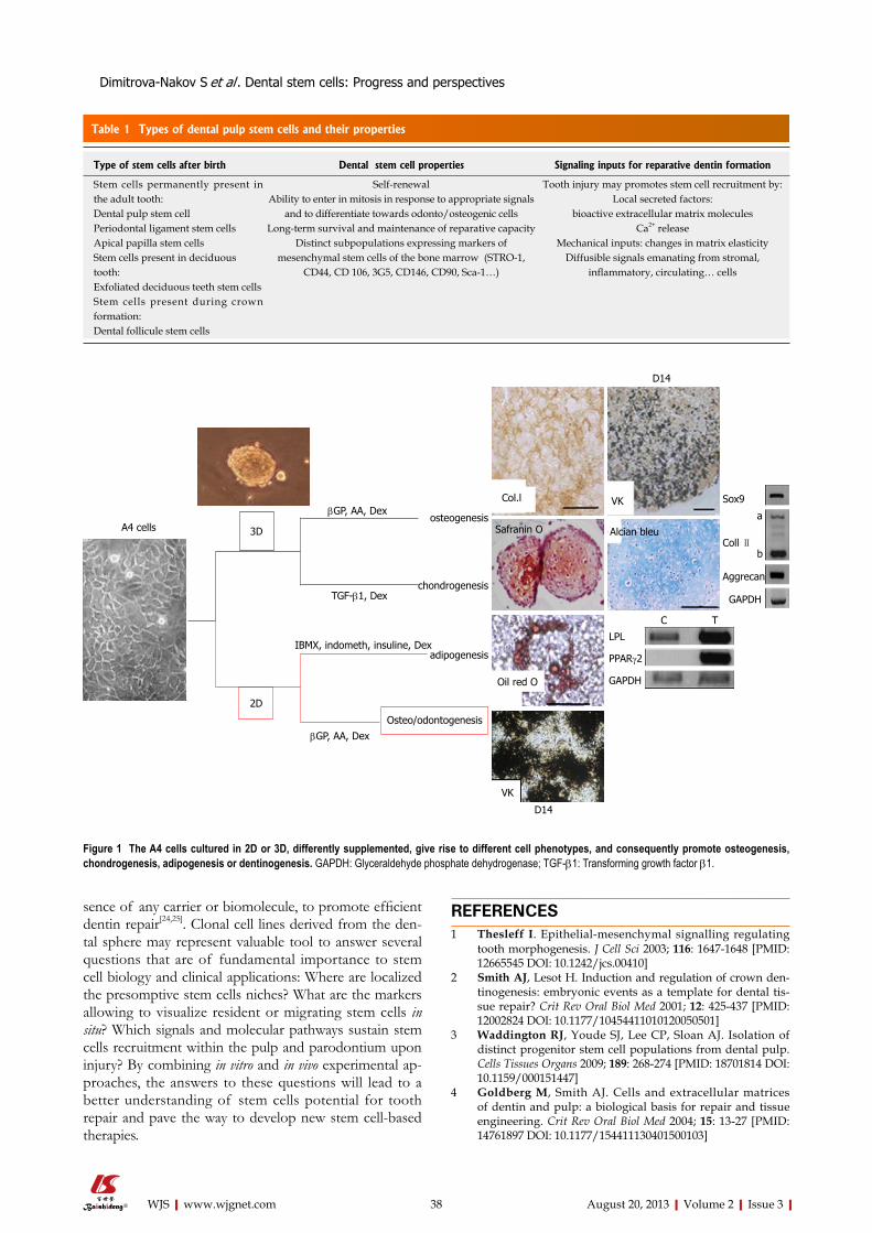
sence of any carrier or biomolecule, to promote efficient dentin repair[24,25]. Clonal cell lines derived from the den-tal sphere may represent valuable tool to answer several questions that are of fundamental importance to stem cell biology and clinical applications: Where are localized the presomptive stem cells niches? What are the markers allowing to visualize resident or migrating stem cells in situ? Which signals and molecular pathways sustain stem cells recruitment within the pulp and parodontium upon injury? By combining in vitro and in vivo experimental ap-proaches, the answers to these questions will lead to a better understanding of stem cells potential for tooth repair and pave the way to develop new stem cell-based therapies.
REFERENCES1 Thesleff I. Epithelial-mesenchymal signalling regulating
tooth morphogenesis. J Cell Sci 2003; 116: 1647-1648 [PMID: 12665545 DOI: 10.1242/jcs.00410]
2 Smith AJ, Lesot H. Induction and regulation of crown den-tinogenesis: embryonic events as a template for dental tis-sue repair? Crit Rev Oral Biol Med 2001; 12: 425-437 [PMID: 12002824 DOI: 10.1177/10454411010120050501]
3 Waddington RJ, Youde SJ, Lee CP, Sloan AJ. Isolation of distinct progenitor stem cell populations from dental pulp. Cells Tissues Organs 2009; 189: 268-274 [PMID: 18701814 DOI: 10.1159/000151447]
4 Goldberg M, Smith AJ. Cells and extracellular matrices of dentin and pulp: a biological basis for repair and tissue engineering. Crit Rev Oral Biol Med 2004; 15: 13-27 [PMID: 14761897 DOI: 10.1177/154411130401500103]
38 August 20, 2013|Volume 2|Issue 3|WJS|www.wjgnet.com
Dimitrova-Nakov S et al . Dental stem cells: Progress and perspectives
Table 1 Types of dental pulp stem cells and their properties
Type of stem cells after birth Dental stem cell properties Signaling inputs for reparative dentin formation
Stem cells permanently present in the adult tooth: Dental pulp stem cell Periodontal ligament stem cellsApical papilla stem cells Stem cells present in deciduous tooth: Exfoliated deciduous teeth stem cells Stem cells present during crown formation: Dental follicule stem cells
Self-renewalAbility to enter in mitosis in response to appropriate signals
and to differentiate towards odonto/osteogenic cells Long-term survival and maintenance of reparative capacity
Distinct subpopulations expressing markers of mesenchymal stem cells of the bone marrow (STRO-1,
CD44, CD 106, 3G5, CD146, CD90, Sca-1…)
Tooth injury may promotes stem cell recruitment by:Local secreted factors:
bioactive extracellular matrix molecules Ca2+ release
Mechanical inputs: changes in matrix elasticityDiffusible signals emanating from stromal,
inflammatory, circulating… cells
A4 cells 3Dosteogenesis
chondrogenesis
adipogenesis
Osteo/odontogenesis
2D
βGP, AA, Dex
TGF-β1, Dex
IBMX, indometh, insuline, Dex
βGP, AA, Dex
Col.l VK
Alcian bleuSafranin O
Oil red O
VK
LPL
GAPDH
PPARg2
C T
Sox9
Coll Ⅱ
Aggrecan
GAPDH
a
b
D14
D14
Figure 1 The A4 cells cultured in 2D or 3D, differently supplemented, give rise to different cell phenotypes, and consequently promote osteogenesis, chondrogenesis, adipogenesis or dentinogenesis. GAPDH: Glyceraldehyde phosphate dehydrogenase; TGF-β1: Transforming growth factor β1.

5 Tziafas D, Smith AJ, Lesot H. Designing new treatment strategies in vital pulp therapy. J Dent 2000; 28: 77-92 [PMID: 10666965]
6 Gronthos S, Mankani M, Brahim J, Robey PG, Shi S. Post-natal human dental pulp stem cells (DPSCs) in vitro and in vivo. Proc Natl Acad Sci USA 2000; 97: 13625-13630 [PMID: 11087820 DOI: 10.1073/pnas.240309797]
7 Batouli S, Miura M, Brahim J, Tsutsui TW, Fisher LW, Gron-thos S, Robey PG, Shi S. Comparison of stem-cell-mediated osteogenesis and dentinogenesis. J Dent Res 2003; 82: 976-981 [PMID: 14630898 DOI: 10.1177/154405910308201208]
8 Pierdomenico L, Bonsi L, Calvitti M, Rondelli D, Arpinati M, Chirumbolo G, Becchetti E, Marchionni C, Alviano F, Fossati V, Staffolani N, Franchina M, Grossi A, Bagnara GP. Mul-tipotent mesenchymal stem cells with immunosuppressive activity can be easily isolated from dental pulp. Transplan-tation 2005; 80: 836-842 [PMID: 16210973 DOI: 10.1097/01.tp.0000173794.72151.88]
9 Iohara K, Zheng L, Ito M, Tomokiyo A, Matsushita K, Na-kashima M. Side population cells isolated from porcine dental pulp tissue with self-renewal and multipotency for dentinogenesis, chondrogenesis, adipogenesis, and neuro-genesis. Stem Cells 2006; 24: 2493-2503 [PMID: 16873765 DOI: 10.1634/stemcells.2006-0161]
10 Shi S, Robey PG, Gronthos S. Comparison of human dental pulp and bone marrow stromal stem cells by cDNA microar-ray analysis. Bone 2001; 29: 532-539 [PMID: 11728923 DOI: 10.1016/S8756-3282(01)00612-3]
11 Huang GT, Gronthos S, Shi S. Mesenchymal stem cells de-rived from dental tissues vs. those from other sources: their biology and role in regenerative medicine. J Dent Res 2009; 88: 792-806 [PMID: 19767575 DOI: 10.1177/0022034509340867]
12 Sloan AJ, Smith AJ. Stem cells and the dental pulp: po-tential roles in dentine regeneration and repair. Oral Dis 2007; 13: 151-157 [PMID: 17305615 DOI: 10.1111/j.1601-0825.2006.01346.x]
13 Fitzgerald M, Chiego DJ, Heys DR. Autoradiographic analy-sis of odontoblast replacement following pulp exposure in primate teeth. Arch Oral Biol 1990; 35: 707-715 [PMID: 2091590 DOI: 10.1016/0003-9969(90)90093-P]
14 Shi S, Gronthos S. Perivascular niche of postnatal mesen-chymal stem cells in human bone marrow and dental pulp. J Bone Miner Res 2003; 18: 696-704 [PMID: 12674330 DOI: 10.1359/jbmr.2003.18.4.696]
15 Engler AJ, Sen S, Sweeney HL, Discher DE. Matrix elasticity directs stem cell lineage specification. Cell 2006; 126: 677-689 [PMID: 16923388]
16 Miura M, Gronthos S, Zhao M, Lu B, Fisher LW, Robey PG,
Shi S. SHED: stem cells from human exfoliated deciduous teeth. Proc Natl Acad Sci USA 2003; 100: 5807-5812 [PMID: 12716973 DOI: 10.1073/pnas.0937635100]
17 Wang J, Wang X, Sun Z, Wang X, Yang H, Shi S, Wang S. Stem cells from human-exfoliated deciduous teeth can dif-ferentiate into dopaminergic neuron-like cells. Stem Cells Dev 2010; 19: 1375-1383 [PMID: 20131979 DOI: 10.1089/scd.2009.0258]
18 Seo BM, Miura M, Gronthos S, Bartold PM, Batouli S, Bra-him J, Young M, Robey PG, Wang CY, Shi S. Investigation of multipotent postnatal stem cells from human periodontal ligament. Lancet 2004; 364: 149-155 [PMID: 15246727 DOI: 10.1016/S0140-6736(04)16627-0]
19 Sonoyama W, Liu Y, Fang D, Yamaza T, Seo BM, Zhang C, Liu H, Gronthos S, Wang CY, Wang S, Shi S. Mesenchymal stem cell-mediated functional tooth regeneration in swine. PLoS One 2006; 1: e79 [PMID: 17183711 DOI: 10.1371/journal.pone.0000079]
20 Sonoyama W, Liu Y, Yamaza T, Tuan RS, Wang S, Shi S, Huang GT. Characterization of the apical papilla and its re-siding stem cells from human immature permanent teeth: a pilot study. J Endod 2008; 34: 166-171 [PMID: 18215674 DOI: 10.1016/j.joen.2007.11.021]
21 Morsczeck C, Götz W, Schierholz J, Zeilhofer F, Kühn U, Möhl C, Sippel C, Hoffmann KH. Isolation of precursor cells (PCs) from human dental follicle of wisdom teeth. Matrix Biol 2005; 24: 155-165 [PMID: 15890265 DOI: 10.1016/j.matbio.2004.12.004]
22 Morsczeck C, Moehl C, Götz W, Heredia A, Schäffer TE, Eckstein N, Sippel C, Hoffmann KH. In vitro differentia-tion of human dental follicle cells with dexamethasone and insulin. Cell Biol Int 2005; 29: 567-575 [PMID: 15951208 DOI: 10.1016/j.cellbi.2005.03.020]
23 Priam F, Ronco V, Locker M, Bourd K, Bonnefoix M, Duch-êne T, Bitard J, Wurtz T, Kellermann O, Goldberg M, Poliard A. New cellular models for tracking the odontoblast pheno-type. Arch Oral Biol 2005; 50: 271-277 [PMID: 15721161 DOI: 10.1016/j.archoralbio.2004.10.007]
24 Harichane Y, Hirata A, Dimitrova-Nakov S, Granja I, Goldberg A, Kellermann O, Poliard A. Pulpal progenitors and dentin repair. Adv Dent Res 2011; 23: 307-312 [PMID: 21677084 DOI: 10.1177/0022034511405322.]
25 Lacerda-Pinheiro S, Dimitrova-Nakov S, Harichane Y, Souyri M, Petit-Cocault L, Legrès L, Marchadier A, Baudry A, Ribes S, Goldberg M, Kellermann O, Poliard A. Concomitant multipotent and unipotent dental pulp progenitors and their respective contribution to mineralised tissue formation. Eur Cell Mater 2012; 23: 371-386 [PMID: 22623164]
P- Reviewer Spagnuolo G S- Editor Song XX L- Editor A E- Editor Lu YJ
39 August 20, 2013|Volume 2|Issue 3|WJS|www.wjgnet.com
Dimitrova-Nakov S et al . Dental stem cells: Progress and perspectives

World Journal of StomatologyW J S
Online Submissions: http://www.wjgnet.com/esps/[email protected]:10.5321/wjs.v2.i3.40
World J Stomatol 2013 August 20; 2(3): 40-47ISSN 2218-6263 (online)
© 2013 Baishideng. All rights reserved.
Basic properties and types of zirconia: An overview
Serkan Saridag, Onjen Tak, Gamze Alniacik
Serkan Saridag, Onjen Tak, Gamze Alniacik, Department of Prosthodontics, Faculty of Dentistry, Kocaeli University, 41700 Kocaeli, TurkeyAuthor contributions: Saridag S and Tak O contributed equally to this work; Saridag S and Tak O contributed to acquisition of data; Saridag S, Tak O and Alniacık G contributed to analysis and interpretation of data, drafting of the manuscript and critical revi-sion.Correspondence to: Dr. Serkan Saridag, Department of Prosthodontics, Faculty of Dentistry, Kocaeli University, Cedit Mh, 41700 Kocaeli, Turkey. [email protected]: +90-262-3442222 Fax: +90-262-3442202Received: January 2, 2013 Revised: April 3, 2013Accepted: May 7, 2013Published online: August 20, 2013
AbstractThis paper describes types and characteristics of zirco-nia materials in relation to their applications in dentist-ry. The zirconia material typically used today by most manufacturers is a tetragonal polycrystalline zirconia, partially stabilized with yttrium oxide. The mechanical properties of zirconia have been extensively investi-gated in the scientific literature and zirconia clearly measures up to any other equivalent manufactured ma-terial. The biocompatibility of zirconia has also been ex-tensively evaluated and no local or systemic adverse re-actions or cytotoxic effects have been found in relation to it. However, ceramic bonding, ageing, light transmis-sion and manufacturing processes are all factors that need to be further evaluated in order to guide the suc-cessful use of zirconia as a prosthetic restorative mate-rial. Milling zirconia to full-contour might be an alterna-tive to traditionally veneered restorations. A potential adhesion mechanism appears to be the combination of air abrasion with aluminum oxide particles (silanated or not), followed by sintering with materials containing special reactive monomers. Changes in zirconia proper-ties before and after the sintering process have also been investigated. It was found that after sintering, surface roughness was greater, and micro hardness was slightly reduced; however, accurate precision of fit
was not affected by the sintering process. Currently, zirconia restorations are manufactured by either soft or hard-milling processes, with the manufacturer of each claiming advantages over the other. Chipping of the veneering porcelain is reported as a common problem and has been labeled as its main clinical setback. As zirconia has demonstrated good mechanical and bio-logical performance, future technology is attempting to improve esthetics and minimize veneer fracture, aiming to create confidence in the dental community towards this all-ceramic system. Milling zirconia to full-contour might be an alternative to traditionally veneered resto-rations. Finally, implications are drawn for manufactur-ing, machining, and widespread use of these materials.
© 2013 Baishideng. All rights reserved.
Key words: Zirconia; Biocompatibility; Porcelain chip-ping; Mechanical properties
Core tip: Although all zirconia is chemically similar, the ultimate product can vary from manufacturer to manu-facturer, with materials of varying density, uniformity homogeneity and crystalline transformation. This can be due to varying grain sizes of the powdered material ultimately affecting strength, with variations producing porosity. One type of restoration will not fit every clini-cal condition but today we have a range of options in zirconia ceramics, including monolithic full-contour type and conventional veneered type zirconia.
Saridag S, Tak O, Alniacik G. Basic properties and types of zirco-nia: An overview. World J Stomatol 2013; 2(3): 40-47 Available from: URL: http://www.wjgnet.com/2218-6263/full/v2/i3/40.htm DOI: http://dx.doi.org/10.5321/wjs.v2.i3.40
INTRODUCTIONZirconium (Zr) is a metal with the atomic number 40. It was first discovered in 1789 by the chemist Martin
REVIEW
40 August 20, 2013|Volume 2|Issue 3|WJS|www.wjgnet.com

Klaproth[1-3]. The material has a density of 6.49 g/cm³, a melting point of 1852 ℃ and a boiling point of 3580 ℃. It has a hexagonal crystal structure and is grayish in color. Zr does not occur in nature in a pure state. It can be found in conjunction with silicate oxide with the mineral name Zircon (ZrO2 × SiO2) or as a free oxide (ZrO2) with the mineral name Baddeleyite[4]. These minerals cannot be used as primary materials in dentistry because of impurities of various metal elements that affect color and because of natural radionuclides like urania and tho-ria, which make them radioactive[5]. Complex and time-consuming processes that result in an effective separation of these elements are necessary in order to produce pure zirconia powders. After purification the material pro-duced can be used as a ceramic biomaterial[4,6,7].
ZrO2 is a polymorphic material and occurs in three forms: monoclinic, tetragonal and cubic. The monoclinic phase is stable at room temperatures up to 1170 ℃, the tetragonal at temperatures of 1170-2370 ℃ and the cubic at over 2370 ℃[8,9]. However, noticeable changes in vol-ume are associated with these transformations: during the monoclinic to tetragonal transformation a 5% decrease in volume occurs when zirconium oxide is heated; con-versely, a 3%-4% increase in volume is observed during the cooling process[4,10] (Figure 1).
STABILIZED ZIRCONIASeveral different oxides are added to zirconia to stabilize the tetragonal and/or cubic phases. Magnesia (MgO), Yt-tria (Y2O3), Calcia (CaO), and Ceria (CeO), amongst oth-ers, allow the generation of multiphase materials known as Partially Stabilized Zirconia (PSZ), whose microstruc-ture at room temperature generally consists of cubic zir-conia as the major phase, with monoclinic and tetragonal zirconia precipitates as the minor phase[4,11,12].
PSZ materials have been tested for their potential use as ceramic biomaterials. Mg-PSZ is one of the most commonly used zirconia-based engineering ceramics[13]. Residual porosity in the mass of the material, a rather coarse grain size (30-40 μm), and difficulties in obtain-ing Mg-PSZ precursors free of impurities, are all factors that have discouraged the interest of ceramic manufac-turers in the development of Mg-PSZ for biomedical applications[4]. It has been reported that reinforcement by phase transformation toughening is less pronounced in Mg-PSZ than in Y-TZP (Yttrium-Tetragonal Zirconia Polycrystals), a finding that is discussed below[13]. Ceria (Ce)-doped zirconia ceramics have been very rarely con-sidered, although they exhibit superior toughness (up to 20 MPa) and show no signs of ageing[14].
TRANSFORMATION/TOUGHENING MECHANISMIn the presence of a small amount of stabilizing oxides, and at room temperature, it is possible to obtain PSZ ceramics in the tetragonal phase only, known as Tetrago-
nal Zirconia Polycrystals (TZP). The finely dispersed tetragonal ZrO2 grains within the cubic matrix, provided that they are small enough, can be maintained in a meta-stable state that is able to transform into the monoclinic phase[11]. Tetragonal-to-monoclinic phase transformation in zirconia can be induced by stress, temperature and surface treatments[15,16]. Low temperature ageing via phase transformation of zirconia hip joint heads in normal at-mospheric conditions has been reported after 10 years of incubation[10]. After the ageing of yttrium-stabilized zirco-nium dioxide in body fluid or water, some tetragonal-to-monoclinic phase transformation on the surface of zir-conium dioxide has also been reported[17,18]. Even though some phase transition does occur, reports indicate that the effect on the material’s mechanical properties is negli-gible[4,10].
Y-TZP (YTTRIUM-TETRAGONAL ZIRCO-NIA POLYCRYSTAL)The addition of approximately 2%-3% of mol yttria (Y2O3) as a stabilizing agent in zirconia allows the sinter-ing of fully tetragonal fine-grained zirconia ceramic ma-terials made of 100% small metastable tetragonal grains and known as Y-TZP[11].
MECHANICAL PROPERTIES AND AGEING OF ZIRCONIAZirconia has mechanical properties similar to those of stainless steel. Its resistance to traction can be as high as 900-1200 MPa and its compression resistance is about 2000 MPa[4]. Cyclical load stresses are also tolerated well by this material. Applying an intermittent force of 28 kN to zirconia substrates, Cales and Stefani found that some 50 billion cycles were necessary to break the samples, but with a force in excess of 90 kN structural failure of the samples occurred after just 15 cycles[19]. Surface treat-ments can also modify the physical properties of zirconia. One property of zirconia that has not been well studied is the phenomenon of low-temperature degradation or “ageing”. Water and nonaqueous solvents can induce the formation of zirconiahydroxides along a crack. This process accelerates expansion of the fracture and can result in reduced strength, toughness, and density, leading to failure of the restoration[14,20-22]. Surface grinding can also reduce strength[23,24]. Kosmac et al[15] confirmed this observation and reported reduced mean strength and re-liability of zirconium oxide after grinding.
Zirconia is characterized by high flexural strength and fracture toughness as a result of a physical property known as transformation toughening[4,25,26]. The incidence of framework fracture was directly related to the design of the FPD, where inlay retained FPDs (IRFPD) showed the highest failure rate[27,28]. The most common complica-tion observed in zirconia-based restorations was fracture of the veneering porcelain, manifesting clinically as chip-
41 August 20, 2013|Volume 2|Issue 3|WJS|www.wjgnet.com
Saridag S et al . Basic properties and types of zirconia

ping fractures of the veneering ceramic with or without exposing the underlying Y-TZP framework[27]. Several fac-tors that may affect the rate of veneering fractures have been investigated. A loss of veneering material may result from an alteration of the crystal structure of the zirconia surface during airborne-particle abrasion of the frame-work before the veneering process. This may result in a change of the temperature expansion coefficients[15,25]. Other factors include the different surface treatments of the frameworks and the bond strength between the ve-neering ceramics and zirconia frameworks[29,30].
Sintering a CAD/CAM-milled lithium disilicate layer-ing veneer cap onto the zirconia coping has significantly increased the mechanical strength of crown restora-tions and represents a cost effective way of fabricating all-ceramic restorations[31]. Milling of new generation full-contour zirconia might be an alternative approach to overcome chipping fractures of veneered zirconia restorations. Fabricating mono-block restorations from pure zirconia could increase the mechanical stability and expand the range of indications[32]. However, no clinical data is available yet.
BIOCOMPATIBILITY OF ZIRCONIAThe biocompatibility of zirconia has been extensively evaluated[4,21,33]. In vitro and in vivo studies have confirmed the high biocompatibility of Y-TZP with the use of very pure zirconia powders that have been purged of their radioactive content[34-39]. No local (cellular) or systemic adverse reactions to the material were reported[4,11,35,40,41]. Recent studies have demonstrated that fewer bacteria ac-cumulate around Y-TZP than titanium[42-44]. This could possibly be explained by different protein adsorption properties[45]. In terms of periodontal health, none of the studies reported any difference or noted any changes in the biological health of the soft and hard tissues around the zirconia-based restorations. Although some data quantified and explored differences in the biocompatibil-ity of zirconia, no instances of gingival inflammation or periodontitis could be shown[46]. These findings have led to the suggestion that zirconium oxide may be a suitable material for manufacturing implant abutments with a low bacterial colonization potential[44].
Zirconia as implant abutment material was first intro-duced in 1996[47]. A randomized controlled clinical trial comparing zirconia and titanium abutments supported by 40 single implants was published[48]. After being in func-
tion for three years, 18 zirconia and 10 titanium abut-ments were followed-up. Both abutment materials exhib-ited survival rates of 100%, as well as similar biological and esthetic outcomes. In an animal study, it was shown that the collagen fiber orientation was similar around zir-conia and titanium implant necks. For both materials, the fibers run parallel-oblique and parallel to the implant sur-face[49]. In a clinical study, a similar degree of plaque accu-mulation was found at zirconia and titanium abutments at three years. In the same study, when zirconia abutments are used as restoration support, there were no significant differences in bone levels between zirconia and titanium abutments after 3-year follow-up[48].
ESTHETIC PROPERTIES AND LIGHT TRANSMISSION OF ZIRCONIAAll ceramic materials more satisfactorily address the de-mand for esthetic restorations than metal ceramic resto-rations with opaque cores[50,51]. However, the translucency of the most durable zirconia-based ceramic crowns is reported to be less than that of lithium disilicate glass ce-ramics, for which excellent esthetic results are document-ed[52-56]. In-Ceram Zirconia (VITA Zahnfabrik, Bad Säck-ingen, Germany), an aluminum oxide-based ceramic with 35% zirconium dioxide, has a relatively low translucency, equal to that of metal ceramic crowns when evaluated using the contrast ratio method[55]. This could be an ob-stacle to achieving an esthetically acceptable restoration. Among nonzirconia core materials, an optimal esthetic result has been reported with Procera AllCeram (Nobel Biocare AB, Göteborg, Sweden), which is a 99.9% alumi-num oxide densely sintered ceramic[57], and IPS Empress (Ivoclar Vivadent AG, Schaan, Liechtenstein) lithium disilicate glass ceramic[58]. The latter evolved in 2005 to IPS e.max Press (Ivoclar Vivadent AG), with improved translucency and mechanical properties[59,60]. Alumina and glass ceramic have, respectively, fair to high relative trans-lucency; nevertheless, their mechanical properties are lower than ZrO2 ceramics[55,61].
Light transmission through Y-TZP varies as a func-tion of: (1) the composition and thickness of the zirconia framework; and (2) the physical characteristics and degree of glazing of the veneering porcelain[62].
Based on this, the use of zirconia ceramics with dif-ferent chemical compositions may be significant for clini-cians. Additionally, measuring the degree of conversion of different resin luting agents beneath zirconia ceramic materials may produce better clinical outcomes[63]. Future studies should be expanded to include new generation full-contour zirconia[64]. Full-contour zirconia milling blanks are created through a unique patent-pending pro-cess. In one process the zirconium oxide powders are milled to further reduce the particle size of zirconium oxide, and mixed with a suitable binder to increase the compaction and density of the green state (compacted powders) and eliminate the closed porosity. The manu-facturers claim that, unlike conventional high-pressure
42 August 20, 2013|Volume 2|Issue 3|WJS|www.wjgnet.com
Saridag S et al . Basic properties and types of zirconia
3%-4% Volume increase
Monoclinic phase
Up to 1170 ℃
Tetragonalphase
1170-2370 ℃
Cubicphase
2370-2680 ℃
5% Volumedecrease
Figure 1 Temperature-related phase transformation of zirconia.

milling blank manufacture, this processing gives full-contour zirconia improved light transmission, providing a lower, more natural shade value[65].
TYPES OF ZIRCONIA FOR MANUFACTURING PROCEDUREThree main types of zirconia are available for use in clinical dentistry[66]. Although they are chemically identi-cal, they have slightly different physical properties (e.g., porosity, density, purity, strength), which may or may not be clinically relevant. Zirconia raw material (as previously mentioned) is not a natural product, but is chemically processed from minerals. With cold isostatic pressing, the powders are shaped into ceramic pre-forms. Cold iso-static pressing is the most accepted procedural technique for shaping Y-TZP and produces stable, chalk-like non-sintered green-stage objects with a very high primary density. The green objects are further stabilized and condensed up to about 95% of the theoretical density by means of sintering without pressure in the oxidized at-mosphere of a special furnace, forming pre-sintered-type oxide-ceramic blanks[11,67]. Additional compression can be achieved with Hot Isostatic Postcompaction (HIP) per-formed at 1000 bar and 50 ℃ below the sintering tem-perature[67]. This procedure removes residual porosity and produces dense, fully-sintered-type oxide-ceramic blanks. Carrying out HIP on Y-TZP results in a gray-black ma-terial that usually requires subsequent heat treatment to oxidize and restore whiteness[68].
Zirconia ceramics are used in dentistry as materials for frameworks, generally fabricated by means of mill-ing the zirconia block using a CAD/CAM machine sys-tem[69-74]. Blocks can be milled either at the green stage, the pre-sintered stage or the completely sintered stage. Green-stage zirconia blocks can be milled using dry carbide burs, pre-sintered zirconia blocks can be milled using carbide burs under cooling liquid, and milling of completely sintered zirconia blocks requires the use of
diamonds under cooling liquid[75]. The three available types of zirconia products are shown in Table 1 together with the milling/grinding technology used in each case.
Frameworks made from green and pre-sintered zirco-nia are milled in an enlarged form to compensate for the shrinkage that occurs during sintering, usually 20%-25% for a partially-sintered framework[76]. The milling process is faster and the wear and tear on hardware is less than when milling from a fully-sintered blank. The framework is subsequently post-sintered in special furnaces (at about 1500 ℃) to reach the fully-sintered stage. The color of the zirconia can be individualized with the addition of oxides to the green-stage framework[68].
The question often arises as to which type of zirconia is best to use. It appears that each has advantages and disadvantages. Fully-sintered HIP zirconia has a denser polycrystalline structure with less porosity than non-HIP material, and this should translate clinically into increased resistance to fracture[77]. On the other hand, some inves-tigators have questioned whether the amount of grinding needed during milling of fully sintered zirconia and the heat that is generated, cause surface and structural defects that can have adverse clinical implications[78]. The marginal fit of either type of material, however, is associated with very acceptable clinical results. Margin fitting of milled zirconia is as good as, if not superior to the fit of a res-toration fabricated from a high noble alloy. Studies have measured the marginal gap of CAD/CAM-milled zirco-nia of both varieties and found it to be 40 to 70 μm[79]. However, compared to the alternative method, milling of fully sintered zirconia blocks is a time consuming process that causes greater wear of the diamond burs and is more expensive. Hence, from that point of view, green-stage zirconia could be regarded as more advantageous[67].
BONDING TO ZIRCONIAThe longevity of an indirect restoration is closely related to the integrity of the cement at the margin[80]. Although the use of zirconia ceramics for dental applications is
43 August 20, 2013|Volume 2|Issue 3|WJS|www.wjgnet.com
Saridag S et al . Basic properties and types of zirconia
Table 1 Three types of zirconia products and their milling/grinding technology (Information provided by manufacturers)
Milling at green stage (non-sintered) Cercon base, Cercon (Degudent, Frankfurt, Germany) Lava Frame, Lava (3M ESPE, Seefeld, Germany) Hint-ELs Zirkon TPZ-G, DigiDent (Girrbach, Pforzheim, Germany) ZirkonZahn, Steger (Steger, Brunneck, Italy) Xavex G 100 Zirkon, Etkon (Etkon, Grafelfingen, Germany)
Grinding at pre-sintered stage In-Ceram YZ Cubes, Cerec InLab (Sirona, Bensheim, Germany)ZS-Blanks, Everest (KaVo, Leutkirch, Germany) Hint-ELs Zirkon TZP-W, DigiDent (Girrbach, Pforzheim, Germany) DC-Shrink, Precident DCS (DCS, Allschwil, Switzerland)LAVA All-Ceramic System (3M ESPE, Seefeld, Germany)Cercon Smart Ceramics (DeguDent, Hanau, Germany)Procera Zirconia (Nobel Biocare, Göteborg, Sweden)
Grinding at completely sintered stage DC-Zirkon, Precident DCS (DCS, Allschwil, Switzerland) Z-Blanks, Everest (KaVo, Leutkirch, Germany) Zirkon TM, Pro 50, Cynovad (Cynovad, Montreal, Canada) Hint-ELs Zirkon TZP-HIP, DigiDent (Girrbach, Pforzheim, Germany)HIP Zirkon, Etkon (Etkon, Grafelfingen, Germany)
ongoing, the best method to achieve a durable bond between the ceramic and the tooth structure is still un-known[81]. The only consensus found in the literature is that hydrofluoric acid etching and common silane agents are not effective with zirconia ceramics[81-83].
Several studies have investigated the bond strength and the durability of various bonding methods used to form high-strength zirconia ceramics. One technique commonly used to condition the ceramic surface is that of air abrasion[77,84-86]. Air abrasion with aliminium ox-ide particles is routinely performed to remove layers of contaminants, thus increasing micromechanical reten-tion between the resin cement and the restoration[80,87,88]. These particles may or may not be silica-coated (with tribochemical treatment)[89-91].
Other techniques for the superficial treatment of zirconia ceramics which have been investigated are laser, plasma spraying and fusing glass pearls to the zirconia surface[92,93]. Higher laser power settings (400-600 mJ) cause excessive material deterioration, making them unsuitable as treatments for zirconia surfaces. Irradia-tion with 200 mJ provides mild surface alterations, with features intermediate between the effects of air abrasion and higher laser intensities[92]. Plasma spraying and glass pearl fusion treatments were found to improve the bond strength of resin cements to the surface. However, they were not compared with conventional methods of sur-face treatments for Y-TZP ceramics, such as air abrasion and tribochemical coating[93].
In other studies several coating agents were used to enhance the formation of chemical bonding with zirconia but only those agents that contain a phosphate monomer agent were effective in establishing a reliable bond with zirconia materials[84,94].
A recent study focusing on the long-term stability of zirconia resin bonding shows that it is directly related to the chemistry of the materials used, including primers. The authors suggest that a more hydrophobic compound is required to better resist the detrimental effect of hy-drolysis in order to gain full benefit from the primers[95-97].
In a novel approach to enhance zirconia resin bond strength, selective infiltration-etching of zirconia-based materials has been tried. This method creates a retentive surface where the adhesive resin can infiltrate and inter-lock in order to establish a strong and a durable bond with zirconia[98-101].
CONCLUSIONSeveral positive characteristics of zirconia, such as bio-compatibility, color and mechanical properties, make the material suitable for use in modern dentistry. However, ceramic bonding, ageing, light transmission and manu-facturing processes are all factors that need to be further evaluated in order to guide the successful use of zirconia as a prosthetic restorative material. Milling zirconia to full-contour might be an alternative to traditionally veneered restorations.
REFERENCES1 Denry I, Kelly JR. State of the art of zirconia for dental appli-
cations. Dent Mater 2008; 24: 299-307 [PMID: 17659331 DOI: 10.1016/j.dental.2007.05.007]
2 Tsuge T. Radiopacity of conventional, resin-modified glass ionomer, and resin-based luting materials. J Oral Sci 2009; 51: 223-230 [PMID: 19550090 DOI: 10.1016/j.dental.2008.05.011]
3 Ban S. Reliability and properties of core materials for all-ceramic dental restorations. Jpn Dent Sci Rev 2008; 44: 3-21 [DOI: 10.1016/j.jdsr.2008.04.001]
4 Piconi C, Maccauro G. Zirconia as a ceramic biomaterial. Biomaterials 1999; 20: 1-25 [PMID: 9916767 DOI: 10.1016/S0142-9612(98)00010-6]
5 Porstendörfer J, Reineking A, Willert HC. Radiation risk estimation based on activity measurements of zirconium oxide implants. J Biomed Mater Res 1996; 32: 663-667 [PMID: 8953157]
6 Boothe GF, Stewart-Smith D, Wagstaff D, Dibblee M. The radiological aspects of zircon sand use. Health Phys 1980; 38: 393-398 [PMID: 7390822 DOI: 10.1097/F00004032-198003000-00014]
7 Christel P, Meunier A, Dorlot JM, Crolet JM, Witvoet J, Sedel L, Boutin P. Biomechanical compatibility and de-sign of ceramic implants for orthopedic surgery. Ann N Y Acad Sci 1988; 523: 234-256 [PMID: 3382124 DOI: 10.1111/j.1749-6632.1988.tb38516.x]
8 Chevalier J, Gremillard L, Virkar AV, Clarke DR. The tetrag-onal-monoclinic transformation in zirconia: Lessons learned and future trends. J Am Ceram Soc 2009; 92: 1901–1920 [DOI: 10.1111/j.1551-2916.2009.03278.x]
9 Suresh A, Mayo MJ, Porter WD, Rawn CJ. Crystallite and grain-size-dependent phase transformations in yttria-doped zirconia. J Am Ceram 2003; 86: 360-362 [DOI: 10.1111/j.1151-2916.2003.tb00025.x]
10 Hjerppe J, Vallittu PK, Fröberg K, Lassila LV. Effect of sin-tering time on biaxial strength of zirconium dioxide. Dent Mater 2009; 25: 166-171 [PMID: 18632146]
11 Christel P, Meunier A, Heller M, Torre JP, Peille CN. Me-chanical properties and short-term in-vivo evaluation of yt-trium-oxide-partially-stabilized zirconia. J Biomed Mater Res 1989; 23: 45-61 [PMID: 2708404 DOI: 10.1002/jbm.820230105]
12 De Aza AH, Chevalier J, Fantozzi G, Schehl M, Torrecillas R. Crack growth resistance of alumina, zirconia and zirconia toughened alumina ceramics for joint prostheses. Biomateri-als 2002; 23: 937-945 [PMID: 11774853 DOI: 10.1016/S0142-9612(01)00206-X]
13 Sundh A, Sjögren G. Fracture resistance of all-ceramic zir-conia bridges with differing phase stabilizers and quality of sintering. Dent Mater 2006; 22: 778-784 [PMID: 16414111 DOI: 10.1016/j.dental.2005.11.006]
14 Chevalier J. What future for zirconia as a biomaterial? Bio-materials 2006; 27: 535-543 [PMID: 16143387 DOI: 10.1016/j.biomaterials.2005.07.034]
15 Kosmac T, Oblak C, Jevnikar P, Funduk N, Marion L. The effect of surface grinding and sandblasting on flexural strength and reliability of Y-TZP zirconia ceramic. Dent Mater 1999; 15: 426-433 [PMID: 10863444 DOI: 10.1016/S0109-5641(99)00070-6]
16 Hannink HJ, Kelly PM, Muddle BC. Transformation Tough-ening in Zirconia-Containing Ceramics. J Am Ceram Soc 2000; 83: 461-487 [DOI: 10.1111/j.1151-2916.2000.tb01221.x]
17 Sato T, Shimada M. Transformation of yttria-doped tetrago-nal ZrO, polycrystals by annealing in water. J Am Ceram Soc 1985; 68: 356-359 [DOI: 10.1111/j.1151-2916.1985.tb15239.x]
18 Shimizu K, Oka M, Kumar P, Kotoura Y, Yamamuro T, Makinouchi K, Nakamura T. Time-dependent changes in the mechanical properties of zirconia ceramic. J Biomed Mater Res 1993; 27: 729-734 [PMID: 8408102]
19 Cales B, Stefani Y. Mechanical properties and surface analy-
44 August 20, 2013|Volume 2|Issue 3|WJS|www.wjgnet.com
Saridag S et al . Basic properties and types of zirconia

sis of retrieved zirconia femoral hip joint heads after an im-plantation time of two to three years. J Mater Sci Mater Med 1994; 5: 376-380 [DOI: 10.1007/BF00058967]
20 Swab JJ. Low temperature degradation of Y-TZP materials. J Mater Sci 1991; 26: 6706-6714 [DOI: 10.1007/BF00553696]
21 Manicone PF, Rossi Iommetti P, Raffaelli L. An overview of zirconia ceramics: basic properties and clinical applications. J Dent 2007; 35: 819-826 [PMID: 17825465 DOI: 10.1016/j.jdent.2007.07.008]
22 Lange FF, Dunlop GL, Davis BI. Degradation during ag-ing of transformation-toughned ZrO2-Y2O3 materials at 250 ℃. J Amer Ceram Soc 1986; 69: 237-240 [DOI: 10.1111/j.1151-2916.1986.tb07415.x]
23 Işeri U, Ozkurt Z, Yalnız A, Kazazoğlu E. Comparison of different grinding procedures on the flexural strength of zirconia. J Prosthet Dent 2012; 107: 309-315 [PMID: 22546308]
24 Luthardt RG, Holzhüter M, Sandkuhl O, Herold V, Schnapp JD, Kuhlisch E, Walter M. Reliability and properties of ground Y-TZP-zirconia ceramics. J Dent Res 2002; 81: 487-491 [PMID: 12161462 DOI: 10.1177/154405910208100711]
25 Peláez J, Cogolludo PG, Serrano B, Lozano JF, Suárez MJ. A prospective evaluation of zirconia posterior fixed dental prostheses: three-year clinical results. J Prosthet Dent 2012; 107: 373-379 [PMID: 22633593 DOI: 10.1016/S0022-3913(12)60094-8]
26 Saridag S, Sevimay M, Pekkan G. Fracture Resistance of Teeth Restored With All-ceramic Inlays and Onlays: An In Vitro Study. Oper Dent 2013 Feb 7; [Epub ahead of print] [PMID: 23391033 DOI: 10.2341/12-211-L]
27 Al-Amleh B, Lyons K, Swain M. Clinical trials in zirconia: a systematic review. J Oral Rehabil 2010; 37: 641-652 [PMID: 20406352 DOI: 10.1111/j.1365-2842.2010.02094.x.]
28 Saridag S, Ozyesil AG, Pekkan G. Fracture strength and bending of all-ceramic and fiber-reinforced composites in inlay-retained fixed partial dentures. J Dent Sci 2012; 7: 159-164 [DOI: 10.1016/j.jds.2012.03.013]
29 Fischer J, Stawarczyk B, Hämmerle CH. Flexural strength of veneering ceramics for zirconia. J Dent 2008; 36: 316-321 [PMID: 18339469 DOI: 10.1016/j.jdent.2008.01.017]
30 Hisbergues M, Vendeville S, Vendeville P. Zirconia: Es-tablished facts and perspectives for a biomaterial in dental implantology. J Biomed Mater Res B Appl Biomater 2009; 88: 519-529 [PMID: 18561291 DOI: 10.1002/jbm.b.31147]
31 Beuer F, Schweiger J, Eichberger M, Kappert HF, Gernet W, Edelhoff D. High-strength CAD/CAM-fabricated veneer-ing material sintered to zirconia copings--a new fabrica-tion mode for all-ceramic restorations. Dent Mater 2009; 25: 121-128 [PMID: 18620748 DOI: 10.1016/j.dental.2008.04.019]
32 Guess PC, Att W, Strub JR. Zirconia in fixed implant prosth-odontics. Clin Implant Dent Relat Res 2012; 14: 633-645 [PMID: 21176095 DOI: 10.1111/j.1708-8208.2010.00317.x]
33 Stanford C, Oates T, Beirne R. Zirconia as an implant and restorative biomaterial. Int J Oral Maxillofac Implants 2006; 21: 841-844 Available from: URL: http://www.docin.com/p-399839391.html
34 Lohmann CH, Dean DD, Köster G, Casasola D, Buchhorn GH, Fink U, Schwartz Z, Boyan BD. Ceramic and PMMA particles differentially affect osteoblast phenotype. Biomateri-als 2002; 23: 1855-1863 [PMID: 11950056]
35 Ichikawa Y, Akagawa Y, Nikai H, Tsuru H. Tissue compat-ibility and stability of a new zirconia ceramic in vivo. J Pros-thet Dent 1992; 68: 322-326 [PMID: 1501183]
36 Torricelli P, Verné E, Brovarone CV, Appendino P, Rus-tichelli F, Krajewski A, Ravaglioli A, Pierini G, Fini M, Gia-varesi G, Giardino R. Biological glass coating on ceramic ma-terials: in vitro evaluation using primary osteoblast cultures from healthy and osteopenic rat bone. Biomaterials 2001; 22: 2535-2543 [PMID: 11516086]
37 Covacci V, Bruzzese N, Maccauro G, Andreassi C, Ricci GA, Piconi C, Marmo E, Burger W, Cittadini A. In vitro evalua-tion of the mutagenic and carcinogenic power of high pu-
rity zirconia ceramic. Biomaterials 1999; 20: 371-376 [PMID: 10048410]
38 Burger W, Richter HG, Piconi C, Vatteroni R, Cittadini A, Boccalari M. New Y-TZP powders for medical grade zirco-nia. J Mater Sci Mater Med 1997; 8: 113-118 [PMID: 15348779]
39 Takamura K, Hayashi K, Ishinishi N, Yamada T, Sugioka Y. Evaluation of carcinogenicity and chronic toxicity associated with orthopedic implants in mice. J Biomed Mater Res 1994; 28: 583-589 [PMID: 8027098]
40 Al-Dohan HM, Yaman P, Dennison JB, Razzoog ME, Lang BR. Shear strength of core-veneer interface in bi-layered ce-ramics. J Prosthet Dent 2004; 91: 349-355 [PMID: 15116036]
41 Josset Y, Oum’Hamed Z, Zarrinpour A, Lorenzato M, Adnet JJ, Laurent-Maquin D. In vitro reactions of human osteoblasts in culture with zirconia and alumina ceramics. J Biomed Mater Res 1999; 47: 481-493 [PMID: 10497283]
42 Rimondini L, Cerroni L, Carrassi A, Torricelli P. Bacterial colonization of zirconia ceramic surfaces: an in vitro and in vivo study. Int J Oral Maxillofac Implants 2002; 17: 793-798 [PMID: 12507238]
43 Welander M, Abrahamsson I, Berglundh T. The mucosal barrier at implant abutments of different materials. Clin Oral Implants Res 2008; 19: 635-641 [PMID: 18492075 DOI: 10.1111/j.1600-0501.2008.01543.x]
44 Scarano A, Piattelli M, Caputi S, Favero GA, Piattelli A. Bacterial adhesion on commercially pure titanium and zirco-nium oxide disks: an in vivo human study. J Periodontol 2004; 75: 292-296 [PMID: 15068118]
45 Milleding P, Carlén A, Wennerberg A, Karlsson S. Protein characterisation of salivary and plasma biofilms formed in vitro on non-corroded and corroded dental ceramic materi-als. Biomaterials 2001; 22: 2545-2555 [PMID: 11516087]
46 Raigrodski AJ, Hillstead MB, Meng GK, Chung KH. Sur-vival and complications of zirconia-based fixed dental pros-theses: a systematic review. J Prosthet Dent 2012; 107: 170-177 [PMID: 22385693 DOI: 10.1016/S0022-3913(12)60051-1]
47 Wohlwend A, Studer S, Scharer P. Das zirkonoxidabut-ment ein neues vollkeramisches konzept zur ästhetischen verbesserung der suprastruktur in der implantologie. Quint Zahnt 1996; 22: 364-381
48 Zembic A, Sailer I, Jung RE, Hämmerle CH. Randomized-controlled clinical trial of customized zirconia and titanium implant abutments for single-tooth implants in canine and posterior regions: 3-year results. Clin Oral Implants Res 2009; 20: 802-808 [PMID: 19486077 DOI: 10.1111/j.1600-0501.2009.01717.x]
49 Tetè S, Mastrangelo F, Bianchi A, Zizzari V, Scarano A. Collagen fiber orientation around machined titanium and zirconia dental implant necks: an animal study. Int J Oral Maxillofac Implants 2009; 24: 52-58 [PMID: 19344025]
50 Anusavice KJ. Phillips’ science of dental materials. 11th ed. St. Louis: Elsevier Health Sciences, 2003: 655-719
51 McLaren EA. All-ceramic alternatives to conventional metal-ceramic restorations. Compend Contin Educ Dent 1998; 19: 307-308, 310, 312 passim; quiz 326 [PMID: 9590952]
52 Culp L, McLaren EA. Lithium disilicate: the restorative ma-terial of multiple options. Compend Contin Educ Dent 2010; 31: 716-720, 722, 724-725 [PMID: 21197940]
53 Koutayas SO, Vagkopoulou T, Pelekanos S, Koidis P, Strub JR. Zirconia in dentistry: part 2. Evidence-based clinical break-through. Eur J Esthet Dent 2009; 4: 348-380 [PMID: 20111760]
54 Cardoso JA, Almeida PJ, Fernandes S, Silva CL, Pinho A, Fischer A, Simões L. Co-existence of crowns and veneers in the anterior dentition: case report. Eur J Esthet Dent 2009; 4: 12-26 [PMID: 19655643]
55 Heffernan MJ, Aquilino SA, Diaz-Arnold AM, Haselton DR, Stanford CM, Vargas MA. Relative translucency of six all-ceramic systems. Part I: core materials. J Prosthet Dent 2002; 88: 4-9 [PMID: 12239472]
56 Heffernan MJ, Aquilino SA, Diaz-Arnold AM, Haselton DR,
45 August 20, 2013|Volume 2|Issue 3|WJS|www.wjgnet.com
Saridag S et al . Basic properties and types of zirconia
46 August 20, 2013|Volume 2|Issue 3|WJS|www.wjgnet.com
Stanford CM, Vargas MA. Relative translucency of six all-ceramic systems. Part I: core materials. J Prosthet Dent 2002; 88: 4-9 [PMID: 12239472]
57 Odman P, Andersson B. Procera AllCeram crowns followed for 5 to 10.5 years: a prospective clinical study. Int J Prostho-dont 2001; 14: 504-509 [PMID: 12066695]
58 Narcisi EM. Narcisi EM. Three-unit bridge construction in anterior single-pontic areas using a metal-free restorative. Compend Contin Educ Dent 1999; 20: 109-112, 114, 116-119, quiz 120 [PMID: 11692325]
59 Conrad HJ, Seong WJ, Pesun IJ. Current ceramic materials and systems with clinical recommendations: a systematic review. J Prosthet Dent 2007; 98: 389-404 [PMID: 18021828]
60 Stappert CF, Guess PC, Chitmongkolsuk S, Gerds T, Strub JR. All-ceramic partial coverage restorations on natural mo-lars. Masticatory fatigue loading and fracture resistance. Am J Dent 2007; 20: 21-26 [PMID: 17380803]
61 Baldissara P, Llukacej A, Ciocca L, Valandro FL, Scotti R. Translucency of zirconia copings made with different CAD/CAM systems. J Prosthet Dent 2010; 104: 6-12 [PMID: 20620365 DOI: 10.1016/S0022-3913(10)60086-8]
62 Hauptmann H, Suttor D, Frank S, Hoescheler H. Material properties of all ceramic zirconia prosthesis. Abstract 2910. J Dent Res 2000; 79: 507
63 Cekic-Nagas I, Egilmez F, Ergun G. Comparison of light transmittance in different thicknesses of zirconia under vari-ous light curing units. J Adv Prosthodont 2012; 4: 93-96 [PMID: 22737314 DOI: 10.4047/jap.2012.4.2.93]
64 Beuer F, Stimmelmayr M, Gueth JF, Edelhoff D, Naumann M. In vitro performance of full-contour zirconia single crowns. Dent Mater 2012; 28: 449-456 [PMID: 22196898 DOI: 10.1016/j.dental.2011.11.024]
65 BruxZir Solid Zirconia crowns & bridges, Clinical Solution Series, Scientific Validation Document 2010: 1-8 Available from: URL: http://www.bruxzir.com/downloads-bruxzir-zirconia-dental-crown/bruxzir-solid-zirconia-business-integration-program.pdf
66 Angela C, Volpato M, Gustavo DL, Garbelotto A, Fredel MC, Bondiol F. Application of zirconia in dentistry: biologi-cal, mechanical and optical considerations. In: Sikalidis C. Advances in Ceramics - Electric and Magnetic Ceramics, Bio-ceramics, Ceramics and Environment. InTech, 2011: 399-420 [DOI: 10.5772/21630]
67 Tinschert J, Natt G, Mautsch W, Augthun M, Spiekermann H. Fracture resistance of lithium disilicate-, alumina-, and zirconia-based three-unit fixed partial dentures: a laboratory study. Int J Prosthodont 2001; 14: 231-238 [PMID: 11484570]
68 Sundh A, Molin M, Sjögren G. Fracture resistance of yttrium oxide partially-stabilized zirconia all-ceramic bridges after veneering and mechanical fatigue testing. Dent Mater 2005; 21: 476-482 [PMID: 15826705]
69 Gehrke P, Alius J, Fischer C, Erdelt KJ, Beuer F. Retentive Strength of Two-Piece CAD/CAM Zirconia Implant Abut-ments. Clin Implant Dent Relat Res 2013 Mar 25; [Epub ahead of print] [PMID: 23527950 DOI: 10.1111/cid.12060]
70 Hamza TA, Ezzat HA, El-Hossary MM, Katamish HA, Shokry TE, Rosenstiel SF. Accuracy of ceramic restorations made with two CAD/CAM systems. J Prosthet Dent 2013; 109: 83-87 [PMID: 23395333 DOI: 10.1016/S0022-3913(13)60020-7]
71 Alghazzawi TF, Lemons J, Liu PR, Essig ME, Janowski GM. The failure load of CAD/CAM generated zirconia and glass-ceramic laminate veneers with different preparation designs. J Prosthet Dent 2012; 108: 386-393 [PMID: 23217471 DOI: 10.1016/S0022-3913(12)60198-X]
72 Kypraiou V, Pelekanos S, Eliades G. Identification of mono-clinic phase in CAD/CAM zirconia FPD frameworks. Eur J Esthet Dent 2012; 7: 418-429 [PMID: 23150870]
73 Biscaro L, Bonfiglioli R, Soattin M, Vigolo P. An in vivo eval-uation of fit of zirconium-oxide based ceramic single crowns, generated with two CAD/CAM systems, in comparison to
metal ceramic single crowns. J Prosthodont 2013; 22: 36-41 [PMID: 22946875 DOI: 10.1111/j.1532-849X.2012.00907.x]
74 Jalalian E, Atashkar B, Rostami R. The effect of preparation design on the fracture resistance of zirconia crown copings (computer associated design/computer associated machine, CAD/CAM system). J Dent (Tehran) 2011; 8: 123-129 [PMID: 22457839]
75 Witkowski S. CAD-CAM in dental technology. Quintessence Dent Technol 2005; 28: 169
76 Raigrodski AJ. Contemporary materials and technologies for all-ceramic fixed partial dentures: a review of the litera-ture. J Prosthet Dent 2004; 92: 557-562 [PMID: 15583562]
77 Keough BE, Kay HB, Sager RD. A ten-unit all-ceramic ante-rior fixed partial denture using Y-TZP zirconia. Pract Proced Aesthet Dent 2006; 18: 37-43; quiz 44 [PMID: 16805348]
78 Luthardt RG, Holzhüter MS, Rudolph H, Herold V, Walter MH. CAD/CAM-machining effects on Y-TZP zirconia. Dent Mater 2004; 20: 655-662 [PMID: 15236940]
79 Hertlein G, Hoscheler S, Frank S, Suttor D. Marginal fit of CAD/CAM manufactured all ceramic zirconia prostheses. J Dent Res 2001; 80: 42-44
80 Valandro LF, Ozcan M, Bottino MC, Bottino MA, Scotti R, Bona AD. Bond strength of a resin cement to high-alumina and zirconia-reinforced ceramics: the effect of surface condi-tioning. J Adhes Dent 2006; 8: 175-181 [PMID: 16830664]
81 Kern M, Wegner SM. Bonding to zirconia ceramic: adhe-sion methods and their durability. Dent Mater 1998; 14: 64-71 [PMID: 9972153]
82 Atsu SS, Kilicarslan MA, Kucukesmen HC, Aka PS. Effect of zirconium-oxide ceramic surface treatments on the bond strength to adhesive resin. J Prosthet Dent 2006; 95: 430-436 [PMID: 16765155]
83 Yoshida K, Tsuo Y, Atsuta M. Bonding of dual-cured resin cement to zirconia ceramic using phosphate acid ester mono-mer and zirconate coupler. J Biomed Mater Res 2006; 77: 28-33 [PMID: 16193486]
84 Blatz MB, Sadan A, Martin J, Lang B. In vitro evaluation of shear bond strengths of resin to densely-sintered high-purity zirconium-oxide ceramic after long-term storage and thermal cycling. J Prosthet Dent 2004; 91: 356-362 [PMID: 15116037]
85 Yang B, Barloi A, Kern M. Influence of air-abrasion on zir-conia ceramic bonding using an adhesive composite resin. Dent Mater 2010; 26: 44-50 [PMID: 19766300 DOI: 10.1016/j.dental.2009.08.008]
86 Xie ZG, Meng XF, Xu LN, Yoshida K, Luo XP, Gu N. Effect of air abrasion and dye on the surface element ratio and resin bond of zirconia ceramic. Biomed Mater 2011; 6: 065004 [PMID: 22002676 DOI: 10.1088/1748-6041/6/6/065004]
87 Wolfart M, Lehmann F, Wolfart S, Kern M. Durability of the resin bond strength to zirconia ceramic after using different surface conditioning methods. Dent Mater 2007; 23: 45-50 [PMID: 16427692]
88 Foxton RM, Cavalcanti AN, Nakajima M, Pilecki P, Sherriff M, Melo L, Watson TF. Durability of resin cement bond to aluminium oxide and zirconia ceramics after air abrasion and laser treatment. J Prosthodont 2011; 20: 84-92 [PMID: 21284762 DOI: 10.1111/j.1532-849X.2010.00678.x]
89 Curtis AR, Wright AJ, Fleming GJ. The influence of surface modification techniques on the performance of a Y-TZP den-tal ceramic. J Dent 2006; 34: 195-206 [PMID: 16112791]
90 Chen L, Suh BI, Kim J, Tay FR. Evaluation of silica-coating techniques for zirconia bonding. Am J Dent 2011; 24: 79-84 [PMID: 21698986]
91 Thompson JY, Stoner BR, Piascik JR, Smith R. Adhesion/ce-mentation to zirconia and other non-silicate ceramics: where are we now? Dent Mater 2011; 27: 71-82 [PMID: 21094526]
92 Cavalcanti AN, Pilecki P, Foxton RM, Watson TF, Oliveira MT, Gianinni M, Marchi GM. Evaluation of the surface roughness and morphologic features of Y-TZP ceramics after different surface treatments. Photomed Laser Surg 2009; 27:
Saridag S et al . Basic properties and types of zirconia

47 August 20, 2013|Volume 2|Issue 3|WJS|www.wjgnet.com
473-479 [PMID: 19405819 DOI: 10.1089/pho.2008.2293]93 Derand T, Molin M, Kvam K. Bond strength of composite
luting cement to zirconia ceramic surfaces. Dent Mater 2005; 21: 1158-1162 [PMID: 16005508]
94 Wegner SM, Kern M. Long-term resin bond strength to zir-conia ceramic. J Adhes Dent 2000; 2: 139-147 [PMID: 11317401]
95 Mirmohammadi H, Aboushelib MN, Salameh Z, Feilzer AJ, Kleverlaan CJ. Innovations in bonding to zirconia based ceramics: Part III. Phosphate monomer resin cements. Dent Mater 2010; 26: 786-792 [PMID: 20494433 DOI: 10.1016/j.dental.2010.04.003]
96 Aboushelib MN, Matinlinna JP, Salameh Z, Ounsi H. Inno-vations in bonding to zirconia-based materials: Part I. Dent Mater 2008; 24: 1268-1272 [PMID: 18417204 DOI: 10.1016/j.dental.2008.02.010]
97 Tanaka R, Fujishima A, Shibata Y, Manabe A, Miyazaki T.
Cooperation of phosphate monomer and silica modification on zirconia. J Dent Res 2008; 87: 666-670 [PMID: 18573988]
98 Aboushelib MN, Feilzer AJ, Kleverlaan CJ. Bonding to zirconia using a new surface treatment. J Prosthodont 2010; 19: 340-346 [PMID: 20202104 DOI: 10.1111/j.1532-849X.2010.00575.x]
99 Komine F, Blatz MB, Matsumura H. Current status of zirconia-based fixed restorations. J Oral Sci 2010; 52: 531-539 [PMID: 21206154]
100 Aboushelib MN. Evaluation of zirconia/resin bond strength and interface quality using a new technique. J Adhes Dent 2011; 13: 255-260 [PMID: 21734959 DOI: 10.3290/j.jad.a19241]
101 Aboushelib MN, Kleverlaan CJ, Feilzer AJ. Selective infiltra-tion-etching technique for a strong and durable bond of resin cements to zirconia-based materials. J Prosthet Dent 2007; 98: 379-388 [PMID: 18021827]
P- Reviewers Chen S, Hotta M, Payne A S- Editor Song XX L- Editor Hughes D E- Editor Lu YJ
Saridag S et al . Basic properties and types of zirconia
World Journal of StomatologyW J S
Online Submissions: http://www.wjgnet.com/esps/[email protected]:10.5321/wjs.v2.i3.48
World J Stomatol 2013 August 20; 2(3): 48-55ISSN 2218-6263 (online)
© 2013 Baishideng. All rights reserved.
Clinical evaluation of implants in patients with maxillofacial defects
Belir Atalay, Hakan Bilhan, Onur Geckili, Caglar Bilmenoglu, Ugur Meric
Belir Atalay, Ugur Meric, Department of Oral and Maxillofacial Surgery, Faculty of Dentistry, Istanbul University, Istanbul 34093, TurkeyHakan Bilhan, Onur Geckili, Caglar Bilmenoglu, Department of Prosthodontics, Faculty of Dentistry, Istanbul University, Is-tanbul 34093, TurkeyAuthor contributions: Atalay B performed the surgeries; Bilhan H designed the study and wrote the manuscript; Geckili O made the prostheses of the patients; Bilmenoglu C and Meric U found the patients and planned the treatments.Correspondence to: Dr. Onur Geckili, Associate Professor, Department of Prosthodontics, Faculty of Dentistry, Istanbul Uni-versity, 2nd floor, Istanbul 34093, Turkey. [email protected]: +90-212-4142020 Fax:+90-212-5352585Received: February 16, 2013 Revised: March 27, 2013Accepted: April 10, 2013Published online: August 20, 2013
AbstractAIM: To show the efficacy of reconstruction and reha-bilitation of large acquired maxillofacial defects due to tumor resections and firearm injuries.
METHODS: The study group comprised of 16 pa-tients (10 men and 6 women) who were operated on because of their maxillofacial defects under local and general anesthesia between June 2007 and June 2011. Prosthetic treatment with the aid of dental implants was performed for all of the patients. Eight patients received an implant supported fixed prosthesis; six patients received implant supported overdentures and two patients received both. Patients were followed up postoperatively for 1 to 4 years. Implant success and survival rates were recorded. Panoramic radiographs were taken preoperatively, immediately after surgery, immediately after loading and at every recall session. Peri-implant and prosthetic complications were re-corded. Subjects were asked to grade their oral health satisfaction after treatment according to 100 mm visual analog scale (VAS) and the oral health related quality of
life of the patients was measured with the short-form Oral Health Impact Profile.
RESULTS: Five implants (3 in the mandible, 2 in the maxilla) in five patients were lost, while the other 53 survived, which brings an overall survival rate of 91.37% on the implant basis, but 68.75% on patient basis. All the failed implants were lost before abutment connection and were therefore regarded as early fail-ures. For all failed implants, new implants were placed after a 2 mo period and the planning was maintained. The mean marginal bone loss (MBL) was 1.4 mm on the mesial side and 1.6 mm on the distal side of the implants. Five of the implants showed MBL > 2 mm (mean MBL = 2.3 mm) but less than 1/2 of the implant bodies and therefore were regarded as not successful but surviving implants. The VAS General Comfort mean score was 85.07, the VAS Speech mean score was 75.25 and the VAS Esthetics mean score was 82.74. No patient reported low scores (score lower than 50) of satisfaction in any of the evaluated factors. The mean of OHIP-14 scores was 5.5.
CONCLUSION: Although further follow up and larger case numbers will give more information about the success of dental implants as a treatment modality in maxillofacial defects patients, the actual results are en-couraging and can be recommended for similar cases.
© 2013 Baishideng. All rights reserved.
Key words: Dental implant; Maxillofacial defect; Over-denture; Prosthesis; Visual analog scale; Marginal bone loss
Core tip: Dental implant treatment is efficient in the reconstruction and rehabilitation of large acquired max-illofacial defects due to tumor resections and firearm injuries. Although further follow up and larger case numbers will give more information about the success of dental implants as a treatment modality in patients
ORIGINAL ARTICLE
48 August 20, 2013|Volume 2|Issue 3|WJS|www.wjgnet.com
with maxillofacial defects, the actual results are encour-aging and can be recommended for similar cases.
Atalay B, Bilhan H, Geckili O, Bilmenoglu C, Meric U. Clini-cal evaluation of implants in patients with maxillofacial defects. World J Stomatol 2013; 2(3): 48-55 Available from: URL: http://www.wjgnet.com/2218-6263/full/v2/i3/48.htm DOI: http://dx.doi.org/10.5321/wjs.v2.i3.48
INTRODUCTION Maxillofacial defects are initiated either by trauma or tu-mor resection. In both cases, the function and esthetics of the patients are impaired and a prosthetic rehabilita-tion is essential. Since removable prosthetic appliances function on soft tissues and the denture bearing areas are supposed to be composed of keratinized mucosa, defect cases create a challenge. Most of the acquired defects are surgically covered with thin mucosa which is not able to support denture bases. In this manner, dental implant treatment is a valuable aid to support the dentures, leav-ing the non-keratinized mucosa unloaded[1]. The use of dental implants in patients after trauma due to oral surgi-cal resections, deformities, accidents or firearm injuries can give patients better function and self confidence by the achievement of retention and stability[1,2].
The structural and functional rehabilitation of maxil-lofacial defects, after oral tumor resection, maxillofacial trauma such as firearm injuries, avascular bone necrosis or large bone cysts, requires prosthetic reconstruction in most of the related patients. Local oral conditions, gen-eral health, as well as psychological, social and economic aspects, determine the final treatment outcome of the prosthetic rehabilitation[3]. The prosthodontic treatment in these patients creates a challenge due to several factors, such as bone volume deficiency, low quality of bone, al-tered anatomy, xerostomia, missing attached gingiva and associated fragile mucosa[4,5].
Maxillofacial defects caused by different reasons rep-resent a challenging problem with regard to restoring op-timal oral function and esthetics. These kinds of wounds exhibit a spectrum of complexity and mostly include ex-tensive soft tissue trauma complicated by burns, foreign bodies, fractures and/or tissue loss. Since the clinician often faces situations with a remarkable tissue loss, dental implants are crucial to secure retention of the prosthetic appliances. Meanwhile, it is well known that dental im-plants enhance patient satisfaction and quality of life[6],
provide improved retention and stability and enhanced chewing function and have the potential to preserve sub-stantial bone[7-9].
The aim of this study was to report the treatment outcome of patients up to 4 years after reconstruction of oral and maxillofacial defects with a dental implant sup-ported prosthesis and focus on prosthetic aspects, implant survival/success, patient satisfaction and quality of life.
MATERIAL AND METHODSPatient recruitment, clinical and radiographic proceduresFifty-eight implants placed in 16 patients with maxillofa-cial defects caused either by trauma, such as firearm inju-ries or accidents, or tumor resections of oral cancers at a university clinic between June 2007 and June 2011 were included in the present study. Informed written consent with regard to treatment and measurement procedures was given by all patients and approval from the university ethics commission was duly obtained. All the implants came from one manufacturer (Straumann®, Basel, Swit-zerland) and were placed by the same oral and maxillofa-cial surgeon.
All the patients suffered from alterations of the oral cavity (Table 1). Seven out of 16 patients (6 male, 1 fe-male) had limitations in jaw opening (microstomia). The alterations were due to firearm injuries (3 patients: 2 male, 1 female) or ablative tumor surgery (13 patients: 8 male, 5 female) (Figures 1A-C). The details of the patients are presented in Table 1. For the patients with firearm injuries (n = 3; Figure 2A and B), the implant treatments were performed 1 year after reconstructive surgeries for the patients with firearm injuries and 2 years after the radiotherapy and/or chemotherapy for the patients who had undergone ablative tumor surgeries.
Surgery was performed as recommended by the man-ufacturer, using a one-stage surgical protocol in 10 pa-tients (Figure 1D) and a two-stage surgical protocol in 6 patients. In all of the patients, large bony reconstructions were carried out by using free monocortico-cancellous iliac bone grafts or vascularized tissue flaps.
Prosthetic treatment of the defect patients was per-formed by 2 prosthodontists with 10 years of clinical experience. After implant surgery, 3 mo for the lower jaw and 6 mo for the upper jaw, osseointegration was waited for and then 8 patients received an implant supported fixed prosthesis (Figures 1E and 2A); six received implant supported overdentures (Figure 1B) and 2 received both (Table 1). The chosen prosthetic superstructures of the patients are presented in Table 1.
All participants received digital (Morita Veraview IC5®, J Morita MFG Corp, Kyoto, Japan) or analog panoramic radiographs (Planmeca®, Proline XC, Helsinki, Finland) using the imaging equipment before the surgery for treat-ment planning, immediately after and every year after loading of the implants for the evaluation of marginal bone levels of the implants.
Recalls were routinely performed 12, 24, 36 and 48 mo after loading. At each recall session, a clinical exami-nation was performed by the same examiner. Implant success and survival rates were determined based on the following criteria: implants fulfilling all of the following criteria were regarded as successful[10]: no pain or tender-ness upon function; 0 mobility (checked by manual ma-nipulation); < 2 mm radiographic bone loss from initial surgery; no exudate history.
49 August 20, 2013|Volume 2|Issue 3|WJS|www.wjgnet.com
Atalay B et al . Evaluation of maxillofacial defects
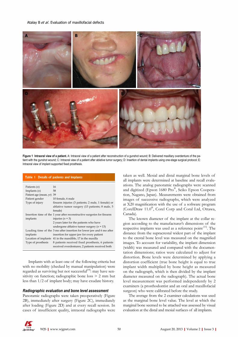
Implants with at least one of the following criteria but with no mobility (checked by manual manipulation) were regarded as surviving but not successful[10]: may have sen-sitivity on function; radiographic bone loss > 2 mm but less than 1/2 of implant body; may have exudate history.
Radiographic evaluation and bone level assessmentPanoramic radiographs were taken preoperatively (Figure 2B), immediately after surgery (Figure 2C), immediately after loading (Figure 2D) and at every recall session. In cases of insufficient quality, intraoral radiographs were
taken as well. Mesial and distal marginal bone levels of all implants were determined at baseline and recall evalu-ations. The analog panoramic radiographs were scanned and digitized (Epson 1680 Pro®, Seiko Epson Coopera-tion, Nagano, Japan). Measurements were obtained from images of successive radiographs, which were analyzed at X20 magnification with the use of a software program (CorelDraw 11.0®, Corel Corp and Coral Ltd, Ottawa, Canada).
The known diameter of the implant at the collar re-gion according to the manufacturer’s dimensions of the respective implants was used as a reference point [11]. The distance from the supracrestal widest part of the implant to the crestal bone level was measured on the magnified images. To account for variability, the implant dimension (width) was measured and compared with the documen-tation dimensions; ratios were calculated to adjust for distortion. Bone levels were determined by applying a distortion coefficient (true bone height is equal to true implant width multiplied by bone height as measured on the radiograph, which is then divided by the implant diameter measured on the radiograph). The actual bone level measurement was performed independently by 2 examiners (a prosthodontist and an oral and maxillofacial surgeon) who were calibrated before the study.
The average from the 2 examiner calculations was used as the marginal bone level value. The level at which the marginal bone seemed to be attached was assessed by visual evaluation at the distal and mesial surfaces of all implants.
50 August 20, 2013|Volume 2|Issue 3|WJS|www.wjgnet.com
Atalay B et al . Evaluation of maxillofacial defects
Figure 1 Intraoral view of a patient. A: Intraoral view of a patient after reconstruction of a gunshot wound; B: Delivered maxillary overdenture of the pa-tient with the gunshot wound; C: Intraoral view of a patient after ablative tumor surgery; D: Insertion of dental implants using one-stage surgical protocol; E: Intraoral view of implant supported fixed prosthesis.
A B C
ED
Table 1 Details of patients and implants
Patients (n) 16Implants (n) 58Patient age (mean, yr) 39Patient gender 10 female, 6 maleType of injury firearm injuries (3 patients; 2 male, 1 female) or
ablative tumor surgery (13 patients; 8 male, 5 female)
Insertion time of the implants
1 year after reconstructive surgeries for firearm injuries (n = 3)2 years later for the patients who have undergone ablative tumor surgery (n = 13)
Loading time of the implants
3 mo after insertion for lower jaw and 6 mo after insertion for upper jaw for every patient
Location of implants 41 in the mandible, 17 in the maxillaType of prosthesis 8 patients received fixed prosthesis, 6 patients
received overdentures, 2 patients received both

Patient satisfaction and oral health related quality of life outcomesSubjects were asked to grade their oral health satisfaction after treatment on a 0-100 mm visual analog scale (VAS) for 4 separate factors: general comfort, speech, esthetics and chewing (Figure 3). The scales were anchored by the extremes of potential responses (e.g., completely satisfied-completely dissatisfied: the higher the score, the more satisfied the subject).
For the determination of quality of life of the pa-tients, all subjects were asked to complete the Turkish
version of the short-form Oral Health Impact Profile (OHIP-14), which has previously been determined to be valid and reliable[12]. Subjects rated each of the 14 items on a 5-point Likert scale from 0 = “never” to 4 = “very of-ten”. Items were added up to yield the total score. Achiev-able OHIP-14 score ranged from 0-56, with lower scores representing higher oral health-related quality of life[13].
RESULTSImplant success, survival and failuresFive implants (3 in the mandible, 2 in the maxilla) in five patients were lost, while the other 53 survived, which brings an overall survival rate of 91.37% on the implant basis and 68.75% on a patient basis. Out of the 53 surviving implants, 48 were regarded as successful according to the criteria proposed by Misch et al[10] and thus the success rate was calculated as 82.75%. All the failed implants were lost before abutment connection and therefore regarded as early failures [14]. For all failed implants, new implants were placed after a 2 mo period and the planning was maintained.
Peri-implant complications and marginal bone loss The mean marginal bone loss (MBL) was 1.4 mm on the mesial side and 1.6 mm on the distal side of the implants. 5 of the implants showed MBL > 2 mm (mean MBL = 2.3 mm) but less than 1/2 of implant bodies and were there-fore regarded as not successful but surviving implants.
The MBL on the distal and mesial aspects of the implants up to 48 mo following loading did not exceed 2
51 August 20, 2013|Volume 2|Issue 3|WJS|www.wjgnet.com
Atalay B et al . Evaluation of maxillofacial defects
A
C D
B
Figure 2 Panoramic radiograph. A: Panoramic radiograph taken after implant surgery; B: Panoramic radiograph taken before implant surgery; C: Panoramic radio-graph taken after implant surgery; D: Panoramic radiograph taken after loading.
General comfort
Satisfied Not satisfied
Speech
Satisfied
Esthetics
Chewing
Satisfied Not satisfied
Satisfied Not satisfied
Not satisfied
Figure 3 The visual analog scale form for general comfort, speech, es-thetics and chewing.
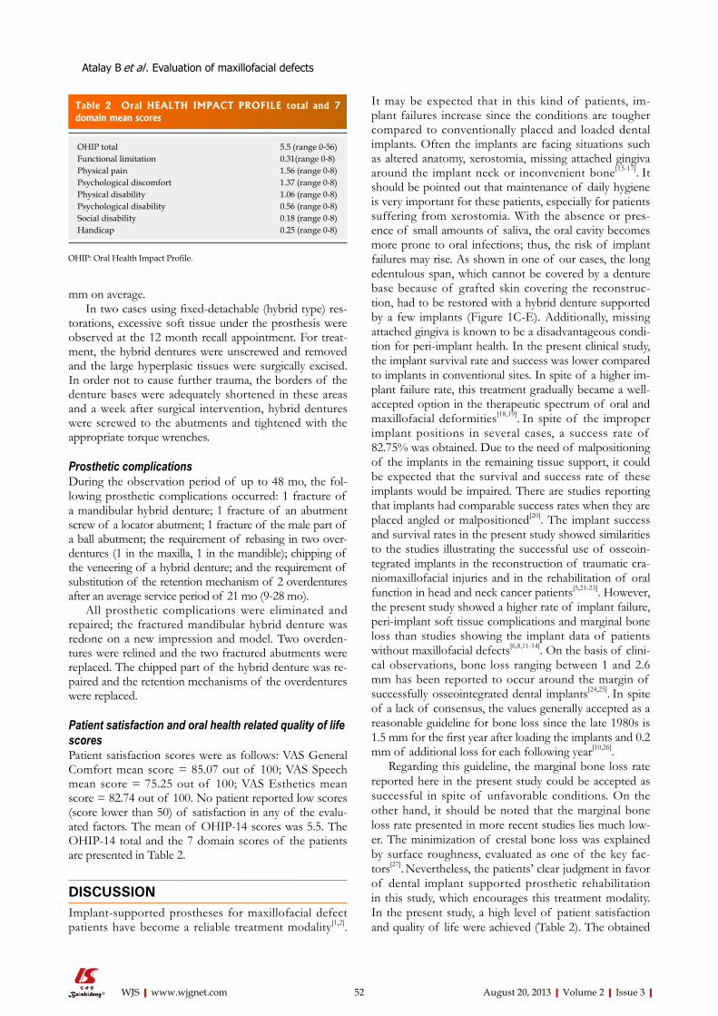
mm on average. In two cases using fixed-detachable (hybrid type) res-
torations, excessive soft tissue under the prosthesis were observed at the 12 month recall appointment. For treat-ment, the hybrid dentures were unscrewed and removed and the large hyperplasic tissues were surgically excised. In order not to cause further trauma, the borders of the denture bases were adequately shortened in these areas and a week after surgical intervention, hybrid dentures were screwed to the abutments and tightened with the appropriate torque wrenches.
Prosthetic complications During the observation period of up to 48 mo, the fol-lowing prosthetic complications occurred: 1 fracture of a mandibular hybrid denture; 1 fracture of an abutment screw of a locator abutment; 1 fracture of the male part of a ball abutment; the requirement of rebasing in two over-dentures (1 in the maxilla, 1 in the mandible); chipping of the veneering of a hybrid denture; and the requirement of substitution of the retention mechanism of 2 overdentures after an average service period of 21 mo (9-28 mo).
All prosthetic complications were eliminated and repaired; the fractured mandibular hybrid denture was redone on a new impression and model. Two overden-tures were relined and the two fractured abutments were replaced. The chipped part of the hybrid denture was re-paired and the retention mechanisms of the overdentures were replaced.
Patient satisfaction and oral health related quality of life scoresPatient satisfaction scores were as follows: VAS General Comfort mean score = 85.07 out of 100; VAS Speech mean score = 75.25 out of 100; VAS Esthetics mean score = 82.74 out of 100. No patient reported low scores (score lower than 50) of satisfaction in any of the evalu-ated factors. The mean of OHIP-14 scores was 5.5. The OHIP-14 total and the 7 domain scores of the patients are presented in Table 2.
DISCUSSIONImplant-supported prostheses for maxillofacial defect patients have become a reliable treatment modality[1,2].
It may be expected that in this kind of patients, im-plant failures increase since the conditions are tougher compared to conventionally placed and loaded dental implants. Often the implants are facing situations such as altered anatomy, xerostomia, missing attached gingiva around the implant neck or inconvenient bone[15-17]. It should be pointed out that maintenance of daily hygiene is very important for these patients, especially for patients suffering from xerostomia. With the absence or pres-ence of small amounts of saliva, the oral cavity becomes more prone to oral infections; thus, the risk of implant failures may rise. As shown in one of our cases, the long edentulous span, which cannot be covered by a denture base because of grafted skin covering the reconstruc-tion, had to be restored with a hybrid denture supported by a few implants (Figure 1C-E). Additionally, missing attached gingiva is known to be a disadvantageous condi-tion for peri-implant health. In the present clinical study, the implant survival rate and success was lower compared to implants in conventional sites. In spite of a higher im-plant failure rate, this treatment gradually became a well-accepted option in the therapeutic spectrum of oral and maxillofacial deformities[18,19]. In spite of the improper implant positions in several cases, a success rate of 82.75% was obtained. Due to the need of malpositioning of the implants in the remaining tissue support, it could be expected that the survival and success rate of these implants would be impaired. There are studies reporting that implants had comparable success rates when they are placed angled or malpositioned[20]. The implant success and survival rates in the present study showed similarities to the studies illustrating the successful use of osseoin-tegrated implants in the reconstruction of traumatic cra-niomaxillofacial injuries and in the rehabilitation of oral function in head and neck cancer patients[5,21-23]. However, the present study showed a higher rate of implant failure, peri-implant soft tissue complications and marginal bone loss than studies showing the implant data of patients without maxillofacial defects[6,8,11-14]. On the basis of clini-cal observations, bone loss ranging between 1 and 2.6 mm has been reported to occur around the margin of successfully osseointegrated dental implants[24,25]. In spite of a lack of consensus, the values generally accepted as a reasonable guideline for bone loss since the late 1980s is 1.5 mm for the first year after loading the implants and 0.2 mm of additional loss for each following year[10,26].
Regarding this guideline, the marginal bone loss rate reported here in the present study could be accepted as successful in spite of unfavorable conditions. On the other hand, it should be noted that the marginal bone loss rate presented in more recent studies lies much low-er. The minimization of crestal bone loss was explained by surface roughness, evaluated as one of the key fac-tors[27]. Nevertheless, the patients’ clear judgment in favor of dental implant supported prosthetic rehabilitation in this study, which encourages this treatment modality. In the present study, a high level of patient satisfaction and quality of life were achieved (Table 2). The obtained
52 August 20, 2013|Volume 2|Issue 3|WJS|www.wjgnet.com
Atalay B et al . Evaluation of maxillofacial defects
Table 2 Oral HEALTH IMPACT PROFILE total and 7 domain mean scores
OHIP total 5.5 (range 0-56)Functional limitation 0.31(range 0-8)Physical pain 1.56 (range 0-8)Psychological discomfort 1.37 (range 0-8)Physical disability 1.06 (range 0-8)Psychological disability 0.56 (range 0-8)Social disability 0.18 (range 0-8)Handicap 0.25 (range 0-8)
OHIP: Oral Health Impact Profile.

VAS and quality of life scores in this pilot study show similarities to the study of Schoen et al[21] which investi-gated the patient satisfaction and quality of life outcome of implant treatment in head and neck cancer patients[1].
Additionally, our results are comparable to other studies concerning treatment with dental implants[6,8,28-30].
In the present study, the patients were not asked to complete the VAS and OHIP-14 questionnaires before the treatment; thus, it was not possible to compare the pre and post treatment scores, which may be regarded as a limitation. All the patients were unable to function with the pre-treatment oral conditions; therefore, the authors did not consider it necessary and moral to constrain the patients in completing the questionnaires before treat-ment. Additionally, in the opinion of the authors, the OHIP-14 questionnaire is very hard to comprehend and could cause misleading results in these patients. The form could be modified for patients with maxillofacial defects just like the previously made modification for edentulous patients as OHIP-EDENT[31].
Early management of injured patients must focus on the basics of resuscitation. The secondary target in the treatment of these cases, however, should focus on tissue preservation, abstaining from unnecessary tissue resec-tion, because the placement of dental implants can be problematic from time to time. The attention paid at the early stage of intervention can have an important impact on the quality of life of patients.
As a general approach at the dental school, the im-plant treatment has to be postponed for a certain period if a major resection and reconstruction has been per-formed. If radiotherapy and/or chemotherapy is ad-ministered, the patient has to wait at least 2 years for the implantation, as suggested previously[5]. The prosthetic complications recorded in the present study were slightly over the average of prosthetic patients treated in the re-lated university clinic. Although complications, such as requirement of rebasing, chipping of veneering material and substitution of retention mechanism, are routinely encountered and well documented in the literature[32], the fracture of a hybrid denture, a locator abutment or of the male part of a ball attachment is not common. The mis-alignment or strategically disadvantageous numbers and positions of implants may be a factor that explains higher rates of complications in the present patient group.
Oral rehabilitation becomes even more complicated with the presence of microstomia[33], which can be en-countered in this kind of patients. Microstomic patients experience considerable limitation in jaw opening and overall jaw mobility. This limitation in the oral opening makes gaining access to the oral cavity difficult, depend-ing on the severity of microstomia. Therefore, traditional approaches for dental restoration should be modified to accommodate microstomia. Various treatment approach-es have been proposed for microstomic patients, with or without endosseous implants. Reduced mouth opening may prevent instruments from safely entering the mouth for insertion of the implants. This is a critical factor in determining whether implant treatment can be provided
and in deciding the number of inserts needed and the best places for insertion[34].
In the present study, 3 patients had a limited intraoral access, requiring modification of the approach. Also, there might be problems with the precision of dental laboratory work because of the inaccurate impressions which were hardly made with the modification meth-ods[33]. Therefore, the precision of fit of the dental frame-works were very limited (Figure 2D). The strains due to the misfit of the denture can be a reason for the failures and prosthetic complications. In cases of firearm injuries, the severity of the defect resulting from facial firearm injuries varies according to the caliber of the weapon used, the distance from which the patient is shot and the part of the body involved[35]. Close range, high velocity firearm wounds can result in devastating functional and esthetic consequences. Maxillofacial traumas are mostly encountered in males (78%) and at a higher rate between the ages of 20-39 years. There are many reasons for max-illofacial trauma, such as fighting (48.2%), falling (26.2%), car accidents (4.2%) and firearm injury (1.2%)[36,37]. The epidemiology of facial fractures varies in type, severity and cause, depending on the population studied[38]. The differences between populations in the causes of maxil-lofacial fractures may be the result of risk factors and cultural differences between countries but are more likely to be influenced by the injury severity[39].
In situations with insufficient bone volume, invasive sur-gical procedures such as maxillary sinus floor elevation or the zygomatic implant placement[19], procedures mainly ac-complished by maxillofacial surgeons, can be an alternative. However, individuals of the related patient group could ap-peal against additional complex surgical interventions after the long and griping procedures they have endured.
Meanwhile, it is a well known fact that the first year is critical for implant failure and for the largest portion of marginal bone loss around dental implants[34]. The results of an investigation showed that practically all implant loss-es occurred during the first 2 years, whereupon a steady state seemed to follow for up to 5 years after loading[40].
Despite disadvantageous loading conditions and poor bone quality and quantity, all the presented cases showed a stable situation around the implants after a period of 12-48 mo of loading time. Although further follow up and larger case numbers will give more information about the success of dental implants as a treatment mo-dality in maxillofacial defect patients, the actual results are encouraging and can be recommended for similar cases. Even although the success and survival rate is slightly lower than conventionally loaded implants due to tougher conditions, dental implants seem to be a valuable aid in the maintenance of comfortable rehabilitation of maxil-lofacial defect patients.
COMMENTSBackgroundIn patients with maxillofacial defects, implant failures may increase, since the conditions are harder compared to conventionally placed dental implants. Often
53 August 20, 2013|Volume 2|Issue 3|WJS|www.wjgnet.com
Atalay B et al . Evaluation of maxillofacial defects
COMMENTS
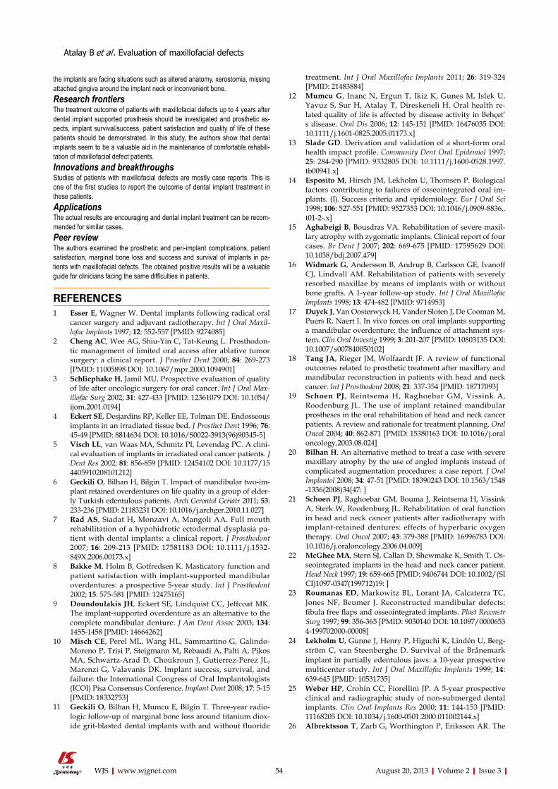
54 August 20, 2013|Volume 2|Issue 3|WJS|www.wjgnet.com
the implants are facing situations such as altered anatomy, xerostomia, missing attached gingiva around the implant neck or inconvenient bone.Research frontiersThe treatment outcome of patients with maxillofacial defects up to 4 years after dental implant supported prosthesis should be investigated and prosthetic as-pects, implant survival/success, patient satisfaction and quality of life of these patients should be demonstrated. In this study, the authors show that dental implants seem to be a valuable aid in the maintenance of comfortable rehabili-tation of maxillofacial defect patients.Innovations and breakthroughsStudies of patients with maxillofacial defects are mostly case reports. This is one of the first studies to report the outcome of dental implant treatment in these patients.ApplicationsThe actual results are encouraging and dental implant treatment can be recom-mended for similar cases.Peer reviewThe authors examined the prosthetic and peri-implant complications, patient satisfaction, marginal bone loss and success and survival of implants in pa-tients with maxillofacial defects. The obtained positive results will be a valuable guide for clinicians facing the same difficulties in patients.
REFERENCES1 Esser E, Wagner W. Dental implants following radical oral
cancer surgery and adjuvant radiotherapy. Int J Oral Maxil-lofac Implants 1997; 12: 552-557 [PMID: 9274085]
2 Cheng AC, Wee AG, Shiu-Yin C, Tat-Keung L. Prosthodon-tic management of limited oral access after ablative tumor surgery: a clinical report. J Prosthet Dent 2000; 84: 269-273 [PMID: 11005898 DOI: 10.1067/mpr.2000.1094901]
3 Schliephake H, Jamil MU. Prospective evaluation of quality of life after oncologic surgery for oral cancer. Int J Oral Max-illofac Surg 2002; 31: 427-433 [PMID: 12361079 DOI: 10.1054/ijom.2001.0194]
4 Eckert SE, Desjardins RP, Keller EE, Tolman DE. Endosseous implants in an irradiated tissue bed. J Prosthet Dent 1996; 76: 45-49 [PMID: 8814634 DOI: 10.1016/S0022-3913(96)90345-5]
5 Visch LL, van Waas MA, Schmitz PI, Levendag PC. A clini-cal evaluation of implants in irradiated oral cancer patients. J Dent Res 2002; 81: 856-859 [PMID: 12454102 DOI: 10.1177/154405910208101212]
6 Geckili O, Bilhan H, Bilgin T. Impact of mandibular two-im-plant retained overdentures on life quality in a group of elder-ly Turkish edentulous patients. Arch Gerontol Geriatr 2011; 53: 233-236 [PMID: 21183231 DOI: 10.1016/j.archger.2010.11.027]
7 Rad AS, Siadat H, Monzavi A, Mangoli AA. Full mouth rehabilitation of a hypohidrotic ectodermal dysplasia pa-tient with dental implants: a clinical report. J Prosthodont 2007; 16: 209-213 [PMID: 17581183 DOI: 10.1111/j.1532-849X.2006.00173.x]
8 Bakke M, Holm B, Gotfredsen K. Masticatory function and patient satisfaction with implant-supported mandibular overdentures: a prospective 5-year study. Int J Prosthodont 2002; 15: 575-581 [PMID: 12475165]
9 Doundoulakis JH, Eckert SE, Lindquist CC, Jeffcoat MK. The implant-supported overdenture as an alternative to the complete mandibular denture. J Am Dent Assoc 2003; 134: 1455-1458 [PMID: 14664262]
10 Misch CE, Perel ML, Wang HL, Sammartino G, Galindo-Moreno P, Trisi P, Steigmann M, Rebaudi A, Palti A, Pikos MA, Schwartz-Arad D, Choukroun J, Gutierrez-Perez JL, Marenzi G, Valavanis DK. Implant success, survival, and failure: the International Congress of Oral Implantologists (ICOI) Pisa Consensus Conference. Implant Dent 2008; 17: 5-15 [PMID: 18332753]
11 Geckili O, Bilhan H, Mumcu E, Bilgin T. Three-year radio-logic follow-up of marginal bone loss around titanium diox-ide grit-blasted dental implants with and without fluoride
treatment. Int J Oral Maxillofac Implants 2011; 26: 319-324 [PMID: 21483884]
12 Mumcu G, Inanc N, Ergun T, Ikiz K, Gunes M, Islek U, Yavuz S, Sur H, Atalay T, Direskeneli H. Oral health re-lated quality of life is affected by disease activity in Behçet’s disease. Oral Dis 2006; 12: 145-151 [PMID: 16476035 DOI: 10.1111/j.1601-0825.2005.01173.x]
13 Slade GD. Derivation and validation of a short-form oral health impact profile. Community Dent Oral Epidemiol 1997; 25: 284-290 [PMID: 9332805 DOI: 10.1111/j.1600-0528.1997.tb00941.x]
14 Esposito M, Hirsch JM, Lekholm U, Thomsen P. Biological factors contributing to failures of osseointegrated oral im-plants. (I). Success criteria and epidemiology. Eur J Oral Sci 1998; 106: 527-551 [PMID: 9527353 DOI: 10.1046/j.0909-8836..t01-2-.x]
15 Aghabeigi B, Bousdras VA. Rehabilitation of severe maxil-lary atrophy with zygomatic implants. Clinical report of four cases. Br Dent J 2007; 202: 669-675 [PMID: 17595629 DOI: 10.1038/bdj.2007.479]
16 Widmark G, Andersson B, Andrup B, Carlsson GE, Ivanoff CJ, Lindvall AM. Rehabilitation of patients with severely resorbed maxillae by means of implants with or without bone grafts. A 1-year follow-up study. Int J Oral Maxillofac Implants 1998; 13: 474-482 [PMID: 9714953]
17 Duyck J, Van Oosterwyck H, Vander Sloten J, De Cooman M, Puers R, Naert I. In vivo forces on oral implants supporting a mandibular overdenture: the influence of attachment sys-tem. Clin Oral Investig 1999; 3: 201-207 [PMID: 10803135 DOI: 10.1007/s007840050102]
18 Tang JA, Rieger JM, Wolfaardt JF. A review of functional outcomes related to prosthetic treatment after maxillary and mandibular reconstruction in patients with head and neck cancer. Int J Prosthodont 2008; 21: 337-354 [PMID: 18717093]
19 Schoen PJ, Reintsema H, Raghoebar GM, Vissink A, Roodenburg JL. The use of implant retained mandibular prostheses in the oral rehabilitation of head and neck cancer patients. A review and rationale for treatment planning. Oral Oncol 2004; 40: 862-871 [PMID: 15380163 DOI: 10.1016/j.oraloncology.2003.08.024]
20 Bilhan H. An alternative method to treat a case with severe maxillary atrophy by the use of angled implants instead of complicated augmentation procedures: a case report. J Oral Implantol 2008; 34: 47-51 [PMID: 18390243 DOI: 10.1563/1548-1336(2008)34[47: ]
21 Schoen PJ, Raghoebar GM, Bouma J, Reintsema H, Vissink A, Sterk W, Roodenburg JL. Rehabilitation of oral function in head and neck cancer patients after radiotherapy with implant-retained dentures: effects of hyperbaric oxygen therapy. Oral Oncol 2007; 43: 379-388 [PMID: 16996783 DOI: 10.1016/j.oraloncology.2006.04.009]
22 McGhee MA, Stern SJ, Callan D, Shewmake K, Smith T. Os-seointegrated implants in the head and neck cancer patient. Head Neck 1997; 19: 659-665 [PMID: 9406744 DOI: 10.1002/(SICI)1097-0347(199712)19: ]
23 Roumanas ED, Markowitz BL, Lorant JA, Calcaterra TC, Jones NF, Beumer J. Reconstructed mandibular defects: fibula free flaps and osseointegrated implants. Plast Reconstr Surg 1997; 99: 356-365 [PMID: 9030140 DOI: 10.1097/00006534-199702000-00008]
24 Lekholm U, Gunne J, Henry P, Higuchi K, Lindén U, Berg-ström C, van Steenberghe D. Survival of the Brånemark implant in partially edentulous jaws: a 10-year prospective multicenter study. Int J Oral Maxillofac Implants 1999; 14: 639-645 [PMID: 10531735]
25 Weber HP, Crohin CC, Fiorellini JP. A 5-year prospective clinical and radiographic study of non-submerged dental implants. Clin Oral Implants Res 2000; 11: 144-153 [PMID: 11168205 DOI: 10.1034/j.1600-0501.2000.011002144.x]
26 Albrektsson T, Zarb G, Worthington P, Eriksson AR. The
Atalay B et al . Evaluation of maxillofacial defects
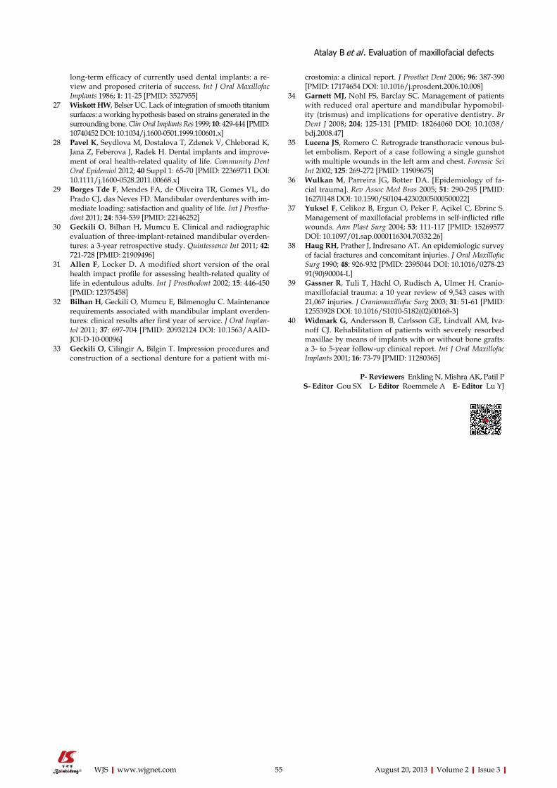
55 August 20, 2013|Volume 2|Issue 3|WJS|www.wjgnet.com
long-term efficacy of currently used dental implants: a re-view and proposed criteria of success. Int J Oral Maxillofac Implants 1986; 1: 11-25 [PMID: 3527955]
27 Wiskott HW, Belser UC. Lack of integration of smooth titanium surfaces: a working hypothesis based on strains generated in the surrounding bone. Clin Oral Implants Res 1999; 10: 429-444 [PMID: 10740452 DOI: 10.1034/j.1600-0501.1999.100601.x]
28 Pavel K, Seydlova M, Dostalova T, Zdenek V, Chleborad K, Jana Z, Feberova J, Radek H. Dental implants and improve-ment of oral health-related quality of life. Community Dent Oral Epidemiol 2012; 40 Suppl 1: 65-70 [PMID: 22369711 DOI: 10.1111/j.1600-0528.2011.00668.x]
29 Borges Tde F, Mendes FA, de Oliveira TR, Gomes VL, do Prado CJ, das Neves FD. Mandibular overdentures with im-mediate loading: satisfaction and quality of life. Int J Prostho-dont 2011; 24: 534-539 [PMID: 22146252]
30 Geckili O, Bilhan H, Mumcu E. Clinical and radiographic evaluation of three-implant-retained mandibular overden-tures: a 3-year retrospective study. Quintessence Int 2011; 42: 721-728 [PMID: 21909496]
31 Allen F, Locker D. A modified short version of the oral health impact profile for assessing health-related quality of life in edentulous adults. Int J Prosthodont 2002; 15: 446-450 [PMID: 12375458]
32 Bilhan H, Geckili O, Mumcu E, Bilmenoglu C. Maintenance requirements associated with mandibular implant overden-tures: clinical results after first year of service. J Oral Implan-tol 2011; 37: 697-704 [PMID: 20932124 DOI: 10.1563/AAID-JOI-D-10-00096]
33 Geckili O, Cilingir A, Bilgin T. Impression procedures and construction of a sectional denture for a patient with mi-
crostomia: a clinical report. J Prosthet Dent 2006; 96: 387-390 [PMID: 17174654 DOI: 10.1016/j.prosdent.2006.10.008]
34 Garnett MJ, Nohl FS, Barclay SC. Management of patients with reduced oral aperture and mandibular hypomobil-ity (trismus) and implications for operative dentistry. Br Dent J 2008; 204: 125-131 [PMID: 18264060 DOI: 10.1038/bdj.2008.47]
35 Lucena JS, Romero C. Retrograde transthoracic venous bul-let embolism. Report of a case following a single gunshot with multiple wounds in the left arm and chest. Forensic Sci Int 2002; 125: 269-272 [PMID: 11909675]
36 Wulkan M, Parreira JG, Botter DA. [Epidemiology of fa-cial trauma]. Rev Assoc Med Bras 2005; 51: 290-295 [PMID: 16270148 DOI: 10.1590/S0104-42302005000500022]
37 Yuksel F, Celikoz B, Ergun O, Peker F, Açikel C, Ebrinc S. Management of maxillofacial problems in self-inflicted rifle wounds. Ann Plast Surg 2004; 53: 111-117 [PMID: 15269577 DOI: 10.1097/01.sap.0000116304.70332.26]
38 Haug RH, Prather J, Indresano AT. An epidemiologic survey of facial fractures and concomitant injuries. J Oral Maxillofac Surg 1990; 48: 926-932 [PMID: 2395044 DOI: 10.1016/0278-2391(90)90004-L]
39 Gassner R, Tuli T, Hächl O, Rudisch A, Ulmer H. Cranio-maxillofacial trauma: a 10 year review of 9,543 cases with 21,067 injuries. J Craniomaxillofac Surg 2003; 31: 51-61 [PMID: 12553928 DOI: 10.1016/S1010-5182(02)00168-3]
40 Widmark G, Andersson B, Carlsson GE, Lindvall AM, Iva-noff CJ. Rehabilitation of patients with severely resorbed maxillae by means of implants with or without bone grafts: a 3- to 5-year follow-up clinical report. Int J Oral Maxillofac Implants 2001; 16: 73-79 [PMID: 11280365]
P- Reviewers Enkling N, Mishra AK, Patil P S- Editor Gou SX L- Editor Roemmele A E- Editor Lu YJ
Atalay B et al . Evaluation of maxillofacial defects

World Journal of StomatologyW J S
Online Submissions: http://www.wjgnet.com/esps/[email protected]:10.5321/wjs.v2.i3.56
World J Stomatol 2013 August 20; 2(3): 56-61ISSN 2218-6263 (online)
© 2013 Baishideng. All rights reserved.
Evaluation of factors affecting the success rate of orthodontic mini-implants by survival analysis
Un-Bong Baik, Mohamed Bayome, Kwang-Heung Han, Jae Hyun Park, Min-Ho Jung, Yoon-Ah Kook
Un-Bong Baik, Smile with Orthodontic Clinic, Seoul 142-100, South KoreaMohamed Bayome, Department of Orthodontics, The Catholic University of Korea, Seoul 137-701, South KoreaKwang-Heung Han, Seoul H Dental Clinic, Seoul 150-037, South KoreaJae Hyun Park, Postgraduate Orthodontic Program, Arizona School of Dentistry and Oral Health, A.T. Still University, Mesa, AZ and Graduate School of Dentistry, Kyung Hee University, Seoul 130-701, South KoreaMin-Ho Jung, Private practice and Department of Orthodontics, School of Dentistry, Seoul National University, Seoul 151-742, South KoreaYoon-Ah Kook, Department of Orthodontics, Seoul St. Mary’s Hos-pital, The Catholic University of Korea, Seoul 137-701, South KoreaAuthor contributions: All the authors contributed equally to this manuscript.Correspondence to: Dr. Yoon-Ah Kook, Department of Ortho-dontics, Seoul St. Mary’s Hospital, The Catholic University of Korea, 505 Banpo-Dong, Seocho-Gu, Seoul 137-701, South Korea. [email protected]: +82-2-22581776 Fax: +82-2-5372374Received: December 17, 2012 Revised: February 17, 2013 Accepted: April 10, 2013Published online: August 20, 2013
AbstractAIM: To investigate the success rate of mini-implants and its characteristics and risk factors by survival analyses.
METHODS: Three hundred and ninety-four mini-implants of the same type were placed by a single clinician. Age, gender, treatment duration, time of failure, side and jaw of implantation and the soft tis-sue at placement site were recorded. Odds ratio, sur-vival curves, and Cox proportional hazard model were applied to evaluate the factors influencing the mini-implants’ success rate.
RESULTS: The cumulative success rate was 88.1%.
The maxilla had a significantly higher success rate than that of the mandible (91.7% vs 83.7%, respectively, P = 0.019). Placement of mini-implants in the attached gingiva (AG) showed a higher success rate than that of the mucogingival junction (MGJ) and mucous mem-brane (MM) (AG, 94.3%; MGJ, 85.8%; MM, 79.4%; P < 0.001). Significant association was found between the jaw and the gingival tissue type (P < 0.001). There were no significant differences between maxilla and mandible when compared within each placement site.
CONCLUSION: The gingival tissue type had the most significant effect on the success rate of the mini-im-plant with higher success rate in the attached gingiva.
© 2013 Baishideng. All rights reserved.
Key words: Mini-implant; Success rate; Survival analy-sis; Gingival tissue; Treatment planning
Core tip: Anchorage reinforcement is a critical factor for successful orthodontic treatment outcome. Mini-implants are applied to achieve various dental movements such as anterior retraction, molar protraction and distaliza-tion, intrusion, extrusion, and correction of midline and occlusal canting. The gingival tissue type had the most significant effect on the success rate of the mini-implant with higher success rate in the attached gingiva.
Baik UB, Bayome M, Han KH, Park JH, Jung MH, Kook YA. Evaluation of factors affecting the success rate of orthodontic mini-implants by survival analysis. World J Stomatol 2013; 2(3): 56-61 Available from: URL: http://www.wjgnet.com/2218-6263/full/v2/i3/56.htm DOI: http://dx.doi.org/10.5321/wjs.v2.i3.56
INTRODUCTIONAnchorage reinforcement is a critical factor for success-ful orthodontic treatment outcome. Mini-implants are
BRIEF ARTICLE
56 August 20, 2013|Volume 2|Issue 3|WJS|www.wjgnet.com

applied to achieve various dental movements such as an-terior retraction, molar protraction and distalization, in-trusion, extrusion, and correction of midline and occlusal canting[1-4].
The factors affecting the success rate of mini-im-plants have been investigated extensively but not all of them are agreed upon regarding their significance by the investigators[5-10]. Type of mini-implant was suggested as a contributor to the success rate[11,12]. Age and gender of patients, the jaw and side receiving the mini-implant, and the type of gingival tissues were not significantly associ-ated with the success rate[8,11,13,14].
However, Lee et al[15] reported a significant effect of age, and Manni et al[12] demonstrated the gender as a sig-nificant factor. In addition, peri-implant soft tissue char-acteristics may be a contributing factor[16]. Moon et al[13] also reported significant differences between placement sites between different teeth. Also, vertical skeletal pat-tern was reported to influence the success rate[10].
Recently, three or more types of mini-implants were placed by more than one operator[10,11,14,16]. However, Lee et al[15] inserted a single type of mini-implants and re-ported that there are no significant differences in the suc-cess rate according to clinicians. In Park et al[17] the mini-implants were placed by one clinician, but the sample size was relatively small for both reports.
However, a well-controlled study with larger sample size of a single type of mini-implants placed by one experienced clinician has not been conducted. This can minimize the effect of the operator- and mini-implant-related factors on the evaluation of success rate.
Therefore, the purpose of this study was to investi-gate the success rate of mini-implants and its characteris-tics and risk factors using the same type of mini-implants placed by single clinician by survival analyses.
MATERIALS AND METHODSA hundred and sixty four patients (47 male, 117 female; mean age 24.0 ± 6.8 years) treated with fixed appliance from July, 2009 to March, 2010 in a private orthodontic clinic were included in this retrospective study. Those who had special medical history such as osteoporosis, thyroid problem, diabetes, and hypertension were excluded.
A total number of 394 mini-implants were placed for anchorage reinforcement by one right-handed experi-enced clinician using a single placement technique (30° to the surface of soft tissue and about 20 N•cm torque on the self drilling miniscrew) and were loaded 3 wk after placement with a similar amount of force. Only one type of mini-implants was used to exclude the effect of the screw material and design (6.0 mm in length and 1.5 mm in diameter, Biomaterials Korea, Seoul, Korea).
The records were examined to retrieve the following data: age, gender, date of mini-implant placement, date of failure (if occurred), date of removal at the end of treatment, location (upper, lower, right, left) and gingival tissue type at placement site [attached gingiva (AG), mu-
cogingival junction (MGJ), mucous membrane (MM)]. The success of the mini-implant was defined as being functionally stable till the end of the treatment without signs of inflammation. Meanwhile, failure was recorded in case of removal of the mini-implant due to looseness.
Statistical analysisSAS 9.2 (SAS Institute Inc. Cary, NC, United States) was used for the statistical analysis. The Fisher exact test significance and odds ratio statistics were calculated. A nonparametric life table method was used to eas-ily visualize the hazard function over time. Association between significant variables was assessed by χ 2 test. Kaplan-Meier survival curves were generated, and the Gehan generalized Wilcoxon test was used to identify the variables associated with implant failure. Prognostic variables associated with implant failure were identified with the Cox proportional hazard model which is a sur-vival model that relate the time passed before an event happens to one or more covariates (in our study: age, gender, jaw, side, and gingival tissue) that might be asso-ciated with that quantity of time. The level of statistical significance was set at 5%.
RESULTSThere was no significant difference in the success rates between implantation sides, gender, and age. However, there were significant differences between upper and low-er implantation (91.7% vs 83.7%, respectively, P = 0.019) and according to the gingival tissue type at the placement site (AG, 94.3%; MGJ, 85.8%; MM, 79.4%; P < 0.001) (Table 1).
The hazard function of mini-implant survival time was regarded as the instantaneous failure rate[18]. As the latest failure event was at 27 wk, the function showed that the risk of failure was highest immediately after place-ment and then decreased to zero till the end of the treat-ment. The linear fit of the hazard function was R2 = 0.62 with a negative slope over time (Figure 1).
The Kaplan-Meier survival curve according to jaw and gingival tissue type (Figure 2) demonstrated high success rates for all subgroups. The Gehan generalized Wilcoxon test revealed that the implants placed in the maxilla had a higher success rate than those placed in the mandible (P = 0.014). Also, those placed in the attached gingiva had a significantly higher survival rate than other subgroups (P < 0.001).
χ 2 test verified a significant association between the jaw and the gingival tissue type (P < 0.001) (Table 2). By Fisher’s exact test and odds ratio analysis, there were no significant differences between maxilla and mandible when compared according to gingival tissue type, inde-pendently (Table 3). The Cox proportional hazard model also showed that the gender and gingival tissue type are significant factors for mini-implant survival (Table 4). The estimated probability of failure was lower for fe-males (P < 0.001) and the attached gingiva (P = 0.019).
57 August 20, 2013|Volume 2|Issue 3|WJS|www.wjgnet.com
Baik UB et al . Factors affecting the success rate of mini-implants

0 20 40 60 80 100 120 140t /wk
1.0
0.8
0.6
0.4
0.2
0.0
The
cum
ulat
ive
surv
ival
rat
e of
impl
ant
0 20 40 60 80 100 120 140t /wk
1.0
0.8
0.6
0.4
0.2
0.0
The
cum
ulat
ive
surv
ival
rat
e of
impl
ant
ATMGJMMAT-censoredMGJ-censoredMM-censored
Figure 2 Kaplan-Meier survival analysis of mini-implant by jaw and gingi-val tissue. A: The survival rate of mini-implants placed in the maxilla was sig-nificantly higher than that of those placed in the mandible; B: The survival rate of mini-implants placed in the attached gingival (AG) was significantly higher than that of those placed in the mucogingival junction (MGJ) and mucous mem-brane (MM). The duration of survival in the censored cases was measured from mini-implant placement to completion of treatment.
A
B
DISCUSSIONWith improvement of mini-implant materials, design and placement technique, recent studies have often reported mini-implant success rates higher than 90%[15,19]. On the other hand, since it is rare that a patient receives only one mini-implant during orthodontic treatment, the success rate faced by clinicians throughout treatment may be sub-stantially lower due to presence of multiple mini-implants in each patient.
In our study, the success rate (88.1%) was slightly lower than that in Lee et al[15] (91.5%), higher than Manni et al[12] (81%) and similar to Cheng et al[16] (89%). Moreover, several studies evaluated numerous factors affecting the success rate of mini-implants[5,14,17,20]. However, most of them assessed many heterogenic variables using a small sample size that increase type Ⅱ errors and decrease statistical power. In our study, to eliminate the factors re-lated to the clinician and the mini-implant, only one clini-cian placed 394 mini-implant of the same type following the same insertion technique.
Recently, Manni et al[12] evaluated 12 different factors affecting the stability of mini-implants. Although the mini-implants in their study were placed by the same cli-nician, they were of 3 different types. Furthermore, the evaluation of too many variables may lead to generation of higher-order interactions resulting in a complicated result interpretation[21]. Our research was limited to only five host variables to avoid such a complication.
Lee et al[15] also evaluated five variables affecting the success rate of the mini-implants. However, the anatomi-cal location and the soft tissue of the insertion site were not included in their study. They found patient’s age to be the only significant factor that affects the success rate of mini-implants. They recommended special caution when planning mini-implants for young patients. On the con-trary, our results showed that the age was not a significant factor in determining the success of mini-implants.
Several reports demonstrated a significant effect for
age on the success rate of mini-implants[15,20]. The higher risk of failure in younger patients could be attributed to their lower bone density[22,23]. However, in agreement with our results, other studies reported no significant differences among age-groups[13,14,17]. This inconsistency among results can be explained by the multifactorial na-ture of the mini-implant success rate. Moreover, it can be argued that Lee et al[15] and Chen et al[20] have overlooked the evaluation of the effect of gingival tissue type at the placement site.
In our study, the gingival tissue type at the placement site was the main factor affecting the success rate. In AG, the placement of mini-implant had a 2.7 times lower fail-ure rate than in MGJ which in turn had a 1.6 times lower failure rate than in MM (Table 1). This was in accord with previous investigations[5,12,16]. Moreover, an animal study showed a significantly higher stability of mini-implants in keratinized gingiva. Within their limited sample size (22 mini-implants), all failed cases (n = 9) were placed in the non-keratinized gingiva[24]. The lower failure rate in the AG could be explained by the non-movable keratinized
58 August 20, 2013|Volume 2|Issue 3|WJS|www.wjgnet.com
Baik UB et al . Factors affecting the success rate of mini-implants
0.08
0.06
0.04
0.02
0.00
0 10 20 30 40 50 60 70 80 90 100 110 120 130 140t /wk
The
inst
anta
neou
s fa
ilure
rat
e of
impl
ant
Figure 1 Instantaneous failure curve of mini-implant: The hazard function shows that the maximum risk is immediately after mini-implant placement and then it declines to zero by time. The linear fit of the hazard function was R2 = 0.623.
MaxillaMandibleMaxilla-censoredMandible-censored

tissue that decreases the susceptibility to irritation and infection. On the other hand, some authors reported no significant differences in the success rate according to soft tissue[14,17,25].
Several studies reported higher success rate of mini-implant placement in the maxilla than that for those placed in the mandible[12,16,20]. On the other hand, some authors reported no significant differences between the upper and lower jaws in mini-implant success rate[11,13,14,25]. In our study, the jaw, initially, was a significant factor af-fecting the success rate. However, with further analysis, a significant association (P < 0.001) was found between the
jaw and the gingival tissue type. The mini-implants placed in the mandible were mainly placed in mucous membrane or MGJ, while those placed in the maxilla were mainly in the attached gingiva. No significant differences in the success rates were found between the mini-implants placed in upper and lower jaws when compared within each gingival tissue type.
This was in agreement with Moon et al[13] who placed all the mini-implants in the attached gingiva and showed no significant difference in the success rate between max-illa and mandible. In addition, in our results, Cox propor-tional hazard model showed no significant effect of the
59 August 20, 2013|Volume 2|Issue 3|WJS|www.wjgnet.com
Baik UB et al . Factors affecting the success rate of mini-implants
Table 1 Mini-implants’ success and failure rates, and ORs statistics by host variables n (%)
Variables Success Failure Total P -value OR (95%CI)
Gender 0.231 Male 96 (84.96) 17 (15.04) 113 Female 252 (89.36) 30 (10.64) 282 0.762 (0.354, 1.274)Jaw 0.019 Maxilla 199 (91.71) 18 (8.29) 217 Mandible 149 (83.71) 29 (16.29) 178 2.152 (1.151, 4.021)R/L side 0.877 Left 117 (88.50) 23 (11.50) 200 Right 171 (87.69) 24 (12.31) 195 1.080 (0.587, 1.986)Age 0.973 < 20 106 (87.60) 15 (12.40) 121 20-30 204 (88.31) 27 (11.69) 231 0.935 (0.477, 1.834) > 30 38 (88.37) 5 (11.63) 43 0.930 (0.316, 2.732)Gingival tissue < 0.001 AG 165 (94.29) 10 (5.71) 175 0.367 (0.163, 3.204) MGJ 109 (85.83) 18 (14.17) 127 2.724 (1.212, 6.124) MM 73 (79.35) 19 (20.65) 92 4.294 (1.903, 9.689) 1.576 (0.775, 3.204)
Fisher’s exact test. R/L: Right/left; AG: Attached gingiva; MGJ: Mucogingival junction; MM: Mucous membrane.
Table 2 Distribution of mini-implants according to gingival tissue type at the placement site in maxilla and mandible n (%)
Mandible Maxilla P -value
Total Failed Total Failed
Attached gingiva 34 (19.1) 4 (11.8) 141 (65.3) 6 (4.3)Mucogingival junction 78 (43.8) 11 (14.1) 49 (22.7) 7 (14.3) < 0.001Mucous membrane 66 (37.1) 14 (21.2) 26 (12.0) 5 (19.2)
Table 3 Independent comparison of failure rate between maxilla and mandible according to gingival tissue type n (%)
Variables Success Failure Total P -value OR (95%CI)
Attached gingiva 0.105 Maxilla 135 (95.74) 6 (4.26) 141 Mandible 30 (88.24) 4 (11.76) 34 3.000 (0.797, 11.293)Mucogingival junction 1 Maxilla 42 (85.71) 7 (14.29) 49 Mandible 67 (85.90) 11 (14.10) 78 0.985 (0.354, 2.740)Mucous membrane 1 Maxilla 21 (80.77) 5 (19.23) 26 Mandible 52 (78.79) 14 (21.21) 66 1.131 (0.362, 3.535)

jaw on the failure rate (P = 0.358). Therefore, the greater failure rate of mini-implants placed in the mandible can be explained by the lake of further analysis to examine any association between the jaw and other factors, such as inflammation, root proximity, and soft tissue mobility.
Similarly, gender was described as a significant factor in several studies. Moon et al[13] reported a higher success rate in male patients while in Antoszewska et al[5] study fe-male subjects had a higher rate. Nevertheless, our results showed no significant difference in the rate according to gender. This was in accordance with several reports[14,17,20]. Interestingly, the Cox proportional hazard model in our study showed that gender was a significant factor. Therefore, future studies might be required to evaluate the influence of gender on the success rate with a larger sample size from both groups with uniform inclusion cri-teria that eliminate other confounding factors.
Time of loading has been evaluated in several reports but no consensus was reached. Trisi et al[26] demonstrated that immediate loading might undermine the stability of dental implants and increase the number of failures. On the contrary, other studies showed a positive influence for the immediate loading[12,27]. However, Miyawaki et al[11] found no correlation between the time of loading and success rate. In addition, Cheng et al[16] and Costa et al[28] achieved success rates of 89% and 87.5% with delayed and immediate loading, respectively. In our study, to minimize the effect of the loading time, all mini-implants were loaded three weeks after placement.
From our results, it is recommended that clinicians place mini-implant in the attached gingiva as long as possible to improve the success rate. However, further prospective controlled studies are required to evaluate the efficiency of different types of temporary anchorage devices used for various clinical situations.
In summary, with the single type of mini-implants used by the same clinician, survival analysis was per-formed to evaluate the success rate of mini-implant. The gender and gingival tissue type had significant effects on the success rate. Mini-implants placed in the attached gin-giva had a higher success rate than that of those placed in the mucogingival junction and mucous membrane. However, no significant differences in the success rate were found according to age, gender, and implantation side and jaw. Therefore, it is recommended for clinician to consider the characteristics of gingival tissue prior to mini-implant insertion.
COMMENTSBackgroundAnchorage reinforcement is a critical factor for successful orthodontic treatment outcome. Mini-implants are applied to achieve various dental movements such as anterior retraction, molar protraction and distalization, intrusion, extrusion, and correction of midline and occlusal canting.Research frontiersWith improvement of mini-implant materials, design and placement technique, recent studies have often reported mini-implant success rates higher than 90%. On the other hand, since it is rare that a patient receives only one mini-implant during orthodontic treatment, the success rate faced by clinicians throughout treatment may be substantially lower due to presence of multiple mini-implants in each patient. Innovations and breakthroughsWith the single type of mini-implants used by the same clinician, survival analy-sis was performed to evaluate the success rate of mini-implant. The gender and gingival tissue type had significant effects on the success rate. Mini-implants placed in the attached gingiva had a higher success rate than that of those placed in the mucogingival junction and mucous membrane.Applications From their results, it is recommended that clinicians place mini-implant in the attached gingiva as long as possible to improve the success rate. However, further prospective controlled studies are required to evaluate the efficiency of different types of temporary anchorage devices used for various clinical situa-tions.Peer reviewThis is a well-written and interesting article about mini-implants success rate. English grammar and language are good. The title reflects the major topic and contents of the study. The abstract clearly describes the research background, objectives, materials and methods, results and conclusions. The study design is appropriate, as well as the used statistical methods. The sample size and the statistical data are adequate for this clinical study.
REFERENCES1 Kook YA, Bayome M, Kim SH, Lee DH, Kim YJ, Kim SG.
Simplified abutment tooth extrusion using a mini-implant. World J Orthod 2010; 11: 387-392 [PMID: 21491006]
2 Lim JK, Jeon HJ, Kim JH. Molar distalization with a mini-screw-anchored sliding jig. J Clin Orthod 2011; 45: 368-377 [PMID: 21965317]
3 Modoni D, Modoni M, Romano G, Verdino A. Lower molar intrusion using skeletal anchorage. J Clin Orthod 2011; 45: 22-24; quiz 39-40 [PMID: 21874778]
4 Upadhyay M, Yadav S, Nanda R. Vertical-dimension control during en-masse retraction with mini-implant anchorage. Am J Orthod Dentofacial Orthop 2010; 138: 96-108 [PMID: 20620840 DOI: 10.1016/j.ajodo.2010.03.014]
5 Antoszewska J, Papadopoulos MA, Park HS, Ludwig B. Five-year experience with orthodontic miniscrew implants: a retrospective investigation of factors influencing success rates. Am J Orthod Dentofacial Orthop 2009; 136: 158. e1-e10; discussion 158. e1-e10; [PMID: 19651342 DOI: 10.1016/
60 August 20, 2013|Volume 2|Issue 3|WJS|www.wjgnet.com
Baik UB et al . Factors affecting the success rate of mini-implants
Table 4 Regression analysis of factors affecting the failure rate of min-implants
Beta SE Wald P -value Exp (B) 95%CI of Exp (B)
Gender 0.444 0.124 12.744 < 0.001 1.559 1.222-1.989Jaw 0.107 0.109 0.956 0.358 1.113 0.898-1.378R/L Side -0.031 0.107 0.085 0.771 0.969 0.785-1.196Age -0.027 0.084 0.101 0.751 0.974 0.826-1.148Gingival tissue 0.162 0.069 5.512 0.012 1.176 1.027-1.347
Cox proportional hazard model. Wald: Wald statistic; Exp (B): Exponential of Beta; R/L: Right/left.
COMMENTS

j.ajodo.2009.03.031]6 Kuroda S, Yamada K, Deguchi T, Hashimoto T, Kyung HM,
Takano-Yamamoto T. Root proximity is a major factor for screw failure in orthodontic anchorage. Am J Orthod Dentofa-cial Orthop 2007; 131: S68-S73 [PMID: 17448389 DOI: 10.1016/j.ajodo.2006.06.017]
7 Justens E, De Bruyn H. Clinical outcome of mini-screws used as orthodontic anchorage. Clin Implant Dent Relat Res 2008; 10: 174-180 [PMID: 18384412 DOI: 10.1111/j.1708-8208.2008.00072.x]
8 Motoyoshi M, Hirabayashi M, Uemura M, Shimizu N. Rec-ommended placement torque when tightening an orthodon-tic mini-implant. Clin Oral Implants Res 2006; 17: 109-114 [PMID: 16441792 DOI: 10.1111/j.1600-0501.2005.01211.x]
9 Wiechmann D, Meyer U, Büchter A. Success rate of mini- and micro-implants used for orthodontic anchorage: a pro-spective clinical study. Clin Oral Implants Res 2007; 18: 263-267 [PMID: 17348892 DOI: 10.1111/j.1600-0501.2006.01325.x]
10 Moon CH, Park HK, Nam JS, Im JS, Baek SH. Relationship between vertical skeletal pattern and success rate of orth-odontic mini-implants. Am J Orthod Dentofacial Orthop 2010; 138: 51-57 [PMID: 20620833 DOI: 10.1016/j.ajodo.2008.08.032]
11 Miyawaki S, Koyama I, Inoue M, Mishima K, Sugahara T, Takano-Yamamoto T. Factors associated with the stability of titanium screws placed in the posterior region for orth-odontic anchorage. Am J Orthod Dentofacial Orthop 2003; 124: 373-378 [PMID: 14560266 DOI: 10.1016/S0889540603005651]
12 Manni A, Cozzani M, Tamborrino F, De Rinaldis S, Menini A. Factors influencing the stability of miniscrews. A retrospec-tive study on 300 miniscrews. Eur J Orthod 2011; 33: 388-395 [PMID: 20926556 DOI: 10.1093/ejo/cjq090]
13 Moon CH, Lee DG, Lee HS, Im JS, Baek SH. Factors associ-ated with the success rate of orthodontic miniscrews placed in the upper and lower posterior buccal region. Angle Orthod 2008; 78: 101-106 [PMID: 18193973 DOI: 10.2319/121706-515.1]
14 Lim HJ, Eun CS, Cho JH, Lee KH, Hwang HS. Factors as-sociated with initial stability of miniscrews for orthodontic treatment. Am J Orthod Dentofacial Orthop 2009; 136: 236-242 [PMID: 19651354 DOI: 10.1016/j.ajodo.2007.07.030]
15 Lee SJ, Ahn SJ, Lee JW, Kim SH, Kim TW. Survival analy-sis of orthodontic mini-implants. Am J Orthod Dentofacial Orthop 2010; 137: 194-199 [PMID: 20152674 DOI: 10.1016/j.ajodo.2008.03.031]
16 Cheng SJ, Tseng IY, Lee JJ, Kok SH. A prospective study of the risk factors associated with failure of mini-implants used
for orthodontic anchorage. Int J Oral Maxillofac Implants 2004; 19: 100-106 [PMID: 14982362]
17 Park HS, Jeong SH, Kwon OW. Factors affecting the clini-cal success of screw implants used as orthodontic anchor-age. Am J Orthod Dentofacial Orthop 2006; 130: 18-25 [PMID: 16849067 DOI: 10.1016/j.ajodo.2004.11.032]
18 Lee ET, Wang JW. Statistical methods for survival data anal-ysis. 3rd ed. Hoboken, NJ: Wiley, 2003
19 Topouzelis N, Tsaousoglou P. Clinical factors correlated with the success rate of miniscrews in orthodontic treatment. Int J Oral Sci 2012; 4: 38-44 [PMID: 22241373 DOI: 10.1038/ijos.2012.1]
20 Chen YJ, Chang HH, Huang CY, Hung HC, Lai EH, Yao CC. A retrospective analysis of the failure rate of three dif-ferent orthodontic skeletal anchorage systems. Clin Oral Im-plants Res 2007; 18: 768-775 [PMID: 17868386 DOI: 10.1111/j.1600-0501.2007.01405.x]
21 Norman GR, Streiner DL. Biostatistics. The base essentials. St Louis: Mosby, 1996
22 Kingsmill VJ, Boyde A. Variation in the apparent density of human mandibular bone with age and dental status. J Anat 1998; 192 (Pt 2): 233-244 [PMID: 9643424]
23 Han S, Bayome M, Lee J, Lee YJ, Song HH, Kook YA. Evalu-ation of palatal bone density in adults and adolescents for application of skeletal anchorage devices. Angle Orthod 2012; 82: 625-631 [PMID: 22077190 DOI: 10.2319/071311-445.1]
24 Ure DS, Oliver DR, Kim KB, Melo AC, Buschang PH. Stability changes of miniscrew implants over time. Angle Orthod 2011; 81: 994-1000 [PMID: 21612317 DOI: 10.2319/120810-711.1]
25 Chen YJ, Chang HH, Lin HY, Lai EH, Hung HC, Yao CC. Stability of miniplates and miniscrews used for orthodon-tic anchorage: experience with 492 temporary anchorage devices. Clin Oral Implants Res 2008; 19: 1188-1196 [PMID: 18983323 DOI: 10.1111/j.1600-0501.2008.01571.x]
26 Trisi P, Rebaudi A. Peri-implant bone reaction to immediate, early, and delayed orthodontic loading in humans. Int J Peri-odontics Restorative Dent 2005; 25: 317-329 [PMID: 16089040]
27 Kuroda S, Sugawara Y, Deguchi T, Kyung HM, Takano-Yamamoto T. Clinical use of miniscrew implants as orth-odontic anchorage: success rates and postoperative discom-fort. Am J Orthod Dentofacial Orthop 2007; 131: 9-15 [PMID: 17208101 DOI: 10.1016/j.ajodo.2005.02.032]
28 Costa A, Raffainl M, Melsen B. Miniscrews as orthodontic anchorage: a preliminary report. Int J Adult Orthodon Orthog-nath Surg 1998; 13: 201-209 [PMID: 9835819]
P- Reviewer Arisan V, Boffano P S- Editor Gou SX L- Editor A E- Editor Lu YJ
61 August 20, 2013|Volume 2|Issue 3|WJS|www.wjgnet.com
Baik UB et al . Factors affecting the success rate of mini-implants

World Journal of StomatologyW J S
Online Submissions: http://www.wjgnet.com/esps/[email protected]:10.5321/wjs.v2.i3.62
World J Stomatol 2013 August 20; 2(3): 62-66ISSN 2218-6263 (online)
© 2013 Baishideng. All rights reserved.
Management of missile injuries to the maxillofacial region: A case series
Ali Ebrahimi, Mohammad Hosein Kalantar Motamedi, Nasrin Nejadsarvari, Hossein Mohammad Kazemi
Ali Ebrahimi, Plastic Surgery, Trauma Research Center, Baqi-yatallah University of Medical Sciences, 14366-14313 Tehran, Iran Mohammad Hosein Kalantar Motamedi, Oral and Maxillofa-cial Surgery, Trauma Research Center, Baqiyatallah University of Medical Sciences, 14366-14313 Tehran, Iran Nasrin Nejadsarvari, Tehran University of Medical Sciences, 14366-14313 Tehran, Iran Hossein Mohammad Kazemi, Trauma Research Center, Baqi-yatallah University of Medical Sciences, 14366-14313 Tehran, Iran Author contributions: All the authors contributed equally to this manuscript.Correspondence to: Mohammad Hosein Kalantar Mo-tamedi, Professor, Oral and Maxillofacial Surgery, Trauma Research Center, Baqiyatallah University of Medical Sciences, 14366-14313 Tehran, Iran. [email protected]: +98-21-88053766 Fax: +98-21-88053766Received: February 27, 2013 Revised: March 24, 2013 Accepted: April 18, 2013Published online: August 20, 2013
AbstractAIM: To assess our management of patients suffering from missile injuries to the maxillofacial region.
METHODS: From December 2009 to September 2012, 40 patients with missile injuries (high velocity gunshot and bullet wounds, explosive injuries and shrapnel etc. ) affecting the maxillofacial region were treated. All ex-cept for 2 patients were males. All had soft tissue inju-ries with or without bone injuries. These patients were referred to the plastic and maxillofacial surgery ward of our hospital. The patients were 19 to 65 years of age (mean 45 years). In 19 cases, there were missile injuries to other parts of the body, especially the lower extremities. All of the patients were managed by early soft tissue debridement, comprehensive reconstruction and antibiotics. This retrospective study was approved by the IRB and ethical committees.
RESULTS: The majority of injuries were caused by high velocity projectiles (88%) and the remaining by car explosions or dynamite blasts (12%). 40 patients were treated surgically. Thirty patients had soft tissue loss (75%) and 20 patients (50%) had bone loss; there was combined soft tissue and bone loss in 10 (25%) patients. Facial fractures were in the orbital bones in 10 cases, maxillary in 7, nasal in 5 and the mandible in 3 cases. We used primary repair in the majority of soft tissue defects (25 of 40 cases). Bone repair was done primarily at the same stage using miniplates, titanium screws or wires. In some cases with a bone defect, iliac bone grafts were used simultaneously or in the later stages (mandibular defects). There was no failure of bone reconstruction in our cases. Infections occurred in two cases and were treated with systemic antibiotics and dressing changes, without any long term sequelae.
CONCLUSION: Our principles for soft tissue recon-structions were according to the reconstructive ladder and included primary repair, local flaps, skin grafts and regional flaps depending on the extent of damage. Pri-mary repair in facial missile defects was not associated with increased morbidity or complications in this series. We recommend this approach when feasible.
© 2013 Baishideng. All rights reserved.
Key words: Missile; Maxillofacial; Management; Prima-ry; Surgery
Core tip: Exposure to missile injuries may result in unique and complex injury patterns from projectiles or fragments. Injuries to the face due to firearms are either high velocity or low energy; high velocity projec-tiles can result in devastating functional and aesthetic consequences, shattering the hard tissues. Early inter-vention in facial firearm injuries resulted in restoration of occlusion and continuity of the jaw, fixation of luxat-ed or extruded teeth, early return of function, preven-tion of segment displacement and tissue contracture,
BRIEF ARTICLE
62 August 20, 2013|Volume 2|Issue 3|WJS|www.wjgnet.com

less scarring and decreased need for major bone graft reconstruction later on.
Ebrahimi A, Motamedi MHK, Nejadsarvari N, Kazemi HM. Management of missile injuries to the maxillofacial region: A case series. World J Stomatol 2013; 2(3): 62-66 Available from: URL: http://www.wjgnet.com/2218-6263/full/v2/i3/62.htm DOI: http://dx.doi.org/10.5321/wjs.v2.i3.62
INTRODUCTION Missile injuries to the maxillofacial region are important health issues, both in the military and civilian population. The range of damage of these injuries represents a con-tinuum of severity from minor injuries to those resulting in lost workdays, long-term disability and fatalities [1].
We managed such patients after primary urgent man-agement at our hospital. The principles of treating blunt trauma to the face are well established; however, missile injuries in this region have special features that provide the surgeon with multiple medical and surgical challenges when dealing with these injuries[2].
SeverityThe severity of these injuries depends upon many fac-tors, including the type of missile and type and site of the injury; damage to the tissue is much more a function of the velocity of the missile than of its mass[2].
Assessment and resuscitationThe most important factor in the care of patients with a missile injury is the initial assessment and resuscitation performed at the emergency department.
Management The management of missile injuries of the maxillofacial re-gion can be divided into three phases: immediate, interme-diate and late[3]. Indeed, most plastic and maxillofacial sur-geons manage patients in the intermediate and late phases but require cooperation between the emergency physician and maxillofacial surgeon for optimal and early manage-ment.
Controversy exists regarding early aggressive inter-vention or a more conservative approach[2]. In this article, we review facial reconstruction after missile injuries with early surgical intervention.
MATERIALS AND METHODSForty patients with missile injuries (high velocity gunshot wounds, explosive injuries) affecting the maxillofacial region were included in this retrospective study within the period from December 2009 to September 2012 con-secutively. These patients were referred to the plastic and maxillofacial surgery ward of our hospital; 38 patients were men and 2 were women, with an age range of 19 to
65 years (mean 45 years). All patients had combined soft tissue with or without bone injuries in the facial region (Figure 1). In 19 cases, there were missile injuries to other parts of the body, especially the lower extremities. We managed all patients with early soft tissue debridement and reconstruction and placed them on antibiotics for one week after primary surgery. This study was approved by the IRB and ethical committee.
Soft tissue management Our principles of reconstruction of the facial soft tissues were by primary intention, including primary repair, lo-cal flaps and regional flaps such as cervicofacial flaps. In periorbital wounds, the orbit and globe were examined carefully for detection of injuries and we requested an ophthalmologist consultation for such patients (Figures 2 and 3). In three cases of gunshot injury with unilateral blindness, globe enucleation was done.
Shell fragments Shell fragments, bullets and shrapnel were removed if they were in the field of operation; otherwise they were left.
Bone management For bone reconstruction, we restored shape, contour rigid-ity and stability to the facial skeleton with different devices, such as titanium screw and plate, wire and arch bar immobi-lization with or without bone grafts (Figure 1). Our method for bone grafting was in the early phase from the iliac crest.
Mandible For treatment of gunshot wounds of the mandible, we used an intraoral or extraoral approach and open reduc-tion and internal fixation with miniplates or reconstruc-tion plates, with or without intermaxillary fixation. In one case, we used a reconstruction plate (Figure 1) and in a later stage iliac bone grafting was done.
Maxilla For treatment of gunshot wounds of the maxilla, we used intraoral incision and open reduction and internal fixation with miniplates with or without intermaxillary fixation.
Frontal boneFor treatment of gunshot wounds of the frontal bone, we used an open approach from the laceration site or coronal incision and, after reduction, internal fixation with mini-plates with or without bone graft was done.
Periorbital fractures All periorbital fractures were operated on during the first week after injury. Upon admission of injured patients, exami-nations were done as indicated (radiography, axial and coro-nal facial CT scans, Doppler ultrasound for carotid artery damage).
Orbital fractures For treatment of gunshot wounds of the orbit, we used
63 August 20, 2013|Volume 2|Issue 3|WJS|www.wjgnet.com
Ebrahimi A et al . Management of missile injuries
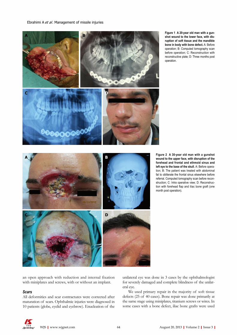
an open approach with reduction and internal fixation with miniplates and screws, with or without an implant.
Scars All deformities and scar contractures were corrected after maturation of scars. Ophthalmic injuries were diagnosed in 10 patients (globe, eyelid and eyebrow). Enucleation of the
unilateral eye was done in 3 cases by the ophthalmologist for severely damaged and complete blindness of the unilat-eral eye.
We used primary repair in the majority of soft tissue defects (25 of 40 cases). Bone repair was done primarily at the same stage using miniplates, titanium screws or wires. In some cases with a bone defect, iliac bone grafts were used
64 August 20, 2013|Volume 2|Issue 3|WJS|www.wjgnet.com
Ebrahimi A et al . Management of missile injuries
Figure 1 A 20-year old man with a gun-shot wound to the lower face, with dis-ruption of soft tissue and the mandible bone in body with bone defect. A: Before operation; B: Computed tomography scan before operation; C: Reconstruction with reconstructive plate; D: Three months post operation.
A B
C D
Figure 2 A 30-year old man with a gunshot wound to the upper face, with disruption of the forehead and frontal and ethmoid sinus and left eye to the base of the skull. A: Before opera-tion; B: The patient was treated with abdominal fat to obliterate the frontal sinus elsewhere before referral. Computed tomography scan before recon-struction; C: Intra operative view; D: Reconstruc-tion with forehead flap and iliac bone graft (one month post operation).
A
C D
B

simultaneously or in the later stages (mandibular defects).
RESULTSA total of 40 patients were treated and followed from 5 months to 3 years. There were 38 male and 2 female pa-tients, with an average age of 37 years (range 19-65 years). Most injuries were caused by high velocity projectiles (88%) and the remaining by car explosions or dynamite (12%). Thirty patients had soft tissue loss (75%) and 20 patients (50%) had bone loss; there was combined soft tissue and bone loss in 10 (25%) patients. Facial fractures were in the orbital bones in 10 cases, maxillary in 7, nasal in 5 and the mandible in 3 cases (Table 1).
There was no failure of bone reconstruction in our cases. Infections occurred in 2 cases and were treated with systemic antibiotics and dressing changes, without any long term sequelae.
DISCUSSIONExposure to missile injuries may result in a unique and complex injury pattern, usually from fragments or bullet wounds which are often fatal if they involve the head. Blast overpressure is the abrupt, rapid rise in atmospheric pressure resulting from explosive detonation, firing of large caliber weapons and accident occupational explosions[4,5]. There are two schools of thought for the management of such patients subjected to missile injuries: early intervention and nonag-gressive conservative intervention[2]. Injuries to the face due to firearms are either high velocity or low energy; high veloci-
ty projectiles can result in devastating functional and aesthetic consequences, shattering the hard tissues[6].
Our principles for soft tissue reconstructions were according to the reconstructive ladder, including primary repair, local flaps, skin grafts and regional flaps depending on the extent of damage (Figure 3). We used primary re-pair in the majority of soft tissue defects (25 of 30 cases) and recommend this approach for these injuries.
We used surgical intervention in all cases. Early inter-vention in facial firearm injuries resulted in restoration of occlusion and continuity of the jaw, fixation of luxated or extruded teeth, early return of function, prevention of segment displacement and tissue contracture, less scar-ring and decreased need for major bone graft reconstruc-tion later in one study[6]. If continuity of the mandible can be obtained, in the subsequent operations there will be no need for maxillomandibular fixation. In this case series study, we had no major complications after early surgical interventions.
All facial wounds were under systemic antibiotic therapy for one week and local antibiotic ointment to prevent secondary infections. There was no failure of bone recon-struction; in our cases, maxillary defects were reconstructed with bicortical bone grafts in the same operation. We had facial wound infections postoperatively in 2 cases and treated them with systemic antibiotics and dressing changes, without any long term sequelae (these two patients had a mandible fracture with a through wound of the oral cavity without any medical immunocompromising factors).
The issue of when to treat maxillofacial firearm inju-ries remains controversial (early or delayed), although not all maxillofacial projectile injuries can be comprehensively treated at the onset[7]. Although all missile wounds are contaminated, the general consensus in the medical litera-ture and textbooks consider these infections to be mostly of odontogenic origin[8]. In composite defects (soft tissue and bone), we used bone graft and soft tissue flaps simul-taneously for coverage of bone and our results were free of any significant resorption or flap necrosis after early operative intervention.
In our study, the most common site of entrance and exit wounds was in the cheek (67%). In another study in Iraq by Kummona, the most common site was also in the cheek
65 August 20, 2013|Volume 2|Issue 3|WJS|www.wjgnet.com
Ebrahimi A et al . Management of missile injuries
Figure 3 A 23-year old man with a gunshot wound defect of the eyebrow. A: Before operation; B: Early post operation; C: One year post operation.
A B C
Table 1 Distribution of facial soft tissue and bone damage in 40 gunshots and blast injured persons
Site of injury n Bone Soft tissue defect Combined soft tissue-bone
1 Periorbital 12 10 6 42 Maxillary 17 7 16 63 Mandible 4 3 2 14 Nasal 5 3 4 35 Frontal 2 2 2 2Total 40 25 30 16

(54.8%). According to our results, the midface is a common site for gunshot injuries and a safe coverage for protection of the cheek in military and civilian people must be designed for combat. The face is the part of the body most subjected to injuries, either by road traffic accidents or missile war inju-ries[9]. In our experience, gunshot injuries of the craniofacial region are not a single site injury and often have associated injuries; thus, a complete evaluation of soft tissue and bones must be done for all patients. We used free bone grafting for 4 patients in our cases and the preferred site for bone graft harvesting was the iliac crest because of combined cortico-cancellous block of bone. For delayed reconstruction of the frontal cranium, we used titanium mesh and soft tissue flap with acceptable results (Figure 3) and without any complica-tions. In our series, the most common associated injuries were ophthalmic injuries (Table 2), seen in 10 patients with unilateral blindness. In another study, the most common injured facial structure was the facial nerve and the second most common was ophthalmic injuries. An important prob-lem in patients with gunshot injuries or blast damage is facial burn blast tattoos that must be managed early post damage by operative intervention. Application of silver sulfadiazine before the operative intervention helps to remove embedded particles better and decrease traumatic tattoos[10]. This pro-cedure is also better to be done early. Advocates of primary management have supported this viewpoint[11-14].
In high velocity gunshot and blast injuries with facial damage to soft tissue and bone, early surgical intervention is beneficial and good results without significant complica-tions can be obtained; we recommend this approach in these types of facial injuries.
COMMENTSBackgroundFirearm projectiles can result in devastating functional and aesthetic conse-quences, shattering the hard tissues. Early intervention in facial firearm injuries is still in debate. Research frontiersExperience has shown that early management results in restoration of occlu-sion and continuity of the jaw, fixation of luxated or extruded teeth, early return of function, prevention of segment displacement and tissue contracture, less scarring and decreased need for major bone graft reconstruction later on. Innovations and breakthroughsInnovations in surgical instruments and hardware, plates and screws have en-
abled rigid fixation of fractures. Applications These advancements have enabled surgeons to better manage gunshot injuries. Peer reviewThe manuscript is a result of good efforts on the part of the authors and may be useful to determine management of maxillofacial injuries secondary to missiles and gunshot wounds.
REFERENCES1 Helmkamp JC, Kennedy RD. Causes of death among U.S.
military personnel: a 14-year summary, 1980-1993. Mil Med 1996; 161: 311-317 [PMID: 8700323]
2 Kummoona R, Muna AM. Evaluation of immediate phase of management of missile injuries affecting maxillofacial region in iraq. J Craniofac Surg 2006; 17: 217-223 [PMID: 16633165 DOI: 10.1097/00001665-200603000-00003]
3 Banks P, Mellor S, Haywood IR. Gunshot wounds. In: Williams JLT, editors. Rowe and Williams Maxillofacial Injuries, 2nd ed. London, England: Churchill Livingstone, 1994: 665
4 Nelson TJ, Wall DB, Stedje-Larsen ET, Clark RT, Chambers LW, Bohman HR. Predictors of mortality in close proximity blast injuries during Operation Iraqi Freedom. J Am Coll Surg 2006; 202: 418-422 [PMID: 16500245 DOI: 10.1016/j.jamcollsurg.2005.11.011]
5 Bird SM. UK statistical indifference to its military casual-ties in Iraq. Lancet 2006; 367: 713-715 [PMID: 16517258 DOI: 10.1016/S0140-6736(06)68281-0]
6 Motamedi MH. Management of firearm injuries to the fa-cial skeleton: Outcomes from early primary intervention. J Emerg Trauma Shock 2011; 4: 212-216 [PMID: 21769208 DOI: 10.4103/0974-2700.82208]
7 Motamedi MH, Behnia H. Experience with regional flaps in the comprehensive treatment of maxillofacial soft-tissue in-juries in war victims. J Craniomaxillofac Surg 1999; 27: 256-265 [PMID: 10626260 DOI: 10.1016/S1010-5182(99)80038-9]
8 Green AW, Flower EA, New NE. Mortality associated with odontogenic infection! Br Dent J 2001; 190: 529-530 [PMID: 11411886]
9 Kummoona RK. Missile war injuries of the face. J Craniofac Surg 2011; 22: 2017-2021 [PMID: 22067852 DOI: 10.1097/SCS.0b013e318231978a]
10 Ebrahimi A, Motamedi MHK. Application of silver sulfadia-zine cream with early surgical intervention in patients suf-fering from combined burn-blast injury facial tattoos. Trauma Monthly 2012; 17: 255-258 [DOI: 10.5812/traumamon.3935]
11 Motamedi MH. Primary treatment of penetrating injuries to the face. J Oral Maxillofac Surg 2007; 65: 1215-1218 [PMID: 17517308 DOI: 10.1016/j.joms.2007.03.001]
12 Motamedi MH. Primary management of maxillofacial hard and soft tissue gunshot and shrapnel injuries. J Oral Maxil-lofac Surg 2003; 61: 1390-1398 [PMID: 14663802 DOI: 10.1016/j.joms.2003.07.001]
13 Motamedi MH, Hashemi HM, Shams MG, Nejad AN. Reha-bilitation of war-injured patients with implants: analysis of 442 implants placed during a 6-year period. J Oral Maxillofac Surg 1999; 57: 907-913; discussion 914-915 [PMID: 10437717 DOI: 10.1016/S0278-2391(99)90005-8]
14 Behnia H, Motamedi MH. Reconstruction and rehabilitation of short-range, high-velocity gunshot injury to the lower face: a case report. J Craniomaxillofac Surg 1997; 25: 220-227 [PMID: 9268901 DOI: 10.1016/S1010-5182(97)80079-0]
P- Reviewers Gokul S, Lee J, Lopez-Jornet P S- Editor Gou SXL- Editor Roemmele A E- Editor Lu YJ
66 August 20, 2013|Volume 2|Issue 3|WJS|www.wjgnet.com
Ebrahimi A et al . Management of missile injuries
COMMENTS
Table 2 Associated injures in the face n (%)
Associated injures
Facial nerve 2 (5)Parotid duct 4 (10)Globe 3 (7.5)Oral mucosa 3 (7.5)Lacrimal duct 2 (5)Total 14 (35)

World Journal of StomatologyWorld J Stomatol 2013 November 20; 2(4): 67-107
ISSN 2218-6263 (online)
www.wjgnet.com
Volume End

World Journal of StomatologyW J S
67 Pharmacogenomicsinoraldiseases
Gokul S, Sapna G
71 Stomatologicalmanagementofheadandneckcancerpatientstreatedwith
chemotherapyandradiotherapy
Bologna-Molina R, Maglia A, Castañeda-Castaneira RE, Molina-Frechero N
79 Molecularbiomarkersofcellproliferationinameloblastomas
Bologna-Molina R, Bedoya-Borella AM, Soria-Moreira L, Soría-Suárez S
86 Cytotoxicityofasilorane-baseddentalcompositeonhumangingival
fibroblasts
Orsini G, Catellani A, Ferretti C, Gesi M, Mattioli-Belmonte M, Putignano A
91 Accuracyoflinearvs spiral tomography: Alveolar crest to sinus/nasal floor height
Yoozbashizadeh M, Fatemitabar SA, Sedighara E, Nikgoo A
97 Surgicalobturatorduplicatingoriginaltissue-formrestoresestheticsand
functioninoralcancer
Patil PG
103 Softtissueaneurysmalbonecystofthemandible:Reportofacase
Jahanbani J, Sadri D, Hassani A, Kavandi F
Contents
EDITORIAL
Quarterly Volume 2 Number 4 November 20, 2013
IWJS|www.wjgnet.com November 20, 2013|Volume 2|Issue 4|
THERAPEUTICS
ADVANCES
MINIREVIEWS
BRIEF ARTICLE
CASE REPORT

ContentsWorld Journal of Stomatology
Volume 2 Number 4 November 20, 2013
EDITORS FOR THIS ISSUE
Responsible Assistant Editor: Xin-Xin Che Responsible Science Editor: Ling-Ling WenResponsible Electronic Editor: Jin-Li Yan Proofing Editor-in-Chief: Lian-Sheng Ma
World Journal of StomatologyRoom 903, Building D, Ocean International Center, No. 62 Dongsihuan Zhonglu, Chaoyang District, Beijing 100025, ChinaTelephone: +86-10-85381891Fax: +86-10-85381893E-mail: [email protected]://www.wjgnet.com
PUBLISHERBaishideng Publishing Group Co., LimitedFlat C, 23/F., Lucky Plaza, 315-321 Lockhart Road, Wan Chai, Hong Kong, ChinaFax: +852-6555-7188Telephone: +852-3177-9906E-mail: [email protected]://www.wjgnet.com
PUBLICATIONDATENovember 20, 2013
COPYRIGHT© 2013 Baishideng Publishing Group Co., Limited. Articles published by this Open-Access journal are dis-tributed under the terms of the Creative Commons At-tribution Non-commercial License, which permits use, distribution, and reproduction in any medium, provided the original work is properly cited, the use is non com-mercial and is otherwise in compliance with the license.
SPECIALSTATEMENTAll articles published in this journal represent the viewpoints of the authors except where indicated oth-erwise.
INSTRUCTIONSTOAUTHORSFull instructions are available online at http://www.wjgnet.com/2218-6263/g_info_20100722180909.htm
ONLINESUBMISSIONhttp://www.wjgnet.com/esps/
IIWJS|www.wjgnet.com
APPENDIX
ABOUT COVER
AIM AND SCOPE
INDEXINg/ABSTRACTINg
November 20, 2013|Volume 2|Issue 4|
NAMEOFJOURNALWorld Journal of Stomatology
ISSNISSN 2218-6263 (online)
LAUNCHDATEDecember 31, 2011
FREQUENCYQuarterly
EDITOR-IN-CHIEFPeter E Murray, BSc (Hons), PhD, Professor, Pathol-ogist, Department of Endodontics, College of Dental Medicine, Nova Southeastern University, 3200 South University Drive, Fort Lauderdale, FL 33328-2018, United States
EDITORIALOFFICEJin-Lei Wang, DirectorXiu-Xia Song, Vice Director
I-V Instructionstoauthors
EditorialBoardMemberofWorldJournalofStomatology ,SridharanGokul,
MDS,Lecturer,OralPathologyandMicrobiology,YMTDentalCollegeand
Hospital,Kharghar,Institutionalarea,sector-4,Kharghar,NaviMumbai410210,
Maharashtra,India
World Journal of Stomatology (World J Stomatol, WJS, online ISSN 2218-6263, DOI: 10.5321) is a peer-reviewed open access academic journal that aims to guide clinical practice and improve diagnostic and therapeutic skills of clinicians.
WJS covers topics concerning oral and craniofacial sciences, oral and craniofacial development/growth, dental tissue regeneration, craniofacial bone and cartilage research, oral and maxillofacial genetic diseases, developmental abnormalities and soft tissue defects, pulpal and periapical diseases, periodontal diseases and oral mucosal diseases, salivary gland diseases, oral and maxillofacial vascular/nervous diseases, jaw bone diseases, taste abnormalities, oral and maxillofacial pain, occlusion and temporomandibular diseases, repair and treatment of tooth defects, loss and dento-maxillofacial deformities, oral and maxillofacial biomechanics and biomaterials, new techniques for diagnosis/treatment of oral and maxillofacial diseases; and stomatology-related evidence-based medicine, epidemiology and nursing. Priority publication will be given to articles concerning diagnosis and treatment of stomatologic diseases. The following aspects are covered: Clinical diagnosis, laboratory diagnosis, differential diagnosis, imaging tests, pathological diagnosis, molecular biological diagnosis, immunological diagnosis, genetic diagnosis, functional diagnostics, and physical diagnosis; and comprehensive therapy, drug therapy, surgical therapy, interventional treatment, minimally invasive therapy, and robot-assisted therapy.
We encourage authors to submit their manuscripts to WJS. We will give priority to manuscripts that are supported by major national and international foundations and those that are of great basic and clinical significance.
World Journal of Stomatology is now indexed in Digital Object Identifier.
I-III EditorialBoardFLYLEAF
World Journal of StomatologyW J S
Online Submissions: http://www.wjgnet.com/esps/[email protected]:10.5321/wjs.v2.i4.67
World J Stomatol 2013 November 20; 2(4): 67-70ISSN 2218-6263 (online)
© 2013 Baishideng Publishing Group Co., Limited. All rights reserved.
Pharmacogenomics in oral diseases
Sridharan Gokul, Gokul Sapna
Sridharan Gokul, Oral Pathology and Microbiology, YMT Den-tal College and Hospital, Kharghar, Navi Mumbai-410210, Ma-harashtra, IndiaGokul Sapna, Department of Periodontics, Nair Hospital Dental College, Mumbai-400008, Maharashtra, IndiaAuthor contributions: Gokul S formulated the idea of writing the editorial; Gokul S and Sapna G together performed literature review and collected relevant data; Sapna G also contributed in proof reading the manuscript.Correspondence to: Sridharan Gokul, MDS, Lecturer, Oral Pathology and Microbiology, YMT Dental College and Hospital, Kharghar, Institutional area, sector-4, Kharghar, Navi Mum-bai-410210, Maharashtra, India. [email protected]: +91-22-27744429 Fax: +91-22-27744427Received: April 6, 2013 Revised: July 11, 2013Accepted: July 17, 2013Published online: November 20, 2013
AbstractThe availability of newer technologies for identification and characterization of the human genome has enabled our understanding of the genetic variations in a majority of human diseases. Human genomic sequence varies in less than 1% among the different population group and these differences known as gene polymorphisms are the primary reasons for differences in individuals’ response to various drug therapy. Also understanding the genetic changes may enable implementation of targeted thera-py, thus providing for effective treatment strategies and minimizing the adverse side effects. Pharmacogenom-ics is a recent development in the field of personalized medicine which focuses on the genetic determinants of drug response at the levels of entire human genome. It primarily deals with tailoring of drug therapy for every individual based on their genetic make-up and identi-fying new target in various diseases for drug therapy. While the application of pharmacogenomics in systemic illness is well researched, its role in oral diseases needs documentation. Identifying specific targets in periodon-titis, head and neck cancer, infections and genetic dis-orders can be beneficial in discovery of new drugs. This editorial provides an overview of basics of pharmacoge-
nomics, its current role in disease management and its potential role in various head and neck diseases.
© 2013 Baishideng Publishing Group Co., Limited. All rights reserved.
Key words: Pharmacogenomics; Oral Cancer; Periodon-tal diseases; Genomic variations; Targeted drug therapy
Core tip: Pharmacogenomics mainly focuses on the genetic determinants of drug response at the level of entire human genome. It primarily deals with tailoring of drug therapy among every individual based on their genetic make-up and identifying new targets in vari-ous diseases for drug therapy. Identification of gene polymorphisms in humans will aid in modulating drug therapy for individual needs as well as leading to discov-ery of target drugs. This editorial provides an overview of basic pharmacogenomics and its usefulness in oral diseases.
Gokul S, Sapna G. Pharmacogenomics in oral diseases. World J Stomatol 2013; 2(4): 67-70 Available from: URL: http://www.wjgnet.com/2218-6263/full/v2/i4/67.htm DOI: http://dx.doi.org/10.5321/wjs.v2.i4.67
INTRODUCTIONPharmacogenomics is a component of individualized medicine focusing on how genetic factors influence in-dividual responses to different medications that may af-fect drug efficacy, side effects and adverse events to drug therapy[1]. The aim of pharmacogenomics is to decrease adverse responses to therapy through determining new therapeutic targets and genetic polymorphisms that affect drug specificity and toxicity[2]. The term pharmacoge-nomics, a relatively new term is often used interchange-ably with a much older term pharmacogenetics though differences exist between the two terms. While pharma-cogenetics refers to the study of how individual genes in-
EDITORIAL
67 November 20, 2013|Volume 2|Issue 4|WJS|www.wjgnet.com

fluences the response to medications, pharmacogenomics is related to the study of how individuals’ genomic com-position as a whole affects their response to medicine[3]. The field of pharmacogenomics uses genetic and ge-nomic information of individuals in order to predict the response of patient groups to drugs and thus guide clini-cal trial and the drug development process. This can be made possible with the development of human genome project which encodes majority of human genes.
Most of the commonly occurring diseases such as cancer, atherosclerosis and neurodegenerative disorders comprise of a group of genetically discrete entities with separate molecular etiologies and possibly different re-sponses to therapy. Pharmacogenomic therapy has been attempted to treat cystic fibrosis[4], acquired immune deficiency syndrome (AIDS), cardiovascular diseases, psychiatric illness and targeted therapy towards epidermal growth factor receptor (EGFR) for treatment of pancre-atic cancer[5] and non small cell lung carcinoma[6]. In addi-tion to the development of new therapeutic agents, phar-macogenomics can also predict outcomes and beneficial therapy regimens. The various targets that are extensively studied are angiotensin converting enzyme regulating car-diovascular functions; analyzing the association between β2 adrenergic receptor polymorphism and asthma[7] and use of apolipoprotein polymorphisms to predict the risk of heart disease and response to treatment[2].
Most of the disorders affecting the head and neck region excluding trauma have genetic implications in various forms. The disorders range from developmental disturbances, microbial infections and bone disorders to various forms of head and neck cancers. An understand-ing of the genetic changes occurring in these disorders may be helpful in implementation of drug therapy aiming to obtain maximum efficacy with minimum side effects and also to evolve targeted drug therapy for the manage-ment of such lesions.
BASICS OF PHARMACOGENOMICSA gene is defined as a specific sequence of nucleotide bases whose sequences carry the instructions for the constructions of proteins. Hundreds of genes reside on each chromosome and the complete human genome is estimated to contain about 30000 genes[8]. More than 99% of DNA sequence is the same across the entire human population. The small genetic dissimilarities have a ma-jor impact on persons’ physical makeup and response to disease as well the effectiveness of the therapy instituted. Pharmacogenomic technologies try to detect these genetic variations in a patient or patient populations to help select drug compounds and doses that are more likely to work.
These genetic variations which account for around 1 million of the 3 billion bases of human genome are termed as polymorphisms. Polymorphisms arise from three fundamental types of DNA sequence variations namely single nucleotide polymorphisms (SNPs) repre-senting nucleotide substitutions, insertions or deletions
and indel of repetitive DNA[9]. Advances in pharma-cogenomics mainly depend on identification of SNPs in the human genome. These arise from mutations affect-ing a single nucleotide, occurring relatively frequently and must exceed in a population of a frequency of 1% to meet the requirement of genetic polymorphism[10]. The main research use of a human SNP map would be to determine the contributions of genes to diseases that have a complex multifactorial basis. More than 1.4 mil-lion SNPs have been identified in human genome till date[8]. Majority of the genetic polymorphisms are found in drug metabolizing enzymes, receptors and transport proteins and produce varying effects on drug metabo-lism[2]. The use of these genetic variations in order to individualize drug therapy and to identify novel targets to enable the development of new drugs for various diseases are the primary reasons for which pharmacoge-nomics is employed.
Drugs in the body are metabolized by enzymes and most of the enzymes belong to the members of cyto-chrome P450 (CYP) system. These enzymes are located in the liver and gastrointestinal tract and include greater than 30 isoforms[11]. Individual variations in the genes that produce these enzymes causes different people to metabolize the same drug differently; less active or inac-tive forms of CYP enzymes that are unable to break-down and efficiently eliminate a drug from the body (slow metabolizers) can cause the drug to build up while very active forms (rapid metabolizers) can cause the body to clear itself of a drug before it has a chance to work[12]. Understanding an individuals’ response to a certain drug can help the clinician to decide the accurate drug dos-age required for effective therapy thereby reducing the chances of overdose or insufficient dosage. Of all the types of CYPs, most of the functional genetic polymor-phisms reside in only few of them namely CYP3A4, CYP2A6, CYP2C9, CYP2C19 and CYP2D6[10]. Of these, CYP3A is involved in the oxidative biotransformation of up to 50% of clinically important therapeutic agents that has resulted in the withdrawal of important drugs like Mibefradil (anti-hypertensive drug), Rezulin (oral anti-hyperglycemic agent) and propulsid (for treatment of gastrointestinal disorders).
Other less common polymorphisms can be seen in drug transport proteins and receptors. Transport proteins are proteins that allow compounds to be transported across cell membranes and P-glycoprotein is a drug trans-port protein known to be involved in the metabolism of many drugs[13]. Identifying this polymorphism would be a valuable tool in determining therapeutic concentrations required for individual patients. Receptors polymor-phisms are helpful for development of new therapeutic agent and to predict outcomes and beneficial treatment regimens. Some of the common receptor targets that have been targeted by the use of drugs include angio-tensin converting enzyme, β2-adrenergic receptor poly-morphism, cystic fibrosis transmembrane conductance regulator and p53 and EGFR for anti-cancer therapy.
68 November 20, 2013|Volume 2|Issue 4|WJS|www.wjgnet.com
Gokul S et al . Pharmacogenomics in oral diseases

APPLICATIONS OF PHARMACOGENOMICS IN ORAL DISEASESGenetic mutations are a common finding in majority of the human diseases affecting the head and neck region. which may contain one or many genetic polymorphisms. Examples of disorders exhibiting single gene muta-tions include Treacher-Collins syndrome, Pierr-Robin syndrome, Crouzon syndrome, Ectodermal dysplasia, achondroplasia and Gorlin syndrome while cleft lip and palate, congenitally missing teeth, dental caries, severe malocclusion, head and neck cancer, periodontal diseases and autoimmune disorders are caused by multiple gene mutations[14].
Chemotherapeutic intervention for cancer therapy is undergoing changes from being an empiric random screening approach to a target directed approach where specific abnormalities in cell functioning are modulated in a drug receptor fashion. The use of small molecules with tyrosine kinase inhibitory activity directed towards the EGFR such as gefitinib and erlotinib are used for treatment of NSCLC, pancreatic and breast cancer[5]. EGFR is a transmembrane glycoprotein member of erbB family of type Ⅰ tyrosine kinase which plays a crucial role through downstream signaling pathways in cell cycle progression, survival and proliferation[15]. Overexpression of EGFR in head and neck cancer are known to be as-sociated with poor prognosis and hence the use of drugs such as tyrosine kinase inhibitors may help in improving the prognosis and survival rate. Adequate understanding of the molecular mechanisms involving various growth factors (such as transforming growth factor, platelet de-rived growth factor, hepatocyte growth factor), cytokines and genetic mutations occurring in carcinogenesis will aid in the development of chemotherapeutic drugs against specific targets for appropriate management and to re-duce patient morbidity and mortality.
Periodontitis is a polymicrobial infection resulting from a complex interaction between oral microbes and host immune response leading to periodontal destruc-tion and alveolar bone resorption. The host response to infection is primarily in the form of inflammatory reaction leading to release of various cytokines, growth factors and matrix metalloproteinases (MMP). Identifica-tion of therapeutic targets which are directed towards the specific host alteration may be helpful as an adjuvant treatment for periodontitis. Monoclonal antibody deriva-tives directed towards MMP are promising as therapeutic agents and mainly involves non-antimicrobial activities of low dose tetracycline and tetracycline analogue doxycy-cline hyclate via the inhibition of MMP-8 and -13 prote-ase mechanisms. The therapeutic action of these agents is primarily due to the modulation of the host response because the low dose formulations of these drugs have lost their antimicrobial property[16]. Research in targeted therapy are also underway for treatment of various other microbial infections like candidiasis, birth defects, orth-
odontic tooth movements but are still at initial stages.To summarize, decoding the human genome with aim
to describe genetic changes in various oral diseases will be beneficial in providing appropriate therapy. The use of pharmacogenomics in determining the functioning of various drugs in individual patients will be a boon for clinicians to decide the appropriate dosage. Research con-cerning targeted drug therapy has advanced exponentially and could be ideal for treatment of diseases like cancer and AIDS without major side effects and also for man-agement. This has been enhanced by the availability of advanced technologies such as SNPs and DNA microar-rays which helps in analyzing genetic changes.
REFERENCES1 Baudhuin LM. Pharmacogenomics: the use of genetics in
guiding pharmacologic therapy. Mayo medical laboratories. Communique 2012. Available from: URL: http://www.may-omedicallaboratories.com/articles/communique/2012/09.html
2 Wieczorek SJ, Tsongalis GJ. Pharmacogenomics: will it change the field of medicine? Clin Chim Acta 2001; 308: 1-8 [PMID: 11412811 DOI: 10.1016/S0009-8981(01)00419-3]
3 Yagil Y, Yagil C. Insights into pharmacogenomics and its impact upon immunosuppressive therapy. Transpl Im-munol 2002; 9: 203-209 [PMID: 12180832 DOI: 10.1016/S0966-3274(02)00022-9]
4 Srivastava M, Eidelman O, Pollard HB. Pharmacogenomics of the cystic fibrosis transmembrane conductance regulator (CFTR) and the cystic fibrosis drug CPX using genome mi-croarray analysis. Mol Med 1999; 5: 753-767 [PMID: 10656877]
5 Jimeno A, Hidalgo M. Pharmacogenomics of epidermal growth factor receptor (EGFR) tyrosine kinase inhibitors. Biochim Biophys Acta 2006; 1766: 217-229 [PMID: 17045403]
6 Rosell R, Felip E, Paz-Ares L. How could pharmacoge-nomics help improve patient survival? Lung Cancer 2007; 57 Suppl 2: S35-S41 [PMID: 17686445 DOI: 10.1016/S0169-5002(07)70426-9]
7 Liggett SB. Molecular and genetic basis of beta2-adrenergic receptor function. J Allergy Clin Immunol 1999; 104: S42-S46 [PMID: 10452787 DOI: 10.1016/S0091-6749(99)70272-1]
8 Maheshwari S, Verma SK, Tariq M, Prabhat KC, Kumar S. Emerging trends in oral health profession: The molecular dentistry. Biol Med 2010; 2: 56-63
9 Nebert DW, Bingham E. Pharmacogenomics: out of the lab and into the community. Trends Biotechnol 2001; 19: 519-523 [PMID: 11711196 DOI: 10.1016/S0167-7799(01)01805-4]
10 Vesell ES. Advances in pharmacogenetics and pharmacoge-nomics. J Clin Pharmacol 2000; 40: 930-938 [PMID: 10975065 DOI: 10.1177/00912700022009666]
11 Gonzalez FJ. Molecular genetics of the P-450 superfamily. Pharmacol Ther 1990; 45: 1-38 [PMID: 2405431 DOI: 10.1016/0163-7258(90)90006-N]
12 Tucker L. Pharmacogenomics: A primer for policy makers. National health policy forum 2008. Available from: URL:www.nhpf.org
13 Hoffmeyer S, Burk O, von Richter O, Arnold HP, Brock-möller J, Johne A, Cascorbi I, Gerloff T, Roots I, Eichelbaum M, Brinkmann U. Functional polymorphisms of the human multidrug-resistance gene: multiple sequence variations and correlation of one allele with P-glycoprotein expression and activity in vivo. Proc Natl Acad Sci USA 2000; 97: 3473-3478 [PMID: 10716719 DOI: 10.1073/pnas.97.7.3473]
14 Slavkin HC. The human genome, implications for oral health and diseases, and dental education. J Dent Educ 2001; 65: 463-479 [PMID: 11425251]
69 November 20, 2013|Volume 2|Issue 4|WJS|www.wjgnet.com
Gokul S et al . Pharmacogenomics in oral diseases

15 Boulougouris P, Elder J. Epidermal growth factor receptor structure, regulation, mitogenic signalling and effects of acti-vation. Anticancer Res 2001; 21: 2769-2775 [PMID: 11724353]
16 Honibald EN, Mathew S, Padmanaban J, Sundaram E, Ra-
mamoorthy RD. Perioceutics: Matrix metalloproteinase in-hibitors as an adjunctive therapy for inflammatory periodon-tal disease. J Pharm Bioallied Sci 2012; 4: S417-S421 [PMID: 23066302]
P- Reviewers: Arabaci T, Vaidya MM S- Editor: Wen LL L- Editor: A E- Editor: Wang CH
70 November 20, 2013|Volume 2|Issue 4|WJS|www.wjgnet.com
Gokul S et al . Pharmacogenomics in oral diseases

World Journal of StomatologyW J S
Online Submissions: http://www.wjgnet.com/esps/[email protected]:10.5321/wjs.v2.i4.71
World J Stomatol 2013 November 20; 2(4): 71-78ISSN 2218-6263 (online)
© 2013 Baishideng Publishing Group Co., Limited. All rights reserved.
Stomatological management of head and neck cancer patients treated with chemotherapy and radiotherapy
Ronell Bologna-Molina, Alvaro Maglia, Raúl Enrique Castañeda-Castaneira, Nelly Molina-Frechero
Ronell Bologna-Molina, Alvaro Maglia, Faculty of Dentistry, Universidad de la República (UDELAR), Montevideo 11600, UruguayRaúl Enrique Castañeda-Castaneira, Nelly Molina-Frechero, Health Care Department, Universidad Autónoma Metropolitana, Xochimilco, 04960 Mexico City, MexicoAuthor contributions: Bologna-Molina R wrote, revised the manuscript and designed the figures; Maglia A, Castañeda-Cas-taneira RE and Molina-Frechero N these co-authors contributed equally to the writing of the manuscript.Correspondence to: Ronell Bologna-Molina, DDS, PhD, Faculty of Dentistry, Universidad de la República (UDELAR), Las Heras 125, Montevideo 11600, Uruguay. [email protected]: +598-2487-3048 Fax: +598-2400-8640Received: July 12, 2013 Revised: July 31, 2013 Accepted: August 5, 2013Published online: November 20, 2013
AbstractTreatment of head and neck cancer with radiotherapy and/or chemotherapy can cause oral damage. Long-term treatment can damage the salivary glands, the oral mucosa, and the maxilla, leading to altered pro-duction of saliva and to multiple infections. These le-sions can be prevented, limited or avoided by thorough evaluation prior to treatment and by therapeutic follow-up and preventive measures. The dentist must have strong medical knowledge of the possible short-, me-dium-, and long-term oral complications of the cancer treatment, and must have knowledge of the protocols for oral management of cancer patients. The availability of a multidisciplinary medical team together with a den-tist to attend to the patient prior to the cancer treat-ment, as well as close communication between team members during and after treatment, is crucial. The aim of the present study was review the stomatological management of head and neck cancer patients treated with chemotherapy and radiotherapy and summarizing current treatments, therapeutic innovation and tissue
regeneration perspectives.
© 2013 Baishideng Publishing Group Co., Limited. All rights reserved.
Key words: Stomatological management; Head and neck; Cancer
Core tip: The aim of the present study was to conduct a review of therapeutic advances in the prevention and management of oral disorders in head and neck cancer patients receiving radio- and chemotherapy. The study focuses on possible risk factors and on the prevention of these disorders.
Bologna-Molina R, Maglia A, Castañeda-Castaneira RE, Molina-Frechero N. Stomatological management of head and neck cancer patients treated with chemotherapy and radiotherapy. World J Stomatol 2013; 2(4): 71-78 Available from: URL: http://www.wjgnet.com/2218-6263/full/v2/i4/71.htm DOI: http://dx.doi.org/10.5321/wjs.v2.i4.71
INTRODUCTIONIn recent decades, an increase in the prevalence of oral cancer has been observed in several countries. Surgery, radiotherapy and chemotherapy continue to be the treat-ments of choice for such cancers, and advances have been made in minimizing their adverse effects[1]. However, treat-ment of cancer with radiotherapy and/or chemotherapy can cause oral damage. Long-term treatment can damage the salivary glands, the oral mucosa, and the maxilla, lead-ing to altered production of saliva and to multiple infec-tions[2]. The surgical treatment of oral and maxillofacial neoplasms can lead to sequelae such as limited speech, eat-ing disorders, alterations in the patient’s sense of taste and smell, and changes in the patient’s physical appearance[3,4]
Lesions of the oral cavity secondary to head and neck
THERAPEUTICS ADVANCES
71 November 20, 2013|Volume 2|Issue 4|WJS|www.wjgnet.com
cancer treatment can be prevented, limited or avoided by thorough evaluation prior to treatment and by therapeu-tic follow-up and preventive measures[5].
The dentist must have strong medical knowledge and be continuously updated on common head and neck ma-lignant neoplasms, their clinical manifestations, therapeu-tic alternatives, and the complications that may occur as a result of their treatment[6].
The availability of a multidisciplinary medical team consisting of an oncologist, a hematologist, a head and neck surgeon, a radiologist, a physiotherapist, a speech therapist, a social worker, and a psychologist together with a dentist to attend to the patient prior to the cancer treatment, as well as close communication between team members during and after treatment, is crucial[5,7].
Ideally, cancer centers should provide oral health care. However, because patients are frequently referred to the family dentist, dentists must have basic knowledge of the protocols for oral management of cancer patients and of the relevant National Cancer Institute guidelines[8].
INITIAL CONTACT WITH THE PATIENTIt is important for the dentist to join the oncology team so that he or she is informed of the type of surgery and radio- and/or chemotherapy the patient will receive[5,9].
At the patient’s first visit, the dentist must perform a complete oral health evaluation to establish an integral oral and maxillofacial management plan before the cancer treatment is initiated[5].
The possible short-, medium- and long-term oral complications of the cancer treatment must be explained to the patient and to his or her close relatives[10] in a simple and didactical way, preferably through the use of images, so that they will be able to identify problems such as xerostomia and mucositis. Patients must be instructed in the importance of dental follow-up care before, dur-ing, and after chemo- and radiotherapy as a means of preventing radiation-associated dental caries and osteo-necrosis. The dentist must provide the patient and the patient’s relatives with an instructional manual on oral hygiene, diet, and measures to be followed before, during, and after cancer treatment[11-14].
The primary objective of providing information to the patient and of diagnosing and treating the patient pri-or to cancer treatment is to eliminate or stabilize any oral lesions that are present and to minimize the possibility of the occurrence of local or systemic infections during or after cancer treatment[14-16].
Patients often show tumor-related symptoms or dental conditions related to an incisional biopsy or pre-surgical therapy. These symptoms must be evaluated and correctly diagnosed so as to differentiate tumor symptoms from previous oral manifestations of dental caries, periodontal disease, pulpal diseases and soft tissue conditions[13,17].
A detailed exploration of the head and neck region must be performed following a preset order from external to internal, and abnormal growth, asymmetries, and cu-taneous lesions must be identified and evaluated. Salivary
glands, muscles, and the temporomandibular joint must be inspected. Palpation of submental, submandibular, and cervical lymph nodes is important and must be followed by an intraoral examination starting with the soft tissues, buccal mucosa, tongue, floor of the mouth, and the hard and soft palates. Any lesion, irritation, erosion, ulceration, or hemorrhage must be identified, and the patient’s gen-eral periodontal state must be evaluated as well[18].
It is essential to note the presence of caries, damaged restorations, pulpal lesions, necrotic teeth, or apical le-sions suggestive of a cyst or granuloma.
The clinical diagnosis is complemented by X-ray imaging with a full set of periapical radiographs and by orthopantomography.
PREVIOUS DENTAL TREATMENTThe essential steps of dental pre-treatment prior to an-ticancer therapy are focused on the elimination or stabi-lization of oral lesions and are aimed at minimizing the presence of potential sites of infection during or after treatment[19,20].
The important goals of such pre-treatment are as follows: (1) To eliminate any deep carious processes that may compromise pulp vitality during cancer treatment; (2) To control pulp and periapical infections two weeks before therapy to ensure tissue healing; (3) To restore or extract any tooth that shows a periapical lesion because such teeth can become infection sites in patients receiv-ing chemo- and radiotherapy and hese treatments affect the immune response; (4) To extract teeth with poor periodontal or pulpal prognosis, such as teeth with deep caries or deep periodontal pockets and non-vital teeth with an expectancy of less than one year in the mouth. The extraction should be performed as soon as possible and at least three weeks before cancer therapy begins to ensure that the healing process is completed before the onset of therapy; (5) To assess the need for extraction of teeth associated with the tumor or radiation site; (6) To eliminate or restore sharp edges of fractured teeth to re-duce mucosal friction or trauma that could aggravate mu-cositis; (7) To assess the need for extraction of retained teeth and impacted third molars that can cause pericoro-nitis; (8) The use of pit and fissure plaque sealants on recently erupted teeth is recommended; (9) To perform dental hygiene and scaling to completely eliminate dental and supra- and infra gingival tartar; (10) To inform the patient of the need to change a cariogenic diet[7] and to suspend the consumption of alcohol, tobacco, and any foods or substances that can damage oral structures; (11) To evaluate the need for adjustment or removal of partial or complete dentures or orthodontic appliances that can cause irritation or trauma. During the cancer treatment, dentures must be used by the patient only when eating; and (12) To encourage the patient to maintain proper oral hygiene and to emphasize a preventive treatment aiming on remineralization to minimize caries formation. The patient must be advised to: use fluoride toothpaste; brush his/her teeth four times a day, including after every meal;
72 November 20, 2013|Volume 2|Issue 4|WJS|www.wjgnet.com
Bologna-Molina et al . Stomatological management of oncological patients
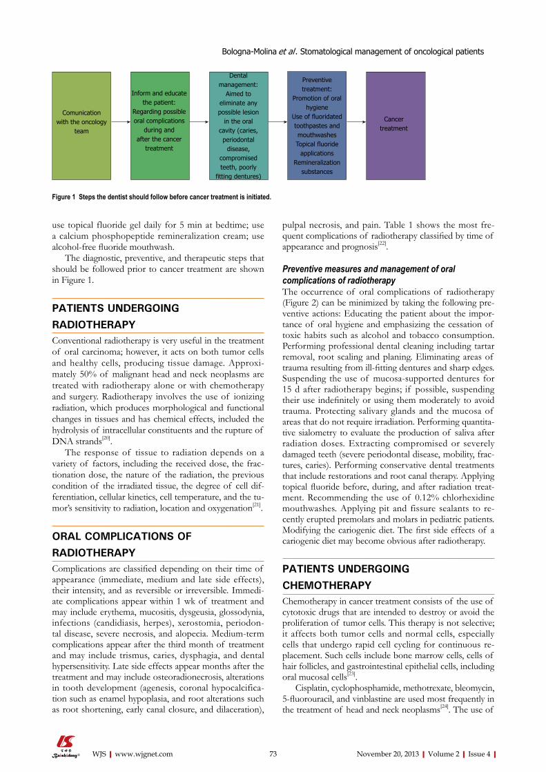
use topical fluoride gel daily for 5 min at bedtime; use a calcium phosphopeptide remineralization cream; use alcohol-free fluoride mouthwash.
The diagnostic, preventive, and therapeutic steps that should be followed prior to cancer treatment are shown in Figure 1.
PATIENTS UNDERGOING RADIOTHERAPYConventional radiotherapy is very useful in the treatment of oral carcinoma; however, it acts on both tumor cells and healthy cells, producing tissue damage. Approxi-mately 50% of malignant head and neck neoplasms are treated with radiotherapy alone or with chemotherapy and surgery. Radiotherapy involves the use of ionizing radiation, which produces morphological and functional changes in tissues and has chemical effects, included the hydrolysis of intracellular constituents and the rupture of DNA strands[20].
The response of tissue to radiation depends on a variety of factors, including the received dose, the frac-tionation dose, the nature of the radiation, the previous condition of the irradiated tissue, the degree of cell dif-ferentiation, cellular kinetics, cell temperature, and the tu-mor’s sensitivity to radiation, location and oxygenation[21].
ORAL COMPLICATIONS OF RADIOTHERAPYComplications are classified depending on their time of appearance (immediate, medium and late side effects), their intensity, and as reversible or irreversible. Immedi-ate complications appear within 1 wk of treatment and may include erythema, mucositis, dysgeusia, glossodynia, infections (candidiasis, herpes), xerostomia, periodon-tal disease, severe necrosis, and alopecia. Medium-term complications appear after the third month of treatment and may include trismus, caries, dysphagia, and dental hypersensitivity. Late side effects appear months after the treatment and may include osteoradionecrosis, alterations in tooth development (agenesis, coronal hypocalcifica-tion such as enamel hypoplasia, and root alterations such as root shortening, early canal closure, and dilaceration),
pulpal necrosis, and pain. Table 1 shows the most fre-quent complications of radiotherapy classified by time of appearance and prognosis[22].
Preventive measures and management of oral complications of radiotherapyThe occurrence of oral complications of radiotherapy (Figure 2) can be minimized by taking the following pre-ventive actions: Educating the patient about the impor-tance of oral hygiene and emphasizing the cessation of toxic habits such as alcohol and tobacco consumption. Performing professional dental cleaning including tartar removal, root scaling and planing. Eliminating areas of trauma resulting from ill-fitting dentures and sharp edges. Suspending the use of mucosa-supported dentures for 15 d after radiotherapy begins; if possible, suspending their use indefinitely or using them moderately to avoid trauma. Protecting salivary glands and the mucosa of areas that do not require irradiation. Performing quantita-tive sialometry to evaluate the production of saliva after radiation doses. Extracting compromised or severely damaged teeth (severe periodontal disease, mobility, frac-tures, caries). Performing conservative dental treatments that include restorations and root canal therapy. Applying topical fluoride before, during, and after radiation treat-ment. Recommending the use of 0.12% chlorhexidine mouthwashes. Applying pit and fissure sealants to re-cently erupted premolars and molars in pediatric patients. Modifying the cariogenic diet. The first side effects of a cariogenic diet may become obvious after radiotherapy.
PATIENTS UNDERGOING CHEMOTHERAPYChemotherapy in cancer treatment consists of the use of cytotoxic drugs that are intended to destroy or avoid the proliferation of tumor cells. This therapy is not selective; it affects both tumor cells and normal cells, especially cells that undergo rapid cell cycling for continuous re-placement. Such cells include bone marrow cells, cells of hair follicles, and gastrointestinal epithelial cells, including oral mucosal cells[23].
Cisplatin, cyclophosphamide, methotrexate, bleomycin, 5-fluorouracil, and vinblastine are used most frequently in the treatment of head and neck neoplasms[24]. The use of
73 November 20, 2013|Volume 2|Issue 4|WJS|www.wjgnet.com
Bologna-Molina et al . Stomatological management of oncological patients
Comunication with the oncology
team
Inform and educate the patient:
Regarding possible oral complications
during and after the cancer
treatment
Dental management:
Aimed to eliminate any possible lesion
in the oral cavity (caries, periodontal
disease, compromised teeth, poorly
fitting dentures)
Preventive treatment:
Promotion of oral hygiene
Use of fluoridated toothpastes and mouthwashesTopical fluoride
applicationsRemineralization
substances
Cancer treatment
Figure 1 Steps the dentist should follow before cancer treatment is initiated.

these drugs may affect the basal epithelial cells that make up the oral mucosal epithelium. When these cells are dam-aged, the replacement of the epithelium is compromised and scaling and ulcerations of the mucosa occur. Further-more, xerostomia caused by salivary gland damage may occur, with resulting alterations in the levels of saliva pro-tectors and ageusia. A high percentage of patients present oral infections, bleeding, or a combination of both, and more than 50% of patients also present complications from surgery and head and neck radiotherapy. Most pa-tients who receive high doses of chemotherapy for head and neck cancer develop severe mucositis[24-26].
Bone marrow suppression is one of the most common side effects of chemotherapy. It is often evident in periph-eral blood after 10-14 d of treatment; typical signs are leu-kopenia, neutropenia, thrombocytopenia and anemia. Hair loss, nausea, vomiting, and palmoplantar erythrodysesthe-sia syndrome are common. The latter is a palmoplantar er-ythema with erosions, burning sensation, and local pain[27].
PATIENTS UNDERGOING BISPHOSPHONATE TREATMENTBisphosphonates are a group of drugs used for the prevention and treatment of bone resorption diseases such as maxillofacial cancer, bone metastasis, malignant hypercalcemia, osteoporosis, Paget’s disease, and multiple myeloma. Their structure is based on that of pyrophos-phate, a metabolite that regulates the precipitation and extraction of bone minerals; similar to pyrophosphate,
they are sensitive to hydrolysis by phosphatases[28-30].When bisphosphonates are incorporated into bone,
osteoclast-mediated bone resorption is prevented, and os-teoclast apoptosis is stimulated. These drugs have affinity for active bone replacement sites and growth plates[30,31].
Bone resorption does not occur in patients under-going bisphosphonate treatment due to inhibition by bisphosphonates of the osteoclastic activity that normally causes decreased bone replacement and a lack of new bone formation. In these patients, the bone is present for a longer period without replacement, making it prone to chronic infections and necrosis. Bisphosphonates also inhibit angiogenesis, leading to diminished bone vascular-ization and induction of bone cell apoptosis[27,32-34].
Osteonecrosis of the jaw is one of the complications of treatment with bisphosphonates in cancer patients. It most often occurs in the mandible and is associated with 53% of dental interventions; 48% of affected patients show a response to treatment when the drug has been used for a period of over eight weeks[35-37].
DIAGNOSTIC CRITERIA FOR BISPHOSPHONATE-RELATED OSTEONE- CROSIS OF THE JAWTreatment with bisphosphonates must be considered in the medical history of patients with ulcerated lesions within the jaw with bone exposure for over eight weeks, necrotic bone, or lesions that do not heal spontaneously.
74 November 20, 2013|Volume 2|Issue 4|WJS|www.wjgnet.com
Complication Characteristics Time of appearance Prognosis
Erythema/ radiodermatitis
Redness/decreased skin thickness; skin dryness due to epidermal basal cell damage
Immediate Reversible
Mucositis Generalized inflammation of the oral mucosa due to basal cell damage; scaling, mucosal ulcerations
Immediate Reversible
Dysgeusia Altered taste (especially to sour and acid tastes) due to taste bud damage Immediate Reversible Glossodynia Pain and burning sensation in the tongue due to taste bud damage and
inflammationImmediate Reversible
Candidiasis and Herpes simplex
Secondary infections resulting from loss of mucosal protection caused by mucositis and xerostomia
Immediate Reversible
Xerostomia Decrease in salivary flow and dryness of the mouth caused by alterations in salivary glands
Immediate Irreversible at high radiation doses (more than 60 Gy)
Periodontal disease Inflammation of the periodontium due to augmented plaque from decreased salivary flow
Immediate Reversible
Alopecia Hair loss from hair follicle atrophy Immediate Reversible Severe necrosis Loss of tissue, scurvy and malodorous ulcerations Immediate Irreversible Trismus Reduced mouth opening caused by fibrosis of the muscles of mastication or of
the temporomandibular jointMedium-term Reversible/irreversible
Caries Damage to the cement-enamel junction, incisal edges and cusps caused by decreased salivation
Medium-term Irreversible
Dysphagia Difficulty swallowing food caused by oropharyngeal alterations; may be evidenced by malnutrition
Medium-term Reversible
Dental hypersensitivity
Dental sensitivity caused by radiation Medium-term Reversible
Osteoradionecrosis Aseptic necrosis of the irradiated bone Late Irreversible Tooth germ alterations
Alteration in odontogenesis in pediatric patients Late Irreversible
Pulp necrosis and pain
Pulp necrosis and pain Late Irreversible
Table 1 Common complications of the head and neck region following radiotherapy
Bologna-Molina et al . Stomatological management of oncological patients

Additional tests such as orthopantomography and computerized tomography scanning are recommended to evaluate the extent of the lesion.
PREVENTIVE MEASURES IN PATIENTS UNDERGOING BISPHOSPHONATE TREATMENTOnce oral or intravenous treatment with bisphospho-nates has been decided upon, preventive measures must
be taken before and after treatment to avoid or minimize osteonecrosis[38,39].
Basic preventive measures prior to treatment focus on eliminating potential sites of infection and extracting teeth with poor prognoses to minimize the risk during therapy[40]. Once treatment has begun, meticulous control of the patient must be maintained so as to detect any sign of osteonecrosis. Good oral hygiene with plaque con-trol and dental cleaning, tartar removal, and periodontal pocket treatment must also be included. Surgical proce-dures must be minimally invasive, performed only when
75 November 20, 2013|Volume 2|Issue 4|WJS|www.wjgnet.com
Mucositis
• Preserve excellent oral hygiene• Avoid irritating foods (spices, coffee, acids, alcohol, tobacco, etc. )• 0.12% chlorhexidine mouthwashes• 2% lidocaine or 1% dyclonine hydrochloride topical anesthetics in an aqueous or viscous solution or mucoadhesive benzocaine hydrochloride to reduce pain• Use of epithelial protective drugs such as kaolin, magnesium hydrochloride, aluminum hydrochloride• Conventional strong analgesics and antiinflammatory drugs• Soft diet• Hydration• If secondary infection occurs, a culture and a cytological test must be performed, and broad-spectrum antibiotics must be used In cases of Candida albicans superinfection , Nystatin oral suspension should be used 4 times daily for 4 min for 4 wk in addition to 200 mg of ketoconazole per day or 100 mg of fluconazole. A cyclovir is prescribed for Herpes simplex
• Zinc supplements: 110-120 mg of zinc sulfate 2 times per day• Diet modification
• Increase hydration• Use of carboxymethyl cellulose saliva substitutes, sorbitol synthetic saliva, artificial salive• Sialogogues with xylitol, such as gum• 5 mg pilocarpine in the morning and at night• 2% pilocaroine droplets applied to the floor of the mouth
Dysgeusia
Xerostomia
Osteoradionecrosis
• Preventive measures• Avoid mucosal trauma• Avoid dental extractions• Irrigation with physiological salution, antibiotics• Chlorhexidine mouthwashes and irrigations• Use of hyperbaric oxygen• Avoid the use of regional or intraligamentary anesthesia, maintain low vasoconstriction
Trismus
• Physiotherapy• Muscle relaxant drugs
Caries from radiation
• Daily application of topical fluride and fluoride mouthwash• Chlorhexidine mouthwash• Meticulous oral hygiene with use of a soft toothbrush• Eliminating cariogenic aiet• Frequent dental check-ups to evaluate oral health
Pain
• Depending on the degree of pain: acetylsalicylic acid, codeine, dihydrocodeine, Tramadol
Figure 2 Treatment steps the dentist must follow in cases of oral complications in radiotherapy patients.
Bologna-Molina et al . Stomatological management of oncological patients
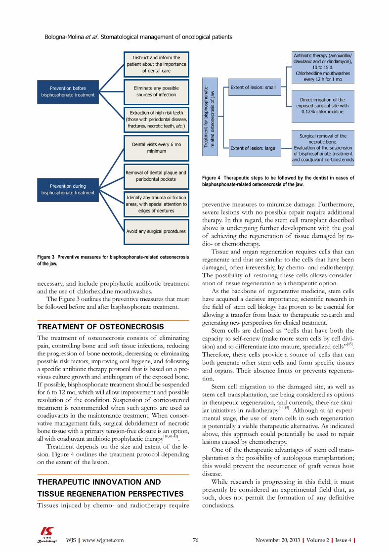
necessary, and include prophylactic antibiotic treatment and the use of chlorhexidine mouthwashes.
The Figure 3 outlines the preventive measures that must be followed before and after bisphosphonate treatment.
TREATMENT OF OSTEONECROSISThe treatment of osteonecrosis consists of eliminating pain, controlling bone and soft tissue infections, reducing the progression of bone necrosis, decreasing or eliminating possible risk factors, improving oral hygiene, and following a specific antibiotic therapy protocol that is based on a pre-vious culture growth and antibiogram of the exposed bone. If possible, bisphosphonate treatment should be suspended for 6 to 12 mo, which will allow improvement and possible resolution of the condition. Suspension of corticosteroid treatment is recommended when such agents are used as coadjuvants in the maintenance treatment. When conser-vative management fails, surgical debridement of necrotic bone tissue with a primary tension-free closure is an option, all with coadjuvant antibiotic prophylactic therapy[10,41-43].
Treatment depends on the size and extent of the le-sion. Figure 4 outlines the treatment protocol depending on the extent of the lesion.
THERAPEUTIC INNOVATION AND TISSUE REGENERATION PERSPECTIVESTissues injured by chemo- and radiotherapy require
preventive measures to minimize damage. Furthermore, severe lesions with no possible repair require additional therapy. In this regard, the stem cell transplant described above is undergoing further development with the goal of achieving the regeneration of tissue damaged by ra-dio- or chemotherapy.
Tissue and organ regeneration requires cells that can regenerate and that are similar to the cells that have been damaged, often irreversibly, by chemo- and radiotherapy. The possibility of restoring these cells allows consider-ation of tissue regeneration as a therapeutic option.
As the backbone of regenerative medicine, stem cells have acquired a decisive importance; scientific research in the field of stem cell biology has proven to be essential for allowing a transfer from basic to therapeutic research and generating new perspectives for clinical treatment.
Stem cells are defined as “cells that have both the capacity to self-renew (make more stem cells by cell divi-sion) and to differentiate into mature, specialized cells”[43]. Therefore, these cells provide a source of cells that can both generate other stem cells and form specific tissues and organs. Their absence limits or prevents regenera-tion.
Stem cell migration to the damaged site, as well as stem cell transplantation, are being considered as options in therapeutic regeneration, and currently, there are simi-lar initiatives in radiotherapy[44,45]. Although at an experi-mental stage, the use of stem cells in such regeneration is potentially a viable therapeutic alternative. As indicated above, this approach could potentially be used to repair lesions caused by chemotherapy.
One of the therapeutic advantages of stem cell trans-plantation is the possibility of autologous transplantation; this would prevent the occurrence of graft versus host disease.
While research is progressing in this field, it must presently be considered an experimental field that, as such, does not permit the formation of any definitive conclusions.
76 November 20, 2013|Volume 2|Issue 4|WJS|www.wjgnet.com
Instruct and inform the patient about the importance
of dental care
Eliminate any possible sources of infection
Extraction of high-risk teeth (those with periodontal disease, fractures, necrotic teeth, etc.)
Dental visits every 6 mo minimum
Removal of dental plaque and periodontal pockets
Identify any trauma or friction areas, with special attention to
edges of dentures
Avoid any surgical procedures
Prevention before bisphosphonate treatment
Prevention during bisphosphonate treatment
Figure 3 Preventive measures for bisphosphonate-related osteonecrosis of the jaw.
Trea
tmen
t fo
r bi
spho
spho
nate
-re
late
d os
teon
ecro
sis
of ja
w
Extent of lesion: small
Extent of lesion: large
Antibiotic therapy (amoxicillin/clavulanic acid or clindamycin),
10 to 15 d.Chlorhexidine mouthwashes
every 12 h for 1 mo
Direct irrigation of the exposed surgical site with
0.12% chlorhexidine
Surgical removal of the necrotic bone.
Evaluation of the suspension of bisphosphonate treatment
and coadjuvant corticosteroids
Figure 4 Therapeutic steps to be followed by the dentist in cases of bisphosphonate-related osteonecrosis of the jaw.
Bologna-Molina et al . Stomatological management of oncological patients
77 November 20, 2013|Volume 2|Issue 4|WJS|www.wjgnet.com
REFERENCES1 Petersen PE. Oral healthcare in people living with cancer.
Oral Oncol 2010; 46: 399-400 [PMID: 20403724 DOI: /10.1016/j.oraloncology.2010.03.018]
2 Meurman JH. Infectious and dietary risk factors of oral cancer. Oral Oncol 2010; 46: 411-413 [PMID: 20381409 DOI: 10.1016/j.oraloncology.2010.03.003]
3 Meurman JH, Grönroos L. Oral and dental health care of oral cancer patients: hyposalivation, caries and infections. Oral Oncol 2010; 46: 464-467 [PMID: 20308007 DOI: 10.1016/j.oraloncology.2010.02.025]
4 Scully C, Petti S. Overview of cancer for the healthcare team: aetiopathogenesis and early diagnosis. Oral Oncol 2010; 46: 402-406 [PMID: 20350835 DOI: 10.1016/j.oraloncol-ogy.2010.02.026]
5 Joshi VK. Dental treatment planning and management for the mouth cancer patient. Oral Oncol 2010; 46: 475-479 [PMID: 20400359 DOI: 10.1016/j.oraloncology.2010.03.010]
6 Porter SR, Fedele S, Habbab KM. Taste dysfunction in head and neck malignancy. Oral Oncol 2010; 46: 457-459 [PMID: 20400364 DOI: 10.1016/j.oraloncology.2010.03.011]
7 Lanza Echeveste DG. Tratamiento odontológico integral del paciente oncológico. Parte I. Comprehensive dental treat-ment of cancer patients. Part I. Odontoestomatología 2011; 13: 14-25
8 Lip and Oral Cavity Cancer Treatment (PDQ®). General Information About Lip and Oral Cavity Cancer. Available from: URL: http://www.cancer.gov/cancertopics/pdq/treatment/lip-and-oral-cavity/HealthProfessional
9 Madrid C, Bouferrache K, Abarca M, Jaques B, Broome M. Bisphosphonate-related osteonecrosis of the jaws: how to manage cancer patients. Oral Oncol 2010; 46: 468-470 [PMID: 20452814 DOI: 10.1016/j.oraloncology.2010.03.016]
10 Bagán J, Blade J, Cozar JM, Constela M, García Sanz R, Gó-mez Veiga F, Lahuerta JJ, Lluch A, Massuti B, Morote J, San Miguel JF, Solsona E. Recommendations for the prevention, diagnosis, and treatment of osteonecrosis of the jaw (ONJ) in cancer patients treated with bisphosphonates. Med Oral Patol Oral Cir Bucal 2007; 12: E336-E340 [PMID: 17664922]
11 Raber-Durlacher JE, Elad S, Barasch A. Oral mucositis. Oral Oncol 2010; 46: 452-456 [PMID: 20403721 DOI: 10.1016/j.oraloncology.2010.03.012]
12 Newton JT. Reactions to cancer: communicating with pa-tients, family and carers. Oral Oncol 2010; 46: 442-444 [PMID: 20381407 DOI: 10.1016/j.oraloncology.2010.03.015]
13 Warnakulasuriya S. Living with oral cancer: epidemiology with particular reference to prevalence and life-style changes that influence survival. Oral Oncol 2010; 46: 407-410 [PMID: 20403722 DOI: 10.1016/j.oraloncology.2010.02.015]
14 van der Waal I. Potentially malignant disorders of the oral and oropharyngeal mucosa; present concepts of manage-ment. Oral Oncol 2010; 46: 423-425 [PMID: 20308005 DOI: 10.1016/j.oraloncology.2010.02.016]
15 Rocha Buelvas A. Cancer oral el papel del odontólogo en la detección temprana y control. The role of the dentist in early detection and treatment of oral cancer. Rev Fac Odont Univ Antioq 2009; 21: 112-121
16 Whitmyer CC, Waskowski JC, Iffland HA. Radiotherapy and oral sequelae: preventive and management protocols. J Dent Hyg 1997; 71: 23-29 [PMID: 9470559]
17 Porter SR, Fedele S, Habbab KM. Xerostomia in head and neck malignancy. Oral Oncol 2010; 46: 460-463 [PMID: 20403723 DOI: 10.1016/j.oraloncology.2010.03.008]
18 Zheng WK, Inokuchi A, Yamamoto T, Komiyama S. Taste dysfunction in irradiated patients with head and neck can-cer. Fukuoka Igaku Zasshi 2002; 93: 64-76 [PMID: 12048909]
19 Dios PD, Lestón JS. Oral cancer pain. Oral Oncol 2010; 46: 448-451 [PMID: 20308009 DOI: 10.1016/j.oraloncology.2010.02.017]
20 Mikhaĭlova NS, Alekseev NA. Case of nonspherocytic ane-mia complicated by glucose-6-phosphate dehydrogenase de-ficiency of the erythrocytes. Probl Gematol Pereliv Krovi 1976; 21: 54 [PMID: 1273065]
21 Cano Pérez S, Gutiérrez Villar MD. Complicaciones de la radioterapia en la cavidad oral. Oral complications of radiotherapy. Semergen 2002; 28: 363-369 [DOI: 10.1016/S1138-3593(02)74087-2]
22 Rocha Buelvas A, Jojoa Pumalpa A. Manejo odontológico de las complicaciones orales secundarias AL tratamiento oncológico con quimioterapia y radioterapia Dental manage-ment of oral complications of cancer treatment with chemo-therapy and radiotherapy. CES Odont 2011; 24: 71-78
23 López-Galindo MP, Bagán JV, Jiménez-Soriano Y, Alpiste F, Camps C. Clinical evaluation of dental and periodontal status in a group of oncological patients before chemother-apy. Med Oral Patol Oral Cir Bucal 2006; 11: E17-E21 [PMID: 16388287]
24 Chaveli Lopez B, Gabaldá Esteve C, Sarrión Pérez M. Dental treatment considerations in the chemoterapy patient. J Clin Exp Dent 2011; 3: e31-42 [DOI: 10.4317/jced.3.e31]
25 Sonis ST. Mucositis as a biological process: a new hy-pothesis for the development of chemotherapy-induced stomatotoxicity. Oral Oncol 1998; 34: 39-43 [DOI: 10.1016/S1368-8375(97)00053-5]
26 Logan RM. Advances in understanding of toxicities of treat-ment for head and neck cancer. Oral Oncol 2009; 45: 844-848 [PMID: 19467918 DOI: 10.1016/j.oraloncology.2009.03.018]
27 Grunberg SM, Deuson RR, Mavros P, Geling O, Hansen M, Cruciani G, Daniele B, De Pouvourville G, Rubenstein EB, Daugaard G. Incidence of chemotherapy-induced nau-sea and emesis after modern antiemetics. Cancer 2004; 100: 2261-2268 [PMID: 15139073 DOI: 10.1002/cncr.20230]
28 Ata-Ali F, Ata-Ali J, Flichy-Fernández AJ, Bágan JV. Osteo-necrosis of the jaws in patients treated with bisphosphonates. J Clin Exp Dent 2012; 4: e60-65 [DOI: 10.4317/jced.50649]
29 Carranza Lira S. Mandible osteonechrosis associated to bisfosfonates. Ginecol Obstet Mex 2007; 75: 655-660 [PMID: 18697439]
30 Madrid C, Abarca M, Bouferrache K. Osteoradionecrosis: an update. Oral Oncol 2010; 46: 471-474 [PMID: 20457536 DOI: 10.1016/j.oraloncology.2010.03.017]
31 Assael LA. Oral bisphosphonates as a cause of bisphos-phonate-related osteonecrosis of the jaws: clinical findings, assessment of risks, and preventive strategies. J Oral Maxil-lofac Surg 2009; 67: 35-43 [PMID: 19371813 DOI: 10.1016/j.joms.2009.01.003]
32 Ruggiero SL, Fantasia J, Carlson E. Bisphosphonate-related osteonecrosis of the jaw: background and guidelines for di-agnosis, staging and management. Oral Surg Oral Med Oral Pathol Oral Radiol Endod 2006; 102: 433-441 [PMID: 16997108 DOI: 10.1016/j.tripleo.2006.06.004]
33 Murad OM, Arora S, Farag AF, Guber HA. Bisphosphonates and osteonecrosis of the jaw: a retrospective study. Endocr Pract 2007; 13: 232-238 [PMID: 17599853 DOI: 10.4158/EP.13.3.232]
34 Landesberg R, Wilson T, Grbic JT. Bisphosphonate-asso-ciated osteonecrosis of the jaw: conclusions based on an analysis of case series. Dent Today 2006; 25: 52, 54-57 [PMID: 16925161]
35 Kerawala CJ. Complications of head and neck cancer surgery - prevention and management. Oral Oncol 2010; 46: 433-435 [PMID: 20435509 DOI: 10.1016/j.oraloncology.2010.03.013]
36 Fantasia JE. Bisphosphonates. What the dentist needs to know: Practical considerations. J Oral Maxilofac Surg 2009; 67: 53-60 [DOI: 10.1016/j.joms.2009.01.011]
37 Silverman SL, Landesberg R. Osteonecrosis of the jaw and the role of bisphosphonates: a critical review. Am J Med 2009; 122: S33-S45 [PMID: 19187811 DOI: 10.1016/j.amjmed.2008.12.005]
Bologna-Molina et al . Stomatological management of oncological patients
78 November 20, 2013|Volume 2|Issue 4|WJS|www.wjgnet.com
38 Ruggiero SL. Bisphosphonate-related osteonecrosis of the jaw: an overview. Ann N Y Acad Sci 2011; 1218: 38-46 [PMID: 20946580 DOI: 10.1111/j.1749-6632.2010.05768.x]
39 Pérez SB, Barrero MV, Hernández MS, Knezevic M, Navarro JM, Millares JR. Bisphosphonate-associated osteonecrosis of the jaw. A proposal for conservative treatment. Med Oral Patol Oral Cir Bucal 2008; 13: E770-E773 [PMID: 19047964]
40 Ferlito S, Puzzo S, Liardo C. Preventive protocol for tooth extractions in patients treated with zoledronate: a case series. J Oral Maxillofac Surg 2011; 69: e1-e4 [PMID: 21316136 DOI: 10.1016/j.joms.2010.10.055]
41 Manfredi M, Merigo E, Guidotti R, Meleti M, Vescovi P. Bisphosphonate-related osteonecrosis of the jaws: a case series of 25 patients affected by osteoporosis. Int J Oral Max-illofac Surg 2011; 40: 277-284 [PMID: 21163625 DOI: 10.1016/
j.ijom.2010.11.002]42 Bagan J, Scully C, Sabater V, Jimenez Y. Osteonecrosis of the
jaws in patients treated with intravenous bisphosphonates (BRONJ): A concise update. Oral Oncol 2009; 45: 551-554 [PMID: 19251474 DOI: 10.1016/j.oraloncology.2009.01.002]
43 Glossary of Stem Cell-Related Terms. Available from: URL: http://www.isscr.org/home/resources/learn-about-stem-cells/stem-cell-glossary#stem. Accessed 01/06/2013.
44 Razvi E. Trends in the stem cells marketplace--report from Select Biosciences Stem Cells 2012 Conference. Regen Med 2012; 7: 291-294 [PMID: 22594323]
45 Coppes RP, van der Goot A, Lombaert IM. Stem cell ther-apy to reduce radiation-induced normal tissue damage. Semin Radiat Oncol 2009; 19: 112-121 [PMID: 19249649 DOI: 10.1016/j.semradonc.2008.11.005]
P- Reviewers: Toros SZ, Yokoyama S- Editor: Gou SX L- Editor: A E- Editor: Wang CH
Bologna-Molina et al . Stomatological management of oncological patients

World Journal of StomatologyW J S
Online Submissions: http://www.wjgnet.com/esps/[email protected]:10.5321/wjs.v2.i4.79
World J Stomatol 2013 November 20; 2(4): 79-85ISSN 2218-6263 (online)
© 2013 Baishideng Publishing Group Co., Limited. All rights reserved.
Molecular biomarkers of cell proliferation in ameloblastomas
Ronell Bologna-Molina, Ana Maria Bedoya-Borella, Liliana Soria-Moreira, Sandra Soría-Suárez
Ronell Bologna-Molina, Molecular Pathology, School of Den-tistry, Universidad de la República (UDELAR), Montevideo 11600, UruguayAna Maria Bedoya-Borella, Biology Department, CBC, Universidad de Buenos Aires (UBA), Buenos Aires C1122AAH, ArgentinaLiliana Soria-Moreira, Anatomy Pathology, School of Dentistry, Universidad de la República (UDELAR), Montevideo 11600, UruguaySandra Soría-Suárez, Biotechnology Laboratory Solutions (LASOBIOTC), Montevideo 11600, UruguayAuthor contributions: Bologna-Molina R wrote, revised the manuscript and designed the figures; Bedoya-Borella AM, Soria-Moreira L and Soría-Suárez S contributed equally to the writing of the manuscript.Correspondence to: Ronell Bologna-Molina, DDS, PhD, Molecular Pathology, School of Dentistry, Universidad de la República (UDELAR), Las Heras 1925, Montevideo 11600, Uruguay. [email protected]: +598-2-4873048 Fax: +598-2-4008640Received: June 29, 2013 Revised: August 12, 2013Accepted: August 20, 2013Published online: November 20, 2013
AbstractCell proliferation is a vital biological process that is im-portant for all living organisms because of its role in growth and the maintenance of tissue homeostasis. The control of this important process differs greatly among benign and malignant neoplasms, and the evaluation of cell proliferation in neoplasms has become a common tool used by pathologists to provide useful information pertaining to diagnosis, clinical behavior, and treatment. The usefulness of information regarding cell proliferation has led to numerous studies on the value of these meth-ods for diagnosing different types of tumors and for clini-cal decision making. Ameloblastomas are no exception. This review discusses the use of several classical molec-ular proliferation markers, including Ki-67, proliferating cell nuclear antigen, cyclin D1 and DNA topoisomerase Ⅱ alpha, to characterize ameloblastomas and proposes the use of new proliferation markers used previously to characterize other neoplasms. The use of these biomark-
ers offers valuable opportunities to evaluate the biologi-cal behavior of this type of odontogenic tumor.
© 2013 Baishideng Publishing Group Co., Limited. All rights reserved.
Key words: Ameloblastoma; Ki-67; Proliferating cell nuclear antigen; cyclin D1; DNA topoisomerase
Core tip: Specific molecular markers are characteristic of particular cellular events such as proliferation, and in this context, “proliferation markers” refer to specific proteins or other factors in actively growing and divid-ing cells, whose presence serves as an indicator for such cells. In this mini-review, we aim to provide an overview of the methods currently available for the as-sessment of proliferation, and we review the different cell proliferation markers used to assess the biological behavior of ameloblastomas. In addition, we propose a new maker.
Bologna-Molina R, Bedoya-Borella AM, Soria-Moreira L, Soría-Suárez S. Molecular biomarkers of cell proliferation in amelo-blastomas. World J Stomatol 2013; 2(4): 79-85 Available from: URL: http://www.wjgnet.com/2218-6263/full/v2/i4/79.htm DOI: http://dx.doi.org/10.5321/wjs.v2.i4.79
INTRODUCTIONNew cells are generated from pre-existing cells through an ordered sequence of events that is repeated. These events constitute the cell cycle. Traditionally, the cell cycle is divided into stages, the duration of which varies de-pending on the cell type.
The division of an original cell into daughter cells requires prior DNA replication and the synthesis of vari-ous proteins associated with this replication step, as well as the production of structures and organelles for the new cells. These processes occur at the interphase of the cycle, which itself is divided into phases: G1 (Gap 1), S
MINIREVIEWS
79 November 20, 2013|Volume 2|Issue 4|WJS|www.wjgnet.com
(Synthesis) and G2 (Gap 2).When all the conditions necessary for division are
met, M stage (or division) starts, which results in the separation of the chromosomes and then the division of the cytoplasm, known as cytokinesis. Some cells can re-main in a state of active metabolism for a very long time without replicating their DNA or dividing. These cells are in G0 phase or the quiescent state. G0 is also considered a post-mitotic state[1].
The different cell types divide in a regulated manner. Certain environmental changes, such as temperature varia-tions, changes in pH, nutrient scarcity and contact with neighboring cells, can slow down the cell cycle. Additionally, the presence of growth factors and hormones can trigger a series of intracellular processes that stimulate cell division. Cell proliferation can be defined as an increase in the num-ber of cells as a result of cell growth and cell division.
In neoplastic processes, the abnormal and uncon-trolled proliferation of cells is observed, and the cell cycle is altered. The assessment of cell proliferation activity in tumors has become a common tool used by histopa-thologists to provide useful information for assessing and predicting the behavior of tumors-that is, their likelihood of local recurrence, their metastatic potential, and the growth of metastases, and thus the likely duration of disease-free survival and survival to death[2]. Today, the most common method for evaluating proliferative activity is the use of im-munohistochemical techniques.
Immunohistochemical staining is widely used in the identification of abnormal cells such as those found in cancerous tumors. This technique is also widely used in basic research to understand the distribution and localiza-tion of biomarkers and differentially expressed proteins in different parts of a biological specimen.
Specific molecular markers are characteristic of par-ticular cellular events such as proliferation, and in this context, “proliferation markers” refer to specific proteins or other factors in actively growing and dividing cells, whose presence serves as an indicator for such cells[3].
Two requirements have been postulated for this type of marker: (1) the antigen should be continuously present dur-ing the cell cycle of all cell types; and (2) the transition to a nonproliferative state from any step of the cell cycle should be followed by a rapid disappearance of the antigen[4].
Odontogenic tumors constitute a group of hetero-geneous lesions that range from hamartomatous or non-neoplastic tissue proliferations to benign and malignant neoplasms with variable aggressiveness.
Odontoma is the most common odontogenic tumor, but it is considered a non-neoplastic lesion. Ameloblas-toma is the most common odontogenic neoplasm. Ac-cording to the 2005 Histological Classification of Tumors of the World Health Organization, ameloblastomas are divided into four variants: solid/multicystic, extraosseous/peripheral, desmoplastic and unicystic. There exist several histological subtypes: follicular, plexiform, acanthoma-tous, granular and basal cell. Although ameloblastomas are classified as benign neoplasms, they can be locally invasive and destructive tumors of the jawbone. The molecular
mechanisms that regulate cell growth and invasion in am-eloblastomas are unknown. Determining the proliferative activity of ameloblastomas may provide important infor-mation regarding the appropriate treatment strategy.
In this mini-review, we aim to provide an overview of the methods currently available for the assessment of proliferation, and we review the different cell prolifera-tion markers used to assess the biological behavior of ameloblastomas.
There are many methods for determining the level of proliferative activity in different types of tumors, includ-ing the analysis of the mitotic index, flow cytometry, sil-ver staining (AgNOR), and immunohistochemistry tech-niques. The last two are the most widely used techniques to study ameloblastomas.
It is important to clarify that there are more specific and sensitive techniques for determining the presence of these proliferation markers such proteomics techniques which allow to know what proteins are present or absent in these tumors. Another technique quantitative, sensitive and highly specific is the real-time polymerase chain reac-tion that allows determining the expression levels of genes in the ameloblastoma.
Both techniques are more expensive and more la-borious than the immunohistochemistry technique that despite having less specificity and sensitivity has the advantage of being able to display “in situ” the presence of proteins, important data for understanding how the tumor proliferates.
MOLECULAR MARKERSSilver binding nucleolar organizer regionSeveral methods have been used for the identification of proliferating cells in tissue sections with the aim of using them as markers of impending malignancy. One among of these methods is the silver binding nucleolar organizer region (AgNOR) technique.
Nucleolar organizer regions (NORs) are segments of DNA that are closely associated with nucleoli, which contain the ribosomal DNA. These regions therefore contribute strongly to the regulation of protein synthesis. NORs are argyrophilic and can therefore be visualized us-ing a silver staining technique, what has led to the use of the term AgNOR[2]. AgNOR staining is a simple one-step staining technique that overcomes the disadvantages of other techniques, such as the requirements for sophisticated equipment and technical expertise, high cost and long run-time[3,5]. The amount of AgNOR protein, estimated during interphase, can be used as a marker of cell proliferation and has prognostic value for several human cancers.
In the study by Seifi et al[6], the number of AgNOR dots in solid/multicystic ameloblastomas was found to be higher than that in unicystic ameloblastomas.
Coleman et al[7] reported that unicystic ameloblasto-mas lined with characteristic epithelium had a significantly lower AgNOR count than solid ameloblastomas, residual dentigerous cysts and keratocystic odontogenic tumors, and these authors concluded that AgNOR counts are not
80 November 20, 2013|Volume 2|Issue 4|WJS|www.wjgnet.com
Bologna-Molina R et al . Cell Proliferation in ameloblastomas
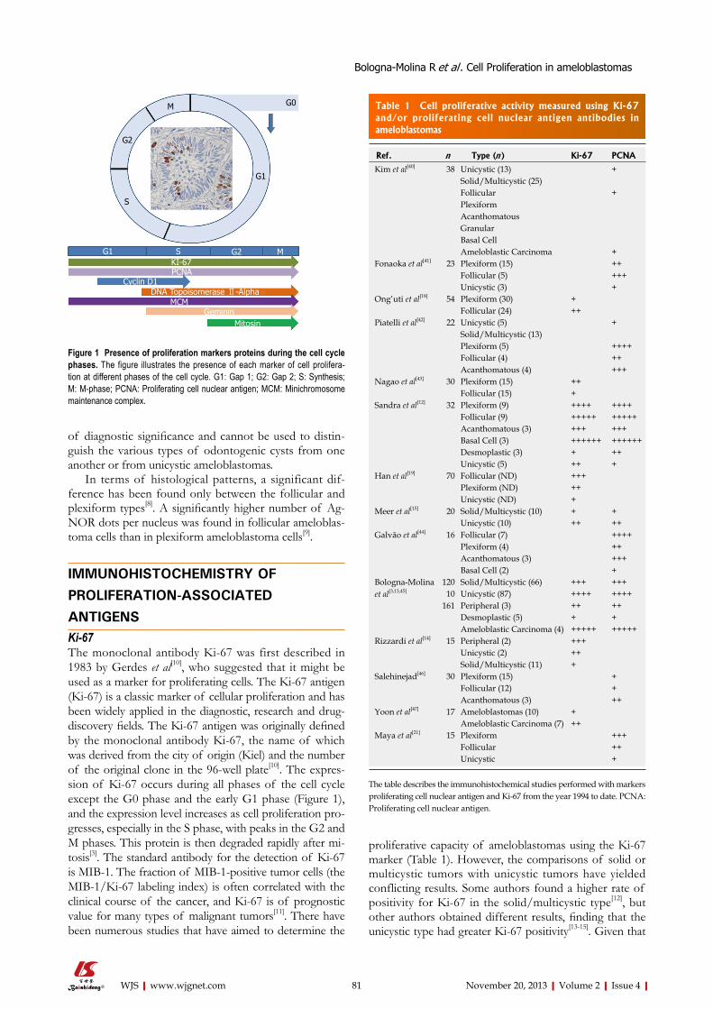
of diagnostic significance and cannot be used to distin-guish the various types of odontogenic cysts from one another or from unicystic ameloblastomas.
In terms of histological patterns, a significant dif-ference has been found only between the follicular and plexiform types[8]. A significantly higher number of Ag-NOR dots per nucleus was found in follicular ameloblas-toma cells than in plexiform ameloblastoma cells[9].
IMMUNOHISTOCHEMISTRY OF PROLIFERATION-ASSOCIATED ANTIGENSKi-67The monoclonal antibody Ki-67 was first described in 1983 by Gerdes et al[10], who suggested that it might be used as a marker for proliferating cells. The Ki-67 antigen (Ki-67) is a classic marker of cellular proliferation and has been widely applied in the diagnostic, research and drug-discovery fields. The Ki-67 antigen was originally defined by the monoclonal antibody Ki-67, the name of which was derived from the city of origin (Kiel) and the number of the original clone in the 96-well plate[10]. The expres-sion of Ki-67 occurs during all phases of the cell cycle except the G0 phase and the early G1 phase (Figure 1), and the expression level increases as cell proliferation pro-gresses, especially in the S phase, with peaks in the G2 and M phases. This protein is then degraded rapidly after mi-tosis[3]. The standard antibody for the detection of Ki-67 is MIB-1. The fraction of MIB-1-positive tumor cells (the MIB-1/Ki-67 labeling index) is often correlated with the clinical course of the cancer, and Ki-67 is of prognostic value for many types of malignant tumors[11]. There have been numerous studies that have aimed to determine the
proliferative capacity of ameloblastomas using the Ki-67 marker (Table 1). However, the comparisons of solid or multicystic tumors with unicystic tumors have yielded conflicting results. Some authors found a higher rate of positivity for Ki-67 in the solid/multicystic type[12], but other authors obtained different results, finding that the unicystic type had greater Ki-67 positivity[13-15]. Given that
81 November 20, 2013|Volume 2|Issue 4|WJS|www.wjgnet.com
M
S
G1
G2
G0
G1 G2 MSKI-67PCNA
Cyclin D1DNA Topoisomerase Ⅱ-Alpha
MCMGeminin
Mitosin
Figure 1 Presence of proliferation markers proteins during the cell cycle phases. The figure illustrates the presence of each marker of cell prolifera-tion at different phases of the cell cycle. G1: Gap 1; G2: Gap 2; S: Synthesis; M: M-phase; PCNA: Proliferating cell nuclear antigen; MCM: Minichromosome maintenance complex.
Ref. n Type (n) Ki-67 PCNA
Kim et al[40] 38 Unicystic (13) +Solid/Multicystic (25)Follicular +PlexiformAcanthomatousGranularBasal CellAmeloblastic Carcinoma +
Fonaoka et al[41] 23 Plexiform (15) ++Follicular (5) +++Unicystic (3) +
Ong’uti et al[18] 54 Plexiform (30) +Follicular (24) ++
Piatelli et al[42] 22 Unicystic (5) +Solid/Multicystic (13)Plexiform (5) ++++Follicular (4) ++Acanthomatous (4) +++
Nagao et al[43] 30 Plexiform (15) ++Follicular (15) +
Sandra et al[12] 32 Plexiform (9) ++++ ++++Follicular (9) +++++ +++++Acanthomatous (3) +++ +++Basal Cell (3) ++++++ ++++++Desmoplastic (3) + ++Unicystic (5) ++ +
Han et al[19] 70 Follicular (ND) +++Plexiform (ND) ++Unicystic (ND) +
Meer et al[15] 20 Solid/Multicystic (10) + +Unicystic (10) ++ ++
Galvão et al[44] 16 Follicular (7) ++++Plexiform (4) ++Acanthomatous (3) +++Basal Cell (2) +
Bologna-Molina et al[3,13,45]
120 Solid/Multicystic (66) +++ +++ 10 Unicystic (87) ++++ ++++161 Peripheral (3) ++ ++
Desmoplastic (5) + +Ameloblastic Carcinoma (4) +++++ +++++
Rizzardi et al[14] 15 Peripheral (2) +++Unicystic (2) ++Solid/Multicystic (11) +
Salehinejad[46] 30 Plexiform (15) +Follicular (12) +Acanthomatous (3) ++
Yoon et al[47] 17 Ameloblastomas (10) +Ameloblastic Carcinoma (7) ++
Maya et al[21] 15 Plexiform +++Follicular ++Unicystic +
Table 1 Cell proliferative activity measured using Ki-67 and/or proliferating cell nuclear antigen antibodies in ameloblastomas
The table describes the immunohistochemical studies performed with markers proliferating cell nuclear antigen and Ki-67 from the year 1994 to date. PCNA: Proliferating cell nuclear antigen.
Bologna-Molina R et al . Cell Proliferation in ameloblastomas
several clinicopathological studies have found that solid ameloblastomas are more aggressive than unicystic am-eloblastomas[16-19], the higher index of cell proliferation in unicystic ameloblastomas (determined using the Ki-67 an-tibody) found in some studies appears contradictory. This finding could be explained by the fact that unicystic ame-loblastomas contain fewer stellate reticulum-like cells than solid/multicystic ameloblastomas, and consequently, most of the cells counted corresponded to basal or suprabasal layers, which are more likely to be positive. In other words, the proportions of the diverse types of epithelial cells, as well as the different mechanisms of growth in unicystic ameloblastoma and solid/multicysticameloblastoma , may influence the results of the proliferation index[13].
When histological subtypes were studied Ong’uti et al[18] in a study of 54 cases of ameloblastoma in Kenya, these researchers found that follicular ameloblastomas had a higher proliferation index than the plexiform vari-ant. These results are similar to those of Han et al[19], who studied a Chinese population and found a slight predomi-nance of a higher proliferation index in the follicular vari-ant. Sandra and colleagues included the basal cell variant in their study, and this variant was found to have greater positivity for Ki-67 than the follicular variant[12]. In our previous study, we found similar results, with the follicu-lar variant having a higher proliferation index[13].
Proliferating cell nuclear antigen Proliferating cell nuclear antigen (PCNA) is a nuclear nonhistone protein that is necessary for DNA synthesis and is an accessory protein for DNA polymerase alpha, the expression of PCNA occurs during all phases of the cell cycle and the level of this protein is elevated dur-ing the G1/S phase of the cell cycle (Figure 1). PCNA expression can be used as a marker of cell proliferation because cells remain in the G1/S phase for a longer time when proliferating. Furthermore, this protein has an es-sential role in nucleic acid metabolism as a component of the DNA replication and repair machinery[20,21].
Multiple studies using PCNA have been performed to determine the rates of cell proliferation in various types of ameloblastomas, but the results are contradic-tory (Table 1). Some authors did not found any relevant differences between the different types and subtypes of ameloblastomas[3,11,22]. This result is most likely because PCNA is also involved in DNA repair. Because there is active ongoing DNA repair in many tumors, PCNA may also be upregulated in non-proliferating cells. Indeed, in some tumors, 100% of cells show positive staining. Therefore, after an initial period of popularity, PCNA is no longer considered a reliable proliferation marker in tumors[2]. Despite this conclusion, there are still numerous studies using PCNA as the first-choice marker of cell prolif-eration in ameloblastomas (Table 1).
Cyclin D1The cyclins, together with cyclin-dependent kinases (CDKs), are the proteins responsible for the orderly
progression of cells through the cell cycle. Cyclin D1 is amplified and/or overexpressed in a substantial propor-tion of different human tumors. Increased cyclin D1 expression occurs relatively early during tumorigenesis[23]. Changes in the genes encoding these proteins as well as changes in the expression levels of these proteins are found during the process of carcinogenesis. The overexpression of this protein leads to uncontrolled cell proliferation and tumor development[23]. Cyclin D1 is the regulatory subunit of the holoenzyme that phosphory-lates and, together with sequential phosphorylation by cyclin E/CDK2, inactivates the cell-cycle inhibiting func-tion of the retinoblastoma protein (pRb). pRb serves as a gatekeeper for the G1 phase, and passage through this restriction point leads to DNA synthesis. Thus, cyclin D1 promotes progression through the G1/S phase of the cell cycle (Figure 1)[24,25].
Follicular and plexiform ameloblastomas express cyclin D1 in many peripheral columnar or cuboidal cells and in some central polyhedral cells. No distinct differ-ence in the reaction was detected between these two main tumor types[26]. Kumam et al[27] found that 19/25 follicular ameloblastomas were positive for staining with the cyclin D1 antibody, as were 9/10 plexiform ameloblastomas and 3/4 unicystic ameloblastomas.
DNA topoisomerase Ⅱ alphaDNA topoisomerases are enzymes that disentangle the topological problems that arise from double-stranded DNA. Many of these problems can be solved by gen-erating either single- or double-strand breaks. However, when it is necessary to alter the DNA topology by in-troducing transient double-strand breaks, only DNA topoisomerase Ⅱ (Top2) can fix the problem[28]. Type Ⅱ topoisomerases change the DNA topology by generating transient DNA double-strand breaks. The DNA topoi-somerase Ⅱ alpha (TOP2α) is one of the major nuclear proteins, with peak expression in the S to G2/M phase. It is involved in nearly every aspect of DNA metabolism, playing an important role in chromosome organization and segregation[28].
Kumamoto et al[26] studied the presence of this pro-tein in tooth germs and ameloblastomas, finding lower expression than that reported for Ki-67.
New cell proliferation markersThe usefulness of a marker for tumor diagnosis must be tested for each tumor type and application. Only those markers that have proven to be useful in practice should be considered. These three new cell proliferation markers have been studied in various types of cancer, although there are not currently any reports demonstrating their usefulness in ameloblastomas.
Geminin and minichromosome maintenance complexMinichromosome maintenance complex (MCM2-7) and geminin have important roles in the prevention of DNA re-replication during the cell cycle. MCM proteins are
82 November 20, 2013|Volume 2|Issue 4|WJS|www.wjgnet.com
Bologna-Molina R et al . Cell Proliferation in ameloblastomas

expressed cells in all phases of the cell cycle, including cells that exit the G0 and enter the G1 phase[29]. Gemi-nin is present from the G1/S transition to the early M phase. Thus, MCM is a G0/G1/S/G2/M-phase marker, and geminin is an S/G2/M-phase marker (Figure 1)[30]. MCM proteins are known to contribute to the regulation of transcription, chromatin remodeling and checkpoint responses. The activated MCM complex appears to play a key role in DNA unwinding, acting as a DNA helicase[31]. Following the initiation of DNA replication during the cell cycle, geminin inhibits the reloading of the MCM complex onto chromatin and prevents DNA re-replica-tion during the same cell cycle[32,33].
In recent years, these two proteins have been studied in various types of malignant neoplasms and have been shown to be very useful prognostic markers[34,35].
MitosinMitosin, also termed centromere protein F (CENP-F), is a member of the human centromeric protein family. This protein is associated with the centromere/kinetochore complex and is expressed in all active phases of the cell cycle, with a maximum in G2 and M phases[36]. At the end of mitosis, CENP-F is rapidly proteolyzed by the protea-some. Accumulating evidence suggests that CENP-F is an important protein involved in chromosome alignment and kinetochore-microtubule interactions.
The cell cycle-specific expression of CENP-F makes it a potential marker of proliferation. Indeed, CENP-F is correlated with tumor proliferation in a variety of human tumors, including lung cancer[37], non-Hodgkin lympho-ma[38] and salivary gland tumors[39].
CONCLUSIONThe evaluation of cell proliferation activity in tumors provides useful information related to diagnosis, clinical behavior, treatment and research.
Note that the use of these biomarkers alone are not useful for the diagnosis of ameloblastoma, the diagnosis is based on clinical and histopathologic features, but yet proliferation molecular biomarkers provide important in-formation when predicting the prognosis of patients with ameloblastoma, so the histopathological types together with proliferation marker expression could be useful tools for evaluating the biological behavior of ameloblastomas.
Over the past several decades, various cell prolifera-tion biomarkers have been demonstrated to be useful in the study of various types of neoplasms, and these mark-ers have been studied in some odontogenic tumors and ameloblastomas. The Ki-67 protein remains an excellent operational marker for determining the growth fraction of a given cell population and is considered the gold standard method for the evaluation of proliferation activity.
Despite the abundance of research, the results regard-ing which type of ameloblastoma has the highest rate of cell proliferation remain controversial. One problem is the lack of standardization regarding how to determine
the cell count among many studies. In addition, amelo-blastomas are polymorphic odontogenic tumors, with various types and variants, and the specific histomor-phology of each type and the different mechanisms of growth may influence the observed proliferation index counting.
In recent years there have been found some new mo-lecular biomarkers directly involved in the proliferation biology of tumors. Today new monoclonal antibodies are being tested in different tumors, hence the impor-tance of new research using these new markers in am-eloblastomas.
REFERENCES1 Alberts B, Johnson A, Lewis J, Raff M, Roberts, K, Walter,P.
Molecular Biology of the Cell, 5th edition. New York: Gar-land Science, 2007: 122-134
2 van Diest PJ, Brugal G, Baak JP. Proliferation markers in tumours: interpretation and clinical value. J Clin Pathol 1998; 51: 716-724 [PMID: 10023332 DOI: 10.1136/jcp.51.10.716]
3 Bologna-Molina R, Mosqueda-Taylor A, Molina-Frechero N, Mori-Estevez AD, Sánchez-Acuña G. Comparison of the value of PCNA and Ki-67 as markers of cell proliferation in ameloblastic tumors. Med Oral Patol Oral Cir Bucal 2013; 18: e174-e179 [PMID: 23229269 DOI: 10.4317/medoral.18573]
4 van Dierendonck JH, Keijzer R, van de Velde CJ, Cornelisse CJ. Nuclear distribution of the Ki-67 antigen during the cell cycle: comparison with growth fraction in human breast can-cer cells. Cancer Res 1989; 49: 2999-3006 [PMID: 2720660]
5 Kahn MA, Mincer HH, Dockter ME, Hermann-Petrin JM. Comparing flow cytometric analysis and nucleolar organizer region enumeration in archival oral premalignant lesions. J Oral Pathol Med 1993; 22: 257-262 [PMID: 8355224 DOI: 10.1111/j.1600-0714.1993.tb01067.x]
6 Seifi S, Shafigh E, Allaie A. Quantitative and qualitative analysis of argyrophilic nuclear organizer regions in follicu-lar cyst, keratocystic odontogenic tumor and ameloblastoma. J Cancer Res Ther 2011; 7: 280-285 [PMID: 22044808 DOI: 10.4103/0973-1482.87017]
7 Coleman HG, Altini M, Groeneveld HT. Nucleolar organiz-er regions (AgNORs) in odontogenic cysts and ameloblasto-mas. J Oral Pathol Med 1996; 25: 436-440 [PMID: 8930822 DOI: 10.1111/j.1600-0714.1996.tb00293.x]
8 do Carmo MA, Silva EC. Argyrophilic nucleolar organizer regions (AgNORs) in ameloblastomas and adenomatoid odontogenic tumours (AOTs). J Oral Pathol Med 1998; 27: 153-156 [PMID: 9563569 DOI: 10.1111/j.1600-0714.1998.tb01932.x]
9 Jain VK, Uma K, Soundarya N, Sangeetha R, Smitha T. Comparative morphometric study of AgNORs in variants of ameloblastoma. J Oral Maxillofac Pathol 2012; 16: 354-358 [PMID: 23248466 DOI: 10.4103/0973-029X.102483]
10 Gerdes J, Schwab U, Lemke H, Stein H. Production of a mouse monoclonal antibody reactive with a human nuclear antigen associated with cell proliferation. Int J Cancer 1983; 31: 13-20 [PMID: 6339421 DOI: 10.1002/ijc.2910310104]
11 Ihmann T, Liu J, Schwabe W, Häusler P, Behnke D, Bruch HP, Broll R, Windhövel U, Duchrow M. High-level mRNA quantification of proliferation marker pKi-67 is correlated with favorable prognosis in colorectal carcinoma. J Cancer Res Clin Oncol 2004; 130: 749-756 [PMID: 15449182 DOI: 10.1007/s00432-004-0612-5]
12 Sandra F, Mitsuyasu T, Nakamura N, Shiratsuchi Y, Ohishi M. Immunohistochemical evaluation of PCNA and Ki-67 in ameloblastoma. Oral Oncol 2001; 37: 193-198 [PMID: 11167148 DOI: 10.1016/S1368-8375(00)00079-8]
13 Bologna-Molina R, Mosqueda-Taylor A, Lopez-Corella E,
83 November 20, 2013|Volume 2|Issue 4|WJS|www.wjgnet.com
Bologna-Molina R et al . Cell Proliferation in ameloblastomas
Almeida OP, Carrasco-Daza D, Garcia-Vazquez F, Farfan-Morales JE, Irigoyen-Camacho ME, Damián-Matsumura P. Syndecan-1 (CD138) and Ki-67 expression in different sub-types of ameloblastomas. Oral Oncol 2008; 44: 805-811 [PMID: 18207448 DOI: 10.1016/j.oraloncology.2007.10.007]
14 Rizzardi C, Leocata P, Ventura L, Zweyer M, Brollo A, Sch-neider M, Melato M. Apoptosis-related factors (TRAIL, DR4, DR5, DcR1, DcR2, apoptotic cells) and proliferative activity in ameloblastomas. Anticancer Res 2009; 29: 1137-1142 [PMID: 19414356]
15 Meer S, Galpin JS, Altini M, Coleman H, Ali H. Prolifer-ating cell nuclear antigen and Ki67 immunoreactivity in ameloblastomas. Oral Surg Oral Med Oral Pathol Oral Radiol Endod 2003; 95: 213-221 [PMID: 12582363 DOI: 10.1067/moe.2003.62]
16 Ueno S, Nakamura S, Mushimoto K, Shirasu R. A clini-copathologic study of ameloblastoma. J Oral Maxillofac Surg 1986; 44: 361-365 [PMID: 3457915 DOI: 10.1016/S0278-2391(86)80031-3]
17 Ledesma-Montes C, Mosqueda-Taylor A, Carlos-Bregni R, de León ER, Palma-Guzmán JM, Páez-Valencia C, Meneses-García A. Ameloblastomas: a regional Latin-American mul-ticentric study. Oral Dis 2007; 13: 303-307 [PMID: 17448213 DOI: 10.1111/j.1601-0825.2006.01284.x]
18 Ong’uti MN, Howells GL, Williams DM. An immunohis-tochemical study of keratin expression in ameloblastoma from a Kenyan population. Oral Dis 1999; 5: 111-116 [PMID: 10522206 DOI: 10.1111/j.1601-0825.1999.tb00074.x]
19 Han B, Li L, Wang H. Expression of Ki-67 antigen in amelo-blastoma and its clinical significance. Hua Xi Kou Qiang Yi Xue Za Zhi 2003; 21: 153-154 [PMID: 12838707]
20 de Oliveira MG, Lauxen Ida S, Chaves AC, Rados PV, Sant’Ana Filho M. Immunohistochemical analysis of the patterns of p53 and PCNA expression in odontogenic cystic lesions. Med Oral Patol Oral Cir Bucal 2008; 13: E275-E280 [PMID: 18449109]
21 Maya R, Sekar B, Murali S. Comparative evaluation of expression of proliferating cell nuclear antigen in vari-ants of ameloblastoma and ameloblastic carcinoma. Indian J Dent Res 2012; 23: 15-19 [PMID: 22842243 DOI: 10.4103/0970-9290.99031]
22 Barboza CA, Pereira Pinto L, Freitas Rde A, Costa Ade L, Souza LB. Proliferating cell nuclear antigen (PCNA) and p53 protein expression in ameloblastoma and adenoma-toid odontogenic tumor. Braz Dent J 2005; 16: 56-61 [PMID: 16113935 DOI: 10.1590/S0103-64402005000100010]
23 Angadi PV, Krishnapillai R. Cyclin D1 expression in oral squamous cell carcinoma and verrucous carcinoma: correla-tion with histological differentiation. Oral Surg Oral Med Oral Pathol Oral Radiol Endod 2007; 103: e30-e35 [PMID: 17197212 DOI: 10.1016/j.tripleo.2006.09.011]
24 Baldin V, Lukas J, Marcote MJ, Pagano M, Draetta G. Cyclin D1 is a nuclear protein required for cell cycle progression in G1. Genes Dev 1993; 7: 812-821 [PMID: 8491378 DOI: 10.1101/gad.7.5.812]
25 Fu M, Wang C, Li Z, Sakamaki T, Pestell RG. Minireview: Cyclin D1: normal and abnormal functions. Endocrinol-ogy 2004; 145: 5439-5447 [PMID: 15331580 DOI: 10.1210/en.2004-0959]
26 Kumamoto H, Kimi K, Ooya K. Detection of cell cycle-related factors in ameloblastomas. J Oral Pathol Med 2001; 30: 309-315 [PMID: 11334468 DOI: 10.1034/j.1600-0714.2001.300509.x]
27 Kumar H, Vandana R, Kumar G. Immunohistochemical ex-pression of cyclin D1 in ameloblastomas and adenomatoid odontogenic tumors. J Oral Maxillofac Pathol 2011; 15: 283-287 [PMID: 22144830]
28 Nitiss JL. DNA topoisomerase II and its growing repertoire of biological functions. Nat Rev Cancer 2009; 9: 327-337 [PMID: 19377505 DOI: 10.1038/nrc2608]
29 Lindner K, Gregán J, Montgomery S, Kearsey SE. Essential
role of MCM proteins in premeiotic DNA replication. Mol Biol Cell 2002; 13: 435-444 [PMID: 11854402 DOI: 10.1091/mbc.01-11-0537]
30 Hamamoto Y, Shomori K, Nosaka K, Haruki T, Teshima R, Ito H. Prognostic significance of Minichromosome mainte-nance protein 7 and Geminin expression in patients with 109 soft tissue sarcomas. Oncol Lett 2010; 1: 703-709 [PMID: 22966367]
31 Ishimi Y. A DNA helicase activity is associated with an MCM4, -6, and -7 protein complex. J Biol Chem 1997; 272: 24508-24513 [PMID: 9305914 DOI: 10.1074/jbc.272.39.24508]
32 McGarry TJ, Kirschner MW. Geminin, an inhibitor of DNA replication, is degraded during mitosis. Cell 1998; 93 : 1043-1053 [PMID: 9635433 DOI: 10.1016/S0092-8674(00)81209-X]
33 Lygerou Z, Nurse P. Cell cycle. License withheld--geminin blocks DNA replication. Science 2000; 290: 2271-2273 [PMID: 11188727]
34 Shomori K, Nishihara K, Tamura T, Tatebe S, Horie Y, Nosaka K, Haruki T, Hamamoto Y, Shiomi T, Nakabayashi M, Ito H. Geminin, Ki67, and minichromosome maintenance 2 in gastric hyperplastic polyps, adenomas, and intestinal-type carcinomas: pathobiological significance. Gastric Cancer 2010; 13: 177-185 [PMID: 20820987 DOI: 10.1007/s10120-010-0558-z]
35 Tamura T, Shomori K, Haruki T, Nosaka K, Hamamoto Y, Shiomi T, Ryoke K, Ito H. Minichromosome maintenance-7 and geminin are reliable prognostic markers in patients with oral squamous cell carcinoma: immunohistochemical study. J Oral Pathol Med 2010; 39: 328-334 [PMID: 20136698]
36 Zhu X, Mancini MA, Chang KH, Liu CY, Chen CF, Shan B, Jones D, Yang-Feng TL, Lee WH. Characterization of a novel 350-kilodalton nuclear phosphoprotein that is specifically involved in mitotic-phase progression. Mol Cell Biol 1995; 15: 5017-5029 [PMID: 7651420]
37 Rattner JB, Rees J, Whitehead CM, Casiano CA, Tan EM, Humbel RL, Conrad K, Fritzler MJ. High frequency of neo-plasia in patients with autoantibodies to centromere protein CENP-F. Clin Invest Med 1997; 20: 308-319 [PMID: 9336656]
38 Erlanson M, Casiano CA, Tan EM, Lindh J, Roos G, Land-berg G. Immunohistochemical analysis of the proliferation associated nuclear antigen CENP-F in non-Hodgkin’s lym-phoma. Mod Pathol 1999; 12: 69-74 [PMID: 9950165]
39 Shigeishi H, Mizuta K, Higashikawa K, Yoneda S, Ono S, Kamata N. Correlation of CENP-F gene expression with tumor-proliferating activity in human salivary gland tumors. Oral Oncol 2005; 41: 716-722 [PMID: 15927522 DOI: 10.1016/j.oraloncology.2005.03.008]
40 Kim J, Yook JI. Immunohistochemical study on proliferat-ing cell nuclear antigen expression in ameloblastomas. Eur J Cancer B Oral Oncol 1994; 30B: 126-131 [PMID: 7913362 DOI: 10.1016/0964-1955(94)90064-7]
41 Funaoka K, Arisue M, Kobayashi I, Iizuka T, Kohgo T, Amemiya A, Totsuka Y. Immunohistochemical detection of proliferating cell nuclear antigen (PCNA) in 23 cases of ameloblastoma. Eur J Cancer B Oral Oncol 1996; 32B: 328-332 [PMID: 8944836 DOI: 10.1016/0964-1955(96)00007-3]
42 Piattelli A, Fioroni M, Santinelli A, Rubini C. Expression of proliferating cell nuclear antigen in ameloblastomas and odontogenic cysts. Oral Oncol 1998; 34: 408-412 [PMID: 9861350 DOI: 10.1016/S1368-8375(98)00027-X]
43 Nagao Y, Wato M, Tanaka A. Histochemistry of Ki-67 an-tigen in ameloblastomas. J Osaka Dent Univ 1999; 33: 53-57 [PMID: 10863475]
44 Galvão BC, Pereira PL, de Almeida FR, de Lisboa Lopes CA, Batista de Souza L. Proliferating cell nuclear antigen (PCNA) and p53 protein expression in ameloblastoma and adeno-matoid adontogenic tumor. Braz Dent J 2005; 16: 56-61 [DOI: 10.1590/S0103-64402005000100010 ]
45 Bologna-Molina R, Mosqueda-Taylor A, Lopez-Corella
84 November 20, 2013|Volume 2|Issue 4|WJS|www.wjgnet.com
Bologna-Molina R et al . Cell Proliferation in ameloblastomas

85 November 20, 2013|Volume 2|Issue 4|WJS|www.wjgnet.com
E, de Almeida OP, Carrasco-Daza D, Farfán-Morales JE, Molina-Frechero N, Damián-Matsumura P. Comparative expression of syndecan-1 and Ki-67 in peripheral and des-moplastic ameloblastomas and ameloblastic carcinoma. Pathol Int 2009; 59: 229-233 [PMID: 19351365 DOI: 10.1111/j.1440-1827.2009.02355.x]
46 Salehinejad J, Zare-Mahmoodabadi R, Saghafi S, Jafarian AH, Ghazi N, Rajaei AR, Marouzi P. Immunohistochemical
detection of p53 and PCNA in ameloblastoma and adenoma-toid odontogenic tumor. J Oral Sci 2011; 53: 213-217 [PMID: 21712626 DOI: 10.2334/josnusd.53.213]
47 Yoon HJ, Jo BC, Shin WJ, Cho YA, Lee JI, Hong SP, Hong SD. Comparative immunohistochemical study of ameloblas-toma and ameloblastic carcinoma. Oral Surg Oral Med Oral Pathol Oral Radiol Endod 2011; 112: 767-776 [PMID: 22014999 DOI: 10.1016/j.tripleo.2011.06.036]
P- Reviewer: Li QQ S- Editor: Wen LL L- Editor: A E- Editor: Wang CH
Bologna-Molina R et al . Cell Proliferation in ameloblastomas

World Journal of StomatologyW J S
Online Submissions: http://www.wjgnet.com/esps/[email protected]:10.5321/wjs.v2.i4.86
World J Stomatol 2013 November 20; 2(4): 86-90ISSN 2218-6263 (online)
© 2013 Baishideng Publishing Group Co., Limited. All rights reserved.
Cytotoxicity of a silorane-based dental composite on human gingival fibroblasts
Giovanna Orsini, Alberto Catellani, Concetta Ferretti, Marco Gesi, Monica Mattioli-Belmonte, Angelo Putignano
Giovanna Orsini, Alberto Catellani, Angelo Putignano, De-partment of Clinical Sciences and Stomatology, Polytechnic Uni-versity of Marche, 66020 Ancona, Italy Concetta Ferretti, Monica Mattioli-Belmonte, Department of Clinical and Molecular Sciences, Polytechnic University of Marche, 66020 Ancona, Italy Marco Gesi, Department of Translational Research and New Technology in Medicine and Surgery, University of Pisa, 56126 Pisa, Italy Author contributions: Orsini G and Catellani A wrote the pa-per; Ferretti C performed cell cultures and Mattioli-Belmonte M performed the scanning electron microscope and the statistical analyses; Orsini G and Putignano A designed the research; Gesi M helped for revision and editing of the final paper.Correspondence to: Giovanna Orsini, DDS, PhD, Associate Professor, Department of Clinical Sciences and Stomatology, Polytechnic University of Marche, Via Tronto 10, 66020 Ancona, Italy. [email protected]: +39-71-2206224 Fax: +39-71-2202324Received: June 13, 2013 Revised: August 5, 2013Accepted: August 20, 2013Published online: November 20, 2013
AbstractAIM: To evaluate the direct and indirect biocompatibility of Filtek Silorane on human gingival fibroblastic cells.
METHODS: Sixty-three standardized cylindrical speci-mens (8 mm diameter and 2 mm thickness) of restor-ative material were prepared using a light emitting diode-curing unit. The sample were built up in one in-crement and divided in 2 groups. In the first group, 21 samples (unpolished samples) were left without a spe-cific polishing procedure; in the second one, 42 sam-ples (polished samples) were polished with 4 different grains of discs. Fibroblast cultures, obtained from gin-giva of 2 subjects without systemic and oral disease, were used to assess the direct and indirect biocompat-ibility. Cells cultured for 48 h in normal culture medium were used as a control.
RESULTS: The scanning electron microscope ob-servations of fibroblasts cultured on the silorane samples, either polished or unpolished, confirmed the good biocompatibility of the material, favouring the cellular spreading. 3-dimethylthiazol-2, 5-diphe-nyltetrazolium bromide tests showed a significant re-duction (P < 0.01) of gingival fibroblasts viability cul-tured both in polished samples (90.05% ± 19.00%) and unpolished samples (78.15% ± 11.00%) com-pared with the control. Cells growth in medium condi-tioned with the samples for 1 wk showed a significant viability reduction (P < 0.01) compared to the con-trol. A reduction of cell viability was observed even in the groups containing the material for 3 wk (polished: 89.45% ± 10.00%; unpolished: 65.97% ± 10.00%), even if the cytotoxicity was reduced after this long time exposure.
CONCLUSION: Although the poor chromatic avail-ability of this material remains a big limit that restricts its use to posterior sectors, the silorane-based material can be considered an option to perform restorations when aesthetic demands are not the priority, such as the class Ⅱ restorations
© 2013 Baishideng Publishing Group Co., Limited. All rights reserved.
Key words: Silorane; Cytotoxicity; Resin composite; Fibroblasts
Core tip: The behaviour of silorane-based materials seems to be comparable to the one observed for con-ventional composite material, thus decreasing the cy-totoxicity after long time exposure. Further studies are still needed to characterize the biological response of these methacrylate-free composite formulations, in or-der to definitely demonstrate their safe use in restora-tive dentistry.
Orsini G, Catellani A, Ferretti C, Gesi M, Mattioli-Belmonte M,
BRIEF ARTICLE
86 November 20, 2013|Volume 2|Issue 4|WJS|www.wjgnet.com

Putignano A. Cytotoxicity of a silorane-based dental composite on human gingival fibroblasts. World J Stomatol 2013; 2(4): 86-90 Available from: URL: http://www.wjgnet.com/2218-6263/full/v2/i4/86.htm DOI: http://dx.doi.org/10.5321/wjs.v2.i4.86
INTRODUCTIONRecently, the use of composite materials for restoring dental elements has significantly increased due to the growing aesthetic demand of patients[1].
Despite extensive improvements in mechanical and aesthetic properties of dental composites, volumetric shrinkage and contraction stress during polymerization are still a problem[1]. Contraction stress transferred to the tooth may lead to cusp deflection or enamel micro cracks; additionally, contraction stress of tooth-composite inter-face can determinate post-operative sensitivity, microleak-age, marginal discoloration and recurrent caries[2].
In several studies different techniques have been investigated in order to minimize polymerization shrink-age and contraction stress[3-7]. At the same purpose low-shrinkage materials have been proposed, but none of them offered significant improvement to Bis-GMA-based composites[8].
In 2007, a low shrinkage dental composite based on silorane monomers has been introduced. This material contains traditional filler particles (quartz) and monomers based on a silane or a siloxane core bonded with several oxirane functional groups. The silorane monomers po-lymerize by a ring-opening polymerization process of the oxirane groups. According to its composition, this resin has two advantages: low polymerization shrinkage, due to the ring-opening oxirane monomer, and increased hydro-phobicity, due to the presence of the siloxanes[9].
The release of substances from dental composite materials after polymerization and their possible toxicity have been widely examined during previous years[10-12]. Several in vitro studies have shown cytotoxic, genotoxic, mutagenic, or estrogenic effects of some monomers re-leased by composite materials[13-17].
Limited information is available about the substance eluted from silorane composite and its cell or tissue com-patibility. Kopperud et al[18] found no substance eluted from Filtek silorane in water, while silorane were found in ethanol solution. Krifka et al[19] revealed no significant signs of cytotoxicity on human pulp-derived cells caused by silorane-based materials, while a slight increase in reac-tive oxygen species was detected.
The aim of present study was to evaluate the biocom-patibility of Filtek silorane. The maintaining of surface architecture after finishing was also investigated. These properties were investigated in polished and unpolished silorane polymerized samples.
As regards biocompatibility, we studied the viability of human fibroblastic cells both after direct contact with silorane composite and after cells conditioning using a medium exposed to silorane.
MATERIALS AND METHODSSixty-three standardized cylindrical specimens (8 mm in diameter and 2 mm in thickness) were prepared using a transparent plastic molds. The molds were positioned on a glass plate and filled with Filtek silorane (3 mol/L ESPE, Seefeld, Germany). The samples were built up in one increment. The specimens were polymerized using a diode unit with a power of 1100 Mw/cm2 for 60 s (LE Demetron I; Kerr, Bioggio, Switzerland). Forty two of these samples were polished using a slow speed hand-piece using 4 polishing discs of different grains (Sof-Lex discs, 3 mol/L ESPE; Seefeld, Germany), from the most (2382 C) to the least (2382 SF) abrasive. The remaining samples were left unpolished. All the samples were proc-essed for observation under a scanning electron micro-scope (SEM: Philips XL20; FEI, Milano, Italy).
Cell cultureCultured fibroblasts were obtained from subjects without systemic and oral disease, after signing informed consent. Biopsies (2 cm × 2 cm were taken from the gingiva of 2 subjects (40 years old), rinsed twice with phosphate buffered saline (PBS) at pH 7.4, containing penicillin (100 U/mL), streptomycin (100 μg/mL) and amphotericin B (2.5 μg/mL; all from Sigma Aldrich, Milan, Italy) and cut in small pieces with a sterile blazer. The tissue frag-ments were placed in culture flasks of 25 cm2 with Dul-becco Modified Essential Medium (DMEM), containing 1 mg/mL of collagenase (all from Sigma Aldrich), and incubated for 3 h at 37 ℃. Afterwards, fragments were in-cubated at 37 ℃ (5% CO2) in Petri plates of 35 mm con-taining DMEM supplemented with 10% of fetal bovine serum (FBS, Life Technologies, Monza, Italy), 4.5 g/L of glucose, penicillin (100 U/mL) and streptomycin (100 μg/mL) all from Sigma Aldrich. The first fibroblast cells were visible after 3-4 d. Culture medium was changed twice a week until cells confluence (2 wk). Using a trypsin/EDTA treatment (0.25% trypsin, 0.02% EDTA; Sigma Aldrich), the cells were detached and cultured in flasks of 75 cm2 until a new confluence was achieved. Cells between the 2nd and the 4th passage of subculture have been used.
For direct toxicity test, silorane samples have been disinfected with alcohol at 70% for 3 h and washed with PBS for 24 h after the alcohol removing. After a condi-tioning treatment in DMEM containing 10% FBS and penicillin (100 U/mL) and streptomycin (100 μg/mL) for 24 h, the medium was discarded and samples considered suitable for cell seeding. Specimens were placed in ultra-low attachment 24/well plates (Corning, Tewksbury, MA, United States) and seeded with 1 × 10⁴ cells/cm2.
To assess indirect toxicity assay, samples disinfected as previously described were placed in agitation in DMEM containing 10% FBS and penicillin (100 U/mL) and streptomycin (100 μg/mL) for 1 and 3 wk. The condi-tioned medium was placed in contact with fibroblasts (1 × 10⁴ cells/cm²) seeded in 24/well polystyrene plates for 48 h. Cells cultured for 48 h in normal culture medium were used as a control.
87 November 20, 2013|Volume 2|Issue 4|WJS|www.wjgnet.com
Orsini G et al . Human fibroblasts response to silorane composite
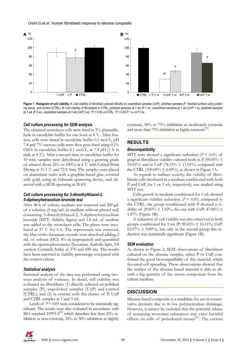
Cell culture processing for SEM analysisThe obtained monolayer cells were fixed in 2% glutaralde-hyde in cacodylate buffer for one hour at 4 ℃. After fixa-tion, cells were rinsed in cacodylate buffer 0.1 mol/L, pH 7.4 and 7% sucrose; cells were then post-fixed using 0.1% OsO4 in cacodylate buffer 0.1 mol/L, at 7.4 pH (1 h in dark at 4 ℃). After a second rinse in cacodylate buffer for 10 min, samples were dehydrated using a growing grade of ethanol (from 25% to 100%) at 4 ℃ with Critical Point Drying at 31.3 ℃ and 72.9 Atm. The samples were placed on aluminium stubs with a graphite-based glue, covered with gold, using an Edwards sputtering device, and ob-served with a SEM operating at 20 kV.
Cell culture processing for 3-dimethylthiazol-2, 5-diphenyltetrazolium bromide testAfter 48 h of culture, medium was removed and 200 μL of a solution (5 mg/mL in medium without phenol red) containing 3-dimethylthiazol-2, 5-diphenyltetrazolium bromide (MTT; Aldrich, Sigma) and 1.8 mL of medium was added to the monolayer cells. The plates were incu-bated at 37 ℃ for 4 h. The supernatant was removed, the blue-violet formazan crystals were dissolved adding 2 mL of solvent (HCL 4% in isopropanol) and quantified with the spectrophotometer (Secoman; Anthelie light, 3.8 version, Contardi, Italia) at 570 and 690 nm. The results have been reported as viability percentage compared with the control culture.
Statistical analysisStatistical analysis of the data was performed using two-ways analysis of variance. In detail, cell viability was evaluated on fibroblasts: (1) directly cultured on polished samples (P), unpolished samples (UnP) and control (CTRL); and (2) in contact with the eluates of P, UnP and CTRL samples at 1 and 3 wk.
Levels of P < 0.05 were considered to be statistically sig-nificant. The results were also evaluated in accordance with ISO standard 10993-5[20] which describes less than 25% in-hibition as non-cytotoxic, 25% to 50% inhibition as slightly
cytotoxic, 50% to 75% inhibition as moderately cytotoxic and more than 75% inhibition as highly cytotoxic[21].
RESULTSBiocompatibilityMTT tests showed a significant reduction (P < 0.01) of gingival fibroblasts viability cultured both in P (90.05% ± 19.03%) and in UnP (78.15% ± 11.01%) compared with the CTRL (100.00% ± 6.00%), as shown in Figure 1A.
As regards to indirect toxicity, the viability of fibro-blastic cells incubated in a medium conditioned with both P and UnP, for 1 or 3 wk, respectively, was studied using MTT test.
Cells growth in medium conditioned for 1 wk showed a significant viability reduction (P < 0.01) compared to the CTRL: the group conditioned with P showed a vi-ability of 29.83% ± 1.92%, the one with UnP: 47.06% ± 1.87% (Figure 1B).
A reduction of cell viability was also observed in both groups conditioned for 3 wk (P: 89.45% ± 10.11%; UnP: 65.97% ± 9.89%), but only in the second group this re-duction was statistically significant (Figure 1B).
SEM evaluationAs shown in Figure 2, SEM observations of fibroblasts cultured on the silorane samples, either P or UnP, con-firmed the good biocompatibility of this material, which favoured cell spreading. These observations showed that the surface of the silorane-based material is able to ab-sorb a big quantity of the serum component from the culture medium.
DISCUSSIONSilorane-based composite is a candidate for use in conser-vative dentistry due to its low polymerization shrinkage. However, it cannot be excluded that the potential release of remaining monomer substances may exert harmful effects on cells of periodontal tissues[22]. The current
88 November 20, 2013|Volume 2|Issue 4|WJS|www.wjgnet.com
120
100
80
60
40
20
0
%
a
UnP P CTRL
120
100
80
60
40
20
0
%
a
UnP 1 wP 1 wCTRL P 3 w UnP 3 w
a
ac
Figure 1 Histogram of cell viability. A: Cell viability of fibroblast cultured directly on unpolished samples (UnP), polished samples (P: finished surface using polish-ing discs) and control (CTRL); B: Cell viability of fibroblasts in CTRL, polished samples at 1 wk (P 1 w), unpolished samples at 1 wk (UnP 1 w), polished samples at 3 wk (P 3 w), unpolished samples at 3 wk (UnP 3 w); aP < 0.05 vs CTRL; cP < 0.05 P 1 w vs P 3 w.
A B
Orsini G et al . Human fibroblasts response to silorane composite
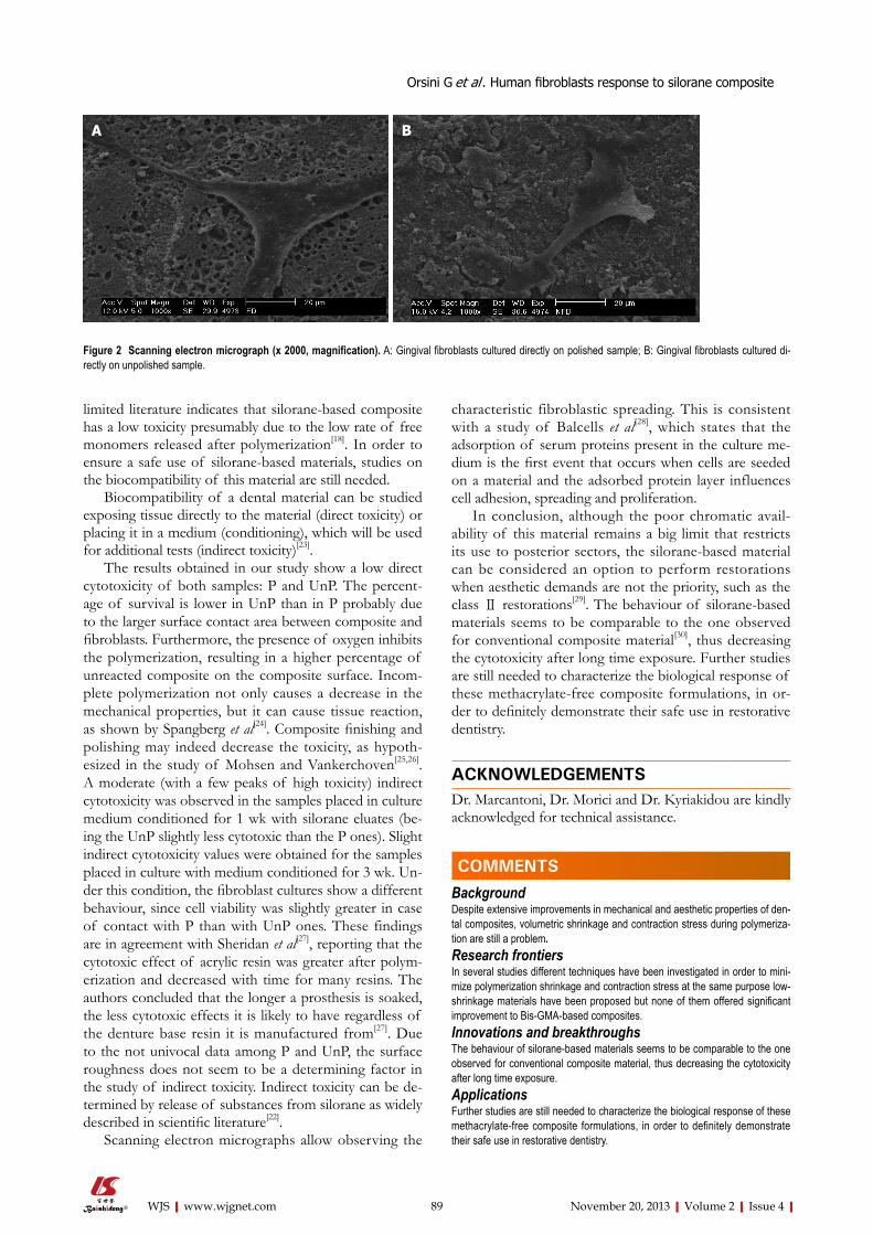
limited literature indicates that silorane-based composite has a low toxicity presumably due to the low rate of free monomers released after polymerization[18]. In order to ensure a safe use of silorane-based materials, studies on the biocompatibility of this material are still needed.
Biocompatibility of a dental material can be studied exposing tissue directly to the material (direct toxicity) or placing it in a medium (conditioning), which will be used for additional tests (indirect toxicity)[23].
The results obtained in our study show a low direct cytotoxicity of both samples: P and UnP. The percent-age of survival is lower in UnP than in P probably due to the larger surface contact area between composite and fibroblasts. Furthermore, the presence of oxygen inhibits the polymerization, resulting in a higher percentage of unreacted composite on the composite surface. Incom-plete polymerization not only causes a decrease in the mechanical properties, but it can cause tissue reaction, as shown by Spangberg et al[24]. Composite finishing and polishing may indeed decrease the toxicity, as hypoth-esized in the study of Mohsen and Vankerchoven[25,26]. A moderate (with a few peaks of high toxicity) indirect cytotoxicity was observed in the samples placed in culture medium conditioned for 1 wk with silorane eluates (be-ing the UnP slightly less cytotoxic than the P ones). Slight indirect cytotoxicity values were obtained for the samples placed in culture with medium conditioned for 3 wk. Un-der this condition, the fibroblast cultures show a different behaviour, since cell viability was slightly greater in case of contact with P than with UnP ones. These findings are in agreement with Sheridan et al[27], reporting that the cytotoxic effect of acrylic resin was greater after polym-erization and decreased with time for many resins. The authors concluded that the longer a prosthesis is soaked, the less cytotoxic effects it is likely to have regardless of the denture base resin it is manufactured from[27]. Due to the not univocal data among P and UnP, the surface roughness does not seem to be a determining factor in the study of indirect toxicity. Indirect toxicity can be de-termined by release of substances from silorane as widely described in scientific literature[22].
Scanning electron micrographs allow observing the
characteristic fibroblastic spreading. This is consistent with a study of Balcells et al[28], which states that the adsorption of serum proteins present in the culture me-dium is the first event that occurs when cells are seeded on a material and the adsorbed protein layer influences cell adhesion, spreading and proliferation.
In conclusion, although the poor chromatic avail-ability of this material remains a big limit that restricts its use to posterior sectors, the silorane-based material can be considered an option to perform restorations when aesthetic demands are not the priority, such as the class Ⅱ restorations[29]. The behaviour of silorane-based materials seems to be comparable to the one observed for conventional composite material[30], thus decreasing the cytotoxicity after long time exposure. Further studies are still needed to characterize the biological response of these methacrylate-free composite formulations, in or-der to definitely demonstrate their safe use in restorative dentistry.
ACKNOWLEDGEMENTSDr. Marcantoni, Dr. Morici and Dr. Kyriakidou are kindly acknowledged for technical assistance.
COMMENTSBackgroundDespite extensive improvements in mechanical and aesthetic properties of den-tal composites, volumetric shrinkage and contraction stress during polymeriza-tion are still a problem. Research frontiersIn several studies different techniques have been investigated in order to mini-mize polymerization shrinkage and contraction stress at the same purpose low-shrinkage materials have been proposed but none of them offered significant improvement to Bis-GMA-based composites.Innovations and breakthroughsThe behaviour of silorane-based materials seems to be comparable to the one observed for conventional composite material, thus decreasing the cytotoxicity after long time exposure.Applications Further studies are still needed to characterize the biological response of these methacrylate-free composite formulations, in order to definitely demonstrate their safe use in restorative dentistry.
89 November 20, 2013|Volume 2|Issue 4|WJS|www.wjgnet.com
Figure 2 Scanning electron micrograph (x 2000, magnification). A: Gingival fibroblasts cultured directly on polished sample; B: Gingival fibroblasts cultured di-rectly on unpolished sample.
COMMENTS
A B
Orsini G et al . Human fibroblasts response to silorane composite
Peer reviewThe authors considered and concluded that the materials are biocompatible.
REFERENCES1 Condon JR, Ferracane JL. Assessing the effect of composite
formulation on polymerization stress. J Am Dent Assoc 2000; 131: 497-503 [PMID: 10770013]
2 Hilton TJ. Can modern restorative procedures and materi-als reliably seal cavities? In vitro investigations. Part 1. Am J Dent 2002; 15: 198-210 [PMID: 12469759]
3 Gonçalves F, Pfeifer CS, Meira JB, Ballester RY, Lima RG, Braga RR. Polymerization stress of resin composites as a function of system compliance. Dent Mater 2008; 24: 645-652 [PMID: 17719626 DOI: 10.1016/j.dental.2007.06.032]
4 Braga RR, Ballester RY, Ferracane JL. Factors involved in the development of polymerization shrinkage stress in resin-composites: a systematic review. Dent Mater 2005; 21: 962-970 [PMID: 16085301]
5 Witzel MF, Ballester RY, Meira JB, Lima RG, Braga RR. Com-posite shrinkage stress as a function of specimen dimensions and compliance of the testing system. Dent Mater 2007; 23: 204-210 [PMID: 16494936 DOI: 10.1016/j.dental.2006.01.016]
6 Chen HY, Manhart J, Hickel R, Kunzelmann KH. Polymer-ization contraction stress in light-cured packable composite resins. Dent Mater 2001; 17: 253-259 [PMID: 11257299 DOI: 10.1016/S0109-5641(00)00079-8]
7 Bouschlicher MR, Vargas MA, Boyer DB. Effect of compos-ite type, light intensity, configuration factor and laser polym-erization on polymerization contraction forces. Am J Dent 1997; 10: 88-96 [PMID: 9545896]
8 Braga RR, Ferracane JL. Alternatives in polymerization con-traction stress management. Crit Rev Oral Biol Med 2004; 15: 176-184 [PMID: 15187035 DOI: 10.1177/154411130401500306]
9 Ilie N, Hickel R. Resin composite restorative materials. Aust Dent J 2011; 56 Suppl 1: 59-66 [PMID: 21564116 DOI: 10.1111/j.1834-7819.2010.01296.x]
10 Yap AU, Han VT, Soh MS, Siow KS. Elution of leachable components from composites after LED and halogen light ir-radiation. Oper Dent 2004; 29: 448-453 [PMID: 15279486]
11 Lee SY, Greener EH, Menis DL. Detection of leached moi-eties from dental composites in fluids simulating food and saliva. Dent Mater 1995; 11: 348-353 [PMID: 8595834 DOI: 10.1016/0109-5641(95)80033-6]
12 Geurtsen W. Substances released from dental resin com-posites and glass ionomer cements. Eur J Oral Sci 1998; 106: 687-695 [PMID: 9584902 DOI: 10.1046/j.0909-8836.1998.eo-s10602ii04.x]
13 Geurtsen W, Lehmann F, Spahl W, Leyhausen G. Cytotoxicity of 35 dental resin composite monomers/additives in perma-nent 3T3 and three human primary fibroblast cultures. J Biomed Mater Res 1998; 41: 474-480 [PMID: 9659618 DOI: 3.0.CO]
14 Volk J, Leyhausen G, Dogan S, Geurtsen W. Additive ef-fects of TEGDMA and hydrogenperoxide on the cellular glutathione content of human gingival fibroblasts. Dent Mater 2007; 23: 921-926 [PMID: 17049977 DOI: 10.1016/j.dental.2006.08.001]
15 Reichl FX, Simon S, Esters M, Seiss M, Kehe K, Kleinsasser N, Hickel R. Cytotoxicity of dental composite (co)monomers and the amalgam component Hg(2+) in human gingival fibroblasts. Arch Toxicol 2006; 80: 465-472 [PMID: 16474958 DOI: 10.1007/s00204-006-0073-5]
16 Yoshii E. Cytotoxic effects of acrylates and methacrylates: re-lationships of monomer structures and cytotoxicity. J Biomed Mater Res 1997; 37: 517-524 [PMID: 9407300]
17 Poplawski T, Pawlowska E, Wisniewska-Jarosinska M, Ksiazek D, Wozniak K, Szczepanska J, Blasiak J. Cytotoxic-ity and genotoxicity of glycidyl methacrylate. Chem Biol Interact 2009; 180: 69-78 [PMID: 19428346 DOI: 10.1016/j.cbi.2009.02.001]
18 Kopperud HM, Schmidt M, Kleven IS. Elution of sub-stances from a silorane-based dental composite. Eur J Oral Sci 2010; 118: 100-102 [PMID: 20156272 DOI: 10.1111/j.1600-0722.2009.00697.x]
19 Krifka S, Seidenader C, Hiller KA, Schmalz G, Schweikl H. Oxidative stress and cytotoxicity generated by dental composites in human pulp cells. Clin Oral Investig 2012; 16: 215-224 [PMID: 21243381 DOI: 10.1007/s00784-010-0508-5]
20 International Standards Organization, ISO 10993-5: Biologi-cal evaluation of Medical Devices-Part 5. Tests for Cytotoxic-ity: In Vitro Methods. Geneva: ISO, 1992
21 Jorge JH, Giampaolo ET, Vergani CE, Machado AL, Pava-rina AC, Carlos IZ. Cytotoxicity of denture base resins: effect of water bath and microwave postpolymerization heat treat-ments. Int J Prosthodont 2004; 17: 340-344 [PMID: 15237883]
22 Schulz SD, König A, Steinberg T, Tomakidi P, Hellwig E, Polydorou O. Human gingival keratinocyte response to sub-stances eluted from silorane composite material reveal im-pact on cell behavior reflected by RNA levels and induction of apoptosis. Dent Mater 2012; 28: e135-e142 [PMID: 22575741 DOI: 10.1016/j.dental.2012.04.018]
23 Schweikl H, Hiller KA, Bolay C, Kreissl M, Kreismann W, Nusser A, Steinhauser S, Wieczorek J, Vasold R, Schmalz G. Cytotoxic and mutagenic effects of dental composite materi-als. Biomaterials 2005; 26: 1713-1719 [PMID: 15576145 DOI: 10.1016/j.biomaterials.2004.05.025]
24 Spangberg L, Rodrigues H, Langeland L, Langeland K. Biologic effects of dental materials. 2. Toxicity of anterior tooth restorative materials on HeLa cells in vitro. Oral Surg Oral Med Oral Pathol 1973; 36: 713-724 [PMID: 4518036 DOI: 10.1016/0030-4220(73)90145-X]
25 Mohsen NM, Craig RG, Hanks CT. Cytotoxicity of urethane dimethacrylate composites before and after aging and leach-ing. J Biomed Mater Res 1998; 39: 252-260 [PMID: 9457555]
26 Vankerckhoven H, Lambrechts P, van Beylen M, Davidson CL, Vanherle G. Unreacted methacrylate groups on the sur-faces of composite resins. J Dent Res 1982; 61: 791-795 [PMID: 7045184 DOI: 10.1177/00220345820610062801]
27 Sheridan PJ, Koka S, Ewoldsen NO, Lefebvre CA, Lavin MT. Cytotoxicity of denture base resins. Int J Prosthodont 1997; 10: 73-77 [PMID: 9484073]
28 Balcells M, Klee D, Fabry M, Höcker H. Quantitative As-sessment of Protein Adsorption by Combination of the Enzyme-Linked Immunosorbent Assay with Radioisotope-Based Studies. J Colloid Interface Sci 1999; 220: 198-204 [PMID: 10607434 DOI: 10.1006/jcis.1999.6527]
29 Goncalves FS, Castro CD, Bueno AC, Freitas AB, Moreira AN, Magalhaes CS. The short-term clinical performance of a silorane-based resin composite in the proximal contacts of class II restorations. J Contemp Dent Pract 2012; 13: 251-256 [PMID: 22917991]
30 Hahnel S, Henrich A, Bürgers R, Handel G, Rosentritt M. Investigation of mechanical properties of modern dental composites after artificial aging for one year. Oper Dent 2010; 35: 412-419 [PMID: 20672725 DOI: 10.2341/09-337-L]
P- Reviewers: Brasileiro B, Eugenia KK, Jeng JH S- Editor: Gou SX L- Editor: A E- Editor: Wang CH
90 November 20, 2013|Volume 2|Issue 4|WJS|www.wjgnet.com
Orsini G et al . Human fibroblasts response to silorane composite

BRIEF ARTICLE
Accuracy of linear vs spiral tomography: Alveolar crest to sinus/nasal floor height
Mahsa Yoozbashizadeh, Seyed Ahmad Fatemitabar, Ehsan Sedighara, Arash Nikgoo
Mahsa Yoozbashizadeh, Seyed Ahmad Fatemitabar, Private Practice, Tehran 16617-56433, IranEhsan Sedighara, Private Practice, Raleigh, NC 72601, United StatesArash Nikgoo, Private Practice, Prospect, Tasmania 7250, Aus-traliaAuthor contributions: Nikgoo A and Fatemitabar SA designed the research plan; Yoozbashizadeh M and Fatemitabar SA performed the majority of the experiments; Sadighara E and Yoozbashizadeh M provided new analytic tools; Sedighara E analyzed the data; Yoozbashizadeh M provided financial sup-port for this study; Nikgoo A wrote the paper.Correspondence to: Arash Nikgoo, DDS, ADC, Private Prac-tice, Prospect, Launceston, Tasmania 7250, Australia. [email protected]: +61-4-24545992 Fax: +61-3-63623644Received: March 10, 2013 Revised: September 19, 2013 Accepted: October 17, 2013Published online: November 20, 2013
AbstractAIM: To determine the accuracy of tomography in the linear measurement of alveolar bone at maxillary si-nus/nose location.
METHODS: Two dry skulls each marked with 10 pairs of guttaperchas placed on buccal and lingual sides of the maxillary ridge were used in this in vitro study. The distance between the alveolar crest and the sinus/nasal floor was measured on tomographic views, pre-pared by linear and spiral techniques. The ridges were then sectioned so that each section would include one pair of buccal and lingual guttaperchas. The actual distances directly measured on the sections were com-pared to those of the equivalent tomographic sections (the magnification co-efficient was applied). Paired t -test was used to statistically analyze the data.
RESULTS: The measurement error with the applica-tion of linear tomography and spiral tomography was
shown to be 0.455 ± 0.838 mm (P = 0.029) and 0.17 ± 0.78 mm (P = 0.347), respectively. There was a statistically significant difference between the liner to-mography values and actual values (P = 0.029). This difference was representative of underestimation. Mc-Namara’s test was used to assess the ± 1 mm error; 73.7% of the linear values and 84.2% of the spiral values were within the ± 1 mm error limit. McNamara’s test did not show any significant differences between the 2 methods in this regard (P = 0.625). The linear values were significantly different to the actual values (P = 0.029) but not to the spiral values (P = 0.185).
CONCLUSION: Spiral tomography has enough ac-curacy for the measurement of alveolar ridge height. Although linear tomography somewhat underestimates the actual values it provides satisfactory accuracy.
© 2013 Baishideng Publishing Group Co., Limited. All rights reserved.
Key words: Linear tomography; Spiral tomography; Maxillary sinus; Dental implants
Core tip: Maxillary partial or complete edentulism rep-resent some challenging conditions in implant dentistry. The position of sinus/nasal floor in partial/complete edentulous maxilla determines the alveolar bone height and consequently the length of the implants that can be used. Although cone beam cone beam computed to-mography and conventional computed tomography are widely used for pre-operative implant treatment plan-ning, they are expensive and can expose patients to relatively high dose of radiation. We demonstrated that tomography can be a good substitute for conventional and cone beam computed tomography for alveolar length measurement at maxilla, although spiral tomog-raphy is more accurate than linear tomography.
Yoozbashizadeh M, Fatemitabar SA, Sedighara E, Nikgoo A.
World Journal of StomatologyW J S
Online Submissions: http://www.wjgnet.com/esps/[email protected]:10.5321/wjs.v2.i4.91
World J Stomatol 2013 November 20; 2(4): 91-96ISSN 2218-6263 (online)
© 2013 Baishideng Publishing Group Co., Limited. All rights reserved.
91 November 20, 2013|Volume 2|Issue 4|WJS|www.wjgnet.com

Yoozbashizadeh M et al . Tomography and implant dentistry
Accuracy of linear vs spiral tomography: Alveolar crest to sinus/nasal floor height. World J Stomatol 2013; 2(4): 91-96 Available from: URL: http://www.wjgnet.com/2218-6263/full/v2/i4/91.htm DOI: http://dx.doi.org/10.5321/wjs.v2.i4.91
INTRODUCTIONAlthough computed tomography (CT) and cone beam computed tomography (CBCT) are frequently used for pre-operative implant planning, their use in the post-operative assessments is limited due to metallic streak ar-tifacts[1,2]. Also CBCT is associated with artifacts such as truncated view and beam hardening artifacts[3,4]. Another disadvantage of computed tomography is its relatively high radiation risk compared to conventional tomogra-phy[5-7].
The position of the maxillary sinus floor influences the height of the alveolar bone and consequently the implant length to be placed. Tomographic views are con-sidered as the most reliable projections in the assessment of potential implant sites prior to surgery since they pro-vide the clinician with the buccolingual information of the anatomic structures[1,5].
The measurement accuracy of most recent tomo-graphic techniques in the assessment of mandibular landmarks has been thoroughly discussed through the literature[8-14]. A recent systematic review has concluded that each landmark possesses a unique error pattern and contributes independently to the measurement inaccura-cy[15]. Since the alveolar crest height meaning the distance between the maxillary sinus floor and the alveolar crest serves as an important factor in the placement of dental implants., The present study aimed to compare the mea-surement accuracy of two tomography techniques, linear and spiral, in the maxilla of dry human skull.
MATERIALS AND METHODSIn this in vitro study, two dry skulls with intact maxillary sinus, nasal floor and foramen magnum, one completely and the other partially edentulous, were used. Each skull was then marked in 10 regions posteroanteriorly. Panoramic scout views were prepared to measure the alveolar ridge height. Each skull was then marked in five regions on each side with 70 guttapercha using glue ev-ery 1 cm and perpendicular to the ridge. The most distal guttapercha was placed in the third molar region and the most anterior one was placed in the lateral incisor region. A total of 20 areas (4 × 5) were marked (Figure 1). Each area was marked with two guttaperchas one on the lin-gual and the other on the buccal aspects of the alveolar ridge.
To prepare tomographic views, skulls were fixed on a wooden jig. Tomographic views were obtained twice, first using multitask Cranex TOME device (Orion Cor-poration Sordex, Helsinki, Finland) and then using Plan-meca Promax (Helsinki, Finland). Kodak X-Omat cas-
sette (Ektavision) and Agfa Single Emulsion (CP-VB) (15 cm × 30 cm) film were used. For spiral tomography, the dental tomo program for the upper jaw was selected at 57 kV, 2.5 mA and 56 s. Slice thickness was set to be 2 mm and the aperture number was 4. For linear tomography, the minimum adjustments, 54 kV and 0.5 mA, were set. Slice thickness was set to be 3 mm (Table 1). In all cases skulls were placed so that the maxillary occlusal plane would be parallel to the horizon. A total number of 20 cross-sections were prepared on each X-ray unit.
The films were processed in an automatic processing machine (OPTIMAX 2010; PROTEK Medizintechnik, oberstenfeld, Germany). Measurements were done on a negatoscope in a semi-dark room using a digital sliding caliper. The view with the clearest gutta-percha was se-lected for measurement on each radiograph. The alveolar crest and the maxillary/nasal floor were then outlined on tracing papers which were superimposed on the radio-graphic views. Bone height was measured in an oblique direction along the medial axis of the alveolar process, similar to the direction of implant placement[5]. The dis-tance between the sinus floor to the alveolar ridge traced on this line was considered as the ridge height (Figure 2). Measurements were done twice by an oral radiologist and an oral radiology senior resident each with a time interval of 2 wk.
The whole alveolar process was cut with electric saw first and then hand jig saw was used to separate the
92 November 20, 2013|Volume 2|Issue 4|WJS|www.wjgnet.com
Figure 1 One of the two dry skulls marked with guttaperchas.
Figure 2 The measurement approach for bone height on film.
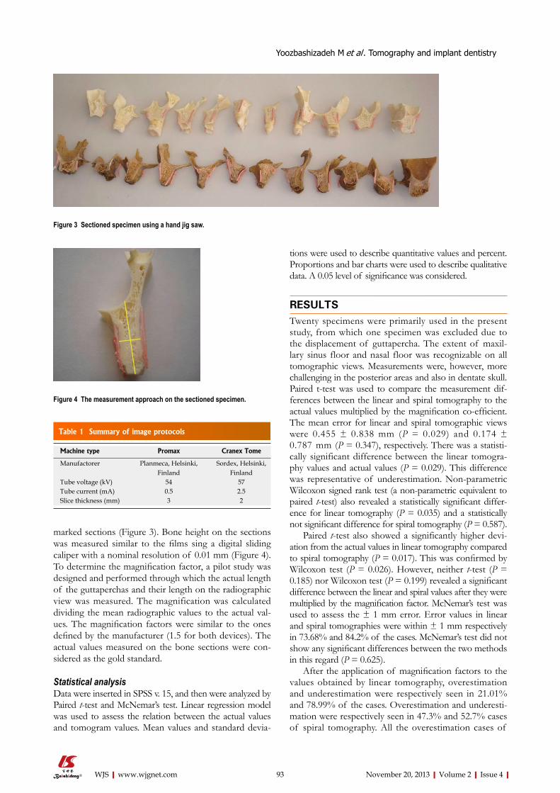
Table 1 Summary of image protocols
marked sections (Figure 3). Bone height on the sections was measured similar to the films sing a digital sliding caliper with a nominal resolution of 0.01 mm (Figure 4). To determine the magnification factor, a pilot study was designed and performed through which the actual length of the guttaperchas and their length on the radiographic view was measured. The magnification was calculated dividing the mean radiographic values to the actual val-ues. The magnification factors were similar to the ones defined by the manufacturer (1.5 for both devices). The actual values measured on the bone sections were con-sidered as the gold standard.
Statistical analysisData were inserted in SPSS v. 15, and then were analyzed by Paired t-test and McNemar’s test. Linear regression model was used to assess the relation between the actual values and tomogram values. Mean values and standard devia-
tions were used to describe quantitative values and percent. Proportions and bar charts were used to describe qualitative data. A 0.05 level of significance was considered.
RESULTSTwenty specimens were primarily used in the present study, from which one specimen was excluded due to the displacement of guttapercha. The extent of maxil-lary sinus floor and nasal floor was recognizable on all tomographic views. Measurements were, however, more challenging in the posterior areas and also in dentate skull. Paired t-test was used to compare the measurement dif-ferences between the linear and spiral tomography to the actual values multiplied by the magnification co-efficient. The mean error for linear and spiral tomographic views were 0.455 ± 0.838 mm (P = 0.029) and 0.174 ± 0.787 mm (P = 0.347), respectively. There was a statisti-cally significant difference between the linear tomogra-phy values and actual values (P = 0.029). This difference was representative of underestimation. Non-parametric Wilcoxon signed rank test (a non-parametric equivalent to paired t-test) also revealed a statistically significant differ-ence for linear tomography (P = 0.035) and a statistically not significant difference for spiral tomography (P = 0.587).
Paired t-test also showed a significantly higher devi-ation from the actual values in linear tomography compared to spiral tomography (P = 0.017). This was confirmed by Wilcoxon test (P = 0.026). However, neither t-test (P = 0.185) nor Wilcoxon test (P = 0.199) revealed a significant difference between the linear and spiral values after they were multiplied by the magnification factor. McNemar’s test was used to assess the ± 1 mm error. Error values in linear and spiral tomographies were within ± 1 mm respectively in 73.68% and 84.2% of the cases. McNemar’s test did not show any significant differences between the two methods in this regard (P = 0.625).
After the application of magnification factors to the values obtained by linear tomography, overestimation and underestimation were respectively seen in 21.01% and 78.99% of the cases. Overestimation and underesti-mation were respectively seen in 47.3% and 52.7% cases of spiral tomography. All the overestimation cases of
93 November 20, 2013|Volume 2|Issue 4|WJS|www.wjgnet.com
Figure 3 Sectioned specimen using a hand jig saw.
Figure 4 The measurement approach on the sectioned specimen.
Machine type Promax Cranex Tome
Manufactorer Planmeca, Helsinki, Finland
Sordex, Helsinki, Finland
Tube voltage (kV) 54 57Tube current (mA) 0.5 2.5Slice thickness (mm) 3 2
Yoozbashizadeh M et al . Tomography and implant dentistry

spiral tomography were within the error limit of ± 1 mm. Mean spiral overestimation was within 0.02 to 0.91 mm. Mean linear overestimation was within 0.38 to 1.5 mm. Mean underestimation values of linear and spiral tomog-raphies were respectively within the ranges of 0.04 to 1.8 mm and 0.03 to 1.4 mm (except for one case of spi-ral tomography which showed an underestimation of 2.19 mm). Figure 5 represent the distribution of errors within the ± 1 mm range.
DISCUSSIONLinear and spiral tomography techniques were compared in the present study in terms of accuracy in the pre-operative assessment of the maxillary bone height. To the best of our knowledge, the literature lacks enough studies on the localization of maxilla, maxillary sinus and nasal floor and most studies have addressed the localiza-tion of mandibular canal.
It is generally accepted that more complex tomo-graphic movements are associated with higher blurring of the background images and less streaking artifacts[1]. Spiral and hypocycloidal movements will reduce the incidence of streaking artifacts[16,17]. In the studies of Lindh et al[18] spiral tomography provided more accurate views of the mandibular canal compared to hypocycloidal tomography. In the present study, the actual values measured directly on the skull sections and the values obtained from linear tomograms were significantly different (P = 0.029). The mean difference was measured 0.455 ± 0.83 mm for linear tomography and 0.174 ± 0.78 mm in spiral tomography. This difference was not significant for spiral tomography (P = 0.347). The significant difference in linear tomogra-phy may be partly due to the quality of images. In spiral tomography the quality of images especially in the ante-rior area was better and the outlines could be more easily detected. Due to the insufficient number of specimens,
however, this comparison between the anterior and the posterior areas was not statistically implacable.
Naitoh et al[19] compared the measurement accuracy of direct laser positioning and reformatted CT. They suggested that other factors including tomographic angle and the placement angle of the object to be projected also influence the measurement accuracy in addition to the motion pattern. They did not find any significant dif-ferences between the two methods in mandible measure-ments (P = 0.526). They believed the significant lower accuracy of the linear tomography found through other studies is due to the difficulties in the adjustment of the projection plan of the object and not due to the image quality.
The main purpose of the present study was to com-pare two types of tomography in terms of linear mea-surements rather than image quality. Statistical analysis failed to reveal any significant differences between the two methods in the measurement of the distance between the alveolar crest and the sinus/nasal floor (P = 0.185). Spiral tomography values obtained through the present study are consistent to the findings of Bou Serhal et al[5] who assessed the measurement accuracy of the spiral tomog-raphy in upper jaw. They reported a mean error of 0.24 ± 0.19 mm which was not significantly different to the actual values (P > 0.05). They stated some quality impairment in the images obtained from the most distal slices and though it was attributed to the placement of more bony structures in the area. Similarly, Kim et al[20] experienced quality impairment in mandibular posterior areas pro-jected by Scanora spiral tomography. Higher image quality in the study of Bou Serhal et al[5] may be attributed to the complete edentulousness of the studied skulls, which may have eliminated the artifacts commonly created by the presence of restorations and natural teeth. Similarly, the quality of images obtained from the anterior areas was higher compared to those of posterior areas in the present study both with spiral and linear tomographies. Also spiral projections were associated with higher image quality in anterior areas compared to linear tomography.
Bou Serhal et al[21] also evaluated the accuracy of con-ventional spiral tomography [Cranex TOME multifunc-tional unit (Orion Corporation Sordex; Helsinki, Finland)] for the localization of the mandibular canal on human fresh cadavers and reported higher mean error values compared to their previous study. They concluded that the information provided by spiral tomography of the posterior mandible using the studied unit is reliable and sufficient for preoperative planning of implant placement. They attributed the different results of the two studies to the fact that the absence of overlying soft tissue provides the observer with a higher resolution of the bony mandi-ble and also more precise adjustments of skull compared to the patient or cadaver in the latter study.
Butterfield et al[17] examined linear tomography in terms of accuracy and validity in the pre-surgical evalua-tion of potential implant sites in mandible. They claimed that linear tomography suffers from prominent dimen-sional instability, which significantly limits its role in
94 November 20, 2013|Volume 2|Issue 4|WJS|www.wjgnet.com
100
80
60
40
20
0
Perc
ent
< -1 -1 to + 1 > +1DFF.CS2
Tomograp
LinearSpiral
Figure 5 Frequency percent of the measurement errors after the applica-tion of magnification factors.
Error < -1 mm -1 to +1 mm > +1 mmLinear 4 (% 21.01) 14 (% 73.68) 1 (% 5.31)Spiral 3 (% 15.8) 16 (% 84.2) 0 (% 0)
Yoozbashizadeh M et al . Tomography and implant dentistry

preoperative assessment of implant sites. Seven observ-ers traced eight anatomic landmarks including the man-dibular cortical bone and inferior alveolar canal on linear tomographic images. Statistically significant differences were found between the perceived and actual anatomic values (P < 0.05). They suggested that since the source to image receptor distance, source to object distance and object to image receptor distance change with a con-stant proportion to each other during the tomographic movements, linear tomography does not hold a constant magnification factor. Consistently, linear tomography in the present study was associated with a significant under-estimation of the measurement values of alveolar crest height (P = 0.029). On the other hand, the pilot linear tomography of guttaperchas conducted by the author of the present study revealed a magnification factor (1.498) which closely approximated the manufacturers (1.5).
Bou Serhal et al[21] measured an actual magnification factor of 1.49 for the Cranex TOME spiral tomogra-phy unit both in vertical and horizontal planes. Closely similar, the magnification factor measured in the present study for the same device was equal to 1.518.
In the present study, spiral tomograms showed un-derestimation and overestimation respectively in 52.7% and 47.3% of the cases. The overestimation in the pres-ent study was higher than that of Bou Serhal study in 2000 (33.3%). Also the linear tomograms of our study presented with underestimation and overestimation in 78.99% and 21.01% of the cases, respectively. Based on these findings, it may be suggested that overestimation occurs more frequently in spiral tomography and under-estimation occurs more frequently in linear tomography. Underestimation would seemingly be preferable in the implant placement especially when the mandibular body above the mandibular canal is considered as a potential implantation site[18]. Loubele et al[3] comparatively as-sessed the measurement accuracy of multi-slice spiral CT, spiral tomography and cone-beam tomography. They measured an overestimation of 1 mm for the spiral tomography. CBCT in their study (except for one case) was only associated with approximate overestimation of 0.5 mm. In the present study, spiral tomography was associated with an overestimation of less than 1 mm (maximum of 0.91 mm). This value was measured to be maximally 1.5 mm with the application of linear tomog-raphy especially in the anterior areas of dentate skulls. However, since the significant difference tended towards underestimation with the application of linear tomogra-phy in the present study, and also due to the higher safe-ty of underestimation compared to overestimation, the measurement accuracy of the linear tomography seems to be within the satisfactory clinical range. On the other hand, underestimation may result in the application of a shorterimplant, which may impair the long term progno-sis survival of the implant and success of the overlying prosthetic restoration[18].
Spiral tomography in the study of Bou Serhal et al[5] was associated with a accuracy of ± 1 mm in the mea-surement of alveolar crest to maxillary sinus distance.
Klinge et al[22] measured the distance between the alveolar crest to the inferior border of the mandibular canal by means of hypocycloidal tomography and reported that only 39% of the cases were associated with a accuracy of within ± 1 mm. Hanazawa et al[23] measured the same distance by spiral tomography and reported that 47.9% of the cases are within the ± 1 mm accuracy. In the pres-ent study, spiral tomography and linear tomography were associated with ± 1 mm accuracy respectively in 84.2% and 73.68% of the cases after the magnification factors were applied.
McNemar’s test did not reveal any significant differ-ences between the two tomographies in terms of the percent of the cases within the ± 1 mm accuracy (P = 0.625). Clinically the value of underestimation and over-estimation is more important than the absolute differ-ence of the perceived and actual values[21].
The values measured on the linear tomograms ob-tained by Promax unit significantly differ to actual values (P < 0.029). This difference tends toward underestima-tion. There is, however, a significant correlation between the linear and spiral tomographies. Therefore it seems that both linear (Promax) and spiral (Cranex Tome) to-mography are associated with sufficiently accuracy in the pre-surgical assessment of potential implant sites in dry skull. Also the magnification factor introduced by the manufacturer is seemingly reliable for both devices. Lin-ear and spiral tomography did not show any significant differences in the present study. This may not be the case in clinical situation where the soft tissue and denti-tion are present. Therefore, studies on human cadaver or in vivo trials are highly recommended to further assess the reliability of the two methods.
COMMENTSBackgroundMaxillary Partial or complete edentulism is a common condition in dentistry and insufficient bone can be a challenge for a clinician who wants to place implants at edentulous Maxilla. The loss of maxillary teeth result in decrease in bone height (and width). The position of the maxillary sinus floor/nasal floor influ-ences the height of the available alveolar bone and consequently the implant length to be placed. Different imaging modalities are used for detemining the hight of available bone at maxillary sinus area. Research frontiersThe imaging modality should be chosen that yields the necessary diagnostic information and results in less radiologic risk. Periapical radiography is of limited value in determining bone quantity due to its magnification, distortion, lack of third dimension and size limitation. Nowadays computed tomography and cone beam computed tomography are frequently used for pre-operative implant planning. Although they can provide us with invaluable information (3D images with high accuracy), they have their own disadvantages such as exposing the patient to the relatively high radiation, beam hardening artifacts and high costs. The hot spot is how to employ a modality which produces 3D images but wouldn’t expose patients to a high radiation dose. The answer can be tomography.Innovation and breakthrough To the best knowledge of the authors of this article, the literature lacks enough studies on the localization of maxillary sinus and nasal floor and most studies have addressed the localization of mandibular canal. Linear and spiral tomogra-phy techniques were compared in the present study in terms of accuracy in the pre-operative assessment of the maxillary bone height. In this study, the actual values measured directly on the skull sections and the values obtained from linear
95 November 20, 2013|Volume 2|Issue 4|WJS|www.wjgnet.com
COMMENTS
Yoozbashizadeh M et al . Tomography and implant dentistry
tomograms were significantly different. This difference was not significant for spi-ral tomography. The significant difference in linear tomography may be partly due to the quality of images. In spiral tomography the quality of images especially in the anterior area was better and the outlines could be more easily detected. Due to the insufficient number of specimens, however, this comparison between the anterior and the posterior areas was not statistically implacable.ApplicationThe study result suggests that spiral tomography has enough accuracy for the measurement of alveolar ridge height. Although linear tomography underesti-mates the actual values, it seems to provide satisfactory accuracy.TerminologyTomography is generic term for describing sectional radiography. X-ray source and film move in opposite direction during exposure in this technique. Conse-quently, structures in the section of interest are sharp while the above and blow sections appears blurred.Peer reviewThis is an interesting study in which authors tested the accuracy of the mea-surements of linear and spiral tomography in maxilla. The results were intrigu-ing and demonstrated that spiral tomography is an accurate imaging modality for pre-operative treatment plan. Although, linear tomography is not perceived as accurate as spiral tomography, it appears to be accurate enough to be uti-lized for a pre-surgical treatment plan.
REFERENCES1 Tyndall DA, Brooks SL. Selection criteria for dental implant site
imaging: a position paper of the American Academy of Oral and Maxillofacial radiology. Oral Surg Oral Med Oral Pathol Oral Radiol Endod 2000; 89: 630-637 [PMID: 10807723 DOI: 10.1067/moe.2000.106336]
2 Ismail YH, Azarbal M, Kapa SF. Conventional linear to-mography: protocol for assessing endosseous implant sites. J Prosthet Dent 1995; 73: 153-157 [PMID: 7722930 DOI: 10.1016/S0022-3913(05)80155-6]
3 Loubele M, Guerrero ME, Jacobs R, Suetens P, van Steen-berghe D. A comparison of jaw dimensional and quality as-sessments of bone characteristics with cone-beam CT, spiral tomography, and multi-slice spiral CT. Int J Oral Maxillofac Implants 2007; 22: 446-454 [PMID: 17622012]
4 Mozzo P, Procacci C, Tacconi A, Martini PT, Andreis IA. A new volumetric CT machine for dental imaging based on the cone-beam technique: preliminary results. Eur Radiol 1998; 8: 1558-1564 [PMID: 9866761 DOI: 10.1007/s003300050586]
5 Bou Serhal C, Jacobs R, Persoons M, Hermans R, van Steen-berghe D. The accuracy of spiral tomography to assess bone quantity for the preoperative planning of implants in the pos-terior maxilla. Clin Oral Implants Res 2000; 11: 242-247 [PMID: 11168215 DOI: 10.1034/j.1600-0501.2000.011003242.x]
6 Ekestubbe A, Thilander A, Gröndahl K, Gröndahl HG. Ab-sorbed doses from computed tomography for dental implant surgery: comparison with conventional tomography. Dento-maxillofac Radiol 1993; 22: 13-17 [PMID: 8508935]
7 Chau AC, Fung K. Comparison of radiation dose for implant imaging using conventional spiral tomography, computed to-mography, and cone-beam computed tomography. Oral Surg Oral Med Oral Pathol Oral Radiol Endod 2009; 107: 559-565 [PMID: 19168378]
8 Kamburoğlu K, Kiliç C, Ozen T, Yüksel SP. Measurements of mandibular canal region obtained by cone-beam computed tomography: a cadaveric study. Oral Surg Oral Med Oral Pathol Oral Radiol Endod 2009; 107: e34-e42 [PMID: 19138636 DOI: 10.1016/j.tripleo.2008.10.012]
9 Brown AA, Scarfe WC, Scheetz JP, Silveira AM, Farman AG. Lin-ear accuracy of cone beam CT derived 3D images. Angle Orthod 2009; 79: 150-157 [PMID: 19123719 DOI: 10.2319/122407-599.1]
10 Ballrick JW, Palomo JM, Ruch E, Amberman BD, Hans MG. Image distortion and spatial resolution of a commer-cially available cone-beam computed tomography machine. Am J Orthod Dentofacial Orthop 2008; 134: 573-582 [PMID: 18929276 DOI: 10.1016/j.ajodo.2007.11.025]
11 Moerenhout BA, Gelaude F, Swennen GR, Casselman JW, Van Der Sloten J, Mommaerts MY. Accuracy and repeatability of cone-beam computed tomography (CBCT) measurements used in the determination of facial indices in the laboratory setup. J Craniomaxillofac Surg 2009; 37: 18-23 [PMID: 18815053 DOI: 10.1016/j.jcms.2008.07.006]
12 Periago DR, Scarfe WC, Moshiri M, Scheetz JP, Silveira AM, Farman AG. Linear accuracy and reliability of cone beam CT derived 3-dimensional images constructed using an orth-odontic volumetric rendering program. Angle Orthod 2008; 78: 387-395 [PMID: 18416632 DOI: 10.2319/122106-52.1]
13 Veyre-Goulet S, Fortin T, Thierry A. Accuracy of linear mea-surement provided by cone beam computed tomography to assess bone quantity in the posterior maxilla: a human cadaver study. Clin Implant Dent Relat Res 2008; 10: 226-230 [PMID: 18384410]
14 Stratemann SA, Huang JC, Maki K, Miller AJ, Hatcher DC. Comparison of cone beam computed tomography imag-ing with physical measures. Dentomaxillofac Radiol 2008; 37: 80-93 [PMID: 18239035 DOI: 10.1259/dmfr/31349994]
15 Lou L, Lagravere MO, Compton S, Major PW, Flores-Mir C. Accuracy of measurements and reliability of landmark iden-tification with computed tomography (CT) techniques in the maxillofacial area: a systematic review. Oral Surg Oral Med Oral Pathol Oral Radiol Endod 2007; 104: 402-411 [PMID: 17709072]
16 Stella JP, Tharanon W. A precise radiographic method to determine the location of the inferior alveolar canal in the pos-terior edentulous mandible: implications for dental implants. Part 2: Clinical application. Int J Oral Maxillofac Implants 1990; 5: 23-29 [PMID: 2391136]
17 Butterfield KJ, Dagenais M, Clokie C. Linear tomogra-phy’s clinical accuracy and validity for presurgical dental implant analysis. Oral Surg Oral Med Oral Pathol Oral Ra-diol Endod 1997; 84: 203-209 [PMID: 9269023 DOI: 10.1016/S1079-2104(97)90070-6]
18 Lindh C, Petersson A, Klinge B. Measurements of distances related to the mandibular canal in radiographs. Clin Oral Implants Res 1995; 6: 96-103 [PMID: 7578787 DOI: 10.1034/j.1600-0501.1995.060205.x]
19 Naitoh M, Kawamata A, Iida H, Ariji E. Cross-sectional imaging of the jaws for dental implant treatment: accuracy of linear tomography using a panoramic machine in com-parison with reformatted computed tomography. Int J Oral Maxillofac Implants 2002; 17: 107-112 [PMID: 11858566]
20 Kim KD, Park CS. Reliability of spiral tomography for im-plant site measurement of the mandible. J Korean Acad Oral Maxillofac Radiol 1997; 27: 27-48
21 Bou Serhal C, van Steenberghe D, Quirynen M, Jacobs R. Lo-calisation of the mandibular canal using conventional spiral tomography: a human cadaver study. Clin Oral Implants Res 2001; 12: 230-236 [PMID: 11359480 DOI: 10.1034/j.1600-0501.2001.012003230.x]
22 Klinge B, Petersson A, Maly P. Location of the mandibular canal: comparison of macroscopic findings, conventional ra-diography, and computed tomography. Int J Oral Maxillofac Implants 1989; 4: 327-332 [PMID: 2639861]
23 Hanazawa T, Sano T, Seki K, Okano T. Radiologic mea-surements of the mandible: a comparison between CT-reformatted and conventional tomographic images. Clin Oral Implants Res 2004; 15: 226-232 [PMID: 15008935 DOI: 10.1111/j.1600-0501.2004.00991.x]
P- Reviewer: da Silva Figueredo C S- Editor: Gou SX L- Editor: A E- Editor: Liu XM
96 November 20, 2013|Volume 2|Issue 4|WJS|www.wjgnet.com
Yoozbashizadeh M et al . Tomography and implant dentistry

World Journal of StomatologyW J S
Online Submissions: http://www.wjgnet.com/esps/[email protected]:10.5321/wjs.v2.i4.97
World J Stomatol 2013 November 20; 2(4): 97-102ISSN 2218-6263 (online)
© 2013 Baishideng Publishing Group Co., Limited. All rights reserved.
Surgical obturator duplicating original tissue-form restores esthetics and function in oral cancer
Pravinkumar G Patil
CASE REPORT
97 November 20, 2013|Volume 2|Issue 4|WJS|www.wjgnet.com
Pravinkumar G Patil, Department of Prosthodontics, Govern-ment Dental College and Hospital, Nagpur, MH 440003, India Author contributions: Patil PG contributed to conception and design.Correspondence to: Pravinkumar G Patil, Assistant Profes-sor, Department of Prosthodontics, Government Dental College and Hospital, GMC Campus, Nagpur, MH 440003, India. [email protected]: +91-712-2744496 Fax: +91-712-2743400Received: May 8, 2013 Revised: August 22, 2013 Accepted: September 3, 2013Published online: November 20, 2013
AbstractOral cancer treatment primarily focused on the surgical removal of cancer tissues followed by surgical/prosthet-ic reconstruction. Restoration of the missing structures immediately after surgery shortens recovery time and allows patient to return to community as a functioning member. The most practiced surgical obturators are simple resin prosthetic bases without incorporation of the teeth. This article highlights a technique to fabricate a surgical obturator that duplicates patient’s original tis-sue form including teeth, alveolus and palatal tissues. The obturator is placed immediately after surgery and make patient feel unaware of surgical deformity. The obturator prosthesis fabricated with this technique sup-ports soft tissues and minimizes the scar contracture. We have clinically tried this technique in 11 patients. Patients’ satisfaction level was recorded on visual ana-logue scale (VAS) and it ranges between 74% and 94% (with average of 87%). Four different prosthodontists have visually evaluated facial asymmetry of patients at 6 mo recall and their average perception on VAS varies between 71% and 93% (with average of 84%).
© 2013 Baishideng Publishing Group Co., Limited. All rights reserved.
Key words: Maxillofacial prosthesis; Maxillectomy; Ob-
turator prosthesis
Core tip: This article highlights a technique to fabricate a surgical obturator that duplicates patient’s original tis-sue form including teeth, alveolus and palatal tissues. Make patient feel unaware of surgical deformity. The obturator prosthesis fabricated with this technique sup-ports soft tissues and minimizes the scar contracture. We have clinically tried this technique in 11 patients. Patients’ satisfaction level was recorded on visual ana-logue scale (VAS) and it ranges between 74% and 94% (with average of 87%). Four different prosthodontists have visually evaluated facial asymmetry of patients at 6 months recall and their average perception on VAS varies between 71% and 93% (with average of 84%).
Patil PG. Surgical obturator duplicating original tissue-form restores esthetics and function in oral cancer. World J Stomatol 2013; 2(4): 97-102 Available from: URL: http://www.wjg-net.com/2218-6263/full/v2/i4/97.htm DOI: http://dx.doi.org/10.5321/wjs.v2.i4.97
INTRODUCTION Treatment of oral cancer necessitates surgical removal of the affected maxillofacial hard and soft tissues[1]. Loss of structural continuity affects esthetic appearance and functional performance like mastication and swallowing[2]. Esthetic disfigurement significantly affects patients’ social and psychological wellbeing[3]. In addition to local and general health psychological, social and economic aspects determine final treatment outcome of the prosthetic reha-bilitation[2]. Patient may get disease free after resection of the cancer tissues but can become a permanent handicap if not rehabilitated in a proper manner. Plastic surgeons face great challenges to reconstruct oral and maxillofacial defects to maintain the functional integrity and esthetic appearance especially in large sized defects and trismus. In

98 November 20, 2013|Volume 2|Issue 4|WJS|www.wjgnet.com
Patil PG. Surgical obturator duplicating original tissue-form
case of malignancies, radiotherapy is a vital parameter in controlling neck metastasis. Though surgical reconstruc-tion restores the defect, replacement of teeth and facial tissue support can only be achieved by prosthodontic re-construction. Obturators are given to maintain an artificial barrier between nasopharynx and oropharynx so that oral intake of food should not be regurgitated from the nose and sufficient negative pressure develops in the oral cav-ity to facilitate deglutition[1]. Depending upon the time of prosthesis given they are classified as immediate surgical (immediate), delayed surgical (7-10 d), interim (4-6 wk) and definitive (After 4-6 mo) obturators[1,4].
Immediate or delayed surgical obturator minimizes scar contracture and disfigurement thereby making a pos-itive effect on the patients’ psychology. Various designs have been proposed for fabrication of the surgical obtu-rator. The design ranges from acrylic resin record base bearing no teeth[5]. With or without wrought-wire clasps[6], to a clasped acrylic resin prosthesis that restores the den-tal arch form[7]. Dentate patients are relatively easier to treat than edentulous patients as maximum retention and stability can be achieved from remaining teeth. The most practiced surgical obturator is a simple resin prosthetic base without incorporation of teeth. Addition of teeth in initial healing phase may cause constant source of ir-ritation and hamper healing process according to many authors. However only anterior teeth can be restored un-til surgical wound is healed in some clinical situations[1,8]. In case of radiotherapy, the tissues become more friable and vulnerable hence the simplest form of prosthesis is advocated in most of the clinical situations[3].
This article highlights a treatment concept which re-stores resected maxillofacial tissues with a surgical obtu-rator that duplicates patient’s original tissue form[9,10]. The technique is developed in our hospital and total 11 cancer patients were treated in last 2 years. Patients were evalu-ated for patients’ satisfaction level and clinicians’ percep-tion for bilateral facial symmetry on visual analogue scale (VAS) at 6 mo recall visit.
TechniquePrior to surgery maxillofacial prosthodontist should examine patient and discuss the plan of treatment with surgeons about a proposed line of incision and amount of resection (Figure 1A).
A pre-surgical impression of the maxillary arch is made with irreversible hydrocolloid and poured to obtain a working cast. An anticipated line of resection is drawn with a marking pencil on the cast following discussion with maxillofacial and plastic surgeons.
The area (of the legion) on the cast is modified to obtain normal anatomical contours (Figure 1B). For example, swollen areas of the lesion on the cast can be scraped-out and defect (ulcer/breach) areas can be built-up with dental stone in order to create the normal ana-tomical tissue form on the cast.
Labial/lingual infrabulge retentive areas of the re-maining healthy teeth are engaged with the retentive clasp
arms. A processed prosthetic base is fabricated in heat polymerizing acrylic resin by incorporating the clasps (Figure 1C).
The processed prosthetic base is reseated on the max-illary cast and an over-impression of the whole cast (along with the seated processed prosthetic base) is made with polyvinyl-siloxane putty in perforated stock metal tray to form putty impression index (PⅡ) (Figure 1D). Facial surface on the defect side of the cast should be com-pletely recorded in the over-impression till border areas.
The PⅡ and the cast are separated from each other. The prosthetic base is reseated on the PⅡ (Figure 1E).
The separated cast is sectioned according to the an-ticipated line of resection. The planned defect section of the cast is separated from the remaining normal portion (Figure 1F). This remaining portion (of normal struc-tures) of the cast is used to fabricate the prosthesis.
The PⅡ (along with the processed prosthetic base) is reseated onto the remaining portion of the cast (Figure 1G).
Prosthetic teeth are created with sprinkle-on tech-nique by incrementally adding tooth-colored autopoly-merizing acrylic resin into the impression areas of teeth in the PⅡ. The facial flange can also be created by adding the pink colored autopolymerizing acrylic resin uniformly 2-3 mm in width (Figure 1H).
After setting the PⅡ along with the prosthesis is sep-arated from the cast (Figure 1I) and then PⅡ is separated from the prosthesis carefully. The excess resin is removed and the flange and teeth areas are finished and polished in a conventional manner (Figure 2)[11,12] Note that the smooth borders and polished surfaces are critical param-eters to avoid any tissue injury. Occlusal surfaces of the posterior teeth can be trimmed off by approximately 2 mm to make them out of occlusion[1,8].
The prosthesis must be disinfected before using it in the mouth with any suitable disinfectant like 2% glutaral-dehyde solution.
Routine minor adjustments are carried out and the prosthesis can be seated in position immediately after surgery. A surgical pack can be placed in the defect area before placement of the obturator if necessary.
CASE REPORTWe have treated 11 patients undergone maxillary partial or subtotal resection with the immediate surgical obtura-tor in last 2 years (Table 1). Out of 11 patients, 6 had Armany’s Class Ⅰ defect, 4 had class Ⅱ defect (Figure 3) and 1 had Class Ⅳ defect[13]. All 11 patients were assessed at the interval of 1, 2, 6 and 12 mo follow up visits for evaluation of healing process and evaluated for patients’ own satisfaction level and clinicians’ perception level for bilateral facial symmetry on VAS. Patients’ satisfaction level was recorded on VAS (with 0 indicating no satisfaction and 100 indicating complete satisfaction) and it ranges between 74% and 94% (with average of 87%) (Table 1). Four dif-ferent prosthodontists have visually evaluated facial asym-
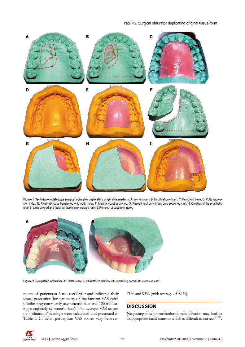
99 November 20, 2013|Volume 2|Issue 4|WJS|www.wjgnet.com
metry of patients at 6 mo recall visit and indicated their visual perception for symmetry of the face on VAS (with 0 indicating completely asymmetric face and 100 indicat-ing completely symmetric face). The average VAS scores of 4 clinicians’ readings were calculated and presented in Table 1. Clinician perception VAS scores vary between
71% and 93% (with average of 84%).
DISCUSSIONNeglecting timely prosthodontic rehabilitation may lead to inappropriate facial contour which is difficult to correct[14-16].
Figure 1 Technique to fabricate surgical obturator duplicating original tissue-form. A: Working cast; B: Modification of cast; C: Prosthetic base; D: Putty impres-sion index; E: Prosthetic base transferred onto putty index; F: Maxillary cast sectioned; G: Reseating of putty index onto sectioned cast; H: Creation of the prosthetic teeth in tooth-colored and facial surface in pink-colored resin; I: Removal of cast from index.
A B C
D E F
G H I
Figure 2 Completed obturator. A: Palatal view; B: Obturator in relation with remaining normal structures on cast.
A B
Patil PG. Surgical obturator duplicating original tissue-form

100 November 20, 2013|Volume 2|Issue 4|WJS|www.wjgnet.com
The significance of immediate surgical obturators has been well documented. Immediate obturators resist tissue con-tracture of the soft tissues that are not supported by the un-derlying osseous structures. During healing phase, surgical wound is protected from external irritants, contaminants, food debris and trauma[17,18].
Advantages of adding flange and teethThe borders of the defect are more prone to collapse due to lack of underlying support. Addition of teeth and labial/buccal flanges provide maximum support to the borders. Facial contours of the immediate obtura-tor described in this article support overlying skin and skin grafts with optimum pressure providing their close adaptation to the cavity walls without getting contracted. After the operation, patients are able to swallow food more readily and to resume a normal diet at an earlier stage which leads to shorter recovery period[4]. Addition of teeth to the obturator allows mastication of semisolid food in initial phase and solid food several days later[4,14]. Speech is minimally altered and in many instances re-mains nearly unchanged[19,20]. Also the awareness of the surgical defect by the tongue is prevented and the patient remains unaware of the size of the defect giving the pa-tient a positive psychological boost. By maintaining facial contour and aesthetics, patients are psychologically better equipped to face rehabilitation[21].
Purpose of replacing teeth immediatelyImmediate restoration of the resected tissues by means the obturator that restores every missing portion of the tissues helps patients undergo unnoticed to the surgery.
This gives patient positive psychological boost during the initial vulnerable period of healing. According to many authors, the posterior teeth should not be added to surgical obturator as they may exert unnecessary stress on the open wound and delay the healing process[3]. This technique describes replacement of dentition that would be missing followed by grinding occlusal contacts of posterior teeth (at least 2 mm) to position them out of occlusion. The facial surfaces must be kept intact to serve the purpose of facial soft tissue support as well as esthetics without disturbing the healing process. Anterior teeth should not be altered unless the incisal contacts hinder the healing tissues. The purpose of adding miss-ing teeth (anteriors or posteriors) may prevent significant psychological trauma to the patient and helps to prevent scar contracture and subsequent disfigurement. The de-veloped facial flange also helps to support the facial soft tissues which can maintain the patient’s original facial es-thetic appearance.
Catch at early healing stageContracture of the wound and scar formation leave very serious facial deformities after cancer surgeries[22]. Even slight depression in either side will also cause major deformity due to face value and patient’s vulnerability towards esthetic appearance. If the radiotherapy follows the surgery the tissues become even more friable and are difficult to manage due to loss of natural laxity. Maxi-mum wound contracture happens in initial stages within 4-6 wk. This is the period in which the losses of intraoral structures also cause difficulty in mastication and de-glutition. Thus in initial stages of healing, patients may
Sr. No
Patient’s surgical defect-type (Armany
Classification)
Age(yr)
Sex (M/F) Diagnosis
Post-surgical radiotherapy
(Y/N)
Patient’s satisfaction level
(VAS at 6 mo)
Clinicians’ observation for facial asymmetry (Average VAS scores of
4 clinicians at 6 mo)
1 Class Ⅱ 47 F Squamous cell carcinoma of right maxilla
Y 86% 84%
2 Class Ⅰ 45 M Adenocarcinoma of left maxillary sinus
Y 78% 86%
3 Class Ⅱ 50 F Squamous cell carcinoma of maxillary sinus
Y 92% 80%
4 Class Ⅳ 63 M Ameloblastoma of palate N 93% 88% 5 Class Ⅰ 50 F Squamous cell carcinoma
of right maxillaY 88% 74%
6 Class Ⅰ 65 F Squamous cell carcinoma of left maxilla
Y 94% 93%
7 Class Ⅰ 65 F Squamous cell carcinoma of right maxilla
Y 83% 71%
8 Class Ⅱ 40 F Squamous cell carcinoma of hard palate
Y 94% 93%
9 Class Ⅰ 36 M Squamous cell carcinoma of Maxillary sinus
Y 81% 86%
10 Class Ⅰ 35 F Squamous cell carcinoma of right maxilla
Y 74% 73%
11 Class Ⅱ 48 M Ameloblastoma of left maxilla
N 94% 92%
Table 1 Details of the patients enrolled and visual analogue scale scores for patients’ satisfaction level and clinicians’ observation for facial asymmetry at 6 mo recall visits
M: Male; F: Female; VAS: Visual analogue scale.
Patil PG. Surgical obturator duplicating original tissue-form

101 November 20, 2013|Volume 2|Issue 4|WJS|www.wjgnet.com
undergo severe psychological depression due to multiple problems. Immediate obturation can create a positive ef-fect on the patients' psychology.
Works as Interim obturatorInterim obturators with teeth may be made using several methods, using a celluloid matrix[2], modifying a surgi-cal obturator[7], using a denture duplicator[23], or using light[11,24] or heat-polymerized acrylic resin[25]. The obtura-tor fabricated with this technique utilizes the PII of pa-tient’s original tissue form and duplicated mostly in heat and slightly in autopolymerizing acrylic resin.
Time and cost effectiveSame surgical obturator can later (for 4-6 mo) serve as an interim obturator following modification of the tissue
surfaces thus it saves time and cost.
Housing for placement of surgical packThe space automatically formed between intaglio surfaces of facial flange and palatal plate can easily be utilized for placement of the surgical pack immediately after the surgery. Thus the obturator can provide supporting and stabilizing medium for the surgical pack.
Future dental implant planningThe same surgical obturator can be an effective tool for implant retained definitive fixed or removable prosthesis. Most of the acquired defects are surgically covered with thin mucosa which is not able to support denture bases. In such situations dental implants are indicated, leaving the vulnerable and/or non keratinized mucosa unload-ed[26]. The use of dental implants is an alternative option to achieve better function and self confidence due to improved retention and stability[27]. Edentulous patients undergoing partial maxillectomy must be treated with implant supported ball and socket attachments to achieve retention to the surgical obturator. Same obturator can be used to prepare the diagnostic as well as surgical template for dental implant placement.
LimitationsAs the teeth and facial flanges of the obturator are cre-ated in auto polymerizing acrylic resin, the free residual monomer may irritate the supporting tissues and hamper the healing process. The light polymerizing acrylic resin can be used alternatively to solve this problem pro-vided the combinations of the light polymerizing acrylic resins and the methylmethacrylate based denture base resins were selected carefully to ensure sufficient bond strength[11,23,24,28].
Future scopeFuture prospective clinical trials with large sample size should be promoted to treat cancer patients with better esthetics and function.
REFERENCES1 Beumer J III, Curtis D, Firtell D. Restoration of acquired
hard palate defects: etiology, disability and rehabilitation. In: Beumer J III, Curtis TA, Marunick MT, eds. Maxillofacial Rehabilitation: Prosthodontic and Surgical Considerations. St. Louis, MO: Medico Dental Media International, 1996: 225-284
2 Kouyoumdjian JH, Chalian VA. An interim obturator pros-thesis with duplicated teeth and palate. J Prosthet Dent 1984; 52: 560-562 [PMID: 6389840 DOI: 10.1016/0022-3913(84)90347-0]
3 Patil PG. Modified technique to fabricate a hollow light-weight facial prosthesis for lateral midfacial defect: a clinical report. J Adv Prosthodont 2010; 2: 65-70 [PMID: 21165271 DOI: 10.4047/jap.2010.2.3.65]
4 Patil PG, Patil SP. Nutrition and cancer. J Am Dent Assoc 2012; 143: 106-107; author reply 107 [PMID: 22298546]
5 Patil PG. The spring retained delayed surgical obturator for total maxillectomy: a technical note. Oral Surgery 2010; 3: 8-10
6 King GE, Martin JW. Cast circumferential and wire clasps
A
C D
B
Figure 3 Post-treatment view immediate and after 6 mo. A: Immediate post-surgical view; B: Obturator in place; C: Immediate post treatment extraoral view; D: Six month post treatment extraoral view.
Patil PG. Surgical obturator duplicating original tissue-form
102 November 20, 2013|Volume 2|Issue 4|WJS|www.wjgnet.com
for obturator retention. J Prosthet Dent 1983; 49: 799-802 [PMID: 6348262 DOI: 10.1016/0022-3913(83)90352-9]
7 Wolfaardt JF. Modifying a surgical obturator prosthesis into an interim obturator prosthesis. A clinical report. J Prosthet Dent 1989; 62: 619-621 [PMID: 2685253 DOI: 10.1016/0022-3913(89)90577-5]
8 Arcuri MR, Taylor TD. Clinical management of the dentate maxillectomy patient. In: Taylor TD, editor. Clinical maxillofa-cial prosthetics. Carol Stream (IL): Quintessence; 2000: 103-120
9 Patil PG. New technique to fabricate an immediate surgical obturator restoring the defect in original anatomical form. J Prosthodont 2011; 20: 494-498 [PMID: 21777335 DOI: 10.1111/j.1532-849X.2011.00739.x]
10 Shambharkar VI, Puri SB, Patil PG. A simple technique to fabricate a surgical obturator restoring the defect in original anatomical form. J Adv Prosthodont 2011; 3: 106-109 [PMID: 21814621 DOI: 10.4047/jap.2011.3.2.106]
11 Gardner LK, Parr GR, Richardson DW. An interim buccal flange obturator. J Prosthet Dent 1991; 65: 862 [PMID: 2072339 DOI: 10.1016/S0022-3913(05)80032-0]
12 Shaker KT. A simplified technique for construction of an in-terim obturator for a bilateral total maxillectomy defect. Int J Prosthodont 2000; 13: 166-168 [PMID: 11203627]
13 Aramany MA. Basic principles of obturator design for par-tially edentulous patients. Part I: classification. J Prosthet Dent 1978; 40: 554-557 [PMID: 364015 DOI: 10.1016/0022-3913(78)90092-6]
14 Heggie AA, MacFarlane WI, Warneke SC. Immediate pros-thetic replacement following major maxillary surgery. Aust N Z J Surg 1980; 50: 370-374 [PMID: 6932848 DOI: 10.1111/j.1445-2197.1980.tb04142.x]
15 Park KT, Kwon HB. The evaluation of the use of a delayed surgical obturator in dentate maxillectomy patients by con-sidering days elapsed prior to commencement of postopera-tive oral feeding. J Prosthet Dent 2006; 96: 449-453 [PMID: 17174663 DOI: 10.1016/j.prosdent.2006.09.019]
16 Lapointe HJ, Lampe HB, Taylor SM. Comparison of maxil-lectomy patients with immediate versus delayed obturator prosthesis placement. J Otolaryngol 1996; 25: 308-312 [PMID: 8902689]
17 Lang BR, Bruce RA. Presurgical maxillectomy prosthesis. J
Prosthet Dent 1967; 17: 613-619 [DOI: 10.1016/0022-3913(67)90133-3]
18 Huryn JM, Piro JD. The maxillary immediate surgical ob-turator prosthesis. J Prosthet Dent 1989; 61: 343-347 [PMID: 2646447 DOI: 10.1016/0022-3913(89)90142-X]
19 Arigbede AO, Dosumu OO, Shaba OP, Esan TA. Evaluation of speech in patients with partial surgically acquired defects: pre and post prosthetic obturation. J Contemp Dent Pract 2006; 7: 89-96 [PMID: 16491151]
20 Oki M, Iida T, Mukohyama H, Tomizuka K, Takato T, Tanigu-chi H. The vibratory characteristics of obturators with different bulb height and form designs. J Oral Rehabil 2006; 33: 43-51 [PMID: 16409516 DOI: 10.1111/j.1365-2842.2006.01528.x]
21 Minsley GE, Warren DW, Hinton V. Physiologic responses to maxillary resection and subsequent obturation. J Prosthet Dent 1987; 57: 338-344 [PMID: 3471944 DOI: 10.1016/0022-3913(87)90309-X]
22 Türkaslan S, Baykul T, Aydın MA, Ozarslan MM. Influ-ence of immediate and permanent obturators on facial con-tours: a case series. Cases J 2009; 2: 6 [PMID: 19121224 DOI: 10.1186/1757-1626-2-6]
23 Kaplan P. Stabilization of an interim obturator prosthesis using a denture duplicator. J Prosthet Dent 1992; 67: 377-379 [PMID: 1507105 DOI: 10.1016/0022-3913(92)90251-5]
24 DaBreo EL. A light-cured interim obturator prosthesis. A clinical report. J Prosthet Dent 1990; 63: 371-373 [PMID: 2184219 DOI: 10.1016/0022-3913(90)90222-X]
25 Arena CA, Evans DB, Hilton TJ. A comparison of bond strengths among chairside hard reline materials. J Prosthet Dent 1993; 70: 126-131 [PMID: 8371174 DOI: 10.1016/0022-3913(93)90006-A]
26 Esser E, Wagner W. Dental implants following radical oral cancer surgery and adjuvant radiotherapy. Int J Oral Maxil-lofac Implants 1997; 12: 552-557 [PMID: 9274085]
27 Cheng AC, Wee AG, Shiu-Yin C, Tat-Keung L. Prosthodon-tic management of limited oral access after ablative tumor surgery: a clinical report. J Prosthet Dent 2000; 84: 269-273 [PMID: 11005898]
28 Fellman S. Visible light-cured denture base resin used in mak-ing dentures with conventional teeth. J Prosthet Dent 1989; 62: 356-359 [PMID: 2681707 DOI: 10.1016/0022-3913(89)90350-8]
P- Reviewers: Haraszthy V, Nyan M S- Editor: Song XX L- Editor: A E- Editor: Wang CH
Patil PG. Surgical obturator duplicating original tissue-form

World Journal of StomatologyW J S
Online Submissions: http://www.wjgnet.com/esps/[email protected]:10.5321/wjs.v2.i4.103
World J Stomatol 2013 November 20; 2(4): 103-107ISSN 2218-6263 (online)
© 2013 Baishideng Publishing Group Co., Limited. All rights reserved.
Soft tissue aneurysmal bone cyst of the mandible: Report of a case
Jahanfar Jahanbani, Donia Sadri, Ali Hassani, Farshid Kavandi
Jahanfar Jahanbani, Donia Sadri, Oral and Maxillofacial Pa-thology Department, Islamic Azad university, Dental Branch, Teh-ran, IranAli Hassani, Oral and Maxillofacial Surgery Department, Islamic Azad university, Dental Branch, Tehran, IranFarshid Kavandi, Oral and Maxillofacial Surgery, Bu Ali Hospi-tal, Damavand alley, Tehran 15994, Iran Author contributions: Jahanbani J and Sadri D designed research and wrote the paper; Hassani A prepared the photograph and sur-gical documents of the case; Kavandi F coordinated things in the surgery and prepared the materials and methods (surgical docu-ment) and photographs of following up, followed up the case. Correspondence to: Donia Sadri, Associate Professor, Oral and Maxillofacial Pathology Department, Islamicazad University, 4, 10th Neyestan, Pasdaran, Tehran 19486, Iran. [email protected]: +98-212-2763449 Fax: +98-212-2763449Received: February 27, 2013 Revised: April 19, 2013Accepted: June 1, 2013Published online: November 20, 2013
AbstractWe report the case of a 17‑year‑old boy with a soft tissue aneurysmal bone cyst (STABC) located in the posterior aspect of the right mandible. Conventional radiography revealed no positive findings. On the com‑puted tomography scan, the lesion appeared to have a non‑uniform intralesional density. Magnetic resonance imaging revealed an abnormal soft tissue masses with cystic component in the superficial part of right man‑dibular body and angle with intact cortex. Following histopathological examination, fibro-histiocytic prolifera‑tion, blood-filled spaces and multinucleated giant cells were seen and the lesion was diagnosed as a STABC. The mass together with underlying bone and perios‑teum on its periphery was surgically resected under general anesthesia. Thirty‑six months after surgery the patient was assessed at outpatient clinic and found no sign of recurrence This may be only the first reported case of the mandible in the English literature of this ex‑tremely rare benign tumor occurring in soft tissue.
© 2013 Baishideng Publishing Group Co., Limited. All rights reserved.
Key words: Bone cysts; Aneurysmal; Mandible; Neo‑plasm; Soft tissue
Core tip: A case of soft tissue aneurysmal bone cyst (STABC) of the mandible is presented in a male teenag‑er. STABC is a rare entity with histological and radiologi‑cal features that are identical to those of aneurysmal bone cyst, except for that STABC is of extra osseous location. The differential diagnosis of STABC in this lo‑cation, includes giant cell tumor of soft tissue and extra skeletal osteosarcoma making it quite a challenge in the process of diagnosis.
Jahanbani J, Sadri D, Hassani A, Kavandi F. Soft tissue aneu-rysmal bone cyst of the mandible: Report of a case. World J Stomatol 2013; 2(4): 103-107 Available from: URL: http://www.wjgnet.com/2218-6263/full/v2/i4/103.htm DOI: http://dx.doi.org/10.5321/wjs.v2.i4.103
INTRODUCTIONAneurysmal bone cyst (ABC) is a benign cystic lesion of bone, composed of blood filled spaces separated by con-nective tissue septa containing fibroblasts, osteoclast-type giant cells and reactive woven bone[1]. Previously, ABC was believed to occur exclusively in bone[1,2], but in re-cent years a few cases of soft tissue ABC (STABC) have been reported[3,4]. The first cases of soft tissue ABC were reported by Salm et al[5]. These cases were categorized as “vascular cystic tumors of soft tissues’’. Recent literature review of well-documented cases shows that STABC is a recognized lesion and extremely rare[6]. To the best of our knowledge the reported cases of STABC have been located in thigh, cervical spine, shoulder and upper extremities[7,8] and STABCs in the jaws have not been re-ported in the English language literature.
CASE REPORT
103 November 20, 2013|Volume 2|Issue 4|WJS|www.wjgnet.com
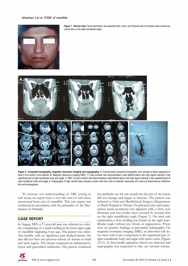
To increase our understanding of ABC arising in soft tissue, we report here a very rare case of soft tissue aneurysmal bone cyst of mandible. This case report was conducted in accordance with the principles of the Dec-laration of Helsinki.
CASE REPORTIn August 2009, a 17-year-old man was referred to a den-tist complaining of a small swelling in his lower right angle of mandible beginning 4 mo ago. The patient was other-wise healthy, with no significant past medical history. He also did not have any previous history of trauma to head and neck region. The dentist suspected an inflammatory lesion and prescribed antibiotics. The patient continued
the antibiotic use for one month but the size of the lesion did not change and began to increase. The patient was referred to Oral and Maxillofacial Surgery, Department of Buali Hospital in Tehran. On physical extra oral exami-nation, facial asymmetry was apparent with, a firm, non fluctuant and non tender mass covered by normal skin on the right mandibular angle (Figure 1). On intra oral examination a firm swelling was found on the right man-dibular angle without any fistula or suppuration. There were no positive findings in panoramic radiography. On magnetic resonance imaging (MRI), an abnormal soft tis-sue mass with cystic component in the superficial part of right mandibular body and angle with intact cortex (Figure 2A-C). In fine-needle aspiration, blood was detected and angiography was requested to rule out vascular lesions.
104 November 20, 2013|Volume 2|Issue 4|WJS|www.wjgnet.com
Jahanbani J et al . STABC of mandible
A
DCB
Figure 2 Computed tomography, magnetic resonance imaging and angiography. A: Coronal-axial computed tomography scan showed a lesion appeared to have a non-uniform intra lesional; B: Magnetic resonance imaging (MRI), T1 post contrast view demonstrated a well defined lesion with high signal intensity in the superficial part of right mandibular body and angle; C: MRI, T2 post contrast view demonstrated a well defined lesion with high signal intensity in the superficial part of right mandibular body and angle; D: Angiography of right carotid artery showed a lesion with only mild to moderate vascularity and ruled out arteriovenous malforma-tion and hemangioma.
Figure 1 Clinical view. Facial asymmetry was apparent with, a firm, non fluctuant and non tender mass covered by normal skin on the right mandibular angle.

The angiography report showed a lesion with only mild to moderate vascularity and ruled out arteriovenous mal-formation and hemangioma (Figure 2D). An incisional bi-opsy through an intraoral approach was performed under general anesthesia. The specimen consisted of 5 pieces of brownish-creamy fragmented tissues with rubbery consis-tency and solid on cut surface totally mesurring 3.0 cm × 2.0 cm × 0.4 cm, sent for histopathological examination. The microscopic evaluation showed spindle cell prolifera-tion, many multi-nucleated giant cells, osteoid formation and pools of blood without any epithelium linings. Three weeks after the incisional biopsy, the patient was hospital-ized and decortication of the lesion was surgically carried out under general anesthesia (Figure 3A and B). During the operation, frozen sections were requested and revealed no malignancy. Grossly, the mass was totally measured
6.5 cm × 4.2 cm × 1.2 cm and showed a soft tissue lesion with hemorrhagic cystic spaces (Figure 3C).
Histopathological examination of the main lesion and the underlying bone and periosteum revealed fibro-histiocyt-ic proliferation, blood-filled spaces and multinucleated giant cells (Figure 4). On reviewing all histopathological sections and paraclinical tests, the definitive diagnosis of STABC was confirmed. After 36 mo of follow up examination, the patient is well and free of any lesion (Figure 5).
DISCUSSIONSTABC is a rare entity with histological and radiological
105 August 20, 2013|Volume 2|Issue 3|WJS|www.wjgnet.com
Figure 3 Clinical features at the surgery time. A: The mass was seen with-out involving adjacent bones; B: Periosteal reaction was seen in underlying bone; C: Gross view of excised lesion showed a solid lesion with eggshell-like rim of bone on its periphery and hemorrhagic cystic space.
Figure 4 Pathological findings. A: Blood-filled space, fibro histiocytic stroma and multinucleated giant cells (HE × 100); B: Blood-filled space and multinucle-ated giant cells (HE × 400).
Figure 5 Coronal-axial computed tomography scan showed the patient is free of any lesion, 12 mo after surgery.
A
C
B
A
B
Jahanbani J et al . STABC of mandible

features that are identical to those of ABC, except for that STABC is of extra osseous location[4,5,7,8].
To the best of our knowledge the reported cases of STABC have been located in thigh, cervical spine, shoul-der and upper extremities[3,8] and STABCs in the jaws have not been reported in the English language literature.
Our reported patient was male and a teenager, the age of the patient is in agreement with Nielsen et al[3] reporting five cases of soft tissue aneurysmal bone cyst in three females and two males who ranged from 8 to 37 years (median 28 years). Their reported cases arose in the soft tissues of upper extremities, tight and groin region as a rapidly growing mass. Clinical evaluation of our patient revealed rapidly growing non fluctuant and non tender mass on the right angle of mandible. Rapidly growing feature of ABC is a result of pathogenesis of this lesion and abnormal hemodynamics that leads to en-larging and hemorrhagic extravasation[3,6]. The maximal diameter of the lesion was 6 cm and macroscopically, the soft tissue mass was consisted of blood-filled cavities separated by septa of various thickness which is similar to the findings in other reported STABC[3,6,7].
Radiographically, the lesion had no positive findings in panoramic radiography. On the computed tomography scan, the lesion appeared to have a non-uniform intra le-sional density.
MRI revealed an abnormal soft tissue mass with cys-tic component in the superficial part of right mandibular body and angle with intact cortex which is similar to the findings in other soft tissue ABC reports[7,8].
The changes seen in MRI appearance of aneurysmal cyst of soft tissue depends on its stage ranging from a primarily solid tumor to a predominantly multicystic le-sion [9].
Histologically our reported case revealed fibro-histio-cytic proliferation, blood-filled spaces and multinucleated giant cells, which is similar to other reports[3,4,6].
The differential diagnosis of STABC in this location, includes giant cell tumor of soft tissue and extra skeletal osteosarcoma[10].
Soft tissue giant cell tumor can be confused with STABC because of the presence of osteoclast- type giant cells in both lesions, but cystic change is the prominent view of STABC in histopathological evaluation[11,12].
Follow up information of our reported patient after 36 mo revealed no tumor recurrence, a similar finding compared to other reported cases[3,8]. On the contrary, local recurrence in soft tissue giant cell tumors are very common[12] and it may be considered as another factor differentiating STABC from soft tissue giant cell tumor.
Extraskeletal telangiectatic osteosarcomas, which are very rare, have gross features similar to those of STABC[10]. However, histologic examination of the STABC shows cells without cytologic atypia that are seen in extra skeletal telangectatic osteosarcoma[3,10].
The etiology of STABC is unclear. Several investiga-tors have proposed trauma and vascular malformation as
etiological factors[1,2]. However recent cytogenetic studies have provided evidence that STABC may be neoplastic in origin[13,14].
Follow-up showed that the patient has been free of any lesion 36 mo after the surgery, a good point to indi-cate that this lesion can be treated by simple excision and this treatment modality was in agreement with the report of Nielsen et al[3].
In conclusion, based on our experience STABC is an extremely rare type of benign soft tissue tumor espe-cially in the head and neck area. Morphologically, it may be confused with a variety of soft tissue tumors. STABC infrequently recurs and complete excision is an appropri-ate treatment.
ACKNOWLEDGMENTSThe authors wish to thank Dr. Seyed Mohammad Ta-vangar for his useful comments on histopathology evalu-ation of the case.
REFERENCES1 Fletcher CD, Krishnan UK, Mertens F. Pathology and Genet-
ics of Tumors of Soft tissues and Bone. 1st ed. Lyon -France: IARC Press, 2002: 338-340
2 Vester H, Wegener B, Weiler C, Baur-Melnyk A, Jansson V, Dürr HR. First report of a solid variant of aneurysmal bone cyst in the os sacrum. Skeletal Radiol 2010; 39: 73-77 [PMID: 19603163 DOI: 10.1007/s00256-009-0751-5]
3 Nielsen GP, Fletcher CD, Smith MA, Rybak L, Rosenberg AE. Soft tissue aneurysmal bone cyst: a clinicopathologic study of five cases. Am J Surg Pathol 2002; 26: 64-69 [PMID: 11756770]
4 Ajilogba KA, Kaur H, Duncan R, McFarlane JH, Watt AJ. Extraosseous aneurysmal bone cyst in a 12-year-old girl. Pe-diatr Radiol 2005; 35: 1240-1242 [PMID: 16172893]
5 Salm R, Sissons HA. Giant-cell tumours of soft tissues. J Pathol 1972; 107: 27-39 [PMID: 4262633]
6 Hao Y, Wang L, Yan M, Jin F, Ge S, Dai K. Soft tissue an-eurysmal bone cyst in a 10-year-old girl. Oncol Lett 2012; 3: 545-548 [PMID: 22740948 DOI: 10.3892/ol.2011.530]
7 Rodríguez-Peralto JL, López-Barea F, Sánchez-Herrera S, Atienza M. Primary aneurysmal cyst of soft tissues (extraos-seous aneurysmal cyst) Am J Surg Pathol 1994; 18: 632-636 [PMID: 8179078]
8 Wang XL, Gielen JL, Salgado R, Delrue F, De Schepper AM. Soft tissue aneurysmal bone cyst. Skeletal Radiol 2004; 33: 477-480 [PMID: 15150676 DOI: 10.1007/s00256-004-0748-z]
9 Shannon P, Bédard Y, Bell R, Kandel R. Aneurysmal cyst of soft tissue: report of a case with serial magnetic resonance imaging and biopsy. Hum Pathol 1997; 28: 255-257 [PMID: 9023413]
10 Mirra JM, Fain JS, Ward WG, Eckardt JJ, Eilber F, Rosen G. Extraskeletal telangiectatic osteosarcoma. Cancer 1993; 71: 3014-3019 [PMID: 8490830]
11 O’Connell JX, Wehrli BM, Nielsen GP, Rosenberg AE. Gi-ant cell tumors of soft tissue: a clinicopathologic study of 18 benign and malignant tumors. Am J Surg Pathol 2000; 24: 386-395 [PMID: 10716152]
12 Oliveira AM, Dei Tos AP, Fletcher CD, Nascimento AG. Pri-mary giant cell tumor of soft tissues: a study of 22 cases. Am J Surg Pathol 2000; 24: 248-256 [PMID: 10680893]
13 Sciot R, Dorfman H, Brys P, Dal Cin P, De Wever I, Fletcher CD, Jonson K, Mandahl N, Mertens F, Mitelman F, Rosai J, Rydholm A, Samson I, Tallini G, Van den Berghe H, Vanni R, Willén H. Cytogenetic-morphologic correlations in an-
106 November 20, 2013|Volume 2|Issue 4|WJS|www.wjgnet.com
Jahanbani J et al . STABC of mandible

eurysmal bone cyst, giant cell tumor of bone and combined lesions. A report from the CHAMP study group. Mod Pathol 2000; 13: 1206-1210 [PMID: 11106078]
14 Pietschmann MF, Oliveira AM, Chou MM, Ihrler S, Nieder-
hagen M, Baur-Melnyk A, Dürr HR. Aneurysmal bone cysts of soft tissue represent true neoplasms: a report of two cases. J Bone Joint Surg Am 2011; 93: e45 [PMID: 21543666 DOI: 10.2106/JBJS.J.00534]
P- Reviewers: Bologna-Molina R, Kok SH, Thomas M, Yapijakis C S- Editor: Gou SX L- Editor: A E- Editor: Wang CH
107 November 20, 2013|Volume 2|Issue 4|WJS|www.wjgnet.com
Jahanbani J et al . STABC of mandible

Baishideng Publishing Group Co., Limited © 2013 Baishideng. All rights reserved.
Published by Baishideng Publishing Group Co., LimitedFlat C, 23/F., Lucky Plaza, 315-321 Lockhart Road,
Wan Chai, Hong Kong, ChinaFax: +852-31158812
Telephone: +852-58042046E-mail: [email protected]
http://www.wjgnet.com