Embed Size (px)
Citation preview

135
foundation in anatomy and physiol-ogy is paramount. The human body has over 600 muscles and over six billion muscle fibers. Of the three types of muscles: cardiac, smooth, and skeletal, our primary focus will be on the skeletal muscle system.
Biomechanics is the spe-cialty that applies the laws of mechanics and physics to human performance in order to gain a greater un-
derstanding through modeling, simula-tion, and measurement.1 Biomechanics requires knowledge in anatomy, kine- Continued on page 136
Welcome to Podiatry Management’s CME Instructional program. Podiatry Management Magazine is approved by the Council on Podiatric Medical Education as a provider of continuing education in podiatric medicine. Podiatry Management Magazine has approved this activity for a maximum of 1.5 continuing education contact hours. This CME activity is free from commercial bias and is under the overall management of Podiatry Management Magazine. You may enroll: 1) on a per issue basis (at $27.00 per topic) or 2) per year, for the special rate of $219 (you save $51). You may submit the answer sheet, along with the other information requested, via mail, fax, or phone. You can also take this and other exams on the Internet at www.podiatrym.com/cme. If you correctly answer seventy (70%) of the questions correctly, you will receive a certificate attesting to your earned credits. You will also receive a record of any incorrectly answered questions. If you score less than 70%, you can retake the test at no additional cost. A list of states currently honoring CPME approved credits is listed on pg. 142. Other than those entities currently accepting CPME-approved credit, Podiatry Management cannot guarantee that these CME credits will be acceptable by any state licensing agency, hospital, managed care organization or other entity. PM will, however, use its best efforts to ensure the widest acceptance of this program possible. This instructional CME program is designed to supplement, NOT replace, existing CME seminars. The goal of this program is to advance the knowledge of practicing podiatrists. We will endeavor to publish high quality manuscripts by noted authors and researchers. If you have any questions or comments about this program, you can write or call us at: Program Management Services, P.O. Box 490, East Islip, NY 11730, (631) 563-1604 or e-mail us at [email protected]. Following this article, an answer sheet and full set of instructions are provided (pg. 142).—Editor
Fundamentals of Care in
Biomechanics—Part 1
Understanding this science is the key to prevention and treatment.
By Mark Mendeszoon, dPM
www.podiatrym.com MARCH 2018 | PODIATRY MANAGEMENT
BioMechanics
siology, physiology, engineering, and physics, such that the mechanisms of injury are highly understood and thus treatment can be effectively executed.
Anatomical Considerations Before one can comprehend the magnitude of biomechanics, our
continuing
Medical education
Goals and Objectives After the completion of this CME, the reader will: 1) Appreciate the different scientif-ic fields that are applicable to biome-chanics 2) Recognize the construct of the human musculature and its actions 3) Appreciate the three major biomechanical theories applicable to foot and ankle motion 4) Understand the phases of the gait cycle 5) Differentiate between walking and running biomechanics 6) Appreciate anatomy and physi-ology of the lower extremity 7) Recognize and appreciate closed and open chain kinetics 8) Understand the role of the mid-tarsal joint and pathomechanics 9) Understand the different lower extremity deformities and their im-pact on overuse injuries10. 10. Re-alize the impact of pathomechanics and overuse injuries
juries of the tendons, and this term should be applied to conditions of the tendon only if acute injury or inflammation is involved. Tendono-sis is the term utilized for chronic intra-substance disease of the tendon. Tenosynovitis is degeneration of the tendon sheaths. As mentioned be-fore, the patella and Achilles tendon cannot develop tenosynovitis for lack of a tendon sheath.5
Ligaments Ligaments are the dense, fibrous connective tissue that connects bone to bone. Ligaments are comprised of attenuated collagen fibers. These fi-bers are designed to provide stability to the joints and control forces ap-plied throughout the joints. There are three types of ligaments in the lower extremity: 1) Capsular ligaments are part of the articular capsule; they
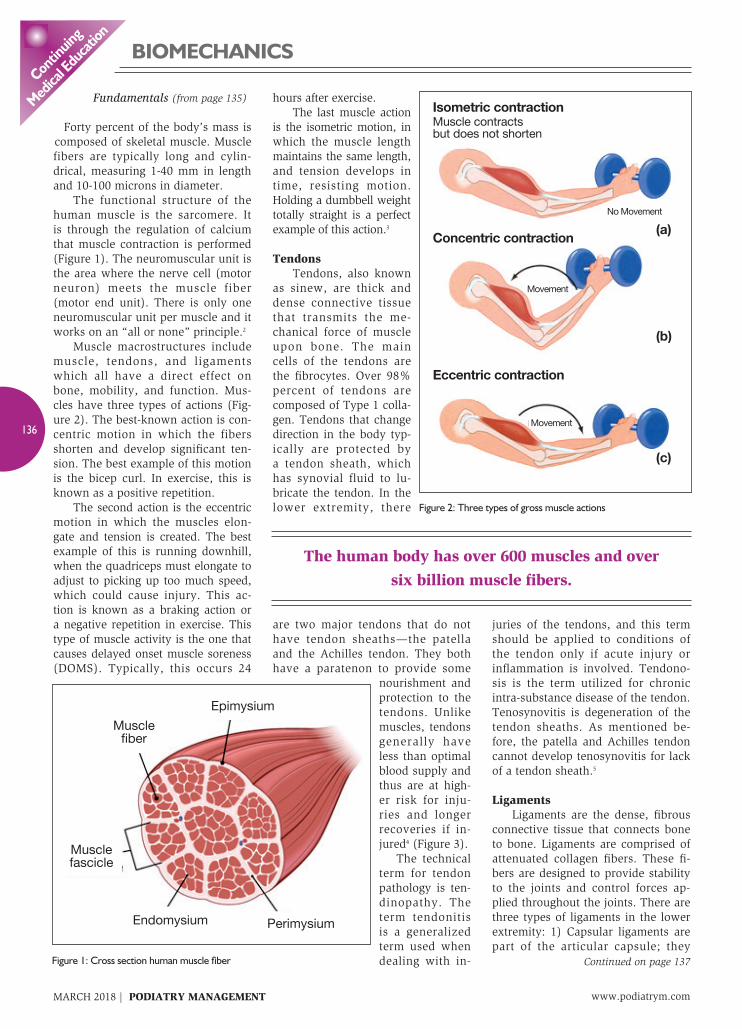
Forty percent of the body’s mass is composed of skeletal muscle. Muscle fibers are typically long and cylin-drical, measuring 1-40 mm in length and 10-100 microns in diameter. The functional structure of the human muscle is the sarcomere. It is through the regulation of calcium that muscle contraction is performed (Figure 1). The neuromuscular unit is the area where the nerve cell (motor neuron) meets the muscle fiber (motor end unit). There is only one neuromuscular unit per muscle and it works on an “all or none” principle.2
Muscle macrostructures include muscle, tendons, and ligaments which all have a direct effect on bone, mobility, and function. Mus-cles have three types of actions (Fig-ure 2). The best-known action is con-centric motion in which the fibers shorten and develop significant ten-sion. The best example of this motion is the bicep curl. In exercise, this is known as a positive repetition. The second action is the eccentric motion in which the muscles elon-gate and tension is created. The best example of this is running downhill, when the quadriceps must elongate to adjust to picking up too much speed, which could cause injury. This ac-tion is known as a braking action or a negative repetition in exercise. This type of muscle activity is the one that causes delayed onset muscle soreness (DOMS). Typically, this occurs 24
hours after exercise. The last muscle action is the isometric motion, in which the muscle length maintains the same length, and tension develops in time, resisting motion. Holding a dumbbell weight totally straight is a perfect example of this action.3
Tendons Tendons, also known as sinew, are thick and dense connective tissue that transmits the me-chanical force of muscle upon bone. The main cells of the tendons are the fibrocytes. Over 98% percent of tendons are composed of Type 1 colla-gen. Tendons that change direction in the body typ-ically are protected by a tendon sheath, which has synovial fluid to lu-bricate the tendon. In the lower extremity, there
are two major tendons that do not have tendon sheaths—the patella and the Achilles tendon. They both have a paratenon to provide some
nourishment and protection to the tendons. Unlike muscles, tendons generally have less than optimal blood supply and thus are at high-er risk for inju-ries and longer recoveries if in-jured4 (Figure 3). The technical term for tendon pathology is ten-dinopathy. The term tendonitis is a generalized term used when dealing with in-
www.podiatrym.comMARCH 2018 | PODIATRY MANAGEMENT
136
contin
uing
Medica
l edu
cation
The human body has over 600 muscles and over six billion muscle fibers.
BioMechanics
Fundamentals (from page 135)
Continued on page 137Figure 1: Cross section human muscle fiber
Figure 2: Three types of gross muscle actions
EpimysiumMuscle
fiber
Musclefascicle
Endomysium Perimysium
Isometric contractionMuscle contractsbut does not shorten
Concentric contraction
Eccentric contraction
(a)
(b)
(c)
Movement
No Movement
Movement

Classification of Foot Pathology Classification of foot patholo-gy can be divided into rearfoot varus and valgus as well as forefoot varus and valgus. Rigid or structural rear-foot varus will remain in a fixed po-sition due to alignment of the tibia, calcaneus, or the position of the sub-talar joint. A flexible rearfoot varus
will be determined by the midtarsal joint. Coleman block testing is very helpful in assessing this type of foot deformity. The direct connection of the rearfoot to the forefoot is via the midtarsal joint. The midtarsal joint consists of the talonavicular joint and the calcanealcuboid joint. Chopart described this joint in de-tail, and thus it is known as Chopart’s
joint. The midtarsal joint also has triplane motion like the subtalar joint, but it also has its majority of motion in the frontal plane, leading mainly to inver-sion and eversion of the joint. There are two axes of motion that comprise the midtarsal joint: the longitu-dinal axis and the oblique axis. The longitudinal axis is parallel with the trans-verse and sagittal planes. Therefore, motion is only in the frontal plane—inver-sion and eversion. Due to the unique alignment of the oblique axis, it has mini-mal motion technically in the sagittal and transverse plane. The average angula-tion of the midtarsal joint is approximately 55 degrees from the sagittal and trans-verse planes (Figure 5). The midtarsal motion is directly related to the subtalar joint’s position. As the subtalar joint goes from maximal pronation to supination, the longitudi-
surround synovial joints and pro-vide mechanical reinforcement to the joint. An example is part of the del-toid ligament of the ankle. 2) Extra capsular ligaments, such as the cal-caneal fibular ligament, provide joint stability. 3) Iintracapsular ligaments allow for more joint motion but gen-erally are weaker ligaments. Exam-ples are the anterior and posterior cruciate ligaments of the knee.6
Historical Background Foot and ankle biomechanics commenced as an interest to phy-sicians in 1935, when Morton dis-cussed anatomic size and hypermo-bility of the foot and ankle.7 In 1942, Manter investigated the subtalar joint and transverse tarsal joint motion.8 In 1953, Hicks reviewed normal foot and ankle mechanics, while in 1960, Elfman evaluated the mechanical re-lationship between the subtalar joint and mid-tarsal joints.9
However, in 1954-1966, Dr. Merton Root truly eval-uated lower extremity me-chanics and defined the normal foot, pathomechan-ics, and the science behind the fabrication of func-tional foot orthoses. This was termed foot morphol-ogy theory. Root mechan-ics identifies the subtalar joint position and motion. Ideally, the subtalar joints have three planes of mo-tion, but motion is main-ly in the frontal plane. A total of 30 degrees of mo-tion is ideal and thus a 2-1 ratio of inversion to ever-sion is within normal limits (Figure 4). This has been used as the primary bio-mechanical foundation in podiatric medical school.10 Inversion is typically 20 de-grees, while eversion is 10 degrees. In recent decades, two more biomechanics theories of some merit have been explored. Kirby empha-sized the tissue stress theo-ry, which relies on the fact
that mechanically-based pathologies of the foot and lower extremity all result from pathological magnitudes of stress acting within the structur-al components of the foot and lower extremity. Stress is an internal mea-sure of how an object resists a loading force. One measures this by dividing the cross-sectional area of that object by the loading force being applied.11
Dananberg proposed the sagittal plane theory. During walking, the center of body mass must pass from behind the weight-bearing foot to in front of it.12 As all of these theories have merit, and as biomechanics is not an absolute science, it has been found over the years that utilization of all three theories is in the best in-terest of patient care.
www.podiatrym.com MARCH 2018 | PODIATRY MANAGEMENT
137
continuing
Medical education
BioMechanics
Fundamentals (from page 136)
Continued on page 138Figure 3: Lower leg anatomy
The functional structure of the human muscle is the sarcomere.

mechanism weakens or reverses, and this allows for further dorsiflexion of the first ray, which limits motion. The line of progression changes and shifts medially, which causes excessive me-dially forefoot loading (Figures 7 & 8). Eventually, a hallux limitus or hallux abductovalgus deformity will develop and over time, a cascade of compen-satory conditions will ensue, such as lesser metatarsalgia, hammertoes, hy-perkeratotic skin conditions, and even arthritis.14
Pes Plano Valgus The etiology of pes plano valgus deformity can be derived from genet-ics, trauma, age, and of course bio-mechanical factors. Pes plano valgus can be further divided into rigid and flexible classifications. Rigid classi-fications, which are less prevalent, include congenital vertical talus and tarsal coalitions. Congenital vertical talus generally needs immediate surgi-cal correction in the infant and tarsal
nal axes of the two midtarsal joints progressively diverge, and thus con-gruity of the midtarsal joint is lost and motion decreases. This relationship of the subtalar and midtarsal joints pro-vides the ideal standard clinical index position in normal foot mechanics and is known as being ‘ locked and loaded’. This position provides the forefoot with all five metatarsal heads being on the same plane and parallel. The calcaneus is perpendicular to the long axis of the tibia and perpendicu-lar to the ground (Figure 6).
Forefoot Varus Forefoot varus will require the subtalar joint to be in a supinated position. Midtarsal joint motion is diminished, and the forefoot is in a varus position in which the forefoot’s plantar plane is inverted. Typically in this foot type, a significant pressure will be placed underneath the fifth metatarsal.
Forefoot Valgus Forefoot valgus will require the subtalar joint to be maximally pronat-ed, resulting in an increased range of motion of the midtarsal joint, with the forefoot plane everted. Opposed to a forefoot varus deformity, a valgus de-formity will cause significant pressure underneath the first metatarsal. As motion and forces continue to move forward from the subtalar and midtarsal joints, there are re-quirements for the first metatarsal phalangeal joint to work effective-
ly. These requirements will necessitate for the subtalar joint to be in a supinated position and for the midtarsal joint to be stable—‘locked and loaded’.13 This will allow for the first metatatarsal head to plantarflex below the plane of the second metatarsal, therefore engaging the windlass mechanism of the plantar fascia. As the first meta-tarsal head plantarflexes with forward momentum, the hallux proximal base will glide on the dorsal aspect of the metatarsal head allowing for ade-quate dorsiflexion. Intrin-sic factors such as a nor-mal metatarsal head and center of axis, along with sesamoid and soft tissue pliability, will assist in
this mechanism of action. Ideally, approximately eighty plus percent of the body’s weight will move along the first metatarsal phalangeal joint, and the joint should dorsiflex greater than 40 degrees in the ideal foot. When faulty mechanics ensue via the subtalar joint excessively pronat-ing and the midtarsal joint becoming
mobile, the first ray will also have impaired mechan-ics. Instability of the first ray will be caused by the peroneal longus losing its me-chanical advan-tage and its abil-ity to plantarflex the first ray, caus-ing dorsiflexion of the first ray and affecting normal range of motion of the joint. In turn, the windlass
www.podiatrym.comMARCH 2018 | PODIATRY MANAGEMENT
138
contin
uing
Medica
l edu
cation
BioMechanics
Fundamentals (from page 137)
Continued on page 139
Figure 4: Normal subtalar joint motion
Figure 5: Midtarsal joint axes of motion
Figure 6: Subtalar and midtarsal joint motion relationship
Pronation Neutral Supination
Delayed onset muscle soreness (DOMS) typically occurs 24 hours after exercise.

The navicular and cuneiform alignment should be parallel, and in a pes planus deformity there will be a ‘fault or sag’ at the joint level. In some situations, the first ray may be elevated with the meta-tarsal head dorsiflexed com-pared to the base of the hal-lux phalanx base (Figure 9). A calcaneal axial and sesamoid axial view will evaluate the calcaneal posi-tion and plane of the meta-tarsal heads and determine if they are neutral or in val-gus or everted position. As pes plano valgus deformity is progressive, it is neces-sary to obtain ankle radio-graphs to evaluate if there is ankle joint deformity. All radiographs should be eval-uated for arthritis.17
Gait Cycle The human adult walking gait cycle is developed at about the age of 4. The adult cycle requires a single leg stance. The walking gait cycle is comprised of two sections: the swing phase and the contact phase. Although the swing phase has a sub-tle impact on biomechanics, it is the contact phase which is the leading cause of lower extremity injuries. The contact phase is approximately 62% and the swing phase 38% of
the gait cycle. The contact phase has three segments: heel contact, mid-stance, and propulsion. The average time it takes to cycle through the contact phase is about 0.4 seconds. With walking, there is roughly four times the body weight applied with each step and the average person should achieve between 5,000 and 10,000 steps per day with the goal of surpassing the higher number.
coalition typically will be addressed as a teenager due to the secondary os-sification centers of the tarsal bones. Regardless, both conditions typically lead towards arthritis with age and thus altered biomechanics.15
Flexible Flatfoot Flexible flatfoot deformity is the most common type of foot deformi-ty and is directly related to impaired biomechanics. Understanding planar dominance of deformity is necessary to institute a proper treatment plan for the patient. The three planes of deformity are sagittal plane, frontal plane, and transverse plane. Sagittal plane deformi-ties include posterior leg muscle group equinus, plantarflexion of the talus, me-dial column breach, and elevated first ray. Frontal plane deformities include calcaneal valgus and everted forefoot. Transverse plane deformity, which is least appreciated, will have an abducted forefoot to rearfoot relationship. I t is imperat ive to obtain weight-bearing radiographs to deter-mine the planar dominance of defor-mity. The important anterior to pos-
terior radiographic measurements to recognize are that the talo-calcaneal angle will be increased. Talonavicular joint coverage, which typically should be 80-90%, is diminished. A bisec-tion of the talus and first metatarsal should be a straight line that will have the talus bisection line exit medial-ly to the first metatarsal. A resultant angle will form. The calcaneal-cuboid angle typically should be 0 degrees and will have an increased angle. In
severe cases, a skew foot deformity may present itself and several faults on radiographs will be noted.16
Sagittal plane radiographs will re-veal a low calcaneal inclination angle less than the ideal 20 plus degrees. Plantar flexion of the talus will be noted. Bisection of the talus and first metatarsal should be a straight line and 0 degrees; however, in a flat foot defor-mity, the talus line will extend below the first metatarsal and create an angle.
www.podiatrym.com MARCH 2018 | PODIATRY MANAGEMENT
139
continuing
Medical education
BioMechanics
Fundamentals (from page 138)
Continued on page 140Figure 7: Normal first ray mechanics
Figure 8: First ray range of motion
Figure 9: Lateral radiograph pes plano valgus
Flexible flatfoot deformity is the most common type of foot deformity and is directly
related to impaired biomechanics.

Running Gait Cycle The running gait cycle differs from the walking gait cycle in that there are greater forces applied, a shorter stance phase, longer swing phase, and unlike the walking gait cycle, there is a float phase. This float phase is where the body is actually completely off the ground. Thus with the float phase in running, the body will absorb up to eight times the body weight with each stride. Running one mile could take about an average of 1,500 steps. Regardless of walking or running, It is within the mid-stance phase that the majority of chronic overuse injuries develop (Figure 10). When evaluating a patient for a biomechanical examination, it is im-perative that an open chain or non-weight-bearing and weight-bearing or closed chain examination are per-formed. Evaluate the patient from head
to toe and investigate for general body relationship and symmetry, muscle flexibility and strength, joint range of motion, leg-length discrepancy. Evalu-ation of shoes and sock gear is a must. In addition to evaluating the patient lying and sitting down, attempt to eval-
uate standing and walking as well. A thorough history and physical is para-mount in assessing a patient’s condi-tion before actual examination. Once a general overview of the body is evaluated, a lower extremity focus should start at the core and its relative structures. The core is the link to providing good posture and trunk stability allowing for the back, legs, and feet to function more
efficiently and effectively. The saying “core to the floor” or “floor to the core” emphasizes the direct relation-ship of the lower extremity. Functional screening tests to eval-uate core strength include: single leg stands and squats, step down squats,
star excursion tests (SEBT), swing test, and one in which the patient, sitting crisscross or Indian style, at-tempts to stand up without leaning towards one side or using hands. PM
References 1 Seigler, Sorin, Foot and ankle joint biomechnics, Sorin Siegler, Journal of Foot and Ankle Biomechanics Vol. 7 s5 2007.
Fundamentals (from page 139)
www.podiatrym.comMARCH 2018 | PODIATRY MANAGEMENT
140
contin
uing
Medica
l edu
cation
BioMechanics
Continued on page 141
Figure 10: Walking & running gait cycle
The majority of overuse conditions develop in the midstance part of the gait cycle.
13 Blackwood C. Brian, Sangeorzon, The Midtarsal Joint Locking Mechanism, Foot and Ankle International, Volume: 26 issue: 12, page(s): 1074-1080, 2005. 14 Faber Frank, Kleinrensink G.J. et al., Mobility of the First Tarsometatarsal Joint in Relation to Hallux Valgus Deformity: Anatomical and Biomechanical Aspects, Foot and Ankle Interna-tional,Volume: 20 issue: 10, page(s): 651-656. 15 Luhmann Scott, Rich Margaret, Painful Idiopathic Rigid Flatfoot in Children and Adolescents, Foot and Ankle Interna-tional, Volume: 21 issue: 1, page(s): 59-66, Jan. 2000. 16 Halabchi Farzin, Mazaheri Reza, Pediatric Flexible Flat-foot; Clinical Aspects and Algorithmic Approach, Iranian Journal of Pediatrics, 2013 Jun; 23(3): 247–260. 17 Lamm Bradley, Stasko Paul, Normal Foot and Ankle Ra-diographic Angles, Measurements, and Reference Points, Journal Foot and Ankle Surgery, Vol. 55, issue 5, 2016.
dr. Mendeszoon is a foot and ankle surgeon at Precision Orthopaedic Specialties, Inc., a multi-specialty orthopedic practice in Chardon, Ohio. He is the Director of the Advanced Foot and Ankle Surgical Fellowship through Univer-sity Hospitals—Richmond Medical Center, and Chief of Podiatry at University Hospital’s Geauga Medical Center. Dr. Mendeszoon is an assistant clinical professor at the Ohio University Heritage Medical School and an instructor of surgery at
Kent State University School of Podiatric Medicine.
continuing
Medical education
141
www.podiatrym.com MARCH 2018 | PODIATRY MANAGEMENT
BioMechanics
2 Frontera WR, Ochala J, Skeletal muscle: a brief review of structure and function, Calcif Tissue Int., 2015 Mar;96(3):183-95. 3 Guo, J, et al., Massage Alleviates Delayed Onset Muscle Soreness after Strenuous Exercise: A Systematic Review and Me-ta-Analysis, Front Physiol, 2017 Sep 27;8. 4 Binstead, JT, Bhimiji SS, Anatomy, Lower Limb, Calf., Stat-Pearls [Internet]. Treasure Island (FL): 2017. 5 McCreesh, Karen, Lewis, Jeremy, Continuum model of ten-don pathology—where are we now? Int J Exp Pathol. 2013 Aug; 94(4): 242–247. 6 Golano Pau, Vega, Jordi et al., Anatomy of the ankle liga-ments: a pictorial essay, Knee Surg Sports Traumatol Arthrosc. 2010 May; 18(5): 557–569. 7 Nester Christopher J, Leardini Alberto, International Foot and Ankle Biomechanics Community (i-FAB): past, present and beyond, J Foot Ankle Res. 2009; 2: 19. 8 Van Ghelue, Bart, Kirby, Kevin, Research and clinical syn-ergy in foot and lower extremity biomechanics, Journal Footwear Science, Vol. 2, 2010. 9 Mann, RA, Biomechanics of the Ankle, Joint Surgery Up to Date pp 73-81. 10 Kirby Kevin, Are Root Biomechanics Dying, Podiatry Today, vol.22, issue 4, 2009. 11 Kirby, Kevin, Prescribing Orthoses: Has Tissue Stress Theory Supplanted Root Theory?, Podiatry Today, vol. 28, issue 4, 2015. 12 Dananberg Howard, Sagittal Plane Biomechanics, J Am Podiatr Med Assoc, 90 (1): 47-50, 2000.
Fundamentals (from page 140)
1) The human body has approximately how many muscles? A) Two hundred B) Four hundred C) Six hundred D) Eight hundred
2) The functional unit of a skeletal muscle is A) Sarcomere B) Myosin C) Endoplasmic reticulum D) Myofibril
3) Delayed onset muscle soreness (DOMS) typi-cally occurs how many hours after exercise? A) 6 hours B) 12 hours C) 18 hours D) 24 hours
4) Which foot and ankle biomechanical theory is generally NOT utilized in evaluating a patient? A) Root theory B) Kirby theory C) Jones theory D) Dananberg theory
5) The majority of overuse conditions develop in which part of the gait cycle? A) Swing phase B) Heel contact C) Midstance D) Toe off
6) The human adult walking gait cycle is devel-oped at about the age of ______ ? A) One B) Two C) Four D) Six
CME eXaMinaTion
See anSwer Sheet on page 143.
Continued on page 142

MARCH 2018 | PODIATRY MANAGEMENT
142
PM’scMe Program
Welcome to the innovative Continuing Education Program brought to you by Podiatry Management Magazine. Our journal has been approved as a sponsor of Continuing Medical Education by the Council on Podiatric Medical Education.
now it’s even easier and more convenient to enroll in PM’s ce program! You can now enroll at any time during the year and submit eligible exams at any time during your enrollment period. cMe articles and examination questions from past issues of Podiatry Management can be found on the internet at http://www.podiatrym.com/cme. Each lesson is approved for 1.5 hours continuing education contact hours. Please read the testing, grading and payment instructions to decide which method of participa-tion is best for you. Please call (631) 563-1604 if you have any questions. A personal operator will be happy to assist you. Each of the 10 lessons will count as 1.5 credits; thus a maximum of 15 CME credits may be earned during any 12-month period. You may select any 10 in a 24-month period.
The Podiatry Management Magazine CME program is approved by the Council on Podi-atric Education in all states where credits in instructional media are accepted. This article is approved for 1.5 Continuing Education Contact Hours (or 0.15 CEU’s) for each examination successfully completed.
PM’s privacy policy can be found at http:// podiatrym.com/privacy.cfm.
home study cMe credits now accepted in Pennsylvania
$
CME eXaMinaTioncon
tinuin
g
Medica
l edu
cation
7) Over 98% percent of tendons are com-posed of A) Muscle B) Ligaments C) Water D) Type 1 collagen
8) ______________ deformity is the most com-mon type of foot deformity and is directly related to impaired biomechanics. A) Rigid flatfoot B) Flexible flatfoot C) Cavus foot D) Charcot foot
9) The etiology of pes plano valgus deformity can be derived from: A) Genetics B) Trauma C) Biomechanical factors D) All of the above
10) It is recommended that the average per-son take how many steps a day to remain active and healthy? A) 5,000 B) 10,000 C) 15,000 D) 20,000
See anSwer Sheet on page 143.
The author(s) certify that they have NO affili-ations with or involvement in any organization or entity with any financial interest (such as honorar-ia; educational grants; participation in speakers’ bureaus; membership, employment, consultancies, stock ownership, or other equity interest), or non-fi-nancial interest (such as personal or professional relationships, affiliations, knowledge, or beliefs) in the subject matter or materials discussed in this manuscript.

Please print clearly...Certificate will be issued from information below.
Name ____________________________________________________________________ Email Address______________________________Please Print: FIRST MI LAST
Address_____________________________________________________________________________________________________________
City__________________________________________________ State_______________________ Zip________________________________
Charge to: _____Visa _____ MasterCard _____ American Express
Card #________________________________________________Exp. Date____________________ Zip for credit card_________________
note: credit card is the only method of payment. checks are no longer accepted.
Signature__________________________________ Email Address_________________________ Daytime Phone_______________________
State License(s)___________________________ Is this a new address? Yes________ No________
check one: ______ I am currently enrolled. (If faxing or phoning in your answer form please note that $2.50 will be charged to your credit card.)
______ I am not enrolled. Enclosed is my credit card information. Please charge my credit card $27.00 for each exam submitted. (plus $2.50 for each exam if submitting by fax or phone).
______ I am not enrolled and I wish to enroll for 10 courses at $219.00 (thus saving me $51 over the cost of 10 individual exam fees). I understand there will be an additional fee of $2.50 for any exam I wish to submit via fax or phone.
note: If you are mailing your answer sheet, you must complete all info. on the front and back of this page and mail with your credit card information to: Program Management services, P.o. Box 490, east islip, ny 11730.
TesTing, grading and PayMenT insTrucTions (1) Each participant achieving a passing grade of 70% or higher on any examination will receive an official computer form stating the number of CE credits earned. This form should be safeguarded and may be used as documentation of credits earned. (2) Participants receiving a failing grade on any exam will be notified and permitted to take one re-examination at no extra cost. (3) All answers should be recorded on the answer form below. For each question, decide which choice is the best answer, and cir-cle the letter representing your choice. (4) Complete all other information on the front and back of this page. (5) Choose one out of the 3 options for testgrading: mail-in, fax, or phone. To select the type of service that best suits your needs, please read the following section, “Test Grading Options”.
TesT grading oPTions Mail-In Grading To receive your CME certificate, complete all information and mail with your credit card information to: Program Management services, P.o. Box 490, east islip, ny 11730. PLease do noT send WiTh signaTure reQuired, as These WiLL noT Be accePTed.
enroLLMenT ForM & ansWer sheeT
$
There is no charge for the mail-in service if you have al-ready enrolled in the annual exam CME program, and we receive this exam during your current enrollment period. If you are not en-rolled, please send $27.00 per exam, or $219 to cover all 10 exams (thus saving $51 over the cost of 10 individual exam fees).
Facsimile Grading To receive your CME certificate, complete all information and fax 24 hours a day to 631-532-1964. Your CME certificate will be dated and mailed within 48 hours. This service is available for $2.50 per exam if you are currently enrolled in the annual 10-exam CME program (and this exam falls within your enrollment period), and can be charged to your Visa, MasterCard, or American Express. If you are not enrolled in the annual 10-exam CME program, the fee is $27 per exam.
Phone-In Grading You may also complete your exam by using the toll-free service. Call 1-800-232-4422 from 10 a.m. to 5 p.m. EST, Monday through Friday. Your CME certificate will be dated the same day you call and mailed within 48 hours. There is a $2.50 charge for this service if you are currently enrolled in the annual 10-exam CME program (and this exam falls within your enrollment period), and this fee can be charged to your Visa, Mastercard, American Express, or Discover. If you are not current-ly enrolled, the fee is $27 per exam. When you call, please have ready: 1. Program number (Month and Year) 2. The answers to the test 3. Credit card information
Over, please
continuing
Medical education
enrollment/Testing informationand answer sheet
143
www.podiatrym.com MARCH 2018 | PODIATRY MANAGEMENT
In the event you require additional CME information, please contact PMS, Inc., at 1-631-563-1604.

144
www.podiatrym.comMARCH 2018 | PODIATRY MANAGEMENT
contin
uing
Medica
l edu
cation
enroLLMenT ForM & ansWer sheeT (continued)
$
Medical education Lesson evaluation
Strongly Strongly agree Agree Neutral Disagree disagree [5] [4] [3] [2] [1]
1) This CME lesson was helpful to my practice ____
2) The educational objectives were accomplished ____
3) I will apply the knowledge I learned from this lesson ____
4) I will makes changes in my practice behavior based on this lesson ____
5) This lesson presented quality information with adequate current references ____
6) What overall grade would you assign this lesson?A B C D
How long did it take you to complete this lesson?
______hour ______minutes
What topics would you like to see in future CME lessons ? Please list :
__________________________________________________
__________________________________________________
__________________________________________________
__________________________________________________
__________________________________________________
__________________________________________________
1. a B c d
2. a B c d
3. a B c d
4. a B c d
5. a B c d
6. a B c d
7. a B c d
8. a B c d
9. a B c d
10. a B c d
circle:
eXaM #3/18Fundamentals of care in Biomechanics—Part 1
(Mendeszoon)
This CME activity is free from commercial biases and is under the overall management of Podiatry Management Magazine.