Embed Size (px)
Citation preview

BioMed CentralOrphanet Journal of Rare Diseases
ss
Open AcceReviewCentronuclear (myotubular) myopathyHeinz Jungbluth*1,2, Carina Wallgren-Pettersson3,4 and Jocelyn Laporte5,6,7,8,9Address: 1Department of Paediatric Neurology, Neuromuscular Service, Evelina Children's Hospital, St Thomas' Hospital, Lambeth Palace Road, London, SE1 7EH, UK, 2Clinical Neuroscience Division, King's College, London, UK, 3Department of Medical Genetics, University of Helsinki, Helsinki, Finland, 4The Folkhälsan Department of Medical Genetics, Helsinki, Finland, 5Department of Neurobiology and Genetics, IGBMC (Institut de Génétique et de Biologie Moléculaire et Cellulaire), Illkirch F-67400, France, 6Inserm, U596, Illkirch F-67400, France, 7CNRS, UMR7104, Illkirch F-67400, France, 8Université Louis Pasteur, Strasbourg F-67000, France and 9Collège de France, Illkirch F-67400, France
Email: Heinz Jungbluth* - [email protected]; Carina Wallgren-Pettersson - [email protected]; Jocelyn Laporte - [email protected]
* Corresponding author
AbstractCentronuclear myopathy (CNM) is an inherited neuromuscular disorder characterised by clinical features of a congenitalmyopathy and centrally placed nuclei on muscle biopsy.
The incidence of X-linked myotubular myopathy is estimated at 2/100000 male births but epidemiological data for other formsare not currently available.
The clinical picture is highly variable. The X-linked form usually gives rise to a severe phenotype in males presenting at birth withmarked weakness and hypotonia, external ophthalmoplegia and respiratory failure. Signs of antenatal onset comprise reducedfoetal movements, polyhydramnios and thinning of the ribs on chest radiographs; birth asphyxia may be the present. Affectedinfants are often macrosomic, with length above the 90th centile and large head circumference. Testes are frequentlyundescended. Both autosomal-recessive (AR) and autosomal-dominant (AD) forms differ from the X-linked form regarding ageat onset, severity, clinical characteristics and prognosis. In general, AD forms have a later onset and milder course than the X-linked form, and the AR form is intermediate in both respects.
Mutations in the myotubularin (MTM1) gene on chromosome Xq28 have been identified in the majority of patients with the X-linked recessive form, whilst AD and AR forms have been associated with mutations in the dynamin 2 (DNM2) gene onchromosome 19p13.2 and the amphiphysin 2 (BIN1) gene on chromosome 2q14, respectively. Single cases with features of CNMhave been associated with mutations in the skeletal muscle ryanodine receptor (RYR1) and the hJUMPY (MTMR14) genes.
Diagnosis is based on typical histopathological findings on muscle biopsy in combination with suggestive clinical features; musclemagnetic resonance imaging may complement clinical assessment and inform genetic testing in cases with equivocal features.Genetic counselling should be offered to all patients and families in whom a diagnosis of CNM has been made.
The main differential diagnoses include congenital myotonic dystrophy and other conditions with severe neonatal hypotonia.
Management of CNM is mainly supportive, based on a multidisciplinary approach. Whereas the X-linked form due to MTM1mutations is often fatal in infancy, dominant forms due to DNM2 mutations and some cases of the recessive BIN1-related formappear to be associated with an overall more favourable prognosis.
Published: 25 September 2008
Orphanet Journal of Rare Diseases 2008, 3:26 doi:10.1186/1750-1172-3-26
Received: 22 April 2008Accepted: 25 September 2008
This article is available from: http://www.ojrd.com/content/3/1/26
© 2008 Jungbluth et al; licensee BioMed Central Ltd. This is an Open Access article distributed under the terms of the Creative Commons Attribution License (http://creativecommons.org/licenses/by/2.0), which permits unrestricted use, distribution, and reproduction in any medium, provided the original work is properly cited.
Page 1 of 13(page number not for citation purposes)

Orphanet Journal of Rare Diseases 2008, 3:26 http://www.ojrd.com/content/3/1/26
Disease nameCentronuclear (myotubular) myopathy
DefinitionCentronuclear myopathy (CNM) is an inherited neu-romuscular disorder defined by a) numerous centrallyplaced nuclei on muscle biopsy and b) clinical features ofa congenital myopathy. Additional but inconsistent his-topathological features comprise a surrounding centralzone either devoid of oxidative enzyme activity or withoxidative enzyme accumulation, and, in patients withmutations in the dynamin 2 (DNM2) gene, radial sarco-plasmic strands surrounding the central area; signs ofnecrosis or excessive regeneration are usually absent in allforms of CNM.
Centronuclear myopathy exists in X-linked recessive(OMIM 310400) [1], autosomal-dominant (OMIM160150) and autosomal-recessive forms (OMIM255200). The term myotubular myopathy [2], introducedbecause of a similar appearance of affected fibres and foe-tal myotubes, is still used by many for the X-linked form,whilst centronuclear myopathy is a term used for both theautosomal-dominant and recessive variants of the condi-tion.
EpidemiologyEpidemiological data are only available for the congenitalmyopathies as a group but not for specific conditions. Theincidence of all congenital myopathies (including centralcore disease, multi-minicore disease, nemaline myopathyand centronuclear myopathy) is estimated at around0.06/1,000 live births, or one-tenth of all cases of neu-romuscular disorders [3]. Regional studies in NorthernIreland [4] and Western Sweden [5], suggest a prevalenceof 3.5 – 5.0/100,000 in a paediatric population. Thesenumbers are likely to be underestimates, as histopatho-logical expression of specific genetic defects may be varia-ble and often non-specific, particularly at a young age.
Based on unpublished observations (JL), the incidence ofmolecularly confirmed myotubular myopathy in France isestimated at 2/100,000 male births per year. Whilst dataregarding the overall incidence and prevalence of CNMare not available, the condition clearly occurs less fre-quently than central core disease and multi-minicore dis-ease, the most common congenital myopathies, andnemaline myopathy (HJ, personal observation).
Clinical descriptionWhilst creatine kinase (CK) is normal or only slightly ele-vated in all forms of centronuclear (myotubular) myopa-thy, the clinical picture is highly variable depending onthe causative mutation.
The X-linked form due to mutations in the myotubularin(MTM1) gene has been clinically well characterised andusually gives rise to a severe phenotype in males present-ing at birth with marked weakness and hypotonia, exter-nal ophthalmoplegia and respiratory failure (Figure 1) [6-19]; a preceding family history of either male neonataldeaths or miscarriages is common. Signs of antenatalonset comprise reduced foetal movements, polyhydram-nios and thinning of the ribs on chest radiographs [20,21]and are only rarely observed in other congenital myopa-thies. Birth asphyxia may be the presenting feature[10,22]. Affected infants are often macrosomic, andlength above the 90th centile and large head circumferencemay serve as a diagnostic clue [23,24]. Testes are fre-quently undescended [25]. In the majority of cases thecourse is fatal within the first months of life, but a propor-tion of affected males may survive into their teens orbeyond [19,26,27]. Although a small proportion of boysmay be only mildly affected in the neonatal period andthereafter [6,18,19,22,27-29], in the majority of long-term survivors survival depends on a substantial degree ofmedical intervention and often constant ventilation. Arange of medical complications in some long-term survi-vors comprising pyloric stenosis and cavernous haeman-giomas of the liver [26] has also been reported and mayindicate wider expression of the defective protein. Addi-tional genital abnormalities have been described inaffected males with contiguous gene syndromes [30], andis due to loss of the adjacent MAMLD1 (Cxorf6) gene [31].
The majority of carriers of the X-linked formare asympto-matic but a few may show signs of mild muscle weakness[10,32-34]. Presentation may be overt in females, espe-cially if additional genetic abnormalities such as skewedX-inactivation [34-39] or structural X-chromosomalabnormalities are present [40]. Urinary incontinence, pri-mary or secondary, may be an additional feature indicat-ing smooth muscle involvement [33,37].
Male infant with X-linked centronuclear ("myotubular") myopathy due to a mutation in the myotubularin (MTM1) geneFigure 1Male infant with X-linked centronuclear ("myotubu-lar") myopathy due to a mutation in the myotubu-larin (MTM1) gene. Note generalised hypotonia and myopathic facial appearance with elongated face and inverted V-shaped mouth. (Reproduced from MedLink®Neurology, with permission)
Page 2 of 13(page number not for citation purposes)

Orphanet Journal of Rare Diseases 2008, 3:26 http://www.ojrd.com/content/3/1/26
Both autosomal-recessive and autosomal-dominantforms have been well documented and differ from the X-linked form regarding age at onset, severity, clinical char-acteristics and prognosis [18,41]. As a general rule, auto-somal-dominant forms have a later onset and mildercourse than the X-linked form, and the autosomal-reces-sive form is intermediate in both respects, but these differ-ences are quantitative rather than qualitative. Most reportsconcerning autosomal-recessive and dominant forms ofCNM predate the molecular resolution of these condi-tions and are likely to reflect genetically heterogeneousconditions; however, the recent identification of the genesimplicated in subgroups of recessive and dominant CNMoffers the prospect of more precise genotype-phenotypecorrelative studies in future.
The autosomal-recessive form [10,41-52] is characterisedby facial weakness including severe involvement of themasticatory muscles [53], and ocular abnormalities suchas ptosis and external ophthalmoplegia. A recent Frenchseries distinguishes early and late onset forms with orwithout ophthalmoplegia; it remains to be seen if thosedistinctions are reflective of underlying genetic heteroge-neity [50]. Weakness is usually prominent proximally butthere may be additional distal weakness and wasting inthe lower limbs, and foot abnormalities are frequentlyfound [54]. Other skeletal deformities including higharched palate and scoliosis are common [55]. Respiratoryinvolvement may be severe [52], and an associated cardi-omyopathy has been documented in a few recurrent andsporadic cases [44,56,57]. As in carriers of the X-linkedform, urinary incontinence may be an associated feature[58]. In the absence of severe cardiorespiratory involve-ment, the prognosis appears favourable. Whilst most ofthe features of supposedly recessive cases of centronuclearmyopathy were reported in genetically unresolved cases,identification of homozygous recessive mutations in theamphiphysin 2 (BIN1) gene in 4 patients form 3 families[59] allows initial genotype-phenotype correlations,although, considering the small number of cases, therange of clinical features associated with mutations in thisgene is likely to expand further in the future. Clinical fea-tures in the patients identified to date suggested a pheno-type of intermediate severity between the X-linkedrecessive and the dominant forms, with onset from birthto childhood and mild progressive proximal weakness butno respiratory impairment severe enough to require ven-tilatory assistance. Cardiac involvement was not presentin these patients but, notably, appears to be a feature inthe BIN1 knockout mouse [60].
Most patients with the autosomal-dominant form ofCNM are more mildly affected than those with the X-linked or autosomal-recessive forms with a widely varia-ble age of onset [18,61-75]. The distribution of weaknessis predominantly proximal with additional distal involve-
ment, external ophthalmoplegia and ptosis; in somecases, prominent calf muscle hypertrophy may be an addi-tional feature [50].
The autosomal-dominant form of CNM due to muta-tions in the dynamin 2 (DNM2) gene may be of variableseverity depending on the part of the protein affected.Dominant DNM2 mutations affecting the dynamin 2middle domain reported to date appear to be associatedwith a mild clinical phenotype characterised by normalearly motor developmental milestones, onset in adoles-cence and a slowly progressive course with loss of inde-pendent ambulation uncommon before the 6th decade[76-78]. In addition to signs of proximal weakness, exer-cise-induced myalgia may be a presenting feature. Ocu-lar involvement, particularly ptosis, is almost invariableand distal muscle involvement, particularly in the lowerlimb, may precede more proximal weakness; the latterfinding corresponds to a sequential pattern with earlyinvolvement of the ankle plantarflexors, namely themedial gastrocnemius, followed by signal changes in theposterior and, eventually, anterior compartment of thethighs [77,78]. Contractures other than those affectingthe Achilles tendon and/or long finger flexors are rare.Electromyogram (EMG) and nerve conduction studiesmay show mild signs of axonal peripheral nerve involve-ment in addition to prominent myopathic changes[77,79]. Whilst dominant mutations affecting thedynamin 2 middle domain have been associated with amild phenotype of CNM, a more severe presentationwith neonatal onset has been recently attributed to het-erozygous de novo dominant mutations affecting thepleckstrin homology (PH) domain of the dynamin 2protein, a protein domain also altered in the CMTDIBneuropathy [80,81]. Like other patients with CNM, thesehad marked ocular involvement including ptosis andophthalmoparesis, and, despite a severe and early pres-entation, gradually improved over time. However, whilstcardiorespiratory function in DNM2-related CNM hasbeen normal in most reported cases those with earlyonset may develop restrictive respiratory impairmentover time [81]. Electrophysiology showed exclusivemyopathic but no neuropathic changes.
Centronuclear myopathy due to a heterozygous de novodominant mutation in the skeletal muscle ryanodinereceptor (RYR1) gene has to date been reported in onlyone case [82], with clinical features comprising extraocu-lar involvement, generalized weakness, moderate bulbarand respiratory impairment similar to multi-minicore dis-ease (MmD), due to recessive mutations in the RYR1 gene[83,84]. Although the frequency of RYR1 mutations inCNM is currently uncertain, malignant hyperthermia sus-ceptibility reported in a case of CNM in the premolecularera [85] may indicate more widespread RYR1 involvementin CNM.
Page 3 of 13(page number not for citation purposes)
Orphanet Journal of Rare Diseases 2008, 3:26 http://www.ojrd.com/content/3/1/26
AetiologyCentronuclear (myotubular) myopathy exists in X-linked,autosomal-recessive and autosomal-dominant forms.
The X-linked recessive form ("myotubular myopathy")has been genetically well characterised. Following initiallinkage studies and assignment of a locus to chromosomeXq28 [40,86-94] mutations in the myotubularin (MTM1)gene have now been identified in more than 90% ofaffected males [19,95-100]; molecular genetic analysis ofthe MTM1 gene is now widely available as a routine diag-nostic service [34,101,102]. Disease-causing sequencechanges include deletions/insertions, nonsense, missenseand splice mutations (approximately 25% each) [97,102].Three substitutions account for 15% of all MTM1 muta-tions; these are the splice mutation c.1261-10A>G(intronic, upstream of exon 12) resulting in the insertionof three amino acids FIQ at position 420 (7.3%), R241Cencoded by exon 9 (4%), and c.141-144 delAGAA result-ing in a frameshift at amino acid 48 in exon 4 (4%) [28].Other mutations have been reported in a few families orare unique. MTM1 mutations are distributed throughoutthe entire coding sequence, but localise most frequently(in descending order) to exons 12, 4, 11, 8 and 9 [97-99,102-107]. Maternal carrier state of MTM1 mutations isestimated at 85% and is thus more common than statisti-cally expected for a severe X-linked disease [97,102];maternal mosaicism has been reported in a few families[97,108,109] with important implications for geneticcounselling regarding future pregnancies.
Genotype-phenotype correlative studies have been diffi-cult because many mutations are private to individualfamilies and clinical severity associated with specificmutations may vary even within the same families; how-ever, one large series demonstrated that, while most muta-tions are associated with the severe phenotype, some non-truncating mutations outside of the catalytic domain maycarry a more favourable prognosis [19,27,28,110].
Screening of the MTM1 gene should be considered infemales with suggestive clinical and histopathological fea-tures; although usually asymptomatic or only mildlyaffected, carriers may manifest severe symptoms in thepresence of skewed X-inactivation and/or structural alter-ations involving the X-chromosome such as interstitialdeletions [34-40].
Myotubularin belongs to the large family of dual-specifi-city phosphatases, playing a role in the epigenetic regula-tion of signalling pathways involved in growth anddifferentiation; mutations in some human myotubularinhomologues have been associated with two specific formsof peripheral neuropathies of the Charcot-Marie-Tooth(CMT) type, CMT 4B1 [111,112] and CMT 4B2 [113,114].
A specific function has been proposed for myotubularinin dephosphorylating phosphatidylinositol 3-phosphate[PtdIns3P] and PtdIns(3,5)P. These two phospholipidsare second messengers with a crucial role in membranetrafficking; by dephosphorylation of PtdIns(3,5)P2, myo-tubularin also produces PtdIns5P, whose function is notfully characterised [95,115-126]. In addition to the cata-lytic site, myotubularins form homo- and heterodimersand contain lipid and protein binding sites; thesedomains include a GRAM-PH, a coiled-coil region and aputative PDZ binding site. Concerning myotubularin, noprotein interactors have been characterised to date in skel-etal muscle. The deleterious effect of specific MTM1 muta-tions may be due to either destabilisation of the 3-Dstructure or loss of enzymatic activity, although it is possi-ble that a few mutations affect existing but not yet identi-fied protein-protein interactions in muscle.
Observations in an MTM1-related mouse model [127]suggest a role of myotubularin in muscle fibre mainte-nance but not in myogenesis. A recent gene expressionprofiling study in muscle harbouring MTM1 mutationsrevealed upregulation of transcripts for cytoskeletal andextracellular matrix proteins within or around atrophicmyofibres, indicating that remodelling of cytoskeletal andextracellular architecture plays a role in the atrophy andintracellular disorganization observed in X-linked myotu-bular myopathy [128]. Prolonged expression but eventualdecrease of developmentally regulated proteins in musclefrom affected infants (see also paragraph on Diagnosticmethods below) suggests maturational delay rather thancomplete developmental arrest in this condition.
Dominant forms of centronuclear myopathy have beenassociated with mutations in two genes, the dynamin 2(DNM2) gene on chromosome 19p13.2 [76] also impli-cated in dominant intermediate (CMTDIB) [80] andaxonal (CMT2) [129,130] forms of Charcot-Marie-Toothdisease, and the skeletal muscle ryanodine receptor(RYR1) gene on chromosome 19q13.1 in one isolatedcase [82].
The DNM2 gene consists of 22 exons [80] and encodes alarge GTPase protein involved in actin cytoskeletonassembly [131] and centrosome cohesion [132]. In addi-tion, DNM2 is implicated in membrane trafficking fromthe plasma membrane and Golgi, to allow the formationand fission of budding vesicles [133]. It is of interest tonote that myotubularin, implicated in the X-linked formof CNM, has also been implicated in membrane traffick-ing and endocytosis, although its precise function remainsto be determined. Recent studies on cells transfected withCNM-related DNM2 mutants suggest lack of localisationto the centrosome and centrosome malfunction as a pos-sible pathogenetic mechanism in DNM2-related CNM.
Page 4 of 13(page number not for citation purposes)

Orphanet Journal of Rare Diseases 2008, 3:26 http://www.ojrd.com/content/3/1/26
However, the impacts of mutations on allosteric enzy-matic activity and membrane remodelling properties ofdynamin 2 remain to be investigated. Recurrent and denovo DNM2 mutations were originally identified follow-ing a positional candidate approach in 11 families with amild form of autosomal-dominant centronuclear myopa-thy [76]. The most common mutation is a 1393C>Tchange found in 6 unrelated families resulting in anarginine to tryptophane substitution at position 465.Dynamins are structurally complex proteins composed of5 different domains; CNM-causing mutations identifiedto date mainly localise to the middle domain involved inprotein self assembly and centrosome localisation [76],whereas those associated with CMTDIB have been identi-fied within the pleckstrin homology (PH) domain [80].More recently, heterozygous de novo dominant DNM2mutations affecting the PH domain have also been identi-fied in a more severe CNM phenotype without any earlyperipheral nerve involvement and characterised by neona-tal onset but gradual improvement over time [81]. Thepotential overlap between myogenic and neurogenic find-ings in families with mutations in this region is currentlybeing explored [77,79].
Features of centronuclear myopathy associated with theskeletal muscle ryanodine receptor (RYR1) gene have todate only been reported in one single case with a de novodominant mutation resulting in a serine to leucine substi-tution at position 4112 [82]; the RYR1 gene had been con-sidered as a candidate in this patient because of thefrequent observation of multiple central nuclei in otherRYR1-related phenotypes and the suggestion of a clinicalcontinuum and overlap of radiological features on muscleMRI. Functional studies on patient-derived, MyoD-trans-formed fibroblasts indicated that cells harbouring thismutation may be hypersensitive to depolarization, but itremains unclear how the change gives rise to the appear-ance of CNM. The frequency of the RYR1-related form ofCNM is currently uncertain.
Mutations in the amphiphysin-2 (BIN1) gene on chromo-some 2q14 have been recently identified in a small pro-portion of cases with the recessive form of centronuclearmyopathy [59] but further genetic heterogeneity isexpected. The BIN1 gene is organised in 20 exons, the pro-tein exists in at least 10 different isoforms subject to alter-native splicing and, in addition to muscle, the gene isexpressed in a number of different tissues including cen-tral and peripheral nervous systems [134,135]. BIN1 wasconsidered a candidate for genetically unresolved forms ofCNM because of functional characteristics shared withother CNM-associated genes, namely a phosphoinositide-regulated role in membrane modelling [136], and thepresence of a muscle phenotype in the Drosophila mela-nogaster mutant [137]. The amphiphysin 2 muscle-spe-
cific isoform features an N-terminal amphipathic helixthought to be involved in creating membrane curvature, aBAR (Bin1, Amphiphysin, RVS167) domain thathomodimerizes and maintains the curvature, a phosphoi-nositide-binding domain, and an SH3 domain interactingwith dynamin 2 and other proteins [136,138]. Functionalstudies on the three mutations identified to date suggestthat BIN1 missense mutations in the N-BAR domain affectmembrane curvature, whilst a truncating mutation in theSH3 domain appears to abolish amphiphysin2-dynamin2 interactions [59]; this indicates the importance ofamphiphysin 2-dynamin 2 coupling for normal musclefunction and suggests a possible alteration of T-tubuleorganisation, as amphiphysin 2 was previously proposedto have a role in this process [137,139].
In addition to the BIN1-related form, heterozygous mis-sense variants in hJUMPY, a novel phosphoinositidephosphatase with functional similarities to myotubularin,were recently identified in two sporadic cases with fea-tures of centronuclear myopathy and an additionalDNM2 mutation in one case [140]. These variants wereshown to decrease the enzymatic activity of hJUMPY in invitro and in cellulo experiments. It is unclear whether thephenotype in those cases is due to digeny or recessiveinheritance with an undetected second mutation, andclarification of the implication of hJUMPY awaits thecharacterisation of additional patients with mutations inthis gene.
Genes implicated in various forms of centronuclearmyopathy have been summarised in Table 1.
Diagnostic methodsThe diagnosis of CNM depends on the presence of typicalhistopathological findings on muscle biopsy in combina-tion with suggestive clinical features; muscle MR imagingmay complement clinical assessment and inform genetictesting in cases with equivocal features.
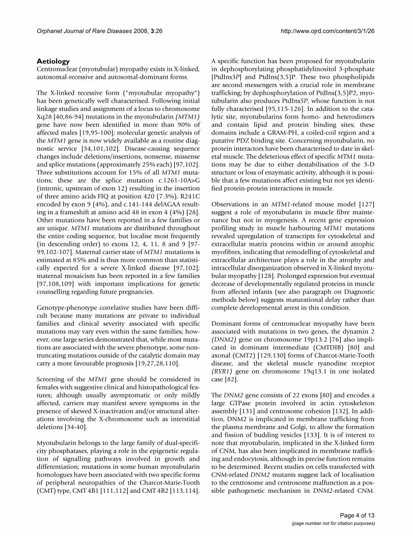
On muscle biopsy, centronuclear (myotubular) myopa-thy is characterised by centrally placed nuclei surroundedby a perinuclear halo devoid of myofilaments [2] andoccupied by mitochondrial and glycogen aggregates (Fig-ure 2) The characteristic central nuclei are seen in all mus-cles, including extra-ocular muscles [141], and may affectup to 90% of fibres [52]. Some autosomal cases of centro-nuclear myopathy may also feature a radial arrangementof sarcoplasmic strands on NADH staining [50]; it cur-rently appears that this is a feature in most cases of centro-nuclear myopathy caused by mutations in the DNM2gene [76]: Whilst radial arrangement of sarcoplasmicstrands appears to be common in mild forms of DNM2-related CNM due to mutations affecting the middledomain [77], this finding appears not to be as prominent
Page 5 of 13(page number not for citation purposes)
Orphanet Journal of Rare Diseases 2008, 3:26 http://www.ojrd.com/content/3/1/26
in more severe and early presentations due to mutationsaffecting the pleckstrin homology (PH) domain, or inother genetically distinct forms of CNM. Type 1 predomi-nance and hypotrophy [14,142] are commonly associatedfeatures, and may precede the appearance of internalnuclei [143]; there may be compensatory type 2 hypertro-phy in a small number of fibres [25], and a deficiency oftype 2B fibres with relative increase in undifferentiatedtype 2C fibres [142].
More recently, Pierson et al. [110] were able to correlateMTM1 mutation type and pathologic findings and coulddemonstrate that missense mutations are associated withincreased myofibre diameter compared to nonsensemutations.
Histopathological changes may progress over time andmarked increases in fat [144] and connective tissue
[37,78,145] can at times be a striking feature. Associatedcore-like structures have occasionally been reported[47,144] and may be associated with mutations in boththe skeletal muscle ryanodine receptor (RYR1) gene [82]and the DNM2 gene [78].
Reported histopathological findings in carriers of the X-linked form range from normal appearance in clinicallyasymptomatic mothers [11] to findings similar to those inaffected males, as reported in a female with the full clini-cal picture of myotubular myopathy due to skewed X-inactivation [37].
On electron microscopy (EM), immaturity of neuromus-cular junctions and junctional changes comprising reduc-tion of acetylcholine receptors on immunoperoxidasestains [141] and simplification of the postsynaptic mem-brane with paucity of secondary synaptic clefts [146] havebeen reported but the molecular basis for this observationremains uncertain. The great majority of CNM-relatedelectron microscopy studies either predate the molecularresolution of these conditions or concern X-linked myotu-bular myopathy, and there are currently not sufficientdata for more detailed EM genotype-phenotype correla-tive studies with a view to the more recently identifiedgenes.
Immunohistochemical studies in CNM are mainly availa-ble for the X-linked form and have demonstrated consist-ent but non-specific abnormalities: persistent foetalexpression pattern of various proteins including the cellsurface protein N-CAM [146], myosin [32,147], vimentinand desmin [145,148,149] have been reported in maleinfants with the X-linked forms, but more recent immu-nohistochemical studies on sequential biopsies in long-term survivors [150] suggest that the expression of devel-opmentally regulated proteins eventually decreases as inhealthy individuals. Other proteins abnormally expressedin myotubular myopathy include laminin and collagencomponents [145].
Muscle MRI findings have been reported in autosomalforms of CNM due to mutations in the DNM2 [77,78] andthe RYR1 [82] genes. Muscle MRI in cases of centronuclear
Table 1: Genes implicated in X-linked recessive, autosomal-recessive and autosomal-dominant centronuclear myopathy.
Gene Gene product Locus Inheritance Reference
MTM1 Myotubularin Xq28 Recessive Laporte et al. (1996) [96]DNM2 Dynamin 2 19p13.2 Autosomal-dominant Bitoun et al. (2005) [76]RYR1* Skeletal muscle ryanodine receptor 19q13.1 Autosomal-dominant Jungbluth et al. (2007) [82]BIN1 Amphiphysin 2 2q14 Autosomal-recessive Nicot et al. (2007) [59]MTMR14** Myotubularin-related protein 14 (hJUMPY) 3p25.3 uncertain Tosch et al. (2006) [140]
*A mutation in the RYR1 gene has only been identified in one isolated case to date.**Mutations in the MTMR14 gene have been identified in only two families with uncertain mode of inheritance.
Muscle biopsy from the quadriceps taken at 3 months of age from a girl with X-linked centronuclear ("myotubular") myopathy due to a mutation in the myotubularin (MTM1) gene and extremely skewed X-inactivation, H&E stain, trans-verse sectionFigure 2Muscle biopsy from the quadriceps taken at 3 months of age from a girl with X-linked centronu-clear ("myotubular") myopathy due to a mutation in the myotubularin (MTM1) gene and extremely skewed X-inactivation, H&E stain, transverse sec-tion. Note marked variability in fibre size, moderate increase in connective tissue and numerous central nuclei.
Page 6 of 13(page number not for citation purposes)

Orphanet Journal of Rare Diseases 2008, 3:26 http://www.ojrd.com/content/3/1/26
myopathy secondary to mutations in the DNM2 geneshow a characteristic progressive sequence (Figure 3) withearly involvement of the ankle plantarflexors and subse-quent signal changes within the hamstring muscles and,finally, the anterior thigh. This sequence and also theprominent adductor longus and rectus femoris involve-ment reported in one family [78] is distinct from caseswith mutations in the RYR1 gene [82,151] and may guidegenetic testing in autosomal cases, particularly as cores onoxidative stains may be an additional finding in bothDNM2- and RYR1-related forms [78,82]. Muscle imagingfindings in MTM1-related CNM have only been reportedin one manifesting female carrier [152] and are currentlynot documented for the recessive form due to mutationsin the amphiphysin2 (BIN1) gene.
DNA sequencing of the exons and exon-intron bounda-ries of the implicated genes is used to confirm the diagno-sis at the molecular level. For MTM1 sequencing, it isrecommendable to start by investigating the more fre-quently implicated exons although mutations have beenfound distributed throughout the gene. With regard toDNM2 mutation screening, mutations identified to dataare clearly concentrated in the middle and PH domainsand these hotspots should be checked first. Due to lack ofclear genotype-phenotype correlations and in cases wherethe genetic segregation of the disease cannot be assessedunambiguously, it is advisable to examine all three genes(MTM1, BIN1 and DNM2) in the search for molecularconfirmation of the diagnosis.
RNA sequencing may be used if tissue or cultured cells areavailable from the patient, as the implicated genes appearto be ubiquitously expressed. For the X-linked form, thevast majority of the known MTM1 mutations lead to adecrease in protein levels in cultured myoblasts, fibrob-lasts or lymphoblastoid cell lines [153]; based on only afew cases studied, mutations in the BIN1 and DNM2 genedo not seem to affect protein levels in such cell types. Inaddition, investigation of the RNA integrity or protein lev-els, although not used routinely, might reveal mutationsin introns or regulatory sequences that remain undetectedby DNA sequencing [100].
Differential diagnosisCentral nuclei on muscle biopsy are not pathognomonic,and other neuromuscular disorders with a secondaryincrease in internal nuclei have to be considered in the dif-ferential diagnosis of CNM. Congenital myotonic dystro-phy is a histopathological phenocopy [154] of the X-linked form, myotubular myopathy, and ought to be con-sidered and excluded in the first instance by obtaining adetailed family history, clinical examination of themother and specific genetic testing as indicated, beforeembarking on a muscle biopsy. In addition, althoughoften already unlikely on clinical grounds, other causes ofsevere neonatal hypotonia ought occasionally to beexcluded by more specific testing, including the other con-genital myopathies, the congenital muscular dystrophies,spinal muscular atrophy, myasthenic disorders and motorneuropathies.
The autosomal-dominant form of myotubular myopathy[50,61-63,76] also has to be differentiated from myotonicdystrophy and other autosomal-dominant disorders withnumerous central nuclei on muscle biopsy, particularly incases where mutations in the currently known CNM geneshave been excluded, as clinical findings such as cataractsor electrical myotonia (i.e. myotonic bursts on needleEMG) [155,156] suggest that some of the familiesreported in the premolecular era were affected by myot-
Selective muscle involvement in a 59-year-old man (A, B, E, F) and his 28-year-old daughter with centronuclear myopathy (C, D, G, H) due to a mutation in the dynamin 2 (DNM2) gene, muscle MRI, transverse, T1-weighted sections from the proximal (A, C) and distal (B, D) thigh and the proximal (E, G) and distal lower leg (F, H)Figure 3Selective muscle involvement in a 59-year-old man (A, B, E, F) and his 28-year-old daughter with centro-nuclear myopathy (C, D, G, H) due to a mutation in the dynamin 2 (DNM2) gene, muscle MRI, transverse, T1-weighted sections from the proximal (A, C) and distal (B, D) thigh and the proximal (E, G) and distal lower leg (F, H). In the thigh there is increased signal inten-sity within the adductor longus (AL), semimembranosus (SM), rectus femoris (RF), biceps femoris (BF), and vastus intermedius (VI) muscles with relative sparing of the adduc-tor magnus (AM), gracilis (G), sartorius (S), semitendinosus (ST), vastus lateralis (VL), and vastus medialis (VM) muscles. Within the lower leg, there is predominant involvement of the gastrocnemius (GA), soleus (SO) an tibialis anterior (TA) muscles with relative sparing of the peroneal group (PG). Muscle involvement, particularly within the thigh, is milder in the daughter compared to her father. The pattern is distinct from that reported in congenital myopathies associated with mutations in the skeletal muscle ryanodine (RYR1) gene. (Fig-ure courtesy of Dr Carsten Boennemann and Dr Joachim Schessl, reproduced from Schessl et al., Neuromuscular Dis-orders 2007; 17:28–32, with permission from Elsevier).
Page 7 of 13(page number not for citation purposes)

Orphanet Journal of Rare Diseases 2008, 3:26 http://www.ojrd.com/content/3/1/26
onic dystrophy rather than autosomal-dominant centro-nuclear myopathy. A facioscapulohumeral distribution ofweakness in other families [45,75] should lead to consid-eration of facioscapulohumeral muscular dystrophy inthe differential diagnosis of autosomal-dominant centro-nuclear myopathy.
ManagementNo curative treatment is currently available for any formof CNM and management is essentially supportive, basedon a multidisciplinary approach.
X-linked myotubular myopathy is the most severe form ofCNM and usually, but not invariably, follows a fatalcourse over days and weeks. Occasionally, long-term sur-vival has been reported but often depends on the degreeof respiratory intervention; a few male infants may bemore mildly affected from the outset with better long-term prognosis [6,18,19,22,27-29]. The decision regard-ing the duration of respiratory support is not an easy one,but as no firm prognostic criteria have yet been estab-lished [19], an at least initially proactive stance should betaken and any respiratory management decision shouldbe made on an individual basis rather than on diagnosisalone. The latter approach is particularly advisable incases with neonatal presentation where the X-linked formhas been excluded, as recently described patients withrecessive mutations in the amphiphysin 2 (BIN1) [59]gene and dominant mutations in the dynamin 2 (DNM2)gene [76] follow a milder course and may even improveover time. Patients who survive beyond the neonatalperiod without immediate ventilatory requirement willneed close monitoring of their respiratory function,including polysomnography studies where needed, andare likely to benefit from initiation of non-invasive noc-turnal ventilation as indicated [157]. Follow-up care ofthose on nighttime ventilation should include regular car-diac assessments considering the risk of associated corpulmonale [158,159]. Respiratory infections should betreated actively.
Feeding difficulties usually feature in infants with X-linked myotubular myopathy and may occur also inpatients with severe recessive and dominant forms ofCNM, requiring input from a speech therapist who mayalso promote normal speech if dysarthria is present.
As in other congenital myopathies, regular physiotherapyis aimed at the preservation of muscle power and functionand the prevention of contractures; considering oftenprominent axial involvement, exercises promoting endur-ance and truncal stability such as swimming and riding[160] may be particularly useful. If orthopaedic complica-tions evolve in the course of the disease, those may bemanaged surgically where conservative approaches have
failed, and only at centres with experience in the manage-ment of neuromuscular disorders. As in other neuromus-cular conditions, post-operative mobilisation ought to berapid in order to avoid adverse effects of prolonged immo-bilisation such as muscle atrophy. In the most severe caseswhere walking can not be achieved without additionalsupport, independent ambulation may be promoted byappropriate rehabilitative measures such as provision ofweight-bearing calipers.
Malignant hyperthermia, an abnormal response to musclerelaxants such as succinylcholine and volatile anaesthetics[161,162], has been previously only reported in onegenetically unresolved case of CNM [85] and may reflectthe recently documented involvement of the RYR1 gene inthe condition [82]. As malignant hyperthermia is not arecognised feature of other genetically determined formsof CNM, this complication ought to be mainly anticipatedin genetically unresolved cases or those due to RYR1mutations, although a cautious approach is generallyadvisable for patients with muscle disorders consideredfor general anaesthesia.
Genetic counsellingGenetic counselling should be offered to all patients andfamilies in whom a diagnosis of CNM has been made.Only the identification of the causative mutation willdetermine the mode of inheritance in each individualfamily. In families with mutations in the myotubularingene (MTM1), it is important to note that some women,who do not show in their lymphocyte-derived DNA themutation identified in their affected son, may still carry arisk of recurrence because of germinal mosaicism for themutation [97,108,109]. Mutational analysis of MTM1 isnow available as a diagnostic service, and in future thesame might apply to screening of the more recently iden-tified dynamin 2 (DNM2) and amphiphysin 2 (BIN1)genes associated with dominant and recessive forms ofCNM, currently mainly available on a research basis. Con-sidering the possible important prognostic implicationsdepending on the underlying genetic defect, there isclearly a need for the establishment of a wider diagnosticnetwork for CNM.
PrognosisThe prognosis of CNM is vaguely related to the mode ofinheritance (see paragraph on Clinical description) withthe X-linked form being more severe than dominant orrecessive forms, respectively; whilst the mortality of X-linked myotubular myopathy is very high in infancy,some rare cases who are usually milder from the outsetmay achieve a reasonable quality of life [27,28].
As only few genetically resolved families with dominantand recessive forms of CNM due to mutations in the
Page 8 of 13(page number not for citation purposes)

Orphanet Journal of Rare Diseases 2008, 3:26 http://www.ojrd.com/content/3/1/26
DNM2 and BIN1 genes have been reported to date, geno-type-phenotype correlations are still emerging (See para-graph on Clinical description).
Unresolved questionsWhilst the genetic basis of the X-linked form of CNM("myotubular myopathy") has been known for a longtime, recent years have seen the genetic resolution of aproportion of the autosomal-dominant and the auto-somal-recessive forms of the condition. Despite thesegenetic advances, a substantial proportion of CNM casesremain currently still genetically unresolved, suggestingthe existence of further gene loci. The pathological mech-anisms leading to the skeletal muscle defects are still notunderstood and, although abnormal positioning of thenuclei is the histopathological hallmark of all geneticallydefined forms of CNM, the link between the mutated pro-teins and abnormal nuclear positioning remainsunknown. Finally, considering that those concern ubiqui-tously expressed proteins, the tissue-specific expression ofsome of the implicated CNM mutations remains unac-counted for. Animal models may provide the basis for anadvanced understanding of CNM and for future rationaltherapies of the condition.
Competing interestsThe authors declare that they have no competing interests.
Authors' contributionsThe authors equally contributed to this review article.They read and approved the final version of the manu-script.
AcknowledgementsHeinz Jungbluth has been supported by the Muscular Dystrophy Campaign (MDC) of Great Britain, the Muscular Dystrophy Association (MDA) of North America and Guy's & St Thomas' Charitable Foundation. Carina Wallgren-Pettersson received funding from the Sigrid Jusélius Foundation, the Academy of Finland, the Association Francaise contre les Myopathies, the Finska Läkaresällskapet and the Medicinska understödsföreningen Liv och Hälsa. Jocelyn Laporte has been supported by the Institut National de la Santé et de la Recherche Médicale (INSERM), the Centre National de la Recherche Scientifique (CNRS), the Collège de France, the Agence Nation-ale de la Recherche (ANR), the Foundation pour la Recherche Médicale (FRM), and the Association Française contre les Myopathies (AFM).
References1. McKusick VA: Mendelian Inheritance in Man. A Catalog of Human Genes
and Genetic Disorders 12th edition. Baltimore, London: The Johns Hop-kins University Press; 1998.
2. Spiro AJ, Shy GM, Gonatas NK: Myotubular myopathy. Archives ofNeurology 1966, 14:1-14.
3. Wallgren-Pettersson C: Congenital nemaline myopathy: a lon-gitudinal study. In J Neurol Sci Volume 89. Issue 1 University of Hel-sinki, Commentationes Physico-Mathematicae1990 111/1990;1989:1-14. Dissertationes 30: 102
4. Hughes MI, Hicks EM, Nevin NC, Patterson VH: The prevalence ofinherited neuromuscular disease in Northern Ireland. Neu-romuscul Disord 1996, 6:69-73.
5. Darin N, Tulinius M: Neuromuscular disorders in childhood: adescriptive epidemiological study from Western Sweden.Neuromuscul Disord 2000, 10:1-9.
6. van Wijngaarden GK, Fleury P, Bethlem J, Meijer H: Familial "myo-tubular" myopathy. Neurology 1969, 19:901-908.
7. Meyers KR, Golomb HM, Hansen JL, McKusick VA: Familial neu-romuscular disease with "myotubes.". Clin Genet 1974,5:327-337.
8. Barth PG, van Wijngaarden GK, Bethlem J: X-linked myotubularmyopathy with fatal neonatal asphyxia. Neurology 1975,25:531-536.
9. Ambler MW, Neave C, Tutschka BG, Pueschel SM, Orson JM, SingerDB: X-linked recessive myotubular myopathy. I. Clinical andpathologic findings in a family. Hum Pathol 1984, 15:566-574.
10. Heckmatt JZ, Sewry CA, Hodes D, Dubowitz V: Congenital cen-tronuclear (myotubular) myopathy: a clinical, pathologicaland genetic study in eight children. Brain 1985, 108:941-964.
11. Keppen LD, Husain MM, Woody RC: X-linked myotubular myop-athy: intrafamilial variability and normal muscle biopsy in aheterozygous female. Clin Genet 1987, 32:95-99.
12. Oldfors A, Kyllerman M, Wahlström J, Darnfors C, Henriksson KG:X-linked myotubular myopathy: clinical and pathologicalfindings in a family. Clin Genet 1989, 36:5-14.
13. Braga SE, Gerber A, Meier C, Weiersmueller A, Zimmermann A,Herrmann U, Liechti S, Moser H: Severe neonatal asphyxia dueto X-linked centronuclear myopathy. Eur J Pediatr 1990,150:132-135.
14. Lo WD, Barohn RJ, Bobulski RJ, Bobulski , Kean J, Mendell JR: Cen-tronuclear myopathy and type 1 hypotrophy without centralnuclei. Distinct nososlogic entities? Arch Neurol 1990,47:273-276.
15. Breningstall GN, Grover WD, Marks HG: Maternal muscle biopsyin X-linked recessive centronuclear (myotubular) myopathy.Am J Med Genet 1991, 39(1):13-18.
16. Tyson RW, Ringel SP, Manchester DK, Shikes RH, Goodman SI: X-linked myotubular myopathy: a case report of prenatal andperinatal aspects. Pediatr Pathol 1992, 12:535-543.
17. Wallgren-Pettersson C, Thomas N: Report on the 20th ENMCsponsored international workshop: myotubular/centronu-clear myopathy. Neuromuscul Disord 1994, 4:71-74.
18. Wallgren-Pettersson C, Clarke A, Samson F, Fardeau M, Dubowitz V,Moser H, Grimm T, Barohn RJ, Barth PG: The myotubular myopa-thies: differential diagnosis of the X-linked recessive, auto-somal dominant and autosomal recessive forms and presentstate of DNA studies. J Med Genet 1995, 32:673-679.
19. McEntagart M, Parsons G, Buj-Bello A, Biancalana V, Fenton I, LittleM, Krawczak M, Thomas N, Herman G, Clarke A, Wallgren-Petters-son C: Genotype-phenotype correlations in X-linked myotu-bular myopathy. Neuromuscul Disord 2002, 12:939-946.
20. Osborne JP, Murphy EG, Hill A: Thin ribs on chest X-ray: a usefulsign in the differential diagnosis of the floppy newborn. DevMed Child Neurol 1983, 25:343-345.
21. Teeuw AH, Barth PG, van Sonderen L, Zondervan HA: 3 examplesof fetal genetic neuromuscular disorders which lead tohydramnion. Ned Tijdschr Geneeskd 1993, 137:908-913.
22. Barth PG, Dubowitz V: X-linked myotubular myopathy – Along-term follow up study. Eur J Ped Neurol 1998, 1:49-56.
23. LeGuennec J-C, Bernier J-P, Lamarche J: High stature in neonatalmyotubular myopathy. Acta Paediatr Scand 1988, 77:610-611.
24. Joseph M, Pai GS, Holden KR, Herman G: X-linked myotubularmyopathy: clinical observations in ten additional cases. Am JMed Genet 1995, 59:168-173.
25. Sarnat HB, Roth SI, Jimenez JF: Neonatal myotubular myopathy:neuropathy and failure of postnatal maturation of fetal mus-cle. Can J Neurol Sci 1981, 8:313-320.
26. Herman GE, Finegold M, Zhao W, de Gouyon B, Metzenberg A:Medical complications in long-term survivors with X-linkedmyotubular myopathy. J Pediatr 1999, 134:206-214.
27. Yu S, Manson J, White S, Bourne A, Waddy H, Davis M, Haan E: X-linked myotubular myopathy in a family with three adult sur-vivors. Clin Genet 2003, 64:148-152.
28. Biancalana V, Caron O, Gallati S, Baas F, Kress W, Novelli G, DàpiceMR, Lagier-Tourenne C, Buj-Bello A, Romero NB, Mandel JL: Char-acterisation of mutations in 77 patients with X-linked myo-tubular myopathy, including a family with a very mildphenotype. Hum Genet 2003, 112:135-142.
Page 9 of 13(page number not for citation purposes)

Orphanet Journal of Rare Diseases 2008, 3:26 http://www.ojrd.com/content/3/1/26
29. Chanzy S, Routon MC, Moretti S, de Gennes C, Mselati JC: Unusualgood prognosis for X-linked myotubular myopathy. Arch Pedi-atr 2003, 10:707-709.
30. Hu LJ, Laporte J, Kress W, Kioschis P, Siebenhaar R, Poustka A, Far-deau M, Metzenberg A, Janssen EA, Thomas N, Mandel JL, Dahl N:Deletions in Xq28 in two boys with myotubular myopathyand abnormal genital development define a new contiguousgene syndrome in a 430 kb region. Hum Mol Genet 1999,5:139-143.
31. Fukami M, Wada Y, Miyabayashi K, Nishino I, Hasegawa T, Norden-skjöld A, Camerino G, Kretz C, Buj-Bello A, Laporte J, Yamada G,Morohashi K, Ogata T: CXorf6 is a causative gene for hypospa-dias. Nat Genet 2006, 38(12):1369-1371.
32. Sawchak JA, Sher JH, Norman MG, Kula RW, Shafiq SA: Centronu-clear myopathy heterogeneity: distinction of clinical types bymyosin isoform patterns. Neurology 1991, 41:135-140.
33. Hammans SR, Robinson DO, Moutou C, Kennedy CR, Dennis NR,Hughes PJ, Ellison DW: A clinical and genetic study of a mani-festing heterozygote with X-linked myotubular myopathy.Neuromuscul Disord 2000, 10:133-137.
34. Wallgren-Pettersson C: Report of the 72nd ENMC Interna-tional Workshop on Myotubular Myopathy, Hilversum, TheNetherlands, 1–3 October 1999. Neuromuscul Disord 2000,10:521-525.
35. Tanner SM, Orstavik KH, Kristiansen M, Lev D, Lerman-Sagie T,Sadeh M, Liechti-Gallati S: Skewed X-inactivation in a manifest-ing carrier of X-linked myotubular myopathy and in her non-manifesting carrier mother. Hum Genet 1999, 104:249-253.
36. Sutton IJ, Winer JB, Norman AN, Liechti-Gallati S, MacDonald F:Limb girdle and facial weakness in female carriers of X-linked myotubular myopathy mutations. Neurology 2001,57:900-902.
37. Jungbluth H, Sewry CA, Buj-Bello A, Kristiansen M, Orstavik KH, Kel-sey A, Manzur AY, Mercuri E, Wallgren-Pettersson C, Muntoni F:Early and severe presentation of X-linked myotubular myop-athy in a girl with skewed X-inactivation. Neuromuscul Disord2003, 13:55-59.
38. Schara U, Kress W, Tucke J, Mortier W: X-linked myotubularmyopathy in a female infant caused by a new MTM1 genemutation. Neurology 2003, 60:1363-1365.
39. Kristiansen M, Knudsen GP, Tanner SM, McEntagart M, Jungbluth H,Muntoni F, Sewry C, Gallati S, Orstavik KH, Wallgren-Pettersson C:X-inactivation patterns in carriers of X-linked myotubularmyopathy. Neuromuscul Disord 2003, 13:468-471.
40. Dahl N, Hu LJ, Chery M, Fardeau M, Gilgenkrantz S, Nivelon-Cheval-lier , Sidaner-Noisette I, Mugneret F, Gouyon JB, Gal A: Myotubularmyopathy in a girl with a deletion at Xq27-q28 and unbal-anced X-inactivation assigns the MTM1 gene to a 600-kbregion. Am J Hum Genet 1995, 56:1108-1115.
41. De Angelis MS, Palmucci L, Leone M, Doriguzzi C: Centronuclearmyopathy: clinical, morphological and genetic characters. Areview of 288 cases. J Neurol Sci 1991, 103:2-9.
42. Sher JH, Rimalovski AB, Athanassiades TJ, Aronson SM: Familialcentronuclear myopathy: a clinical and pathological study.Neurology 1967, 17:727-742.
43. Radu H, Killyen I, Ionescu V, Radu A: Myotubular (centronuclear)(neuro-) myopathy. I. Clinical, genetical and morphologicalstudies. Eur Neurol 1977, 15:285-300.
44. Verhiest W, Brucher JM, Goddeeris P, Lauweryns J, De Geest H:Familial centronuclear myopathy associated with "cardio-myopathy.". Br Heart J 1976, 38:504-509.
45. Serratrice G, Pellissier JF, Faugére MC, Gastaut JL: Centronuclearmyopathy: possible central nervous system origin. MuscleNerve 1978, 1:62-69.
46. Pavone L, Mollica F, Grasso A, Pero G: Familial centronuclearmyopathy. Acta Neurol Scand 1980, 62:33-40.
47. Fitzsimons RB, McLeod JG: Myopathy with pathological featuresof both centronuclear myopathy and multicore disease. JNeurol Sci 1982, 57:395-405.
48. Martin JJ: On some myopathies with oculomotor involvement.Acta Neurol Belg 1987, 87:207-228.
49. Müller B, Mostacciuolo ML, Danieli GA, Grimm T: Problems ingenetic counseling in a family with "atypical" centronuclearmyopathy. Am J Med Genet 1989, 32:417-419.
50. Jeannet PY, Bassez G, Eymard B, Laforêt P, Urtizberea JA, Rouche A,Guicheney P, Fardeau M, Romero NB: Clinical and histologic find-
ings in autosomal centronuclear myopathy. Neurology 2004,62(9):1484-1490.
51. Moosa A, Dawood AA: Centronuclear myopathy in black Afri-can children–report of 4 cases. Neuropediatrics 1987, 18:213-217.
52. Zanoteli E, Oliveira AS, Kiyomoto BH, Schmidt B, Gabbai AA: His-topathological aspects in ten patients with childhood onset.Arq Neuropsiquiatr 1998, 56:1-8.
53. Zanoteli E, Guimaraes AS, Martins RJ, Yamashita HK, Toledo CS,Oliveira AS, Gabbai AA: Temporomandibular joint involve-ment in a patient with centronuclear myopathy. Oral Surg OralMed Oral Pathol Oral Radiol Endod 2000, 90:118-121.
54. Siegel M: Foot deformity in myotubular myopathy. Pathologyof intrinsic foot musculature. Arch Neurol 1983, 40:589.
55. Pages M, Cesari JB, Pages AM: Centronuclear myopathy. Com-plete review of the literature apropos of a case. Ann Pathol1982, 2:301-310.
56. Gospe SM Jr, Armstrong DL, Gresik MV, Hawkins HK: Life-threat-ening congestive heart failure as the presentation of centro-nuclear myopathy. Pediatr Neurol 1987, 3:117-120.
57. Bataille J, Guillon F, Urtizberea A, Estournet B, Richard S, Barois A:Pathological anatomy of the heart in myopathies and infan-tile muscular atrophies. Ann Med Interne (Paris) 1991, 142:5-8.
58. Alonso JL, Cavaliere MJ, Gagioti SM, Atalla AA, Nascimento I, Dias JC:Myotubular myopathy: clinical, electrophysiological and his-tological study of a case. Arq Neuropsiquiatr 1981, 39:450-472.
59. Nicot AS, Toussaint A, Tosch V, Kretz C, Wallgren-Pettersson C,Iwarsson E, Kingston H, Garnier JM, Biancalana V, Oldfors A, MandelJL, Laporte J: Mutations in amphiphysin 2 (BIN1) disrupt inter-action with dynamin 2 and cause autosomal recessive cen-tronuclear myopathy. Nat Genet 2007, 39(9):1134-1139.
60. Muller AJ, Baker JF, DuHadaway JB, Ge K, Farmer G, Donover PS,Meade R, Reid C, Grzanna R, Roach AH, Shah N, Soler AP, Prender-gast GC: Targeted disruption of the murine Bin1/Amphiphysin II gene does not disable endocytosis but resultsin embryonic cardiomyopathy with aberrant myofibril for-mation. Mol Cell Biol 2003, 23(12):4295-4306.
61. Reske-Nielsen E, Hein-Sørensen O, Vorre P: Familial centronu-clear myopathy: a clinical and pathological study. Acta NeurolScand 1987, 76:115-122.
62. Ferrer X, Vital C, Coquet M, Deleplanque B, Ellie E, Lagueny A, JulienJ: Myopathie centronucléaire autosomique dominante. RevNeurol (Paris) 1992, 148:622-630.
63. Cartier L, Hernandez JE: [Late centronuclear myopathy: auto-somal dominant form]. Revista Medica de Chile 1996,124:209-216.
64. Edström L, Wroblewski R, Mair WGP: Genuine myotubularmyopathy. Muscle Nerve 1982, 5:604-613.
65. Isaacs H, Badenhorst ME: Centronuclear myopathy–an inher-ited neuromuscular disorder. A report of 3 cases. S Afr Med J1991, 80:247-250.
66. Karpati G, Carpenter S, Nelson RF: Type I muscle fiber atrophyand central nuclei. A rare familial neuromuscular disease. JNeurol Sci 1970, 10:489-500.
67. McLeod JG, Baker WDEC, Lethlean AK, Shorey CD: Centronu-clear myopathy with autosomal dominant inheritance. J Neu-rol Sci 1972, 15:375-387.
68. Schochet SS, Zellweger H, Ionasescu V, McCormick WF: Centronu-clear myopathy: disease entity or syndrome? Light- and elec-tron microscopic study of two cases and review of theliterature. J Neurol Sci 1972, 16:215-228.
69. Kinoshita M, Satoyoshi E, Matsuo N: "Myotubular myopathy" and"type I fiber atrophy" in a family. J Neurol Sci 1975, 26:575-582.
70. Mortier W, Michaelis E, Becker J, Gerhard L: CentronucleäreMyopathie mit autosomal dominanten Erbgang. Humangene-tik 1975, 27:199-215.
71. Pépin B, Mikol J, Goldstein B, Haguenau M, Godlewski S: Formefamiliale de myopathie centronucleaire de l'adulte. Rev Neurol(Paris) 1976, 132:845-857.
72. Bill P, Cole G, Proctor NSF, Saffer D, Botes A: Crural hypertrophyassociated with centronuclear myopathy. J Neurol NeurosurgPsychiat 1979, 42:542-547.
73. Torres CF, Griggs RC, Goetz JP: Severe neonatal centronuclearmyopathy with autosomal dominant inheritance. Arch Neurol1985, 42:1011-1014.
74. Lovaste MG, Aldovini D, Ferrari G: Centronuclear myopathywith unusual clinical picture. Eur Neurol 1987, 26:153-160.
Page 10 of 13(page number not for citation purposes)

Orphanet Journal of Rare Diseases 2008, 3:26 http://www.ojrd.com/content/3/1/26
75. Felice KJ, Grunnet ML: Autosomal dominant centronuclearmyopathy: report of a new family with clinical features sim-ulating facioscapulohumeral syndrome. Muscle Nerve 1997,20:1194-1196.
76. Bitoun M, Maugenre S, Jeannet PY, Lacène E, Ferrer X, Laforêt P, Mar-tin JJ, Laporte J, Lochmüller H, Beggs AH, Fardeau M, Eymard B,Romero NB, Guicheney P: Mutations in dynamin 2 cause domi-nant centronuclear myopathy. Nat Genet 2005,37(11):1207-1209.
77. Fischer D, Herasse M, Bitoun M, Barragan-Campos , Chiras J, LaforetP, Fardeau M, Eymard B, Guicheney P, Romero NB: Characteriza-tion of the muscle involvement in dynamin 2-relatedcentro-nuclear myopathy. Brain 2006, 129:1463-1469.
78. Schessl J, Medne L, Hu Y, Zou Y, Brown MJ, Huse JT, Torigian DA,Jungbluth H, Goebel HH, Bönnemann CG: MRI in DNM2-relatedcentronuclear myopathy: evidence for highly selective mus-cle involvement. Neuromuscul Disord 2007, 17(1):28-32.
79. Echaniz-Laguna A, Nicot AS, Carré S, Franques J, Tranchant C, Don-daine N, Biancalana V, Mandel JL, Laporte J: Subtle central andperipheral nervous system abnormalities in a family withcentronuclear myopathy and a novel dynamin 2 gene muta-tion. Neuromuscul Disord 2007, 17(11–12):955-959.
80. Züchner S, Noureddine M, Kennerson M, Verhoeven K, Claeys K, DeJonghe P, Merory J, Oliveira SA, Speer MC, Stenger JE, Walizada G,Zhu D, Pericak-Vance MA, Nicholson G, Timmerman V, Vance JM:Mutations in the pleckstrin homology domain of dynamin 2cause dominant intermediate Charcot-Marie-Tooth disease.Nat Genet 2005, 37(3):289-294.
81. Bitoun M, Bevilacqua JA, Prudhon B, Maugenre S, Taratuto AL, Mon-ges S, Lubieniecki F, Cances C, Uro-Coste E, Mayer M, Fardeau M,Romero NB, Guicheney P: Dynamin 2 mutations cause sporadiccentronuclear myopathy with neonatal onset. Ann Neurol2007, 62(6):666-670.
82. Jungbluth H, Zhou H, Sewry CA, Robb S, Treves S, Bitoun M,Guicheney P, Buj-Bello A, Bönnemann C, Muntoni F: Centronuclearmyopathy due to a de novo dominant mutation in the skele-tal muscle ryanodine receptor (RYR1) gene. Neuromuscul Dis-ord 2007, 17(4):338-345.
83. Jungbluth H, Zhou H, Hartley L, Halliger-Keller B, Messina S, LongmanC, Brockington M, Robb SA, Straub V, Voit T, Swash M, Ferreiro A,Bydder G, Sewry CA, Müller C, Muntoni F: Minicore myopathywith ophthalmoplegia caused by mutations in the ryanodinereceptor type 1 gene. Neurology 2005, 65(12):1930-1935.
84. Jungbluth H: Multi-minicore Disease. Orphanet J Rare Dis 2007,2:3.
85. Quinn RD, Pae WE, McGary SA, Wickey GS: Development ofmalignant hyperthermia during mitral valve replacement.Ann Thorac Surg 1992, 53:1114-1116.
86. Thomas NST, Sarfarazi M, Roberts K: X-linked myotubularmyopathy (MTM1): evidence for linkage to Xq28 markers.Cytogenet Cell Genet 1987, 46:(Abstr) 704..
87. Darnfors C, Larsson HE, Oldfors A, Kyllerman M, Gustavson KH,Bjursell G, Wahlstroem J: X-linked myotubular myopathy: alinkage study. Clin Genet 1990, 37:335-340.
88. Lehesjoki A-E, Sankila E-M, Miao J, Somer M, Salonen R, Rapola J, dela Chapelle A: X-linked neonatal myotubular myopathy: onerecombination detected with polymorphic DNA markersfrom Xq28. J Med Genet 1990, 27:288-291.
89. Starr J, Lamont M, Iselius J, Harvey J, Heckmatt J: A linkage study ofa large pedigree with X-linked centronuclear myopathy. JMed Genet 1990, 27:281-283.
90. Thomas NST, Williams H, Cole G, Roberts K, Clarke A, Liechti-Gal-lati S, Braga S, Gerber A, Meier C, Moser H: X-linked neonatalcentronuclear/myotubular myopathy: evidence for linkageto Xq28 DNA marker loci. J Med Genet 1990, 27:284-287.
91. Liechti-Gallati S, Müller B, Grimm T, Kress W, Mueller C, BoltshauserE, Moser H, Braga S: X-linked centronuclear myopathy: map-ping the gene to Xq28. Neuromuscul Disord 1991, 1:239-245.
92. Dahl N, Samson F, Thomas NST, Hu LJ, Gong W, Herman G, LaporteJ, Kioschis P, Poustka A, Mandel JL: X-linked myotubular myopa-thy (MTM1) mapped between DXS304 and DXS305, closelylinked to the DXS455 VNTR and a new, highly informativemicrosatellite marker. J Med Genet 1994, 31:922-924.
93. Liechti-Gallati S, Wolff G, Uwe-Peter K, Braga S: Prenatal diagnosisof X-linked centronuclear myopathy by linkage analysis. Pedi-atr Res 1993, 33:201-204.
94. Janssen EA, Hensels GW, van Oost BA, Hamel BC, Kemp S, Baas F,Weber JW, Barth PG, Bolhuis PA: The gene for X-linked myotu-bular myopathy is located in a 8 Mb region at the border ofXq27.3 and Xq28. Neuromuscul Disord 1994, 4:455-461.
95. Laporte J, Hu LJ, Kretz C, Mandel JL, Kioschis P, Coy JF, Klauck SM,Poustka A, Dahl N: A gene mutated in X-linked myotubularmyopathy defines a new putative tyrosine phosphatase fam-ily conserved in yeast. Nature Genet 1996, 13:175-182.
96. Laporte J, Guiraud-Chaumeil C, Vincent MC, Mandel JL, Tanner SM,Liechti-Gallati S, Wallgren-Pettersson C, Dahl N, Kress W, BolhuisPA, Fardeau M, Samson F, Bertini E: Mutations in the MTM1 geneimplicated in X-linked myotubular myopathy. ENMC Inter-national Consortium on Myotubular Myopathy. EuropeanNeuro-Muscular Center. Hum Mol Genet 1997, 6:1505-1511.
97. Laporte J, Biancalana V, Tanner SM, Kress W, Schneider V, Wallgren-Pettersson , Herger F, Buj-Bello A, Blondeau F, Liechti-Gallati S, Man-del JL: MTM1 Mutations in X-linked myotubular myopathy.Hum Mutat 2000, 15:393-409.
98. Tanner SM, Schneider V, Thomas NS, Clarke A, Lazarou L, Liechti-Gallati S: Characterization of 34 novel and six known MTM1gene mutations in 47 unrelated X-linked myotubular myop-athy patients. Neuromuscul Disord 1999, 9:41-49.
99. Herman GE, Kopacz K, Zhao W, Mills PL, Metzenberg A, Das S:Characterization of mutations in fifty North Americanpatients with X-linked myotubular myopathy. Hum Mutat2002, 19:114-121.
100. Tsai TC, Horinouchi H, Noguchi S, Minami N, Murayama K, HayashiYK, Nonaka I, Nishino I: Characterization of MTM1 mutationsin 31 Japanese families with myotubular myopathy, includinga patient carrying 240 kb deletion in Xq28 without malehypogenitalism. Neuromuscul Disord 2005, 5(3):245-252.
101. Flex E, De Luca A, D'Apice MR, Buccino A, Dallapiccola B, Novelli G:Rapid scanning of myotubularin (MTM1) gene by denaturinghigh-performance liquid chromatography (DHPLC). Neu-romuscul Disord 2002, 12:501-505.
102. Bertini E, Biancalana V, Bolino A, Buj-Bello A, Clague M, Guicheney P,Jungbluth H, Kress W, Musaro A, Nandurkar N, Pirola L, Romero N,Senderek J, Suter U, Sewry C, Tronchere H, Wallgren-Pettersson C,Wishart MJ, Laporte J: 118th ENMC International Workshop onAdvances in Myotubular Myopathy. 26–28 September 2003,Naarden, The Netherlands. (5th Workshop of the Interna-tional Consortium on Myotubular Myopathy). NeuromusculDisord 2004, 14:387-396.
103. De Gouyon BM, Zhao W, Laporte J, Mandel JL, Metzenberg A, Her-man GE: Characterization of mutations in the myotubularingene in twenty six patients with X-linked myotubular myop-athy. Hum Mol Genet 1997, 6:1499-1504.
104. Nishino I, Minami N, Kobayashi O, Ikezawa M, Goto Y, Arahata K,Nonaka I: MTM1 gene mutations in Japanese patients with thesevere infantile form of myotubular myopathy. NeuromusculDisord 1998, 8:453-458.
105. Donnelly A, Haan E, Manson J, Mulley J: A novel mutation in exonb (R259C) of the MTM1 gene is associated with a mild myo-tubular myopathy. Hum Mutat 1998, 11(4):334.
106. Buj-Bello A, Biancalana V, Moutou C, Laporte J, Mandel JL: Identifi-cation of novel mutations in the MTM1 gene causing severeand mild forms of X-linked myotubular myopathy. HumanMutation 1999, 14:320-325.
107. De Luca A, Torrente I, Mangino M, Bertini E, Dallapiccola B, NovelliG: A novel mutation (R27 1X) in the myotubularin genecauses a severe myotubular myopathy. Human Heredity 1999,49:59-60.
108. Vincent MC, Guiraud-Chaumeil C, Laporte J, Manouvrier-Hanu S,Mandel JL: Extensive germinal mosaicism in a family with Xlinked myotubular myopathy simulates genetic heterogene-ity. J Med Genet 1998, 35:241-243.
109. Hane BG, Rogers RC, Schwartz CE: Germline mosaicism in X-linked myotubular myopathy. Clin Genet 1999, 56:77-81.
110. Pierson CR, Agrawal PB, Blasko J, Beggs AH: Myofiber size corre-lates with MTM1 mutation type and outcome in X-linkedmyotubular myopathy. Neuromuscul Disord 2007, 17(7):562-568.
111. Bolino A, Muglia M, Conforti FL, LeGuern E, Salih MA, Georgiou DM,Christodoulou K, Hausmanowa-Petrusewicz I, Mandich P, SchenoneA, Gambardella A, Bono F, Quattrone A, Devoto M, Monaco AP:Charcot-Marie-Tooth type 4B is caused by mutations in the
Page 11 of 13(page number not for citation purposes)

Orphanet Journal of Rare Diseases 2008, 3:26 http://www.ojrd.com/content/3/1/26
gene encoding myotubularin-related protein-2. Nat Genet2000, 25:17-19.
112. Berger P, Schaffitzel C, Berger I, Ban N, Suter U: Membrane asso-ciation of myotubularin-related protein 2 is mediated by apleckstrin homology-GRAM domain and a coiled-coil dimer-ization module. Proc Natl Acad Sci USA 2003,100(21):12177-12182.
113. Azzedine H, Bolino A, Taïeb T, Birouk N, Di Duca M, Bouhouche A,Benamou S, Mrabet A, Hammadouche T, Chkili T, Gouider R, Ravaz-zolo R, Brice A, Laporte J, LeGuern E: Mutations in MTMR13, anew pseudophosphatase homologue of MTMR2 and Sbf1, intwo families with an autosomal recessive demyelinatingform of Charcot-Marie-Tooth disease associated with early-onset glaucoma. Am J Hum Genet 2003, 72:1141-1153.
114. Senderek J, Bergmann C, Weber S, Ketelsen UP, Schorle H, Rudnik-Schoneborn S, Buettner R, Buchheim E, Zerres K: Mutation of theSBF2 gene, encoding a novel member of the myotubularinfamily, in Charcot-Marie-Tooth neuropathy type 4B2/11p15.Hum Mol Genet 2003, 12:349-356.
115. Laporte J, Blondeau F, Buj-Bello A, Tentler D, Kretz C, Dahl N, Man-del JL: Characterization of the myotubularin dual specificityphosphatase gene family from yeast to human. Hum Mol Genet1998, 7:1703-1712.
116. Cui X, De VI, Slany R, Miyamoto A, Firestein R, Cleary ML: Associa-tion of SET domain and myotubularin-related proteins mod-ulates growth control. Nat Genet 1998, 18:331-337.
117. Blondeau F, Laporte J, Bodin S, Superti-Furga , Payrastre B, Mandel JL:Myotubularin, a phosphatase deficient in myotubular myop-athy, acts on phosphatidylinositol 3-kinase and phosphati-dylinositol 3-phosphate pathway. Hum Mol Genet 2000,9(15):2223-2229.
118. Taylor GS, Maehama T, Dixon JE: Myotubularin, a protein tyro-sine phosphatase mutated in myotubular myopathy, dephos-phorylates the lipid second messenger, phosphatidylinositol3-phosphate. Proc Natl Acad Sci USA 2000, 97:8910-8915.
119. Chaussade C, Pirola L, Bonnafous S, Blondeau F, Brenz-Verca S, Tron-chère H, Portis F, Rusconi S, Payrastre B, Laporte J, Van ObberghenE: Expression of myotubularin by an adenoviral vector dem-onstrates its function as a phosphatidylinositol 3-phosphate[PtdIns(3)P] phosphatase in muscle cell lines: involvementof PtdIns(3)P in insulin-stimulated glucose transport. MolEndocrinol 2003, 17:2448-2460.
120. Tsujita K, Itoh T, Ijuin T, Yamamoto A, Shisheva A, Laporte J, Tak-enawa T: Myotubularin regulates the function of the lateendosome through the gram domain-phosphatidylinositol3,5-bisphosphate interaction. J Biol Chem 2004,279:13817-13824.
121. Tronchère H, Laporte J, Pendariest C, Chaussade C, Liaubet L, PirolaL, Mandel JL, Payrastre B: Production of phosphatidylinositol 5-phosphate by the phosphoinositide 3-phosphatase myotubu-larin in mammalian cells. J Biol Chem 2004, 279(8):7404-7312.
122. Laporte J, Blondeau F, Buj-Bello A, Mandel JL: The myotubularinfamily: from genetic disease to phosphoinositide metabo-lism. Trends Genet 2001, 17:221-228.
123. Nandurkar HH, Layton M, Laporte J, Selan C, Corcoran L, CaldwellKK, Mochizuki Y, Majerus PW, Mitchell CA: Identification of myo-tubularin as the lipid phosphatase catalytic subunit associ-ated with the 3-phosphatase adapter protein, 3-PAP. ProcNatl Acad Sci USA 2003, 100:8660-8665.
124. Laporte J, Bedez F, Bolino A, Mandel JL: Myotubularins, a large dis-ease-associated family of cooperating catalytically active andinactive phosphoinositides phosphatases. Hum Mol Genet 2003,12(Spec No 2):R285-292.
125. Clague MJ, Lorenzo O: The myotubularin family of lipid phos-phatases. Traffic 2005, 6(12):1063-1069.
126. Robinson FL, Dixon JE: Myotubularin phosphatases: policing 3-phosphoinositides. Trends Cell Biol 2006, 16(8):403-412.
127. Buj-Bello A, Laugel V, Messadeq N, Zahreddine H, Laporte J, PellisierJF, Mandel JL: The lipid phosphatase myotubularin is essentialfor skeletal muscle maintenance but not for myogenesis inmice. Natl Acad Sci USA 2002, 99:15060-15065.
128. Noguchi S, Fujita M, Murayama K, Kurokawa R, Nishino : Geneexpression analyses in X-linked myotubular myopathy. Neu-rology 2005, 65(5):732-737.
129. Fabrizi GM, Ferrarini M, Cavallaro T, Cabrini I, Cerini R, Bertolasi L,Rizzuto N: Two novel mutations in dynamin-2 cause axonalCharcot-Marie-Tooth disease. Neurology 2007, 69(3):291-295.
130. Bitoun M, Stojkovic T, Prudhon B, Maurage CA, Latour P, VermerschP, Guicheney P: A novel mutation in the dynamin 2 gene in aCharcot-Marie-Tooth type 2 patient: clinical and pathologi-cal findings. Neuromuscul Disord 2008, 18(4):334-338.
131. Unsworth KE, Mazurkiewicz P, Senf F, Zettl M, McNiven M, Way M,Holden DW: Dynamin is required for F-actin assembly andpedestal formation by enteropathogenic Escherichia coli(EPEC). Cell Microbiol 2007, 9(2):438-449.
132. Thompson HM, Cao H, Chen J, Euteneuer U, McNiven MA:Dynamin 2 binds gamma-tubulin and participates in centro-some cohesion. Nat Cell Biol 2004, 6(4):335-342.
133. Jones SM, Howell KE, Henley JR, Cao H, McNiven MA: Role ofdynamin in the formation of transport vesicles from thetrans-Golgi network. Science 1998, 279(5350):573-577.
134. Butler MH, David C, Ochoa GC, Freyberg Z, Daniell L, Grabs D, Cre-mona O, De Camilli P: Amphiphysin II (SH3P9; BIN1), a mem-ber of the amphiphysin/Rvs family, is concentrated in thecortical cytomatrix of axon initial segments and nodes ofranvier in brain and around T tubules in skeletal muscle. J CellBiol 1997, 137(6):1355-1367.
135. Ramjaun AR, Micheva KD, Bouchelet I, McPherson PS: Identifica-tion and characterization of a nerve terminal-enrichedamphiphysin isoform. J Biol Chem 1997, 272(26):16700-16706.
136. Peter BJ, Kent HM, Mills IG, Vallis Y, Butler PJ, Evans PR, McMahonHT: BAR domains as sensors of membrane curvature: theamphiphysin BAR structure. Science 2004, 303(5657):495-499.
137. Razzaq A, Robinson IM, McMahon HT, Skepper JN, Su Y, Zelhof AC,Jackson AP, Gay NJ, O'Kane CJ: Amphiphysin is necessary fororganization of the excitation-contraction coupling machin-ery of muscles, but not for synaptic vesicle endocytosis inDrosophila. Genes Dev 2001, 15(22):2967-2979.
138. Owen DJ, Wigge P, Vallis Y, Moore JD, Evans PR, McMahon HT:Crystal structure of the amphiphysin-2 SH3 domain and itsrole in the prevention of dynamin ring formation. EMBO J1998, 17(18):5273-5285.
139. Lee E, Marcucci M, Daniell L, Pypaert M, Weisz OA, Ochoa GC, Far-sad K, Wenk MR, De Camilli P: Amphiphysin 2 (Bin1) and T-tubule biogenesis in muscle. Science 2002,297(5584):1193-1196.
140. Tosch V, Rohde HM, Tronchère H, Zanoteli E, Monroy N, Kretz C,Dondaine N, Payrastre B, Mandel JL, Laporte J: A novel PtdIns3Pand PtdIns(3,5)P2 phosphatase with an inactivating variantin centronuclear myopathy. Hum Mol Genet 2006,15(21):3098-3106.
141. Bergen BJ, Carry MP, Wilson WB, Barden MT, Ringel SP: Centronu-clear myopathy: extraocular and limb muscle findings in anadult. Muscle Nerve 1980, 3:165-171.
142. Sasaki T, Shikura K, Sugai K, Nonaka I, Kumagai K: Muscle histo-chemistry in myotubular (centronuclear) myopathy. BrainDev 1989, 11:26-32.
143. Danon MJ, Giometti CS, Manaligod JR, Swisher C: Sequential mus-cle biopsy changes in a case of congenital myopathy. MuscleNerve 1997, 20:561-569.
144. Goebel HH, Meinck HM, Reinecke M, Schimrigk K, Mielke U: Cen-tronuclear myopathy with special consideration of the adultform. Eur Neurol 1984, 23:425-434.
145. Ven PF van der, Jap PH, Wetzels RH, ter Laak HJ, Ramaekers FC,Stadhouders AM, Sengers RC: Postnatal centralization of mus-cle fibre nuclei in centronuclear myopathy. Neuromuscul Disord1991, 1:211-220.
146. Fidzianska A, Goebel HH: Aberrant arrested in maturation neu-romuscular junctions in centronuclear myopathy. J Neurol Sci1994, 124:83-88.
147. Ven F van der, Jap PH, ter Laak HJ, Nonaka I, Barth PG, Sengers RC,Stadhouders AM, Ramaekers FC: Immunophenotyping of con-genital myopathies: disorganization of sarcomeric, cytoskel-etal and extracellular matrix proteins. J Neurol Sci 1995,129:199-213.
148. Sarnat HB: Myotubular myopathy: arrest of morphogenesis ofmyofibres associated with persistence of fetal vimentin anddesmin. Four cases compared with fetal and neonatal mus-cle. Can J Neurol Sci 1990, 17:109-123.
Page 12 of 13(page number not for citation purposes)
Orphanet Journal of Rare Diseases 2008, 3:26 http://www.ojrd.com/content/3/1/26
Publish with BioMed Central and every scientist can read your work free of charge
"BioMed Central will be the most significant development for disseminating the results of biomedical research in our lifetime."
Sir Paul Nurse, Cancer Research UK
Your research papers will be:
available free of charge to the entire biomedical community
peer reviewed and published immediately upon acceptance
cited in PubMed and archived on PubMed Central
yours — you keep the copyright
Submit your manuscript here:http://www.biomedcentral.com/info/publishing_adv.asp
BioMedcentral
149. Sarnat HB: Vimentin and desmin in maturing skeletal muscleand developmental myopathies. Neurology 1992, 42:1616-1624.
150. Sewry CA: The role of immunocytochemistry in congenitalmyopathies. Neuromuscular Disorders 1998, 8:394-400.
151. Jungbluth H, Davis MR, Müller C, Counsell S, Allsop J, ChattopadhyayA, Messina S, Mercuri E, Laing NG, Sewry CA, Bydder G, Muntoni F:Magnetic resonance imaging of muscle in congenital myopa-thies associated with RYR1 mutations. Neuromuscul Disord2004, 14(12):785-790.
152. Pénisson-Besnier I, Biancalana V, Reynier P, Cossée M, Dubas F:Diagnosis of myotubular myopathy in the oldest known man-ifesting female carrier: a clinical and genetic study. Neuromus-cul Disord 2007, 17(2):180-185.
153. Laporte J, Kress W, Mandel J: Diagnosis of X-linked myotubularmyopathy by detection of myotubularin. Ann Neurol 2001,50:42-46.
154. Dubowitz V: Myotubular myopathy (centronuclear myopa-thy). In Dubowitz V: Muscle biopsy – A practical approach 2nd edition.Baillière Tindall; 1985:443-459.
155. Hawkes CH, Absolon MJ: Myotubular myopathy associatedwith cataract and electrical myotonia. J Neurol Neurosurg Psychi-atry 1975, 38:761-764.
156. Hulsmann N, Gullotta F, Okur H: Cytopathology of an unusualcase of centronuclear myopathy. L. J Neurol Sci 1981,50:311-333.
157. Wallgren-Pettersson C, Bushby K, Mellies U, Simonds A: 117th
ENMC workshop: Ventilatory Support in Congenital Neu-romuscular Disorders: Congenital Myopathies, CongenitalMuscular Dystrophies, Congenital Myotonic Dystrophy andSMA (II). April 4–6th Naarden, the Netherlands. NeuromusculDisord 2003, 14:56-69.
158. Howard RS, Wiles CM, Hirsch NP, Spencer GT: Respiratoryinvolvement in primary muscle disorders: assessment andmanagement. Q J Med 1993, 86:175-189.
159. Akiyama C, Nonaka I: A follow-up study of congenital non-pro-gressive myopathies. Brain Dev 1996, 18:404-408.
160. Hagberg UJM, Carroll JE, Brooke MH: Endurance exercise train-ing in a patient with central core disease. Neurology 1980,30:1242-1244.
161. Denborough MA, Ebeling P, King JO, Zapf P: Myopathy and malig-nant hyperpyrexia. Lancet 1970, 1(7657):1138-1140.
162. Denborough MA, Dennett X, Anderson RM: Central-core diseaseand malignant hyperpyrexia. Br Med J 1973, 1:272-273.
Page 13 of 13(page number not for citation purposes)