Embed Size (px)
Citation preview
48 The Open Surgical Oncology Journal, 2010, 2, 48-56
1876-5041/10 2010 Bentham Open
Open Access
Indocyanine Green Fluorography in Plastic Surgery and Microvascular Surgery
Ryuichi Azuma*,1
, Megumi Takikawa1, Shinichirou Nakamura
1, Kaoru Sasaki
1,
Satoshi Yanagibayashi1, Naoto Yamamoto
1, Tomoharu Kiyosawa
1 and Yuji Morimoto
2
1Department of Plastic Surgery, Natinal Defense Medical College, Japan
2Department of Integrative Physiology and Bio-Nano Medicine National Defense Medical College, Japan
Abstract: Indocyanine green (ICG) fluorography which is used to visualize the arteries, veins, and lymphatic vessels
through the skin surface has recently been developed. This article includes an overview of our clinical experiences as well
as descriptions of the latest knowledge on the utilization of ICG fluorography in the fields of plastic surgery and
microvascular surgery.
Imaging of the Capillary Blood Flow: To predict the future survival and necrosis, ICG fluorography is useful in various
situations such as to examine injured limbs, skin flaps with a poor blood flow or ischemic limbs due to arteriosclerosis
obliterans.
Imaging of the Arteries: Practical pictures of a subcutaneous artery can be obtained for preoperative evaluations of the
nutrient vessels of a skin flap and the blood flow within a skin flap. It is particularly useful for planning the use of a
perforator-based flap.
Imaging of the Lymphatic Vessels: ICG lymphography makes it possible to easily detect the functional lymphatic vessels
in lymphaticovenous anastomosis for lymphedema of the extremities after either a surgical lymph node dissection or
radiotherapy.
Imaging of the Veins: With near-infrared skin imaging after the systemic administration of ICG, a clear picture of the
subcutaneous veins, approximately 1 to 2 cm from the surface, can be obtained. ICG venography has various utilizations
in flap surgery and peripheral vascular surgery. A clinical application of this method in internal shunt creation is presented
in this article. It is expected that various new uses and applications for ICG fluorography will therefore be developed in
the future.
Keywords: Indocyanine green, fluorography, angiography, lymphography, perforator.
INTRODUCTION
In recent years, due to significant advances in near-infrared imaging equipment, it has now become possible to use indocyanine green (ICG) fluorography to visualize the arteries, veins, and lymphatic vessels through the skin surface. This article includes an overview of our experiences using PDE (Hamamatsu Photonics K.K., Japan) as well as providing descriptions of the latest knowledge on the utilization of ICG fluorography in the fields of plastic surgery and microvascular surgery.
CURRENTLY ESTABLISHED TECHNIQUES
(1) Imaging of the Capillary Blood Flow
In the practice of plastic surgery, it is often necessary to predict the future survival and necrosis of various tissues by evaluating the blood flow of such tissues. In limbs with a deep burn injury or a crush wound, pedicle flaps with an unstable blood flow after transplantation, and ischemic limbs
*Address correspondence to this author at the Department of Plastic
Surgery, Natinal Defense Medical College, 3-2 Namiki Tokorozawa
Saitama, Japan; Tel: 81-4-2995-1511; Fax: 81-4-2997-5156;
E-mail: [email protected]
and fingers due to arteriosclerosis obliterans, if a site can be predicted to become necrotic at an early stage, then such diagnostic information would help to reduce the number of procedures, thereby shortening the treatment duration, and improving the final functional prognosis. It is sometimes difficult to evaluate the presence of a blood flow, and previously determinations have been made based on the gross color tone, the presence of a return of the color of blood after the skin is pressed and released (refilling), and the state of hemorrhaging after puncturing with an injection needle, etc., but these methods are based on the subjective opinions of an examiner and the accuracy largely depends on their experience.
With ICG fluorography, a subject can be examined at once, thus providing an advantage since it is easy to viscerally ascertain a region where a blood flow exists. The evaluation of a blood flow in a skin flap using ICG fluorography has been reported in 1995 by Eren, et al. [1] using skin flaps created for rat abdomens, in 2002 by Holm, et al. [2] using skin flaps for the face, the extremities, and the abdomen, and in 2004 by Mothes, et al. [3] using injured hands. In 2005, Yamaguchi, et al. [4] evaluated the blood flow of a transverse rectus abdominis musculocutaneous

ICG Fluorography in Plastic and Microvascular Surgery The Open Surgical Oncology Journal, 2010, Volume 2 49
(TRAM) flap for breast reconstruction using ICG fluorography after elevating the skin flap during surgery and reported that necrosis of the skin flap area was thus avoided, and thereafter the usefulness of this treatment modality has been reported by many plastic surgeons.
Case 1
The patient was a 60-old-year female. She was caught in the high-temperature rollers of a printer and had heat-press injuries on her right thumb, second finger, and third finger (Fig. 1a). To save her fingers, abdominal flaps were created to cover these three fingers (Fig. 1b).
Fig. (1a). heat press injury at right thumb, second finger and third
finger.
Fig. (1b). The fingers are covered with abdominal flaps after
debriment of the necrosed skin.
On Day 24, on which it was estimated that a sufficient blood flow was supplied from the hand, a second surgical operation was performed to separate the flaps from the abdomen. Before the separation, the base of the abdominal flap was clamped with forceps, 12.5 mg of ICG was injected from the vein of the opposite forearm, and the area was visualized using a near-infrared video camera (Fig. 1c).
At approximately 18 seconds after the intravenous injection, fluorescence of the subcutaneous artery was observed in the abdominal flap, and subsequently, a relatively even fluorescence was observed in areas within the skin flap that had a blood flow (Figs. 1d, 1e).
When separating the skin flap, some dark parts had to be used because the phalanx needed to be completely covered up to the tip, thus causing a disruption of the wound to occur in these parts, but the parts where fluorescence had been observed completely survived (Fig. 1f).
Fig. (1c). ICG fluorography before the flap separation on day 24.
Fig. (1d). 20 s after injection; arterial phase.
Fig. (1e). 40 s after injection; capillary phase.
(2) Imaging of the Arteries
The first clinical utilization of arteriography using ICG was for visualizing blood vessels at the fundus of the eye. Blood vessels at the fundus are the only vessels that are optically exposed to the body surface, thus it would not be
50 The Open Surgical Oncology Journal, 2010, Volume 2 Azuma et al.
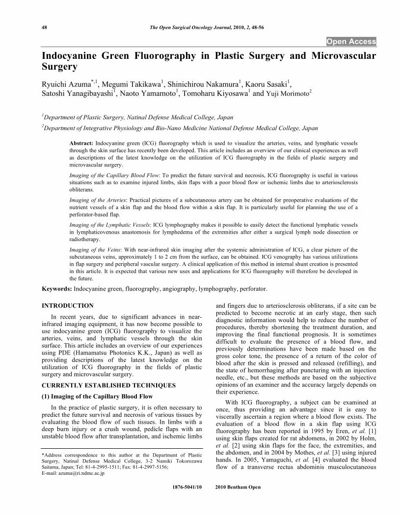
Fig. (1f). After the flap separation.
difficult to obtain a fluorescence image. Fundus angiography has been widely performed for over 30 years since the report by Flower, et al. [5] in 1973. Following its clinical application in the ophthalmologic field, perioperative ICG angiography has also been used to confirm the patency of an anastomosis site of brain blood vessels [6] and of coronary arteries [7] while simultaneously confirming a revascularized perfusion area. In contrast, regarding the exogenous imaging of subcutaneous vessels, because the visualization of subcutaneous vessels is performed through tissues that cause both light reflection and scattering, such as skin and subcutaneous fat, high quality images therefore could not be obtained in the past. However, in recent years, it has become possible to obtain extremely high-sensitivity and high-resolution images by combining a high-resolution CCD camera, an LED light source with sharp wavelength characteristics, and an optical filter, and practical pictures of a subcutaneous artery can be obtained for preoperative evaluations of the nutrient vessels of a skin flap and the blood flow within a skin flap; the author, et al. [8] previously reported the usefulness of this method for the preoperative evaluation of pedicled perforator flaps for various sites throughout the whole body.
Case 2
A 62-Year-Old Male
Immediate reconstruction with a vascularized fibula and a skin flap of the perforating branch of the peroneal artery was performed for a defect 10 cm in size in the mucosa of the mouth floor under the sublingual region and the median part of the mandible after a resection of a cancerous growth occurring on the mouth floor (Fig. 2a).
On the day before the surgery, 10 mg of ICG was injected from the right femoral artery and near-infrared angiography was performed on the lateral side of the leg (Fig. 2b). Immediately after the injection, three main perforating branches from the peroneal artery were identified near the back of the fibula (Figs. 2c-2e).
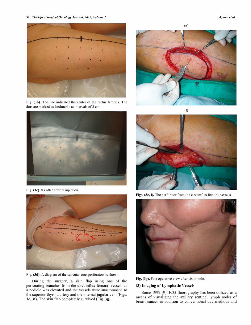
During the surgery, a skin flap was elevated using a perforator from the peroneal vessels as a pedicle which was the middle one of the previously identified perforators (Fig. 2g), and the vessels were then anastomosed to the superior thyroid artery and the facial vein, respectively (Fig. 2h). The fibula and the skin flap completely survived.
Fig. (2a). preoperative appearance.
Fig. (2b). ICG angiography before the surgery.
Fig. (2c). 4 s after arterial injection.
Case 3
A 73-Year-Old Female
The patient had undergone radiotherapy for squamous cell carcinoma in the left buccal mucosa 16 years earlier and the tumor had completely healed, but a few years later, an impairment of mouth-opening and a buccal fistula developed due to contracture of the skin and the mucosa of the buccal
ICG Fluorography in Plastic and Microvascular Surgery The Open Surgical Oncology Journal, 2010, Volume 2 51
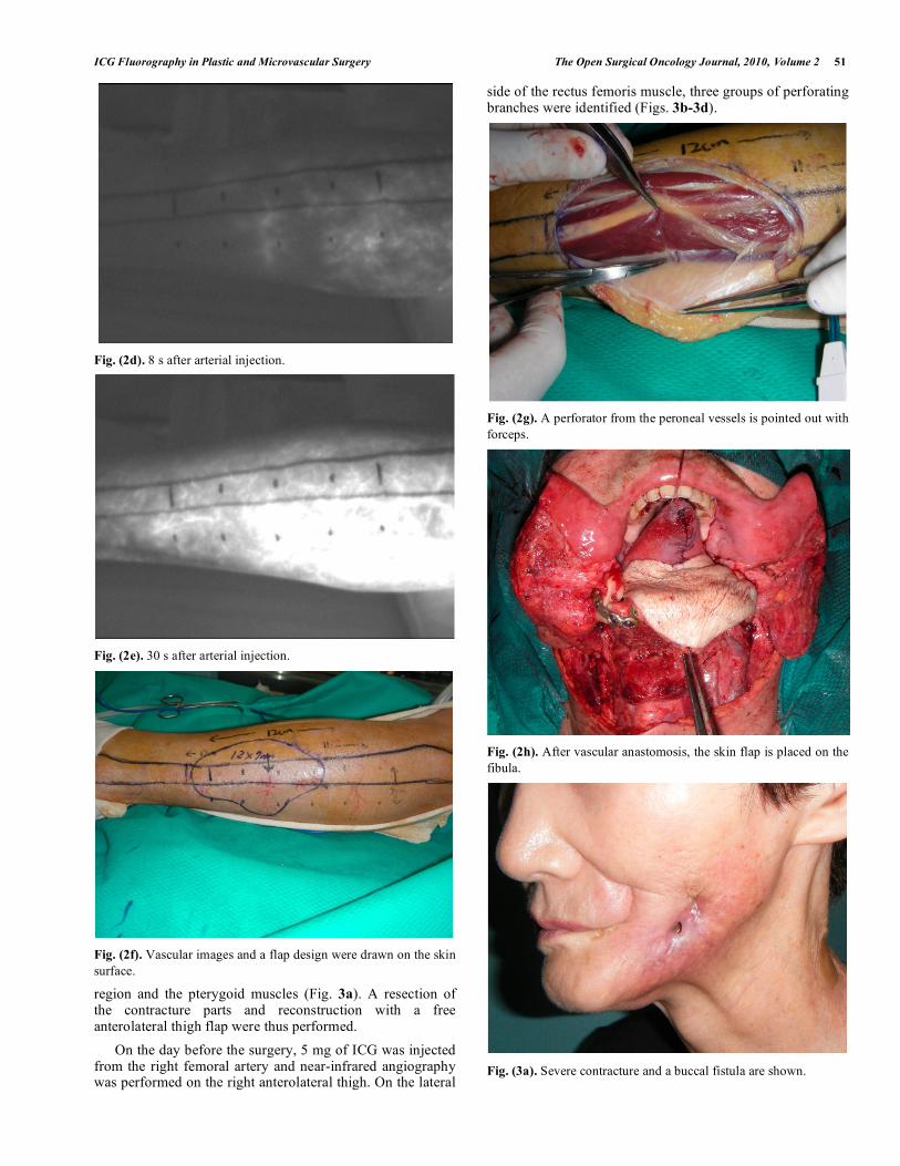
Fig. (2d). 8 s after arterial injection.
Fig. (2e). 30 s after arterial injection.
Fig. (2f). Vascular images and a flap design were drawn on the skin
surface.
region and the pterygoid muscles (Fig. 3a). A resection of the contracture parts and reconstruction with a free anterolateral thigh flap were thus performed.
On the day before the surgery, 5 mg of ICG was injected from the right femoral artery and near-infrared angiography was performed on the right anterolateral thigh. On the lateral
side of the rectus femoris muscle, three groups of perforating branches were identified (Figs. 3b-3d).
Fig. (2g). A perforator from the peroneal vessels is pointed out with
forceps.
Fig. (2h). After vascular anastomosis, the skin flap is placed on the
fibula.
Fig. (3a). Severe contracture and a buccal fistula are shown.
52 The Open Surgical Oncology Journal, 2010, Volume 2 Azuma et al.
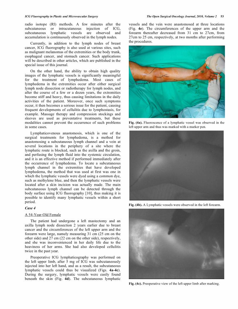
Fig. (3b). The line indicated the center of the rectus femoris. The
dots are marked as landmarks at intervals of 3 cm.
Fig. (3c). 8 s after arterial injection.
Fig. (3d). A diagram of the subcutaneous perforators is shown.
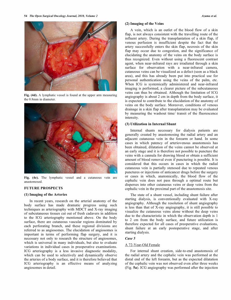
During the surgery, a skin flap using one of the perforating branches from the circumflex femoral vessels as a pedicle was elevated and the vessels were anastomosed to the superior thyroid artery and the internal jugular vein (Figs. 3e, 3f). The skin flap completely survived (Fig. 3g).
(e)
(f)
Figs. (3e, f). The perforator from the circumflex femoral vessels.
Fig. (3g). Post operative view after six months.
(3) Imaging of Lymphatic Vessels
Since 1999 [9], ICG fluorography has been utilized as a means of visualizing the axillary sentinel lymph nodes of breast cancer in addition to conventional dye methods and
ICG Fluorography in Plastic and Microvascular Surgery The Open Surgical Oncology Journal, 2010, Volume 2 53
radio isotope (RI) methods. A few minutes after the subcutaneous or intracutaneous injection of ICG, subcutaneous lymphatic vessels are observed and accumulation is continuously observed in the lymph nodes.
Currently, in addition to the lymph nodes of breast cancer, ICG fluorography is also used at various sites, such as malignant melanomas of the extremities or the body trunk, esophageal cancer, and stomach cancer. Such applications will be described in other articles, which are published in the special issue of this journal.
On the other hand, the ability to obtain high quality images of the lymphatic vessels is significantly meaningful for the treatment of lymphedema. Most cases of lymphedema in the extremities occur after either surgical lymph node dissection or radiotherapy for lymph nodes, and after the course of a few or a dozen years, the extremities become stiff and heavy, thus causing limitations in the daily activities of the patient. Moreover, once such symptoms occur, it then becomes a serious issue for the patient, causing frequent developments of cellulitis due to lymphostasis, for example. Massage therapy and compression stockings and sleeves are used as preventative treatments, but these modalities cannot prevent the occurrence of such problems in some cases.
Lymphaticovenous anastomosis, which is one of the surgical treatments for lymphedema, is a method for anastomosing a subcutaneous lymph channel and a vein at several locations in the periphery of a site where the lymphatic route is blocked, such as the axilla and the groin, and perfusing the lymph fluid into the systemic circulation, and it is an effective method if performed immediately after the occurrence of lymphedema. To locate a subcutaneous lymph channel in the extremities that have developed lymphedema, the method that was used at first was one in which the lymphatic vessels were dyed using a common dye, such as methylene blue, and then the lymphatic vessels were located after a skin incision was actually made. The main subcutaneous lymph channel can be detected through the body surface using ICG fluorography [10], thus making it is possible to identify many lymphatic vessels within a short period.
Case 4
A 54-Year-Old Female
The patient had undergone a left mastectomy and an axilla lymph node dissection 2 years earlier due to breast cancer and the circumferences of the left upper arm and the forearm were large, namely measuring 31 cm (25 cm on the other side) and 27 cm (22 cm on the other side), respectively, and she was inconvenienced in her daily life due to the heaviness of her arms. She had also developed cellulitis twice in the past year.
Preoperative ICG lymphaticography was performed on the left upper limb, after 5 mg of ICG was subcutaneously injected into her left hand, and as a result, the subcutaneous lymphatic vessels could thus be visualized (Figs. 4a-4c). During the surgery, lymphatic vessels were easily found beneath the skin (Fig. 4d). The subcutaneous lymphatic
vessels and the vein were anastomosed at three locations (Fig. 4e). The circumferences of the upper arm and the forearm thereafter decreased from 31 cm to 27cm, from 27cm to 25 cm, respectively, at two months after performing the procedures.
Fig. (4a). Fluorescence of a lymphatic vessel was observed in the
left upper arm and thus was marked with a marker pen.
Fig. (4b). A Lymphatic vessels were observed in the left forearm.
Fig. (4c). Preoperative view of the left upper limb after marking.
54 The Open Surgical Oncology Journal, 2010, Volume 2 Azuma et al.
Fig. (4d). A lymphatic vessel is found at the upper arm measuring
the 0.8mm in diameter.
Fig. (4e). The lymphatic vessel and a cutaneous vein are
anastomosed.
FUTURE PROSPECTS
(1) Imaging of the Arteries
In recent years, research on the arterial anatomy of the body surface has made dramatic progress using such techniques as arteriography with MDCT and X-ray imaging of subcutaneous tissues cut out of fresh cadavers in addition to the ICG arteriography mentioned above. On the body surface, there are cutaneous vascular regions dominated by each perforating branch, and these regional divisions are referred to as angiosomes. The elucidation of angiosomes is important in terms of performing flap surgery, and it is necessary not only to research the structure of angiosomes, which is universal in many individuals, but also to evaluate variations in individual cases in preoperative examinations. ICG arteriography is a less invasive diagnostic modality, which can be used to selectively and dynamically observe the arteries of a body surface, and it is therefore believed that ICG arteriography is an effective means of analyzing angiosomes in detail.
(2) Imaging of the Veins
A vein, which is an outlet of the blood flow of a skin flap, is not always consistent with the travelling route of the influent artery. During the transplantation of a skin flap, if venous perfusion is insufficient despite the fact that the artery successfully enters the skin flap, necrosis of the skin flap may occur due to congestion, and the significance of elucidating the anatomy of the veins on the body surface is thus recognized. Even without using a fluorescent contrast agent, when near-infrared rays are irradiated through a skin surface for observation with a near-infrared camera, cutaneous veins can be visualized as a defect (seen as a black area), and this has already been put into practical use for personal authentication using the veins of the palm, etc. When ICG is systemically administered and near-infrared imaging is performed, a clearer picture of the subcutaneous veins can thus be obtained. Although the limitation of ICG angiography is about 2 cm in depth from the body surface, it is expected to contribute to the elucidation of the anatomy of veins on the body surface. Moreover, conditions of venous drainage in a skin flap after transplantation may be evaluated by measuring the washout time/ transit of the fluorescence intensity.
(3) Utilization in Internal Shunt
Internal shunts necessary for dialysis patients are generally created by anastomosing the radial artery and an adjacent cutaneous vein in the forearm or hand. In some cases in which patency of arteriovenous anastomosis has been obtained, dilatation of the veins cannot be observed at an early stage and it is therefore not possible to puncture the veins with a cannula for drawing blood or obtain a sufficient amount of blood removal even if puncturing is possible. It is considered that this occurs in cases in which the radial cutaneous vein is partially stenosed due to repeated venous punctures or injections of anticancer drugs before the surgery or cases in which, anatomically, the blood flow of the cephalic vein does not pass through a optimal route but disperses into other cutaneous veins or deep veins from the cephalic vein in the proximal part of the anastomosis site.
The state of a shunt vessel, including shunt failure after starting dialysis, is conventionally evaluated with X-ray angiography. Although the resolution of shunt angiography is less than that of X-ray angiography, it is still possible to visualize the cutaneous veins alone without the deep veins due to the characteristic in which the observation depth is 1 to 2 cm from the body surface, and future utilization is therefore expected for all cases of preoperative evaluations, shunt failure at an early postoperative stage, and after starting dialysis.
Case 5
A 72-Year-Old Female
For internal shunt creation, side-to-end anastomosis of the radial artery and the cephalic vein was performed at the distal end of the left forearm, but as the expected dilatation of the cephalic vein was not observed even after three weeks (Fig. 5a). ICG angiography was performed after the injection
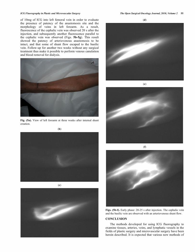
ICG Fluorography in Plastic and Microvascular Surgery The Open Surgical Oncology Journal, 2010, Volume 2 55
of 10mg of ICG into left femoral vein in order to evaluate the presence of patency of the anastomosis site and the morphology of veins in left forearm. As a result, fluorescence of the cephalic vein was observed 20 s after the injection, and subsequently another fluorescence parallel to the cephalic vein was observed (Figs. 5b-5g). This result showed the patency of arteriovenous anastomosis to be intact, and that some of shunt flow escaped to the basilic vein. Follow-up for another two weeks without any surgical treatment thus make it possible to perform venous canulation and blood removal for dialysis.
Fig. (5a). View of left forearm at three weeks after internal shunt
creation.
(b)
(c)
(d)
(e)
(f)
Figs. (5b-f). Early phase: 20-25 s after injection. The cephalic vein
and the basilic vein are observed with an arteriovenous shunt flow.
CONCLUSION
The methods developed for using ICG fluorography to examine tissues, arteries, veins, and lymphatic vessels in the fields of plastic surgery and microvascular surgery have been herein described. It is expected that various new methods of
56 The Open Surgical Oncology Journal, 2010, Volume 2 Azuma et al.
the use and application of this modality will be developed in the future.
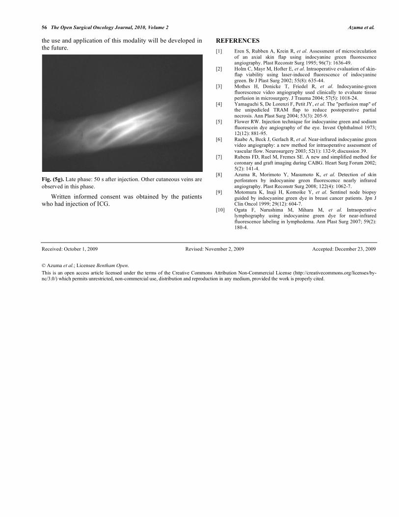
Fig. (5g). Late phase: 50 s after injection. Other cutaneous veins are
observed in this phase.
Written informed consent was obtained by the patients who had injection of ICG.
REFERENCES
[1] Eren S, Rubben A, Krein R, et al. Assessment of microcirculation
of an axial skin flap using indocyanine green fluorescence angiography. Plast Reconstr Surg 1995; 96(7): 1636-49.
[2] Holm C, Mayr M, Hofter E, et al. Intraoperative evaluation of skin-flap viability using laser-induced fluorescence of indocyanine
green. Br J Plast Surg 2002; 55(8): 635-44. [3] Mothes H, Donicke T, Friedel R, et al. Indocyanine-green
fluorescence video angiography used clinically to evaluate tissue perfusion in microsurgery. J Trauma 2004; 57(5): 1018-24.
[4] Yamaguchi S, De Lorenzi F, Petit JY, et al. The "perfusion map" of the unipedicled TRAM flap to reduce postoperative partial
necrosis. Ann Plast Surg 2004; 53(3): 205-9. [5] Flower RW. Injection technique for indocyanine green and sodium
fluorescein dye angiography of the eye. Invest Ophthalmol 1973; 12(12): 881-95.
[6] Raabe A, Beck J, Gerlach R, et al. Near-infrared indocyanine green video angiography: a new method for intraoperative assessment of
vascular flow. Neurosurgery 2003; 52(1): 132-9; discussion 39. [7] Rubens FD, Ruel M, Fremes SE. A new and simplified method for
coronary and graft imaging during CABG. Heart Surg Forum 2002; 5(2): 141-4.
[8] Azuma R, Morimoto Y, Masumoto K, et al. Detection of skin perforators by indocyanine green fluorescence nearly infrared
angiography. Plast Reconstr Surg 2008; 122(4): 1062-7. [9] Motomura K, Inaji H, Komoike Y, et al. Sentinel node biopsy
guided by indocyanine green dye in breast cancer patients. Jpn J Clin Oncol 1999; 29(12): 604-7.
[10] Ogata F, Narushima M, Mihara M, et al. Intraoperative lymphography using indocyanine green dye for near-infrared
fluorescence labeling in lymphedema. Ann Plast Surg 2007; 59(2): 180-4.
Received: October 1, 2009 Revised: November 2, 2009 Accepted: December 23, 2009
© Azuma et al.; Licensee Bentham Open.
This is an open access article licensed under the terms of the Creative Commons Attribution Non-Commercial License (http://creativecommons.org/licenses/by-nc/3.0/) which permits unrestricted, non-commercial use, distribution and reproduction in any medium, provided the work is properly cited.