Embed Size (px)
Citation preview
ARTICLE
Crystal structure of TcpK in complex with oriT DNAof the antibiotic resistance plasmid pCW3Daouda A.K. Traore 1,2, Jessica A. Wisniewski3, Sarena F. Flanigan1, Paul J. Conroy 1,
Santosh Panjikar 1,4, Yee-Foong Mok5, Carmen Lao 3, Michael D.W. Griffin5,
Vicki Adams 3, Julian I. Rood 3 & James C. Whisstock1,6,7
Conjugation is fundamental for the acquisition of new genetic traits and the development of
antibiotic resistance in pathogenic organisms. Here, we show that a hypothetical Clostridium
perfringens protein, TcpK, which is encoded by the tetracycline resistance plasmid pCW3, is
essential for efficient conjugative DNA transfer. Our studies reveal that TcpK is a member of
the winged helix-turn-helix (wHTH) transcription factor superfamily and that it forms a dimer
in solution. Furthermore, TcpK specifically binds to a nine-nucleotide sequence that is present
as tandem repeats within the pCW3 origin of transfer (oriT). The X-ray crystal structure of
the TcpK–TcpK box complex reveals a binding mode centered on and around the β-wing,which is different from what has been previously shown for other wHTH proteins. Structure-
guided mutagenesis experiments validate the specific interaction between TcpK and the DNA
molecule. Additional studies highlight that the TcpK dimer is important for specific DNA
binding.
DOI: 10.1038/s41467-018-06096-2 OPEN
1 Department of Biochemistry and Molecular Biology, Infection and Immunity Program, Monash Biomedicine Discovery Institute, Monash University, Clayton3800 VIC, Australia. 2 Faculté des Sciences et Techniques, Université des Sciences Techniques et Technologiques de Bamako (USTTB), BP E3206 Bamako,Mali. 3 Department of Microbiology, Infection and Immunity Program, Monash Biomedicine Discovery Institute, Monash University, Clayton 3800 VIC,Australia. 4 Australian Synchrotron, Clayton 3168 VIC, Australia. 5 Biochemistry and Molecular Biology, Bio21 Molecular Science and Biotechnology Institute,University of Melbourne, Parkville 3010 VIC, Australia. 6 ARC Centre of Excellence in Advanced Molecular Imaging, Monash University, Clayton 3800 VIC,Australia. 7 EMBL Australia, Monash University, Clayton 3800 VIC, Australia. These authors contributed equally: Daouda A.K. Traore, Jessica A. Wisniewski.These authors jointly supervised this work: Vicki Adams, Julian I. Rood, James C. Whisstock. Correspondence and requests for materials should be addressedto V.A. (email: [email protected]) or to J.I.R. (email: [email protected]) or to J.C.W. (email: [email protected])
NATURE COMMUNICATIONS | (2018) 9:3732 | DOI: 10.1038/s41467-018-06096-2 | www.nature.com/naturecommunications 1
1234
5678
90():,;
In the Gram-positive pathogenic bacterium Clostridium per-fringens the conjugative tetracycline resistance plasmid pCW3is representative of a large class of closely related toxin and
antibiotic resistance plasmids1. Conjugative transfer of this plas-mid is mediated by the highly conserved tcp locus, which encodesat least eight proteins that are known to be essential for efficientDNA transfer2. Functionally, the conjugative machinery is knownto be homologous, at least in part, to the type 4 secretion systems(T4SS) characterized in Gram-negative bacteria.
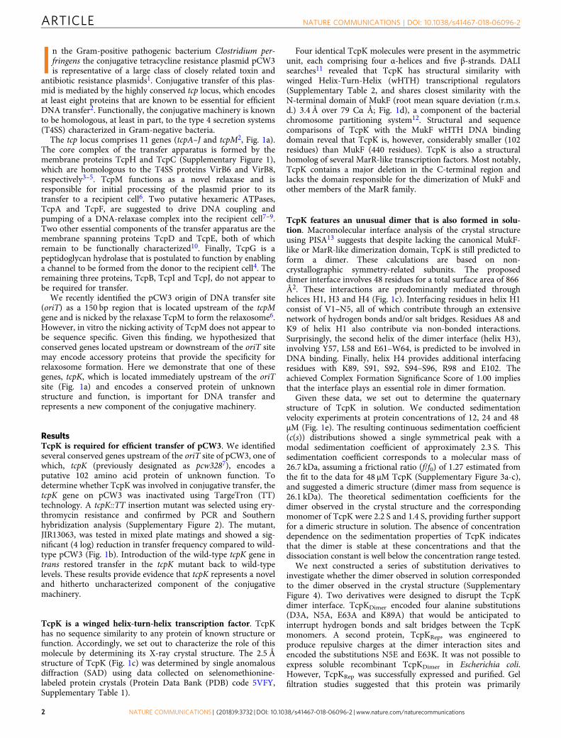
The tcp locus comprises 11 genes (tcpA–J and tcpM2, Fig. 1a).The core complex of the transfer apparatus is formed by themembrane proteins TcpH and TcpC (Supplementary Figure 1),which are homologous to the T4SS proteins VirB6 and VirB8,respectively3–5. TcpM functions as a novel relaxase and isresponsible for initial processing of the plasmid prior to itstransfer to a recipient cell6. Two putative hexameric ATPases,TcpA and TcpF, are suggested to drive DNA coupling andpumping of a DNA-relaxase complex into the recipient cell7–9.Two other essential components of the transfer apparatus are themembrane spanning proteins TcpD and TcpE, both of whichremain to be functionally characterized10. Finally, TcpG is apeptidoglycan hydrolase that is postulated to function by enablinga channel to be formed from the donor to the recipient cell4. Theremaining three proteins, TcpB, TcpI and TcpJ, do not appear tobe required for transfer.
We recently identified the pCW3 origin of DNA transfer site(oriT) as a 150 bp region that is located upstream of the tcpMgene and is nicked by the relaxase TcpM to form the relaxosome6.However, in vitro the nicking activity of TcpM does not appear tobe sequence specific. Given this finding, we hypothesized thatconserved genes located upstream or downstream of the oriT sitemay encode accessory proteins that provide the specificity forrelaxosome formation. Here we demonstrate that one of thesegenes, tcpK, which is located immediately upstream of the oriTsite (Fig. 1a) and encodes a conserved protein of unknownstructure and function, is important for DNA transfer andrepresents a new component of the conjugative machinery.
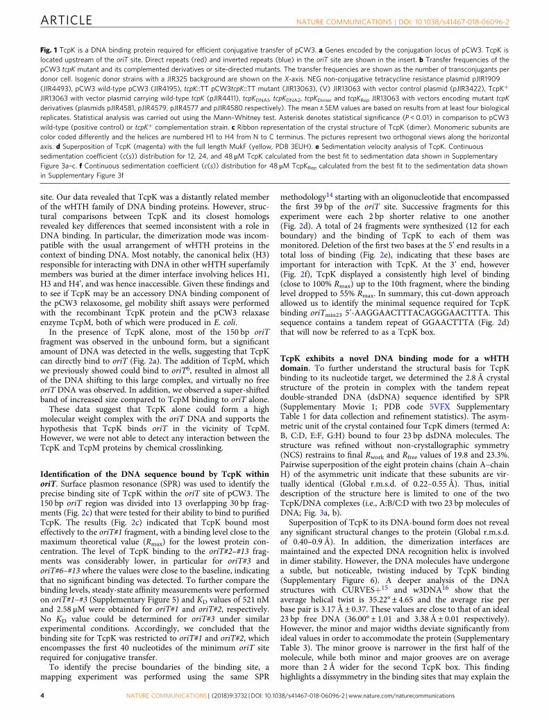
ResultsTcpK is required for efficient transfer of pCW3. We identifiedseveral conserved genes upstream of the oriT site of pCW3, one ofwhich, tcpK (previously designated as pcw3287), encodes aputative 102 amino acid protein of unknown function. Todetermine whether TcpK was involved in conjugative transfer, thetcpK gene on pCW3 was inactivated using TargeTron (TT)technology. A tcpK::TT insertion mutant was selected using ery-thromycin resistance and confirmed by PCR and Southernhybridization analysis (Supplementary Figure 2). The mutant,JIR13063, was tested in mixed plate matings and showed a sig-nificant (4 log) reduction in transfer frequency compared to wild-type pCW3 (Fig. 1b). Introduction of the wild-type tcpK gene intrans restored transfer in the tcpK mutant back to wild-typelevels. These results provide evidence that tcpK represents a noveland hitherto uncharacterized component of the conjugativemachinery.
TcpK is a winged helix-turn-helix transcription factor. TcpKhas no sequence similarity to any protein of known structure orfunction. Accordingly, we set out to characterize the role of thismolecule by determining its X-ray crystal structure. The 2.5 Åstructure of TcpK (Fig. 1c) was determined by single anomalousdiffraction (SAD) using data collected on selenomethionine-labeled protein crystals (Protein Data Bank (PDB) code 5VFY,Supplementary Table 1).
Four identical TcpK molecules were present in the asymmetricunit, each comprising four α-helices and five β-strands. DALIsearches11 revealed that TcpK has structural similarity withwinged Helix-Turn-Helix (wHTH) transcriptional regulators(Supplementary Table 2, and shares closest similarity with theN-terminal domain of MukF (root mean square deviation (r.m.s.d.) 3.4 Å over 79 Cα Å; Fig. 1d), a component of the bacterialchromosome partitioning system12. Structural and sequencecomparisons of TcpK with the MukF wHTH DNA bindingdomain reveal that TcpK is, however, considerably smaller (102residues) than MukF (440 residues). TcpK is also a structuralhomolog of several MarR-like transcription factors. Most notably,TcpK contains a major deletion in the C-terminal region andlacks the domain responsible for the dimerization of MukF andother members of the MarR family.
TcpK features an unusual dimer that is also formed in solu-tion. Macromolecular interface analysis of the crystal structureusing PISA13 suggests that despite lacking the canonical MukF-like or MarR-like dimerization domain, TcpK is still predicted toform a dimer. These calculations are based on non-crystallographic symmetry-related subunits. The proposeddimer interface involves 48 residues for a total surface area of 866Å2. These interactions are predominantly mediated throughhelices H1, H3 and H4 (Fig. 1c). Interfacing residues in helix H1consist of V1–N5, all of which contribute through an extensivenetwork of hydrogen bonds and/or salt bridges. Residues A8 andK9 of helix H1 also contribute via non-bonded interactions.Surprisingly, the second helix of the dimer interface (helix H3),involving Y57, L58 and E61–W64, is predicted to be involved inDNA binding. Finally, helix H4 provides additional interfacingresidues with K89, S91, S92, S94–S96, R98 and E102. Theachieved Complex Formation Significance Score of 1.00 impliesthat the interface plays an essential role in dimer formation.
Given these data, we set out to determine the quaternarystructure of TcpK in solution. We conducted sedimentationvelocity experiments at protein concentrations of 12, 24 and 48μM (Fig. 1e). The resulting continuous sedimentation coefficient(c(s)) distributions showed a single symmetrical peak with amodal sedimentation coefficient of approximately 2.3 S. Thissedimentation coefficient corresponds to a molecular mass of26.7 kDa, assuming a frictional ratio (f/f0) of 1.27 estimated fromthe fit to the data for 48 μM TcpK (Supplementary Figure 3a-c),and suggested a dimeric structure (dimer mass from sequence is26.1 kDa). The theoretical sedimentation coefficients for thedimer observed in the crystal structure and the correspondingmonomer of TcpK were 2.2 S and 1.4 S, providing further supportfor a dimeric structure in solution. The absence of concentrationdependence on the sedimentation properties of TcpK indicatesthat the dimer is stable at these concentrations and that thedissociation constant is well below the concentration range tested.
We next constructed a series of substitution derivatives toinvestigate whether the dimer observed in solution correspondedto the dimer observed in the crystal structure (SupplementaryFigure 4). Two derivatives were designed to disrupt the TcpKdimer interface. TcpKDimer encoded four alanine substitutions(D3A, N5A, E63A and K89A) that would be anticipated tointerrupt hydrogen bonds and salt bridges between the TcpKmonomers. A second protein, TcpKRep, was engineered toproduce repulsive charges at the dimer interaction sites andencoded the substitutions N5E and E63K. It was not possible toexpress soluble recombinant TcpKDimer in Escherichia coli.However, TcpKRep was successfully expressed and purified. Gelfiltration studies suggested that this protein was primarily
ARTICLE NATURE COMMUNICATIONS | DOI: 10.1038/s41467-018-06096-2
2 NATURE COMMUNICATIONS | (2018) 9:3732 | DOI: 10.1038/s41467-018-06096-2 | www.nature.com/naturecommunications
monomeric (Supplementary Figure 4d), a finding confirmed bysedimentation velocity experiments (Fig. 1f).
To examine the function of these derivatives in vivo, weperformed conjugation experiments using the tcpK mutantJIR13063. The tcpKDimer and tcpKRep mutants were cloned intothe complementation vector, pJIR3422, and introduced into thetcpK mutant for subsequent testing. Both tcpKDimer and tcpKRep
restored the conjugation frequency to wild-type levels (Fig. 1b),
which suggested that monomeric TcpK was functional in vivowhen expressed under these in trans conditions.
TcpK forms a complex with DNA from the pCW3 oriT site.Studies of other conjugation systems suggest that accessory pro-teins within relaxosome complexes can either bind the relaxaseenzyme or bind directly to the plasmid in the vicinity of the oriT
d
DNA recognition helix
MukFMukF
β-wing
TcpK
a
b
c
H1
H2
H3
H4
H1′
H2′
H3′
1×101
1×100
1×10⁻¹
1×10⁻²
1×10⁻³
1×10⁻⁴
1×10⁻⁵
1×10⁻⁶
1×10⁻⁷
p<0.01SEMn=≥4
* **
**
NEGpC
W3
tcpK::T
T
tcpK::T
T(V)
tcpK::T
T(V)
tcpK::T
T(tcpK
+ )
tcpK::T
T(tcpK
+ )
tcpK::T
T(tcpK
DNA1)
tcpK::T
T(tcpK
DNA2)
tcpK::T
T(tcpK
Dimer)
tcpK::T
T(tcpK
Rep)
Tra
nsfe
r fr
eque
ncy
(tra
nsco
njug
ants
per
dono
r ce
ll)
H4′
Chain AChain B
H1
H2
H3
H4
H1′
H2′H3′
H4′
Chain AChain B
90°
TcpK48 μM24 μM12 μM
TcpKRep
48 μM
tcpK
oriT
tcpM tcpA tcpB tcpF tcpG tcpH I JtcpC D E
e
f
5
3
2
2
1
0
c(s
)c
(s)
0
4
4 6 8 10
Sedimentation coefficient (S)
200.0
0.1
0.2
0.3
0.4
0.5
4 6 8 10
Sedimentation coefficient (S)
NATURE COMMUNICATIONS | DOI: 10.1038/s41467-018-06096-2 ARTICLE
NATURE COMMUNICATIONS | (2018) 9:3732 | DOI: 10.1038/s41467-018-06096-2 | www.nature.com/naturecommunications 3
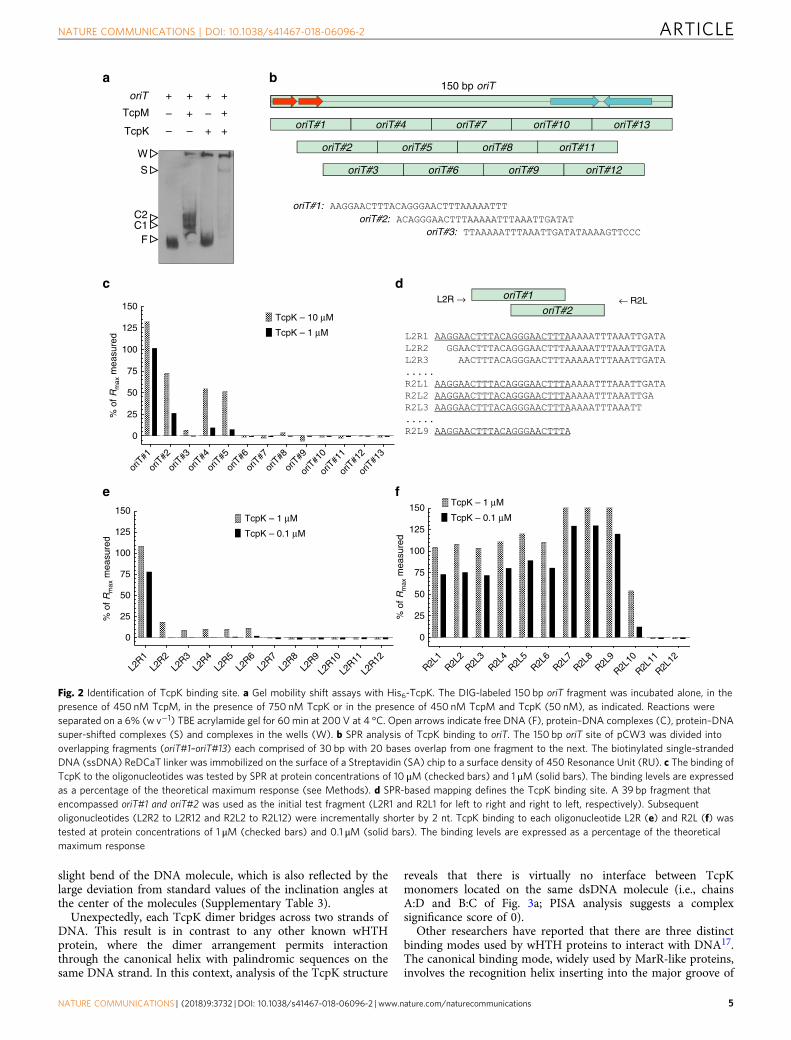
site. Our data revealed that TcpK was a distantly related memberof the wHTH family of DNA binding proteins. However, struc-tural comparisons between TcpK and its closest homologsrevealed key differences that seemed inconsistent with a role inDNA binding. In particular, the dimerization mode was incom-patible with the usual arrangement of wHTH proteins in thecontext of binding DNA. Most notably, the canonical helix (H3)responsible for interacting with DNA in other wHTH superfamilymembers was buried at the dimer interface involving helices H1,H3 and H4’, and was hence inaccessible. Given these findings andto see if TcpK may be an accessory DNA binding component ofthe pCW3 relaxosome, gel mobility shift assays were performedwith the recombinant TcpK protein and the pCW3 relaxaseenzyme TcpM, both of which were produced in E. coli.
In the presence of TcpK alone, most of the 150 bp oriTfragment was observed in the unbound form, but a significantamount of DNA was detected in the wells, suggesting that TcpKcan directly bind to oriT (Fig. 2a). The addition of TcpM, whichwe previously showed could bind to oriT6, resulted in almost allof the DNA shifting to this large complex, and virtually no freeoriT DNA was observed. In addition, we observed a super-shiftedband of increased size compared to TcpM binding to oriT alone.
These data suggest that TcpK alone could form a highmolecular weight complex with the oriT DNA and supports thehypothesis that TcpK binds oriT in the vicinity of TcpM.However, we were not able to detect any interaction between theTcpK and TcpM proteins by chemical crosslinking.
Identification of the DNA sequence bound by TcpK withinoriT. Surface plasmon resonance (SPR) was used to identify theprecise binding site of TcpK within the oriT site of pCW3. The150 bp oriT region was divided into 13 overlapping 30 bp frag-ments (Fig. 2c) that were tested for their ability to bind to purifiedTcpK. The results (Fig. 2c) indicated that TcpK bound mosteffectively to the oriT#1 fragment, with a binding level close to themaximum theoretical value (Rmax) for the lowest protein con-centration. The level of TcpK binding to the oriT#2–#13 frag-ments was considerably lower, in particular for oriT#3 andoriT#6–#13 where the values were close to the baseline, indicatingthat no significant binding was detected. To further compare thebinding levels, steady-state affinity measurements were performedon oriT#1–#3 (Supplementary Figure 5) and KD values of 521 nMand 2.58 μM were obtained for oriT#1 and oriT#2, respectively.No KD value could be determined for oriT#3 under similarexperimental conditions. Accordingly, we concluded that thebinding site for TcpK was restricted to oriT#1 and oriT#2, whichencompasses the first 40 nucleotides of the minimum oriT siterequired for conjugative transfer.
To identify the precise boundaries of the binding site, amapping experiment was performed using the same SPR
methodology14 starting with an oligonucleotide that encompassedthe first 39 bp of the oriT site. Successive fragments for thisexperiment were each 2 bp shorter relative to one another(Fig. 2d). A total of 24 fragments were synthesized (12 for eachboundary) and the binding of TcpK to each of them wasmonitored. Deletion of the first two bases at the 5’ end results in atotal loss of binding (Fig. 2e), indicating that these bases areimportant for interaction with TcpK. At the 3’ end, however(Fig. 2f), TcpK displayed a consistently high level of binding(close to 100% Rmax) up to the 10th fragment, where the bindinglevel dropped to 55% Rmax. In summary, this cut-down approachallowed us to identify the minimal sequence required for TcpKbinding oriTmin23 5’-AAGGAACTTTACAGGGAACTTTA. Thissequence contains a tandem repeat of GGAACTTTA (Fig. 2d)that will now be referred to as a TcpK box.
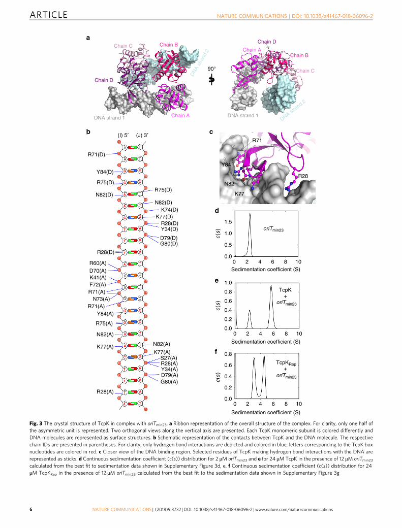
TcpK exhibits a novel DNA binding mode for a wHTHdomain. To further understand the structural basis for TcpKbinding to its nucleotide target, we determined the 2.8 Å crystalstructure of the protein in complex with the tandem repeatdouble-stranded DNA (dsDNA) sequence identified by SPR(Supplementary Movie 1; PDB code 5VFX SupplementaryTable 1 for data collection and refinement statistics). The asym-metric unit of the crystal contained four TcpK dimers (termed A:B, C:D, E:F, G:H) bound to four 23 bp dsDNA molecules. Thestructure was refined without non-crystallographic symmetry(NCS) restrains to final Rwork and Rfree values of 19.8 and 23.3%.Pairwise superposition of the eight protein chains (chain A–chainH) of the asymmetric unit indicate that these subunits are vir-tually identical (Global r.m.s.d. of 0.22–0.55 Å). Thus, initialdescription of the structure here is limited to one of the twoTcpK/DNA complexes (i.e., A:B/C:D with two 23 bp molecules ofDNA; Fig. 3a, b).
Superposition of TcpK to its DNA-bound form does not revealany significant structural changes to the protein (Global r.m.s.d.of 0.40–0.9 Å). In addition, the dimerization interfaces aremaintained and the expected DNA recognition helix is involvedin dimer stability. However, the DNA molecules have undergonea subtle, but noticeable, twisting induced by TcpK binding(Supplementary Figure 6). A deeper analysis of the DNAstructures with CURVES+15 and w3DNA16 show that theaverage helical twist is 35.22° ± 4.65 and the average rise perbase pair is 3.17 Å ± 0.37. These values are close to that of an ideal23 bp free DNA (36.00° ± 1.01 and 3.38 Å ± 0.01 respectively).However, the minor and major widths deviate significantly fromideal values in order to accommodate the protein (SupplementaryTable 3). The minor groove is narrower in the first half of themolecule, while both minor and major grooves are on averagemore than 2 Å wider for the second TcpK box. This findinghighlights a dissymmetry in the binding sites that may explain the
Fig. 1 TcpK is a DNA binding protein required for efficient conjugative transfer of pCW3. a Genes encoded by the conjugation locus of pCW3. TcpK islocated upstream of the oriT site. Direct repeats (red) and inverted repeats (blue) in the oriT site are shown in the insert. b Transfer frequencies of thepCW3 tcpK mutant and its complemented derivatives or site-directed mutants. The transfer frequencies are shown as the number of transconjugants perdonor cell. Isogenic donor strains with a JIR325 background are shown on the X-axis. NEG non-conjugative tetracycline resistance plasmid pJIR1909(JIR4493), pCW3 wild-type pCW3 (JIR4195), tcpK::TT pCW3tcpK::TT mutant (JIR13063), (V) JIR13063 with vector control plasmid (pJIR3422), TcpK+
JIR13063 with vector plasmid carrying wild-type tcpK (pJIR4411), tcpKDNA1, tcpKDNA2, tcpKDimer and tcpKRep JIR13063 with vectors encoding mutant tcpKderivatives (plasmids pJIR4581, pJIR4579, pJIR4577 and pJIR4580 respectively). The mean ± SEM values are based on results from at least four biologicalreplicates. Statistical analysis was carried out using the Mann–Whitney test. Asterisk denotes statistical significance (P < 0.01) in comparison to pCW3wild-type (positive control) or tcpK+ complementation strain. c Ribbon representation of the crystal structure of TcpK (dimer). Monomeric subunits arecolor coded differently and the helices are numbered H1 to H4 from N to C terminus. The pictures represent two orthogonal views along the horizontalaxis. d Superposition of TcpK (magenta) with the full length MukF (yellow, PDB 3EUH). e Sedimentation velocity analysis of TcpK. Continuoussedimentation coefficient (c(s)) distribution for 12, 24, and 48 μM TcpK calculated from the best fit to sedimentation data shown in SupplementaryFigure 3a–c. f Continuous sedimentation coefficient (c(s)) distribution for 48 μM TcpKRep calculated from the best fit to the sedimentation data shownin Supplementary Figure 3f
ARTICLE NATURE COMMUNICATIONS | DOI: 10.1038/s41467-018-06096-2
4 NATURE COMMUNICATIONS | (2018) 9:3732 | DOI: 10.1038/s41467-018-06096-2 | www.nature.com/naturecommunications
slight bend of the DNA molecule, which is also reflected by thelarge deviation from standard values of the inclination angles atthe center of the molecules (Supplementary Table 3).
Unexpectedly, each TcpK dimer bridges across two strands ofDNA. This result is in contrast to any other known wHTHprotein, where the dimer arrangement permits interactionthrough the canonical helix with palindromic sequences on thesame DNA strand. In this context, analysis of the TcpK structure
reveals that there is virtually no interface between TcpKmonomers located on the same dsDNA molecule (i.e., chainsA:D and B:C of Fig. 3a; PISA analysis suggests a complexsignificance score of 0).
Other researchers have reported that there are three distinctbinding modes used by wHTH proteins to interact with DNA17.The canonical binding mode, widely used by MarR-like proteins,involves the recognition helix inserting into the major groove of
cL2R →
150
125
100
75
50
25
0
oriT
#1
oriT
#2
oriT
#3
oriT
#4
oriT
#5
oriT
#6
oriT
#8
oriT
#7
oriT
#9
oriT
#10
oriT
#11
oriT
#12
oriT
#13
% o
f Rm
ax m
easu
red
150
TcpK – 10 μM
TcpK – 1 μM
TcpK – 1 μM
TcpK – 0.1 μM
TcpK – 1 μM
TcpK – 0.1 μM
L2R1
L2R2
L2R3
L2R4
L2R5
L2R6
L2R9
L2R8
L2R10
L2R11
L2R12
L2R7
R2L1
R2L2
R2L3
R2L4
R2L5
R2L6
R2L9
R2L8
R2L10
R2L11
R2L12
R2L7
125
100
75
50
25
0
% o
f Rm
ax m
easu
red
150
125
100
75
50
25
0
% o
f Rm
ax m
easu
red
← R2LoriT#1oriT#2
b
e
aoriT
TcpM
TcpK
W
S
C2C1
F
+
– –
– –
f
d
oriT#1:oriT#2:
oriT#3:
150 bp oriT
oriT#5 oriT#11oriT#2 oriT#8
oriT#1 oriT#7 oriT#13oriT#4 oriT#10
oriT#3 oriT#9oriT#6 oriT#12
+++
+
+ +
+
Fig. 2 Identification of TcpK binding site. a Gel mobility shift assays with His6-TcpK. The DIG-labeled 150 bp oriT fragment was incubated alone, in thepresence of 450 nM TcpM, in the presence of 750 nM TcpK or in the presence of 450 nM TcpM and TcpK (50 nM), as indicated. Reactions wereseparated on a 6% (w v−1) TBE acrylamide gel for 60min at 200 V at 4 °C. Open arrows indicate free DNA (F), protein–DNA complexes (C), protein–DNAsuper-shifted complexes (S) and complexes in the wells (W). b SPR analysis of TcpK binding to oriT. The 150 bp oriT site of pCW3 was divided intooverlapping fragments (oriT#1–oriT#13) each comprised of 30 bp with 20 bases overlap from one fragment to the next. The biotinylated single-strandedDNA (ssDNA) ReDCaT linker was immobilized on the surface of a Streptavidin (SA) chip to a surface density of 450 Resonance Unit (RU). c The binding ofTcpK to the oligonucleotides was tested by SPR at protein concentrations of 10 μM (checked bars) and 1 μM (solid bars). The binding levels are expressedas a percentage of the theoretical maximum response (see Methods). d SPR-based mapping defines the TcpK binding site. A 39 bp fragment thatencompassed oriT#1 and oriT#2 was used as the initial test fragment (L2R1 and R2L1 for left to right and right to left, respectively). Subsequentoligonucleotides (L2R2 to L2R12 and R2L2 to R2L12) were incrementally shorter by 2 nt. TcpK binding to each oligonucleotide L2R (e) and R2L (f) wastested at protein concentrations of 1 μM (checked bars) and 0.1 μM (solid bars). The binding levels are expressed as a percentage of the theoreticalmaximum response
NATURE COMMUNICATIONS | DOI: 10.1038/s41467-018-06096-2 ARTICLE
NATURE COMMUNICATIONS | (2018) 9:3732 | DOI: 10.1038/s41467-018-06096-2 | www.nature.com/naturecommunications 5
c.b
R28
K77
N82
Y84
R71
a
Chain AChain B
Chain C
Chain D
DNA strand 1Chain A
Chain BChain C
DN
A st
rand
2
DNA strand 2
Chain D
DNA strand 1
90°
d
e
f
oriTmin23
TcpK+
oriTmin23
TcpKRep+
oriTmin23
R71(D)A
G
G
G
G
G
G
A
A
A
A
A
A
A
A
A
A
A
A
T
T
T
T
T
T
T
T
T
T
T
T
C
C
C
C
C
C
A T
T
R75(D)
1
2
3
23
22
21
20
4
5
6
7
8
9
10
10
9
8
7
6
5
4
3
2
1
11
11
12
12
14
14
15
16
17
18
19
13
13
15
16
17
18
19
20
21
22
23
(I) 5′ (J) 3′
R75(D)
N82(D)
R28(D)
R60(A)D70(A)K41(A)F72(A)R71(A)
K77(A)K77(A)
R75(A)
R28(A)
R71(A)Y84(A)
Y34(A)
Y34(D)R28(D)
D79(A)
D79(D)
G80(A)
G80(D)
1.5
1.0
1.0
0.5
0.0
0.0
0.0
0.2
0.2
0.4
0.4
0.6
0.6
0.8
0.8
0 2 4 6 8 10
0 2 4 6 8 10
0 2 4 6 8 10
Sedimentation coefficient (S)
Sedimentation coefficient (S)
Sedimentation coefficient (S)
c(s
)c
(s)
c(s
)
G
G C
C
TA
R28(A)S27(A)
N73(A)
N82(A)
N82(A)
N82(D)K74(D)
K77(D)
Y84(D)
Fig. 3 The crystal structure of TcpK in complex with oriTmin23. a Ribbon representation of the overall structure of the complex. For clarity, only one half ofthe asymmetric unit is represented. Two orthogonal views along the vertical axis are presented. Each TcpK monomeric subunit is colored differently andDNA molecules are represented as surface structures. b Schematic representation of the contacts between TcpK and the DNA molecule. The respectivechain IDs are presented in parentheses. For clarity, only hydrogen bond interactions are depicted and colored in blue, letters corresponding to the TcpK boxnucleotides are colored in red. c Closer view of the DNA binding region. Selected residues of TcpK making hydrogen bond interactions with the DNA arerepresented as sticks. d Continuous sedimentation coefficient (c(s)) distribution for 2 μM oriTmin23 and e for 24 μM TcpK in the presence of 12 μM oriTmin23
calculated from the best fit to sedimentation data shown in Supplementary Figure 3d, e. f Continuous sedimentation coefficient (c(s)) distribution for 24μM TcpKRep in the presence of 12 μM oriTmin23 calculated from the best fit to the sedimentation data shown in Supplementary Figure 3g
ARTICLE NATURE COMMUNICATIONS | DOI: 10.1038/s41467-018-06096-2
6 NATURE COMMUNICATIONS | (2018) 9:3732 | DOI: 10.1038/s41467-018-06096-2 | www.nature.com/naturecommunications
the DNA (Supplementary Figure 7a). In the second mode, usedby RFX1, the recognition helix interacts with the minor grooveand the wing inserts into the major groove (SupplementaryFigure 7b). The last mode of interaction, which is used by PCG2-DBD, involves the wing inserting into the minor groove and thehelix interacting with the major groove (SupplementaryFigure 7c).
Comparison of the TcpK/DNA complex with these wHTH/DNA assemblies reveals that TcpK interacts with DNA through anovel binding mode. Here the β-wing (β3–β4) sits within themajor groove (Fig. 3c and Supplementary Figure 7d) while thecanonical helix (H3) only makes a single DNA contact throughR60 to the phosphate backbone. Additional residues makingcontact with the DNA are all located outside the canonical helix.These interactions are through the loops N22-K31 and E69-N73,and, in particular, the sidechain of R28, which stretches into theminor groove. Additionally, the protein makes hydrogen bondinteractions with the inter TcpK box bases (-CAG-). For example,R71 makes contacts via Nε, NH1 and NH2 with the adenosine(A13/N7) and the guanosine (G14/N7 and G14/OP2) respectively(Fig. 3b). In addition, K41, D70, F72 and N73 interact with thephosphate backbone.
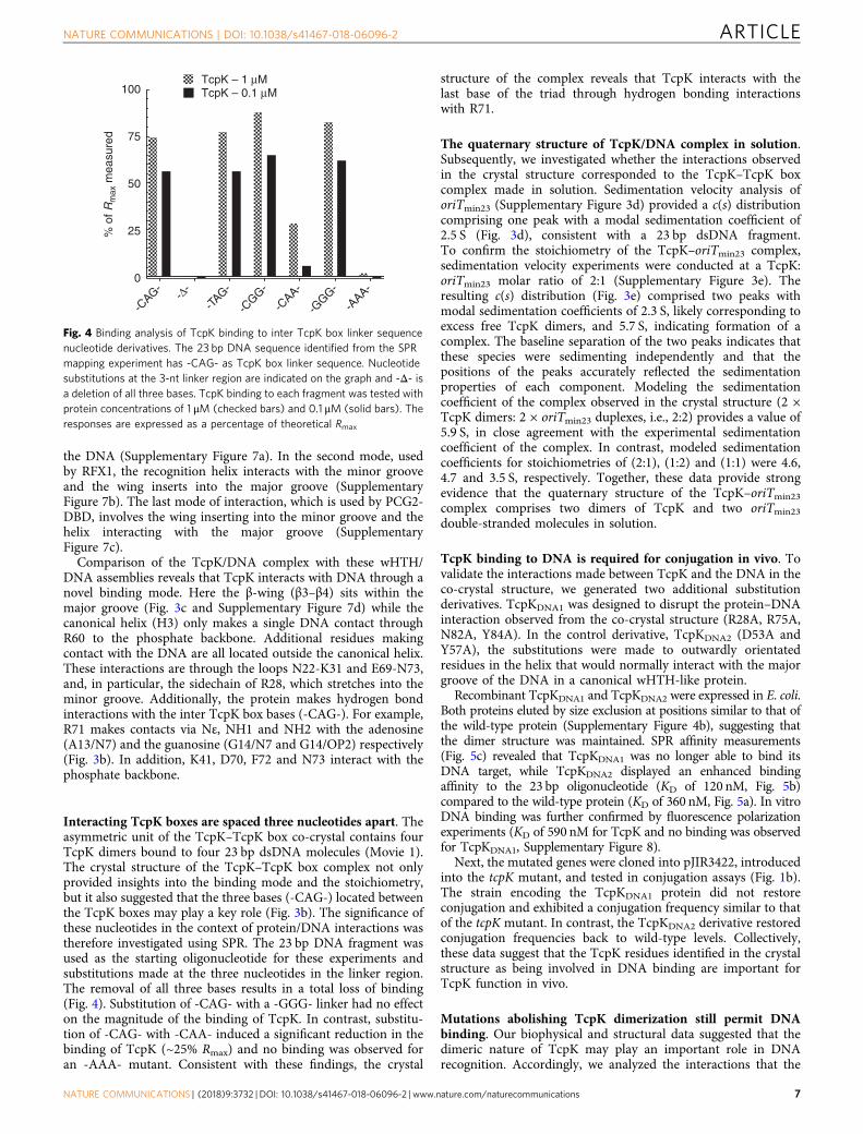
Interacting TcpK boxes are spaced three nucleotides apart. Theasymmetric unit of the TcpK–TcpK box co-crystal contains fourTcpK dimers bound to four 23 bp dsDNA molecules (Movie 1).The crystal structure of the TcpK–TcpK box complex not onlyprovided insights into the binding mode and the stoichiometry,but it also suggested that the three bases (-CAG-) located betweenthe TcpK boxes may play a key role (Fig. 3b). The significance ofthese nucleotides in the context of protein/DNA interactions wastherefore investigated using SPR. The 23 bp DNA fragment wasused as the starting oligonucleotide for these experiments andsubstitutions made at the three nucleotides in the linker region.The removal of all three bases results in a total loss of binding(Fig. 4). Substitution of -CAG- with a -GGG- linker had no effecton the magnitude of the binding of TcpK. In contrast, substitu-tion of -CAG- with -CAA- induced a significant reduction in thebinding of TcpK (~25% Rmax) and no binding was observed foran -AAA- mutant. Consistent with these findings, the crystal
structure of the complex reveals that TcpK interacts with thelast base of the triad through hydrogen bonding interactionswith R71.
The quaternary structure of TcpK/DNA complex in solution.Subsequently, we investigated whether the interactions observedin the crystal structure corresponded to the TcpK–TcpK boxcomplex made in solution. Sedimentation velocity analysis oforiTmin23 (Supplementary Figure 3d) provided a c(s) distributioncomprising one peak with a modal sedimentation coefficient of2.5 S (Fig. 3d), consistent with a 23 bp dsDNA fragment.To confirm the stoichiometry of the TcpK–oriTmin23 complex,sedimentation velocity experiments were conducted at a TcpK:oriTmin23 molar ratio of 2:1 (Supplementary Figure 3e). Theresulting c(s) distribution (Fig. 3e) comprised two peaks withmodal sedimentation coefficients of 2.3 S, likely corresponding toexcess free TcpK dimers, and 5.7 S, indicating formation of acomplex. The baseline separation of the two peaks indicates thatthese species were sedimenting independently and that thepositions of the peaks accurately reflected the sedimentationproperties of each component. Modeling the sedimentationcoefficient of the complex observed in the crystal structure (2 ×TcpK dimers: 2 × oriTmin23 duplexes, i.e., 2:2) provides a value of5.9 S, in close agreement with the experimental sedimentationcoefficient of the complex. In contrast, modeled sedimentationcoefficients for stoichiometries of (2:1), (1:2) and (1:1) were 4.6,4.7 and 3.5 S, respectively. Together, these data provide strongevidence that the quaternary structure of the TcpK–oriTmin23
complex comprises two dimers of TcpK and two oriTmin23
double-stranded molecules in solution.
TcpK binding to DNA is required for conjugation in vivo. Tovalidate the interactions made between TcpK and the DNA in theco-crystal structure, we generated two additional substitutionderivatives. TcpKDNA1 was designed to disrupt the protein–DNAinteraction observed from the co-crystal structure (R28A, R75A,N82A, Y84A). In the control derivative, TcpKDNA2 (D53A andY57A), the substitutions were made to outwardly orientatedresidues in the helix that would normally interact with the majorgroove of the DNA in a canonical wHTH-like protein.
Recombinant TcpKDNA1 and TcpKDNA2 were expressed in E. coli.Both proteins eluted by size exclusion at positions similar to that ofthe wild-type protein (Supplementary Figure 4b), suggesting thatthe dimer structure was maintained. SPR affinity measurements(Fig. 5c) revealed that TcpKDNA1 was no longer able to bind itsDNA target, while TcpKDNA2 displayed an enhanced bindingaffinity to the 23 bp oligonucleotide (KD of 120 nM, Fig. 5b)compared to the wild-type protein (KD of 360 nM, Fig. 5a). In vitroDNA binding was further confirmed by fluorescence polarizationexperiments (KD of 590 nM for TcpK and no binding was observedfor TcpKDNA1, Supplementary Figure 8).
Next, the mutated genes were cloned into pJIR3422, introducedinto the tcpK mutant, and tested in conjugation assays (Fig. 1b).The strain encoding the TcpKDNA1 protein did not restoreconjugation and exhibited a conjugation frequency similar to thatof the tcpK mutant. In contrast, the TcpKDNA2 derivative restoredconjugation frequencies back to wild-type levels. Collectively,these data suggest that the TcpK residues identified in the crystalstructure as being involved in DNA binding are important forTcpK function in vivo.
Mutations abolishing TcpK dimerization still permit DNAbinding. Our biophysical and structural data suggested that thedimeric nature of TcpK may play an important role in DNArecognition. Accordingly, we analyzed the interactions that the
100TcpK – 1 μMTcpK – 0.1 μM
75
50
25
0
-CAG-
-TAG-
-CGG-
-CAA-
-AAA-
-Δ-
-GGG-
% o
f Rm
ax m
easu
red
Fig. 4 Binding analysis of TcpK binding to inter TcpK box linker sequencenucleotide derivatives. The 23 bp DNA sequence identified from the SPRmapping experiment has -CAG- as TcpK box linker sequence. Nucleotidesubstitutions at the 3-nt linker region are indicated on the graph and -Δ- isa deletion of all three bases. TcpK binding to each fragment was tested withprotein concentrations of 1 μM (checked bars) and 0.1 μM (solid bars). Theresponses are expressed as a percentage of theoretical Rmax
NATURE COMMUNICATIONS | DOI: 10.1038/s41467-018-06096-2 ARTICLE
NATURE COMMUNICATIONS | (2018) 9:3732 | DOI: 10.1038/s41467-018-06096-2 | www.nature.com/naturecommunications 7
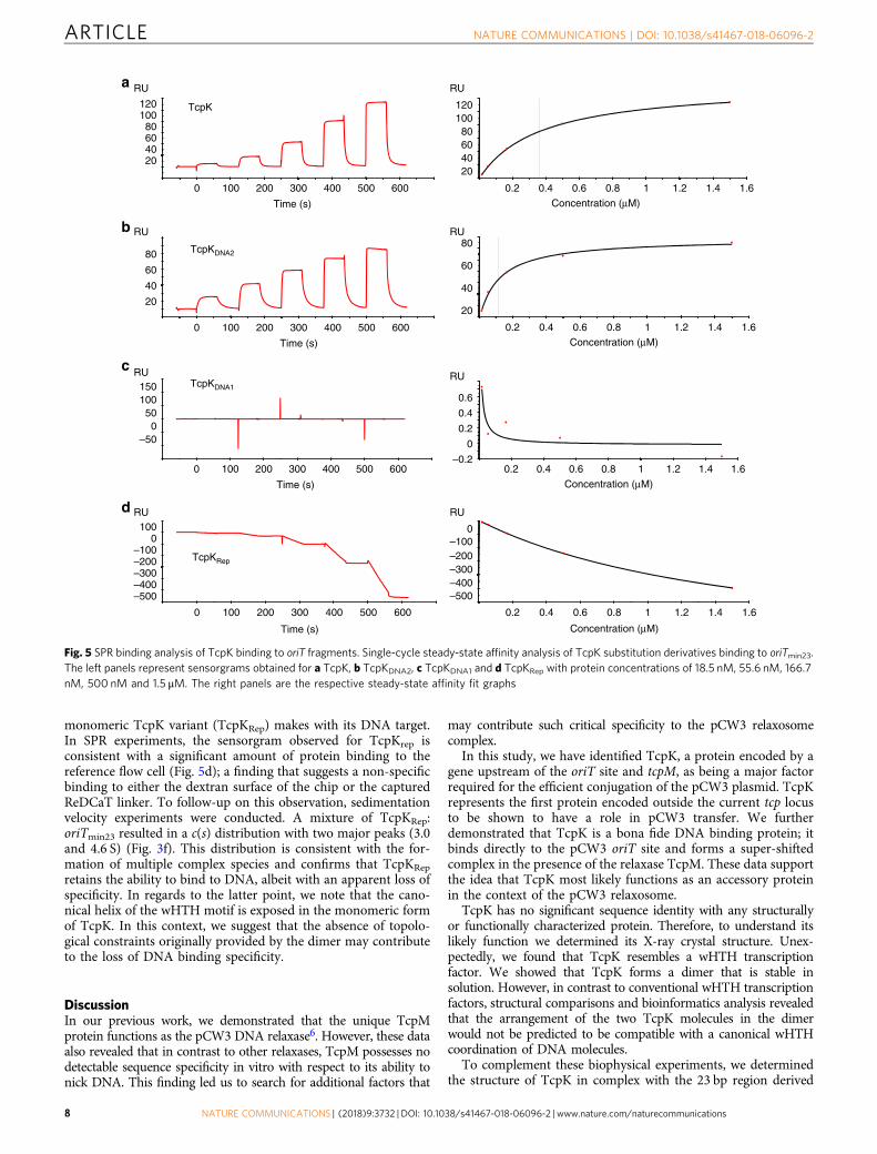
monomeric TcpK variant (TcpKRep) makes with its DNA target.In SPR experiments, the sensorgram observed for TcpKrep isconsistent with a significant amount of protein binding to thereference flow cell (Fig. 5d); a finding that suggests a non-specificbinding to either the dextran surface of the chip or the capturedReDCaT linker. To follow-up on this observation, sedimentationvelocity experiments were conducted. A mixture of TcpKRep:oriTmin23 resulted in a c(s) distribution with two major peaks (3.0and 4.6 S) (Fig. 3f). This distribution is consistent with the for-mation of multiple complex species and confirms that TcpKRep
retains the ability to bind to DNA, albeit with an apparent loss ofspecificity. In regards to the latter point, we note that the cano-nical helix of the wHTH motif is exposed in the monomeric formof TcpK. In this context, we suggest that the absence of topolo-gical constraints originally provided by the dimer may contributeto the loss of DNA binding specificity.
DiscussionIn our previous work, we demonstrated that the unique TcpMprotein functions as the pCW3 DNA relaxase6. However, these dataalso revealed that in contrast to other relaxases, TcpM possesses nodetectable sequence specificity in vitro with respect to its ability tonick DNA. This finding led us to search for additional factors that
may contribute such critical specificity to the pCW3 relaxosomecomplex.
In this study, we have identified TcpK, a protein encoded by agene upstream of the oriT site and tcpM, as being a major factorrequired for the efficient conjugation of the pCW3 plasmid. TcpKrepresents the first protein encoded outside the current tcp locusto be shown to have a role in pCW3 transfer. We furtherdemonstrated that TcpK is a bona fide DNA binding protein; itbinds directly to the pCW3 oriT site and forms a super-shiftedcomplex in the presence of the relaxase TcpM. These data supportthe idea that TcpK most likely functions as an accessory proteinin the context of the pCW3 relaxosome.
TcpK has no significant sequence identity with any structurallyor functionally characterized protein. Therefore, to understand itslikely function we determined its X-ray crystal structure. Unex-pectedly, we found that TcpK resembles a wHTH transcriptionfactor. We showed that TcpK forms a dimer that is stable insolution. However, in contrast to conventional wHTH transcriptionfactors, structural comparisons and bioinformatics analysis revealedthat the arrangement of the two TcpK molecules in the dimerwould not be predicted to be compatible with a canonical wHTHcoordination of DNA molecules.
To complement these biophysical experiments, we determinedthe structure of TcpK in complex with the 23 bp region derived
20
40
60
80
RU
TcpKDNA2
20
40
60
80RUb
20406080
100120
0.2
RU
Concentration (μM)
20406080
100120
0
RU
Time (s)
TcpK
a
–500
50100150
RUTcpKDNA1
–0.2
0
0.2
0.4
0.6
RUc
–500–400–300–200–100
0100
RU
TcpKRep
–500–400–300–200–100
0
RUd
100 200 300 400 500 600
0
Time (s)
100 200 300 400 500 600
0
Time (s)
100 200 300 400 500 600
0
Time (s)
100 200 300 400 500 600
0.4 0.6 0.8 1 1.2 1.4 1.6
0.2
Concentration (μM)
0.4 0.6 0.8 1 1.2 1.4 1.6
0.2
Concentration (μM)
0.4 0.6 0.8 1 1.2 1.4 1.6
0.2
Concentration (μM)
0.4 0.6 0.8 1 1.2 1.4 1.6
Fig. 5 SPR binding analysis of TcpK binding to oriT fragments. Single-cycle steady-state affinity analysis of TcpK substitution derivatives binding to oriTmin23.The left panels represent sensorgrams obtained for a TcpK, b TcpKDNA2, c TcpKDNA1 and d TcpKRep with protein concentrations of 18.5 nM, 55.6 nM, 166.7nM, 500 nM and 1.5 μM. The right panels are the respective steady-state affinity fit graphs
ARTICLE NATURE COMMUNICATIONS | DOI: 10.1038/s41467-018-06096-2
8 NATURE COMMUNICATIONS | (2018) 9:3732 | DOI: 10.1038/s41467-018-06096-2 | www.nature.com/naturecommunications
from oriT. These data revealed that TcpK binds DNA in a mostunexpected fashion. In contrast to other wHTH proteins the β-wing of TcpK represents the primary point of contact with DNAand the canonical wHTH DNA binding helix is buried within thedimerization interface. In addition, and consistent with the resultsof our sedimentation velocity experiments, we found that eachTcpK dimer bound to two TcpK boxes, but that these bindingevents take place in trans (i.e., to two different molecules of DNA).
To reconcile these in vitro data with the in vivo function ofTcpK, we tested the ability of a range of different mutants torescue conjugative transfer of a pCW3 tcpK mutant. The resultsshowed that TcpK residues within the β-wing that are requiredfor direct binding to the TcpK box are also required for efficientconjugative transfer, supporting the idea that TcpK is a bona fideDNA binding protein that likely functions in the context of therelaxosome. However, mutations shown to abolish TcpK dimer-ization in vitro had no detectable consequence on conjugationefficiency in vivo.
What are the implications of the integrated genetics and bio-physical data for our understanding of the mechanism of transferof members of the pCW3 family of conjugative antibiotic resis-tance and toxin plasmids? The oriT genetic data obtained pre-viously6 and DNA binding data obtained here are in goodagreement. We previously showed that the region containing thetandem repeat TcpK boxes in oriT are essential for efficienttransfer. Here we demonstrate that TcpK is required for transferand is capable of binding TcpK boxes.
Our in vitro data suggested that formation of the TcpK dimerwas obligatory for specific DNA binding, and that this processinvolved the coordination of complexes that contain two DNAmolecules. In contrast, the in vivo data revealed that monomericforms of TcpK possess wild-type function in the context ofconjugative transfer. The different nature of the DNA targets usedin these studies may be the source of these apparently contra-dictory findings. In the bacterial cell the oriT target for TcpKbinding is located on a large 47 kb supercoiled DNA molecule,whereas the in vitro studies were carried out using linear 23 bpfragments. In addition, we cannot rule out the possibility that theTcpK dimers are functioning in another biological process thatmay or may not be related to conjugative transfer. Indeed, wenote that the canonical helix that binds DNA in other wHTHproteins forms much of the dimerization interface in TcpK, and istherefore no longer able to interact with DNA. Such an interac-tion might therefore have served to help drive the evolution ofthis molecule as a specialized component tightly restricted so thatit functions in the context of conjugative plasmid DNA.
In conclusion, we identify TcpK as a crucial new component ofthe pCW3 conjugative machinery. Our data suggest that itsfunction is to decorate the origin of transfer of pCW3 and tointeract specifically with sequences that are only present in thisregion of the plasmid. In doing so we suggest that TcpKmost likely functions within the relaxosome and may contributewith other protein partners to achieve the proper recruitmentof TcpM, a relaxase that lacks detectable sequence specificityin vitro.
MethodsBacterial strains, genetics and molecular methods. Bacterial strains and plas-mids are described in Supplementary Table 4. C. perfringens and E. coli strains werecultured as previously described6. Molecular and genetics methods, including gelshift experiments and conjugative matings, were as before6. The tcpK mutant wasconstructed by TargeTron mutagenesis. An intron insertion site between residues153 and 154 in the antisense strand of the 309 bp tcpK gene was identified using theIntron Finder software18 and intron primers re-targeted to the tcpK gene byprimer-mediated PCR mutagenesis, as previously described19. The 350 bp PCRproduct was cloned between the HindIII and BsrGI sites in the clostridial Targe-Tron vector pJIR356219 to generate pJIR4347, which confers thiamphenicol
resistance. C. perfringens strain JIR4195 [JIR325(pCW3)]20 was transformed withpJIR4347 and thiamphenicol-resistant transconjugants plated onto medium con-taining erythromycin (5 μg mL−1) to select for integration of the group II intron.Potential mutants that were tetracycline and erythromycin resistant, but thiam-phenicol sensitive, were analyzed by PCR and Southern hybridization to confirminsertion of the intron in the tcpK gene. The resultant tcpK::TT mutant wasdesignated as JIR13063.
For complementation studies, the wild-type tcpK gene was amplified by PCRand then cloned into the BamHI/Asp718 sites of the E. coli–C. perfringens shuttlevector pJIR34224,21 to generate pJIR4349. The complementation plasmid wasintroduced into the tcpK mutant by electroporation. The presence of thecomplementation plasmid in the C. perfringens strain was confirmed by restrictionendonuclease and sequence analysis after plasmid rescue in E. coli. Mutantderivatives of tcpK (Dimer, Repulse, DNA1 and DNA2) were synthesized with therequired mutations (Genscript) and subcloned from pUC57 (Genscript) intopJIR3422 by digestion with BamHI/Asp718, as for the wild-type gene. Eachconstruct was introduced separately into the tcpK mutant via electroporation andconjugation experiments were conducted as before22,23. Complementation vectorswere checked as for the wild-type gene to confirm the strain constructions.
Expression and purification of recombinant proteins. The genes encoding tcpKand its variants were codon optimized (Genscript) for expression in E coli. Theywere cloned first into pUC57 and then subcloned into pGL12 between the EcoRIand BamHI restriction sites24. This cloning resulted in the expression of proteinswith an N-terminal hexa-histidine tag followed by a TEV (tobacco etch virus)protease cleavage site. C41(DE3)pLysS was used as the expression host. Cells weregrown in 2YT at 37 °C until the OD600 reached 0.6. The cultures were transferred to16 °C for 30 min, protein expression was induced by the addition of 1 mM iso-propyl β-D-1-thiogalactopyranoside (IPTG) and growth was continued for 18 h at16 °C. The culture was harvested by centrifugation at 5000 × g, the pellets wereresuspended in buffer A (50 mM Tris HCl, 300 mM NaCl, 10 mM imidazole, 2 mM2-mercaptoethanol, pH 7.4) and lysed by sonication for 6 × 30 s pulses at 10 mAwith 30 s rest between the pulses. The lysate was loaded on to a 1 mL Hi-TrapTALON column (GE Healthcare) equilibrated with buffer A, then washed withbuffer B (50 mM Tris HCl, 1 M NaCl, 20 mM imidazole, 2 mM 2-mercaptoethanol,10% (w v−1) glycerol, pH 7.4) and the protein eluted in buffer C (50 mM Tris HCl,500 mM NaCl, 250 mM imidazole, 2 mM 2-mercaptoethanol, 10% (w v−1) glycerol,pH 7.4). The fractions were diluted twofold in TEV Buffer (100 mM Tris HCl, 150mM NaCl, 0.2 mM EDTA, pH 8.0) and then incubated overnight with the TEVprotease at a 1:100 (w w−1) (TEV/protein) ratio. The cleaved product was thenreverse purified on the 1 mL TALON column. The flow through was collected,concentrated and further purified by size exclusion chromatography on a Superdex75 16/60 (GE Healthcare) equilibrated with buffer D (25 mM Hepes, 150 mMNaCl, pH 7.4). When protein was used for crystallization experiments, the peakfraction was buffer exchanged into buffer E (25 mM MES, 50 mM NaCl, pH 6.5)and further purified by cation exchange chromatography on a Hi-Trap S FF (GEHealthcare) column. The protein was eluted using a gradient obtained by mixingbuffer E and buffer F (25 mM MES, 1 M NaCl, pH 6.5). Purified selenomethionine-labeled TcpK was obtained in a similar fashion, but the cells were grown in M9media supplemented with 50 mg L−1 selenomethionine and the buffers weresupplemented with 5 mM 2-mercaptoethanol. The production of TcpM and the gelmobility shift assays were performed as described previously6.
Protein crystallization and X-ray data collection. All crystals were obtained byhanging drop vapor diffusion with a 500 μL reservoir volume. For TcpK alone, thedrops consisted of 1.5 μL TcpK (10 mgmL−1), 0.2 μL phenol (0.1 M) and 1.3 μL ofthe reservoir (1 M Na citrate, 0.1 M Na cacodylate, pH 6.5). Selenomethionine-labeled TcpK was obtained by microseeding with the same reservoir conditions.The seed stock was obtained by crushing unlabeled TcpK crystals then dilutingtenfold with the reservoir solution. The drop consisted of 1.5 μL labeled TcpK (10mgmL−1), 0.5 μL seed stock, 0.2 μL phenol (0.1 M) and 1 μL of the reservoir.TcpK–oriTmin23 crystals were grown with a reservoir containing 50% (v v−1) 2-methyl-2,4-pentanediol, 0.1 mM imidazole, pH 7.0. The drop consisted of 1.5 μLTcpK–oriTmin23 and 1.5 μL of the reservoir. Oligonucleotides were purchased fromSigma and dissolved in water as a 4 mM stock solution. Equal volumes of com-plementary strands were mixed, heated to 95 °C for 5 min and cooled to roomtemperature. TcpK (0.6 mgmL−1) and the double-stranded oriTmin23 (2 mM) weremixed at a protein/DNA molar ratio of 2:1. The complex was concentrated to 380μM for crystallization. Crystals were harvested using a silicon loop (Mitegen),soaked in the reservoir solution supplemented with either 25% (w v−1) sucrose or25% (w v−1) glucose, then flash cooled in liquid nitrogen. X-ray diffraction datawere collected on the MX2 beamline at the Australian Synchrotron. The best TcpKnative crystal diffracted to 2.5 Å and was prone to radiation damage. Intensitieswere recorded from two distinct areas of the same crystal (Supplementary Table 1)and used for the refinement. Selenomethionine TcpK protein crystals also werehighly prone to X-ray radiation damage. Anomalous diffraction data werecollected at the selenium edge from four different protein crystals to resolutionbetween 3.0 and 3.2 Å on the MX2 beamline. From each crystal, a maximum of 90°of X-ray data were recorded. One of these crystals allowed collection of datasets atthree different positions. Each set of X-ray data was processed using XDS25. The
NATURE COMMUNICATIONS | DOI: 10.1038/s41467-018-06096-2 ARTICLE
NATURE COMMUNICATIONS | (2018) 9:3732 | DOI: 10.1038/s41467-018-06096-2 | www.nature.com/naturecommunications 9
resulting intensity data from each dataset were analyzed using Rd plot26
generated from the software XDSSTAT for radiation damage analysis allowedselection of a number of frames for final data processing for each dataset. Theseven datasets were scaled together using XSCALE25. The merging statistics ofscaled data (Supplementary Table 1) indicated anomalous signal up to 4.5 Åresolution.
Structure determination. The structure of TcpK was solved using the SADphasing protocol of Auto-Rickshaw27 that included substructure determinationusing SHELXD28, heavy atom refinement using BP329 density modification usingPIRATE30 and initial model building using BUCCANEER30. Further phaseimprovement, model completion and partial model refinement were carried outusing MRSAD protocol of Auto-Rickshaw31, which resulted in a model with anRwork of 40% and Rfree of 45%. The model was used as a starting model for MRphasing in Auto-Rickshaw against the native dataset. Density modification, phaseextension and fourfold NCS averaging were carried out using RESOLVE32 to 2.5 Åresolution and further model building was performed using BUCCANEER. Thisallowed 399 residues to be built in four fragments out of 408 residues in theasymmetric unit and 393 residues were sequenced automatically. Initial refinementof the model was carried out in the REFMAC530 and resulted in 27.4% Rwork and32.7% Rfree. The structure was improved iteratively with manual model building inCOOT33 and refinement in BUSTER-TNT34.
The structure of TcpK–oriTmin23 was determined by Molecular Replacement inPHASER35 in two steps. The initial search was performed using the coordinates of asingle chain of TcpK. Upon inspection of PHASER’s output and the crystal packing,the difference Fourier maps displayed positive density, especially in the vicinity ofthe β-loop. Since no additional TcpK molecules could be placed in the unit cell, themissing information was therefore attributed to the presence of DNA in the crystal.Following on from this partial solution, the coordinates of the protein were fixedand a new search performed with a 23 bp dsDNA fragment generated in Coot33.However, this procedure failed and the search was repeated using a shorter dsDNAfragment (10 bp), which yielded several partial solutions that subsequently failedPHASER packing tests. These imperfect solutions were carefully examined in Cootand PyMOL, and used to manually position the DNA molecules in the electrondensity map. After several cycles of rebuilding and refinements, the final model wasdeposited in the PDB with Rwork and Rfree 0.5values of 19.8 and 23.3% respectively.Final models were validated with Molprobity36 and deposited in the PDB under theaccession numbers 5VFY and 5VFX.
Surface plasmon resonance. SPR experiments were performed on a Biacore T200(GE Healthcare) operated at 20 °C. All experiments were performed in duplicateand were based on the Re-usable DNA Capture Technology (ReDCaT)14. The 150bp oriT region (24697–24846) of pCW3 was divided into 13 overlapping fragments(oriT#1–oriT#13; Fig. 2d). Each fragment comprised 30 bp with a consecutiveoverlap of 20 bases from one fragment to the next. These fragments were generatedusing the Perl Overlapping Oligo Producer (POOP) script14. The complementaryReDCaT sequence was positioned at the 3’ end of the reverse oligonucleotide.Oligonucleotides (Sigma) were annealed together by incubating at 95 °C for 10 minthen cooling to 20 °C on the bench. The protein was prepared as described earlierand dialyzed against the running buffer, which consisted of 10 mM HEPES pH 7.4,200 mM NaCl, 3 mM EDTA and 0.05% (w v−1) Tween 20. The biotinylatedReDCaT linker was immobilized on two flow cells (FC1 and FC2) of a Streptavidin(SA) chip (GE Healthcare) by pulse injections of a 100 nM solution at 5 μLmin−1.The injections were stopped when the density on the surface reached approxi-mately 450 Resonance Units (RU). The flow cells then were washed with three 60 sinjections of the regeneration solution (1 M NaCl, 50 mM NaOH).
For the initial screening, test dsDNA at 500 nM was captured for 60 s at 10 μLmin−1 on the active flow cell (FC2). The protein (10 μM or 1 μM) was flowed overthe two surfaces at 30 μL min−1 with 60 s contact time and 300 s dissociation time.The chip then was regenerated by injection of the regeneration solution for 60 s.Stability values were recorded 10 s after the end of protein injection. The bindinglevel of TcpK to each fragment was expressed as a percentage of the theoreticalmaximum response (Theoretical Rmax= (molar mass TcpK/molar mass oriT#n) ×RU ligand captured × stoichiometry × 0.78 and %Rmax measured= (measured R/theoretical Rmax) × 100). As suggested before14 the absolute values are notimportant for the analysis, but instead it is the relative binding levels among thefragments that provide the most valuable information.
For the mapping experiments, the two hits (oriT#1 and oriT#2) were combinedto form a 39 bp oligonucleotide L2R1 (left to right, relative to the top strand).Subsequent oligonucleotides were then synthetized (L2R2–L2R12), each shorter by2 nt relative to one another. To identify the second boundary, the same startingoligonucleotide was used. Hence, R2L1 (right to left) and L2R1 are equivalent.However, R2L1 is oriented in the opposite direction on the sensor chip. Thisprocedure guaranteed that the protein was able to interact with the oligonucleotideregardless of its orientation on the sensor chip. It also confirmed that the ReDCaTlinker did not compensate for the loss of nucleotides from the test fragment.Protein concentrations used for the mapping were 1 μM and 0.1 μM and thebinding level was expressed in the same manner as for the initial screeningexperiment.
For the initial kinetic (multicycle kinetic) measurements, approximately 250 RUof DNA was captured and TcpK was flowed over both FC1 (RedCat linker) andFC2 (oriT#1, oriT#2 or oriT#3) at protein concentrations of 125 nM, 250 nM, 500nM, 1 μM, 2.5 μM, 5 μM and 10 μM with an another 1 μM sample re-injected at theend of the experiment as internal duplicate. Subsequent single-cycle kineticexperiments were performed with 50 RU of DNA captured on FC2 and proteinconcentrations 18.5 nM–1.5 μM TcpK, TcpKDNA1, TcpKDNA2 and TcpKRep (60 scontact time at 30 μLmin−1).
Analytical ultracentrifugation. Sedimentation velocity experiments were per-formed using an XL-I analytical ultracentrifuge (Beckman Coulter) equipped withultraviolet–visible scanning optics. Buffer reference (400 μL; 20 mM HEPES, 150mM NaCl, pH 7.4) and sample solutions (380 μL) were loaded into 12 mm double-sector cells with quartz windows, and centrifuged at 50,000 rpm (201,600 × g) and20 °C using an An-60Ti rotor. Radial absorbance data were recorded at appropriatewavelengths in continuous mode. Sedimentation velocity data were fitted to acontinuous sedimentation coefficient distribution (c(s)) model using SEDFIT37.The partial specific volume of TcpK (0.7436 mL g−1), buffer density (1.006 g mL−1), and buffer viscosity (1.031 cp) were calculated using SEDNTERP38. The partialspecific volume39 used for oriTmin23 was 0.54 mL g−1 and partial specific volumesfor various TcpK/oriT complex stoichiometries were calculated from the weightedaverage of protein and DNA. The molecular weight of TcpK was calculated fromsedimentation coefficients in SEDFIT using the frictional ratio obtained from the fitto the sedimentation velocity data for 48 μM TcpK. Theoretical sedimentationcoefficients were calculated from the TcpK and TcpK/oriTmin23 complex crystalstructure coordinates by hydrodynamic modeling under experimental conditionsdescribed above using HYDROPRO40.
Fluorescence polarization DNA binding. Fluorescence polarization DNA bindingexperiments were performed using a PHERAstar microplate reader (BMG Lab-tech). Alexa488 5’-labeled oriTmin23 was purchased from Sigma and annealed withthe complementary non-labeled strand. The experiment was performed at 20 °C ina final reaction buffer consisting of 10 mM HEPES pH 7.4, 150 mM NaCl, 3 mMEDTA, 0.05% (w v−1) Tween 20, 0.5% (w v−1) bovine serum albumin. The DNAwas incubated at a final concentration of 5 nM with various protein concentrationsinto a final volume of 50 μL. Gain and focus adjustments were performed on aprotein-free sample. Background subtracted mP with the fits are reported inSupplementary Figure 8.
Data availabilityAll relevant data supporting the findings of the study are available in this article and itsSupplementary Information files. The coordinates are available in the Protein Data Bankunder accession codes 5VFY and 5VFX. Additional data are available from the corre-sponding authors upon request.
Received: 6 February 2018 Accepted: 15 June 2018
References1. Li, J. et al. Toxin plasmids of Clostridium perfringens. Microbiol. Mol. Biol.
Rev. 77, 208–233 (2013).2. Wisniewski, J. A. & Rood, J. I. The Tcp conjugation system of Clostridium
perfringens. Plasmid 91, 28–36 (2017).3. Teng, W. L., Bannam, T. L., Parsons, J. A. & Rood, J. I. Functional
characterization and localization of the TcpH conjugation protein fromClostridium perfringens. J. Bacteriol. 190, 5075–5086 (2008).
4. Bantwal, R. et al. The peptidoglycan hydrolase TcpG is required for efficientconjugative transfer of pCW3 in Clostridium perfringens. Plasmid 67,139–147 (2012).
5. Porter, C. J. et al. The conjugation protein TcpC from Clostridium perfringensis structurally related to the type IV secretion system protein VirB8 fromGram-negative bacteria. Mol. Microbiol. 83, 275–288 (2012).
6. Wisniewski, J. A. et al. TcpM: a novel relaxase that mediates transfer of largeconjugative plasmids from Clostridium perfringens. Mol. Microbiol. 99,884–896 (2016).
7. Bannam, T. L., Teng, W. L., Bulach, D., Lyras, D. & Rood, J. I. Functionalidentification of conjugation and replication regions of the tetracyclineresistance plasmid pCW3 from Clostridium perfringens. J. Bacteriol. 188,4942–4951 (2006).
8. Parsons, J. A., Bannam, T. L., Devenish, R. J. & Rood, J. I. TcpA, an FtsK/SpoIIIE homolog, is essential for transfer of the conjugative plasmid pCW3 inClostridium perfringens. J. Bacteriol. 189, 7782–7790 (2007).
9. Steen, J. A., Bannam, T. L., Teng, W. L., Devenish, R. J. & Rood, J. I. Theputative coupling protein TcpA interacts with other pCW3-encoded proteins
ARTICLE NATURE COMMUNICATIONS | DOI: 10.1038/s41467-018-06096-2
10 NATURE COMMUNICATIONS | (2018) 9:3732 | DOI: 10.1038/s41467-018-06096-2 | www.nature.com/naturecommunications
to form an essential part of the conjugation complex. J. Bacteriol. 191,2926–2933 (2009).
10. Wisniewski, J. A., Teng, W. L., Bannam, T. L. & Rood, J. I. Two novel membraneproteins, TcpD and TcpE, are essential for conjugative transfer of pCW3 inClostridium perfringens. J. Bacteriol. 197, 774–781 (2015).
11. Holm, L. & Rosenstrom, P. Dali server: conservation mapping in 3D. NucleicAcids Res. 38, W545–W549 (2010).
12. Woo, J. S. et al. Structural studies of a bacterial condensin complex revealATP-dependent disruption of intersubunit interactions. Cell 136, 85–96(2009).
13. Krissinel, E. & Henrick, K. Inference of macromolecular assemblies fromcrystalline state. J. Mol. Biol. 372, 774–797 (2007).
14. Stevenson, C. E. et al. Investigation of DNA sequence recognition by astreptomycete MarR family transcriptional regulator through surface plasmonresonance and X-ray crystallography. Nucleic Acids Res. 41, 7009–7022 (2013).
15. Lavery, R., Moakher, M., Maddocks, J. H., Petkeviciute, D. & Zakrzewska, K.Conformational analysis of nucleic acids revisited: curves. Nucleic Acids Res.37, 5917–5929 (2009).
16. Zheng, G., Lu, X. J. & Olson, W. K. Web 3DNA-a web server for the analysis,reconstruction, and visualization of three-dimensional nucleic-acid structures.Nucleic Acids Res. 37, W240–W246 (2009).
17. Liu, J. et al. Structural basis of DNA recognition by PCG2 reveals a novel DNAbinding mode for winged helix-turn-helix domains. Nucleic Acids Res. 43,1231–1240 (2015).
18. Carter, G. P. et al. TcsL is an essential virulence factor in Clostridium sordelliiATCC 9714. Infect. Immun. 79, 1025–1032 (2011).
19. Cheung, J. K. et al. The VirSR two-component signal transduction systemregulates NetB toxin production in Clostridium perfringens. Infect. Immun.78, 3064–3072 (2010).
20. Scott, P. T. & Rood, J. I. Electroporation-mediated transformation of lysostaphin-treated Clostridium perfringens. Gene 82, 327–333 (1989).
21. Adams, V. et al. Utility of the clostridial site-specific recombinase TnpX toclone toxic-product-encoding genes and selectively remove genomic DNAfragments. Appl. Environ. Microbiol. 80, 3597–3603 (2014).
22. Rood, J. I. Transferable tetracycline resistance in Clostridium perfringensstrains of porcine origin. Can. J. Microbiol. 29, 1241–1246 (1983).
23. Rood, J. I., Maher, E. A., Somers, E. B., Campos, E. & Duncan, C. L. Isolationand characterization of multiply antibiotic-resistant Clostridum perfringensstrains from porcine feces. Antimicrob. Agents Chemother. 13, 871–880(1978).
24. Law, R. H. et al. The high resolution crystal structure of the human tumorsuppressor maspin reveals a novel conformational switch in the G-helix. J.Biol. Chem. 280, 22356–22364 (2005).
25. Kabsch, W. Integration, scaling, space-group assignment and post-refinement.Acta Crystallogr. D Biol. Crystallogr. 66, 133–144 (2010).
26. Diederichs, K. Some aspects of quantitative analysis and correction ofradiation damage. Acta Crystallogr. D Biol. Crystallogr. 62, 96–101 (2006).
27. Panjikar, S., Parthasarathy, V., Lamzin, V. S., Weiss, M. S. & Tucker, P. A.Auto-rickshaw: an automated crystal structure determination platform as anefficient tool for the validation of an X-ray diffraction experiment. ActaCrystallogr. D Biol. Crystallogr. 61, 449–457 (2005).
28. Schneider, T. R. & Sheldrick, G. M. Substructure solution with SHELXD. ActaCrystallogr. D Biol. Crystallogr. 58, 1772–1779 (2002).
29. Pannu, N. S. & Read, R. J. The application of multivariate statistical techniquesimproves single-wavelength anomalous diffraction phasing. Acta Crystallogr.D Biol. Crystallogr. 60, 22–27 (2004).
30. Collaborative Computational Project, Number 4. The CCP4 suite: programsfor protein crystallography. Acta Crystallogr. D Biol. Crystallogr. 50, 760–763(1994).
31. Panjikar, S., Parthasarathy, V., Lamzin, V. S., Weiss, M. S. & Tucker, P. A. Onthe combination of molecular replacement and single-wavelength anomalousdiffraction phasing for automated structure determination. Acta Crystallogr. DBiol. Crystallogr. 65, 1089–1097 (2009).
32. Terwilliger, T. C. Maximum-likelihood density modification. Acta Crystallogr.D Biol. Crystallogr. 56, 965–972 (2000).
33. Emsley, P., Lohkamp, B., Scott, W. G. & Cowtan, K. Features and developmentof Coot. Acta Crystallogr. D Biol. Crystallogr. 66, 486–501 (2010).
34. Bricogne G. B. E., et al. BUSTER 2.10.1 (Global Phasing Ltd, Cambridge,2016).
35. McCoy, A. J. et al. Phaser crystallographic software. J. Appl. Crystallogr. 40,658–674 (2007).
36. Davis, I. W. et al. MolProbity: all-atom contacts and structure validation forproteins and nucleic acids. Nucleic Acids Res. 35, W375–W383 (2007).
37. Schuck, P. Size-distribution analysis of macromolecules by sedimentationvelocity ultracentrifugation and lamm equation modeling. Biophys. J. 78,1606–1619 (2000).
38. Laue T. M., Shah B. D., Ridgeway T. M., Pelletier S. L. in AnalyticalUltracentrifugation in Biochemistry and Polymer Science (eds Harding S. E.,Rowe A. J., Horton J. C.) 90–125 (Royal Society of Chemistry, Cambridge,1992).
39. Durchschlag H. in Thermodynamic Data for Biochemistry and Biotechnology(ed. Hinz H.-J.) 45–128 (Springer, Berlin, 1986).
40. Ortega, A., Amoros, D., Garcia & de la Torre, J. Prediction of hydrodynamicand other solution properties of rigid proteins from atomic- and residue-levelmodels. Biophys. J. 101, 892–898 (2011).
AcknowledgementsThis research was supported by Project Grant GNT1104502 from the AustralianNational Health and Medical Research Council. J.A.W. was the recipient of an AustralianPostgraduate Award. We thank the CSIRO Collaborative Crystallization Centre (www.csiro.au/C3), Melbourne, Australia, for the crystallization experiments. X-ray diffractionexperiments were undertaken on the MX beamlines at the Australian Synchrotron, partof ANSTO.
Author contributionsD.A.K.T. designed and performed the experiments, analyzed the data and wrote themanuscript. J.A.W. designed and performed the experiments, analyzed data and wrotethe manuscript. S.F.F. performed the experiments. P.J.C. and S.P. performed theexperiments and analyzed the data. Y.-F.M. and C.L. performed the experiments. M.D.W.G. analyzed the data and wrote the manuscript and the corresponding section ofthe manuscript. V.A. designed and performed the experiments, analyzed the data, andwrote the manuscript. J.I.R. and J.C.W. led the research, analyzed the data and wrotethe manuscript.
Additional informationSupplementary Information accompanies this paper at https://doi.org/10.1038/s41467-018-06096-2.
Competing interests: The authors declare no competing interests.
Reprints and permission information is available online at http://npg.nature.com/reprintsandpermissions/
Publisher's note: Springer Nature remains neutral with regard to jurisdictional claims inpublished maps and institutional affiliations.
Open Access This article is licensed under a Creative CommonsAttribution 4.0 International License, which permits use, sharing,
adaptation, distribution and reproduction in any medium or format, as long as you giveappropriate credit to the original author(s) and the source, provide a link to the CreativeCommons license, and indicate if changes were made. The images or other third partymaterial in this article are included in the article’s Creative Commons license, unlessindicated otherwise in a credit line to the material. If material is not included in thearticle’s Creative Commons license and your intended use is not permitted by statutoryregulation or exceeds the permitted use, you will need to obtain permission directly fromthe copyright holder. To view a copy of this license, visit http://creativecommons.org/licenses/by/4.0/.
© The Author(s) 2018
NATURE COMMUNICATIONS | DOI: 10.1038/s41467-018-06096-2 ARTICLE
NATURE COMMUNICATIONS | (2018) 9:3732 | DOI: 10.1038/s41467-018-06096-2 | www.nature.com/naturecommunications 11