Embed Size (px)
Citation preview

CONCEPTS OF BIOLOGYChapter 6 REPRODUCTION AT THE CELLULAR LEVEL
PowerPoint Image Slideshow

FIGURE 6.1
A sea urchin begins life as a single cell that (a) divides to form two cells, visible by scanning electron microscopy. After four rounds of cell division, (b) there are 16 cells, as seen in this SEM image. After many rounds of cell division, the individual develops into a complex, multicellular organism, as seen in this (c) mature sea urchin. (credit a: modification of work by Evelyn Spiegel, Louisa Howard; credit b: modification of work by Evelyn Spiegel, Louisa Howard; credit c: modification of work by Marco Busdraghi; scale-bar data from Matt Russell)

FIGURE 6.2
There are 23 pairs of homologous chromosomes in a female human somatic cell. These chromosomes are viewed within the nucleus (top), removed from a cell in mitosis (right), and arranged according to length (left) in an arrangement called a karyotype. In this image, the chromosomes were exposed to fluorescent stains to distinguish them. (credit: “718 Bot”/Wikimedia Commons, National Human Genome Research)

FIGURE 6.3
A cell moves through a series of phases in an orderly manner. During interphase, G1 involves cell growth and protein synthesis, the S phase involves DNA replication and the replication of the centrosome, and G2 involves further growth and protein synthesis. The mitotic phase follows interphase. Mitosis is nuclear division during which duplicated chromosomes are segregated and distributed into daughter nuclei. Usually the cell will divide after mitosis in a process called cytokinesis in which the cytoplasm is divided and two daughter cells are formed.

FIGURE 6.4
Animal cell mitosis is divided into five stages—prophase, prometaphase, metaphase, anaphase, and telophase—visualized here by light microscopy with fluorescence. Mitosis is usually accompanied by cytokinesis, shown here by a transmission electron microscope. (credit “diagrams”: modification of work by Mariana Ruiz Villareal; credit “mitosis micrographs”: modification of work by Roy van Heesbeen; credit “cytokinesis micrograph”: modification of work by the Wadsworth Center, NY State Department of Health; donated to the Wikimedia foundation; scale-bar data from Matt Russell)
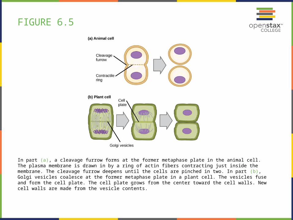
FIGURE 6.5
In part (a), a cleavage furrow forms at the former metaphase plate in the animal cell. The plasma membrane is drawn in by a ring of actin fibers contracting just inside the membrane. The cleavage furrow deepens until the cells are pinched in two. In part (b), Golgi vesicles coalesce at the former metaphase plate in a plant cell. The vesicles fuse and form the cell plate. The cell plate grows from the center toward the cell walls. New cell walls are made from the vesicle contents.
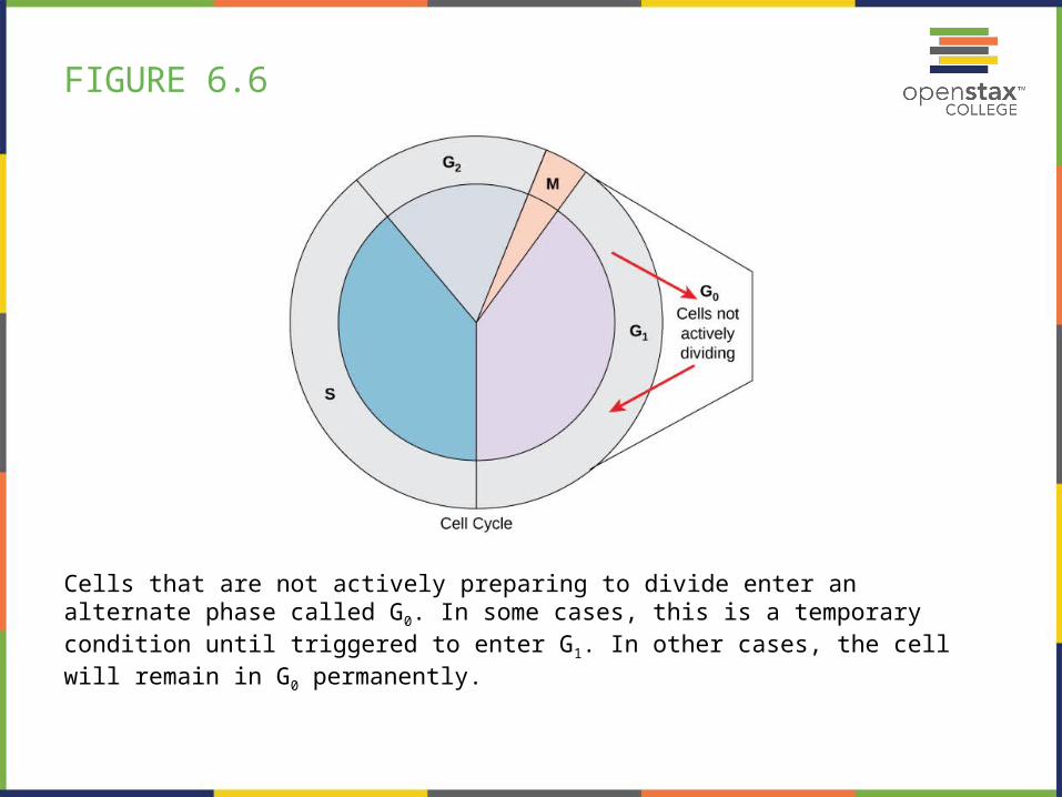
FIGURE 6.6
Cells that are not actively preparing to divide enter an alternate phase called G0. In some cases, this is a temporary condition until triggered to enter G1. In other cases, the cell will remain in G0 permanently.

FIGURE 6.7
The cell cycle is controlled at three checkpoints. Integrity of the DNA is assessed at the G1 checkpoint. Proper chromosome duplication is assessed at the G2 checkpoint. Attachment of each kinetochore to a spindle fiber is assessed at the M checkpoint.

FIGURE 6.8
(a) The role of p53 is to monitor DNA. If damage is detected, p53 triggers repair mechanisms. If repairs are unsuccessful, p53 signals apoptosis.
(b) A cell with an abnormal p53 protein cannot repair damaged DNA and cannot signal apoptosis. Cells with abnormal p53 can become cancerous. (credit: modification of work by Thierry Soussi)

FIGURE 6.9
The binary fission of a bacterium is outlined in five steps. (credit: modification of work by “Mcstrother”/Wikimedia Commons)

This PowerPoint file is copyright 2011-2013, Rice University. All Rights Reserved.