Embed Size (px)
Citation preview

Operational Guide
Companion Animal Zoonotic Diseases


©2010 American Humane Association Copyright Notice: In receiving these Operational Guides in electronic file format, the Recipient agrees to the following terms: 1) Recipient will not post the electronic copy on any internet or intranet websites, 2) Recipient will not distribute electronic copy(ies), and 3) Recipient will not edit the contents of the Operational Guides received and will not incorporate content into Recipient’s written shelter materials without obtaining additional copyright permission from American Humane. The printing or distributing of copies outside the Recipient’s organization without the prior written consent of American Humane constitutes a violation of U.S. copyright law and the limited-use permission implicit in the receipt of the electronic files.
© 2010 American Humane Association i Operational Guide for Animal Care and Control Agencies: Companion Animal Zoonotic Diseases

ii © 2010 American Humane Association Operational Guide for Animal Care and Control Agencies: Companion Animal
Zoonotic Diseases
About the Author
Jim Babbitt, DVM Senior Veterinarian, San Diego Humane Society & SPCA

Table of Contents Introduction ............................................................................................................................... 1
Transmission ......................................................................................................................... 1 Shelter Design ....................................................................................................................... 3 Animal Entry ......................................................................................................................... 4 Cleaning and Disinfection..................................................................................................... 5 Education About Zoonotic Diseases ..................................................................................... 8
Major or Common Zoonotic Diseases .................................................................................... 10 Animal Bites and Scratches ................................................................................................ 10 Bubonic Plague ................................................................................................................... 10 Camphylobacter .................................................................................................................. 11 Other Names ....................................................................................................................... 11 Cat Scratch Fever ................................................................................................................ 12 Cutaneous Larval Migrans .................................................................................................. 13 Giardiasis ............................................................................................................................ 14 Lyme Disease ...................................................................................................................... 15 Psitticosis ............................................................................................................................ 16 Rabies .................................................................................................................................. 17 Ringworm ........................................................................................................................... 18 Salmonellosis ...................................................................................................................... 21 Scabies ................................................................................................................................ 23 Shigellosis ........................................................................................................................... 23 Toxoplasmosis .................................................................................................................... 24 Visceral Larva Migrans ....................................................................................................... 25
Less Common Zoonotic Diseases ........................................................................................... 27 Anthrax ............................................................................................................................... 27 Babesiosis ........................................................................................................................... 27 Brucellosis........................................................................................................................... 28 Colibacillosis....................................................................................................................... 29 Cryptosporidosis ................................................................................................................. 29 Dipylidiasis ......................................................................................................................... 30 Cause ................................................................................................................................... 30 Symptoms in Animals ......................................................................................................... 30 Echinococcosis .................................................................................................................... 30 Other Names ....................................................................................................................... 30 Transmission ....................................................................................................................... 30 Ehrilicosis ........................................................................................................................... 31 Encephalitozoonosis ........................................................................................................... 32 Hantavirus ........................................................................................................................... 32 Leptospirosis ....................................................................................................................... 33 Newcastle Disease .............................................................................................................. 33 Other Names ....................................................................................................................... 33 Q-Fever ............................................................................................................................... 34 Rocky Mountain Spotted Fever .......................................................................................... 35 Sporotrichosis ..................................................................................................................... 35
© 2010 American Humane Association ii Operational Guide for Animal Care and Control Agencies: Companion Animal Zoonotic Diseases

iii © 2010 American Humane Association Operational Guide for Animal Care and Control Agencies: Companion Animal
Zoonotic Diseases
Streptococcosus................................................................................................................... 36 Trichostrongylus ................................................................................................................. 36 Tularemia ............................................................................................................................ 37 Tuberculosis ........................................................................................................................ 37
Rare, Emerging, or Questionable Zoonotic Diseases ............................................................. 39 Bordetellosis ....................................................................................................................... 39 Leishmaniasis ...................................................................................................................... 39 Rat Bite Fever ..................................................................................................................... 40 West Nile Virus................................................................................................................... 40 Treatment in Animals ......................................................................................................... 40
Glossary .................................................................................................................................. 42 Diseases Carried by Different Species of Animals ................................................................. 43
Reptiles ............................................................................................................................... 43 Rodents ............................................................................................................................... 43 Carnivores (dogs and cats) .................................................................................................. 43 Rabbits ................................................................................................................................ 43
Zoonotic Disease Fallacies ..................................................................................................... 44 References ............................................................................................................................... 45
General References ............................................................................................................. 47 Rabies Protocol Chart .............................................................. Error! Bookmark not defined.

Introduction A zoonotic disease or zoonoses is defined as an infectious disease shared by people and animals. In general, the risk of acquiring a zoonotic disease in a shelter is low. In fact, healthy people who contract most zoonotic diseases will be asymptomatic or only experience very mild symptoms. However, education about these diseases and ways to reduce the risk of transmission is important for a number of reasons. First, animals coming into a shelter are more likely to have or to acquire a zoonotic disease. Uncertain (or non-existent) prior veterinary care, prior history of roaming or scavenging, and uncertain temperament put animals coming into our shelters at greater risk for zoonotic diseases than the general population of pets. Second, as the number of immune-compromised people in our population increase, these diseases take on added importance. Immune-compromised people (e.g., cancer patients, organ transplant patients, HIV patients, people receiving large doses of glucocorticoids, pregnant women, those with chronic alcohol malnutrition, splenectomized people) often experience more serious symptoms with these diseases and are generally more susceptible to such illnesses. Third, any outbreak of zoonotic disease associated with a shelter creates these possible consequences:
• Members of the shelter staff or general public become ill or even die.
• The shelter attracts unfavorable publicity. News media may be quick to publicize or even
sensationalize any human disease outbreak traced back to exposure to shelter animals.
• Staff morale suffers, not only due to human illness, but also by the possible need to destroy animals suspected in an outbreak.
• Legal implications complicate the situation. The potential legal liability greatly increases if you cannot show you have made every effort to minimize the possibility of a human contracting a zoonotic disease in your shelter.
• Monetary losses put the shelter in financial danger. This might result from the direct costs of dealing with an outbreak, the loss of staff time due to illness, and even decreased contributions as a result of unfavorable publicity.
Transmission Zoonotic diseases can be transmitted from animals to humans in two ways:
1. Direct transmission requires immediate, or close, contact between the reservoir (sick animal) and the susceptible individual (person). This contact may also be with body secretions (i.e., blood, urine, stool, saliva) from a sick animal.
2. Indirect transmission of a zoonotic disease occurs when a susceptible person comes into contact with the disease-causing organism (“germs”) on objects (animate or inanimate) that serve merely as passive carriers of the disease (commonly referred to as “fomites” or “vehicles”). In an animal shelter, human hands are the most important fomite. By coming into contact with germs on
© 2010 American Humane Association 1 Operational Guide for Animal Care and Control Agencies: Companion Animal Zoonotic Diseases

a sick animal, in body secretions from a sick animal, or on inanimate objects (like cage doors), our hands become contaminated with these germs. The germs are then transferred to another animal, our own mouths or eyes, or to another person.
While we cannot usually control what diseases “walk through” our front doors, once that diseased animal is inside, we can influence the outcome of that disease. This influence is not only important for the sick animal but also for any other animals or humans exposed to that original sick animal. Many factors influence the likelihood of disease transmission from animals to people. We must look at these factors because any action taken in direct response helps to reduce the potential of disease transmission. These same factors also influence the spread of other diseases between animals (non-zoonotic diseases). Any steps we take to minimize the possibility of zoonotic diseases automatically reduce the possibility of transmission of non-zoonotic diseases (i.e., URI, canine and feline distemper, ringworm, canine parvovirus) between animals. These factors control the probability of disease transmission from animals to people (or other animals):
• How long the animal sheds (passes) the disease-causing germs in question. Obviously, the longer the germs are being passed, the greater are the chances of a susceptible individual being exposed to them. Treatment for the disease in question may help to reduce this time.
• The length of the incubation period for the disease in question. The
incubation period is the time between exposure of a susceptible individual and the appearance of symptoms. The longer this is, the more likely that a diseased animal will escape disease detection when it enters into our shelter. The animal may already be in the general population before it starts to show symptoms.
• How obvious the symptoms of the disease are in the animal. The more subtle the symptoms, the more likely it is to be overlooked until exposure of people or other animals has already taken place. An example of this is ringworm (superficial dermatophytosis). Often the first sign of a problem in the shelter is a volunteer or employee who develops symptoms of ringworm infection.
• The stability or resistance of the germ that causes the disease. A germ that is extremely resistant to environmental factors or disinfection remains viable for longer periods of time in our shelters.
• Population density of animals within our shelters. The more animals we have per cubic foot, the easier it is for diseases to spread directly from animal to animal. It is also more stressful on the indivi-dual animals, which make them more susceptible to disease in the first place. Another, more subtle, challenge is that large, dense populations of animals make it more difficult for staff to evaluate individual animals for the initial signs of disease.
• Husbandry practices. How well you are able to clean, feed, and house the animals either reduces or
2 © 2010 American Humane Association Operational Guide for Animal Care and Control Agencies: Companion Animal
Zoonotic Diseases

increases the likelihood of disease transmission.
• Control of pests like fleas and ticks. Some diseases are only spread by the bites of these parasites. These pests also may debilitate the animals making them more susceptible to disease.
We will look at each step of an animal’s journey through an animal shelter and look at some things we can do to reduce the danger of disease transmission to our staff, volunteers, adopters, and animals. In general, we strive to achieve two main goals — first, to reduce the disease load in the shelter (decrease the number of diseased animals and reduce the severity and duration of the disease in those animals that are sick); second, to reduce exposure by susceptible individuals to the germs that cause these diseases. Again, keep in mind that although we are specifically discussing prevention of zoonotic diseases, these steps are exactly the same as those to prevent spread of diseases between animals. Remembering this fact justifies the expense and difficulty of implementing a program to prevent rarely seen zoonotic diseases.
Shelter Design Before an animal starts its journey through the shelter, consider the overall design and layout of the physical facility. Obviously available finances play a role, but even relatively minor changes in the structure, materials used, and furnishings influences the ease or difficulty of disease transmis-sion. Available resources on this subject include the American Humane Associa-tion’s Operational Guide for building design. However, here are some of the specific, zoonoses-related building concerns:
Isolating known or suspected ill animals. The more separation the better, but even if ill animals must be mixed (e.g., ringworm cats mixed with URI cats), it is better than mixing well individuals with ill ones. The longer the isolation lasts the better. Isolation or segregation of animals according to age is also important. Do not house kittens or puppies with adults. Minimizing stress on all animals. Separating dogs and cats, minimizing noise pollution, allowing an animal to occupy the same cage throughout its stay at the shelter (to minimize “moving” stress), and creating areas in the runs or cages that allow an animal to “hide” are just some of the considerations that may influence the type and location of cages and wards. For example, having cages for cats or dogs that can be cleaned without having to handle or move them (double sided condos for cats or guillotine runs for dogs) can be helpful. Having rooms, cages, and countertops that are easy to clean and disinfect. Avoid, therefore, carpeting, non-washable bedding, or non-washable toys. While non-porous surfaces meet this criteria, these surfaces are also rather “sterile” looking and often not very appealing when the animal is on display to the public. Sometimes compromises need to be made — weighing the needs of disease prevention against the need to make our animals as appealing as possible to potential adopters. Providing barriers to minimize the direct transmission of diseases. These barriers may be separate rooms (better to have numerous smaller rooms than one large one). On the other hand, barriers to minimize aerosolization of germs can be nothing more than hanging towels over
© 2010 American Humane Association 3 Operational Guide for Animal Care and Control Agencies: Companion Animal Zoonotic Diseases

cage fronts or the use of “sneeze guards” that cover the front of cages or runs. Use distance as an effective barrier. Place facing cages at least 5 feet apart. Do not put animals with infectious problems (like diarrhea) in cages that are over other cages. Monitoring the symptoms of disease. If you house 15 kittens in a room and find severe diarrhea in the litterbox in the morning, how do you determine which kitten(s) is involved? Can you separate them if needed? How do you determine who is eating and who isn’t? Sometimes confining animals at specific times (nights or at feeding time) and then housing them communally can help. Enhancing staffs ability to monitor animals for disease then communicating this information to the veterinary staff or other staff members. This may be as sophisticated as a computer system or as simple as a chalkboard in a central location. The use of quarantine or contagious signs on cages or wards is essential. Ensuring adequate air ventilation. In general, the more air circulation the better. This helps to ensure a drier environment, minimizes the number of germs per cubic foot of air in the rooms, and minimizes odors and materials in the air that might irritate the mucus membranes of the animals. The ideal is 10 to 15 exchanges of air per hour, but any airflow is better than nothing. Certainly, centralized heating and air conditioning systems, with proper ventilation, are ideal, but just opening a window or having outdoor access can help. The use of fans is controversial due to risks of driving airborne disease from one area to another, but certainly they can increase airflow and
dry the environment. Using a fan to direct the flow of air outward through a window is better than directing the flow of air inward. Directing traffic flow. Ideally, shelter traffic progresses from well animals to sick animals. The less that staff or animals must move from the “sick” wards or cages to the “well” wards or cages the better. Isolate any areas used for known zoono-tically ill animals from day-to-day shelter traffic and designate them as staff-only areas, keeping them off limits to the public and volunteers. Clearly mark these areas as containing a known ill animal.
Animal Entry The first step in an animal’s journey through our shelters is initial entry. Examine incoming animals as soon as possible. Ideally, do this before placing them in the general population. While an extended quarantine period is ideal, it is not possible in most shelters. Therefore, educate staff about the symptoms of common zoonotic diseases. Run appropriate tests on any animals with suspicious disease signs. Unfortunately, there are no simple, accurate tests for many of these diseases. If an animal shows symptoms possibly associated with a zoonotic disease, isolate it away from all other animals. Also, place some form of identification (i.e., collar, microchip) on the animal so that staff can positively identify it at any time for examination or follow-up during its stay in the shelter. Administer all appropriate vaccines and treatments to all healthy animals as soon as possible. These include routine treatment for external and internal parasites. Use some form of reminder or notification system to alert staff to any
4 © 2010 American Humane Association Operational Guide for Animal Care and Control Agencies: Companion Animal
Zoonotic Diseases

animals that need a repeat treatment or vaccine in the future. Vaccines are important, but it is critical to realize that vaccines are of limited value in any animal exposed to disease prior to coming into the shelter or exposed within a short period of time after coming into the shelter. Empha-sis on husbandry and stress reduction will yield far better results in disease reduction than using a different or “better” vaccine. Set up a system of rechecks and treatments for any animals suspected of being sick. Isolate any sick animal at least until the symptoms of the disease disappear. In some diseases, keep animals isolated for some time after symptoms disappear. How effectively you isolate sick animals will depend on your facility. Put a system in place to notify other staff members and the veterinary department about animals that may be exhibiting symptoms of a zoonotic disease. Do it in a way that other people who are not as informed about the dangers of disease transmission, like volunteers, can understand the danger and the need to avoid contact with that animal or that ward. You can use signs, a centralized chalk or “Dry Write” board, or a computer system. Allow an animal some time to “de-stress” before doing any major procedures, such as surgery. The amount of time allowed depends on age, species, temperament, population of animals in shelter, and the ability of a shelter to house animals for extended periods. Assuming an animal arrived healthy when it first came into the shelter, its care and treatment after it enters is crucial to its ability to resist disease.
How do staff members monitor the animal while it is in the shelter? Educate staff about the zoonotic disease symptoms. Also, remember that many animals affected with zoonotic diseases show no or very few symptoms. That means staff must take all the described steps with all animals entering the shelter. Weighing animals on a regular basis often gives an early warning signal to the presence of disease or even stress. This is especially important in puppies, kittens, and in animals kept in a communal setting. Keep weight measurements in a log or on the animal’s individual cage card or record.
Cleaning and Disinfection Clearly spell out the shelter’s cleaning and disinfection protocols, then educate staff on the proper use of chemical cleansers and disinfectants. Four levels of cleaning are possible: physical cleaning, sanitation (bringing the level of germs present to a safe level, where most exposed animals will not become sick), disinfection (killing most germs present, including all viruses, bacteria, or fungi), and sterilization (killing of all life forms, including bacterial spores, fungal spores, and parasite eggs). While we strive for true disinfection, good sanitation is the best that we accomplish in most shelter environments. Follow these general techniques: Proceed in cleaning of cages and wards from “clean” rooms or cages to “dirty” (germ contaminated). Ideally, the personnel that clean a room with a known ill animal should not clean or even handle the well animals. Remove all toys, dishes, blankets, and such. Launder or dispose of these objects as appropriate. Use of
© 2010 American Humane Association 5 Operational Guide for Animal Care and Control Agencies: Companion Animal Zoonotic Diseases

disposable litter boxes, bowls, and the like facilitates this process. Wash all towels and bedding in the hottest water available. Although laundering these materials does not disinfect them, it does mechanically remove most germs present. Remember to clean all objects that an animal comes into contact with; this includes collars, leashes, carriers, muzzles, and scales. If you use mops, remove the heads frequently, launder them and then allow them to dry thoroughly between uses. Alternating mop heads can be useful and inexpensive. Using disposable mops (e.g., Swiffer) can be very useful, although expensive. The ideal situation is to make your supplies area specific. That is each area has its own set of brushes, blankets, and bowls. This way, healthy animals are not exposed to sick or isolated animals even indirectly. The first level of cleaning is the most important and unfortunately the most overlooked in many shelters. This is the physical removal of waste like stool, urine, hair, skin dander, or nasal secretions. None of the other steps in the cleaning or disinfection process will be effective if this is not done first. In fact, many chemicals used for disinfection are not effective in the face of organic waste material. In addition, many of the disease causing germs we face are resistant to common disinfectants. Ultimately, perform physical cleaning without further contaminating the environment. For example, do not just spray the material with a high-pressure hose to move it out of the kennel or cage. This just aerosolizes the material contaminating walls, cage doors, and the air. Ideally, pick it up with a shovel or scoop; then wet down the run down with water and squeegee the material down to the drain. After removing large waste
material, wash the area well with soap and water. Rinse very well and squeegee the water down the drain. Do not allow pools of standing water to just air dry. This merely redeposits suspended contaminants on the floor. Standing pockets of water also dilute many of disinfectants beyond the concentration required to be effective. Do not use the same cloth to dry different cages or runs. Ideally use paper towels for this purpose. After the floor is dry, apply a chemical agent to sanitize the surfaces. The specific agent used depends on the type of disease that may have been present in that area, the type of surface material present (non porous vs. porous), and training of staff performing the task. For all disinfectants, contact time and dilution are important. Because time is always at a premium in most of our shelters, in a common day-to-day setting it is often difficult to allot the time for these agents to “sit.” However, if a known outbreak of a zoonotic disease takes place, allow this time. Most disinfectants used in animal shelters require a minimum contact time of 10 minutes; more time is required if the water is hard or the temperatures are cold. Quaternary Ammonium Compounds (“Quats”) such as Roccal®, Parvosol®, and A33®, are commonly used. They are less corrosive than bleach, less irritating to mucus membranes, good at controlling odors, and in general more “forgiving” concerning technique of usage than other disinfectants. Quats are not effective at killing non-enveloped virus agents like Parvovirus (or at least not very effective). Dilute at 1:32 or 1:64 for best results in a shelter environment. Bleach (sodium hypochlorite) diluted at 1:32 is probably the most common
6 © 2010 American Humane Association Operational Guide for Animal Care and Control Agencies: Companion Animal
Zoonotic Diseases

disinfectant used today. It is effective against most of the germs present in a shelter environment. When used in a ringworm-contaminated area, dilute it at 1:10. Bleach is inexpensive and easy to use, but there are some important limitations and drawbacks. First, it is NOT effective in the presence of organic material. Very small amounts of organic material (like stool, hair, skin dander) will render it less or even non-effective. It also requires a minimum of 10 minutes contact time for it to be completely effective (30 minutes for ringworm). Rinse it thoroughly with plain water. It is very corrosive to metals and very irritating to mucus membranes. Once diluted, it is only effective for 24 hours. Train staff thoroughly in its usage. Bleach is often used in footbaths, but it is relatively ineffective because the bath quickly becomes contaminated with organic material. Potassium peroxymononsulfate (Trifectant®, VirkonS®) is a relatively new class of disinfectant. These have all the advantages of Quats, but they also are reported to kill non-enveloped viruses like Parvovirus. It is also effective in the face of moderate amounts of organic material, so its use in footbaths would seem to be effective. It is not, however, effective against ringworm spores. The most overlooked, but perhaps single-most-important objects to wash and clean well in shelters, are the hands of everyone involved with caring for and handling animals, including prospective adopters. Numerous studies have shown how easy it is for germs to not only survive on our hands, but to easily passed from one animal to another or from an animal to us or another person. This is especially important with children. It is important to
wash often and thoroughly. This is much more readily accomplished with plentiful and convenient sinks. The actual scrubbing of our hands is the important step, so it is not as important to use a “disinfectant” soap. Wash your hands in between each animal or at least in between each ward where you work, especially after handling known ill animals. Using liquid soap dispensers rather than bar soap is best. The use of hand sanitizers is NOT a substitute for washing. These materials rely on alcohol for their disinfection. Hand sanitizers require a 60-second contact time for complete effectiveness, and very few people use enough to have it on their hands for a full minute. In the face of a known zoonotic disease outbreak, the use of personal protection materials like gowns, booties, gloves, and masks can be useful. In general, these materials are too cumbersome and time consuming in a day-to-day setting. Instead, emphasize regular hand washing. Take these additional steps to minimize the spread of zoonotic diseases:
• Use separate refrigerators for human food and animal supplies (e.g., dog and cat food, veterinary supplies, laboratory materials, fecal samples).
• Do not allow eating, application of cosmetics, or applying contact lens in animal areas. In addition, these activities should also be done only after thorough hand washing.
• Do not use your mouth as a “third hand.”
© 2010 American Humane Association 7 Operational Guide for Animal Care and Control Agencies: Companion Animal Zoonotic Diseases

Education About Zoonotic Diseases The final step we hope most animals take in their journey through the shelters is adoption, so you must inform potential adopters what steps to take to minimize the possibility of exposure to zoonotic diseases from their new pet. This can be difficult to do without frightening some people and requires extensive staff training and preparation. Therefore, an organized and ongoing program of education including manuals, training sessions, informative signs, and a system of evaluation is very important. This step is not only important because it helps minimize the spread of diseases between animals and to people but also if your shelter is ever subjected to an OSHA review or worse, media scrutiny after an incident of zoonotic disease in your community. Direct the first stage of your education program toward staff and include the following:
• Usual symptoms of common zoonotic diseases in animals. Early recognition of diseases in animals minimizes the number of people exposed. Early treatment of some diseases may also shorten the amount of time an animal is contagious.
• Steps taken to minimize the transmission of these diseases within the shelter. Include those steps taken every day in the shelter to minimize disease transmission, but also discuss specific extra steps to be taken in the face of an outbreak of zoonotic disease.
• Symptoms of specific diseases in humans. Staff members need to recognize any problem early and know how to seek appropriate care.
Remember that early detection is a must, not only for the welfare of the people involved, but because there are many diseases that are not only contagious from animals to people but also from people to animals. In other words, an infected staff member can spread the disease to other people and to other animals. An example of this is common ringworm (superficial dermatophytosis).
Direct the second stage of your education program toward the public. Educating the public about zoonotic diseases is not only useful for preventing diseases, but it may also boost adoptions. There is a great deal of misinformation concerning zoonotic diseases and the “danger” of pet ownership. We must honestly inform the public of the risks and emphasize that in the vast majority of cases, the proven benefits of pet ownership outweigh the risks. When formulating a program of public education, try to not direct it to any one group of people (i.e., immune-compromis-ed people) but rather direct efforts to the population as a whole. Certainly, when we realize that immune-compromised people (e.g., cancer patients, HIV patients, organ transplant recipients, or splenectomized patients) are at more risk, we can alleviate some of their fear. However, the basic steps of zoonotic disease prevention are the same for everyone and should be emphasized as such. Include printed materials in your public education program, such as putting them into adoption packets. The program should also involve training adoption counselors to properly answer basic questions about
8 © 2010 American Humane Association Operational Guide for Animal Care and Control Agencies: Companion Animal
Zoonotic Diseases

zoonotic diseases. When formulating your program, keep the following in mind:
• Keep it basic and simple. • Make sure it is up to date. • Emphasize to the staff that any
information a person shares with them is confidential.
• Emphasize the benefits of pet ownership, but do not downplay the risks.
• Above all, do not offer veterinary or medical advice. Refer people to their own personal physician or veterinarian for specific informa-tion and recommendations.
When formulating your program, consider seeking the advice of your local occupational health agency or the state or local public health agencies to help prepare materials.
Suggested Diseases to Include Information on in Adopter Information Packets These diseases are so common and/or public knowledge (misconceptions?) is so widespread that having printed information in your standard adoption packs would be helpful to both the public and to your adoption counselors.
• Fleas • Ticks • Visceral Larval Migrans
(roundworms) • Cutaneous Larval Migrans
(hookworms) • Tapeworms • Rabies • Ringworm
© 2010 American Humane Association 9 Operational Guide for Animal Care and Control Agencies: Companion Animal Zoonotic Diseases

Major or Common Zoonotic Diseases There are literally hundreds of diseases classified as zoonotic. It is beyond the scope of this manual to cover even a significant percentage of them. Therefore, the diseases covered here are common, have received some degree of publicity, or experience some degree of public awareness. In other words, you may get questions concerning them. Check the References appendixes for sources of more detailed information concerning zoonoses. Disease coverage is divided into three parts.
1. Those diseases considered most important in the management of a shelter. This might be because of the frequency of the disease itself, because of the degree of public awareness of the disease, or because of the potential serious-ness of any outbreak.
2. Those diseases that have been seen with reasonable frequency and for which a definite zoonotic link has been established.
3. Emerging or new diseases
Animal Bites and Scratches
General Facts • Certainly, this is one of the most
common sources of zoonotic diseases. • Many different bacteria can be
involved (Pasteurella, Capnocytophaga).
• Depth of wound, location of wound, and type of animal involved are all factors that determine the severity of infection.
Symptoms in Animals N/A
Symptoms in People • Swelling, pain, drainage, loss of
function of area of body bitten, fever • Septicemia (“blood poisoning”); can
be life threatening
Prevention or Control • Use of protective equipment like
gloves, catch poles, squeeze cages, and nets
• Proper design of cages and runs to facilitate restraint of animals
• Euthanasia of aggressive animals • Education concerning proper
techniques of animal handling • Education concerning proper methods
of reading animals’ body language • Promptly and thoroughly cleaning all
wounds with antiseptic soap and copious amounts of water
• Seeking medical attention for all but the most superficial wounds
• Keeping tetanus vaccinations up to date for all staff members
References: 3
Bubonic Plague
Other Names Plague, Black Death, Pneumonic Plague
Cause Yersinia pestis (bacteria)
Transmission • Primary source of exposure of humans
and animals is through direct contact with rodents or more importantly the rodent flea. A rodent flea that is infected can carry the bacteria for months. Rats are the most important reservoir, but in Southwest United States, ground squirrels and prairie dogs are very important reservoirs.
10 © 2010 American Humane Association Operational Guide for Animal Care and Control Agencies: Companion Animal
Zoonotic Diseases

• Rodent fleas can also be present on dogs, cats, rabbits, and other warm-blooded animals.
• Airborne droplets that originate from coughing or sneezing can spread pneumonic form of the disease. This can occur from human to human or from animal to human.
• Cats can become infected from ingestion of an infected rodent.
Symptoms in Animals • Dogs usually have brief self-limiting
illness. • Rodents may rapidly die, or more often
are asymptomatic. • Cats have fever, lymph gland swelling,
abscesses (from lymph glands), and often rapidly die.
Symptoms in Humans • Lymph gland swelling, fever,
pneumonia • Development of black patches of skin
that die (“Black Death”)
Diagnosis in Animals • It can be suspected based on clinical
signs, especially swollen lymph glands and fever, but must be positively diagnosed using laboratory tests.
• Only qualified personnel should handle any animal that is suspected of plague when diagnostic tests are attempted.
• Microscopic examination of impression smears from aspirates of infected lymph nodes show large numbers of the bacteria (“safety pin” appearance).
• Blood tests • Cultures from aspirates or draining
abscesses
Treatment of Animals Although various antibiotics can be used, because of the extreme danger these animals pose to humans, it is very questionable to attempt treatment in a shelter setting.
Prevention or Control • Flea treatment in all animals admitted
into the shelter. Advantage® is one flea treatment safely used in a wide variety of animals seen in a typical shelter.
• Avoid taking wild rodents into the shelter.
• Prevent dogs and especially cats from hunting or coming into contact with wild rodents.
• Report any confirmed cases to your local public health authorities.
• Recommend that all people with even casual contact with animals diagnosed with plague see their physicians immediately.
• Vaccines for humans are available but are not widely used or recommended.
References: 5-6
Camphylobacter
Other Names Vibriosis
Cause Camphylobacter jejuni (bacteria)
Transmission • Most human cases are of unknown
origin, but ingestion of undercooked meat is the most common identified cause.
• Exposure to the stool of infected dogs and cats, especially kittens and puppies, can be involved.
© 2010 American Humane Association 11 Operational Guide for Animal Care and Control Agencies: Companion Animal Zoonotic Diseases

• Numerous cases of human infection from aerosolized bacteria have occurred in shelters, zoos, or reserves. These result primarily from using high-pressure hoses to clean or flush stool from cages or runs.
• Animals can shed the bacteria in their stool for weeks to months after their active infection has resolved.
• Camphylobacter is a very common cause of diarrhea in puppies and kittens in a high-stress, crowded environment like animal shelters.
Symptoms in Animals • Diarrhea, especially in young animals,
the nature and duration of which can vary widely
• Adult animals are often asymptomatic carriers.
Symptoms in Humans • Acute gastrointestinal illness — the
classic “food poisoning” • Diarrhea, vomiting, abdominal pain • Usually of short duration and self
limiting in humans with normal immune systems
Diagnosis in Animals • Culture of bacteria by qualified
laboratory, requiring special techniques and conditions
• Probably not indicated in individual cases of diarrhea, but may be useful in widespread outbreaks within a shelter
Prevention or Control • Good hygiene (Wash your hands!) • Physically pick up fecal matter and
dispose of before cleaning wards or cages.
• Do not hose runs or cages with high-pressure devices before fecal matter is physically picked up.
• Thoroughly cook all meat eaten by humans or animals.
References: 6-7
Cat Scratch Fever
Other Names Cat Scratch Disease
Cause Bartonella hensale (bacteria)
Transmission • Exposure to infected cat by bite or
scratch wound. Licking of a pre-existing wound.
• Bite or scratch from other animal (rodents).
• Some cases have no history of exposure to cats or other animals.
• Cats are exposed to the bacteria through the bite of an infected flea.
• Ticks and ear mites may also play a role in cat-to-cat transmission.
Symptoms in Animals • Most cats (maybe all of them) are
asymptomatic. • The bacteremia (active infection) is of
short duration (10-14 days). This is the only time that the cat is contagious to humans.
Symptoms in Humans • Pustule develops near bite or scratch
wound within 10 days after bite occurs. This can persist for one to two weeks.
• Swollen lymph glands develop 14-21 days after bite or scratch occurs.
• Most cases resolve within two to four months.
• Some people can develop lesions in their eyes or central nervous systems.
12 © 2010 American Humane Association Operational Guide for Animal Care and Control Agencies: Companion Animal
Zoonotic Diseases

Diagnosis in Animals Blood tests (serology) can detect antibodies to the bacteria, but this only determines that the cat has been exposed previously. Approximately 65 percent of the cats in the United States test positive. Cats only become seropositive (develop antibodies) after the bacteremia is resolved — in other words, after they are no longer contagious.
Treatment in Animals • No effective treatment has been
shown, but antibiotics may be useful. • Cats are rarely treated because disease
is asymptomatic and self limiting.
Prevention or Control • Wash hands after handling cats. • Avoid bites and scratches. Wash
wounds quickly if they do occur. • Flea control is very important. • By the time disease is recognized in
humans, the cat is no longer contagious; in other words; it is not necessary to get rid of the cat.
• Declawing the cat has been shown to not reduce the incidence of the disease.
References: 8-9
Cutaneous Larval Migrans
Other Names Creeping Eruptions
Cause The larva of the dog or cat hookworm penetrates the skin of humans and migrates under the surface of the skin (Ancylostoma brazilienze, Ancylostoma caninium).
Transmission • Direct skin contact with the larva of
hookworms, usually from the soil. • Seen primarily in warm climates (The
Southeast United States has much higher incidence than the rest of the country.)
• Seen mostly in children. • Areas with moist sandy soil
(playgrounds, sandboxes, beaches) are often heavily contaminated with hookworm larva.
Symptoms in Animals • Most animals are asymptomatic or
only occasionally show symptoms • Diarrhea, often with blood • Anemia, especially in puppies or
kittens • Sudden death, especially in puppies or
kittens
Symptoms in Humans • Inflamed tracts under the skin that are
intensely pruritic (itchy). These usually start near or on the feet. They are linear and move 2-3 mm per day.
Diagnosis in Animals Microscopic examination of the feces. A centrifuged Zinc Sulfate flotation is much more accurate that a routine “fecalizer” type of examination. Commercial laboratories usually perform these Zinc Sulfate floatations. Remember that a negative floatation test does not eliminate the possibility that the animal has hookworms.
Treatment in Animals • Pyrantel (Strongid®) or fenbendazole
(Panacur®) are very effective worming medications.
• Routinely administer worming medications to all animals coming into
© 2010 American Humane Association 13 Operational Guide for Animal Care and Control Agencies: Companion Animal Zoonotic Diseases

the shelter upon entry. Repeat medication at two- to three-week intervals in puppies and kittens until they are 16 weeks of age.
• Preventative medications are commonly contained in the medications given for heartworm prevention and in some flea medication (Revolution®).
Prevention or Control • Wear shoes, and encourage children to
do so. • Pick up the stools of all animals
quickly and dispose them appropriately.
• Avoid walking (especially barefoot) in areas where many dogs or cats have defecated like “dog beaches” or “dog parks.”
• Prevent dogs and cats from using public areas to defecate in (school yards or parks).
• Cover sandboxes when not in use. • Provide good public education as to
the danger of allowing dogs and cats to free roam.
• Provide good public education to stress the importance of picking up after their animals.
References: 10
Giardiasis
Other names Beaver Fever
Cause Giardia lamblia (protozoa)
Transmission • Giardia is the most common protozoan
parasite of humans. • Many animals are capable of being
infected and passing cysts in their
stool, including dogs, cats, birds, horses, and cattle.
• It is most commonly contracted by ingesting water or food contaminated with cysts of Giardia.
• Humans are considered the natural host for Giardia, so human to animal transmission can also occur.
Symptoms in Animals • Diarrhea, especially in puppies and
kittens, possibly severe enough to cause weight loss and dehydration
• Adults are often asymptomatic.
Symptoms in Humans • Diarrhea of varying degrees of severity
and duration • Abdominal cramps and discomfort
Diagnosis in Animals • Giardia ELISA test on stool available
both through commercial laboratories and as an “in house” SNAP test
• Zinc Sulfate centrifuged fecal examination, more accurate than standard “fecalizer” examination
• Giardia IFA test available but not as sensitive as ELISA test
Treatment in Animals • No treatment is universally successful
in preventing the shedding of cysts in the stool. Treatment is effective in minimizing diarrhea or symptoms in infected animals.
• Metronidazole (Flagyl®) or fenbendazole (Panacur®) have been used commonly.
• A vaccine is available that may help to minimize shedding of cysts.
Prevention or Control • Hand washing and good hygiene are
very important.
14 © 2010 American Humane Association Operational Guide for Animal Care and Control Agencies: Companion Animal
Zoonotic Diseases

• Promptly pick up stools in runs and cages.
• Diagnose and treat any animals showing diarrhea, especially kittens and puppies.
• Bathing of animals in addition to treating them when they are infected with Giardia will help to prevent re-infection from feces on their haircoat.
• Quaternary ammonium disinfectants (“Quats”) are very effective in killing the cysts in the environment, but these compounds rapidly lose their effective-ness in the presence of large amounts of organic matter. Therefore, phys-ically picking up all fecal matter prior to disinfection is essential.
References: 11-13
Lyme Disease
Other Names Lyme arthritis
Cause Borrelia burgdorfei (spirochete bacteria)
Transmission • Tick bite • Ixodes (“Deer Tick”) mainly involved,
but the germ has been found in virtually all common tick species.
• The disease is not spread directly from animals to humans. Animals can bring ticks into areas of human habitation, which increases the exposure risk for people.
• Lyme Disease is now the most common tick transmitted disease in the United States.
Symptoms in Animals • Cats are usually asymptomatic.
• Dogs can have fever, lethargy, or lameness (often “shifting leg” lameness).
• Symptoms often occur months after the tick bite took place.
• In some dogs, serious kidney disease can be seen.
• The arthritis that develops can be progressive and prolonged. Over time it can lead to permanent bone damage within the joints (osteoarthritis).
Symptoms in Humans • Skin rash that develops in the area of
the original tick bite is the prime symptom initially (erythema chronicum migrans).
• Fever, lethargy, and arthritis are common.
• Heart involvement is seen in some humans.
• Symptoms are often chronic waxing and waning.
Diagnosis in Animals • Animals usually have elevated
antibody titers (serology), but this only means the animal has been exposed and may or may not be suffering from the disease at this point.
• Clinical symptoms must be present to even consider a diagnosis.
• History of tick exposure • At best, it is a difficult disease to
positively diagnose in animals.
Treatment in Animals • Antibiotics may be useful in early
stages but they need to be given over an extended period of time (30 days minimum).
• Symptoms often recur after the antibiotic treatment.
• NSAIDS like Rimadyl® may help to ease pain of arthritis.
© 2010 American Humane Association 15 Operational Guide for Animal Care and Control Agencies: Companion Animal Zoonotic Diseases

Control or Prevention • Vaccines for dogs help to prevent
symptoms, but because dogs do not directly transmit the disease to humans, vaccinating the dog does not prevent human infection.
• Tick control on dogs is important. Monthly flea/tick treatments like Frontline® or Advantix® are helpful.
• Prevention of tick exposure in humans. Avoiding areas likely to be tick infested like brushy or forested areas. Wearing long pants and long-sleeved shirts.
• Prompt removal of attached ticks. • Studies in animals have shown that the
risk of disease transmission from an infected tick is very low during the first 24 hours of attachment. The risk is 50 percent after 48 hours of attach-ment. The risk is 100 percent after 72 hours.
• DEET can repel ticks from humans. • Use caution not to squeeze the body of
the tick when removing. Only put traction on the head and neck of the tick to avoid expressing the stomach contents of the tick into the blood-stream of a person.
References: 14-15
Psitticosis
Other Names Parrot Fever, Ornithosis, Chlamydiosis
Cause • Chlamydia psittaci (bacteria) • Possibly other strains of Chlamydia
Transmission • Inhalation of dried feces or respiratory
secretions from an infected bird
• Direct contact with contaminated feces or respiratory secretions
• Mammalian species of Chlamydia (from cats, for example) only rarely spread to humans
Disease in Birds • Many birds may be asymptomatic until
stressed (like coming into a shelter). • Virtually any symptom that an ill bird
can have may be seen. • Respiratory, heart, liver, and gastro-
intestinal problems; arthritis
Disease in Humans • Symptoms typically develop one to
two weeks following exposure. • Fever, malaise, headaches, and
respiratory signs like coughing and pneumonia are seen.
• Heart problems sometimes occur. • Abortion and uterine infections can be
seen in late-term pregnant women. • Symptoms can be life threatening.
Diagnosis in Birds • Clinical signs • Antibody titers (serology) • Reportedly African Greys, cockatiels,
and budgies typically have negative titers even when they are actively shedding the germs.
• ELISA test for germs in the stools of the infected birds are reliable but should be run by qualified laboratory.
Treatment in Birds • Antibiotics • In a shelter environment, the decision
to treat is questionable because of the zoonotic potential.
Prevention or Control • Ask all people relinquishing birds,
what the original source of the bird
16 © 2010 American Humane Association Operational Guide for Animal Care and Control Agencies: Companion Animal
Zoonotic Diseases

was. If the bird was bred and/or sold through a reputable dealer in the United States, it is likely certified Psittacosis free. If the bird was purchased overseas (Mexico), it has to be treated as a suspect.
• Only trained personnel that are clothed properly (masks, gloves, gowns) should handle birds suspected of having psittacosis.
• Birds can be treated prophylactically with Chlortetracycline, but it questionable in a shelter setting if this is justified.
References: 16-17
Rabies
Other Names Hydrophobia, Lyssa
Cause Rhabdovirus or Lyssavirus (virus)
Transmission • Bite from an infected animal • Salivary contamination of preexisting
wound • Cases with no history of bite/scratch
transmission are considered very rare. • Most cases that initially of unknown
exposure have a bite wound of which the victim was unaware (especially bites from bats).
• In the United States, few cases involve domestic animals. This is primarily because of the extreme public health measures that have been taken — like mandatory vaccinations of dogs and cats, enforcement of leash laws, and the reduction in the stray dog population.
• The current main reservoirs of rabies are:
o East Coast of United States — Raccoons
o West Coast of United States — Skunks and bats
o Mexico — Domestic dog • Most all warm-blooded animals are
susceptible in varying degrees to rabies, including dogs, cats, horses, cattle, rabbits, rodents, and birds.
• Realistically, most small animals, like rabbits, are unlikely to survive the initial exposure (bites) to rabies. Therefore, they are rarely diagnosed with rabies.
Symptoms in Animals • Central nervous system signs • Most animals exhibit fairly distinct
stages or groups of symptoms, but atypical cases are sometimes seen.
• Prodromal stage lasts two to three days. Usually animals show appre-hension, anxiety, and a change in personality.
• Furious stage lasts one to seven days. Usually animals are restless and show increased response or sensitivity to visual and auditory stimuli. As they become more restless, they start to roam, becoming progressively more irritable and vicious. They start to have seizures and develop muscle incoordination.
• Paralytic or dumb stage lasts two to four days. Usually animals show progressive paralysis starting in the hind limbs and ascending. Often they will show a change in their voices as the vocal cords become paralyzed.
• Death occurs within five to 14 days after the onset of clinical signs.
• Virus is only shed in the saliva during the active stages of the disease. This is why only a 10-day quarantine is
© 2010 American Humane Association 17 Operational Guide for Animal Care and Control Agencies: Companion Animal Zoonotic Diseases

required. (It is possible that the CDC may recommend a longer quarantine in the future, but 10 days is the current requirement.)
Symptoms in Humans • Pain and swelling at bite site • Paresis/paralysis follows, including
paralysis of the muscles of the larynx and the muscles of swallowing, which leads to difficulty and pain on swallowing, hence the name “Hydrophobia,” fear of water.
• Death invariably occurs.
Diagnosis in Animals • Microscopic examination of the
unfrozen brain by a qualified pathologist gives the definitive diagnosis.
• Microscopic examination of skin taken from the muzzle of the dog can give an ante mortem (before death) diagnosis, but is not a substitute for proper quarantine and if needed, brain examination.
• Clinical symptoms
Treatment There is no treatment.
Prevention or Control • Mandatory vaccination of all dogs. • Cats also should be vaccinated for
rabies. • Prompt and vigorous washing of all
bite wounds with soap and water. • Pre-exposure rabies vaccines for all
persons in high-risk jobs (like shelter personnel).
Protocols for Animals Suspected of Exposure to Rabies • Any domestic animal that has been
bitten or scratched by a wild
carnivorous animal or bat that is not available for testing is assumed to be exposed to rabies.
• If the domestic animal is unvaccinated for rabies, either euthanize it immed-iately, or hold it for a six-month quarantine in a licensed facility (like a shelter or veterinary hospital).
• If the domestic animal has proof of current rabies vaccine, home quarantine for 45 days is considered adequate.
• More information, see Rabies Protocol Chart
References: 18-20
Ringworm
Other Names Superficial Dermatophytosis, Fungal Dermatitis
Cause(s) • Microsporum canis (fungus), most
commonly seen in dogs and cats • Microsporum gypseum (fungus),
found living in the soil • Trichophyton mentagrophytes
(fungus), most commonly seen in rodents
Transmission • Direct contact with infected animal,
most common means of transmission. • Contact with fomite or object
contaminated with fungal spores from infected animal
• Spores can live for years in environment.
Symptoms in Animals • Many animals (e.g., dogs, cats,
rodents, horses) are asymptomatic or show only very minimal symptoms
18 © 2010 American Humane Association Operational Guide for Animal Care and Control Agencies: Companion Animal
Zoonotic Diseases

that are difficult or impossible to note on basic physical examination.
• Typically causes patchy hair loss in a roughly circular pattern.
• Hairs break off at the level of the skin, leaving “stubbles” of hair.
• Often hairless areas are scaly, and the skin may be darkly pigmented.
• In young animals (kittens especially) the lesions are most commonly seen around the head, face, and ears.
• It can affect the toenails causing odd toenail growth patterns.
• Most cats do not have pruritis (itching).
• It can cause a wide variety of skin and hair coat lesions, so consider any animal showing hair loss or other dermatitis a ringworm suspect.
• In general, dogs are over diagnosed with ringworm. In other words, most dogs with skin lesions do not have ringworm.
• Cats are under diagnosed with ringworm. In other words, more cats have ringworm than we suspect.
• Longhaired cats (Persians) may be more susceptible to ringworm, or it just may be more difficult to diagnose because of subtle symptoms.
Symptoms in People • Similar symptoms to those described
in animals • Lesions are usually reddened, scaly
and may be pruritic (itchy). • Lesions most commonly occur on
hands, arms, neck, and face. These are the areas that come into contact with the infected animal.
• Often is mild and self-limiting
Diagnosis in Animals • Physical appearance of lesions and the
age and type of animal involved are
important considerations. View any skin lesion on the face of a young kitten as a probable ringworm case until proven otherwise.
• A Woods Light® is an ultraviolet lamp that emits a very specific wavelength ultraviolet light that causes some ringworm lesions to fluoresce. This can be a very valuable, simple screening test, but it is very easy to both under and over diagnose ringworm.
o 50 percent of ringworm cases will not fluoresce, so negative results do not rule out ringworm.
o In those animals that do have fluorescence, it is very important to note that it is the hairs that are important. Fluorescing hairs are significant, not fluorescing skin or scabs. The fluorescence should be a bright “apple green” color.
o It may be helpful to allow the light to warm up for several minutes before using it and to shine it for several minutes on the animal’s hair coat before attempting to read any fluorescence.
o A 110 V Woods Light with a magnifying glass built into it is easier to use than a small battery powered light.
• DTM fungal culture (Dermatophyte Test Media) is the most accurate test, but again it must be used and read properly to be accurate.
• The flat petri dish type of culture media containers is easier to use and probably more accurate that the “jar” type of containers.
o Pluck some of the hairs from the periphery of the lesions and
© 2010 American Humane Association 19 Operational Guide for Animal Care and Control Agencies: Companion Animal Zoonotic Diseases

place them onto the media. If fluorescing hairs can be used, all the better.
o Use a toothbrush (a new wrapped toothbrush is not sterile, but it is very unlikely that any pathogenic fungi are present) to vigorously brush the hair on the lesion and also the entire body and then place the hairs and skin dander on the DTM media.
o Do not tighten the cap on the media. The fungi need air to grow.
o Look at the DTM daily. Any color change (to red) should take place before or immed-iately after any visible fungal colony is present to be signi-ficant. Almost any fungi will cause the media to change color if you wait long enough.
o After fungal colonies are visible, send them to a commercial laboratory to identify the exact species of fungi involved. This may be valuable if you are trying to determine the source of a zoonotic outbreak in the absence of a known ringworm-infected animal.
Treatment in Animals • It is very questionable to attempt
treatment of ringworm in a shelter setting. In general, do so only if your shelter can meet the following criteria:
o You have the ability to isolate the affected animal(s) for a minimum of six weeks.
o You are willing to treat vigorously for a minimum of six weeks.
Keep in mind that the priorities of treatment in a shelter setting are somewhat different than in a home with an individual animal. Preventing transmission of the disease to humans, other animals and preventing environmental contamination are the first priorities to consider. Curing the affected animal is the secondary goal. When you diagnose ringworm in a shelter animal, you should assume that the infection is generalized (affects the whole body) even if you can only see localized lesions. If you do decide to treat an affected animal the following steps are useful: Clip the hair. Although you can clip just visible lesions, clipping the entire body of the animal is best. Remember that shed and broken hairs are the primary means of transmission. Anything that can be done to minimize these will minimize the risk of zoonotic disease transmission or environ-mental contamination. Also whole body clipping makes it easier for your staff to treat the animal and to monitor the pro-gression of the disease as it responds to treatment. Use a dedicated pair of clippers, and dispose of the hair as infected biological waste. Any staff members involved should wear gloves and disposable gowns. Ideally use a separate room and/or table from your regular treatment or surgical tables is best. Sedation or general anesthesia is often needed to do a good safe job. Dip or rinse the animal in Lyme Dip at weekly or biweekly intervals for a minimum of six weeks. Topical dilute bleach has also been used, but bleach loses its effectiveness in the face of organic matter (e.g., hair, skin dander). Lyme Dip has been shown to be effective and safe in
20 © 2010 American Humane Association Operational Guide for Animal Care and Control Agencies: Companion Animal
Zoonotic Diseases

the vast majority of animals. In a clipped animal, it is easy to dip or sponge on. Use a systemic (internal) medication. Itraconazole (Sporonox®) and Griseo-fulvin (Fulvicin®) have been used successfully. Itraconazole is probably the treatment of choice at this point and is available through compounding pharma-cies in liquid form. Both of these medica-tions are expensive, but because the majority of ringworm patients are small kittens, the cost per animal is still low. Treat for a minimum of six weeks. If the lesions are visibly filling in with hair, start taking toothbrush samples for DTM cultures. A series of three consecutive negative cultures is needed to be comfort-able with discontinuing treatment. Even then, warn a prospective adopter that recurrence is possible, but very unlikely. Any treatment approach less than described is an invitation for zoonotic disease to occur in your staff and in adopters. Even casual visitors to your shelter may be at risk.
Prevention or Control Design any prevention measures to minimize the contamination of the environment, staff, or other animals with broken, shed hairs from a ringworm-infected animal. Thoroughly sweep cages and wards (the use of disposable “Swiffer” type of mops may help). Only vacuum after the gross hairs and skin dander has been removed. Dispose of the vacuum bag promptly. It is possible to contaminate your cleaning tools. Known ringworm animals should only be handled by trained staff members wearing
protective clothes (gowns and gloves). Fortunately, the contagion of ringworm decreases quickly after aggressive treatment is started. Disinfection of contaminated rooms and cages can be difficult. The spores of these fungi are very resistant to disinfection and environmental factors. A 10 percent bleach solution with a 30-minute contact time can be effective, but prior physical removal of hair and other organic material is essential. Have staff routinely use tape hair rollers to minimize the transfer of potentially contaminated hair from room to room. Rooms and cages can be cultured using material taken by using a toothbrush to wipe down the room (e.g., floors, cage doors). This can be useful to try to determine where a zoonotic outbreak may have originated if no infected animals can be identified. Vaccination for ringworm is generally not recommended in a shelter environment. The vaccine has not been shown to prevent or cure ringworm. It only seems to be effective in hiding or clearing the visible lesions, making it harder to identify affected animals.
References: 21-23
Salmonellosis
Other Names Enteric Paratyphosis
Cause Salmonella typhimurium (bacteria)
Transmission • Passed in feces of infected animal
© 2010 American Humane Association 21 Operational Guide for Animal Care and Control Agencies: Companion Animal Zoonotic Diseases

• Many animals harbor and carry Salmonella in their digestive tracts but show no symptoms themselves.
• It can be directly transmitted by fecal-soiled hand-to-mouth contact.
• More commonly transmitted by contaminated fomites
• Food and water are the most common fomites.
• Veterinary equipment like thermo-meters or endoscopes can be involved.
• Respiratory spread possible but not common
• Salmonella is quite resistant and can survive in environment for long periods.
• Carnivorous animals that have access to raw meat have high incidence.
Symptoms in Animals • Many are asymptomatic and are only
carriers. • Fever, lethargy, and anorexia can be
seen in acute or early phase of disease. • Gastrointestinal signs like diarrhea and
vomiting are the most common symptoms.
• Bacteria can gain entry into blood stream (septicemia) and cause serious and widespread disease in internal organs.
• Conjunctivitis has been seen occasionally in cats.
• Only a small number of animals will die of the disease, but those that recover will continue to shed germs in their stools for six weeks following recovery.
Symptoms in Humans • Acute gastrointestinal disease, with
vomiting, diarrhea, and abdominal cramps most common symptoms
• May also cause blood infection as in animals
Diagnosis in Animals • Fecal cultures with special culture
media are most accurate, but because germs are only shed intermittently, negative culture does not eliminate diagnosis.
• Three consecutive negative fecal cultures are needed before an individual animal can be declared free of Salmonella.
• You may need commercial lab to determine specific type of Salmonella present in the face of an outbreak of unknown origin. Certain strains of Salmonella are more likely to originate from specific sources or species.
Treatment in Animals • Symptomatic supportive treatment in
acute phase of disease • Antibiotics can be used, but are often
reserved for animals with more serious symptoms.
• Antibiotic resistance is becoming a serious problem in many locations.
Prevention or Control • Public education about the dangers of
Salmonella in certain types of pets, like reptiles
• Emphasize the importance of good hygiene especially in the children of prospective adopters rather than just emphasizing the potential danger that the animal poses.
• Prompt and aggressive physical removal and cleaning of fecal matter from runs, cages, and yards
• Remember that even an animal with normal appearing stool may be carrying and shedding Salmonella germs.
• Emphasize good hygiene among staff and volunteers. Hand washing, especially prior to eating, drinking, or
22 © 2010 American Humane Association Operational Guide for Animal Care and Control Agencies: Companion Animal
Zoonotic Diseases

applying cosmetics is particularly important.
• Control wild bird populations, as these can be source of contamination.
• Thoroughly cook all human food.
References: 1-2
Scabies
Other Names Sarcoptes, Sarcops, Sarcoptic Mange
Cause • Sarcoptes scabei (mite) • Many varieties can cause human
disease. Specific species will have a preferred host. For example, the dog scabies mite will only reproduce on dogs, but it will infect any animal causing skin lesions.
Symptoms in Animals • Intense itching (pruritis). This often
causes self-trauma from scratching that in turn, will cause hair loss, thickening and scaling of the skin.
• Secondary bacterial skin infections are common.
• It most commonly starts in elbows, ears, and face.
• Many animals will have a positive pedal-pinna reflex, meaning that if you rub the margin of the ear flap (pinna) between your fingers, the dog will start to move its hind leg on that side in a scratching motion.
Symptoms in Humans Papules or rash that is intensely pruritic
Diagnosis in Animals • Microscopic examination of skin
scrapings • Many times only a few mites are
present on the animals. Therefore, they
can be difficult to find on scrapings. Negative scrapings do not completely eliminate the possibility of mites being present.
• Trial treatment with ivermectin can be used. If ivermectin is used and the animal shows marked improvement within seven to 14 days, this is strong evidence that Sarcoptes mites were present.
Treatment in Animals • Ivermectin is the simplest and most
effective treatment that is widely used. • Some of the topical flea medications
(Frontline®, Revolution®) are reportedly effective against Sarcoptes mites, but there are conflicting anecdotal results.
• Topical organo-phospate or Lyme Sulfur dips can be used, but in general these are less effective and more difficult to use than ivermectin. May be of value in Collies or Shetland Sheepdogs that are more sensitive to ivermectin.
Prevention or Control • Promptly diagnose and treat any
infected animal. • Because the mites do not reproduce on
humans, if the original source animal is cured, the problem will go away in any humans infected.
• Good hygiene measures • Physically clean the environment, but
the mites do not survive off the animals for long periods of time.
• Discard any non-washable items from cages or runs like blankets or pillows.
Shigellosis
Other Names Dysentery
© 2010 American Humane Association 23 Operational Guide for Animal Care and Control Agencies: Companion Animal Zoonotic Diseases

Cause Shigella (bacteria), many different strains
Symptoms in Animals • Primarily affects primates • Causes much the same symptoms as
Salmonella
Symptoms in Humans Causes much the same symptoms as Salmonella
Diagnosis in Animals Fecal cultures (much the same as Salmonella)
Treatment in Animals Similar to treatment for Salmonella, but antibiotic resistance is uncommon.
Prevention or Control Similar to Salmonella, but primarily involves primates.
References: 24-25
Toxoplasmosis
Cause Toxoplasmosis gondii (protozoan)
Life Cycle Cats are the definitive host. Although many warm and cold-blooded animals can contract it, only in cats can the parasite complete its life cycle, which is very complicated and involves many stages. Eggs (oocytes) are passed in the stool of an acutely infected cat. These are unsporulated at first. At this stage the eggs are not infective to other animals. They are also very susceptible to disinfectants and environmental factors (drying).
After 24-72 hours the eggs sporulate. Now they are infective and very resistant to disinfectants and environmental factors. After eggs are ingested by another animal they hatch and form cysts (bradyzoites) in various body tissues. These cysts persist for the life of the animal. They stimulate an inflammatory reaction, and this is what causes any symptoms that may be seen.
Transmission to Cats Cats contract the disease by ingesting raw meat from an animal that has the dormant cysts present in its tissues.
Transmission to Humans Humans can become infected by any of the following routes:
• Ingesting cysts located in muscle tissue (meat) that is undercooked or raw.
• Ingesting sporulated eggs (oocytes) that have passed in the feces of an acutely infected cat. The most common source of these eggs is in food, soil, or water that has been contaminated with the feces of an infected cat.
• An unborn fetus can contract the disease from the blood of a newly infected pregnant woman. This is transplacental infection.
Symptoms in Animals • Cats rarely develop any symptoms. • Can cause eye, brain, lung, or muscle
symptoms • After a cat becomes infected, it only
passes eggs in its stool for two to three weeks. It never again passes eggs.
Symptoms in Humans • Symptoms are caused by the
inflammatory reaction to the dormant cysts.
24 © 2010 American Humane Association Operational Guide for Animal Care and Control Agencies: Companion Animal
Zoonotic Diseases

• Eye, brain, respiratory, and muscle symptoms can be seen.
• Most immune-competent humans have no symptoms.
• If a pregnant women contracts the disease, the unborn fetus can contract the disease and develop severe symptoms before birth. Eye and brain symptoms are most common. Abortion can also be seen.
Diagnosis in Animals • Perform microscopic examination of
the stool for the eggs (oocytes). These eggs are only passed by the cat for two to three weeks and are very small and difficult to find.
• Serology or blood tests for antibodies to Toxoplasmosis are of limited useful-ness because by the time the cat shows antibodies, they are no longer passing eggs in their stool. A cat that tests negative should be considered at risk. In other words, it has never had the disease and can contract it, if exposed.
Treatment in Animals • Cats are rarely treated because they are
usually asymptomatic. • By the time a diagnosis is made, the
cats are no longer a danger because they are no longer passing eggs in their stool.
• Various antibiotics have been used.
Prevention in Cats • Keep cat indoors to prevent hunting. • Avoid feeding raw meat.
Prevention or Control in Humans • Avoid eating raw or undercooked
meat. • Avoid eating unwashed vegetables,
especially those that grow on or near the ground.
• Avoid gardening outdoors or at least wear heavy rubber gloves.
• Outdoor garden areas have often been used as community litterboxes for long periods of time and are heavily contaminated with the eggs of Toxoplasma.
• Have someone (not the pregnant woman) clean the cat’s litterbox. Clean it at least once every 24 hours, as the eggs are not infective at this time. Dispose of the stool properly.
Advice to Families of Pregnant Women • Do they have to get rid of the family
cat? NO! • Cats are only infective for a very short
period of their lives. • A few precautions in dealing with the
litterbox will be very effective in reducing the risk even further.
• Extensive studies have never shown any evidence of contamination of the cat’s haircoat or body with Toxoplas-mosis eggs.
• There are conflicting reports, but most studies have shown no higher evidence of Toxoplasmosis antibodies in cat owning AIDs patients, pregnant women, or veterinary personnel than in the general population in the United States.
References: 26-27
Visceral Larva Migrans
Other Names Toxocariasis
Cause(s) • Toxocara canis (dog roundworm) • Toxocara cati (cat roundworm) • Baylisacaris procyonis (raccoon
roundworm) © 2010 American Humane Association 25 Operational Guide for Animal Care and Control Agencies: Companion Animal Zoonotic Diseases

Transmission • Eggs are passed in the stool of an
infected animal. These eggs survive for long periods of time in soil. An infected dog can pass a hundred thousand eggs per day in their stool. Soil samples taken from parks, schoolyards, and sandboxes in the Southeastern United States have contained millions of eggs per square foot.
• Humans, especially children, ingest the eggs either directly from the stool or more commonly, from contaminated soil.
Symptoms in Animals • Puppies can be infected before birth
and will start to pass eggs before they are two weeks of age.
• Diarrhea and vomiting are seen especially in puppies and kittens.
• Unthriftiness, failure to grow, or a pot-bellied appearance is commonly seen.
• Coughing or respiratory signs are also common, especially in puppies.
Symptoms in Humans • Symptoms are primarily caused by
larva of the worms that form after the ingested eggs hatch. These larva start to migrate through body tissues and organs and create a marked inflam-matory reaction. It is this reaction that causes the symptoms seen. The specific symptoms depend on which organ the larva are migrating through.
• Fever, cough, or respiratory symptoms are sometimes seen.
• The eyes are a common location of symptoms. Visceral (Ocular) larval migrans is the most common source of acquired blindness in children in the United States.
Diagnosis in Animals • Microscopic examination of the stool
of dogs or cats. A Zinc Sulfate centrifuged sample is most accurate.
• Physical symptoms and age of animal. Assume that any puppy under 16 weeks of age that has not been treated with an effective broad spectrum deworming medication is infected.
Treatment in Animals • There are a variety of good, safe, broad
spectrum deworming agents (antihel-menthics) available today. Pyrantel (Strongid®) and fenbendazole (Panacur®) are the most common.
• Treat all puppies and kittens starting as early as two weeks of age, and treat every two weeks until they are 16 weeks.
• Recommend that all adopters follow up with worming treatments.
• Pregnant dogs can be treated prophy-lactically to minimize transplacental transmission.
• Preventive medications in some heart-worm or flea treatments can be useful.
Prevention or Control • Emphasize good hygiene, especially
hand washing. Children are most susceptible. They are also the most difficult to get to comply with good hand-washing practices.
• Prevent dogs and cats from having access to public grounds like playgrounds or schoolyards.
• Emphasize need to pick up the stools of animals promptly.
• Cover all children’s sandboxes when not in use.
• Children should avoid dog parks and dog beaches.
• There is nothing that is effective against the eggs in the soil.
References: 50
26 © 2010 American Humane Association Operational Guide for Animal Care and Control Agencies: Companion Animal
Zoonotic Diseases

Less Common Zoonotic Diseases
Anthrax
Other Names Woolsorter’s Disease, Malignant Edema, Malignant Pustule, Charbon
Cause Bacillus anthracis (bacteria)
Transmission • Herbivorous animals ingest spores in
the soil. • Most animals are susceptible, but
cattle, horses, goats, and sheep are most commonly infected.
• Humans become infected by handling contaminated carcasses, hair, or wool.
• It can be contracted by inhalation or ingestion of spores. This is the means involved in several well-publicized cases involving Anthrax spores sent through the mail.
Symptoms in Animals • Acute onset of fever, body swelling
(edema), and spontaneous bleeding • Usually die within one to three days
Symptoms in Humans • Cutaneous form is the most common
naturally occurring form in humans. Red papules develop on skin within one to seven days of exposure. These gradually turn black, and the skin sluffs away (dies). After this, the bacteria gain entry into the blood and causes generalized body organ failure to occur.
• Pneumonia can be seen. • Fatality rate depends of route of
exposure, but many (more than 50
percent) will die unless treated very early.
Diagnosis in Animals • Microscopic examination of blood,
saliva, or body tissues • Culture of blood or respiratory
secretions
Treatment in Animals • This is a disease that should not be
treated or dealt with in a shelter setting.
• Various antibiotics are used.
Prevention or Control • Vaccine is available for livestock. • Vaccine is available for high-risk
humans. • Avoid contact with any animals or
animal body parts that have been suspected of having Anthrax.
• Spores are very difficult to kill or remove from environment.
• This is a reportable disease. Notify public health authorities at once.
References: 28
Babesiosis
Other Names Piroplasmosis
Cause Babesia divergins (protozoa) Babesia microti
Transmission • Naturally found in certain species of
mice and deer • Transmitted to animals and to humans
by Ixodes ticks • Rare in humans, but much more
common in AIDs patients,
© 2010 American Humane Association 27 Operational Guide for Animal Care and Control Agencies: Companion Animal Zoonotic Diseases

splenectomized people, and the elderly.
Symptoms in Animals • Most animals only show mild transient
fever and lethargy. • It can cause anemia and
thromboembolism (blood clots).
Symptoms in Humans • Fever, chills, headache, and fatigue
that waxes and wanes over two to four weeks
• Most people recover spontaneously.
Diagnosis in Animals Microscopic examination of blood for the protozoan parasite
Treatment in Animals Various anti-Babesia drugs are available.
Prevention or Control • Tick control is most important. • Also control deer and mice
populations. This is not the same type of Babesia that causes disease in Pit Bull dogs and Greyhounds. There has only been one reported case of disease in humans caused by the species of Babesia (B. gibsoni and B. canis) that causes disease in dogs.
References: 29-30
Brucellosis
Other Names Bang’s Disease, Contagious Abortion, Malta Fever, Undulant Fever
Cause • Brucella canis in dogs (bacteria) • Brucella abortus in cattle and sheep
• Brucella suis in pigs • Brucella melitnesis in sheep and goats
Transmission • Ingestion of unpasteurized milk • Direct contact with infected animals,
especially contact with aborted fetuses, birthing fluids, urine, or birthing membranes
Symptoms in Animals • Reproductive system problems are the
primary symptom. • Abortion, stillbirths, testicular
abnormalities, infertility • Backbone (spinal) problems are
common in dogs. • Fever and lethargy are usually mild
and of short duration.
Symptoms in Humans Swollen lymph glands, fever headache, chills, often waxing and waning over long periods of time
Diagnosis in Animals • Blood tests, including serology • Culture of germ from blood stream or
from reproductive system secretions • Difficult to diagnose
Treatment in Animals Various antibiotics can be used, but recurrences are very common even following prolonged treatment periods.
Prevention or Control • Avoid contact with birthing fluids in
animals that have aborted or that have any history of infertility.
• Suspect that any male dog that has evidence of testicular abnormalities, including swelling or atrophy of having Brucellosis. Most dogs, however, will not be infected.
28 © 2010 American Humane Association Operational Guide for Animal Care and Control Agencies: Companion Animal
Zoonotic Diseases

• Although many people will keep dogs with Brucellosis (breeders especially) all people involved should be educated about the risk of zoonotic infections.
References: 31
Colibacillosis
Other Names White Scours, Colitoxemia, Food Poisoning
Cause • Escherichia coli (E coli) (bacteria) • Many different strains (serotypes),
some of which are species specific and some of which aren’t
Transmission • Milk, eggs, and meat products
contaminated with fecal matter of infected animal are the most common means of transmission.
• Direct contact with infected dogs/cats
Symptoms in Animals • Gastrointestinal problems like
vomiting and diarrhea • Blood infection (septicemia) with
generalized internal organ damage and even death occasionally seen
Symptoms in Humans • Acute gastrointestinal disease with
vomiting, diarrhea, and abdominal cramps
• Blood infections can also be seen.
Diagnosis in Animals • Stool culture • Tests (immunoassays) for bacterial
toxins in stool
Prevention or Control • Good hygiene (Wash your hands!) • Physically pick up fecal matter and
dispose of before cleaning wards or cages.
• Do not hose runs or cages with high-pressure devices before fecal matter is physically picked up.
• Thoroughly cook all meat eaten by humans or animals.
References: 51
Cryptosporidosis
Cause Cryptosporidium (protozoa related to the common coccidia organism)
Transmission • Ingestion of contaminated food or
water most common • Direct fecal-oral contamination
Symptoms in Animals • Most cases asymptomatic • Diarrhea and general digestive upset
Symptoms in Humans • Most cases are either asymptomatic or
have very mild symptoms. • Usually self-limiting • In immune-compromised people, it can
cause severe diarrhea, fever, marked weight loss, and dehydration.
• It can also cause multi-organ involvement and be life threatening.
Diagnosis in Animals • Microscopic examination of stool for
eggs (oocysts) requires special stains and equipment that are only available through commercial laboratories.
• PCR and ELISA tests can be run on stool.
© 2010 American Humane Association 29 Operational Guide for Animal Care and Control Agencies: Companion Animal Zoonotic Diseases

Treatment in Animals • Usually not considered because
disease is self limiting and mild • Antibiotics • Supportive
Prevention or Control • Emphasize the importance of good
hygiene, especially in the children of prospective adopters rather than just emphasizing the potential danger that the animal poses.
• Prompt and aggressive physical removal and cleaning of fecal matter from runs, cages, and yards
• Remember that even an animal with normal appearing stool may be carrying and shedding Crypto-sporidium germs.
• Emphasize good hygiene among staff and volunteers. Hand washing, especially prior to eating, drinking, or applying cosmetics is especially important.
References: 32-33
Dipylidiasis
Other Names Tapeworm, Flea Tapeworm, Pinworms (not correct)
Cause Dipylidium canium (tapeworm)
Transmission • Ingestion of a flea infected with the
larva of the Dipylidium organism • Ingestion of the segments of the
tapeworm (proglottids) that are passed in the stool of an infected animal is NOT infective to humans or to other animals
Symptoms in Animals • Most asymptomatic • Mild digestive symptoms • Perianal dermatitis • Passage of the tapeworm segments in
the stool or around the anus of an infected animal
Symptoms in Humans Most asymptomatic or very mild digestive upsets
Diagnosis in Animals Segments of the tapeworm seen in the stool or around the anus area of an infected animal
Treatment in Animals • Droncit® (praziquantel) • Broad spectrum dewormers like
pyrantel (Strongid®) are NOT effective.
Prevention or Control • Flea control • Good personal hygiene • Treat all animals promptly that show
symptoms.
References: 52
Echinococcosis
Other Names Hydatid Disease
Cause Echinococcus granulosis (tapeworm)
Transmission • Ingestion of eggs passed in the stool of
infected animal • Dogs that are used in herding sheep
and goats are the most common source.
30 © 2010 American Humane Association Operational Guide for Animal Care and Control Agencies: Companion Animal
Zoonotic Diseases

• Dogs contract it most commonly from eating infected rodents.
Symptoms in Animals Most asymptomatic or have very mild digestive upset
Symptoms in Humans • After eggs are ingested, larva hatch
and migrate to different organs of the body (e.g., liver, brain, lungs). These larva later form a cyst that can grow to a very large size. The specific symp-toms that develop depend on the organ involved and the size of the cyst.
• It may require five to 20 years for the cyst to grow large enough to cause symptoms.
Diagnosis in Animals Microscopic examination of stool for the eggs requires special techniques and equipment that only commercial laboratories commonly have.
Treatment in Animals • Droncit® (praziquantel) • Broad-spectrum dewormers such as
pyrantel (Strongid®) are NOT effective.
Prevention or Control • Emphasize the importance of good
hygiene, especially in the children of prospective adopters rather than just emphasizing the potential danger that the animal poses.
• Prompt and aggressive physical removal and cleaning of fecal matter from runs, cages, and yards
• Emphasize good hygiene among staff and volunteers. Hand washing, especially prior to eating, drinking, or applying cosmetics is especially important.
• Rodent control
Ehrilicosis
Other Names Tick-borne fever
Cause Ehrlicia canis (bacteria-rickettsia)
Transmission Bite from infected tick (Rhipicephalus sanguineus or Brown Dog Tick)
Symptoms in Animals • Dogs most commonly affected • Fever, lymph gland swelling, lethargy • Nosebleeds (epistaxis) common • Generalized organ involvement • Can wax and wane over long period of
time
Symptoms in Humans • Fever and lethargy • Heart problems • Blood problems
Diagnosis in Animals • Serology (blood tests for antibodies to
the germ) • Complete blood counts
Treatment in Animals • Antibiotics for long periods of time
(months) • Recurrences are common even after
long periods of treatment.
Prevention or Control • Tick control on dogs important. • Monthly flea/tick treatments like
Frontline® or Advantix® helpful. • Prevention of tick exposure in humans
such as avoiding areas likely to be tick © 2010 American Humane Association 31 Operational Guide for Animal Care and Control Agencies: Companion Animal Zoonotic Diseases

infested like brushy or forested areas. Wearing long pants and long sleeved shirts.
• DEET can repel ticks from humans. • Use caution not to squeeze the body of
the tick when removing. Only put traction on the head and neck of the tick to avoid expressing the stomach contents of the tick into the bloodstream of a person.
References: 53
Encephalitozoonosis
Other Names Nosematosis
Cause • Encephalitozoan cunniculi (protozoa) • Different strains affect different
animals. • Strain I affects rabbits and humans. • Strain III affects dogs and humans.
Transmission Ingestion or inhalation of spores found in urine or stool from infected animal
Symptoms in Animals • Can affect many different organs • Neurological symptoms most common
in rabbits • Puppies often show “failure to thrive”
Symptoms in Humans • Most are asymptomatic. • In immune-compromised people, a
wide variety of symptoms may develop.
Diagnosis in Animals Serology (blood tests for antibodies to the germ)
• Microscopic examination of tissues from infected animal
• Microscopic examination of urine or stool for germ requires special techniques and equipment.
Treatment in Animals Fenbedazole (Panacur®), Oxibendazole®, Albendazole®.
Prevention or Control • Good personal hygiene measures • Good sanitation to remove spores from
environment contaminated with urine or stool from infected animal
• Quats very effective
References: 34
Hantavirus
Cause Hantavirus (Sin Nombre strain) (virus)
Transmission • Most commonly transmitted by
inhalation of dust contaminated with urine or dried feces of infected rodents
• Cats can also contract from same source.
Symptoms in Animals Most are asymptomatic.
Symptoms in Humans • Respiratory symptoms, primarily
pulmonary edema or pneumonia • Often fatal
Diagnosis in Animals Serology (blood tests for antibodies to the germ)
Treatment in Animals None
32 © 2010 American Humane Association Operational Guide for Animal Care and Control Agencies: Companion Animal
Zoonotic Diseases

Prevention or Control • Rodent control • Do not allow feral rodents to be kept
as pets or admitted to shelter. • Control areas where cat or dog food is
kept to minimize access to rodents.
References: 35-36
Leptospirosis
Other Names Weil’s Disease, Hemorrhagic Fever, Dairy Worker’s Disease
Cause • Leptospira, many different strains
(bacteria) • Not species specific although certain
strains are more likely to be found in certain species.
o L. canicola (dogs) o L. icterohaemorrhagiaerats
(rodents) o L. Pomona (pigs)
Transmission • Ingestion of germs from surfaces or
materials contaminated with urine from infected animal
• Standing water, most common (lakes, puddles, ponds)
• Inhalation can occur. • Through the conjunctiva of the eye (by
rubbing eyes with contaminated hands) • Bite wounds or through pre-existing
wounds contaminated with urine
Symptoms in Animals • Fever, lethargy, and loss of appetite • Liver symptoms, including jaundice • Kidney failure
Symptoms in Humans • Many are asymptomatic.
• Fever, weakness, lethargy • Pain in testes of males affected • Skin rashes • Kidney failure • Liver symptoms, including jaundice
Diagnosis in Animals • Microscopic examination of urine for
germ requires special techniques and equipment available at commercial laboratories.
• Culture is difficult and takes a long time.
• Serology (blood tests for antibodies) • Special tests (microscopic
agglutination) only available through commercial laboratories
Treatment in Animals • Antibiotics • Supportive
Prevention or Control • Good personal hygiene • Good sanitation of areas where dogs
urinate • Rodent control • Avoid high pressure washing of areas
where standing urine present. • Vaccines for dogs; however, vaccines
do not protect against all strains of Leptospira.
References: 38-39
Newcastle Disease
Other Names Pseudo Fowl Pest
Cause Paranyxoviridae (Newcastle Disease Virus)
© 2010 American Humane Association 33 Operational Guide for Animal Care and Control Agencies: Companion Animal Zoonotic Diseases

Transmission Inhalation of dust contaminated with respiratory secretions from infected birds, both wild and domestic
Symptoms in Animals • Affects birds primarily • Respiratory symptoms, like coughing
and congestion • Central nervous system symptoms, like
paralysis or twisting of the head and neck
Symptoms in Humans • Primarily severe conjunctivitis
(inflammation of eyes) • Fever, lethargy, loss of appetite
Diagnosis in Animals • ELISA test for virus • Microscopic examination of tissues at
autopsy
Treatment in Animals None
Prevention or Control • Good personal hygiene • Wear mask, gloves, eye protection,
and gowns when working with large numbers of birds in close quarters like during an impound or investigation of cock fighting or bird smuggling.
• Vaccine for birds
References: 54
Q-Fever
Other Names Query Fever, Balkan Influenza, Abattoir Fever
Cause Coxiella burnetti (bacteria-rickettsia)
Transmission • Although primarily associated with
sheep, goats and cattle, dogs and cats can be infected.
• Germs are shed in all body secretions, including urine, stool, and milk.
• Inhalation or ingestion of germs from birthing fluids and/or membranes is the primary source of infection.
Symptoms in Animals • Dogs and cats are usually
asymptomatic. • Reproductive problems can be seen,
especially abortions (primary symptom in sheep and goats).
Symptoms in Humans • Fever and lethargy • Heart problems, including endocarditis
(infection/inflammation of the lining of the heart)
• Headaches • May wax and wane, but most cases
resolve within two to three months
Diagnosis in Animals Serology (blood tests for antibodies).
Treatment in Animals • Antibiotics can minimize or eliminate
symptoms but does not always eliminate infection.
• Recurrences common
Prevention or Control • Good hygiene measures • Wear protective clothing, including
gloves, masks, and gowns, when handling birthing fluids or materials. This should be done especially when dealing with abortions, miscarriages, or early deliveries.
• Germ is easily killed by disinfection with bleach.
34 © 2010 American Humane Association Operational Guide for Animal Care and Control Agencies: Companion Animal
Zoonotic Diseases

• Do not use high pressure cleaning devices in areas where birthing fluids are present.
References: 40-41
Rocky Mountain Spotted Fever
Other Names American Tick Typhus
Cause Rickettsia rickettsia (bacteria-rickettsia)
Transmission • Seen in dogs, rabbits and wild rodents • Transmitted by tick bites
Symptoms in Animals • Fever and lethargy • Skin lesions, like swelling and redness
of lips, nose, prepuce and ears • Vesicles (blisters) on skin • Bruising (petechial and ecchymotic
hemorrhages) under skin • Heart and kidney symptoms
Diagnosis in Animals • Serology (blood tests for antibodies) • PCR testing for germs in blood or in
skin wounds
Treatment in Animals • Antibiotics • Supportive care
Prevention or Control • Tick control on dogs important • Monthly flea/tick treatments like
Frontline® or Advantix® helpful. • Prevention of tick exposure in humans.
Avoiding areas likely to be tick infested like brushy or forested areas. Wearing long pants and long sleeved shirts.
• Prompt removal of attached ticks. • Studies in animals have shown that the
risk of disease transmission from an infected tick is very low during the first 24 hours of attachment. The risk is 50 percent after 48 hours of attachment. The risk is 100 percent after 72 hours.
• DEET can repel ticks from humans. • Use caution not to squeeze the body of
the tick when removing. Only put traction on the head and neck of the tick to avoid expressing the stomach contents of the tick into the blood-stream of a person.
References: 42-43
Sporotrichosis
Cause Sprothrix schenchii (fungus)
Transmission • Fungus lives in soils rich in decaying
organic material. • Dogs and cats primarily are infected
though puncture wounds like bites, scratches, or thorns.
• Humans primarily are exposed from draining wounds in cats and dogs. Pre-existing wounds are contaminated with material from these wounds.
Symptoms in Animals • Non-healing infected wounds that do
not respond to antibiotics • Swollen lymph nodes
Symptoms in Humans • Non-healing infected bite wounds that
do not respond to antibiotics • Swollen lymph nodes
© 2010 American Humane Association 35 Operational Guide for Animal Care and Control Agencies: Companion Animal Zoonotic Diseases

Diagnosis in Animals • Microscopic examination of fluids
from draining wounds • Serology (blood tests for antibodies)
Treatment in Animals Antifungal drugs
Prevention or Control • Wear gloves and protective clothing
when cleaning or treating any open wounds especially in cats.
• Good personal hygiene • Wash hands with Chlorhexiderm or
Povidone-Iodine soaps.
References: 44-45
Streptococcosus
Cause Streptococci, many different species and strains (bacteria)
Transmission • Can occur from animal to humans and
from humans to animals • Bite or scratch wounds • Contamination of pre-existing wounds
with material from draining wounds or abscesses of infected animal
• Ingestion of contaminated food
Symptoms in Animals • Abscesses or wound infections that can
involve Necrotizing Fascitis or “flesh eating infections”
• Pharyngitis and tonsillitis (“sore throat”)
• Umbilical infections in kittens
Symptoms in Humans • Abscesses or infected wounds that can
also involve Necrotizing Fascitis • Sore throat or “Strep throat”
• Heart problems • “Scarlet Fever”
Diagnosis in Animals • Symptoms • Culture of bacteria
Treatment in Animals • Drainage and treatment of abscesses
and wounds • Antibiotics
Prevention or Control • Good hygiene • Dogs and cats probably not major
source of infection for humans • Gloves and masks when treating open
wounds in dogs and cats
References: 54-55
Trichostrongylus
Other Names Trichostrongyliasis
Cause Trichostrongylus different species (intestinal worm)
Transmission • Common intestinal parasite of sheep,
goats, horses, and cattle • Oral ingestion of raw vegetables,
especially those that grow on or in the ground, that are contaminated with feces from infected animals most common source (Use of manure for fertilizer increases risk.)
Symptoms in Animals • Digestive signs, like vomiting and
diarrhea • Weight loss
36 © 2010 American Humane Association Operational Guide for Animal Care and Control Agencies: Companion Animal
Zoonotic Diseases

Symptoms in Humans • Most asymptomatic • Digestive signs, like vomiting,
diarrhea, or abdominal pain
Diagnosis in Animals Microscopic examination of stool for eggs
Treatment in Animals Broad spectrum deworming medications like pyrantel (Strongid®)
Prevention or Control • Good hygiene • Wash all raw vegetables. • Diagnose and treat all animals as soon
as possible.
References: 56
Tularemia
Other Names Francis’ Disease, Rabbit Fever
Cause Francisella tularensis (bacteria)
Transmission • Major reservoir is rabbits, but it has
been seen in many animals, including dogs and cats.
• It can be spread through bite wounds, more commonly spread by contamina-tion of pre-existing wounds with body fluids or tissues of infected animals.
o Examples would be cleaning and dressing rabbits used for food or performing autopsies on infected rabbits.
• Tick bites
Symptoms in Animals • Fever • Lymph gland swelling
• Swellings under skin that ultimately develop into abscesses
Symptoms in Humans • Fever, headache, and nausea • Local swellings under skin near
original wound that ultimately ulcerate and drain
Diagnosis in Animals • Serology (blood tests for antibodies) • Culture of draining wounds
Treatment in Animals Antibiotics
Prevention or Control • Good hygiene • Wear gloves when handling draining
wounds or abscesses especially in rabbits.
• Wear gloves when performing autopsies on rabbits.
• Tick control
References: 46-47
Tuberculosis
Other Names Consumption
Cause Mycobacterium different species (bacteria)
Transmission • Inhalation of respiratory secretions
from infected animal • Ingestion or contamination of pre-
existing wounds with secretions from infected anima
• May spread from human to animal and animal to human
© 2010 American Humane Association 37 Operational Guide for Animal Care and Control Agencies: Companion Animal Zoonotic Diseases

• Requires more prolonged and frequent exposure to infect susceptible animal or human than most bacteria
• Often a bigger concern in dogs and cats living in areas where homeless people, drug users, migrant workers, or HIV patients live.
Symptoms in Animals • Respiratory signs, including coughing,
often with blood in the sputum • Skin signs, including draining wounds
and nodules under the skin • Gastrointestinal signs, including
vomiting and diarrhea
Symptoms in Humans • Respiratory signs, including coughing,
often with blood in the sputum • Fever, weight loss, and lethargy • Skin signs, including ulcers, draining
wounds, and nodules under the skin
Diagnosis in Animals • History — Where did the animal
originate from? • Microscopic examination of
respiratory secretions or fluid from draining wounds requires special techniques and stains.
• Skin testing (does not work well in cats)
• Serology (blood tests for antibodies). • Can be difficult to diagnose • Microscopic examination of biopsies
from skin nodules
Treatment in Animals • Very questionable if treatment should
be considered in shelter setting • Antibiotics (requires long-term usage,
years)
Prevention or Control • Vaccine available for humans
• Diagnose and either treat or euthanize any infected or suspect animals.
• Public education about the danger faced by both people and animals living in circumstances that promote the transmission of this disease.
• Good hygiene • Wear gloves and masks when handling
animals suspected of having disease.
References: 58-59
38 © 2010 American Humane Association Operational Guide for Animal Care and Control Agencies: Companion Animal
Zoonotic Diseases

Rare, Emerging, or Questionable Zoonotic Diseases
Bordetellosis
Other Names Kennel Cough, Infectious Tracheobronchitis, Canine Cough
Cause Bordetella bronchiseptica (Bacteria)
Transmission • Inhalation of respiratory secretions
from infected animal • The risk of human infection from dogs
is considered very low even in immune-compromised people.
Symptoms in Animals • Coughing • Nasal discharge • Pneumonia
Symptoms in Humans Respiratory signs, like coughing, nasal discharge, pneumonia
Diagnosis in Animals • Symptoms and history • Cultures of respiratory secretions
Treatment in Animals Antibiotics
Prevention or Control • Diagnosis and treatment of infected
animals • Good hygiene • Masks, protective clothing when
handling known infected animals
Reference 62
Leishmaniasis
Cause Leishmania different species (protozoa)
Transmission • Most common in Texas and Oklahoma • Bite from infected sand fly • Contamination of pre-existing wounds
with blood or fluid from draining wound of infected animal
Symptoms in Animals • Painful ulcers and nodules in area
where original sand fly bite took place • Lesions may occur in other places over
time. • Fever, weight loss (often dramatic),
anemia • Diarrhea occasionally seen
Symptoms in Humans • Ulcers and nodules under skin in area
where original sand fly bite took place • Lesions may occur in other places over
time. • Fever, weight loss, diarrhea • Can be fatal
Diagnosis in Animals • Questionable if this should be treated
in shelter setting • Anti-protozoa drugs • Treatment rarely curative
Prevention or Control • Control of sand flies • Keep dogs and cats indoors, especially
at night. • Wear gloves when handling or treating
animals with suspicious symptoms, especially wounds that have not
© 2010 American Humane Association 39 Operational Guide for Animal Care and Control Agencies: Companion Animal Zoonotic Diseases

responded to traditional antibiotic treatment.
References: 48
Rat Bite Fever
Other Names Haverhill Fever, Streptobacillary Fever
Cause • Streptobacillus moniliformis • Spirillum minus
Transmission • Bite from infected rodent, especially
rats • Bite from infected dog or cat
Symptoms in Animals • Rats are usually asymptomatic. • Guinea pigs and mice usually die very
suddenly with few signs, but draining wounds or abscesses can be seen.
Symptoms in Humans • Fever • Rash on skin in areas away from bite • Arthritis • Respiratory signs, including
pneumonia • Heart signs, including endocarditis
Diagnosis in Animals Culture fluids from draining wounds or pharynx
Treatment in Animals Antibiotics
Prevention or Control • Wash any bite wounds from rodents
very aggressively. • Wear protective gloves when handling
any rodents.
• Educate staff how to properly handle rodents.
• Wild rodent control
References: 3
West Nile Virus
Cause Flaviivirdae (virus)
Transmission • Bite from infected mosquito • Dogs, cats, horses, and other mammals
have been diagnosed with WNV but certain species of birds are the main reservoir for WNV.
• Direct transmission from animals to humans has not been seen.
Symptoms in Animals • Most dogs and cats are probably
asymptomatic, but a few cases of encephalitis (brain inflammation) have been seen.
• Many infected horses will show encephalitis.
Symptoms in Humans • Most infected humans will be
asymptomatic. • The risk of more serious symptoms
(encephalitis) is greater in older people or in immune-compromised people.
Diagnosis in Animals • Serology (blood test for antibodies) • PCR test for virus in brain of dead
animal
Treatment in Animals • None — most animals will be
asymptomatic. • Symptomatic treatment is all that is
available.
40 © 2010 American Humane Association Operational Guide for Animal Care and Control Agencies: Companion Animal
Zoonotic Diseases

Prevention/Control • Mosquito control • Stay indoors during evening hours and
use DEET repellants. • Mosquito abatement programs • Vaccination available for horses
References: 60-61
© 2010 American Humane Association 41 Operational Guide for Animal Care and Control Agencies: Companion Animal Zoonotic Diseases

Glossary Amphixenoses Disease normally spread from either human to animals or from animals to humans. Anthropozoonoses Disease normally spread from animals to humans Antibodies Proteins that a living organism has that provide immunity or protection from a specific disease Active Antibodies Antibodies that organism produces on its own in response to exposure to disease-causing organisms or vaccinations Passive Antibodies Antibodies that are derived by an organism from another source like its mother’s milk or from an injection Common Source Exposure Simultaneous exposure of many susceptible individuals within a population (shelter staff) to a disease (Example would be contaminated food eaten by many people at the same time.) Course of Disease Time from the onset of symptoms of a specific disease to either resolution of the disease or the death of the infected organism Disease A dysfunction in or an abnormality of a living organism Emerging Zoonoses Zoonotic disease caused by apparently new germ or by previously known germs that are appearing in places or in species in
which they were previously not known to cause disease Fomite Object that passively transfers disease-causing organisms (germs) from reservoir (animal) to susceptible individual (Examples would be hands, bowls, towels, bedding.) Germ Disease causing organism; may be virus, bacteria, fungus, protozoa, or worm Incubation Period Length of time from initial exposure to specific disease to the onset of symptoms of that disease Infectious Disease A disease caused by an infectious organism (germ) Latent Period Time between onset of disease and the shedding of the disease causing organisms Reservoir Alternative host or passive carrier of disease causing germ; often the place where a disease-causing germ persists in an environment Virulence How likely a germ is to cause symptoms in a susceptible individual; the more likely symptoms are to be seen, the higher the virulence of the germ Zooanthropozoonoses Disease normally spread from humans to animals Zoonosis Disease spread from animals to humans
42 © 2010 American Humane Association Operational Guide for Animal Care and Control Agencies: Companion Animal
Zoonotic Diseases
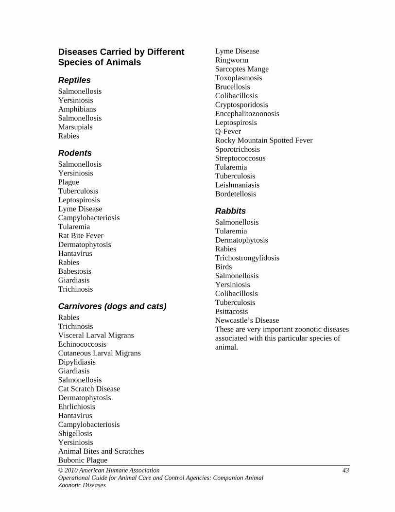
Diseases Carried by Different Species of Animals
Reptiles Salmonellosis Yersiniosis Amphibians Salmonellosis Marsupials Rabies
Rodents Salmonellosis Yersiniosis Plague Tuberculosis Leptospirosis Lyme Disease Campylobacteriosis Tularemia Rat Bite Fever Dermatophytosis Hantavirus Rabies Babesiosis Giardiasis Trichinosis
Carnivores (dogs and cats) Rabies Trichinosis Visceral Larval Migrans Echinococcosis Cutaneous Larval Migrans Dipylidiasis Giardiasis Salmonellosis Cat Scratch Disease Dermatophytosis Ehrlichiosis Hantavirus Campylobacteriosis Shigellosis Yersiniosis Animal Bites and Scratches Bubonic Plague
Lyme Disease Ringworm Sarcoptes Mange Toxoplasmosis Brucellosis Colibacillosis Cryptosporidosis Encephalitozoonosis Leptospirosis Q-Fever Rocky Mountain Spotted Fever Sporotrichosis Streptococcosus Tularemia Tuberculosis Leishmaniasis Bordetellosis
Rabbits Salmonellosis Tularemia Dermatophytosis Rabies Trichostrongylidosis Birds Salmonellosis Yersiniosis Colibacillosis Tuberculosis Psittacosis Newcastle’s Disease These are very important zoonotic diseases associated with this particular species of animal.
© 2010 American Humane Association 43 Operational Guide for Animal Care and Control Agencies: Companion Animal Zoonotic Diseases

Zoonotic Disease Fallacies Fallacy: Children get pinworms from the family dog or cat. Fact: Dogs and cats do not get pinworms. It is impossible for people to contract pinworms from the family pet. Often people mistake tapeworm segments visible around the pet’s anus for pinworms. These are two completely different parasites. See Dipylidiasis. Fallacy: Children get lice from the family dog (cat). Fact: Lice are very species specific. Dog lice do not infect cats or people, for example. Children get lice from other children. Fallacy: Children will get worms if the family puppy licks them. Fact: While the puppy may be a potential source of danger, children contract the parasites by ingesting the feces of the puppy or more often, dirt contaminated with eggs of parasites. See Visceral Larval Migrans. Fallacy: Pregnant women should never touch a cat for fear of contracting Toxoplasmosis. Fact: People contract Toxoplasmosis from ingestion of the eggs of the organism that have typically contaminated soil or food. Eating uncooked meat or raw vegetables is also a potential source of infection. See Toxoplasmosis.
Fallacy: People can contract Canine Distemper and get Multiple Sclerosis. Fact: Although the symptoms in a person with Multiple Sclerosis and a dog with Distemper are similar, there is no known connection between the two diseases. There has been no reduction in the numbers of people diagnosed with MS since 1960 while the number of dogs diagnosed with Distemper has fallen drastically primarily due to an effective vaccine for Distemper. Fallacy: Because there are many states in which there has not been a case of Rabies diagnosed in dogs or cats for many years, we can relax our guard against the disease. Fact: Although we have greatly reduced the incidence of Rabies in our pet animals, the incidence of the disease in the wild population of animals (especially raccoons and skunks) has increased greatly. Many of these animals adapt very readily to suburban life and are a major risk factor to start a Rabies epidemic in many areas. See Rabies.
44 © 2010 American Humane Association Operational Guide for Animal Care and Control Agencies: Companion Animal
Zoonotic Diseases

References 1. Michael Rand. Zoonotic Diseases.
htpp://research.ucsb.edu/connect/pro/disease.html: pg 4
2. Greene, C. Salmonellosis: C. Greene Infectious Diseases of the dog and cat. 2nd ed. Philadelphia:W.B. Saunders, 1998; 235-241
3. Greene, C., Goldstein E., Wright J. Dog and Cat Bite Infections: C Greene Infectious Diseases of the dog and cat. 2nd ed. 330-335
4. Macy, D. Plague: C Greene Infectious Diseases of the dog and cat. 2nd ed. 330-335
5. Michael Rand. Zoonotic Diseases. pg 8-10
6. Fox, J Camphylobacter Infections: C Greene Infectious Diseases of the dog and cat. 2nd ed. 226-229
7. Michael Rand. Zoonotic Diseases. pg 19-21
8. Preitschwerd, E, Greene, C Bartonellosis: C Greene Infectious Diseases of the dog and cat. 2nd ed. 337-343
9. Michael Rand. Zoonotic Diseases. pg 41-43
10. Michael Rand. Zoonotic Diseases. pg 102-103
11. Barr, S Giardiasis: C Greene Infectious Diseases of the dog and cat. 2nd ed. 482-487
12. Michael Rand. Zoonotic Diseases. pg 88-89
13. L. Tilley, Smith, F. The 5-Minute Veterinary Consult Canine and Feline. 2nd ed. Philadelphia: Lippincott, Williams, and Wilkins 2000; 742-743
14. Michael Rand. Zoonotic Diseases. pg 18-19
15. Greene, C., Appel M., Straubinger, R Lyme Borreliosis: C
Greene Infectious Diseases of the dog and cat. 2nd ed. 282-291
16. Greene, C. Immunocompromised People and Pets: C Greene Infectious Diseases of dogs and cats. 2nd ed. 714
17. Michael Rand. Zoonotic Diseases. pg 39-40
18. Michael Rand. Zoonotic Diseases. 71-73
19. Greene, C., Dreesen, D Rabies: C Greene Infectious Diseases of dogs and cats. 2nd ed. 114-126
20. Compendium of Animal Rabies Control, 1998 National Association of Public Health Veterinarians, Inc.
21. Foil, C. Dermatophytosis: C Greene Infectious Diseases of dogs and cats. 2nd ed. 362-371
22. Moriello, KA, DeBoer, D Feline Dermatophytosis. Recent advances and recommendations for treatment. Veterinary Clinics of North America. Small Animal Practice 1995; 25:901-921
23. Michael Rand. Zoonotic Diseases. 48-49
24. Greene, C. Shigellosis: C Greene Infectious Diseases of the dog and cat. 2nd ed. 240-241
25. Michael Rand. Zoonotic Diseases. pg 6-7
26. Duby, J., Lappin, M. Toxoplasmosis and Neosporosis: C Greene Infectious Diseases of dogs and cats. 2nd ed. 493-503
27. Michael Rand. Zoonotic Diseases. pg 81-83
28. Michael Rand. Zoonotic Diseases. pg 24-25
29. Taboada, J. Babesiosis: C Greene Infectious Diseases of dogs and cats. 2nd ed. 473-482
30. Michael Rand. Zoonotic Diseases. pg 80-81
© 2010 American Humane Association 45 Operational Guide for Animal Care and Control Agencies: Companion Animal Zoonotic Diseases

31. Carmicheal, L., Greene, C. Canine Brucellosis: C Greene Infectious Diseases of dogs and cats. 2nd ed. 248-257
32. Barr, S., Cryptosporidiosis: C Greene Infectious Diseases of dogs and cats. 2nd ed. 518-522
33. Michael Rand. Zoonotic Diseases. pg 87-88
34. Deeb, B., Carpenter, J. Neurologic and Musculoskeletal Diseases: K Quesenberry, J. Carpenter. Ferrets, Rabbits, and Rodents Clinical Medicine and Surgery, 2nd ed. St. Louis: W. B. Saunders 2004. 203-206
35. Greene, C. Hantavirus: C Greene Infectious Diseases of dogs and cats. 2nd ed. 111-112
36. Michael Rand. Zoonotic Diseases. 60-64
37. Wolf, A., Histoplasmosis: C Greene Infectious Diseases of dogs and cats. 2nd ed. 378-383
38. Greene, C., Miller, M., Brown, C. Leptospirosis: C Greene Infectious Diseases of dogs and cats. 2nd ed. 273-282
39. Michael Rand. Zoonotic Diseases. pg 16-17
40. Greene, C., Breitschwerdt, E., Q Fever: C Greene Infectious Diseases of dogs and cats. 2nd ed. 162-164
41. Michael Rand. Zoonotic Diseases. 43-44
42. Greene, C. Breitschwerdt, E. Rocky Mountain Spotted Fever: C Greene Infectious Diseases of dogs and cats. 2nd ed. 155-162
43. Michael Rand. Zoonotic Diseases. 45-46
Operational Guide for Animal Care and Control Agencies: Companion Animal Zoonotic Diseases
44. Rossen, E., Dunstan, R. Sporotrichosis: C Greene Infectious Diseases of dogs and cats. 2nd ed. 399-402
45. Michael Rand. Zoonotic Diseases. pg 49-50
46. Kaufman, A. Tularemia: C Greene Infectious Diseases of dogs and cats. 2nd ed. 300-302
47. Michael Rand. Zoonotic Diseases. pg 32-33
48. Lappendel, R., Ferrer, L. Leishmaniasis: C Greene Infectious Diseases of dogs and cats. 2nd ed. 450-458
49. Michael Rand. Zoonotic Diseases. pg 33-34
50. Michael Rand. Zoonotic Diseases. pg 100-101, 106-107
51. Michael Rand. Zoonotic Diseases. pg 21-22
52. Michael Rand. Zoonotic Diseases. pg 111-112
53. Michael Rand. Zoonotic Diseases. pg 44-45
54. Michael Rand. Zoonotic Diseases. pg 77-78
55. Michael Rand. Zoonotic Diseases. pg 33-34
56. Greene, C., Prescott, J. Streptococcal and Other Gram-Positive Bacterial Infections: C Greene Infectious Diseases of dogs and cats. 2nd ed. 205-214
57. Michael Rand. Zoonotic Diseases. pg 105
58. Michael Rand. Zoonotic Diseases. pg 10-11
59. Greene, C., Gunn-Moore, D. Tuberculous Mycobacterial Infections: C Greene Infectious Diseases of dogs and cats. 2nd ed. 313-321
60. CDC Bulletin on West Nile Virus: http://www.cdc.gov./ncidad/dvbid/westnile
61. Austgen, L. West Nile virus infection: Essential facts for small-animal practitioners. Veterinary Medicine May 2005. pg. 26-32
46 © 2010 American Humane Association
© 2010 American Humane Association 47 Operational Guide for Animal Care and Control Agencies: Companion Animal Zoonotic Diseases
62. Ford, R., Baden, S. Canine Infectious Tracheobronchitis: C Greene Infectious Diseases of dogs and cats. 2nd ed. pg. 33-38
General References Hurley, K. Zoonotic Diseases in Animal Shelters: Considerations for Shelter Professionals; [email protected] Miller, L., Zawistowski, S., Shelter Medicine for Veterinarians and Staff: Ames, Iowa: Blackwell Publishing, 2004.
National Association of State Public Health Veterinarians, Inc., Compendium of Measures to Prevent Disease Associated with Animals in Public Settings, 2005 Seibert, P., Director of the VIN OSHA Center, Common Zoonotic Diseases of Concern, Posted on Veterinary Information Network, 2003. The Center for Food Security and Public Health of Iowa State University. http://www.cfsph.iastate.edu/DiseaseInfo/

Rabies Protocol Chart
BITE SOURCE SITUATION ANIMAL DISPOSITIONPOST-EXPOSURE
PROPHYLAXIS IN HUMANS
Rodent Any episode Usually not examined
None, but consult public health officials if
circumstances of bite warrant
Dog, cat, ferret Healthy, owned
Confine; observe for at least 10 days,
None or consider, if unprovoked; Yes, if
CNS signs develop in animal
especially if unprovoked
attack
Healthy, stray available or
escaped Euthanize immediately; submit head for exam
Yes; stop if lab results negative, continue if animal unavailable
CNS signs or
illness Euthanize immediately; submit head for exam Yes; if negative FA result, stop.
Wild carnivore Any episode
If captured, euthanize immediately; submit head
for examination Yes, if positive or animal at large; if
negative, FA result, stop.
Inoculation of Any episode Not applicable No post-exposure treatment required. attenuated
vaccine
48 © 201 American Humane Association Operational Guide for Animal Care and Control Agencies: Companion Animal Zoonotic Diseases