Embed Size (px)
Citation preview
Ophthalmology in Primary Care: A Student Health Perspective
Thomasine Gorry, M.D., M.G.A.Assistant Professor of Ophthalmology
Scheie Eye InstituteUniversity of Pennsylvania

• No financial interests in topics discussed
Overview
• Review basic eye exam
• Throughout the presentation identify “Red Flag”signs and symptoms or complaints
• Focus on Anterior Segment Findings: more prevalent in younger population
• Contact Lens Issues
• Posterior segment : focus on patient complaints
• Refractive Surgery
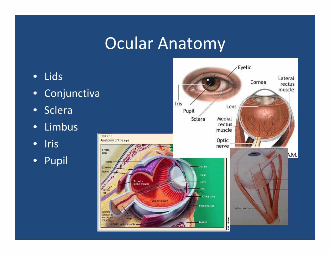
Ocular Anatomy
• Lids
• Conjunctiva
• Sclera
• Limbus
• Iris
• Pupil
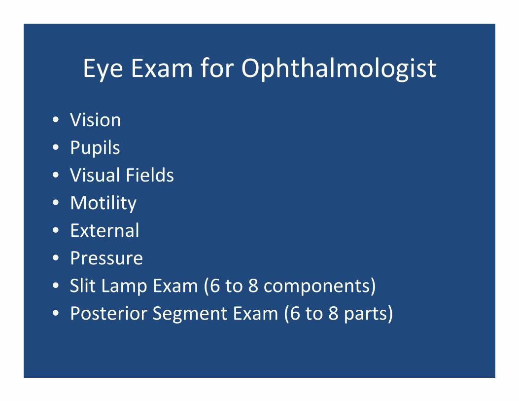
Eye Exam for Ophthalmologist
• Vision• Pupils• Visual Fields• Motility• External• Pressure• Slit Lamp Exam (6 to 8 components)• Posterior Segment Exam (6 to 8 parts)
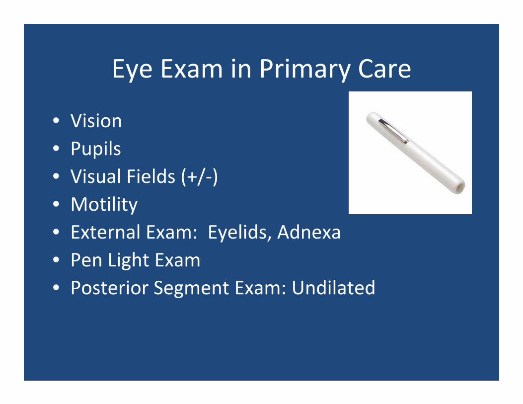
Eye Exam in Primary Care
• Vision• Pupils• Visual Fields (+/‐)• Motility• External Exam: Eyelids, Adnexa• Pen Light Exam• Posterior Segment Exam: Undilated
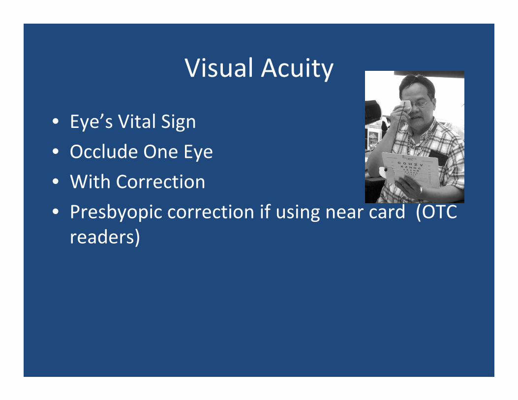
Visual Acuity
• Eye’s Vital Sign
• Occlude One Eye
• With Correction
• Presbyopic correction if using near card (OTC readers)
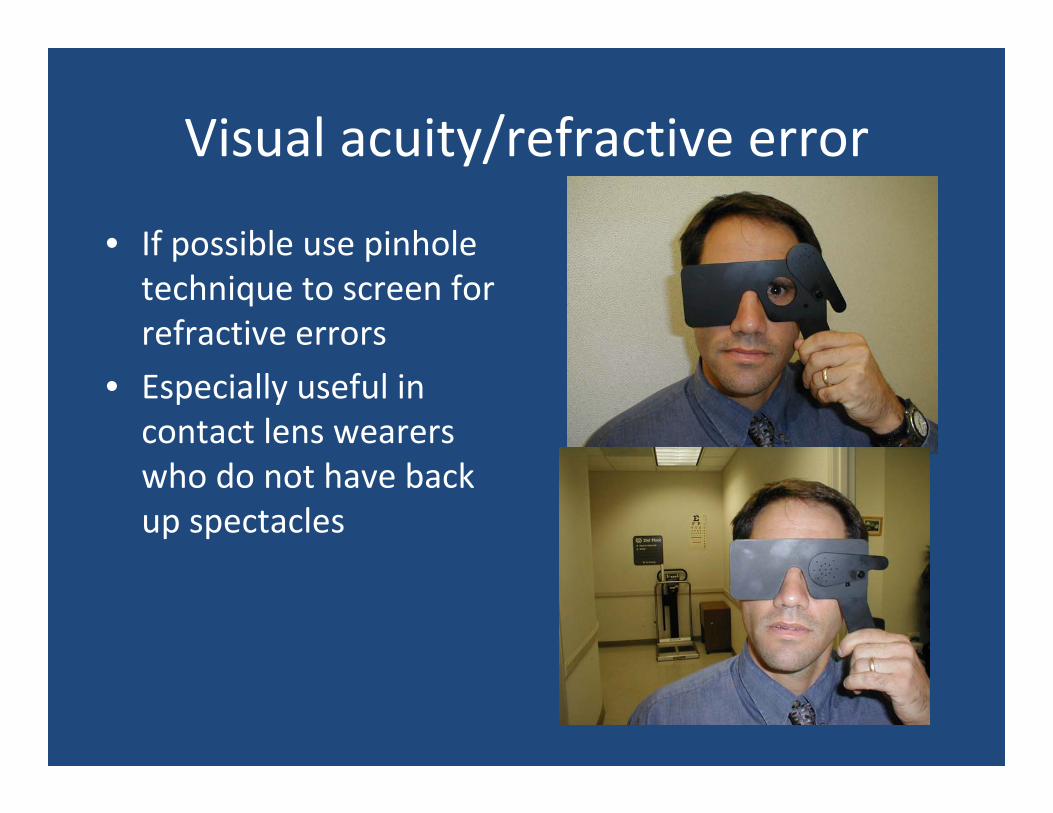
Visual acuity/refractive error
• If possible use pinhole technique to screen for refractive errors
• Especially useful in contact lens wearers who do not have back up spectacles
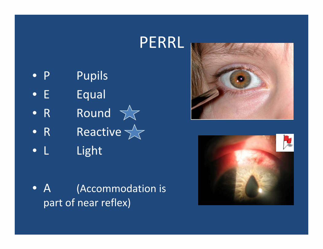
PERRL
• P Pupils
• E Equal
• R Round
• R Reactive
• L Light
• A (Accommodation is part of near reflex)

Swinging Flashlight Test and Marcus Gunn Pupil (RAPD)
• Signifies Optic Nerve Disease
• Based on Consensual Light Reflex
• Will see paradoxical dilation of the pupil in response to light
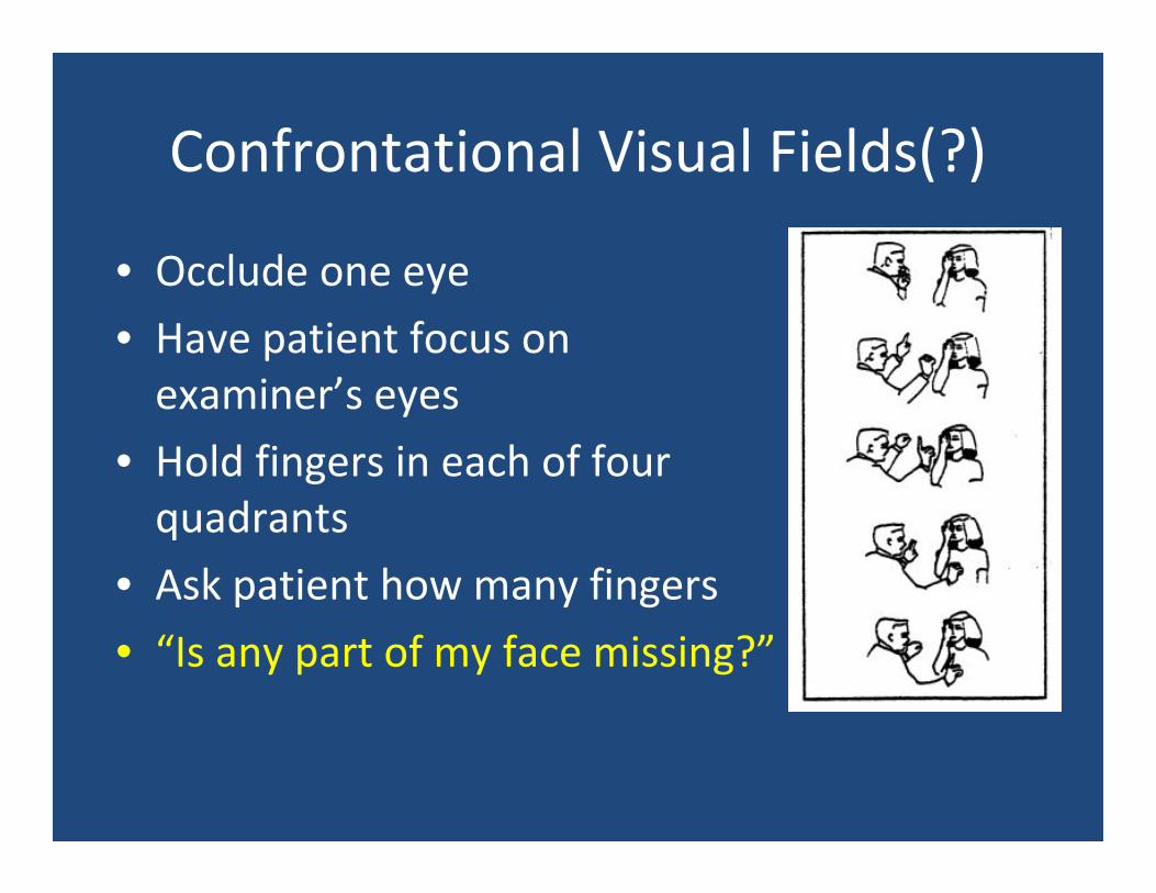
Confrontational Visual Fields(?)
• Occlude one eye
• Have patient focus on examiner’s eyes
• Hold fingers in each of four quadrants
• Ask patient how many fingers
• “Is any part of my face missing?”
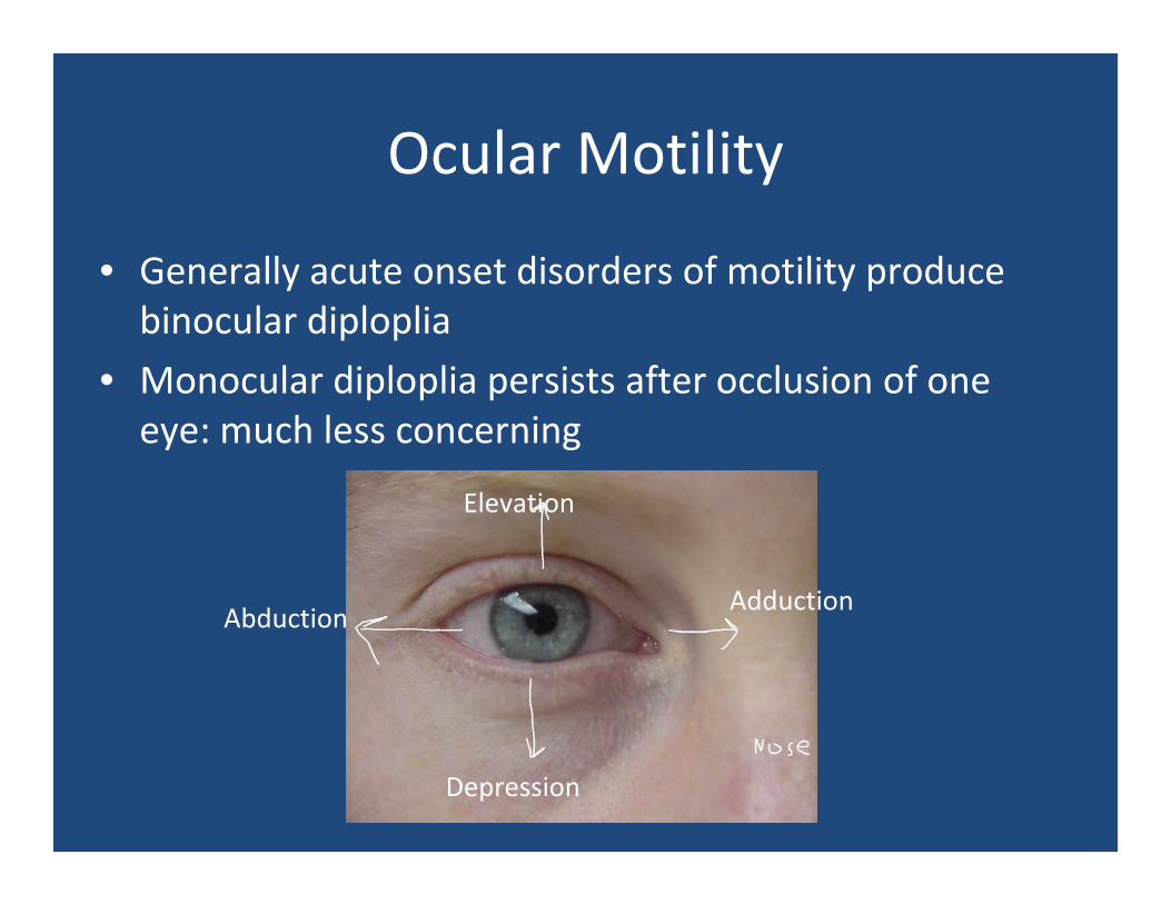
Ocular Motility
• Generally acute onset disorders of motility produce binocular diploplia
• Monocular diploplia persists after occlusion of one eye: much less concerning
Adduction
Elevation
Depression
Abduction
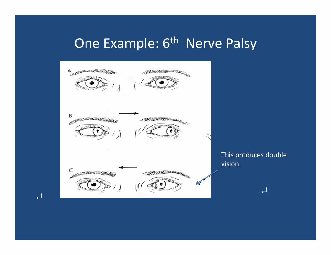
One Example: 6th Nerve Palsy
↵
This produces double vision.
↵
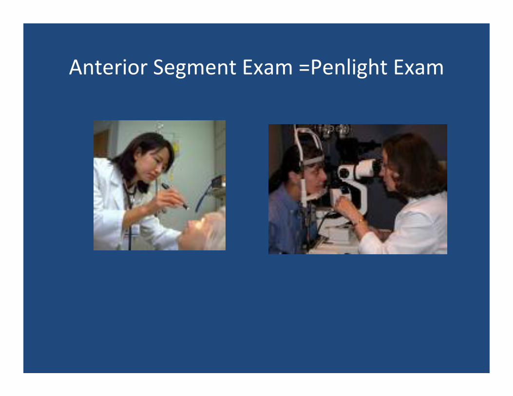
Anterior Segment Exam =Penlight Exam

Penlight exam
• Pattern of erythema
• Corneal Clarity
• Fluroescein staining
• Conjunctival swelling (chemosis)
• Anterior Chamber—blood or pus
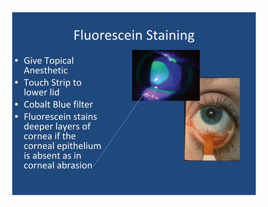
Fluorescein Staining
• Give Topical Anesthetic
• Touch Strip to lower lid
• Cobalt Blue filter• Fluorescein stains deeper layers of cornea if the corneal epithelium is absent as in corneal abrasion
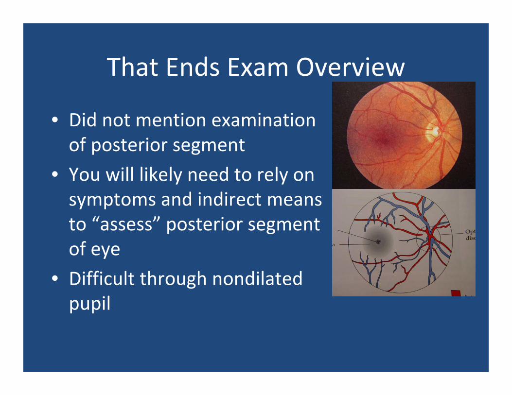
That Ends Exam Overview
• Did not mention examination of posterior segment
• You will likely need to rely on symptoms and indirect means to “assess” posterior segment of eye
• Difficult through nondilatedpupil
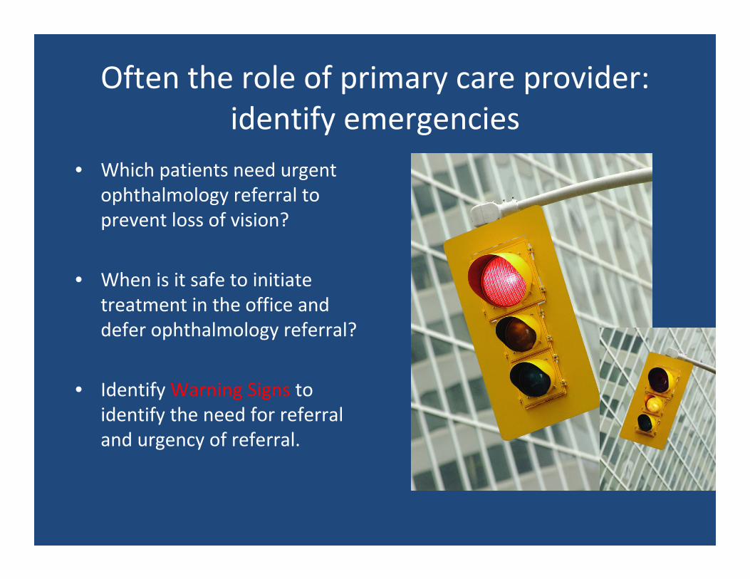
Often the role of primary care provider: identify emergencies
• Which patients need urgent ophthalmology referral to prevent loss of vision?
• When is it safe to initiate treatment in the office and defer ophthalmology referral?
• Identify Warning Signs to identify the need for referral and urgency of referral.
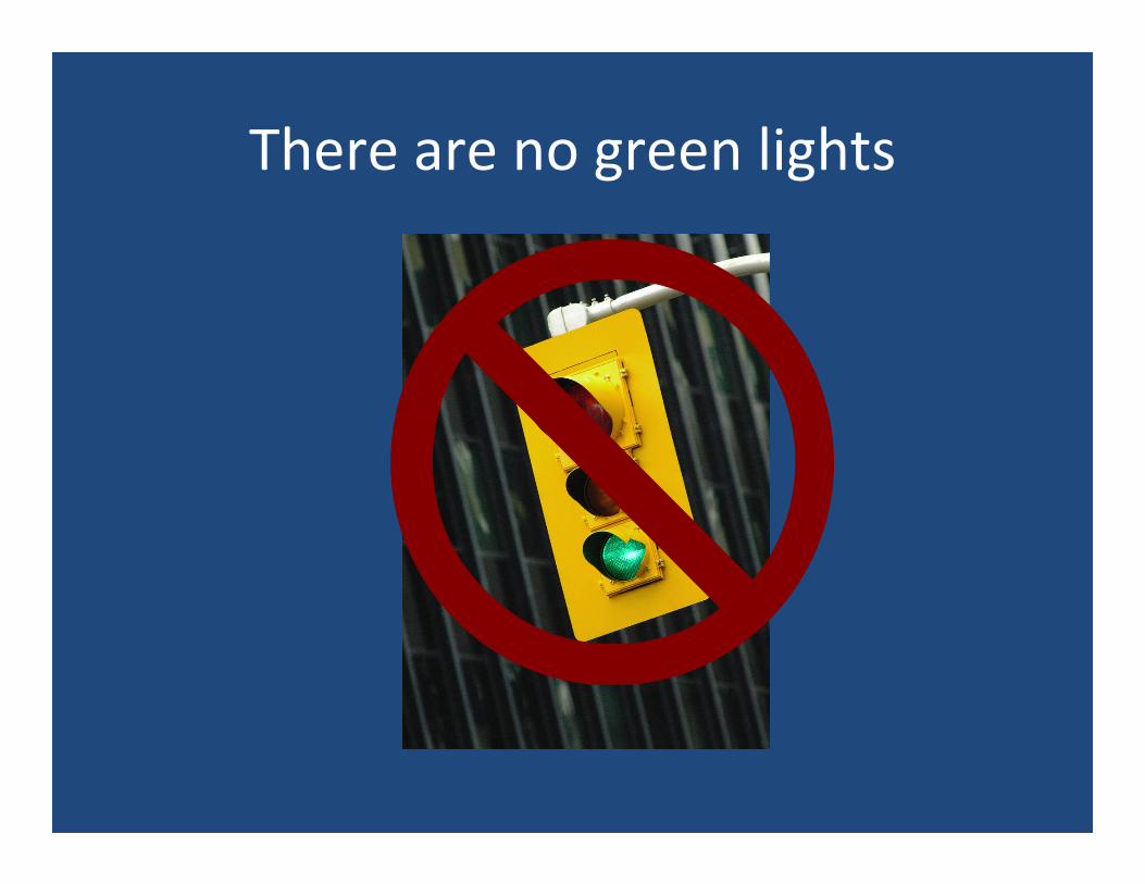
There are no green lights
Complaints are few, diagnoses multiple
• Blurred
• Red / Swollen
• Pain
• Itchy
• Obscuration/distortion
• Photopsia
Potential Warning Signs in the History• Decreased Vision
• Foreign body sensation severe enough that patient cannot keep the eyes open
• Severe Photophobia and Pain
Uveitis, corneal abrasion
• TraumaVegetable matter? High Velocity? Metal?
• Contact Lens Wear
High risk for keratitis (corneal infections)
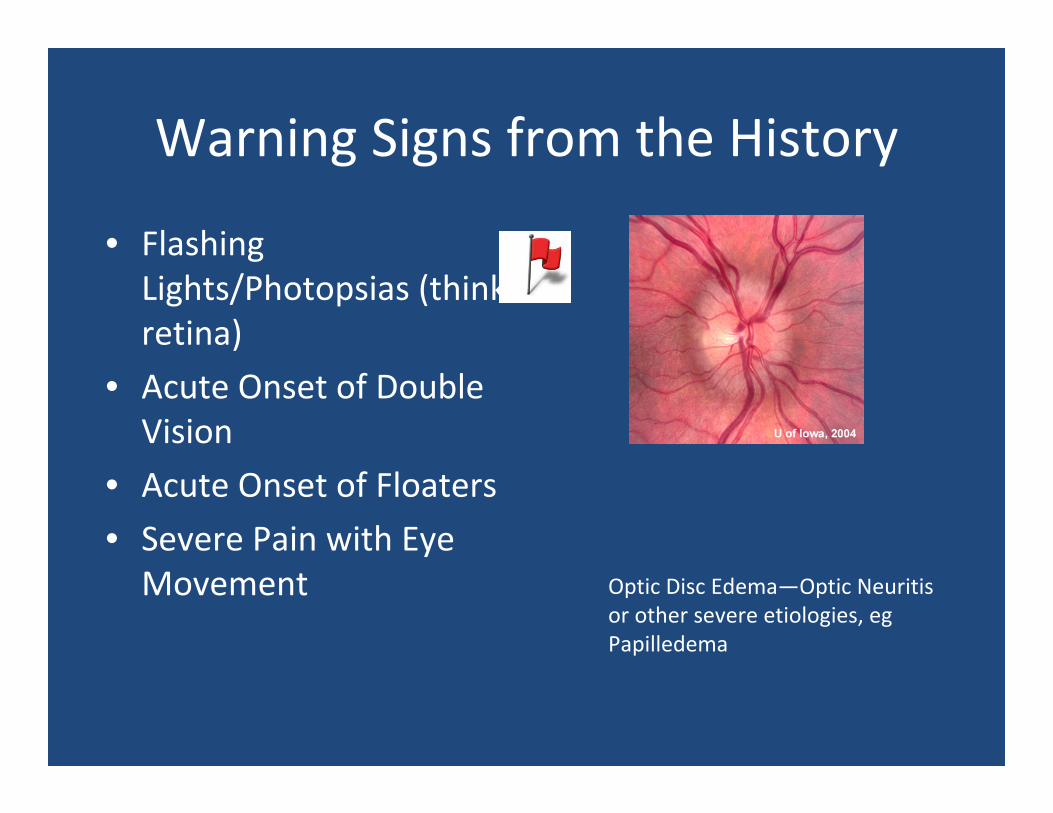
Warning Signs from the History
• Flashing Lights/Photopsias (think retina)
• Acute Onset of Double Vision
• Acute Onset of Floaters
• Severe Pain with Eye Movement Optic Disc Edema—Optic Neuritis
or other severe etiologies, egPapilledema
Potential Warning Signs on Exam
• Observe for:
– General patient discomfort
– Ability to tolerate light
– Erythema of periorbital structures
– Purulent discharge (conjunctivitis or bacterial keratitis)
– Eye tearing
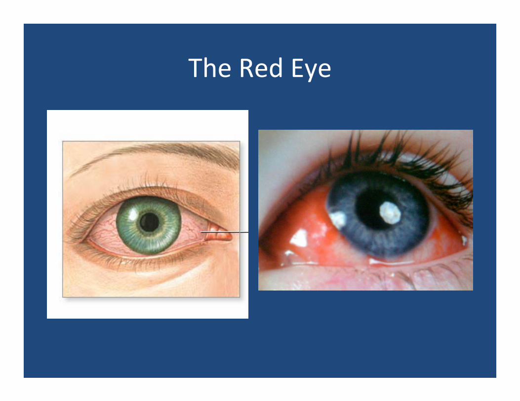
The Red Eye
Red Eye
• Huge Differential
• Most important Feature is vision
• Good vision usually less severe process
• Also important is Pain
• True pain may herald more severe pathologies
The Red Eye: History• Ask:
– Has the vision changed?– Are you a contact lens wearer?– Itchy?– Pain? Photophobia? (severe or mild)– Sick contacts?– Other eye affected?– Recent URI?– Sore throat?– Foreign Body Sensation– Note other medical problems: DM, autoimmune diseases
The Red Eye: Exam
• ? Vision• ? Pupils• Look at the lid‐‐edematous, warm and tender think preceptal cellulitis
• Look for discharge• Laterality• Observe patient for level of discomfort• ?Staining
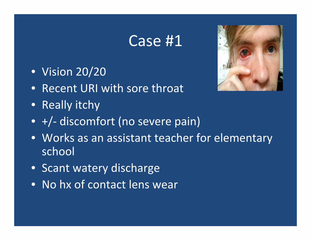
Case #1
• Vision 20/20 • Recent URI with sore throat• Really itchy• +/‐ discomfort (no severe pain)• Works as an assistant teacher for elementary school
• Scant watery discharge• No hx of contact lens wear
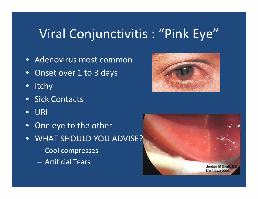
Viral Conjunctivitis : “Pink Eye”
• Adenovirus most common
• Onset over 1 to 3 days
• Itchy
• Sick Contacts
• URI
• One eye to the other
• WHAT SHOULD YOU ADVISE?– Cool compresses
– Artificial Tears
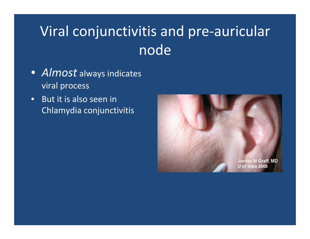
Viral conjunctivitis and pre‐auricular node
• Almost always indicates viral process
• But it is also seen in Chlamydia conjunctivitis
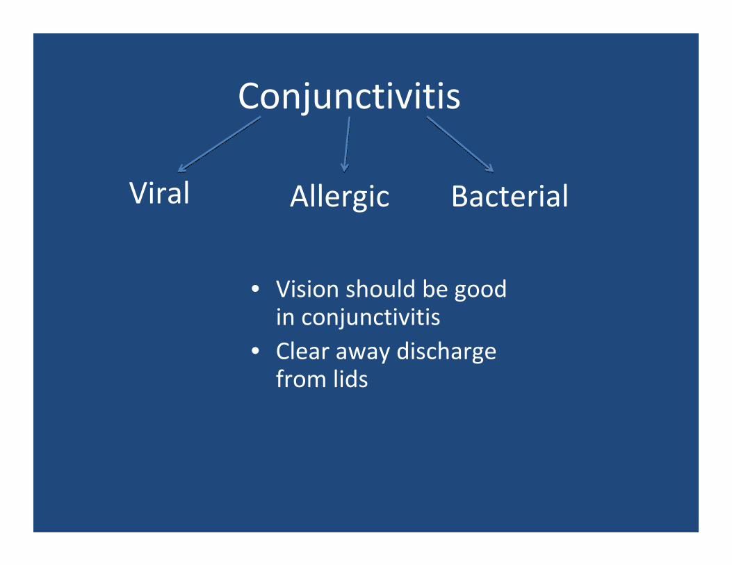
Conjunctivitis
Viral
• Vision should be good in conjunctivitis
• Clear away discharge from lids
Allergic Bacterial
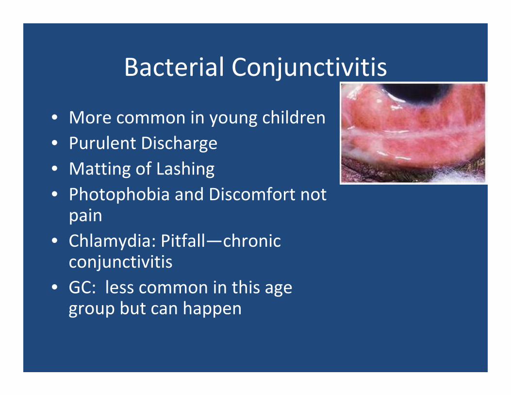
Bacterial Conjunctivitis
• More common in young children• Purulent Discharge• Matting of Lashing• Photophobia and Discomfort not pain
• Chlamydia: Pitfall—chronic conjunctivitis
• GC: less common in this age group but can happen
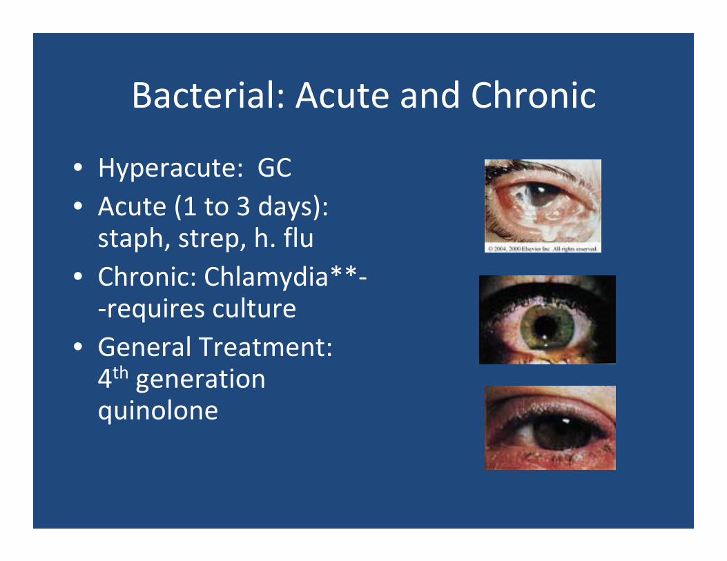
Bacterial: Acute and Chronic
• Hyperacute: GC• Acute (1 to 3 days): staph, strep, h. flu
• Chronic: Chlamydia**‐‐requires culture
• General Treatment: 4th generation quinolone
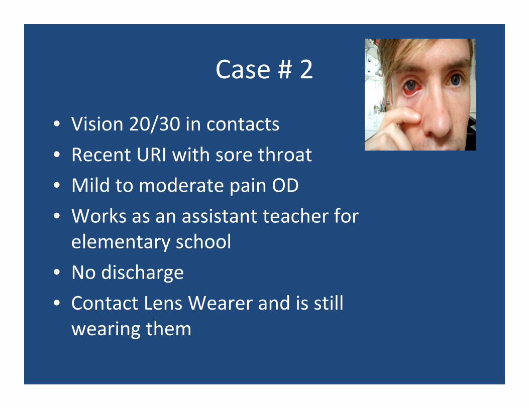
Case # 2
• Vision 20/30 in contacts
• Recent URI with sore throat
• Mild to moderate pain OD
• Works as an assistant teacher for elementary school
• No discharge
• Contact Lens Wearer and is still wearing them
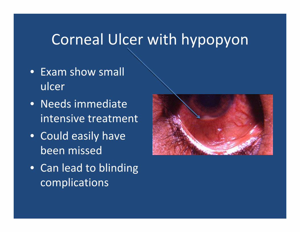
Corneal Ulcer with hypopyon
• Exam show small ulcer
• Needs immediate intensive treatment
• Could easily have been missed
• Can lead to blinding complications
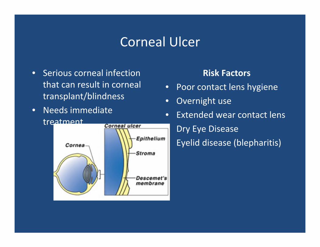
Corneal Ulcer
• Serious corneal infection that can result in corneal transplant/blindness
• Needs immediate treatment
Risk Factors
• Poor contact lens hygiene
• Overnight use
• Extended wear contact lens
• Dry Eye Disease
• Eyelid disease (blepharitis)
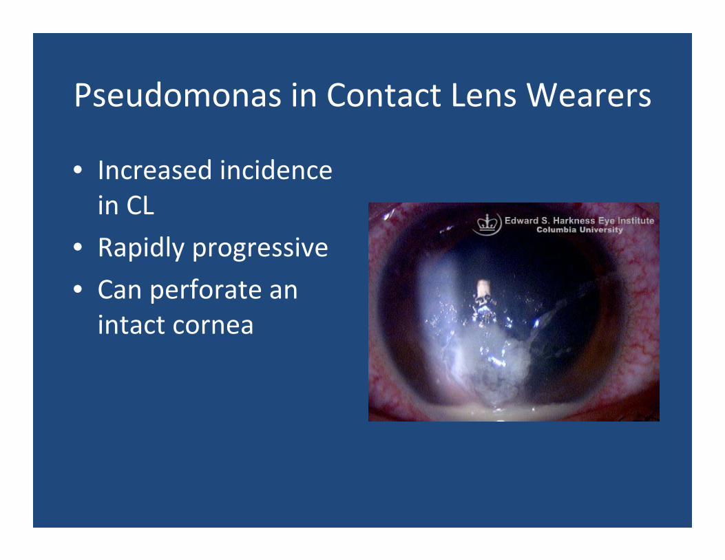
Pseudomonas in Contact Lens Wearers
• Increased incidence in CL
• Rapidly progressive
• Can perforate an intact cornea
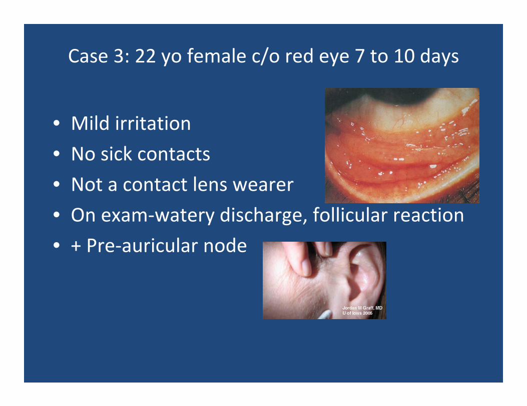
Case 3: 22 yo female c/o red eye 7 to 10 days
• Mild irritation
• No sick contacts
• Not a contact lens wearer
• On exam‐watery discharge, follicular reaction
• + Pre‐auricular node
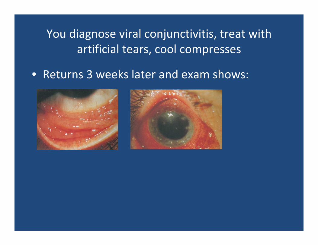
You diagnose viral conjunctivitis, treat with artificial tears, cool compresses
• Returns 3 weeks later and exam shows:
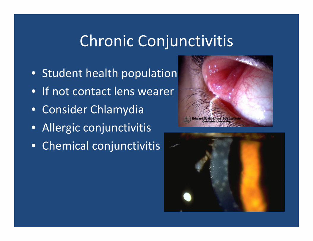
Chronic Conjunctivitis
• Student health population
• If not contact lens wearer
• Consider Chlamydia
• Allergic conjunctivitis
• Chemical conjunctivitis
Adult Inclusion ConjunctivitisChlamydia Conjunctivitis
• Sexually transmitted disease
• C. trachomatis immunotypes D‐K
• Transmission through direct contact of eye with infected genital or urinary secretions.
• Indirect transmission through poorly chlorinated swimming pools.
• Eye to eye transmission possible

31 year old male complains of mild to moderate ocular itching every fall
• Still wears contacts
• Less comfortable
• Clear nasal discharge
• Works as bike messenger
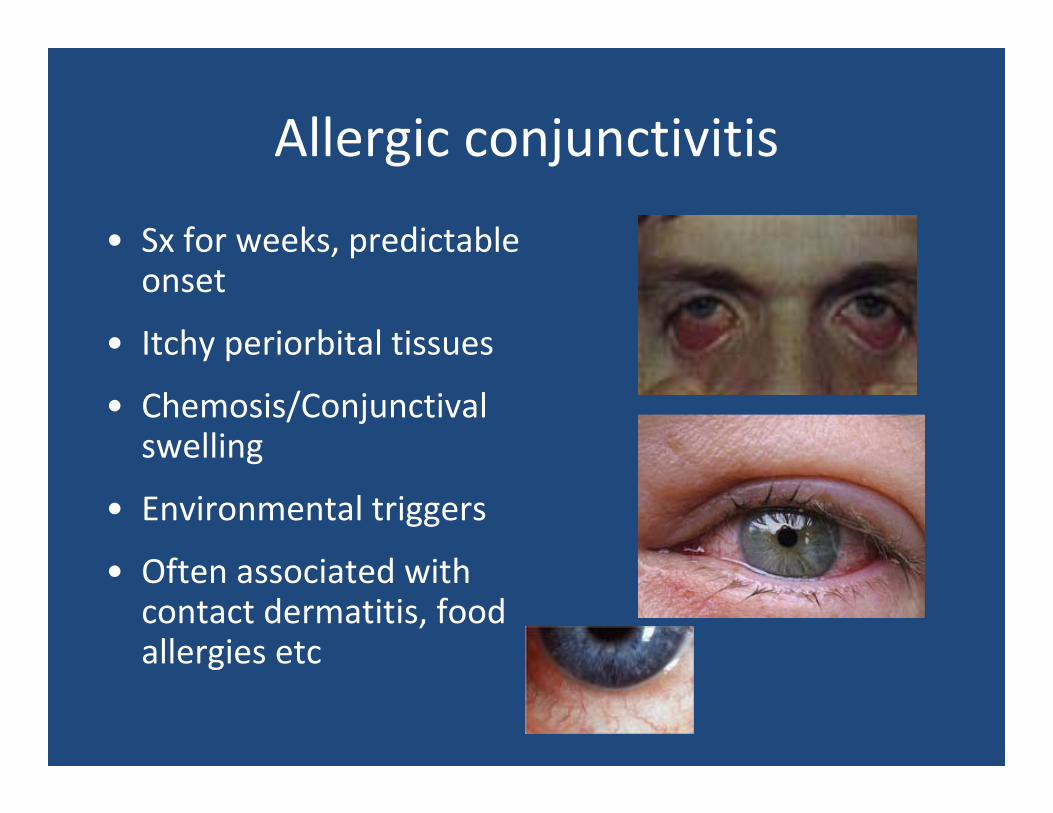
Allergic conjunctivitis
• Sx for weeks, predictable onset
• Itchy periorbital tissues
• Chemosis/Conjunctivalswelling
• Environmental triggers
• Often associated with contact dermatitis, food allergies etc
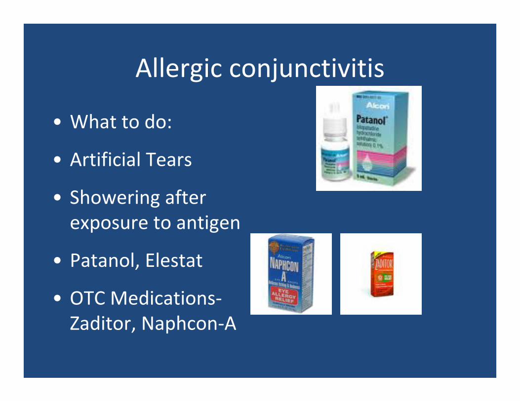
Allergic conjunctivitis
• What to do:
• Artificial Tears
• Showering after exposure to antigen
• Patanol, Elestat
• OTC Medications‐Zaditor, Naphcon‐A
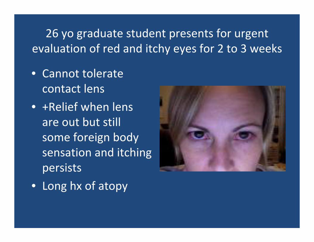
26 yo graduate student presents for urgent evaluation of red and itchy eyes for 2 to 3 weeks
• Cannot tolerate contact lens
• +Relief when lens are out but still some foreign body sensation and itching persists
• Long hx of atopy
DDX?
• Viral conjunctivitis
• Allergic conjunctivitis
• Blepharitis
• Chlamydia conjunctivitis
• Contact lens over wear
• Giant Papillary conjunctivitis
• Less likely corneal ulcer
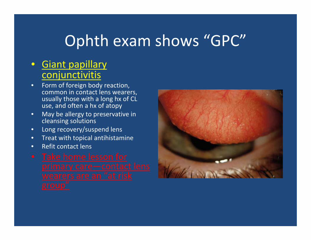
Ophth exam shows “GPC”• Giant papillary conjunctivitis
• Form of foreign body reaction, common in contact lens wearers, usually those with a long hx of CL use, and often a hx of atopy
• May be allergy to preservative in cleansing solutions
• Long recovery/suspend lens• Treat with topical antihistamine• Refit contact lens
• Take home lesson for primary care—contact lens wearers are an “at risk group”
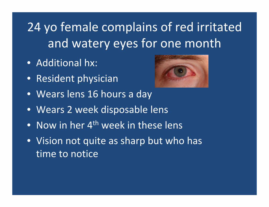
24 yo female complains of red irritated and watery eyes for one month
• Additional hx:
• Resident physician
• Wears lens 16 hours a day
• Wears 2 week disposable lens
• Now in her 4th week in these lens
• Vision not quite as sharp but who has time to notice
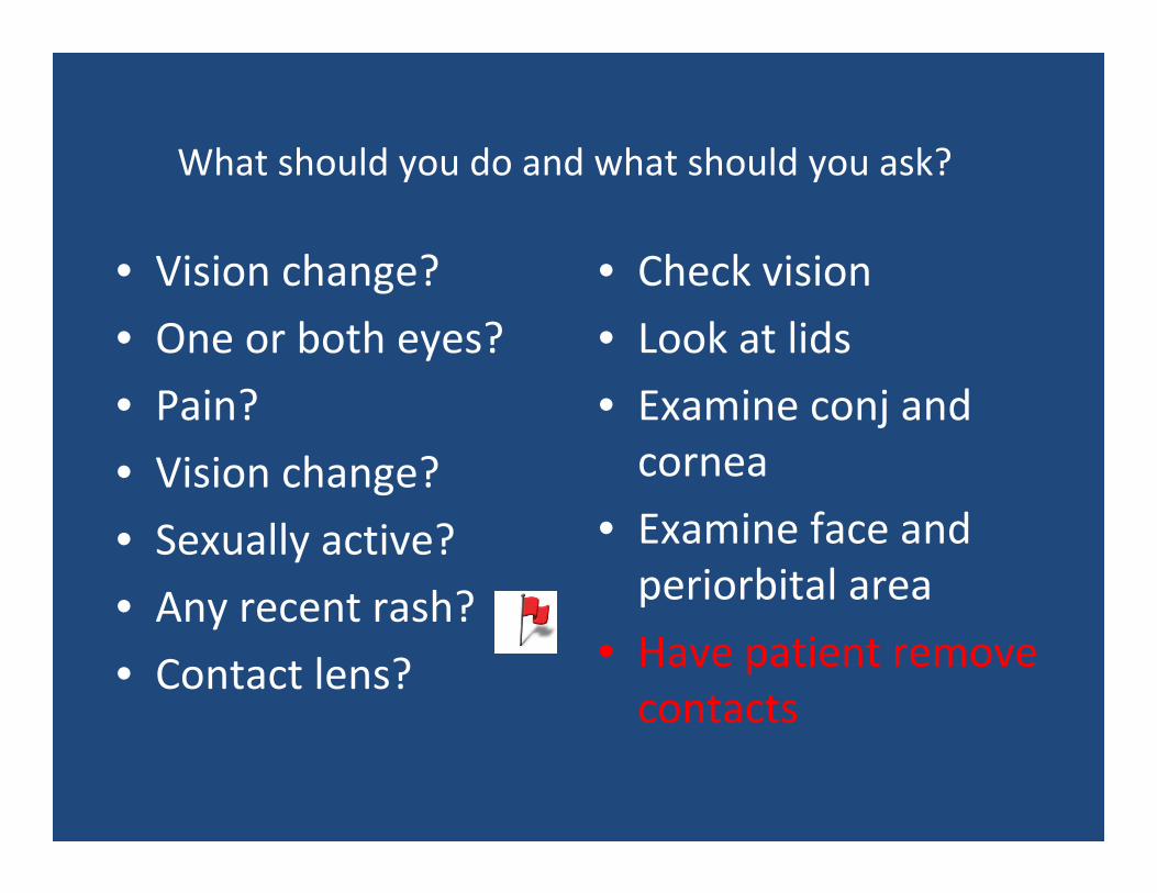
• Vision change?
• One or both eyes?
• Pain?
• Vision change?
• Sexually active?
• Any recent rash?
• Contact lens?
• Check vision
• Look at lids
• Examine conj and cornea
• Examine face and periorbital area
• Have patient remove contacts
What should you do and what should you ask?
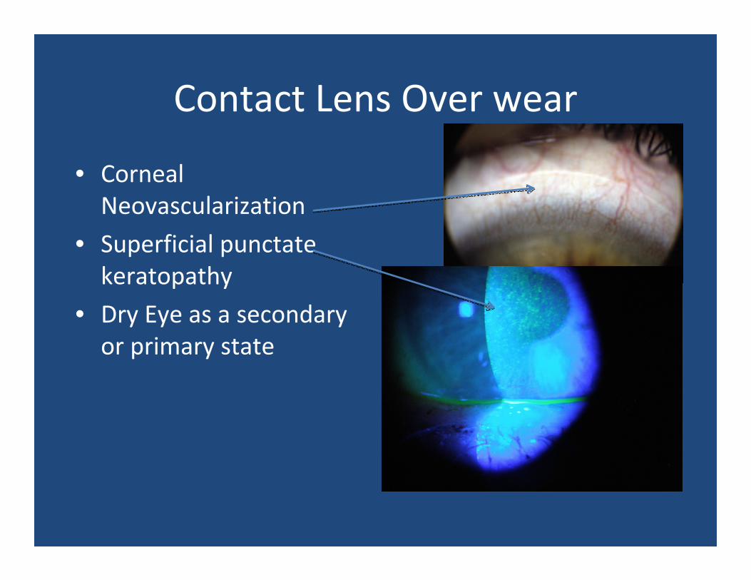
Contact Lens Over wear
• Corneal Neovascularization
• Superficial punctatekeratopathy
• Dry Eye as a secondary or primary state
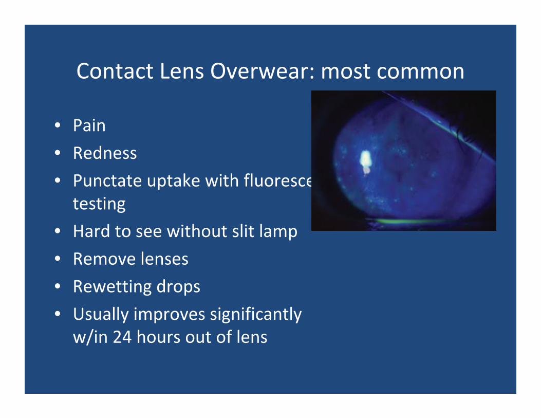
Contact Lens Overwear: most common
• Pain
• Redness
• Punctate uptake with fluoresceintesting
• Hard to see without slit lamp
• Remove lenses
• Rewetting drops
• Usually improves significantly w/in 24 hours out of lens
Contact lens overwear:What you can do
• Get patient out of lenses
• Encourage patient to see ophthorather than continually calling for contacts
• Encourage all patients to have spectacles and carry them
• Decrease dependence on lens
• Never sleep in lenses
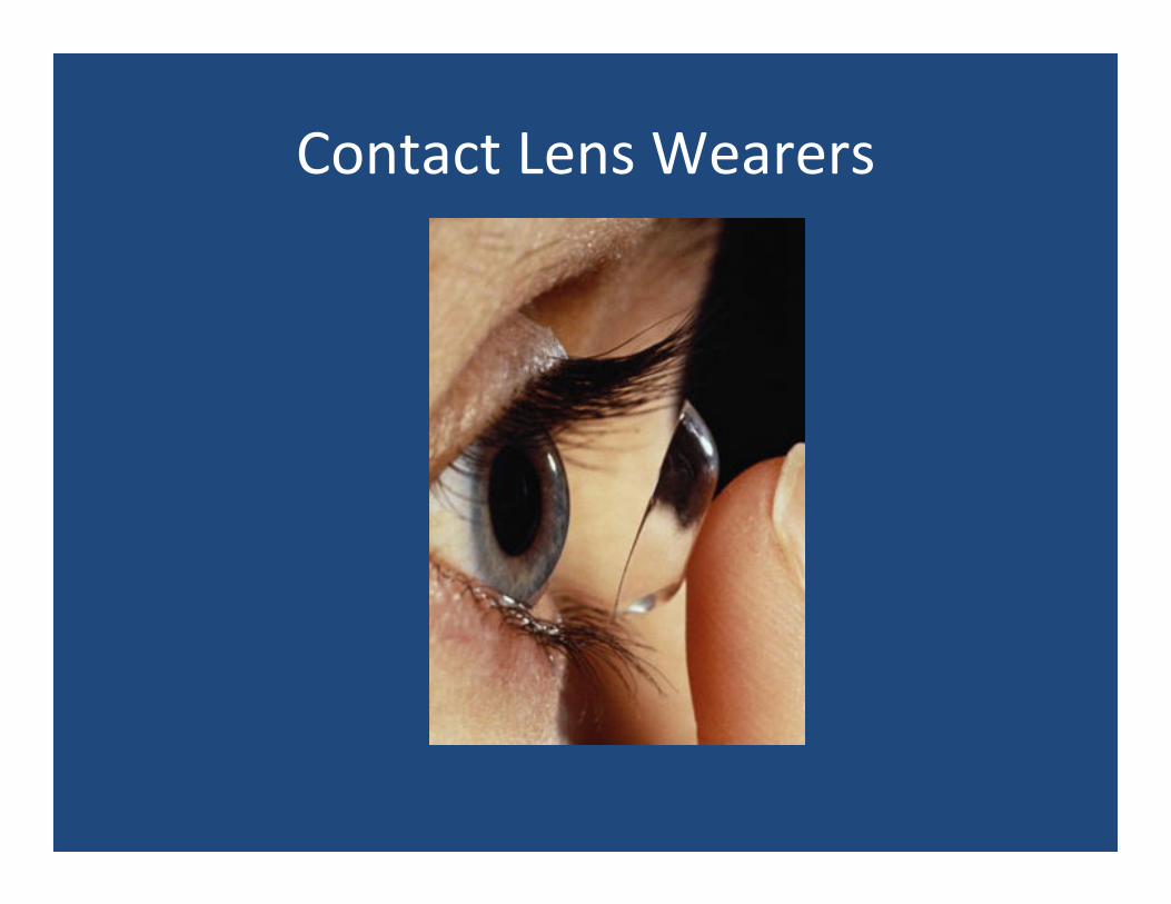
Contact Lens Wearers
General Guidelines
• Daily disposable lenses are safest???• Sleeping in lens increasing risk of infection 8x• Wash hands • Clean lens‐rubbing • Do not use longer than directed• Any problems: remove lens and throw it out (SCL)• Have a pair of glasses• Multipurpose cleaners
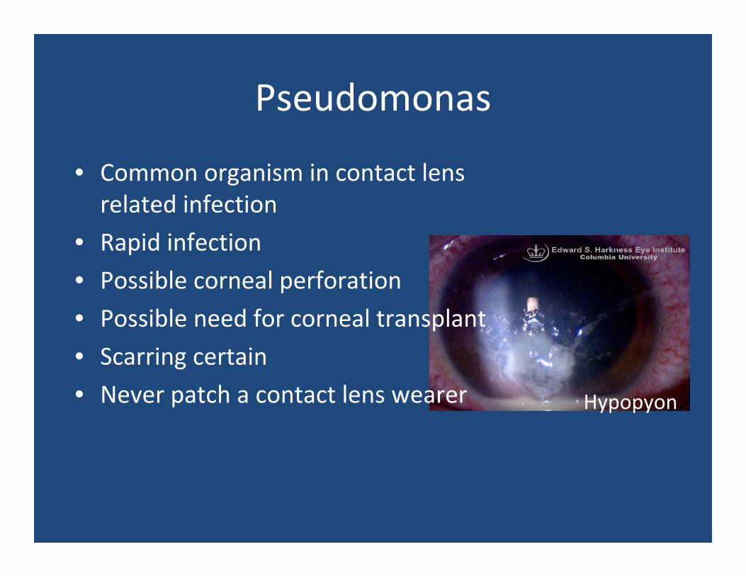
Pseudomonas
• Common organism in contact lens related infection
• Rapid infection
• Possible corneal perforation
• Possible need for corneal transplant
• Scarring certain
• Never patch a contact lens wearer Hypopyon
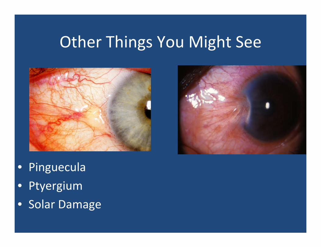
Other Things You Might See
• Pinguecula
• Ptyergium
• Solar Damage

32 year old male graduate student with severe right eye pain, tearing during midterm exams
• Vision is blurred
• Took lens out immediately
• No relief
• No trauma
• Recently has “sore” on lips
• Looks injected to penlight
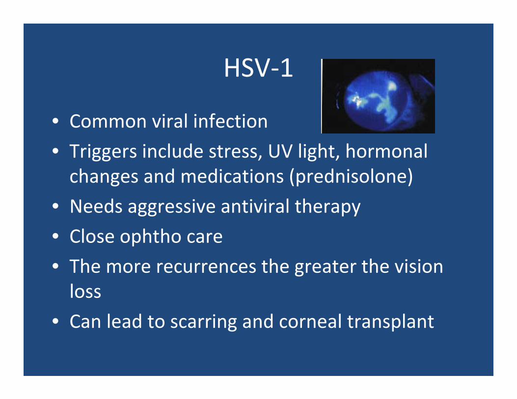
HSV‐1
• Common viral infection
• Triggers include stress, UV light, hormonal changes and medications (prednisolone)
• Needs aggressive antiviral therapy
• Close ophtho care
• The more recurrences the greater the vision loss
• Can lead to scarring and corneal transplant
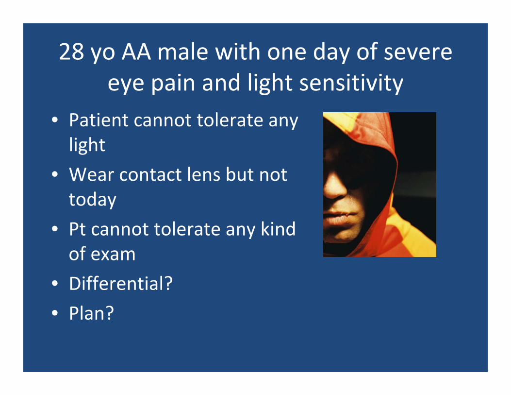
28 yo AA male with one day of severe eye pain and light sensitivity
• Patient cannot tolerate any light
• Wear contact lens but not today
• Pt cannot tolerate any kind of exam
• Differential?
• Plan?
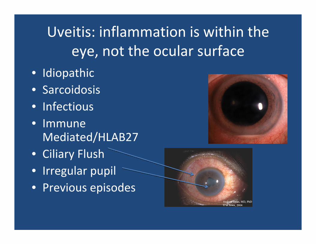
Uveitis: inflammation is within the eye, not the ocular surface
• Idiopathic• Sarcoidosis• Infectious• Immune Mediated/HLAB27
• Ciliary Flush• Irregular pupil• Previous episodes
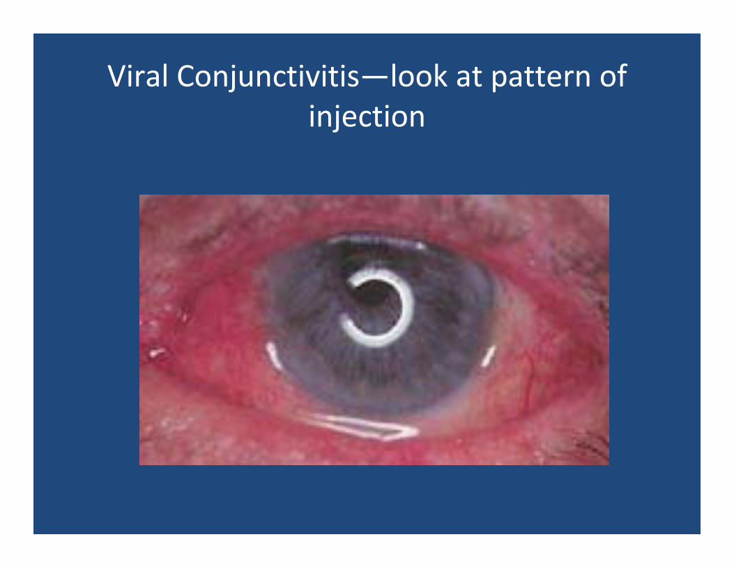
Viral Conjunctivitis—look at pattern of injection
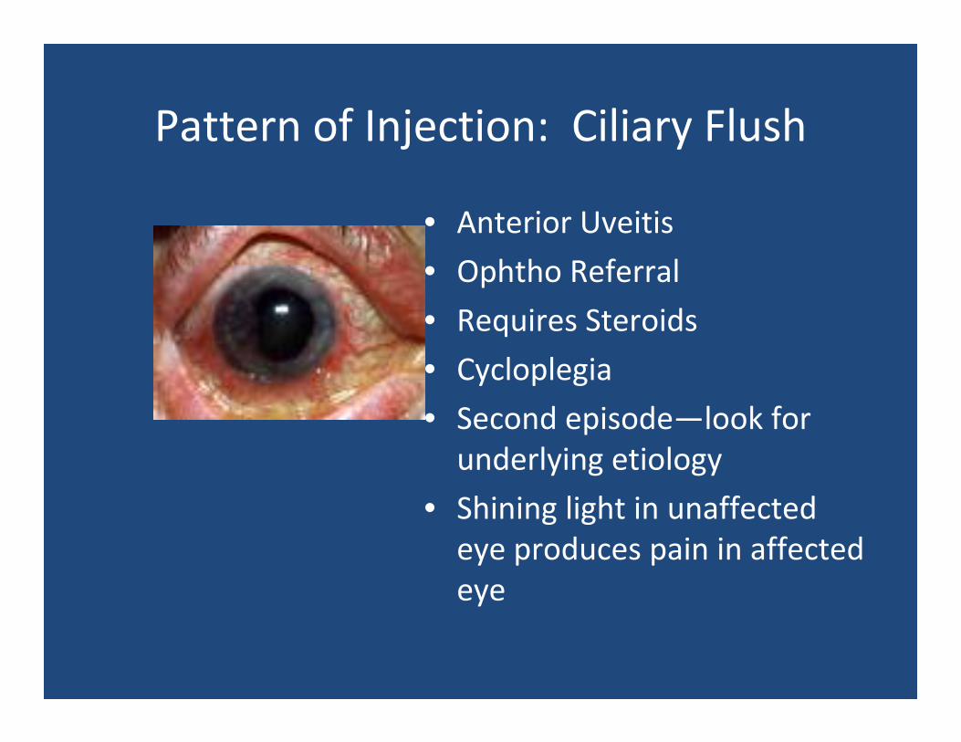
Pattern of Injection: Ciliary Flush
• Anterior Uveitis
• Ophtho Referral
• Requires Steroids
• Cycloplegia
• Second episode—look for underlying etiology
• Shining light in unaffected eye produces pain in affected eye
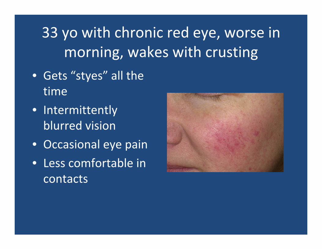
33 yo with chronic red eye, worse in morning, wakes with crusting
• Gets “styes” all the time
• Intermittently blurred vision
• Occasional eye pain
• Less comfortable in contacts
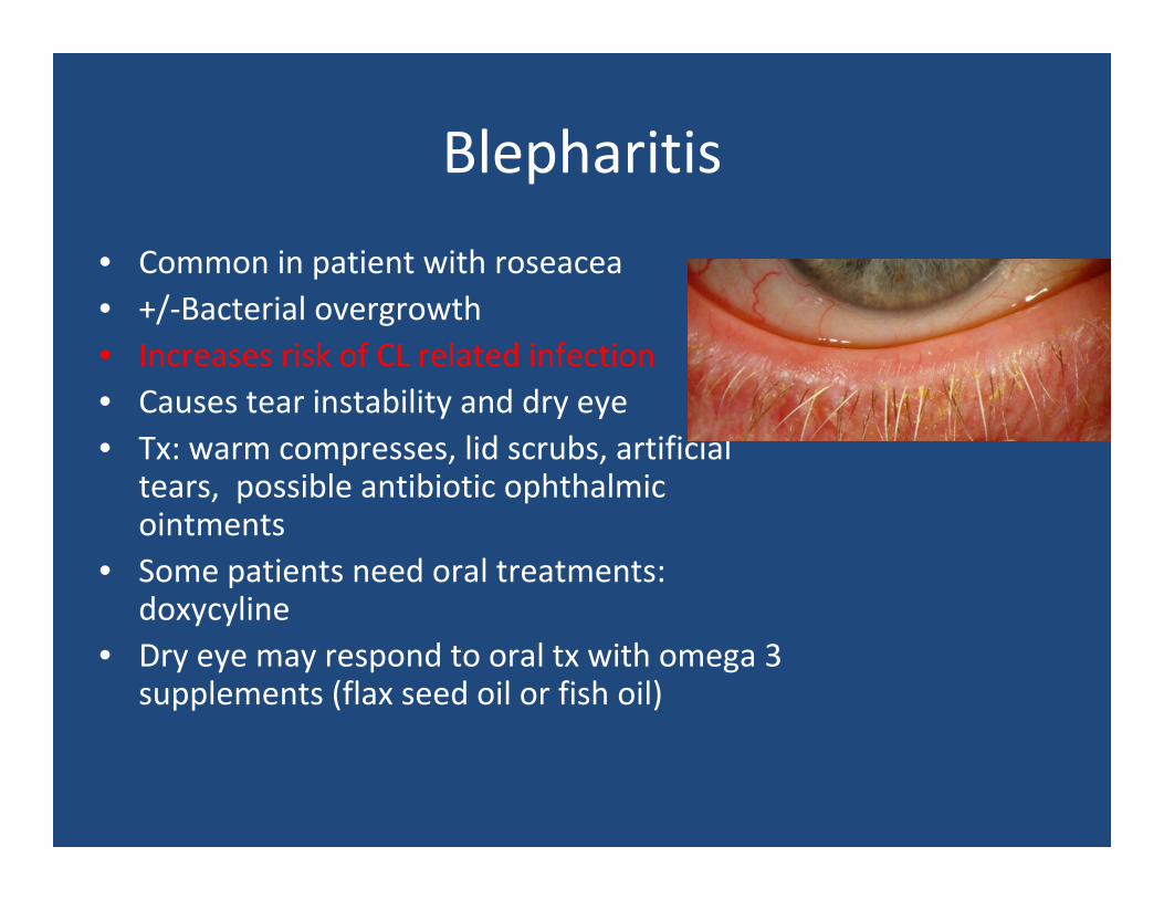
Blepharitis
• Common in patient with roseacea• +/‐Bacterial overgrowth• Increases risk of CL related infection• Causes tear instability and dry eye• Tx: warm compresses, lid scrubs, artificial
tears, possible antibiotic ophthalmic ointments
• Some patients need oral treatments: doxycyline
• Dry eye may respond to oral tx with omega 3 supplements (flax seed oil or fish oil)
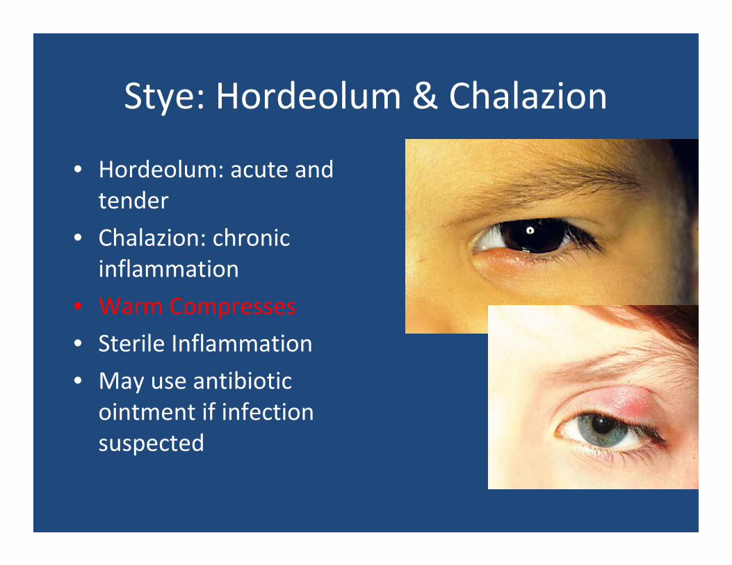
Stye: Hordeolum & Chalazion
• Hordeolum: acute and tender
• Chalazion: chronic inflammation
• Warm Compresses
• Sterile Inflammation
• May use antibiotic ointment if infection suspected
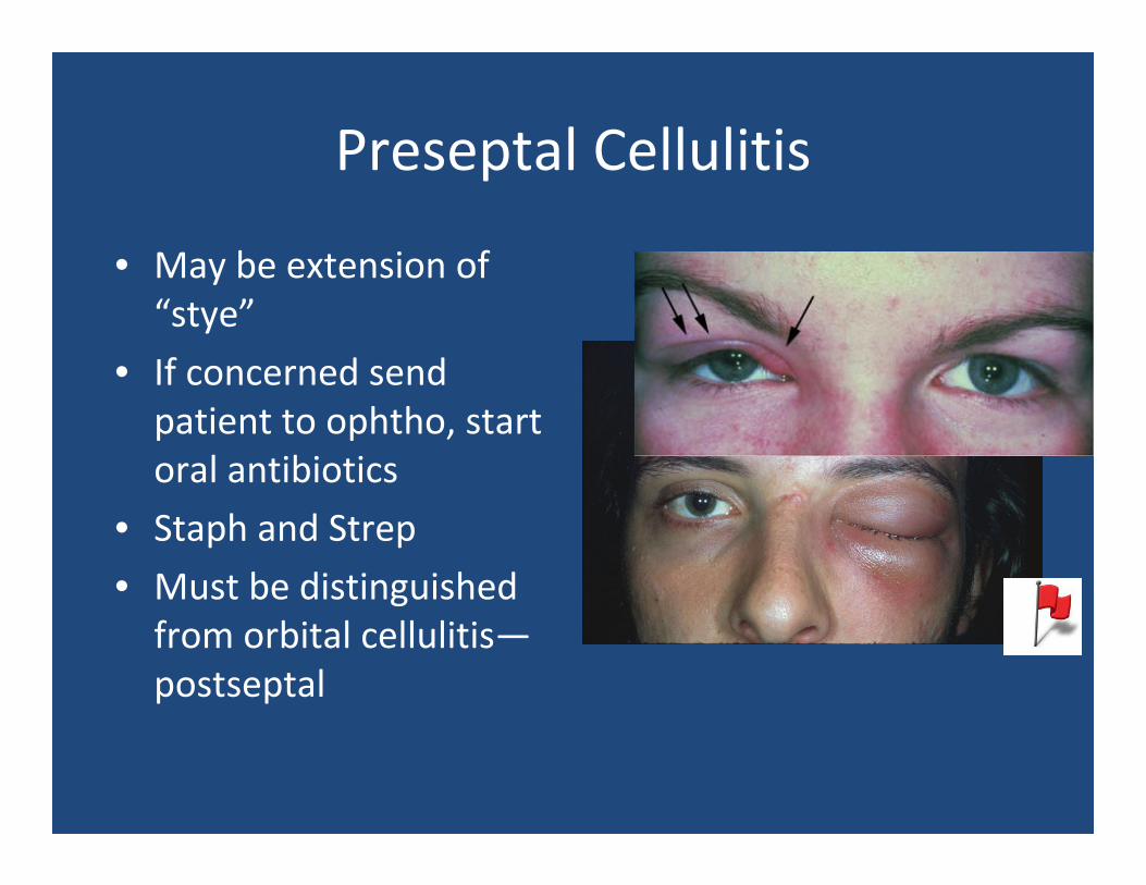
Preseptal Cellulitis
• May be extension of “stye”
• If concerned send patient to ophtho, start oral antibiotics
• Staph and Strep
• Must be distinguished from orbital cellulitis—postseptal
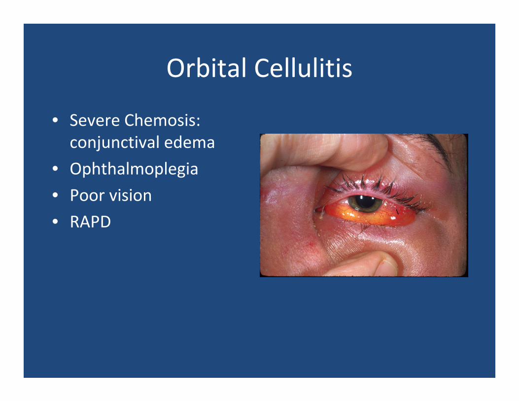
Orbital Cellulitis
• Severe Chemosis: conjunctival edema
• Ophthalmoplegia
• Poor vision
• RAPD
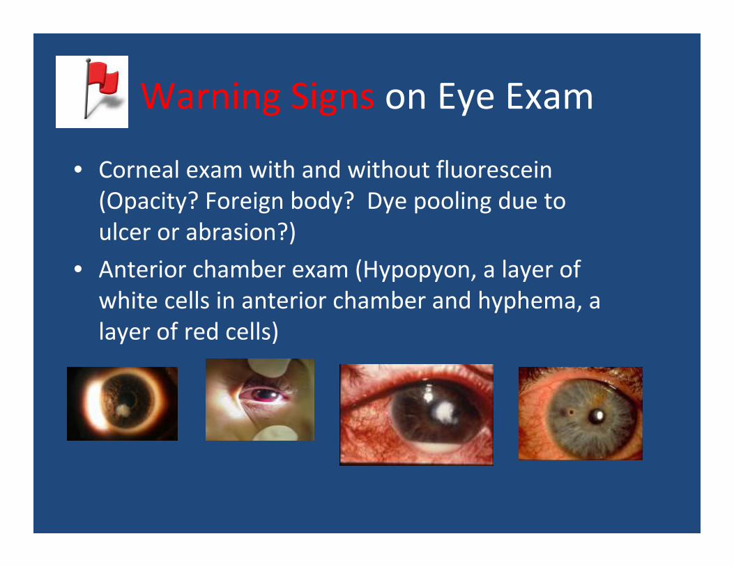
Warning Signs on Eye Exam
• Corneal exam with and without fluorescein(Opacity? Foreign body? Dye pooling due to ulcer or abrasion?)
• Anterior chamber exam (Hypopyon, a layer of white cells in anterior chamber and hyphema, a layer of red cells)
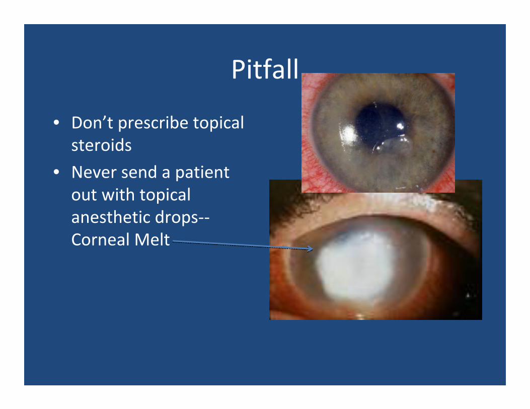
Pitfall
• Don’t prescribe topical steroids
• Never send a patient out with topical anesthetic drops‐‐Corneal Melt

Problems with PupilsBigger Problems
• Relative Afferent Pupillary Defect
• Mydriasis: Dilated pupil
• Miosis: Constricted pupil
• Irregular: Trauma, uveitis
• Anisocoria 20% of normal population
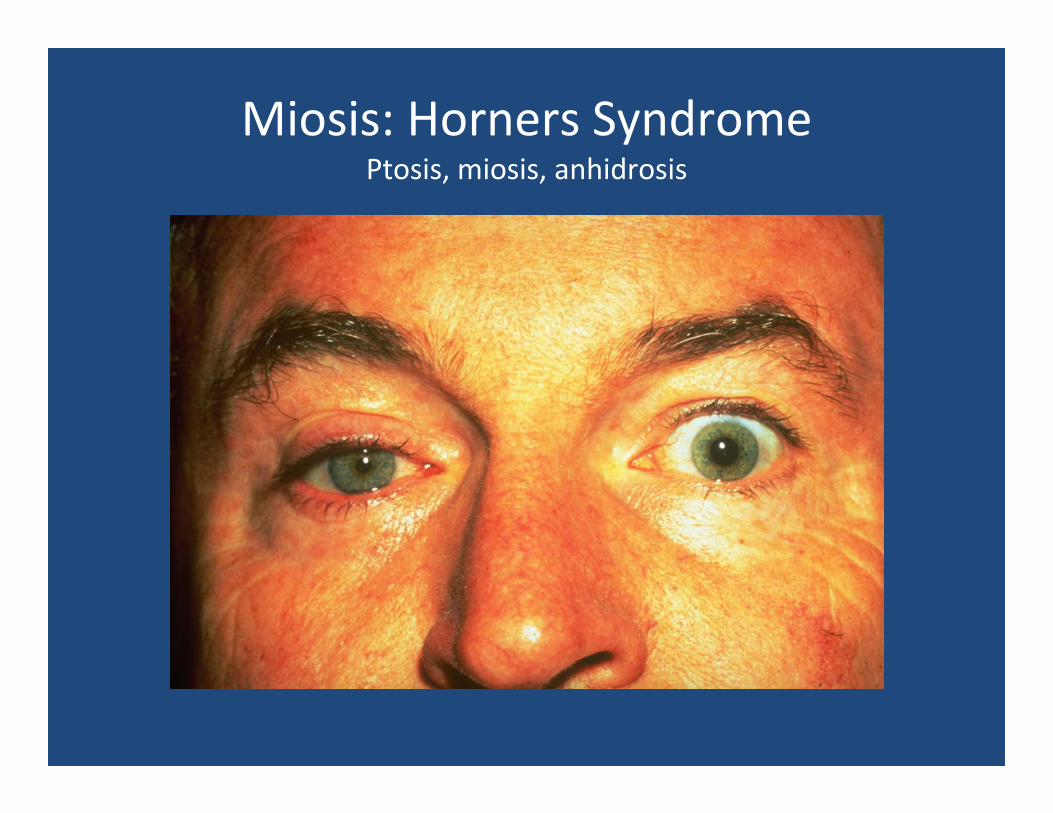
Miosis: Horners SyndromePtosis, miosis, anhidrosis
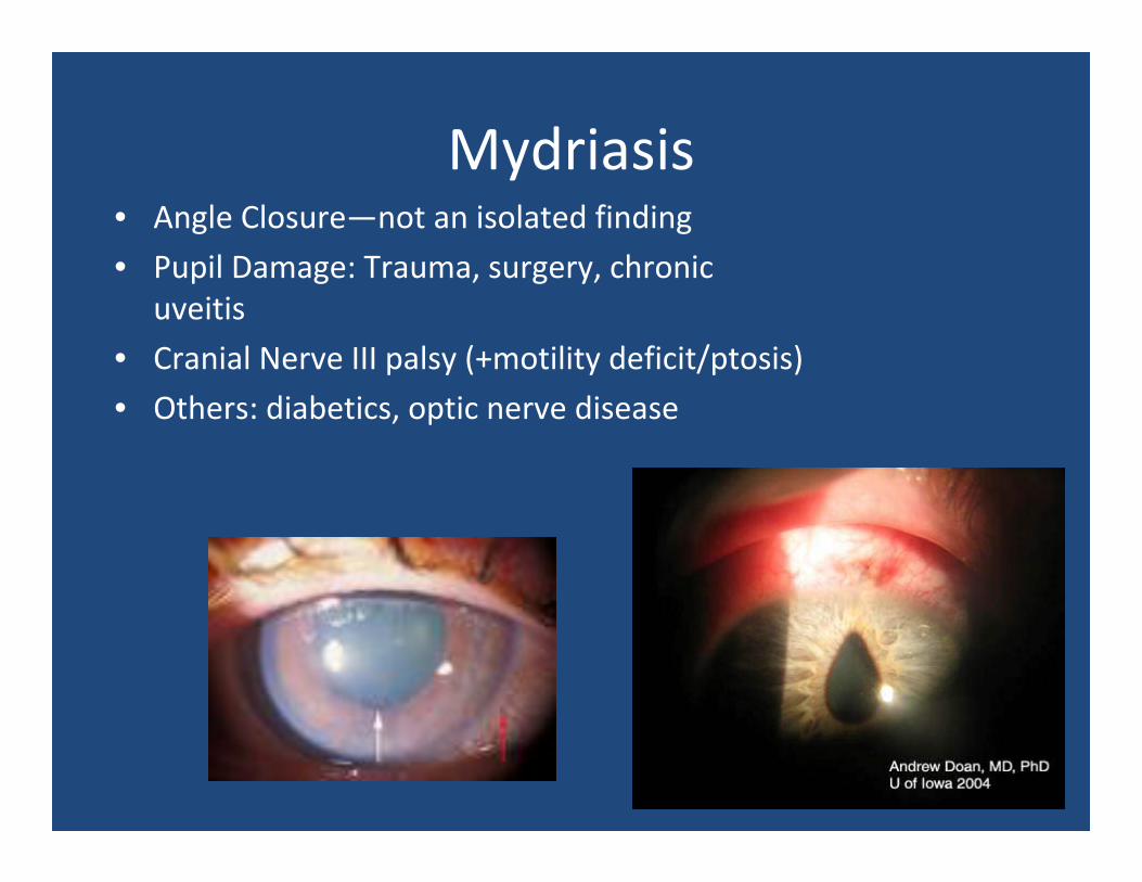
Mydriasis• Angle Closure—not an isolated finding
• Pupil Damage: Trauma, surgery, chronic uveitis
• Cranial Nerve III palsy (+motility deficit/ptosis)
• Others: diabetics, optic nerve disease
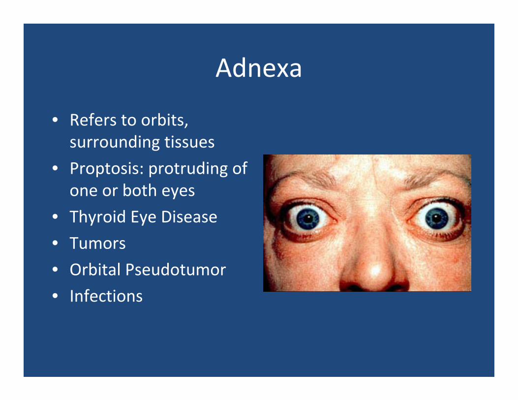
Adnexa
• Refers to orbits, surrounding tissues
• Proptosis: protruding of one or both eyes
• Thyroid Eye Disease
• Tumors
• Orbital Pseudotumor
• Infections
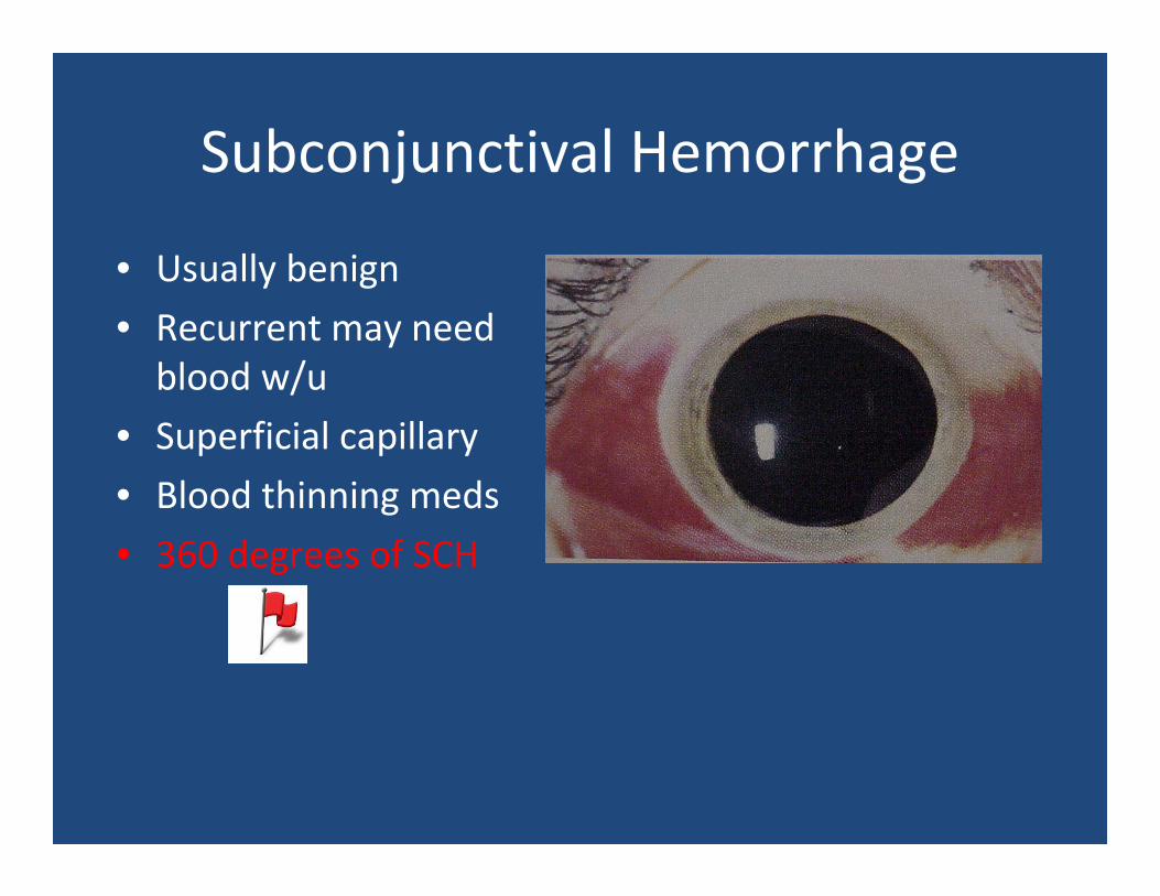
Subconjunctival Hemorrhage
• Usually benign
• Recurrent may need blood w/u
• Superficial capillary
• Blood thinning meds
• 360 degrees of SCH
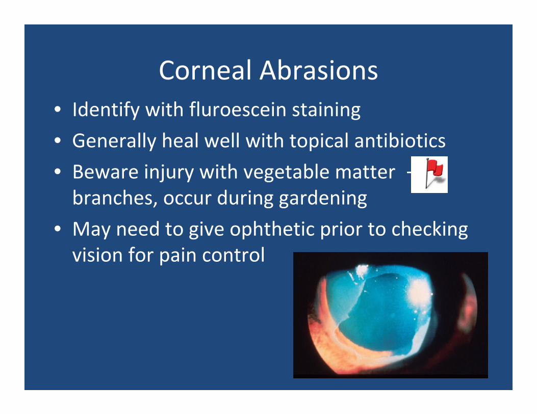
Corneal Abrasions• Identify with fluroescein staining
• Generally heal well with topical antibiotics
• Beware injury with vegetable matter —branches, occur during gardening
• May need to give ophthetic prior to checking vision for pain control
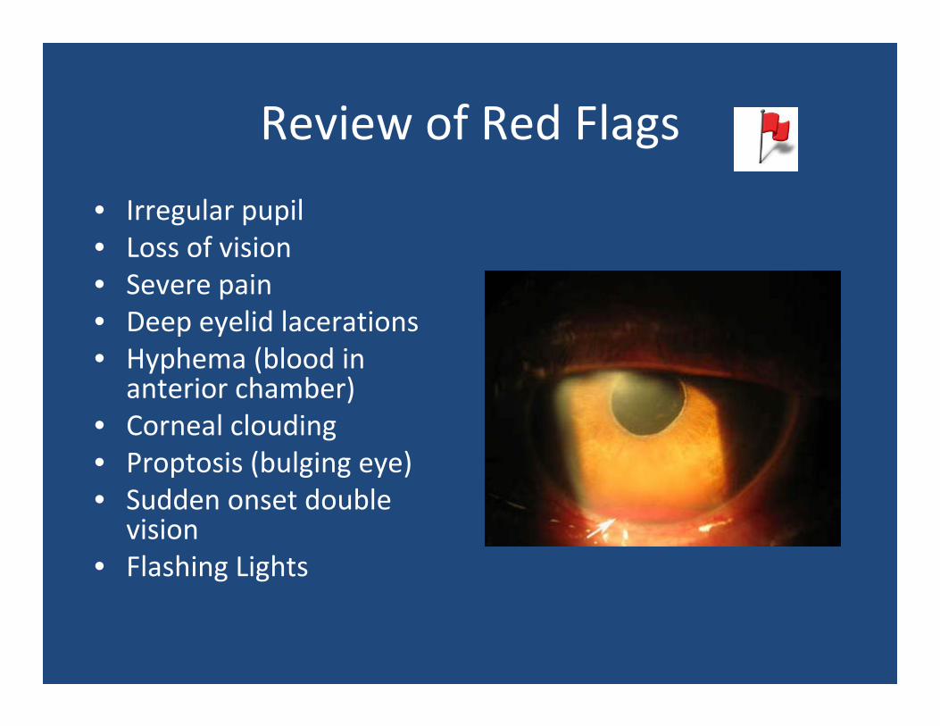
Review of Red Flags
• Irregular pupil• Loss of vision• Severe pain• Deep eyelid lacerations• Hyphema (blood in
anterior chamber)• Corneal clouding• Proptosis (bulging eye)• Sudden onset double
vision• Flashing Lights

A thought about medications

23 yo male presents to you for moderate SOB and wheezing
• HPI– 5 days ago hit in eye during basketball game
– Seen in ED and by ophtho
– Given eye drops for elevated pressure
DDX?
Concomitant Chest Trauma
Bronchospasm ?
Upon questions notes that parent told him he had brief episode of “bronchitis” as child
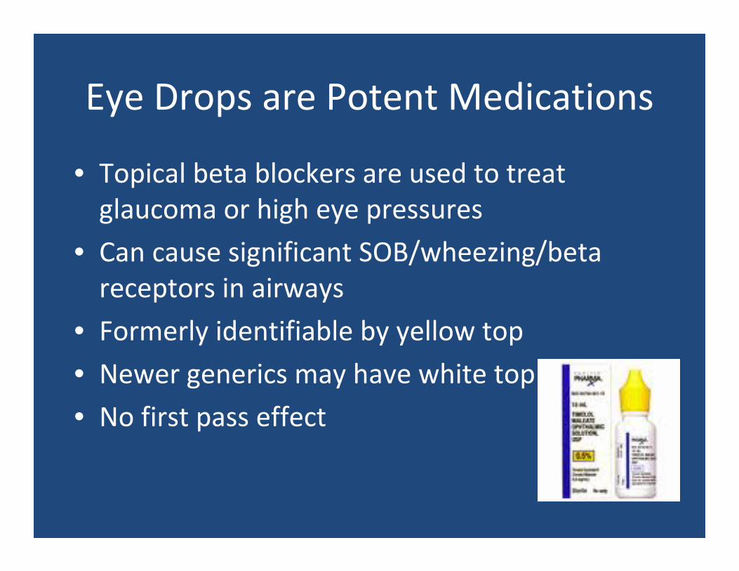
Eye Drops are Potent Medications
• Topical beta blockers are used to treat glaucoma or high eye pressures
• Can cause significant SOB/wheezing/beta receptors in airways
• Formerly identifiable by yellow top
• Newer generics may have white top
• No first pass effect
Pitfall case: 25 yo female present with sudden loss of vision right eye, no double vision, gets eye pain when
she moves her eye
Vision is 20/100
• Eye appears normal on your exam
• You attempt to look at ON with ophthalmoscope—looks normal
• What should you do?
REFER
Optic Neuritis
• May be seen in young females, sometimes in the student health population
• May be associated with MS
• May be associated with infectious disease
• May be associated with autoimmune disorders
• Vision is generally poor
• Requires specialty care and imaging
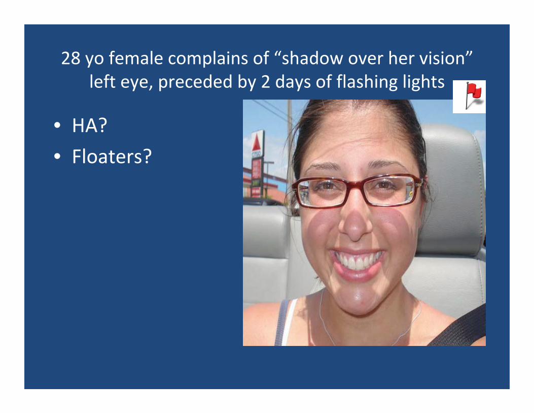
28 yo female complains of “shadow over her vision”left eye, preceded by 2 days of flashing lights
• HA?
• Floaters?
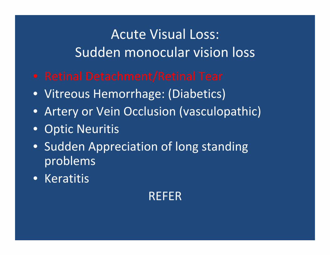
Acute Visual Loss: Sudden monocular vision loss
• Retinal Detachment/Retinal Tear• Vitreous Hemorrhage: (Diabetics)• Artery or Vein Occlusion (vasculopathic)• Optic Neuritis• Sudden Appreciation of long standing problems
• KeratitisREFER
30 yo female student c/o gradually decreasing vision in left eye
• Had been seen by optometrist who gave a new pair of glasses but this doesn’t seem to help the problem
• You do full exam and it is normal
• You refer her to neurology—find nothing wrong with her—suggest stress as possible etiology
• Where is the red flag?
• Persistent vision loss in young person
Refer to Ophthalmology
• If patient has persist unresolved sx, be sure he/she sees an ophthalmologist
• Evaluation by optometrist may not be medical, as with all professions, it may vary widely
• Dx: Orbital Tumor (hemangioma, meningioma etc.)
• Exceedingly rare
• Take home point is that chronic vision loss should have a clear diagnosis
Refractive Surgery
• LASIK and other options
• Lens Implants
• INTACS
• At Scheie, prefer patient of age greater than 25—stable refraction
• Consider surgery if patient running into trouble with CL
Ophthalmology In Primary Care
• Challenge for primary care givers because of limited exam, huge responsibility
• Minimal ophthalmology training for most primary care providers
• Wide breadth of problems
• Vision is your vital sign
Thank you
GO FLYERS!!!!
• AAO GUIDELINES for Recommended Intervals for Regular Eye Exams
• If you have any of these risk factors for eye problems, you may need to see your Eye M.D. more often than recommended below:
• Family history of eye problems• African American over age 40• Diabetes• History of eye injury
AAO GUIDELINES FOR SCREENINGStudent Health Age Group
• Age 20 to 39• Most young adults have healthy eyes, but they still need to
take care of their vision by wearing protective eyewear when playing sports, doing yard work, working with chemicals, or taking part in other activities that could cause an eye injury.
• Have a complete eye exam at least once between the ages of 20 and 29 and at least twice between the ages of 30 and 39.
• Also, be aware of symptoms that could indicate a problem. See an Eye M.D. if you experience any eye conditions, such as:
• Visual changes or pain• Flashes of light• Seeing spots or ghost‐like images• Lines appear distorted or wavy• Dry eyes with itching and burning
• Age 40 to 64: AAO GUIDELINES• As of July 2007, the American Academy of Ophthalmology has issued a new eye disease screening
recommendation for aging adults.• The Academy now recommends that adults with no signs or risk factors for eye disease get a baseline
eye disease screening at age 40—the time when early signs of disease and changes in vision may start to occur. Based on the results of the initial screening, an ophthalmologist will prescribe the necessary intervals for follow‐up exams.
• For individuals at any age with symptoms of or at risk for eye disease, such as those with a family history of eye disease, diabetes or high blood pressure, the Academy recommends that individuals see their ophthalmologist to determine how frequently their eyes should be examined.
• The new recommendation does not replace regular visits to the ophthalmologist to treat ongoing disease or injuries, or for vision examinations for eye glasses or contact lenses. Much like mammograms at 40 or colon screenings at 50, this new eye disease screening is a reminder to adults as they age that they need to maintain their eye health.
• Why the New Recommendation?• A baseline evaluation is important because it may detect eye diseases common in adults aged 40 and
older. The evaluation creates greater opportunity for early treatment and preservation of vision.• A thorough ophthalmologic evaluation can uncover common abnormalities of the visual system and
related structures, as well as less common but extremely serious ones, such as ocular tumors. This evaluation can also uncover evidence of many forms of systemic disease that affect the eyes, like hypertension and diabetes. With appropriate intervention, potentially blinding diseases such as glaucoma, cataract and diabetic retinopathy often have a favorable outcome.
• Several common eye diseases can impact people 40 and older without them knowing there is any problem with their eyes.
• Age 65 and Over• Seniors age 65 and over should have complete eye exams by their Eye M.D. every one to two years to
check for cataracts, glaucoma, age‐related macular degeneration, diabetic retinopathy and other eyeconditions.