Embed Size (px)
Citation preview
OphthalmonicsOphthal monicsmology Mne
Dhaval Patel
MD (AIIMS)
1st Edition
Ophthalmonics(Ophthalmology Mnemonics)
MD (AIIMS)Dhaval [email protected]
by ophthalmonics.blogspot.com1st edition, January 2014
This is a compilation effort from my preparation notes and other sources, thusany contributions or comments are welcomed in the effort to improve this book.Therefore, feel free to e mail me at-
Ophthalmonics Dhaval Patel MD
1
OPHTHALMONICS
(Ophthalmology Mnemonics)
Thank you GOD
This manual is collection of the mnemonics I made, found in books or internet
when studying for the Final MD exam and Senior Residency Entrance Exam in
ophthalmology. Till last few months of exam, I believed that I will understand and
remember all important facts and will hardly need specially devised mnemonics for it.
As exam fever comes nearer, all things started to evaporate.
I do not pretend that this manual will cause a lot improvement in your
preparation, despite that, I am proud of what I have produced and hope you will find
it a useful memory aid and help to increase your confidence in memorizing some
confusing but important facts!
Many of these may be just simple fundamentals, but during your exams or MCQ
test, there may not be time to recollect your basics and you may give wrong answer. I
hope this manual of mnemonics may help to reduce your evaporation.
I hope it may help you to seal or laser your breaks of knowledge.
Good luck!
-Dhaval Patel MD
January 2014
Ophthalmonics Dhaval Patel MD
2
Basic Sciences .................................. 7
Optics ......................................... 22
Cornea ........................................ 29
Lens ........................................... 48
Glaucoma ..................................... 57
Neurophthalmology ......................... 62
Strabismus .................................... 68
Retina ......................................... 77
Uvea ........................................... 86
Oculoplasty ................................... 93
Community Ophthalmology ............... 104
Miscellaneous ............................... 107
Ophthalmonics Dhaval Patel MD
3
Basic Sciences Heterochromia Hypersensitivity Spindled cell Tumors Achromatopsia: Cells in Plexiform Layer Visual acuity VA tests Lacrimal Gland Treponemal Tests DNA viruses Synaptic Body Visual Cycle Clues Differentiation of Retinal Cells Corneal quadrants Ishihara Clues Color vision deficiency Dimensional characteristics of the optic nerve IOP-elevating potential LASERs LASER Properties Sterilization in ophthalmology Neuroectoderm Surface ectoderm Mesoderm Neural crest Refractive indices Cavernous Sinus Optic Nerve Stroma of Choroid Medial orbital wall Floor of Orbit Nerves outside Annulus at SOF Whitnall tubercle attachment Retinal Layers Ciliary Epithelial Layers Ophthalmic Artery branches Superior Ophthalmic Vein Facial Blocks Hyaloid Remnants Stimulus for Goldman Perimetry Lacrimal Duct TGFB1 and Dystrophies Wilbrand’s Knee Combination H1 antagonists/mast cell inhibitors
Optics Galilean telescope Hard lens Bending of Light Ray Soft lens IMAGE Basic Lens Formula: PROPERTIES OF LIGHT Axis of Eye Angles of the Eye Reflex convergence Hyperopia Direct ophthalmoscope Prism Deviation Focal Points Aberrations of Thick lenses Near triad Hypermetropia types Myopia types Berliner’s seven methods of Slit lamp illumination Pentacam Specular Zones RAF Ruler Topography color Maps
Cornea Schirmer's test Adenovirus Bacterial adhesion Bacteria which can invade corneal epithelium primarily: Non-infectious from suppurative infiltrates Epithelial cells of the limbus and central cornea Follicles and Papillae Gland of Conjunctiva Fungal Corneal Ulcer Deep corneal Neovascularization Waring’s Classification of Congenital corneal opacities 5 Layers of Amniotic Membrane Amniotic Membrane Components Autologus serum making Blue Sclera Prominent Corneal Nerves More visible corneal nerves Enlarged corneal nerves
Ophthalmonics Dhaval Patel MD
4
Causes of chronic catarrhal conjunctivitis Treatment of dry eye Layers of Cornea Cornea Verticiliata: (Vortex Keratopathy) Red Eye Alkali Injury: Rate of Penetration Keratoconus Signs Keratoconus Features Trachoma Acute hemorrhagic conjunctivitis Spring Catarrh Reis-Buckler dystrophy Tears: Composition Interstitial keratitis: Causes Stromal dystrophies Membranous or pseudomembranous conjunctivitis Filamentous keratitis Megalocornea Tests for Dry Eye Work-up Randleman’s post-LASIK Ectasia risk factors Rabinowitz Criteria Hughes Classification and prognosis in acid injuries of the eye Chemical Injury Management Guidelines Filamentous Fungi Antifungal Side Effects Non Infective PUK LASIK Flap Complications Iris Atrophy
Lens OVD Characteristics Grading of nucleus hardness Local causes of complicated cataract Cataract DD Microspherophakia Differential diagnosis of leukocoria in infants Drugs causing cataract Weil Marchesani Syndrome Posterior Subcapsular Cataract Anterior Subcapsular Cataract Iris shadow Ectopia Lentis: Causes IOL generations
LenStar Steroid Induced Cataract IOL Power post refractive surgery Apple’s six factors for PCO prevention
Glaucoma Genes in Glaucoma Jonas ISNT rule Steroid Induced Glaucoma: Pathogenesis Spaeth Grading System Secondary Glaucoma Iridocorneal Endothelial Syndrome Trabecular pigmentation Angle structures Buphthalmos Indiana Bleb Grading System Beta Blocker Side Effects Neuroprotection in Glaucoma
Neurophthalmology Supranucear Eye movement control Optic Chiasma Optic Atrophy VEP in AION Pseudotumor cerebri Downbeat nystagmus Physiologic Nystagmus Aetiological classification of Optic Neuritis Horner Syndrome diagnosis Visual field defects Nystagmus Visual Cortex Papilloedema: Clinical features Argyll Robertson pupil (ARP) Small pupils Parinaud’s Syndrome Uniocular diplopia Pupillary Fibres Toxic Amblyopia
Strabismus Eye movements Muscle Actions Nerve supply to EOM Angle of muscles Insertion of Recti Exceptions to Law Deviations of Eye Sagitalization and Desagitalization Amblyopia Types

Ophthalmonics Dhaval Patel MD
5
Amblyopia Management Squint management Rule of 6 Anomalies of binocular vision Fourth nerve palsy DRS Types Uniocular diplopia Crossed-Uncrossed Diplopia Microtropia Congenital nystagmus Nystagmus description Actions of Superior oblique muscle A and V patterns FADEN operation Vergence Amplitude Named Transposition Surgeries
Retina Retina Blood Supply Angioid Streaks Bardet–Biedl syndrome Peripheral Retinal Degeneration Drusen DD Pseudoglioma Congenital Leucocoria DD Shields Staging of Coats Disease Hyperfluoroscence in FA FFA Functions of RPE CME Retinal Examination Choroidal neovascular membrane Goldmann’s 3 mirror lens Bull’s Eye Maculopathy Drug induced Maculopathy Rubeosis Iridis Salt and Peeper Retinopathy Cherry Red Spot: DD Treatment of Retinal Detachment Background DR Preproliferative DR PVR Grade B Goldberg Staging of Sickle Retinopathy
Uvea Seclusio and Occlusio Suspicious Choroidal Nevi Nodules in uveitis: Granulomatous Uveitis Causes Vitreous Seeds DD 4 signs of POHS
Behcet's Disease Reiter Syndrome Ophthalmic Tuberculosis Posterior scleritis features Vogt Koyanagi Harada Syndrome Immunosuppressants Seronegative spondyloarthropathies Kaplan’s 4 step management of Intermediate Uveitis Revised criteria for diagnosis of VKH
Oculoplasty Retinoblastoma: International Classification Group E Retinoblastoma Clark and WHO 2006 classification of Malignant Melanoma Lid coloboma Merkel Cell Carcinoma Ptosis classification Werner’s Classification for Grave’s Ophthalmopathy Thyroid-Related Orbitopathy Thyroid Ophthalmopathy muscle involement Epicanthal Folds Periorbital Cellulitis Chandler’s Staging of Orbital Cellulitis Orbital Pathology Exophthalmos Signs of Acute Dacryocystitis Clinical Picture of Symblepharon Post Enucleation Socket Syndrome Hordeolum Lid retraction Craniosynostosis Chalasis of Lids TRO features Thyroid Eye Disease Surgery Eye signs of thyrotoxicosis Umbilicated Eyelid lesion Rhabdomyosarcoma types HP types of Adenocystic carcinoma Acquired Entropion Acquired Ectropion Bleprophimosis Syndrome BPES
Community Ophthalmology Blindness Vision 2020 Disease prevention and control
Ophthalmonics Dhaval Patel MD
6
Vision 2020 Strategic approaches NPCB main objectives WHO Primary Eye Care Elements Primary Eye Care Principles Childhood blindness Estimation Methods WHO’s SAFE Trachoma Strategy
Miscellaneous Ocular Drug Delivery Systems Zones of Operation Theater Systemic Steroids Side Effects Basal View (Submentovertical View)
Caldwell Luc View Water’s View (OM) Gradenigo syndrome WOLFRAM syndrome GEMSS Syndrome Waardenburg Syndrome Waardenburg Syndrome GENES Lyme Disease Necrobiotic Xanthogranuloma Ocular features of acromegaly Systemic features of Marfan syndrome Ocular features of Marfan’s syndrome
Ophthalmonics Dhaval Patel MD
7
Basic Sciences
Heterochromia
Difference in colour of the iris in the same eye is called heterochromia iridis.
Difference in colour between the iris of the two eyes is called heterochromia iridium.
Hypersensitivity
OCP is type 2-twO hypersensitivity reaction.
SJS-EM-TEN is type 3-thrEE hypersensitivity reaction.
Spindled cell Tumors
SLAM
o Spindle cell SCC - Keratin +
o Leiomyosarcoma - Smooth muscle actin (SMA)+
o Atypical fibroxanthoma (AFX) - CD68+, Vimentin+, CD99+, Procollagen-1 but really
considered diagnosis of exclusion
o Melanoma - S100+
Achromatopsia:
BuT GoD RePly
Blue: Tritanopia
Ophthalmonics Dhaval Patel MD
8
Green: Deuteranopia
Red: Protanopia
Cells in Plexiform Layer
The outer plexiform layer is composed of interconnections between photoreceptor
synaptic bodies, horizontal cells, and bipolar cells. The inner plexiform layer is composed
of connections between bipolar cells, amacrine cells, and ganglion cells.
Outer PHoB
Inner BAG
Visual acuity
ViSoCo Separation
3 components of VA
o Detection of presence or absence of stimulus, i.e. Minimum visible/ detection
o Ability to distinguish between more than one identifiable feature in a visible
target, i.e. Minimum resolvable.
o Minimum recognisable
o Judgement of location of visual target relative to another element of the same
target, i.e. Minimum separable Hyperacuity
VA tests
DeSoCo (Disco..!!)
Ophthalmonics Dhaval Patel MD
9
o Detection acuity tests: catford drum, stycar graded ball, boeck candy, dot visual
acuity
o Resolution acuity tests: OKN, VEP, PLT(teller card)
o Recognition acuity tests:
Direction identification: sjogren’s hand test, landolt’s C, snellen’s E, arrows
Letter identification: snellen, sheridan’s, lipman’s HOTV, fook’s dymbol
Picture identification
Lacrimal Gland
Orbital lobe of lacrimal gland is anterior and Palpebral lobe is posterior.
??? how can this basic anatomy has controversy…ooops..but I think it is controversial.few
books write palpebral is anterior and orbital is posterior.!!
Yes…!! Duane’s 2007 says that it’s actually Superior and Inferior. Orbital is superior and
Palpebral is posterior…but by reading it thoroughly, the above controversial line seems
true…still it’s difficult to believe for me. Many other textbooks quotes reverse..!! or we
can just remember it like superior and inferior lobes of the lacrimal gland. I don’t know
exactly which is anterior and which is posterior..!!
Treponemal Tests
Fluorescent treponemal antibody-absorption (FTA-ABS) and microhemagglutination of
Treponema pallidum (MHA-TP) are the closest to a “gold standard” for syphilis testing.
The Venereal Disease Research Laboratory (VDRL) and rapid plasma reagin (RPR) tests reflect
treponemal infection and revert to normal when treated.
Ophthalmonics Dhaval Patel MD
10
FTA, MHA are Always positive
VDRL, RPR tests Reverts back to normal
DNA viruses
HHAPPPPPy
o Herpes - HSV, VZV, CMV (blueberry muffin baby), Roseola (HHV6 and 7 - half a
dozen roses), Kaposi's (HHV8), possibly PR
o Hepadna - Hepatitis B
o Adeno
o Papilloma= HPV. 6,11,16,18 in Gardasil. 6,11 = warts. 16,18 = dysplasia; E6-p53;
E7-Retinoblastoma
o Polyoma = JC John Cunningham Virus - PML - why Efalizumab taken off market;
Merkel Cell = CK20 - paranuclear dot
o Papova = old name that includes Papilloma and Polyoma
o Pox = Molluscum, Smallpox, Vaccinia; (Orf = parapox)
o Parvo B19 = slapped cheeks, lacy rash, anemia, joint pain in adults. Bad in
pregnancy, sickle cell.
Synaptic Body
The synaptic body of a rod is called a spherule, whereas that of the cone is called a
pedicle.
Visual Cycle Clues
Ophthalmonics Dhaval Patel MD
11
lighT phase: 11 Trans retinal with opsin
dark phase: 11 cis retinal with opsin
Dark: Depolarization of photoreceptor Displays NT (release of NT)
Light: Hyperpolarization of photoreceptor Hides NT (no release of NT)
Differentiation of Retinal Cells
GCAHRBM God Can Always Help Revealing Best Messages.
The inner layer of the optic cup contains the pluripotent retinal progenitor cells, which
differentiate in a specific chronologic sequence and defined histogenic order into the final
seven retinal cell types. In general, the Ganglion cells differentiate first, followed by the
cone photoreceptors, amacrine cells, horizontal cells, and finally, the rod photoreceptors,
bipolar cells, and Müller cells.
Corneal quadrants
TINS (thickness in descending order)
temporal (28%), inferior (19%), nasal (11%), and superior (4%).
(so remember this..its not like ISNT of glaucoma)
Ishihara Clues
Reads first 7 plates (except “12”) incorrectly and unable to read the rest: red-green
deficiency
Reads “26” as 6 and “42 as 2: protan defect
Reads “26 as 2 and “42 as 4: deutan defect
Ophthalmonics Dhaval Patel MD
12
Remember Dhaval Patel : from 26 & 42, if one reads first letter correctly, its
Deutan, if second letter correctly, its Protan. (D for first and P for second. Think
about it once)
Color vision deficiency
Retinal disease: Blue yellow defect Ru-BY
Optic Nerve disease: Red Green defect O-RGan
Dimensional characteristics of the optic nerve
125-1017
intraocular (1)
intraorbital (25)
intracanalicular (10)
intracranial (17)
IOP-elevating potential
DPLFHT
in decreasing order
dexamethasone > prednisolone > loteprednol etabonate > fluorometholone >
hydrocortisone > tetrahydrotriamcinolone.
LASERs
Ophthalmonics Dhaval Patel MD
13
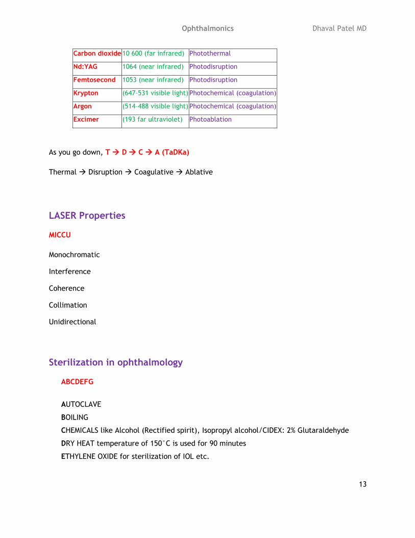
Carbon dioxide 10 600 (far infrared) Photothermal
Nd:YAG 1064 (near infrared) Photodisruption
Femtosecond 1053 (near infrared) Photodisruption
Krypton (647–531 visible light) Photochemical (coagulation)
Argon (514–488 visible light) Photochemical (coagulation)
Excimer (193 far ultraviolet) Photoablation
As you go down, T D C A (TaDKa)
Thermal Disruption Coagulative Ablative
LASER Properties
MICCU
Monochromatic
Interference
Coherence
Collimation
Unidirectional
Sterilization in ophthalmology
ABCDEFG
AUTOCLAVE
BOILING
CHEMICALS like Alcohol (Rectified spirit), Isopropyl alcohol/CIDEX: 2% Glutaraldehyde
DRY HEAT temperature of 150°C is used for 90 minutes
ETHYLENE OXIDE for sterilization of IOL etc.

Ophthalmonics Dhaval Patel MD
14
FUMIGATION of operation theatre/ FORMALIN vapour
GAMMA-IRRADIATION: Gamma rays from Cobalt-60
Neuroectoderm
MORE
Muscles of pupil
Optic Nerve
Retina (with RPE)
Epithelium of Iris
Epithelium of Cilliary Body
Surface ectoderm
S1 L2 E3 (you can remember SLE- which is surface ectoderm disease)
Skin of Eyelids and its derivatives viz. cilia, tarsal glands, conjunctival gland
Lens,
Lacrimal Gland,
Epithelium of Conjunctiva,
Epithelium of Cornea,
Epithelium of lacrimal passage
Mesoderm
MeSS
Extraocular muscles
Sclera (small area temporally)
Ophthalmonics Dhaval Patel MD
15
Schlemm's canal
Neural crest
STOC’S
Stroma of Iris and ciliary body
Trabecular meshwork
Orbital cartilage and bone
Ciliary muscles
Corneal stroma and endothelium
Connective tissue of extraocular muscles
Sclera
Refractive indices
8303 (from anterior to posterior)
cornea 1.38
aqueous humour 1.33
lens 1.40
vit humour 1.33
Cavernous Sinus
Rule of 3
Ophthalmonics Dhaval Patel MD
16
3 Afferent veins: Sphenoparietal sinus (Vault veins), Superficial Middle cerebral Vein
(Brain), Ophthalmic vein (Orbit)
3 Efferent Veins: Superior petrosal sinus, Inferior Petrosal Sinus, Communicating vein to
pterygoid plexus
3 Contents; Cranial Nerves (III,IV, V1,V2 & VI)
3 Areas Drain into it: Vault Bones, Brain (Cerebral Hemisphere), Orbit
3 Nerves: Motor(III,IV,VI),Sensory (V1,V2), Sympathetic
Optic Nerve
Optic Nerve head as Nasal side, so blind spot is temporal side.
Stroma of Choroid
haLLer layer: Larger, outer
Sattler later: Smaller, inner
Medial orbital wall
Nose it at medial side of orbit, which SMEL
Sphenoid (lesser wing)
Maxilla
Ethmoid
Lacrimal
Floor of Orbit
Ophthalmonics Dhaval Patel MD
17
PZM (PayZaMa)
Palatine
Zygoma
Maxilla
Nerves outside Annulus at SOF
LFT
Lacrimal
Frontal
Trochlear
Whitnall tubercle attachment
4 “L”
Levator aponeurosis
Ligament of LR (check ligament)
Lockwood’s ligament
Lateral canthal ligament
Retinal Layers
10 –outer to inner
RPE o-NP i-NP GNI
Ophthalmonics Dhaval Patel MD
18
RPE
Photoreceptor layer
ELM
Outer Nuclear
Outer Plexiform
Inner Nucelar
Inner Plexiform
Ganglion cell layer
Nerve Fiber layer
ILM
Ciliary Epithelial Layers
OPIN
Outer Pigmented
Inner Non-pigmented
Generally if you know embryology, you don’t need this but still..
Ophthalmic Artery branches
PALS MS MD
Posterior ethmoidal
Anterior ethmoidal
Ophthalmonics Dhaval Patel MD
19
Lacrimal
Supratrochlear
Muscular
Supraorbital
Medial palpebral
Dorsal nasal
Superior Ophthalmic Vein
PALS MAC
Posterior ethmoidal
Anterior ethmoidal
Lacrimal
Superior vortex
Muscular
Anterior ciliary veins
Central retinal vein
Facial Blocks
LOAN
van Lint’s block: Blocking the peripheral branches of facial nerve
O’Brien’s block: Facial nerve trunk block at the neck of mandible
Ophthalmonics Dhaval Patel MD
20
Atkinson’s block: In it superior branches of the facial nerve are blocked by injecting
anaesthetic solution at the inferior margin of the zygomatic bone.
Nadbath block: facial nerve is blocked as it leaves the skull through the stylomastoid
foramen.
Hyaloid Remnants
B is Behind
Bergmeister's papilla: at optic nerve
Mittendorf dot: behind lens
Stimulus for Goldman Perimetry
Roman numeral: Size of the target in square millimeters (Each successive number is an
increase by a factor of four)
Arabic numeral: Intensity of the light presented (Each successive number is 3.15 times
brighter than the previous one)
Lowercase letter: Minor filter (The ‘‘a’’ is the darkest, and each progressive letter is an
increase by 0.1 log unit)
Lacrimal Duct
IPL
It passes inferiorly, posteriorly and laterally.
TGFB1 and Dystrophies
Ophthalmonics Dhaval Patel MD
21
GREAT
Granular
Reis Buckler
EBMD
lAttice
Thiel Behnke
TGFB1: aka BIGH3, 5q31.2, Protein produced by corneal epithelium, Phenotypic
heterogeneity
Wilbrand’s Knee
Just think this “KNEE” as “NI” and you have the answer Nasal and Inferior
The inferior nasal retinal fibers cross in the anterior chiasm and are thought to loop
anteriorly in the contralateral optic nerve before traveling posteriorly, leading to the term
Wilbrand's knee (NI=Nasal + Inferior). It is now thought that Wilbrand's knee may be an
artifact.
Combination H1 antagonists/mast cell inhibitors
POKAN
Ketotifen (Zaditor)
Olopatadine (Patanol)
Nedocromil sodium (Alocril)
Azelastine hydrochloride (Optivar)
Pemirolast (Alamast)
Ophthalmonics Dhaval Patel MD
22
Optics
Galilean telescope
o pOsitive lens: Objective lens
o nEgative lens: Eye piece
In keplerian telescope, both are positive lens.
Hard lens
SAM FAP: Steeper Add Minus, Flatter Add Plus
Bending of Light Ray
HLA: Higher to Lower RI Away from normal
When a light ray passes from a medium with a higher refractive index to a medium with a
lower refractive index, is it bent away from the normal
Soft lens
LARS: Left Add Right Substract
IMAGE
DEV DO: Erect Virtual
IIR IO: Inverted Real

Ophthalmonics Dhaval Patel MD
23
Basic Lens Formula:
UnDer Vater
U+D=V
(this is very basic of optics and all know and understand it, but still in case if one get
confused, use the mnemonic)
PROPERTIES OF LIGHT
R2D2TIPS
1. Reflection
2. Refraction
3. Dispersion
4. Diffraction
5. Total internal reflection
6. Interference
7. Polarization
8. Scattering
Axis of Eye
FOVea
o Fixation Axis: This is a straight line that joins center of rotation of eyeball with
fixation point

Ophthalmonics Dhaval Patel MD
24
o Optical Axis: A line passing through center of cornea, center of lens and posterior
pole of retina is the optical axis of eyeball
o Visual Axis: A line joining point of fixation with fovea and passing through nodal
point of eyeball is called visual axis. Nodal point of eyeball is just anterior to
posterior capsule of lens. Fixation point is the point which is being seen with fovea
at any particular moment.
o Pupillary Line: This is a straight line that passes through center of pupil
Angles of the Eye
o Angle Alpha is the angle formed between optical axis and visual axis. AOV
o Angle Kappa is the angle formed between visual axis and pupillary axis. KaVPa
o Angle Gamma is the angle formed between optical axis and fixation axis. GOF =
FOG
POsitive angle Kappa results in pseudoeXotropia. K-POX
Reflex convergence
FAT-P
1. Proximal convergence: Psychological awareness of a near object initiates this type
of convergence.
2. Tonic: It means that when the patient is awake there is an inherent tone in the
extraocular muscles.
Ophthalmonics Dhaval Patel MD
25
3. Fusional: It is initiated by a bi-temporal retinal image disparity and is not associated
with change in refractive status of eyeball. It ensures that image of an object falls on
corresponding retinal points in the two eyes.
4. Accommodative: It is initiated by act of accommodation. It means that when we
accommodate; we converge. It is a part of near reflex. One dioptre of accommodation
is accompanied by 4-5 prism diopters of accommodative convergence and it remains
fairly constant. Abnormalities of accommodative convergence are associated with
squint.
Hyperopia
Total hyperopia= manifest hyperopia (absolute hyperopia + facultative hyperopia) +
latent hyperopia. T=MafL
Manifest: Both part of hyperopia that can and cannot be corrected by the power of
accommodation
Absolute: That part of hyperopia that cannot be corrected by the power of
accommodation
Facultative: That part of hyperopia that can be corrected by the power of
accommodation
Latent: That part of hyperopia that can be corrected by the tone of ciliary muscle
Direct ophthalmoscope
In DO, the optic disc may not be focused as you see it, as hypermetropic patients require
more “plus” (green numbers) lenses for clear focus of the fundus while myopia patients
require more “minus” (red numbers). MiRe
Ophthalmonics Dhaval Patel MD
26
Prism
Light is Bent towards Base of Prism.
Image is shifted towards Apex of Prism. (this is for virtual image)
Deviation
Minus lens Measures More
Focal Points
primary focal point (F1) , The point along the optical axis at which an object must be
placed for parallel rays to emerge from the lens. Thus, the image is at infinity.
secondary focal point (F2), The point along the optical axis at which parallel incoming
rays are brought into focus. It is equal to 1/lens power in diopters (D). The object is now
at infinity.
Aberrations of Thick lenses
C-CODS
1. Spherical aberration:
2. Coma:
3. Astigmatism of oblique incidence:
4. Chromatic aberration:
5. Distortion:
Ophthalmonics Dhaval Patel MD
27
Near triad
CAM
Convergence
Accommodation
Miosis
Hypermetropia types
CAPAI
Curvatural
Axial
Positional
Aphakia
Index
Myopia types
CAPAI
Curvatural
Axial
Positional
Accomodation
Index
Ophthalmonics Dhaval Patel MD
28
Berliner’s seven methods of Slit lamp illumination
DD RIO SS
Diffuse illumination
Direct focal illumination
Retroillumination
Indirect illumination
Oscillating illumination of koeppe
Specular reflection
Sclerotic scatter
Pentacam
5 things: 3D-PSC
3D anterior chamber analyser
Densitometry of lens
Pachymetry
Scheimpflug image of anterior segment
Corneal topography
Specular Zones
Bright towards bowman’s and Dark towards descemet’s
Bright boundary is between zone 1 and 2.
Ophthalmonics Dhaval Patel MD
29
Dark boundary is between zone 3 and 4.
RAF Ruler
ABCD
Accommodation: Blurring
Convergence: Diplopia
It means while checking for accommodation, you need to see for diplopia and while
checking for convergence you need to see for Diplopia.
Topography color Maps
Red is Raised
Blue is Below
Green is Ground plane/ Reference plane
Cornea
Schirmer's test
Test 1 measures 2 thing,
Test 2 measures 1 thing,
Test 3 measures NOSE thing.
A Schirmer's I (without anesthesia): basal and reflex tear secretion
Ophthalmonics Dhaval Patel MD
30
Schirmer's II (with anesthesia): basal tear secretion
Schirmer’s III is with nose irritation
(though some book says different )
Adenovirus
Pharyngoconjunctival fever is caused by serotypes 3, 4, 7, 11 of adenovirus. It is
associated with keratitis in 30% cases. (3+4 =7, 7+4=11)
Epidemic keratoconjunctivitis is caused by serotypes 8, 9, 17, 37 of adenovirus. It is
associated with keratitis in 80% cases. (E= Eight, 8+9 = 17)
Bacterial adhesion
S.Aureus uses Adhesins to bind bowman’s membrane and stroma
PsEudomonas uses Proteases and Elastases to invade stroma
Bacteria which can invade corneal epithelium primarily:
Cornea Has Lost Normal Strength.
Corynebacter
Hemophilus
Listeria
Niesseria
Shigella
Ophthalmonics Dhaval Patel MD
31
Non-infectious from suppurative infiltrates
PEDAL
Pain
Epithelial defects (> 1mm)
Discharge
Anterior chamber reaction (uveitis, hypopyon)
Location central
Suppurated are associated with PEDAL
Epithelial cells of the limbus and central cornea
Limbus: CK 5/14+ve, CK 19+ve, P63+ve, Vimentin+ve
5+14 =19
Central Cornea: CK 3/12+ve, CX 43+ve
3 x 4 = 12, 4.3
Follicles and Papillae
Follicles are usually seen in viral and chlamydial Conjunctivitis.
F-VC (FeViCHol)
Trachoma
Acute follicular conjunctivitis
Chronic follicular conjunctivitis
Benign (School) folliculosis

Ophthalmonics Dhaval Patel MD
32
Papillae are usually seen in allergic and bacterial conjunctivitis.
P-AB
Trachoma
Spring catarrh
Allergic conjunctivitis
Giant papillary conjunctivitis
Gland of Conjunctiva
See Little Kittens Walking, Going My Home
Serous:Lacrimal
Krause
Wolfring
Mucous: Goblet (maximum at inferonasal)
Manz (encircles limbus)
Henle’s Crypts
Sweaty Molly
Sweat Gland is Moll’s Gland, ECCRINE
Meibomian: Sebaceous, HOLOCRINE
Zeis: Modified Sebaceous, APOCRINE
Fungal Corneal Ulcer
We Saw Vegetative DPS Film
Wessley’s yellow ring
Ophthalmonics Dhaval Patel MD
33
Symptoms less than sign
Vegetative material trauma
Dry looking ulcer
Pseudohypopyon ??
Satellite lesion
Feathery Finger like extension into surrounding stroma
Deep corneal Neovascularization
DISCU Graft
Disciform keratitis
Interstitial keratitis
Sclerosing keratitis
Chemical burns
Ulcer-deep
Graft rejection
Waring’s Classification of Congenital corneal opacities
STUMPED
Sclerocornea
Trauma
Ulcers
Ophthalmonics Dhaval Patel MD
34
Metabolic disorder
Peter’s anomaly
Endothelial dystrophy
Dermoid
5 Layers of Amniotic Membrane
EBC For Sure
1. a single layer of highly metabolically active, columnar to cuboidal epithelium
2. a thin basement membrane
3. a compact layer made of reticular fibres virtually devoid of cells;
4. a loose network of reticulum containing fibroblasts, called the fibroblast layer;
and
5. a spongy layer of wavy bundles of reticulum bathed in mucin, which forms the
interface with the chorion
Amniotic Membrane Components
E-CG
B-CL fine
S-TAP
Epithelium: Cytokines, Growth factors,
Basal lamina: Collagen IV/VII, Laminin 1/5, Fibronectin
Stromal matrix: TGF-beta, Anti-inflammatory and anti-angiogenic proteins, Protease-
inhibition factors
Ophthalmonics Dhaval Patel MD
35
Autologus serum making
These critical steps in the production of serum eyedrops should therefore be standardised.
These include: CCDS
1. Clotting phase: duration and temperature
2. Centrifugation: centrifugal force and duration
3. Dilution: dilution factor and diluent
4. Storage: container, temperature, duration
Blue Sclera
A POEM
Anemia
PXE
OI
EDS
MFS (?)
Prominent Corneal Nerves
KING oF MALASIA
Keratoconus
KCS
Ichthyosis
Ophthalmonics Dhaval Patel MD
36
NF
Graft Failure
FECD
MEN I-II
Amyloidosis
LGV
Leprosy
Advanced age
Sipple-Garlin (MEN)
Idiopathic
Acanthameba
More visible corneal nerves
I can C Korneal Filaments
Ichthyosis
Congenital glaucoma
Corneal edema
Keratoconus
Fuchs corneal dystrophy
Enlarged corneal nerves
Sum Day A Men II b Have NF
Sum = Refsum
Day = Riley-Day syndrome
A = Acanthamoeba perineuritis
Men 2b = Multiple endocrine neoplasia type 2b
Ophthalmonics Dhaval Patel MD
37
Have = Hansen Disease (leprosy)
NF = Neurofibromatosis
Causes of chronic catarrhal conjunctivitis
LEGS
Local irritation with rubbing lashes
Error of refraction
General irritation with dust, smoke, wind or heat
Sequelae of acute conjunctivitis
Treatment of dry eye
SPOT
Systemic steroids (in autoimmune cases)
Protective glasses and contact lenses
Occulsion of puncti to reduce tear drainage
Tear substitutes (eye drops, eye gel)
Treatment of any associated diseases
Layers of Cornea
To help you remember the corneal layers, you might use this trick:
Decemet's membrane is Deep while
Bowman's layer is high up in the Bell tower

Ophthalmonics Dhaval Patel MD
38
EBSDEin Read as "Ebstein "
Epithelium
Bowman's membrane
Stroma
Descemet's membrane
Endothelium
Cornea Verticiliata: (Vortex Keratopathy)
ABCDEF
Arthritis – Diclofenac
Breast Cancer – Tamoxifen
Cardiac – Amiodarone
Dementia/Depression – CPZ
Enzyme Deficiency
Fabry’s Disease
Red Eye
UG SOCK
Uveitis
Glaucoma
Scleritis
Ophthalmonics Dhaval Patel MD
39
Orbital Disease
Conjunctivitis
Keratitis
Alkali Injury: Rate of Penetration
ASPC: As Soon Post Chemical Mechanisms
Ammonium hydroxide
Sodium hydroxide
Potassium hydroxide
Calcium hydroxide
Magnesium hydroxide
Keratoconus Signs
CONES
Central scarring & Fleischer ring
Oil drop reflex / Oedema (hydrops)
Nerves prominent
Excessive bulging of lower lid on downgaze (Munson’s sign)
Striae (Vogt’s)
Keratoconus Features
I Had FAMOVS Plans
Ophthalmonics Dhaval Patel MD
40
Irregular circles on placido disc, Irregular retinoscopic reflex, Irregular astigmatism
Hydrops
Fleisher ring
Astigmatism
Myopia, Munson sign
Oil droplet reflex on distant direct ophthalmoscopy
Vogt’s lines
Stromal thinning
Protrusion of cone
Trachoma
HALF PSC
Herbert’s pit
Arlt’s line
Leber’s cells
Follicles
Papillary hyperplasia and Pannus
SAFE management
Corneal ulcers

Ophthalmonics Dhaval Patel MD
41
Acute hemorrhagic conjunctivitis
PACE
Picornavirus
Adenovirus 11
Coxsackie virus A-24
Enterovirus 70
Spring Catarrh
Cobble stone : cobble stone papillae (not follicles)
Can : cupid’s bow outline
Provide : pseudogerantoxon, pavement stone
Maximum : Maxwell Lyon sign (ropy discharge)
Shield : shield ulcer of cornea
In Hot : Horner- Tranta’s dots
Summer : Summer problem (NOT SPRING..!!)
Reis-Buckler dystrophy
4 R + 2 F
Recurrent corneal erosions
Reticular pattern
Reduced corneal sensations
Ophthalmonics Dhaval Patel MD
42
Recurrence after graft
Fibrous tissue replaces epithelial basement membrane and bowman’s membrane
Ferritin lines in epithelium.
Tears: Composition
Water PLUSS
Water
Protein
Lysozyme
Urea
Salts and
Sugar
Interstitial keratitis: Causes
TIC TACS
Tuberculosis
Inherited syphilis (Congenital syphilis)
Trypanosomiasis
Acquired syphilis
Cogan's syndrome
Sarcoidosis
Ophthalmonics Dhaval Patel MD
43
Stromal dystrophies
Marilyn Monroe Always Gets Her Men in LA California.
Macular dystrophy - Mucopolysaccharide - Alcian blue
Granular dystrophy - Hyaline - Masson trichrome
Lattice dystrophy - Amyloid - Congo Red
Membranous or pseudomembranous conjunctivitis
ABCDE PV Her GC Ligneous
(I’ll try to make this better friends)
A = Adenovirus
B = beta-hemolytic streptococcus
C = Candida & Chlamydia & Chemical
D = Diptheria
E = EBV & Erythema multiforme
P = Pemphigoid
V =Vernal
Her = HSV
GC = GC
Ligneous
Filamentous keratitis
ABCDEF NPO
Ophthalmonics Dhaval Patel MD
44
A = Aerosol & Atopic
B = beta radiation
C = cataract surgery
D = DM
E = ectodermal dysplasia
F = FB
HS keratitis
N = neurotrophic keratitis
P = prolonged occlusion & ptosis
O = Osler-Weber-Rendu disease
Megalocornea
MAD FX
Marfan
Alport’s syndrome
Down syndrome, Dwarfism
Facial hemiatrophy
X-linked
Tests for Dry Eye Work-up
SSSS CC O
Ophthalmonics Dhaval Patel MD
45
Tear film Stability: TBUT, NIBUT, Ocular ferning, impression cytology
Diagnostic dye Staining: Fluorescein, rose bengal, Lissamine green
Corneal Sensation: cotton swab, cochet-bonnet
Secretion: schirmers, Phenol red
Tear film Composition: Osmolarity, lysozyme, lectoferin
Clearence: Fluorescein clearance Test
Others: meniscometry, interferometry
Randleman’s post-LASIK Ectasia risk factors
RandleMan CAT
RSBT: reduced residual stromal bed thickness
Myopia high
CCT reduced pre-operatively
Age young
Topography abnormal
Rabinowitz Criteria
four quantitative videokeratographic indices as an aid for screening patients for
keratoconus.
KISS
K value greater than 47.2 D
inferior–superior dioptric asymmetry (I-S value) over 1.2
Sim-K astigmatism greater than 1.5 D
Ophthalmonics Dhaval Patel MD
46
skewed radial axes (SRAX) greater than 21 degrees
Hughes Classification and prognosis in acid injuries of the eye
ESCLator
Epithelial opacity, defect
Stromal edema, opacity
Conjunctival involvement
Limbal ischemia
Chemical Injury Management Guidelines
In Acute Chemical Assault, Care Always Stood Beneficial To Patients.
Immediate irrigation
Antibiotics
Cycloplegics
Antiglaucoma drugs/ AMG?
Collagenase inhibitors (acetylcystine, doxycycline)
Ascorbic acid
Steroids
Bandage contact lens
Tear Substitute
PK- Therapeutic/ Tectonic etc
Ophthalmonics Dhaval Patel MD
47
Filamentous Fungi
Filamantous Absent pigment: Fusarium, Aspergillus
Filamentous CACHing pigment: Curvelaria, Alternaria, Claidosporum, Helminthosporum
Non-pigmented
o Fusarium solani
o Aspergillus fumigatous, flaus, niger
o Acremonium
o Paecilomyces
Pigmented
o Curvelaria
o Alternaria
o Cladosporium
o Helminthosporum (diechslera)
Antifungal Side Effects
Polyene: renal toxicity (poly-uria)
Imidazole: hepatic toxicity (i-cterus)
Non Infective PUK
SPAM DATE
Superior limbic keratoconjunctivitis
Pellucid marginal degeneration, phlyctenulosis
Acne Rosacea
Ophthalmonics Dhaval Patel MD
48
Marginal keratitis
Dellen
Arthritis (RA)
Terriens marginal degeneration
Exposure keratopathy
LASIK Flap Complications
Free flap: Flat corneas (due to suction does not build up)
Button-holes: Steep corneas
Iris Atrophy
HSV: Sectoral Atrophy
HZV: Diffuse Atrophy
Lens
OVD Characteristics
Visco Elastics Should Possess CCD
The Rheologic characteristics:
Viscosity (reflects a solution's resistance to flow, which is in part a function of the
molecular weight of the substance)
Ophthalmonics Dhaval Patel MD
49
Elasticity (Elasticity refers to the ability of a solution to return to its original shape after
being stressed)
Surface tension
Pseudoplasticity = rheofluidity (refers to a solution's ability to transform when under
pressure, from a gel-like substance to a more liquid substance)
Coatability: It measures the adhesion capacity of OVDs. It is inversely proportional to
surface tension and the contact angle between the OVD and a solid material.
Cohesiveness: Cohesiveness is the degree to which material adheres to itself.
Dispersiveness: It is the tendency of a material to disperse when injected into the
anterior chamber.
Grading of nucleus hardness
slit-lamp biomicroscopy:
GYABB
Gray, Yellow, Amber, Brown & Black
Local causes of complicated cataract
RIGID
long standing Retinal detachment
Inflammatory conditions: chronic iridocyclitis,chorioretinitis
Glaucoma
Ophthalmonics Dhaval Patel MD
50
Intraocular tumors
Degenerative conditions: retnitis pigmentosa, degenerative myopia.
local Drugs: corticosteroids, pilocarpine, adrenaline eye drops
Cataract DD
CATARAct
Congenital
Aging
Toxicity
Accident
Radiation
Abnormal Metabolism
Microspherophakia
PALM Will C
Peter’s anomaly
Alport
Lowe
Marfans
Weil Marchesani
Ophthalmonics Dhaval Patel MD
51
Congenital Rubela
Differential diagnosis of leukocoria in infants
PREDICT
Presistent hyperplastic primary vitreous
Retinoblastoma (the most important cause)
Retinopathy of prematurity
Endophthalmitis
Dysplasia of retina
Inflammatory cyclitic membrane
Congenital Cataract (the most common cause)
Coat's disease- unilateral extensive leakage from retinal vessels resulting in large
masses of subretinal lipids
Toxocariasis
Drugs causing cataract
ABCD
Amiodarone
Busulphan
Chlorpromazine
Dexamethasone

Ophthalmonics Dhaval Patel MD
52
Weil Marchesani Syndrome
6 “S”
Short
Stubby fingers
Stupid
Spherophakia
Subluxated Lens
Shallow AC
Posterior Subcapsular Cataract
"I Got A BSC"
Ionizing radiation
Glass blower's cataract
Atopic dermatitis
Busulfan
Steroids
Chloroquine
Anterior Subcapsular Cataract
ASC
Ophthalmonics Dhaval Patel MD
53
Amiodarone
Shock- electric
Cholinergics like pilocarpine, chlorpromazine
Iris shadow
Iris Shadow is visible in Immature Senile cataract.
Ectopia Lentis: Causes
ECTOPIC BMW
Ehler-danlos
Choroidal tumors
Trauma
Overstratched zonules (megalocornea)
Isolated AR
Cystathione Beta synthase deficiency (homocystinuria)
Buphthalmos
Marfan’s Syndrome
Weil-marchesani syndrome
IOL generations
RAI-APMP
Ophthalmonics Dhaval Patel MD
54
1. Ridley’s Posterior chamber PMMA: 8.32 mm, +24D Rayner Ltd, UK
2. AC IOL: Barron, Strampeli, Choyce
3. Iris-supported, including iridiocapsular IOL implanted after ECCE
4. AC IOL Modern
5. PCIOL Modern
6. Modern IOLs
a) Monofocal IOLs designed specifically for in-the-bag implantation - Small, single
piece modified C-loop designs - Foldabe IOLs, designed for small incision surgery
b) AC IOLs - Kelman (flexibility) - Choyce (footplates) - Clemente (fine-tuning, no-
hole, three point fixation)
7. Premium IOLs: Designed for special functions (refractive surgery, MICS, presbyopic
correction, multifocal, accommodative IOL, telescopic IOL, light adjustable IOL, etc.)
8. Phakic IOLs are sometimes referred as 8th generation.
LenStar
LM-PEP
The LenStar LS 900 device comparing to the IOL Master additionally enables
Pachymetry
macular retinal thicknes
lens thickness
Eccentricity of visual axis
pupil diameter measurement
K, AL, ACD and W2W are measured by both.
Ophthalmonics Dhaval Patel MD
55
Steroid Induced Cataract
Mechanism: NCCLO
1. Inhibition of the Na-K-ATPase pump mechanism, which increases the permeability of the
lens to cation
2. Conformational changes in specific amino groups of the lens crystallins, which lead to the
development of disulfide bonds and protein aggregation.
3. Decreased expression of cadherin (a family of cell–cell adhesion molecules that control
the calcium-dependent cell adhesion of lens proteins that are necessary to prevent
cataract formation)
4. Binding of corticosteroids to lens proteins forming lysine-ketosteroid adducts that cause
aggregation of lens crystallin proteins
5. Corticosteroid-induced oxidative stress caused by accelerated gluconeogenesis, with
reduced levels of glutathione sulphate attributed to the possible inhibition of glucose-6-
phosphate dehydrogenase.
IOL Power post refractive surgery
1. Man Cannot Bypass History.
2. Combination Can Mask All Loopholes.
3. No Great Man Has Try hard. (This is weird but still….)
1. Approaches that rely entirely on historical data
Clinical history method
Feiz–Mannis Method
Corneal Bypass Method
2. Combination of prior data and current corneal measurements
Ophthalmonics Dhaval Patel MD
56
Modified Computerized Videokeratography
Arramberi Double K Method
Masket Formula
Latkany Formula
3. Approaches that require no prior data
Gaussian Optics Formula
Modified Maloney Method
Haigis-L Formula
Trial hard contact lens method
Apple’s six factors for PCO prevention
CHI-BES
Surgery-related
Capsulorrhexis slightly smaller than the diameter of the IOL optic
Hydrodissection enhanced cortical clean-up
In-the-bag IOL fixation
IOL-related
Biocompatible IOL to reduce stimulation of cellular proliferation
Enhancement of the contact between the IOL optic and the posterior capsule
Square truncated optic edge
Ophthalmonics Dhaval Patel MD
57
Glaucoma
Genes in Glaucoma
MOWN
1. MYOC gene (chromosome 1q21-q31), coding for the glycoprotein myocilin that is found
in the trabecular meshwork and other ocular tissues
2. OPTN gene on chromosome 10p, which codes for optineurin
3. WDR36 gene on chromosome 5q22
4. NTF4 gene on chromosome 19q13.3
Jonas ISNT rule
Inferior and Superior neural rim is normally the thickest with the Nasal and Temporal
thinner
Steroid Induced Glaucoma: Pathogenesis
STEROID
Stabilization of lysosomal membranes increased GAG
TIGR gene expression decrease the MMP
Expression of collagen, elastin, laminin, fibronectin increased TM resistance
Ophthalmonics Dhaval Patel MD
58
Resistance to outflow increased due to biological edema caused by ECM –PG + GAG
Outflow-enhancing PGs such as PGF2α decreased
Inhibition of phagocytosis by endothelial cells accumulation of debris in TM
Decreased proteinases including fibrinolytic enzymes, stromolysin, MMPs
Spaeth Grading System
1. Iris Insertion:
A = Anterior to Schwalbe's line (SL)
B = Between SL and scleral spur
C = Scleral spur visible (common in blacks and Asians)
D = Deep: ciliary body visible (common in whites)
E = Extremely deep: greater than 1 mm of ciliary body is visible
2. Angle of Anterior Chamber
The angular width that is measured as the angle between a line parallel to the
corneal endothelium at Schwalbe's line and a line parallel to the anterior
surface of the iris.
3. Curvature of Iris
b = bowing anteriorly
p = plateau configuration
f = flat
c = concave posterior bowing
4. Pigmentation of Posterior Trabecular Meshwork (PTM)

Ophthalmonics Dhaval Patel MD
59
Viewing at 12 o'clock in the angle with mirror at 6 o'clock position,
pigmentation graded on a scale of 0 (no PTM pigment seen) to 4+ (intense PTM
pigment).
Secondary Glaucoma
LIPPINS
Lens
Iridoscisis
PXE
Pigmentary
ICE (Iritis)
Neovascular
Seclusio Pupilae (Trauma-Angle Recession)
Iridocorneal Endothelial Syndrome
ICE
Iris Naevus
Chandler Syndrome
Essential Iris Atrophy
Trabecular pigmentation
PIGMENT
Ophthalmonics Dhaval Patel MD
60
Pseudoexfoliation & Pigment dispersion syndrome
Iritis
Glaucoma (Post angle closure Glaucoma)
Melanosis of angle (oculodermal melanosis)
Endocrine (Diabetes & Addison’s Syndrome)
Naevus (Cogan-reese syndrome)
Trauma
Angle structures
I Can See Till Schwalbe's Line
Iris root
Cilliary Body
Scleral spur
Trabecular Meshwork
Schwalbe's Line
Buphthalmos
5 “B”
Boys
Bilateral 2/3rd
Blephrospasm
Blue eyes
Bulls eye
Ophthalmonics Dhaval Patel MD
61
Indiana Bleb Grading System
HEVS
Height
Extent
Vascularity
Seidel Test
Beta Blocker Side Effects
ABCD’S
Allergic blephroconjunctivitis
Blurred vision – burning
Corneal hyposthesia
Dryness of eye
SPK- Stinging
Neuroprotection in Glaucoma
CANN-VANG
Calcium channel Blockers
Antiglaucoma medications (Betaxolol, Brimonidine)
NMDA Antagonists (Memantine, Eliprodil)
Ophthalmonics Dhaval Patel MD
62
NOS Inhibitors (Aminoguanidine)
Vaccinations (MBP immunization)
Antioxidants (Catalase, superoxide dismutase and vitamins C and E)
Neurotrophins
Ginkgo Biloba extracts
Neurophthalmology
Supranucear Eye movement control
Five pathways: ViP Supra Nuclear Pathway
Vergence eye movements
Persuits
Saccade
Non-optic control
Position maintenance system
Optic Chiasma
Distribution of Nerve fibres
Ophthalmonics Dhaval Patel MD
63
Lower Nasal fibres: Lower and Anterior in chiasma
UPper Nasal fibres: Upper and Posterior in chiasma
Macular fibres: Central in chiasma
Optic Atrophy
ICING
Ischemia
Compressed Nerve
ICP raised
Neuritis
Glaucoma
VEP in AION
AAION has decreased Amplitude. (Axonal abnormality)
NAION has decreased Latency. (Myelination abnormality)
Pseudotumor cerebri
Idiopathic IDEA
Idiopathic
Infections-Otitis media, mastoiditis, viral infections etc
Ophthalmonics Dhaval Patel MD
64
Drugs-Steroid withdrawl, Vitamin A intoxication, Nalidixic acid, amidarone, cyclosporin,
minocycline
Endocrine-obese, amennorrheic woman of child bearing age, Hypoparathyroidism
Anaemia
Downbeat nystagmus
DoWNBEAT
Degeneration, Demyelination or Drugs (Lithium)
Wernicke's Encephalopathy
Neoplasm or paraneoplastic cerebeller degeneration
Brainstem disease (Syringomyelia)
Encephalitis
Arnold-Chiari malformation
Trauma or Toxin
Physiologic Nystagmus
LOVE
Latent nystagmus ?
Optokinetic
Vestibular
Ophthalmonics Dhaval Patel MD
65
Endpoint nystagmus
Aetiological classification of Optic Neuritis
DePIN
Demyelinating
Parainfectious
Infectious
Non-infectious
Horner Syndrome diagnosis
o Cocaine: Confirmation of diagnosis
o Hydroxyamphetamine: Height/ Level of horner syndrome (pre-post ganglionic)
Visual field defects
PITS
parietal radiations: inferior quadrantanopia (‘pie on the floor’)
Temporal radiations: superior quadrantanopia (‘pie in the sky’)
Nystagmus
COWS (cold-opposite, warm-same) indicates fast phase of the nystagmus.

Ophthalmonics Dhaval Patel MD
66
When cold water is poured into the right ear the patient will develop left jerk nystagmus
(i.e. fast phase to the left).
When warm water is poured into the right ear the patient will develop right jerk
nystagmus (i.e. fast phase to the right).
Visual Cortex
The cuneus gyrus receives projections from the superior retina and the lingual gyrus
from the inferior retina.
Papilloedema: Clinical features
Blurring : Blurring of margins of optic disc
Has : Hyperemia of the disc
Reduced physiological : Reduced physiological cup
Pulse : pulsation of vein absent
So : striations (paton’s folds)
2-6 : 2-6D hyperopia
Elevated : Elevated disc
Fan must : Macular fan
Flame : Flame and punctate hemorrhage
Cotton : Cotton wool spots
Argyll Robertson pupil (ARP)
ARP
The acronym ARP can stand for 'accomodation reflex present'.

Ophthalmonics Dhaval Patel MD
67
Thus in Argyll Robertson pupil (ARP), accomodation reflex (near reflex) is present but light
reflex is absent.
Small pupils
A MOrPHine' :
Argyll -Robertson pupil
Morphine
Organophosphate poisoning
Pontine hemorrhage
Horners syndrome
Parinaud’s Syndrome
CLUES
Convergence Retraction nystagmus
Light Near Dissociation
Upgaze Palsy
Eyelid Retraction
Skew Deviation
Uniocular diplopia
ABCD
Astigmatism
Ophthalmonics Dhaval Patel MD
68
Behavioural
Cataract
Dislocated lens
Pupillary Fibres
Pupillary Fibers are DM- DorsoMedial in oculomotor nerve which are spared in DM.
(Diabetes Mellitus)
Toxic Amblyopia
METAL CC
Methyl alcohol
Ethyl alcohol
Tobacco
Arsenic
Lead
Carbon dioxide
Cannabis
Strabismus
Ophthalmonics Dhaval Patel MD
69
Eye movements
Versions are in the same direction.
Vergences are in opposite directions.
Muscle Actions
SIN RAD
Superior are Intorters & Recti are Adductors
Nerve supply to EOM
SO4 LR6
Superior oblique: 4th CN
Lateral Rectus: 6th CN
All others: 3rd CN
Angle of muscles
Superior and inferior rectus muscles make an angle of 23 and reflected tendons of the
superior and inferior oblique muscles of 51.
Insertion of Recti
MILS (spiral of Tillaux) (or you can reverse it and make it SLIM)
Medial rectus: 5.5 mm
Ophthalmonics Dhaval Patel MD
70
Inferior rectus: 6.5 mm
Lateral rectus: 6. 9 mm
Superior rectus: 7.7 mm
The superior and inferior oblique muscles insert posterior to the equator.
Exceptions to Law
Exception to Sherrington’s law: DRS
Exception to Hering’s law: DVD
Deviations of Eye
P for S, S for P
Primary deviation: Sound eye fixates
Secondary deviation: Paretic eye fixates
Sagitalization and Desagitalization
Desagitalization Decreases Depression action of SO
And vice versa for sagitalization
Amblyopia Types
SAF-ON
Strabismic amblyopia
Ophthalmonics Dhaval Patel MD
71
Anisometropic amblyopia
Form vision deprivation
Organic amblyopia
Nystagmus related amblyopia
Amblyopia Management
4O
Optical correction of refractive error
Occlusion therapy
Orthoptic exercise
Operative measures
Squint management
ROOOP
Refraction
Occlusion
Orthoptics
Operative correction
Prism correction
Ophthalmonics Dhaval Patel MD
72
Rule of 6
6 months:
fixation reflex
macular stereopsis
accommodation reflex
6 years:
visual acuity (6/6)
binocular vision??
Anomalies of binocular vision
SACAD
Suppression
Amblyopia
Confusion
Abnormal retinal correspondence (ARC)
Diplopia
Fourth nerve palsy
GOTS worse
Hypertropia Worse in
Gaze Opposite, Tilt Same
Ophthalmonics Dhaval Patel MD
73
DRS Types
Number of Ds equals the syndrome number
Type 1: abDuction
Type 2: aDDuction
Type 3: aDD and abDuction
Uniocular diplopia
ABCD
Astigmatism
Behavioral: psychogenic
Cataract
Dislocated lens
Crossed-Uncrossed Diplopia
eXotropia: X= crossed diplopia
esotropia: uncrossed diplopia
Microtropia
3 A
Anisometropia
Ophthalmonics Dhaval Patel MD
74
Angle small
Absent central field (Central suppression scotoma)
Congenital nystagmus
CONGENITAL
Convergence & eye closure dampens
Oscillopsia absent
Null zone that is present, increases foveation time which results in increased acuity
Gaze poisition does not change the horizontal direction of nystagmus
Equal amplitude and frequency in each eye
Near acuity is good
Inversion of optokinetic response
Turning of head to acheive null point
Abolishes in sleep
Latent (occlusion) nystagmus occurs
Nystagmus description
DWARF
Direction= plane of movement-horizontal, vertical
Ophthalmonics Dhaval Patel MD
75
Waveform = Pendular or Jerky
Amplitude= fine or coarse
Rest= At primary position or gaze evoked
Frequency= How often the eye moves
Actions of Superior oblique muscle
SOLID
Superior Oblique -
Lateral rotation (Abduction)
Intorsion
Depression
A and V patterns
VISA: V pattern IOOA, SOOA A pattern
MALE: for treatment, Medial rectus toward the Apex and Lateral rectus toward the Empty
space
FADEN operation
M for M, L for L
Most effective for MR
Ophthalmonics Dhaval Patel MD
76
Least effective for LR
Vergence Amplitude
cOnvergence amplitude: base Out prism
dIvergence amplitude: base In prism
(This is not difficult to understand once you apply logic, but here is a simple trick also)
Named Transposition Surgeries
Superior HeCK
For SR:
Helveston: with sclera
Callahan: MR/2 + LR/2
Knapp: MR + LR
Lateral HOJ
For LR:
Hummelstein: SR + IR
O’conors: SR + IR with LR cinch
Jenson: SR/2 + IR/2
Mar Pet (mar-pit)

Ophthalmonics Dhaval Patel MD
77
For MR:
Peter’s: SO
Retina
Retina Blood Supply
Outer fOur layers: chOriocapillaries
Inner sIx layers: retInal artery central
Angioid Streaks
PEPSI-LITE
Pseudoxanthoma elasticum (PXE)
Ehlers-Danlos
Penicillamine, Paget's disease of bone
Sickle Cell Anemia
Idiopathic (50%)
Lead poisoning
Increased Phosphate
Thalassemia, Tuberous sclerosis
Epilepsy
Ophthalmonics Dhaval Patel MD
78
Bardet–Biedl syndrome
RPC MeH (5 cardinal features)
Retinopathy 90-100%
Polydactyly 75%
Congenital obesity
Mental retardation 85%
Hypogenitalism 50%
Peripheral Retinal Degeneration
MES
Myopia
Marfans ??
EDS
Stickler’s
Drusen DD
AGEING
Alport Syndrome
Glomerulonephritis
Exudates
Ophthalmonics Dhaval Patel MD
79
Inherited
North Carolina macular dystrophy
starGardt and Fundus flavimaculatous
Pseudoglioma
TT RR PP
Toxocara
Tuberculoma
RD
Retrolental fibroplasia
Plastic iridocyclitis with vitreous abscess
PHPV
Congenital Leucocoria DD
IN Familial COP
Incontinentia pigmenti
Norries disease
FEVR
Cicatritial ROP
Coat’s
Ocular toxocariasis
Ophthalmonics Dhaval Patel MD
80
PHPV
Shields Staging of Coats Disease
TED-GP
1. Retinal telangiectasia (T) only
2. Telangiectasia and exudation (E): Extrafoveal, Foveal
3. Exudative retinal detachment (D): Subtotal, Total
4. Total retinal detachment and glaucoma (G)
5. Advanced end stage disease often with phthisis (P) bulbi
Hyperfluoroscence in FA
PLAST
1. Pooling
2. Leakage
3. Autofluorescence
4. Staining
5. Transmission, or window, defect
FFA
Pooling: due to breakdown of Outer BRB
Leakage: due to breakdown of Inner BRB
Ophthalmonics Dhaval Patel MD
81
Functions of RPE
AV-PINC
Absorption of Light, avascular outer retina maintained by PEDF
Visual cycle
Phagocytosis of photoreceptor outer segment
Immune privilege
Nutrients
Cytokines secretion, GH secretion
CME
DEPRIVEN
o Diabetes
o Epinepherine
o Pars planitis
o Retinitis pigmentosa
o Irvine-Gass Syndrome
o Venous occlusion
o E2-prostaglandin
o Nicotinic acid and Niacin
Ophthalmonics Dhaval Patel MD
82
Retinal Examination
MVP-D
M (macula) V (vessels) P (periphery) D (disk)
Choroidal neovascular membrane
HAMMAR
Histoplasmosis
ARMD
Multifocal Choroiditis
Myopia
Angiod
Rupture of the choroid
Goldmann’s 3 mirror lens
C-PEG
o 64D lens
o The central part provides a 30° upright view of the posterior pole.
o The peripheral mirror 67 (intermediate in size and square-shaped) enables
visualization between the equator and the ora serrata.
o The equatorial mirror 73(largest and oblong-shaped) enables visualization from
30° to the equator.
o The gonioscopy mirror 59 (smallest and dome-shaped) may be used for visualizing
the extreme retinal periphery and pars plana.
Ophthalmonics Dhaval Patel MD
83
Bull’s Eye Maculopathy
Bull’s PICS
Bardet-biedl, Batten’s
Phenothiazine and other drugs esp CHQ
Inverse RP
Cone dystrophy (MC)
Stargardt’s
Drug induced Maculopathy
CPMT
CHQ, Canthaxanthins
Phenothiazime
Methoxyfluorane
Tamoxifen
Rubeosis Iridis
DEVS
DR
Eales
crVo
Sickle cell retinopathy (SC not SS)
Ophthalmonics Dhaval Patel MD
84
Salt and Peeper Retinopathy
MR SaLT
Myotonic dystrophy, Mayon-Batten
Rubella, RP sine pigmento
Syphilis
LCA
Thioridazine toxicity
Cherry Red Spot: DD
Quickly Pick My Tea BAGS
Quinine toxicity
niemann Pick's disease
Multiple sulfatase deficiency, Metachromatic leucodystrophy
Tay sach's disease
Berlin's edema
CRAO
Gaucher's disease, Gangliosidosis
Sandhoff's disease, Sialidosis
Ophthalmonics Dhaval Patel MD
85
Treatment of Retinal Detachment
"6 S"
Sealing of retinal breaks
SRF drainage (SRF is Subretinal fluid)
Scleral buckling
SF6 pneumatic retinopexy
Sectioning vitreous (vitrectomy)
laSer prophylaxis
Background DR
HARM
HAemorrhaeg
Hard Exudates
Retinal Edema
Microaneurysms
Preproliferative DR
VADIC
Venous change (dilatation loops, bending)
Arteriolar change (narrowing, silver wiring)
Dark blot hemorrhage
Ophthalmonics Dhaval Patel MD
86
IRMA
Cotton wool spots
PVR Grade B
WoRST
Wrinkling of retinal surface
Rolled edges of retinal break
Stiffness of retina
Tortuosity of retinal vessels
Goldberg Staging of Sickle Retinopathy
Obviously All Sickle Has This
1. Peripheral arterial occlusions
2. Peripheral arteriovenous anastomoses
3. Neovascular and fibrous proliferations: Sea-fan fronds are the hallmark of stage III PSR
4. Vitreous hemorrhage: more commonly in the Hb SC than the Hb SS genotype (23%
versus 3%)
5. Retinal detachment: TRD
Uvea
Ophthalmonics Dhaval Patel MD
87
Seclusio and Occlusio
Seclusio pupillae: 360 posterior Synechia
Occlusio papillae: pupillary membrane
Suspicious Choroidal Nevi
The mnemonic TFSOM UHHD for the phrase:
To Find Small Ocular Melanomas Using Helpful Hints Daily
Thickness >2 mm
Subretinal Fluid
Visual Symptoms
Orange pigment
Tumor Margin within 3 mm of the optic disc
Ultrasonographic
Hollowness; absence of a surrounding Halo
Absence of Drusen
Factors predictive of metastases include posterior tumor margin touching the optic disc,
documented growth, and greater tumor thickness.
Nodules in uveitis:
BBB: Bussaca nodules, Big, Base of iris
KPS: Koeppe nodules, Pupillary margin, Smaller
Berlin nodules: angle
Ophthalmonics Dhaval Patel MD
88
Granulomatous Uveitis Causes
TVS@3 MILs (TVS bike @ 3 miles per second)
Tuberculosis
Vogt–Koyanagi–Harada syndrome
Sarcoidosis
Sympathetic ophthalmia
Syphilis
Multiple sclerosis associated uveitis
Intraocular foreign body
Lens-induced uveitis
Vitreous Seeds DD
MIL
Microbial Endophthalmitis
Intermediate uveitis
Leukemic infiltrates
4 signs of POHS
PACJ
Punched-out chorioretinal lesions (Histo spots)

Ophthalmonics Dhaval Patel MD
89
Absent vitritis
CNVM
Juxtapapillary atrophic pigmentary changes
Behcet's Disease
ORAL UPSET
Occlusive periphlebitis
Retinitis
Anterior uveitis
Leakage from retinal vessels
Ulceration (aphthous/genital)
Pustules after skin trauma (Pathergy test)
Scratching leaves lines (dermatographism)
Erythema nodosum
Thrombophlebitis
Reiter Syndrome
PICK GUN
Planter Fascitis
Inflamed Joints
Conjunctivitis
Circinate Balanitis
Keratoderma Blenorrhagia
Ophthalmonics Dhaval Patel MD
90
Gum ulceration
Urethritis
Nail Dystrophy
Ophthalmic Tuberculosis
BCG GP
Busacca and Koeppe nodules
Choroiditis
Granuloma in choroid
Granulomatous uveitis
Periphlebitis
Posterior scleritis features
POST SCLER
Proptosis
Ophthalmoplegia
Swelling of disc
Thickening of sclera (US/CT) & T sign (fluid in sub-Tenon’s space)
Subretinal exudates
Choroidal foLds
Exudative RD
Ring choroidal detachment
Ophthalmonics Dhaval Patel MD
91
Vogt Koyanagi Harada Syndrome
MEVA PUDding
Meningo Encephalitis
Vitiligo – Suguira sign
Alopecia
Poliosis
Uveitis
Deafness
Immunosuppressants
4 main categories of therapy:
ACT-B
1. AntiMetabolites: Azathioprine, Methotrexate, and Mycophenolate mofetil
2. Cytotoxic agents (Alkylating): Cyclophosphamide and Chlorambucil
3. T-Cell suppressors: Tacrolimus, Cyclosporine
4. Biological (TNF-A inhibitors): AEIou Adalimumab, Etanercept, Infliximab
Doses
Azathioprine (3-5- mg/kg/day), Methotrexate (20-40 mg/m2), and Mycophenolate mofetil (1
gm BD oral)
Cyclophosphamide (10-15 mg/kg weekly) and Chlorambucil (0.1-0.2 mg/kg/day)
Tacrolimus (0.05-0.1 mg/kg BD oral), Cyclosporine (10-15 mg/kg/day)
Ophthalmonics Dhaval Patel MD
92
Adalimumab (40 mg every two week), Etanercept (50 mg weekly SC), Infliximab (3-5 mg/kg
every month)
Seronegative spondyloarthropathies
PAIR
Psoriatic arthritis
Ankylosing spondylitis
Inflammatmy bowel disease
Reiter syndrome/Postinfectious or reactive arthritis
Kaplan’s 4 step management of Intermediate Uveitis
PCVI
1. PST
2. Cryo / Laser
3. Vitrectomy
4. Immunosuppressive therapy
Revised criteria for diagnosis of VKH
POBNI
o Complete VKH: Criteria 1–5 must be present
o Incomplete VKH: Criteria 1–3 and either 4 or 5 must be present
o Probable VKH (isolated ocular disease): Criteria 1–3 must be present
Ophthalmonics Dhaval Patel MD
93
1. Penetrating trauma/ Surgery: ABSENT
2. Other ocular disease: RULED OUT
3. Bilateral ocular involvement: diffuse choroiditis, subretinal fluid bullous serous retinal
detatchments, Ocular depigmentation
4. Neurological/auditory findings: Meningismus, Tinnitus, Cerebrospinal fluid plenocytosis
5. Intergumentory finding: Alopecia, Poliosis, Vitiligo
Oculoplasty
Retinoblastoma: International Classification
ABCDE
A: smAll < 3 mm
B: Bigger > 3 mm, macula, SRF
C: Contained seeds
D: Diffuse Seeds
E: Extensive (>50% of globe, NVI, Opaque media)
Group E Retinoblastoma
VAL-POND
Vitreous face touch
Aseptic orbital cellulitis

Ophthalmonics Dhaval Patel MD
94
Lens touch
Phthisis bulbi
Opaque media from hemorrhage
NVG
Diffuse infiltrating RB
Clark and WHO 2006 classification of Malignant Melanoma
LANS
o Lentigo maligna melanoma
o Acral lentiginous melanoma
o Nodular melanoma
o Superficial spreading melanoma
Lid coloboma
Upper lid colobomata are generally Isolated
Low lid colobomata are generally syndromic
Merkel Cell Carcinoma
AEIOU
Asymptomatic
Expanding lesions
Ophthalmonics Dhaval Patel MD
95
Immunosuppressed (HIV etc)
Older than 50
UV exposed sites
Ptosis classification
(both congenital and acquired)
T 2MAN (T2 is the real man)
Traumatic
Mechanical- Myogenic
Aponeurotic
Neurogenic
Werner’s Classification for Grave’s Ophthalmopathy
NO SPECS: 0-6
0. No signs/ symptoms
1. Only signs
2. Soft tissue involvement
3. Proptosis
4. EOM involvement
5. Corneal Involvement
Ophthalmonics Dhaval Patel MD
96
6. Loss of Sight
Thyroid-Related Orbitopathy
VISA
Vision
Inflammation
Strabismus
Appearance/exposure
Thyroid Ophthalmopathy muscle involement
I M Stuart Little
Inferior, medial, superior, lateral rectus in order of their involvement
Epicanthal Folds
PITS
Palpebralis (simple), broader above
Inversus, broader below
Tarsalis, equally broad above and below
Supraciliaris, origin from eyebrow
Periorbital Cellulitis
Ophthalmonics Dhaval Patel MD
97
SIGHT
Sinusitis
Insect bite
Globular Glandular spread
Hematogenous spread
Trauma
Chandler’s Staging of Orbital Cellulitis
POSIC
Preseptal cellulitis, which may develop in the early stages of ethmoid sinusitis
Orbital cellulitis
Subperiosteal abscess
Intraorbital abscess
Cavernous sinus thrombosis
Orbital Pathology
VEIIN
Vascular abnormalities
Endocrine
Inflammatory
Infectious
Ophthalmonics Dhaval Patel MD
98
Neoplastic
Exophthalmos
VINDICATE
Vascular
Inflammatory
Neoplasm
Deficiency
Degenerative
Intoxication
Idiopathic
Congenital
Autoimmune
Trauma
Endocrine
Signs of Acute Dacryocystitis
RATE
Marked Redness of skin over the sac
Regurgitation test: negative due to congestion of canaliculi
Abscess formation with flactuation
Ophthalmonics Dhaval Patel MD
99
Tender swelling of lacrimal sac
Marked Edema of skin over the sac
Clinical Picture of Symblepharon
Blade
Bad cosmetic appearance
Limitation of ocular motility and diplopia
Ankyloblepharon
Diminution of vision in cases of corneal affection
Exposure Keratopathy and chronic conjuctivitis
Post Enucleation Socket Syndrome
PESS
Ptosis
Enophthalmos
Deep Upper Sulcus
Slack lower lid
Hordeolum
EXTERNAL HORDEOLUM (STYE): It is an acute suppurative inflammation of gland of the
Zeis or Moll.
INTERNAL HORDEOLUM: CHALAZION: It is also called a tarsal or meibomian cyst. It is a
chronic non-infective granulomatous inflammation of the meibomian gland.
Ophthalmonics Dhaval Patel MD
100
Lid retraction
4 MP
4M= Myasthenia Gravis, Marcus Gunn jaw winkling syndrome, Myotonic causes like
dystrophica myotonica, Metabolic cuses like uraemia, cirrhosis
4P= Perinauds syndrome, Parkinson's Disease/Progressive supranuclear palsy, Ptosis of
other eye, Palsy (aberrant III cranial nerve regeneration)
Craniosynostosis
SPOT:
Scaphocephaly: sagittal suture closure (aka 'dolichocephaly')
Plagiocephaly: unilateral Coronal suture (anterior) or Lamboid (posterior)
Oxycephaly: coronal suture plus any other suture, like the lambdoid
Trigonocephaly: metopic suture closure
Chalasis of Lids
Blephrochalasis: Young age
Dermatochalasis: Old age
Arrange B and D in alphabetical order.
TRO features
Ophthalmonics Dhaval Patel MD
101
PROLS
Proptosis
Restrictive myopathy
Optic neuropathy
Lid retraction
Soft tissue involvement
Thyroid Eye Disease Surgery
Correction should be done in following order:
OSQE
Orbital Surgery
SQuint surgery
Eyelid surgery
The rationale for this sequence is that orbital decompression may affect both ocular
motility and eyelid position, and extraocular muscle surgery may also influence eyelid
position.
Eye signs of thyrotoxicosis
DR Joffroy May Validate Symptoms
Dalrymple sign- rim of sclera is seen all around the cornea, on looking straight
forward.
Ophthalmonics Dhaval Patel MD
102
Rosenbach's sign- fine tremor of the upper eyelids on slight closure of the eye.
Joffroy's sign- lack of wrinkling of the forehead when a patient looks upward.
Moebius sign- lack of convergence on looking to near object.
Von Graefe's sign (lid lag sign)-lagging of the upper eyelid on looking downward
without moving the head.
Stellwag's sign-staring look with infrequent blinking
Umbilicated Eyelid lesion
uMBiliKated
Molluscum
Basal cell carcinoma
Keratoacanthoma
Rhabdomyosarcoma types
BEAP
Botryoid
Embryonal
Alveolar
Pleomorphic
HP types of Adenocystic carcinoma
Ophthalmonics Dhaval Patel MD
103
Total BCSC
Tubular
Basaloid
Cribriform
Sclerosing
Comedocarcinoma
Acquired Entropion
IMSC
Involutional, Mechanical, Spastic, Cicatritial
Acquired Ectropion
IMPC
Involutional, Mechanical, Paralytic, Cicatritial
Bleprophimosis Syndrome BPES
BPET
Blephrophimosis
Ptosis
Epicanthus Inversus (Ectropion medial)
Telecanthus

Ophthalmonics Dhaval Patel MD
104
Community Ophthalmology
Blindness
NPCB categorization: LESMA
1. Low vision: < 6/18 - 6/60 in better eye
2. Economic blindness: < 6/60 - 3/60 in better eye
3. Social blindness: < 3/60 - 1/60 in better eye
4. Manifest blindness: < 1/60 in better eye
5. Absolute blindness: No perception of light in better eye
For WHO,
Moderate visual impairment is 1
Severe visual impairment is 2
Ophthalmonics Dhaval Patel MD
105
Blindness is 3, 4, 5
Vision 2020 Disease prevention and control
TORCC
Cataract
Childhood blindness
Trachoma
Refractive errors and low vision
Onchocerciasis
Vision 2020 Strategic approaches
E-MUST
Effective disease prevention and control
Mobilization of resources
Use of appropriate and affordable technology
Strengthening of existing eye care infrastructure
Training of eye health personnel
NPCB main objectives
Aware Blinds Can Voluntarily Deliver Human resources
Awareness in community
Ophthalmonics Dhaval Patel MD
106
Backlog of blindness through identification and treatment of blind
Comprehensive eye care facilities in every district
Voluntary organizations/private practitioners in eye care
Quality of service delivery
Human resources development for providing eye care services
WHO Primary Eye Care Elements
ELEMENTS
Education (awareness in community)
Immunization (measles)
Essential Drugs
Mother and Child (Ophthalmia neonatorum)
Endemic Disease Treatment (leprosy, corneal ulcer)
Nutrition (proper food supply)
Treatment of common diseases
Sanitation and Safe Water (Trachoma)
Primary Eye Care Principles
FAIC
Fair distribution
Appropriate technology
Ophthalmonics Dhaval Patel MD
107
Inter-sectoral coordination
Community participation
Childhood blindness Estimation Methods
BUCK
Blind school survey
Under 5 Mortality Rate
Community Based Rehabilitation Project
KIM: Key Informant Methods
WHO’s SAFE Trachoma Strategy
SAFE
Surgery
Antibiotics
Facial cleanliness
Environmental improvement
Miscellaneous
Ophthalmonics Dhaval Patel MD
108
Ocular Drug Delivery Systems
CV-CAP
Conventional: Suspension, Solution, Emulsion, Ointment, Insert, Gels
Vesicular: Liposomes, Niosomes, Discomes, Pharmacosomes
Control Release: Implants, Hydrogels, Dendrimers, Iontophoresis, Collagen Sheild, Polymeric,
Solutions, Contect Lenses, Cyclodextrin, Micro Emulsion, Micro Needle, Nanosuspension
Advanced: Scleral Plugs, Gene Delivery, siRNA, Stem Cell
Particulate: Microparticles, Nano Particles
Zones of Operation Theater
ORAD
Outer zone
Restricted zone
Aseptic zone
Disposal zone
Systemic Steroids Side Effects
STEROID
S- pSychosis, headaches, pSeudotumour cerebri
T- Thrombosis (venous)
E- Endocrinal- suppressed hypothalamic-pituitary adrenal axis, retarded growth, cushingoid
state
R- Retention of fluid & sodium but loss of potassium & systemic alkalosis
Ophthalmonics Dhaval Patel MD
109
O- Osteoporosis & myopathies
I- Immunosuppression with secondary infections esp. TB & fungal
D- Diabetes & hypertension, Duodenal & gastric (peptic) ulcers
Basal View (Submentovertical View)
BaSE
Sphenoid sinus
Ethmoid sinus
Caldwell Luc View
Fishing Lovely Front For Me
Superior orbital Fissure
Lamina papiracea
Frontal sinus (best)
Forament rotundum
Maxillary sinus
Ethmoid sinus
OR
SSS
Superior Orbital Fissure
Ophthalmonics Dhaval Patel MD
110
Sinuses
Sella turtica
Water’s View (OM)
Maximus Spherical Front In the Zyme
Maxillary sinus
Sphenoid sinus
Frontal sinus
Infratemporal fossa
Zygomatic arch
Gradenigo syndrome
EAR
Ear discharge
Abducens nerve palsy (causes diplopia)
Retro-orbital pain (due to 5th nerve involvement)
WOLFRAM syndrome
DIDMOAD
Diabetes Incepidus
Diabetes Mellitus
Ophthalmonics Dhaval Patel MD
111
Optic Atrophy
Deafness
GEMSS Syndrome
GEMSS
Glaucoma
Ectopia lentis
Microspherophakia
Stiffness of the Joints
Short stature
Waardenburg Syndrome
MDS Has Broad Philosophy
Microcornea
Dystopia canthorum (Telecanthus)
SNHL
Heterochromia
Broad nasal root
Piebaldism
Waardenburg Syndrome GENES
Ophthalmonics Dhaval Patel MD
112
Pack your Mittens, Pack your SOX
Pack: PAX3 = type 1
Mittens: MITF = types 2
Pack: PAX3 = type 3
SOX10 = type 4
Lyme Disease
TICK’s CRAP
Tick borne
Iritis and Intermediate uveitis
Conjunctivitis
Keratitis
Swelling of disc, Star ant macula
Cardiac arrhythmias
Rash (EM)
Arthritis
Palsies
Necrobiotic Xanthogranuloma
CUBIK
Conjunctivitis
Ophthalmonics Dhaval Patel MD
113
Uveitis
Blindness
Iritis
Keratitis
Ocular features of acromegaly
ACROM
Angiod streaks
Chiasmal syndrome
Retinopathy
Optic atrophy, papilloedema
Muscle enlargement
Systemic features of Marfan syndrome
MARFANS
Mitral prolapse
Aortic dissection
Regurgitant aortic valve
Fingers long (arachnodactyly)

Ophthalmonics Dhaval Patel MD
114
Arm span>height
Nasal voice (high arched palate)
Sternal excavation
Ocular features of Marfan’s syndrome
CLUMPS
Cupping (glaucoma)
Lattice
Upward lens subluxation
Myopia
Cornea Plana
Sclera blue