Embed Size (px)
Citation preview
1
OPIOID DEPENDENCE:
BRAIN STRUCTURE AND FUNCTION
A magnetic resonance imaging, neuropsychological and electromagnetic study
Reetta Kivisaari
Faculty of Medicine, University of Helsinki
Helsinki Medical Imaging Center and
BioMag laboratory
Helsinki University Central Hospital
Helsinki, Finland
Academic dissertation
To be publicly discussed with the permission of the Faculty of Medicine of the University of
Helsinki, in Auditorium XII, Helsinki University, on January 25th, 2008, at 12 o’clock noon
2
Supervisors Docent Taina Autti,
Helsinki Medical Imaging Center,
Helsinki University Central Hospital, Helsinki, Finland
Docent Seppo Kähkönen,
BioMag Laboratory,
Helsinki University Central Hospital, Helsinki, Finland and
Cognitive Brain Research Unit, Department of Psychology,
University of Helsinki, Helsinki, Finland
Reviewers Docent Riitta Parkkola,
Department of Radiology
Turku University Central Hospital, Finland
Professor Hannu Alho
Research Unit of Substance Abuse Medicine
University of Helsinki, Finland
Official opponent Professor Emeritus Mikko Salaspuro
Research Unit of Substance Abuse Medicine
University of Helsinki, Finland
ISBN 9789529233069 (paperback)
ISBN 9789521044991 (PDF)
Yliopistopaino
Helsinki 2008

3
To Juhana, Oskari, Kasperi, and Marleena
4
Contents
List of publications 6Abbreviations 7Abstract 8
1. Literature review 91.1 Opioids 91.2 Opioid abuse in Finland 91.3 Opioid dependence
1.3.1 Neurobiology 91.3.2 Neuropsychological changes 111.3.3 Eventrelated potentials 111.3.4 Structural brain changes 12
1.4 Magnetoencephalographyelectroencephalography (MEGEEG) 121.4.1 Auditory evoked potentials and magnetic fields 131.4.2 Mismatch negativity (MMN) 141.4.3 Neurochemical features of eventrelated potentials and fields 15
1.5 Magnetic Resonance Imaging (MRI) 16
2. Aims of the study 17
3. Materials and methods 18 3.1 Subjects 18 3.2 Neuropsychological tests (Studies II and IV) 20 3.3 Combined MEGEEG (Study III) 20
3.3.1 Measurement 203.3.2 Analysis of MEGEEG 21
3.4 Brain MRI (Studies I and IV) 213.4.1 Imaging 213.4.2 Analysis of MRI 22
3.5 Statistical analysis 24
4. Results 25 4.1 Cognitive performance (Studies II and IV) 25 4.2 MEG (Study III) 27 4.3 EEG (Study III) 29 4.4 Brain imaging (Studies I and IV) 30 4.5 Correlations between brain structure, opiod abuse and neuropsychological performance (Studies I and IV) 31
5. Discussion 325.1. Changes in neural basis of preattentive attention 33
5.1.1 Neurochemical modulation of preattentive attention 335.1.2 N1 response in patients with benzodiazepine codependence 34
5
5.2 Cognitive function during early withdrawal 345.2.1 Cognition and neural pathways 345.2.2 Dissociations between the neuropsychological tests 355.2.3 Effect of abstinence duration on neuropsychologicalfunction 355.2.4 Neurocognitive and structural correlations 35
5.3 Changes in brain structure in opioid dependents 365.3.1 Brain atrophy 365.3.2 Brain maturation 365.3.3 The effects of polysubstance abuse 37
5.4 Limitations 38 5.5 Conclusion 38
6. Acknowledgements 40
7. References 42
6
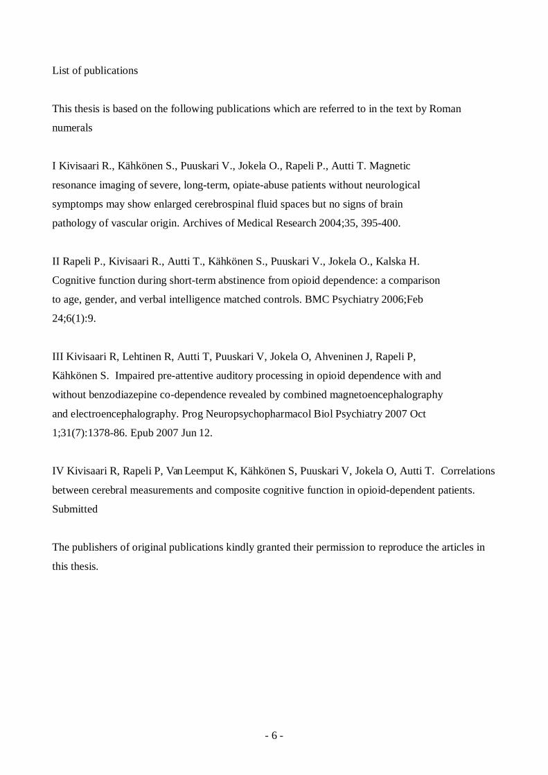
List of publications
This thesis is based on the following publications which are referred to in the text by Roman
numerals
I Kivisaari R., Kähkönen S., Puuskari V., Jokela O., Rapeli P., Autti T. Magnetic
resonance imaging of severe, longterm, opiateabuse patients without neurological
symptomps may show enlarged cerebrospinal fluid spaces but no signs of brain
pathology of vascular origin. Archives of Medical Research 2004;35, 395400.
II Rapeli P., Kivisaari R., Autti T., Kähkönen S., Puuskari V., Jokela O., Kalska H.
Cognitive function during shortterm abstinence from opioid dependence: a comparison
to age, gender, and verbal intelligence matched controls. BMC Psychiatry 2006;Feb
24;6(1):9.
III Kivisaari R, Lehtinen R, Autti T, Puuskari V, Jokela O, Ahveninen J, Rapeli P,
Kähkönen S. Impaired preattentive auditory processing in opioid dependence with and
without benzodiazepine codependence revealed by combined magnetoencephalography
and electroencephalography. Prog Neuropsychopharmacol Biol Psychiatry 2007 Oct
1;31(7):137886. Epub 2007 Jun 12.
IV Kivisaari R, Rapeli P, Van Leemput K, Kähkönen S, Puuskari V, Jokela O, Autti T. Correlations
between cerebral measurements and composite cognitive function in opioiddependent patients.
Submitted
The publishers of original publications kindly granted their permission to reproduce the articles in
this thesis.
7
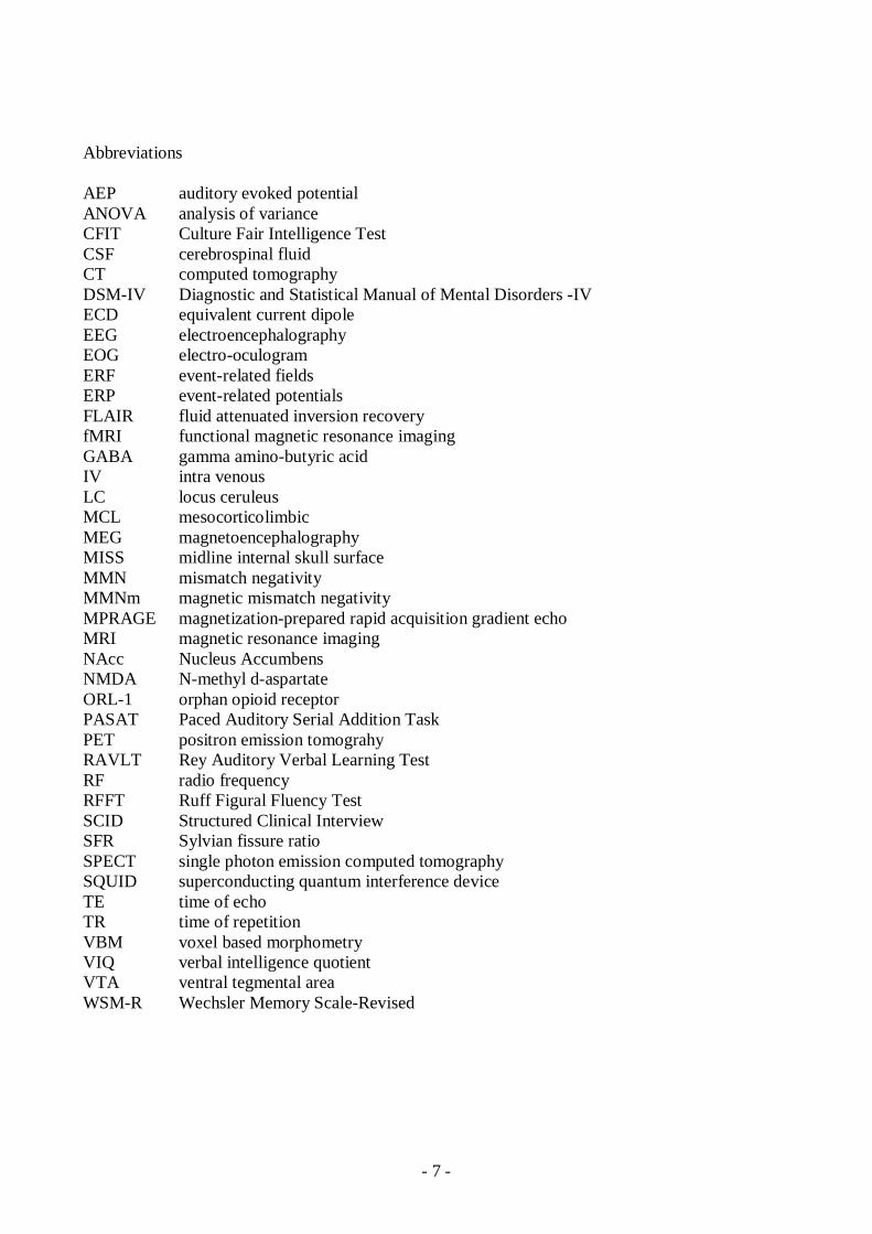
Abbreviations
AEP auditory evoked potentialANOVA analysis of varianceCFIT Culture Fair Intelligence TestCSF cerebrospinal fluidCT computed tomographyDSMIV Diagnostic and Statistical Manual of Mental Disorders IVECD equivalent current dipoleEEG electroencephalographyEOG electrooculogramERF eventrelated fieldsERP eventrelated potentialsFLAIR fluid attenuated inversion recoveryfMRI functional magnetic resonance imagingGABA gamma aminobutyric acidIV intra venousLC locus ceruleusMCL mesocorticolimbicMEG magnetoencephalographyMISS midline internal skull surfaceMMN mismatch negativityMMNm magnetic mismatch negativityMPRAGE magnetizationprepared rapid acquisition gradient echoMRI magnetic resonance imagingNAcc Nucleus AccumbensNMDA Nmethyl daspartateORL1 orphan opioid receptorPASAT Paced Auditory Serial Addition TaskPET positron emission tomograhyRAVLT Rey Auditory Verbal Learning TestRF radio frequencyRFFT Ruff Figural Fluency TestSCID Structured Clinical InterviewSFR Sylvian fissure ratioSPECT single photon emission computed tomographySQUID superconducting quantum interference deviceTE time of echoTR time of repetitionVBM voxel based morphometryVIQ verbal intelligence quotientVTA ventral tegmental areaWSMR Wechsler Memory ScaleRevised

8
Abstract
Background: Opiod dependence is a chronic severe brain disorder associated with enormous healthand social problems. The relapse back to opioid abuse is very high especially in early abstinence,but neuropsychological and neurophysiological deficits during opioid abuse or soon after cessationof opioids are scarcely investigated. Also the structural brain changes and their correlations with thelength of opioid abuse or abuse onset age are not known. In this study the cognitive functions,neural basis of cognitive dysfunction, and brain structural changes was studied in opioiddependentpatients and in age and sex matched healthy controls.
Materials and methods: All subjects participating in the study, 23 opioid dependents of whom, 15were also benzodiazepine and five cannabis codependent and 18 healthy age and sex matchedcontrols went through Structured Clinical Interviews (SCID) to obtain DSMIV axis I and IIdiagnosis and to exclude psychiatric illness not related to opioid dependence or personalitydisorders.Simultaneous magnetoencephalography (MEG) and electroencephalography (EEG) measurementswere done on 21 opioiddependent individuals on the day of hospitalization for withdrawal therapy.The neural basis of auditory processing was studied and preattentive attention and sensory memorywere investigated.During the withdrawal 15 opioiddependent patients participated in neuropsychological tests,measuring fluid intelligence, attention and working memory, verbal and visual memory, andexecutive functions. Fifteen healthy subjects served as controls for the MEGEEG measurementsand neuropsychological assessment.The brain magnetic resonance imaging (MRI) was obtained from 17 patients after approximatelytwo weeks abstinence, and from 17 controls. The areas of different brain structures and the absoluteand relative volumes of cerebrum, cerebral white and gray matter, and cerebrospinal fluid (CSF)spaces were measured and the Sylvian fissure ratio (SFR) and bifrontal ratio were calculated. Alsocorrelation between the cerebral measures and neuropsychological performance was done.
Results: MEGEEG measurements showed that compared to controls the opioiddependent patientshad delayed mismatch negativity (MMN) response to novel sounds in the EEG and P3am on thecontralateral hemisphere to the stimulated ear in MEG. The equivalent current dipole (ECD) ofN1m response was stronger in patients with benzodiazepine codependence than those withoutbenzodiazepine codependence or controls.In early abstinence the opioid dependents performed poorer than the controls in tests measuringattention and working memory, executive function and fluid intelligence. Test results of the CultureFair Intelligence Test (CFIT), testing fluid intelligence, and Paced Auditory Serial Addition Test(PASAT), measuring attention and working memory correlated positively with the days ofabstinence.MRI measurements showed that the relative volume of CSF was significantly larger in opioiddependents, which could also be seen in visual analysis. Also Sylvian fissures, expressed by SFRwere wider in patients, which correlated negatively with the age of opioid abuse onset. In controlsthe relative gray matter volume had a positive correlation with composite cognitive performance,but this correlation was not found in opioid dependents in early abstinence.
Conclusions: Opioid dependents had wide Sylvian fissures and CSF spaces indicatingfrontotemporal atrophy. Dilatation of Sylvian fissures correlated with the abuse onset age. Duringearly withdrawal cognitive performance of opioid dependents was impaired. While intoxicated thepreattentive attention to novel stimulus was delayed and benzodiazepine codependence impairedsound detection. All these changes point to disturbances on frontotemporal areas.

9
1. Literature review
1.1 Opioids
Opium is prepared from the milky juice of the unripe seed capsules of poppy, Papaver somniferum,by drying and powdering. Some of the opioids are natural products isolated from opium asmorphine or semisynthetic as heroin which is synthesized from morphine. A wide spectrum ofsynthetic opioid receptor ligands, which are produced mainly for treatment of severe pain, alsoexists. Methadone and buprenorphine are synthetic opioids used in heroin withdrawal ormaintenance therapy. Buprenorphine came to the market in 1980 and the abuse potential was soonrecognized (Strang 1985). Buprenorphine naloxone tablets were introduced to avoid the abusepotential of buprenorphine. Naloxone is an opioid receptor antagonist and it blocks the effects ofopioids. A recent study showed that 80% of drug abusers felt the intravenous (IV) use ofbuprenorphinenaloxone was a ‘bad’ experience (Alho et al. 2007), demonstrating the effectivenessof the combination.
1.2 Opioid abuse in Finland
In Finland in the year 2002 approximately 1610021100 substance abusers existed, in which 42005900 using opioids. In recent years opioid abuse has become more common, 1997 in Finland wasonly an estimated 15003300 opiod users (STAKES: Huumetilanne Suomessa 2006). Differentfrom other European countries buprenorphine is the main abused opioid in Finland (EMCDDAAnnual report 2006; Table TDI 26) and it is typically used IV as 79.3 % of drug abusers in Finlandpreferred IV route as method of administration. This is among the highest figures in Europe(EMCDDA Annual report 2006; Table TDI 17). Benzodiazepine codependence is also verycommon and probably simultaneous use of buprenorphine and benzodiazepine combined withalcohol is adding the risk of death (EMCDDA Annual report 2006, http://ar2006.emcdda.europa.eu/en/page011en.html#10.3). In the Finnish adult population (1564 years) approximately 0.50.7%use amphetamine or opioids, but among the youngest age group (1525) the prevalence of abusers is0.91.3% (STAKES: Huumetilanne Suomessa 2006).
1.3 Opioid dependence
1.3.1 Neurobiology
Opioids are compounds that act by binding to specific opioid receptors and mediate their actionthrough the opioid system. Opioid receptors are widely spread throughout the neuroaxis. Threemajor groups of opioid receptors are mu (µ), kappa ( ), and delta ( ). All divided into subtypes asµ1, µ2, 1, 2, 3, 1, and 2 (Dhawan et al. 1996). Also an additional opioid receptor ORL1 (orphanopioid receptor) has been identified (Fukuda et al. 1994). All major groups have endogenous opioidligands –endorfin have a preference for receptor, enkephalins for receptors, dynorphins for receptors and nociceptin for ORL1receptor (Lord et al. 1977, Pugsley 2002). The abusedsubstances, morphine and heroin, are full receptor agonists and buprenorphine is a partial receptor agonist and receptor antagonist (Altman et al. 1996). All opioids induce euphoria, whichis the cause of abuse of these substances.
Opioids have very high addictive potential and the reinforcing affects of these drugs are induced byactivation of the mesocorticolimbic (MCL) dopamine reward pathway (Bassareo et al 1995, DiChiara 1995, Di Chiara 2002, Koob 1992, Xi & Stein 2002). This same reward pathway plays acritical role in addiction of any abused substance or addictive behaviour (Nestler 2005, Wise 1996).
10
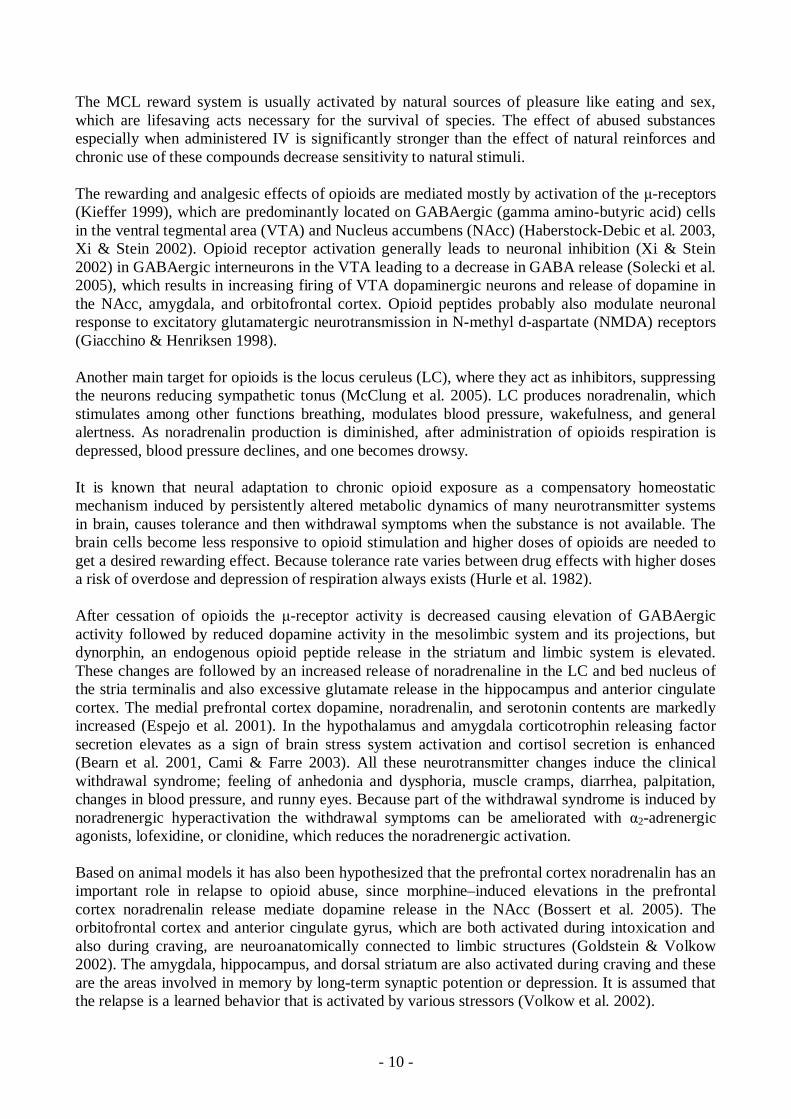
The MCL reward system is usually activated by natural sources of pleasure like eating and sex,which are lifesaving acts necessary for the survival of species. The effect of abused substancesespecially when administered IV is significantly stronger than the effect of natural reinforces andchronic use of these compounds decrease sensitivity to natural stimuli.
The rewarding and analgesic effects of opioids are mediated mostly by activation of the receptors(Kieffer 1999), which are predominantly located on GABAergic (gamma aminobutyric acid) cellsin the ventral tegmental area (VTA) and Nucleus accumbens (NAcc) (HaberstockDebic et al. 2003,Xi & Stein 2002). Opioid receptor activation generally leads to neuronal inhibition (Xi & Stein2002) in GABAergic interneurons in the VTA leading to a decrease in GABA release (Solecki et al.2005), which results in increasing firing of VTA dopaminergic neurons and release of dopamine inthe NAcc, amygdala, and orbitofrontal cortex. Opioid peptides probably also modulate neuronalresponse to excitatory glutamatergic neurotransmission in Nmethyl daspartate (NMDA) receptors(Giacchino & Henriksen 1998).
Another main target for opioids is the locus ceruleus (LC), where they act as inhibitors, suppressingthe neurons reducing sympathetic tonus (McClung et al. 2005). LC produces noradrenalin, whichstimulates among other functions breathing, modulates blood pressure, wakefulness, and generalalertness. As noradrenalin production is diminished, after administration of opioids respiration isdepressed, blood pressure declines, and one becomes drowsy.
It is known that neural adaptation to chronic opioid exposure as a compensatory homeostaticmechanism induced by persistently altered metabolic dynamics of many neurotransmitter systemsin brain, causes tolerance and then withdrawal symptoms when the substance is not available. Thebrain cells become less responsive to opioid stimulation and higher doses of opioids are needed toget a desired rewarding effect. Because tolerance rate varies between drug effects with higher dosesa risk of overdose and depression of respiration always exists (Hurle et al. 1982).
After cessation of opioids the receptor activity is decreased causing elevation of GABAergicactivity followed by reduced dopamine activity in the mesolimbic system and its projections, butdynorphin, an endogenous opioid peptide release in the striatum and limbic system is elevated.These changes are followed by an increased release of noradrenaline in the LC and bed nucleus ofthe stria terminalis and also excessive glutamate release in the hippocampus and anterior cingulatecortex. The medial prefrontal cortex dopamine, noradrenalin, and serotonin contents are markedlyincreased (Espejo et al. 2001). In the hypothalamus and amygdala corticotrophin releasing factorsecretion elevates as a sign of brain stress system activation and cortisol secretion is enhanced(Bearn et al. 2001, Cami & Farre 2003). All these neurotransmitter changes induce the clinicalwithdrawal syndrome; feeling of anhedonia and dysphoria, muscle cramps, diarrhea, palpitation,changes in blood pressure, and runny eyes. Because part of the withdrawal syndrome is induced bynoradrenergic hyperactivation the withdrawal symptoms can be ameliorated with 2adrenergicagonists, lofexidine, or clonidine, which reduces the noradrenergic activation.
Based on animal models it has also been hypothesized that the prefrontal cortex noradrenalin has animportant role in relapse to opioid abuse, since morphine–induced elevations in the prefrontalcortex noradrenalin release mediate dopamine release in the NAcc (Bossert et al. 2005). Theorbitofrontal cortex and anterior cingulate gyrus, which are both activated during intoxication andalso during craving, are neuroanatomically connected to limbic structures (Goldstein & Volkow2002). The amygdala, hippocampus, and dorsal striatum are also activated during craving and theseare the areas involved in memory by longterm synaptic potention or depression. It is assumed thatthe relapse is a learned behavior that is activated by various stressors (Volkow et al. 2002).

11
The most important change in the drug use pattern is when abuse, which is an impulsive behavior,changes to dependence, which is a compulsive behavior. During impulsive behavior increasingtension is followed by an act that gives one pleasure and relief (positive reinforcement). Later thisimpulsive act is regretted and one feels guilty. During compulsive behavior anxiety and stress isfollowed by an act that gives one relief from anxiety (negative reinforcement). This behaviorbecomes an obsession that leads to anxiety and a repetitive cycle of compulsive acts (Koob et al.2004).
The central problem of addiction treatment is not detoxification, but keeping abstinence andavoiding relapses, since months or years after withdrawal symptoms have vanished some stimulimight induce craving and a possible return to opioid abuse. The craving can be induced by exposureto the substance, drugassociated cues (Carter & Tiffany 1999), or stress (Sinha 2001).
1.3.2 Neuropsychological changes
The effects of opioids on neuropsychological functioning have scarcely been studied compared tothe volume of research on effects of stimulants or cannabis. Some evidence fromneuropsychological studies show that patients with opioid dependence have shortterm impairmentsin attention, concentration, working memory, verbal and visual memory, and executive functions(Mintzer et al. 2005, VerdejoGarcia et al. 2004). Even a general intellectual decline has beenshown while intoxicated or very recently detoxified (Rounsaville et al. 1982). Guerra et al. (1987)showed that after rapid detoxification heroin abusers who had shown a deficit in attention, workingand episodic memory, and verbal fluency during abuse did not differ from controls after one to twoweeks of abstinence. It seems that opioid abuse induces partially transient alterations of cognitions.On the other hand, after long term abstinence a consistent deficit in executive functioning,especially in impulse control has been found (Davis et al. 2002, Lee & Pau 2002, Ornstein et al.2000, Pau et al. 2002).
The prefrontal cortex is involved in cognitive functions such as planning, anticipation andestablishment of goals, organization and motivation of behaviour, defined as executive functions(Fellows 2007). The functional imaging studies of substance abusers also point to those frontalpathways related to cognition (Volkow et al. 2002, Yucel et al. 2007).
1.3.3 Eventrelated potentials
Only a couple of electroencephalography (EEG) studies exist in which the eventrelated potentials(ERP) of opioid dependents have been studied. These studies have evaluated the auditory responseP300, which is a late cognitive auditory response, considered a manifestation of active operationssince it is elicited during target detection tasks.
These studies have shown that after cessation of opioids the P300 response is attenuated for months(Papageorgiou et al. 2004). In patients with cocaine and opioid dependence the buprenorphineadministration enhanced P300 amplitude back to same level as while intoxicated or in controls(Kouri et al. 1996). Attenuation of the response is probably due to physiological abnormalitiesduring the withdrawal.
12
1.3.4 Structural brain changes
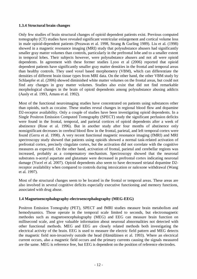
Only few studies of brain structural changes of opioid dependent patients exist. Previous computedtomography (CT) studies have revealed significant ventricular enlargement and cortical volume lossin male opioiddependent patients (Pezawas et al. 1998, Strang & Gurling 1989). Liu et al. (1998)showed in a magnetic resonance imaging (MRI) study that polysubstance abusers had significantlysmaller gray matter volumes than controls, particularly in the prefrontal lobe and to a smaller extentin temporal lobes. Their subjects however, were polysubstance abusers and not all were opioiddependents. In agreement with these former studies Lyoo et al (2006) reported that opioiddependent patients have significantly smaller gray matter densities in the frontal and temporal areasthan healthy controls. They used voxel based morphometry (VBM), which can differentiate thedensities of different brain tissue types from MRI data. On the other hand, the other VBM study bySchlaepfer et al. (2006) showed diminished white matter volumes on the frontal areas, but could notfind any changes in gray matter volumes. Studies also exist that did not find remarkablemorphological changes in the brain of opioid dependents among polysubstance abusing addicts(Aasly et al. 1993, Amass et al. 1992).
Most of the functional neuroimaging studies have concentrated on patients using substances otherthan opioids, such as cocaine. These studies reveal changes in regional blood flow and dopamineD2receptor availability. Only a couple of studies have been investigating opioid dependents. In aSingle Positron Emission Computed Tomography (SPECT) study the significant perfusion deficitswere found in the frontal, temporal, and parietal cortices of opioid dependents after a week ofabstinence (Rose et al. 1996). But in another study after four months of abstinence onlynonsignificant decreases in cerebral blood flow in the frontal, parietal, and left temporal cortex werefound (Gerra et al. 1998). A very recent functional magnetic resonance imaging (fMRI) and MRIspectroscopy study showed that patients using opioids showed a normal taskrelated activation ofprefrontal cortex, precisely cingulate cortex, but the activation did not correlate with the cognitivemeasures as expected. On the other hand, activation of frontal, parietal and cerebellar regions wasincreased, probably as a compensatory mechanism. Spectroscopy showed that the neuronalsubstrates nacetyl aspartate and glutamate were decreased in prefrontal cortex indicating neuronaldamage (Yucel et al. 2007). Opioid dependents also seem to have decreased striatal dopamine D2receptor availability when compared to controls during intoxication or naloxone withdrawal (Wanget al. 1997).
Most of the structural changes seem to be located in the frontal or temporal areas. These areas arealso involved in several cognitive deficits especially executive functioning and memory functions,associated with drug abuse.
1.4 Magnetoencephalographyelectroencephalography (MEGEEG)
Positron Emission Tomograhy (PET), SPECT and fMRI studies measure brain metabolism andhemodynamics. Those operate in the temporal scale limited to seconds, but electromagneticmethodes such as magnetoencephalography (MEG) and EEG can measure brain function onmillisecond scale, and give valuable information about neuronal abnormalities not detected withother functional methods. MEG and EEG are closely related methods both investigating theelectrical activity of the brain. EEG is used to measure the electric field pattern and MEG detectsthe magnetic field noninvasively outside the head (Hämäläinen et al. 1993). Where an electricalcurrent occurs, also a magnetic field occurs and the primary currents causing the signals measuredare the same. MEG is reference free, but EEG is dependent on the position of reference electrodes.

13
The electrical potentials and magnetic fields produced by neurons are so weak that to be able todetect them outside the skull there must by coherent activity of thousands of cells. It is believed thatthe measured activity is mainly induced by synchronous pyramidal postsynaptic potentials.Pyramidal cells are predominantly oriented perpendicular to the cortex (Figure 1). MEG can notdetect the radial sources because of its physical basis, but the magnetic fields induced by tangentialprimary currents can be measured. Therefore activity in sulcus, where the current is orientedparallel to the device is better measured with MEG. Superconducting Quantum Interference Device(SQUID) magnetometers are able to measure these very weak signals outside the head and collectinformation of the cerebral activity. MEG locates the sources accurately in contrast to EEG, becausethe skull, extracerebral tissues, and cerebrospinal fluid are almost transparent to magnetic field, butthey transmit electricity distorting the EEG signal. On the other hand with EEG it is also possible todetect the radial sources invisible to MEG. Therefore, a combined MEG and EEG technique canprovide a comprehensive view of brain function with high spatial and temporal accuracy (Virtanenet al. 1996, Virtanen et al. 1997)
Figure 1. Electrical currents of pyramidal cells (yellow arrows) induce the magnetic field (greenarrows) measured with MEG. Courtesy of Elekta Neuromag Oy
The spontaneous activity of the brain can be recorded, but it is also possible to investigate thestimuluselicited currents, ERPs, and magnetic fields called EventRelated Fields (ERF). Thestimulus has to be repeated many times and the responses must be averaged to be able to detect thedesired response curve among spontaneous activity. The measured electromagnetic activity outsidethe head can be explained with numerous different sources in the brain. This so called inverseproblem has many mathematical solutions. With the selection of channels over for example theauditory cortex one can limit the number of possible electrical sources and can get a good spatialresolution of brain activity.
These ERPs and ERFs reveal information of the neural basis of perception and cognition providingan objective and high temporal resolution index of auditory processing in the human brain.
1.4.1 Auditory evoked potentials and magnetic fields
The sonic air pressure waves are transformed into neural signals in the auditory pathway. First theexternal and middle ear filters and amplifies the sound waves and the auditory ossicles transmit thevibrations to the inner ear and cochlear fluids. The movement of the fluid in the cochlea is

14
transformed to the neural signals. The sensory cells adapted to high frequencies are located in thebase and those to low frequencies in the apex of the cochlea. This organized pattern of fibersremains all the way to auditory cortex. The afferent nervefibers from the inner ear are located in thebrain nerve number VIII; nervus vestibulocochlearis. The nervefibers synapse at ipsilateral cochlearnucleus. The major outputs project into the hemisphere contralateral to the stimulated ear. Thesecondorder neurons ascend into the superior olivary nuclei bilaterally and via lateral lemniscus tothe colliculus inferior. From there the pathway continues through the medial geniculate nucleus ofthe thalamus to the auditory cortex in the temporal lobe. The primary auditory cortex is located inthe supratemporal cortex in Brodmans’ areas 41 and 42.
The auditory ERP in EEG and ERF in MEG are thought to reflect the processing of the heardinformation. The cortical responses peak 50800 ms after stimulus onset and are defined as longlatency components following the earlier brain stem and subcortical auditory responses (Hari et al.1980). Longlatency ERP and ERF represent a sum of neural activity from several sources. Thereflections are classified by the polarity (P positive or N negative) and approximate latency of thepeak or succession. The ERF responses have an additional m for magnetic. The first of the longlatency response P1 (P1m) peaks approximately 50 ms after stimulus onset. The most conspicuousof the auditory responses is N1 (N1m), a negative reflection that peaks at an average of 100 ms afterstimulus onset and reflects the activity of at least three different sources (Näätänen & Picton 1987).The main sources are located on the supratemporal auditory cortex immediately posterior to theprimary auditory cortex and two other components in the superior temporal gyrus and around motorcortex (Hari et al. 1980, Näätänen & Picton 1987). These responses can be found in both auditorycortices, on contra or ipsilateral to the stimulated ear (Figure 2). On the ipsilateral side the latenciesare usually slightly longer than on contralateral hemisphere (Pantev et al. 1998).
Figure 2. Equivalent current dipoles (green) of N1m auditory responses shown on auditory corticeson the contralateral and ipsilateral to the stimulated ear. The magnetic field around the electricalcurrent dipole is also shown.
1.4.2 Mismatch negativity (MMN)
The electric mismatch negativity MMN (and its magnetic counterpart MMNm) is a cognitiveresponse indexing the neural basis of sensory memory and involuntary attention (Kujala et al. 2006,Näätänen et al. 1993). It is elicited without subjects’ attention when the train of standard stimulus iserupted with a deviant stimulus differing from the standard in some respect. It is assumed that the
15
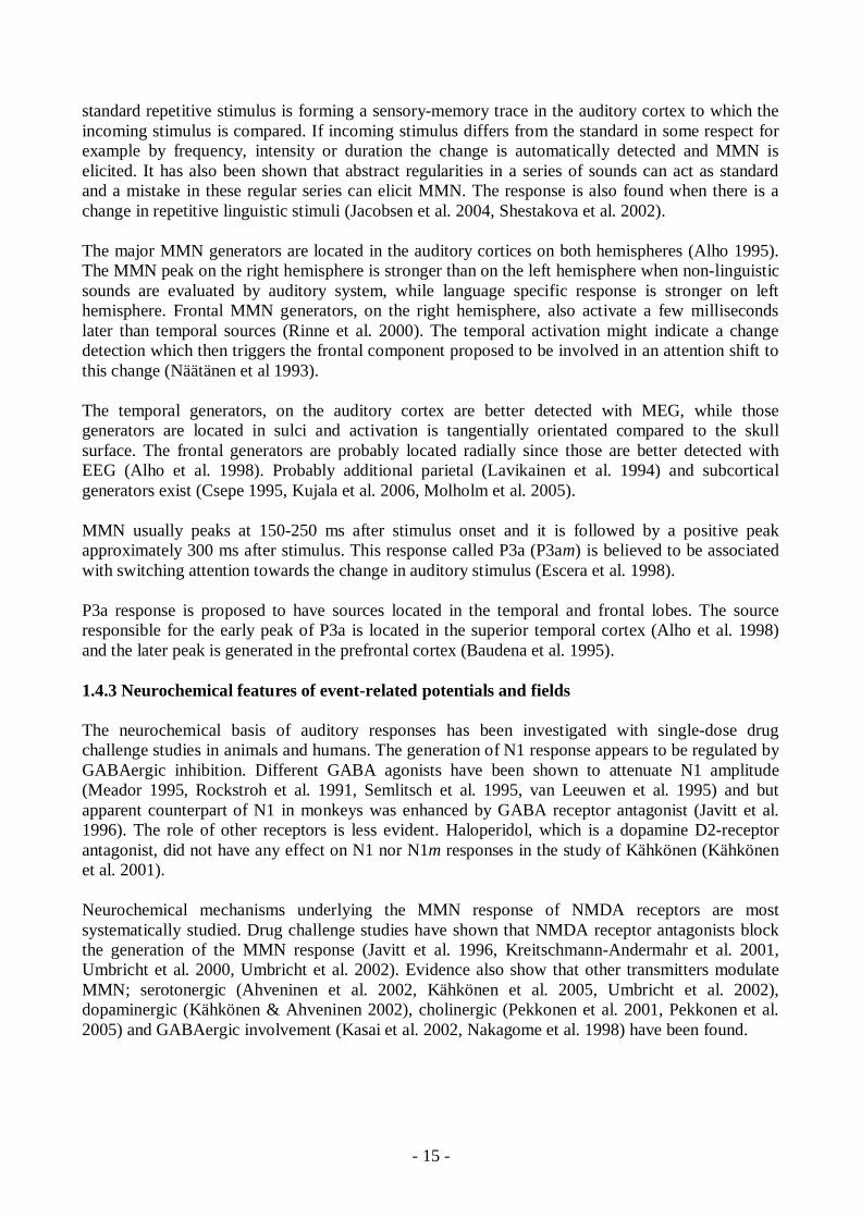
standard repetitive stimulus is forming a sensorymemory trace in the auditory cortex to which theincoming stimulus is compared. If incoming stimulus differs from the standard in some respect forexample by frequency, intensity or duration the change is automatically detected and MMN iselicited. It has also been shown that abstract regularities in a series of sounds can act as standardand a mistake in these regular series can elicit MMN. The response is also found when there is achange in repetitive linguistic stimuli (Jacobsen et al. 2004, Shestakova et al. 2002).
The major MMN generators are located in the auditory cortices on both hemispheres (Alho 1995).The MMN peak on the right hemisphere is stronger than on the left hemisphere when nonlinguisticsounds are evaluated by auditory system, while language specific response is stronger on lefthemisphere. Frontal MMN generators, on the right hemisphere, also activate a few millisecondslater than temporal sources (Rinne et al. 2000). The temporal activation might indicate a changedetection which then triggers the frontal component proposed to be involved in an attention shift tothis change (Näätänen et al 1993).
The temporal generators, on the auditory cortex are better detected with MEG, while thosegenerators are located in sulci and activation is tangentially orientated compared to the skullsurface. The frontal generators are probably located radially since those are better detected withEEG (Alho et al. 1998). Probably additional parietal (Lavikainen et al. 1994) and subcorticalgenerators exist (Csepe 1995, Kujala et al. 2006, Molholm et al. 2005).
MMN usually peaks at 150250 ms after stimulus onset and it is followed by a positive peakapproximately 300 ms after stimulus. This response called P3a (P3am) is believed to be associatedwith switching attention towards the change in auditory stimulus (Escera et al. 1998).
P3a response is proposed to have sources located in the temporal and frontal lobes. The sourceresponsible for the early peak of P3a is located in the superior temporal cortex (Alho et al. 1998)and the later peak is generated in the prefrontal cortex (Baudena et al. 1995).
1.4.3 Neurochemical features of eventrelated potentials and fields
The neurochemical basis of auditory responses has been investigated with singledose drugchallenge studies in animals and humans. The generation of N1 response appears to be regulated byGABAergic inhibition. Different GABA agonists have been shown to attenuate N1 amplitude(Meador 1995, Rockstroh et al. 1991, Semlitsch et al. 1995, van Leeuwen et al. 1995) and butapparent counterpart of N1 in monkeys was enhanced by GABA receptor antagonist (Javitt et al.1996). The role of other receptors is less evident. Haloperidol, which is a dopamine D2receptorantagonist, did not have any effect on N1 nor N1m responses in the study of Kähkönen (Kähkönenet al. 2001).
Neurochemical mechanisms underlying the MMN response of NMDA receptors are mostsystematically studied. Drug challenge studies have shown that NMDA receptor antagonists blockthe generation of the MMN response (Javitt et al. 1996, KreitschmannAndermahr et al. 2001,Umbricht et al. 2000, Umbricht et al. 2002). Evidence also show that other transmitters modulateMMN; serotonergic (Ahveninen et al. 2002, Kähkönen et al. 2005, Umbricht et al. 2002),dopaminergic (Kähkönen & Ahveninen 2002), cholinergic (Pekkonen et al. 2001, Pekkonen et al.2005) and GABAergic involvement (Kasai et al. 2002, Nakagome et al. 1998) have been found.
16
1.5 Magnetic Resonance Imaging (MRI)
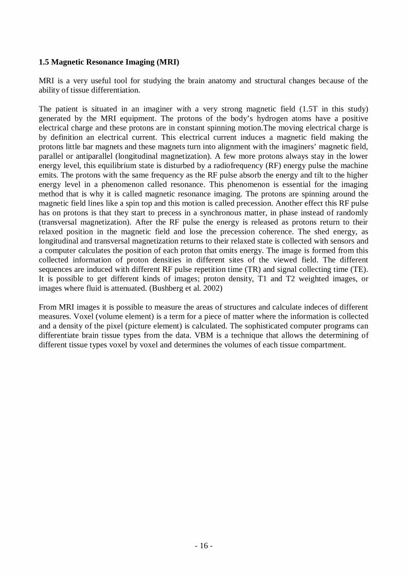
MRI is a very useful tool for studying the brain anatomy and structural changes because of theability of tissue differentiation.
The patient is situated in an imaginer with a very strong magnetic field (1.5T in this study)generated by the MRI equipment. The protons of the body’s hydrogen atoms have a positiveelectrical charge and these protons are in constant spinning motion.The moving electrical charge isby definition an electrical current. This electrical current induces a magnetic field making theprotons little bar magnets and these magnets turn into alignment with the imaginers’ magnetic field,parallel or antiparallel (longitudinal magnetization). A few more protons always stay in the lowerenergy level, this equilibrium state is disturbed by a radiofrequency (RF) energy pulse the machineemits. The protons with the same frequency as the RF pulse absorb the energy and tilt to the higherenergy level in a phenomenon called resonance. This phenomenon is essential for the imagingmethod that is why it is called magnetic resonance imaging. The protons are spinning around themagnetic field lines like a spin top and this motion is called precession. Another effect this RF pulsehas on protons is that they start to precess in a synchronous matter, in phase instead of randomly(transversal magnetization). After the RF pulse the energy is released as protons return to theirrelaxed position in the magnetic field and lose the precession coherence. The shed energy, aslongitudinal and transversal magnetization returns to their relaxed state is collected with sensors anda computer calculates the position of each proton that omits energy. The image is formed from thiscollected information of proton densities in different sites of the viewed field. The differentsequences are induced with different RF pulse repetition time (TR) and signal collecting time (TE).It is possible to get different kinds of images; proton density, T1 and T2 weighted images, orimages where fluid is attenuated. (Bushberg et al. 2002)
From MRI images it is possible to measure the areas of structures and calculate indeces of differentmeasures. Voxel (volume element) is a term for a piece of matter where the information is collectedand a density of the pixel (picture element) is calculated. The sophisticated computer programs candifferentiate brain tissue types from the data. VBM is a technique that allows the determining ofdifferent tissue types voxel by voxel and determines the volumes of each tissue compartment.
17
2. Aims of the study
The general aim was to investigate the brain structural and functional changes in a group of longterm opioid dependent patients.
The purposes of the present study were to find:
1. If structural brain changes in longterm opioid dependents exist compared to healthycontrols (I,IV).
2. How opioid dependents perform in neuropsychological tests during early opioid withdrawal(II,IV).
3. Are there changes in preattentive auditory processing in opioid dependents (III).4. If any correlation appears between structural brain changes and neuropsychological tests
(IV).5. How the age of abuse onset and the length of abuse history affects these structural and
functional changes (IIV).
18
3. Materials and methods
3.1 Subjects (Studies IIV)
Twenty three opioiddependent patients (Table 1) and 18 healthy controls with no history of drugabuse were attended this study. Opioid dependents were admitted to an inpatient drugdetoxification unit in the Helsinki University Central Hospital for withdrawal and evaluation forthe methadone maintenance program. The criteria for the methadone maintenance program were aminimum age of 20 years, four years of documented opioid dependence, and the failure ofinstitutional or longlasting outpatient withdrawal therapy, which also served as criteria forinclusion in the study. Exclusion criteria for methadone maintenance therapy were uncontrolledpolysubstance abuse, physical or psychiatric illness that made the routine of therapy impossible,and alcohol dependence. In this study, additional exclusion criteria for both patients and controlswere major head trauma, chronic neurological illness or ongoing medication for neurologicalsymptoms, and metallic foreign objects in the body.
All patients had severe longterm opioid dependence (Table 1). They had used opioids from 5 to 26years (mean 12±8), mainly IV. The self reported daily doses of different opioids were 0.051.5 g forstreet heroin, 232 mg for buprenorphine and 250750 mg for ethyl morphine. Fifteen patients (5women, mean age 31.5 ± 6.0) also abused benzodiazepines daily (approximate equivalent dose todiazepam 38 ± 21 mg according to Ashton table (Ashton 2005)). Before the study patients gaveurine samples twice a week from four to six weeks to exclude other illicit substance abuse thanopioids or benzodiazepines.
Patients were in good physical health as determined by a physical examination, laboratoryevaluation including a complete blood count, electrolytes, glucose, renal, and thyroid analyses. HIVantibody test was negative in all patients tested (one patient refused). All except two of the patientshad positive hepatitis C antibody analysis. Hepatic enzymes were mildly elevated in 7 patients andmoderately in 1 patient.
The Structured Diagnostic Interviews (SCID I and SCID II, American psychiatric Association,1994) (First et al. 1994a, First et al. 1994b) were done on all patients while they were hospitalizedby trained psychiatrists to obtain the DSMIV (Diagnostic and Statistical Manual of mentaldisorders) diagnosis of axes I and II. Sixteen patients fulfilled the criteria of DSMIV for antisocialpersonality disorder and 13 had multiple diagnoses on SCID II. (Table 1)
Controls were healthy volunteers from hospital staff and their friends with no experience of illicitdrugs. All had used alcohol in social occasions, but did not meet the criteria of abuse or dependenceon alcohol. Patients and controls were age and sex matched and in the subgroup attending theneuropsychological tests verbal intelligence also matched. The control subjects had no DSMIVaxis I or II diagnosis in the SCID evaluation.
The simultaneous MEGEEG measurement carried out to 21 patients while intoxicated (III) and 15healthy controls. Neuropsychological tests were done to 15 patients (II, IV) and 15 age, gender andverbal intelligence matched controls. Brain MRI was obtained from 17 patients during thewithdrawal therapy in the clinic (I, IV) and 17 controls. Written informed consent was obtainedfrom all subjects and the study had the approval of the local ethical committee.
19
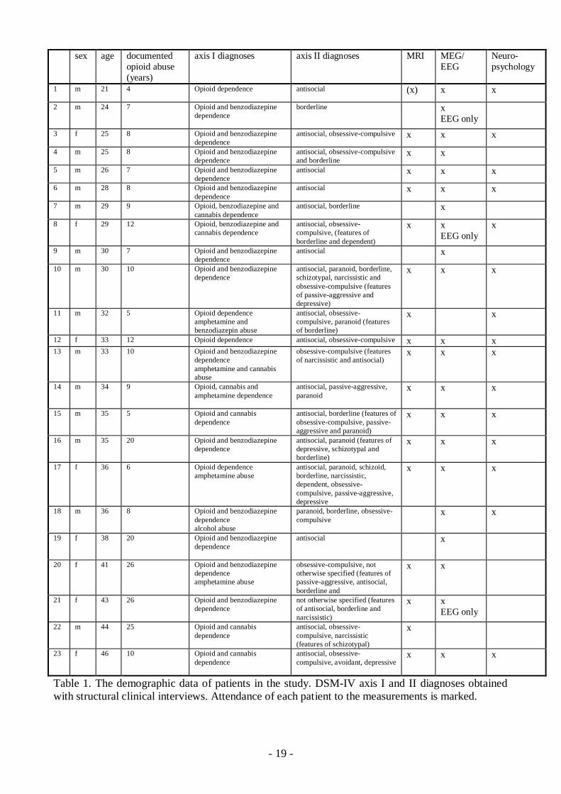
Table 1. The demographic data of patients in the study. DSMIV axis I and II diagnoses obtainedwith structural clinical interviews. Attendance of each patient to the measurements is marked.
sex age documentedopioid abuse(years)
axis I diagnoses axis II diagnoses MRI MEG/EEG
Neuropsychology
1 m 21 4 Opioid dependence antisocial (x) x x
2 m 24 7 Opioid and benzodiazepinedependence
borderline xEEG only
3 f 25 8 Opioid and benzodiazepinedependence
antisocial, obsessivecompulsive x x x
4 m 25 8 Opioid and benzodiazepinedependence
antisocial, obsessivecompulsiveand borderline
x x
5 m 26 7 Opioid and benzodiazepinedependence
antisocial x x x
6 m 28 8 Opioid and benzodiazepinedependence
antisocial x x x
7 m 29 9 Opioid, benzodiazepine andcannabis dependence
antisocial, borderline x
8 f 29 12 Opioid, benzodiazepine andcannabis dependence
antisocial, obsessivecompulsive, (features ofborderline and dependent)
x xEEG only
x
9 m 30 7 Opioid and benzodiazepinedependence
antisocial x
10 m 30 10 Opioid and benzodiazepinedependence
antisocial, paranoid, borderline,schizotypal, narcissistic andobsessivecompulsive (featuresof passiveaggressive anddepressive)
x x x
11 m 32 5 Opioid dependenceamphetamine andbenzodiazepin abuse
antisocial, obsessivecompulsive, paranoid (featuresof borderline)
x x
12 f 33 12 Opioid dependence antisocial, obsessivecompulsive x x x13 m 33 10 Opioid and benzodiazepine
dependenceamphetamine and cannabisabuse
obsessivecompulsive (featuresof narcissistic and antisocial)
x x x
14 m 34 9 Opioid, cannabis andamphetamine dependence
antisocial, passiveaggressive,paranoid
x x x
15 m 35 5 Opioid and cannabisdependence
antisocial, borderline (features ofobsessivecompulsive, passiveaggressive and paranoid)
x x x
16 m 35 20 Opioid and benzodiazepinedependence
antisocial, paranoid (features ofdepressive, schizotypal andborderline)
x x x
17 f 36 6 Opioid dependenceamphetamine abuse
antisocial, paranoid, schizoid,borderline, narcissistic,dependent, obsessivecompulsive, passiveaggressive,depressive
x x x
18 m 36 8 Opioid and benzodiazepinedependencealcohol abuse
paranoid, borderline, obsessivecompulsive
x x
19 f 38 20 Opioid and benzodiazepinedependence
antisocial x
20 f 41 26 Opioid and benzodiazepinedependenceamphetamine abuse
obsessivecompulsive, nototherwise specified (features ofpassiveaggressive, antisocial,borderline and
x x
21 f 43 26 Opioid and benzodiazepinedependence
not otherwise specified (featuresof antisocial, borderline andnarcissistic)
x xEEG only
22 m 44 25 Opioid and cannabisdependence
antisocial, obsessivecompulsive, narcissistic(features of schizotypal)
x
23 f 46 10 Opioid and cannabisdependence
antisocial, obsessivecompulsive, avoidant, depressive
x x x

20
3.2 Neuropsychological tests (Studies II and IV)
The battery of cognitive tests including the tests measuring working memory, episodic memory,executive function, verbal and visual learning and memory, and fluid intelligence were used.
Working memory performance was tested with the Digit Span subtest from the Wechsler MemoryScaleRevised (WSMR) and a computerized version of Paced Auditory Serial Addition task(PASAT) from the FORAMENRehab softwear package. During the Digit Span Test one has torepeat the series of numbers in a same order as heard. Numbers are given one per second. This testmeasures verbal working memory storage. During the PASAT test one is presented a numberbetween 1 and 9 every 1.6 seconds and they have to add the two previous numbers heard and sayout loud the result. The PASAT test performance requires continuous storage of incomingnumbers, rapid arithmetic processing, and executive control of interference of heard and calculatednumbers. This requires complex working memory function.
The immediate verbal learning was studied with the Rey Auditory Verbal Learning Test (RAVLT)in which three learning trials of 15 presented words were given to all participants. The sum of theresults of the three tries was calculated to form a result for the test. Visual memory was measuredby the Benton Visual Retention test. In this test 10 drawings, one at the time, are shown and thesedesigns have to be reproduced onto plain paper from memory as exactly as possible.
Executive function was measured with a modified Stroop task. In the first part of this test therewere 50 words, names of different colors written with black ink, and in the second part the samewords were written with different ink colors. The color of the ink was different from the writtenword. The result was the time of naming the ink colours in the second part subtracted with thereading time of the first list. Inhibition of not reading the words in the second part and the time ofnaming the colors affect the test result. The Ruff Figural Fluency (RFFT) test, which forms fromtwo parts of unique designs and preservative errors, measures the executive function, planning andfluency of action. The squares with five dots in each are presented and one has to draw as manydifferent figures as possible drawn with connecting at least two dots with straight lines in eachsquare.
Culture Fair Intelligence Test (CFIT), includes a group of visuospatial reasoning tasks that aresensitive to fluid intelligence deficit due to various origins. The performance of the test reflectsfluid and general intelligence needed in highly demanding novel problem solving situations.
The composite cognitive function was calculated as a sum of the zscore of all test results. Onlyone score per tests was used if the test yielded more than one score. The result of each test was firststandardized to zscores which were calculated from the individual’s test score by subtracting thegroup’s mean score and dividing the difference by the group’s standard deviation. These z scoreswere summed up to obtain person’s composite cognitive function.
3.3 Combined MEGEEG (Study III)
3.3.1 Measurements
MEG and EEG recordings were carried out a 306channel MEG, consisting of 204 planargradiometers and 102 magnetometers (Vectorview, Neuromag TM) and a 60channel EEG. Theposition of the subjects´ head relative to the recording instrument was determined by measuringthe magnetic fields produced by marker coils attached to the scalp. The locations of these coils in

21
relation to cardinal points on the head (nasion, left and right preauricular points) were determinedbefore the experiment using an Isotrak 3Ddigitizer (Polhemus, Colchester, VT, U.S.A.). Duringthe measurement the subject sat in a comfortable chair in a magnetically and electrically shieldedroom (Euroshield, Finland) and watched a silent video. Auditory stimulus was presentedmonaurally to the left ear through a plastic tube and an earpiece and the subject was instructed toignore the tones. The stimulus block consisted of the standard (80%) stimulus (700 Hz with 60 msduration, including 5 ms rise and fall times) randomly embedded with infrequent (6.6% for eachtype) deviant tones differing in frequency (larger deviant 400 Hz and smaller deviant 600 Hz) andnovel sounds (10 different complex sound bursts in random order). The interstimulus interval was599 ms. Each twochannel sensor unit measured two independent magnetic field gradientcomponents ∂Bz/∂x and ∂Bz/∂y, with the zaxis being normal to the local helmet surface. Therecording bandpass was 0.03–172 Hz and the sampling rate was 600 Hz. Epoch was averagedfrom 150 ms prestimulus until 600 ms poststimulus.
AEPs were recorded with an electrode cap (Virtanen et al. 1996) and an amplifier (Virtanen et al.1997) specifically designed and built for simultaneous EEG and MEG measurements. The noseelectrode was used as a reference. Vertical and horizontal electrooculograms (EOG) wererecorded. First responses in the train, and all the epochs coinciding with EOG or MEG changesexceeding 150 µV or 3000 fT/cm, respectively, were omitted from averaging. At least 100artefactfree standard and deviant responses were recorded and averaged.
3.3.2 Analysis of MEGEEG
Digital bandpass filtering was performed offline at 240 Hz for P1/P1m, at 130 Hz for N1/N1m,at 215 Hz for MMN/MMNm and P3a/P3am. The analysis period was 500 ms. Distinct ERP/ERFpeaks were obtained from latency ranges of 3070 ms for P1/P1m, 60150 ms for N1/N1m, 100250 for MMN/MMNm and 170500 ms for P3a/P3am. The responses were judged significantwhen they were two standard deviations (SD) larger than the prestimulus noise.
MEG analysis was done with a source modelling program with software provided by NeuromagLtd. (Hämäläinen et al. 1993). The dipole modelling was performed using a subset of 64 channelsseparately over each auditory cortex using one’s own brain MRI image if available. In the fourcases MRI images were not obtained the sphere model was used.
For EEG analysis, the peak latencies of P1 and N1 were determined from standard responses at thechannel Cz. The peak latencies of the MMN and P3a were determined from subtraction curves atthe electrode sites Fz or FCz depending on which channel a given deflection was larger.Subtraction curves were calculated by subtracting the standard AEP from the deviant. The P1, N1,MMN, and P3a amplitudes were determined from the averaged amplitude of 15 channels in groupsof three (prefrontal; AF1, AFz, AF2, frontal; F1, Fz, F2, frontocentral; FC1, FCz, FC2, central; C1,Cz, C2, centroparietal; CP1, CPz, and CP2).
3.4 Brain MRI (Studies I and IV)
3.4.1 Imaging
Brain MRIs were acquired with a 1.5 T Siemens Magnetom imager. After scout images, axial andcoronal T2 and proton density weighted images with a spin echo sequence, 3000/1485 (TR/TE)with a slice thickness of 5.0 mm, axial fluid attenuated inversion recovery (FLAIR) 9999/105(TR/TE) with a slice thickness of 5.0 mm, and a three dimensional (3D) magnetizationprepared
22
rapid acquisition gradient echo (MPRAGE 9.7/4.0 (TR/TE) with a slice thickness of 1mm wereobtained. The 3Dseries were used in MEG analysis to localize the equivalent current dipoles(ECD).
3.4.2 Analysis of MRI
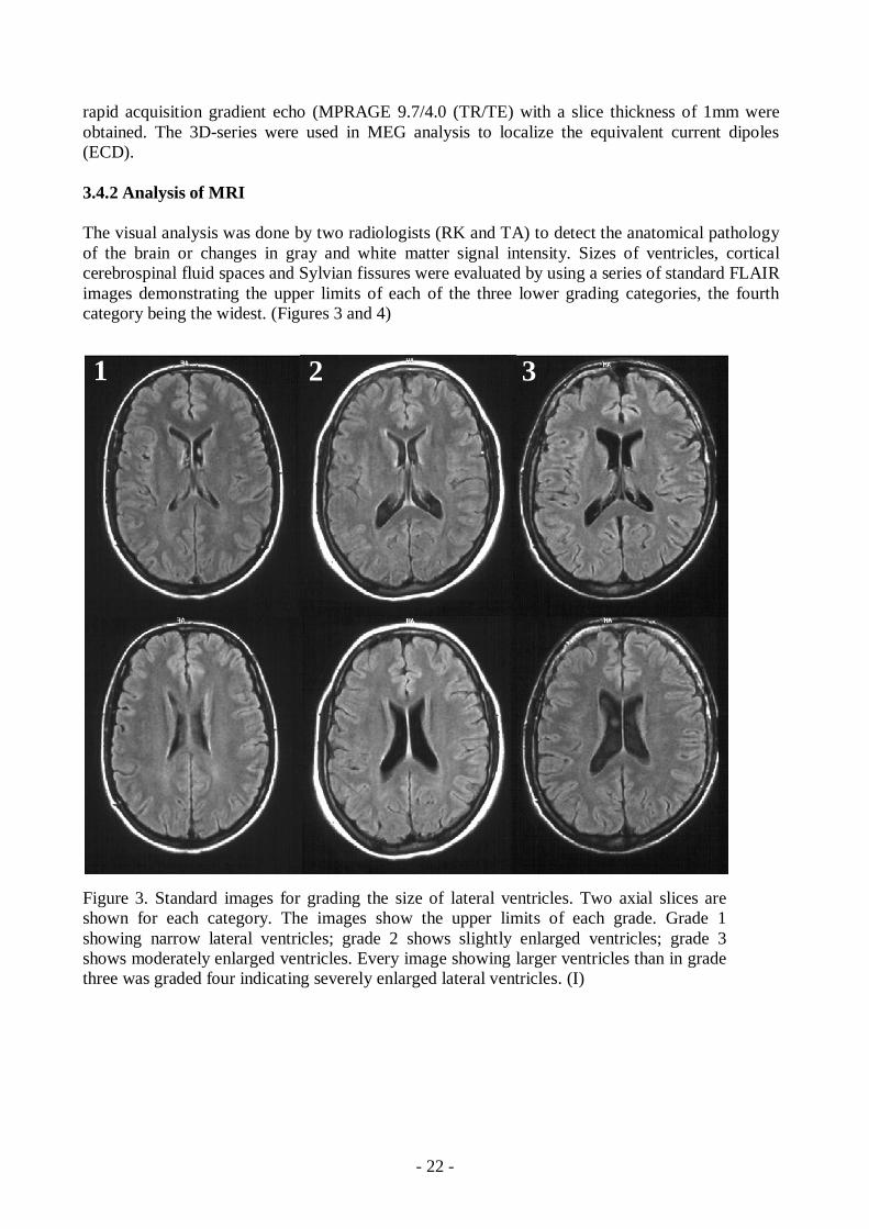
The visual analysis was done by two radiologists (RK and TA) to detect the anatomical pathologyof the brain or changes in gray and white matter signal intensity. Sizes of ventricles, corticalcerebrospinal fluid spaces and Sylvian fissures were evaluated by using a series of standard FLAIRimages demonstrating the upper limits of each of the three lower grading categories, the fourthcategory being the widest. (Figures 3 and 4)
Figure 3. Standard images for grading the size of lateral ventricles. Two axial slices areshown for each category. The images show the upper limits of each grade. Grade 1showing narrow lateral ventricles; grade 2 shows slightly enlarged ventricles; grade 3shows moderately enlarged ventricles. Every image showing larger ventricles than in gradethree was graded four indicating severely enlarged lateral ventricles. (I)
1 2 3

23
Figure 4. Standard images used grading Sylvian fissures. Axial slices through Sylvian fissuresshow maximal sizes of the spaces in grades 13. Grade four is not shown. Grade 1 showingnarrow Sylvian fissures; grade 2 shows slightly enlarged temporal parts of Sylvian fissures;grade 3 shows enlarged frontal and temporal parts of Sylvian fissures. Every image whichshowed larger Sylvian fissures than in grade three was graded four. (I)
The measurements of different brain areas were done in the slice of the 3Dseries were theaqueduct was best seen defined as the midsagittal image. The areas of the midline internal skullsurface (MISS), vermis and corpus callosum were obtained (Laissy et al. 1993). FLAIR imageswere used while measuring the Sylvian fissure ratio (SFR) defined as the average width of bothSylvian fissures divided by the transpineal temporal brain width (van Zagten et al. 1999). Bifrontalratio was measured at the level of third ventricle from T2 axial images. Bifrontal ratio was definedas a distance between the most lateral tips of the anterior horns of lateral ventricles divided by thebrain width in the same line and level (Aylward et al. 1991). (Figure 5)
1 2 3
24
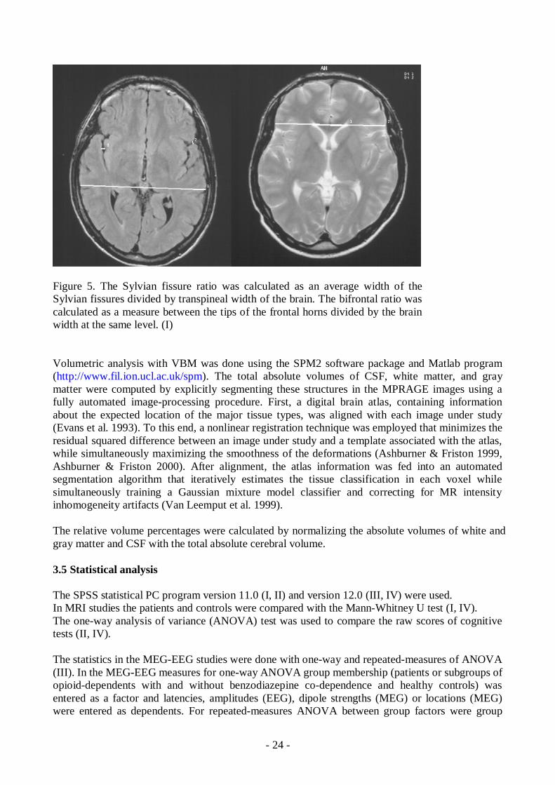
Figure 5. The Sylvian fissure ratio was calculated as an average width of theSylvian fissures divided by transpineal width of the brain. The bifrontal ratio wascalculated as a measure between the tips of the frontal horns divided by the brainwidth at the same level. (I)
Volumetric analysis with VBM was done using the SPM2 software package and Matlab program(http://www.fil.ion.ucl.ac.uk/spm). The total absolute volumes of CSF, white matter, and graymatter were computed by explicitly segmenting these structures in the MPRAGE images using afully automated imageprocessing procedure. First, a digital brain atlas, containing informationabout the expected location of the major tissue types, was aligned with each image under study(Evans et al. 1993). To this end, a nonlinear registration technique was employed that minimizes theresidual squared difference between an image under study and a template associated with the atlas,while simultaneously maximizing the smoothness of the deformations (Ashburner & Friston 1999,Ashburner & Friston 2000). After alignment, the atlas information was fed into an automatedsegmentation algorithm that iteratively estimates the tissue classification in each voxel whilesimultaneously training a Gaussian mixture model classifier and correcting for MR intensityinhomogeneity artifacts (Van Leemput et al. 1999).
The relative volume percentages were calculated by normalizing the absolute volumes of white andgray matter and CSF with the total absolute cerebral volume.
3.5 Statistical analysis
The SPSS statistical PC program version 11.0 (I, II) and version 12.0 (III, IV) were used.In MRI studies the patients and controls were compared with the MannWhitney U test (I, IV).The oneway analysis of variance (ANOVA) test was used to compare the raw scores of cognitivetests (II, IV).
The statistics in the MEGEEG studies were done with oneway and repeatedmeasures of ANOVA(III). In the MEGEEG measures for oneway ANOVA group membership (patients or subgroups ofopioiddependents with and without benzodiazepine codependence and healthy controls) wasentered as a factor and latencies, amplitudes (EEG), dipole strengths (MEG) or locations (MEG)were entered as dependents. For repeatedmeasures ANOVA between group factors were group
25
membership and within subject variables were electrode location (prefrontal, frontal, frontocentral,central, centroparietal) in EEG or hemisphere in MEG measurement analysis. Differences betweenthe subgroups were calculated with the Fisher’s LSD test. Statistical significance was set at 0.05(twotailed).
Correlation between different measurements and age of abuse onset, years of opioid abuse and daysof abstinence were calculated with the Pearson correlation coefficient. (IV)
4. Results
4.1 Cognitive performance (Studies II and IV)
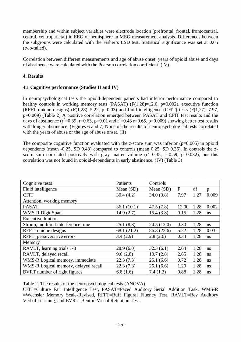
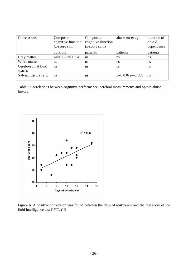
In neuropsychological tests the opioiddependent patients had inferior performance compared tohealthy controls in working memory tests (PASAT) (F(1,28)=12.0, p=0.002), executive function(RFFT unique designs) (F(1,28)=5.22, p=0.03) and fluid intelligence (CFIT) tests (F(1,27)=7.97,p=0.009) (Table 2) A positive correlation emerged between PASAT and CFIT test results and thedays of abstinence (r2=0.39, r=0.63, p=0.01 and r2=0.43 r=0.65, p=0.009) showing better test resultswith longer abstinence. (Figures 6 and 7) None of the results of neuropsychological tests correlatedwith the years of abuse or the age of abuse onset. (II)
The composite cognitive function evaluated with the zscore sum was inferior (p=0.005) in opioiddependents (mean 0.25, SD 0.43) compared to controls (mean 0.25, SD 0.36). In controls the zscore sum correlated positively with gray matter volume (r2=0.35, r=0.59, p=0.032), but thiscorrelation was not found in opioiddependents in early abstinence. (IV) (Table 3)
Cognitive tests Patients ControlsFluid intelligence Mean (SD) Mean (SD) F df pCFIT 30.4 (4.2) 34.0 (3.8) 7.97 1,27 0.009Attention, working memoryPASAT 36.1 (10.1) 47.5 (7.8) 12.00 1,28 0.002WMSR Digit Span 14.9 (2.7) 15.4 (3.8) 0.15 1,28 nsExecutive funtionStroop, modified interference time 25.1 (8.8) 24.5 (12.0) 0.30 1,28 nsRFFT, unique designs 68.1 (21.2) 86.3 (22.6) 5.22 1,28 0.03RFFT, perseverative errors 3.4 (2.9) 2.8 (2.6) 0.34 1,28 nsMemoryRAVLT, learning trials 13 28.9 (6.0) 32.3 (6.1) 2.64 1,28 nsRAVLT, delayed recall 9.0 (2.8) 10.7 (2.8) 2.65 1,28 nsWMSR Logical memory, immediate 22.3 (7.3) 25.1 (6.6) 0.72 1,28 nsWMSR Logical memory, delayed recall 22.3 (7.3) 25.1 (6.6) 1.20 1,28 nsBVRT number of right figures 6.8 (1.6) 7.4 (1.3) 0.88 1,28 ns
Table 2. The results of the neuropsychological tests (ANOVA)CFIT=Culture Fair Intelligence Test, PASAT=Paced Auditory Serial Addition Task, WMSR=Wechsler Memory ScaleRevised, RFFT=Ruff Figural Fluency Test, RAVLT=Rey AuditoryVerbal Learning, and BVRT=Benton Visual Retention Test.
26
Correlations Compositecognitive function(zscore sum)
Compositecognitive function(zscore sum)
abuse onset age duration ofopioiddependence
controls patients patients patientsGray matter p=0.032 r=0.594 ns ns nsWhite matter ns ns ns nsCerebrospinal fluidspaces
ns ns ns ns
Sylvian fissure ratio ns ns p=0.036 r=0.585 ns
Table 3 Correlations between cognitive performance, cerebral measurements and opioid abusehistory.
Figure 6. A positive correlation was found between the days of abstinence and the test score of thefluid intelligence test CFIT. (II)

27
Figure 7. PASAT test results measuringattention and complex working memorycorrelated positively with the duration ofwithdrawal. (II)
4.2 MEG (Study III)
While intoxicated the repeated measures ANOVA revealed a significant main effect on N1m dipolestrength (F(2,26)= 5.9, p=0.008). The post hoc analysis showed that this effect was due tosignificantly stronger source activity in opioiddependent patients with benzodiazepine codependence than in opioiddependent patients without benzodiazepine codependence (p=0.005) orin controls (p=0.013). N1m group X hemisphere interactions were not significant.
Table 4. The mean strength of equivalent current dipoles of auditory responses in both hemispheresin patients and controls. Also the results of patient subgroups with or without benzodiazepine codependence are shown. BZ benzodiazepine*Patients with benzodiazepine codependence compared to patients without benzodiazepine codependence p=0.005 and controls p=0.013. (Repeated measures ANOVA)
MEG contralateral hemisphere ipsilateral hemisphereSourceactivity(nAm)
all patients patients with BZdependence
patientswithout BZdependence
controls all patients patients with BZdependence
patientswithout BZdependence
controls
P1m 12.3 (7.6) 13.2 (8.4) 9.7 (4.0) 9.5 (5.2) 7.8 (4.1) 9.2 (3.8) 4.7 (3.1) 9.9 (6.1)
N1m 20.3 (10.3) 23.3 (10.8)* 14.5 (6.6) 15.9 (7.9) 13.3 (8.2) 16.9 (7.4)* 5.5 (1.9) 10.1 (8.6)
MMNlargerdeviant
26.7 (19.5) 29.8 (20.9) 19.9 (15.6) 35.5 (24.1) 19.5 (13.1) 21.8 (14.4) 12.5 (4.6) 18.2 (8.2)
MMNsmallerdeviant
20.1 (10.9) 20.7 (10.3) 17.1 (18.4) 31.3 (30.2) 19.5 (13.1) 20.9 (14.8) 14.7 (2.1) 15.3 (7.2)
MMNnovel 34.4 (15.5) 37.3 (15.4) 22.8 (10.8) 57.4 (48.8) 21.2 (11.8) 22.1 (13.9) 19.6 (7.7) 24.8 (13.1)
P3a 20.6 (16.3) 24.6 (17.8) 10.1 (1.3) 37.7 (36.7) 22.7 (13.8) 25.3 (13.6) 14.6 (13.7) 20.8 (11.0)
28
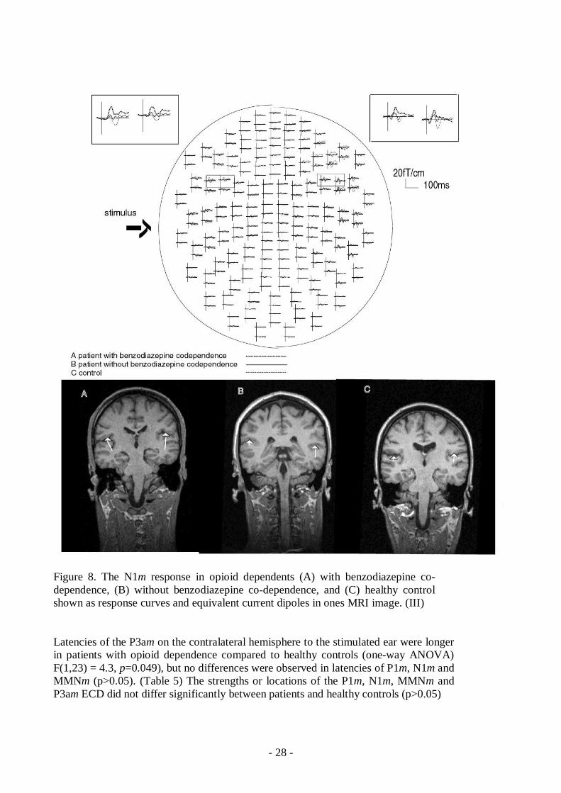
Figure 8. The N1m response in opioid dependents (A) with benzodiazepine codependence, (B) without benzodiazepine codependence, and (C) healthy controlshown as response curves and equivalent current dipoles in ones MRI image. (III)
Latencies of the P3am on the contralateral hemisphere to the stimulated ear were longerin patients with opioid dependence compared to healthy controls (oneway ANOVA)F(1,23) = 4.3, p=0.049), but no differences were observed in latencies of P1m, N1m andMMNm (p>0.05). (Table 5) The strengths or locations of the P1m, N1m, MMNm andP3am ECD did not differ significantly between patients and healthy controls (p>0.05)
29
MEGlatency msmean (SD)
contralateralhemisphere
ipsilateralhemisphere
patients controls patients controlsP1 46.9 (11.1) 45.6 (14.3) 63.8 (8.0) 62.5 (14.8)N1 88.6 (18.0) 88.9 (17.1) 109.4 (17.7) 117.2 (27.1)MMN (dev1) 148.0 (26.3) 139.7 (19.9) 153.0 (23.7) 160.1 (18.3)MMN (dev2) 151.6 (24.5) 157.1 (24.0) 159.8 (26.8) 163.9 (17.0)MMN (novel) 136.0 (19.6) 125.1 (14.1) 151.1 (31.7) 140.0 (14.4)P3a 223.4 (42.0) * 198.5 (10.1) * 230.0 (24.6) 212.0 (19.0)
Table 5. Latencies of auditory responses in MEG measurement.*≤0.05, oneway ANOVA
4.3 EEG (Study III)
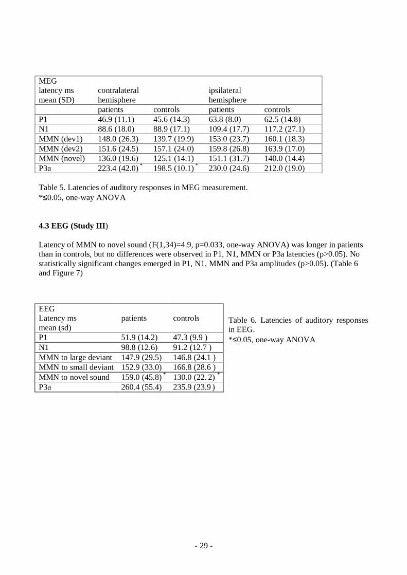
Latency of MMN to novel sound (F(1,34)=4.9, p=0.033, oneway ANOVA) was longer in patientsthan in controls, but no differences were observed in P1, N1, MMN or P3a latencies (p>0.05). Nostatistically significant changes emerged in P1, N1, MMN and P3a amplitudes (p>0.05). (Table 6and Figure 7)
Table 6. Latencies of auditory responsesin EEG.*≤0.05, oneway ANOVA
EEGLatency msmean (sd)
patients controls
P1 51.9 (14.2) 47.3 (9.9 )N1 98.8 (12.6) 91.2 (12.7 )MMN to large deviant 147.9 (29.5) 146.8 (24.1 )MMN to small deviant 152.9 (33.0) 166.8 (28.6 )MMN to novel sound 159.0 (45.8) * 130.0 (22. 2) *
P3a 260.4 (55.4) 235.9 (23.9 )
30
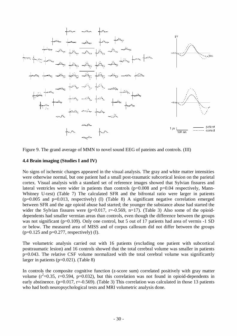
Figure 9. The grand average of MMN to novel sound EEG of pateints and controls. (III)
4.4 Brain imaging (Studies I and IV)
No signs of ischemic changes appeared in the visual analysis. The gray and white matter intensitieswere otherwise normal, but one patient had a small posttraumatic subcortical lesion on the parietalcortex. Visual analysis with a standard set of reference images showed that Sylvian fissures andlateral ventricles were wider in patients than controls (p=0.008 and p=0.04 respectively, MannWhitney Utest) (Table 7) The calculated SFR and the bifrontal ratio were larger in patients(p=0.005 and p=0.013, respectively) (I) (Table 8) A significant negative correlation emergedbetween SFR and the age opioid abuse had started; the younger the substance abuse had started thewider the Sylvian fissures were (p=0.017, r=0.569, n=17). (Table 3) Also some of the opioiddependents had smaller vermian areas than controls, even though the difference between the groupswas not significant (p=0.109). Only one control, but 5 out of 17 patients had area of vermis 1 SDor below. The measured area of MISS and of corpus callosum did not differ between the groups(p=0.125 and p=0.277, respectively) (I).
The volumetric analysis carried out with 16 patients (excluding one patient with subcorticalposttraumatic lesion) and 16 controls showed that the total cerebral volume was smaller in patientsp=0.043. The relative CSF volume normalized with the total cerebral volume was significantlylarger in patients (p=0.021). (Table 8)
In controls the composite cognitive function (zscore sum) correlated positively with gray mattervolume (r2=0.35, r=0.594, p=0.032), but this correlation was not found in opioiddependents inearly abstinence. (p=0.017, r=0.569). (Table 3) This correlation was calculated in those 13 patientswho had both neuropsychological tests and MRI volumetric analysis done.

31
Visual analysis Grade 1 Grade 2 Grade 3 Grade 4 ppatients 4 10 2 1Size of the Sylvian fissurescontrols 12 4 1 0.008patients 7 4 6 Size of the ventriclescontrols 12 4 1 0.04patients 3 8 5 1Size of the cortical sulcicontrols 3 12 2 0.26
Table 7. The size of the cerebrospinal fluid spaces graded with standard images.(MannWhitney U test)
patients controlsMeasurements (n=17) mean (SD) mean (SD) pVermis (cm2) 12.2 (1.30) 13.0 (1.47) 0.1MISS (cm2) 156 (10.92) 162 (11.14) 0.1Corpus callosum (cm2) 7.12 (0.79) 7.47 (1.00) 0.3Distance between tips of thelateral ventricle anterior horns (cm) 3.5 (0.18) 3.2 (0.35) 0.01Width of the frontal brain (cm) 10.9 (0.41) 10.8 (0.41) 0.7Bifrontal ratio 0.317 (0.015) 0.296 (0.030) 0.01Average of the width of theSylvian fissures (cm) 0.315 (0.023) 0.174 (0.003) 0.005Width of the temporal brain (cm) 13.2 (0.48) 13.2 (0.53) 0.8Sylvian fissure ratio 0.024 (0.011) 0.013 (0.004) 0.005Volumes (n=16)total volume (ml) 1360 (0.116) 1436 (0.113) 0.04Gray matterrelative to total cerebral volume 0.537 (0.009) 0.541 (0.007) 0.34White matterrelative to total cerebral volume 0.320 (0.006) 0.327 (0.011) 0.067Cerebrospinal fluid spacesrelative to total cerebral volume 0.142 (0.013) 0.132 (0.013) 0.021Table 8. Cerebral measurements in patients and controls.MISS=midsagittal internal skull surface. (MannWhitney U test)
4.5 Correlations between brain structure, opioid abuse and neuropsychological performance(Studies I and IV)
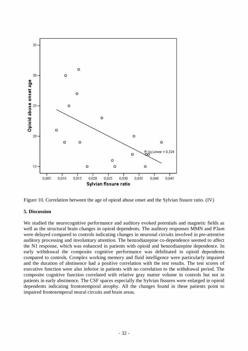
SFR had a negative correlation with the opioid abuse onset age. The Sylvian Fissures were wider inthe individuals who started the opioid abuse younger (r2=0.32, r=0.569., p=0.017, n=17). (Figure10) Correlations between age of the subject or the length of the opioid abuse history and SFR werenot found. The CFIT and PASAT test results correlated with the days of abstinence. In opioiddependents in early abstinence the composite cognitive function (zscore sum) did not correlatewith an abuse history or any cerebral measurements but in controls the positive correlation betweengray matter volume and composite cognitive function was found (r2=0.35, r=0.594, p=0.032).(Table 3) Electromagnetic measures did not correlate with opioid abuse history, cerebralmeasurements, or neuropsychological performance.
32
Figure 10. Correlation between the age of opioid abuse onset and the Sylvian fissure ratio. (IV)
5. Discussion
We studied the neurocognitive performance and auditory evoked potentials and magnetic fields aswell as the structural brain changes in opioid dependents. The auditory responses MMN and P3amwere delayed compared to controls indicating changes in neuronal circuits involved in preattentiveauditory processing and involuntary attention. The benzodiazepine codependence seemed to affectthe N1 response, which was enhanced in patients with opioid and benzodiazepine dependence. Inearly withdrawal the composite cognitive performance was debilitated in opioid dependentscompared to controls. Complex working memory and fluid intelligence were particularly impairedand the duration of abstinence had a positive correlation with the test results. The test scores ofexecutive function were also inferior in patients with no correlation to the withdrawal period. Thecomposite cognitive function correlated with relative gray matter volume in controls but not inpatients in early abstinence. The CSF spaces especially the Sylvian fissures were enlarged in opioiddependents indicating frontotemporal atrophy. All the changes found in these patients point toimpaired frontotemporal neural circuits and brain areas.

33
5.1. Changes in neural basis of preattentive attention
MMN and P3a are suggested to reflect the operation of auditory sensory memory and attentionswitching to irrelevant tones in ones’ surrounding (Escera et al. 1998, Grillon et al. 1990, Sams etal. 1985, Woods 1992), indexing the simplest form of working memory. Both responses seem tohave temporal and frontal subcomponents reflecting different states of detection and orienting ofsound change.
While intoxicated the patients with opioid dependence showed delayed latencies of MMN responseto novel sounds in EEG and delayed P3am at the contralateral hemisphere to the stimulated ear inMEG. The temporal MMN subcomponent is first elicited indexing the sound change detection in atrain of repetitive series of similar tones and the subsequent involuntary attention shift to this soundchange is probably reflected by the later frontal MMN subcomponent (Alho et al. 1998, Rinne et al.2000, Takegata et al. 2001). Since MMN latencies to novel sounds were increased in EEG, but notin MEG, it can be assumed that the frontal MMN component is specifically affected the frontalMMN is probably radially orientated and invisible to MEG (Rinne et al. 2000).
P3a is also proposed to have temporal and frontal located sources. The source responsible for theearly peak of P3a is located in the superior temporal cortex (Alho et al. 1998) and the later peak isgenerated in the prefrontal cortex (Baudena et al. 1995). The delayed P3am activity mainly reflectedthe earlier superior temporal source on the contralateral hemisphere to the stimulated ear. This thussuggests that impairment of attention shifting in opioid dependent patients also involves superiortemporal regions, in addition to the presumed frontal MMN generator abnormalities. The change infrontotemporal interactions has been found in these same patients when the spontaneous activitywas studied (Fingelkurts et al. 2006a, Fingelkurts et al. 2006b). This finding of changes in bothtemporal and frontal generators probably also reflects the impairment of these connections. Theseresults suggest that patients with opioid dependence have some impairment in processing of novelsounds at different phases of preattentive auditory processing.
5.1.1 Neurochemical modulation of preattentive attention
The clearest evidence exists of the role of NMDA receptors involved in MMN generation. It isshown in animal and human studies that NMDA antagonists block MMN elicitation (Javitt et al.1996, KreitschmannAndermahr et al. 2001, Umbricht et al. 2000, Umbricht et al. 2002). In opioiddependence evidently some changes also occur in the excitatory system as a NMDA subunitexpression and density is enhanced in NAcc and decreased in the forebrain in opioid dependentrodents (Murray et al. 2007), but exact role of glutamatergic neurotransmission at the NMDAreceptor site in opioid dependence is not clear. It might be that the NMDA system has some role inthe changes found in auditory preattentive processing.
Dopamine has a crucial role in development of opioid dependence in humans (Volkow et al. 2002).Some evidence shows that chronic opioid use changes the D2receptor binding in the cortical areas,hippocampus, and midbrain (Elwan & Soliman 1995). Previous studies in healthy individualsshowed MMN and P3a amplitude changes after a single dose of dopamine D2receptor antagonisthaloperidol (Kähkönen et al. 2001, Kähkönen et al. 2002), so it can be assumed that dopamine is inpart related to involuntary attention change. Interestingly, dopamine in the NAcc has been shown tomediate novelty responding in rats (Saigusa et al. 1999).
Benzodiazepines are known to impair active and passive attention, studied by MMN and P3 inhealthy subjects in singledose studies (Javitt et al. 1996, Lucchesi et al. 2005, Rockstroh et al.
34
1991, Rosburg et al. 2004), but the effects of chronic benzodiazepine abuse have not been studiedyet. We did not find differences in MMN or P3a in patients with and without benzodiazepine codependence. Even though benzodiazepine codependence did not affect these responses, we clearlyhad two subgroups, which appeared in the N1 response.
5.1.2 N1 response in patients with benzodiazepine codependence
Opioid dependents with benzodiazepine codependence demonstrated stronger N1m source activitycompared to patients without benzodiazepine codependence or healthy controls, suggesting thatauditory processing is changed in opioid dependence when patients also abuse benzodiazepines. Itis not surprising that N1m is changed, since GABAergic system is modulated in both opioid andbenzodiazepine dependence. Increased N1m source activity at the auditory cortex may be related toreduced inhibition and increased excitability of the cortical neurons reflecting neural adaptation tochronic simultaneous benzodiazepine and opioid effects.
Previous single dose drug challenge studies in animals have shown controversial results sinceadministration of a GABAA antagonist enhanced exogenous auditory responses resembling the N1whereas it was attenuated by several different GABAA agonists (Javitt et al. 1996, Meador 1995)Also in humans GABAA agonist lorazepam, decreased N1m source activity in MEG study (Rosburget al. 2004). As the N1m has been suggested to reflect the sound detection (Parasuraman and Beatty,1980), this function may be impaired in these subjects chronically using GABAA agonists andopioids.
5.2 Cognitive function during early withdrawal
5.2.1 Cognition and neural pathways
The auditory preattentive response MMN and P3a delay in MEGEEG measurement indexeschanges in perception and cognition. Also the neuropsychological tests showed that compositecognitive efficiency was inferior in patients in early abstinence and they especially had problems incomplex working memory, executive function, and fluid intelligence (II). As mentioned, opioiddependence modulates the mesolimbic dopamine pathway which projects to the NAcc andprefrontal cortex (Gerra et al. 2004, Volkow et al. 2002). This same monoamine pathway has acrucial role in cognitive functions (Buhot et al. 2000, Coull 1998) and it is linked to cognitiveimpairments in opioid addicts (Volkow et al. 2002). Transient dysfunction of the prefrontaldopamine system is also found under chronic stress (Izzo et al. 2005). During early opioidabstinence the high stress system activation shown as elevated cortisol level is common, but startsto normalize during the second week of abstinence (Harris & Gewirtz 2005). High cortisol levelsare especially pronounced among individuals with antisocial personality disorder (Gerra et al.2003), which was also diagnosed in most of our patients. It is also known that high cortisol levelsmay associate with a working memory deficit (Elzinga & Roelofs 2005, Roozendaal et al. 2004). Ithas been suggested that the stress system induced episodic memory impairment needs a morechronic stress system abnormality than working memory impairment (McEwen 2007, Wolf et al.2001). Working memory and fluid intelligence test results seem to be better with longer abstinenceso the stress induced by withdrawal might play a role in cognitive impairment in early abstinence.On the other hand, the more permanent deficiency in executive function is in line with previousstudies showing deficits even in late abstinence (Lee & Pau 2002, Pau et al. 2002).
35
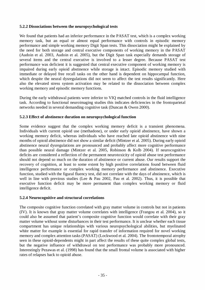
5.2.2 Dissociations between the neuropsychological tests
We found that patients had an inferior performance in the PASAT test, which is a complex workingmemory task, but an equal or almost equal performance with controls in episodic memoryperformance and simple working memory Digit Span tests. This dissociation might be explained bythe need for both storage and central executive components of working memory in the PASAT(Audoin et al. 2003, Audoin et al. 2005), but the Digit Span task especially demands storage ofseveral items and the central executive is involved to a lesser degree. Because PASAT testperformance was deficient it is suggested that central executive component of working memory isimpaired during early opioid abstinence while storage is intact. Episodic memory studied withimmediate or delayed free recall tasks on the other hand is dependent on hippocampal function,which despite the neural dysregulations did not seem to affect the test results significantly. Herealso the elevated stress system activation may be related to the dissociation between complexworking memory and episodic memory functions.
During the early withdrawal patients were inferior to VIQ matched controls in the fluid intelligencetask. According to functional neuroimaging studies this indicates deficiencies in the frontoparietalnetworks needed in several demanding cognitive task (Duncan & Owen 2000).
5.2.3 Effect of abstinence duration on neuropsychological function
Some evidence suggest that the complex working memory deficit is a transient phenomena.Individuals with current opioid use (methadone), or under early opioid abstinence, have shown aworking memory deficit, whereas individuals who have reached late opioid abstinence with ninemonths of opioid abstinence did not show a similar deficit (Mintzer et al. 2005). During early opioidabstinence neural dysregulations are pronounced and probably affect more cognitive performancethan possible neural damage (Mintzer et al. 2005, Robinson & Kolb 2004). If neurocognitivedeficits are considered a reflection of the permanent neurotoxicity of opioid abuse test performanceshould not depend so much on the duration of abstinence or current abuse. Our results support therecovery of cognition, at least to some extent by high positive correlations found between fluidintelligence performance or complex working memory performance and abstinence. Executivefunction, studied with the figural fluency test, did not correlate with the days of abstinence, which iswell in line with previous studies (Lee & Pau 2002, Pau et al. 2002). Thus, it is possible thatexecutive function deficit may be more permanent than complex working memory or fluidintelligence deficit.
5.2.4 Neurocognitive and structural correlations
The composite cognitive function correlated with gray matter volume in controls but not in patients(IV). It is known that gray matter volume correlates with intelligence (Frangou et al. 2004), so itcould also be assumed that patient’s composite cognitive function would correlate with their graymatter volume without some disturbances in their test performance. It is unclear whether each tissuecompartment has unique relationships with various neuropsychological abilities, but myelinatedwhite matter for example is essential for rapid transfer of information required for novel workingmemory and complex attention tasks (PASAT) (Lockwood et al. 2004). The frontotemporal atrophyseen in these opioiddependents might in part affect the results of these quite complex global tests,but the negative influence of withdrawal on test performance was probably more pronounced.Interestingly Pezawas et al. (1998) has found that the small frontal volume is associated with higherrates of relapses back to opioid abuse.
36
5.3 Changes in brain structure in opioid dependents
5.3.1 Brain atrophy
Opioid dependent patients showed dilatation of lateral ventricles and in cortical CSF spaces theSylvian fissures were enlarged. Volumetric analysis confirmed that the relative CSF volumepercentage was larger in opioid dependents than in age and sex matched controls. This is indicativeof brain atrophy, usually assessed as enlarged sulci and ventricles. This finding is in line with theprevious CT studies showing significant ventricular enlargement in opioid dependents (Pezawas etal. 1998, Strang & Gurling 1989).
The dilatation of Sylvian Fissures is probably reflecting the atrophy of frontal and temporalcortical and subcortical areas (Guo et al. 2001, LeMay 1984). This is well in agreement with Lyooet al (2006) as they showed that opioid dependents have diminished gray matter volumes in frontaland temporal areas. Another MRI study also showed that polysubstance abusers had significantlysmaller gray matter volumes than controls, particularly in the prefrontal lobe (Liu et al. 1998).Only a few of these patients were opioid dependents though. MRI spectroscopy of longtermheroin users have shown decreased Nacetylaspartate levels in gray matter in frontal areas, whichis considered a sign of neural damage (Haselhorst et al. 2002). A recent human study showed awidespread axonal damage in autopsy specimens of the polydrug abusers’ brain and this changewas more pronounced in young subjects, but only a few patients showed changes in myelin(Buttner et al. 2006). Also an animal study has shown the decrease of neuronal size in the ventraltegmental area (VTA) in rats after morphine administration (SklairTavron et al. 1996).Decreasedfrontal white matter volumes in chronic substance abusers have also been reported (Schlaepfer etal. 2006).
Jankovic et al (1991) showed abnormally increased serum autoantibodies to brain antigens, S100protein (a calciumbinding protein localized to astroglial cells), neuron spesific enolase (aglycolytic enzyme that is localized primarily in the neuronal cytoplasm) and myelin basic proteinin the patients with opiate abuse. Furthermore, they found that the incidence of autoantibodies waspositively related to the duration of drug abuse. These markers have been considered to beassociated with neuronal death as well as with abnormal myelin.
5.3.2 Brain maturation
Very interestingly enlargement of Sylvian fissures had a negative correlation with the age of opioidabuse onset even though it did not correlate with the duration of opioid abuse. In normaldevelopment, these dorsolateral frontal cortex and superior parts of the temporal lobes are the last tomature in early adulthood and frontotemporal white matter even later (Gogtay et al. 2004, Sowell etal. 2004, Yakolev et al. 1967). Most of our patients had begun their substance abuse in adolescenceor early adulthood, which might have in part disturbed this late maturation process of brain.
Brain maturation is a complex sequence of development of gray and white matter regulated bygenetic codes. This process is influenced positively or negatively by environmental factors.Brain maturation starts during fetal life, but the volume of the new born baby’s brain is only aquarter or a third of the volume of adult brain.
The mature white matter consists of large groups of myelinated axons interconnecting brain areas.Myelin is an electrically insulating phospholipid fatty sheet around neurons and axons affecting the
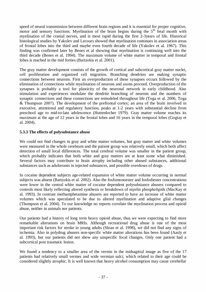
37
speed of neural transmission between different brain regions and it is essential for proper cognitive,motor and sensory functions. Myelination of the brain begins during the 5th fetal month withmyelination of the cranial nerves, and is most rapid during the first 23years of life. Historicalhistological studies by Yakolev and Lecours showed that myelination continues in association areasof frontal lobes into the third and maybe even fourth decade of life (Yakolev et al. 1967). Thisfinding was confirmed later by Benes et al showing that myelination is continuing well into thethird decade (Benes et al. 1994). The maximum volume of white matter in temporal and frontallobes is reached in the mid forties (Bartzokis et al. 2001).
The gray matter development consists of the growth of cortical and subcortical gray matter nuclei,cell proliferation and organized cell migration. Branching dendrites are making synapticconnections between neurons. First an overproduction of these synapses occurs followed by theelimination of connections while myelination of neurons and axons proceed. Overproduction of thesynapses is probably a tool for plasticity of the neuronal network in early childhood. Alsostimulation and experiences modulate the dendrite branching of neurons and the numbers ofsynaptic connections and these connections are remodeled throughout life (Toga et al. 2006, Toga& Thompson 2007). The development of the prefrontal cortex; an area of the brain involved inexecutive, attentional and regulatory function, peaks at 12 years with substantial decline frompreschool age to midtolate adolescence (Huttenlocher 1979). Gray matter volume reaches itsmaximum at the age of 12 years in the frontal lobes and 16 years in the temporal lobes (Gogtay etal. 2004).
5.3.3 The effects of polysubstance abuse
We could not find changes in gray and white matter volumes, but gray matter and white volumeswere measured in the whole cerebrum and the patient group was relatively small, which both affectdetection of small local differences. The total cerebral volume was smaller in the patient group,which probably indicates that both white and gray matters are at least some what diminished.Several factors may contribute to brain atrophy including other abused substances, additionalsubstances such as adulterants in injected substances, and possible overdoses of drugs.
In cocaine dependent subjects agerelated expansion of white matter volume occurring in normalsubjects was absent (Bartzokis et al. 2002). Also the fosfomonoester and fosfodiester concentrationswere lower in the central white matter of cocaine dependent polysubstance abusers compared tocontrols most likely reflecting altered synthesis or breakdown of myelin phospholipids (MacKay etal. 1993). In contrast methamphetamine abusers are reported to have an increase of white mattervolumes which was speculated to be due to altered myelination and adaptive glial changes(Thompson et al. 2004). To our knowledge no reports correlate the myelination process and opioidabuse, neither in animals nor patients.
Our patients had a history of long term heavy opioid abuse, thus we were expecting to find moreremarkable alterations on brain MRIs. Although recreational drug abuse is one of the mostimportant risk factors for stroke in young adults (Sloan et al. 1998), we did not find any signs ofischemia. Also in polydrug abusers nonspecific white matter alterations has been found (Aasly etal. 1993), but our patients did not show any unspecific focal changes. Only one patient had asubcortical post traumatic lesion.
We found a tendency to a smaller area of the vermis in the midsagittal image as five of the 17patients had relatively small vermes and wide vermian sulci, which related to their age could beconsidered slightly atrophic. It is well known that heavy alcohol consumption may cause cerebellar

38
and cerebral atrophy (Hayakawa et al. 1992), which are more severe with increasing age(Pfefferbaum et al. 1997). On the other hand, after abstinence, some of these changes may bereversible (Pfefferbaum et al. 1995). In this study those four patients who admitted to heavy alcoholuse in youth, showed no reduction of the area of vermes, but the ones with small vermian areas didnot have a history of alcohol abuse.
5.4 Limitations
A limitation of our study is that most of our patients fulfilled DSMIV criteria for antisocialpersonality disorder. The P3 amplitudes during the active task have been shown to be reduced insubjects with antisocial personality disorder (Bauer et al. 1994). To our knowledge, it is not knownwhether antisocial personality disorder may change preattentive auditory processing measured withMMN and P3a. The previous MRI imaging studies of patients with personality disorders haveshown frontotemporal atrophy and ventricular enlargement (Dolan et al. 2002, Raine et al. 2000).Some evidence indicates that personality disorders may be associated with lower prefrontal graymatter, lower posterior hippocampal volume, and higher callosal white matter volume or somereduction in gray matter volumes (Brambilla et al. 2004, Pridmore et al. 2005).
Previous cannabis abuse was common among our patients as well as benzodiazepine dependenceand longterm abuse was diagnosed in many. Longterm cannabis abuse and benzodiazepine abuseboth have an adverse effect on cognitive function (Barker et al. 2004, Solowij et al. 2002).Alsolongterm benzodiazepine use may lead to some minor morphological changes, such as slightventricular dilatation, also found in our patients (Lader et al. 1984, Moodley et al. 1993).
Current benzodiazepine medication during withdrawal and at the time of cognitive testing wascommon. In the normal population benzodiazepines have adverse affects on several cognitivefunctions, but the acute effect on opioid dependents and opioid dependents with benzodiazepine codependence is not known. On the other hand, the 2adrenergic receptor agonist, lofexidine, whichwas given to (Barch 2004) the patients of this study, may improve reduced working memoryperformance.
In the neuropsychological test groups patients and controls were verbal intelligence quotient (VIQ)matched whereas in most other studies the matching is based on education. Since substance abusetypically onsets at a young age, which results in skipping school and dropping out it is mostprobable that these individuals do not achieve the level of education they could. Some other opioidstudies have also matched the groups by VIQ or premorbid IQ (Davis et al. 2002, Ornstein et al.2000).
5.5 Conclusion
Our results of structural and functional changes in opioid dependents appear in the frontotemporalareas where higher order cognition such as executive function, working memory, and fluidintelligence are situated. Thus, higher order cognition disturbance, interruption of anticipation andestablishment of goals, and impulsive behaviour during early opioid abstinence are likely to beassociated. This on the other hand may indicate relapses back to substance abuse.
Changes in auditory perception were found since preattentive auditory processing was disturbed inthe opioid dependent individuals and benzodiazepine codependence further modulated the auditoryresponse indicating changes in the frontotemporal neural pathways.

39
Opioid dependents also showed the dilatation of cerebrospinal fluid spaces especially infrontotemporal areas, indicating brain atrophy. The structural changes seen in opioid dependentscorrelated with the age of opioid abuse onset, which indicates the vulnerability of adolescents todrug abuse.
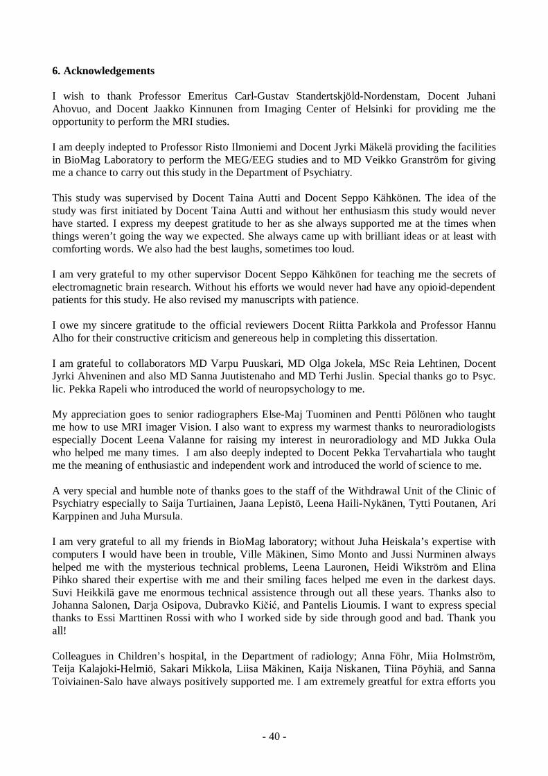
40
6. Acknowledgements
I wish to thank Professor Emeritus CarlGustav StandertskjöldNordenstam, Docent JuhaniAhovuo, and Docent Jaakko Kinnunen from Imaging Center of Helsinki for providing me theopportunity to perform the MRI studies.
I am deeply indepted to Professor Risto Ilmoniemi and Docent Jyrki Mäkelä providing the facilitiesin BioMag Laboratory to perform the MEG/EEG studies and to MD Veikko Granström for givingme a chance to carry out this study in the Department of Psychiatry.
This study was supervised by Docent Taina Autti and Docent Seppo Kähkönen. The idea of thestudy was first initiated by Docent Taina Autti and without her enthusiasm this study would neverhave started. I express my deepest gratitude to her as she always supported me at the times whenthings weren’t going the way we expected. She always came up with brilliant ideas or at least withcomforting words. We also had the best laughs, sometimes too loud.
I am very grateful to my other supervisor Docent Seppo Kähkönen for teaching me the secrets ofelectromagnetic brain research. Without his efforts we would never had have any opioiddependentpatients for this study. He also revised my manuscripts with patience.
I owe my sincere gratitude to the official reviewers Docent Riitta Parkkola and Professor HannuAlho for their constructive criticism and genereous help in completing this dissertation.
I am grateful to collaborators MD Varpu Puuskari, MD Olga Jokela, MSc Reia Lehtinen, DocentJyrki Ahveninen and also MD Sanna Juutistenaho and MD Terhi Juslin. Special thanks go to Psyc.lic. Pekka Rapeli who introduced the world of neuropsychology to me.
My appreciation goes to senior radiographers ElseMaj Tuominen and Pentti Pölönen who taughtme how to use MRI imager Vision. I also want to express my warmest thanks to neuroradiologistsespecially Docent Leena Valanne for raising my interest in neuroradiology and MD Jukka Oulawho helped me many times. I am also deeply indepted to Docent Pekka Tervahartiala who taughtme the meaning of enthusiastic and independent work and introduced the world of science to me.
A very special and humble note of thanks goes to the staff of the Withdrawal Unit of the Clinic ofPsychiatry especially to Saija Turtiainen, Jaana Lepistö, Leena HailiNykänen, Tytti Poutanen, AriKarppinen and Juha Mursula.
I am very grateful to all my friends in BioMag laboratory; without Juha Heiskala’s expertise withcomputers I would have been in trouble, Ville Mäkinen, Simo Monto and Jussi Nurminen alwayshelped me with the mysterious technical problems, Leena Lauronen, Heidi Wikström and ElinaPihko shared their expertise with me and their smiling faces helped me even in the darkest days.Suvi Heikkilä gave me enormous technical assistence through out all these years. Thanks also toJohanna Salonen, Darja Osipova, Dubravko Ki , and Pantelis Lioumis. I want to express specialthanks to Essi Marttinen Rossi with who I worked side by side through good and bad. Thank youall!
Colleagues in Children’s hospital, in the Department of radiology; Anna Föhr, Miia Holmström,Teija KalajokiHelmiö, Sakari Mikkola, Liisa Mäkinen, Kaija Niskanen, Tiina Pöyhiä, and SannaToiviainenSalo have always positively supported me. I am extremely greatful for extra efforts you

41
put up at work to help me. Special thanks go to docent Kirsi Lauerma who always understooddifficulties when organizing time between clinical and scientific work and family life.
Sputnik crew, Minna Mannerkoski, Sami Soljanlahti, Reija Utela and Sari Kivistö, we definitelyhave been in same craft and we all have tried to fly to same direction. Thank you for flying with me.
I wish to deeply thank my dear friend Sanna Seitsonen, who has helped me almost daily withscientific, professional or family life problems. There is no question that couldn’t be first askedfrom Sanna and even most probably even get answered.
I am extremely grateful to my parents Leena and Arto Kivisaari for their support all my life. Theyhave always encouraged me with neverending optimism. I also want to thank my brothers Riku andTero Kivisaari and their families. Special thanks go to Tero and Riikka for the opportunity for niceholidays in Istanbul. I owe my sincere gratitude to my parentsinlaw Taija and Hannu Tulkki forsupporting us through out these years loaded with work. I am grateful to family Nieminen, theyhave always been there to have fun with.
My sincerest appreciation goes to my family. My wonderful children Oskari, Kasperi, and Marleenahave more than anyone kept me in touch with real life and reminded me what really is important. Iam extremely privileged to have you. You can not even remember the time when this thesis was notunder work and the result was a huge disappoinment for you, all this work ended up in 49 pages!During these years J.K. Rowling wrote more than 3000 pages. My dear husband Juhana has alwayssupported me with this project that seemed endless and he has kept the household running while Ihave been engaged with scientific and clinical work. You four make my life worth living.
Very special thanks go to all the participants of the study. It was very time consuming and hard forboth patients and controls. Especially patients with withdrawal symptoms showed incrediblepatience through out the investigations.
The financial support for the studies was provided by Finnish Medical Sociaty, Orion CorporationResearch Funds, Pehr Oscar Klingendahl Foundation, Waldemar von Frencells’ Foundation, and OyH Lundbeck AB
Helsinki, January 2008
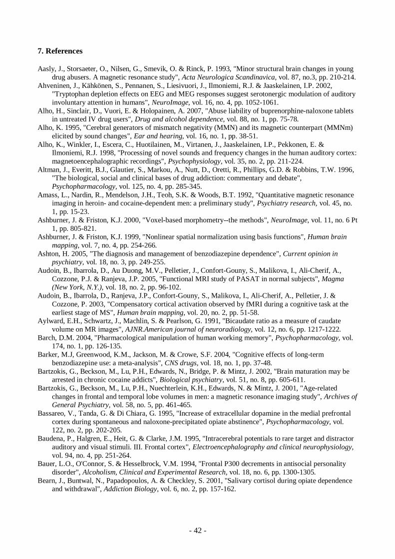
42
7. References
Aasly, J., Storsaeter, O., Nilsen, G., Smevik, O. & Rinck, P. 1993, "Minor structural brain changes in youngdrug abusers. A magnetic resonance study", Acta Neurologica Scandinavica, vol. 87, no.3, pp. 210214.
Ahveninen, J., Kähkönen, S., Pennanen, S., Liesivuori, J., Ilmoniemi, R.J. & Jaaskelainen, I.P. 2002,"Tryptophan depletion effects on EEG and MEG responses suggest serotonergic modulation of auditoryinvoluntary attention in humans", NeuroImage, vol. 16, no. 4, pp. 10521061.
Alho, H., Sinclair, D., Vuori, E. & Holopainen, A. 2007, "Abuse liability of buprenorphinenaloxone tabletsin untreated IV drug users", Drug and alcohol dependence, vol. 88, no. 1, pp. 7578.
Alho, K. 1995, "Cerebral generators of mismatch negativity (MMN) and its magnetic counterpart (MMNm)elicited by sound changes", Ear and hearing, vol. 16, no. 1, pp. 3851.
Alho, K., Winkler, I., Escera, C., Huotilainen, M., Virtanen, J., Jaaskelainen, I.P., Pekkonen, E. &Ilmoniemi, R.J. 1998, "Processing of novel sounds and frequency changes in the human auditory cortex:magnetoencephalographic recordings", Psychophysiology, vol. 35, no. 2, pp. 211224.
Altman, J., Everitt, B.J., Glautier, S., Markou, A., Nutt, D., Oretti, R., Phillips, G.D. & Robbins, T.W. 1996,"The biological, social and clinical bases of drug addiction: commentary and debate",Psychopharmacology, vol. 125, no. 4, pp. 285345.
Amass, L., Nardin, R., Mendelson, J.H., Teoh, S.K. & Woods, B.T. 1992, "Quantitative magnetic resonanceimaging in heroin and cocainedependent men: a preliminary study", Psychiatry research, vol. 45, no.1, pp. 1523.
Ashburner, J. & Friston, K.J. 2000, "Voxelbased morphometrythe methods", NeuroImage, vol. 11, no. 6 Pt1, pp. 805821.
Ashburner, J. & Friston, K.J. 1999, "Nonlinear spatial normalization using basis functions", Human brainmapping, vol. 7, no. 4, pp. 254266.
Ashton, H. 2005, "The diagnosis and management of benzodiazepine dependence", Current opinion inpsychiatry, vol. 18, no. 3, pp. 249255.
Audoin, B., Ibarrola, D., Au Duong, M.V., Pelletier, J., ConfortGouny, S., Malikova, I., AliCherif, A.,Cozzone, P.J. & Ranjeva, J.P. 2005, "Functional MRI study of PASAT in normal subjects", Magma(New York, N.Y.), vol. 18, no. 2, pp. 96102.
Audoin, B., Ibarrola, D., Ranjeva, J.P., ConfortGouny, S., Malikova, I., AliCherif, A., Pelletier, J. &Cozzone, P. 2003, "Compensatory cortical activation observed by fMRI during a cognitive task at theearliest stage of MS", Human brain mapping, vol. 20, no. 2, pp. 5158.
Aylward, E.H., Schwartz, J., Machlin, S. & Pearlson, G. 1991, "Bicaudate ratio as a measure of caudatevolume on MR images", AJNR.American journal of neuroradiology, vol. 12, no. 6, pp. 12171222.
Barch, D.M. 2004, "Pharmacological manipulation of human working memory", Psychopharmacology, vol.174, no. 1, pp. 126135.
Barker, M.J, Greenwood, K.M., Jackson, M. & Crowe, S.F. 2004, "Cognitive effects of longtermbenzodiazepine use: a metaanalysis", CNS drugs, vol. 18, no. 1, pp. 3748.
Bartzokis, G., Beckson, M., Lu, P.H., Edwards, N., Bridge, P. & Mintz, J. 2002, "Brain maturation may bearrested in chronic cocaine addicts", Biological psychiatry, vol. 51, no. 8, pp. 605611.
Bartzokis, G., Beckson, M., Lu, P.H., Nuechterlein, K.H., Edwards, N. & Mintz, J. 2001, "Agerelatedchanges in frontal and temporal lobe volumes in men: a magnetic resonance imaging study", Archives ofGeneral Psychiatry, vol. 58, no. 5, pp. 461465.
Bassareo, V., Tanda, G. & Di Chiara, G. 1995, "Increase of extracellular dopamine in the medial prefrontalcortex during spontaneous and naloxoneprecipitated opiate abstinence", Psychopharmacology, vol.122, no. 2, pp. 202205.
Baudena, P., Halgren, E., Heit, G. & Clarke, J.M. 1995, "Intracerebral potentials to rare target and distractorauditory and visual stimuli. III. Frontal cortex", Electroencephalography and clinical neurophysiology,vol. 94, no. 4, pp. 251264.
Bauer, L.O., O'Connor, S. & Hesselbrock, V.M. 1994, "Frontal P300 decrements in antisocial personalitydisorder", Alcoholism, Clinical and Experimental Research, vol. 18, no. 6, pp. 13001305.
Bearn, J., Buntwal, N., Papadopoulos, A. & Checkley, S. 2001, "Salivary cortisol during opiate dependenceand withdrawal", Addiction Biology, vol. 6, no. 2, pp. 157162.
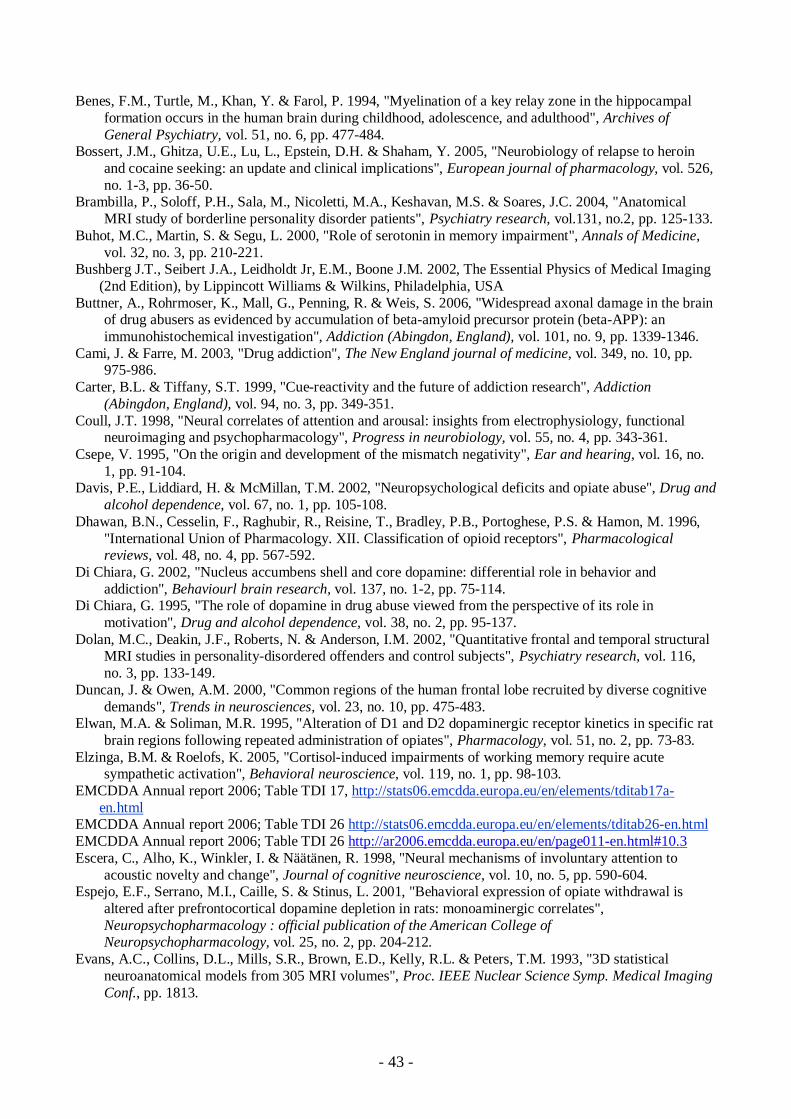
43
Benes, F.M., Turtle, M., Khan, Y. & Farol, P. 1994, "Myelination of a key relay zone in the hippocampalformation occurs in the human brain during childhood, adolescence, and adulthood", Archives ofGeneral Psychiatry, vol. 51, no. 6, pp. 477484.
Bossert, J.M., Ghitza, U.E., Lu, L., Epstein, D.H. & Shaham, Y. 2005, "Neurobiology of relapse to heroinand cocaine seeking: an update and clinical implications", European journal of pharmacology, vol. 526,no. 13, pp. 3650.
Brambilla, P., Soloff, P.H., Sala, M., Nicoletti, M.A., Keshavan, M.S. & Soares, J.C. 2004, "AnatomicalMRI study of borderline personality disorder patients", Psychiatry research, vol.131, no.2, pp. 125133.
Buhot, M.C., Martin, S. & Segu, L. 2000, "Role of serotonin in memory impairment", Annals of Medicine,vol. 32, no. 3, pp. 210221.
Bushberg J.T., Seibert J.A., Leidholdt Jr, E.M., Boone J.M. 2002, The Essential Physics of Medical Imaging(2nd Edition), by Lippincott Williams & Wilkins, Philadelphia, USA
Buttner, A., Rohrmoser, K., Mall, G., Penning, R. & Weis, S. 2006, "Widespread axonal damage in the brainof drug abusers as evidenced by accumulation of betaamyloid precursor protein (betaAPP): animmunohistochemical investigation", Addiction (Abingdon, England), vol. 101, no. 9, pp. 13391346.
Cami, J. & Farre, M. 2003, "Drug addiction", The New England journal of medicine, vol. 349, no. 10, pp.975986.
Carter, B.L. & Tiffany, S.T. 1999, "Cuereactivity and the future of addiction research", Addiction(Abingdon, England), vol. 94, no. 3, pp. 349351.
Coull, J.T. 1998, "Neural correlates of attention and arousal: insights from electrophysiology, functionalneuroimaging and psychopharmacology", Progress in neurobiology, vol. 55, no. 4, pp. 343361.
Csepe, V. 1995, "On the origin and development of the mismatch negativity", Ear and hearing, vol. 16, no.1, pp. 91104.
Davis, P.E., Liddiard, H. & McMillan, T.M. 2002, "Neuropsychological deficits and opiate abuse", Drug andalcohol dependence, vol. 67, no. 1, pp. 105108.
Dhawan, B.N., Cesselin, F., Raghubir, R., Reisine, T., Bradley, P.B., Portoghese, P.S. & Hamon, M. 1996,"International Union of Pharmacology. XII. Classification of opioid receptors", Pharmacologicalreviews, vol. 48, no. 4, pp. 567592.
Di Chiara, G. 2002, "Nucleus accumbens shell and core dopamine: differential role in behavior andaddiction", Behaviourl brain research, vol. 137, no. 12, pp. 75114.
Di Chiara, G. 1995, "The role of dopamine in drug abuse viewed from the perspective of its role inmotivation", Drug and alcohol dependence, vol. 38, no. 2, pp. 95137.
Dolan, M.C., Deakin, J.F., Roberts, N. & Anderson, I.M. 2002, "Quantitative frontal and temporal structuralMRI studies in personalitydisordered offenders and control subjects", Psychiatry research, vol. 116,no. 3, pp. 133149.
Duncan, J. & Owen, A.M. 2000, "Common regions of the human frontal lobe recruited by diverse cognitivedemands", Trends in neurosciences, vol. 23, no. 10, pp. 475483.
Elwan, M.A. & Soliman, M.R. 1995, "Alteration of D1 and D2 dopaminergic receptor kinetics in specific ratbrain regions following repeated administration of opiates", Pharmacology, vol. 51, no. 2, pp. 7383.
Elzinga, B.M. & Roelofs, K. 2005, "Cortisolinduced impairments of working memory require acutesympathetic activation", Behavioral neuroscience, vol. 119, no. 1, pp. 98103.
EMCDDA Annual report 2006; Table TDI 17, http://stats06.emcdda.europa.eu/en/elements/tditab17aen.html
EMCDDA Annual report 2006; Table TDI 26 http://stats06.emcdda.europa.eu/en/elements/tditab26en.htmlEMCDDA Annual report 2006; Table TDI 26 http://ar2006.emcdda.europa.eu/en/page011en.html#10.3Escera, C., Alho, K., Winkler, I. & Näätänen, R. 1998, "Neural mechanisms of involuntary attention to
acoustic novelty and change", Journal of cognitive neuroscience, vol. 10, no. 5, pp. 590604.Espejo, E.F., Serrano, M.I., Caille, S. & Stinus, L. 2001, "Behavioral expression of opiate withdrawal is
altered after prefrontocortical dopamine depletion in rats: monoaminergic correlates",Neuropsychopharmacology : official publication of the American College ofNeuropsychopharmacology, vol. 25, no. 2, pp. 204212.
Evans, A.C., Collins, D.L., Mills, S.R., Brown, E.D., Kelly, R.L. & Peters, T.M. 1993, "3D statisticalneuroanatomical models from 305 MRI volumes", Proc. IEEE Nuclear Science Symp. Medical ImagingConf., pp. 1813.

44
Fellows, L.K. 2007, "Advances in understanding ventromedial prefrontal function: the accountant joins theexecutive", Neurology, vol. 68, no. 13, pp. 991995.
Fingelkurts, A.A., Fingelkurts, A.A., Kivisaari, R., Autti, T., Borisov, S., Puuskari, V., Jokela, O. &Kähkönen, S. 2006a, "Increased local and decreased remote functional connectivity at EEG alpha andbeta frequency bands in opioiddependent patients", Psychopharmacology, vol. 188, no. 1, pp. 4252.
Fingelkurts, A.A., Fingelkurts, A.A., Kivisaari, R., Autti, T., Borisov, S., Puuskari, V., Jokela, O. &Kähkönen, S. 2006b, "Reorganization of the composition of brain oscillations and their temporalcharacteristics in opioid dependent patients", Progress in neuropsychopharmacology & biologicalpsychiatry, vol. 30, pp. 14531465.
First, M.B., Spitzer, R.L., Gibbon, M. & Williams, J.B.W. (eds) 1994a, Structured Clinical Interview forDSMIV Axis I Disorders, Version 2.0 edn, New York State Psychiatric Institute, New York, NY.
First, M.B., Spitzer, R.L., Gibbon, M., Williams, J.B.W. & Benjamin, L. (eds) 1994b, Structured ClinicalInterview For DSMIV Axis II Personality Disorders, Version "20 edn, New York State PsychiatricInstitute, New York, NY.
Frangou, S., Chitins, X. & Williams, S.C. 2004, "Mapping IQ and gray matter density in healthy youngpeople", NeuroImage, vol. 23, no. 3, pp. 800805.
Fukuda, K., Kato, S., Mori, K., Nishi, M., Takeshima, H., Iwabe, N., Miyata, T., Houtani, T. & Sugimoto, T.1994, "cDNA cloning and regional distribution of a novel member of the opioid receptor family", FEBSletters, vol. 343, no. 1, pp. 4246.
Gerra, G., Calbiani, B., Zaimovic, A., Sartori, R., Ugolotti, G., Ippolito, L., Delsignore, R., Rustichelli, P. &Fontanesi, B. 1998, "Regional cerebral blood flow and comorbid diagnosis in abstinent opioid addicts",Psychiatry research, vol. 83, no. 2, pp. 117126.
Gerra, G., Ceresini, S., Esposito, A., Zaimovic, A., Moi, G., Bussandri, M., Raggi, M.A. & Molina, E. 2003,"Neuroendocrine and behavioural responses to opioid receptorantagonist during heroin detoxification:relationship with personality traits", International clinical psychopharmacology, vol. 18, no. 5, pp. 261269.
Gerra, G., Garofano, L., Santoro, G., Bosari, S., Pellegrini, C., Zaimovic, A., Moi, G., Bussandri, M., Moi,A., Brambilla, F. & Donnini, C. 2004, "Association between lowactivity serotonin transporter genotypeand heroin dependence: behavioral and personality correlates", Am.J.Med.Genet.B.Neuropsychiatr.Genet., vol. 126, no. 1, pp. 3742.
Giacchino, J.L. & Henriksen, S.J. 1998, "Opioid effects on activation of neurons in the medial prefrontalcortex", Progress in neuropsychopharmacology & biological psychiatry, vol. 22, no. 7, pp. 11571178.
Gogtay, N., Giedd, J.N., Lusk, L., Hayashi, K.M., Greenstein, D., Vaituzis, A.C., Nugent, T.F.,3rd, Herman,D.H., Clasen, L.S., Toga, A.W., Rapoport, J.L. & Thompson, P.M. 2004, "Dynamic mapping of humancortical development during childhood through early adulthood", Proceedings of the National Academyof Sciences of the United States of America, vol. 101, no. 21, pp. 81748179.
Goldstein, R.Z. & Volkow, N.D. 2002, "Drug addiction and its underlying neurobiological basis:neuroimaging evidence for the involvement of the frontal cortex", The American Journal of Psychiatry,vol. 159, no. 10, pp. 16421652.
Grillon, C., Courchesne, E., Ameli, R., Geyer, M.A. & Braff, D.L. 1990, "Increased distractibility inschizophrenic patients. Electrophysiologic and behavioral evidence", Archives of General Psychiatry,vol. 47, no. 2, pp. 171179.
Guerra, D., Sole, A., Cami, J. & Tobena, A. 1987, "Neuropsychological performance in opiate addicts afterrapid detoxification", Drug and alcohol dependence, vol. 20, no. 3, pp. 261270.
Guo, X., Steen, B., Matousek, M., Andreasson, L.A., Larsson, L., Palsson, S., Sundh, V. & Skoog, I. 2001,"A populationbased study on brain atrophy and motor performance in elderly women", The journals ofgerontology. Series A, Biological sciences and medical sciences; The journals of gerontology.Series A,Biological sciences and medical sciences, vol. 56, no. 10, pp. M6337.
HaberstockDebic, H., Wein, M., Barrot, M., Colago, E.E., Rahman, Z., Neve, R.L., Pickel, V.M., Nestler,E.J., von Zastrow, M. & Svingos, A.L. 2003, "Morphine acutely regulates opioid receptor traffickingselectively in dendrites of nucleus accumbens neurons", The Journal of neuroscience : the officialjournal of the Society for Neuroscience, vol. 23, no. 10, pp. 43244332.
Hämäläinen, M., Hari R., Ilmoniemi R., Knuutila J., Lounasmaa OV. 1993,"Magnetoencephalographytheory,instrumentation, and applications to noninvasive studies of the working human brain", Reviews ofModern Physics, vol. 65, no. 2, pp.413497.

45
Hari, R., Aittoniemi, K., Jarvinen, M.L., Katila, T. & Varpula, T. 1980, "Auditory evoked transient andsustained magnetic fields of the human brain. Localization of neural generators", Experimental brainresearch.Experimentelle Hirnforschung.Experimentation cerebrale, vol. 40, no. 2, pp. 237240.
Harris, A.C. & Gewirtz, J.C. 2005, "Acute opioid dependence: characterizing the early adaptationsunderlying drug withdrawal", Psychopharmacology, vol. 178, no. 4, pp. 353366.
Haselhorst, R., DurstelerMacFarland, K.M., Scheffler, K., Ladewig, D., MullerSpahn, F., Stohler, R.,Seelig, J. & Seifritz, E. 2002, "Frontocortical Nacetylaspartate reduction associated with longterm i.v.heroin use", Neurology, vol. 58, no. 2, pp. 305307.
Hayakawa, K., Kumagai, H., Suzuki, Y., Furusawa, N., Haga, T., Hoshi, T., Fujiwara, Y. & Yamaguchi, K.1992, "MR imaging of chronic alcoholism", Acta Radiologica (Stockholm, Sweden : 1987), vol. 33, no.3, pp. 201206.
Hurle, M.A., Mediavilla, A. & Florez, J. 1982, "Morphine, pentobarbital and naloxone in the ventralmedullary chemosensitive areas: differential respiratory and cardiovascular effects", The Journal ofpharmacology and experimental therapeutics, vol. 220, no. 3, pp. 642647.
Huttenlocher, P.R. 1979, "Synaptic density in human frontal cortex developmental changes and effects ofaging", Brain research, vol. 163, no. 2, pp. 195205.
Izzo, E., Sanna, P.P. & Koob, G.F. 2005, "Impairment of dopaminergic system function after chronictreatment with corticotropinreleasing factor", Pharmacology, biochemistry, and behavior, vol. 81, no.4, pp. 701708.
Jacobsen, T., Schroger, E. & Alter, K. 2004, "Preattentive perception of vowel phonemes from variablespeech stimuli", Psychophysiology, vol. 41, no. 4, pp. 654659.
Jankovic, B.D., Horvat, J., Djordjijevic, D., Ramah, A., Fridman, V. & Spahic, O. 1991, "Brainassociatedautoimmune features in heroin addicts: correlation to HIV infection and dementia", The Internationaljournal of neuroscience, vol. 58, no. 12, pp. 113126.
Javitt, D.C., Steinschneider, M., Schroeder, C.E. & Arezzo, J.C. 1996, "Role of cortical NmethylDaspartate receptors in auditory sensory memory and mismatch negativity generation: implications forschizophrenia", Proceedings of the National Academy of Sciences of the United States of America, vol.93, no. 21, pp. 1196211967.
Kasai, K., Yamada, H., Kamio, S., Nakagome, K., Iwanami, A., Fukuda, M., Yumoto, M., Itoh, K., Koshida,I., Abe, O. & Kato, N. 2002, "Do high or low doses of anxiolytics and hypnotics affect mismatchnegativity in schizophrenic subjects? An EEG and MEG study", Clinical neurophysiology : officialjournal of the International Federation of Clinical Neurophysiology, vol. 113, no. 1, pp. 141150.
Kieffer, B.L. 1999, "Opioids: first lessons from knockout mice", Trends in pharmacological sciences, vol.20, no. 1, pp. 1926.
Koob, G.F. 1992, "Neural mechanisms of drug reinforcement", Annals of the New York Academy ofSciences, vol. 654, pp. 171191.
Koob, G.F., Ahmed, S.H., Boutrel, B., Chen, S.A., Kenny, P.J., Markou, A., O'Dell, L.E., Parsons, L.H. &Sanna, P.P. 2004, "Neurobiological mechanisms in the transition from drug use to drug dependence",Neuroscience and biobehavioral reviews, vol. 27, no. 8, pp. 739749.
Kouri, E.M., Lukas, S.E. & Mendelson, J.H. 1996, "P300 assessment of opiate and cocaine users: effects ofdetoxification and buprenorphine treatment", Biological psychiatry, vol. 40, no. 7, pp. 617628.
KreitschmannAndermahr, I., Rosburg, T., Demme, U., Gaser, E., Nowak, H. & Sauer, H. 2001, "Effect ofketamine on the neuromagnetic mismatch field in healthy humans", Brain research.Cognitive brainresearch, vol. 12, no. 1, pp. 109116.
Kujala, T., Tervaniemi, M. & Schroger, E. 2006, "The mismatch negativity in cognitive and clinicalneuroscience: Theoretical and methodological considerations", Biological psychology, .
Kähkönen, S. & Ahveninen, J. 2002, "Combination of magneto and electroencephalography in studies ofmonoamine modulation on attention", Methods and findings in experimental and clinicalpharmacology, vol. 24 Suppl C, pp. 2734.
Kähkönen, S., Ahveninen, J., Jaaskelainen, I.P., Kaakkola, S., Näätänen, R., Huttunen, J. & Pekkonen, E.2001, "Effects of haloperidol on selective attention: a combined wholehead MEG and highresolutionEEG study", Neuropsychopharmacology : official publication of the American College ofNeuropsychopharmacology, vol. 25, no. 4, pp. 498504.
Kähkönen, S., Ahveninen, J., Pekkonen, E., Kaakkola, S., Huttunen, J., Ilmoniemi, R.J. & Jaaskelainen, I.P.2002, "Dopamine modulates involuntary attention shifting and reorienting: an electromagnetic study",

46
Clinical neurophysiology : official journal of the International Federation of Clinical Neurophysiology,vol. 113, no. 12, pp. 18941902.
Kähkönen, S., Makinen, V., Jaaskelainen, I.P., Pennanen, S., Liesivuori, J. & Ahveninen, J. 2005,"Serotonergic modulation of mismatch negativity", Psychiatry research, vol. 138, no. 1, pp. 6174.
Lader, M.H., Ron, M. & Petursson, H. 1984, "Computed axial brain tomography in longtermbenzodiazepine users", Psychological medicine, vol. 14, no. 1, pp. 203206.
Laissy, J.P., Patrux, B., Duchateau, C., Hannequin, D., Hugonet, P., AitYahia, H. & Thiebot, J. 1993,"Midsagittal MR measurements of the corpus callosum in healthy subjects and diseased patients: aprospective survey", AJNR.American journal of neuroradiology, vol. 14, no. 1, pp. 145154.
Lavikainen, J., Huotilainen, M., Pekkonen, E., Ilmoniemi, R.J. & Näätänen, R. 1994, "Auditory stimuliactivate parietal brain regions: a wholehead MEG study", Neuroreport, vol. 6, no. 1, pp. 182184.
Lee, T.M. & Pau, C.W. 2002, "Impulse control differences between abstinent heroin users and matchedcontrols", Brain injury : [BI], vol. 16, no. 10, pp. 885889.
LeMay, M. 1984, "Radiologic changes of the aging brain and skull", AJR.American journal ofroentgenology, vol. 143, no. 2, pp. 383389.
Liu, X., Matochik, J.A., Cadet, J.L. & London, E.D. 1998, "Smaller volume of prefrontal lobe inpolysubstance abusers: a magnetic resonance imaging study", Neuropsychopharmacology : officialpublication of the American College of Neuropsychopharmacology, vol. 18, no. 4, pp. 243252.
Lockwood, A.H., Linn, R.T., Szymanski, H., Coad, M.L. & Wack, D.S. 2004, "Mapping the neural systemsthat mediate the Paced Auditory Serial Addition Task (PASAT)", Journal of the InternationalNeuropsychological Society : JINS; Journal of the International Neuropsychological Society : JINS,vol. 10, no. 1, pp. 2634.
Lord, J.A., Waterfield, A.A., Hughes, J. & Kosterlitz, H.W. 1977, "Endogenous opioid peptides: multipleagonists and receptors", Nature, vol. 267, no. 5611, pp. 495499.
Lucchesi, L.M., Braga, N.I., Manzano, G.M., Pompeia, S. & Tufik, S. 2005, "Acute neurophysiologicaleffects of the hypnotic zolpidem in healthy volunteers", Progress in neuropsychopharmacology &biological psychiatry, vol. 29, no. 4, pp. 557564.
Lyoo, I.K., Pollack, M.H., Silveri, M.M., Ahn, K.H., Diaz, C.I., Hwang, J., Kim, S.J., YurgelunTodd, D.A.,Kaufman, M.J. & Renshaw, P.F. 2006, "Prefrontal and temporal gray matter density decreases in opiatedependence", Psychopharmacology, vol. 184, no. 2, pp. 139144.
MacKay, S., Meyerhoff, D.J., Dillon, W.P., Weiner, M.W. & Fein, G. 1993, "Alteration of brainphospholipid metabolites in cocainedependent polysubstance abusers", Biological psychiatry, vol. 34,no. 4, pp. 261264.
McClung,C.A., Nestler, E.J. & Zachariou V. 2005, "Regulation of gene expression by chronic morphine andmorphine withdrawal in the locus ceruleus and ventral tegmental area", The Journal of neuroscience :the official journal of the Society for Neuroscience, vol. 25, no. 25, pp. 60056015.
McEwen, B.S. 2007, "Physiology and neurobiology of stress and adaptation: central role of the brain",Physiological Reviews, vol. 87, no. 3, pp. 873904.
Meador, K.J. 1995, "Cholinergic, serotonergic, and GABAergic effects on the ERP",Electroencephalography and clinical neurophysiology. Supplement, vol. 44, pp. 151155.
Mintzer, M.Z., Copersino, M.L. & Stitzer, M.L. 2005, "Opioid abuse and cognitive performance", Drug andalcohol dependence; Drug and alcohol dependence, vol. 78, no. 2, pp. 225230.
Molholm, S., Martinez, A., Ritter, W., Javitt, D.C. & Foxe, J.J. 2005, "The neural circuitry of preattentiveauditory changedetection: an fMRI study of pitch and duration mismatch negativity generators",Cerebral cortex (New York, N.Y.: 1991), vol. 15, no. 5, pp. 545551.
Moodley, P., Golombok, S., Shine, P. & Lader, M. 1993, "Computed axial brain tomograms in longtermbenzodiazepine users", Psychiatry research, vol. 48, no. 2, pp. 135144.
Murray, F., Harrison, N.J., Grimwood, S., Bristow, L.J. & Hutson, P.H. 2007, "Nucleus accumbens NMDAreceptor subunit expression and function is enhanced in morphinedependent rats", European journal ofpharmacology, vol. 562, no. 3, pp. 191197.
Näätänen, R., Paavilainen, P., Tiitinen, H., Jiang, D. & Alho, K. 1993, "Attention and mismatch negativity",Psychophysiology, vol. 30, no. 5, pp. 436450.
Näätänen, R. & Picton, T. 1987, "The N1 wave of the human electric and magnetic response to sound: areview and an analysis of the component structure", Psychophysiology, vol. 24, no. 4, pp. 375425.

47
Nakagome, K., Ichikawa, I., Kanno, O., Akaho, R., Suzuki, M., Takazawa, S., Watanabe, H. & Kazamatsuri,H. 1998, "Overnight effects of triazolam on cognitive function: an eventrelated potentials study",Neuropsychobiology, vol. 38, no. 4, pp. 232240.
Nestler, E.J. 2005, "Is there a common molecular pathway for addiction?", Nature neuroscience, vol. 8, no.11, pp. 14451449.
Ornstein, T.J., Iddon, J.L., Baldacchino, A.M., Sahakian, B.J., London, M., Everitt, B.J. & Robbins, T.W.2000, "Profiles of cognitive dysfunction in chronic amphetamine and heroin abusers",Neuropsychopharmacology : official publication of the American College ofNeuropsychopharmacology, vol. 23, no. 2, pp. 113126.
Pantev, C., Ross, B., Berg, P., Elbert, T. & Rockstroh, B. 1998, "Study of the human auditory cortices usinga wholehead magnetometer: left vs. right hemisphere and ipsilateral vs. contralateral stimulation",Audiology & NeuroOtology, vol. 3, no. 23, pp. 183190.
Papageorgiou, C.C., Liappas, I.A., Ventouras, E.M., Nikolaou, C.C., Kitsonas, E.N., Uzunoglu, N.K. &Rabavilas, A.D. 2004, "Longterm abstinence syndrome in heroin addicts: indices of P300 alterationsassociated with a short memory task", Progress in neuropsychopharmacology & biological psychiatry,vol. 28, no. 7, pp. 11091115.
Pau, C.W., Lee, T.M. & Chan, S.F. 2002, "The impact of heroin on frontal executive functions", Archives ofclinical neuropsychology : the official journal of the National Academy of Neuropsychologists, vol. 17,no. 7, pp. 663670.
Pekkonen, E., Hirvonen, J., Jaaskelainen, I.P., Kaakkola, S. & Huttunen, J. 2001, "Auditory sensory memoryand the cholinergic system: implications for Alzheimer's disease", NeuroImage, vol. 14, no. 2, pp. 376382.
Pekkonen, E., Jaaskelainen, I.P., Kaakkola, S. & Ahveninen, J. 2005, "Cholinergic modulation ofpreattentive auditory processing in aging", NeuroImage, vol. 27, no. 2, pp. 387392.
Pezawas, L.M., Fischer, G., Diamant, K., Schneider, C., Schindler, S.D., Thurnher, M., Ploechl, W., Eder, H.& Kasper, S. 1998, "Cerebral CT findings in male opioiddependent patients: stereological, planimetricand linear measurements", Psychiatry research, vol. 83, no. 3, pp. 139147.
Pfefferbaum, A., Sullivan, E.V., Mathalon, D.H. & Lim, K.O. 1997, "Frontal lobe volume loss observed withmagnetic resonance imaging in older chronic alcoholics", Alcoholism, Clinical and ExperimentalResearch, vol. 21, no. 3, pp. 521529.
Pfefferbaum, A., Sullivan, E.V., Mathalon, D.H., Shear, P.K., Rosenbloom, M.J. & Lim, K.O. 1995,"Longitudinal changes in magnetic resonance imaging brain volumes in abstinent and relapsedalcoholics", Alcoholism, Clinical and Experimental Research, vol. 19, no. 5, pp. 11771191.
Pridmore, S., Chambers, A. & McArthur, M. 2005, "Neuroimaging in psychopathy", The Australian andNew Zealand Journal of Psychiatry; The Australian and New Zealand Journal of Psychiatry, vol. 39,no. 10, pp. 856865.
Pugsley M.K. 2002, "The diverse molecular mechanisms responsible for the actions of opioids on thecardiovascular system" Pharmagology & therapeutics, vol. 93, no 1, pp 5175.
Raine, A., Lencz, T., Bihrle, S., LaCasse, L. & Colletti, P. 2000, "Reduced prefrontal gray matter volumeand reduced autonomic activity in antisocial personality disorder", Archives of General Psychiatry, vol.57, no. 2, pp. 11927; discussion 1289.
Rinne, T., Alho, K., Ilmoniemi, R.J., Virtanen, J. & Näätänen, R. 2000, "Separate time behaviors of thetemporal and frontal mismatch negativity sources", NeuroImage, vol. 12, no. 1, pp. 1419.
Robinson, T.E. & Kolb, B. 2004, "Structural plasticity associated with exposure to drugs of abuse",Neuropharmacology, vol. 47 Suppl 1, pp. 3346.
Rockstroh, B., Elbert, T., Lutzenberger, W. & Altenmuller, E. 1991, "Effects of the anticonvulsantbenzodiazepine clonazepam on eventrelated brain potentials in humans", Electroencephalography andclinical neurophysiology, vol. 78, no. 2, pp. 142149.
Roozendaal, B., McReynolds, J.R. & McGaugh, J.L. 2004, "The basolateral amygdala interacts with themedial prefrontal cortex in regulating glucocorticoid effects on working memory impairment", TheJournal of neuroscience : the official journal of the Society for Neuroscience, vol. 24, no. 6, pp. 13851392.
Rosburg, T., Marinou, V., Haueisen, J., Smesny, S. & Sauer, H. 2004, "Effects of lorazepam on theneuromagnetic mismatch negativity (MMNm) and auditory evoked field component N100m",
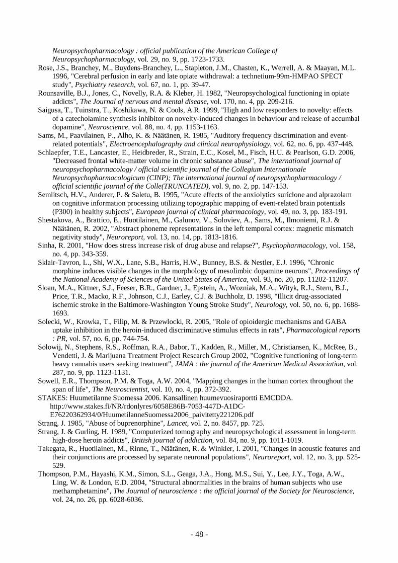
48
Neuropsychopharmacology : official publication of the American College ofNeuropsychopharmacology, vol. 29, no. 9, pp. 17231733.
Rose, J.S., Branchey, M., BuydensBranchey, L., Stapleton, J.M., Chasten, K., Werrell, A. & Maayan, M.L.1996, "Cerebral perfusion in early and late opiate withdrawal: a technetium99mHMPAO SPECTstudy", Psychiatry research, vol. 67, no. 1, pp. 3947.
Rounsaville, B.J., Jones, C., Novelly, R.A. & Kleber, H. 1982, "Neuropsychological functioning in opiateaddicts", The Journal of nervous and mental disease, vol. 170, no. 4, pp. 209216.
Saigusa, T., Tuinstra, T., Koshikawa, N. & Cools, A.R. 1999, "High and low responders to novelty: effectsof a catecholamine synthesis inhibitor on noveltyinduced changes in behaviour and release of accumbaldopamine", Neuroscience, vol. 88, no. 4, pp. 11531163.
Sams, M., Paavilainen, P., Alho, K. & Näätänen, R. 1985, "Auditory frequency discrimination and eventrelated potentials", Electroencephalography and clinical neurophysiology, vol. 62, no. 6, pp. 437448.
Schlaepfer, T.E., Lancaster, E., Heidbreder, R., Strain, E.C., Kosel, M., Fisch, H.U. & Pearlson, G.D. 2006,"Decreased frontal whitematter volume in chronic substance abuse", The international journal ofneuropsychopharmacology / official scientific journal of the Collegium InternationaleNeuropsychopharmacologicum (CINP); The international journal of neuropsychopharmacology /official scientific journal of the Colle(TRUNCATED), vol. 9, no. 2, pp. 147153.
Semlitsch, H.V., Anderer, P. & Saletu, B. 1995, "Acute effects of the anxiolytics suriclone and alprazolamon cognitive information processing utilizing topographic mapping of eventrelated brain potentials(P300) in healthy subjects", European journal of clinical pharmacology, vol. 49, no. 3, pp. 183191.
Shestakova, A., Brattico, E., Huotilainen, M., Galunov, V., Soloviev, A., Sams, M., Ilmoniemi, R.J. &Näätänen, R. 2002, "Abstract phoneme representations in the left temporal cortex: magnetic mismatchnegativity study", Neuroreport, vol. 13, no. 14, pp. 18131816.
Sinha, R. 2001, "How does stress increase risk of drug abuse and relapse?", Psychopharmacology, vol. 158,no. 4, pp. 343359.
SklairTavron, L., Shi, W.X., Lane, S.B., Harris, H.W., Bunney, B.S. & Nestler, E.J. 1996, "Chronicmorphine induces visible changes in the morphology of mesolimbic dopamine neurons", Proceedings ofthe National Academy of Sciences of the United States of America, vol. 93, no. 20, pp. 1120211207.
Sloan, M.A., Kittner, S.J., Feeser, B.R., Gardner, J., Epstein, A., Wozniak, M.A., Wityk, R.J., Stern, B.J.,Price, T.R., Macko, R.F., Johnson, C.J., Earley, C.J. & Buchholz, D. 1998, "Illicit drugassociatedischemic stroke in the BaltimoreWashington Young Stroke Study", Neurology, vol. 50, no. 6, pp. 16881693.
Solecki, W., Krowka, T., Filip, M. & Przewlocki, R. 2005, "Role of opioidergic mechanisms and GABAuptake inhibition in the heroininduced discriminative stimulus effects in rats", Pharmacological reports: PR, vol. 57, no. 6, pp. 744754.
Solowij, N., Stephens, R.S., Roffman, R.A., Babor, T., Kadden, R., Miller, M., Christiansen, K., McRee, B.,Vendetti, J. & Marijuana Treatment Project Research Group 2002, "Cognitive functioning of longtermheavy cannabis users seeking treatment", JAMA : the journal of the American Medical Association, vol.287, no. 9, pp. 11231131.
Sowell, E.R., Thompson, P.M. & Toga, A.W. 2004, "Mapping changes in the human cortex throughout thespan of life", The Neuroscientist, vol. 10, no. 4, pp. 372392.
STAKES: Huumetilanne Suomessa 2006. Kansallinen huumevuosiraportti EMCDDA.http://www.stakes.fi/NR/rdonlyres/6058E86B7053447DA1DCE76220362934/0/HuumetilanneSuomessa2006_paivitetty221206.pdf
Strang, J. 1985, "Abuse of buprenorphine", Lancet, vol. 2, no. 8457, pp. 725.Strang, J. & Gurling, H. 1989, "Computerized tomography and neuropsychological assessment in longterm
highdose heroin addicts", British journal of addiction, vol. 84, no. 9, pp. 10111019.Takegata, R., Huotilainen, M., Rinne, T., Näätänen, R. & Winkler, I. 2001, "Changes in acoustic features and
their conjunctions are processed by separate neuronal populations", Neuroreport, vol. 12, no. 3, pp. 525529.
Thompson, P.M., Hayashi, K.M., Simon, S.L., Geaga, J.A., Hong, M.S., Sui, Y., Lee, J.Y., Toga, A.W.,Ling, W. & London, E.D. 2004, "Structural abnormalities in the brains of human subjects who usemethamphetamine", The Journal of neuroscience : the official journal of the Society for Neuroscience,vol. 24, no. 26, pp. 60286036.
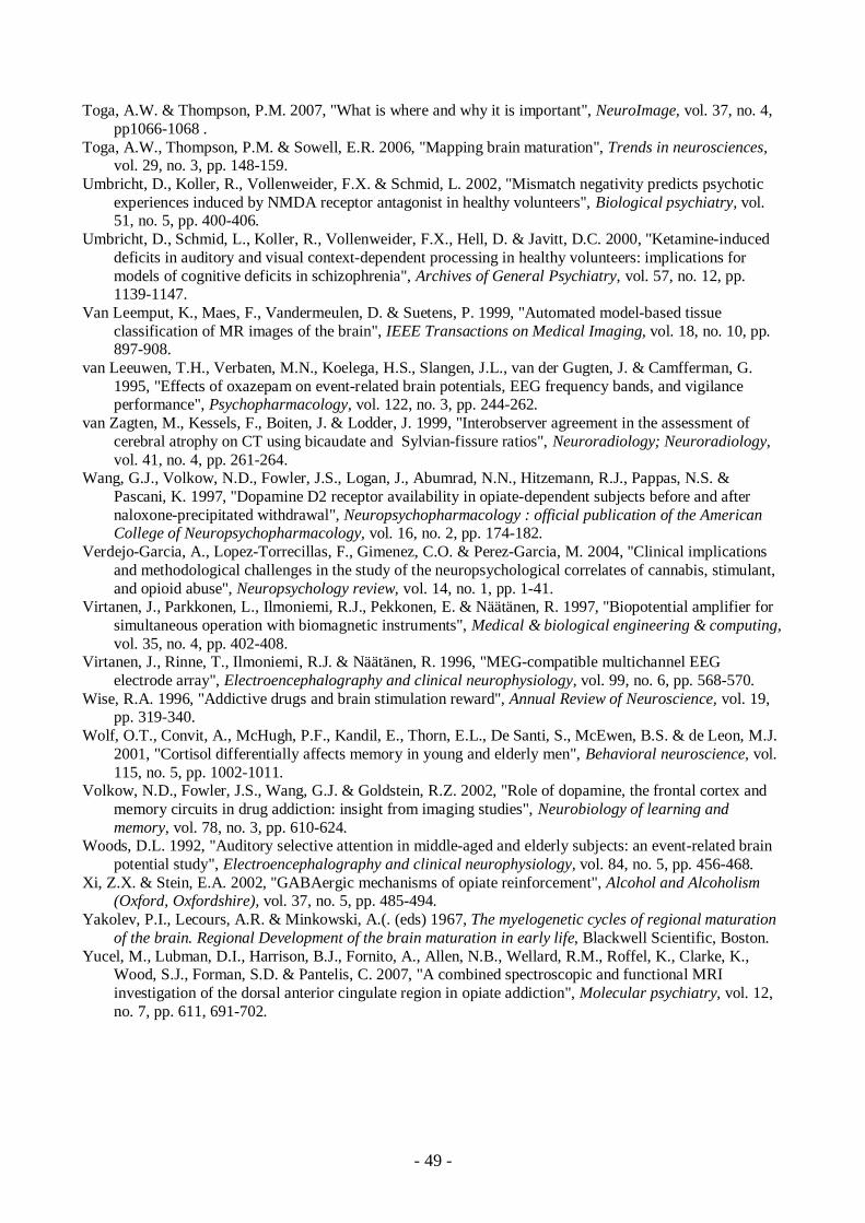
49
Toga, A.W. & Thompson, P.M. 2007, "What is where and why it is important", NeuroImage, vol. 37, no. 4,pp10661068 .
Toga, A.W., Thompson, P.M. & Sowell, E.R. 2006, "Mapping brain maturation", Trends in neurosciences,vol. 29, no. 3, pp. 148159.
Umbricht, D., Koller, R., Vollenweider, F.X. & Schmid, L. 2002, "Mismatch negativity predicts psychoticexperiences induced by NMDA receptor antagonist in healthy volunteers", Biological psychiatry, vol.51, no. 5, pp. 400406.
Umbricht, D., Schmid, L., Koller, R., Vollenweider, F.X., Hell, D. & Javitt, D.C. 2000, "Ketamineinduceddeficits in auditory and visual contextdependent processing in healthy volunteers: implications formodels of cognitive deficits in schizophrenia", Archives of General Psychiatry, vol. 57, no. 12, pp.11391147.
Van Leemput, K., Maes, F., Vandermeulen, D. & Suetens, P. 1999, "Automated modelbased tissueclassification of MR images of the brain", IEEE Transactions on Medical Imaging, vol. 18, no. 10, pp.897908.
van Leeuwen, T.H., Verbaten, M.N., Koelega, H.S., Slangen, J.L., van der Gugten, J. & Camfferman, G.1995, "Effects of oxazepam on eventrelated brain potentials, EEG frequency bands, and vigilanceperformance", Psychopharmacology, vol. 122, no. 3, pp. 244262.
van Zagten, M., Kessels, F., Boiten, J. & Lodder, J. 1999, "Interobserver agreement in the assessment ofcerebral atrophy on CT using bicaudate and Sylvianfissure ratios", Neuroradiology; Neuroradiology,vol. 41, no. 4, pp. 261264.
Wang, G.J., Volkow, N.D., Fowler, J.S., Logan, J., Abumrad, N.N., Hitzemann, R.J., Pappas, N.S. &Pascani, K. 1997, "Dopamine D2 receptor availability in opiatedependent subjects before and afternaloxoneprecipitated withdrawal", Neuropsychopharmacology : official publication of the AmericanCollege of Neuropsychopharmacology, vol. 16, no. 2, pp. 174182.
VerdejoGarcia, A., LopezTorrecillas, F., Gimenez, C.O. & PerezGarcia, M. 2004, "Clinical implicationsand methodological challenges in the study of the neuropsychological correlates of cannabis, stimulant,and opioid abuse", Neuropsychology review, vol. 14, no. 1, pp. 141.
Virtanen, J., Parkkonen, L., Ilmoniemi, R.J., Pekkonen, E. & Näätänen, R. 1997, "Biopotential amplifier forsimultaneous operation with biomagnetic instruments", Medical & biological engineering & computing,vol. 35, no. 4, pp. 402408.
Virtanen, J., Rinne, T., Ilmoniemi, R.J. & Näätänen, R. 1996, "MEGcompatible multichannel EEGelectrode array", Electroencephalography and clinical neurophysiology, vol. 99, no. 6, pp. 568570.
Wise, R.A. 1996, "Addictive drugs and brain stimulation reward", Annual Review of Neuroscience, vol. 19,pp. 319340.
Wolf, O.T., Convit, A., McHugh, P.F., Kandil, E., Thorn, E.L., De Santi, S., McEwen, B.S. & de Leon, M.J.2001, "Cortisol differentially affects memory in young and elderly men", Behavioral neuroscience, vol.115, no. 5, pp. 10021011.
Volkow, N.D., Fowler, J.S., Wang, G.J. & Goldstein, R.Z. 2002, "Role of dopamine, the frontal cortex andmemory circuits in drug addiction: insight from imaging studies", Neurobiology of learning andmemory, vol. 78, no. 3, pp. 610624.
Woods, D.L. 1992, "Auditory selective attention in middleaged and elderly subjects: an eventrelated brainpotential study", Electroencephalography and clinical neurophysiology, vol. 84, no. 5, pp. 456468.
Xi, Z.X. & Stein, E.A. 2002, "GABAergic mechanisms of opiate reinforcement", Alcohol and Alcoholism(Oxford, Oxfordshire), vol. 37, no. 5, pp. 485494.
Yakolev, P.I., Lecours, A.R. & Minkowski, A.(. (eds) 1967, The myelogenetic cycles of regional maturationof the brain. Regional Development of the brain maturation in early life, Blackwell Scientific, Boston.
Yucel, M., Lubman, D.I., Harrison, B.J., Fornito, A., Allen, N.B., Wellard, R.M., Roffel, K., Clarke, K.,Wood, S.J., Forman, S.D. & Pantelis, C. 2007, "A combined spectroscopic and functional MRIinvestigation of the dorsal anterior cingulate region in opiate addiction", Molecular psychiatry, vol. 12,no. 7, pp. 611, 691702.