Embed Size (px)
Citation preview

1
OPRA Implant System
Instructions for Use

2
Table of Contents
1 INTRODUCTION ....................................................................................................... 5
2 OPRA – IMPLANT SYSTEM ....................................................................................... 6 2.1 Manufacturer .............................................................................................................. 6 2.2 Directions for use ........................................................................................................ 6 2.3 Product liability .......................................................................................................... 6 2.4 System Description ..................................................................................................... 6 2.5 List of components ..................................................................................................... 8 2.6 Marking, packaging, storage and sterility .................................................................... 9 2.8 Identification and traceability ................................................................................... 10 2.9 Single use ................................................................................................................. 10
3 INDICATIONS AND CONTRAINDICATIONS .............................................................. 10 3.1 Indications ................................................................................................................ 10 3.2 Contraindications ...................................................................................................... 10 3.3 Warnings .................................................................................................................. 11 3.4 Precautions ............................................................................................................... 12
4 CLINICAL CONSIDERATIONS................................................................................... 13 4.1 Reported Adverse Events........................................................................................... 16
4.1.1 Adverse Event (AE) ...................................................................................................... 16 4.1.2 Serious Adverse Event (SAE) ....................................................................................... 19
4.2 Probable Benefit ....................................................................................................... 20
5 TEAM ASSESSMENT .............................................................................................. 22 5.1 Introduction .............................................................................................................. 22 5.2 Q-TFA ....................................................................................................................... 23 5.3 Professional evaluation ............................................................................................. 23 5.4 Patient information .................................................................................................. 23
6 SURGICAL TREATMENT STAGE 1 (S1) ..................................................................... 24 6.1 Preoperative Evaluation ............................................................................................ 24
6.1.1 Clinical examination .................................................................................................... 24 6.1.2 X-ray ............................................................................................................................. 24 6.1.3 Computed tomography ............................................................................................... 24 6.1.4 Technical specification ................................................................................................ 24 6.1.5 Bone quality ................................................................................................................. 25 6.1.6 Determining normal anatomical femur length .......................................................... 25 6.1.7 Planning of Fixture diameter and positioning ............................................................ 25
6.2 Preoperative preparations ........................................................................................ 26 6.2.1 Stock check .................................................................................................................. 26 6.2.2 Antibiotic prophylaxis.................................................................................................. 26 6.2.3 Setting up ..................................................................................................................... 26 6.2.4 Surgical instrument management .............................................................................. 26
6.3 Surgical technique .................................................................................................... 26 6.4 Postoperative management ...................................................................................... 27
6.4.1 X-ray ............................................................................................................................. 27 6.4.2 Antibiotic prophylaxis.................................................................................................. 27

3
6.4.3 Suture removal ............................................................................................................ 27 6.4.4 Mobilization between S1 and S2 ................................................................................ 27
7 SURGICAL TREATMENT STAGE 2 (S2) ..................................................................... 28 7.1 Preoperative Evaluation ............................................................................................ 28
7.1.1 X-ray ............................................................................................................................. 28 7.1.2 Determining Abutment length.................................................................................... 28
7.2 Preoperative preparations ........................................................................................ 28 7.2.1 Stock check .................................................................................................................. 28 7.2.2 Antibiotic prophylaxis.................................................................................................. 28 7.2.3 Setting up ..................................................................................................................... 29 7.2.4 Surgical instrument management .............................................................................. 29
7.3 Surgical technique .................................................................................................... 29 7.4 Postoperative management ...................................................................................... 29
7.4.1 Dressing ....................................................................................................................... 29 7.4.2 Antibiotic prophylaxis.................................................................................................. 29 7.4.3 Suture removal ............................................................................................................ 29 7.4.4 Mobilization and training program ............................................................................. 29
8 PROSTHETIC PROTOCOL ........................................................................................ 32 8.1 OPRA Rotasafe.......................................................................................................... 32 8.2 Connector to the OPRA Rotasafe ............................................................................... 32 8.3 Distance and optional adapters ................................................................................. 32 8.4 Knee prosthesis ........................................................................................................ 32 8.5 Foot prosthesis ......................................................................................................... 32
9 FOLLOW-UP AND CHECKS ..................................................................................... 33 9.1 Check-up schedule .................................................................................................... 33 9.2 Amputation status .................................................................................................... 33
9.2.1 Possible complications ................................................................................................ 34 9.3 Inspection of components ......................................................................................... 34
9.3.1 Tightening of Abutment Screw ................................................................................... 34 9.3.2 Possible complications ................................................................................................ 34
9.4 X-ray ......................................................................................................................... 35 9.5 Pain assessment ....................................................................................................... 35 9.6 Treatment Options .................................................................................................... 36 9.7 Fixture Removal ........................................................................................................ 36
10 ABUTMENT REPLACEMENT .................................................................................. 36 10.1 Handling instructions .............................................................................................. 36
11 HYGIENE INSTRUCTIONS ..................................................................................... 37 11.1 General instructions ................................................................................................ 37
11.1.1 Instructions for cleaning the skin penetration area ................................................ 37 11.1.2 In case of irritation or infection ................................................................................ 37
12 MEDICATION ....................................................................................................... 38 12.1 Recommended medication ...................................................................................... 38 12.2 Contraindicated medication .................................................................................... 38 12.3 Relative contraindicated medication ....................................................................... 38
13 SUMMARY OF CLINICAL STUDIES ........................................................................ 38 13.1 Introduction ............................................................................................................ 38

4
13.2 Clinical Experience with the OPRA device ................................................................ 38 13.3 Relevant Clinical Literature ...................................................................................... 39 13.4 Conclusions drawn from the studies......................................................................... 41
14 Appendix 1: Cleaning and Sterilization Instructions – Surgical Instruments .............. 43
5
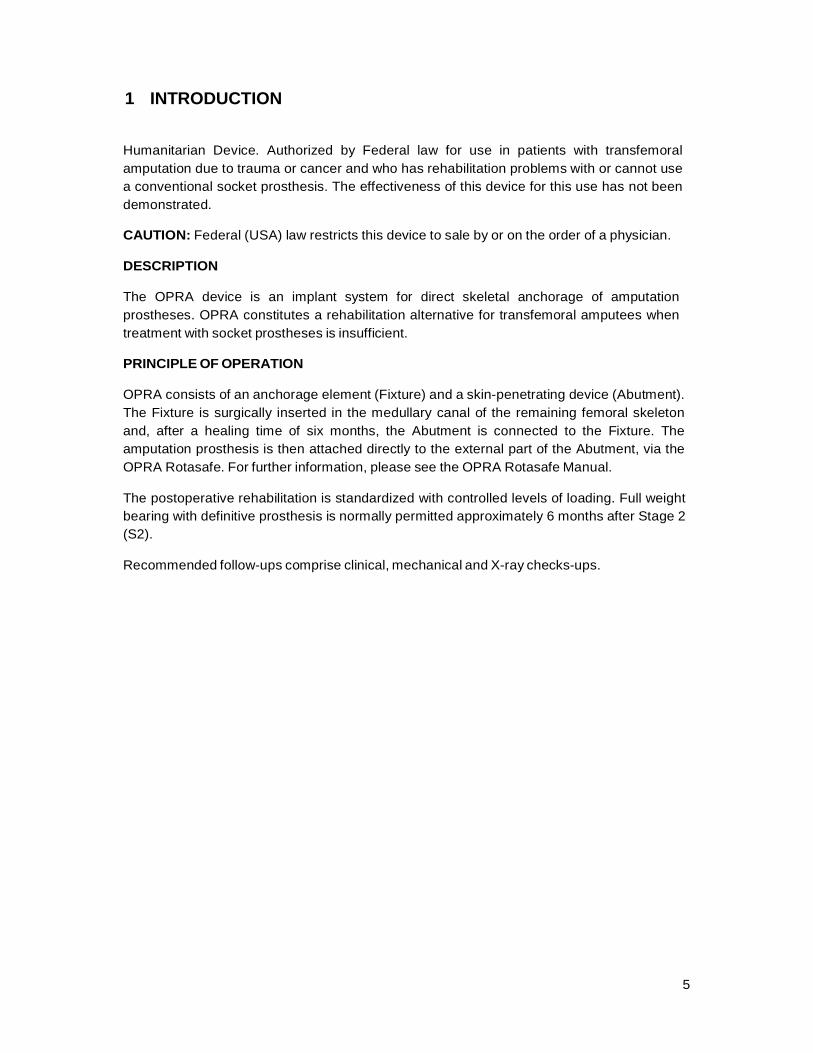
1 INTRODUCTION
Humanitarian Device. Authorized by Federal law for use in patients with transfemoral
amputation due to trauma or cancer and who has rehabilitation problems with or cannot use
a conventional socket prosthesis. The effectiveness of this device for this use has not been
demonstrated.
CAUTION: Federal (USA) law restricts this device to sale by or on the order of a physician.
DESCRIPTION
The OPRA device is an implant system for direct skeletal anchorage of amputation
prostheses. OPRA constitutes a rehabilitation alternative for transfemoral amputees when
treatment with socket prostheses is insufficient.
PRINCIPLE OF OPERATION
OPRA consists of an anchorage element (Fixture) and a skin-penetrating device (Abutment).
The Fixture is surgically inserted in the medullary canal of the remaining femoral skeleton
and, after a healing time of six months, the Abutment is connected to the Fixture. The
amputation prosthesis is then attached directly to the external part of the Abutment, via the
OPRA Rotasafe. For further information, please see the OPRA Rotasafe Manual.
The postoperative rehabilitation is standardized with controlled levels of loading. Full weight
bearing with definitive prosthesis is normally permitted approximately 6 months after Stage 2
(S2).
Recommended follow-ups comprise clinical, mechanical and X-ray checks-ups.

6
2 OPRA – IMPLANT SYSTEM
2.1 Manufacturer
INTEGRUM AB
Krokslätts Fabriker 50
SE-431 37 Mölndal
Sweden
Phone: +46 (0)31 / 760 10 60
Fax: +46 (0)31 / 15 52 60
Email: [email protected]
www.integrum.se
2.2 Directions for use
The OPRA device is implanted in two surgical stages: Stage 1 (S1) and Stage 2 (S2).
Figures 1 and 2 show the components as implanted after the surgical stages. Instructions for
the surgeries are found in the Surgical treatment Stage 1 (S1) and Surgical treatment Stage
2 (S2) Sections.
2.3 Product liability
Integrum AB is responsible for the product’s performance only when the product is used in
accordance with this Instruction for Use.
2.4 System Description
The implant components are described below, divided into the two surgical stages and
external prosthetic components.
Implant components for the Stage 1 surgery (Figure 1):
• Fixture
• Central Screw
• Healing Cylinder
• Cylinder Screw
• Graft Screw
Figure 1. Components - S1: Fixture, Central Screw, Healing Cylinder, Cylinder Screw and Graft Screw inserted in the bone.

7
Implant components for the Stage 2 surgery (Figure 2):
• Abutment
• Abutment Screw
Figure 2. Components - S2: Abutment and Abutment Screw inserted in the Fixture. (superior cortex removed for visualization of the device).
External prosthetic components:
The implant components should be connected to external prosthetic components through the connection device OPRA Rotasafe. For detailed information, see the OPRA Rotasafe Manual.
The OPRA Implant System is approved for use only with the prosthetic components manufactured by Otto Bock, specified in the Prosthetic Protocol, Section 8. Use of prosthetics other than the components specified is considered off-label use of the device.

8
2.5 List of components Table 1: List of OPRA Implant System Components
DESIGNATION
Ref.no.
Central Screw
IBC0001
Healing Cylinder
IBC0002
Cylinder Screw
IBC0017
Graft Screw
IBC0018
Fixture
Diameter (mm)
Fixture
Ø 16
IBC0008
Fixture
Ø 16.5
IBC0009
Fixture
Ø 17
IBC0010
Fixture
Ø 17.5
IBC0011
Fixture
Ø 18
IBC0012
Fixture
Ø 18.5
IBC0013
Fixture
Ø 19
IBC0014
Fixture
Ø 19.5
IBC0015
Fixture
Ø 20
IBC0016
Abutment
Length (mm)
Abutment
72
IBC0026
Abutment
72+1*
IBC0050

9
Abutment
72+2**
IBC0051
Abutment
77
IBC0027
Abutment
77+1*
IBC0057
Abutment
77+2**
IBC0058
Abutment Screw
Abutment Screw
82
IBC0033
Abutment Screw
87
IBC0034
OPRA Rotasafe
IBK0018
* Pressfit diameter increased by 0,01 mm.
** Pressfit diameter increased by 0,02 mm.
2.6 Marking, packaging, storage and sterility
Components are marked with serial number and size when appropriate.
Each component is supplied in separate packaging. All items can be stored at ordinary room
conditions.
All implant components are delivered sterile.
Inspect packages for puncture or other damage prior to surgery. If the packaging is damaged
or if sterility cannot be guaranteed for any other reason, please return the device to Integrum
AB. Do not resterilize the components.
2.7 Labels
The label is specified in Figure 3. The inner packaging comes with 5 labels. On the outer
packaging there is 1 label.
Figure 3. Label specification.
10
2.8 Identification and traceability
For purposes of traceability, attach the label from inner packaging to the patient’s medical
records when installing the component.
2.9 Single use
The OPRA implant system is intended for single use and must under no circumstances be reused. There are risks of mechanical fatigue and infection if the implant is reinstalled in another patient.
Components must unconditionally be rejected and returned to Integrum AB if:
• The packaging is damaged.
• The expiry date has passed.
• Sterility, for any other reason, cannot be guaranteed.
• They have been in contact with a patient, even if not installed.
3 INDICATIONS AND CONTRAINDICATIONS
3.1 Indications
The OPRA device is indicated for patients who have transfemoral amputation due
to trauma or cancer and who have rehabilitation problems with, or cannot use, a
conventional socket prosthesis. The OPRA device is intended for skeletally
mature patients.
The patient failed to receive benefit from a socket prostheses due to problems such as:
o Recurrent skin infections and ulcerations in the socket contact area
o Pain
o A short stump preventing the use of socket prosthesis
o Volume fluctuation in the stump
o Soft tissue scarring
o Extensive area of skin grafting
o Socket retention problems due to excessive perspiration
o Restricted mobility
3.2 Contraindications
The contraindications for the OPRA device follow:
• The patient’s skeletal growth is not complete. Completed skeletal growth is defined through the finding of generally closed epiphyseal zones on X-ray.
• The patient has atypical skeletal anatomy which may affect treatment with OPRA. Examples of atypical skeletal anatomy:
o Skeletal dimensions outside defined interval.
o Development anomalies.
o Conditions which are not amenable to device insertion such as deformities, fracture, infection.
• The patient would have less than 2 mm of remaining cortex bone available around the implant, if implanted.

11
• The patient has osteoporosis.
• The patient is older than 65 years or younger than 22 years.
• The patient’s body weight is higher than 220 lbs including the prosthesis.
• Do not treat patients with the following concurrent diseases:
o Severe peripheral vascular disease.
o Diabetic mellitus with complications.
o Skin disorders involving the residual extremity.
o Neuropathy or neuropathic disease and severe phantom pain.
o Active infection or dormant bacteria.
• The patient is pregnant.
• The patient is not expected to be able to comply with treatment and follow up requirements.
3.3 Warnings
• The OPRA device a sterile single use device. Do not use past the expiration date or if the package is opened or damaged.
• Smoking negatively impacts anchoring of the OPRA device in the femur.
• Healing problems can occur in obese patients.
• Patients with the OPRA device that undergo elective surgery for any reason are risk for infection so they should be treated with antibiotics prophylactically (e.g. cephalosporin intravenously).
• Patients with a medical history of previous infection on the amputated side should be carefully evaluated with laboratory analysis including sedimentation rate, CRP, WBC to verify there is no on-going infection. Further dormant bacteria should be excluded, especially in the skeleton. We recommend intramedullary culturing.
• Joint problems that might affect ambulation (i.e joints of the contralateral limb, the sacroiliac joint, the ipsilateral hip joint i.e. inflammatory, non-inflammatory disease or rheumatoid arthritis) may negatively affect the outcome of the treatment.
• Extension defects in hip joints should be avoided. Extension defects exceeding 10 degrees result in adverse bio-mechanical stress of the implant system, which could lead to impaired gait pattern and increase the risk of complications.
• Concurrent diseases might affect a patient’s treatment with OPRA.
• The following drugs may negatively affect the anchoring of the OPRA device in the femur and cause loosening of the Fixture:
o Steroids for systemic use
o Chemotherapy agents.
• A patient will typically not be a suitable candidate for treatment with the OPRA device if:
o The patient is using the prosthesis every day per week more than 13 hours; or
o The patient does not report more than moderate trouble and moderate reduction of quality of life.
In these instances, alternative treatments, such as socket modifications, general amputee rehabilitation, or soft tissue or bone surgery might better address rehabilitation problems.
12
• The following drugs should not be used, as they may affect bone remodeling, during the first year of treatment:
o NSAID (Non Steroid Anti Inflammatory Drugs) and ASA (acetylic salicylic acid)
two weeks preoperatively or for continued use postoperatively.
o Bisphosphonates
o Other drugs that might affect bone remodeling.
3.4 Precautions
• For at least 6 months after Stage 1 (S1) surgery, the Fixture must not be subjected to direct load. Full load with definitive prosthesis is normally permitted approximately 6 months post-operatively Stage 2 (S2) following check-up by the physician responsible for the treatment.
• Mobilization must be carried out according to individualized training programs.
• OPRA is intended for use with normal physical activity. Instructions on physical activity are included in the Patient Labeling.
• Please note both uni- and bilaterally amputated patients have been treated with the OPRA device. However only a few bilateral patients were treated with the OPRA device. Therefore the outcomes in bilateral patients are unknown and study results cannot support any definitive conclusions
• If the patient’s bone quality is judged to be suboptimal, the mobilization should be carried out at a reduced pace.
• In the event of pain or other discomfort, mobilization should be discontinued until the cause of the symptoms has been established.
• Prosthetic components should be chosen to minimize the risk of overloading the implant system. If the prosthesis is overloaded, the Fixture could be severely damaged.
• The components should be inspected for crack formation and signs of wear in the connection to external prosthetic components. Signs of wear in the connection between Fixture and Abutment include dark coloring of secretion or tissue.
• The Abutment Screw should be tightened with a counter torque device clockwise to 12 Nm torque. Tightening must be carried out in accordance with the protocol in Section 9.3.1.
• The healthcare professional should inform the patient of the following special care to be exercised:
o While riding a bike, the patient’s knee joint might lock in the fully stretched position which can seriously damage the Fixture. The patient should always position the bike seat low enough that the artificial knee cannot fully stretch out while cycling. The patient should never stand up while cycling.
o The patient should always check carefully that the prosthesis is adequately attached to the Abutment
o The patient should never try to fix any problems with the device or use any tools on the device as that may damage the Abutment and the Fixture.
o The patient should never run, jump or climb, should always use a cane or crutches for longer walks, never lift or carry heavy items and never subject the OPRA Implant System to high torques.

13
o The patient should always protect the Abutment when he or she is in hot or cold places.
o In the sauna, wrap a wet towel around the Abutment to protect it from heat.
o Protect the amputated limb when in a cold environment.
o The patient should always avoid damaging themselves or others with the Abutment.
o Protecting the Abutment during sleep is recommended. The protection will be provided by your prosthetist.
If the OPRA ROTASAFE is damaged in any way, the patient should contact his or her prosthetist.
If the OPRA ROTASAFE has been immersed in water, the patient should contact his or her prosthetist.
Change of Abutment must be considered if:
o There is movement in the connection between Fixture and Abutment, despite repeated tightening;
o Dark-colored secretion continues, despite repeated tightening; or
o The Abutment is deformed or mechanical complication is suspected.
4 CLINICAL CONSIDERATIONS
Since 1990, patients with amputations having osseoanchored devices implanted have been
followed clinically in order to assess the safety and effectiveness of the devices, for the benefit
of the patients as well as optimization and standardization of the surgical and rehabilitative
procedures. A prospective investigation, entitled the OPRA Implant System Study, was
performed, beginning in 1999. The OPRA Implant System study was a prospective
investigation on bone-anchored amputation prostheses. 51 subjects were treated in the
study, which at the time of the last surgery in the study (December 2007), constituted about
1/3 of all patients ever treated with the OPRA Implant System or pre OPRA devices.
Study Design
A prospective investigation was performed at Sahlgrenska University Hospital, Gothenburg,
Sweden on transfemoral bone-anchored amputation prostheses. The study began in 1999. Each
of the 51 subjects served as his/her own historical control, as the study was not randomized. Six
subjects were bilateral subjects. Forty-five patients were unilateral subjects. Due to the small
sample size of the bilateral patients, this group was unable to separated and studied alone. The
length of the study was 2 years.
Criteria for inclusion into this prospective study were:
Transfemoral amputee patients with problems using a conventional socket prosthesis
Undergone pre-operative Radiographic assessment including CT of the femur stump
Skeletal maturity
Normal anatomy
Body weight less than 100 kg (225 lb)
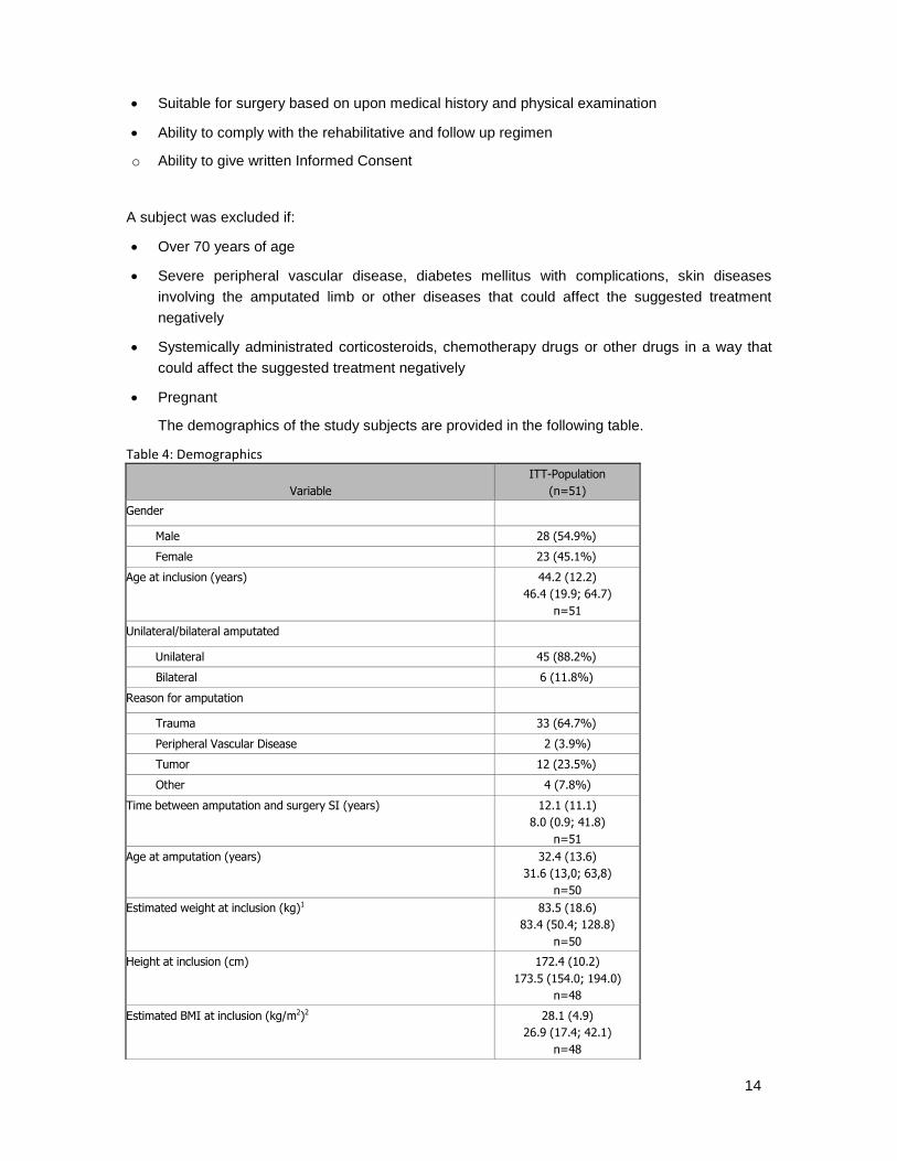
14
Suitable for surgery based on upon medical history and physical examination
Ability to comply with the rehabilitative and follow up regimen
o Ability to give written Informed Consent
A subject was excluded if:
Over 70 years of age
Severe peripheral vascular disease, diabetes mellitus with complications, skin diseases
involving the amputated limb or other diseases that could affect the suggested treatment
negatively
Systemically administrated corticosteroids, chemotherapy drugs or other drugs in a way that
could affect the suggested treatment negatively
Pregnant
The demographics of the study subjects are provided in the following table.
Table 4: Demographics
Variable
ITT-Population
(n=51) Gender
Male 28 (54.9%)
Female 23 (45.1%)
Age at inclusion (years) 44.2 (12.2)
46.4 (19.9; 64.7)
n=51
Unilateral/bilateral amputated
Unilateral 45 (88.2%)
Bilateral 6 (11.8%)
Reason for amputation
Trauma 33 (64.7%)
Peripheral Vascular Disease 2 (3.9%)
Tumor 12 (23.5%)
Other 4 (7.8%)
Time between amputation and surgery SI (years) 12.1 (11.1)
8.0 (0.9; 41.8)
n=51 Age at amputation (years) 32.4 (13.6)
31.6 (13,0; 63,8)
n=50 Estimated weight at inclusion (kg)1 83.5 (18.6)
83.4 (50.4; 128.8)
n=50 Height at inclusion (cm) 172.4 (10.2)
173.5 (154.0; 194.0)
n=48
Estimated BMI at inclusion (kg/m2)2 28.1 (4.9)
26.9 (17.4; 42.1)
n=48
15
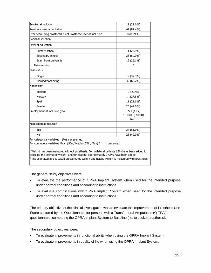
Smoker at inclusion 11 (21.6%)
Prosthetic user at inclusion 42 (82.4%)
Ever been using prosthesis if not Prosthetic user at inclusion 8 (88.9%)
Social description
Level of education
Primary school 11 (23.9%)
Secondary school 23 (50.0%)
Exam from University 12 (26.1%)
Data missing 5
Civil status
Single 19 (37.3%)
Married/cohabiting 32 (62.7%)
Nationality
England 1 (2.0%)
Norway 14 (27.5%)
Spain 11 (21.6%)
Sweden 25 (49.0%)
Employment at inclusion (%) 35.1 (41.7)
10.0 (0.0; 100.0)
n=51
Medication at inclusion
Yes 26 (51.0%)
No 25 (49.0%)
For categorical variables n (%) is presented.
For continuous variables Mean (SD) / Median (Min; Max) / n= is presented.
1 Weight has been measured without prosthesis. For unilateral patients 12% have been added to calculate the estimated weight, and for bilateral approximately 27.3% have been added. 2 The estimated BMI is based on estimated weight and height. Height is measured with prosthesis
The general study objectives were:
To evaluate the performance of OPRA Implant System when used for the intended purpose,
under normal conditions and according to instructions.
To evaluate complications with OPRA Implant System when used for the intended purpose,
under normal conditions and according to instructions.
The primary objective of the clinical investigation was to evaluate the improvement of Prosthetic Use
Score captured by the Questionnaire for persons with a Transfemoral Amputation (Q-TFA )
questionnaire, comparing the OPRA Implant System to Baseline (i.e. to socket prosthesis).
The secondary objectives were:
To evaluate improvements in functional ability when using the OPRA Implant System.
To evaluate improvements in quality of life when using the OPRA Implant System.

16
To evaluate the frequency of possible medical complications when using the OPRA Implant
System.
To evaluate the type and frequency of mechanical complications when using the OPRA Implant
System.
Additional details regarding the OPRA Implant Study are provided in Section 13, Summary of
Clinical Studies.
4.1 Reported Adverse Events
The OPRA Implant Study followed subjects for 2 years. Early loosening will, in our experience,
be the most common complication requiring removal of the OPRA Implant System. All of the
patients with implant failures showed signs of loosening, pain or infection, within the first year
of loading; however, an additional patient was not identified as a failure until later.
Early loosening was the most common complication requiring surgical removal of the OPRA
Implant System and removal was normally performed within the first two years after the Stage 2
surgery. Out of 51 subjects, 4 implant revisions due to loosening of the Fixture occurred. Out of
these 4 cases, 3 patients lost their implant within the first year following the Stage 2 surgery and
1 patient lost their implant shortly after the study was concluded. No implant fracture or re-
amputation has been reported with the OPRA Implant System.
4.1.1 Adverse Event (AE) Treatment emergent Adverse Events (AEs) were reported in the OPRA study. Adverse Events
(AEs) were captured from the enrollment of the subject and until the subject had the 24 month visit.
An Adverse Event was defined as any undesirable clinical occurrence in a subject whether it was
considered to be related to the OPRA Implant System or not. All Adverse Events during the study
were to be recorded. An AE could be both objective and subjective. The primary Safety variable
was time to revision. Adverse events were captured as the following:
Onset of Adverse Event
Pre-specified AEs
Superficial Infection
Deep Infection
Pain
Mechanical complication of OPRA
Skeletal fracture
Loosening of OPRA
Other non-pre-specified AEs
Severity of Adverse Event
The AEs were classified as mild, moderate or severe with respect to their intensity. The following
definitions were used;
Mild: AE which was easily tolerated.
Moderate: AE which causes sufficient discomfort to interfere with daily activities.
Severe: AE which caused marked limitation in activity, some assistance may have been
needed, medical intervention/therapy required, hospitalization was possible.

17
The AEs were evaluated for seriousness. A Serious Adverse Event (SAE) was defined as any
untoward medical occurrence that:
Resulted in death
Was life-threatening
Required inpatient hospitalization or prolongation of existing hospitalization
Resulted in persistent or significant disability/incapacity
Was a congenital anomaly/birth defect
The relationship to the OPRA Implant System was classified as:
Not related: The Adverse Event was definitely not related to the OPRA Implant System.
Probably Unrelated: Cause and effect relationship between the AE and OPRA Implant System was not been demonstrated, was improbable, but not impossible.
Possibly Related: A direct cause and effect relationship between the AE and the OPRA Implant System was not been demonstrated, but is possible or likely.
Related: There is a direct cause and effect relationship between the AE and the OPRA Implant System.
In the Safety Population consisting of 51 subjects, 46 (90%) reported a total of 101 treatment
emergent AEs, including events both related and unrelated to the device. Among the 101
treatment emergent AEs, 84 AEs reported by 44 (86%) subjects were considered related or
probably related to the device. All device related events, with the exception of implant
removal, were resolved. In two cases of implant failure, the subjects elected to have the
device re-implanted.
The most frequently reported adverse events that were considered related or possibly related
to the device were:
Infection with 44 events reported in 31 subjects:
o Superficial infection with 40 events reported in 28 subjects;
o Deep infection, 4 events reported in 3 subjects;
Pain with 6 events reported in 6 subjects;
Injury, with 4 events reported in 4 subjects;
Mechanical complication of the Abutment or Abutment Screw with 9 events; reported in 4 subjects; and
Loosening of the Fixture with 4 events reported in 4 subjects.
Table 2 lists the adverse events most frequently reported as related or possibly related to the device.
Table 2: Adverse Events (Safety Population)
System Organ Class
PT
Safety Population
(n=51)
AEs
Subjects with AEs
n (%)
Any AE 84 44 (86.3%)
General disorders and administration site conditions
20 12 (23.5%)
Chills 1 1 (2.0%)
Impaired healing 1 1 (2.0%)
Mechanical complication of implant 9 4 (7.8%)

18
System Organ Class
PT
Safety Population
(n=51)
AEs
Subjects with AEs
n (%)
Pain 6 6 (11.8%)
Pyrexia 2 2 (3.9%)
Wound necrosis 1 1 (2.0%)
Infections and infestations 44 31 (60.8%)
Infection 44 31 (60.8%)
Superficial 40 28 (54.9%)
Deep 4 3 (5.9%)
Injury, poisoning and procedural complications 13 13 (25.5%)
Loosening of the fixture resulting in device removal/failure
4 4 (7.8%)
Fracture 3 3 (5.9%)
Injury 4 4 (7.8%)
Joint injury 1 1 (2.0%)
Post procedural haematoma 1 1 (2.0%)
Musculoskeletal and connective tissue disorders 3 3 (5.9%)
Myositis 1 1 (2.0%)
Soft tissue necrosis 2 2 (3.9%)
Skin and subcutaneous tissue disorders 4 4 (7.8%)
Blister 1 1 (2.0%)
Skin necrosis 3 3 (5.9%)
*4 events of trauma resulting from falls
As shown in the table above, a total of 28 subjects experienced a superficial infection. Three
subjects experienced a deep infection. In the study, none of the superficial infections
developed into a deep infection. No patient who developed a deep infection had a previous
superficial infection.
Table 3 shows the distribution of subjects with treatment emergent adverse events for the
different time periods throughout the study. This table shows the number of subjects with
treat emergent adverse events whether or not they were deemed to be related, possibly
related, or not related to the OPRA Implant System. Please note, Table 3 shows ‘subjects
with events’ at each time point; therefore, one subject may be represented multiple times in
the table if they experienced an adverse event at more than one time point. However, as
Table 3 counts ‘subjects with events’, not ‘total events’, if a subject had multiple events occur
within one time period, it would only be captured once. Please also note, all adverse events
listed in Table 2 are captured in Table 3; however, they are categorized differently, such that
major adverse events, such as infection, pain and loosening are called out; while, minor
events, such as chills or bruising, are captured as other.
Table 3. Subjects with treatment emergent Adverse Events over time (Safety Population)
Adverse Events
Immed. Post-op Surgery
After Immed. Post-op
Immed. Post-op Surgery 2
After Immed. Post-op
3 months – 6 months (n=51)
6 months – 12 months (n=48)
12 months – End of Study (n=48)

19
1 (n=51) Surgery 1 – Surgery 2 (n=51)
(n=51) Surgery 2 – 3 months (n=51)
Subjects with Events n (%)
Subjects with Events n (%); Nonex
Subjects with Events n (%)
Subjects with Events n (%)
Subjects with Events n (%)
Subjects with Events n (%)
Subjects with Events n (%)
Operative Site Events
Superficial Infection
6 (11.8%) 3 (5.9%) 4 (7.8%) 13 (27.1%) 12 (25.0%)
Deep Infection 2 (3.9%) 3 (5.9%)
Pain 1 (2.0%) 3 (6.3%) 3 (6.3%)
Onset of loosening of OPRA IMPLANT SYSTEM
1 (2.0%) 3* (5.5%)
Skeletal fracture 1 (2.0%) 2 (3.9%) 1 (2.1%)
Trauma 2 (3.9%) 2 (3.9%) 3 (6.3%)
Mechanical complication of OPRA IMPLANT SYSTEM
1 (2.1%) 4 (8.3%)
Systemic Events
Myocardial infarction; Nonex
Pulmonary emboli; Nonex
Urinary tract infection
1 (2.1%)
Other 3 (5.9%) 6 (11.8%) 1 (2.0%) 2 (3.9%) 4 (8.3%) 4 (8.3%)
Immediately Post-op Surgery is defined within 42 days. * 1 patient showed signs of loosening of OPRA within the study but the fixture was removed 4 months after the 24 month follow-up x None; denotes that no events were reported in these categories shaded in dark grey in the table.
The risks associated with this device should be compared to the amputated population as a
whole. For instance the incidence of pain, skin sores and discomfort from a socket suspended
prosthesis is in the order of 50% during a four week period.1 For the OPRA Implant System
the incidence of pain and discomfort is less than 15 % over a period of 2 years, and
superficial infections have an incidence of 55 % during a 2 year period. The incidence of
revision requiring removal of the entire OPRA Implant system was 8%, well in line with limb
salvage procedures, which are often considered as an alternative to transfemoral amputation
on patient with distal bone tumours.1,2
4.1.2 Serious Adverse Event (SAE)
Among the 101 treatment emergent AEs, 47 AEs reported by 28 (55%) subjects were
1 Schwartz A, Kabo JM, Eilber FC, Eilber FR, Eckardt JJ. Cemented Distal Femoral Endoprostheses for
Musculoskeletal Tumor. Clin Orthop Related Res; 468; 2198-2210, 2010.
2 Gosheger G, Gebert C, Ahrens H, Streitbuerger A, Winkelmann W, Hardes J. Endoprosthetic Reconstruction
in 250 Patients with Sarcoma. Clin Orthop Related Res; Number 450. 164-171, 2006.

20
considered serious. The most frequent SAEs were:
• Infection, reported by 8 (16%) subjects with 10 events, whereof:
o Superficial infection, reported by 4 (8%) subjects with 4 events.
o Deep infection, reported by 4 (8%) subjects with 6 events.
• Secondary surgical intervention (including reoperation, component
replacement/revision, removal): 13 events (25.5%), specific to implant removal (3
implants removed during the study and 1 shortly after the study, giving 4 events in 4
patients; 8%).
4.2 Probable Benefit
The main efficacy measure was the Q-TFA. The Questionnaire for Persons with a Transfemoral
Amputation (Q-TFA) is a new self-report measure developed for nonelderly transfemoral amputees
using a socket- or osseointegrated prosthesis to reflect use, mobility, problems, and global health,
each in a separate score (0-100).3
The use of the OPRA Implant System was able to provide
subjects with benefit as measured by increase in prosthetic use (both number of days and hours
per day), level of function, and quality of life. The number of subjects stratified by hours per day
of prosthesis use are reported at baseline, 12 months and 24 months in Figure 4. Figure 4
shows that the number of subjects using their prosthetic more than 12 hours a day increased.
Figure 4: Hours per Day of Prosthesis Use by Visit
The number of subjects stratified by days per week of prosthesis use are reported at baseline,
12 months and 24 months in Figure 5.
3 Hagberg K, Brånemark R, Hägg O. Questionnaire for Persons with a Transfemoral Amputation (Q-TFA):
initial validity and reliability of a new outcome measure. J Rehabil Res Dev. 2004 Sep; 41(5):695-706.
0
5
10
15
20
25
30
35
Baseline 12 months 24 months
Nu
nb
er
of
Sub
ject
s
Visit
Hours per Day of Prosthesis Use
0-6 Hours
7-12 Hours
>12 Hours

21
Figure 5: Days of Prosthesis Use per Week by Visit
Figure 5 shows the increase of prosthetic use from baseline to 2 years. The prosthetic use
score, level of function, mobility, and improvement in quality of life significantly increased
from baseline to 12 and 24 months, while the problem score significantly decreased during
the same periods.
As the primary endpoint, the mean Prosthetic use score at baseline was 46.7 (Standard
Deviation 36.7) out of 100. The score increased significantly, from baseline to 12 months,
mean score (79.7 (22.7) and was sustained at 24 months, mean score 79.9 (27.1).The
OPRA Implant System was also able to provide subjects with benefits such as longer
walking distances, easier attachment and de- attachment of the prosthesis and increased
sitting comfort. Implant cumulative survival rate after two years of follow up is 92% and 93%
on patient or implant level, respectively.
The average of the Q-TFA Prosthetic Use Score stratified by baseline score and the
changes in scores at 12 and 24 months are shown in Figure 6. Figure 6 shows that low
prosthetic users (<25) saw a large increase in prosthetic use at 2 years. The moderate
prosthetic users saw a slight increase and the high functional prosthetic users saw a slight
decrease.
0
5
10
15
20
25
30
35
40
45
Baseline 12 months 24 months
Nu
mb
er
of
Sub
ject
s
Visit
Days per Week of Prosthesis Use
0 Days
1 - 3 Days
4 - 6 Days
7 Days

22
Figure 6: Mean Q-TFA Prosthetic Use Score by Visit
The average of the Q-TFA Problem Score stratified by baseline score and the changes in
scores at 12 and 24 months post-procedure are shown in Figure 7. All groups showed a
decrease in the problem score at two years.
5 TEAM ASSESSMENT
5.1 Introduction
To assess whether a patient is suitable for treatment with the OPRA device is a major
undertaking. We recommend the following guidelines developed by The Center of
Orthopaedic Osseointegration at the Department Orthopaedics at Sahlgrenska University
Hospital Gothenburg, Sweden. Team assessment is performed to reach consensus in
0
10
20
30
40
50
60
70
80
90
100
Baseline 12 mos 24 Mos
Me
an Q
-TFA
Pro
sth
eti
c U
se
Visit
Q-TFA Prosthetic Use Score
Baseline < 25
Baseline: 25 - 50
Baseline: 51 - 75
Baseline > 75
Figure 7: Mean Q-TFA Problem Score by Visit
0
10
20
30
40
50
60
70
80
90
Baseline 12 mos 24 Mos
Me
an Q
_TFA
Pro
ble
m S
core
Visit
Q-TFA Problem Score
Baseline < 25
Baseline: 25 - 50
Baseline: 51 - 75
Baseline > 75

23
relation to benefits and risks with the treatment. The team consists of at least one
orthopaedic surgeon, one prosthetist and one physiotherapist. Thorough analysis and clinical
examination of the patient is completed.
5.2 Q-TFA
In order to establish the suitability for treatment, the patient is asked to answer the Q-TFA
(Questionnaire for Persons with Transfemoral Amputations). The answers to the Q-TFA
should be reviewed by the team and the patient together to determine suitability. Questions
1,2,3,4 and 5 relate to frequency of prosthetic usage.
A patient will typically not be a suitable candidate for treatment with the OPRA device if:
• The patient is using the prosthesis every day per week more than 13 hours; or
• The patient does not report more than moderate trouble and moderate reduction of
quality of life.
In these instances, alternative treatments, such as socket modifications, general amputee
rehabilitation, or soft tissue or bone surgery might better address rehabilitation problems.
5.3 Professional evaluation
The prosthetist should carefully examine the present prosthetic solutions and state, based on
the prosthetist’s best knowledge, whether new sockets or other solutions could have a major
benefit for the patient.
The orthopaedic surgeon should carefully inform the patient about the surgical protocol and
the potential risks of the treatment. The prosthetist and the physiotherapist should carefully
inform the patient about the details of the rehabilitation program and the prosthetic solutions
available for the OPRA device. The patient should receive the OPRA Patient Labeling.
The patient should be given ample time for additional information requests. No decision on
treatment should be taken at the team assessment. The recommendation is to wait 10-14
days for contemplation by the patient to make an informed decision.
5.4 Patient information
Ensure that the patient receives the OPRA Patient Labeling, where hazards and warnings
are clearly stated.

24
6 SURGICAL TREATMENT STAGE 1 (S1)
6.1 Preoperative Evaluation
6.1.1 Clinical examination
Preoperative amputation status includes:
• Registration of active and passive hip mobility. Extension defects in hip joints should
be avoided. Extension defects exceeding 10 degrees result in adverse biomechanical
stress of the implant system, which could lead to impaired gait pattern and increase
the risk of complications.
• Assessment of the residual extremity with regard to skin, soft tissue and skeleton.
• Laboratory analysis: The recommendation is to make a complete kidney and liver
function analysis to determine the patient’s capability to synthesize all type of oral or
intravenous antibiotics, in case of post-operative or later infections that may occur
during the long-term use of this device.
6.1.2 X-ray
Preoperative X-ray examination of the residual bone is carried out in frontal and lateral
projection, with a ruler.
To optimize the survey of the residual bone’s distal parts where the degree of mineralization
is lower, supplementary soft tissue examination is also recommended on two levels.
Survey of skeletal structure
Particular attention is focused on:
• The skeletal dimension.
• Skeletal quality.
• Any development anomalies.
• Any residual conditions after fracture, infection etc.
6.1.3 Computed tomography
Preoperative Stage 1 (S1) computed tomography examination is carried out.
The purpose of the examination is:
• To complement survey of the skeletal structure.
• To verify that the length of the residual bone provides space for external prosthetic
components. This is done by determining the normal anatomical femur length.
• To provide a reliable basis for planning the Fixture dimension.
6.1.4 Technical specification
• Skeletal window.
• Overall view of the frontal and lateral projection including the whole femur bilaterally.
The purpose of this overall view is to make it possible to measure the length of the
femur and to ensure that the cross section projections are at right angles (±10°) in
relation to the longitudinal axis of the femur.
• Cross section covering the residual bone’s distal 120 mm. Section thickness
maximum 3 mm. Section distance 3 mm. To ensure adequate planning of the Fixture

25
diameter, all cross sections must be documented in 1:1 scale and have a scale of
length enclosed.
6.1.5 Bone quality
This treatment has mostly been used for patients who have had their amputation for some
time. In the OPRA clinical study, an average of more than 10 years had passed since the
amputation. Amputation leads to reduced function as compared to an able bodied person and
the skeleton will not be loaded in a normal physiological way, thus the cortical bone quality in
the diaphyseal part of the femur bone tend to change over time. This bone may change to
more like spongious or trabecular bone. To some extent, bone quality can be judged by x-ray.
Evaluation of bone quality should be performed peroperatively. At least 2 mm of remaining
bone tissue should be available around the implant.
Depending on the bone quality, different drills are used during the surgical procedure. Bone
grafting is not recommended except for the distal end of the bone, and bone grafting should
always be performed distally. Please see the OPRA Stage 1 - Surgical Technique Manual for
further details.
6.1.6 Determining normal anatomical femur length
Femur length is measured bilaterally. The measurement originates from the proximal
boundary of the trochanter major to central knee joint line and central residual bone’s distal
end respectively. Via secondary comparison the distance from the residual bone to normal
anatomical position of the knee joint line can be determined. The minimum recommended
distance to provide space for external prosthetic components is 200 mm. The recommended
remaining length of the bone is 130-350 mm measured from the top of the greater trochanter
to the distal bone end.
6.1.7 Planning of Fixture diameter and positioning
In all cross sections corresponding to 80 mm (approx. 27 cross sections) of the residual
bone, where it is planned to place the Fixture, a circle is drawn so that the thickness of the
cortical bone does not fall below 2 mm anywhere on the circumference and optimal
endosteal contact is maintained (Figure 8). The resulting diameter of the circle determines
the Fixture size and the location of the circle determines the positioning of the Fixture.
Other preoperative evaluation is carried out in accordance with the procedures of the treating
unit.

26
Figure 8. Planning of Fixture diameter and positioning.
The estimated size of the Fixture must be within the intervals of 16.5-19.5 mm. If the dimension is not
within this interval, this constitutes a contraindication to treatment based on atypical skeleton anatomy.
6.2 Preoperative preparations
6.2.1 Stock check
Ensure that all Fixture sizes and other relevant components are in stock. Check particularly
that the planned Fixture size, as well as 0.5 mm larger and 0.5 mm smaller diameters, are
available.
6.2.2 Antibiotic prophylaxis
The procedure is carried out under antibiotic prophylaxis. Use e.g. penicillinase stable
penicillin (isoxa penicillin) such as Cloxacillin 2 g x 3 IV or cephalosporins such as
Cefuroxime 1.5 g x 3 IV. In case of over-sensitivity to the above preparations, Clindamycin
600 mg x 3 IV should be used. Antibiotic infusion should be started at least 30 minutes
before the planned start of surgery.
6.2.3 Setting up
The positioning of the patient on the operating table must allow for fluoroscopy using a C-arm
in two right angle levels.
Prepare for possible autologous bone transplantation from the iliac crest. Preferably the bone
should be harvested ipsilaterally.
6.2.4 Surgical instrument management
Instructions for cleaning and sterilization of the surgical instruments are provided in Appendix
1.
6.3 Surgical technique
The surgery should be carried out in accordance with the stepwise instructions with the
stepwise instructions and illustrations in Appendix 1, OPRA Stage 1 – Surgical Technique.
The surgery should be performed by a trained physician with expertise in orthopedic surgical
techniques. .

27
6.4 Postoperative management
6.4.1 X-ray
Postoperative X-ray evaluation is carried out in frontal and lateral projection. Fixture position
and signs of any possible complications are evaluated.
6.4.2 Antibiotic prophylaxis
Intravenous antibiotic should be administered within one hour of surgery and maintained as
intravenous administration for 24 hours. Oral administration of antibiotics is then continued
until two days after suture removal. Use oral antibiotics which correspond to the parenteral
treatment. .
6.4.3 Suture removal
Sutures should be removed after approximately 3 weeks.
6.4.4 Mobilization between S1 and S2
Mobilization including joint movement, strength and fitness training is carried out under the
supervision of a physiotherapist. Prevention of extension defect is an important element.
If skin status permits use of socket prosthesis, preoperative prosthesis-wearing patients can
begin using adapted socket prosthesis 1-3 weeks after suture removal. If a socket is used
between S1 and S2, make sure that there is no end-bearing in the socket to reduce the risk
of loading the Fixture.
For at least 6 months after Stage 1 (S1), the Fixture must not be subjected to direct load.

28
7 SURGICAL TREATMENT STAGE 2 (S2)
7.1 Preoperative Evaluation
7.1.1 X-ray
Preoperative X-ray examination of the residual bone is carried out in frontal and lateral
projection. There must be no signs of the Fixture coming loose or other complications.
7.1.2 Determining Abutment length
Preoperative Stage 2 (S2) determining of Abutment length (Figure 9):
• Measure the distance (X) from the Fixture’s distal edge to residual bone’s distal edge.
The X-ray examination should be carried out using a measurement rod in order to obtain the
correct magnification factor. Alternatively the magnification factor can be calculated based on
the Fixture’s known dimension.
• Calculate the smallest Abutment length by adding 52 mm to the measured distance
(X). The Abutment Screw should always be 10 mm longer than the Abutment.
Figure 9. Determination of Abutment length.
7.2 Preoperative preparations
7.2.1 Stock check
Ensure that all relevant components are in stock.
7.2.2 Antibiotic prophylaxis
The procedure is carried out under antibiotic prophylaxis. Use e.g. penicillinase stable
penicillin (isoxa penicillin) such as Cloxacillin 2 g x 3 IV or cephalosporins such as
Cefuroxime 1.5 g x 3 IV. In case of allergy to the above antibiotics, Clindamycin 600 mg x 3
IV should be used. Antibiotic infusion should be started approximately 30 minutes before the
planned start of surgery.

29
7.2.3 Setting up
Ipsilateral gluteal support may be used. Sterile dressing should leave at least 15 cm free skin
area distally on the residual extremity.
7.2.4 Surgical instrument management
Instructions for cleaning and sterilization of the surgical instruments are provided in Appendix
1.
7.3 Surgical technique
The surgery should be carried out in accordance with the stepwise instructions and illustrations
in Appendix 2, OPRA Stage 2 – Surgical Technique. The surgery should be performed by a
trained physician with expertise in orthopedic surgical techniques.
7.4 Postoperative management
7.4.1 Dressing
Dressing should be changed at 2-3 day intervals until the skin has healed to the bone
surface.
7.4.2 Antibiotic prophylaxis
Antibiotic prophylaxis should be given up to suture removal + 2 days.
Switch to oral administration as soon as possible. Use oral antibiotics which correspond to
the parenteral treatment.
7.4.3 Suture removal
Sutures should be removed after approximately 3 weeks.
7.4.4 Mobilization and training program
Mobilization is carried out under the supervision of a physiotherapist in accordance with
general principles for treatment of osseoanchored implants. Initial loading is carried out with a
short training prosthesis, followed by training with a full-length prosthesis according to the
training program below.
Training prosthesis:
Week 1-2 post-op Stage 2 (S2)
• Stay immobilized.
Week 3 post-op Stage 2 (S2)
• Active movement training of the hip joint without load.
Week 6 post-op Stage 2 (S2)
• Initiate loading with short training prosthesis that only reach to the knee joint, and an
initial a load of maximum 20 kg. Avoid rotations.
• Increase approximately 10 kg per week until full bodyweight is reached. However, the
load must be adapted to the patient’s body size and strength. Exercise 2x15 minutes
per day, increasing to 2x30 minutes/day.

30
• If pain occurs above 5 on a Visual Analogue Scale (VAS) – the patient should abstain
from all training for 1-2 days or until pain has decreased to a more pain-free level.
Return to training using a decreased load. If pain remains above 5, the patient should
contact the treating physician. For Pain Assessment, see Section 9.5.
Week 10-14 post-op Stage 2 (S2)
With training prosthesis, if full body weight is reached and training conducted without
pain:
• General fitness exercises including kneeling in all four and kneeling down.
• Return to the treating physician for a decision concerning full-length prosthesis.
With Full-length prosthesis:
• Initiate training with full-length prosthesis and an initial load of maximum 20 kg while
walking.
• Increase approximately 10 kg per week.
• Use the prosthesis maximum 2x60 minutes daily indoors.
• Walking exercises should be carried out with parallel bars and with 2 crutches.
• In a standing position, the maximum load is half bodyweight.
• Do not use training prosthesis.
As rehabilitation continues, if pain occurs above 5 on a VAS, rest completely from all
kinds of training during 1-2 days. Return using decreased load. If pain remains above
5, the patient should contact the treating physician. For Pain Assessment, see Section
9.5.
Week 12-16 post-op Stage 2 (S2)
• Training of balance and gait pattern.
• Always use 2 crutches. Use of stairs.
• Fitness cycling with light load.
• Sitting down and sitting down to standing up.
• Fitness training with training prostheses.
Week 14-18 post-op Stage 2 (S2)
• Prostheses might be used the entire day.
• Transferring of body weight while standing.
• Walking up-hill with two supports.
Week 16-24 post-op Stage 2 (S2)
• Walking exercises with one support at the physiotherapist and at home.
• Always use 2 crutches for longer walks outdoors.
• Walking slightly uphill, in rough terrain, over obstacles.
• Turning.
• Fitness training with full-length prostheses.

31
Week 22-26 post-op Stage 2 (S2)
If full body weight is reached and training conducted without pain:
• Walking without support during training.
• Return to the treating physician for a decision about the use of one support more
frequently while walking.
If the patient’s bone quality is judged to be suboptimal, the mobilization should be carried out
at a reduced pace.

32
8 PROSTHETIC PROTOCOL
8.1 OPRA Rotasafe
The OPRA Implant System is connected to external prosthetic components through the
connection device OPRA Rotasafe. For detailed information, see the OPRA Rotasafe
Manual.
The OPRA Implant System is approved for use only with the prosthetic components
manufactured by Otto Bock, specified below. Use of prosthetics other than the components
specified is considered off-label use of the device.
8.2 Connector to the OPRA Rotasafe
Male adapters:
• 4R54 Socket Adapter with Pyramid Adapter
• 4R77 Socket Adapter with Pyramid Adapter, rotatable
Female adapters (for connection directly onto the knee for longer stumps):
• 4R55 Socket Adapter with Pyramid Receiver
• 4R51 Socket Adapter with Pyramid receiver, rotatable
M6 bolts must be used for attachment of the adapters.
8.3 Distance and optional adapters
For shorter stumps where there is space between the end of the abutment and top of the
knee, choice of;
• 4R72=32
• 4R72=45
• 4R72=60
• 4R72=75
8.4 Knee prosthesis
• C-leg 3C98 with shin tube 2R81 with torsion adapter. If no space available, use 2R80
without torsion adaptor.
8.5 Foot prosthesis
• 1D10
• Trias Carbon Composite Foot 1C30
• Axtion Carbon Fibre Foot

33
9 FOLLOW-UP AND CHECKS
9.1 Check-up schedule
After Stage 2 (S2) the following check-ups are recommended (Table 4):
Action
Day
21
Month
3
Month
6
Month
12
Continued
Amputation status • • • •
Every 6 months
Inspection of
components
•
•
•
•
Every 6 months
X-ray
• • Years 2, 3, 5, 7, 10, 15 etc.
Table 4. Check-up schedule.
9.2 Amputation status
Clinical examination of the residual extremity comprises the following elements:
• Inspection of skin penetration.
• Inspection of soft tissue configuration when standing.
• Assessment ofosseoanchorage:
o Pain when loading is a potential sign of loosening.
o A mobile implant system is a sign of loosening.
o A radiolucent zone around the entire Fixture is a potential sign of loosening.
• Joint evaluation:
o Assessment of range of movement in the hip joint, particularly with regard to
extension.
o Evaluate joints to specifically check for any signs of ipsilateral leg joint disease
or contralateral leg joint disease as well as back disorders including the
sacroiliac joints.
• Examination may include clinical examination, x-ray or MRI.
• Symptoms might include pain, swelling, or reduced activity level.
• If there are signs of adverse effects on the contralateral extremity,
ipsilateral hip joint or the sacroiliac joint, the patient should be
evaluated over time according to applicable standard scoring methods
(Harris Hip Score, Knee Society Score, etc.) If a reduction in the total
score is seen, determine if there are compounding factors that may
affect the joint. If the cause of the joint problem cannot be identified,
discontinue use of the prosthetic until the adverse effect is reduced or
eliminated. In the OPRA Implant System study, joint disease related
adverse events were not reported up to 2 years of follow up.

34
9.2.1 Possible complications
• Superficial infection around the skin penetration area is the most common
complication during the treatment. Common signs of infection are local redness and
swelling as well as discolored secretion. Note that moderately serous or dark-colored
secretion may normally occur. In case of suspected superficial infection, culture tests
must be taken. Treatment can be carried out by intensifying local cleaning. Antibiotics
should be prescribed on relatively wide indications in order to reduce the risk of
progressive deep infection.
• In case of pronounced infection with signs of involvement of subfascial tissue as well
as possible general effects on the patient, deep infection must be suspected. After
culture testing, antibiotic treatment must be started as soon as possible.
• Excessive soft tissue distally on the residual extremity may affect the skin penetration
area. In the event of recur-rent infection problems, soft tissue plasty may be
considered.
• Restricted extension ability in the hip joint may lead to impaired gait pattern in the
patient, with unfavorable mechanical loads on the implant system. Extension defects
exceeding 10º must be avoided.
9.3 Inspection of components
Inspection of components comprises the following elements:
• Assessment of any crack formation as well as signs of wear in the connection to
external prosthetic component.
• Assessment of signs of wear in the connection between Fixture and Abutment such
as dark coloring of secretion or tissue.
9.3.1 Tightening of Abutment Screw
The Abutment Screw should be tightened as follows, with the patient lying on his/her back:
• Set the torque wrench to 12 Nm use a 12 mm socket.
• Hold the square of the Abutment with a 14 mm spanner. Place the 14 mm spanner so
that it is pointing downwards and ask the patient to press it gently against the
underlying surface.
• Tighten until the torque wrench releases and note how many degrees the Abutment
Screw may be turned. Enter this in the medical record.
• If the rotation is less than 3 degrees the screw should be tightened again within 1
week. If the rotation is less than 3 degrees it should be tightened again after 2 weeks.
• If the rotation at the 2-week check is less than 3 degrees it should be checked again
after 4 weeks. If there is still rotation, check again after 8 weeks and so on. The
period between checks should never be more than 6 months.
9.3.2 Possible complications
Abutment change must be considered in the event of:
• Wear, in the following cases;
o If there is movement in the connection between the Fixture and Abutment.
The assessment is done by loosening the Abutment Screw with a 12 mm
spanner 2 full turns. Use a counter torque device (14 mm spanner) on the
35
Abutment. Check for rotation by trying to rotate the Abutment manually or by
using the 14 mm spanner. Do not use more than 5 Nm torque.
o If dark-coloured secretion continues, despite repeated tightening.
Choose an oversized Abutment i.e. +1 (pressfit diameter increased by 0,01 mm)
or +2 (pressfit diameter increased by 0,02 mm) respectively.
• Bent Abutment (the Abutment is deformed or mechanical complication is suspected).
Replace with the same Abutment size.
The Abutment should be replaced in accordance with Section 10.
9.4 X-ray X-ray examination is carried out in frontal and lateral projection. Assessment of the X-ray examination comprises:
• Signs of loosening of the implant.
• Signs of deep infection.
X-ray examination can also be used for analysis of patients with load pain. Please see the
following section 9.5.for Pain assessment.
9.5 Pain assessment
If the patient experiences pain above 5 on a Visual Analogue Scale (VAS), the following
procedure should be followed:
• The patient should abstain from all training for 1-2 days or until pain has decreased to
a more pain-free level. Return to training using a decreased load.
• If pain remains above 5, the patient should contact the treating physician who should
evaluate the cause.
Potential causes of load pain may be:
• Deep infection.
• Skeletal stress fracture.
• Fractured Fixture.
• Commencement of loosening
• General overloading.
The cause of pain can be determined by means of x-ray. Depending on the cause, treatment
should be carried out according to the following Section 9.6 for Revision options.

36
9.6 Treatment Options
A summary of the possible complications and the corresponding treatment options are
presented below:
• Suspected overload – Stop using external prosthesis and do not load the implant
until pain free or until the cause is defined.
• Superficial infection - Improved hygiene routines and/or antibiotic treatment.
o Deep infection - Antibiotic treatment after culturing test of bacteria. o Integrum follows the principles developed for joint arthroplasties. Stable implants
are treated without implant removal, which unstable implants normally require implant removal in addition to antibiotic treatment.
• Mechanical complications, due to;
o Loose Abutment Screw - Retightening of Abutment Screw. o Bending or fracture - Exchange of Abutment and/or Abutment Screw. o Wear - Exchange of Abutment to an oversized Abutment.
• Severe and persistent pain - Removal of Abutment. If pain remains – Removal of
Fixture.
• Skeletal fracture - Treated according to routines for skeletal fractures.
• Loose or fractured Fixture - Confirmation of status by X-ray followed by removal of
the Fixture.
9.7 Fixture Removal
The following options are available if fixture removal is necessary:
• Placement of a new Fixture after the site has healed completely.
• If the Fixture is not replaced, the patient can return to his or her previous condition.
10 ABUTMENT REPLACEMENT
10.1 Handling instructions
• Ensure that the right size of Abutment is in stock.
• Carry out measures under sterile conditions.
• Record the position of the Abutment to the Fixture. With a sterile marker pen, the
position of the corner on the Abutment’s distal square connection can be marked on
the residual extremity.
• Loosen and remove the Abutment Screw and Abutment, using the counter torque
device.
• Inspect and clean the Fixture’s connecting part.
• Apply new Abutment and Abutment Screw. Observe great care when positioning the
new Abutment. Tighten the Abutment Screw in combination with counter torque
device to 12 Nm torque. Tighten again after walking with the prosthesis for 5 minutes.
• An appointment for further inspection and retightening within 2 weeks must be
arranged.

37
11 HYGIENE INSTRUCTIONS 11.1 General instructions
When the prosthetic phase begins, a strict hygiene regime is required. Hygiene routines
should be carried out in accordance with the OPRA Patient Labeling, including cleaning of
the skin penetration area every morning and evening. It is preferable to use an alcohol based
hand rub before inspection and cleaning. If an alcohol rub is not available, wash hands
thoroughly prior to inspecting and cleaning the area. A hand mirror may be useful for
inspecting the skin penetration area. The patient should not let other people touch the area
immediately around the Abutment.
There are no restrictions regarding bathing and swimming as long as carried out according to
instructions for protection of the skin penetration area described below.
11.1.1 Instructions for cleaning the skin penetration area
• Moisten a clean gauze bandage or a compress with sterile saline solution (0,9%
NaCI). Wind the compress around the Abutment, press it gently against the skin and
clean the skin with a circular movement (as with dental floss). Repeat this cleaning
twice daily, e.g. morning and evening.
• If there is dry tissue immediately around the Abutment this may be removed using a
dry swab or a swab moistened with sterile saline solution (0,9 % NaCI).
• If the skin area closest to the Abutment becomes dry and chapped, a thin application
of an ointment, e.g. Vaseline Petroleum Jelly, twice daily is recommended.
• It is not unusual for a small amount of fluid to seep from the skin penetration area,
especially in connection with vigorous physical activity. If a small amount of fluid leaks
out, wind a clean gauze bandage or compress around the Abutment and to change it
daily.
• For bathing or swimming, Vaseline Petroleum Jelly should be gently applied onto
the skin penetration area and a silicon liner (provided by the prosthetist) used as a
"bathing cap". It is very important to clean the skin penetration area carefully after
bathing.
11.1.2 In case of irritation or infection
• If the patient has a cold the skin penetration area may become irritated and it is
important to urge extra meticulous hand hygiene when cleaning, using alcohol-based
rubs.
• At early signs of infection the patient should be urged to clean one or more times
extra during the day.
• If irritation continues, with flushing, swelling, fever and/or aching, the patient should
be advised to consult the treating physician.
• In case of high temperature and/or severe pain the patient should be urged to go the
hospital emergency department.

38
12 MEDICATION 12.1 Recommended medication
For optimum bone healing, additional daily calcium 1 g as well as vitamin preparations
including vitamins C and D are recommended for at least the first 6 months after Stage 1
(S1).
12.2 Contraindicated medication
The following drugs should may negatively affect the anchoring of the OPRA device in the
femur and cause loosening of the Fixture:
• Steroids for systemic use.
• Chemotherapy agents. 12.3 Relative contraindicated medication
The following drugs should not be used during the first year of treatment:
• NSAID (Non Steroid Anti Inflammatory Drugs) and ASA (acetylic salicylic acid)
two weeks preoperatively or for continued use postoperatively.
• Bisphosphonates.
• Other drugs that might affect bone remodeling.
13 SUMMARY OF CLINICAL STUDIES 13.1 Introduction
Since 1990, patients with amputations having osseoanchored devices implanted have been
followed clinically in order to assess the safety and effectiveness of the devices, for the benefit of
the patients as well as optimization and standardization of the surgical and rehabilitative
procedures.
13.2 Clinical Experience with the OPRA device
13.2.1 OPRA Implant Study - Summary of Clinical Investigation Report A prospective investigation was performed at Sahlgrenska University Hospital, Gothenburg,
Sweden on transfemoral bone-anchored amputation prostheses. The study began in 1999. Each
of the 51 subjects served as his/her own historical control, as the study was not randomized. Six
subjects were bilateral subjects. Forty-five patients were unilateral subjects. Due to the small
sample size of the bilateral patients, this group was unable to separated and studied alone. The
length of the study was 2 years. Early loosening was the most common complication requiring
surgical removal of the OPRA Implant System and removal was normally performed within the first
two years after the Stage 2 surgery. No implant fracture or re-amputation have been reported with
the OPRA Implant System.
The study showed that the OPRA Implant System improved Prosthetic Use, led to better
Mobility, caused less Problems, improved Overall Situation, and improved general physical
HRQL at 24 months compared to the subjects scores preoperatively. With any orthopaedic
implant system, there are risks and complications, which are summarized in Section 4.1,

39
Adverse Events. In the study, there were no reports of superficial infections that developed into
deep infections. No patient who developed a deep infection had a previous superficial infection.
The incidence of revision requiring removal of the entire OPRA Implant system was 8%, well in
line with limb salvage procedures, which are often considered as an alternative to transfemoral
amputation on patient with distal bone tumors.2,4
In summary, the OPRA Implant study showed the following results for the OPRA Implant
System:
• Increased use and function at 12 and 24 months relative to the use of previous
prosthetics systems at Baseline. This is supported by the primary as well as
secondary and tertiary efficacy variables.
• Approximately 89% of the legs included in the study reached full loading of the OPRA
Implant System by the end of the study; 24 months.
• The prespecified AEs were the most common AEs.
• The most frequently reported AEs were superficial infections.
• SAEs resulting in surgical intervention were dominated by treatable mechanical
failure.
• 4 out of the 51 (8%) patients had their implants removed due to loosening or
persistent pain.
In a post market survey of subjects in the OPRA Implant study, 35 of 45 subjects
responded to the question, “Do you think the advantages outweigh the disadvantages
when you add up surgeries and rehabilitation and possible complications (e.g.,
complications as abutment changes, superficial infections, etc.)?” Responses were
provided on a 1 to 5 scale, 5 the most positive response and 1 the least. The 36 subjects
averaged 8.5 years (range 4.9 to 13.1) since the 2nd surgery, and gave responses for the
5 year postoperative interval as well as for the time at which the survey was completed.
Thirty-one subjects (89%) gave a score of 5 for both time points, 2 (6%) subjects
responded with a 4 at both time points, 1 (3%) subject responded with a 4 at the first time
point and a 5 at the second time point, and 1 (3%) subject responded with a 2 at both
time points.
13.3 Relevant Clinical Literature
The following three articles have been published based on the same patient population as the
OPRA Implant Study or portions thereof. Please note that the term osseointegration is used in the
literature but is a claim that cannot be supported by the sponsor at this time.
4 Schwartz A, Kabo JM, Eilber FC, Eilber FR, Eckardt JJ. Cemented Distal Femoral Endoprostheses
for Musculoskeletal Tumor. Clin Orthop Relat Res; 468; 2198-2210, 2010.

40
The study performed by Nebergall et al.5 addresses radiostereometric analysis (RSA) and
periprosthetic bone remodeling, to assess long-term fixation of the implant system (OPRA). The
following number of implants were analyzed with RSA at each follow-up interval: 47 implants at 6
months, 42 implants at 1 year, 40 implants at 2 years, 15 implants at 5 years, 12 implants at 7
years, and 3 implants at 10 years. The RSA analysis for the OPRA system indicated stable
fixation of the implant (no substantial motion) up to 7 years after the second surgical procedure.
At 5 years, the median (Standard Error) migration of the implant was very small (-0.02 (0.06)
mm). The rotational movement was 0.42 (0.32) degrees around the longitudinal axis. There was
no statistically significant difference in median rotation or migration at any follow-up time.
Although some implants showed slight initial motion, the implants had stabilized at the 5-year
follow-up. Of the 3 implants that loosened, the motion detected using RSA was only slightly
greater than the median degree of motion in the rest of the cohort. Unfortunately, films for the
latest follow-up were only available for the failed implants and films were not taken just prior to
implant removal. Kinematics at the latest follow-up did not necessarily indicate loosening or
substantial migration. Cancellation of the cortex appeared in at least 1 zone in over half of the
patients at 2 years but the prevalence had decreased by the 5-year follow-up, indicating a
stabilization of bone remodeling. The majority of radiographs showed only minimal amounts of
bone remodeling around the implant, and ultimately this remodeling did not compromise implant
fixation of performance. Even the cases that experienced more moderate bone loss did not show
any indication of loosening or implant failure.
Nebergall et al. concluded that there are several distinct advantages in using the OPRA system
over the use of a conventional socket prosthesis. The transcutaneous nature of the OPRA system
permits easy attachment and removal of the artificial limb through a quick-release mechanism.
Ease of proper attachment also eliminates discomfort from wearing a limb that is improperly fitted.
Similarly, since the skin-to-prosthesis interface is minimized and since the dermatological problems
often associated with prosthesis attachment occur less frequently; there was only 1 superficial
infection per patient every 2 years. Nebergall concluded that he OPRA system provides a solution
for patients who are unsuitable candidates for a conventional socket prosthesis, due either to
amputation that has been at too high a level or due to damage to the stump that has been too
severe to allow fitting of a socket prosthesis. The rehabilitation problems identified by Nebergall et
al. are consistent with the adverse events summarized in Section 4.1.
Tranberg et al.6 included post-operative data showing that patients who had an osseointegrated
transfemoral prosthesis increased their hip extension by 7.3° (p=0.007), changing from -2.6° (range
-13.4° to 10.7°) to -9.9° (range -29.4° to 5°). Moreover, the pre-operative anterior pelvic tilt was
reduced by 4.0° (p=0.016), changing from 21.7° (range 11.9-34.8°) to 17.7° (range 5.5-25.7°).
Values for hip extension and pelvic tilt changed toward those of controls. These results confirm that
patients treated with osseointegrated transfemoral prosthesis encounter significant changes of their
kinematic pattern in terms of hip extension and anterior pelvic tilt. Even though the changes were
5 Nebergall A, Bragdon C, Antonellis A, Kärrholm J, Brånemark R, Malchau H.Stable fixation of an
osseointegated implant system for above-the-knee amputees: titel RSA and radiographic
evaluation of migration and bone remodeling in 55 cases. Acta Orthop. 2012 Apr;83(2):121-8.
6 Tranberg R, Zügner R, Kärrholm J. Improvements in hip- and pelvic motion for patients with osseointegrated
trans-femoral prostheses. Gait Posture. 2011 Feb;33(2):165-8.

41
moderate they may, in the long-term have a positive influence on lumbar biomechanics and could
contribute to reducing the risk of further problems with low back pain.
Hagberg et al.7 presented the first report on prospective outcomes for individuals treated with bone-
anchored transfemoral amputation prostheses (OI-prostheses) using the method of
osseointegration. The aim was to analyze general and condition-specific health related quality of
life (HRQL) at 2-year follow-up as compared to the preoperative situation. The study population
consisted of the first 18 consecutively treated patients (8 male/10 female, mean age 45 years) in
the OPRA investigation with amputations mainly caused by trauma and tumor. At inclusion, the
mean time since the amputation was 15 years (10 months - 33 years). Three of the 18 patients
were not prosthetic users (wore a prosthesis at least once a week) prior to this study. Thirteen (13)
of the 15 who wore prostheses used a vacuum socket prosthesis, and two used a silicon liner with
their socket prosthesis. Two self-report questionnaires were answered preoperatively and at
follow-up: the SF-36 Health Survey (SF-36) and the Questionnaire for persons with a Transfemoral
Amputation (Q-TFA). At follow-up 17/18 patients used the OI-prosthesis; one did not due to pain
and loosening of the implant. It was determined that this implant was loose, possibly as a result of
osteoporosis from being an amputee for 32 years. However, this patient requested to be treated
again, and in 2003, the surgical procedures were repeated, with an individually designed
rehabilitation protocol. Two patients experienced superficial infections at the skin penetration sites,
for which they had to temporarily abstain from prosthesis use, but it was maintained that infection
was not a significant event in this study. Four scales of the SF-36 (Physical Functioning, Role
Functioning Physical, Bodily Pain and Physical Component Score) and all four scores of Q-TFA
(Prosthetic Use, Prosthetic Mobility, Problems and Global Health) were statistically significantly
improved at follow-up showing superior general physical HRQL, increased prosthetic use, better
prosthetic mobility, fewer problems and a better overall amputation situation. Two-year follow up
data on the Health-Related Quality of Life (HRQL) of the first 18 consecutively treated patients
under this prospective study protocol demonstrated that most (17 out of 18 subjects) of the
individuals treated with the OPRA TFA OI-prosthesis experienced considerable improvement of
their general well-being, as well as condition-specific well-being, compared to before the implant
surgery. Based on the experience from these 18 prospectively followed patients, the success rate
for the OPRA device at 2 years post-implantation was 94%.
13.4 Conclusions drawn from the studies
The clinical experience presented indicates that osseoanchored devices lead to an acceptable level
of patient satisfaction and are associated with an acceptable complication risk level. Studies have
shown increased prosthetic use, better prosthetic mobility, fewer problems and a better overall
amputation situation. There is a definite risk of infection. However, the clinical evidence shows that
use of osseoanchored devices leads to manageable infectious complications in transfemoral
amputees. Further, longer-term post-marketing data supports the findings from the OPRA Implant
7 Hagberg K, Brånemark R, Gunterberg B, Rydevik B. Osseointegrated trans-femoral amputation prostheses:
prospective results of general and condition-specific quality of life in 18 patients at 2-year follow-up. Prosthet
Orthot Int. 2008 Mar;32(1):29-41.

42
study and supports a positive benefit/risk assessment.8 The reported clinical experience with use
of the OPRA Implant System confirmed associated improvements in patient comfort, function and
quality of life. The cumulative implant success rate (defined as lack of implant removal or revision)
was over 90 % at 2 years follow-up.
Integrum AB
Krokslätts Fabriker 50
SE-431 37 Mölndal
SWEDEN
Phone: +46 (0)31 / 760 10 60
Fax: +46 (0)31 / 15 52 60
[email protected] 0086
8 Hagberg K, Brånemark R. One hundred patients treated with osseointegrated transfemoral amputation
prostheses--rehabilitation perspective. J Rehabil Res Dev. 2009;46(3):331-44. PubMed PMID: 19675986.
43
14 Appendix 1: Cleaning and Sterilization Instructions – Surgical
Instruments The surgeries should be carried out with the OPRA Surgical Instrument Kits for Stage 1 and
Stage 2 respectively. The instruments are delivered non-sterile. All instruments are reusable
and should be reprocessed by trained personnel according to the procedure below and as
described in the “Care, Maintenance, Cleaning and Sterilization Instructions: Integrum Manual
Reusable Surgical Instruments”. There is no maximum number of reuse cycles, but individual
components should be replaced if they are worn or the system is malfunctioning.
The following should be carried out before the first surgery:
• Ensure that the instrument kit contains all instruments according to the checklist
attached to the surgical manual.
• Make a test assembly of the instruments according to the surgical manual in order to
assure the functionality of the instrument kit.
• Clean and decontaminate the instruments according to the “Care, Maintenance, Cleaning
and Sterilization Instructions: Integrum Manual Reusable Surgical Instruments”. Protect sharp
instruments from metal contact in order to prevent blunt edges. Make sure that all
instruments are completely dry and pay extra attention to instruments with cavities or
channels. If needed, dry with compressed air.
The following should be carried out after each surgery:
• Disassemble all instruments before cleaning.
• Instruments with cavities or channels should be rinsed with water and brushed inside
before cleaning. Clean and decontaminate according to the “Care, Maintenance, Cleaning
and Sterilization Instructions: Integrum Manual Reusable Surgical Instruments”. Protect sharp
instruments from metal contact in order to prevent blunt edges. Make sure that all
instruments are completely dry and pay extra attention to instruments with cavities or
channels. If needed, dry with compressed air.
• Make a general inspection in order to assure the functionality of the instruments.
Assemble all components with threads to check for damages and visually inspect the
drills for burrs. Replace any worn or malfunctioning component. It is the responsibility of the health care facility to ensure that the recommended sterilization process is followed correctly and to verify the sterilization equipment achieves the recommended parameters. Integrum AB is responsible for the instrument’s performance only when the instruments are reprocessed in accordance with the recommended procedure.