Embed Size (px)
Citation preview
ARTICLE IN PRESS
Optics & Laser Technology 42 (2010) 769–774
Contents lists available at ScienceDirect
Optics & Laser Technology
0030-39
doi:10.1
n Corr
E-m
horacio
journal homepage: www.elsevier.com/locate/optlastec
Optical and optoacoustic measurements of the absorption properties ofspherical gold nanoparticles within a highly scattering medium
Vincent Cunningham n, Horacio Lamela n
Grupo de Optoelectronica y Tecnologıa Laser (GOTL), Universidad Carlos III de Madrid, 28911 Leganes, Madrid, Spain
a r t i c l e i n f o
Article history:
Received 18 September 2009
Received in revised form
26 November 2009
Accepted 26 November 2009Available online 22 December 2009
Keywords:
Absorption and scattering
Optical and optoacoustics measurements
Gold nanoparticles
92/$ - see front matter & 2009 Elsevier Ltd. A
016/j.optlastec.2009.11.025
esponding authors. Tel.: +34 916 248 807; fa
ail addresses: [email protected] (V. Cunn
@ing.uc3m.es (H. Lamela).
a b s t r a c t
In the present paper the requirements for optical parameter characterization of absorbing materials
located within a highly scattering medium has been addressed. The measurement scheme incorporates
the optoacoustic technique where a single acoustic transducer is used to detect ultrasonic transients
generated from laser irradiation. The absorbing medium is based on different concentrations of
spherical gold nanoparticles (SGNP’s), these are currently being considered as non-toxic targeted optical
contrast agents for both medical imaging and cancer therapeutics. In this paper we present results
which demonstrate the two main advantages the optoacoustic technique has over other measurement
schemes. These are the possibility to obtain information on the position and dimensions of absorbing
bodies using a time of flight analysis (TOF) and secondly, the higher sensitivity of the optoacoustics
compared to optical transmission techniques. The former advantage is of particular interest for imaging
applications and the latter for detection and characterization of absorbing materials surrounded by high
levels of high scattering mediums. We present for the first time the characterization of SGNP within a
highly scattering medium. To further demonstrate the feasibility of the optoacoustic technique, the
scattering coefficient of the surrounding medium has also been characterized.
& 2009 Elsevier Ltd. All rights reserved.
1. Introduction
Continuing progress in the field of nanoscience has directlyinfluenced advances in nanoscale cancer diagnostics and therapy.Due to their optical properties, metal based nanoparticles areproving to be a novel and efficient tool in modern medicalapplications. The absorption and scattering properties of theseparticles, which are dependent on their dimensions and shape, arecontrolled during their synthesis. When illuminated by opticalradiation, these particles act as localized heat sources where twoimportant processes occur, first, thermal expansion whichproduces acoustic waves due to the thermoelastic effect, com-monly referred to as optoacoustics [1], and secondly, photo-thermal cell destruction leading to tumor remission [2]. Anondestructive technique based on dynamic light scattering(DLS) has also been proposed to measure the dimensions ofGNP in blood solutions [2,3].
Spectroscopic techniques are commonly used to characterizeattenuation of GNP, i.e. the sum of the absorption and scattering.The main disadvantage to this technique is that it is not practical
ll rights reserved.
x: +34 916 249 430.
ingham),
for in vivo trials. In [4] the author’s have presented in vivo trialsusing a spectrophotometer combined with an integrating sphereto perform a temporal analysis of the absorption spectrum of goldnanoparticles within the abdomen of an anesthetized mouse.
Ideally an online technique that could quantify the absorptioncoefficient of optical contrast agents within a highly scatteringmedium would provide vital information on the true opticalproperties of the media. This technique would increase thephysiological definition of the objects under investigation andalso increase diagnostic capabilities. The increasing demand for atool which is capable of determining the optical properties ofnanoparticles within the highly scattering confines of human softtissue has motivated the research work presented in this paper.
The work presented in this paper describes the experimentalcharacterization technique for the absorption of different con-centrations of SGNPs within a highly scattering medium, wherethe scattering component of the SGNPs may be neglected [5]. Thismeasurement is based on the optoacoustic technique whichrelates the amplitude of the signal obtained from the acoustictransducer to the optical energy absorbed. This technique has alsobeen used to measure the scattering coefficient of the surround-ing medium. The results obtained have been compared with thoseusing collimated optical transmission, where good agreement isshown.
ARTICLE IN PRESS
Blackened Water Tank
PMT Housing
Nd:YAG Laser Source
V. Cunningham, H. Lamela / Optics & Laser Technology 42 (2010) 769–774770
2. Theory
The attenuation of light as it travels through an attenuatingmedium is defined by the Beer–Lambert law, where the initiallaser fluence (H0, J cm�2) on the medium is related to theresulting fluence (H, J cm�2) after travelling an optical pathdistance (d, cm) within the medium, given by [6]:
H¼H0 expð�eabsCabsdabs�escaCscadscaÞ ð1Þ
The resulting laser fluence is given in terms of the absorptionand scattering parameters of the medium, here Cabs and Csca
represents the concentration of the absorbing nanoparticle andscattering media, respectively, the units are given in nM and %milk in diluent, respectively. The terms eabs and esca are theattenuation coefficients of the different media, with units cm�1
concentration�1. The terms dabs and dsca are the linear dimensionsof each medium given in centimetre. The product of theattenuation and concentration parameters gives the absorptionand scattering coefficients mabs cm�1 and msca cm�1, respectively.
The medium attenuation described by Eq. (1) assumes that thescattering is isotropic, however the electromagnetic waves fromthe optical source are directionally dependent thus decreasing thescattering coefficient. The light scattering anisotropy is describedby the ‘g’ parameter which is a measure of the mean cosine of thescattering angle g=/cos(y)S. The reduced scattering coefficientm0sca is described by m0 sca ¼ ð1�gÞmsca [7]. In soft tissues, typicalvalues are mabsE0.5–5.0 cm�1 and mscaE0.2–400 cm�1 with0.8ogo0.99 [7]. For healthy female breast tissue g is 0.9 at755 nm and the reduced scattering coefficient is 11 cm�1 [8].
The pressure generated in the medium can be expressed interms of the absorbed laser fluence (Habs) as pm=GmabsHabs [9],where G, the Gruneisen coefficient, indicates the thermo-acousticconversion efficiency. The exponential nature of the medium maybe appreciated; as the absorbed energy increases so too does thepressure induced.
Laser Driver
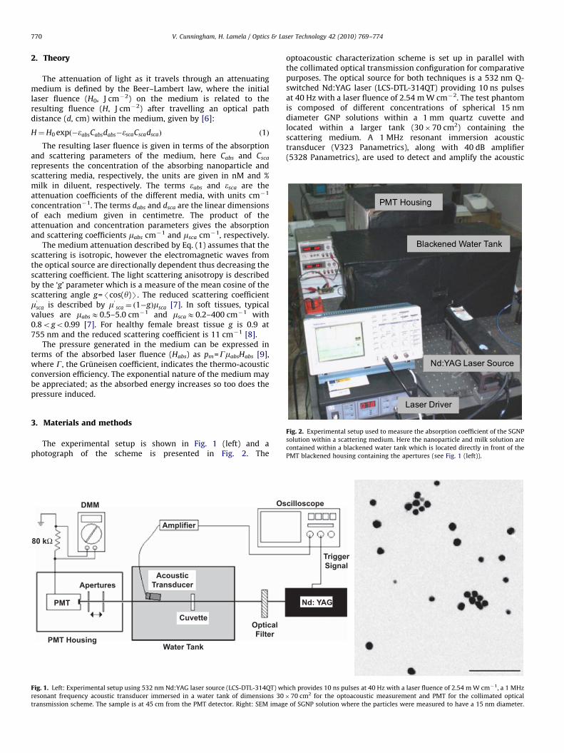
Fig. 2. Experimental setup used to measure the absorption coefficient of the SGNP
solution within a scattering medium. Here the nanoparticle and milk solution are
contained within a blackened water tank which is located directly in front of the
PMT blackened housing containing the apertures (see Fig. 1 (left)).
3. Materials and methods
The experimental setup is shown in Fig. 1 (left) and aphotograph of the scheme is presented in Fig. 2. The
DMM
80 kΩ
Apertures
PMT
PMT HousingWater Tank
OpticalFilter
Cuvette
AcousticTransducer
Amplifier
Os
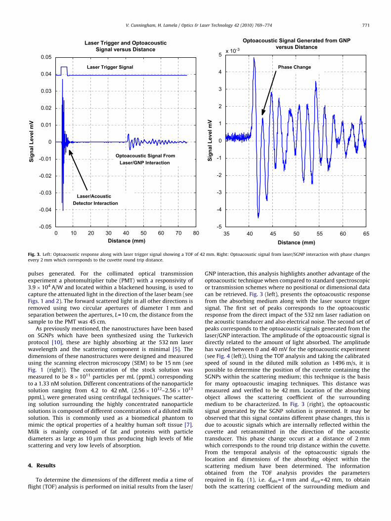
Fig. 1. Left: Experimental setup using 532 nm Nd:YAG laser source (LCS-DTL-314QT) wh
resonant frequency acoustic transducer immersed in a water tank of dimensions 30
transmission scheme. The sample is at 45 cm from the PMT detector. Right: SEM imag
optoacoustic characterization scheme is set up in parallel withthe collimated optical transmission configuration for comparativepurposes. The optical source for both techniques is a 532 nm Q-switched Nd:YAG laser (LCS-DTL-314QT) providing 10 ns pulsesat 40 Hz with a laser fluence of 2.54 m W cm�2. The test phantomis composed of different concentrations of spherical 15 nmdiameter GNP solutions within a 1 mm quartz cuvette andlocated within a larger tank (30�70 cm2) containing thescattering medium. A 1 MHz resonant immersion acoustictransducer (V323 Panametrics), along with 40 dB amplifier(5328 Panametrics), are used to detect and amplify the acoustic
cilloscope
TriggerSignal
Nd: YAG
ich provides 10 ns pulses at 40 Hz with a laser fluence of 2.54 m W cm�1, a 1 MHz
�70 cm2 for the optoacoustic measurement and PMT for the collimated optical
e of SGNP solution where the particles were measured to have a 15 nm diameter.
ARTICLE IN PRESS
0 10 20 30 40 50 60 70 80-0.05
-0.04
-0.03
-0.02
-0.01
0
0.01
0.02
0.03
0.04
0.05
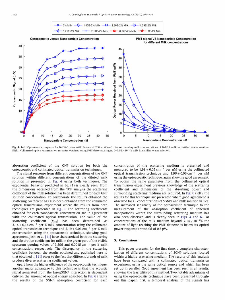
Laser Trigger and OptoacousticSignal versus Distance
Sign
al L
evel
mV
Distance (mm)35 40 45 50 55 60 65
-5
-4
-3
-2
-1
0
1
2
3
4
5x 10⁻³
Optoacoustic Signal Generated from GNPversus Distance
Sign
al L
evel
mV
Distance (mm)
Laser Trigger Signal
Optoacoustic Signal From Laser/GNP Interaction
Laser/Acoustic Detector Interaction
Phase Change
Fig. 3. Left: Optoacoustic response along with laser trigger signal showing a TOF of 42 mm. Right: Optoacoustic signal from laser/SGNP interaction with phase changes
every 2 mm which corresponds to the cuvette round trip distance.
V. Cunningham, H. Lamela / Optics & Laser Technology 42 (2010) 769–774 771
pulses generated. For the collimated optical transmissionexperiment a photomultiplier tube (PMT) with a responsivity of3.9�104 A/W and located within a blackened housing, is used tocapture the attenuated light in the direction of the laser beam (seeFigs. 1 and 2). The forward scattered light in all other directions isremoved using two circular apertures of diameter 1 mm andseparation between the apertures, L=10 cm, the distance from thesample to the PMT was 45 cm.
As previously mentioned, the nanostructures have been basedon SGNPs which have been synthesized using the Turkevichprotocol [10], these are highly absorbing at the 532 nm laserwavelength and the scattering component is minimal [5]. Thedimensions of these nanostructures were designed and measuredusing the scanning electron microscopy (SEM) to be 15 nm (seeFig. 1 (right)). The concentration of the stock solution wasmeasured to be 8�1011 particles per mL (ppmL) correspondingto a 1.33 nM solution. Different concentrations of the nanoparticlesolution ranging from 4.2 to 42 nM, (2.56�1012–2.56�1013
ppmL), were generated using centrifugal techniques. The scatter-ing solution surrounding the highly concentrated nanoparticlesolutions is composed of different concentrations of a diluted milksolution. This is commonly used as a biomedical phantom tomimic the optical properties of a healthy human soft tissue [7].Milk is mainly composed of fat and proteins with particlediameters as large as 10 mm thus producing high levels of Miescattering and very low levels of absorption.
4. Results
To determine the dimensions of the different media a time offlight (TOF) analysis is performed on initial results from the laser/
GNP interaction, this analysis highlights another advantage of theoptoacoustic technique when compared to standard spectroscopicor transmission schemes where no positional or dimensional datacan be retrieved. Fig. 3 (left), presents the optoacoustic responsefrom the absorbing medium along with the laser source triggersignal. The first set of peaks corresponds to the optoacousticresponse from the direct impact of the 532 nm laser radiation onthe acoustic transducer and also electrical noise. The second set ofpeaks corresponds to the optoacoustic signals generated from thelaser/GNP interaction. The amplitude of the optoacoustic signal isdirectly related to the amount of light absorbed. The amplitudehas varied between 0 and 40 mV for the optoacoustic experiment(see Fig. 4 (left)). Using the TOF analysis and taking the calibratedspeed of sound in the diluted milk solution as 1496 m/s, it ispossible to determine the position of the cuvette containing theSGNPs within the scattering medium; this technique is the basisfor many optoacoustic imaging techniques. This distance wasmeasured and verified to be 42 mm. Location of the absorbingobject allows the scattering coefficient of the surroundingmedium to be characterized. In Fig. 3 (right), the optoacousticsignal generated by the SGNP solution is presented. It may beobserved that this signal contains different phase changes, this isdue to acoustic signals which are internally reflected within thecuvette and retransmitted in the direction of the acoustictransducer. This phase change occurs at a distance of 2 mmwhich corresponds to the round trip distance within the cuvette.From the temporal analysis of the optoacoustic signals thelocation and dimensions of the absorbing object within thescattering medium have been determined. The informationobtained from the TOF analysis provides the parametersrequired in Eq. (1), i.e. dabs=1 mm and dsca=42 mm, to obtainboth the scattering coefficient of the surrounding medium and
ARTICLE IN PRESS
0 5 10 15 20 25 30 350
5
10
15
20
25
30
35
40
45
PMT signal VS Nanoparticle Concentrationfor different Milk concentrations
Nanoparticle Concentration nM
Opt
ical
Pow
er R
ecei
ved
from
PM
T (p
W)
0 5 10 15 20 25 30 35 40 450
5
10
15
20
25
30
35
40
Nanoparticle Concentration nM
Opt
oaco
ustic
Sig
nal (
mV)
Optoacoustic versus Nanoparticle Concentration
0% Milk 1.43E-2% Milk 2.86E-2% Milk 4.29E-2% Milk
5.71E-2% Milk 7.14E-2% Milk 8.57E-2% Milk 1E-1% Milk
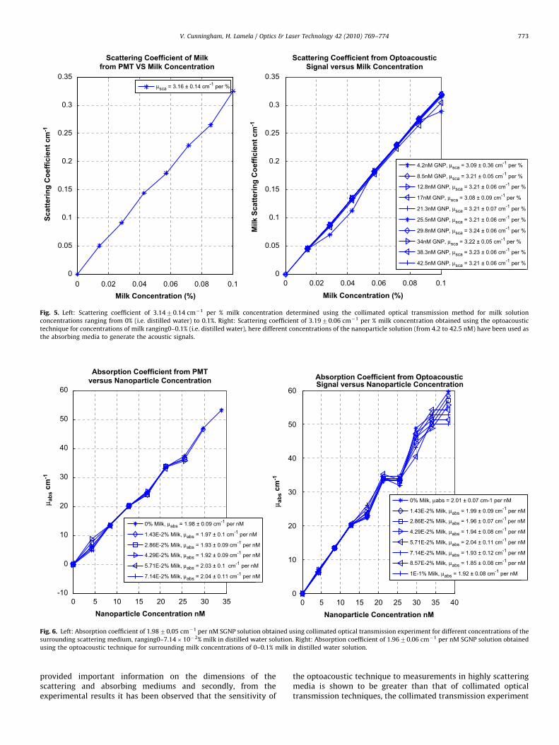
Fig. 4. Left: Optoacoustic response for Nd:YAG laser with fluence of 2.54 m W cm�1 for surrounding milk concentrations of 0–0.1% milk in distilled water solution.
Right: Collimated optical transmission response obtained using PMT detector, ranging 0–7.14�10�2% milk in distilled water solution.
V. Cunningham, H. Lamela / Optics & Laser Technology 42 (2010) 769–774772
absorption coefficient of the GNP solution for both theoptoacoustic and collimated optical transmission techniques.
The signal response from different concentrations of the GNPsolution within different concentrations of the diluted milksolution is presented in Fig. 4 using both techniques. Theexponential behavior predicted in Eq. (1) is clearly seen. Fromthe dimensions obtained from the TOF analysis the scatteringcoefficient of the milk solution has been determined for each GNPsolution concentration. To corroborate the results obtained thescattering coefficient has also been obtained from the collimatedoptical transmission experiment where the results from bothtechniques are presented in Fig. 5. The scattering coefficientsobtained for each nanoparticle concentration are in agreementwith the collimated optical transmission. The value of thescattering coefficient (asca) has been determined as3.1470.14 cm�1 per % milk concentration using the collimatedoptical transmission technique and 3.1970.06 cm�1 per % milkconcentration using the optoacoustic technique, showing goodagreement. Joshi et al. [11] have characterized both the scatteringand absorption coefficient for milk in the green part of the visiblespectrum quoting values of 3.596 and 0.0015 cm�1 per % milkconcentration, respectively. The discrepancy in the scatteringcoefficient between the results obtained and published here tothat obtained in [11] owes to the fact that different brands of milkproduce diverse scattering coefficient values.
Apart from the higher efficiency of the optoacoustic technique,another major advantage to this technique is that the acousticsignal generated from the laser/SGNP interaction is dependentonly on the amount of optical energy absorbed. In Fig. 6 (right),the results of the SGNP absorption coefficient for each
concentration of the scattering medium is presented andmeasured to be 1.9870.05 cm�1 per nM using the collimatedoptical transmission technique and 1.9670.06 cm�1 per nMusing the optoacoustic technique, again showing good agreement.To obtain the same parameter from the collimated opticaltransmission experiment previous knowledge of the scatteringcoefficient and dimensions of the absorbing object andsurrounding scattering medium are required. In Fig. 6 (left), theresults for this technique are presented where good agreement isobserved for all concentrations of SGNPs and milk solution values.The increased sensitivity of the optoacoustic technique to themeasurement of the absorption coefficient of sphericalnanoparticles within the surrounding scattering medium hasalso been observed and is clearly seen in Figs. 4 and 6. Forconcentrations of the milk medium beyond 7.14�10�2% theamount of light reaching the PMT detector is below its opticalpower response threshold of 0.5 pW.
5. Conclusions
This paper presents, for the first time, a complete character-ization of different concentrations of SGNP solutions locatedwithin a highly scattering medium. The results of this analysishave been compared with a collimated optical transmissionexperiment using the same optical source and which has beenset up in parallel. Good agreement has been seen in all results,showing the feasibility of this method. Two notable advantages ofusing the optoacoustic technique have been presented through-out this paper, first, a temporal analysis of the signals has
ARTICLE IN PRESS
0 0.02 0.04 0.06 0.08 0.10
0.05
0.1
0.15
0.2
0.25
0.3
0.35
Scattering Coefficient of Milkfrom PMT VS Milk Concentration
Milk Concentration (%)
Scat
terin
g C
oeffi
cien
t cm
-1
μsca = 3.16 ± 0.14 cm-1 per %
0 0.02 0.04 0.06 0.08 0.10
0.05
0.1
0.15
0.2
0.25
0.3
0.35
Scattering Coefficient from OptoacousticSignal versus Milk Concentration
Milk Concentration (%)
Milk
Sca
tterin
g C
oeffi
cien
t cm
-1
4.2nM GNP, μsca = 3.09 ± 0.36 cm-1 per %
8.5nM GNP, μsca = 3.21 ± 0.05 cm-1 per %
12.8nM GNP, μsca = 3.21 ± 0.06 cm-1 per %
17nM GNP, μsca = 3.08 ± 0.09 cm-1 per %
21.3nM GNP, μsca = 3.21 ± 0.07 cm-1 per %
25.5nM GNP, μsca = 3.21 ± 0.06 cm-1 per %
29.8nM GNP, μsca = 3.24 ± 0.06 cm-1 per %
34nM GNP, μsca = 3.22 ± 0.05 cm-1 per %
38.3nM GNP, μsca = 3.23 ± 0.06 cm-1 per %
42.5nM GNP, μsca = 3.21 ± 0.06 cm-1 per %
Fig. 5. Left: Scattering coefficient of 3.1470.14 cm�1 per % milk concentration determined using the collimated optical transmission method for milk solution
concentrations ranging from 0% (i.e. distilled water) to 0.1%. Right: Scattering coefficient of 3.1970.06 cm�1 per % milk concentration obtained using the optoacoustic
technique for concentrations of milk ranging0–0.1% (i.e. distilled water), here different concentrations of the nanoparticle solution (from 4.2 to 42.5 nM) have been used as
the absorbing media to generate the acoustic signals.
0 5 10 15 20 25 30 35-10
0
10
20
30
40
50
60
Absorption Coefficient from PMTversus Nanoparticle Concentration
Nanoparticle Concentration nM
μ abs
cm
-1
μ abs
cm
-1
0% Milk, μabs = 1.98 ± 0.09 cm-1 per nM
1.43E-2% Milk, μabs = 1.97 ± 0.1 cm-1 per nM
2.86E-2% Milk, μabs = 1.93 ± 0.09 cm-1 per nM
4.29E-2% Milk, μabs = 1.92 ± 0.09 cm-1 per nM
5.71E-2% Milk, μabs = 2.03 ± 0.1 cm-1 per nM
7.14E-2% Milk, μabs = 2.04 ± 0.11 cm-1 per nM
0 5 10 15 20 25 30 35 400
10
20
30
40
50
60
Absorption Coefficient from Optoacoustic Signal versus Nanoparticle Concentration
Nanoparticle Concentration nM
0% Milk, μabs = 2.01 ± 0.07 cm-1 per nM
1.43E-2% Milk, μabs = 1.99 ± 0.09 cm-1 per nM
2.86E-2% Milk, μabs = 1.96 ± 0.07 cm-1 per nM
4.29E-2% Milk, μabs = 1.94 ± 0.08 cm-1 per nM
5.71E-2% Milk, μabs = 2.04 ± 0.11 cm-1 per nM
7.14E-2% Milk, μabs = 1.93 ± 0.12 cm-1 per nM
8.57E-2% Milk, μabs = 1.85 ± 0.08 cm-1 per nM
1E-1% Milk, μabs = 1.92 ± 0.08 cm-1 per nM
Fig. 6. Left: Absorption coefficient of 1.9870.05 cm�1 per nM SGNP solution obtained using collimated optical transmission experiment for different concentrations of the
surrounding scattering medium, ranging0–7.14�10�2% milk in distilled water solution. Right: Absorption coefficient of 1.9670.06 cm�1 per nM SGNP solution obtained
using the optoacoustic technique for surrounding milk concentrations of 0–0.1% milk in distilled water solution.
V. Cunningham, H. Lamela / Optics & Laser Technology 42 (2010) 769–774 773
provided important information on the dimensions of thescattering and absorbing mediums and secondly, from theexperimental results it has been observed that the sensitivity of
the optoacoustic technique to measurements in highly scatteringmedia is shown to be greater than that of collimated opticaltransmission techniques, the collimated transmission experiment
ARTICLE IN PRESS
V. Cunningham, H. Lamela / Optics & Laser Technology 42 (2010) 769–774774
is capable of determining the concentration of the nanoparticlesolution for surrounding milk concentrations below 7.14�10�2%,whereas the optoacoustic technique provides reliable results forconcentrations as high as 0.1%. The former advantage is importantwhen considering imaging applications and the latter is ofimportance when considering the SNR in detecting nanoparticleslocated deep within highly scattering mediums.
References
[1] Oraevsky AA. In: Gold and silver nanoparticles as contrast agents foroptoacoustic tomography. CRC Press; 2009. 373–386.
[2] Surbhi L, Clare SE, Halas NJ. Nanoshell-enabled photothermal cancer therapy:impending clinical impact. Accounts of Chemical Research2008;41(12):1842–51.
[3] Xie H, Gill-Sharp KL, O’Neall DP. Quantitative estimation of gold nanoshellconcentrations in whole blood using dynamic light scattering, nanomedicine:nanotechnology. Biology and Medicine 2007;3:89–94.
[4] Niidome T, Akiyama Y, Shimoda K, Kawano T, Mori T, Katayama Y, Niidome Y.In vivo monitoring of intravenously injected gold nanorods using near-infrared light. Small 2008;4(7):1001–7.
[5] Jain PK, Lee KS, El-Sayed IH, El-Sayed MA. Calculated absorption andscattering properties of gold nanoparticles of different size, shape, andcomposition: applications in biological imaging and biomedicine. Journal ofPhysical Chemistry 2006;110.
[6] Karabutov AA, Pelivanov IM, Podymova NB, Skipetrov SE. Determination ofthe optical characteristics of turbid media by the laser optoacoustic method.Quantum Electronics 1999;29(12):1054–9.
[7] Tuchin V. In: Tissue Optics. Washington: SPIE Press; 2000.[8] Khokhlova TD, Pelivanov IM, Kozhushko VV, Zharinov AN, Solomatin VS,
Karabutov AA. Optoacoustic imaging of absorbing objects in a turbidmedium: ultimate sensitivity and application to breast cancer diagnostics.Applied Optics 2007;46.
[9] Esenaliev RO, Karabutov AA, Oraevsky AA. Sensitivity of laser opto-acousticimaging in detection of small deeply embedded tumors. IEEE Journal ofSelected Topics in Quantum Electronics 1999;5(4):981–8.
[10] Turkevich J, Stevenson PS, Hillier J. Coagulation of colloidal gold. Discussionsof the Faraday Society 1951;58(11).
[11] Joshi N, Donner C, Jensen HW. Noninvasive measurement of scatteringanisotropy in turbid materials by nonnormal incident illumination. OpticsLetters 2006;31(7):936–8.