Embed Size (px)
Citation preview
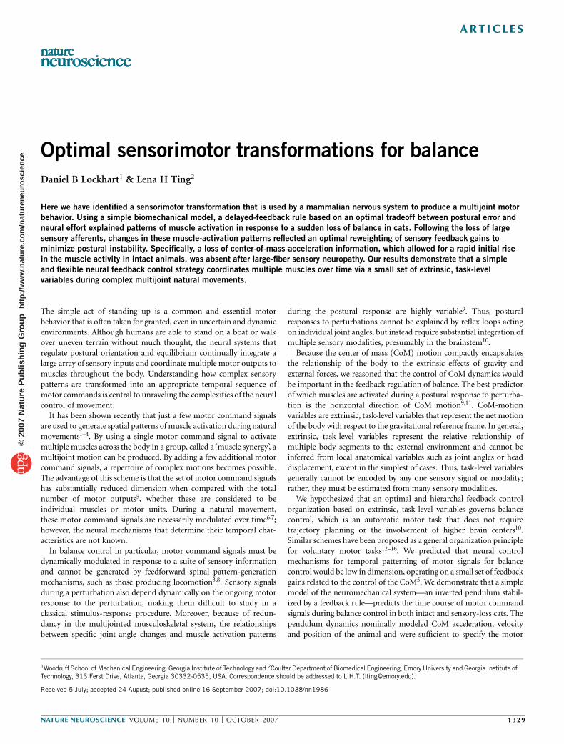
Optimal sensorimotor transformations for balance
Daniel B Lockhart1 & Lena H Ting2
Here we have identified a sensorimotor transformation that is used by a mammalian nervous system to produce a multijoint motor
behavior. Using a simple biomechanical model, a delayed-feedback rule based on an optimal tradeoff between postural error and
neural effort explained patterns of muscle activation in response to a sudden loss of balance in cats. Following the loss of large
sensory afferents, changes in these muscle-activation patterns reflected an optimal reweighting of sensory feedback gains to
minimize postural instability. Specifically, a loss of center-of-mass-acceleration information, which allowed for a rapid initial rise
in the muscle activity in intact animals, was absent after large-fiber sensory neuropathy. Our results demonstrate that a simple
and flexible neural feedback control strategy coordinates multiple muscles over time via a small set of extrinsic, task-level
variables during complex multijoint natural movements.
The simple act of standing up is a common and essential motorbehavior that is often taken for granted, even in uncertain and dynamicenvironments. Although humans are able to stand on a boat or walkover uneven terrain without much thought, the neural systems thatregulate postural orientation and equilibrium continually integrate alarge array of sensory inputs and coordinate multiple motor outputs tomuscles throughout the body. Understanding how complex sensorypatterns are transformed into an appropriate temporal sequence ofmotor commands is central to unraveling the complexities of the neuralcontrol of movement.
It has been shown recently that just a few motor command signalsare used to generate spatial patterns of muscle activation during naturalmovements1–4. By using a single motor command signal to activatemultiple muscles across the body in a group, called a ‘muscle synergy’, amultijoint motion can be produced. By adding a few additional motorcommand signals, a repertoire of complex motions becomes possible.The advantage of this scheme is that the set of motor command signalshas substantially reduced dimension when compared with the totalnumber of motor outputs5, whether these are considered to beindividual muscles or motor units. During a natural movement,these motor command signals are necessarily modulated over time6,7;however, the neural mechanisms that determine their temporal char-acteristics are not known.
In balance control in particular, motor command signals must bedynamically modulated in response to a suite of sensory informationand cannot be generated by feedforward spinal pattern-generationmechanisms, such as those producing locomotion3,8. Sensory signalsduring a perturbation also depend dynamically on the ongoing motorresponse to the perturbation, making them difficult to study in aclassical stimulus-response procedure. Moreover, because of redun-dancy in the multijointed musculoskeletal system, the relationshipsbetween specific joint-angle changes and muscle-activation patterns
during the postural response are highly variable9. Thus, posturalresponses to perturbations cannot be explained by reflex loops actingon individual joint angles, but instead require substantial integration ofmultiple sensory modalities, presumably in the brainstem10.
Because the center of mass (CoM) motion compactly encapsulatesthe relationship of the body to the extrinsic effects of gravity andexternal forces, we reasoned that the control of CoM dynamics wouldbe important in the feedback regulation of balance. The best predictorof which muscles are activated during a postural response to perturba-tion is the horizontal direction of CoM motion9,11. CoM-motionvariables are extrinsic, task-level variables that represent the net motionof the body with respect to the gravitational reference frame. In general,extrinsic, task-level variables represent the relative relationship ofmultiple body segments to the external environment and cannot beinferred from local anatomical variables such as joint angles or headdisplacement, except in the simplest of cases. Thus, task-level variablesgenerally cannot be encoded by any one sensory signal or modality;rather, they must be estimated from many sensory modalities.
We hypothesized that an optimal and hierarchal feedback controlorganization based on extrinsic, task-level variables governs balancecontrol, which is an automatic motor task that does not requiretrajectory planning or the involvement of higher brain centers10.Similar schemes have been proposed as a general organization principlefor voluntary motor tasks12–16. We predicted that neural controlmechanisms for temporal patterning of motor signals for balancecontrol would be low in dimension, operating on a small set of feedbackgains related to the control of the CoM5. We demonstrate that a simplemodel of the neuromechanical system—an inverted pendulum stabil-ized by a feedback rule—predicts the time course of motor commandsignals during balance control in both intact and sensory-loss cats. Thependulum dynamics nominally modeled CoM acceleration, velocityand position of the animal and were sufficient to specify the motor
Received 5 July; accepted 24 August; published online 16 September 2007; doi:10.1038/nn1986
1Woodruff School of Mechanical Engineering, Georgia Institute of Technology and 2Coulter Department of Biomedical Engineering, Emory University and Georgia Institute ofTechnology, 313 Ferst Drive, Atlanta, Georgia 30332-0535, USA. Correspondence should be addressed to L.H.T. ([email protected]).
NATURE NEUROSCIENCE VOLUME 10 [ NUMBER 10 [ OCTOBER 2007 1329
ART ICLES©
2007
Nat
ure
Pub
lishi
ng G
roup
ht
tp://
ww
w.n
atur
e.co
m/n
atur
eneu
rosc
ienc
e

command signals activating both proximal and distal leg muscles. Toour surprise, the resulting patterns of muscle activation were alsosimilar to the optimal patterns predicted using an objective criteriondrawn from optimal control theory. Moreover, the predictions wererobust to peripheral sensory neuropathy, which disrupts the encodingof local joint-angle changes by large-diameter sensory afferents17.Following re-adaptation subsequent to this acute sensory deficit,temporal motor patterns were markedly altered, but in the modelresulted simply from a loss of CoM acceleration feedback. Ourapproach of defining temporal patterns of muscle activity in normaland neurologically impaired animals with a neuromechanical modelusing feedback of extrinsic, task-level variables provides new insightinto the robust structure of general sensorimotor transformations formovement control.
RESULTS
Simple feedback transformation for postural control
We examined temporal patterns of muscle activation that wereevoked by translation of the support surface in unrestrained, freelystanding cats. The perturbations elicited a maximal response in the lefthindlimb muscles and minimal responses in the other limbs18. Thetemporal responses in the majority of the extensors were similar,suggesting that common neural signals modulated multiple muscles(Fig. 1). The response to single-ramp perturbations was also similar tothat observed in humans10,19: an initial burst followed by a long plateauof activity (Fig. 1a). The initial burst of the elicited responses had ashape similar to that of the temporal pattern of support-surfaceacceleration, but with a latency of 30–50 ms. The durations of themuscular response and perturbation were also similar. However,the animal’s sensory system cannot directly encode the motion of thesupport surface, only the effects of that motion on the animal’s ownbody. The time lags between CoM kinematics and muscle-activationpatterns led us to hypothesize that the nervous system uses an estimateof overall body movement in a feedback manner to regulate temporalpatterns of muscle activation.
To functionally and quantitatively test this hypothesis, we developeda simple computational model of postural control (Fig. 2) andevaluated how well it explained muscle-activation patterns that weobserved experimentally. We parameterized the model first on the basisof experimental data, and second to maximize an objective-costfunction based on performance measures. The model consisted of aninverted pendulum actuated at the base by a muscle and mounted on amoveable support surface (Fig. 2a). The experimentally recordedacceleration of the support surface was used as the only input to themodel. The hypothesized neural controller constructed temporalpatterns of muscle activation from linear combinations of the simu-lated CoM position, velocity and acceleration signals12,14 (Fig. 2b),delayed in time to account for latencies due to neural conductionand computation, and assumed to be estimated by the nervoussystem from an ensemble of sensory signals10,20,21. An advantage ofthis forward modeling approach is that the biomechanical viabilityof the sensorimotor transformation is implicitly tested; solutionsderived from measured kinematic signals do not generally stabilizethe system22.
Identification of model parameters
Variations of only four parameters in our model, three feedbackgains and one delay, were sufficient to reproduce salient features ineach of the experimentally measured muscle-activation patterns (seeMethods; variability accounted for (VAF) ¼ 88 ± 2% s.e.m. acrosscats, VAF 4 80% for all muscles across all cats). For each muscle ineach animal, we identified three feedback gains and one delaythat best matched the muscle activation and kinematics of the simula-tion to experimental data. We dubbed this process temporal systemidentification (TSyID) because model parameters were found bymatching features of the data in the temporal domain. Althoughwe nominally modeled only the net motion of the CoM, andnot individual joints, muscles crossing all joints were analyzed to testwhether CoM kinematics were sufficient to reproduce all muscle-activation patterns.
Figure 1 Example of muscle-activation patterns
evoked during balance corrections to support-
surface motion in the horizontal plane. Cats were
freely standing on a movable platform that could
move in the horizontal plane. EMGs were
measured in the left hindlimb. (a) Responses to a
ramp-and-hold perturbation in the 2251 direction
evoked a classic EMG pattern of an initial burstfollowed by a long plateau in multiple extensor
muscles, whereas the flexor muscles (STEN) were
quiescent. The initial burst shape followed the
perturbation acceleration after a short delay of
about 30 ms. The duration of the plateau followed
the velocity of the perturbations. (b) Responses to
a dual-ramp perturbation in which the direction of
support-surface motion is reversed 20–90 ms
after onset of the initial ramp. These
perturbations allowed us to test the robustness of
the feedback model by altering the acceleration
waveform associated with the initial ramp, as well
as the CoM velocity and position at the onset of
the secondary ramp. Because muscle activity is
direction-specific, the extensor muscles originally
activated by the perturbation were shut off, and
flexor muscles were activated. Muscle
abbreviations: biceps femoris anterior, BFMA;
flexor digitorum longus, FDL; flexor hallicus longus, FHL; medial gastrocnemius, MGAS; peroneous brevis, PERB; semimembranosus anterior, SEMA;semimembranosus posterior, SEMP; sartorius anterior, SRTA; semitendinosus, STEN; vastus medialis, VMED.
0 100 200 300 400 500 0 100 200 300 400 500 600
SRTA
VMED
ADFM
SEMP
SEMA
STEN
BFMA
MGAS
PERB
FDL
FHL
Platform displacement
Platformvelocity
Platformacceleration
180°
0°
90° 270°
180°
0°
90° 270°
0.05 m
0.15 m s–1
5 m s–2
a Single-ramp perturbation b Dual-ramp perturbation
Time (ms) Time (ms)
1330 VOLUME 10 [ NUMBER 10 [ OCTOBER 2007 NATURE NEUROSCIENCE
ART ICLES©
2007
Nat
ure
Pub
lishi
ng G
roup
ht
tp://
ww
w.n
atur
e.co
m/n
atur
eneu
rosc
ienc
e
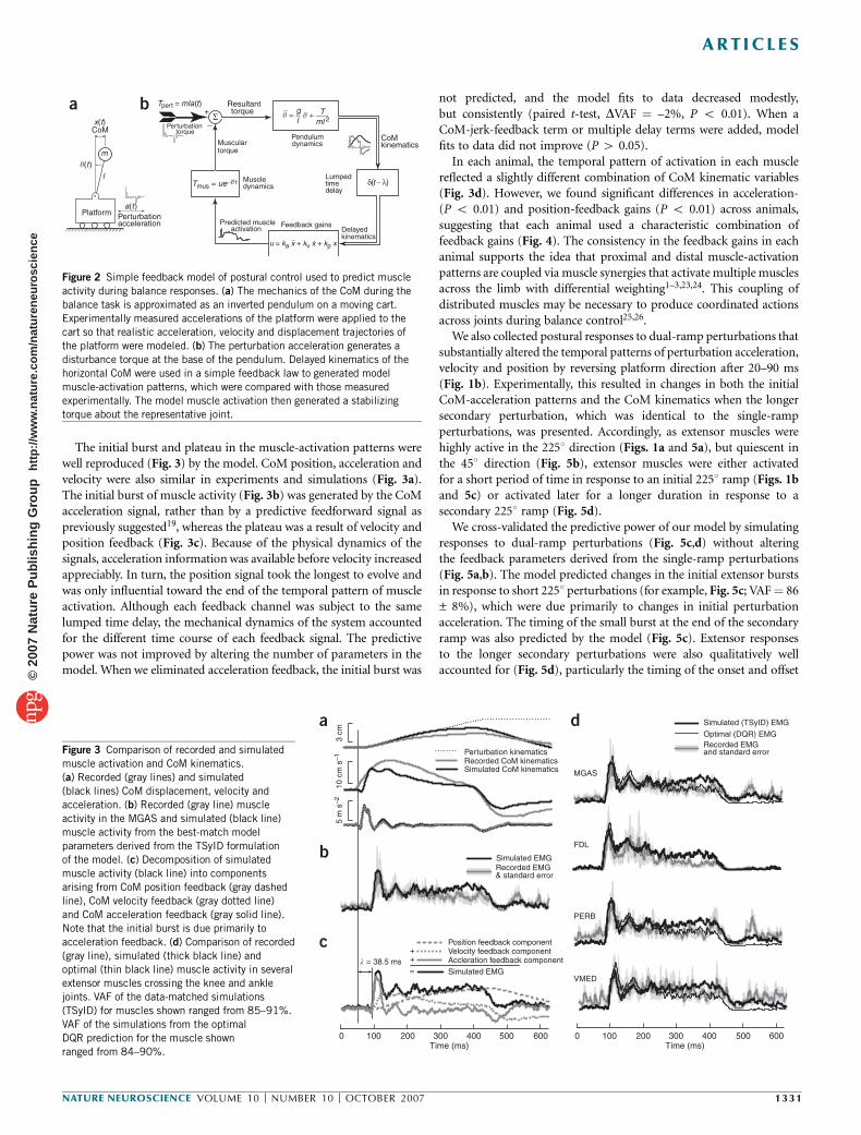
The initial burst and plateau in the muscle-activation patterns werewell reproduced (Fig. 3) by the model. CoM position, acceleration andvelocity were also similar in experiments and simulations (Fig. 3a).The initial burst of muscle activity (Fig. 3b) was generated by the CoMacceleration signal, rather than by a predictive feedforward signal aspreviously suggested19, whereas the plateau was a result of velocity andposition feedback (Fig. 3c). Because of the physical dynamics of thesignals, acceleration information was available before velocity increasedappreciably. In turn, the position signal took the longest to evolve andwas only influential toward the end of the temporal pattern of muscleactivation. Although each feedback channel was subject to the samelumped time delay, the mechanical dynamics of the system accountedfor the different time course of each feedback signal. The predictivepower was not improved by altering the number of parameters in themodel. When we eliminated acceleration feedback, the initial burst was
not predicted, and the model fits to data decreased modestly,but consistently (paired t-test, DVAF ¼ –2%, P o 0.01). When aCoM-jerk-feedback term or multiple delay terms were added, modelfits to data did not improve (P 4 0.05).
In each animal, the temporal pattern of activation in each musclereflected a slightly different combination of CoM kinematic variables(Fig. 3d). However, we found significant differences in acceleration-(P o 0.01) and position-feedback gains (P o 0.01) across animals,suggesting that each animal used a characteristic combination offeedback gains (Fig. 4). The consistency in the feedback gains in eachanimal supports the idea that proximal and distal muscle-activationpatterns are coupled via muscle synergies that activate multiple musclesacross the limb with differential weighting1–3,23,24. This coupling ofdistributed muscles may be necessary to produce coordinated actionsacross joints during balance control25,26.
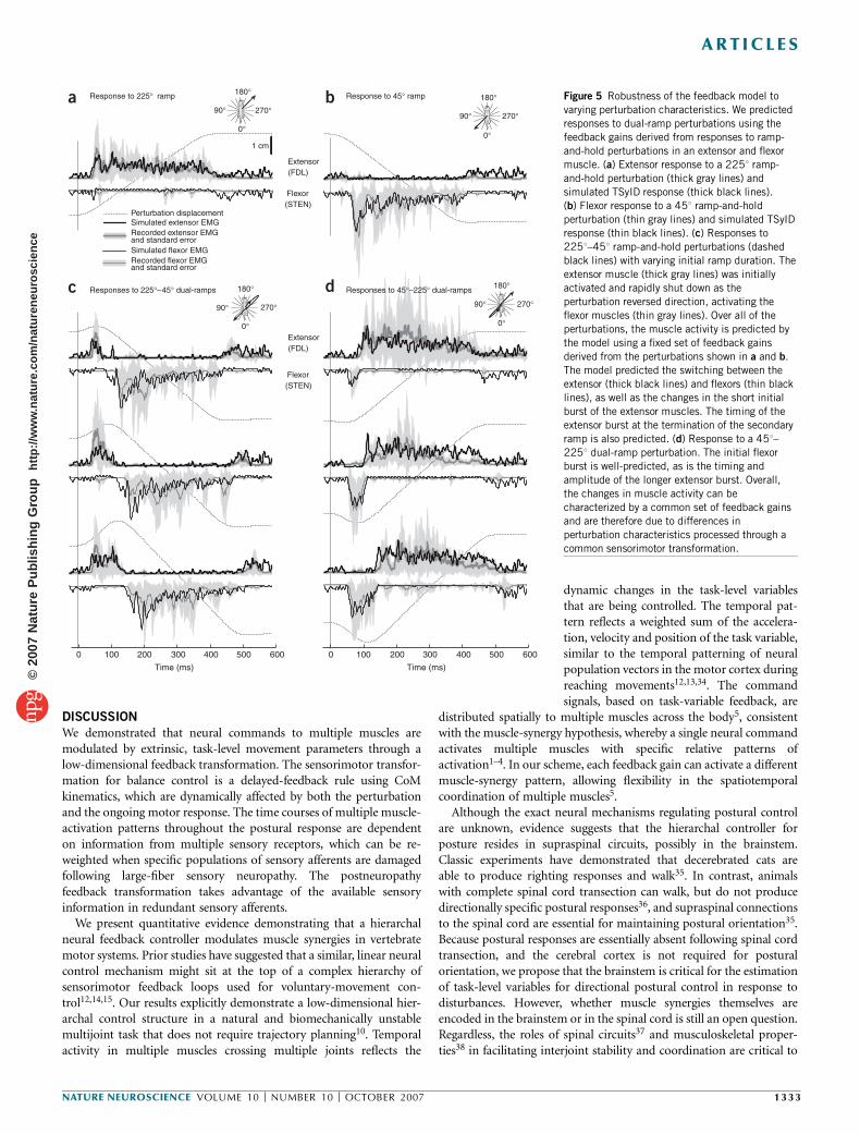
We also collected postural responses to dual-ramp perturbations thatsubstantially altered the temporal patterns of perturbation acceleration,velocity and position by reversing platform direction after 20–90 ms(Fig. 1b). Experimentally, this resulted in changes in both the initialCoM-acceleration patterns and the CoM kinematics when the longersecondary perturbation, which was identical to the single-rampperturbations, was presented. Accordingly, as extensor muscles werehighly active in the 2251 direction (Figs. 1a and 5a), but quiescent inthe 451 direction (Fig. 5b), extensor muscles were either activatedfor a short period of time in response to an initial 2251 ramp (Figs. 1band 5c) or activated later for a longer duration in response to asecondary 2251 ramp (Fig. 5d).
We cross-validated the predictive power of our model by simulatingresponses to dual-ramp perturbations (Fig. 5c,d) without alteringthe feedback parameters derived from the single-ramp perturbations(Fig. 5a,b). The model predicted changes in the initial extensor burstsin response to short 2251 perturbations (for example, Fig. 5c; VAF ¼ 86± 8%), which were due primarily to changes in initial perturbationacceleration. The timing of the small burst at the end of the secondaryramp was also predicted by the model (Fig. 5c). Extensor responsesto the longer secondary perturbations were also qualitatively wellaccounted for (Fig. 5d), particularly the timing of the onset and offset
0 100 200 300 400 500 600
a
b
c
Time (ms)
Recorded EMG & standard error
Simulated EMG
=
Position feedback component
� = 38.5 msVelocity feedback componentAccleration feedback component
+
Simulated EMG
+
10 c
m s
–15
m s
–23
cm
Perturbation kinematicsRecorded CoM kinematicsSimulated CoM kinematics
d
0 100 200 300 400 500 600Time (ms)
MGAS
FDL
PERB
VMED
Recorded EMG and standard error
Optimal (DQR) EMG
Simulated (TSyID) EMG
Perturbationacceleration
CoM
Platform
m
l
+
Predicted muscleactivation Delayed
kinematics
Feedback gains
Lumpedtimedelay
Muscledynamics
Muscular torque
Resultanttorque
CoMkinematics
Perturbation torque
a b
Pendulumdynamics
�..
= gl
� + Tml 2
Tpert = mla(t)
Tmus = ue−t/τ δ(t− λ)
x(t)
a(t)
�(t)
–Σ
u = ka x + kv x + kp x¨
.
Figure 2 Simple feedback model of postural control used to predict muscle
activity during balance responses. (a) The mechanics of the CoM during the
balance task is approximated as an inverted pendulum on a moving cart.
Experimentally measured accelerations of the platform were applied to thecart so that realistic acceleration, velocity and displacement trajectories of
the platform were modeled. (b) The perturbation acceleration generates a
disturbance torque at the base of the pendulum. Delayed kinematics of the
horizontal CoM were used in a simple feedback law to generated model
muscle-activation patterns, which were compared with those measured
experimentally. The model muscle activation then generated a stabilizing
torque about the representative joint.
Figure 3 Comparison of recorded and simulated
muscle activation and CoM kinematics.
(a) Recorded (gray lines) and simulated
(black lines) CoM displacement, velocity and
acceleration. (b) Recorded (gray line) muscle
activity in the MGAS and simulated (black line)
muscle activity from the best-match model
parameters derived from the TSyID formulation
of the model. (c) Decomposition of simulated
muscle activity (black line) into components
arising from CoM position feedback (gray dashed
line), CoM velocity feedback (gray dotted line)
and CoM acceleration feedback (gray solid line).
Note that the initial burst is due primarily to
acceleration feedback. (d) Comparison of recorded
(gray line), simulated (thick black line) and
optimal (thin black line) muscle activity in severalextensor muscles crossing the knee and ankle
joints. VAF of the data-matched simulations
(TSyID) for muscles shown ranged from 85–91%.
VAF of the simulations from the optimal
DQR prediction for the muscle shown
ranged from 84–90%.
NATURE NEUROSCIENCE VOLUME 10 [ NUMBER 10 [ OCTOBER 2007 1331
ART ICLES©
2007
Nat
ure
Pub
lishi
ng G
roup
ht
tp://
ww
w.n
atur
e.co
m/n
atur
eneu
rosc
ienc
e

of the muscles. To our surprise, the antagonistic, flexor muscleresponses in both cases (Fig. 5c,d) were similar to those predicted bythe model (Fig. 5c).
Optimal control model of posture
We compared the neural feedback gains identified through TSyIDwith an optimal solution predicted without a priori knowledgeof the experimental CoM kinematics or muscle activations. We usedan optimal control formulation that was similar to the linearquadratic regulator formulation devised previously in postural controlmodels27. We dubbed our approach a delayed quadratic regulator(DQR) because it incorporated a time delay and determinedoptimal feedback gains via a quadratic cost function. In contrastwith other models28–30, we used experimentally measured platformacceleration to generate perturbations with realistic temporal charac-teristics. The delay was set to the value estimated through TSyID (B30ms), thus representing a constraint of the neural transmission andcomputation time, as the optimal delay would be a nonphysiologicalvalue of zero.
The optimal solution predicted a postural response with initial burstand plateau regions that were similar to those measured experimentally(for example, Fig. 3d; VAF ¼ 78 ± 6% s.e.m., across all muscles in allcats, VAF 4 70% in all muscles) and were also similar to thoseestimated through TSyID (Fig. 3d). Using measured perturbationacceleration was critical for achieving a physiologically relevantmuscle-activation pattern because of the dependence of the sensoryfeedback on the exact perturbation characteristics.
Changes following sensory loss
We observed notable changes in temporal patterns of muscle activationin animals with large-fiber (group I) proprioceptive deficit as a result ofpyridoxine overdose. Proprioceptive afferents are critical for generatingthe proper timing in postural responses; postural responses are delayedfollowing proprioceptive loss but not following vestibular or visual
loss17,31–33. The pyridoxine intoxication affected sensory afferent fiberslarger than 10 mm in diameter, including primary muscle spindleafferents, Golgi tendon organ afferents and large cutaneous afferents17.In the days following the pyridoxine injections, the loss of group Iafferents was devastating; animals became ataxic, and in many cases lostthe ability to stand independently. Over a period of about 2 weeks,animals regained the ability to stand and be mobile, although group Iafferents remained nonfunctional, as determined by stretch-reflex testsand histological measurements17. Sensory-deficit animals could standand maintain their balance, presumably using smaller group II affer-ents. We analyzed postural responses after a B3-week period ofre-adaptation, when animals had recovered postural control andother motor functions.
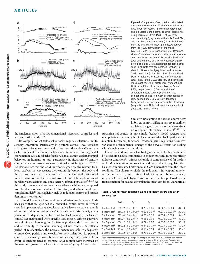
In the animal with the greatest sensory deficit, the initial burst(Fig. 3) of the temporal muscle-activation pattern was absent (Fig. 6).Instead, muscle activity was characterized by a long, slow rise in muscleactivation. These changes were previously described simply as anincrease in response latency and amplitude17.
Changes in temporal patterns of muscle activation followinglarge-fiber sensory loss were due to decreased CoM-acceleration feed-back. A 50% reduction of the acceleration feedback gain accounted forthe diminished initial peak in animals with the complete loss ofafferents in the group I (o10-mm diameter; cat Be and cat Kn) range(Table 1). In these animals, fits were improved when the accelerationfeedback was explicitly set to zero (DVAF ¼ +2%, P o 0.01). Inanimals with only a partial loss of group I afferents (Table 1), accel-eration bursts were still present, and fits were degraded when accelera-tion feedback was set to zero (DVAF ¼ –3%, P o 0.01). Although thequantitative effects of removing acceleration were modest because theacceleration component contributes only a small amount to the overalltemporal variance of the signal, the qualitative loss of the initial burstwas very noticeable.
The optimal muscle-activation pattern predicted by the DQR modelin the absence of acceleration feedback was notably similar to what wasactually measured (for example, Fig. 6e; VAF¼ 65 ± 6%). The resultingoptimal pattern was characterized by a slow rise in muscle activation,which was expected as a result of the loss of acceleration feedback. Anincrease in velocity gain was predicted to compensate for the loss of theinitial burst (5% increase, P{ 0.01). The optimal prediction has onlyslightly decreased postural stability compared with the intact conditionand produced qualitatively similar CoM velocity and displacementtrajectories (Figs. 3a and 6a).
Therefore, after the recovery of balance control in sensory-impairedanimals, the extrinsic, task-level variables of CoM position and velocityare still regulated, albeit with different sensory pathways. Redundancyand neural plasticity mechanisms presumably upregulate the contribu-tion of group II sensory signals in the regulation of postural control.Thus, the new temporal patterns of muscle activation that the animalachieves after a period of re-adaptation reflect a process of optimalreweighting of sensory feedback to maintain control of extrinsic, task-level variables.
80
90
100V
AF
0
0.01
0.02
0.03
0.04
k a
0.6
0.7
0.8
0.9
k v
5.5
6.0
6.5
kp
30
32
34
36
Cat Be
� (m
s)
Cat Kn Cat Sq Cat So
a
b
c
d
e
Figure 4 Summary of model (TSyID) fits and parameter values (mean ± s.d.)
for all muscles in four cats. (a) VAF was greater than 80% in all muscles in
each cat. (b) Acceleration feedback gain, ka, for all muscles in each cat.
(c) Velocity feedback gain, kv, for all muscles in each cat. (d) Position
feedback gain, kp, for all muscles in each cat. These results demonstrate
that similar feedback gains are used across all muscles in each animal,
suggesting that they are activated by a common neural command signal.
(e) Delay, l, for all muscles in each cat. For cats Be, Kn, Sq and So, 9, 7,10 and 8 left hindlimb muscles were analyzed, respectively. In cat Kn,
4 left forelimb muscles were also included.
1332 VOLUME 10 [ NUMBER 10 [ OCTOBER 2007 NATURE NEUROSCIENCE
ART ICLES©
2007
Nat
ure
Pub
lishi
ng G
roup
ht
tp://
ww
w.n
atur
e.co
m/n
atur
eneu
rosc
ienc
e
DISCUSSION
We demonstrated that neural commands to multiple muscles aremodulated by extrinsic, task-level movement parameters through alow-dimensional feedback transformation. The sensorimotor transfor-mation for balance control is a delayed-feedback rule using CoMkinematics, which are dynamically affected by both the perturbationand the ongoing motor response. The time courses of multiple muscle-activation patterns throughout the postural response are dependenton information from multiple sensory receptors, which can be re-weighted when specific populations of sensory afferents are damagedfollowing large-fiber sensory neuropathy. The postneuropathyfeedback transformation takes advantage of the available sensoryinformation in redundant sensory afferents.
We present quantitative evidence demonstrating that a hierarchalneural feedback controller modulates muscle synergies in vertebratemotor systems. Prior studies have suggested that a similar, linear neuralcontrol mechanism might sit at the top of a complex hierarchy ofsensorimotor feedback loops used for voluntary-movement con-trol12,14,15. Our results explicitly demonstrate a low-dimensional hier-archal control structure in a natural and biomechanically unstablemultijoint task that does not require trajectory planning10. Temporalactivity in multiple muscles crossing multiple joints reflects the
dynamic changes in the task-level variablesthat are being controlled. The temporal pat-tern reflects a weighted sum of the accelera-tion, velocity and position of the task variable,similar to the temporal patterning of neuralpopulation vectors in the motor cortex duringreaching movements12,13,34. The commandsignals, based on task-variable feedback, are
distributed spatially to multiple muscles across the body5, consistentwith the muscle-synergy hypothesis, whereby a single neural commandactivates multiple muscles with specific relative patterns ofactivation1–4. In our scheme, each feedback gain can activate a differentmuscle-synergy pattern, allowing flexibility in the spatiotemporalcoordination of multiple muscles5.
Although the exact neural mechanisms regulating postural controlare unknown, evidence suggests that the hierarchal controller forposture resides in supraspinal circuits, possibly in the brainstem.Classic experiments have demonstrated that decerebrated cats areable to produce righting responses and walk35. In contrast, animalswith complete spinal cord transection can walk, but do not producedirectionally specific postural responses36, and supraspinal connectionsto the spinal cord are essential for maintaining postural orientation35.Because postural responses are essentially absent following spinal cordtransection, and the cerebral cortex is not required for posturalorientation, we propose that the brainstem is critical for the estimationof task-level variables for directional postural control in response todisturbances. However, whether muscle synergies themselves areencoded in the brainstem or in the spinal cord is still an open question.Regardless, the roles of spinal circuits37 and musculoskeletal proper-ties38 in facilitating interjoint stability and coordination are critical to
Extensor(FDL)
Flexor(STEN)
1 cm
a Response to 225° ramp b Response to 45° ramp
c Responses to 225°–45° dual-ramps d Responses to 45°–225° dual-ramps
Extensor(FDL)
Flexor(STEN)
180°
0°
90° 270°180°
0°
90° 270°
180°
0°
90° 270°
180°
0°
90° 270°
Recorded extensor EMG and standard error
Simulated extensor EMG Perturbation displacement
Recorded flexor EMG and standard error
Simulated flexor EMG
0 100 200 300 400 500 6000 100 200 300 400 500 600
Time (ms) Time (ms)
Figure 5 Robustness of the feedback model to
varying perturbation characteristics. We predicted
responses to dual-ramp perturbations using the
feedback gains derived from responses to ramp-
and-hold perturbations in an extensor and flexor
muscle. (a) Extensor response to a 2251 ramp-
and-hold perturbation (thick gray lines) and
simulated TSyID response (thick black lines).(b) Flexor response to a 451 ramp-and-hold
perturbation (thin gray lines) and simulated TSyID
response (thin black lines). (c) Responses to
2251–451 ramp-and-hold perturbations (dashed
black lines) with varying initial ramp duration. The
extensor muscle (thick gray lines) was initially
activated and rapidly shut down as the
perturbation reversed direction, activating the
flexor muscles (thin gray lines). Over all of the
perturbations, the muscle activity is predicted by
the model using a fixed set of feedback gains
derived from the perturbations shown in a and b.
The model predicted the switching between the
extensor (thick black lines) and flexors (thin black
lines), as well as the changes in the short initial
burst of the extensor muscles. The timing of the
extensor burst at the termination of the secondary
ramp is also predicted. (d) Response to a 451–
2251 dual-ramp perturbation. The initial flexorburst is well-predicted, as is the timing and
amplitude of the longer extensor burst. Overall,
the changes in muscle activity can be
characterized by a common set of feedback gains
and are therefore due to differences in
perturbation characteristics processed through a
common sensorimotor transformation.
NATURE NEUROSCIENCE VOLUME 10 [ NUMBER 10 [ OCTOBER 2007 1333
ART ICLES©
2007
Nat
ure
Pub
lishi
ng G
roup
ht
tp://
ww
w.n
atur
e.co
m/n
atur
eneu
rosc
ienc
e
the implementation of a low-dimensional, hierarchal controller andwarrant further study15,39.
The computation of task-level variables requires substantial multi-sensory integration. Particularly in postural control, local variablesarising from visual, vestibular and various proprioceptive afferents areeach insufficient to account for body orientation and multisegmentalcoordination. Local feedback of sensory signals cannot explain posturalbehaviors in humans or cats, particularly in situations of sensoryconflict where an erroneous sensory signal must be ignored5,29,40,41.We demonstrate that the CoM kinematic signals are the relevant task-level variables that encapsulate the relationship between the body andthe extrinsic reference frame and define the temporal patterns ofmuscle activation used in postural control. But CoM motion cannotbe reliably derived from any single sensory afferent population9,10,40. Asthis study does not address how the task-level variables are computedfrom local, anatomical variables, further study and validation of morecomplex models28 that explicitly include redundant sensor and muscledynamics is warranted.
Our model defines a framework for understanding functional feed-back gains that are specified at a hierarchal control level, but whosespecific implementation at a local, anatomical level may vary as a resultof sensory and motor redundancy5. Our data demonstrate that, after aperiod of re-adaptation, the task-level feedback hierarchy for balancecontrol was maintained when specific local sensory afferent pathwayswere eliminated. Loss of group I afferents produced short-term ataxiaand an inability to maintain standing balance control. But after aperiod of re-adaptation, the nervous system was able to adequatelyestimate CoM position and velocity, but not acceleration, for posturalcontrol. Presumably, contributions of sensory information fromgroup II afferents used to estimate CoM motion were increased bythe nervous system to make up for the loss of group I information.
Similarly, reweighting of position and velocityinformation from different sensory modalitiesexplains changes in body motion when visualor vestibular information is absent29,30. The
surprising robustness of our simple feedback model suggests thatmanipulating the strength of local sensory-feedback pathways tomaintain hierarchal, functional feedback gains regulating task-levelvariables is a fundamental strategy of the nervous system for dealingwith changing sensory conditions.
Hierarchal and functional feedback gains may be flexibly modulatedby descending neural commands to modify postural responses underdifferent conditions5. Animals were able to compensate well for the lossof CoM acceleration information and were able to regulate theirbalance with only small differences in CoM kinematics from the intactcondition. This illustrates nicely the redundancy in temporal muscle-activation patterns; acceleration feedback is not biomechanicallynecessary for adequate balance control but reflects a preferred neuraltransformation for balance control in the intact condition. Our animals
0 100 200 300 400 500 600
a
b
c
Time (ms)
=
Position feedback componentVelocity feedback componentAccleration feedback component
++
Perturbation kinematicsRecorded CoM kinematicsSimulated (TSyID) kinematics
0 100 200 300 400 500 600
Time (ms)
10 c
m s
–15
m s
–23
cm
MGAS
FDL
Recorded EMG and standard error
Simulated (TSyID) EMG
Optimal (DQR) EMGSimulated (TSyID) EMG
d
e
f
Perturbation kinematicsRecorded CoM kinematicsOptimal (DQR) kinematics
Recorded EMG and standard error
Optimal (DQR) EMG
=
Position feedback componentVelocity feedback componentAccleration feedback component
++
MGAS
FDL
� = 30ms
� = 30ms
Table 1 Grand mean feedback gains and delay before and after
sensory loss
%VAF kp kv ka l
Cat Be intact 89 ± 2 5.7 ± 0.1 0.75 ± 0.06 0.022 ± 0.004 32 ± 2
Sensory loss# 88 ± 3 5.4 ± 0.2** 0.69 ± 0.07* 0.011 ± 0.009** 30 ± 1
Cat Kn Intact 87 ± 4 6.4 ± 0.1 0.65 ± 0.10 0.034 ± 0.004 34 ± 5
Sensory loss# 88 ± 7 5.9 ± 0.1* 0.68 ± 0.06 0.016 ± 0.007** 30 ± 1
Cat Sq Intact 89 ± 2 5.6 ± 0.2 0.72 ± 0.08 0.018 ± 0.008 31 ± 2
Sensory loss& 88 ± 2 6.2 ± 0.2** 0.64 ± 0.09** 0.027 ± 0.007** 40 ± 6
Cat So intact 91 ± 1 5.5 ± 0.2 0.65 ± 0.08 0.019 ± 0.080 30 ± 1
Sensory loss& 85 ± 2 5.4 ± 0.2 0.75 ± 0.75** 0.019 ± 0.007 32 ± 3
#complete sensory loss in group I range; for example, no afferents 410-mm diameter. &partialsensory loss in group I range; for example, some afferents 410-mm diameter. *sensory losscondition is significantly different from the intact condition at P o 0.05 level. **sensory losscondition is significantly different from the intact condition at the P o 0.01 level.
Figure 6 Comparison of recorded and simulated
muscle activation and CoM kinematics following
large-fiber neuropathy. (a) Recorded (gray lines)
and simulated CoM kinematics (thick black lines)
using parameters from TSyID. (b) Recorded
muscle activity (gray lines) in the MGAS and FDL,
and simulated muscle activity (thick black lines)
from the best-match model parameters derivedfrom the TSyID formulation of the model
(VAF ¼ 87 and 90%, respectively). (c) Decompo-
sition of simulated muscle activity (black line) into
components arising from CoM position feedback
(gray dashed line), CoM velocity feedback (gray
dotted line) and CoM acceleration feedback (gray
solid line). Note that acceleration feedback is
absent. (d) Recorded (gray lines) and simulated
CoM kinematics (thick black lines) from optimal
DQR formulation. (e) Recorded muscle activity
(gray lines) in the MGAS and FDL and simulated
muscle activity (thick black lines) from optimal
DQR formulation of the model (VAF ¼ 77 and
83%, respectively). (f) Decomposition of
simulated muscle activity (black line) into
components arising from CoM position feedback
(gray dashed line), CoM velocity feedback
(gray dotted line) and CoM acceleration feedback
(gray solid line). Note that acceleration feedback(gray solid line) is absent.
1334 VOLUME 10 [ NUMBER 10 [ OCTOBER 2007 NATURE NEUROSCIENCE
ART ICLES©
2007
Nat
ure
Pub
lishi
ng G
roup
ht
tp://
ww
w.n
atur
e.co
m/n
atur
eneu
rosc
ienc
e
were trained long-term on the postural perturbation procedure andtheir responses were comparable to an optimal solution. However,under different conditions, the desired tradeoff between neural effortand biomechanical stabilization may vary, altering the optimalfeedback-gain values. The latencies and spatiotemporal patterns of res-ponse in naive animals are similar to trained animals, but the amplitudeand duration are increased40. With experience, greater motion of theCoM may be tolerated in return for lower energy expenditure andneural effort. We predict that variations in postural responses as a resultof cognitive and emotional influences, or prior experience10,42, may beexplained by variations in the strength of hierarchal feedback gains.
Our findings demonstrate that the nervous system uses accelerationinformation for generating motor commands; further investigationinto the nature of acceleration encoding is warranted. Accelerationfeedback is lost following sensory loss in the group I range, implicatingmuscle spindles, Golgi tendon organs and cutaneous receptors in theencoding of acceleration. As early as the 1950s, it has been postulatedthat muscle spindles can encode acceleration43,44. It is also possible thatthe acceleration information is encoded in the Golgi tendon organs, asforces in an active muscle may vary in relation to acceleration. Thecontributions of cutaneous receptors, which can respond dynamicallyto pressure on the skin45, cannot be ruled out either. Althoughacceleration feedback is responsible for the fast initial rise in muscleactivation, our results are consistent with previous findings that velocityfeedback accounts for the bulk of the functional response46 (Fig. 3c).
The simple neural transformation identified here, and its adaptationfollowing sensory loss, may reflect general neuromotor control strate-gies and warrants further investigation. Many muscles are activated byweighted combinations of the same temporal patterns, which isconsistent with a reduced dimension descending control structure forextrinsic, task-level variables using muscle synergies4,13,15,47,48. Simi-larly, muscle synergies may arise naturally in optimal motor beha-viors48. The recognition that task-level, and not anatomical, variablesare regulated in balance control could help to identify neural circuitsthat underlie balance control. Moreover, it is likely that a similartransformation occurs in other types of motor responses where thenervous system must alter a motor output in response to a perturba-tion, as in fingertip grip response45, or in responses to perturbationsduring reaching49. Finally, the comparison of experimentally measuredresponses to different optimal control solutions may help test hypo-theses regarding the nature of neuromotor adaptation.
METHODSData collection. We collected data from postural perturbations in the standing
cat. Detailed experimental and training procedures have been described
previously18. Three cats (of mean mass 3.8 kg) were trained to stand freely
with each foot on a force plate. Muscle activity from chronic indwelling
electrodes was recorded from a subset of 8–15 left hindlimb muscles in each of
four cats; in one animal (cat Kn), four forelimb muscles were also recorded.
Two types of perturbations were applied along a diagonal axis to elicit
maximal extensor and flexor activity in the left hindlimb, and to minimize the
contributions of the other three limbs18. Left hindlimb extensor muscles were
maximally activated when the platform translated forward and to the right18
(Fig. 1a). The first type of perturbation was a single ramp-and-hold translation
in the forward-right direction (2251) of 5-cm amplitude, 370-ms duration and
15-cm s–1 mean velocity. The second type of perturbation was a dual ramp that
began movement in the forward-right direction, but then reversed direction
after a variable duration of 20–90 ms. This altered the initial acceleration of the
platform and elicited a short response of the extensor muscles, followed by a
response in flexor muscles (Fig. 1b).
We collected postural responses in the same cats following peripheral
neuropathy that was induced by pyridoxine (vitamin B6) intoxication17.
Pyridoxine overdose initiates sensory loss in the peripheral and central processes
of myelinated primary afferent fibers in humans, dogs, rats and cats; our doses
were designed to induce deficits only in the group I range (10–20 mm)17.
Five trials of each perturbation type were collected per day. We analyzed data
from 3 different days in intact animals. After pyridoxine intoxication, we
analyzed the last day of data collection, when animals had recovered the ability
to stand on the platform. Position and acceleration of the support surface,
ground reaction forces and electromyograms (EMGs) were collected at
1,000 Hz. Forces were low-pass filtered at 100 Hz, and raw EMG data were
high-pass filtered at 35 Hz, demeaned, rectified and low-pass filtered at 40 Hz.
CoM kinematics of the cat were computed by integrating the summed ground
reaction forces. Average kinematic, kinetic and EMG data for each condition
were computed for each day of data collection.
Feedback control model. A model of an inverted pendulum on a cart was used
to represent the overall dynamics of the standing cat. The neural controller was
modeled as a linear feedback loop with a lumped time delay. Using this model,
we used two optimization techniques to determine the appropriate feedback
gains and delay: (i) a TSyID technique in which temporal EMG and kinematic
responses were used in a tracking optimization and (ii) a data-independent
optimal solution using a quadratic cost function, referred to as a DQR.
The inverted pendulum reasonably approximated the dynamics of the cat, as
the limb axis rotates about the toe joint like an inverted pendulum and joint
angle changes are r61 (ref. 9). The linearized equation of motion for the
pendulum is:€y ¼ g
ly+
T
ml2
where y is the angle of the pendulum with respect to the vertical, m and l are
the mass and height of the CoM of the cat, respectively, and T is the applied
torque at the pin joint. A mass of 4 kg and a height of 20 cm were used.
Because the actual platform kinematics were not identical to the specified
kinematics, and because the exact perturbation characteristic were critical to
the predicted EMG signals, we applied recorded platform-acceleration data to
the model. Support-surface perturbations were modeled by converting mea-
sured linear platform acceleration into a disturbance torque about the base of
the pendulum and linearizing:
Tpert ¼ ml � aðtÞ
where T is disturbance torque, and a(t) is the measured linear acceleration of
the support surface.
The neural controller was modeled as three feedback channels on the
horizontal position, velocity and acceleration of the pendulum and the lumped
time-delay at the input to the feedback loop. Because neural latencies exist
between the onset of a perturbation and the onset of the EMG response (30–
60 ms), this delay could not be ignored when predicting temporal EMG
patterns. The control effort of our system u, which is equivalent to the
predicted EMG signal, was a linear combination of time-delayed horizontal
CoM position (x), velocity (.x) and acceleration (€x):
uðtÞ ¼ ka€xðt � lÞ+kv.xðt � lÞ+kpxðt � lÞ
where t is time, l is a time delay, and kp, kv and ka are feedback gains for
position, velocity and acceleration, respectively. The three feedback gain values
and time delay were chosen via optimization methods. The predicted EMG, u,
was transformed to torque on the pin joint of the pendulum using a first-order
muscle model with a time constant of 40 ms27.
TSyID. System identification in the time domain was carried out to determine
values for the three feedback gains and time delay using a tracking optimization
that minimized the error between recorded and predicted EMG and CoM
kinematics. A cost index, J, was defined to be a linear combination of the square
of error terms, and the maximum of the square of error terms:
minkp ;kv ;ka ;l
J ¼ZT
0
ðSieTi miei+ maxS
imie
2i Þdt
24
35
where em, ep, ev and ea are the differences between recorded and predicted EMG,
and CoM position, velocity and acceleration, respectively, and mm, mp, mv and ma
NATURE NEUROSCIENCE VOLUME 10 [ NUMBER 10 [ OCTOBER 2007 1335
ART ICLES©
2007
Nat
ure
Pub
lishi
ng G
roup
ht
tp://
ww
w.n
atur
e.co
m/n
atur
eneu
rosc
ienc
e

are weighting coefficients used for normalizing across units (set to 1.0, 0.01,
0.02 and 1.0, respectively). A set of admissible gains was defined by extending a
liberal window around the nominal region of convergence.
Optimal parameter values quantified by J were found using a MATLAB
(Mathworks) optimization package (fmincon.m). White Gaussian noise with a
mean of zero was injected into each feedback channel30,50. This noise caused
gradient-descent optimization methods to converge to a more repeatable
solution. Variances for the noise in the feedback channels were set to be
sp ¼ 0.0001 m, sv ¼ 0.001 m s–1 and sa ¼ 0.01 m s–2.
DQR optimization. In addition to the TSyID optimization, a quadratic cost
index was used to predict EMG and CoM kinematics a priori, that is, without
reference to the recorded data. The quadratic cost index penalized deviations
from zero of the pendulum position, velocity and acceleration, the predicted
EMG and the final position of the pendulum:
minkp ;kv ;ka
J ¼ZT
0
ðxTQx+ru2Þdt+O � xðTÞ
24
35
where Q ¼ diag(qpqpqp) is the weighting for CoM kinematic deviation, r is a
weighting on predicted EMG and O is a weighting vector for the terminal state
values. The same weightings were used as in the TSyID formulations. Because
optimization always pushes the time delay to the minimum allowable value, the
delay length was set to be equal to the value found in the TSyID method. Thus,
the DQR optimization only incorporated the three feedback gains that were
found using a MATLAB optimization package (fmincon.m).
ACKNOWLEDGMENTSWe thank J.M. Macpherson and P.J. Stapley for use of the data and helpfulcomments, R.J. Peterka for his technical guidance, A. Koenig for help with thesimulations, and J.L. McKay for his insightful editorial assistance. This work wassupported by Whitaker Grant RG–02–0747.
AUTHOR CONTRIBUTIONSD.B.L. developed and validated the TSyID and DQR formulations, performed thesimulations and data analysis and contributed to writing the manuscript. L.H.T.conceptualized the model, developed the dual-ramp perturbations, participatedin experiments, supervised the research and wrote the manuscript.
Published online at http://www.nature.com/natureneuroscience
Reprints and permissions information is available online at http://npg.nature.com/
reprintsandpermissions
1. Tresch, M.C., Saltiel, P. & Bizzi, E. The construction of movement by the spinal cord.Nat. Neurosci. 2, 162–167 (1999).
2. Ting, L.H. & Macpherson, J.M. A limited set of muscle synergies for force control during apostural task. J. Neurophysiol. 93, 609–613 (2005).
3. d’Avella, A. & Bizzi, E. Shared and specific muscle synergies in natural motor behaviors.Proc. Natl. Acad. Sci. USA 102, 3076–3081 (2005).
4. Flash, T. & Hochner, B. Motor primitives in vertebrates and invertebrates. Curr. Opin.Neurobiol. 15, 660–666 (2005).
5. Ting, L.H. Dimensional reduction in sensorimotor systems: a framework for under-standing muscle coordination of posture. in Computational Neuroscience: TheoreticalInsights into Brain Function, Progress in Brain Research 165 (eds. Cisek, P., Drew, T. &Kalaska, J.F.) 301–325 (Elsevier, Amsterdam, 2007).
6. Cappellini, G., Ivanenko, Y.P., Poppele, R.E. & Lacquaniti, F. Motor patterns in humanwalking and running. J. Neurophysiol. 95, 3426–3437 (2006).
7. d’Avella, A., Saltiel, P. & Bizzi, E. Combinations of muscle synergies in the constructionof a natural motor behavior. Nat. Neurosci. 6, 300–308 (2003).
8. Poppele, R. & Bosco, G. Sophisticated spinal contributions to motor control. TrendsNeurosci. 26, 269–276 (2003).
9. Ting, L.H. & Macpherson, J.M. Ratio of shear to load ground-reaction force may underliethe directional tuning of the automatic postural response to rotation and translation.J. Neurophysiol. 92, 808–823 (2004).
10. Horak, F.B. & Macpherson, J.M. Postural orientation and equilibrium. in Handbook ofPhysiology, Section 12, 255–92 (American Physiological Society, New York, 1996).
11. Gollhofer, A., Horstmann, G.A., Berger, W. & Dietz, V. Compensation of translational androtational perturbations in human posture: stabilization of the centre of gravity.Neurosci. Lett. 105, 73–78 (1989).
12. Todorov, E. Direct cortical control of muscle activation in voluntary arm movements: amodel. Nat. Neurosci. 3, 391–398 (2000).
13. Graziano, M. The organization of behavioral repertoire in motor cortex. Annu. Rev.Neurosci. 29, 105–134 (2006).
14. Scott, S.H. Optimal feedback control and the neural basis of volitional motor control.Nat. Rev. Neurosci. 5, 532–546 (2004).
15. Todorov, E., Li, W. & Pan, X. From task parameters to motor synergies: a hierarchicalframework for approximately optimal control of redundant manipulators. J. Robot Syst.22, 691–710 (2005).
16. Loeb, G.E., Brown, I.E. & Cheng, E.J. A hierarchical foundation for models ofsensorimotor control. Exp. Brain Res. 126, 1–18 (1999).
17. Stapley, P.J., Ting, L.H., Hulliger, M. & Macpherson, J.M. Automatic postural responsesare delayed by pyridoxine-induced somatosensory loss. J. Neurosci. 22, 5803–5807(2002).
18. Macpherson, J.M. Strategies that simplify the control of quadrupedal stance. II.Electromyographic activity. J. Neurophysiol. 60, 218–231 (1988).
19. Diener, H.C., Horak, F.B. & Nashner, L.M. Influence of stimulus parameters on humanpostural responses. J. Neurophysiol. 59, 1888–1905 (1988).
20. Bosco, G., Poppele, R.E. & Eian, J. Reference frames for spinal proprioception: limbendpoint based or joint-level based? J. Neurophysiol. 83, 2931–2945 (2000).
21. Nashner, L.M. Adapting reflexes controlling the human posture. Exp. Brain Res. 26,59–72 (1976).
22. Risher, D.W., Schutte, L.M. & Runge, C.F. The use of inverse dynamics solutions in directdynamics simulations. J. Biomech. Eng. 119, 417–422 (1997).
23. Torres-Oviedo, G., Macpherson, J.M. & Ting, L.H. Muscle synergy organization is ro-bust across a variety of postural perturbations. J. Neurophysiol.96, 1530–1546 (2006).
24. Torres-Oviedo, G. & Ting, L.H. Muscle synergies characterizing human postural respon-ses. J. Neurophysiol. published online 25 July 2007 (doi:10.1152/jn.01360.2006).
25. Alexandrov, A.V., Frolov, A.A. & Massion, J. Biomechanical analysis of movement strate-gies in human forward trunk bending. I. Modeling. Biol. Cybern. 84, 425–434 (2001).
26. van Antwerp, K.W., Burkholder, T.J. & Ting, L.H. Interjoint coupling effects on musclecontributions to endpoint force and acceleration in a musculoskeletal model of the cathindlimb. J. Biomech. published online 17 July 2007 (doi:10.1016/j.jbiomech.2007.06.001).
27. He, J.P., Levine, W.S. & Loeb, G.E. Feedback gains for correcting small perturbations tostanding posture. Proceedings of the 28th IEEE Conference on Decision and Control,1989. 1, 518–526 (1991).
28. Jo, S. & Massaquoi, S.G. A model of cerebellum stabilized and scheduled hybrid long-loop control of upright balance. Biol. Cybern. 91, 188–202 (2004).
29. Kuo, A.D. An optimal state estimation model of sensory integration in human posturalbalance. J. Neural Eng. 2, S235–S249 (2005).
30. Peterka, R.J. Sensorimotor integration in human postural control. J. Neurophysiol. 88,1097–1118 (2002).
31. Inglis, J.T., Horak, F.B., Shupert, C.L. & Jones-Rycewicz, C. The importance ofsomatosensory information in triggering and scaling automatic postural responses inhumans. Exp. Brain Res. 101, 159–164 (1994).
32. Inglis, J.T. & Macpherson, J.M. Bilateral labyrinthectomy in the cat: effects on thepostural response to translation. J. Neurophysiol. 73, 1181–1191 (1995).
33. Runge, C.F., Shupert, C.L., Horak, F.B. & Zajac, F.E. Role of vestibular information ininitiation of rapid postural responses. Exp. Brain Res. 122, 403–412 (1998).
34. Georgopoulos, A.P., Schwartz, A.B. & Kettner, R.E. Neuronal population coding ofmovement direction. Science 233, 1416–1419 (1986).
35. Deliagina, T.G., Zelenin, P.V., Beloozerova, I.N. & Orlovsky, G.N. Nervous mechanismscontrolling body posture. Physiol. Behav. published online 21 May 2007 (doi:10.1016/j.physbeh.2007.05.023).
36. Macpherson, J.M. & Fung, J. Weight support and balance during perturbed stance in thechronic spinal cat. J. Neurophysiol. 82, 3066–3081 (1999).
37. Hyngstrom, A.S., Johnson, M.D., Miller, J.F. & Heckman, C.J. Intrinsic electrical proper-ties of spinal motoneurons vary with joint angle. Nat. Neurosci. 10, 363–369 (2007).
38. Lin, D.C. & Rymer, W.Z. Damping actions of the neuromuscular system with inertialloads: soleus muscle of the decerebrate cat. J. Neurophysiol. 83, 652–658 (2000).
39. Todorov, E. Optimality principles in sensorimotor control. Nat. Neurosci. 7, 907–915(2004).
40. Nashner, L.M. Fixed patterns of rapid postural responses among leg muscles duringstance. Exp. Brain Res. 30, 13–24 (1977).
41. Macpherson, J.M., Everaert, D.G., Stapley, P.J. & Ting, L.H. Bilateral vestibular loss incats leads to active destabilization of balance during pitch and roll rotations of thesupport surface. J. Neurophysiol. 97, 4357–4367 (2007).
42. Macpherson, J.M. The force constraint strategy for stance is independent of priorexperience. Exp. Brain Res. 101, 397–405 (1994).
43. Schafer, S.S. The acceleration response of a primary muscle-spindle ending to rampstretch of the extrafusal muscle. Experientia 23, 1026–1027 (1967).
44. Boyd, I.A. The histological structure of the receptors in the knee joint of the catcorrelated with their physiologic response. J. Physiol. (Lond.) 124, 476–488 (1954).
45. Johansson, R.S., Riso, R., Hager, C. & Backstrom, L. Somatosensory control of precisiongrip during unpredictable pulling loads. I. Changes in load force amplitude. Exp. BrainRes. 89, 181–191 (1992).
46. Jeka, J., Kiemel, T., Creath, R., Horak, F.B. & Peterka, R.J. Controlling human uprightposture: velocity information is more accurate than position or acceleration. J. Neuro-physiol. 92, 2368–2379 (2004).
47. Poggio, T. & Bizzi, E. Generalization in vision and motor control. Nature 431, 768–774(2004).
48. Chhabra, M. & Jacobs, R.A. Properties of synergies arising from a theory of optimal motorbehavior. Neural Comput. 18, 2320–2342 (2006).
49. Soechting, J.F. & Lacquaniti, F. Quantitative evaluation of the electromyographicresponses to multidirectional load perturbations of the human arm. J. Neurophysiol.59, 1296–1313 (1988).
50. van der Kooij, H., Jacobs, R., Koopman, B. & Grootenboer, H. A multisensory integrationmodel of human stance control. Biol. Cybern. 80, 299–308 (1999).
1336 VOLUME 10 [ NUMBER 10 [ OCTOBER 2007 NATURE NEUROSCIENCE
ART ICLES©
2007
Nat
ure
Pub
lishi
ng G
roup
ht
tp://
ww
w.n
atur
e.co
m/n
atur
eneu
rosc
ienc
e