Embed Size (px)
Citation preview
Disclaimer: The Great Ormond Street Paediatric Intensive Care Training Programme was developed in 2004 by the clinicians of that Institution, primarily for use within Great Ormond Street Hospital and the Children’s Acute Transport Service (CATS). The written information (known as Modules) only forms a part of the training programme. The modules are provided for teaching purposes only and are not designed to be any form of standard reference or textbook. The views expressed in the modules do not necessarily represent the views of all the clinicians at Great Ormond Street Hospital and CATS. The authors have made considerable efforts to ensure the information contained in the modules is accurate and up to date. The modules are updated annually. Users of these modules are strongly recommended to confirm that the information contained within them, especially drug doses, is correct by way of independent sources. The authors accept no responsibility for any inaccuracies, information perceived as misleading, or the success of any treatment regimen detailed in the modules. The text, pictures, images, graphics and other items provided in the modules are copyrighted by “Great Ormond Street Hospital” or as appropriate, by the other owners of the items. Copyright 2004-2005 Great Ormond Street Hospital. All rights reserved.
Optimisation of Pre- and Post-operative Cardiac Patient Author: Aparna Hoskote 2004
Allan Goldman 2004 Updated by Richard Paget February 2010 Associated clinical guidelines/protocols:
Cardiac ICU Manual Fundamental Knowledge: List of topics relevant to PIC that will have been covered in membership examinations. They will not be repeated here. Information for Year 1 ITU Training (basic):
Year 1 ITU curriculum Pathophysiology:
Neonatal presentation of congenital heart disease and pre-operative management
Determinants of flow across the ductus arteriosus. Basic effects of cardio-pulmonary bypass, cardioplegia, cross-clamping
and hypothermia Understand the ITU post-op management and common complications Familiarity with routine post operative monitoring devices: PA, LA lines Routine post operative monitoring devices: PA, LA lines
Curriculum Notes for Year 1: Understand neonatal presentation and pre-operative management of congenital heart disease
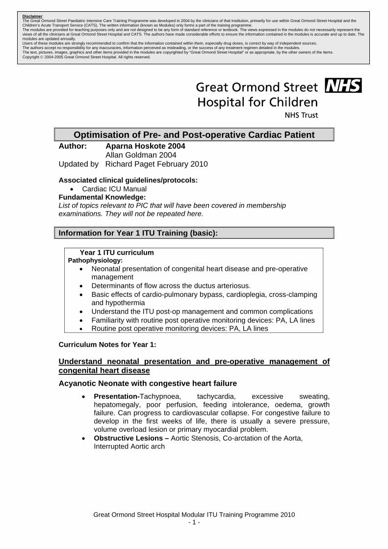
Acyanotic Neonate with congestive heart failure
Presentation-Tachypnoea, tachycardia, excessive sweating, hepatomegaly, poor perfusion, feeding intolerance, oedema, growth failure. Can progress to cardiovascular collapse. For congestive failure to develop in the first weeks of life, there is usually a severe pressure, volume overload lesion or primary myocardial problem.
Obstructive Lesions – Aortic Stenosis, Co-arctation of the Aorta, Interrupted Aortic arch
Great Ormond Street Hospital Modular ITU Training Programme 2010 - 1 -
Volume Overload Lesions: Ventricular Septal Defect, AVSD, Patent Ductus Arteriosus. (sats can be lowered)
Primary Myocardial Lesions: Transient myocardial ischaemia, Coronary
artery anomaly. myocarditis and Cardiomyopathies (such as IDM, Pompe’s disease, primary myocarditis).
Neonate with cyanosis
Duct Dependent Pulmonary Blood Flow Lesions:
Severe Tetralogy of Fallot, Ebstein’s anomaly, Tetralogy of Fallot with Pulmonary Atresia
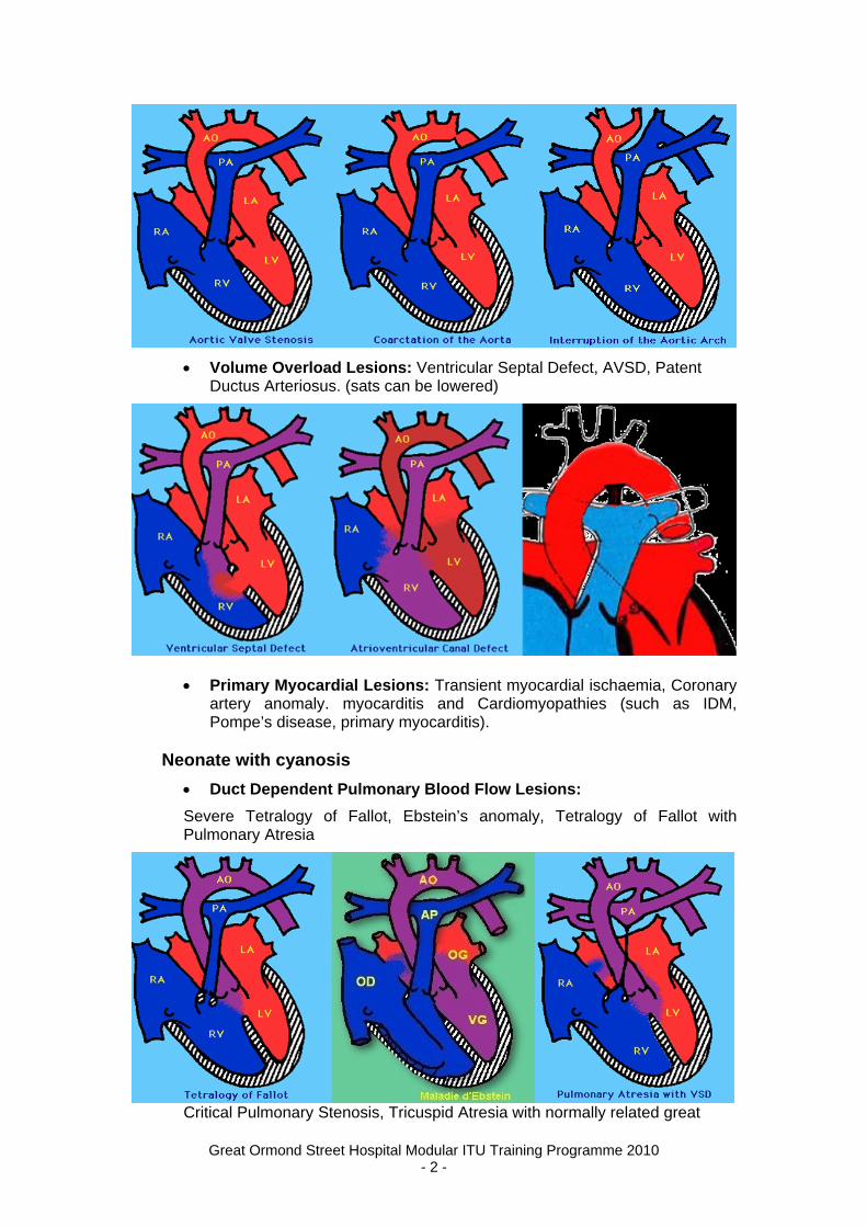
Critical Pulmonary Stenosis, Tricuspid Atresia with normally related great
Great Ormond Street Hospital Modular ITU Training Programme 2010 - 2 -
arteries, Pulmonary Atresia with Intact ventricular Septum(IVS), Heterotaxy
Duct dependent Systemic Blood flow Lesions
HLHS,Interrupted Aortic Arch, Critical Aortic Co-arctation, Critical Aortic Stenosis
Duct independent Mixing Lesions Truncus Arteriosus, Total Anomalous pulmonary venous return without obstruction, D-Transposition of the Great Arteries.
Neonate with cyanosis and congestive heart failure
HLHS, Truncus Arteriosus, Aorto Pulmonary Window.
Neonate with cyanosis and shock
Great Ormond Street Hospital Modular ITU Training Programme 2010 - 3 -
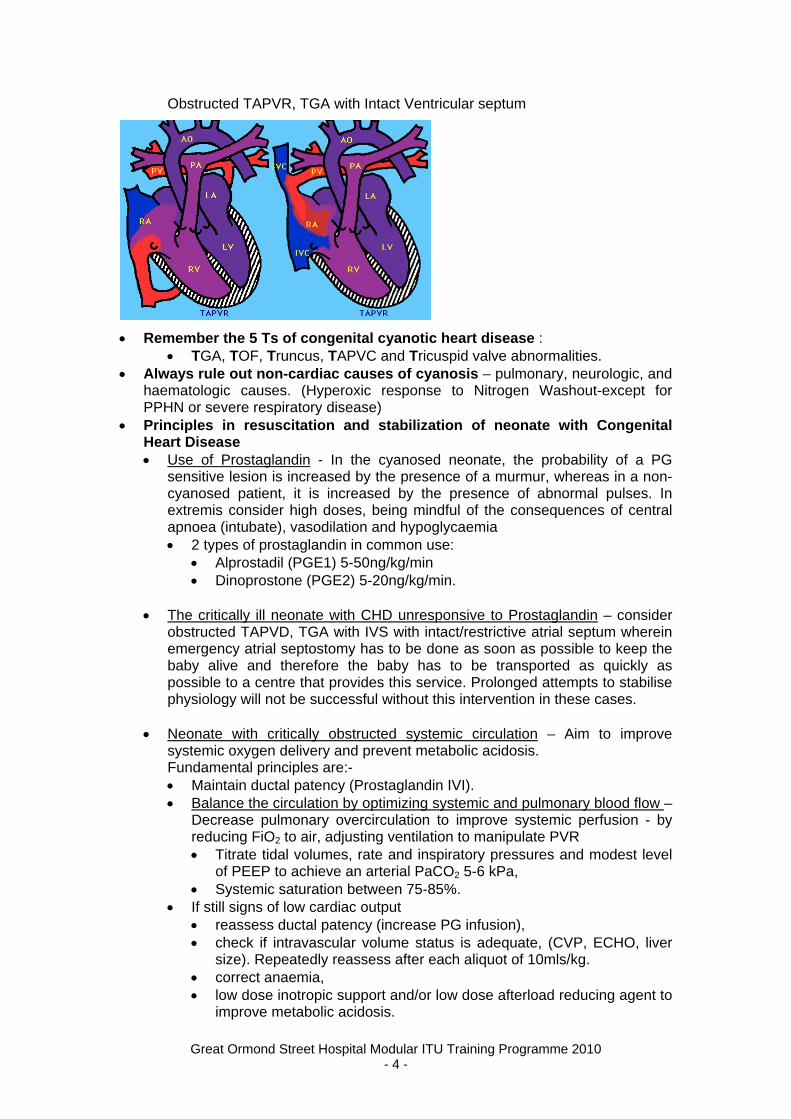
Obstructed TAPVR, TGA with Intact Ventricular septum
Remember the 5 Ts of congenital cyanotic heart disease : TGA, TOF, Truncus, TAPVC and Tricuspid valve abnormalities.
Always rule out non-cardiac causes of cyanosis – pulmonary, neurologic, and haematologic causes. (Hyperoxic response to Nitrogen Washout-except for PPHN or severe respiratory disease)
Principles in resuscitation and stabilization of neonate with Congenital Heart Disease Use of Prostaglandin - In the cyanosed neonate, the probability of a PG
sensitive lesion is increased by the presence of a murmur, whereas in a non-cyanosed patient, it is increased by the presence of abnormal pulses. In extremis consider high doses, being mindful of the consequences of central apnoea (intubate), vasodilation and hypoglycaemia 2 types of prostaglandin in common use:
Alprostadil (PGE1) 5-50ng/kg/min Dinoprostone (PGE2) 5-20ng/kg/min.
The critically ill neonate with CHD unresponsive to Prostaglandin – consider
obstructed TAPVD, TGA with IVS with intact/restrictive atrial septum wherein emergency atrial septostomy has to be done as soon as possible to keep the baby alive and therefore the baby has to be transported as quickly as possible to a centre that provides this service. Prolonged attempts to stabilise physiology will not be successful without this intervention in these cases.
Neonate with critically obstructed systemic circulation – Aim to improve
systemic oxygen delivery and prevent metabolic acidosis. Fundamental principles are:-
Maintain ductal patency (Prostaglandin IVI). Balance the circulation by optimizing systemic and pulmonary blood flow –
Decrease pulmonary overcirculation to improve systemic perfusion - by reducing FiO2 to air, adjusting ventilation to manipulate PVR Titrate tidal volumes, rate and inspiratory pressures and modest level
of PEEP to achieve an arterial PaCO2 5-6 kPa, Systemic saturation between 75-85%.
If still signs of low cardiac output reassess ductal patency (increase PG infusion), check if intravascular volume status is adequate, (CVP, ECHO, liver
size). Repeatedly reassess after each aliquot of 10mls/kg. correct anaemia, low dose inotropic support and/or low dose afterload reducing agent to
improve metabolic acidosis.
Great Ormond Street Hospital Modular ITU Training Programme 2010 - 4 -
Consider dysrythmia (ECG) and treat as required (remember electrolytes)
Indications for intubation Apnoeas or Respiratory Failure High dose PG IVI Shock Requirement for Cardioversion/ Balloon Atrial Septostomy/Surgery In General: using cardiotoxic anaesthesia in a shocked child with CHD
has significant associated risk. Senior discussion is mandatory. Benefits of ventilation also include lowering metabolic demands and
oxygen consumption via adequate sedation and paralysis. EFFECTS OF CARDIO-PULMONARY BYPASS, CARDIOPLEGIA, CROSS
CLAMPING AND HYPOTHERMIA
Great Ormond Street Hospital Modular ITU Training Programme 2010 - 5 -
Priming Considerations
The volume required to prime an adult CPB circuit is roughly 1500 mls. Adult prime/circulating volume ratio is 1:3
The volume required to prime a neonatal CPB circuit is up to 470 mls. A 3kg neonate has a circulating volume of 250-300 mls. This would cause unacceptable haemodilution. Hence, packed red cells are added to the pump prime in order to maintain adequate oxygen carrying capacity of the blood/perfusate. The neonatal prime/circulating volume ratio is 1.8:1.
The other constituents of the prime for a heart lung bypass circuit include crystalloid, colloid, Heparin, Mannitol, NaHCO3, CaCl2 and Aprotinin.
Banked blood represents a significant metabolic load to the patient, containing significant levels of glucose, lactate and histamine as well as high serum potassium levels.
Pre-bypass ultrafiltration - used to remove these substrates. The replacement fluid used is Plasmalyte A, and is buffered with NaHCO3 Once PBUF is completed, mannitol, top-up heparin and CaCl2 are added to the prime to achieve normal serum level.
Blood flow on CPB Blood flow on CPB is based on a cardiac index of 2.4, increasing to 2.8 during rewarming. Cardiac index = L/min/m2 BSA BSA= square root of ht (cm) multiplied weight (kg) divided by 3600 Cardiac index is increased in cyanotic disease due to collateral circulation. The current strategy in GOS is continuous antegrade regional perfusion where possible and total circulatory arrest when necessary. Modified ultrafiltration is used to reduce oedema and attenuate inflammation. Hypothermia is used to protect the patient against the effects of hypoxia and ischaemia. Using hypothermia, the cellular metabolism is reduced to its absolute minimum in the hope that it will minimise the potential for injury to the brain and other vital organs.
Mild hypothermia – 32-35 oC Moderate hypothermia – 25-28 oC Deep hypothermia – 15-22 oC
Deep hypothermic circulatory arrest - There is no circulation of blood in the body at all thereby providing a motionless and bloodless field and thus allowing the surgeon an excellent surgical exposure by eliminating the need for multiple cannulae within the surgical field. In order to achieve a safe cardiac arrest, a cross clamp is placed across the aorta between the aortic valve and the aortic cannula but distal to the origin of the coronary arteries. A small cannula is placed in the aortic root proximal to the aortic cross clamp and is used to inject the cardioplegia solution into the aortic root from where it perfuses into the coronary arteries in the presence of a competent aortic valve.
Cardioplegia – A cold, high potassium solution is used as a myocardial protection strategy to halt the mechanical contractions of the heart and to allow intracardiac procedures to be performed in a motionless field. Since blood flow is interrupted and cardiac ischaemia ensues, this myocardial protection strategy is designed to sufficiently reduce myocardial oxygen consumption so that resumption of myocardial function at the end of the procedure can occur with a minimum of dysfunction.
Great Ormond Street Hospital Modular ITU Training Programme 2010 - 6 -
Typically, a blood cardioplegia solution is obtained by mixing 4 parts of oxygenated blood to 1 part of crystalloid solution and the temperature of the solution is around 3-5 degree centigrade. Addition of blood to the cardioplegic solution enhances oxygen delivery, especially at the microcirculation level.
Minimizing calcium in the cardioplegia solution reduces the risk of intracellular accumulation of calcium during ischemia and reperfusion and, hence, prevents injury. However, complete elimination of calcium from the solution is not advised because of the risk of the calcium paradox phenomenon in which rapid calcium accumulation leading to cellular injury is observed. The addition of magnesium also adds a protective effect to the hypoxic-ischemic immature heart. This probably is the result of the antiarrhythmic effect of magnesium, its inhibition of calcium entry into the myocytes, and decreased uptake of sodium by myocytes during ischemia, which is exchanged for calcium during reperfusion.
Hypothermic low flow cardiopulmonary bypass
This technique allows continuous low-flow perfusion to the organs during the operation, which may lead to an increase in oxygen supply, better nutrient supply, and better achievement of homogeneous hypothermia during bypass. The finding that DHCA was associated with neurologic morbidity has led researchers to investigate the use of HLFB. Recent trials comparing the 2 methods have reported lower rates of neural dysfunction in the group of patients undergoing HLFB. Specifically, when the patient with CPB is aged 4 years, DHCA is associated with lower levels of motor coordination and planning but not with significantly lower intelligence quotient (IQ) or a worse overall neurologic status. Regional Perfusion New techniques are used to minimise the potential for brain injury and is employed in cases such as Norwood stage I/ Reconstructive arch surgery. Regional flow to the right carotid system can be used to provide the brain with oxygenated blood flow during the arrest period. In the Norwood Stage 1 surgery - the arterial line is split. The cerebral branch is clamped and the Ductus is cannulated for PA transection. The innominate artery is cannulated with ‘Olive’ for arch reconstruction. Flow: 25-30ml/kg/min Advantages: continuous O2 supply, no reperfusion injury Disadvantages: continuous oedema generation Modified Ultrafiltration The aim of ultrafiltration is to remove excess body water and to help reduce the levels of inflammatory mediators induced by cardiopulmonary bypass. Modified ultrafiltration is started in the immediate post bypass period, shunting the blood from the arterial cannula via the filter back to the right atrium, aiming to bring the haematocrit up to 40-45%.
Great Ormond Street Hospital Modular ITU Training Programme 2010 - 7 -
THE ITU POST-OPERATIVE MANAGEMENT AND COMMON COMPLICATIONS
General principles Anticipate the type of patient and potential problems Understand lesion/post
correction basic physiology and common postoperative issues. Ask if needed. Arrival and handover
Quick assessment focusing on cardiovascular stability and adequate ventilation (vital signs and chest movement) as patient arrives from theatre.
Get handover from the anaesthetist and the surgeon and review pre and intra-operative history. Use proforma. Note operation performed and the duration of CPB and aortic cross
clamping (latter being time of ‘cardioplegia’)? Check if there are any concerns regarding anatomy/repair (e.g.
coronaries, size of shunt, etc.) or pulmonary hypertension? Note pressures (RAP/CVP, LAP and MAP), cardiac rhythm and oxygen
saturations in theatre. Ask specifically for intra-operative problems and any complications
encountered - arrhythmia, pacemaker use, coming off CPB problems, bleeding and blood products given, and medications given.
Ask what blood products are available (unused from theatre). Initial Assessment
Review current support drugs – inotropes and/or vasodilators Review with nurses that the patient is safely connected to ICU support and
monitoring equipment. Check ventilator settings Review anaesthetic, CPB charts and operation or any intra-operative TOE
notes. Look for difficult airway, anaesthetic medications given, heart function, and residual lesions.
Detailed physical examination Request: ABG/ K+/ iCa++/HCT, CXR
and formal FBC and coagulation profile in all pts if bleeding excessively Document clinical findings and plan in chart Review as soon as possible CXR (check position of ETT, lines, drains, any
pneumothorax/effusion), and first set of blood results – ventilation settings may need adjustment according to ABG.
Request baseline ECG and postoperative Echo if Cardiology intra-operative TOE not done.
Ongoing Management
Adequate Opiate Analgesia. Titrate other sedatives as required. Long acting or IVI Muscle relaxant if cardiovascularly unstable Support the circulation-see lesion specific modules. Consider/treat Arrhythmias- see specific module. Bleeding > 3mls/kg/hr investigate. >10 ml/kg/hour needs urgent surgical
review fluid resuscitate, send FBC and coagulation profile, Consider TEG &
replace factors as indicated (most likely to need cryoprecipitate and platelets).
Consider (after senior discussion) Aprotinin (kallikrein inhibitor) 10,000 kiu/kg/hour, Tranexamic Acid (antifibrinolytic) and rFVIIa
Watch for tamponade (see below). Packed cells 4 mls/kg raises Hb by 1g/dl
Follow unit antibiotic policy
Great Ormond Street Hospital Modular ITU Training Programme 2010 - 8 -
RECOGNITION AND MANAGEMENT OF COMPLICATIONS AFTER CONGENITAL HEART SURGERY
The following are general complications seen following congenital heart surgery. It is vital that these complications are recognised early and managed appropriately. 1. CARDIAC TAMPONADE
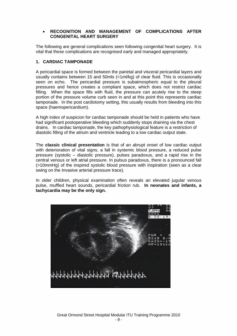
A pericardial space is formed between the parietal and visceral pericardial layers and usually contains between 15 and 50mls (<1ml/kg) of clear fluid. This is occasionally seen on echo. The pericardial pressure is subatmospheric equal to the pleural pressures and hence creates a compliant space, which does not restrict cardiac filling. When the space fills with fluid, the pressure can acutely rise to the steep portion of the pressure volume curb seen in and at this point this represents cardiac tamponade. In the post cardiotomy setting, this usually results from bleeding into this space (haemopericardium). A high index of suspicion for cardiac tamponade should be held in patients who have had significant postoperative bleeding which suddenly stops draining via the chest drains. In cardiac tamponade, the key pathophysiological feature is a restriction of diastolic filling of the atrium and ventricle leading to a low cardiac output state.
The classic clinical presentation is that of an abrupt onset of low cardiac output with deterioration of vital signs, a fall in systemic blood pressure, a reduced pulse pressure (systolic – diastolic pressure), pulses paradoxus, and a rapid rise in the central venous or left atrial pressure. In pulsus paradoxus, there is a pronounced fall (>10mmHg) of the inspired systolic blood pressure with inspiration (seen as a clear swing on the Invasive arterial pressure trace). In older children, physical examination often reveals an elevated jugular venous pulse, muffled heart sounds, pericardial friction rub. In neonates and infants, a tachycardia may be the only sign.
Great Ormond Street Hospital Modular ITU Training Programme 2010 - 9 -
The CXR usually shows a marked increase in the cardiac shadow, although this is not always as impressive as expected. Transthoracic echo as depicted above is usually sufficient for defining the size and amount of fluid. It must, however be stressed that in the urgent situation in which a post cardiotomy patient is about to suffer an imminent cardiac arrest and where there is a high index of suspicion of cardiac tamponade, the definitive procedure should be to immediately open the chest and not wait for an echocardiogram. Furthermore, the echocardiogram is not also always accurate in diagnosing cardiac tamponade, particularly where there is a posterior collection. If any doubt one should proceed to median sternotomy. Whilst awaiting a surgeon to open the chest, the patient should be managed with a colloid challenge of 10-20mls/kg (depending on the underlying cardiac condition) and the use of inotropes. If the patient is on vasodilators, these should be stopped. In summary, suspect a cardiac tamponade in patients following congenital heart surgery if:- There is a sudden deterioration in their condition There is a sudden tachycardia associated with increased atrial filing pressures Systemic hypotension, low pulse pressure and a marked swing on the arterial
trace with inspiration and expiration. This particularly so if there has been a sudden reduction of blood loss from the chest drains.
Initial treatment includes volume resuscitation, increase in inotropes, stop vasodilators and diuretic therapy. If cardiac output remains inadequate, urgently call the Cardiology and Cardiothoracic surgical registrars. Also get urgent cross match and blood if available in the local or central blood bank. Obtain an echo if the patient has stable cardiac output. If the patient is in profound or deteriorating shock proceed to urgent median sternotomy for surgical evacuation of the tamponade. A drainage tube should be left in the mediastinum and placed on low-pressure suction following re-exploration.
2. MEDIASTINITIS Mediastinitis is a severe deep-seated infection complicating congenital heart surgery and usually requires surgical re-exploration and washout of the mediastinum. It should be suspected in any patient following congenital heart surgery who has signs and symptoms of infection including high fever, abnormal laboratory data with elevated or depressed white blood cell count with a left shift, and raised CRP. These patients often have a red and inflamed sternal wound. The sternal wound should be meticulously checked for instability when there are concerns about mediastinitis. There should be a very high index of suspicion of mediastinitis if the chest wound becomes unstable, when there is pus coming from the chest drain, and when blood cultures are positive for staphylococcus aureus. The most common organism causing mediastinitis is staphylococcus aureus.
Management includes:-
Urgent antibiotics. Treatment at GOS is Vancomycin (dose) in the first instance until sensitivities are available. Patient should then undergo surgical re-exploration and wash out of the chest. Clearly labelled deep mediastinal swabs or pus from the chest should be sent to the laboratory urgently.
An irrigation system is usually set up in theatre with betadine irrigation of the chest.
Great Ormond Street Hospital Modular ITU Training Programme 2010 - 10 -
The betadine irrigation is continued for at least 48 hours followed by saline irrigation for a further 48 hours until the cultures from the effluent from the saline irrigation is sterile.
Intravenous antibiotics should be continued for at least 3 weeks. In the first instance, this will be Vancomycin and if the staphylococcus is sensitive to Flucloxacillin, this is usually continued for 6 weeks (usually intravenous for at least 3 weeks). Consideration should always be given to whether the patient could have an immune deficiency or DiGeorge syndrome.
2. PHRENIC NERVE INJURY/ DIAPHRAGM PARALYSIS This is a recognised complication of cardiothoracic surgery and can be
attributable to either nerve resection, nerve stretch, electrical or cold injury from topical cardiac hypothermia. Prevalence is between 0.5 – 2.2%, with the incidence being higher in the under 2 year olds. It is more commonly seen after BT shunts, atrial septectomy, PA banding, PDA ligation and coarctation repair.
Infants are more susceptible to respiratory compromise because of a greater reliance on abdominal breathing, and weak intercostals muscles compounded by smaller airways.
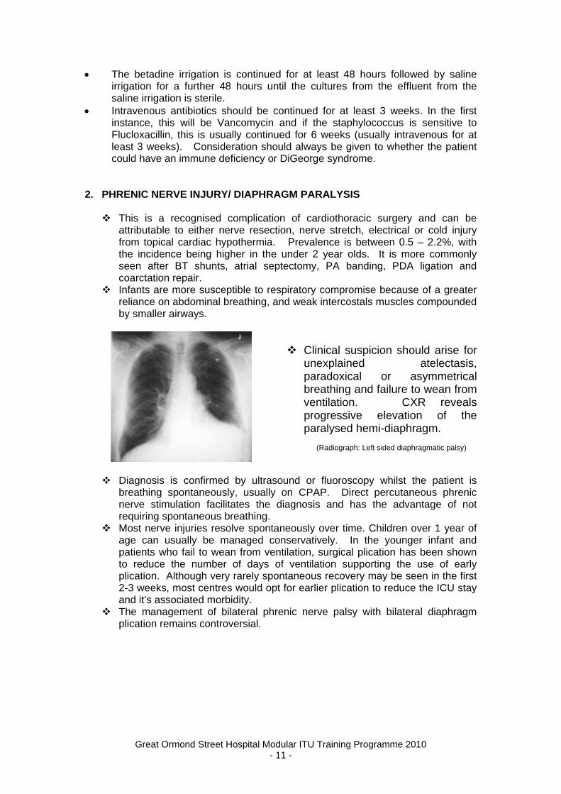
Clinical suspicion should arise for unexplained atelectasis, paradoxical or asymmetrical breathing and failure to wean from ventilation. CXR reveals progressive elevation of the paralysed hemi-diaphragm.
(Radiograph: Left sided diaphragmatic palsy)
Diagnosis is confirmed by ultrasound or fluoroscopy whilst the patient is
breathing spontaneously, usually on CPAP. Direct percutaneous phrenic nerve stimulation facilitates the diagnosis and has the advantage of not requiring spontaneous breathing.
Most nerve injuries resolve spontaneously over time. Children over 1 year of age can usually be managed conservatively. In the younger infant and patients who fail to wean from ventilation, surgical plication has been shown to reduce the number of days of ventilation supporting the use of early plication. Although very rarely spontaneous recovery may be seen in the first 2-3 weeks, most centres would opt for earlier plication to reduce the ICU stay and it’s associated morbidity.
The management of bilateral phrenic nerve palsy with bilateral diaphragm plication remains controversial.
Great Ormond Street Hospital Modular ITU Training Programme 2010 - 11 -
3. CHYLOTHORAX
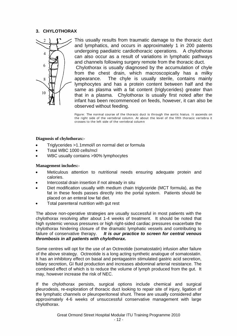
This usually results from traumatic damage to the thoracic duct and lymphatics, and occurs in approximately 1 in 200 patents undergoing paediatric cardiothoracic operations. A chylothorax can also occur as a result of variations in lymphatic pathways and channels following surgery remote from the thoracic duct. Chylothorax is usually diagnosed by the accumulation of chyle from the chest drain, which macroscopically has a milky appearance. The chyle is usually sterile, contains mainly lymphocytes and has a protein content between half and the same as plasma with a fat content (triglycerides) greater than that in a plasma. Chylothorax is usually first noted after the infant has been recommenced on feeds, however, it can also be observed without feeding. Figure: The normal course of the thoracic duct is through the aortic hiatus. It ascends on the right side of the vertebral column. At about the level of the fifth thoracic vertebra it crosses to the left side of the vertebral column
Diagnosis of chylothorax:-
Triglycerides >1.1mmol/l on normal diet or formula Total WBC 1000 cells/mcl WBC usually contains >90% lymphocytes
Management includes:-
Meticulous attention to nutritional needs ensuring adequate protein and calories.
Intercostal drain insertion if not already in situ Diet modification usually with medium chain triglyceride (MCT formula), as the
fat in these feeds passes directly into the portal system. Patients should be placed on an enteral low fat diet.
Total parenteral nutrition with gut rest The above non-operative strategies are usually successful in most patients with the chylothorax resolving after about 1-4 weeks of treatment. It should be noted that high systemic venous pressures or high right-sided cardiac pressures exacerbate the chylothorax hindering closure of the dramatic lymphatic vessels and contributing to failure of conservative therapy. It is our practice to screen for central venous thrombosis in all patients with chylothorax. Some centres will opt for the use of an Octreotide (somatostatin) infusion after failure of the above strategy. Octreotide is a long acting synthetic analogue of somatostatin. It has an inhibitory effect on basal and pentagastrin stimulated gastric acid secretion, biliary secretion, GI fluid production and increases abdominal arterial resistance. The combined effect of which is to reduce the volume of lymph produced from the gut. It may, however increase the risk of NEC. If the chylothorax persists, surgical options include chemical and surgical pleurodesis, re-exploration of thoracic duct looking to repair site of injury, ligation of the lymphatic channels or pleuroperitoneal shunt. These are usually considered after approximately 4-6 weeks of unsuccessful conservative management with large chylothorax.
Great Ormond Street Hospital Modular ITU Training Programme 2010 - 12 -
In patients with large chylothorax, serum immunoglobulin should also be monitored with immunoglobulin replacement as required. Although the use of intravenous immunoglobulin for this indication has become extremely restricted. These patients often also require regular albumin replacement therapy and if the chylothorax is large, it is our practice to replace half to all the hourly drainage losses worth 4.5% albumin (obviously depending on the serum albumin levels). 4. PROTEIN LOSING ENTEROPATHY Protein losing enteropathy (PLE) has been reported in almost all situations in which there is chronically elevated central venous pressure including chronic heart failure, chronic constrictive pericarditis and following atrial switch operations. From a pathophysiological point of view, it is presumed the combination of increased lymph production from increased IVC pressure and impaired lymph drainage from increased SVC pressure contribute to this complication. In our practice this generally occurs as an important complication after the Fontan operation. It is characterised by depressed serum albumin concentration with no obvious cause. Clinical suspicion may be aroused by late postoperative onset of generalised oedema and pleural or peritoneal fluid collections. Patients may or may not have diarrhoea. The diagnosis is characterised by Alpha 1 anti trypsin in the stool, as clearance of this protein is abnormally high. These patients also classically have low total serum calcium due to low serum proteins. Specific studies show excessive loss of serum proteins from the GIT and endoscopic examination shows prominent lymph vessels in the jejunal mucosa. Approximately 13% of Fontan patients develop evidence of protein losing enteropathy by 10 years. It is associated with a near 50% mortality by 5 years after diagnosis. Treatment is geared towards re-operations that result in a lower right atrial pressure. There is, however, a considerable early mortality after re operation. The use of valves in the IVC orifice are almost never carried out today. Other strategies aimed at correcting haemodynamic abnormalities when right atrial pressure is low has resulted in some resolution. Creating a late fenestration has been reported with some success. Non-surgical treatments have included the use of steroids and heparin. It is now also believed that GI protein loss occurs before clinical onset of PLE.
Treatment strategies tailored to the severity of the disease include:-
Symptomatic relief with diuretics and supplemental protein Strategies to holt intestinal protein leak using steroids or heparin Alteration of cardiovascular physiology via fenestration, atrial pacing or heart
transplant 4. Suggested reading Textbook of Paediatric Cardiology. Prof R H Anderson Paediatric Cardiac Intensive Care – Eds A Chang, F Hanley, G Wernovsky, D Wessel. Preoperative Care of CHD. Bovey E L. Current Status of Staged Reconstruction for Hypoplastic Left Heart Syndrome. Pediatric Cardiology 19:308-315, 1998
Great Ormond Street Hospital Modular ITU Training Programme 2010 - 13 -
Cardiology in the Young. Controversies relating to the hypoplastic left heart syndrome. 2004 supplement 1 pages 1-130. Tsang V, Stark J. Coarctation of the Aorta. Edition 2005 in press Fedderley R T. Left Ventricular Outflow Obstruction. Pediatric Clinics of North America, 46, 369-383. 1999 Operative Cardiac surgery Edited by TJ Gardner and Thomas L Spray (used as source for many of pictures of surgical procedures) 5. Additional useful information and internet sites http://www.pediheart.org/practitioners/defects/ventriculoarterial/TOF_PA.htm http://www.emedicine.com/radio/topic685.htm http://www.emedicine.com/ped/topic2539.htm http://www.emedicine.com/EMERG/topic575.htm http://www.rch.org.au/cardiology/defects.cfm?doc_id=3011 http://www.emedicine.com/ped/topic2550.htm http://www.emedicine.com/ped/topic2526.htm http://circ.ahajournals.org/cgi/content/full/91/6/1782 6. Available guidelines Cardiac ICU guidelines
Information for Year 2 ITU Training (advanced):
Year 2 ITU curriculum Pathophysiology:
Immunomodulatory effects of cardio-pulmonary bypass. Neuro-hormonal effects of surgical stress. .
Central Cardiac Audit Database and Operative outcomes pH-stat and alpha-stat strategies of managing pH during cooling
Curriculum Notes for Year 2: Effects of Cardiopulmonary bypass In General
Cardiopulmonary bypass (CPB) causes more pronounced harmful effects in infants because of the immaturity of peripheral tissues and organ function. The disparity between the CPB circuit size and the patient size also increases the subsequent inflammatory reaction resulting from exposure to the foreign surface area.
The systemic inflammatory response to CPB is complex and involves activation and interaction of many systems. It promotes increased capillary permeability and interstitial oedema. This response contributes to pulmonary and renal dysfunction in the postoperative period.
Great Ormond Street Hospital Modular ITU Training Programme 2010 - 14 -
Immunomodulatory effects The inflammatory response to CPB is inversely proportional to the
patient's age. The synthetic surfaces of the bypass circuit are associated with activation of inflammatory mediators. Effects include activation of the complement system, including plasma-activated complement 3 (C3a). A potent stimulator of platelet aggregation, C3a promotes mast cell release, increases vascular permeability, and stimulates WBCs to release oxygen free radicals and lysosomal enzymes. C3a levels are linked to the duration of CPB.
Neutrophil activation has been linked to this inflammatory reaction, with neutrophil expression linked to the duration of CPB. Their activation increases production of cytokines, such as (IL)-8 and IL-6 and TNF. Expression of binding proteins on endothelial surfaces leads to extravascular migration of neutrophils and subsequent tissue injury. Activated neutrophils may obstruct the capillaries, limiting reperfusion of ischemic tissue (ie, no-reflow phenomenon). In addition, neutrophil activation stimulates the release of lysosomal enzymes, such as elastase and proteinase, in addition to the release of oxygen free radicals.
Arachidonic acid cascade activation leads to the generation of leukotrienes, prostaglandins, and thromboxane A2. With various effects on the vascular reactivity and continued inflammatory state.
Coagulation system effects
Blood contact with the surface of the bypass machine activates platelets and increases thrombus formation. Tissue factor release leads to the
generation of thrombin, that initiate a positive-feedback cycle of
coagulation and inflammatory cascades. If not corrected, this process leads to a hypercoagulable state, which, in addition to activation of fibrinolysis, can cause excessive bleeding.
Stress response
Low perfusion, hypothermia, anesthesia, and surgery cause the release of hormones including catecholamines, cortisol, growth hormone, glucagon, corticotropin (or ACTH), TSH, and endorphins. Levels of thyroid hormone decreases the first few days after CPB. Decreased renal and hepatic function after CPB leads to decreased clearance of vasoactive inflammatory mediators. The lung is also normally responsible for metabolizing and clearing many of these hormones, particularly catecholamines. Exclusion of the lungs from the circulation after CPB leads to the accumulation and increased circulating levels.
Glucose metabolism
Hyperglycemia accompanies the stress response associated with CPB. Hyperglycemia has been associated with worsened outcomes after myocardial infarction, stroke, postoperative wound infections, and severe head injury. Insulin protocols and use of a glucose-insulin-potassium solution to ensure tight glucose control after cardiac surgery in adults have been associated with lower mortality, improved hemodynamics, and decreased need for reoperations, as well as less renal failure.
A more common complication of pediatric CPB is hypoglycaemia,due to the decreased glycogen stores and reduced hepatic gluconeogenesis. Neurologic consequences of hypoglycemia are aggravated by hypothermia and other factors that may modify cerebral perfusion.
Great Ormond Street Hospital Modular ITU Training Programme 2010 - 15 -
Cardiac effects
Conflicting data describes the relative sensitivity to ischaemia of the neonatal heart. Reasons for improved tolerance to ischemia in the neonatal heart include the increased glycolytic capability of the immature myocardium and enhanced preservation of high-energy phosphates because of decreased levels of 5'-nucleotidase, which catalyzes the breakdown of adenosine monophosphate (AMP) to adenosine. However, an increased accumulation of lactic acid as a result of anaerobic metabolism is hypothesized as a cause of ischemic intolerance in the neonatal heart.
CNS effects
Neurologic injury after routine CPB is uncommon in neonates, but the risk is increased when deep hypothermic circulatory arrest (DHCA) is required. Although permanent injury is relatively uncommon, evidence of neurologic injury is observed in as many as 25% of infants who undergo DHCA. Neurologic morbidity includes seizures, strokes, changed tone and mental status, motor disorders, abnormal cognitive functioning, and postpump choreoathetosis. Areas most vulnerable to ischemic injury include the neocortex, hippocampus, and striatum.
Another potential mechanism of brain injury involves binding of glutamate to the N -methyl-D-aspartate receptor (NMDAR). This binding increases the amount of intracellular calcium and subsequently activates proteases, phospholipases, and deoxyribonucleases (DNAses) and promotes generation of free radicals. The net result of these processes is cell injury, cell death, or both.
Microemboli can be detected in patients during CPB. The long-term effect of these emboli is not well defined.
Pulmonary effects
Lung injury is mediated in 1 of 2 ways. Leukocyte and complement activation cause an inflammatory response, or a mechanical effect leads to surfactant loss and atelectasis. These both reduce static and dynamic compliance and functional residual capacity and increase the alveolar-arterial (A-a) gradient. Heamodilution reduces oncotic pressure and causes extravasation of fluid into the lung parenchyma. CPB activates complement and leukocyte degranulation, causing injury to the capillary membrane and platelet activation, both of which eventually lead to increased pulmonary vascular resistance.
Renal effects
CPB leads to production of renin, angiotensin, catecholamines, and antidiuretic hormone. In turn, these substances cause renal vasoconstriction and reduce renal blood flow.
Risk factors for postoperative renal dysfunction include preoperative renal disease, contrast-related renal injury, and profound post-CPB reduction in cardiac output. After CPB, 8% of patients have acute renal insufficiency, as indicated by oliguria and increased creatinine levels.
After spontaneous urine output is observed, diuretics are effective for inducing diuresis and reversing renal cortical ischemia associated with CPB, but their use does not alter the time to the recovery of renal function.
BNP-Increased postoperative Brain Natriuretic Peptide (BNP) levels (and longer acting NT-ProBNP subtype) is primarily released by the ventricles in
Great Ormond Street Hospital Modular ITU Training Programme 2010 - 16 -
response to myocardial stretch. It’s name is derived from it’s initial isolation from a pig’s brain. It encourages sodium (Natriuresis) and water loss from the kidneys and is commonly used as prognostic and predictive marker in adult congestive heart failure (CHF). A recombinant analogue (Nesiritide) has also been used as CHF treatment. Higher levels are associated with longer bypass times in children. The biologic activity of the natriuretic hormone system is transiently impaired however (decoupled from intracellular cGMP). Research is being conducted into its possible role as a predictor of perioperative morbidity.
Methods used to moderate CPB effects (not universal)
miniaturization of the circuit and the oxygenator the use of steroids and aprotinin biocompatible circuitry vacuum-assist venous drainage (VAVD)- allows further miniaturization of the
CPB circuit because VAVD allows the use of decreased-diameter cannulas and tubing
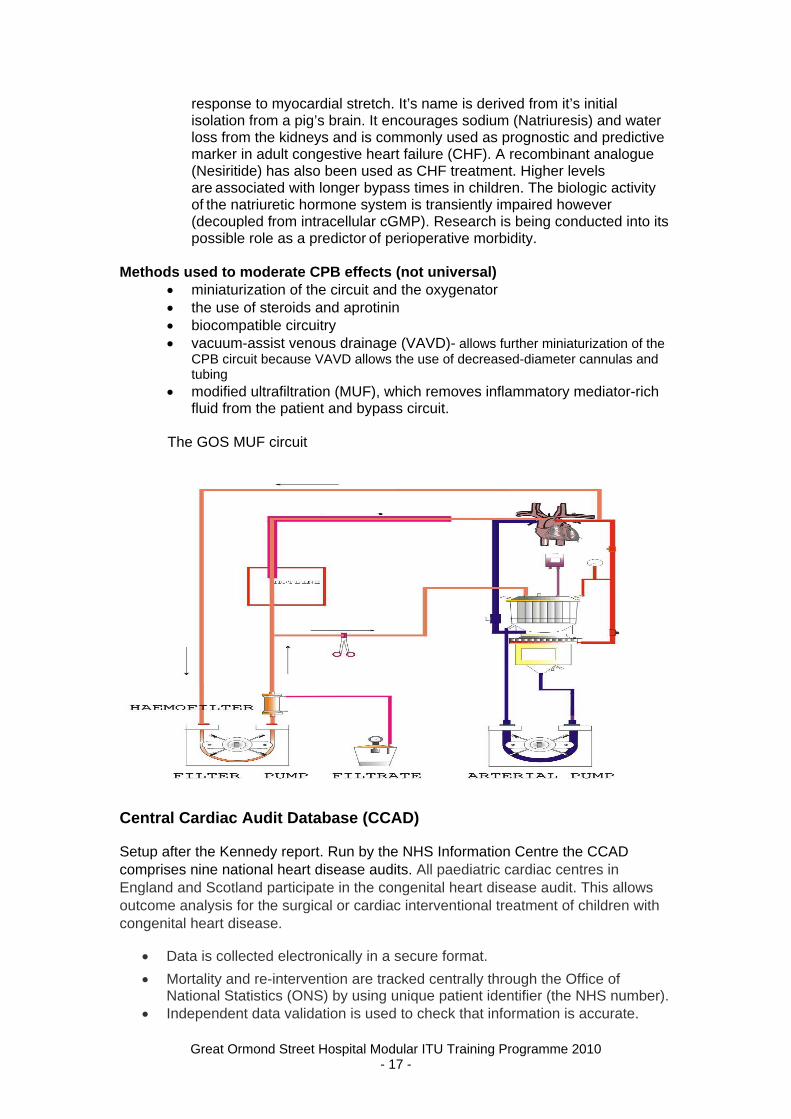
modified ultrafiltration (MUF), which removes inflammatory mediator-rich fluid from the patient and bypass circuit.
The GOS MUF circuit
Central Cardiac Audit Database (CCAD)
Setup after the Kennedy report. Run by the NHS Information Centre the CCAD comprises nine national heart disease audits. All paediatric cardiac centres in England and Scotland participate in the congenital heart disease audit. This allows outcome analysis for the surgical or cardiac interventional treatment of children with congenital heart disease.
Data is collected electronically in a secure format.
Mortality and re-intervention are tracked centrally through the Office of National Statistics (ONS) by using unique patient identifier (the NHS number).
Independent data validation is used to check that information is accurate.
Great Ormond Street Hospital Modular ITU Training Programme 2010 - 17 -
Access to detailed information for patients, healthcare professionals and the public about the care they receive and national survival rates via the congenital heart disease portal.
http://www.ccad.org.uk/congenital
Other sources of information: References.
1. Yates AR, Dyke PC 2nd, Taeed R, et al. Hyperglycemia is a marker for poor outcome in the postoperative pediatric cardiac patient. Pediatr Crit Care Med. Jul 2006;7(4):351-5. [Medline].
2. Wypij D, Newburger JW, Rappaport LA, et al. The effect of duration of deep hypothermic circulatory arrest in infant heart surgery on late neurodevelopment: the Boston Circulatory Arrest Trial. J Thorac Cardiovasc Surg. Nov 2003;126(5):1397-403. [Medline].
3. Ballweg JA, Wernovsky G, Ittenbach RF, et al. Hyperglycemia after infant cardiac surgery does not adversely impact neurodevelopmental outcome. Ann Thorac Surg. Dec 2007;84(6):2052-8. [Medline].
4. Backer CL, Kelle AM, Stewart RD, et al. Aprotinin is safe in pediatric patients undergoing cardiac surgery. J Thorac Cardiovasc Surg. Dec 2007;134(6):1421-6; discussion 1426-8. [Medline].
5. Goldberg CS, Bove EL, Devaney EJ, et al. A randomized clinical trial of regional cerebral perfusion versus deep hypothermic circulatory arrest: outcomes for infants with functional single ventricle. J Thorac Cardiovasc Surg. Apr 2007;133(4):880-7. [Medline].
6. Visconti KJ, Rimmer D, Gauvreau K, et al. Regional low-flow perfusion versus circulatory arrest in neonates: one-year neurodevelopmental outcome. Ann Thorac Surg. Dec 2006;82(6):2207-11; discussion 2211-3. [Medline].
7. Bellinger DC, Wypij D, Kuban KC, et al. Developmental and neurological status of children at 4 years of age after heart surgery with hypothermic circulatory arrest or low-flow cardiopulmonary bypass. Circulation. Aug 3 1999;100(5):526-32. [Medline].
8. Duebener LF, Hagino I, Sakamoto T, et al. Effects of pH management during deep hypothermic bypass on cerebral microcirculation: alpha-stat versus pH-stat. Circulation. Sep 24 2002;106(12 Suppl 1):I103-8. [Medline].
9. Fraser CD, Andropoulos DB. Neurologic monitoring for special cardiopulmonary bypass techniques. Semin Thorac Cardiovasc Surg Pediatr Card Surg Annu. 2004;7:125-32. [Medline].
10. Friesen RH, Campbell DN, Clarke DR, Tornabene MA. Modified ultrafiltration attenuates dilutional coagulopathy in pediatric open heart operations. Ann Thorac Surg. Dec 1997;64(6):1787-9. [Medline].
11. Hickey E, Karamlou T, You J, Ungerleider RM. Effects of circuit miniaturization in reducing inflammatory response to infant cardiopulmonary bypass by elimination of allogeneic blood products. Ann Thorac Surg. Jun 2006;81(6):S2367-72. [Medline].
12. Jaggers J, Lawson JH. Coagulopathy and inflammation in neonatal heart surgery: mechanisms and strategies. Ann Thorac Surg. Jun 2006;81(6):S2360-6. [Medline].
13. Karamlou T, Hickey E, Silliman CC, et al. Reducing risk in infant cardiopulmonary bypass: the use of a miniaturized circuit and a crystalloid prime improves cardiopulmonary function and increases cerebral blood flow. Semin Thorac Cardiovasc Surg Pediatr Card Surg Annu. 2005;3-11. [Medline].
14. Kozik DJ, Tweddell JS. Characterizing the inflammatory response to cardiopulmonary bypass in children. Ann Thorac Surg. Jun 2006;81(6):S2347-54. [Medline].
15. Schultz JM, Karamlou T, Swanson J, et al. Hypothermic low-flow cardiopulmonary bypass impairs pulmonary and right ventricular function more than circulatory arrest. Ann Thorac Surg. Feb 2006;81(2):474-80; discussion 480. [Medline].
16. Tsui SS, Schultz JM, Shen I, Ungerleider RM. Postoperative hypoxemia exacerbates potential brain injury after deep hypothermic circulatory arrest. Ann Thorac Surg. Jul 2004;78(1):188-96; discussion 188-96. [Medline].
17. Ungerleider RM. Cerebral protection in infant cardiac surgery. Ann Surg. Dec 2003;238(6 Suppl):S100-3. [Medline].
18. Ungerleider RM, Gaynor JW. The Boston Circulatory Arrest Study: an analysis. J Thorac Cardiovasc Surg. May 2004;127(5):1256-61. [Medline].
Great Ormond Street Hospital Modular ITU Training Programme 2010 - 18 -
Great Ormond Street Hospital Modular ITU Training Programme 2010 - 19 -
pH-stat and alpha-stat strategies of managing pH during cooling Alpha-stat strategy: The blood gas is measured at 37 oC regardless of
sample temperature. The imidazole ring on histidine is maintained in the alpha state (constant ratio of unprotonated histidine imidazole groups to H+ concentration). This keeps CO2 stores constant, but allows PaCO2 and pH to drift with temperature change. The patient gets increasingly alkalotic as temperature falls.
pH-stat strategy: The blood gas is measured as ‘true’ value at temperature of the sample. The solubility of CO2 increases 4.4%/ oC, therefore blood becomes more alkalotic as PaCO2 falls. The ventilation is decreased and/or CO2 added to keep PaCO2 levels at normal (37 oC) values to keep pH constant (stat).
NB: In-line PaCO2 monitoring is mandatory during this period. Ongoing debate about the best pH strategy for brain protection during CPB. pH-stat strategy: carbon dioxide is added during hypothermia. To maintain
neutral pH at 17oC this requires a PaCO2 of 58 mmHg, but if this arterial blood gas sample is analysed at 37oC the pH will be 7.06 and the PaCO2 11.5 kPa, i.e. severe acidosis. Adding carbon dioxide has the benefit that it is a cerebral vasodilator and this potentially may help to protect from hypoxic brain injury.
alpha-stat strategy: With a decrease in temperature, the ratio (termed alpha) of dissociated to nondissociated imidazole groups of histidine, an essential component of the protein buffer system, remains constant. It is thought that this optimises intracellular enzyme function. The PaCO2 of arterial blood measured at 37oC is kept constant, but the actual CO2content falls as a direct function of declining temperature and pH rises.
In summary Advantages of pH-stat strategy
Improvement of cerebral blood flow and cerebral oxygenation Improvement of brain cooling efficiency
Disadvantages of pH-stat strategy Risk of micro-embolism and of free radical mediated damage
Advantages of alpha-stat strategy Preserves cerebral autoregulation Optimises intracellular enzyme function
Disadvantages of alpha-stat strategy Less metabolic suppression Greater susceptibility to neurological injury
The current local strategy is determined by the depth of hypothermia pH stat used for first 15 min of cooling Cerebral autoregulation no longer
responsive to PaCO2 below 22 oC (after 15 min cooling) Alpha stat used for remainder of pump run- used for minimum of 5 min before
DHCA Ventilation increased to remove excess CO2 prior to DHCA Patient ‘alkalotic’ prior to DHCA. Minimum cooling time 20min
Alpha stat during rewarming - keeps cerebral vessels ‘tight’ and decreases blood brain volume and potential reduction in embolic delivery.
References: 1. Bellinger DC, Wypij D, du Plessis AJ, et al. Developmental and neurologic effects of alpha-stat versus pH-stat
strategies for deep hypothermic cardiopulmonary bypass in infants. J Thorac Cardiovasc Surg. Feb 2001;121(2):374-83. [Medline].
2. Khairul Anuar Abdul Aziz* and Ayo Meduoye. Is pH-stat or alpha-stat the best technique to follow in patients undergoing deep hypothermic circulatory arrest? Interact CardioVasc Thorac Surg 2010;10:271-282.
doi:10.1510/icvts.2009.214130 http://icvts.ctsnetjournals.org/cgi/content/full/10/2/271