Embed Size (px)
Citation preview

IAEA-TECDOC-1310
Optimization of synthesis and quality control procedures for the preparation of 18F and 123I labelled
peptides for nuclear medicine
September 2002

The originating Section of this publication in the IAEA was:
Industrial Applications and Chemistry Section International Atomic Energy Agency
Wagramer Strasse 5 P.O. Box 100
A-1400 Vienna, Austria
OPTIMIZATION OF SYNTHESIS AND QUALITY CONTROL PROCEDURES FOR THE PREPARATION OF 18F AND 123I LABELLED PEPTIDES
FOR NUCLEAR MEDICINE IAEA, VIENNA, 2002 IAEA-TECDOC-1310 ISBN 92–0–116802–0
ISSN 1011–4289 © IAEA, 2002
Printed by the IAEA in Austria September 2002

FOREWORD
The development of radiopharmaceuticals based on biochemical concepts, which have potential for use in in vivo diagnostics in nuclear medicine, is an active and fertile area of research. Of particular relevance is the use of biomolecules such as monoclonal antibodies and peptides labelled either with positron or gamma emitting radionuclides. Fluorine-18 (18F) and Iodine-123 (123I) are excellent cyclotron-produced radionuclides that are being used to make radiopharmaceuticals for positron emission tomography (PET) and single photon emission tomography (SPECT) studies.
The Internatinal Atomic Energy Agency (IAEA) realized the potential of 18F and 123I labelled peptides for in vivo diagnosis and took into account the interest and research potential of many scientists from developing Member States for acquiring or expanding expertise in this field. Although considerable progress in 18F and 123I chemistry for radiolabelling of biomolecules has been reported in the recent past, there was still a need to do more research to optimize selective labelling procedures of peptides. Such research work would involve use of prosthetic chemical groups, which would facilitate coupling of these halogens to peptides. Also important are the quality control aspect and in vitro and in vivo evaluation in experimental animals.
In 1997, upon the recommendation of a group of experts, the IAEA organized a Co-ordinated Research Project (CRP) on Optimization of Synthesis and Quality Control Procedures for the Preparation of 18F and 123I Labelled Peptides. Eight scientists from Asia, Latin America and Europe participated in the CRP. The project was concluded in 2000.
The participating laboratories reached a proficiency level that allowed them to use the technology and skills of radiohalogenation of peptides and proteins via the prosthetic group approach to label compounds of clinical interest. Laboratory procedures for the synthesis and labelling of three prosthetic groups, including procedures for their isolation and formulation prior to peptide coupling are included in this report. The results of intercomparison exercises on the performance of several chromatography techniques for quality control as well as the protocols for biological evaluation are also reported. This report is valuable for research groups working in the field of protein based radiopharmaceutical development. It would advance research in the promising field of radiolabelled peptides for peptide receptor based diagnosis and therapy. Developing Member States having programmes in cyclotron based nuclear medicine would benefit from research in this area.
The IAEA wishes to thank all the participants in the CRP for their valuable contributions. The IAEA officer responsible for this CRP was H. Vera Ruiz of the Division of Physical and Chemical Sciences.

EDITORIAL NOTE
This publication has been prepared from the original material as submitted by the authors. The views expressed do not necessarily reflect those of the IAEA, the governments of the nominating Member States or the nominating organizations.
The use of particular designations of countries or territories does not imply any judgement by the publisher, the IAEA, as to the legal status of such countries or territories, of their authorities and institutions or of the delimitation of their boundaries.
The mention of names of specific companies or products (whether or not indicated as registered) does not imply any intention to infringe proprietary rights, nor should it be construed as an endorsement or recommendation on the part of the IAEA.
The authors are responsible for having obtained the necessary permission for the IAEA to reproduce, translate or use material from sources already protected by copyrights.

CONTENTS
1. INTRODUCTION ...........................................................................................................................1
2. SCOPE OF THE CO-ORDINATED RESEARCH PROJECT........................................................2
2.1. Selection of the peptide.............................................................................................................2 2.2. Labelling methods.....................................................................................................................2
2.2.1. Labelling using the SIB method .....................................................................................3 2.2.2. Labelling with 18F ...........................................................................................................3
2.3. In vitro and in vivo evaluation studies ......................................................................................3
3. LABORATORY PROTOCOLS ......................................................................................................3
3.1. Synthesis of the reference compound for chemotactic peptide.................................................3 3.2. Precursor synthesis....................................................................................................................4 3.3. Radiosynthesis of SIB and SFB................................................................................................6 3.4. Conjugation of the prosthetic group to peptide or protein ........................................................7 3.5. Quality control ..........................................................................................................................7 3.6. Biological evaluation ................................................................................................................8
3.6.1. Somatostatin receptor binding peptides..........................................................................8 3.6.2. Pretreatment, competition and displacement studies......................................................9 3.6.3. Growth inhibition of HTB-121 and AR42J cells by SSTR binding agonists .................9 3.6.4. Chemotactic peptides......................................................................................................9 3.6.5. Cell preparation ..............................................................................................................9 3.6.6. Competitive inhibition of [3H]fMLF binding ...............................................................10 3.6.7. Superoxide production assay ........................................................................................10 3.6.8. v 3 integrin antagonists..............................................................................................10
4. COLLABORATIVE ACTIVITIES ...............................................................................................11
5. CONCLUSIONS OF THE CRP ....................................................................................................11
6. PUBLICATIONS AND PRESENTATIONS ORIGINATING FROM THE CRP .......................13
6.1. International publications........................................................................................................13 6.2. Presentations in national and international conferences .........................................................13 6.3. National publication................................................................................................................14
BIBLIOGRAPHY ..................................................................................................................................15
REPORTS BY PARTICIPANTS IN THE CO-ORDINATED RESEARCH PROJECT
123I labelled vasoactive intestinal peptide: Optimization of the radioiodination method, in vivo and in vitro assays ..........................................................................23 O.R. Pozzi, E.O. Sajaroff, M. Edreira, S.I. Gomez, A. ManziniOptimization of syntheses, quality control procedures and in vitro/in vivo evaluation of
18F and 123I radiopharmaceuticals based on peptides.........................................................................59 E. Bortoleti de Araújo, C. Pagano, G. da Silva, N.P.S. de Pereira, E. Muramoto, M.T. ColturatoRadioiodination of vasoactive intestinal peptide (VIP)..........................................................................73 Y. Wang, L. Wang, D. YinDevelopment and preclinical evaluation of radiolabelled somatostatin receptor
agonists and v 3-integrin antagonists ...........................................................................................93 G. Stöcklin, H.J. Wester, R. Haubner, M. SchotteliusOptimization of synthesis and quality control procedures for the preparations of 18F and 123I labelled peptides...................................................................................105 S.C. Archimandritis, S. Potamianos, A.D. Varvarigou

Preparation of 125I labelled compound..................................................................................................117 H. Rafii, D. Beiki, M. Matlubi, A.R. Jalilian, F. Motamedi, A.R., Karimian, R. Najafi, M. Babaei M. Kamali Dehghan, G.R., Shah-Hossaini, S.K. Shafahi, F. KeshavarziOptimization of synthesis and quality control procedures for the preparation of 18F-labelled peptides ................................................................................................127 J.K. AmarteyEvaluation of protein acylation agents for the radioiodination of peptides: Application to labelling octreotide..................................................................................................143 M. Zalutsky, G. Vaidyanathan
List of Participants ...............................................................................................................................151

1
1. INTRODUCTION
Modern development of tracers for PET and SPET is based on biochemical concepts. For this purpose natural substrates and biomolecules, as well as drugs, are labelled with short-lived “organic” positron or single photon emitters. 18F (T½ 110 min) is an ideal analogue tracer, which can often be used to label a biomolecule without producing radical changes in its biological behaviour. In some cases the metabolism of a biomolecule is even simplified when a hydroxyl group is replaced by fluorine, as in the case of fluorodeoxyglucose (FDG). Among the single photon emitters, 123I (T½ 13.1 h) is the radionuclide of choice when tracers are required to probe biochemical functions with SPET. However, biochemical or physiological changes may be considerably greater than of 18Fdue to the larger size and greater lipophilicity of iodine. A further problem is the in vivo stability of the iodine label, which deserves special attention.
Considerable progress has been achieved in 18F labelling chemistry during the last two decades. This is particularly true for no-carrier-added (n.c.a.) labelling via nucleophilic substitution reactions. Nucleophilic [18F] fluoride can be obtained in extremely large activity levels via the 18O(p,n)18Fprocess using an enriched H2
18O target. Automated syntheses are available for most important tracers such as 2-[18F]FDG. In the case of iodine, numerous radioiodination procedures have been developed. N.c.a. radioiodination is mainly carried out by electrophilic iodinations via in situ oxidation of radioiodide. In many cases, such as in peptides and proteins, prosthetic group labelling methods currently being investigated intensively are needed to obtain stable products under mild conditions, which may not be obtained by direct oxidation.
Both, 18F and 123I radiopharmaceuticals, have half-lives suitable for a limited transportation within the so-called satellite concept. Even in the case of 18F, the satellite concept is already practised in many countries whereby hospitals with a PET scanner but no cyclotron can be served if they can be reached within reasonable travel time. Fluorodexyglucose labelled with 18F has gained additional importance in conjunction with its application in a non-PET mode using a gamma camera with a high-energy collimator, or simple gamma-gamma coincidence imaging devices. Both, 18F and 123I, are radionuclides, which are interesting for developing countries with cyclotrons because they can be used to develop labelled biomolecules for diagnostic nuclear medicine studies. Both radionculides can be produced in most of the existing cyclotrons in these countries. In centres with low energy cyclotrons, 123I (and 124I) can be produced by irradiation of isotopically enriched tellurium targets.
The general scope of this CRP focused on the optimization of syntheses, quality control, invitro and in vivo evaluation of 18F and 123I radiopharmaceuticals based on peptides with known or anticipated clinical potential. Selective labelling procedures using prosthetic groups were applied to both fluorine and iodine. Studies included investigation on the fate of the label, stability in vivo,biodistribution and pharmacokinetic studies in rodents and in cell culture. With respect to 123I, the work aimed at developing a simplified labelling kit using solid state systems.
The first Research Co-ordination Meeting (RCM) that was held in August 1997 took up and decided on the criteria for selecting the peptides and agreed upon a set of recommended laboratory protocols for the CRP participants to follow and further optimize.
Eight scientists from reputed laboratories from Argentina, Brazil, China, Germany, Greece, the Islamic Republic of Iran, Saudi Arabia and the United States of America participated in the CRP. Three RCMs were held where the participants presented their scientific results: August 1997 in São Paulo, Brazil, April 1999 in Athens, Greece, and November 2000 in Shanghai, People’s Republic of China. Reports describing the research work of all participants are included herein. Also included are related scientific publications and presentations in international conferences done under the auspices of the CRP.

2
2. SCOPE OF THE CO-ORDINATED RESEARCH PROJECT
2.1. SELECTION OF THE PEPTIDE
Tumours express numerous specific receptors for peptide and protein ligands, and the number of promising bioactive peptides and proteins is increasing. Over the last three decades agents have evolved, ranging from large proteins to antibody fragments, smaller recognition units, such as single chain antigen binding domain (scFv), and small biologically active synthetic peptides. Moreover, increasing knowledge about the special structure of the corresponding binding sites, cloning and sequencing of the peptide receptors, and the nature of the receptor-ligand interaction allow the design of increasingly potent ligands.
In clinical application, octreotide, which represents an in vivo stable analogue of the neuropeptide somatostatin, played a major role during the last years. There are, however, numerous other regulatory peptides and proteins which have been labelled and evaluated for tumour targeting.
The choice of a model peptide originally made for the CRP considered several factors: (a) the peptide should lend itself to iodine and fluorine labelling; (b) its molecular weight should allow a separation of the peptide from its labelled product; (c) it should have a reasonable in vivo stability; (d) it should be available at a reasonable price; and most importantly, (e) it should exhibit a high affinity to a receptor type that is expressed in significant density on various tumours. A peptide which fulfilled these conditions and showed promise in clinical application is the vasoactive intestinal peptide (VIP) (Virgolini, et al. 1994, 1995; Kurtaran, et al., 1996). The VIP-R is expressed with high receptor density in various tumours, predominantly of the gastroenteropancreatic (GEP) type, together with the somatostatin receptor (SST-R, Reubi 1995). VIP is a 28 amino acid peptide of the glucagonsecretin receptor family. It has two tyrosines, which lend themselves for direct radioiodination, and three lysine groups available for prosthetic group iodine or fluorine labelling, as well as several side chain amino groups for prosthetic group radioiodination. Direct radioiodination was performed both by iodogen and the lactoperoxidase methods, the latter not leading to an oxidation of the methionine (Angelberger, et al., 1995). Differences in the biological activity were not observed, however. It would be interesting to apply prosthetic group radioiodination and compare these products with those of the direct radioiodination. For preliminary labelling studies, IgG could be used as a model compound. A separation of the labelled product from the starting IgG would, however, not be possible in this case. Labelling studies should begin with 125I, followed by 123I and eventually by 18F.
Based on the preliminary results obtained during the first year of the CRP, it was at the 2nd
RCM in April 1999 that, in order to permit each participating country to optimize peptide labelling techniques in a cost-effective manner, a work plan involving two different peptides was devised. Initial studies were done with the chemotactic peptide fMLP and, once procedures were optimized, the methodology was applied to a protected peptide as VIP and a somatostatin analogue as RC160 peptide.
2.2. LABELLING METHODS
Initial studies utilized 125I or 131I for cost effectiveness. When adequate experience was gained with radioiodination techniques, 123I was used. Because of the high cost of VIP peptide, labelling methodologies were performed initially using human immunoglobulin IgG as a model compound. Human IgG was purchased by from a commercial vendor, and 20-50 mg was supplied to each of the participating laboratories. Human IgG was selected as the model protein because of the wealth of data available concerning the radioiodination of this protein. In addition, the mole fractions of tyrosine and lysine in human IgG reasonably approximate those found in the peptides of interest, such as VIP.
Direct and indirect methods for iodination were followed. The direct iodination of human IgG was performed as described by Angelberger et al (1995). This procedure is a mild method employing lactoperoxidase as the oxidizing agent. The concentration of protein, buffer pH, reaction time and

3
reaction temperature will be identical to the conditions described in the above publication. Separation of labelled protein from free iodide will be accomplished using a disposable 1 × 10-cm Sephadex G-25 gel filtration column as described (Zalutsky and Narula, 1987). When VIP is to be labelled, purification of the labelled peptide will be accomplished using the reverse-phase HPLC procedure described by Angelberger et al. (1995).
2.2.1. Labelling using the SIB method
The initial plan also included the synthesis of the precursors N-succinimidyl 3-(tri-n-butylstannyl)benzoate (ATE) which is needed for the synthesis of radioiodinated SIB. The USA (Zalutsky) provided ATE to each of the CRP laboratories to serve as a standard to facilitate the synthesis and purification of ATE by each group. Laboratories were encouraged to label SIB both by the ATE method and via Cu(I) catalyzed radioiodination. The latter is of interest as a substitute for radioiododestannylation because of its more convenient chemistry. The procedure outlined in Zalutsky and Narula (1987) was followed for the synthesis of ATE. It was also recommended to characterize each batch of ATE by NMR and mass spectroscopy using the authentic ATE standard supplied by Zalutsky for comparison.
The laboratory protocols for protein/peptide labelling using ATE as well as using SIB generated by Cu(I) Exchange was also performed following recommended procedures.
2.2.2. Labelling with 18F
Two labelling methods were investigated: (a) multi-step procedure which utilizes N-succinimidyl 4-[18F]flurobenzoate (SFB) and (b) one-step method involving the synthesis of 4-nitrophenyl 2-[18F]fluoropropionate ([18F]FPNp. The SFMB procedure was preferred because of its simplicity; however, more information was required concerning its effect on peptide biological activity. With both reagents, the conditions for reaction of the 18F-labelled acylation agent with peptide were the same as those utilized for radioiodination.
Quality control recommended procedures for human IgG and radiolabelled peptides were also discussed at the 1st RCM.
2.3. IN VITRO AND IN VIVO EVALUATION STUDIES
A critical feature of the project was to determine the biological integrity of peptide radiohalogenation. The specific activity of the preparation should be as high as possible to avoid saturation of the peptide receptors. In addition, separation of radiolabelled VIP from cold peptide was necessary for the same reason. Choice of a positive VIP receptor human tumour line was based on the literature and availability at a given institution. Human pancreatic carcinoma lines were ideal for these experiments because of the high number of VIP receptors that they express.
In vivo stability of radioiodinated (and, if possible, 18F-labelled) IgG and VIP were also foreseen to be investigated in normal mice. Protocols for this evaluation were also discussed and agreed upon at the initial stages of the CRP. Details of these procedures were included in the report of the first RCM.
3. LABORATORY PROTOCOLS
3.1. SYNTHESIS OF THE REFERENCE COMPOUND FOR CHEMOTACTIC PEPTIDE
Synthesis of a reference compound is necessary in order to confirm the modification on a structure produced by the labelling procedure when prosthetic groups are used, and to study the biological activity of the labelled peptide by in vitro studies.

4
This compound was synthesized to serve as a reference standard in the HPLC analysis during the labelling procedures. In in vitro assays it is necessary to be used in the competitive binding and superoxide production assays to study the binding affinity and the potency of the labelling peptide.
Although a sufficient reactivity of the acylating agent is of great importance, high basicity and low sterical hindrance of the amino component is essential for satisfactory yields. The hexapeptide fNleLFNleYK (N-formyl-Nle-Leu-Phe-Nle-Tyr-Lys) was used since it has a free amino group, the lysine -amino group, which could be derived by a reaction with an active ester, i.e. N-succinimidyl-3-iodo-benzoate (SIB).
This synthesis was accomplished in two steps: (a) by synthesis of cold SIB (N-succinimidyl-3-iodo-benzoate) and (b) by coupling this prosthetic group to the peptide.
N-succinimidyl-3-iodo-benzoate (unlabelled SIB) is prepared as described by Garg P.K, by the reaction of 3-iodobenzoic acid with N-hydroxysuccinimide and dicyclo-hexylcarbodiimide. The compound is purified by flash chromatography with 10% ethyl acetate anhydrous in hexane as solvent. The SIB cold is obtained as a white crystalline compound mp. 153-154 °C and used as reference standard in HPLC for the labelling studies, and in the next step of the synthesis.
Hexapeptide fNleLFNleYK (N-formyl-Nle-Leu-Phe-Nle-Tyr-Lys) is derived by the reaction of a solution of the peptide with SIB in dimethylformamide (DMF) as the solvent and triethylamine (TEA). The peptide is dissolved by adding small crystals of LiCl. The reaction mixture is then incubated at room temperature overnight. The derived peptide was isolated and purified from the reaction mixture by HPLC with a reverse-phase column using a gradient system with acetonitrile/water containing 0.1% TFA as the solvent.
Structure of the derived peptide is confirmed by amino acid analysis, and mass spectra (MS).
SFB is synthesized from 4-fluorobenzoic acid by acitivation with TSTU in acetonitrile. Typically the acid (10 mg) is basified with triethylamine in acetonitrile and the salt evaporated to dryness. TSTU (10 mg) also dissolved in acetonitrile (1 mL) is added and heated at 95 ±5oC for 20 min. The product is purified on Sep-Sap C-18 cartridge.
The required amount of the activated ester in dry acetonitrile is placed in a reacti-vial and the solvent is evaporated to dryness at ambient temperature aided by a steady steam of nitrogen gas. The peptide, dissolved in acetonitrile/DMF (9:1 v/v), is added to the dry residue to give approximately 1:1 molar ratio of peptide to SFB. Twenty microliter of a 73 mM solution of TEA in acetonitrile is added and heated at a preset temperature in a heating block at 95 ± 5oC for 15 min. The pH(paper) of the reaction solution is approximately 10. The fluorobenzoyl-conjugated peptide is then purified on SEPPAK C-18 cartridge, and the isolated SFB-peptide conjugate characterized by MS.
The details and the results obtained by each participating laboratory are described in the individual reports.
3.2. PRECURSOR SYNTHESIS
Under the framework of this CRP selective labelling procedure using prosthetic groups was applied.
Synthesis of the precursor N-succinimidyl 3-(tri-n-butylstannyl) benzoate (ATE) of the prosthetic group for radioiodination of the peptides could be accomplished by two different methods given by: (a) Zalutsky, et al., 1987, (b) Wilbur, 1989.
The participating laboratory of Argentina modified the second one. It was shown to be reliable and reproducible.

5
The details and results obtained by the participants using each methods are described in the individual reports.
METHOD (A)
C-OH C-OLi
LiBr
(n-Bu)3SnCl
C-O-Sn(n-Bu)3
Sn(n-Bu)3
NHS
DCC
Sn(n-Bu)3
N
O
O
O O
C-O-
OO
2eq.n-BuLi/THF
-100oC
(1)
(2)
(3)(4)
In this method, ATE can be synthesized in three steps from m-bromobenzoic acid.
The m-Br-benzoic acid (1) reacted with n-BuLi in THF medium at very low temperature (-100oC), followed by the quenching of the dilition anion (2) with tri-butyl tin chloride to produce (3). After purification of this intermediate by flash chromatography and solvent removal, it reacts with N-hydroxysuccinimide (NHS) and dicyclohexylcarbodiimide (DCC) in THF medium at room temperature to produce ATE (4). Final product can also be purified by flash chromatography and characterized.
METHOD (B)
In the synthesis path the precursor is synthesized in three steps from ethyl 3-bromobenzoate. The original synthesis reported by Wilbur D.S. described the synthesis of N-succinimidyl 4-(tri-n-butylstannyl) benzoate from methyl 4-bromobenzoate. The synthesis used in this CRP is almost the same, but with little changes. The starting point was from ethyl 3-bromobenzoate, and there were some changes in reaction time due to usage of an ethyl ester with Br in meta position (instead of methyl ester with Br in para position) of the aromatic ring.
C
O
OCH2CH3
Br
C
O
OCH2CH3
nBu3Sn
C
O
OH
nBu3Sn
C
O
O
nBu3Sn
N
O
O
(1) (2)
(4) (3)

6
The synthetic pathway involved the use of ethyl 3-bromobenzoate (1) in a metal-halogen exchange reaction using hexabutylditin, followed by base hydrolysis of the intermediate ethyl ester (2). Conversion of the benzoate (3) to the desired succinimide ester (4) can be accomplished by reaction with dicyclohexylcarbodiimide (DCC) and N-hydroxysuccinimide (NHS) in anhydrous tetrahydrofuran overnight. Because only 0.1 µmol of ATE is needed for the labelling of peptides from one batch of organic synthesis, enough ATE can be obtained for more than 9000 labelling experiments.
3.3. RADIOSYNTHESIS OF SIB AND SFB
In order to ascertain the in vivo stability of proteins and peptides labelled by the SIB method, several participating laboratories performed direct radioiodination for comparison. Direct iodination can be accomplished using the classical oxidants like iodogen or chloramine-T.
Radioiodination of ATE is made by the reaction of this precursor with an oxidant and radioiodine in anhydrous condition, with an aliquot of acetic acid and chloroform as the best solvent. TBPH is generally used as the oxidant. Iodogene gave equally good radiochemical yield. After incubation of the reactants for a reasonable period of time at room temperature the desired product is isolated. The SIB is isolated by Sep Pak or HPLC (silica column) in anhydrous solvent. Quality control of the isolated SIB must be made to assure that the prosthetic group is clean of its precursor (ATE) in order to improve the radiochemical yield of the next step, the conjugation of SIB to the peptide. SIB can be also generated via Cu(I) catalyzed redioiodination of a bromobenzoic acid followed by the reaction with a N-succinimidyl derivative (TSTU). The experimental details of the reaction conditions are given in the respective country’s report.
Fluorination reaction is carried out on ethyl 4-(N, N, N–trimethylammonium) benzoate.triflate precursor (TMAB.OTf). TMAB.OTf is synthesized by the method reported earlier (Haka, et al.). Aqueous [18F]-fluoride is produced by the 18O (p, n) 18F reaction. Fluoride activity is trapped in kryptofix® 2.2.2. and potassium carbonate in acetonitrile/water solution, and is dried by azeotropic distillation with aliquots of acetonitrile. The solid residue is resolubilized in CH3CN containing the required amount of the precursor. The reaction mixture is then heated at 90-100oC.
Alternative prosthetic fluorination involves the use of 4--nitrophenol 2-[18F]fluoro-propionate (Wester, et al., 1996).
Hydrolysis of the fluorinated intermediate is performed in 1M NaOH heated for 10 min at 100oC. The solution is acidified with HCl (1M, 0.3 mL) and the fluorobenzoic acid isolated by SEPPAK C-18.
Synthesis of SFB can be obtained using O-(N-succinimidyl)-tetramethyluroniumtetra-fluoroborate (TSTU). In this case the fluorobenzoic acid is added to a solution of TMA.OH in acetonitrile and evaporated to dryness. TSTU, also dissolved in 250 µL of acetonitrile, is added, and the reactants are heated at 95oC for 10 min. SFB is purified on activated C-18 SEPPAK. Alternatively, the activated ester is loaded onto a semi-preparative normal phase column and eluted with hexane/ethylacetate/acetic acid 700/300/10 as the mobile phase.
SFB fraction was collected and evaporated to dryness, assisted by a stream of nitrogen gas. Purity of SFB was confirmed by HPLC on C-18 column.
Unlike the SIB coupling to chemotactic peptide, 4-nitrophenol 2-[18F]fluoropropionate precursor to RGD peptide derivatives HOBt was used as a catalyst of the conjugation reaction.
The details and the results obtained by the participants are described in the individual reports.

7
3.4. CONJUGATION OF THE PROSTHETIC GROUP TO PEPTIDE OR PROTEIN
After the radiolabelling procedure to obtain the prosthetic group, conjugation of it to the peptide or protein was made.
Selective labelling procedure using prosthetic group was applied because stability of radiolabelled peptide is very important, if one wants assurance that the observation of radioactivity is a true indicator of what is happening with the labelled molecule.
Under the framework of this CRP a prosthetic group radiolabelled with radioiodine and fluorine was used.
In the radioiodination of peptide, after the synthesis of [125/131I]N-succinimidyl-3-iodobenzoate (SIB) that was isolated by HPLC, conjugation of it to the peptide in basic condition was made. Depending on solubility of the peptide two different solvents were used: dimethylformamide (anhydrous) with triethylamine for chemotactic peptide, and borate buffer (pH8.5) for VIP.
Isolation of the labelled peptide and its separation from the underivatized one can be accomplished by HPLC. Carrier free radiolabelled peptide was isolated in both cases. So specific activity of the radiolabelled peptide was limited only by the specific activity of the radioiodine.
In the radioiodination of protein, human IgG and BSA were made in borate buffer at different basic pHconditions. SIB isolated by Sep Pak was used in the conjugation in both cases.
Required amount of the activated ester [18F]-SFB in dry acetonitrile is placed in a reacti-vial and the solvent evaporated to dryness at ambient temperature aided by a steady steam of nitrogen gas. The peptide dissolved in acetonitrile/DMF (9:1 v/v) is added to the dry residue of the SFB. TEA in acetonitrile is added and heated at 90±5oC in a heating block. The pH(paper) of the reaction solution is approximately 10.
Fluorobenzoyl-conjugated peptide can be purified on SEPPAK C-18 cartridge.
Conjugation of SFB to proteins can be performed in borate buffer at alkaline pH.
The details and results obtained by the participants are described in the country reports.
3.5. QUALITY CONTROL
Quality control procedures are of great importance in order to assure the quality of products and their suitability for potential use in humans. For that, it is necessary to develop a suitable quality control protocol for assessing each stage towards the final product using different chromatographic techniques. Each technique must give accurate information concerning the relevant stage of application. Especially in this particular case of peptide halogenation, which incorporates radiolabelled intermediates, the most suitable Q.C. protocol has to be applied.
Furthermore, due to the short half-life of 18F-Fluorine, quality assurance (e.g. activity monitoring during the synthesis at specified positions to follow subsequent reaction steps in different vessels) becomes a major part of quality control since the time for sophisticated quality control procedures on the final product is limited.
For practical reasons, when working with short half-life radioisotopes like 123I and 18F, it is more useful to select a method that is less time consuming but capable to give accurate results for the radiochemical purity of each particular product.

8
A number of quality control procedures were used by each country for the determination of the radiochemical purity of the different intermediates and of the final product. For each case the corresponding quality control protocols are included in the individual reports.
Detailed studies on the measurement of radiochemical purity of the labelled 131I-SIB and of the conjugation to peptides were carried out in Greece where four conventional chromatographic systems like PC, TLC, ITLC-SG and size exclusion chromatography (SEC) as well as four high performance liquid chromatography (HPLC) systems were evaluated.
Combining the results presented in each report, a conclusion can be drawn that combination of TLC and SEPPAK chromatography containing silica gel and HPLC proved successful for characterization and purification of 131I-SIB and 18F-SFB.
SEC and ITLC-SG can be successfully used to confirm protein binding to S*IB.
Of all the chromatographic techniques used, HPLC on reverse phase columns seem to be the technique of choice for quality control of the final product. In particular:
Radiochemical purity of the radiolabelled ATE (131I-SIB, 18F-SFB) can be easily and accurately determined by TLC on silica gel plates with 30% ethyl acetate in hexane as well as with HPLC on reverse phase C18 columns used for eluting an acetonitrile water gradient system; HPLC on reverse phase C18 columns used to elute acetonitrile water gradient system is the main quality control procedure for the 131I-SIB, 18F-SFB and S*IB-peptide conjugation; Quality control of the S*IB-IgG conjugate is determined by ITLC-SG in 85% methanol; *I-VIP can also be characterized by reverse phase C18 HPLC.
REMARKS
The S*IB-peptide conjugate must be characterized and purified by HPLC. This is necessary for different reasons:
The absence of unreacted peptide is critical since extended amounts of cold peptide will not only lower specific activity and block receptor sides but also will lead to undesired pharmacological effects. For that, UV spectrum on the HPLC eluate is giving invaluable information. Avoidance of artifacts that can show conjugation even in peptides with blocked amine residue, as in the case of some TLC chromatograms.
The conjugating reactions must be preferably performed in silicon coated tubes in order to avoid loss of radioactivity and elimination of the specific activity of the final product. To avoid toxicity effects, care must be taken to remove chemicals or solvents used for separation (e.g. acetonitrile, TFA, or methanol). Furthermore, isotonicity of the injected products must be secured for human use.
3.6. BIOLOGICAL EVALUATION
3.6.1. Somatostatin receptor binding peptides
Somatostatin receptor binding peptides can be evaluated in vitro and in vivo using the rat pancreatic acinar tumour cell line AR42J, which can be obtained from the European Collection of Cell Cultures (ECACC) in Salisbury, UK. Cells are maintained in RPMI 1640, supplemented with 10% FCS and 2 mM L-glutamine. To establish tumour growth, subconfluent monolayer cells are treated with 1mM EDTA, suspended, centrifuged and resuspended in RPMI 1640. Nude mice (male and female, 6-8 weeks) are inoculated s.c. in the flank with 2.5-3 × 10⁷ cells suspended in 100 µl of RPMI 1640. Ten days after inoculation the mice show solid palpable tumour masses (tumour weight 150-500 mg) that can be used for experiments.

9
About 370 kBq (10 µCi) of the labelled peptide (in 100 µl of PBS (pH7.4) is injected intravenously (i.v.) into the tail vein of nude mice bearing an AR42J tumour. The animals ( n =3 for each time point) are sacrificed at different time points (e.g. 10, 30, and 60 min p.i. for 18F-labelled peptides and 30, 60, 120, 240 min p.i. for radioiodinated peptides) and the organs of interest are dissected. Radioactivity is measured in weighted tissue samples using a gamma counter. Data should be expressed in% iD/g tissue (mean ±SD).
3.6.2. Pretreatment, competition and displacement studies
Pretreatment, competition and displacement studies can be carried out, as follows:
0.8 mg/kg Tyr3-octreotide (20 µg/mouse) are injected i.v. into the tail vein of tumour (AR42J) bearing mice as a 100 µl bolus 10 min prior to the injection of 370 kBq (10 µCi) of radioligand in 100 µl of PBS. Competition is performed by coinjecting 0.8 mg/kg Tyr3-octreotide (20 µg/mouse) in 100µl of PBS.For displacement studies, 0.8 mg/kg Tyr3-octreotide (20 µg/mouse) is administered 10 min after injection of the radiolabelled tracer. All animals are sacrificed 30 min after injecting the radioligand. Subsequent determination of the activity accumulation in all organs of interest is performed as described above.
3.6.3. Growth inhibition of HTB-121 and AR42J cells by SSTR binding agonists
The biological activity of SSTR agonists can be assessed by thymidine incorporation assay on HTB-121 (a human breast cancer cell line) or AR42J. The cells (105/reaction) are incubated with increasing concentration of the cold peptide conjugate in the presence of 1 µCi (37 kBq) of [3H]-thymidine. Further, the cells are isolated and counted and the incorporated radioactivity compared with the control.
3.6.4. Chemotactic peptides
Using the assays described, the ability of labelled peptide conjugates to bind to human polymorphonuclear leukocytes (PMN) and their biological activity (superoxide production) can be determined (Vaidyanathan, et al., 1995, with modifications).
3.6.5. Cell preparation
Human PMNs are isolated using a density gradient centrifugation method as described in the literature (Vaidyanathan, et al., 1995; Immunology & Immunochemistry Fundamentals; R.A.Margni, 5th Edition, Editorial Médica Panamericana, 1996).
ISOLATION PROCEDURE
(1) Blood collect: 30-40 mL of human blood from normal voluntary is used in each experiment (sodium citrate 3.8% as anticoagulant in relation blood/anticoagulant 9:1). The initial count of total leukocytes present is made in a Neubauer camera making a dilution 1/20 of a sample of the blood with 3% acetic acid.
(2) Buffy-coat: in a 15 mL tube with 5% Dextran (PM 100,000-200,000) in phosphate-buffered saline pH7.4 (PBS), blood is added in at the rate of blood/dextran 1:5. It is important to add the blood slowly (drop by drop) in the centre of the tube. The cells are allowed to settle 45 min at anmbient temperature. In this step normally 30% of the cells is lost. The supernatant is washed in PBS or culture media (RPMI 1640, Sigma R1383), pH7.4, and centrifuged twice at 400 × g for 10 min. The cellular pellet is resuspended in culture media.

10
(3) Gradient separation: Percoll density 1.130 ± 0.005 g/mL (Sigma P1644) and PBS to prepare two solutions of different densities (1.119 and 1.077 g/mL). Cells are resuspended in culture media (4 mL) from step (2) and layered over Percoll gradients consisting of 3 mL of each of two densities (1.119 and 1.077 g/mL) in 10 mL conical tubes. PMNs are harvested from the interface between the two solutions following centrifugation at 1000 × g for 25 min at 20oC.The cells are washed in culture media (RPMI 1640, Sigma R1383), pH7.4, and centrifuged twice at 400 × g for 10 min. The cells are then resuspended in a small volume of incubation buffer (1.7 mM KH2PO4, 8.0 mM Na2HPO4, 117.0 mM NaCl, 0.15 mM CaCl2, 0.5 mM MgCl2,1 mM PMSF, pH7.4).
(4) Counting of cells and determination of cell viability: a sample of isolated cells from (3) is diluted 1:2 with 0.4% Trypan Blue. After 4 min they are counted in a Neubauer camera, i.e. viable cells and death cells (stained cells). Finally, cell suspension is diluted at a concentration of 1 × 10⁷ cells/mL. Cell preparation should containe 95% or more PMNs as assessed by light microscopy of a sample of Giemsa stained specimens, cell yields of 35 × 106 - 50 × 106 cell from 40 mL of blood are obtained.
3.6.6. Competitive inhibition of [3H]fMLF binding
PMNs (106 per tube) are incubated for 45 min at 37 °C under 95% O2- 5% CO2 in glass tubes with [3H]fMLF 10 nM (Dupont-NEN, NET563 lot 3345173, 60 Ci/mmol) in the absence (total binding) or presence of the indicated increasing concentration of the unlabelled ligands in a total volume of 0.15 mL.
After incubation, the reaction mixture is diluted 1:20 with ice cold incubation buffer (3 mL) to stop the binding process, and rapidly centrifuged (1000 × g, 10 min, 4oC) to separate cells from free ligand. The resulting pellet is washed in PBS, ice cold pH7.4, and centrifuged three times at 400 × g for 10 min, 4oC. Finally the cellular pellet is dissolved in 0.2 mL of sodium dodecyl sulfate (SDS) 1% and transferred quantitatively into glass scintillation vials. To each vial 15 mL of Packard Ultima Gold scintillation cocktail is added and tritium activity is counted in a Packard Tricarb Model 2700TR liquid scintillation counter.
Non-specific binding is determined by co-incubating cells with the tracer [3H]fMLF in the presence of 10 µM of the ligand. Non-specific binding is subtracted and the per cent specific binding is normalized to total binding.
3.6.7. Superoxide production assay
The peptide conjugate solution is prepared in TRIS buffer, 170mM, pH7.4 containing 0.15mM CaCl2, 0.5mM MgCl2, 0.1mM phenylmethylsulfonylfluoride (PMSF), 0.1% BSA and 0.05% DMSO. A concentration range of 10-10 to 10-5 M of the peptide conjugate is used. In a typical test 106 cells in 0.1 mL are incubated for 60 s at 37oC in the buffer in the presence of (1 nmol) of luminol in a sample cuvette. The sample is placed in the spectrometer after addition of the conjugate and the chemiluminescence signal recorded. A positive control experiment is performed in the presence of 10-
6 M of the chemotactic peptide fMLF. The signal intensity of the peptide conjugate is compared with and normalized against the fMLF control. Concentration of the conjugate effecting 50% of maximum response (ED50) was then estimated from the signal versus concentration plot.
3.6.8. v 3 integrin antagonists
Biodistribution studies of v 3 integrin antagonists can be evaluated in mice using a murine osteosarcoma and a xenotransplanted human melanoma model (M21). M21, which highly expresses the v 3 integrin and M21-L that can be selected as a negative control — weak expression of the

11
v 3 integrin. Human M21 and M21-L melanoma cells were cultured in a humidified atmosphere with 5% CO2. The cell culture medium was RPMI 1640 supplemented with 10% fetal calf serum and gentamycine. Tumour xenografts can be obtained by s.c. injection of 5 × 10⁶ cells (M21) or 1.5 × 10⁷
cells (M21-L) in the left flank of female nude mice. Nude mice bearing M21 or M21-L tumours (300-500 mg) are i.v. injected with about 370 kBq of radiolabelled peptides. Blood, plasma, liver, kidney, muscle, heart, brain, lung, spleen, colon, femur and tumour are removed at 10, 60 and 120 min p.i. and weighed. Tissue radioactivity is measured using a counter. Results should be expressed as a per centage of the injected dose per gram of tissue (% ID/g) (mean standard deviation (SD), n=3).
4. COLLABORATIVE ACTIVITIES
The general scope of this CRP focused on the optimization of synthesis, quality control, in vitroand in vivo evaluation of 18F and 123I radiopharmaceuticals based on peptides with known or anticipated clinical potential.
The CRP was established for the development of skills and methodologies in the labelling of peptides or protein, eventually, with a prosthetic group.
In order to accomplish the overall work plan decided by the participants of the CRP, collaborative activities were agreed upon.
The experts from Germany and the USA provided important advice to the contract holders. They were also consulted about the difficulties. Upon the experts’ initiative and guidance, the contract holders gained good experience and performed a significant exchange of information between them. The CRP afforded the participants opportunity to meet and interact with one another at periodic intervals to share data gathered and results obtained, discuss technical problems and come up with solutions. As a result, closer relationships and future technical co-operation projects are being agreed between them.
After the 2nd RCM in Greece, in order for each participating country to optimize peptide labelling techniques in a time effective framework based on the different skills and experiences of the laboratories, USA and Argentina sent to each participating laboratory a prosthetic group for radioiodination (ATE).
For the first 18 months of the CRP, USA also provided to participating laboratories ATE as a reference standard and human IgG as a model compound.
5. CONCLUSIONS OF THE CRP
During the period of the CRP scientists of the participating countries reached a methodological level, which would allow them in the future to transfer the technology of radiohalogenation of peptides (and proteins) to compounds of clinical interest via prosthetic group labelling. In particular:
(1) Several synthetic routes for the production of the prosthetic groups 3-[123,131I]SIB, 4-[123,131I]SIB, 4-[18F]SFB and 2-[18F]FPNp were investigated and compared. Suitable procedures for their isolation and formulation prior to peptide coupling were investigated.
(2) Radiolabelling of peptides and model compounds were studied with respect to optimizing reaction conditions (e.g. pH, temp, buffer, concentration, solvents). The preferred use of 131I in these studies reflects the availability and cost effectiveness of this radionuclide in the participating countries. Transfer of the obtained results to 123I will need no further development and no modification of the recommended procedures.

12
(3) In order to include standardized chromatographic separations and purification methodology for radiolabelled peptides, several chromatographic methods (HPLC, TLC, paper chromatography, paper electrophoresis and solid phase extraction (SepPak) were compared with respect to time consumption, cost effectiveness, reproducibility and validity as well as radiochemical and chemical purity of the desired product. Recommended procedures were also established.
(4) For most of the in vitro studies used for testing biological integrity and unaffected receptor affinity, standardized protocols were proven and given.
(5) Promising peptides selected at the beginning of the CRP were successfully radiolabelled and evaluated in vitro and in vivo. During the CRP preclinical and clinical testing of similar analogues by other groups revealed that they do not lend themselves to further preclinical and clinical evaluation. Thus, new peptides (e.g. octreotates, glycated peptides, integrin antagonists) were identified, synthesized, radiolabelled and evaluated in vitro and in vivo. One compound that was tested in humans seems to be a lead compound of a new generation of somatostatin receptor ligands.
(6) The experience gained by the participating laboratories would allow them to adapt the methodology to the continuously growing field of radiolabelled peptides and proteins.
(7) The participating scientists established a network of communication, co-operation and interaction, giving them the opportunity for further bilateral and multilateral projects, hence further increasing the scientific benefits of the CRP.
(8) The scientific outcome of this CRP was published continuously and resulted in six publications in international journals, nine presentations in international and national conferences and one national publication (see Section 6).
For the future closer contact and interaction between the participating groups as well as with other research groups are advisable to select biologically important molecules with a high potential for clinical use. Furthermore, the technology that was used and advanced in the project needs further development to fulfill the requirements needed to improve the physicochemical behaviour of radiohalogenated peptides. During the final discussion at the last RCM, an outlook for future development in the field of peptide and protein labelling was discussed to allow the participating countries and all Member States of the IAEA to participate and contribute in future research projects. This outlook includes:
Use of radiolabelled peptides for peptide receptor based diagnosis and therapy will further increase.
Due to the requirements for these peptides (in vivo stability, specificity, selectivity) development of suitable labelling precusors (peptides) will be very sophisticated and time consuming. Close contact with pharmaceutical companies is recommended.
Prosthetic groups have to be developed, which not only introduce radiohalogens under mild conditions, but also allow finetuning of physicochemical behaviour (e.g. clearance, uptake in the kidneys, activity retention in the tissue of interest).
To evaluate new radiolabelled peptides, a close co-operation with local, national or international partners or consultants with knowledge in receptor binding studies and the design of biological experiments should be considered.

13
6. PUBLICATIONS AND PRESENTATIONS ORIGINATING FROM THE CRP
6.1. INTERNATIONAL PUBLICATIONS
HAUBNER, R., WESTER, H.J., BURKHARD, F., SENEKOWITSCH-SCHMIDTKE, R., WEBER, W., GOODMAN, S.L., KESSLER, H., SCHWAIGER, M., Glycosylated RGD-peptides: Tracer for tumour targeting and angiogenesis imaging with improved biokinetics, J. Nucl. Med. (2001).
HAUBNER, R., WESTER, H.J., WEBER, W., MANG, C., ZIEGLER, S., KESSLER, H., SCHWAIGER, M., Non-invasive imaging of V 3 integrin expression using a 18F-labelled RGD-containing glycopeptide and positron emission tomography (2001).
HAUBNER, R., WESTER, REUNING, U., et al., Radiolabelled V 3 integrin antagonists: a new class of tracers for tumour targeting, J. Nucl. Med. 49 (1999) 1061-1071.
LEISNER, M., KESSLER, H., SCHWAIGER, M., WESTER, H.J., Synthesis of Na-D-Phe1-Amoadori derivatives of Tyr3-octreotide: precursors for 123I-/18F-labelled SSTR-binding SPECT/PET tracers with improved biodistribution, J. Lab. Compds. Radiopharm. 42 (1999) 549-551.
POTAMIANOS, S., VARVARIGOU, A.D., COSTOPOULOS, B., MITSOCAPAS, N., ARCHIMANDRITIS, S.C. Establishing a quality control protocol for the radiochemical evaluation of radioiodinated biomolecules with application in nuclear medicine, J. Radional. Nucl. Chem. 242(1999) 2.
VAIDYANATHAN, G., AFFLECK, D., WELSH, P., SRINIVASAN, A., SCHIMDT, M., ZALUTSKY, M.R., Radioiodination and Astatination of Octreotide by Conjugation Labelling, Nucl. Med. & Biol. 27 (2000).
6.2. PRESENTATIONS IN NATIONAL AND INTERNATIONAL CONFERENCES
AMARTEY, J.K, et al., 18F labelled somatostatin analog (18F-RC-160): a potential radiopharmaceutical for cancer detection by PET imaging, 91st American Association for Cancer Research (AACR) Meeting, San Francisco, USA, March-April 2000.
ARAUJO, E.B., TAVARES, L.C., LAVINAS, T., MURAMOTO, E., PEREIRA, N.P.S., SILVA, C.P.G., "Indirect labelling of proteins with radioiodine", 5th ENAN Congress De Janeiro, 15-20 October 2000.
JALILIAN, A.R., RAFII, H., AFARIDEH, H., SHAFIEI, A., and NAJAFI R., Synthesis and quality control of a new fluorine-18 protein labelling agent, The 6th Congress of Pharmaceutical Sciences of Iran, Isfahan, Iran, August 1998.
POTAMIANOS, S., MITSOCAPAS, N., ARCHIMANDRITIS, S.C., Development of a radiochemical quality control protocol for peptides radioiodinated with N-Succinimidyl-3-iodobenzoate to be applied in nuclear medicine, 6th World Hellenic Biomedica Congress, Athens, Greece, October 2000.
POZZI, O.R., EDREIRA, M., SAJAROFF, E., High affinity chemotactic peptides labelled using N-Succinimidyl-3-[131I]Iodobenzoate, Eur. J. Nucl. Med., 28(8) (2001) 968 abstract.
POZZI, O.R., EDREIRA, M., CASTIGLIA, S., Indirect labelling of peptides (VIP) and proteins (IgG) with radioiodine through prosthetic groups. Synthesis and labelling with radioiodine of organostannyl compounds, XVI Latin-American Congress, Latin-American Association of Nuclear Medicine and Biology, (ALASBIMN), Buenos Aires, Argentina, October 1999.
SANTOS, J.S., COLTURATO, M.T., ARAVIO, E.B., "Production of a prosthetic group for labelling proteins with radioiodine", 5th ENAN Congress, Rio De Janeiro, Brazil, Oct. 2000.

14
SCHOTTELIUS, M., VAIDYANATHAN, G., SENEKOWITSCH-SCHMIDTKE, R., ZALUTSKY, R., KESSLER, H., SCHWAIGER, M, WESTER, H.J., Glycation of radioiodinated octreotides increases renal excretion and tumour uptake. Nucl. Med. Commun. 21 (2000) 579, abstract.
SCHOTTELIUS, M., VAIDYANATHAN, G., SENEKOWITSCH-SCHMIDTKE, R., ZALUTSKY, R., KESSLER, H., SCHWAIGER, M, WESTER, H.J., New radioiodinated and glycated Octreotide Derivatives with Renal Exrectionm, Nucl. Med. 39 (2000) A12, abstract.
SCHOTTELIUS, M., VAIDYANATHAN, G., SENEKOWITSCH-SCHMIDTKE, R., ZALUTSKY, R., KESSLER, H., SCHWAIGER, M, WESTER, H.J.,125I-labelled Tyr3-Thr8-Octreotide Derivatives for Endoradiotherapy of SSTR Expressing Tumours, Nucl. Med. 39 (2000) A13, abstract.
SCHOTTELIUS, M., SENEKOWITSCH-SCHMIDTKE, R., SCHEIDHAUER, K., KESSLER, H., SCHWAIGER, M, WESTER, H.J., Glycated radioiodinated octreotides and octreotates with renal excretion and increased tumour uptake for SSTR scintigraphy and peptide receptor radionuclide therapy (PRRT) J. Nucl. Med. 41 (2000) 40, abstract.
6.3. NATIONAL PUBLICATION
JALILIAN, A.R., RAFFII, H., AFARIDEN, H., SHAFIEI, A., NAJAFI, R. Synthesis of [18F]-N-succinimidyl-4-fluoromethyl benzoate and it's protein labelling property in detection of malignancies, Scient. Bulletin of the Atomic Energy Organization of Iran, No. 18 (1999) 1-18.

15
BIBLIOGRAPHY
AKIZAWA, H., et al., Renal metabolism of 111In-DTPA-d-Phe1-octreotide in vivo, Bioconj. Chem. 9(1998) 662-670.
AMARTEY, J.K., Technetium-99m labelled somatostatin and analogs: synthesis, characterization and in vivo evaluation, Nucl. Med. Biol. 20 (1993) 539-543.
ANDERSON, C.J., et al., Radiotherapy, toxicity and dosimetry of Copper-64-TETA-octreotide in tumour-bearing rats, J. Nucl. Med. 39 (1998) 1944-1951.
ANDERSON, C.J., et al., In vitro and in vivo evaluation of copper-64-octreotide conjugates, J. Nucl. Med. 36 (1995) 2315-2325.
ANDERSON, P., et al., Internalization of In-111 into human neuroendocrine tumour cells after incubation with Indium-111-DTPA-d-Phe1-octreotide, J. Nucl. Med. 37 (1996) 2002-2006.
ANGELBERGER, P., et al., Radiopharmaceutical development of 123I vasoactive intestinal peptide (VIP) for receptor scintigraphy in oncology, J. Label. Compd. Radioph. 37 (1995) 502-503.
AUMAILLY, M., et al., Arg-Gly-Asp constrained within cyclic pentapeptides. Strong and selective inhibitors of cell adhesion to vitronectin and laminin fragment P1, FEBS Lett. 291 (1991) 50-54.
BABICH J.W., et al., Technetium-99m-labelled hydrazino nicotinamide derivatized chemotactic peptide analogs for imaging focal Sites of bacterial infection, J.Nucl.Med. 34 (1993) 1964-1974.
BAKKER, W.H., et al., [111In-DTPA-D-Phe1]-octreotide, a potential radiopharmaceutical for imaging of somatostatin receptor-positive tumours: synthesis, radiolabelling and in vitro validation. Life Sci.49 (1991) 1583-1591.
BAKKER, W.H., et al., Iodine-131 labelled octreotide: not an option for somatostatin receptor therapy. Eur J Nucl Med. 23 (1996) 775-781
BAKKER, W.H., et al., Receptor scintigraphy with a radioiodinated somatostatin analogue: radiolabelling, purification, biologic activity, and in vivo application in animals, J. Nucl. Med. 31(1990) 1501-1509.
BLOCK, M., et al., Tc-99m-, Re-188- and Y-90- labelled V 3 antagonists: promising tracers for tumour induced angiogenesis, J. Nucl. Med. 41 (2000) 41.
BLOK, D., et al., Peptide radiopharmaceuticals in nuclear medicine, Eur. J. Nucl. Med. 26 (1999) 1511-1519.
BOLTON, A.E., HUNTER, W.M., The labelling of proteins to I-125 containing a cylating agent, Biochem. J., 133 (1973) 529-539.
BUTT, W.R., Problems of iodination, Clinical and Biochemical Analysis 14 (1984) 19-35.
CARRON, C.P., et al., A Peptidomimetic anatagonist of the integrin V 3 inhibits Leydig cell tumour growth and the development of hypercalcemia of malignancy, Cancer Res. 58 (1998) 1930-1935.
CLEZARIN, P., Recent insights into the role of integrins in cancer metastasis, Cell Mol. Life Sci. 54 (1998) 541-548.
DECRISTOFORO, C., MATHER, S.J., Technetium-99m somatostatin analogues: effect of labelling methods and peptide sequence, Eur. J. Nucl. Med. 26 (1999) 869-876.
DEWANJEE, M.K., Radioiodination: Theory, Practice and Biomedical Applications, Kluwer Academic Publishers, London.
FISCHMAN, A.J., et al., A ticket to ride: peptide radiopharmaceuticals, J. Nucl. Med., 34 (12) (1993) 2253-2262.

16
FISCHMAN, A.J., et al., Imaging focal sites of bacterial infection in rats with indium-111-labelled chemotactic peptide analogs, J. Nucl. Med. 32 (1991) 483-491.
FRAKER, J., SPEAL, J.C., Protein and cell membrane iodinations with a sparingly soluble chloroamide, 1,3,4,6-tetrachloro-3a, 6a-diphenylglycoluril, Biochemical and Biophysical Research Communications 80 (4) (1978) 849-857.
GARG S., ZALUTSKY, M.R., et al. Radioiodination of a monoclonal antibody using N-succinimidyl 5-iodo-3-pyridinecarboxylate, Nucl. Med. Biol. 20 (7) (1993) 835-842.
GARG, P.K., ALSTON, K.L., WELSH, P.C., ZALUTSKY, M.R. (1996) Enhanced binding and inertness to dehalogenation of -melanotropic peptides labelled using N-succinimidyl 3-iodobenzoate, Bioconjugate Chem. 7 (1996) 233-239.
GARG, P.K., ALSTON, K.L., ZALUTSKY, M.R., Catabolism of radioiodinated murine monoclonal antibody F(ab´)2 fragment labelled using N-succinimidyl 3-iodobenzoate and iodogen methods, Bioconj. Chem. 6 (4) (1995) 493-501.
GARG, P.K., ZALUTSKY, M.R., et al. Synthesis of radioiodinated N-succinimidyl iodobenzoate: optimization for use in antibody labelling. Appl. Radiat. Isot. 40 (6) (1989) 485-490.
GARG, P.K., ZALUTSKY, M.R., et al., Enhanced binding and inertness to dehalogenation of -melanotropic peptides labelled using N-succinimidyl 3-Iodobenzoate, Bioconj. Chem. 7 (1996) 233-239.
GARG, P.K., ZALUTSKY, M.R., et al., N-succinimidyl 4-methyl-3-(tri-n-butylstannyl) benzoate. Synthesis and potential utility for the radioiodination of monoclonal antibodies, Nucl. Med. Biol. 20 (4) (1993) 379-387.
GARG, S., GARG, P.K., ZALUTSKY, M.R., N-Succinimidyl 5-(Trialkylstannyl)-3-pyridinecarboxylates: a new class of reagents for protein radioiodination, Bioconjugate Chem. 2(1991) 50-56.
GARG, S., ZALUTSKY, M.R., et al., Acylation reagents for the radiohalogenation of peptides, J. Lab. Comp. Radiopharmaceuticals 32 (1993) 212-213.
GUHLKE, S., COENEN, H.H., STOCKLIN, G., Fluoroacylation agents based on small n.c.a. [18F]flurocarboxylic acids, Appl. Radiat. Isot. 45 (6) (1994) 715-727.
GUHLKE, S., WESTER, H.J., BIERSACK, H.J., STOCKLIN, G., Simplified synthesis of n.c.a [18F] fluoropropionic acid chloride in view of remote controlled labelling of peptides and proteins, J. Label. Compds. Radiopharm. 35 (1994) 194-196.
GUHLKE, S., WESTER, H.-J., BRUNS, C., STÖCKLIN, G. (2[18F]Fluoro-propionyl-(D)phe1)-octreotide, a potential radiopharmaceutical for quantitative somatostatin receptor imaging with PET: synthesis, radiolabelling, in vitro validation and biodistribution in mice, Nucl. Med. Biol. 21 (1994) 819-825.
GUOPING, W., XIALING, Z., Preparation of several new polypeptides and proteins labelled with I-125, J. Radioanalytical and Nuclear Chemistry, Articles, 206 (1) (1996) 155-163.
GURRATH, M., MUELLER, G., KESSLER H., AUMAILLY, M., TIMPL, R., Conformation/activity studies of rationally designed potent and anti-adhesive RGD peptides, Eur. J. Biochem. 210 (1992) 911-921.
HAKA M.S., KILBOURN M.R., WATKINS L and TOORONGIAN S.A. Aryltrimethylammonium Trifluoromethanesulfonates as Precursors to Aryl [18F]-Fluorides: Improved synthesis of [18F]-GBR-13119. J. Label. Compds. Radiopharm. 27 (1989) 823-833.
HAUBNER, R., FINSINGER, D., KESSLER, H., Stereoisomeric peptide libraries and peptidomimetics for designing selective inhibitors of the V 3 integrin for a new cancer therapy, Angew. Chem. Int., Ed. Engl. 36 (1997) 1374-1389.

17
HAUBNER, R., et al., Glycosylated RGD-peptides: tracer for tumour targeting and angiogenesis imaging with improved biokinetics, J. Nucl. Med. (2001, 02) in press.
HAUBNER, R., et al., Non-invasive imaging of V 3 integrin expression using a 18F-labelled RGD-containing glycopeptide and positron emission tomography, submitted.
HAUBNER, R., WESTER, REUNING, U., et al., Radiolabelled V 3 integrin antagonists: a new class of tracers for tumour targeting, J. Nucl. Med. 49 (1999) 1061-1071.
HOFFMANN, M., BURKHARD, F., HESSLER, G., KESSLER, H., C-Glycosid analogues of N4-(2-Acetamido-2-Deoxy-ß-D-glucopyranosyl)-L-asparagine: synthesis and conforma-tional analysis of a cyclic C-glycopeptide, Helv. Chim. Acta. 79 (1996) 1519-1532.
HOFLAND, L.J., et al., Internalization of the radioiodinated somatostatin analog [125I]octreotide by mouse and human pituitary tumour cells: Increase by unlabelled octreotide, Endocrinology 136 (1995) 3698-3706.
HUNTER, R.M., GREENWOOD, F.C., Preparation of iodine-131I labelled human growth hormone of high specific activity, Nature 194 (1962) 495.
JOHNSTONE, A., THORPE, R., Immunochemistry in Practice, Blackwell Science Ltd. (1996) 134.
KERR, J.S., et al., SLEE, A.M., Novel small molecule v integrin antagonists: comparative anti cancer efficacy with known angiogenesis inhibitors, Anticancer Res. 19 (1999) 959-968.
KHAWLI, L.A., KASSIST, A.I., Nucl. Med. Biol. 16 (7) (1989) 727-733.
KOSUGI, M., SHIMIZU, OHTANI A., MIGITA, J., Palladium catalized reaction of hexabutylditin with arylbromides, preparation of negatively substituted aryltributyltin, Chem. Lett. (1981) 829-830.
KOZIOROWISKY, J., HENSSEN, C., WEINREICH, R., A new convenient route to radioiodinated N-succinimidyl 3- and 4-iodobenzoate, two reagents for radioiodination of proteins. Appl. Radiat. Isot. 49 (8) (1998) 955-959.
KRENNING, E.P., et al., Somatostatin receptor scintigraphy with [111In-DTPA-D-Phe1]- and [123I]-Tyr3]-octreotide: the Rotterdam experience with more than 1000 patients, Eur. J. Nucl. Med. 20(1993) 716-731.
LAMBERTS, S.W.J., BAKKER, W.H., REUBI, J.C., KRENNING, E.P. Somatostatin receptor imaging in vivo localization of tumours with a radiolabelled somatostatin analog, J. Steroid Biochem. Mol. Biol. 37 (1990) 1079-1082.
LAMBERTS, S.W.J., BAKKER, W.H., REUBI, J.C., KRENNING, E.P., Somatostatin receptor imaging in the localization of endocrine tumours, N. Engl. J. Med. 323 (1990) 1246-9.
LANG, G., ECKELMAN, W.C., Labelling proteins at high specific activity using N-succinimidyl 4-[18F]-(fluoromethyl) benzoate, Appl. Radiat. Isot. 48 (1997) 169-173.
LANG, G., ECKELMAN, W.C., One-step synthesis of 18F Labelled [18F]- N-succinimidyl 4-(fluoromethyl) benzoate for protein labelling, Appl. Radiat. Isot. 45 (1994) 1155-1163.
LEHNINGER, A L., Biochemistry, Ed.; New York, Worth Publishers Inc., 2nd Ed., (1977) 713-717.
LEISNER, M., KESSLER, H., SCHWAIGER, M., WESTER, H.J., Synthesis of Na-D-Phe1-amoadori derivatives of Tyr3-octreotide: precursors for 123I-/18F-labelled SSTR-binding SPECT/PET tracers with improved biodistribution, J. Lab. Compds. Radiopharm. 42 (1999) 549-551.
LEWIS, J.S., SRINIVASAN, A., SCHMIDT, M.A., ANDERSON, C.J., In vitro and in vivo evaluation of 64Cu-TETA-Tyr3-octreotate. A new somatostatin analog with improved target tissue uptake, Nucl. Med. Biol. 26 (1999) 267-273.
LINDEGREN, S., SKARNEMARK, G., JACOBSSON, L., KARLSSON, B., Chloramine-T in high-specific-activity radioiodination of antibodies using N-succinimidyl-3(trimethyl-stannyl) benzoate as an intermediate, Nucl. Med. Biol. 25 (7) (1998) 659-665.
18
LISTER-JAMES, J., et al., Small peptides radiolabelled with 99mTc, Q.J. Nucl. Med., 40 (1996) 221-233.
MARKWELL, M.A.K. A new solid-state reagent to iodinate proteins, Anal Biochem, 125 (1982) 427-432.
MOERLIN, S.M., Regioespecific aromatic radioiodination via n.c.a. copper (I) assisted iododebromination, Radiochimica Acta 50 (1990) 55-61.
NIEDEL J., WILKINSON S., CUATRECASAS P., Receptor mediated uptake and degradation of 125I-chemotactic peptide by human neutrophils, J. Biol. Chem. 10 (1979) 700-710.
O´CONNOR, M.K.,et al., Dosimetry and biodistribution of an iodine-123 labelled somatostatin analog in patients with neuroendocrine tumours, J. Nucl. Med. 33 (1992) 1613-1619.
OTTAWAY, C.A., BERMAERTS, B., CHAN, B., Greenberg, G.R., Specific binding of vasoactive intestinal peptide to human circulating mononuclear cells, Can. J. Physiol. Pharmacol, 61 (1983) 664-671.
PAGE, R.L., GARG, P.D., VAIDYANATHAN, G., ZALUTSKY, M.R., Preclinical evaluation and PET imaging of [18F]-labelled Me1-14 F(ab’)2 fragment in normal dogs, Nucl. Med. Biol. 21 (1994) 911-919.
PICHLER, B., et al., Studies with a prototype high resolution PET scanner based on LSO-APD modules, IEEE Trans. Nucl. Sci. 45 (1998) 1298-1302.
REIST, C.J., ARCHER, G.E., WIKSTRAND, C.J., BIGNER, D.D., ZALUTSKY, M.R., Improved targeting of an anti-epidermal growth factor receptor variant III monoclonal antibody in tumour xenografts after labelling using N-succinimidyl 5-iodo-3-pyridinecarboxylate, Cancer Res. 57 (1997) 1510-1515.
REIST, C.J., GARG, P.K., ALSTON, BIGNER, D.D., ZALUTSKY, M.R., Radioiodination of internalizing monoclonal antibodies using N-succinimidyl 5-iodo-3-pyridinecarboxylate. Cancer Res. 56 (1996) 4970-4977.
REUBI, J.C., KVOLS, L.K., KRENNING, E.P., LAMBERTS, S.W.J., Distribution of somatostatin receptors in normal and tumour tissue. Metabolism 39 (suppl) (1990) 78-81.
ROGERS, B.E., et al., Localization of iodine-125-mIP-Des-Met14-bombesin (7-13)NH2 in ovarian carcinoma induced to express the gastrin releasing peptide receptor by adenoviral vector-mediated gene transfer, J. Nucl. Med. 38 (1997) 1221-1229.
SALACINSKY, P.R.P., et al., Iodination of proteins, glycoproteins and peptides using a solid-phase oxidizing agent, 1,3,4,6-Tetrachloro-3 ,6 -diphenyl glycoluril (Iodogen), Analyt. Biochem. 117(1981) 136-146.
SCHOTTELIUS, M., SENEKOWITSCH-SCHMIDTKE, R., KESSLER, H., SCHWAIGER, M, WESTER, H.J., First in vivo evaluation of [In-111]maltose-Lys0(DOTA)Tyr3-Octreotide reveals high potential of glycated and radiometalated octreotide derivatives for diagnosis and therapy, Nucl. Med. Commun..21 (2000) 579 (abstract).
SCHOTTELIUS, M., et al., Glycation of radioiodinated octreotides increases renal excretion and tumour uptake, Nucl. Med. Commun. 21 (2000) 579 (abstract).
SCHOTTELIUS, M., et al., New radioiodinated and glycated octreotide derivatives with renal excretion, Nucl. Med. 39 (2000) A12 (abstract).
SCHOTTELIUS, M., et al.,125I-labelled Tyr3-Thr8-octreotide derivatives for endoradiotherapy of SSTR expressing tumours, Nucl. Med. 39 (2000) A13 (abstract).

19
SCHOTTELIUS, M., et al., Glycated radioiodinated octreotides and octreotates with renal excretion and increased tumour uptake for SSTR scintigraphy and peptide receptor radionuclide therapy (PRRT), J. Nucl. Med. 41 (2000) 40P (abstract).
SCHUSTER, J.M., ZALUTSKY, M.R., et al. Improved therapeutic efficacy of a monoclonal antibody radioiodinated using N-Succinimidyl 3-(tri-n-butylstannyl) benzoate, Cancer Res. 51 (1991) 4164-4169.
SMITH-JONES, P.M., et al., (Gallium-67/68[desferrioxamine-B-succinyl-(D)phe1]-octreotide, a potential radiopharmaceutical for PET imaging of somatostatin receptor-positive tumours: synthesis, radiolabelling, in vitro and preliminary in vivo studies. J. Nucl. Med. 35 (1994) 317-325.
SPRADAU, T.W., et ak., Synthesis and biological evaluation of Tc-99m-cyclopentadienyltricarbonyltechnetium-labelled octreotide, Nucl. Med. Biol. 26 (1999) 1-7.
STILL, W.C.; KAHN, M.; MITRA, A., A rapid chromatographic technique for preparative separations with moderate resolution. J. Org. Chem. 43 (143) (1978) 2923-2925.
STROMBLAD, S., CHERESH, D.A., Integrins, angiogenesis and vascular cell survival, Chem Biol. 3(996) 911-921.
TENNENBERG, S.D., ZEMLAN, F.P., SOLOMKIN, J.S., J. Immunol. 141 (1988) 3937-3944.
THAKUR, M.L., et al., Radiolabelled somatostatin analogs in prostate cancer, Nucl. Med. Biol. 24(1997) 105-113.
VAIDYANATHAN, G., et al., Radioiodination and astatination of octreotide by conjugation labelling, Nucl. Med. & Biol. 27 (2000) 329-337.
VAIDYANATHAN, G., ZALUTSKY, M.R., et al. Radioiodination of proteins using N-succinimidyl-4-hydroxi-3-iodobenzoate, J. Lab. Comp. Radiopharma. 32 (1993 378-380.
VAIDYANATHAN, G., ZALUTSKY, M.R., Fluorine-18-labelled [Nle4-D-Phe7]- -MSH, and -melanocyte stimulating hormone analogue, Nucl. Med. Biol. 24 (1997) 171-178.
VAIDYANATHAN, G., ZALUTSKY, M.R., Fluorine-18-Labelled chemotactic peptides: A potential approach for the PET imaging of bacterial infection, Nucl. Med. Biol. 22 (1995) 759-764.
VAIDYANATHAN, G., ZALUTSKY, M.R., Improved synthesis of N-succinimidyl 4-[18F] fluorobenzoate and its application to the labelling of monoclonal antibody fragment, Bioconj. Chem. 5(1994) 352-356.
VAIDYANATHAN, G., ZALUTSKY, M.R., Labelling proteins with fluorine-18 using N-succinimidyl 4-[18F] fluorobenzoate, Nucl. Med. Biol. 19 (1992) 275-281.
VAIDYANATHAN, G., ZALUTSKY, M.R., Protein radiohalogenation: observations on the design of N-Succinimidyl ester acylation agents, Bioconj. Chem. 1 (1990 269-273.
VAIDYANATHAN, G., ZALUTSKY, M.R., Radioiodination of antibodies via N-succinimidyl 2,4-dimethoxy-3-(tri-n-butylstannyl) benzoate, Bioconj. Chem. 1 (1990) 387-393.
VIRGOLINI, I., et al., In vitro and in vivo studies of three radiolabelled somatostatin analogues: 123I-Octreotide (OCT), 123I-Tyr-OCT and 111In-DTPA-D-Phe-1-OCT, Eur. J. Nucl. Med., 23 (1996) 1388-1399.
VIRGOLINI, I., et al., Vasoactive intestinal peptide receptor scintigraphy, J. Nucl. Med., 36 (1995) 1732-1739.
WESTER, H.J., et al., PET-pharmacokinetics of [18F]-octreotide: a comparison with [67Ga]-DFO- and [86Y]-DTPA-octreotide, Nucl. Med. Biol. 24 (1997) 275-286.

20
WESTER, H.J., et al., 18F- and 131I-labelling of the octadecapeptide apamin: a selective blocker of Ca+2
dependent K+ - channel. Synthesis and in vivo evaluation in NMRI mice, J. Label. Compds. Radiopharm. 37 (1995) 513-515.
WESTER, H.J., HAMACHER, K., STOCKLIN, G., A comparative study of n.c.a. fluorine-18 labelling of peptides and proteins via acylation and photochemical conjugation, Nucl. Med. Biol. 23(1996) 365-372.
WILBUR, D.S., et al., Development of a stable radioiodinating reagent to label monoclonal antibodies for radiotherapy of cancer, J. Nucl. Med. 30 (1989) 216-226.
WILBUR, D.S., Radiohalogenation of proteins: an overview of radionuclides, labelling methods, and reagents for conjugate labelling, Bioconj. Chem. 3 (1992) 433-70.
WOOD, F.T., WU, M.M., GERHART, J.C., The radioactive labelling of proteins with an iodination reagent, Anal. Biochem. 69 (1975) 339-349.
ZALUTSKY, M.R., et al., Radiohalogenation of a monoclonal antibody using an N-succinimidyl 3-(tri-n-butylstannyl) benzoate intermediate, Cancer Res. 48 (1988) 1446-50.
ZALUTSKY, M.R., Growth factor receptors as molecular targets for cancer diagnosis and therapy, Q. J. Nucl. Med. 41 (1997) 71-77.
ZALUTSKY, M.R., NARULA, A.S., A method for the radiohalogenation of proteins resulting in decreased thyroid uptake of radioiodine, Appl. Radiat. Isotop. 38 (12) (1987) 1051-1055.
ZALUTSKY, M.R., NOSKA, M.A., COLAPINTO, E.V., GARG, P.K., BIGNER, D.D., Enhanced tumour localization and in vivo stability of a monoclonal antibody radioiodinated using N-succinimidyl 3-(tri-n-butylstannyl)benzoate, Cancer Res. 49 (1989) 5543-5549.

REPORTS BY PARTICIPANTS IN THE CO-ORDINATED RESEARCH PROJECT


23
123I LABELLED VASOACTIVE INTESTINAL PEPTIDE: OPTIMIZATION OF THE RADIOIODINATION METHOD, IN VIVO AND IN VITRO ASSAYS
O.R. POZZI, E.O. SAJAROFF, M. EDREIRA, S.I. GOMEZ, A. MANZINI National Atomic Energy Commission, Ezeiza Atomic Centre, Buenos Aires, Argentina
Abstract
In the framework of the CRP, our country has worked on the optimization of synthesis, quality control, in vitro and in vivo evaluation of 123I radiopharmaceuticals based on peptides. We have worked on selective labelling procedures using prosthetic groups with the goal to create a strong carbon-halogen bond, which will be resistant to in vivo dehalogenation and other catabolic processes. The method utilizes the labelling agent, reactive with -amino lysine groups, N-succinimidyl 3-iodobenzoate. This conjugation agent was radiolabelled by using an organometallic intermediate to facilitate the reaction. The organometallic N-succinimidyl 3-(tri-n-butylstannyl) benzoate (ATE) was made in a three-step synthesis pathway. The yields for the reactions of this synthetic pathway were: 56.4% for the first reaction, 67% for the second, and 58% for the ATE (469 mg, 0.92 mmol). Because of only 0.1 µmol of ATE is needed for the labelling of peptides, from one batch of organic synthesis we obtained ATE to make more than 9000 labelling. The N-succinimidyl 3-(tri-n-butylstannyl) benzoate (ATE) was radiolabelled in 55-85% radiochemical yield to obtain the N-succinimidyl 3-iodobenzoate ( [131I]SIB ). Parameters like reactive concentration and isolation method of the labelling agent were studied. The labelling agent [131I]SIB was subsequently conjugated to a human IgG and a peptide. A chemotactic peptide was used as a model peptide. A potent chemotactic peptide N-formyl-norleucyl-leucyl-phenylalanyl-norleucyl-tyrosyl-lysine (fNleLFNleYK) was derivatized by reaction with the labelling agent in 59-75% of radiochemical yield. This derivatized peptide bound specifically to human polymorphonuclear leukocytes in vitro and exhibited biological activity in a superoxide production assay. Binding affinity IC50: 36 nM, in the displacing of [3H]fMLF binding, and IC50: 68 nM, in the displacing of the fNleLFNleYK-[131I]SIB conjugate, for the derivatized peptide were obtained. Because of both IC50 were higher of than those for the underivatized peptide the affinity of the derivatized peptide is somewhat lower than that. With the HPLC condition used the peaks corresponding to 131I-labelled peptide, unlabelled peptide and [131I]SIB are well resolved; so carrier free radiolabelled peptide can be isolated. Under these circumstances, the specific activity of the radiolabelled peptide is limited only by the specific activity of the radioiodine. The thyroid uptake of the radioiodinated peptide was very low. This result indicate that this radiohalogenation method yield a labelled molecule very stable to in vivo dehalogenation. Rapid localization (within 1 h) of radiolabelled chemotactic peptide at sites of experimental infection was observed.
1. OBJECTIVE OF THE RESEARCH
Because of the importance of labelled peptides in multiple areas in nuclear medicine the development and optimization of methodologies for the labelling of peptides with 18F and 123I is a very important goal.
Our objective focuses on the optimization of synthesis, quality control, in vitro and in vivoevaluation of 123I radiopharmaceuticals based on peptides. Selective labelling procedures using prosthetic groups were applied. Furthermore, studies include investigation on the fate of the label, stability in vivo, biodistribution and pharmacokinetics studies in rodents and in cell were made.
2. INTRODUCTION
The most common procedure employed in the radioiodination of proteins and peptides has been the reaction of an in situ prepared electrophilic radioiodine species with functional groups on a native protein, often referred to as “direct” labelling. An alternative to direct iodination is conjugation of a small radioiodinated molecule to the protein. Radioiodination using small molecule conjugates are more difficult to conduct because they involve more chemical steps. However, conjugate labelling offers some benefits which cannot be obtained by direct labelling, It provides: the possibility to label radioiodine in molecule that cannot be directly labelled, stabilizing radioiodine to in vivodehalogenation by enzymes, the labelling does not expose the peptide to harsh oxidants and

24
reductants, a method of labelling which can potentially provide some control of the secondary distribution of the radioiodine.
Stability of the radioiodine-labelled protein is very important if one wants to be assured that the observation of radioactivity is a true indicator of what is happening with the peptide.
The most common functional group used for conjugation of radiolabelled molecules to proteins is the amine group, generally -amino groups of lysine residues. Compounds that contain amine reactive functionalities such as active esters, imidates esters, aldehydes, and isothiocyanate groups have been used in protein conjugation.
There are a large number of different active esters that may be synthesized. The most commonly used, active ester is the N-hydroxysuccinimide ester. The N-hydroxysuccinimide (NHS) ester had been found to be relatively stable to hydrolysis/methanolysis in the radiolabelling medium. The benzoate ester was found to be the most stable radiochemical compound toward hydrolysis.
A complete review about this works has been reported by Wilbur D.S. (1992)1. Zalutsky M. R. and his co-workers began their works with m-iodobenzoic acid conjugates of proteins2. They made the radioiodination of N-succinimidyl ester of 3-(tri-n-butylstannyl) benzoate (ATE) with t-butyl-hydroperoxide as the oxidant and then coupled it to a protein.
After that they published many studies with aryl iodides3,4,5,6,7,8,9,10,11,12 .They studied the influence of different type of oxidants, bulk of the alkyl substituent on tin, meta vs. para configuration of the alkyltin benzoate active ester, the substitution with different groups (methoxy, methyl, hidroxy) in different position on the aromatic ring. They worked with heterocycles too, a pyridine ring, N-succinimidyl 5-(trialkylstannyl)-3-pyridinecarboxylates13,14,15 in order to determine whether an iodinated, electron-deficient heterocycle would be inert to dehalogenation in vivo. They found this radiolabelling agent, that can provide a positive charge on its pyridine ring at lysosomal pH, could be useful for monoclonal antibodies that internalize into the cell after receptor binding, like growth factor receptors16. They studied too the radioiodination of two peptides of potential utility for targeting melanoma cells, -melanocyte-stimulating hormone ( -MSH) and its more potent analogue [Nle4,D-Phe7] -MSH17. These peptides were radioiodination with SIB to determine whether this labelling method resulted in improved in vitro and in vivo characteristics.
The goal of all this works is to achieve peptides, or protein (antibodies), radioiodination: inert to dehalogenation in vivo, and without damage in its biological activity a high specific activity. These properties are necessary for the rational development of peptide-based imaging agents.
3. MATERIALS AND METHODS
3.1. Organic synthesis
The N-succinimidyl 3-(tri-n-butylstannyl) benzoate (ATE) was synthesized in three steps from ethyl 3-bromobenzoate.
The synthesis of ATE was made based on the synthesis path published by Wilbur D.S.18. In this paper they described the synthesis of N-succinimidyl 4-(tri-n-butylstannyl) benzoate from methyl 4-bromobenzoate.
Our synthesis pathway is almost the same, but with little changes. The starting point is from ethyl 3-bromobenzoate, and there were some changes in reactions time because we used an ethyl ester with Br in meta position (instead of methyl ester with Br in para position) of the aromatic ring.
All reagents used were reagent grade or better and were used as purchased. Ethyl 3-bromo benzoate, hexabutylditin, tetrakis(triphenylphospine)palladium (0) were obtained from Aldrich
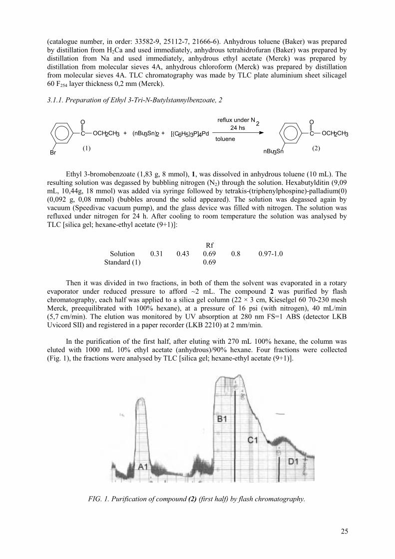
25
(catalogue number, in order: 33582-9, 25112-7, 21666-6). Anhydrous toluene (Baker) was prepared by distillation from H2Ca and used immediately, anhydrous tetrahidrofuran (Baker) was prepared by distillation from Na and used immediately, anhydrous ethyl acetate (Merck) was prepared by distillation from molecular sieves 4A, anhydrous chloroform (Merck) was prepared by distillation from molecular sieves 4A. TLC chromatography was made by TLC plate aluminium sheet silicagel 60 F254 layer thickness 0,2 mm (Merck).
3.1.1. Preparation of Ethyl 3-Tri-N-Butylstannylbenzoate, 2
C
O
OCH2CH3
Br
(nBu3Sn)2 [(C6H5)3P]4Pd+ + C
O
OCH2CH3
nBu3Sn
reflux under N 224 hs
toluene (1) (2)
Ethyl 3-bromobenzoate (1,83 g, 8 mmol), 1, was dissolved in anhydrous toluene (10 mL). The resulting solution was degassed by bubbling nitrogen (N2) through the solution. Hexabutylditin (9,09 mL, 10,44g, 18 mmol) was added via syringe followed by tetrakis-(triphenylphospine)-palladium(0) (0,092 g, 0,08 mmol) (bubbles around the solid appeared). The solution was degassed again by vacuum (Speedivac vacuum pump), and the glass device was filled with nitrogen. The solution was refluxed under nitrogen for 24 h. After cooling to room temperature the solution was analysed by TLC [silica gel; hexane-ethyl acetate (9+1)]:
Rf Solution 0.31 0.43 0.69 0.8 0.97-1.0
Standard (1) 0.69
Then it was divided in two fractions, in both of them the solvent was evaporated in a rotary evaporator under reduced pressure to afford ~2 mL. The compound 2 was purified by flash chromatography, each half was applied to a silica gel column (22 × 3 cm, Kieselgel 60 70-230 mesh Merck, preequilibrated with 100% hexane), at a pressure of 16 psi (with nitrogen), 40 mL/min (5,7 cm/min). The elution was monitored by UV absorption at 280 nm FS=1 ABS (detector LKB Uvicord SII) and registered in a paper recorder (LKB 2210) at 2 mm/min.
In the purification of the first half, after eluting with 270 mL 100% hexane, the column was eluted with 1000 mL 10% ethyl acetate (anhydrous)/90% hexane. Four fractions were collected (Fig. 1), the fractions were analysed by TLC [silica gel; hexane-ethyl acetate (9+1)].
FIG. 1. Purification of compound (2) (first half) by flash chromatography.

26
TABLE I. FRACTIONS FROM FIG. 1
Fraction Rf : TLC [silica gel; hexane-ethyl acetate (9+1)](A1) 0.93s (B1) 0.79s 0.68m 0.42m 0.30m(C1) 0.42s 0.30-0.21s (D1) 0.25s
standard (1) 0.68 Compound (2) 0.68 (1)s: strong, m: medium, w: weak, vw: very weak
In the purification of the second half, after eluting with 300 mL 100% hexane, the column was eluted with 1200 mL 7% ethyl acetate (anhydrous)/93% hexane. Four fractions were collected (Fig. 2), the fractions were analysed by TLC [silica gel; hexane-ethyl acetate (93+7)].
Because the fraction (B1) from the purification of the first half (Fig. 1) is not pure it was purificated again. After evaporated under reduced pressure to afford ~2 mL, it was applied to a silica gel column (22 × 3 cm, Kieselgel 60 70-230 mesh Merck, preequilibrated with 7% ethyl acetate (anhydrous)/93% hexane) and eluted with 7% ethyl acetate (anhydrous)/93% hexane. Four fractions were collected (Fig. 3), the fractions were analysed by TLC [silica gel; hexane-ethyl acetate (93+7)].
TABLE II. FRACTIONS FROM FIG. 2
Fraction Rf : TLC [silica gel; hexane-ethyl acetate (93+7)](A2) 0.90s (B2) 0.76s - (C2) 0.76vw 0.31s 0.14s (D2) 0.14s
standard (1) 0.61 Compound (2) 0.61 (1)s: strong, m: medium, w: weak, vw: very weak
FIG. 2. Purification of compound (2) (second half) by flash chromatography.

27
TABLE III. FRACTIONS FROM FIG. 3
Fraction Rf : TLC [silica gel; hexane-ethyl acetate (93+7)](B3) 0.76s -
Compound (2) 0.61 (1)s: strong, m: medium, w: weak, vw: very weak
Fractions B2 and B3 were joined. We obtained a liquid clear, with a little light green color. We obtained 1.983 gr (4,5 mmol) of compound (2), the yield was 56.4%.
3.1.2. Preparation of 3-tri-n-butylstannylbenzoic acid, 3
C
O
OCH2CH3
nBu3Sn
KOH+ C
O
OH
nBu3Sn
Ethanol absolute
reflux 22 hs(2) (3)
To a solution of 2 (1.029 g, 2.34 mmol) in absolute ethanol (20 mL) was added KOH (0.186 g, 3.32 mmol). The resulting solution was refluxed for 22 hs. The reaction was followed by chromatography [TLC silica gel; hexane-ethyl acetate (93+7)], TLC were made at 2, 6 and 22 hs until the reaction medium showed complete consumption of the starting material (compound (2) Rf=0.77-0.76). Chromatographic analysis by TLC silica gel; hexane-acetic acid (6+1) showed single spot at Rf=0.74. The solution was cooled and poured into an ice-cold solution of acetic acid (3.9 mmol, 0.235 g) and 37 mL water, pH5.6. The solution was then extracted with diethyl ether (3 × 25 mL). The ether phase was washed with water until pH 5 in the aqueous phase, dried over MgSO4 (2 g), filtered and evaporated under reduced pressure to afford 0.65 g, 1.58 mmol (67%) of 3.
FIG. 3. Purification of compound (2) (fraction B1) by flash chromatography.

28
3.1.3. Preparation of N-succinimidyl 3-tri-n-butylstannylbenzoate (ATE), 4
To a solution of 3 (0.650 g, 1.58 mmol) in anhydrous THF (16 mL) was added N,N’-dyclohexylcarbodiimide (DCC) (0.399 g, 1.93 mmol) followed by N-hydroxysuccinimide (NHS) (0.222 g, 1.93 mmol). The resulting solution was stirrer in an ice-water bath until the ice melts (approximately 3 h) and then 17 h more. The reaction was analysed by chromatography [TLC silica gel; hexane-ethyl acetate]:
Hexane (100-x) + ethyl acetate ×%
Rf
x = 7% 0.03 0.12 x = 25% 0.13w 0.19w 0.33s 0.64w 0.90w x = 50% 0.57w 0.76s 0.88w 0.92w
The reaction mixture was filtered to remove precipitated dicyclohexylurea, the solution concentrated under reduced pressure. A solution of 50% ethyl acetate(anhydrous)-hexane (5 mL) was added and the resulting solution was again filtered, the solution was analysed by TLC [TLC silica gel; hexane-ethyl acetate ]:
Hexane (100-x) + ethyl acetate
×%
Rf
x = 35% 0.33w 0.42w 0.54s - - 0.84w 0.93w x = 50% 0.54w 0.6w 0.73s 0.89w 0.93w
FIG. 4. Purification of compound (4) ATE by flash chromatography. UV profile at 280 nm, flow: 40 mL/min, chart speed: 2 mm/min.

29
TABLE IV. FRACTIONS FROM FIG. 4
Fractions Hexane + ethyl acetate (7+3) Rf
Hexane + ethyl acetate (9+1), Rf
fraction 1 - 0.5m 0.61w 0.81s 0-0.07 0.3 fraction 2 0.3w 0.36w 0.4-0.59s 0-0.2 fraction 3 0.3m 0-0.03
The solution was concentrated to ~3 mL under reduced pressure and ATE (4) was isolated by flash chromatography. This material was applied to a silica gel column (22 × 3 cm, Kieselgel 60 70-230 mesh Merck, preequilibrated with 35% ethyl acetate (anhydrous)/65% hexane) and eluted with the same solvent mixture (Fig. 4). Three fractions were collected and analysed by TLC to assess the purity of the samples.
Fraction 2 was concentrated to ~3 mL under reduced pressure and applied to a silica gel column (22 × 3 cm, Kieselgel 60 70-230 mesh Merck, preequilibrated with 21% ethyl acetate (anhydrous)/-79% hexane) and eluted with the same solvent mixture (Fig. 5). Four main fractions were collected and analysed by TLC to assess the purity of the samples.
TABLE V. FRACTIONS FROM FIG. 5
Fractions Hexane + ethyl acetate (7+3) Rf
fraction 1 0.84 fraction 2 0.78 fraction 3 0.50w 0.48s fraction 4 0.48s
FIG. 5. Purification of compound (4) ATE (fraction 2 fig.4) by flash chromatography UV profile at 280 nm, flow: 40 ml/min, chart speed: 2 mm/min.

30
The impure fraction 3 was concentrated to ~3 mL under reduced pressure and purificated again. It was applied to a silica gel column (22 × 3 cm, Kieselgel 60 70-230 mesh Merck, preequilibrated with 21% ethyl acetate (anhydrous)/79% hexane) and eluted with the same solvent mixture. The elution profile showed a single peak with a single spot at Rf=0.48 in TLC [TLC silica gel; hexane-ethyl acetate (7+3)].
This pure fraction was joined with fraction 4 (from Table V and Fig. 6), we obtained 469 mg (0.92 mmol), 58% yield of ATE, 4.
The asssigned structure is a complete agreement with the observed 1H-RMN spectra: (CDCl3,) 8,20 (s, 1H, C-2H), 8.07 (d, 1H, C-6H), 7.77 (d, 1H, C-4H), 7.44 (t, 1H, C-5H), 2.90 (s, 4H, CO-
CH2-CH2-CO), 1.67-0.92 (m, 27H, 3 × n-C4H9). Mass spectrum: m/e 452 (M+ - 57, 42.83%, 395 (M+ - 114, 12,50%). Copies of both spectra are attached at the final of this report.
3.2. Radioiodination of proteins (IgG) with SIB
The radioiodination of ATE (4) and Human IgG was made based on the paper published by Zalutsky M.R.,et al 2 .
3.2.1. Iodination (cold) of ATE, 4
Four standard solutions were prepared, as follows:
(a) NaI 1M: 300 mg of sodium iodide in 2 mL 0,01 N NaOH (recently prepared), (b) Acetic acid 1M: 300 mg of acetic acid glacial (290 µl) in 4,7 mL of chloroform (anhydrous). (c) t-butylhidroperoxide 1M: 100 µl of TBPH 5-6M (anhydrous in decane, Aldrich 41666-5) and a
few crystals of anhydrous Na2SO4 in 400 µl of chloroform (anhydrous). (d) ATE 0,1 M: 24 mg in 470 µl of chloroform anhydrous.
In a little conical vial the reagents were added: 20 µl (2 µmol of ATE 0,1M) of (d); 8µl of (a), (b) and (c), (8 µmol of each of them).
The mixture was stirred magnetically at room temperature for 90 min. The reaction was examined by TLC (silica gel, 30% ethyl acetate in hexane) at 35 and 90 min. At both reaction times the chromatography showed two spots at Rf = 0.48 and Rf = 0.24 of similar size.
The first Rf value is ATE, and the second one had the same value as N-succinimidyl-3-iodo-benzoate (Zalutsky, et al.2).
3.2.2. Radioiodination of ATE and human IgG
3Na*I , AcOH / CHCl
3TBPH / CHCl
*IO
O
O
O
C N
R Sn3
NC
O
O
O
O
ATE SIB
O
O
O
O
C N
I*
+ H2N protein proteinborate bufferpH 8.5
H
O
C N
I*

31
We made four radioiodination of ATE and labelling of Human IgG. Two of them with Na[125I] and the other two, with Na[131I]. Na[125I] was purchased from Dupont/NEN (NEZ-033H, as 0.1 N NaOH solution pH7-11, 17.5 Ci/mg, 371,7 mCi/mL ). Na[131I] came from our reactor-production plant, to assure alkaline pH we added one drop of 0,02 M NaOH into the vial.
In a little conical vial the reagents were added (in order) : 10 µl (1 µmol of ATE 0,1M) of (d), 1 mCi Na[125/131I] in a volume no greater than 30µl, 50µl (50 µmol of acetic acid) of (b), 50µl (50 µmol of TBPH) of (c).
The reaction mixture was stirred at room temperature for 30 min and 125/131I-SIB was isolated by chromatography over a Sep-Pak silica gel cartridge (Waters Classic WAT051900). The column was first saturated with hexane and the reaction mixture was loaded on the column with the help of 3 × 100 µl of hexane. Following the elution with 4 × 10 mL hexane, then 5 × 5 mL of 8% ethyl acetate (anhydrous) in hexane, and finally 30% ethyl acetate in hexane in eight fractions: 6 × 1 mL and 2 × 5 mL. The product 125/131I-SIB eluted in the fractions of 30% ethyl acetate in hexane.
The 30% ethyl acetate eluate was evaporated to ~1 mL with a stream of nitrogen, transferred to a eppendorf vial (1,5 mL total capacity) and evaporated to dryness with a stream of nitrogen. Human IgG (Sigma I-4506) 200 µg ( 200µg/50 µl) in 100 mM borate buffer pH8.5, was added and the mixture was stirrer in an ice-water bath for 30 min. The reaction was terminated by addition of 300 µlof 200 mM glycine in borate buffer pH8.5 followed by an incubation of 5 min at room temperature.
The 125/131I-labelled IgG was separated from other reactions products using a 1,2 × 5 cm (disposable 5mL syringe) Sephadex G-25 column, eluted with phosphate buffer 10 mM pH7.4, collecting 0,5 mL fractions, flow: 1,9 mL/min . Aliquots of the fractions were taken and counted to make the elution profile.
3.3. Radiaiodinationof a chemotactic peptide with SIB
In the second RCM of the CRP in Athens on 26-30 April 1999, it was agreed to perform most of the radiochemical initial development studies with the chemotactic peptide N-formyl-methionyl-leucyl-phenylalanine (fMLF).
HCONHCH(CH2CH2SCH3)CONHCH(CH2CH(CH3)2)CONHCH(CH2C6H6)COOH).
Although a sufficient reactivity of the acylating agent is of great importance, high basicity and low sterical hindrance of the amino component is essential for satisfactory yields. We chose the hexapeptide fNleLFNleYK (N-formyl-Nle-Leu-Phe-Nle-Tyr-Lys, Sigma F0267) since it has a free amino group, the lysine -amino group, which could be derived by reaction with an active ester like the N-succinimidyl-3-iodo-benzoate (SIB).
All reagents used were reagent grade or better and were used as purchased. 3-Iodobenzoic acid was obtained from Aldrich (catalogue number 13,858-4). Anhydrous tetrahydrofuran (Baker) was prepared by distillation from Na and used immediately, anhydrous ethyl acetate (Merck) was prepared by distillation from molecular sieves 4A; anhydrous chloroform (Merck) was prepared by distillation from molecular sieves 4A.
High performance liquid chromatography was conducted with an HPLC Waters Millennium equipped with a pump model 600, with a diode array detector. As radioisotope detector we used a gamma counter (crystal of NaI(Tl) ) with a loop of 200 µL, which was interfaced using an analogue interfaced to a LKB recorder 2210.

32
3.3.1. Synthesis of fNleLFNleYK-SIB conjugate
This compound was synthesized as a reference standard in the HPLC analysis and to be used in the competitive binding assays.
The N-succinimidyl-3-iodo-benzoate (unlabelled SIB) was prepared as described by Garg P.K. et al19 dissolving 3-iodobenzoic acid (IBA) (1 g, 4 mmol) in 50 mL of anhydrous tetrahydrofuran (THF) then N,N’-dyclohexylcarbodiimide (DCC) (1.03 g, 5 mmol) followed by N-hydroxy-succinimide (NHS) (0.575 g, 5 mmol) were added. The resulting solution is clear but in a few minutes a white precipitated of dicyclohexylurea appeared. The mixture was stirred at room temperature for 24 h. After cooling (refrigerator) the precipitated of dicyclohexylurea was filtered off and the solvent was evaporated in a rotatory evaporator. The desired compound was purified by flash chromatography. The residue was applied to a silica gel column (22x3 cm, Kieselgel 60 70-230 mesh Merck, pre-equilibrated with 10% ethyl acetate anhydrous in hexane) at a pressure of 2 bar (with nitrogen), 38 mL/min (5.4 cm/min). The elution was monitored by UV absorption at 254 nm FS=1 ABS (detector LKB Uvicord SII) and registered in a paper recorder (LKB 2210) at 2 mm/min. The column was eluted with 10% ethyl acetate (anhydrous) in hexane and finally with 30% ethyl acetate (anhydrous) in hexane. The SIB cold was obtained as a white crystalline compound mp. 153-154°C.This compound was used as reference standard in HPLC studies and in the synthesis of the fNleLFNleYK-IBA.
The hexapeptide fNleLFNleYK (N-formyl-Nle-Leu-Phe-Nle-Tyr-Lys, Sigma F0267) was derivatized by the reaction of a solution of the peptide in dimethylformamide (DMF) (60 mM, 20µL,1.2 µmol) with SIB in dimethylformamide (DMF) (120 mM, 20 µL, 2.4 µmol) and triethylamine (TEA) 4µL . The peptide had been solubilized by the addition of small crystals of LiCl. The reaction mixture was incubated at room temperature overnight. The desired product was isolated from the reaction mixture by HPLC with a reverse-phase column (Vydac 218TP5415, 150 × 4.6 mm, 5µ;sample capacity: at optimum resolution 1-200 µg, at practical range 0.2-10 mg) with the following elution condition: at a flow rate of 1 mL/min solvent A [acetonitrile/water (25/75) containing 0.1% TFA] over 10 min follow by 0 to 100% linear gradient of solvent B [acetonitrile/water (45/55) containing 0.1% TFA ] in solvent A over 20 min, follow by 100% solvent B up to the end of the HPLC run. The retention times for IBA, SIB, fNleLFNleYK and fNleLFNleYK-IBA were 9 (7-11), 22(21-23), 23(20-25), 38(37-39) min, respectively. But to avoid the presence of TFA at 32 min (before the elution of the desired fNleLFNleYK-IBA) we stop the flow, prime the pump line with water for 2 min at 10 mL/min then we wash the column for 28 min with 100% water at 1.5 mL/min, at 62 min we stop the flow again prime the pump line with acetonitrile 100% at 10 mL/min for 2 min, and finally we made the elution of the fNleLFNleYK-IBA with 100% acetonitrile a 1 mL/min. In this condition we eluted the fNleLFNleYK-IBA as a single peak at 70 min, this fraction was evaporated to dryness with a stream of nitrogen. We made this synthesis four times in order to obtain fNleLFNleYK-IBA enough to make the analysis to confirm the structure and all the programmed experiments. We obtained 0.62 mg (0.59 µmol), yield 49% as average, in each synthesis. We confirmed the structure by amino acid analysis, Applied Biosystems with hydrolizer Model 420, derivatization with PITC and separation of the PTC-amino-acids by liquid chromatography; and mass spectra. MS, m/z 1054 (MH+). Amino acid analysis: Leu = 1.03, Phe = 0.95, Tyr = 1.05, Nle = 1.91.
3.3.2. Synthesis of the radiodination agent [131I]SIB
(1) REACTION
The radioiodination of ATE was made based on papers published by Zalutsky, M.R.2,17,20.
We tested different amount of ATE and tert-butylhydroperoxide (TBPH), from 0.1 to 0.6 µmol and 0.2 to 15 µmol respectively. We found that the best condition for our laboratory facilities was: in a little (500 µl) conical vial 15 µl ethyl-acetate (anhydrous), 20 µL of 2% acetic acid/chloroform (anhydrous), 10 µL of ATE/CHCl3 (anhydrous) 10 mM (0,1 µmol), and 20 µL of TBPH/CHCl3

33
(anhydrous) 20 mM (0.4 µmol) were added to sodium [131I]-iodide. After 15-20 min reaction at room temperature [131I]SIB was isolated by HPLC.
Two different kind of labelling were made, with sodium [131I]-iodide at 5 µL (reaction in two phase) and with sodium [131I]-iodide evaporated to dryness in basic condition adding 5µL NaOH 0.02N (reaction in one phase). The Na[131I] came from our reactor production plant and from Nordion Canada.
Because of the adsorption of [131I]SIB in the vial and in the syringe used to transfer to the HPLC part of the activity was lost, for that reason we tried to change the solvent of the reaction. The idea was to try to improve the transfer of the activity to the HPLC without impair the reaction. We tested with ethyl acetate (anhydrous) and aprotic solvent like DMF and DMSO added to the one phase reaction.
(2) QUALITY CONTROL AND ISOLATION BY HPLC
After the reaction, the [131I]SIB was isolated and controlled by high-performance liquid chromatography. We used two different kind of column: silica column and reverse-phase column.
Only the first experiments were made with a Hypersilica Silica Hewlett-Packard 10 µ,150 × 4.6 mm eluted with ethyl-acetate/hexane/acetic acid (30/70/0.12). In this condition the retention time for [131I]SIB was 12.1 min and 9.5 for ATE. But after few runs we observed a progressive widening and overlapping of these two peaks. Due to this problem and because of the delay to receive a new column we decided to set up isolation proceeding by reverse-phase HPLC.
The reverse-phase HPLC was made with a column Vydac 218TP5415, 5µ, 150 × 4.6 mm., with the following elution condition: at a flow rate of 1 mL/min solvent A [acetonitrile/water (25/75)] over 12 min follow by 0 to 100% linear gradient of solvent B [acetonitrile/water (45/55)] in solvent A over 20 min, follow by 100% solvent B up to the end of the HPLC run. UV signal at 220 nm, 254 nm and radioactive signal were monitored. The retention times were: TBPH 7-19 min (very wide peak), IBA 11(9-13) min, SIB 24 (23-25) min, ATE does not elute from the column in this condition.
The HPLC fraction containing the [131I]SIB was evaporated for 15-20 min with a stream of nitrogen to remove the acetonitrile. This solution was passed through an activated Waters C18 Sep Pak cartridge (Waters Classic WAT051900), follow by 10 mL of air to dried it. The cartridge column was washed with 2 × 0.3 mL ethyl-acetate/hexane (20/80) to remove the remains of water and finally the [131I]SIB was eluted with 2 × 0.3 mL anhydrous chloroform. In this way we obtained the [131I]SIB in anhydrous condition. Chromatographic controls of the product were made.
3.3.3. Synthesis of fNleLFNleYK-[131I]SIB conjugate
The [131I]SIB/CHCl3 isolated in 3.3.2. was evaporated under a stream of nitrogen at room temperature, when the volume is 50 µL the activity was transferred in 5-10 µL volume at the bottom of a conical vial (Wheaton, 500 µL) and evaporated to dryness with a stream of nitrogen. To the [131I]SIB residue were added 5 µL (0.3 µmol) 60 mM of fNleLFNleYK in dimethylformamide and 1 µL of TEA. The peptide had been solubilized by the addition of small crystals of LiCl. After 5-15 min at room temperature the fNleLFNleYK-[131I]IBA was isolated from the reaction mixture by HPLC with a reverse-phase column (Vydac 218TP5415, 150 × 4.6 mm, 5µ) with the following elution condition: at a flow rate of 1 mL/min solvent A [acetonitrile/water (25/75) containing 0.1% TFA ] over 10 min follow by 0 to 100% linear gradient of solvent B [acetonitrile/water (45/55) containing 0.1% TFA ] in solvent A over 20 min, follow by 100% solvent B up to the end of the HPLC run. The retention times for [131I]IBA, [131I]SIB, fNleLFNleYK and fNleLFNleYK-[131I]IBA were 9 (7-11), 22(21-23), 23(20-25), 38(37-39) min, respectively.
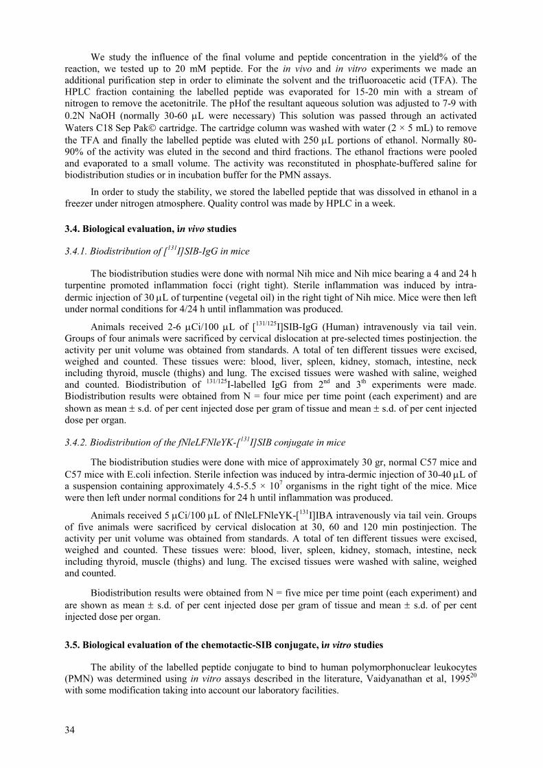
34
We study the influence of the final volume and peptide concentration in the yield% of the reaction, we tested up to 20 mM peptide. For the in vivo and in vitro experiments we made an additional purification step in order to eliminate the solvent and the trifluoroacetic acid (TFA). The HPLC fraction containing the labelled peptide was evaporated for 15-20 min with a stream of nitrogen to remove the acetonitrile. The pHof the resultant aqueous solution was adjusted to 7-9 with 0.2N NaOH (normally 30-60 µL were necessary) This solution was passed through an activated Waters C18 Sep Pak cartridge. The cartridge column was washed with water (2 × 5 mL) to remove the TFA and finally the labelled peptide was eluted with 250 µL portions of ethanol. Normally 80-90% of the activity was eluted in the second and third fractions. The ethanol fractions were pooled and evaporated to a small volume. The activity was reconstituted in phosphate-buffered saline for biodistribution studies or in incubation buffer for the PMN assays.
In order to study the stability, we stored the labelled peptide that was dissolved in ethanol in a freezer under nitrogen atmosphere. Quality control was made by HPLC in a week.
3.4. Biological evaluation, in vivo studies
3.4.1. Biodistribution of [131I]SIB-IgG in mice
The biodistribution studies were done with normal Nih mice and Nih mice bearing a 4 and 24 h turpentine promoted inflammation focci (right tight). Sterile inflammation was induced by intra-dermic injection of 30 µL of turpentine (vegetal oil) in the right tight of Nih mice. Mice were then left under normal conditions for 4/24 h until inflammation was produced.
Animals received 2-6 µCi/100 µL of [131/125I]SIB-IgG (Human) intravenously via tail vein. Groups of four animals were sacrificed by cervical dislocation at pre-selected times postinjection. the activity per unit volume was obtained from standards. A total of ten different tissues were excised, weighed and counted. These tissues were: blood, liver, spleen, kidney, stomach, intestine, neck including thyroid, muscle (thighs) and lung. The excised tissues were washed with saline, weighed and counted. Biodistribution of 131/125I-labelled IgG from 2nd and 3th experiments were made. Biodistribution results were obtained from N = four mice per time point (each experiment) and are shown as mean ± s.d. of per cent injected dose per gram of tissue and mean ± s.d. of per cent injected dose per organ.
3.4.2. Biodistribution of the fNleLFNleYK-[131I]SIB conjugate in mice
The biodistribution studies were done with mice of approximately 30 gr, normal C57 mice and C57 mice with E.coli infection. Sterile infection was induced by intra-dermic injection of 30-40 µL of a suspension containing approximately 4.5-5.5 × 10⁷ organisms in the right tight of the mice. Mice were then left under normal conditions for 24 h until inflammation was produced.
Animals received 5 µCi/100 µL of fNleLFNleYK-[131I]IBA intravenously via tail vein. Groups of five animals were sacrificed by cervical dislocation at 30, 60 and 120 min postinjection. The activity per unit volume was obtained from standards. A total of ten different tissues were excised, weighed and counted. These tissues were: blood, liver, spleen, kidney, stomach, intestine, neck including thyroid, muscle (thighs) and lung. The excised tissues were washed with saline, weighed and counted.
Biodistribution results were obtained from N = five mice per time point (each experiment) and are shown as mean ± s.d. of per cent injected dose per gram of tissue and mean ± s.d. of per cent injected dose per organ.
3.5. Biological evaluation of the chemotactic-SIB conjugate, in vitro studies
The ability of the labelled peptide conjugate to bind to human polymorphonuclear leukocytes (PMN) was determined using in vitro assays described in the literature, Vaidyanathan et al, 199520
with some modification taking into account our laboratory facilities.

35
In order to determine the effect of derivatization with SIB on the potency of the peptide and in the biological activity, competitive binding to PMN and superoxide production were made.
3.5.1. Cell preparation
Human PMNs were isolated using a density gradient centrifugation method as described in the literature (Vaidyanathan et al, 199520
; Immunology and Immunochemistry, Fundamentals; R.A.Margni, 5th Edition, Editorial Médica Panamericana, 1996), with the collaboration of the Faculty of Pharmacy and Biochemistry, Buenos Aires University.
ISOLATION PROCEDURE
(a) Blood collect: 30-40 mL of blood from normal voluntary was used in each experiment. We used sodium citrate 3.8% as anticoagulant in relation blood/anticoagulant 9:1. The initial count of total leukocytes present was made in a Neubauer camera making a dilution 1/20 of a sample of the blood with 3% acetic acid.
(b) Buffy-coat: in a 15 mL tube with 5% Dextran (PM 100,000-200,000) in phosphate-buffered saline pH7.4 (PBS) we added the blood in relation blood/dextran 1:5. Is important to add the blood slowly drop by drop in the centre of the tube. The cells were allowed to settle 45 min at room temperature. In this step normally 30% of the cells are lost. The supernatant was washed in PBS or culture media (RPMI 1640, Sigma R1383), pH7.4, and centrifuged twice at 400 × g for 10 min. The cellular pellet was resuspended in culture media.
(c) Gradient separation: we used Percoll density 1.130 ± 0.005 g/mL (Sigma P1644) and PBS to prepare two solutions of different densities (1.119 and 1.077 g/mL). The cells resuspended in culture media (4 mL) from step (2) was layered over Percoll gradients consisting of 3 mL of each of two densities (1.119 and 1.077 g/mL) in 10 mL conical tubes. PMNs were harvested from the interface between the two solutions following centrifugation at 1000 × g for 25 min at 20oC. The cells were washed in culture media (RPMI 1640, Sigma R1383), pH7.4, and centrifuged twice at 400 × g for 10 min. The cells were then resuspended in a small volume of incubation buffer (1.7 mM KH2PO4, 8.0 mM Na2HPO4, 117.0 mM NaCl, 0.15 mM CaCl2,0.5 mM MgCl2, 1 mM PMSF, pH7.4).
(d) Cell count and determination of cell viability: a sample of isolated cells from (3) was diluted 1:2 with 0.4% Trypan Blue and after 4 min they were count in a Neubauer camera. We count viable cells from death cells (stained cells). Finally cell suspension were diluted at a concentration of 1×10⁷ cells/mL. Cell preparation contained more than 95% PMNs as assessed by light microscopy of a sample of Giemsa-stained specimens. Cell yields of 35 × 106 - 50 × 106 cell from 40 mL of blood were obtained.
3.5.2. Competitive inhibition of [3H]fMLF binding
We made competitive inhibition of [3H]fMLF binding with fMLF, hexapeptide fNleLFNleYK (N-formyl-Nle-Leu-Phe-Nle-Tyr-Lys, Sigma F0267) and fNleLFNleYK-IBA (cold). This derivatized peptide was synthesized in our laboratory as we described in 3.3.1.
The derivatized peptide fNleLFNleYK-IBA has a poor solubility en aqueous solution. The addition of ethanol is necessary in order to make soluble the derivatized peptide. Because of this poor solubility the higher concentration of peptide we needed the higher concentration of ethanol was necessary. The highest concentration of ethanol that we needed to dissolve this highest concentration of peptide was 2.15% (v/v). This is the peptide concentration necessary in the determination of the non-specific binding, 1 × 10 –5 M.
Due to this problem we tested the influence of the ethanol in the binding process to the receptor in the cell.

36
We made six types of different competitive inhibition of [3H]fMLF binding assays with three different ligands fMLF, fNleLFNleYK and fNleLFNleYK-IBA:
(a) with fMLF, without ethanol (b) with fMLF, with 2.15% (v/v) of ethanol at all the different concentration of the fMLF used in
the assay (c) with fNleLFNleYK-IBA(cold), with 2.15% (v/v) of ethanol at all the different concentration of
the fNleLFNleYK-IBA(cold) used in the assay (d) with fNleLFNleYK-IBA(cold), with% (v/v) of ethanol as a function of the concentration of the
fNleLFNleYK-IBA(cold) used in the assay, between 0.65-2.15% (v/v) (e) with fNleLFNleYK, with 2.15% (v/v) of ethanol at all the different concentration of the
fNleLFNleYK used in the assay (f) with fNleLFNleYK, with% (v/v) of ethanol as a function of the concentration of the
fNleLFNleYK used in the assay, between 0.65-2.15% (v/v).
PROTOCOL
PMNs (106 per tube) were incubated for 45 min at 37 °C, under 95% O2- 5% CO2, in glass tubes with [3H]fMLF 10 nM (Dupont-NEN, NET563 lot 3345173, 60 Ci/mmol) in the absence (total binding) or presence of the indicated increasing concentration of the unlabelled ligands fMLF, fNleLFNleYK or fNleLFNleYK-IBA, and ethanol in the experiments (b), (c) and (e); in a total volume of 0.15 mL.
After incubation, the reaction mixture was diluted 1:20 with ice-cold incubation buffer (3 mL) to stop the binding process, and rapidly centrifuged (1000 × g, 10 min, 4oC) to separate cells from free ligand. The resulting pellet was washed in PBS, ice-cold pH7.4, and centrifuged three times at 400 × g for 10 min, 4oC. Finally the cellular pellet was dissolved in 0.2 mL of sodium dodecyl sulfate (SDS) 1% and transferred quantitatively into glass scintillation vials. To each vial 15 mL of Packard Ultima Gold scintillation cocktail was added and tritium activity was counted in a Packard Tricarb Model 2700TR liquid scintillation counter.
Non-specific binding was determined by co-incubating cells with the tracer [3H]fMLF and the presence of 10 µM of the same unlabelled ligand fMLF, fNleLFNleYK or fNleLFNleYK-IBA. This was subtracted from each concentration and the per cent specific binding was normalized to total binding.
3.5.3. fNleLFNleYK-[131I]SIB conjugate competitive binding assays
We made competitive inhibition of fNleLFNleYK-[131I]IBA binding with fMLF, hexapeptide fNleLFNleYK (N-formyl-Nle-Leu-Phe-Nle-Tyr-Lys, Sigma F0267) and fNleLFNleYK-IBA (cold). This derivatized peptide was synthesized in our laboratory as we described in 3.3.1.
The derivatized peptide fNleLFNleYK-IBA has a poor solubility en aqueous solution. The addition of ethanol is necessary in order to make soluble the derivatized peptide. Because of this poor solubility the higher concentration of peptide we needed the higher concentration of ethanol was necessary. The highest concentration of ethanol that we needed to dissolve the highest concentration of peptide was 2.15% (v/v). This is the peptide concentration necessary in the determination of the non-specific binding, 1 × 10 –5 M.
Due to this problem we tested the influence of the ethanol in the binding process to the receptor in the cell.
We made three types of different competitive inhibition of fNleLFNleYK-[131I]IBA binding assays with three different ligands fMLF, fNleLFNleYK and fNleLFNleYK-IBA:

37
(a) with fMLF, without ethanol (b) with fNleLFNleYK-IBA(cold), with% (v/v) of ethanol as a function of the concentration of the
fNleLFNleYK-IBA(cold) used in the assay, between 0.65-2.15% (v/v) (c) with fNleLFNleYK with% (v/v) of ethanol as a function of the concentration of the
fNleLFNleYK used in the assay, between 0.65-2.15% (v/v).
PROTOCOL
PMNs (106 per tube) were incubated for 45 min at 37°C, under 95% O2-5% CO2, in glass tubes with 0.5 µCi (in 3 µL) of fNleLFNleYK-[131I]IBA in the absence (total binding) or presence of the indicated increasing concentration of the unlabelled ligands fMLF, fNleLFNleYK or fNleLFNleYK-IBA, in a total volume of 0.15 mL.
After incubation, the reaction mixture was diluted 1:20 with ice-cold incubation buffer (3 mL) to stop the binding process, and rapidly centrifuged (1000 × g, 10 min, 4oC) to separate cells from free ligand. The resulting pellet was washed in PBS, ice-cold pH7.4, and centrifuged three times at 400 × g for 10 min, 4oC. The tubes were counted for 131I activity in a Packard Model Cobra II gamma counter.
Non-specific binding was determined by co-incubating cells with the tracer and the presence of 10 µM of the same unlabelled ligand fMLF, fNleLFNleYK or fNleLFNleYK-IBA. This was subtracted from each concentration and the per cent specific binding was normalized to total binding.
3.5.4. Superoxide production assay
We made superoxide production assays with fMLF and fNleLFNleYK-IBA (cold). This derivatized peptide was synthesized in our laboratory as we described in 3.3.1. The derivatized peptide fNleLFNleYK-IBA has a poor solubility en aqueous solution. The addition of ethanol is necessary in order to make soluble the derivatized peptide. Because of this poor solubility the higher concentration of peptide we needed the higher concentration of ethanol was necessary. The highest concentration of ethanol that we needed to dissolve the highest concentration of peptide was 2.10% (v/v). The highest concentration of peptide used in these experiments was 1.52 × 10⁻⁶ M.
Due to this problem we tested the influence of the ethanol in the binding process to the receptor in the cell.
We made four types of different assays with two ligands fMLF and fNleLFNleYK-IBA:
(a) with fMLF, without ethanol (b) with fMLF, with 2.10% (v/v) of ethanol at all the different concentration of the fMLF used in
the assay (c) with fNleLFNleYK-IBA(cold), with 2.10% (v/v) of ethanol at all the different concentration of
the fNleLFNleYK-IBA(cold) used in the assay (d) with fNleLFNleYK-IBA(cold), with% (v/v) of ethanol as a function of the concentration of the
fNleLFNleYK-IBA(cold) used in the assay, between <0.01–2.10% (v/v).
PROTOCOL
PMNs were resuspended in Hank`s balanced salt solution (HBSS; with Ca++ and Mg++ and without phenol red) at a density of 0.5 × 10⁶ cells/mL. Cytochalasin B (1mg/mL in DMSO) was added to a final concentration of 5 µg/mL and the cells were incubated for 15 min a 37°C. Seven hundred microlitres (0.7 mL) of the cell suspension was the added to reaction tubes containing varying concentrations of peptide plus cytochromo C (80 µM final concentration) in a total volume of 1.0 mL per reaction, and ethanol in experiments (b) and (c). The tubes were incubated at 37°C for 10 min, placed on ice to quench the reaction, and then centrifuged at 400 × g for 5 min to pellet the cells. The superoxide production was monitored by measuring the absorbance of the supernatant at 550 nm.
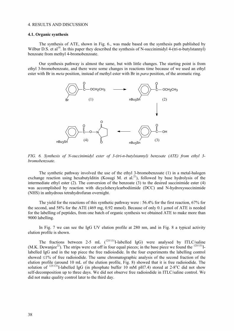
38
4. RESULTS AND DISCUSSION
4.1. Organic synthesis
The synthesis of ATE, shown in Fig. 6., was made based on the synthesis path published by Wilbur D.S. et al18. In this paper they described the synthesis of N-succinimidyl 4-(tri-n-butylstannyl) benzoate from methyl 4-bromobenzoate.
Our synthesis pathway is almost the same, but with little changes. The starting point is from ethyl 3-bromobenzoate, and there were some changes in reactions time because of we used an ethyl ester with Br in meta position, instead of methyl ester with Br in para position, of the aromatic ring.
C
O
OCH2CH3
Br
C
O
OCH2CH3
nBu3Sn
C
O
OH
nBu3Sn
C
O
O
nBu3Sn
N
O
O
(1) (2)
(4) (3)
FIG. 6. Synthesis of N-succinimidyl ester of 3-(tri-n-butylstannyl) benzoate (ATE) from ethyl 3-bromobenzoate.
The synthetic pathway involved the use of the ethyl 3-bromobenzoate (1) in a metal-halogen exchange reaction using hexabutylditin (Kosugi M. et al.21), followed by base hydrolysis of the intermediate ethyl ester (2). The conversion of the benzoate (3) to the desired succinimide ester (4) was accomplished by reaction with dicyclohexylcarbodiimide (DCC) and N-hydroxysuccinimide (NHS) in anhydrous tetrahydrofuran overnight.
The yield for the reactions of this synthetic pathway were : 56.4% for the first reaction, 67% for the second, and 58% for the ATE (469 mg, 0.92 mmol). Because of only 0.1 µmol of ATE is needed for the labelling of peptides, from one batch of organic synthesis we obtained ATE to make more than 9000 labelling.
In Fig. 7 we can see the IgG UV elution profile at 280 nm, and in Fig. 8 a typical activity elution profile is shown.
The fractions between 2-5 mL (125/131I-labelled IgG) were analysed by ITLC/saline (M.K. Dewanjee22). The strips were cut off in four equal pieces; in the base piece we found the 125/131I-labelled IgG and in the top piece the free radioiodide. In the four experiments the labelling control showed 1% of free radioiodide. The same chromatographic analysis of the second fraction of the elution profile (around 10 mL of the elution profile, Fig. 8) showed that it is free radioiodide. The solution of 125/131I-labelled IgG (in phosphate buffer 10 mM pH7.4) stored at 2-8oC did not show self-decomposition up to three days. We did not observe free radioiodide in ITLC/saline control. We did not make quality control later to the third day.

39
4.2. Radioiodination of proteins (IgG) with SIB
In Table VI the results of our four radioiodination experiments are shown.
TABLE VI. RESULTS OF FOUR RADIOIODINATION OF ATE AND LABELLING OF IgG
1st - Na[131I] 2nd- Na[131I] 3th- Na[125I] 4th- Na[125I] expressed as% of ( )
Initial activity mCi (1) 6.85 9.7 5.08 4.9 Loaded activity in the Sep Pak%, (2)
6.65 mCi 97.1% of
(1)
9.3 mCi 95.9% of
(1)
4.78 mCi 94.1% of
(1)
4.65 mCi 94.9% of
(1)
(1)
Washed activity with 100% hexane,%
15.8 17.1 10.1 10.8
Washed activity with 8% ethyl acetate,%
19.5 21.5 17.5 15.3
1 mL 0.35 0.7 1.8 1.2 1 mL 7 8.5 8.7 7.8 1 mL 20.1 21.6 28.7 26.7 1 + 1 mL 30.1 29.7 45.5 16.8
Eluted activity of 125/131I-SIB with 30% ethyl acetate, %
2 + 1 mL 32.2 32.1 50.3 21.7
3 + 1 mL 33.6 32.9 52 23.3 4 + 5 mL 36.4 37.9 56 28.9 9 + 5 mL 38.1 39.7 57.1 34
125/131I-SIB mCi evaporated (3)
2.53 mCi 38.1% of
(2)
3.7 mCi 39.7% of
(2)
2.72 mCi 57.1% of
(2)
1.58 mCi 34% of (2)
(2)
Activity transferred to reaction vial with IgG (4)
1.92 mCi 76% of (3) 28% of (1)
2.77 mCi 75% of (3) 29% of (1)
2.14 mCi 78.7% of
(3)42.1% of
(1)
1.38 mCi 87.2% of
(3)28.1% of
(1)Activity transferred and loaded into G25 column (5)
0.81 mCi 42% of (4)
1.27 mCi 46% of (4)
1 mCi 47% of (4)
0.95 mCi 69% of (4)
125/131I-SIB-IgG Final activity recovered between 2-5 mL elution profile
0.33 mCi 41% of (5) 4.8% of (1)
0.59 mCi 47% of (5) 6.1% of (1)
0.48 mCi 48% of (5) 9,5% of (1)
0.54 mCi 57% of (5) 11% of (1)
(1) expressed as% of initial activity (2) expressed as% of loaded activity in the Sep Pak (3) expressed as% of eluted activity from 30% ethyl acetate and evaporated (4) expressed as% of activity that react with IgG (5) expressed as% of activity loaded to G25 shepadex column
means: in the same vial (the elution was collected in the same vial)

40
FIG. 8. Typical elution profile 123/131I-labelled IgC.
4.3. Radioiodination of chemotactic peptides using SIB
4.3.1. Synthesis of fNleLFNleYK-SIB conjugate
We obtained 0.62 mg (0.59 µmol), yield 49% as average, in each synthesis, we made this synthesis four times in order to obtain fNleLFNleYK-IBA enough to make the analysis to confirm the structure and all the programmed experiments.
The structure was confirmed by mass spectra and amino acid analysis. MS, m/z 1054 (MH+). Amino acid analysis: Leu = 1.03, Phe = 0.95, Tyr = 1.05, Nle = 1.91.
The retention times for IBA, SIB, fNleLFNleYK and fNleLFNleYK-IBA were 9 (7-11), 22(21-23), 23(20-25), 38(37-39) min, respectively. In Fig. 9 we can see the HPLC profile after 20 min reaction between SIB and the peptide.
In Fig. 10 we can see the HPLC of a macro synthesis and isolation proceeding of fNleLFNleYK-IBA described in 3.3.1. In this condition (washing the column with water previous the elution of the derivatized peptide, as described in 3.3.1) the fNleLFNleYK-IBA eluted a 70 min.
0 2 4 6 8 10 12 1420
40
60
80
100
Shepadex G25 column, 5 x 1,2 cm , 200µg IgGTypical Elution Profile 125/131I-labeled IgG
activ
ity
ml
FIG. 7. IgG–UV elution profile, Shepadex G25 1.2 × 5 cm. 200 µg IgG Human/50 µL, flow: 1.9 mL/min, FS = 1 ABS, chart speed = 0.5mm/s.
20
40
60
80
100
0 1 2 3 4 5 6ml
% scale

41
FIG. 9. HPLC, UV 220 nm, after 20 min RT reaction of 0.18 µmol of fNleLFNleYK/DMF (60 mM, 3 µL) with 0.03 µmol of SIB/DMF (3 mM, 10 µL) and TEA 3 µL. (see condition of HPLC in 3.3.1).
FIG. 10. HPLC, UV 220 nm, of the representative experimental procedure of macro synthesis between fNleLFNleYK and SIB described in 3.3.1. The peak at 70 min is the fNleLFNleYK-IBA.

42
4.3.2. Synthesis of the radiodination agent [131I]SIB
In Fig. 11 we can see the yields for the radioiodination reaction at different concentration of ATE and the oxidant (TBPH). Each point become from only one reaction.
In Figs 12 and 13 we can see the chromatograms (UV and radioactive) of the synthesis of [131I]SIB. The retention times were: TBPH 7-19 min (very wide peak), IBA 11(9-13) min, SIB 24(23-25) min, ATE does not elute from the column in this condition.
The TBPH has a chromatographic behaviour, which gives wide peaks, than becomes wider at higher concentration. At more than 0.4 µmol of TBPH its peak could overlap the peak of SIB.
Based on the results of Fig. 11 and, taking into account the chromatographic behaviour of TBPH and SIB, we chose as the most appropriate the condition described in 3.3.2.1.
When radioactive concentration of the sodium [131I]-iodide was low and the required activity for the reaction had a volume 5-10 µL we made the evaporation of this solution en basic condition as described in 3.3.2.1.
Radiochemical yield of 55-85% in the one phase reaction and 40-55% in the reaction made in two phase were obtained. These values are the maximum and the minimun of the yield% obtained in all the experiments we made.
0 2 4 6 8 10 12 14 1630
40
50
60
70
80
ATE 0.1 µmol ATE 0.3 µmol ATE 0.6 µmol
yiel
d %
µµµµmol TBPH
FIG. 11. Dependence of radioiodination yields on oxidant concentration at different ATEconcentration. All reactions were made at room temperature 15 min, 50 µL final volume, with 20 µLof acetic acid 2% /CHCl3, and Na[131I] at 5 µL.

43
In the one phase reaction we tested the solvent effect on the radioiodination yield and the per centage of activity transfered from the reaction vial to the HPLC. We observed that adding ethyl-acetate to the reaction improved transfer of activity to the HPLC from 56-85% (without ethyl-acetate) to 90-94% with the added of 15 µL with the ethyl-acetate. We did not observe an statistically significant change in the radioiodination yield% by the addition of ethyl-acetate, DMF or DMSO.
The chromatographic control of the [131I]SIB isolated by reverse-phase HPLC as described in 3.3.2.2 showed that it has 15-25% of [131I]IBA. This per centage is function of the reaction type, being higher in the two-phase reaction. Chromatographic controls of the two phase radioiodination reaction made with an aliquot immediately after the reaction, and before to complete the isolation process with Sep Pak, showed it has 15-18% of [131I]IBA.
With the isolation process described in 3.3.2.2, we recovered 55-65% of the [131I]SIB produced in the radioiodination reaction, in anhydrous state (chloroform solution) and ready to make its conjugation to the peptide.
In Fig. 14 we can see a typical HPLC elution profile on a silica column of the synthesis of the radioiodination agent [131I]SIB that we made in the laboratory of Dr. M. Zalutsky, Duke University, USA.
FIG. 12. Reverse phase HPLC, UV 220 nm, of the synthesis of the radioiodination agent [131I]SIB as described in 3.3.2. In this case the Na[131]I was spiked with NaI (cold) to see the UV signal of the SIB.

44
0 5 10 15 20-0.5
0.0
0.5
1.0
1.5
2.0
2.5
-0.5
0.0
0.5
1.0
1.5
2.0
2.5
131SIB(76 %)
ATE
kcpm
UV, 254 nm radioactive
Abs
min
FIG. 14. Typical HPLC elution profile of the synthesis of the radioiodination agent [131I]SIBmade in the laboratory of Dr. M. Zalutsky, Duke University, USAHPLC: silica column Alltech 250 × 4.6 mm, hexane/ethyl acetate/acetic acid (70:30:0.12),isocratic 1 mL/min.
FIG. 13. Reverse phase HPLC, radioactive signal, of the same synthesis of the radioiodinationagent [131I]SIB described Fig. 12. Registered in a paper recorder(LKB 2210) at 100 mv, 0.1mm/s.

45
4.3.3. Synthesis of fNleLFNleYK-[131I]SIB conjugate
In Figs 15 and 16 we can see the chromatograms (UV and radioactive) of the synthesis of fNleLFNleYK-[131I]IBA. The retention times for [131I]IBA, [131I]SIB, fNleLFNleYK and fNleLFNleYK-[131I]IBA were 9 (7-11), 22(21-23), 23(20-25), 38(37-39) min, respectively.
In Table VII we can see the radiochemical yield% obtained for the reaction synthesis of fNleLFNleYK-[131I]IBA.
FIG. 15. HPLC elution profile, radioactive signal, of the synthesis of fNleLFNleYK-[131I]IBA described in 3.3.3. Registered in a paper recorder(LKB 2210) at 100 mv, 0.1mm/s.
fNleLFNleYK-[131I]IBA38.4 min
8 min
12 min

46
TABLE VII. RADIOCHEMICAL YIELD% OBTAINED FOR REACTION SYNTHESIS
[fNleLFNleYK] TEA Finalvolume
[fNleLFNleYK]final Yield%
60 mM, 5 µL 1 µL 6 µL 50 mM 59-75 60 mM, 5 µL 1.2 µL 7.5 µL 40 mM 50-71 60 mM, 5 µL 2.5 µL 15 µL 20 mM 35-61
In the stability study the fNleLFNleYK-[131I]IBA did not show radiation damage or self-decomposition up to 7 days. We did not observe free radioiodide or other more polar by-products than the labelled peptide in the HPLC runs. We did not make quality control later to the seventh day.
4.4. In vivo studies
4.4.1. Biodistribution of [131I]SIB-IgG in mice
In Figs 17-20 biodistribution results are shown (average from 2nd and 3rd experiments).
FIG. 16. HPLC elution profile, UV 220 nm, of the same reaction synthesis of fleLFNleYK-[131I]IBA showed in chromatogram Fig. 15.

47
FIG. 17. Biodistribution of 4 h, expressed as% injected dose per organ.
bloo
d
liver
sple
en
kidn
ey
stom
ach
inte
stin
e
thyr
oid
norm
al le
g
infe
cted
leg
lung
0
2
4
6
8
10
12
14
16 Normal mice - Inflamed miceBiodistribution 24 hr 131/125I-labeled IgG
normal Miceinflamed mice
%ID
FIG. 18. Biodistribution at 24 h, expressed as% injected dose per organ.

48
bloo
d
liver
sple
en
kidn
ey
stom
ach
inte
stin
e
thyr
oid
norm
al le
g
infe
cted
leg
lung
0
5
10
15
20
25
30
35
40
45
50 normal mice - inflamed miceBiodistribution 4 hr 131/125I-labeled IgG
normal miceinflamed mice
% ID
/g
FIG. 19. Biodistribution at 4 h, expressed as% injected dose per grame of tissue.
bloo
d
liver
sple
en
kidn
ey
stom
ach
inte
stin
e
thyr
oid
norm
al le
g
infe
cted
leg
lung
0
5
10
15
20
25
30
35
40
45
50 Normal mice - inflamed miceBiodistribution 24 hr 131/125I-labeled IgG
normal miceinflamed mice
% ID
/g
FIG. 20. Biodistribution at 24 h, expressed as% injected dose per grame of tissue.

49
The results of biodistribution of [131I]SIB-IgG in Figs 17-20 showed a very low thyroid and stomach uptake of radioiodine. These results indicate that the labelling of proteins using the ATE intermediate produced labelled proteins with a decreased rate of dehalogenation in vivo. This result is similar to that obtained by Zalutsky, et al 2.
The ratio of the%ID/g between normal and inflamed leg are 3.8 at 24 h, and 2.3 at 4 h. These results showed an accumulation in the inflamed leg.
In Fig. 20 we can see that in the inflamed mice, in almost all the tissues, the uptake is lower than in the normal mice. The reason could be the progressive accumulation of activity by the inflammation.
4.4.2. Biodistribution of fNleLFNleYK-[131I]SIB conjugate in mice
Tissue distributions of the labelled peptide in normal and infected mice at different times are shown in Figs 21-25.
In Figs 21 and 22 we can see that in the inflamed mice, in all the tissues, the uptake is lower than in the normal mice. This general trend is similar to that seen with [131I]SIB-IgG.
In normal mice we observed that highest uptake was seen in liver. With the same peptide, labelled with [18F]SFB (Vaidyanathan, G., Zalutsky, M., 1995 20), and studied in BALB/c mice, the highest uptake was seen in kidney. The remainder blood activity was higher than those for fNleLFNleYK-[18F]SFB conjugate20. In general the clearance of 131I, from most of the tissue, was slower than the clearance observed for the peptide labelled with [18F]SFB20. Factors such as lipophilicity and catabolism of label of the labelled peptide must be considered.
The thyroid uptake (%ID) was very low: 0.10±0.04, 0.11±0.05, 0.09±0.03 at 30, 60, 120 min respectively. These values are similar than those obtained from other labelled peptide, radioiodinated with SIB by the group of Dr. M. Zalutsky, the -Melanotropic peptides17 and Octreotide23.
In Figs 23 and 24 we can see that the higher ratio between infected and normal leg was observed at 60 min, these ratio are lower than those observed for [131I]SIB-IgG in its maximum (24 h).
In Fig. 25 we can see that the clearance from blood, and uptake, is lower for the infected mice.
Normal C57 Mice
0
5
10
15
20
25
Blood Liver Spleen Kidneys Stomach Intestine Lung
% ID/g
30 min60 min120 min
FIG. 21. Biodistribution of fNleLFNleYK[131I]-IBA in normal mice. Expressed as% injected doseper grame of tissue.

50
FIG. 22. Biodistribution of fNleLFNleYK[131I]-IBA in infected mice (4.5-4.55 × 10⁷ E.coli/mice. Expressed as% injected dose per gram of tissue.
FIG. 23. Ratio between infected and normal leg (%ID/g) of fNleLFNleYK[131I]-IBA uptake in infected mice.
FIG. 24. Relation between infected and normal leg (%ID) of fNleLFNleYK[131I]-IBA uptake in infected mice.

51
FIG. 25. Blood clearance of fNleLFNleYK[131I]-IBA in infected mice and normal mice.
4.5. Biological evaluation of the chemotactic-SIB conjugate, in vitro studies
4.5.1. Competitive Inhibition of [3H]fMLF and fNleLFNleYK-[131I]IBA binding
In order to determine the effect of derivatization with SIB in the biological activity of the peptide, we made competitive binding to human PMNs using the prototypical ligand [3H]fMLF and fNleLFNleYK-[131I]IBA as the tracers. In the next figures the inhibition on the binding of the tracers by three different ligands are shown. As we explained in 3.5, we made several types of binding assays to explore the effect of derivatization and the effect of the ethanol in the process.
Concentrations of the peptide required to produce a 50% inhibition of the tracer binding to human PMNs (IC50) are shown, too.
TABLE VIII. BINDING AFFINITY (nM, IC50) OF CHEMOTACTIC PEPTIDE ANALOGS (IC50:CONCENTRATION OF PEPTIDE REQUIRED TO PRODUCE 50% INHIBITION OF [3H]fMLF BINDING)
fMLF fNleLFNleYK fNleLFNleYK-IBA With ethanol (2.15% (v/v) ) 1418 (b) 118 (e) 78 (c)
Without ethanol or at the minimum possible concentration of ethanol
30 (a) 18 (f) 36 (d)
Vaidyanathan, Zalutsky, 1995 20
(fNleLFNleYK-SFB conjugate)28 10-12 40-50
(fNleLFNleYK-SFB conjugate)
Babich J.W., et al, 1993 24 20 1
TABLE IX. BINDING AFFINITY (NM, IC50) OF CHEMOTACTIC PEPTIDE ANALOGS (IC50: concentration of peptide required to produce 50% inhibition of fNleLFNleYK-[131I]IBA binding)
fMLF fNleLFNleYK fNleLFNleYK-IBA 26 11 68

52
As we can see in the figures and both tables, ethanol affects the binding process. For that reason it is important to compare experiments in the same condition or at least make them in the most similar condition.
The results of the experiments made without ethanol or at the minimum possible concentration of ethanol, showed that increasing concentrations of fNleLFNleYK-IBA displaced the binding of [3H]fMLF with an IC50 value of 36 nM, the highest of the three peptide tested, suggesting a reduction in binding affinity due to SIB conjugation.
The IC 50 for fNleLFNleYK was the lowest of the three peptides, so it has the highest affinity of all. We obtained an IC50 value for fMLF and fNleLFNleYK a little higher than to those reported by Vaidyanathan, Zalutsky (1995)20 and Babich, J.W., et al. (1993)24.
The results of the experiments with fNleLFNleYK-[131I]IBA, as the tracer, showed that the concentration of unlabelled derivatized peptide (fNleLFNleYK-IBA) required to displace 50% of the binding was approximately 2 times higher compared with the displacement of [3H]fMLF. Vaidyanathan, Zalutsky (1995)20 and Babich, J.W., et al. (1993)24 reported similar behavior (but 10 times higher); the IC50 for displacement of a [18F]-labelled peptide and [99mTc]-labelled peptide, respectively, by unlabelled peptide was higher than the value for the displacement of [3H]fMLF. Babich, J.W., et al. suggested that the increase in binding avidity could be explained if the radiolabelled species contains more than one peptide unite per Tc-glucoheptonate and binding is cooperative. But because of we used [131I]SIB as the radioiodination agent the formation of molecules with more than one peptide unit per 131I is not likely using this labelling reaction.
4.5.2. Superoxide production
Superoxide production was utilized as an indicator of biological activity.
In order to determine the effect of derivatization with SIB in the biological activity of the peptide, we made assays with fMLF and fNleLFNleYK-IBA. As we explained in 3.5, we made several types of assays to explore the effect of derivatization and the effect of the ethanol in the process.
The concentrations of the peptide that produce a 50% of a maximal response (IC50) are shown too.
As we can see in the figures and table, ethanol affects the process. For that reason it is important to compare experiments in the same condition or at least make them in the most similar condition.
We did not make yet the assay of superoxide production by the underivatized hexapeptide fNleLFNleYK, for that reason we are going to use bibliographic data to make the comparison. The results of the experiments, which were made without ethanol or at the minimum possible concentration of ethanol, indicated that SIB conjugation did not seem to compromise the efficacy of the hexapeptide for superoxide production.
FIG. 26. Effect of fMNL and ethanol on [3H] fMLF binding to human PMNs.

53
FIG. 27. Effect of fNleLFNleYK[131I]-IBA and ethanol on [3H] fMLF binding to human PMNs.
FIG. 28. Effect of fNleLFNleYK-IBA and ethanol on [3H] fMLF binding to human PMNs.
FIG. 29. Specific binding of [3H] fMLF binding to human PMNs. Effect of graded concentrations of unlabelled chemotactic peptide analogs at the same concentration of ethanol [2.15% (v/v)].

54
FIG. 30. Specific binding of [3H] fMLF binding to human PMNs. Effect of graded concentrations of unlabelled chemotactic peptide analogs at the same concentration of ethanol [2.15% (v/v)].
FIG. 31. Specific binding of fNleLFNleYK-[131I]IBA to human PMNs. Effect of graded concentrations of unlabelled chemotactic peptide analogs without ethanol or at the minimum possible concentration of ethanol.
FIG. 32. Superoxide production by fMLF and effect of ethanol in the process.

55
FIG. 33. Superoxide production by fNleLFNleYK-IBA. and effect of ethanol in the process.
FIG. 34. Superoxide production by fMLF and fNleLFNleYK-IBA. Effect of graded concentrations of unlabelled chemotactic peptide analogs without ethanol or at the minimum possible concentration of ethanol.
FIG. 35. Superoxide production by fMLF and fNleLFNleYK-IBA. Effect of graded concentrations of unlabelled chemotactic peptide analogs at the same concentration of ethanol [2.10% (v/v)].
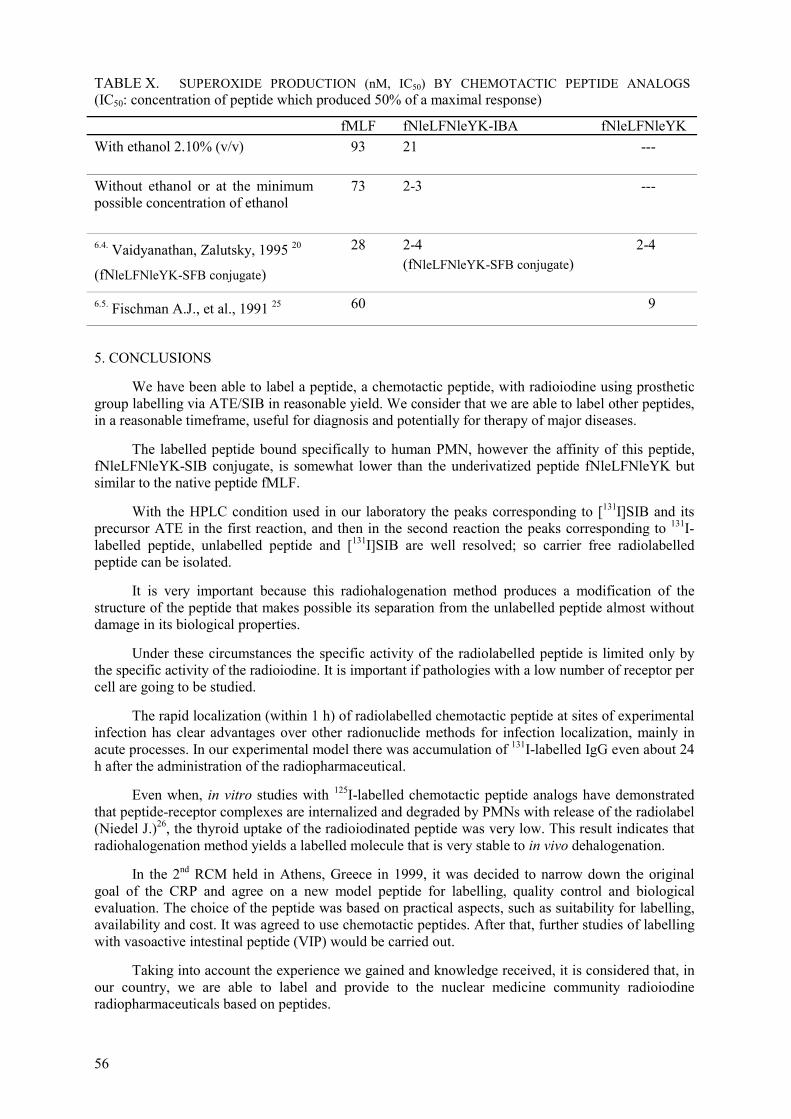
56
TABLE X. SUPEROXIDE PRODUCTION (nM, IC50) BY CHEMOTACTIC PEPTIDE ANALOGS (IC50: concentration of peptide which produced 50% of a maximal response)
fMLF fNleLFNleYK-IBA fNleLFNleYK With ethanol 2.10% (v/v) 93 21 ---
Without ethanol or at the minimum possible concentration of ethanol
73 2-3 ---
6.4. Vaidyanathan, Zalutsky, 1995 20
(fNleLFNleYK-SFB conjugate)
28 2-4 (fNleLFNleYK-SFB conjugate)
2-4
6.5. Fischman A.J., et al., 1991 25 60 9
5. CONCLUSIONS
We have been able to label a peptide, a chemotactic peptide, with radioiodine using prosthetic group labelling via ATE/SIB in reasonable yield. We consider that we are able to label other peptides, in a reasonable timeframe, useful for diagnosis and potentially for therapy of major diseases.
The labelled peptide bound specifically to human PMN, however the affinity of this peptide, fNleLFNleYK-SIB conjugate, is somewhat lower than the underivatized peptide fNleLFNleYK but similar to the native peptide fMLF.
With the HPLC condition used in our laboratory the peaks corresponding to [131I]SIB and its precursor ATE in the first reaction, and then in the second reaction the peaks corresponding to 131I-labelled peptide, unlabelled peptide and [131I]SIB are well resolved; so carrier free radiolabelled peptide can be isolated.
It is very important because this radiohalogenation method produces a modification of the structure of the peptide that makes possible its separation from the unlabelled peptide almost without damage in its biological properties.
Under these circumstances the specific activity of the radiolabelled peptide is limited only by the specific activity of the radioiodine. It is important if pathologies with a low number of receptor per cell are going to be studied.
The rapid localization (within 1 h) of radiolabelled chemotactic peptide at sites of experimental infection has clear advantages over other radionuclide methods for infection localization, mainly in acute processes. In our experimental model there was accumulation of 131I-labelled IgG even about 24 h after the administration of the radiopharmaceutical.
Even when, in vitro studies with 125I-labelled chemotactic peptide analogs have demonstrated that peptide-receptor complexes are internalized and degraded by PMNs with release of the radiolabel (Niedel J.)26, the thyroid uptake of the radioiodinated peptide was very low. This result indicates that radiohalogenation method yields a labelled molecule that is very stable to in vivo dehalogenation.
In the 2nd RCM held in Athens, Greece in 1999, it was decided to narrow down the original goal of the CRP and agree on a new model peptide for labelling, quality control and biological evaluation. The choice of the peptide was based on practical aspects, such as suitability for labelling, availability and cost. It was agreed to use chemotactic peptides. After that, further studies of labelling with vasoactive intestinal peptide (VIP) would be carried out.
Taking into account the experience we gained and knowledge received, it is considered that, in our country, we are able to label and provide to the nuclear medicine community radioiodine radiopharmaceuticals based on peptides.

57
REFERENCES
[1] WILBUR, D.S., Radiohalogenation of proteins: An overview of radionuclides, labelling methods, and reagents for conjugate labelling, Bioconjugate Chem. 3(6) (1992) 433-470.
[2] ZALUTSKY, M.R., NARULA, A.S., A method for the radiohalogenation of proteins resulting in decreased thyroid uptake of radioiodine, Appl. Radiat. Isot. 38(12) (1987) 1051-1055.
[3] ZALUTSKY, M.R., et al., Radiohalogenation of a monoclonal antibody using an N-Succinimidyl 3-(tri-n-butylstannyl) benzoate intermediate, Cancer Res. 48 (1988) 1446-50.
[4] GARG, P.K., ZALUTSKY, M.R., et al., Synthesis of radioiodinated N-succinimidyl iodobenzoate: optimization for use in antibody labelling, Appl. Radiat. Isot. 40(6) (1989) 485-90.
[5] ZALUTSKY, M.R., et al., Enhanced tumour localization and in vivo stability of a monoclonal antibody radioiodinated using N-Succinimidyl 3-(tri-n-butylstannyl) benzoate, Cancer Res. 49(1989) 5543-49.
[6] VAIDYANATHAN, G., ZALUTSKY, M.R., Protein radiohalogenation: observations on the design of N-Succinimidyl ester acylation agents, Bioconjugate Chem., 1 (1990) 269-273.
[7] VAIDYANATHAN, G., ZALUTSKY, M.R., Radioiodination of antibodies via N-Succinimidyl 2,4-dimethoxy-3-(tri-n-butylstannyl) benzoate, Bioconjugate Chem. 1 (1990) 387-393.
[8] SCHUSTER, J.M., ZALUTSKY, M.R., et al., Improved therapeutic efficacy of a monoclonal antibody radioiodinated using N-Succinimidyl 3-(tri-n-butylstannyl) benzoate, Cancer Res. 51(1991) 4164-69.
[9] VAIDYANATHAN, G., ZALUTSKY, M.R., Labelling proteins with Fluorine-18 using N-succinimidyl 4-[18F]Fluorobenzoate, Nucl. Med. Biol. 19(3) (1992) 275-281.
[10] VAIDYANATHAN, G., ZALUTSKY, M.R., et al., Radioiodination of proteins using N-succinimidyl-4-hydroxi-3-iodobenzoate. J.Lab.Comp.Radiopharmaceuticals 1993;32:378-380.
[11] GARG S., ZALUTSKY M.R.,et al. Acylation reagents for the radiohalogenation of peptides. J. Lab. Comp. Radiopharm. 32 (1993) 212-213.
[12] GARG, P.K., ZALUTSKY, M.R., et al., N-succinimidyl 4-methyl-3-(tri-n-butylstannyl) benzoate. Synthesis and potential utility for the radioiodination of monoclonal antibodies, Nucl. Med. Biol. 20(4) (1993) 379-387.
[13] GARG, S., ZALUTSKY, M.R., et al., N-succinimidyl 5-(trialkylstannyl)-3-pyridine carboxylates: a new class of reagents for protein radioiodination, Bioconjugate Chem. 2 (1991) 50-56.
[14] GARG, S., ZALUTSKY, M.R., et al., Radioiodination of a monoclonal antibody using N-succinimidyl 5-Iodo-3-pyridinecarboxylate, Nucl. Med. Biol. 20(7) (1993) 835-842.
[15] REIST, C., ZALUTSKY, M.R., et al., Radioiodination of internalizing monoclonal antibodies using N-succinimidyl 5-Iodo-3-pyridinecarboxylate, Cancer Res. 56 (1996) 4970-77.
[16] ZALUTSKY, M.R., Growth factor receptors as molecular targets for cancer diagnosis and therapy, Q. J. Nucl. Med. 41 (1997) 71-77.
[17] GARG, P.K., ZALUTSKY, M.R., et al., Enhanced binding and inertness to dehalogenation of -melanotropic peptides labelled using N-succinimidyl 3-Iodobenzoatem Bioconjugate Chem.
7 (1996) 233-239. [18] WILBUR, D.S., HADLEY, S.W., HYLARIDES, M.D., FRITZBERG, A.R., et al.,
Development of a stable radioiodinating reagent to label monoclonal antibodies for radiotherapy of cancer, J. Nucl. Med. 30 (1989) 216-226.
[19] GARG, P.K., ALSTON, K.L., ZALUTSKY, M.R., Catabolism of radioiodinated murine monoclonal antibody F(ab´)2 fragment labelled using N-succinimidyl 3-Iodobenzoate and iodogen methods, Bioconjugate Chem., 6(4) (1995) 493-501.
[20] VAIDYANATHAN, G., ZALUTSKY, M.R., Fluorine-18 labelled chemotactic peptides: a potential approach for the PET imaging of bacterial infection, Nucl. Med. Biol. 22(6) (1995) 759-764.
[21] KOSUGI, M., SHIMIZU, OHTANI, A., MIGITA, J., Palladium catalized reaction of hexabutylditin with arylbromides, preparation of negatively substituted aryltributyltin, Chem. Lett. (1981) 829-830.

58
[22] DEWANJEE M.K., Radioiodination: Theory, Practice and Biomedical Applications, Kluwer Academic Publishers, London.
[23] VAIDYANATHAN G., AFFLECK D., WELSH P., SRINIVASAN A., SCHIMDT M., ZALUTSKY M.R., Radioiodination and astatination of octreotide by conjugation labelling, Nuclear Med. & Biol. 27 (2000) 329-337.
[24] BABICH, J.W., SOLOMON, H., PIKE, M.C., KROON, D., GRAHAM, W., ABRAMS, M.J., TOMPKINS, R.G., RUBIN, R.H., FISCHMAN, A.J., Technetium-99m-labelled hydrazino nicotinamide derivatized chemotactic peptide analogs for imaging focal sites of bacterial infection, J. Nucl. Med. 34 (1993) 1964-1974.
[25] FISCHMAN, A.J., PIKE, M.C., KROON, D., FUCELLO, A.J., REXINGER, D., TENKATE C., WILKINSON, R., RUBIN, R.H., STRAUSS, H.W., Imaging focal sites of bacterial infection in rats with Indium-111 labelled chemotactic peptide analogs, J. Nucl. Med. 32 (1991) 483-491.
[26] NIEDEL, J., WILKINSON, S., CUATRECASAS, P., Receptor mediated uptake and degradation of 125I-chemotactic peptide by human neutrophils, J. Biol. Chem. 10 (1979) 700-710.

59
OPTIMIZATION OF SYNTHESES, QUALITY CONTROL PROCEDURES AND IN VITRO/IN VIVO EVALUATION OF 18F AND 123IRADIOPHARMACEUTICALS BASED ON PEPTIDES
E. BORTOLETI DE ARAÚJO, C. PAGANO, G. DA SILVA, N.P.S. DE PEREIRA, E. MURAMOTO, M.T. COLTURATO Instituto de Pesquisas Energéticas e Nucleares, São Paulo, Brazil
Abstract
Since the discovery of peptide receptors and synthesis of small, biologically active peptides, it has been recognized that these molecules can provide new approaches for radiopharmaceutical development. Radiohalogenation via prosthetic groups has provided a useful route for labelling proteins, especially those lacking tyrosyl groups in their structure. The ATE prosthetic group [N-succinimidyl 3-(tri-n-butylstannyl) benzoate] was synthesized in a two step reaction: tri-n-butyl tin 3-(tri-n-butylstannyl)-benzoate was synthesized from m-bromobenzoic acid in THF and n-butyllithium at -95oC, followed by the quenching the dilithio-anion with tri-n-butyl tin chloride. The stannyl ester obtained reacted with NHS and DCC in dry THF for 12 hours at room temperature and the reaction mixture was filtered and the product purified by flash chromatography to obtain ATE in a pure form. Iodination of ATE was performed with 131I and the radiochemical purity obtained was 72.22 ± 5.50 at pH3.0-4.0. The purification on Sep-Pack system indicated that about 40% of the initial radioactivity are recuperated in the 30% ethyl acetate in hexane fraction. The per cent of iodinated precursor depends of the TBHP concentration used in the reaction mixture. The coupling of radioiodinated ATE with the protein was studied using human IgG, (500 µg) and mildly alkaline conditions. After Sephadex G-25 purification step, about 40% of activity was isolated as labelled protein. The N-succinimidyl-p-radioiodobenzoate was also obtained using an alternative approach: p-Br-benzoic acid was first iodinated in DMSO and high temperature in a reaction catalyzed by CuCl. After Sep-Pack purification, the p-radioiodobenzoic acid reacts with TSTU to produce the succinimidyl esther. Direct labelling of IgG using Iodogen method showed a radiochemical purity of 80-90% determined by ITLC-SG in methanol 85%. IgG labelled by direct method and by ATE prosthetic group was administered in normal mice. The results from biological distribution studies showed that using direct labelling protein method, the per cent administered dose present on thyroid and stomach are greater, what suggest the presence of free iodine in high level when compared with indirect labelling method. Human IgG labelled with ATE prosthetic group results in a protein with reduced dehalogenation in vivo and represents an important alternative to the conventional protein labelling methods.
1. INTRODUCTION
During the last 20 years investigators in nuclear medicine have focussed on monoclonal antibodies (MoAbs) that are potentially ideal agents for a variety of applications in both benign and malignant disorders. Experience over the past decade has helped to elucidate the advantages and limitations of MoAbs [1,5].
In recent years the search for agents with specific targeting has led to a variety of molecules such as fragments, chimeric and humanized antibodies, immunoadhesins and single chain antigen binding proteins and variable domain peptide molecules. Since the discovery of peptide receptors and the synthesis of small, biologically active peptides, it has been recognized that these molecules can provide new approaches for radiopharmaceutical development. In many cancers an overexpression of receptors is observed which makes such receptors an attractive target for tumour imaging.
Receptors for somatostatin are expressed on a variety of human tumours and their metastases. Somatostatin has a very short biological half-life of several minutes, hampering sufficient receptor binding to allow adequate imaging. Rational design led to analogues with reduced metabolic degradation through peptidases as in the octapeptide octreotide [1,5].
One of the first clinically important applications was described by Bakker et al. [2], who reported on the localization of endocrine-related tumours with the Tyr-3-octreotide. By incorporating

60
a DTPA chelator at the N-terminal site of the octreotide molecule, pentetreotide was developed, which is used as the well-known 111-In-radiollabelled OctreoScan in diagnosis and staging of neuroendocrine tumours [3,6].
Vasoactive intestinal peptide (VIP) is a 28 amino acid peptide of the glucagon-secretin family that maintains a broad range of biologic activity mediated by cell surface membrane receptors. VIP is widely distributed throughout the body and various tumour cells express significantly higher amounts of VIP receptor as compared with normal peripheral blood cells or tissues. Iodinated VIP has been used successfully in the imaging and localization of intestinal adenocarcinomas and endocrine tumours [4].
The chemotatic peptide N-formyl-Met-Leu-Phe (N-For-MLF) is a bacterial product that binds to polymorphonuclear leukocytes and mononuclear macrophages, possibly via cell membrane receptors. Binding of this peptide or of many synthetic analogues initiates leukocyte chemotaxis [8].
Many other peptides and receptor systems have been investigated in experimental animals and in vitro studies and have been suggested as imaging agents [1,5,7].
Direct radioiodination is the method, which has been most frequently employed for labelling proteins with high specific activity.
Many scientific investigations have been carried out using eletrophilic iodination method with Chloramine-T [9], Iodogen [10] and Lactoperoxidase [11] as oxidant agents. It has been noted that even under mild oxidizing conditions some proteins lose a portion of their biologic activity, most likely from oxidation of thiols or from iodination of a tyrosine that is involved in a specific interaction such as binding [12]. The principal problem with the use of radioiodinated proteins is the in vivodehalogenation, presumably related to the structural similarity between the iodophenyl group and thyroid hormones.
Some prosthetic groups developed for indirect labelling of proteins as Bolton and Hunter [13] and Woods’s reagent [14], do not appear to alter biologic activity of the labelled proteins, but the in vivo use of these reagents has been found to be limited in some cases as a result of in vivodeiodination probably do to the substitution of the iodine ortho to a hydroxyl group on an aromatic ring as in the direct labelling.
Studies developed by Zalutsky et al [15] and Wilbur et al [16] demonstrated that antibodies can be radioiodinated using the synthesized precursor N-succinimidyl-(tri-n-butylstannyl)benzoate intermediate (ATE). ATE is the precursor, which is needed for the synthesis of radioiodinated SIB (N-succinimidyl iodobenzoate). The use of SIB reagent for protein labelling significantly reduced the thyroid uptake of radioiodine.
SIB can be also generated via Cu(I) catalyzed radioiodination of a Br-benzoic acid followed by the reaction with a N-succinimidyl derivative (TSTU) [17,18].
18F can be introduced in peptides via [18F]fluoroacylation using small prosthetic groups. [18F]Fluoroacylation via activated esters, such as p-nitrophenyl and succinimidyl esters of sterically small [18F]fluorocarboxylic acids, are well suited for this purpose [19].
2. MATERIALS, METHODS AND RESULTS
2.1. Radioiodination of IgG by direct method
- Preparation of iodogen tubes Iodogen (1mg) was dissolved in dichloromethane (1mL) and 10 to 50 µL of this solution
dispersed in the bottom of a polypropylene iodination vial and evaporated to dryness under nitrogen. These dried vials were stored at –85oC.

61
- Labelling procedures using iodogen as oxidant agent IgG solution (100µg /10µL phosphate buffer, 0.5M pH7.5) was added to the iodination vial
followed by Na131I solution (131I in NaOH 10-3 M 3.7MBq, 30-50µL). The iodination was allowed to proceed for 30 min at room temperature with gentle stirring; it was terminated by decanting the mixture from the reaction vial [10].
- Labelling procedures using lactoperoxidase/H2O2 as oxidant agent 5 µg of Lactoperoxidase and very low concentrations of H2O2 (5-20 µL) were mixed with
50 µg of IgG/10 µL of phosphate buffer pH7.0 and 131NaI (3,7MBq/20 µL) and gentle stirring for 15 min at room temperature [11].
- Radiochemical purity and purification The radiochemical purity of labelled IgG was determined by ITLC-SG chromatography using
methanol 85% as solvent (Rf Iodinated IgG= 0; 131I= 1.0) or by zone electrophoresis using Whatman 3MM paper, 0.05M barbital buffer pH8.6 and field of 150V for 40 min.
Purification of labelled IgG was accomplished by 1×10 cm Sephadex G 25 gel filtration column (Pharmacia) using PBS as eluent [15].
The radiochemical purity after 24 hours (using 10 µg iodogen) decrease to 85.02 ± 1.19%.
The labelled protein eluted from 4 to 6 mL and the free iodide from 11 to 13 mL in the Sephadex G25 purification step.
TABLE I. LABELLING IgG BY DIRECT METHOD: INFLUENCE OF IODOGEN CONCENTRATION
Iodogen (µg)/100 µg IgG % Radiochemical purity 10 88.14 ± 1.55 20 84.37 ± 0.81 50 83.47 ± 0.29
N = 5.
TABLE II. LABELLING IgG USING LACTOPEROXIDASE: INFLUENCE OF H2O2 CONCENTRATION
H2O2 concentration (nmol) % Radiochemical purity 0.036 10.84 ± 4.03 0.072 27.27 ± 10.89 0.183 78.78 ± 6.30 0.366 85.34 ± 1.03 0.730 85.58 ± 1.63 1.098 84.85 ± 0.99 1.46 85.66 ± 1.69 1.83 83.18 ± 0.62
N = 3.

62
2.2. Synthesis of ATE
(a) preparation of tri-n-butyl tin 3-(tri-n-butylstannyl)-benzoate [15]
- Using an appropriated apparatus, 2.5g of m-bromo benzoic acid was introduced to 50 mL of stirred THF. The temperature of this solution was decreased to about –100oC using liquid nitrogen and a silicon oil bath.
- 15.6 mL of n-butyllithium in hexane 1.6M was added over 30 min and the temperature was maintained less than –95oC.
- The mixture was then allowed to warm to –75oC for 30 min, following by quenching the dilithio-anion with 3.6 mL of tri-n-butyl tin chloride diluted in 6 mL of dry THF. After stirring for 30 min at –75oC, the reaction mixture was allowed to warm to room temperature and stirred for an additional 1.5 hour.
- After dilution with 100 mL of water, the organic part was extracted three times with about 100 mL of ether, washed with 5% aqueous NaOH (20 mL) and then water until the aqueous extract was neutral.
- The ether extract was dried over anhydrous Na2SO4 and solvent was evaporated - The product was purified by flash chromatography over silica gel, using an system similar to
described by Still et al (20). The column was previously saturated with hexane, the product was introduced and subsequently eluted with a mixture of hexane:ethyl acetate (70:30).
- The elution fractions were monitored by TLC (silica gel; hexane-ethyl acetate 70:30) - Solvent removed of the desired fraction gave 1.009g of the tri-n-butyl tin 3-(tri-n-butylstannyl-
benzoate as a clear liquid. - The product was analysed by HPLC (silica column, and hexane:ethyl acetate: acetic acid
70:30:0.2 as solvent) and by IR.
(b) Synthesis of N-succinimidyl 3-(tri-n-butylstannyl)-benzoate (ATE) [15]
- To 1.0g of stannyl ester obtained above were added 0,246g of NHS (N-hydroxysuccinimide) and 0.433g of DCC (dicyclohexylcarbodiimide).
- The mixture was stirred in 20 mL of dry THF for 12 hours at room temperature. - The reaction mixture was filtered to remove precipitated dicyclohexylurea. - Solvent removal followed by routine flash chromatography over silica gel using 10% ethyl acetate
in hexane gave 0.632g of purified ATE. - The final product was analysed by IR and the spectrum compared with IR spectrum from a
sample of ATE supplied by Dr. Zalutsky (Fig. 1). - The HPLC profile of both compounds were determined and can be compared on Fig. 2.
2.3. Labelling of ATE with 131I: SIB formation (a) Iodination of ATE (cold) [15] - the stocks solutions were prepared:
(1) – NaI 1M – 300mg NaI/2 mL NaOH 0.01N (2) – Acetic Acid (AA) 1M – 57µL AA/1.0 mL chloroform (3) – t-butylhydroperoxide (TBHP) 1M in chloroform and Na2SO4 (to remove water) (4) – ATE 0.1M – 5.1 mg/100µL chloroform
The reaction was conducted in a little conical vial:
8 µL (1) + 8 µL (2) + 8 µL (3) + 20µL (4)
The reaction mixture was stirred by 30 min at room temperature. TLC silica gel chromatography was performed in 30% ethyl acetate in hexane and showed two spots at Rf 0.48 and 0.24 as described by Zalutsky, et al. [15] that corresponds to ATE and SIB, respectively.
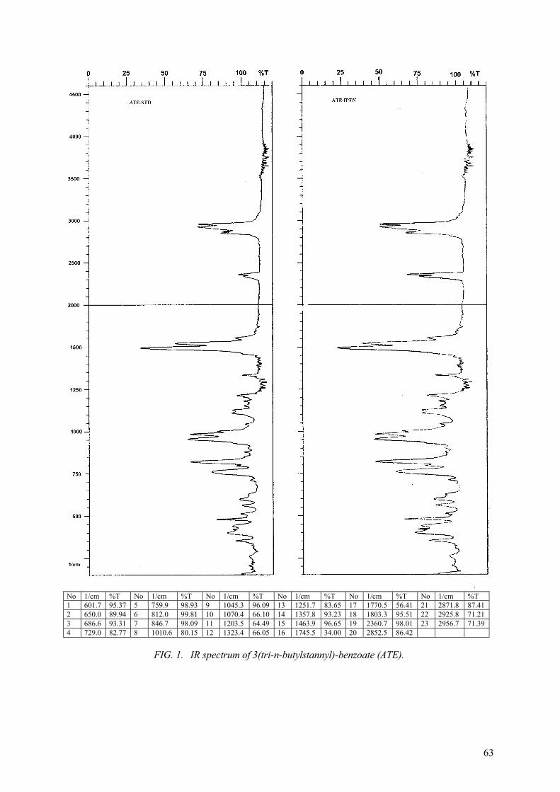
63
No 1/cm %T No 1/cm %T No 1/cm %T No 1/cm %T No 1/cm %T No 1/cm %T1 601.7 95.37 5 759.9 98.93 9 1045.3 96.09 13 1251.7 83.65 17 1770.5 56.41 21 2871.8 87.41 2 650.0 89.94 6 812.0 99.81 10 1070.4 66.10 14 1357.8 93.23 18 1803.3 95.51 22 2925.8 71.21 3 686.6 93.31 7 846.7 98.09 11 1203.5 64.49 15 1463.9 96.65 19 2360.7 98.01 23 2956.7 71.39 4 729.0 82.77 8 1010.6 80.15 12 1323.4 66.05 16 1745.5 34.00 20 2852.5 86.42
FIG. 1. IR spectrum of 3(tri-n-butylstannyl)-benzoate (ATE).

64
FIG. 2. HPLC profile from ATE: synthesized at IPEN (ATE-IPEN) and synthesized by Dr. Zalutsky laboratory (ATE-STD) – Silica column (10 µm); hexane:ethyl acetate:acetic acid (70:30:0.2) as solvent; 1 mL/min flux.
(b) Radioiodination of ATE using 131I
The reactions were performed using stock solutions as prepared to cold iodination. Na131Isolution (1) was prepared diluting Na131I in NaOH 0.01N (pH9-11) or in PBS (pH7.0)–3.7-7.4MBq/5µL.
The reactions were conducted in a conical vial: 5µL (1) + 50 µL (2) + 50 µL (3) + 10µL (4).
The influence of TBHP concentration and pHon labelling yield were determined. Radiochemical purity was determined using the TLC system described above.
TABLE III. RADIOIODINATION OF ATE: INFLUENCE OF TBHP CONCENTRATION
TBHP mols(×10-5) % SIB
1.8 20.20 3.6 51.44 5.0 78.63 ± 1.04 (N = 3)7.2 73.06 ± 5.75 (N = 3)14.4 70.24

65
TABLE IV. RADIOIODINATION OF ATE: INFLUENCE OF PH
PH % SIB % free iodide
3.0-4.0 74.51 ± 5.94 9.40 ± 2.90
5.0-5.5 69.48 ± 4.15 11.40 ± 2.23 N = 5.
(c) Sep-Pack purification of SIB
The reaction mixtures, which were prepared with pH3.0-4.0 and 5.0-5.5, were purified using the Sep-Pack system described by Zalutsky, et al. [15]: the reaction mixture were transferred to a Sep-Pack silica column pre-saturated with hexane. The columns were eluted with 40 mL hexane, 25 mL of 8% ethyl acetate in hexane and 16 mL of 30% ethyl acetate in hexane. The product 131I-SIB was eluted in the last fraction.
TABLE V. % OF INITIAL ACTIVITY IN SEP-PACK PURIFICATION FRACTIONS
Fraction Labelling mixtures at pH 5.0-5.5 (% of labelling activity)
Labelling Mixtures at pH3.0- 4.0 (% of labelling activity)
hexane 17.54 26.52 24.89 18.12 10.88 11.43 21.35
8% 11.50 7.28 11.70 21.40 28.0 22.04 25.08 30% 46.32 36.40 31.68 35.68 34.30 31.77 36.26 Sep-Pack 28.90 31.20 31.68 12.8 14.77 14.69 14.27 Reaction tube 4.20 4.68 0.12 9.17 11.81 19.07 1.45
The 30% ethyl acetate in hexane fraction were analysed by HPLC system previously described and only one peak with Rt 10.6 min were observed.
- N-SUCCINIMIDYL-P-RADIOIODOBENZOATE VIA CU(I) CATALYZED RADIOIODINATION
- Radioiododebromination [17]
The following reaction conditions were used:
p-Br-benzoic acid – 50 µL of 0.2M solution in DMSO 5 µl of Na131I – 3.7MBq (100µCi) Reaction time – 30, 45 and 60 min (Table VI) Reaction temperature – 155, 165 and 175oC (Table VII) CuCl – 100 µL of different concentration solutions in DMSO (Table VIII).
Radiochemical purity of these labelling conditions were determined using Whatmann 3MM paper and chloroform: acetic acid (9:1) as solvent mixture (Rf of free iodide = 0.0; Rf of p-radioiodobenzoic acid = 1.0).

66
- Separation of p-radioiodobenzoic acid
p-radioiodobenzoic acid was purified as described by Wester et al [18]: after pre-saturation of an RP-cartridge with water and fixation of the product, it was eluted with CH3CN.
The p-radioiodo benzoic acid purified were analysed by the HPLC system previously described and one major radioactivity peak with Rt = 5.17 min were observed (Fig. 3).
- Conversion of p-radioiodobenzoic acid to SIB using TSTU (18)\
131IO
OO
O131I TSTU/CH3CN
BASE N
O
O
TSTU=0-(N-succinimidyl)-N,N,N’,N’-tetramethyluronium tetrafluoroborate
The purified p-radioiodobenzoic acid recuperated in acetonitrile (300-500 µL) was transferred to an conical glass tube and 100µL of 0.2M TSTU in acetonitrile and 10 µL of 1 N NaOH solution were added. The reaction was conducted at 60oC for 2-30 min. The reaction mixtures were analysed by HPLC system described above and some radioactive peaks were observed, (Fig. 4 upper). The labelling mixtures were purified by Sep-Pack silica pre-saturated with hexane and eluted with 20 mL hexane and 3 × 10 mL of 30% ethyl acetate in hexane. The fractions were analysed by the same HPLC system and the first 10 mL fraction of 30% ethyl acetate in hexane exhibited one radioactivity peak with Rt 10.6 min (Fig. 4 down) probably related to the SIB because presents the same Rt that SIB produced by ATE labelling.
TABLE VI. p-RADIOIODOBENZOIC ACID: INFLUENCE OF REACTION TIME Labelling conditions: 165oC
CuCl: 100µL 0,1M solution/DMSO p-Br-Ar– 50 µL 0,2M solution/DMSO
5µL 131I-3.7MBq (100µCi)
Reaction time % p-radioiodobenzoic acid
30 68.28 ± 0.26 45 72.94 ± 3.49 60 79.48 ± 5.03
N = 3.
TABLE VII. p-RADIOIODOBENZOIC ACID: INFLUENCE OF REACTION TEMPERATURE Labelling conditions: CuCl: 100µL 0,1M p-Br-Ar – 50 µL 0,2M solution in DMSO
5 µL 131I - 3.7MBq (100µCi) 60 min
Temperature (oC) % p-radioiodobenzoic acid
155 81.59 ±1.26 165 82.57 ± 2.25 175 37.33 ± 8.82
N = 3.

67
TABLE VIII. p-RADIOIODOBENZOIC ACID: INFLUENCE OF CUCL/P-BR RATION
CuCl/p-Br % p-radioiodobenzoic ac.
10-1 82.57 ± 2,25 10-2 93.19 ±1.57 10-3 8.25 ± 1.41
N = 5.
FIG. 3. HPLC profile of the p-iodine [131I]benzoic acid after purification step.

68
FIG. 4. HPLC profile of SIB generated by p-radioiodine benzoic acid reacted with TSTU before (upper) and after (down) Sep-Pack purification step.

69
The influence of time reaction in the formation of SIB were observed in the HPLC profiles of labelling mixtures. The% SIB is not significantly increased after 5 min of reaction until 30 min. However, using only 2 min the SIB produced is <10% of the p-radioiodobenzoic acid initial activity.
The% SIB obtained with Sep-Pack purification in the first 10 mL of the 30% ethyl acetate in hexane elution fraction is about 40-50% of the p-radioiodobenzoic acid initial activity.
2.4. Labelling IgG with 131I-SIB
The 30% ethyl acetate fraction from Sep-Pack column elution was evaporated to 1.0 mL by passing a stream of N2 gas, transferred to a eppendorf vial and evaporated to dryness. Human IgG (Sandoglobulina–Sandoz) 500µg/50µL in 0.1M borate buffer (pH8.5 and 9.3) was added and the mixture was stirred in an ice bath for 30 min. The reaction was terminated by addition of 300µL of 0.2M glycine in borate buffer/5 min at room temperature. Radiochemical purity of the labelling was performed by ITLC silica gel using 85% methanol as solvent.
2.5. Biological distribution studies of labelled IgG
Biological distribution of labelled IgG by direct and indirect methods was performed in normal mice.
The 131I labelled IgG was purified by Sephadex G 25 column eluted with PBS.
TABLE IX. % IgG LABELLED WITH SIB X PH
pH % IgG labelled
8.5 40.42 ± 1.76 9.3 14.34 ± 1.67
N = 5.
TABLE X. % DOSE/ORGAN OF IgG LABELLED BY DIRECT METHOD IN NORMAL MICE
% Administered dose/organ Organ 1 hour 4 hours 24 hours
total blood 13.99 ± 1.31 6.68 ± 0.12 2.37 ± 1.15 kidneys 4.84 ± 0.06 2.95 ± 0.29 1.89 ± 0.88 liver 68.42 ± 5.00 40.54 ± 3.88 4.20 ± 0.89 heart 0.39 ± 0.07 0.20 ± 0.05 0.08 ± 0.04 spleen 2.53 ± 0.29 1.05 ± 0.25 0.43 ± 0.19 stomach 12.18 ± 0.70 8.56 ± 2054 0.95 ± 0.70 lung 1.77 ± 0.90 0.71 ± 0.36 0.14 ± 0.03 thyroid 1.15 ± 0.06 4.58 ± 3.20 11.50 ± 7.97 intestines 14.17 ± 1.42 17.69 ± 3.74 4.24 ± 2.31

70
TABLE XI. % DOSE/ORGAN OF IgG LABELLED BY ATE PROSTHETIC GROUP IN NORMAL MICE
% Administered dose/organ Organ 1 hour 4 hours 24 hours
total blood - 14.75 ± 0.32 6.21 ± 0.57 kidneys 12.82 ± 0.44 0.63 ± 0.01 1.00 ± 0.05 liver 49.04 ± 4.53 2.32 ± 0.33 1.00 ± 0.05 heart 3.42 ± 0.10 0.18 ± 0.01 0.27 ± 0.01 spleen 2.65 ± 0.05 0.12 ± 0.02 0.06 ± 0.01 stomach 2.82 ± 0.40 0.20 ± 0.01 0.11 ± 0.01 lung 16.35 ± 2.41 0.41 ± 0.10 0.19 ± 0.04 thyroid 0.03 ± 0.00 0.06 ± 0.03 0.03 ± 0.01 intestines 19.64 ± 0.11 1.94 ± 0.15 0.78 ± 0.12
TABLE XII. %DOSE/G OF IgG LABELLED BYDIRECT METHOD IN NORMAL MICE
% Administered dose/organ Organ 1 hour 4 hours 24 hours
kidneys 9.43 ± 0.20 5.52 ± 0.39 2.25 ± 0.94 liver 36.66 ± 2.85 22.91 ± 0.44 2.79 ± 0.90 heart 2.21 ± 0.20 1.01 ± 0.21 0.31 ± 0.14 spleen 14.18 ± 4.32 7.89 ± 1.66 4.29 ± 2.12 muscle 1.34 ± 0.26 0.70 ± 0.21 0.27 ± 0.19 stomach 25.25 ± 1.67 17.49 ± 4.76 3.46 ± 2.16 lung 3.75 ± 0.19 1.86 ± 0.34 0.54 ± 0.14 intestines 4.18 ± 0.48 5.45 ± 1.22 1.49 ± 0.88
TABLE XIII. % DOSE/G OF IgG LABELLED BYATE PROSTHETIC GROUP IN NORMAL MICE
% Administered dose/organ Organ 1 hour 4 hours 24 hours
kidneys 23.21 ± 2.30 1.17 ± 0.10 0.53 ± 0.04 liver 27.92 ± 0.16 1.20 ± 0.19 0.49 ± 0.07 heart 19.42 ± 0.76 0.95 ± 0.05 0.54 ± 0.04 spleen 18.57 ± 0.43 0.83 ± 0.01 0.45 ± 0.06 muscle 2.73 ± 0.47 0.23 ± 0.12 0.28 ± 0.12 stomach 4.47 ± 0.48 0.39 ± 0.08 0.17 ± 0.03 lung 49.20 ± 1.92 1.55 ± 0.18 0.75 ± 0.12 intestines 5.21 ± 0.29 0.61 ± 0.01 0.19 ± 0.03
3. CONCLUSIONS
The labelling IgG using the direct method with Iodogen as oxidant agent results in a good radiochemical yield (greater than 80%) using only 10µg of Iodogen.
The labelling of IgG using enzimatic method results in a good radiochemical yield using very low concentrations of H2O2. Sephadex G-25 elution of the labelled IgG using 1.83 nmol of H2O2 shows no evidence of protein degradation or agregation, with the same elution profile observed in the case of labelling with Iodogen.
The results obtained in the radioiododebromination showed that conversion yield is related to the time reaction, temperature and CuCl/p-Br ration; p-radioiodobenzoic acid can be obtained with a radiochemical purity greater than 90% and after purification about 83% of the initial

71
activity is recuperated; the reaction of p-radioiodo benzoic acid with TSTU results in SIB formation. After purification, about 40-50% of the initial p-radioiodo benzoic acid activity could be recuperated as SIB.
ATE was synthesized in three steps from m-bromo benzoic acid and purified resulting in a relatively good yield; the iodination of ATE produced SIB in a yield of about 40% of the initial radioiodine activity; labelling IgG with SIB resulted in a labelling protein yield of about 40% of the SIB initial activity.
The results from the biological distribution studies showed that using direct method for radioiodination of IgG, the% administered dose present on thyroid and stomach are greater, what suggest the presence of free iodine in high level when compared with the animals that received IgG labelled by indirect SIB method.
REFERENCES
[1] BLOK, D., et al., Peptide radiopharmaceuticals in nuclear medicine Eur. J. Nucl. Med. 26(1999) 1511-1519.
[2] BAKKER, W.H., et al., Receptor scintigraphy with a radioiodinated somatostatin analogue: radiolabelling, purification, biologic activity, and in vivo application in aninmals, J. Nucl. Med. 31 (1990) 1501-1509.
[3] VIRGOLINI, I., et al., In vitro and in vivo studies of three radiolabelled somatostatin analogues: 123I-Octreotide (OCT), 123I-Tyr-OCT and 111In-DTPA-D-Phe-1-OCT, Eur. J. Nucl. Med. 23 (1996) 1388-1399.
[4] VIRGOLINI, I., et al., Vasoactive Intestinal Peptide receptor scintigraphy, J. Nucl. Med. 36(1995) 1732-1739.
[5] FISCHMAN, A.J., et al., A ticket to ride: peptide radiopharmaceuticals, J. Nucl. Med. 34(12) (1993) 2253-2262.
[6] BAKKER, W.H., et al., [111In-DTPA-D-Phe1]-Octreotide, a potential radiopharmaceutical for imaging of somatostatin receptor-positive tumours: synthesis, radiolabelling and in vitrovalidation, Life Sciences 49 (1991) 1583-1591.
[7] LISTER-JAMES, J., et al., Small peptides radiolabelled with 99mTc, Q.J. Nucl. Med. 40 (1996) 221-233.
[8] BABICH, J.W., et al., Technetium-99m-labelled hydrazino nicotinamide derivatized chemotactic peptide analogs for imaging focal sites of bacterial infection, J. Nucl. Med. 34(1993) 1964-1974.
[9] HUNTER, R.M., GREENWOOD, F.C., Preparation of iodine-131I labelled human growth hormone of high specific activity, Nature, 194 (1962) 495 pp.
[10] SALACINSKY, P.R.P., et al., Iodination of proteins, glycoproteins and peptides using a solid-phase oxidizing agente, 1,3,4,6-Tetrachloro-3 , 6 -diphenyl glycoluril (iodogen), Anal. Biochem. 117 (1981) 136-146.
[11] ANGELBERGER, P., et al., Radiopharmaceutical development of 123I-vasoactive intestinal peptide (VIP) for receptor scintigraphy in oncology, J. Label. Compd. Radioph. 37 (1995) 502-503.
[12] WILBUR, D.S., et al., Development of a stable radioiodinating reagent to label monoclonal antibodies for radiotherapy of cancer, J. Nucl. Med. 30 (1989) 216-226.
[13] BOLTON, A.E., HUNTER, W.M., The labelling of proteins to I-125 containing acylating agent, Biochem. J. 133 (1973) 529-539.
[14] WOOD, F.T., WU, M.M., GERHART, J.C., The radioactive labelling of proteins with an iodination reagent, Anal. Biochem. 69 (1975) 339-349.
[15] ZALUTSKY, M.R., NARULA, A.S., A method fvor the radiohalogenation of proteins resulting in decreased thyroid uptake of radioiodine, Appl. Radiat. Iso. 38(12) (1987) 1051-1055.
[16] WILBUR, D.S., et al., Development of a stable radioiodinating reagent to label monoclonal antibodies for radiotherapy of cancer, J. Nucl. Med. 30 (1989) 216-226.

72
[17] MOERLIN, S.M., Regioespecific aromatic radioiodination via n.c.a. copper (I) assisted iododebromination, Radioch. Acta 50 (1990) 55-61.
[18] WESTER, H.J., et al., 18F- and 131I labelling of the octadecapeptide Apamin: a selective blocker of Ca+2 dependent K+ channel. Synthesis and in vivo evaluation in NMRI mice, J. Label. Compds. Radiopharm. 37 (1995) 513-515.
[19] STILL, W.C., KAHN, M., MITRA, A., A rapid chromatographic technique for preparative separations with moderate resolution, J. Org. Chem. 43(143) (1978) 2923-2925.
[20] GUHLKE, S., COENEN, H.H., STOCKLIN, G., Fluoroacylation agents based on small n.c.a. [18F]Flurocarboxylic acids, Appl. Radiat. Iso. 45(6) (1994) 715-727.

73
RADIOIODINATION OF VASOACTIVE INTESTINAL PEPTIDE (VIP)
Y. WANG, L. WANG, D. YIN Academia Sinica, Shanghai Institute of Nuclear Research, Radiopharmaceutical Centre, Shanghai, China
Abstract
In recent years, increasing biochemical and radiochemical research has been performed to develop radiolabelled peptides as specific ligands for tumour associated receptors. VIP, a 28-amino acid peptide containing two tyrosines and three lysines, has demonstrated that various tumour cells express significantly higher amounts of VIP-receptors[1] and could be applied to the clinic diagnosis. For these purposes, radiohalogenation of VIP by direct and indirect method was studied. Direct labelling works well for radioiodine but is limited to dehalogenation of labelling products in vivo[2]. Conjugate labelling methods including Bolton-hunter and wood reagents were developed but introduction of such a molecule to peptides may lead to the decrease of biological activity in vivo[3]. In order to resolve these problems, N-Succinimidyl-3-(tri-n-butylstannyl) benzoate (ATE) was elected for the radioiodination of VIP and already employed to radioiodination of IgG successfully[4]. The in vitro stability and biological activity would be compared in these two methods. Vasoactive intestinal peptide (VIP) and human immunoglobulin (IgG) were radioiodinated by direct and indirect methods. Iodogen was employed in direct method and N-Succinimidyl-3-(tri-n-butylstannyl) benzoate (ATE) was applied as a prosthetic group in the conjugation labelling. The subject of our study was optimizing the radiohalogenation of IgG and VIP followed by separation and analysis of reaction products. The advantages and disadvantages were illustrated by comparing the in vitro stability and biological activity in these two methods. Na123I was prepared by nuclear reaction of 124Te(p, 2n)123I using cyclone-30. More than 95% of radiochemical purity, more than 95% of radionuclide purity and about 100 mCi/mL of radioactivity concentration were obtained. ATE was supplied by Dr. Pozzi and radioiodinated with iodogen and 96% of labelling efficiency was obtained. The stability of radioactive S125IB kept well in dark at 4 . Human IgG was radiolabelled by direct (iodogen) and indirect method (ATE precursor), 96% and 77% of labelling efficiency were obtained, respectively. Optimization of labelling conditions and biological activity was studied. More than 76% of 125I-IgG kept active. The emphasis focused on the radiolabelling of VIP. The optimization of labelling conditions, various kinds of analysing and isolation methods, stability and biological activity were investigated in our Lab. The results showed that iodogen oxidant worked well with 69% of labelling efficiency. S125IB could be conjugated with VIP easily, yielding 75%. HPLC was an effective method to separate radiolabelled VIP from other compounds. 125IBA-VIP showed better in vitro stability than 125I-VIP. The primary studies showed no significant difference in biological activity. We can conclude that iodogen was an efficient oxidant for I- to I+ orI2, which can react with ATE, IgG and VIP. As a prosthetic group, ATE could connect radioactive iodine and proteins or peptides and worked well. So the conjugation of S125IB and IgG or VIP was easy to perform. The biological activity of products in direct and indirect labelling was nearly of the same. But the indirect product showed better stability than direct labelled product. The labelling of VIP with iodine-123 instead of iodine-125 should not affect the drawed conclusion. We will make further study to complete the synthesis of 123I-VIP.
1. INTRODUCTION
Radiohalogenation of VIP by direct and indirect method was studied to develop radiolabelled peptides as specific ligands for tumour associated receptors. We selected N-Succinimidyl-3-(tri-n-butylstannyl) benzoate (ATE) for the radioiodination of VIP and employed it to radioiodination of IgG successfully.
Our whole work included of preparation of Na123I, synthesis of ATE and radioiodination of proteins and peptides.
2. MATERIALS AND METHODS:
2.1. Materials
(1) Vasoactive intestinal peptide (VIP), iodogen were purchased from Sigma. N-hydroxysuccinimide (NHS), N-chlorosuccinimide(NCS), m-bromobenzoic acid, tri-n-butyl tin

74
chloride, dicyclohexyl carbodiimide (DCC) were purchased from Fluka. Na125I (361mCi/mL, >99.95%) was from Amersham. phenylmethyl sulfonylfluouride (PMSF) was from Amersco, oxalyl chloride was from Fluka.
(2) N-Succinimidyl-3-(tri-n-butylstannyl) benzoate (ATE) was provided by Prof. Pozzi. (3) Sephadex G-25(fine) and G-10 (fine) were purchased from Fluka. (4) Triethylammonium-formiate (TEAF) was prepared by neutralizing 50% formic acid and
triethylamine in our lab as Alexander wildman [5]. (5) SGC7901 cell lines was obtained from Shanghai Institute of Cell Biology and cultured in
RPMI 1640 medium. (6) m-iodobenzoic acid (IBA), Tetrahydrofuran(THF), LiAlH4, pyridine, triethylamine(TEA),
formic acid, dimethylfomamide(DMF), bovine serum albumin (BSA) and other reagents were purchased inland as analytical grade while CH3CN as HPLC grade .
2.2. Methods
2.2.1. General
(1) Thin layer chromatography (TLC) was carried out on silica gel GF254 and paper chromatography (PC) using Xinhua 1# paper.
(2) Paper electrophoresis (PE) was accomplished using Whatman 3MM paper with 0.1 M barbital buffer (pH8.6) at 300 V.
(3) All results of TLC, PC and PE were examined on multiple channel pulse analyser with NaI(T1) detector.
(4) High pressure liquid chromatography (HPLC) was performed on double 501 pump equipped with a Waters 486 UV spectrophotometer and FDG-101 -ray flow detector in series. The HPLC separation system consisted of Vydac C18 column (10µm, 2.5×250 mm, German) and eluted with TEAF (0.25 mol/l, pH3) in CH3CN at a flow rate of 1mL/min. Waters 990 Photodiode Array detector was used and gave the UV spectrum of peptides.
(5) Reversed phase chromatography (RPC) was carried out with Sep-Pak C18, the eluant of TEAF/ CH3CN and radioactivity detector was same as indicated in HPLC.
(6) Preparative column flash chromatography was accomplished with silica gel, 200-300 mesh. (7) Size exclusion chromatography (SEC) was performed on Sephadex G-25 (10×250 mm) and
Sephadex G-10 (8×300 mm) column.
2.2.2. Preparation of 123I-NaI
The 123I-NaI is produced by nuclear reaction of 124Te(p, 2n)123I using cyclone 30 made by IBA Co. in Belgium.
The specifications for the 123I-NaI are, as follows:
Radionuclide purity: >95% Radiochemical purity: >95% Radioactivity concentration: ~100 mCi/mL pH 7.0 ~ 9.0
This product is suitable for the labelling of peptides and has been successfully used for the labelling of MIBG.
2.2.3. Synthesis of ATE
The synthesis procedure of ATE is the following (Zalutsky, [4]):

75
COOH
Br
n-BuLi/THF
-100 ¡æ
COOLi
Li
(n-Bu) 3SnCl
COOSn(n-Bu) 3
Sn(n-Bu) 3
NHSDCC
Sn(n-Bu) 3
CO-O-N
O
O
The synthesis system was set up in accordance with the following procedure:
A solution of m-bromobenzoic acid (1.25 g) in dry tetrahydrofuran (THF, 35 mL) was placed in a 100 mL reaction bottle, maintained under nitrogen atmosphere and cooled to –95°C using ether-liquid nitrogen bath while stirring. To the above, 7.8 mL of n-butyllithium(1.6 mol/L hexane) was added slowly over 30 min while stirring and maintaining the temperature at –95°C. At this stage yellow precipitate was observed. The dilithio-anion thus generated was stirred at the temperature of –75°C for an additional 30 min. The tri-n-butyl tin chloride (2.73 mL) in 10 mL dry THF was added slowly to the reaction mixture over 30 min, stirring for an additional 30 min while keeping the temperature at –75°C. The cooling bath was removed and the reaction mixture was slowly brought to room temperature and allowed to stir for 1.5 hours. The solution is colourless. The reaction was quenched by addition of 50 mL of water. After extracting it three times with 20 mL diethyl ether, the combined organic phases were washed with 10 mL 5% NaHCO3 solution and H2O (3×100 mL ) till neutral. The organic phase was dried over MgSO4 and concentrated on a rotary evaporator. Routine flash chromatography over silica gel (200-300 mesh) of the residual oil gave the desired compound, trimethyl-stannyl-3-(tri-butylstannyl) benzoate, in 27% yield as an oil (colourless and turned to turbid if exposed to air; The oil is volatilizable). TLC analysis show Rf value 0.4 (30% ethyl acetate as developing agent) while the Rf of tributyl tin chloride approach to 1 and 0 for m-bromobenzoic acid.
2.2.4. Radioiodination of proteins and peptides
(1) Radioiodination of ATE (iodogen method)
Synthesis of cold SIB
(NHS)
Added 0.35 g pyridine to a flask with 1 g m-iodobenzoic acid (IBA) in toluene and stirred. Then added dropwise 0.42 mL oxalyl chloride, stirred for 30min at room temperature. The solution was cooled to room temperature after heating to remove excess oxalyl chloride, to which 0.46g N-hydroxysuccinimide was added and refluxed for 30min. The product was separated through silica gel-flash chromatography (from 100% petroleum ether to 70% petroleum ether and 30% ethyl acetate). 0.86g white solid of SIB was obtained in yielding of 65%.

76
1HNMR: (FIG. 1) 2.95 (s, 4H, –CO–CH2CH2–CO–); 7.24 (t, 1H, C4-H); 8.01 (d, 1H, C5-H); 8.16 (d, 1H, C6-H); 8.50 (s, 1H, C2-H)
IR(KBr): (FIG. 2) max(cm-1): 1770, 1733, 1596, 1563, 1471, 1421, 1413, 1214, 1072, 994, 730, 603.
Radioiodination of ATE by Iodogen method [2]
(a) Optimization of radioiodination of ATE
In order to optimize the radioiodination of ATE, the effect of reaction time (t), amount of iodogen, and different mole ratio of ATE to Na125I were studied in our Lab. (Tables I, II)
TABLE I. THE EFFECT OF DIFFERENT MOLE RATIO OF ATE TO NA125Ia
Mole ratio 0.22 1.0 3.3 5.5 8.2 12.3 24.6 Labelling ratioa (%) 86 87 94 96 94 87 86
a the reaction was performed with 7µg Iodogen for 5 min.
TABLE II. THE EFFECT OF T AND DIFFERENT AMOUNT OF IODOGEN
t(min) Labelling efficiencyb (%)
Iodogen(µg) Labelling efficiencyb (%)
5 96 6.9 93 15 97 9.6 94 13.8 96
b the reaction was performed at mole ratio of ATE to Na125I at 6:1.
The radioiodination of ATE was accomplished in 5 min at room temperature and the yielding could reach 96%.
(b) Isolation of S125IB
50 µl ATE (0.25 mg/mL) and 60 µl iodogen (0.33 mg/mL) were added to a 1 mL glass vial coated with 1 µl no carrier-added Na125I, and stirred for 5 min at room temperature.
S125IB was isolated by Sep-Pak silica gel and identified by TLC [4]: A Sep-Pak silica gel cartridge was first saturated with hexane and the reaction mixture was loaded on the column with the help of 300µl of hexane. Following elution with 40 mL of hexane and 25 mL of 8% ethyl acetate in hexane, the product of radioactive S125IB was isolated in 12-15 mL of 30% ethyl acetate in hexane.

77
FIG. 1. NMR of SIB.
FIG. 2. IR of SIB.

78
A radio TLC examination (30% ethyl acetate in hexane) showed that the product at Rf (0.23) had the same Rf value as cold SIB, no significant ATE (Rf 0.5) was found.
(c) The stability of S125IB was investigated by TLC method The 30% ethyl acetate eluate containing S125IB was evaporated to dryness and stored at 4� in
dark for 2 days. TLC examination showed no significant radioactive impurities.
(d) Choice of two different solvents In this reaction, 5% EtOAC in CH3OH [2] or CHCl3 [6] were studied as the solvent of ATE.
The first solvent did not benefit the separation of S125IB for its high boiling points (EtOAc b.p. = 88°C, CH3OH b.p. = 65°C, CHCl3 b.p.=61°C). So CHCl3 was applied to the reaction.
In order to facilitate the reaction, Na125I was pre-coated on the wall of the vial. The results showed the utility of radioactivity was greatly improved from 20% to 88%.
(2) Radioiodination of human IgG (direct method–iodine [7])
(a) Optimization of IgG radiolabelling
The influence of reaction time (t), iodogen, pH and different mole ratio of Na125I to IgG was considered to optimize the reaction, Fig. 3 and Table III.
So we can say that Iodogen was an efficient oxidant in 95.85% of labelling efficiency at room temperature for only 5 min. The mole ratio of Na125I to IgG between 1.2 and 1.8, pH between 7.5 and 8.5, and 5-10 µg Iodogen were appropriate for the labelling. The temperature kept below 25 and the decomposition of Iodogen at 37°C would happen(8).
TABLE III. THE EFFECT OF REACTION TIME AND DIFFERENTAMOUNT OF IODINE
t(min) Labelling efficiencya (%)
Iodogen(µg) Labelling efficiencya (%)
2 92.9 5 94.63 5 95.85 7 95.85 8 91.43 10 94.58 20 59.42
a the reaction was performed at mole ratio of Na125I to IgG at 1.5:1.
5 6 7 8 950
60
70
80
90
100
labe
ling
effic
ienc
y(%
)
pH 0.8 1.0 1.2 1.4 1.6 1.8 2.0 2.2
60
80
100
labe
ling
effic
ienc
y(%
)
Na125 I:IgG
FIG. 3. The effect of different mole ratio of Na125I to IgG and pH.

79
(b) Radiolabelling and isolation of IgG
A 1 mL glass tube was coated with 20 µg Iodogen, 30 µl human IgG (1 mg/mL 0.5 M PB pH7.5) and 1.5 µl no carrier-added Na125I were added above, vortexed for 5 min at room temperature. 125I-IgG was separated by SEC.
SEC: (sephadex G-25, 10×250 mm, 0.05 mol/l PB pH7.5)
95.85% of radioactivity was of tR 4.27min, 4.15% of radioactivity was of tR 16.72 min (Fig 4.).
FIG. 4. SEC of 125I-IgG.
(c) The biological activity of 125I-IgG
The biological activity of 125I-IgG was determined by the combining experiment of antigen-antibody. Goat anti-human IgG was added to the 125I-IgG solution and incubated 30 min at 37°C. After having been centrifuged at 3500g for 20min (4°C), the precipitant was counted by gamma counter. The biological activity was more than 76.78%.
(3) Radioiodination of human IgG (indirect method—ATE intermediate [4, 6])
50 µl human IgG (10 mg/mL 0.1 M borate, pH8.5).
S125IB solution was done and mixed at 0°C for 40 min. The reaction was terminated with 300 µlglycine(0.2 mol/l 0.1 M borate, pH8.5) and incubated 5min at room temperature. The labelled IgG(125IBA-IgG) was analysed by PE(0.1 M barbital buffer pH8.6, 300 V, 40 min) and SEC (sephadex G-25).

80
PE: 76.8%, 125IBA-IgG (Fig 5.).
SEC: tR 10.30 min, 79.48%, 125IBA-IgG (Fig 6.)
The primary studies indicated that conjugation of S125IB and IgG completed well even in low temperature. The isolation was accomplished easily by sephadex G-25.
-12 -10 -8 -6 -4 -2 0 2 4 6
0
2000
4000
6000
8000
cpm
(--) (+)
FIG. 5. PE of 125IBA-IgG.
FIG. 6. SEC of IBA IgG.

81
(4) Radioiodination of VIP (direct method–iodogen [1])
VIP was identified by Mass spectrum (Fig 7.).
(a) The optimization of synthesis of 125I-VIP
TABLE IV. THE EFFECT OF REACTION TIME(T) AND DIFFERENT MOLE RATIO OF VIP TO NA125Ia
t(min) Labelling efficiencyb(%)
Mole ratio Labelling efficiencyc(%)
10 37 50:1 71.48 20 65 3:1 76.57 30 96 2:1
1:171.5064.98
a the per centage was determined by separation of sephadex G-10 (8×300 mm, 0.05 M NH4Ac, pH6.5) b the reaction was performed at 25ºC c the reaction was performed at 10ºC.
The radioiodination was performed well for 30 min at 25°C with 96% of labelling efficiency. Carrier NaI may have no effect on the reaction [9].
(b) HPLC was calibrated with unlabelled VIP (Fig. 8)
Vydac C18,(10 µm, 25×250 mm); TEAF/CH3CN: 74/26 (v/v), 0.4ml/min, 0-25 min; 74/26(v/v), 1ml/min, 25-60 min; 60/40(v/v), 60-100 min; UV 280nm.
FIG. 7. Ms of VIP.

82
FIG. 8. HPLC of VIP(UV280).
VIP was injected into the HPLC system (column: Vydac C18, 10 µm, 2.5×250 mm) and eluted with 26% (v/v) CH3CN in aqueous TEAF at a flow rate of 0.4 mL/min for 25 min, and changed to 1 mL/min for another 35 min. Then 125I-VIP is to be eluted with 40% (v/v) CH3CN in TEAF for 20 min.
VIP tR=20.41 min, detected at UV 280 nm.
(c) Radioiodination and separation of 125I-VIP
10 µl VIP solution (0.5 mg/mL) in 0.3 mol/l PB (pH7.5) and 1 µl no carrier-added Na125I were added to Iodogen tube (20 µg/100 µl), vortexed at 25°C for 30 min. The solution was purified by HPLC as indicated in II and detected at 260 nm. 125I-VIP was identified respectively by PC(n-butyl alcohol: pyridine: acetic acid : water 30: 20: 6: 4), PE (0.1 mol/l barbital pH8.6, 300V, 25-40 min) and SEC (Sephadex G-10, 8×300 mm, 0.05 mol/l NH4Ac, pH6.5).
FIG. 9. HPLC of 125I-VIP (UV280nm, radio).

83
It is proven that radioactive p2, p3 and p4 were the isomers of 125I-VIP. (The tR of VIP was 20.77 min.).
Identification: by PC, PE and SEC.
The radioactive peaks were testified respectively and the results of P2, P3, P4 were described, as follows:
PC: Rf values of P2, P3, P4 were of the same range from 0 to 0.2. PE: the main radioactivity was located on the starting point for all of P2, P3, P4 (Fig. 10). SEC: tR=3.95-3.99 min for all of P2, P3, P4. (Fig. 11).
FIG. 10. PE of p2, p3 and p4.
FIG. 11. SEC of P2, P3, P4.

84
Based on these results, we can say that P2, P3, P4 had the characters of 125I-VIP and may be the different forms of 125I-VIP.
(d) Its in vitro stability was determined by trichloroacetic acid (TCA) precipatation method [1, 10]. 125I-VIP was stored in 0.05 mol/l PB (5% BSA, 1.92 mmol/l PMSF) at 4°C for two weeks and the results was shown, as follows:
TABLE V. STABILITY OF LABELLED PRODUCTS BY DIRECT AND INDIRECT METHOD
t(day) 0 4 7 13 125IBA-VIPa(%) 41.71 40.36 40.52 48.00
125I-VIPa(%) 21.56 16.49 14.79 17.54 a the per centage was determined by the rate of precipitation counts to total counts.
(e) Biological activity was measured by cell binding experiment [11].
10-200 µl 125I-VIP solution was added to SGC7901 cells (5×10⁶ cells/tube). In the nonspecific binding (NSB) tube, unlabelled VIP was added about 104 time of 125I-VIP. After having been incubated at 37ºC for 60 min and centrifuged at 3500 g for 5 min (4ºC), the precipitant was washed with pre-cooled buffer (3×1 mL, 4ºC) and counted by gamma counter.
Specific binding (SB) was calculated by subtracting NSB from total binding (TB) and followed the results.
TABLE VI. BIOLOGICAL ACTIVITY OF 125I-VIP
Peak No. tR(min) % of SB/TB % of SB/NSB
2 29.95 37.30 59.68
4 47.01 34.64 46.99
Ta 22.43 28.98
a the radioactive products were mixed together.
(f) Discussion
PC, SEC and RPC were applied to the analysis and separation of 125I-VIP as above. The results showed that PC failed to separate any products and only one wide peak (Rf = 0.3-0.7) was obtained. SEC (sephadex G-10) was able to separate unreacted Na125I from 125I-VIP (Fig. 12), but can’t isolate 125I-VIP from VIP and other radioactive macromolecular impurities. Sep-Pak C18 cartridge [12] was applied to eliminate most inorganic iodine species with 90/10(v/v) TEAF/CH3CN but failed to separate 125I-VIP from VIP (Fig. 13).
By comparing the results of SEC (tR 3.87 min, 96%) (Fig. 12) and HPLC (68.90%) (Fig. 9), we concluded that there must be other radioactive impurities in SEC fractions of tR 3.96 min except 125I-VIP. We think it may be some uncertain forms of inorganic iodine species. Some contrast experiments were performed to identify these supposes. 1 µl no carrier-added Na125I was added to Iodogen tube and vortexed at 25ºC for 30min. The results (SEC: tR 5.8min, 94.48%; HPLC: tR9.21,97.50%) and PE (Fig. 17) coincided with expectations (Figs 14-16). The detailed information of the chemical form remained unknown.

85
TABLE VII. COMPARISON OF RADIOACTIVE P1 OF HPLC & CONTRAST EXPERIMENTS
tR(min) of HPLC
tR(min) of SEC Rf of PC PE
P1 9.37 3.94 18.79 0.1 0.75 Contrast 9.21 5.80 18.29 0.1 0.7
FIG. 12. SEC of 125I-VIP.
FIG. 13. RPC of 125I-VIP.

86
FIG. 14. SEC of Na125I+Iodogen and of P1.
FIG. 15. RPC of Na125I+Iodogen.
(5) Radioiodination of VIP(indirect method–ATE intermediate)
(a) Optimization of conjugatin of VIP and S125IB
The effect of reaction time and mole ratio of VIP to SIB is shown in Table VIII.

87
TABLE VIII. THE EFFECT OF REACTION TIME (T) AND DIFFERENT MOLE RATIO OF VIP TO SIB
t(min) 15 20 30 45 60 Conjugation
ratio(%) 70.16a 54.41b 75.15a 66.11a 54.35b
a VIP reacted with no carrier added S125IB and VIP exceeded greatlyb VIP reacted with equivalent cold SIB with trifle of S125IB.
The appropriate reaction time is 15-30 min. Excess SIB did not benefit the reaction.
(b) Conjugation of VIP and S125IB
S125IB was dissolved in 5µl dimethylfomamide (DMF). 5 µl VIP (5 mg/mL 0.1 M borate, pH8.5) was added and stirred 20 min at room temperature. Radiolabelled VIP (125IBA-VIP) was analysed by TLC(n-butyl alcohol: pyridine: acetic acid: water 30:20:6:4) and purified by HPLC (Vydac C18, a linear gradient with eluant 74/26—60/40 TEAF/CH3CN (v/v) in 30 min at 1 mL/min, UV 280 nm). (Fig. 18) 125IBA-VIP was identified by TLC.
TLC: 75.15% of radioactivity remained at original point implying 125IBA-VIP; and 24.85% of radioactivity had the characters of S125IB (Rf 0.9).
(c) The in vitro stability and biological activity of 125IBA-VIP was accomplished with the same method as Iodogen method
There was no significant decreasing of 125IBA-VIP after 2 weeks store at 4ºC (Table V).
These primary studies showed that there was no significant difference in direct and indirect labelling products.
TABLE IX. COMPARISON OF BIOLOGICAL ACTIVITY OF DIRECT AND INDIRECT LABELLED VIP
% of SB/TB % of SB/NSB 125I-VIP 20.69 26.09
125IBA-VIP 21.71 27.73
(d) The choice of solvent
DMF (TEA modify pH8.5) and borate buffer (0.1 mol/l, pH8.5) were investigated as the solvent of VIP in the indirect method. SIB failed to conjugate with VIP using DMF in our Lab.
(e) Discussion
There was an interesting phenomenon in the direct labelling of VIP by Iodogen method. The UV spectrum of VIP after reaction showed that the maximum absorbance wavelength was 260 nm but VIP and VIPOX was 280 nm (Figs 19, 20). This phenomenon was not found in the direct labelling of VIP by Chloramine T method [13] and in the indirect labelling of VIP with ATE (Fig. 21). we detected the VIP at 280 nm and got good result. So we concluded it must not be the VIPOX and VIP, we think it maybe some uncertain forms of VIP conjugated with Iodogen, the mass spectrum gave the Mw. of 3527 and we did not find the loss of biological activity. (Fig. 22).

88
FIG. 16. HPLC of Na125I+Iodogen.
FIG. 17. PE of p1 and of Na125I+IgG.

89
FIG. 18. HPLC of 125IBA-VIP(UV280) and of 125IBA-VIP(radio).
FIG. 19. UV spectrum of VIP(IG).

90
FIG. 20. UV spectrum of VIP.
FIG. 21. HPLC of VIPox(UV280)(CT) and of VIPox(UV280)(air).

91
FIG. 22. Ms of VIP(IG).
3. CONCLUSION
Na123I was produced by nuclear reaction of 124Te(p, 2n)123I using cyclone 30. >95% of radiochemical purity, >95% of radionuclide purity and about 100 mCi/mL of radioactivity concentration was obtained.
ATE was synthesized as Prof. Zalutsky and obtained in 27% yield as oil.
ATE was radioiodinated with Iodogen and 96% of labelling efficiency was obtained. The stability of radioactive S125IB kept well in the dark at 4ºC.
Human IgG was radiolabelled by direct (Iodogen) and indirect method (ATE precursor), 96% and 77% of labelling efficiency was obtained respectively. Optimization of labelling conditions and biological activity were studied. More than 76% of 125I-IgG kept active.
The emphasis focused on the radiolabelling of VIP. The optimization of labelling conditions, various kinds of analysing and isolation methods, stability and biological activity were investigated in our Lab.. The results showed that Iodogen oxidant worked well with 69% of labelling efficiency. S125IB could be conjugated with VIP easily, yielding 75%. HPLC was an effective method to separate radiolabelled VIP from other compounds.
125IBA-VIP showed better in vitro stability than 125I-VIP. The primary studies showed no significant difference in biological activity.
REFERENCES
[1] VIRGOLINI, I., KURTARAN, A., et al. Vasoactive intestinal peptide (VIP) scintigraphy, J. Nucl. Med. 36 (1995) 1732-1739.
[2] KOZIOROWSKI J., HENSSEN C., WEINREICH, R., A new convenient route to radioiodinated N-succinimidyl 3- and 4-iodobenzoate, two reagents for radioiodination of proteins. Appl. Radiat. Isot. 49(8) (1998) 955-959.

92
[3] SCOTT WILBUR D., Radiohalogenation of proteins: an overview of radionuclides, labelling methods, and reagents for conjugate labelling, Bioconjugate Chem. 3(6) 433-470
[4] ZALUTSKY, M.R., NARULA, A.S., A method for the radiohalogenation of protein resulting in decreased thyroid uptake of radioiodine, Appl. Radiat. Isot. 38(12) (1987) 1051-1055.
[5] ALEXANDER, WILDMAN, Studies on the mechanism of the leuckart reaction. J.A.C.S. 70(1948) 1187.
[6] LINDEGREN, S., SKARNEMARK, G., JACOBSSON, L., KARLSSON, B., Chloramine-T in high-specific-activity radioiodination of antibodies using N-succinimidyl-3(trimethylstannyl) benzoate as an intermediate. Nucl. Med. Biol. 25(7) (1998) 659-665.
[7] WANG GUOPING, ZHAO XIALING, Preparation of several new polypeptides and proteins labelled with I-125, J. Radioanal. and Nucl. Chem., Articles 206(1) (1996) 155-163.
[8] BUTT. W.R., Problems of iodination, Clinical and Biochemical analysis 14 (1984) 19-35. [9] FRAKER, P.J., SPEAL, J.C., Protein and cell membrane iodinations with a sparingly soluble
chloroamide, 1,3,4,6-tetrachloro-3a, 6a-diphenylglycoluril, Biochemical and Biophysical Research Comm. 80(4) (1978) 849-857.
[10] MARKWELL, M.A.K., A new solid-state reagent to iodinate proteins. Anal. Biochem. 125(1982) 427-432.
[11] OTTAWAY, C.A., BERMAERTS, C., CHAN, B., GREENBERG, G.R., Specific binding of vasoactive intestinal peptide to human circulating mononuclear cells, Can. J. Physiol. Pharmacol. 61 (1983) 664-671.
[12] MARIE, J.C., HUI BON HOA, D., JACKSON, R., et al., The biological relevance of HPLC-purified Vasoactive intestinal polypeptide monoiodinated at tyrosine 10 or tyrosine 22. Regulatory peptides 12 (1985) 113-123.
[13] MARTIN, J.L., ROSE, K., HUGHES, G.J., MAGISTRETTI, P.J., [mono[125I]iodo-Tyr10,
Meto17]-Vasoactive Intestinal Polypeptide. (Preparation, Characterization, and use for radioimmunoassay and receptor binding), J. Biological Chem. 261 (12) 5320-5327.

93
DEVELOPMENT AND PRECLINICAL EVALUATION OF RADIOLABELLED SOMATOSTATIN RECEPTOR AGONISTS AND v 3-INTEGRIN ANTAGONISTS
G. STÖCKLIN, H.J. WESTER, R. HAUBNER, M. SCHOTTELIUS Department of Nuclear Medicine, Technical University Munich, Germany
Abstract
Tumours express specific receptors for peptide ligands. This can be exploited for tumour targeting. New bioactive peptides are available, in particular new somatostatin analogs and RGD-Peptides for targeting the
V 3-integrin. The design and optimization of radiolabelled peptides with respect to their receptor affinity, tumour uptake, biodistribution, pharmacokinetics and stability may provide better tracers for tumour imaging and therapy. Based on two basic structures, new radioiodinated and carbohydrated somatostatin analogs and cyclic RGD-peptides were developed and evaluated.
. v 3- INTEGRIN ANTAGONISTS
. .. .. .. . Introduction
Numerous studies have shown that the integrin v 3 is an important receptor affecting tumour growth, local invasiveness and metastatic potential [1]. This glycoprotein mediates adhesion and migration of tumour cells on a variety of extracellular matrix proteins. Furthermore, v 3 is strongly expressed on activated endothelial cells and plays a critical role in the angiogenic process [2]. In contrast, expression of v 3 is weak in resting endothelial cells and most normal organ systems. Inhibition of v 3 is currently being evaluated as a new strategy for tumour specific anti-cancer therapy. The affinity of integrins towards different ligands is critically determined by the conformation of the tripeptide sequence arginine-glycine-aspartic acid (RGD). Thus, the design of RGD-containing peptides with the corresponding conformation allows selective targeting of specific integrins. In previous studies we used spatial screening techniques [3,4] for the development of the first v selective inhibitor cyclo(-Arg-Gly-Asp-D-Phe-Val-) [5]. More recently, peptidomimetic vantagonists have been developed [6,7]. Inhibition of v 3 function by these peptidic and non-peptidic antagonists has been shown to inhibit tumour growth in animal studies [6,7]. Future developments of anti- v 3 directed therapy and translation of these encouraging experimental data to clinical studies would be greatly facilitated by non-invasive techniques that allow serial studies of v 3-positive tumours. In this study radiolabelled analogs of cyclo(-Arg-Gly-Asp-D-Phe-Val-) suitable for the imaging of v 3 expression using PET and SPECT were developed.
1.2. Materials and methods
Human M21 and M21-L melanoma cells were kindly provided by the Departments of Immunology and Vascular Biology, The Scripps Research Institute, La Jolla, California. Analytical as well as preparative RP-HPLC was performed on Sykam equipment (Gilching, Germany) using a YMC-Pack J’sphere H80 (4 mm, 150 × 20 mm) column (YMC Co., Ltd, Kyoto, Japan). For radioactivity measurements, the outlet of the UV detector was connected to a well-type NaI(Tl) detector from EG&G (Munich, Germany). Mass spectra were recorded on the LC-MS system LCQ from Finnigan (Bremen, Germany) using the Hewlett Packard series 1100 HPLC system. NMR-spectra were recorded on a Bruker AC 250 or Bruker AMX 500 (Karlsruhe, Germany) at 300 K. For all experiments, the solvent signal was used for calibration.
1.2.1. Synthesis of the sugar amino acids
The benzyl protected sugar amino acid 3-acetamido-2,6-anhydro-4,5,7-tri-O-benzyl-3-deoxy-b-D-glycero-D-gulo-heptonic acid (SAA1(Bn3)) was synthesized according to Hoffmann, et al. [8]. Synthesis of 3-acetamido-2,6-anhydro-3-deoxy-b-D-glycero-D-gulo-heptonic acid (SAA1) and the Fmoc-protected sugar amino acid (7-amino-L-glycero-L-galacto-2,6-anhydro-7-deoxyheptanoic acid; SAA2) was described elsewhere [9].

94
1.2.2. Peptide and glycopeptide synthesis
Loading of the TCP-resin, synthesis of the peptides and subsequent cyclization, selective removal of the Dde- and Z-protection group, conjugation of the sugar amino acids using EDCi•HCl/HOBt (SAA1) or HATU/HOAt (SAA2), removal of the benzyl groups of cyclo(-Arg-Gly-Asp-D-Tyr-Lys(SAA(Bn3))-) and of the side chain protection groups of the peptides were carried out following the protocols described elsewhere. [10]. Side chains were protected with 4-methoxy-2,3,6-trimethylbenzenesulfonyl (Mtr) or 2,2,4,6,7-pentamethyl dihydrobenzo furan-5-sulfonyl (Pbf) for arginine, benzyloxycarbonyl (Z) or 1-(4,4-dimethyl-2,6-dioxo-cyclohex-1-ylidene)ethyl (Dde) for lysine and tert.butyl (tBu) for aspartic acid and tyrosine. Because of the sensitivity of the D-3-iodo-tyrosine towards reducing conditions using hydrogen/palladium, two different synthesis routes for the reference peptide and the labelling precursor have been introduced. The resulting peptides are cyclo(-Arg(Mtr)-Gly-Asp(OtBu)-D-Tyr(tBu)-Lys(Z)-) (peptide precursor for labelling) and cyclo(-Arg(Pbf)-Gly-Asp(OtBu)-D-3-iodo-Tyr-Lys(Dde)-) (peptide precursor for reference compound). The crude, cyclic peptides and glycopeptides were purified by RP-HPLC. The fluorination precursor (cyclo(-Arg-Gly-Asp-D-Phe-Lys(SAA)-)) and the reference glycopeptide will be described elsewhere [11].
1.2.3. Radioiodination
The peptides cyclo(-Arg-Gly-Asp-D-Phe-Tyr-) [12], cyclo(-Arg-Gly-Asp-D-Tyr-Val-) [12] and cyclo(-Arg-Gly-Asp-D-Tyr-Lys(SAA1)-) (Fig.1) were labelled with iodide-125 or iodide-123 using the Iodo-GenTM method. The peptides (0.3–0.5 µmol) were dissolved in 200 µl PBS pH7.4. The solutions were added to Eppendorf caps coated with 150 µg Iodo-GenTM and combined with 5 - 10 µL n.c.a. [125I]NaI (30 - 80 MBq) or 25 µL c.a. [123I]NaI (185 MBq). After 30 min at RT, the solutions were removed from the solid oxidizing reagent.
Purification was carried out using RP-HPLC. Radiochemical purity was generally > 95%. After removing the solvent, the residue was triturated with water, passed through a C-18 Sep-Pak column, washed two times with water (2 mL) and eluted with 2 mL methanol. The methanol was removed and the residue was dissolved with PBS pH7.4 to obtain solutions with an activity concentration of 370 kBq/100 µL ready for use in animal experiments. The overall RCY was about 50%.
1.2.4. Synthesis of cyclo(Arg-Gly-Asp-D-Phe-Lys(([18F]Fprop)SAA2)) ([18F]Galacto-RGD)
N. c.a. [18F]fluoride (t1/2 =109.7 min) was produced via the 18O(p,n)18F nuclear reaction. The glycopeptide was labelled using n.c.a. 4-nitrophenyl 2-[18F]fluoropropionate ([18F]NpFP) (about 70 TBq/mmol) which was prepared according to [13]. For peptide conjugaton, cyclo(-Arg-Gly-Asp-D-Phe-Lys(SAA)-) (Fig.1) (6 µmol) was dissolved in 150 µl DMSO and added to a cap coated with [18F]NpFP (185 MBq). Following this, 30 mmol potassium salt of 1-HOBt in 50 µl DMSO was added and allowed to stand for 15 min at 70°C. Isolation of the 18F-labelled glycopeptide was carried out using RP-HPLC with a MeCN/water/0.1% TFA gradient (10–50% MeCN in 20 min; flow 10 mL/min, tR = 11.6 min, K’ = 5.1). The solvent was removed and the residue was dissolved in PBS pH7.4 to obtain solutions ready for use in animal experiments.
1.2.5. Biological assay
Purification of v 3 and v 5 as well as the isolated integrin binding assay have been described elsewhere [12]. The inhibitory capacities of the cyclic peptides were quantified by measuring their effect on the interactions between immobilized integrin and biotinylated soluble ligands (vitronectin or fibrinogen). Recombinant human v 3 and recombinant soluble human v 5were used in this study and gave identical results to the native placental integrins v 3 and v 5. The integrin preparations differs somewhat over time, thus the linear peptide Gly-Arg-Gly-Asp-Ser-Pro-Lys as well as the v 3 selective cyclo(-Arg-Gly-Asp-D-Phe-Val-) are used as internal standards to allow inter-assay comparability.

95
1.2.6. In vivo studies
1.2.6.1. Tumour models
Biodistribution of cyclo(-Arg-Gly-Asp-[125I]D-Tyr-Val-) and cyclo(-Arg-Gly-Asp-[125I]D-Tyr-Lys(SAA1)-) was evaluated in mice using a murine osteosarcoma and a xenotransplanted human melanoma model (M21). Biodistribution of [18F]Galacto-RGD was evaluated in mice using two xenotransplanted human melanoma models: M21, which highly express the v 3 integrin and M21-L, which was selected for weak expression of the avb3 integrin and as negative control. Human M21 and M21-L melanoma cells were cultured in a humidified atmosphere with 5% CO2. The cell culture medium was RPMI 1640 supplemented with 10% fetal calf serum and gentamycine. Tumour xenografts were obtained by s.c. injection of 5 × 10⁶ cells (M21) or 1.5 × 10⁷ cells (M21-L) in the left flank of female nude mice.
1.2.6.2. Biodistribution studies
Nude mice bearing M21 or M21-L tumours (300-500mg) were i.v. injected with about 370 kBq of radiolabelled peptides. Blood, plasma, liver, kidney, muscle, heart, brain, lung, spleen, colon, femur and tumour were removed at 10, 60 and 120 min p.i. and weighted. Tissue radioactivity was measured using a -counter. Results are expressed as the per centage of the injected dose per gram of tissue (%ID/g) (mean, standard deviation (SD), n=3).
1.2.6.3. Gamma camera imaging
BALB/c mice bearing osteosarcoma were injected intravenously with 5.6 MBq cyclo(-Arg-Gly-Asp-[123I]D-Tyr-Lys(SAA1)-) or [99mTc]cyclo(-Arg-Gly-Asp-D-Phe-Lys(Asp-Lys-Cys-Lys)). The animals were sacrificed at 4 h after injection of the 123I-labelled glycopeptide and planar camera images were obtained (Fig.2) (Siemens Multispect 3, Siemens Medical Systems Hoffman Estates, Illinois). Acquisition time was 20 min per image.
1.2.6.4. PET studies with a dedicated small animal scanner
PET-imaging of tumour bearing mice was performed using a prototype small animal positron tomograph, Munich Avalanche Photodiode PET (MADPET) [14] (Fig.2) (reconstructed image resolution is 2.5 mm (full width at half maximum) in a transaxial field-of-view of 7.5 cm, the slice thickness is 2 mm). Ninety minutes p.i. of a 5.5 MBq [18F]Galacto-RGD, animals were positioned prone inside the tomograph and the tumour region was measured for 35 min.
One M21 tumour bearing mouse was imaged three times: a) Without pretreatment, b) with 6 mg/kg c(Arg-Gly-Asp-D-Phe-Val-), and c) with 18 mg/kg c(Arg-Gly-Asp-D-Phe-Val-) injected 10 min before the tracer. For comparison, one melanoma M21-L bearing mouse was imaged as negative control. Tumour volume was approximately 0.5 mL for both tumour models. Circular regions of interest with a diameter of 5 mm were placed at the location of the maximum tracer uptake in the tumour and in the contralateral thorax wall (reference region). Relative tracer uptake was expressed as the ratio between mean counts in the tumour and in the reference region (tumour-to-background ratio, T/BG).
1.3. Results
The inhibitory capacities of cyclo(-Arg-Gly-Asp-3-iodo-D-Tyr-Val-) were in the same range as the values found for cyclo(-Arg-Gly-Asp-D-Phe-Val-). cyclo(-Arg-Gly-Asp-D-Phe-3-iodo-Tyr-) and the glycosylated derivatives cyclo(-Arg-Gly-Asp-D-Tyr-Lys(SAA1)-) and cyclo(-Arg-Gly-Asp-3-iodo-D-Tyr-Lys(SAA1)-) revealed 2- to 4-fold higher IC50-values for all integrins. The selectivity of these peptides is comparable with the selectivity of cyclo(-Arg-Gly-Asp-D-Phe-Val-). The biological activities are about 100–150 times higher for the v 3 integrin than for v 5 or 3. The negative control peptide cyclo(-Arg-D-Ala-Asp-Tyr-Val-) showed no activity in the range of the test system for
v 3 and 3 ( 5 was not tested).
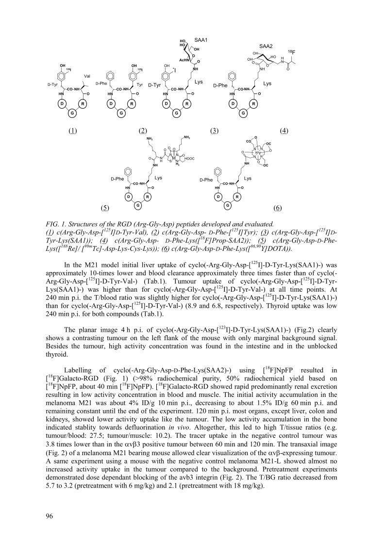
96
125I
D-Tyr
Val
OH
CO NHOHN
D
G
R
D-Phe Tyr
125IOH
D
G
R
OHNCO NH
*I
D-Tyr Lys
OH
D
G
R
OHN
NH
O
OH
HOHO
AcHN O
SAA1
NHCO
OO
OHOH
HONH
O
18F
D
G
R
OHNCO NH
NH
LysD-Phe
SAA2
(1) (2) (3) (4)
(5)
O NSH
D-Phe Lys
D
G
R
ONHCO
HN
NH
NH2
HOOCN
OO O
N N
O
H2
NH2
*M
NH
D
G
R
OCO
HN
NH
LysD-Phe
NN
OCO
O
ONN
OCCO
O90Y
(6)
FIG. 1. Structures of the RGD (Arg-Gly-Asp) peptides developed and evaluated. (1) c(Arg-Gly-Asp-[125I]D-Tyr-Val), (2) c(Arg-Gly-Asp- D-Phe-[125I]Tyr); (3) c(Arg-Gly-Asp-[125I]D-Tyr-Lys(SAA1)); (4) c(Arg-Gly-Asp- D-Phe-Lys([18F]Prop-SAA2)); (5) c(Arg-Gly-Asp-D-Phe-Lys([188Re]/ [99mTc]-Asp-Lys-Cys-Lys)); (6) c(Arg-Gly-Asp-D-Phe-Lys([86,90Y]DOTA)).
In the M21 model initial liver uptake of cyclo(-Arg-Gly-Asp-[125I]-D-Tyr-Lys(SAA1)-) was approximately 10-times lower and blood clearance approximately three times faster than of cyclo(-Arg-Gly-Asp-[125I]-D-Tyr-Val-) (Tab.1). Tumour uptake of cyclo(-Arg-Gly-Asp-[125I]-D-Tyr-Lys(SAA1)-) was higher than for cyclo(-Arg-Gly-Asp-[125I]-D-Tyr-Val-) at all time points. At 240 min p.i. the T/blood ratio was slightly higher for cyclo(-Arg-Gly-Asp-[125I]-D-Tyr-Lys(SAA1)-) than for cyclo(-Arg-Gly-Asp-[125I]-D-Tyr-Val-) (8.9 and 6.8, respectively). Thyroid uptake was low 240 min p.i. for both compounds (Tab.1).
The planar image 4 h p.i. of cyclo(-Arg-Gly-Asp-[123I]-D-Tyr-Lys(SAA1)-) (Fig.2) clearly shows a contrasting tumour on the left flank of the mouse with only marginal background signal. Besides the tumour, high activity concentration was found in the intestine and in the unblocked thyroid.
Labelling of cyclo(-Arg-Gly-Asp-D-Phe-Lys(SAA2)-) using [18F]NpFP resulted in [18F]Galacto-RGD (Fig. 1) (>98% radiochemical purity, 50% radiochemical yield based on [18F]NpFP, about 40 min [18F]NpFP). [18F]Galacto-RGD showed rapid predominantly renal excretion resulting in low activity concentration in blood and muscle. The initial activity accumulation in the melanoma M21 was about 4% ID/g 10 min p.i., decreasing to about 1.5% ID/g 60 min p.i. and remaining constant until the end of the experiment. 120 min p.i. most organs, except liver, colon and kidneys, showed lower activity uptake like the tumour. The low activity accumulation in the bone indicated stablity towards defluorination in vivo. Altogether, this led to high T/tissue ratios (e.g. tumour/blood: 27.5; tumour/muscle: 10.2). The tracer uptake in the negative control tumour was 3.8 times lower than in the v 3 positive tumour between 60 min and 120 min. The transaxial image (Fig. 2) of a melanoma M21 bearing mouse allowed clear visualization of the v -expressing tumour. A same experiment using a mouse with the negative control melanoma M21-L showed almost no increased activity uptake in the tumour compared to the background. Pretreatment experiments demonstrated dose dependant blocking of the avb3 integrin (Fig. 2). The T/BG ratio decreased from 5.7 to 3.2 (pretreatment with 6 mg/kg) and 2.1 (pretreatment with 18 mg/kg).

97
3
0% 100%
A B
C D
0% 100%
tumor
K
tumor
I
T
E F
FIG. 2. PET images using an animal scanner (A-D) and -camera device (E and F). Left (A-D): Transaxial images of human melanoma M21(A-C) and M21L (D) bearing mice on the Munich high resolution animal PET scanner using 100-200µCi of compound (4), Fig.1, 90 min p.i.. (A,D) without pretreatment, (B) with 6 mg C(RGDfV)/kg and (C) with 18 mg C(RGDfV)/kg 10 min prior injection of the tracer. Gamma-camera images of osteosarcoma bearing mice using 200µCi of compound Tc-99m-(5), Fig. 1 (E) and compound (3) Fig. 1 (F) and 4 h post injection (K=kidney, I=intestine, T=thyroid).
OH
OHO
HO
OH
O
OH
OH OHO
OOH
OHO
OH
O
OH
NH
OH
NH2
NH
NH
O
O
SS
ONH
H
O
O NH
NH
NH3+
N
OH
NHO
OH
+
123I
O
O
OH
NH
OH
NH3
NH
NH
O
O
SS
ONH
H
O
O NH
NH
NH3+
N
OH
NHO
OH
+
123I
[123I]Tyr3-Octreotide
[123I]Tyr3-Maltotriose-Octreotate
Thr8
Thr8-ol
FIG. 3. Some structures of the peptides developed and evaluated, exemplified for [125I]Tyr3-octreotide ([125I]TOC) and [125I]Maltotriose-Tyr3-Thr8-octreotide ([125I]Maltotriose-Tyr3-octreotate, [125I]Mtr-TOCA).

98
TABLE I. BIODISTRIBUTION DATA FOR CYCLO(-ARG-GLY-ASP-[125I]-D-TYR-VAL-) ([125I]P2) AND CYCLO(-ARG-GLY-ASP-[125I]-D-TYR-LYS(SAA1)-) ([125I]GP2) IN MELANOMA-BEARING NUDE MICE
organ [125I]P2 [125I]GP2 organ [125I]P2 [125I]GP2 (n = 3) (n = 4) (n = 3) (n = 4) time p.i. [min]
melanoma melanoma time p.i. [min] melanoma melanoma
blood heart 10 0.77 ± 0.02 2.20 ± 0.11 10 0.60 ± 0.07 1.31 ± 0.03 60 0.17 ± 0.02 0.41 ± 0.11 60 0.22 ± 0.07 0.46 ± 0.10 120 - 0.23 ± 0.08 120 - 0.35 ± 0.08 240 0.06 ± 0.02 0.19 ± 0.09 240 0.07 ± 0.02 0.27 ± 0.06 serum brain 10 1.43 ± 0.01 3.64 ± 0.18 10 0.09 ± 0.02 0.17 ± 0.02 60 0.29 ± 0.03 0.57 ± 0.15 60 0.05 ± 0.01 0.07 ± 0.01 120 - 0.32 ± 0.10 120 - 0.05 ± 0.01 240 0.09 ± 0.03 0.24 ± 0.10 240 0.02 ± 0.01 0.05 ± 0.01 liver lung 10 21.96 ± 2.78 2.59 ± 0.24 10 2.36 ± 0.46 3.91 ± 0.35 60 11.23 ± 1.95 1.22 ± 0.32 60 0.93 ± 0.21 1.51 ± 0.40 120 - 0.72 ± 0.19 120 - 0.98 ± 0.21 240 0.78 ± 0.28 0.56 ± 0.18 240 0.29 ± 0.10 0.83 ± 0.20 kidneys spleen 10 12.09* 6.38 ± 0.31 10 1.41 ± 0.36 2.25 ± 0.11 60 3.30 ± 0.12 2.23 ± 0.43 60 0.78 ± 0.26 1.23 ± 0.32 120 - 1.62 ± 0.34 120 - 0.99 ± 0.24 240 0.28 ± 0.16 1.30 ± 0.48 240 0.10 ± 0.04 0.79 ± 0.30 muscle intestine 10 0.42 ± 0.04 0.84 ± 0.05 10 - 2.21 ± 0.20 60 0.25 ± 0.05 0.50 ± 0.19 60 - 2.61 ± 0.61 120 - 0.29 ± 0.03 120 - 2.16 ± 0.41 240 0.10 ± 0.03 0.23 ± 0.07 240 - 2.92 ± 0.63 tumour thyroid 10 2.07 ± 0.32 2.71 ± 0.18 10 2.21 ± 0.64 8.80 ± 3.29 60 1.30 ± 0.13 2.05 ± 0.55 60 1.95 ± 0.46 7.69 ± 2.54 120 - 1.81 ± 0.30 120 - 14.32 ± 5.21 240 0.41 ± 0.15 1.69 ± 0.54 240 0.29 ± 0.08 11.82 ± 6.54 * experiment gave only one data point.
1.4. Conclusion
We introduced radioiodinated cyclic RGD-peptides for imaging the v 3 integrin status. These first generation tracers showed receptor specific accumulation in different tumour mouse models. However, these tracers also revealed high activity retention in liver and intestine, which limits the application for tumour imaging. We improved the pharmacokinetics by introducing sugar amino acids, which increased the hydrophilicity and markedly reduced the tracer uptake by the liver. Moreover, introduction of a sugar amino acid SAA2 allows 18F-labelling of the amino methyl function of the glycopeptide using radiolabelled acylation reagents. These modifications are unlikely to influence the v 3 affinity and selectivity of the compound. Biodistribution data, in vivo competition experiments as well as imaging studies using a small animal PET scanner, confirmed our design approach and demonstrated the v 3 specific binding of the developed antagonists Most recently, preliminary data have been presented on 99mTc-, 186Re- and 90Y-labelled peptides based on cyclo(-Arg-Gly-Asp-D-Phe-Lys-) [15], which may be useful tracers for SPECT imaging and PRRT.

99
-1,0 -0,5 00
2
4
6
8
log Pow
activ
ity a
ccum
ulat
ion
[%iD
/g]
A
BC
D
FIG. 4. Correlation between acitivity accumulation [%iD/g tissue] and lipophilicity (log POW) of [125I]TOC (A), [125I]Gluc-TOC (B), [125I]Malt-TOC (C) and [125I]Mtr-TOC (D) in the liver of AR42J tumour bearing nude mice 30 min p.i. (n = 5).
2. GLYCATED RADIOIODINATED OCTRTEOTIDES AND OCTREOTATES
2.1. Introduction
[123I]Tyr3-octreotide (TOC) (Fig.3) was the first radiolabelled SRIF analog that was applied for scintigraphic in vivo localization of SSTR2-expressing primary tumours and metastases [16]. It was already shown in earlier studies, that two major drawbacks restricted the suitablity of this new tracer: its predominant hepatobiliary excretion and low tumour retention [17]. Due to these disadvantages interpretation of planar as well as of SPET- images of the abdominal region are complicated considerably. In a pharmacokinetic study with patients with neuroendocrine tumours 55% of the administered activity was found in the feces 40 h p.i.. While average tumour doses were app. 0.05-0.81 kgGy/GBq ([131I]TOC), activity calculated for the gallbladder and the lower colon was 0.54 Gy/GBq and 4.43 Gy/GBq, respectively [18]. Therefore [131I]TOC is not suited for PRRT [19], and there are certainly restrictions with respect to its suitability for tumour scintigraphy.
To make radioiodinated derivatives of TOC accessible for these applications, we synthesized a new class of radioiodinated, carbohydrate-conjugated octreotide analogs by conjugation of Tyr3-octreotide (TOC) with different aldoses via Maillard reaction and subsequent radioiodination [20]. The glucose- ([125I]Gluc-TOC), maltose-([125I]Malt-TOC) and maltotriose-([125I]Mtr-TOC) derivatives of [125I]TOC were evaluated in comparative biodistribution studies using AR42J tumour bearing nude mice [21,22]. Furthermore, to increase tumour uptake, octreotate (Thr8-octreotide) was prepared and and glycated in the same way as described for octreotide ([125I]TOCA, [125I]Gluc-TOCA, [125I]Malt-TOCA and [125I]Mtr-TOCA) (Fig.3). Internalization studies revealed high uptake of these new derivatives [23,24]. In a first patient study the suitablility of these new series of compounds for SSTR-imaging in vivo was demonstrated [24].
2.2. Methods
Preparation of the peptides will be published elsewhere. The peptides were labelled with 125I (or 123I for -camera imaging) using the IodoGen® method. A solution of 100-500 µg of peptide in 200 µlof PBS (pH7.4) was transferred to an Eppendorf cap coated with 150 µg of Iodogen. After adding 5-10 µl (18 - 37 MBq) n.c.a. of [125I]NaI the cap was vortexed and the labelling reaction was allowed to proceed for 20 min at RT. The peptide solution was then removed from the insoluble oxidizing agent. Separation of the labelled products from the unreacted peptide was achieved using gradient RP-HPLC. The solvent of the collected fraction was then evaporated to dryness and the residue was redissolved in PBS to yield a solution of radiolabelled peptide with an activity concentration of 370 kBq/100 µl.

100
2.2.1. Determination of lipophilicity
To a solution of app. 100000 cpm of radiolabelled peptide in 500 µl of PBS (pH7.4), 500 µl of octanol were added (n = 6). Vials were vortexed for 3 min and then centrifuged at 15000 rpm for 6 min Aliquots of 100 µl of PBS and octanol each were measured in a -counter.
2.2.2. In vivo studies
2.2.2.1. Tumour model
RPMI 1640 medium, PBS-Buffer and FCS (Fetal calf serum) were purchased from Biochrom Seromed (Berlin, Germany). 200 mM L-Glutamine was purchased from Gibco BRL Life Technologies (Karlsruhe, Germany). AR42J is a rat pancreatic acinar tumour cell line and was obtained from ECACC (European Collection of Cell Cultures, Salisbury, UK). Cells were maintained in RPMI 1640, supplemented with 10% FCS and 2 mM L-glutamine. To establish tumour growth, subconfluent monolayer cells were treated with 1mM EDTA, suspended, centrifuged and resuspended in RPMI 1640. Nude mice (male and female, 6-8 weeks) were inoculated s.c. in the flank with 2.5-3 × 10⁷ cells suspended in 100 µl of RPMI 1640. Ten days postinoculation all mice showed solid palpable tumour masses (tumour weight 150 - 500 mg) and were used for the experiments.
2.2.2.2. Biodistribution studies
The 125I-labelled peptides, 370 kBq (10 µCi) in 100 µl of PBS (pH7.4), were injected i.v. into the tail vein of nude mice bearing an AR42J tumour. The animals (n = 5) were sacrificed 10, 30 and 60 min p.i., and the organs of interest were dissected. The radioactivity was measured in weighted tissue samples using a gamma counter. Data are expressed in% iD/g tissue (mean ± SD, n = 5).
2.2.2.3. Pretreatment, competition and displacement studies
For pretreatment studies, 0.8 mg/kg Tyr3-octreotide (20 µg/mouse) was injected i.v. into the tail vein as a 100 µl bolus 10 min prior to the injection of 370 kBq (10 µCi) of radioligand in 100 µl of PBS. Cmpetition was performed by coinjection of 0.8 mg/kg Tyr3-octreotide (20 µg/mouse) in 100 µlof PBS. For displacement studies, 0.8 mg/kg Tyr3-octreotide (20 µg/mouse) were administered 10 min after the injection of radioiodinated tracer. All animals were sacrificed 30 min post injection of the radioligand. Subsequent determination of the activity accumulation in all organs of interest was performed as described above (biodistribution studies).
2.3. Results
2.3.1. Peptide synthesis
Solid phase peptide synthesis yielded TOC in quantitative yield and in high purity, and subsequent synthesis steps were performed without further purification of the peptide. The yield of the carbohydrate conjugation differed considerably with the aldose used. While for the synthesis of the glucose-derivative yields never exceeded 80%, the derivatizations with maltose and maltotriose were almost quantitative. Deprotection and subsequent RP-HPLC afforded the glycosylated peptides in high purity (UV purity at 220 nm >95%) in overall yields of 17 to 35%.
2.3.2. Radiolabelling
No diiodinated byproducts were observed. In RP-HPLC, K’-values of the radioiodinated products and the respective unlabelled peptides differed by app.1 unit for all compounds. Therefore chromatographic separation of the radiolabelled peptides from an excess of unreacted educt yielded the 125I-glycopeptides in high radiochemical purity (>99.8%) and high specific activity. Radiochemical yields ranged from 45 to 85%, depending on the UV-purity of the respective unlabelled peptide.

101
(A) (B)
Blood Intestine Kidney Liver Muscle Tumor Adrenals Pancreas
0
5
10
15
20
25
30
0
5
10
15
20
25
30ac
tivity
acc
umul
atio
n [%
ID/g
]
TumorBlood
TumorIntestine
TumorKidney
TumorLiver
0
10
20
30
0
10
20
30
FIG. 5(A). Biodistribution (%ID/g, mean +/-SD, n=4) of [125I]TOC(white bars) and [125I]Mtr-TOCA (grey bars) in AR42J tumour bearing nude mice 1 h after injection.
FIG. 5(B). Tumour/tissue activity ratios (n=4, mean+/-SD) in AR42J tumour bearing nude mice 1 h after injection of [125I]TOC (white bars) and [125I]Mtr-TOCA (grey bars).
FIG. 6. Gamma camera image of a metastasised carcionoid (liver, peritoneum) of the small intestine imaged 1 h after application of 196 MBq [123I]Mtr-TOCA.

102
2.3.3. Lipophilicity
While log POW for the parent compound [125I]TOC is still > 0, all glycosylated derivatives exhibit log POW -values below zero in the following order: [125I]Gluc-TOC> [125I]TOCA>[125I]Mtr-TOC> [125I]Malt-TOC>[125I]Gluc-TOCA>[125I]Mtr-TOCA>[125I]Malt-TOCA. Fig.4 shows the correlation between the liver uptake at early time points and the lipophilicity of the octreotide derivatives investigated.
2.3.4. Biodistribution
Compared to [125I]TOC all glycated derivatives exhibit faster blood clearance. The acitivity accumulation in the liver and intestine is considerably decreased 1h p.i. (liver: [125I]Gluc-, [125I]Malt-, [125I]Mtr-TOC 1.0-2.7, [125I]TOCA: 1.2, [125I]Gluc-, [125I]Malt-, [125I]Mtr-TOCA: 0.5-0.8%iD/g vs [125I]TOC: 3.2; intestine: [125I]Gluc-TOC: 4.9, [125I]Malt-TOC: 1.4, [125I]TOCA: 5.5, [125I]Gluc-, [125I]Malt-, [125I]Mtr-TOCA: 2.5-4.6%iD/g vs [125I]TOC: 9.2). [125I]Gluc-TOC and [125I]Gluc-, [125I]Malt-, [125I]Mtr-TOCA exhibit a strong increase in tumour uptake (t) and other SSTR+ tissues (t: [125I]Gluc-, [125I]Malt-, [125I]Mtr-TOCA: 13-25%iD/g vs [125I]TOC: 5.8 1h p.i.), resulting in t/kidney ratios for these compounds of 5.7-9.0 vs 0.8 for [125I]TOC. While at 1h p.i. t/liver and t/intestine ratios ranged from 1.8-5.4 and 0.6-3.9 for TOC and all glycated TOC´s, values ranged from 11.2-32 and 2.4-9.6 for TOCA and all glycated TOCA´s. A comparison of the biodistribution data of [125I]TOC and [125I]Mtr-TOCA is shown in Fig.5. In a first patient study a metastasized carcinoid (liver, peritoneum) of the small intestine was imaged using 196 MBq [123I]Mtr-TOCA and compared to [111In]Octreoscan®. The images showed all metastases already 1h p.i. with increasing tumour to non-tumour ratios up to 43 h.
TABLE II. TISSUE BIODISTRIBUTION OF [125I]TOC & ITS GLYCOSYLATED DERIVATIVES*
organ time p.i. [min]
[125I]TOC [125I]Gluc-TOC [125I]Malt-TOC [125I]Mtr-TOC
blood 10 1.69 ± 0.54 2.60 ± 0.45 3.42 ± 1.09 30 1.06 ± 0.24 1.58 ± 0.48 1.38 ± 0.20 2.66 ± 0.19 60 1.23 ± 0.30 0.88 ± 0.38 0.74 ± 0.13 liver 10 7.36 ± 0.92 3.51 ± 0.77 2.43 ± 0.78 30 5.95 ± 1.19 2.44 ± 0.16 1.33 ± 0.32 2.66 ± 0.17 60 3.76 ± 1.28 1.73 ± 0.59 0.96 ± 0.17 intestine 10 3.58 ± 1.20 2.80 ± 0.72 1.46 ± 0.47 30 7.02 ± 1.32 3.99 ± 1.19 1.25 ± 0.55 9.61 ± 1.69 60 10.6 ± 2.8 5.28 ± 1.27 1.48 ± 0.18 kidney 10 5.55 ± 0.71 7.44 ± 1.77 9.63 ± 3.24 30 6.70 ± 1.59 6.35 ± 0.66 7.23 ± 1.39 3.31 ± 0.58 60 6.95 ± 1.44 6.25 ± 1.94 6.78 ± 1.52 tumour 10 4.95 ± 1.53 4.42 ± 1.15 6.79 ± 2.58 30 6.72 ± 2.60 10.1 ± 2.8 5.30 ± 1.87 4.87 ± 2.15 60 5.53 ± 1.06 8.73 ± 1.45 5.24 ± 0.61 pancreas 10 1.65 ± 0.55 4.10 ± 0.82 3.61 ± 1.71 30 1.22 ± 0.37 4.48 ± 0.67 2.21 ± 0.59 2.12 ± 0.44 60 1.17 ± 0.32 3.36 ± 1.20 1.92 ± 0.26 adrenals 10 1.63 ± 0.76 2.08 ± 0.40 3.08 ± 0.92 30 1.63 ± 0.74 2.40 ± 0.73 2.07 ± 0.21 2.98 ± 0.82 60 1.25 ± 0.21 2.05 ± 0.97 1.93 ± 0.68 muscle 10 0.39 ± 0.14 0.60 ± 0.13 0.94 ± 0.79 30 0.18 ± 0.03 0.39 ± 0.37 0.33 ± 0.14 0.51 ± 0.08 60 0.26 ± 0.08 0.17 ± 0.05 0.20 ± 0.08 *Groups of 5 nude mice bearing AR42J tumours. Mean value (± standard dev) are shown.

103
2.3.5. Competition experiments
Both for [125I]Gluc-TOC and for [125I]Malt-TOC binding to SSTR-rich tissues in vivo was shown to be specific. While tumour accumulation was reduced in the pretreatment- and coinjection-experiments, resulting in tumour/SSTR-negative tissue-ratios <1, displacement of the radioiodinated tracers by injection of 20 µg of Tyr3-octreotide/mouse 10 min p.i. of the labelled compound was less effective and therefore caused a smaller decrease in tumour uptake of the radioiodinated tracer. Accumulation in SSTR positive organs such as pancreas and adrenals was decreased to the same degree as in the tumour. Therefore ratios of tumour to SSTR-expressing organs remained almost unaffected by all experimental conditions.
2.4. Conclusion
Glycosylation is a powerful tool for the modification of radiohalogenated and radiometalated [25] bioactive peptides. Conjugation with a carbohydrate moiety increases the overall hydrophilicity of the molecule without changing the net charge of the peptide and thus significantly improves the pharmacokinetics. Their fast blood clearance, low accumulation in liver and intestine and high tumour uptake make glycosylated, radioiodinated derivatives of octreotide (TOC) and especially octreotate (TOCA) very promising ligands for in vivo SST-receptor targeting.
REFERENCES
[1] CLEZARIN, P., Recent insights into the role of integrins in cancer metastasis, Cell Mol. Life Sci. 54 (1998) 541-548.
[2] STROMBLAD, S., CHERESH, D.A., Integrins, angiogenesis and vascular cell survival, Chem. Biol. 3 (996) 911-921.
[3] GURRATH, M., MÜLLER, G., KESSLER H., et al., Confomation/activity studies of rationally designed potent and anti-adhesive RGD peptides, Eur. J. Biochem. 210 (1992) 911-921.
[4] HAUBNER R., FINSINGER, D., KESSLER, H., Stereoisomeric Peptide Libraries and Peptidomimetics for Designing Selective Inhibitors of the v 3 Integrin for a New Cancer Therapy, Angew Chem. Int. Ed. Engl. 36 (1997) 1374-1389.
[5] AUMAILLY, M., GURRATH, M., MÜLLER, G., et al., Arg-Gly-Asp constrined within cyclic pentapeptides. Strong and selective inhibitors of cell adhesion to vitronectin and laminin fragment P1, FEBS Lett. 291 (1991) 50-54.
[6] CARRON, C.P., MEYER, D.M., PEGG, J.A., et al., A Peptidomimetic anatagonist of the integrin v inhibits Leydig cell tumour growth and the development of hypercalcemia of malignancy, Cancer Res. 58 (1998) 1930-1935.
[7] KERR, J.S., WEXLER, R.S., MOUSA, S.A., et al., Novel small molecule v integrin antagonists: comparative anti cancer efficacy with known angiogenesis inhibitors, Anticancer Res. 19 (1999) 959-968.
[8] HOFFMANN, M., BURKHARD, F., HESSLER, G., KESSLER, H., C-Glycosid Analogues of N4-(2-Acetamido-2-Deoxy-ß-D-Glucopyranosyl)-L-Asparagine: Synthesis and conformational Analysis of a Cyclic C-Glycopeptide. Helv. Chim. Acta. 79 (1996) 1519-1532.
[9] HAUBNER, R., WESTER, H.J., BURKHARD, F., et al., Glycosylated RGD-Peptides: Tracer for Tumour Targeting and Angiogenesis Imaging with Improved Biokinetics, J. Nucl. Med. (2001).
[10] HAUBNER, R., WESTER, H.J., WEBER, W., et al., Non invasive imaging of v 3 integrin expression using a 18F-labelled RGD-containing glycopeptide and positron emission tomography (submitted).
[11] HAUBNER, R., WESTER, H.J., MANG, C., et al., Synthesis and in vitro evaluation of a RGD-containing glycopeptide for the non-invasive determination of the v 3 expression using positron emission tomography (manuscript in preparation).
[12] HAUBNER, R., WESTER, H.J., REUNING, U., et al., Radiolabelled v 3 integrin antagonists: a new class of tracers for tumour targeting, J. Nucl. Med. 49 (1999) 1061-1071.

104
[13] WESTER, H.J., HAMACHER, K., STOECKLIN, G., A comparative study of n.c.a. fluorine-18 labelling of Peptides and proteins via acylation and photochemical conjugation, Nucl. Med. Biol. 23 (1996) 365-372.
[14] PICHLER, B., BOENING, G., LORENZ, E., et al., Studies with a prototype high resolution PET scanner based on LSO-APD modules, IEEE Trans. Nucl. Sci. 45 (1998) 1298-1302.
[15] BOCK, M., BRUCHERTSEIFER, F., HAUBNER, R., et al., Tc-99m-, Re-188- and Y-90- labelled V 3 anatgonists: promising tracers for tumour induced angiogenesis, J. Nucl. Med. 41 (2000) 41 pp.
[16] LAMBERTS, S.W.J., BAKKER, W.H., REUBI, J.C., KRENNING, E.P., Somatostatin receptor imaging in the localization of endocrine tumours, N. Engl. J. Med. 323 (1990) 1246-9.
[17] KRENNING, E.P., KWEKKEBOOM, D.J., BAKKER, W.H., et al., Somatostatin receptor scintigraphy with [111In-DTPA-D-Phe1]- and [123I]-Tyr3]-octreotide: the Rotterdam experience with more than 1000 Patients, Eur. J. Nucl. Med. 20 (1993) 716-731.
[18] O´CONNOR, M.K., KVOLS, L.K., BROWN, M.L., et al., Dosimetry and Biodistribution of a Iodine-123 labelled somatostatin analog in patients with neuroendocrine tumours, J. Nucl. Med. 33 (1992) 1613-1619.
[19] BAKKER, W.H., BREEMANN, W.A.P., PLUIM, M.E., et al., Iodine-131 labelled octreotide: not an option for somatostatin receptor therapy, Eur. J. Nucl. Med. 23 (1996) 775-781.
[20] LEISNER, M., KESSLER, H., SCHWAIGER, M., WESTER, H.J., Synthesis of Na-D-Phe1-Amoadori derivatives of Tyr3-octreotide: precursors for I-123-/F-18-labelled SSTR-binding SPECT/PET tracers with improved biodistribution, J. Lab. Compds. Radiopharm. 42 (1999) 549-551.
[21] SCHOTTELIUS, M., VAIDYANATHAN, G., SENEKOWITSCH-SCHMIDTKE, R., et al., Glycation of radioiodinated octreotides increases renal excretion and tumour uptake, Nucl. Med. Commun. 21 (2000) 579 (abstr).
[22] SCHOTTELIUS, M., VAIDYANATHAN, G., SENEKOWITSCH-SCHMIDTKE, R., et al., New radioiodinated and glycated Octreotide Derivatives with Renal Exrection, Nucl. Med. 39(2000) A12 (abstr).
[23] SCHOTTELIUS, M., VAIDYANATHAN, G., SENEKOWITSCH-SCHMIDTKE, R., et al., 125I-labelled Tyr3-Thr8-Octreotide Derivatives for Endoradiotherapy of SSTR Expressing Tumours, Nucl. Med. 39 (2000) A13 (abstr).
[24] SCHOTTELIUS, M., SENEKOWITSCH-SCHMIDTKE, R., SCHEIDHAUER, K., et al., Glycated radioiodinated octreotides and octreotates with renal excretion and increased tumour uptake for SSTR scintigraphy and peptide receptor radionuclide therapy (PRRT), J .Nucl. Med. 41 (2000) 40P (abstr).
[25] SCHOTTELIUS, M., SENEKOWITSCH-SCHMIDTKE, R., KESSLER, H., et al., First in vivoevaluation of [In-111]maltose-Lys0(DOTA)Tyr3-Octreotide reveals high potential of glycated and radiometalated octreotide derivatives for diagnosis and therapy, Nucl. Med. Commun. 21(2000) 579 (abstr).
[24] SCHOTTELIUS, M., SENEKOWITSCH-SCHMIDTKE, R., SCHEIDHAUER, K., et al., Glycated radioiodinated octreotides and octreotates with renal excretion and increased tumour uptake for SSTR scintigraphy and peptide receptor radionuclide therapy (PRRT), J. Nucl. Med. 41 (2000) 40P (abstr).
[25] SCHOTTELIUS, M., SENEKOWITSCH-SCHMIDTKE, R., KESSLER, H., et al., First in-vivo evaluation of [In-111]maltose-Lys0(DOTA)Tyr3-Octreotide reveals high potential of glycated and radiometalated octreotide derivatives for diagnosis and therapy, Nucl. Med. Commun. 21 (2000) 579 (abstr).

105
OPTIMIZATION OF SYNTHESIS AND QUALITY CONTROL PROCEDURES FOR THE PREPARATIONS OF 18F AND 123I LABELLED PEPTIDES
S.C. ARCHIMANDRITIS, S. POTAMIANOS, A.D. VARVARIGOU National Centre for Scientific Research “Demokritos”, Athens, Greece
Abstract
Radiolabelled biomolecules like proteins and peptides, are playing now days an important role in experimental and clinical Nuclear Medicine. Radioiodination techniques remain important, with improvements accounting for high purity, specific activity and better in vivo stability. Radioiodination using prosthetic groups is the method of choice in cases where the molecules are lacking of thyrosyl groups in their structure and are also sensitive to circulating dehalogenase enzymes. This investigation was based on the need to optimize labelling and quality control techniques for these molecules. The N-succinimidyliodobenzoate (SIB) was used in this study as the prosthetic group for the radioiodination. Optimization of SIB synthesis and modification of the protocol resulted in an improved mean yield of SIB. The combination of TLC and column chromatography using silica gel proved suitable in identifying SIB. Furthermore, the ability of SIB to couple to protein was also used to confirm the presence of SIB. In this case, SEC and ITLC-SG proved suitable to confirm protein binding of SIB. Column chromatography using silica gel containing Sep-Pak was appropriate for SIB purification. Concerning SIB conjugation to peptides, high radioiodination yields were only possible for peptides with amino-containing-side-chain amino acids. Furthermore, lysine containing peptides retained stability, at 4oC, for at least 24 h and reverse phase HPLC proved the most suitable technique for assessing conjugation of SIB to peptide. The biological evaluation of the radiolabelled product was made in normal mice. SIB and SIB-peptide conjugates were tested comparatively and a number of tentative but interesting inferences were drawn. SIB and its peptide conjugates exhibited good in vivo stability as evidenced by low thyroid accumulation and were cleared via the kidneys. A time dependant decrease in the% dose per gram of tissue indicates possible adrenal metabolism of SIB and SIB-peptide conjugates. Finally the fate of the SIB-Lanreotide conjugate was preliminary evaluated in colon tumour bearing nude mice. A tentative but encouraging inference was made that SIB-Lanreotide may exhibits tumour targeting capacity.
1. INTRODUCTION
The aim of the present project was to optimize peptide radioidination, using SIB as the radioiodinating intermediate and to develop suitable quality control procedures for the radiolabelled peptides and of the intermediate products
Radioiodination of biomolecules still remains important especially since radioisotopes of the same element (iodine) can be used interchangeably for diagnosis (131I/123I) and/or therapy (131I) in a single radiolabelling protocol. However, a major problem encountered by radioiodinated biomolecules is their instability, resulting in free radioiodide accumulation in the thyroid due to circulating dehalogenase enzymes. One way of overcoming this in vivo de-iodination is through the development of non-phenolic aromatic compounds, structurally different from iodotyrosine and thyroxine and thus unsuitable dehalogenase substrates. An intermediate, like ATE, is first radiolabelled and after a purification step, the radiolabelled intermediate (N-succinimidyl-iodo benzoate; SIB) is used to label protein or peptide. Apart from the reported in vivo stability of biomolecules labelled with this intermediate [1, 2], a further advantage is that it can be labelled with 18F (for application in PET) and 211At (an -emitter which can be used as a radiotherapeutic radionuclide for internalized biomolecules).
2. MATERIALS AND METHODS
2.1. Optimization of SIB synthesis
The original ATE radioiodination protocol of Zalutsky and Narula [1] was used with two minor modifications. The first one entailed a reduction to half of the original reaction volumes for the reactants but maintaining the total final volume. More specifically, to an eppendorf tube 10-20 µlNa125I (37 MBq/1 mCi in 0.01 M NaOH) were added, followed by 25 µl 1 M acetic acid, 25 µl 1 M t-

106
butylhydroperoxide (T-BHP), 5 µl 0.1 M ATE. Final volume was made up to 140 µl with chloroform. The second modification entailed a change in the order of adding the reactants. Briefly, to a normal 1 mL Eppendorf tube 10-20 µl Na125I (148 MBq/4 mCi in 0.01 M NaOH) were added followed by 25 µl 1 M acetic acid, 5 µl 0.1 M ATE and 25 µl 1 M T-BHP. Final volume was made up to 140 µl with chloroform. In all instances, reaction proceeded for 30 min, at room temperature, with continuous stirring. Sep-Pak chromatography, containing silica gel, was used to purify SIB. Thin layer chromatography (TLC), instant thin layer chromatography using silica gel (ITLC-SG) and Sep-Pak chromatography were used to characterize the reaction mixture and the purified SIB. The chromatograms were developed in 30% ethyl acetate in hexane.
2.2. SIB radioiodination yields
The SIB radioiodination yield obtained for the ATE radioiodination protocol with the first modification is depicted in Fig. 1. Three chromatographic methods were used.
0
20
40
60
80
100
Sep-Pac k TLC ITLC-SG
Rad
ioio
dina
tion
yiel
d (%
)
FIG. 1. Mean radioiodination yield of SIB, according to the 3 chromatographic methods.
TLC and silica gel containing Sep-Pak chromatography gave similar values (51.6% and 49.5%, respectively) for mean radioiodination yield of SIB. Improved SIB radioiodination yields (76%) were possible when using the ATE radioiodination protocol with the second modification. TLC is the most suitable technique for estimating the radioiodination yield of SIB in the crude reaction mixture, as it provides better resolution between SIB and other by-products. ITLC-SG gives higher mean radioiodination yields, but cannot resolve between SIB and SIB by-products.
2.3. Chromatographic characterization of SIB
A typical profile for ATE crude reaction mixture eluted through a silica gel containing Sep-Pak column is presented in Fig. 2. This profile was obtained by eluting with four 10 mL fractions of hexane, immediately followed by five 5 mL fractions of 8% ethyl acetate in hexane and finally by six 3 mL fractions of 30% ethyl acetate in hexane. This type of chromatography was used both to characterize the reaction mixture and to purify SIB.
Purified SIB was eluted in the second fraction (at 71 mL) with 30% ethyl acetate in hexane, as confirmed by TLC. The first fraction (at 68 mL) contained SIB but also contained other impurities. Radioactive peaks eluted at 10 mL and 45 mL with hexane and 8% ethyl acetate in hexane, respectively, were attributed to radioiodination by-products, as confirmed by a control sample containing all reagents with the exception of ATE. This control was also characterized by TLC and Sep-Pak chromatography containing silica gel.
A typical profile for ATE crude reaction mixture characterized by TLC is depicted in Fig. 3.

107
0
20
40
60
0 2 0 40 6 0 80 1 00
Elu t io n v o lu m e (m l)R
adio
activ
ity (
Ci)
FIG. 2. Typical elution profile for ATE crude reaction mixture through silica gel containing Sep-Pak column, depicting per centage yield of SIB.
0
10000
20000
30000
40000
0 50 100 150 200 250Dis tan ce (m m )
Det
ecto
r Res
pons
e
FS
FIG. 3. Typical radioactive scan of TLC chromatogram for ATE crude reaction mixture, depicting per centage yield of SIB. “S” represents the point of application and “F” represents the solvent front.
2.4. Confirmation of SIB suitability
The approach adopted for identification of SIB, was to purify SIB from the reaction mixture, using Sep-Pack column chromatography. The next step was to attempt to label a protein with the purified SIB, as an indirect method of SIB confirmation.
3. RADIOIODINATION OF IGG AND ITS CHROMATOGRAPHIC CHARACTERIZATION
3.1. Radioiodination of protein
Sep-Pack purified fraction was analysed by TLC and then dried under N2. To the dried fraction 100 µl borate buffer (BB) 100 mM, pH8.5 was added and mixed by vortexing before adding 100 µl(20 mg/mL) IgG in BB. The mixture was placed in ice for 30 min and vortexed, before adding 300 µl(200 mM) glycine in borate buffer. At the end of 5 min, aliquots were removed and analysed by PC, ITLC-SG and size exclusion chromatography (SEC).
3.2. 3.2. Chromatographic characterization of radioiodinated IgG
Characterization of IgG reaction mixture by SEC resulted in a single peak (1 mL), attributed to radiolabelled IgG. No free SIB was apparent.
Characterization of IgG reaction mixture, using ITLC-SG, resulted in a profile, where most of the radioactivity remained at the point of application, as expected for any radiolabelled protein. ITLC-
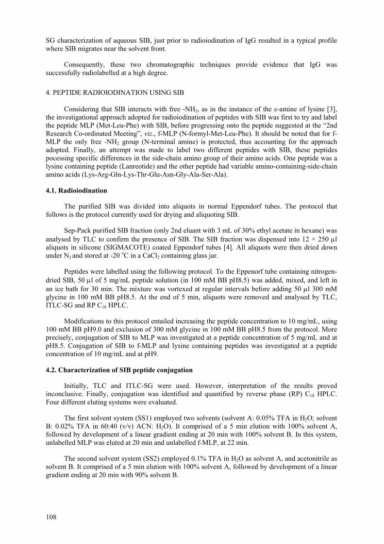
108
SG characterization of aqueous SIB, just prior to radioiodination of IgG resulted in a typical profile where SIB migrates near the solvent front.
Consequently, these two chromatographic techniques provide evidence that IgG was successfully radiolabelled at a high degree.
4. PEPTIDE RADIOIODINATION USING SIB
Considering that SIB interacts with free -NH2, as in the instance of the -amine of lysine [3], the investigational approach adopted for radioiodination of peptides with SIB was first to try and label the peptide MLP (Met-Leu-Phe) with SIB, before progressing onto the peptide suggested at the “2nd Research Co-ordinated Meeting”, viz., f-MLP (N-formyl-Met-Leu-Phe). It should be noted that for f-MLP the only free -NH2 group (N-terminal amine) is protected, thus accounting for the approach adopted. Finally, an attempt was made to label two different peptides with SIB, these peptides pocessing specific differences in the side-chain amino group of their amino acids. One peptide was a lysine containing peptide (Lanreotide) and the other peptide had variable amino-containing-side-chain amino acids (Lys-Arg-Gln-Lys-Thr-Glu-Asn-Gly-Ala-Ser-Ala).
4.1. Radioiodination
The purified SIB was divided into aliquots in normal Eppendorf tubes. The protocol that follows is the protocol currently used for drying and aliquoting SIB.
Sep-Pack purified SIB fraction (only 2nd eluant with 3 mL of 30% ethyl acetate in hexane) was analysed by TLC to confirm the presence of SIB. The SIB fraction was dispensed into 12 × 250 µlaliquots in silicone (SIGMACOTE) coated Eppendorf tubes [4]. All aliquots were then dried down under N2 and stored at -20 oC in a CaCl2 containing glass jar.
Peptides were labelled using the following protocol. To the Eppenorf tube containing nitrogen-dried SIB, 50 µl of 5 mg/mL peptide solution (in 100 mM BB pH8.5) was added, mixed, and left in an ice bath for 30 min. The mixture was vortexed at regular intervals before adding 50 µl 300 mM glycine in 100 mM BB pH8.5. At the end of 5 min, aliquots were removed and analysed by TLC, ITLC-SG and RP C18 HPLC.
Modifications to this protocol entailed increasing the peptide concentration to 10 mg/mL, using 100 mM BB pH9.0 and exclusion of 300 mM glycine in 100 mM BB pH8.5 from the protocol. More precisely, conjugation of SIB to MLP was investigated at a peptide concentration of 5 mg/mL and at pH8.5. Conjugation of SIB to f-MLP and lysine containing peptides was investigated at a peptide concentration of 10 mg/mL and at pH9.
4.2. Characterization of SIB peptide conjugation
Initially, TLC and ITLC-SG were used. However, interpretation of the results proved inconclusive. Finally, conjugation was identified and quantified by reverse phase (RP) C18 HPLC. Four different eluting systems were evaluated.
The first solvent system (SS1) employed two solvents (solvent A: 0.05% TFA in H2O; solvent B: 0.02% TFA in 60:40 (v/v) ACN: H2O). It comprised of a 5 min elution with 100% solvent A, followed by development of a linear gradient ending at 20 min with 100% solvent B. In this system, unlabelled MLP was eluted at 20 min and unlabelled f-MLP, at 22 min.
The second solvent system (SS2) employed 0.1% TFA in H2O as solvent A, and acetonitrile as solvent B. It comprised of a 5 min elution with 100% solvent A, followed by development of a linear gradient ending at 20 min with 90% solvent B.

109
The third solvent system (SS3) comprised of isocratic elution with 0.1% TFA in 45:55 acetonitrile: water (3).
The fourth solvent system (SS4) employed a linear gradient using 0.1% TFA in H2O as solvent A, and acetonitrile as solvent B. It entailed a 15 min elution with 75% solvent A, followed by a linear gradient elution of 1 min with 50% solvent A and then by a linear gradient ending at 24 min with 10% solvent B before returning to 75% solvent A by 25 min. In this system, unlabelled MLP was eluted at 6.48 min and unlabelled f-MLP, at 9.5 min.
Of the four HPLC solvent systems, the last one (SS4) proved to be the best choice for our work as it allowed for faster elution and better resolution of peaks.
4.2.1. SIB-MLP conjugation
SIB-MLP conjugation was first analysed using the SS1 HPLC solvent system. Initially, when employing the reaction mixture comprising of SIB, an old MLP preparation and glycine, the UV detector detected two peaks and the radioactive detector detected six peaks. Using control samples the very pronounced first peak, at 7.6 min, was attributed to the TEA/DMF present in the reaction mixture and the smaller second peak, at 20.5 min, was attributed to MLP. On the basis of the different radioactive elution profiles obtained for each control, SIB resulted in a characteristic radioactive profile of t1 = 22.6 min (29.7%) and t2 = 24.3 min (70.3%). These peaks were also evident in the reaction mixtures containing MLP and/or glycine. Furthermore, peaks at ti = 26.3 min (3.9%), tii =27.7 min (8.5%) and tiii = 28.9 min (11.1%) were attributed to three forms of SIB-MLP. It appears that the total yield of SIB-MLP is low (~ 20%).
Repeating the SIB-MLP conjugation but employing only SIB, and freshly prepared MLP in the reaction mixture, three peaks were detected by the radioactive detector when analysed with the SS1 HPLC solvent system. The two peaks, at t1 = 22 min, (17.8%) and t2 = 23.6 min (73.5%), were attributed to some forms of SIB. However, only one peak was attributable to SIB-MLP (t = 28.3 min) and this had a low radioiodination yield (8.8%). When analysing the same reaction mixture with the SS4 HPLC solvent system three peaks were attributed to some forms of SIB, these being at t1 = 8.93 min, (13.54%), t2 = 11.58 min (8.28%) and t3 = 12.95 min (69.35%). Here again, a single peak was attributable to SIB-MLP (t = 23.28 min) which had a low radioiodination yield (8.84%).
4.2.2. SIB-f-MLP conjugation
SIB-f-MLP conjugation was also analysed using the SS1 HPLC solvent system. Controls were also included for f-MLP. In an attempt to improve the radioiodination yield of the peptide, the peptide concentration was increased to 10 mg/mL and pH adjusted to 9.0. Here again the characteristic peaks, attributed to some form of SIB (t1 = 22 min (21.9%) and t2 = 23.7 min (50%)), were evident. However, only one peak can be attributed to SIB-f-MLP (t = 27 min). In this instance a low radioiodination yield is also evident for SIB-f-MLP (10.2%), of similar order of magnitude to SIB-MLP (8.8%).
4.2.3. SIB conjugation to peptides with different amino containing side chains
The peptides “KRQKTENGASA” and “Lanreotide” were used in order to investigate the significance of the side chain amino functional group, as is the case of amine (lysine), amide (glutamine and asparagine), guanidinium (arginine) and imidazolium (histidine) for SIB conjugation to the respective amino acids. Specifically, “KRQKTENGASA” was obtained from an in-house collaboration and “Lanreotide” was obtained under the IAEA CRP.
An aqueous SIB preparation, in borate buffer pH9.0 (“SIB control”), was kept in ice and analysed immediately and subjected to 1 h post labelling using the SS4 HPLC system (Fig. 4). Reaction mixtures, containing SIB and a peptide with variable amino-containing-side-chain amino acids (KRQKTENGASA, Fig. 5) or SIB and a lysine-containing peptide (Lanreotide, Fig. 6), were also analysed in a similar fashion.

110
4.2.3.1. Aqueous SIB control
Analysis of aqueous SIB revealed four radioactive peaks (t1 = 6.8 min; t2 = 8.8 min; t3 = 12.7 min; t4 = 20.3 min). When considering the per centage radioactivity for the peak at t4=20.3 min, from the decrease of 39.8% to 13.7% at 1h post labelling, it can be inferred that this peak is due to SIB itself. The remaining three peaks are attributed to some form of SIB/SIB by-product.
A
0 ,0 6
0 ,1 6
0 ,2 6
0 ,3 6
0 ,4 6
0 ,5 6
0 ,6 6
0 5 1 0 1 5 2 0 2 5 3 0 3 5D is t a n c e ( m m )
Det
ecto
r Res
pons
e
B
0 ,0 6
0 ,2 6
0 ,4 6
0 ,6 6
0 ,8 6
0 5 1 0 1 5 2 0 2 5 3 0 3 5Elu t io n v o lu m e ( m l)
Det
ecto
r Res
pons
e
FIG. 4. Fate of SIB immediately (A) and 1 h (B) after adding aqueous buffer, characterized by RP-C18-HPLC. The peaks were detected by a radioactive detector. Arrow indicates reactive SIB.
4.2.3.2. SIB conjugation to KRQKTENGASA
HPLC analysis of the reaction mixture (SIB and KRQKTENGASA) revealed four radioactive peaks (t1 = 4.2 min; t2 = 6.8 min; t3 = 12.7 min; t4 = 20.3 min) immediately after labelling and 1 h later. There is a pronounced decrease in the per centage of the radioactivity from 35.2% immediately after labelling, to 1.5% 1 h post labelling for the peak at t4 = 20.3 min, this being “synonymous” with very reactive SIB, as inferred from the aqueous SIB control. The peaks at t2 = 6.8 min and t3 = 12.7 min are possibly due to some form of SIB/SIB by-product, especially as they are also evident in the aqueous SIB control. The appearance of a new peak at t1 = 4.2 min, where the per centage radioactivity increases from 30.1% immediately after labelling, to 70.5% 1 h later, in conjunction with the fact that the cold peptide is eluted at t = 3.2 min in this system, suggests that this peak is SIB-labelled peptide. Furthermore, the same profile was evident for the SIB-KRQKTENGASA after 24 h at 4oC. This illustrates the stability of the successful conjugation between SIB and a peptide with variable amino-containing-side-chain amino acids (KRQKTENGASA).

111
A
0 ,2 5
0 ,2 9
0 ,3 3
0 ,3 7
0 ,4 1
0 ,4 5
0 ,4 9
0 ,5 3
0 5 1 0 1 5 2 0 2 5 3 0 3 5R e t e n t io n T im e ( m l )
Det
ecto
r Res
pons
e
B
0 ,2 0
0 ,6 0
1 ,0 0
1 ,4 0
0 5 1 0 1 5 2 0 2 5 3 0 3 5R e t e n t io n T im e ( m in )
Det
ecto
r Res
pons
e
FIG. 5. Fate of SIB and peptide (KRQKTENGASA) immediately (A) and 1 h (B) after labelling in aqueous medium, characterised by RP-C18-HPLC. The peaks were detected by a radioactive detector. Arrow indicates SIB-peptide.
4.2.3.3. SIB conjugation to Lanreotide
HPLC analysis of the reaction mixture (SIB and Lanreotide), revealed two radioactive peaks (t1 = 8.6 min; t2 = 22.5 min) immediately after labelling and 1 h later. The peaks at t2 = 8.6 min is possibly due to some form of SIB/SIB by-product, especially as it is also evident in the aqueous SIB control. The appearance of a new peak at t2 = 22.5 min, where the per centage radioactivity increases from 48.7% immediately after labelling, to 60.2% 1 h later, suggests that this peak is SIB-labelled peptide. Furthermore, the same profile was also evident for the SIB-Lanreotide after six days, when kept at -8oC. This illustrates the stability of the successfully conjugated SIB-lysine containing peptide (Lanreotide).
0 ,3 0
0 ,5 0
0 ,7 0
0 ,9 0
1 ,1 0
1 ,3 0
0 5 1 0 1 5 2 0 2 5 3 0 3 5E lu t io n v o lu m e ( m l )
Det
ecto
r Res
pons
e
FIG. 6. Fate of SIB and peptide (Lanreotide) 1 h after labelling in aqueous medium, characterized by RP-C18-HPLC. The peaks were detected by a radioactive detector. Arrow indicates SIB-peptide.

112
Two points are worth noting concerning the conjugation experiments of SIB to peptides. Firstly, the SIB-labelled peptides are not necessarily eluted at the same elution volumes as the cold, unlabelled peptides. This is not surprising since the peptides used are small molecules and, depending on the amino acid residues labelled with SIB, one would expect that the contribution of SIB to the overall charge and, consequently, the overall polarity of the peptide is significant, thus influencing the elution of the peptide. Secondly, concerning the choice of peptides “KRQKTENGASA” and “Lanreotide”, these were used as two extreme situations, the former containing a selection of amino acid residues with different types of side chain amino functional groups whilst the latter possessed a single amino acid residue with one type of side chain amino functional group. Continued investigation in order to determine the influence of the side chain of amino acid residues on SIB conjugation is required. This should entail the use of a panel of peptides of a specific amino acid constitution, such as that of Lanreotide for instance, whereby specific residues can be replaced by residues with different side chain amino functional groups.
5. BIOLOGICAL EVALUATION OF SIB AND SIB-PEPTIDE PREPARATIONS
For the biological evaluation of SIB and SIB-peptide preparations, two approaches were adopted. In the first instance, biodistribution of these preparations in normal mice was investigated. In the second instance, a very preliminary evaluation of the tumour targetting capacity of SIB-Lanreotide was performed in tumour bearing nude mice.
5.1. Biological evaluation of SIB and SIB-peptide preparations in normal mice
For the radiobiological evaluation of the products, female Swiss mice were used. Saline solutions (185–740 kBq/0.1 mL or 5-20 µCi/0.1 mL) of SIB or SIB-peptide preparations (SIB-MLP, SIB-f-MLP and SIB-KRQKTENGASA) were administered via the tail vein of the animals. For each time point (30 min, 1 h and 2 h) three to five mice were sacrificed. The main organs were removed, weighed and together with blood, urine and muscle samples were counted in a gamma scintillation counter against a standard of the injected solution. The results were expressed as per centage dose per gram tissue and are presented in Fig. 7.
Uptake of SIB and all SIB-peptide preparations was low in heart, spleen and muscle, not exceeding 0.9% dose per gram tissue at all time points (results not shown).
These preliminary biodistribution studies tentatively indicate the likely presence of five distinct features concerning SIB and its peptide preparations.
Low thyroid levels, at 30 min, for SIB (0.83%) and its peptide conjugates (0.51%- 1.26%), is particularly significant especially since this indicates satisfactory in vivo stability. Interestingly, the highest levels for thyroid are at 30 min, which decrease further with time, possibly implicating metabolism of the agents taken up by the thyroid. The consistently higher values in the blood for SIB-KRQKTENGASA (1.70%-2.64%), compared to the other preparations (0.42% -1.07%), is suggestive of its slower blood clearance. High urinary levels (54%-67%) at 2 h for SIB and its peptide conjugates are indicative of kidney clearance. Furthermore, the higher kidney levels at 30 min for SIB-KRQKTENGASA (9.5%) compared to the remaining preparations (1.3%-3.4%) implies that kidney clearance of SIB-KRQKTENGASA is slower. For SIB and SIB-peptide preparations the trend, where stomach levels increase with time, from 30 min (1.81%-3.08%) to 2 h (3.1%-5.2%), indicates that these preparations accumulate into the stomach.
For SIB and SIB-peptide preparations, elevated levels in the adrenals at 30 min (2.9%-4.7%), which drop by 2 h (0.54%-1.04%), may reflect adrenal metabolism for these preparations. This does not appear unlikely especially when one considers that SIB has a structural resemblance to aromatc amino acids which are precursors of alkaloids (morphine, codeine), the thyroid hormone thyroxine and the adrenal hormone adrenalin [5]. More specifically, the amino acid precursor of adrenalin and noradrenalin is tyrosine, which has some resemblance to SIB.

113
A
BIODISTRIBUTION AT 30 MINUTES
0123456789
10
Bloo
d
Live
r
Kidn
eys
Stom
ach
Inte
stin
es
Thyr
oid
Lung
s
Adre
nals
% D
OSE
PER
GR
AM
OR
GA
N
SIB
SIB-f -MLP
SIB-MLP
SIB-LysP
BBIODISTRIBUTION AT 1 HOUR
0123456789
10
Bloo
d
Live
r
Kidn
eys
Stom
ach
Inte
stin
es
Thyr
oid
Lung
s
Adre
nals
% D
OSE
PER
GR
AM
OR
GA
N
SIB
SIB-f -MLP
SIB-MLP
SIB-LysP
CBIODISTRIBUTION AT 2 HOURS
0123456789
10
Bloo
d
Live
r
Kidn
eys
Stom
ach
Inte
stin
es
Thyr
oid
Lung
s
Adre
nals
% D
OSE
PER
GR
AM
OR
GA
N
SIB
SIB-f -MLP
SIB-MLP
SIB-LysP
FIG. 7. Biodistribution of SIB and SIB-peptide preparations in normal female mice at 30 min (A), 1 h (B) and 2 h (C). Results expressed as mean values, taken from five mice per time point. SIB-LysP refers to SIB-KRQKTENGASA.
5.2. Biological evaluation of SIB and SIB-Lanreotide in tumour bearing mice
Preliminary radiobiological evaluation, on the tumour-targetting capacity of SIB-Lanreotide, was performed in colon tumour-bearing nude mice. Saline solutions of SIB (33.7 MBq/0.1 mL or 910 µCi/0.1 mL) or SIB-Lanreotide (16.4 MBq/0.1 mL or 443 µCi/0.1 mL) preparations were administered via the tail vein of the animal. One animal was used for each preparation. After 2 h the mice were sacrificed and scanned using a gamma camera (Fig. 8). Following the scan, the main organs were removed, weighed and, together with blood, urine and muscle samples were counted in a gamma scintillation counter. The results were expressed as per centage total counts per gram tissue and are presented in Fig. 9.
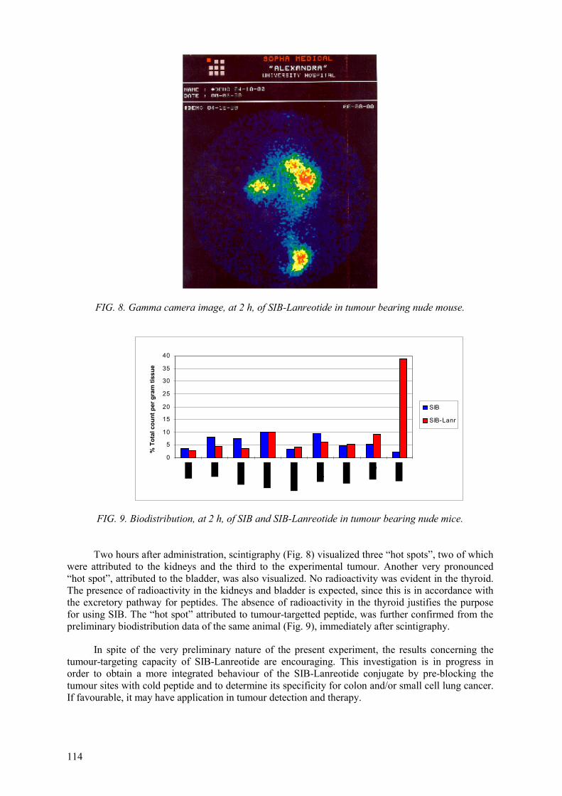
114
FIG. 8. Gamma camera image, at 2 h, of SIB-Lanreotide in tumour bearing nude mouse.
0
5
10
15
20
25
30
35
40
% T
otal
cou
nt p
er g
ram
tiss
ue
SIB
SIB-Lanr
FIG. 9. Biodistribution, at 2 h, of SIB and SIB-Lanreotide in tumour bearing nude mice.
Two hours after administration, scintigraphy (Fig. 8) visualized three “hot spots”, two of which were attributed to the kidneys and the third to the experimental tumour. Another very pronounced “hot spot”, attributed to the bladder, was also visualized. No radioactivity was evident in the thyroid. The presence of radioactivity in the kidneys and bladder is expected, since this is in accordance with the excretory pathway for peptides. The absence of radioactivity in the thyroid justifies the purpose for using SIB. The “hot spot” attributed to tumour-targetted peptide, was further confirmed from the preliminary biodistribution data of the same animal (Fig. 9), immediately after scintigraphy.
In spite of the very preliminary nature of the present experiment, the results concerning the tumour-targeting capacity of SIB-Lanreotide are encouraging. This investigation is in progress in order to obtain a more integrated behaviour of the SIB-Lanreotide conjugate by pre-blocking the tumour sites with cold peptide and to determine its specificity for colon and/or small cell lung cancer. If favourable, it may have application in tumour detection and therapy.

115
6. CONCLUSION
In keeping with the plan laid out at the 2nd RCM held in Athens, Greece for the labelling of the peptide, f-MLP, with SIB, the following goals were attained:
a) concerning optimization of SIB synthesis:
initially, for the labelling protocol employed, the mean yield of SIB was of the order of 52%, as assessed by TLC modification of the protocol, where the oxidant (T-BHP) was added last, resulted in an improved mean yield of SIB, of the order of 76%, as assessed by TLC the combination of TLC and column chromatography using silica gel proved suitable in identifying SIB furthermore, the ability of SIB to couple to protein was also used to confirm the presence of SIB; in this case, SEC and ITLC-SG proved suitable to confirm protein binding of SIB.
These results indicated that column chromatography using silica gel containing Sep-Pak, was appropriate for SIB purification.
b) concerning SIB conjugation to peptide:
SIB exhibited limited coupling (~9%) to free terminal amine (MLP) and non-amine containing peptide (f-MLP) high radioiodination yields (71%) were only possible with peptides containing amino-containing-side-chain amino acids. Lysine containing peptide retained stability, at 4oC, for at least 24 h. Reverse Phase HPLC proved the most suitable technique for assessing conjugation of SIB to peptide.
c) concerning the biological evaluation in normal mice of SIB and SIB-peptide conjugates, a number of tentative but interesting inferences can be made:
SIB and its peptide conjugates exhibit good in vivo stability since thyroid accumulation is low SIB-KRQKTENGASA has the slowest blood clearance compared to the other preparations SIB and SIB-peptides are cleared via the kidneys, the clearance being slowest with SIB-lysine-containing-peptide SIB and SIB-peptide preparations accumulate in the stomach Adrenal metabolism of SIB and SIB-peptide conjugates appears possible.
d) concerning the biological evaluation in colon tumour bearing nude mice of the SIB-Lanreotide conjugate, a tentative, but very encouraging inference can be made:
SIB-Lanreotide exhibits tumour-targetting capacity.
Furthermore, the approaches used to successfully overcome the problems encountered in the attempt to optimize peptide radioidination using SIB as the radioiodinating intermediate, resulted in development of a suitable quality control protocol. With this quality control protocol it was possible to assess each stage leading to the final product, using different chromatographic techniques. Each technique provided invaluable information concerning the relevant stage of its application in peptide radioiodination.
ACKNOWLEDGEMENTS
Part of this work was presented at the 6th World Hellenic Biomedical Congress, 11-14 October 2000 in Athens, Greece under the title of “Development of a Radiochemical Quality Control Protocol for Peptides Radioiodinated with N-Succinimidyl-3-Iodobenzoate to be Applied in Nuclear Medicine”. IAEA’s contribution was acknowledged at the Congress.

116
REFERENCES
[1] ZALUTSKY, M.R., NARULA, A S., Appl. Radiat. Isot. 38 (1987) 1051-1055. [2] WILBUR, D.S., HADLEY S.W., HYLARIDES, M.D., et al., J. Nucl. Med. 30 (1989) 216-226. [3] VAIDYANATHAN, G., ZALUTSKY, M.R., Nucl. Med. Biol. 22 (1995) 759-764. [4] TENNENBERG, S.D., ZEMLAN, F.P., SOLOMKIN, J.S., J. Immunol. 141 (1988) 3937-3944. [5] LEHNINGER, A.L., Biochemistry, (2nd ed.), New York, Worth Publishers Inc. (1977)
713-717.
117
PREPARATION OF 125I LABELLED COMPOUND
H. RAFII, D. BEIKI, M. MATLUBI, A.R. JALILIAN, F. MOTAMEDI, A.R., KARIMIAN, R. NAJAFI, M. BABAEI M. KAMALI DEHGHAN, G.R. SHAH-HOSSAINI, S.K. SHAFAHI, F. KESHAVARZI Atomic Energy Organization of the Islamic Republic of Iran, Nuclear Research Centre for Agriculture and Medicine, Karadj, Islamic Republic of Iran
Abstract
Iodinated compounds with 131I, 125I and 123I have been widely used for biochemical function studies. In conjunction with SPECT, [123I] labelled proteins have various diagnostic and therapeutic applications in nuclear medicine. In this study, synthesis and quality control of [18F]radiofluorinated and radioiodinated of some proteins and peptides as well as their biological behaviors are considered to be investigated.
1. INTRODUCTION
Development radiohalogenated peptides with 18F and 123I for biochemical studies is one of the most active areas of radiopharmaceutical research.
Direct radiofluorination of peptides via [18F]fluoride due to the high oxidation potential of fluoride is impossible as well via electrophilic [18F]fluorine leads to a low specific activity of labelled product. Therefore using prosthetic group such as {18F]N-succinimidyl-4-(fluoromethyl)benzoate, SFMB, is the only way . An approach for synthesizing [18F]SFMB via a multi-step reaction as well as preparation of [18F]labelled Serun Albumin according to Lang (1994) was reported by us in the 2nd RCM on 26-30 April 1999 at Athens, Greece.
Radioiodinated of some peptides like Vasoactive Intestinal Peptide, VIP, can be possible by both direct and indirect methods because of its two tyrosine and three lysine groups respectively on the main chain of molecule. It must be noted that preparation of radioiodinated VIP as a specific ligand for tumour associated receptors has already shown a promise in clinical applications. Since this peptide is expensive, it was decided at the first RCM to study IgG as a model compound. [125I]IgG was prepared by direct method using chloramine-T as a suitable oxidant according to Johnstone (1996) and reported by us in previous meeting. Based on the 2nd RCM at Athens, it was decided to use other peptides such as Formyl-Methyl-Leucyl-Phenylalanine, FMLF, as model compound because of its suitability for labelling, availability and cost. FMLF is a chemotactic peptide of great interest for the imaging (Vaidyanathan, 1995) and does not lend itself for direct radioiodination. Study of radiolabelled FMLF by indirect method using ATE/SIB has been the objective of our research project and once the procedures have been optimized, the method will be applied to VIP.
2. MATERIALS AND METHODS
2.1. Materials
N-chlorosccinimide (NCS), tri-n-buthyl tin chloride, n-butyl litium, dicyclohexyl carbodiimide (DCC), as well as n-hexane, ethylacetate and acetic acid of HPLC grade were purchased from Aldrich. All other chemicals were reagent grade and were bought from Merck and Aldrich Company. Silica SEPPAK cartriges (WAT020520) were obtained from Waters. ATE was provided generously by Drs. Pozzi and Salutsky through the IAEA and was also prepared by us. Amersham provided 125Iand our colleagues in Tehran NRC, Mr. Nabardi and his co-workers provided 131I. Sephadex G-50 as well as FMLF was purchased from Sigma Chemical Co.

118
2.2. Methods
TLC analysis was performed on percoated silica gel plastic plates (Merck) and visualized by UV at 254 nm. An HPLC system (Shimatzo) equipped with radioactive and UV detectors, a computer for peak analysis and a Si Kromasil column 5µ (250 × 4.6 mm) eluted with hexane, ethylacetate, acetic acid (70:29.88:0,12) at flow rate of 0.7 mL/min was used for analysis and quality control. Mass spectroscopy was performed using a MAT-MS-311spectrometer. 1H-NMR spectra were recorded on a BRUKER-FT-80. The chemical shifts (ppm) were reported to TMS as internal standard. Radioactivity was measured using a ionization chamber detector (Capintec CRC-15R). Flash chromatography was carried out on a column of 2 × 20 cm packed with Sephadex G-50, eluted by Phosphate Buffer Saline (PBS) pH7.4. [131I]labelled FMLF animal biodistribution was recorded on a SOPHA SPECT scanner (SWM International Company) as well it was performed with [125I]labelled FMLF by measering the different discarded organs activities.
2.2.1. Synthesis of tri-n-butthy stannyl -3 and -4-(tri-n-buthyl stannyl)benzoate
This compound was synthesized according to Garg (1989). The desired product was in the form of clear bright liquid in > 30% yield. 1H-NMR (CDCl3) spectrum of tri-n-butthy stannyl -4-(tri-n-buthyl stannyl)benzoate showed: 0.9-1.7 (m, 54H, 6 × n-but.); 7.59 (d, 2H, C3-H & C5-H), 7.8 (d, 2H, C2-H & C6-H) Fig. 1.
2.2.2. Synthesis of N-succinimidyl -3 and -4-(tri-n-buthyl stannyl)benzoate (ATE)
The desired compound was obtained according to Zalutsky (1987) in the form of oil liquid in 70% yield. 1H-NMR (CDCl3) spectrum of N-succinimidyl-4-(tri-n-buthyl stannyl)benzoate showed: 0.9-1.6 (m, 27H, 3 × n-but.), 2.89 (s, 2H, N-suc.), 7.6 (d, 2H, C3-H & C5-H), 8.03 (d, 2H, C2-H & C6-H) Fig. 2. Mass spectrum, m/e 452[M-(n-but.), 100% ], 395[M-(n-but.),30%] Fig. 2.
FIG. 1. Synthesis of tri-n-butthy stannyl -3 and -4-(tri-n-buthyl stannyl)benzoate.

119
FIG. 2. Synthesis of N-succinimidyl -3 and -4-(tri-n-buthyl stannyl)benzoate (ATE).
2.2.3. Synthesis of N-succinimidyl-3 and-4-iodobenzoate, (SIB)
This compound was prepared accorrding to both Zalutsky (1987) and Khawli (1989) using N-chlorosuccinimide (NCS) and chloramine-T, respectively, as oxident agents (starting material). TLC analysis of the reaction mixture with 30% ethylacetate in n-hexane showed the Rfs of 0.0, 0.07, 0.25and 0.37 for I, NCS, ATE and SIB, respectively. The desired SIB was obtained in 80% yield. 1H-NMR (CDCl3) spectrum of N-succinimidyl-4-iodobenzoate showed 2.9 (s, 2H, N-suc.), 7.8 (d, 2H, C3-H & C5-H), 7.9 (d, 2H, C2-H & C6-H) Fig. 3.
3. LABELLING
3.1. Synthesis of [125I and 131I]N-succinimidyl-3-iodobenzoate,[ 125I and 131I]SIB
[125I and 131I] iodinated SIB was synthesized according to Zalutsky (1987) using NCS as starting agent. Briefly, in a conical vial of 2 mL the following reagents were added:
10 µl ATE dissolved in anhydrous chloroform; 1 µmol of 0.1M ATE(for making anhydrous chloroform, a few grame anhyrdrous sodium sulphate was added to about 10 mL chloroform). >30 µl [125I and 131I]NaI in 0.02N NaOH, about 0.5 to 1.5 mCi; 50 µl acid acetic glacial in anhydrous chloroform; 50 µmol acid acetic, 290 µl acid acetic in 4.7 mL chloroform; 50 µl NCS; 1M NCS in anhydrous chloroform (the solubility of NCS in chloroform is not good).

120
The reaction mixture was stirred at room temperature for 30 min and radioiodinated SIB was isolated, as follows:
a silica gel cartrige was saturated by 25 mL n-hexane; the reaction mixture was loaded on the column with help of 3 × 100 µl hexane (fraction 1); the column was washed by 8 × 5 mL hexane (fraction 2-9), 5 × 5 mL 8% ethylacetate in hexane (fraction 10-14), 6 × 1 mL (fraction 15-20)and finally 2 × 5 mL (fraction 21-22) 30% ethylacetate in hexane. The activity of each fraction was measured to calculate labelling yield (calculated yield is about 40-60%. Relative activity per mL of each fraction, elution profile, is shown in Fig. 4.
HPLC analysis of KI, NCS, ATE, SIB cold, fractions 10-11 and fractions 16-19 in the elution profile were compared to identify each fraction, Table I and Fig. 5. It was seen that the fractions 16-19 contained desired labelled SIB. Therefore, the 30% ethylacetate in hexane fractions were pooled and evaporated to a small volume with a steam of nitrogen, transferred to a conical vial and by continuing evaporation of eluent to dryness, labelled SIB was obtained.
FIG. 3. Synthesis of N-succinimidyl-3 and-4-iodobenzoate, (SIB).

121
FIG. 4. Relative activity per mL of each fraction to calculate labeling yield.
FIG. 5. Comparison of HPLC analysis of KI, NCS, ATE, SIB cold, fractions 10-11 and fractions 16-19 in the elution profile to identify each fraction.

122
TABLE I. HPLC ANALYSIS FOR IDENTIFICATION OF [I-125 & 131] SIB AND ITS PURITY1
Determined NCS & I I ATE SIB & NCS Injected KI 4.4 * 7.6 NCS 4.9 * 20.8 ATE 4,6 6,5 10.1 * SIB cold 6.7 19.9 * [131I]fr.11-12 4.4 11.5 * 20.0 [131I]fr.16-19 7.1 19.6 * [125I]fr.16-10 6.3 20.0
1Taken with: Shimatzu HPLC system, Si Kromasil 100,5 µ (250 × 4.6 mm) column, 30% ethylacetate in hexane (a drop of acetic acid), 0.7 mL/min flow rate.
*Dominant peak.
3.2. Radioiodination of FMLF
Labelling studies were done both by direct method using chloramine-T according to Khawli (1989) and indirect method using [125I and 131I]SIB according to Zalutsky (1987). Since FMLF dose not lend itself for direct radioiodination, it was seen that, after labelling, the most unreacted radioiodine was isolated from the reaction mixture by a column packed with Sephadex G-50 eluted with 0.01M PBS, pH7, 4, Fig. 6.
Briefly, radioioidination of FMLF via [125I and 131I]SIB was carried out, as follows:
50 µl FMLF was added to the labelled SIB (200 µl FMLF/50 µl 0.1 M borate buffer, pH8.5, 9.0, 9.5 and 10.0) and the mixture was stirred in ice-water bath for 30 min. 300 mL glycine (200 mM in 0.1 M borate buffer, pH8.5) was added to terminate the reaction followed by an incubation of 5 min at room temperature. The labelled product was isolated from reaction mixture using a column Sephadex G-50 eluted with PBS, pH7.4 at a flow rate of 1 mL/min. The eluted of 0.5 mL was collected in a test tube and counted to get elution profile. The fractions between 20-22 mL were labelled FMLF, Fig 7.
FIG. 6. Isolation of the most unreacted radioiodine from reaction mixture by a column packed with Sephadex G-50 eluted with 0.01M PBS, pH7, 4 after labelling.

123
FIG. 7. Labelling of fractions between 20-22 mL.
4. BIOLOGICAL STUDIES
Biodistribution studies were performed according to Vaidyanathan (1995) and Zalutsky (1987) both in normal mice and the ones bearing 50 µl turpentine for 24h, promoted inflammation in right leg (twelve normal mice and twelve perinjected turpentine ones weighing 20-28 g). Each animal was received 6-12 µCi [125I]FMLF intravenously via tail vein (three mice per time point), sacrificed 0.5, 2, 4 and 24 hours postinjection and dissected. The tissues of interest were discarded, washed with saline, dried, weighed and counted by a gamma counter to obtain biodistribution. The tissue biodistribution results were expressed as the per centage of the injected dose, localized per gram of tissue, Figs 8 and 9. Presence of radioactivity in urine and bladder encouraged us to make the SPECT scan on the mice which were received [131I] iodide and [131I]FMLF. These scans confirmed the difference radioactivity localized in different organs, especially in thyroid, Fig. 10.

124
FIG. 8. Results of study of biological behavior of labelled FMLF in mice.
FIG. 9. Results of study of biological behavior of labelled FMLF in mice.

125
FIG. 10. Comparative SPECT scan performed with normal mice 2h after the animals received about 30 µCi [131I] iodide and [131I] SIB–FMLF.
5. RESULTS OBTAINED
(a) The synthesis of following intermediate products was performed, Figs (1, 2, 3):
tri-n-buthyl stannyl-3 & 4-(tri-n-buthyl stannyl)benzoate in >30% yield. N-succinimidyl-3 & 4-(tri-n-buthyl stannyl) benzoate, ATE in 70% yield. N-succinimidyl-3 & 4-iodobenzoate, SIB in 80% yield. The procedure for ATE synthesis and its intermadiate products must be reconsidered because of low yield of intermadiate products.
(b) Radioiodination of SIB with radioiodine 125 and 131 was performed satisfactorily in radiochemical yield more than 40-70% (Fig. 4). Comparative HPLC analysis of IK, NCS, ATE, SIB(cold) and the elution profile of radioiodinated SIB shows a high radiochemical purity in [125I and 131I] SIB, 99%, Fig. 5 and Table I.
(c) FMLF labelling was done both by direct method using chloramine-T and indirect method via radioiodinated SIB and saw that FMLF does not lent itself for direct labelling using chloramine-T fig (6). FMLF labelling via radioiodined SIB was performed at different pH8.5, 9.0, 9.5, 10.0 and saw that radiochemical yields decrease with increasing pH, Fig. 7, and the best yield was found at pH8.5.
(d) Biological behavior of labelled FMLF was studied in mice and 24h after the animal received 6-12 µCi [125I] SIB–FMLf, Figs 8 and 9.

126
A comparative SPECT scan was performed with normal mice 2h after the animals received about 30 µCi [131I] iodide and [131I] SIB–FMLF to distinguish why high labelled FMLP has been localized in bladder and urine, Fig. 10.
6. CONCLUSIONS
Mode of preparation of ATE should be reviewed because of very sensitivity of the reaction for preparation tri-n-buthyl stannyl-3- (tri-n-butyl stannyl) benzoated.
Radioiodination of ATE with (125I) and (131I) was performed successfully and labelled SIB was isolated in high radiochemical purity.
Some peptides such as FMLF do not lend themselves for direct labelling method using chloromine-T. Therefore their labelling must be considered by indirected method. Labelling of FMLF was performed satisfactoryly via [125I] and [131I] SIB at pH8.5. With increasing in pH, yield of labelled FMLF decreased perhaps because of interaction OH to carboxyl group of SIB resulting m-iodobenzoic acid. Using HPLC instead of Sephadex-50 for isolation of labelled FMLF is in our programme.
Biological studies of labelled FMLF shows a low uptake of thyroid but a high at urine and bladder. Perhaps because of FMLF molecular weight is low in comparison to other peptides and for this reason this molecule could pass from blood to urine.
REFERENCES
[1] LANG, L., ECKELMAN, W.C., Appl. Radiat. Isot. 12 (1994) 1155 pp. [2] JOHNSTONE, A., THORPE, R., In: Immunochemistry in practice, Blackwell Science Ltd.
(1996) 134 pp. [3] VAIDYANATHAN, G., ZALUTSKY, M.R., Nucl. Med. Biol. (1995) 760-764. [4] GARG, P.K., et al. Appl. Radiat. Isot. 40(6) (1989) 485-490. [5] ZALUTSKY, M.R., NARVLA, A.S., Appl. Radiat. Isot. 38(12) (1987) 1051-1055. [6] KHAWLI, L.A., KASSIST, A.I., Nucl. Med. Biol. 16(7) (1989) 727-733.

127
OPTIMIZATION OF SYNTHESIS AND QUALITY CONTROL PROCEDURES FOR THE PREPARATION OF 18F-LABELLED PEPTIDES
J.K. AMARTEY King Faisal Specialist Hospital and Research Centre, Riyadh, Saudi Arabia
Abstract
Radiohalogenation via prosthetic groups has provided a useful route for labelling proteins, peptides and drug molecules. This method is the only option available as far as molecules that are not amenable to the classical radiohalogenation reactions are concerned. This pertains to proteins and peptides lacking tyrosyl groups in their structure. More importantly, radiofluorination by electrophilic method has not been developed for labelling these macromolecules. The need to optimize methods and techniques to enable efficient labelling and fully exploit the potential biochemical application of these molecules prompted this investigation. Reaction conditions were optimized to prepare ethyl 4-[18F]-benzoate from an ammonium precursor, ethyl 4-trimethylammoniumbenzoate.triflate in excellent yield. The fluorinated ester was hydrolyzed quantitatively to the acid. The acid was then converted to the activated N-succinimidylfluorobenzoate (SFB) using O- (N-succinimidyl)-tetramethyluroniumtetrafluoroborate also typically in greater than 90% radiochemical yield. The activated ester was purified either by HPLC or SEPPAK cartridge and was conjugated to a potent chemotactic peptide (Formyl-Nle-Leu-Phe-Nle-Tyr-Lys) as a model in acetonitrile. The conjugate was purified chromatographically or by SEPPAK cartridges. To ascertain the retention of biological activity of the peptide after these chemical manipulations, the superoxide production assay was employed. The purified [19F]-peptide conjugate specifically bound and activated human polymorphonuclear leukocytes to generate superoxide in a dose dependent manner. Biodistribution in normal mice showed that the conjugated peptide did not suffer any significant dehalogenation in vivo. This was indicated by the low uptake of activity in bone. The methodology developed with the chemotactic peptide was used to label RC-160 (cyclic–D-Phe-Cys-Tyr-D-Trp-Lys (Boc)-Val-Cys-Trp-NH2) a SST analog. The conjugate peptide inhibited the growth of human cancer cell line in a dose dependent manner as expected. The biodistribution showed that the [18F]-RC-160 bound to tumour xenograft in nude mice. The main objective of the project was to optimize reaction conditions and quality control procedures for prosthetic labelling of peptides. Hence the aims of the project were to: – improve fluorination of prosthetic group such as fluorobenzoate, synthesis and isolation of the succinimidyl
activated ester of the prosthetic group and reaction conditions to couple the activated ester to a model chemotactic peptide, and to develop an isolation method for the peptide conjugate;
– perform in vitro and in vivo experiments to ascertain the biological activity of the conjugate; – apply the methods and techniques developed to label another biologically active peptide and evaluate its
potential application as a cancer detection agent.
1. INTRODUCTION
Radiolabelled peptides continue to emerge as molecules for targeting numerous diseases, most often cancers. One of such peptides is somatostatin (SST) a naturally occurring peptide with a wide spectrum of biological functions. Several cancers overexpress somatostatin receptors (SSTR) more than normal tissue and hence radiolabelled SST has been used in combination with nuclear imaging devices to detect these cancers in vivo. One of the synthetic analogs of SST, octreotide, has been labelled with 123I and 111In nuclides and used successfully in human patients [1-2]. Attempts have been made to employ other nuclides such as 99mTc to label SST and analogs [3-5]. The favourable kinetics of these peptides pertaining to their receptor-ligand interactions imply that labelling with short lived positron emitting radionuclides will allow use of the better resolution and quantitative characteristics of PET camera for these studies. Hence efforts to label octreotide with 18F and gallium-68 have been reported [6-10].
Many investigators are pursuing fluorination of several potentially useful peptides, polypeptides and proteins. The prosthetic approach has been very attractive because other methods have either been tedious or given very low radiochemical yields. Additionally, use of the prosthetic label introduces a fluorine atom covalently bound to an aromatic ring. Chemically this carbon-fluorine bond is more stable than the aliphatic counterpart [11-17]. Moreover, the electrophilic fluorination of
128
bioactive molecules such as proteins and peptides has not been intensively pursued because of several obvious biological and chemical reasons.
This report covers optimization of synthesis and quality control methods of fluorinated aromatic prosthetic groups, such as N-succinimidylfluorobenzoate, and application of the activated ester to label a model chemotactic peptide and RC-160. Additionally, the specificity of the labelled peptide was evaluated by in vitro and in vivo experiments.
2. MATERIALS AND METHODS
The chemicals used in the study were all purchased from Aldrich (St. Louis, MO, USA) and were used without further purification except where stated. The chemotactic peptide (Formyl-Nle-Leu-Phe-Nle-Tyr-Lys) was purchased from Sigma (USA). RC-160 or [Lys5(Boc)]-Vapreotide was purchased from DebioPharm, Lausanne, Switzerland. Acetonitrile was kept over molecular sieves. SEPPAK cartridges were purchased from Waters-Millipore (USA). The High Pressure Liquid Chromatography (HPLC) analysis was carried out on Econosil C-18, or silica, 10 µ columns (semipreparative, 250 mm × 10 mm or analytical, 250 mm × 4.6 mm). TLC was run on silica gel (aluminum backing) plates with 1:1 v/v ethylacetate: hexane as the mobile phase. The detection was by a JASCO chromatographic system equipped with a variable wavelength ultraviolet monitor and in tandem with a Canberra flow through radioactivity detector. The UV absorption was monitored at 254 nm. Chromatograms were acquired and analysed using BORWIN® software. NMR was run on JNM-GX270 Spectrometer.
Wallac 1480-Wizard® (Turku, Finland) gamma counter was used to count biodistribution and radioligand binding assay samples. Fluoride was produced on a CS30 cyclotron.
2.1. Fluorination reaction
The fluorination reaction was carried out on ethyl 4-(N, N, N–trimethylammonium) benzoate.triflate precursor (TMAB.OTf). The TMAB.OTf was synthesized by the method reported earlier [18]. Aqueous [18F]-fluoride was produced by the 18O (p, n) 18F reaction. The fluoride activity (2-20 mCi, 74-740 MBq) was trapped in kryptofix® 2.2.2. (5 mg) and potassium carbonate (1 mg) in acetonitrile/water solution (950 µL/50 µL), and dried by azeotropic distillation with aliquots of acetonitrile. The solid residue was resolubilized in 0.2-0.3 mL of DMSO, DMF or CH3CN containing the required amount of the precursor (typically 2 mg). In DMSO the reactants were heated at 150oCand aliquots taken for analysis at various time points. In DMF or CH3CN the reaction mixture was carried out at 90-100oC. Fractions were taken for chromatographic analysis at 2, 5, 10, and 20 min.
2.2. Hydrolysis of the fluorinated intermediate
Hydrolysis was performed in 0.25 mL of 1M NaOH heated for 10 min at 100oC. The solution was acidified with HCl (1M, 0.3 mL) and the fluorobenzoic acid isolated by SEPPAK C-18 and AG50W-X8 cationic column in tandem. The acid was isolated alternatively with only the SEPPAK C-18 cartridge. The product was eluted with methanol or acetonitrile after washing with water and drying with a gentle flow of nitrogen (or argon) gas.
2.3. Synthesis of N-succinimidyl-4-fluorobenzoate (SFB)
The acid fraction in a reacti-vial (2 mL) was basified with either triethylamine or tetramethylammonium hydroxide (TMA.OH) and evaporated to dryness, accelerated by a stream of nitrogen (or argon) gas. The drying was repeated with 2 × 0.5 mL portions of CH3CN. Di-N-succinimidylcarbonate (DSC) in 0.2 mL of dry CH3CN were added to the acid and heated at 155oCfor 10 min. The product was analysed on TLC-SG and also by HPLC. The SFB was purified by SEPPAK silica, eluting with CH3CN or Hexane-Ethylacetate-Acetic acid 700/300/10 on silica HPLC column. The product was dried by passage of a steady steam of nitrogen gas.

129
The synthesis of SFB has been achieved in a higher and consistent yield using O-(N-succinimidyl)-tetramethyluroniumtetrafluoroborate (TSTU). In this case the fluorobenzoic acid was added to 50 µL of 10% solution of TMA.OH in acetonitrile and evaporated to dryness. TSTU, 5 mg also dissolved in 250 µL of acetonitrile was added and the reactants heated at 95oC for 10 min. The reaction mixture was applied to a SEPPAK silica cartridge previously activated with anhydrous ether. The cartridge was washed with 1 mL portions of hexane/ethylacetate, 7:3 v/v. The first fraction was discarded, and the SFB collected in the next 3 mL. Alternatively, the activated ester was loaded onto a semi-preparative normal phase column (Econosil silica, 10 µ, 10 × 250 mm), and eluted with hexane/ethylacetate/acetic acid 700/300/10 as the mobile phase at a flow rate of 4 mL/min. The SFB was purified on activated C-18 SEPPAK as a third method. In this case the reaction mixture was diluted with 1% acetic acid solution in water (1 mL), loaded onto the SEPPAK and flushed through. The cartridge was washed with 65/35 mixture of water/acetonitrile (1.5 mL). The cartridge was dried with nitrogen gas and the product eluted with acetonitrile (1.5 mL).
The SFB fraction was collected and evaporated to dryness assisted by a stream of nitrogen gas. The purity of SFB was confirmed by HPLC on Econosil C-18 column, the mobile phase was methanol/water/formic acid, 800/200/1 at a flow rate of 2 mL/min.
2.4. Synthesis of SFB conjugate of amine compounds
SFB was used to fluorinate several model amine containing molecules including BSA, L-Phenylalanine, IgG and RC-160. In the case of proteins, 100-1000 µg of the substrate dissolved in borate buffer 0.1 mL (0.1 M) pH8.0 were added to the dry residue of SFB and allowed to incubate at room temperature for 30 min. The reaction mixture was then analysed by HPLC (Shodex , size exclusion column) eluted with phosphate buffered saline (PBS) 0.1 M, pH7.4. The extent of labelling was also determined by trichloroacetic acid precipitation.
Coupling reaction with the somatostatin analog was performed in borate buffer or acetonitrile with a trace of DMF. The peptide (250 µg) dissolved in acetonitrile was added to the dry residue of the SFB. This was followed by the addition of the required amount of triethylamine (TEA) typically 20 µL of 73 mM solution in acetonitrile. The reaction mixture was then heated at a set temperature and duration.
2.5. Conjugation of SFB to chemotactic peptide (Formyl-Nle-Leu-Phe-Nle-Tyr-Lys)
The required amount of the activated ester in dry acetonitrile or hexane/ethylacetate was placed in a reacti-vial and the solvent was evaporated to dryness at ambient temperature aided by a steady steam of nitrogen gas. The peptide dissolved in acetonitrile/DMF (9:1 v/v) was added to the dry residue to give approximately 1:1 molar ratio of peptide to SFB. Twenty microliter of a 73 mM solution of TEA in acetonitrile was added and incubated at ambient temperature or heated at a preset temperature in a heating block. The pH(paper) of the reaction solution was approximately 10. The reaction yield was determined by HPLC analysis of the reaction mixture on the reverse phase column, using the system described above. The effect of time, temperature, peptide concentration and solvent on the reaction yield was investigated.
The coupling reaction was repeated in borate buffer (pH8.0 and 8.5) and in borate-acetonitrile-DMF system. In this case the reactants were incubated at ambient temperature (22oC) or warmed at 50oC for 15 min. The reaction mixture was then analysed by HPLC using the reverse phase system. The fluorobenzoyl-conjugated peptide was purified on SEPPAK C-18 cartridge. The cartridge was activated with ethanol (5 mL) followed by water wash (10 mL). The reaction mixture was diluted with water and quantitatively transferred onto the SEPPAK and washed with water (5-10 mL). The cartridge was rid of excess water with air or nitrogen gas. The labelled peptide was eluted with 1 mL of ethanol. A fraction of the purified peptide conjugate in ethanol was evaporated to dryness with a stream of nitrogen. The dry residue was subjected to electron spray mass spectrometric analysis. The peptide conjugate for biochemical testing was constituted in DMSO diluted with appropriate buffer or

130
0.9% sodium chloride solution for injection. The conditions developed were used to synthesize the [18F]-SFB conjugate of the chemotactic peptide (scheme below).
2.6. Luminol-dependent superoxide assay
The fluorobenzoyl-peptide conjugate was used to activate isolated human polymorphonuclear (PMN) leukocytes and the reactive oxygen species produced were measured using a luminescent spectrometer. Briefly, the cells were isolated by standard salt gradient method. The peptide conjugate solution was prepared in TRIS buffer, 170mM, pH7.4 containing 0.15mM CaCl2, 0.5mM MgCl2,0.1mM phenylmethylsulfonylfluoride (PMSF), 0.1% BSA and 0.05% DMSO. The concentration range of 10-10 to 10-5 M of the peptide conjugate was used. In a typical test 106 cells in 0.1 mL were incubated for 60 s at 37oC in the buffer in the presence of (1 nmol) of luminol in a sample cuvette. The sample was place in the spectrometer after addition of the conjugate and the chemiluminescence signal recorded. A positive control experiment was performed in the presence of 10-6 M of the chemotactic peptide fMLF. The signal intensity of the peptide conjugate was compared with and normalized against the fMLF control. The concentration of the conjugate effecting 50% of maximum response was then estimated from the signal versus concentration plot.
2.7. Biodistribution of the peptide conjugate in normal mice
Each mouse was injected with 0.1 mL of the 18F labelled peptide solution containing approximately 20 µCi (740 kBq) of radioactivity. Four mice were sacrificed by cervical dislocation at 30 min, 2 h and 4 h postinjection and organs and tissues of interest were dissected, wiped of excess blood, weighed and counted in a gamma counter calibrated for 18F. The per cent injected dose/gram was then calculated for the samples. The injected dose per mouse was estimated by counting a standard sample taken from the injectate. The animal experiments were carried out in accordance with institutional, national and international guidelines for humane use of animals for research.
2.8. Synthesis of RC-160 SFB conjugate
RC-160 was coupled to purified [18/19F]-SFB using the method developed with the chemotactic peptide. In this case two buffer/solvent systems were investigated, acetonitrile: borate buffer, 0.1M pH=8.5 (9:1 v/v) or acetonitrile/DMF with triethylamine in acetonitrile (73 mM) as the base. The reaction was performed at 90±5°C for 30 min. The reaction mixture was purified by the HPLC-RP system described above, after deprotection with TFA or HCl at ambient temperature for 5 min.
2.9. Growth inhibition of HTB-121 cells by RC-160 conjugate
The biological activity of the RC-160 conjugate was assessed by thymidine incorporation assay on HTB-121, a human breast cancer cell line. The cells (105/reaction) were incubated with increasing concentration of the [19F]-RC-160 conjugate in the presence of 1 µCi (37 kBq) of [3H]-thymidine. The cells were isolated and counted and the incorporated radioactivity compared with the control.
2.10. Biodistribution of the RC-160 conjugate in mice
The biodistribution was performed in normal female mice to ascertain the distribution profile of the labelled peptide. Mice (25-30 g) were each injected via the tail vein 0.1 mL of HPLC purified conjugate formulated in saline for injection containing 5% ethanol. Each dose contains 20 µCi (740 kBq) of radioactivity. Group of animals was sacrificed at 30 and 120 min and then organs and tissues of interest were dissected, wiped of excess blood, weighed and counted in a gamma counter. The per centage of the injected dose per gram was then calculated for all tissues using a stored sample of the injection solution to estimate the total dose injected per mouse. The biodistribution was repeated with mice bearing the xenograft of the cell line.

131
2.11. Results and discussion
The fluorination reaction in CH3CN was very efficient and consistently gave greater than 90% labelling yield within 10 min. Ten minute alkaline hydrolysis at 100oC usually gave a quantitative yield of the fluorinated acid. The SEPPAK C-18 /AG50W- X8 purification was attempted in an effort to remove any unreacted TMAB.OTf, however, it was apparently converted probably to the hydroxybenzoic acid and could not be removed effectively. The method was changed to remove the unreacted TMAB.OTf before hydrolysis with the AG50W-X8. This effort did result in only a slight improvement in the final reaction yield.
The purity of the acid as well as the SFB was ascertained by TLC-SG in 1:1 hexane: ethylacetate or by HPLC on C-18 column using 800:200:1 methanol: water: formic acid as the mobile phase. The fluorination reaction yields in the three solvent systems are shown (Table I) below. Rf-values of 0.0, 0.1, 0.3 and 0.7 were determined for 18F-, the acid, SFB and the fluorinated ethylbenzoate respectively. Representative TLC and HPLC chromatographic scans are shown in Figs 1-6. The mass spectral analysis agreed with the expected structure (m/z = 237).
DCC + NHS, DSC and TSTU activation have been attempted with various degrees of success. The DSC gave a better result than DCC + NHS and easier to handle in terms of chromatographic separation and purification. However, the reaction yield of SFB has not been very consistent although very good results have been recorded several times. These inconsistent results can be attributed to incomplete drying of the acid. The results are shown (Table II) below. The TSTU activation gave the highest and consistent yield.
The results of SFB conjugation to some amine containing molecules are shown (Table III) below. The yield with IgG was extremely low as a result of faster hydrolysis of the SFB in the aqueous medium. Several purification methods have been attempted on the SFB reaction mixture but the HPLC method gave the purest product as indicated by the extent of coupling yield with the chemotactic peptide (Tables IV and V). The [18F]-SFB purified by SEPPAK gave comparable conjugation yield to the HPLC purified material.
TABLE I. FLUORINATION RADIOCHEMICAL YIELD
Solvent Per cent Yield DMSO 74.0 ± 13.6 (n =16) DMF 73.0 ± 21.0 (n = 3) CH3CN 90.7 ± 7.7 (n = 18)
TABLE II. SFB REACTION YIELD
Activating Agent Per cent Yield DSC 77.2 ± 16.9 (n = 16) TSTU 96.1 ± 2.7 (n = 7)
TABLE III. SFB COUPLING REACTION YIELD
Substrate Per cent Yield
IgG 16 Phenylalanine 68 RC-160 54

132
FIG. 1. A representative TLC scan of the fluorination reaction mixture.
FIG. 2. A representative TLC scan of the 18F-fluorobenzoic acid.
FIG. 3. A representative TLC scan of the 18F-succinimidylbenzoate.

133
FIG. 4. HPLC chromatograms of radiofluorination reaction mixture. The top (UV) and bottom (radioactivity).
FIG. 5. HPLC chromatograms of 18F-fluorobenzoic acid. The top (radioactivity) and bottom (UV).

134
FIG. 6. HPLC chromatograms of SEPPAK C-18 purified 18F-SFB conjugate. The top (radioactivity) and bottom (UV).
The results showed that the effect of concentration on the reaction yield was not significantly different in the 1-5 mg/mL range (Table V). The reaction in pure DMF gave equally good results. It facilitated the dissolution of the peptide and also minimized the hydrolysis of SFB and as a result the amount of acid by-product formed was negligible. The reaction in acetonitrile and borate buffer or borate buffer alone also gave good coupling yield, however the competing hydrolytic reaction was extensive. When the reaction was performed at ambient temperature in either pure organic or aqueous medium the coupling yields were inconsistent and lower than when heated. When the coupling was performed at pHbelow 8, there was only a trace of the conjugate. Hence heating is required to attain any significant conjugation. Representative HPLC chromatograms of the chemotactic and SFB conjugation reaction mixture and the purified conjugate are shown in Figs 7 and 8. SEPPAK purification method for the labelled peptide was very efficient and gave a good recovery of the product. However, this was also accomplished by HPLC.
TABLE IV. THE EFFECT OF BORATE BUFFER PH ON THE COUPLING OF SFB TO THE CHEMOTACTIC PEPTIDE AT AMBIENT TEMPERATURE
pH Per cent reaction yield*
7.24.7 ± 2.4 (n=3)
8.026.5 ± 5.5 (n=4)
8.548.4 ± 14.6 (n=5)
*The experiments were performed with 3 mg/mL of peptide concentration. The values are means and standard deviation.
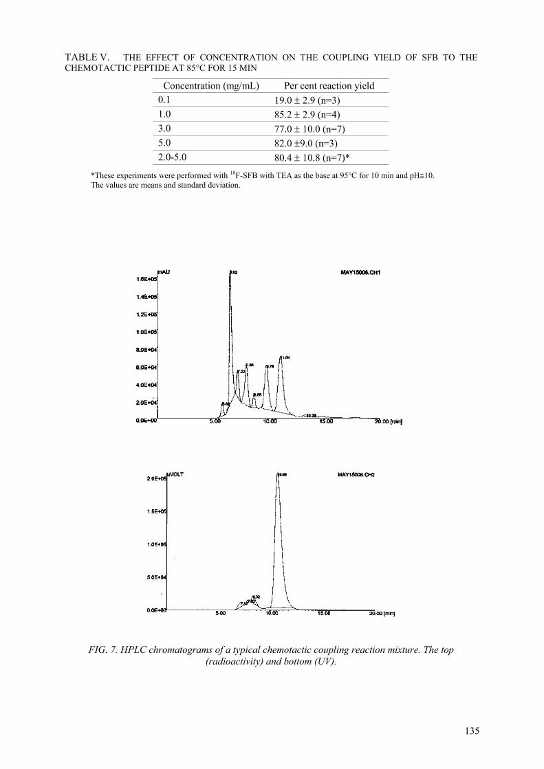
135
TABLE V. THE EFFECT OF CONCENTRATION ON THE COUPLING YIELD OF SFB TO THE CHEMOTACTIC PEPTIDE AT 85°C FOR 15 MIN
Concentration (mg/mL) Per cent reaction yield 0.1 19.0 ± 2.9 (n=3) 1.0 85.2 ± 2.9 (n=4) 3.0 77.0 ± 10.0 (n=7) 5.0 82.0 ±9.0 (n=3) 2.0-5.0 80.4 ± 10.8 (n=7)*
*These experiments were performed with 18F-SFB with TEA as the base at 95°C for 10 min and pH 10.The values are means and standard deviation.
FIG. 7. HPLC chromatograms of a typical chemotactic coupling reaction mixture. The top (radioactivity) and bottom (UV).

136
FIG. 8. HPLC chromatograms of purified 18F-chemotactic-SFB conjugate. The top (radioactivity) and bottom 19F-conjugate(UV).
The sequence of addition of the reagents was found to be vital during the conjugation reaction. When the base (TEA) was added to the peptide before addition to the dry SFB, a gelatinous precipitate was observed consequently reducing the coupling yield. Therefore the peptide was always added to the dry SFB followed by the addition of the TEA preferably diluted in acetonitrile (73 mM).
The F-19-chemotactic conjugate was subjected to electron spray (ES) mass spectrometry (Fig. 9). The results confirmed the presence of the expected molecular ion at 946 (M+1 = 947). A peak at m/z = 269 corresponds to a fragment of the lysyl-fluorobenzoyl group. It can be inferred that the coupling indeed occurred through the amine function of the lysine. The base peak was the fragmentation at the amide bond of leucine and the phenylalanine groups (m/z = 432).
The superoxide assay results are shown in Fig. 10. The data showed that the conjugate binds to the PMN cells and elicit the expected biological activity. The estimated concentration effecting 50% of maximum response ED50 was about 5x10⁻⁹ M. The value was in the range previously reported [19-20] and implies that the conjugate was much more potent than the classical f-MFL. This observation suggests that the fluorination reaction conditions did not alter the biological activity of the peptide to any significant extent.
[18F]-RC-160 radiochemical yield was much lower than that obtained with the chemotactic peptide. The low yield could be due to steric factors and low basicity of the phenylalanine terminal amino group in comparison with the lysine -amine. It must be mentioned that the total synthesis time

137
to obtain the peptide conjugate was approximately 120 min (data not shown). The automation of some of the purification steps may reduce the synthesis time. In view of that the use of solid phase extraction and purification methods becomes attractive.
The fluorinated conjugate of RC-160 showed concentration-dependent growth inhibition of the HTB-121 cells. This is in agreement with the observed high affinity binding of the radioiodinated analog of the peptide to SSTR expressed by this cell line (unpublished data).
Biodistribution of the fluorinated chemotactic conjugate in normal CBA/J mice (Table VI) showed localization of radioactivity in the liver, lung, and spleen. The kidneys also showed significant uptake. The data implies that the hepatobiliary and the urinary systems are the main elimination routes of the radiotracer.
The [18F]-RC-160 showed highest localization in the lung, liver, and spleen (Table VII). There was significant accumulation in the kidneys as well indicative of renal and hepatobiliary routes of excretion of the tracer. Additionally there was accumulation of the tracer in the tumour xenograft. It is interesting to note that only these three organs had less than unity tumour to tissue ratio.
FIG. 9. The ES-MS of the 19F-SFB-chemotactic peptide conjugate.
FIG. 10. The effect of concentration of fluorinated chemotactic peptide on the production of superoxides by human PMN leukocytes.

138
TABLE VI. BIODISTRIBUTION OF [18F]-FLUOROBENZOYL- (FORMYL-NLE-LEU-PHE-NLE-TYR-LYS) CONJUGATE IN NORMAL MICE
Tissue/Time 30 min 2 h 4 h Blood 3.5 ± 0.3 1.6 ± 0.6 1.2 ± 0.5 Liver* 53.6 ± 11.6 9.2 ± 1.8 3.6 ± 0.7 Lung 63.6 ± 20 13.9 ± 1.1 8.7 ± 3.0 Kidney 20.0 ± 5.5 4.6 ± 0.2 1.8 ± 0.6 Small intestine# 4.2 ± 1.4 1.1 ± 0.4 0.4 ± 0.2 Large intestine# 2.8 ± 1.1 1.5 ± 1.0 1.1 ± 0.4 Heart 9.6 ± 2.6 2.2 ± 1.1 0.7 ± 0.1 Muscle 3.4 ± 1.6 1.5 ± 0.3 0.5 ± 0.2 Bone + marrow 6.3 ± 2.1 1.0 ± 0.2 1.5 ± 0.7 Spleen 11.1 ± 0.2 4.4 ± 0.9 2.9 ± 0.6
The values are means and standard deviation of per centage injected dose gram (n = 3). *The values are for the whole organ. # The intestines were with content.
TABLE VII. THE BIODISTRIBUTION OF LABELLED RC-160 IN NORMAL FEMALE MICE. THE VALUES ARE MEAN AND STANDARD DEVIATION OF PER CENT INJECTED DOSE/ GRAM, (N=4)
Time/tissue 30 min 120 min Tumour/ tissue
Blood 1.73 ± 0.35 0.86 ± 0.11 1.6 Liver 13.28 ± 2.71 8.99 ± 2.27 0.15 Lung 6.67 ± 1.37 5.37 ± 1.16 0.25
Kidney 1.67 ± 0.33 0.91 ± 0.04 1.5 Heart 0.60 ± 0.18 0.31 ± 0.05 4.4
Muscle 0.37 ± 0.10 0.16 ± 0.06 8.6 Bone 0.42 ± 0.03 0.45 ± 0.20 3.0
Spleen 3.76 ± 0.19 5.24 ± 0.40 0.26 Tumour* ----- 1.38 ± 0.21 ---
(Preliminary study * n = 2. The tumour: tissue ratios are at 120 min).
4. CONCLUSIONS
Conditions have been systematically investigated to optimize the fluorination of ethylbenzoate precursor via the nucleophilic substitution route. Greater than 90% reaction yield was attained in 10 min at 95oC using acetonitrile as the solvent. The fluorobenzoic acid intermediate was obtained quantitatively by alkaline hydrolysis of the fluorinated ethylbenzoate. The acid was activated by TSTU to obtain the SFB in excellent yields. The activated ester was coupled to the model chemotactic peptide in very good yield when acetonitrile and DMF mixture was used as the solvent with TEA as the base. Generally the duration of the entire radiolabelling steps was 105-120 min, and the overall recovery was approximately 30-35% (decay corrected). The results of in vitro and in vivo experiments showed that the chemotactic peptide retained its biological activity.
The methodology developed with the chemotactic peptide was adopted to label RC-160. The conjugate peptide inhibited the growth of human cancer cell line in a dose dependent manner. Additionally, the [18F]-RC-160 bound to tumour xenograft in nude mice. A method has been developed for general fluorination of peptides and molecules containing a reactive amino function using 4-fluorobenzoyl prosthetic group.

139
OH
O
F
O
O
F
N
O
O
O
O
F
CH3
4-Fluorobenzoic acid
4-Fluoro-N-succinimidylbenzoate (SFB)
Ethyl-4-fluorobenzoate
O
O
N+
CH3
CH3
CH3
CH3Ethyl-4-trimethylammoniumbenzoate.triflate (TMAB.OTf)
OTf-
A List of the Fluorination Intermediates
N+
CH3
CH3CH3
O CH3
O
O CH3
O
F
O N
O
F
O
O
NH
O
F
R
Fluorination
i. Hydrolysis
ii. Activation
Acylation (R = peptide)
A general scheme for prosthetic fluorination of peptides

140
ACKNOWLEDGEMENT
The author wishes to thank the following: Drs. Ranjit S. Parhar, Mohamed Rashed and Ibrahim Al-Jammaz for their help. Technical assistance from Aaron A. Kwaasi and Basem Abu-Durrah is appreciated. Finally, I thank the Research Centre Administration for providing the support (RAC#2000-016).
REFERENCES
[1] BAKKER, W.H., KRENNING, E.P., BREEMAN, W.A., et al., Receptor scintigraphy with a radioiodinated somatostatin analogue: Radiolabelling, purification, biologic activity, and in vivo application in animals, J. Nucl. Med. 31 (1990) 1501-1509.
[2] BAKKER, W.H., ALBERT, A., BRUNS, C., et al., [111In-DTPA-D-Phe1]-Octreotide, a potential radiopharmaceutical for imaging of somatostatin receptor-positive tumours: synthesis, radiolabelling and in vitro validation, Life Sci. 49 (1991) 1583-1591.
[3] AMARTEY, J.K., Technetium-99m labelled somatostatin and analogs: synthesis, characterization and in vivo evaluation, Nucl. Med. Biol. 20 (1993) 539-543.
[4] THAKUR, M.L., KOLAN, H., LI, J., WIADERKIEWICZ, R., et al., Radiolabelled somatostatin analogs in prostate cancer, Nucl. Med. Biol. 24 (1997) 105-113.
[5] SPRADAU, T.W., EDWARDS, B., ANDERSON, C.J., et al., Synthesis and biological evaluation of Tc-99m-cyclopentadienyltricarbonyltechnetium-labelled octreotide, Nucl Med. Biol. 26 (1999) 1-7.
[6] GUHLKE, S., WESTER, H.J., BIERSACK, H. J., STOCKLIN, G., Simplified synthesis of n.c.a [18F] fluoropropionic acid chloride in view of remote controlled labelling of peptides and proteins, J. Label. Compds. Radiopharm. 35 (1994) 194-196.
[7] GUHLKE, S., COENEN, H.H., STOCKLIN, G., Fluoroacylation agents based on small n.c.a [18F] fluorocarboxylic acids, Appl. Radiat. Isot. 45 (1994) 715-727.
[8] WESTER, H.J., HAMACHER, K., STOCKLIN, G., A comparative study of n.c.a. fluorine-18 labelling of proteins via acylation and photochemical conjugation, Nucl. Med. Biol. 23 (1996) 354-372.
[9] WESTER, H.J., BROCKMANN, J., ROSCH, F., et al., PET-pharmacokinetics of [18F]-octreotide: A comparison with [67Ga]-DFO- and [86Y]-DTPA-octreotide, Nucl. Med. Biol. 24(1997) 275-286.
[10] SMITH-JONES, P.M., STOLZ, B., BRUNS, C., et al., Gallium-67/68[desferrioxamine-B-succinyl-(D)phe1]-octreotide, a potential radiopharmaceutical for PET imaging of somatostatin receptor-positive tumours: synthesis, radiolabelling, in vitro and preliminary in vivo studies, J. Nucl. Med. 35 (1994) 317-325.
[11] GUHLKE, S., WESTER, H.J., BRUNS, C., STOCKLIN, G., (2-[18F] Fluoropropionyl-(D)phe1)-octreotide a potential radiopharmaceutical for quantitative somatostatin receptor imaging with PET: Synthesis, radiolabelling, in vitro validation and biodistribution in mice, Nucl. Med. Biol. 21 (1994) 819-825.
[12] LANG, G., ECKELMAN, W.C., One-step synthesis of 18F labelled [18F]- N-succinimidyl 4-(fluoromethyl) benzoate for protein labelling, Appl. Radiat. Isot. 45 (1994) 1155-1163.
[13] LANG, G., ECKELMAN, W.C., Labelling proteins at high specific activity using N-succinimidyl 4-[18F]-(fluoromethyl) benzoate, Appl. Radiat. Isot. 48 (1997) 169-173.
[14] VAIDYANATHAN, G., ZALUTSKY, M.R., Improved synthesis of N-succinimidyl 4-[18F] fluorobenzoate and its application to the labelling of monoclonal antibody fragment, Bioconj. Chem. 5 (1994) 352-356.
[15] VAIDYANATHAN, G., ZALUTSKY, M.R., Fluorine-18-labelled [Nle4-D-Phe7]- -MSH, and -melanocyte stimulating hormone analogue, Nucl. Med. Biol. 24 (1997) 171-178.
[16] PAGE, R.L., GARG P.D., VAIDYANATHAN, G., ZALUTSKY, M.R., Preclinical evaluation and PET imaging of [18F]-labelled Me1-14 F(ab’)2 fragment in normal dogs, Nucl. Med. Biol. 21 (1994) 911-919.
[17] VAIDYANATHAN, G., ZALUTSKY, M.R., Labelling proteins with fluorine-18 using N-succinimidyl 4-[18F] fluorobenzoate, Nucl. Med. Biol. 19 (1992) 275-281.

141
[18] HAKA, M.S., KILBOURN, M.R., WATKINS, L., TOORONGIAN, S.A., Aryltrimethyl-ammonium Trifluoromethanesulfonates as precursors to Aryl [18F]-Fluorides: Improved synthesis of [18F]-GBR-13119, J. Label. Compds. Radiopharm. 27 (1989) 823-833.
[19] VAIDYANATHAN, G., ZALUTSKY, M.R., Fluorine-18-Labelled chemotactic peptides: A potential approach for the PET imaging of bacterial infection, Nucl. Med. Biol., 22 (1995) 759-764.
[20] NIEDEL, J., WILKINSON, S., CUATRECASAS, P., Receptor-mediated uptake and degradation of [125I]-chemotactic peptide by human neutrophils, J. Biol. Chem. 254 (1979) 10700-10706.


143
EVALUATION OF PROTEIN ACYLATION AGENTS FOR THE RADIOIODINATION OF PEPTIDES: APPLICATION TO LABELLING OCTREOTIDE
M. ZALUTSKY, G. VAIDYANATHAN Duke University Medical Centre, Department of Radiology, Durham, North Carolina, United States of America
Abstract
The purpose of this study was to investigate the utility of two acylation agents originally developed for protein labelling — N-succinimidyl 3-[131I]iodobenzoate and N-succinimidyl 5-[131I]iodopyridine-3-carboxylate — for the radioiodination of peptides. Because of the widespread interest in imaging and treating malignancies that overexpress somatostatin receptors, octreotide was selected as the model peptide. Using these reagents, octreotide was coupled to 3-iodobenzoyl and 3-iodonicotinoyl templates, yielding [N-(3-iodobenzoyl)-D-Phe1]octreotide (IBO) and [N-(3-iodonicotinoyl)-D-Phe1]octreotide (INO), respectively. The IC50 values for the binding of IBO and INO to somatostatin receptor expressing CA20948 rat pancreatic tumour membranes were 0.90 nM and 0.13 nM, respectively, compared with 0.35 nM for octreotide itself. Yields for the preparation of [131I]IBO and [131I]INO from N-succinimidyl 3-[131I]iodobenzoate and N-succinimidyl 5-[131I]iodopyridine-3- carboxylate, were 35-50%. In vitro assays with AR42J rat pancreatic tumour cells demonstrated considerably higher receptor-specific retention of cell-internalized radioiodine activity for [131I]INO compared with [125I]IBO.A tissue distribution study with both conjugates revealed low levels of activity in the thyroid, consistent with a low degree of deiodination of these radioiodinated peptide conjugates.
1. INTRODUCTION
During the past decade, much of the attention originally directed at the development of monoclonal antibodies as radiopharmaceuticals has been redirected to peptides. Compared with monoclonal antibodies and other proteins, peptides offer several potential advantages for tumour targeting. Because of the considerably smaller size of peptides compared with proteins, they clear more rapidly from the blood pool and normal organs, minimizing radiation dose to these tissues. In addition, the penetration of peptides into tumours is significantly faster than proteins. For these reasons, peptides are ideal molecular carriers for use in tandem with short half-life radionuclides such as 123I for single photon emission tomography and 18F for positron emission tomography.
Because peptides and proteins are chemically similar in that both are composed of amino acids, it should be possible to adapt protein radiohalogenation strategies originally developed for use with monoclonal antibodies for labelling peptides. However, in doing so, it is important to take into account differences between peptides and proteins with regard to aqueous solubility, knowledge of residues involved in receptor binding, effect of the acylation agent on lipophilicity and other physical properties of the carrier molecule, and sensitivity to extremes of pH.
Our laboratory has developed a number of acylation agents for the radiohalogenation of proteins. The most widely utilized reagent of this group of compounds for radioiodination has been N-succinimidyl 3-[131I]iodobenzoate ([131I]SIB) [1]. Use of [131I]SIB for labelling an anti-tenascin monoclonal antibody was shown to decrease thyroid accumulation by more than an order of magnitude, and to increase tumour retention of radioiodine in mouse xenografts compared with antibody labelled using iodogen [2]. Another acylation agent that has been investigated is N-succinimidyl 5-[131I]iodopyridine-3-carboxylate (SIPC) [3]. SIPC has been shown to be useful for labelling internalizing antibodies, resulting in higher retention of radioiodine in tumour cells compared with antibody labelled using SIB [4].
The potential utility of SIB and SIPC for labelling proteins has been explored [5]; however, their applicability to peptide labelling has not been investigated except in two reports [6, 7]. The purpose of the current study was to evaluate SIB and SIPC for labelling octreotide, a peptide of considerable interest for the diagnosis and treatment of tumours that express somatostatin receptors (SSTR) [8]. Because octreotide derivatives are known to undergo internalization after binding to

144
SSTR [9-11] SIPC could offer advantages in comparison with SIB for labelling internalizing peptides such as octreotide.
2. EXPERIMENTAL
2.1. Materials
[125/131I]SIB and [125/131I]SIPC were prepared and purified by HPLC using previously reported procedures [1-3]. Octreotide (Sandostatin®) was obtained as an aqueous solution (0.5 mg/mL) from the Duke University Medical Centre Pharmacy. The peptide was protected at the ,-amino group of Lys5 with a tert-butyloxycarbonyl (Boc) group (Boc-octreotide) following a literature procedure [12]. Indium-111 labelled octreotide (Octreoscan®; 1000 Ci/mmol) was purchased from Mallinkrodt (St. Louis, Missouri, USA) for use as the hot ligand in the receptor binding experiments. The CA20948 cell membranes, which served as the SSTR target in the binding measurements, were kindly provided by Dr. Carolyn Anderson of Washington University. AR42J rat pancreatic tumour cells were obtained from American Type Culture Collection (Manassas, Virginia, USA) and grown in Dulbecco’s modified Eagle’s media containing 2 mM glutamine, 10% FCS and 5g/l glucose. Cultures were maintained in a humidified 5% CO2 atmosphere at 37oC.
2.1.1. Macro-level synthesis of [N-(3-Iodobenzoyl)-D-phe1]octreotide (IBO)
To a solution of Boc-octreotide (50 :l, 18 mM) in DMF containing 1% triethylamine a large excess (4-5 mg) of SIB was added. The reaction was allowed to continue overnight at room temperature. The product, [N-(3-iodobenzoyl)-D-phe1-N,-(Boc)-lys5]octreotide (Boc-IBO), was isolated by semi-preparative HPLC on a Perkin-Elmer Series 4 Liquid Chromatograph connected to a Perkin-Elmer LC-95 UV/visible spectrophotometer detector and a Perkin-Elmer LCI-100 Laboratory Computing Integrator. A Waters: Bondapak C18 (10 :m, 3.9 300 mm) column was eluted with the following gradient consisting of solvents 0.1% trifluoroacetic acid in water (a) and 0.1% trifluoroacetic acid in acetonitrile (b) at a flow rate of 1 mL/min: 0-8 min, 27% b; 8-23 min, 27 to 80% b. Solvents were then evaporated from the HPLC fractions containing Boc-IBO, and the residue was reconstituted in 1 mL of ethanol. To remove the Boc group, ethanol was first evaporated from the above solution (0.5 mL) and the residue was treated with 300 :l of trifluoroacetic acid at room temperature for 5 min. Trifluoroacetic acid was evaporated and the product was isolated by semi-preparative HPLC using the conditions described above.
2.1.2. Macro-level synthesis of [5-Iodonicotinoyl-D-phe1]octreotide (INO)
First, the key reagent 5-iodonicotinic acid was obtained from the hydrolysis of SIPC, previously prepared from its corresponding stannyl precursor as described in a prior publication [3]. A suspension of SIPC (40 mg; 0.12 mmol) in 5 mL of 1N NaOH was then stirred at room temperature for about 4 h. The unreacted ester that remained was extracted into ethyl acetate; the aqueous layer was adjusted to pH5 with 1N HCl. The precipitate formed in this manner was extracted with ethyl acetate. Evaporation of ethyl acetate yielded 19.6 mg (68%) of a white solid which had a melting point of 226-227oC. This iodonicotinoyl derivative was attached to the amino group of D-Phe1 by solid phase synthesis at Mallinkrodt Medical, Inc. (St. Louis, Missouri, USA) in the laboratory of Dr. A. Srinivasan.
2.1.3. Synthesis of radioiodinated octreotide conjugates
[125I]IBO
This procedure was performed with either 125I or 131I, and no significant differences in radioiodination behavior were observed for the two radionuclides. For example, [125I]SIB was prepared by the iododestannylation of N-succinimidyl 3-(tri-n-butylstannyl)benzoate and purified by HPLC using previously described conditions [2]. The Boc-octreotide (0.2mg in10:l ethanol) was added to a 1-mL Reacti® vial and the solvent was evaporated. The HPLC fractions in which the [125I]SIB eluted were concentrated under a flow of argon to a small volume, transferred to the Reacti®

145
vial in 10 :l portions, and finally concentrated to dryness with argon. Then, 2,6-di-tert-butylpyridine (4:l), 2,6-di-tert-butyl-4-methylpyridine (1 mg), 1-hydroxybenzotriazole (1 mg), and 25:l of acetonitrile were added to this residual mixture. The vial was capped and heated in an oil bath at 70oCfor 45 min. Reverse-phase HPLC was used to isolate the Boc-[125I]IBO in about 50-70% radiochemical yield. The HPLC fractions in which the Boc-[125I]IBO was eluted were combined and purged with argon for about 10 min. After dilution with water, the product was passed through an ENV tC18 cartridge that previously had been activated with ethanol and water. The cartridge was subsequently washed with 5 mL of water. Finally, the activity was eluted with 250 :l portions of ethanol. The ethanol was evaporated to dryness and the residual activity was reacted with TFA (200 :l) for 5 min at room temperature. [125I]IBO was isolated by reverse-phase HPLC in about 70% radiochemical yield. The activity was concentrated as above using a C18 cartridge and reconstituted in the buffer required for the receptor binding and cell internalization assays.
[131I]INO
N-succinimidyl 5-(tri-n-butylstannyl)pyridine-3-carboxylate served as the tin precursor for the preparation of [131I]SIPC. Previously described methods for the labelling of this acylation agent and its isolation by normal-phase HPLC were utilized [6]. The radiosynthesis of [131I]INO was accomplished by reaction of Boc-octreotide with [131I]SIPC following the procedure described in the previous section for the preparation of [125I]IBO. No significant differences were observed in radiochemical yields for the synthesis of [131I]INO and [131I]IBO.
2.1.4. Evaluation of the Binding of IBO and INO to somatostatin receptors
A competitive inhibition assay using 111In-labelled octreotide as the hot ligand was performed to determine the effect of these labelling procedures on the binding of octreotide to somatostatin receptors. A protocol reported by Anderson and co-workers was followed [13]. About 50-60:gCA20948 cell membranes were added to each well in a 96-well plate, and approximately 2 × 10⁴ counts per min of 111In-labelled octreotide were added and incubated for 2 h at room temperature. This was done both in the absence and the presence of increasing concentrations of either octreotide, IBO or INO. In each well, the final volume of incubation buffer was 0.25 mL. This incubation buffer contained 50 mM Tris-HCl, 5.0 mM MgCl2 6H2O, Aprotinin (0.5 :g/mL), Bacitracin (1.0 mM), Leupeptin (10 :g/mL), Pepstatin A (10 :g/mL), PMSF (0.5 mM) and 1% bovine serum albumin (BSA); the final pHwas adjusted to 7.4. After the completion of the incubation period, the plate was affixed to a vacuum manifold in order to dry the wells. The wells were washed with 0.25 mL of ice-cold buffer and dried again. The filters were removed using a punch apparatus and counted using in an automated gamma counter. The binding data were analysed using the KELL program purchased from Biosoft (Ferguson, Missouri, USA).
2.1.5. Internalization of [125I]IBO and [131I]INO by AR42J cells
These experiments were done using an internalization assay protocol described in the literature for evaluating the internalization of other radiolabelled octreotide analogues [11]. Briefly, about 2 × 10⁵ counts per min each of [125I]IBO and [131I]INO were incubated at 37oC for selected time intervals with 5 × 10⁵ AR42J rat pancreatic carcinoma cells both in the presence or absence of a blocking dose of 1 :M octreotide. The medium for this assay consisted of Dulbecco’s modified Eagle’s medium supplemented with 30 mM HEPES, 2 mM L-glutamine, 1 mM sodium pyruvate, penicillin (105 U/l), fungizone (0.5 mg/l) and 0.2% BSA adjusted to pH7.4. After the incubation, the cells were washed twice with ice-cold internalization medium. To determine the fraction of radioactivity associated with the intracellular compartment, the cells were incubated with 1 mL of 20 mM sodium acetate in Hanks’ balanced salt solution, pH5.0, for 10 min at 37oC. After removing the supernatant, the cells were washed once with this acidic solution, and then solubilized in 1 N NaOH. The cells were counted for 125I and 131I activity using a dual-channel automated gamma counter with a cross-over correction applied for 131I into the125I counting window. 2.1.6. Paired-label biodistribution of [131I]IBO and [125I]INO in normalmMice
Male BALB/c mice were injected with 200 kBq each of [131I]IBO and [125I]INO via the tail vein. Groups of five mice were killed by an overdose of halothane at 5 min, 30 min, and 1 h after

146
injection. The tissues of interest were removed, blot-dried, and weighed. Blood was obtained by retro-orbital bleeding, and urine was collected using a Pasteur pipette. The tissues were counted using a dual-channel automated gamma counter along with dose standards of appropriate intensity. Results were expressed as per cent injected dose per gram of tissue (%ID/g) unless otherwise specified. The statistical significance of differences between 125I and 131I levels in various tissues was evaluated by the paired Students t-test.
3. RESULTS AND DISCUSSION
Radioiodinated octreotide derivatives could be potentially useful reagents for the diagnosis and treatment of malignancies that overexpress the somatostatin receptor. 123I has favourable characteristics for single photon computed tomography and 131I, and possibly, 125I, might be of value for targeted radiotherapy. However, octreotide does not contain a tyrosine residue, excluding the possibility of labelling this peptide by a direct radioiodination method. For this reason, an analogue of octreotide was developed in which the phenylalanine at position 3 was replaced with a tyrosine [14]. Although [Tyr3]-octreotide can be radioiodinated, and exhibits high affinity binding to somatostatin receptors, like other radioiodinated peptides labelled on constituent tyrosine residues, radioiodinated [Tyr3]-octreotide is extensively deiodinated in vivo. For this reason, we investigated the potential of two conjugation-labelling methods for providing a radioiodinated octreotide analogue that would be more inert to dehalogenation.
The effect of coupling iodobenzoyl and iodonicotinoyl templates to octreotide on the ability of the modified peptide to bind to somatostatin receptors was evaluated in a competition receptor binding assay. Membranes of the CA20948 rat pancreatic tumour cell served as the target and 111In-labelled octreotide was utilized as the hot ligand. The ability of IBO and INO to displace 111In-labelled octreotide was similar to that of octreotide. The IC50 values which were calculated from the KELL program for octreotide, IBO and INO were 0.35, 0.90 and 0.13 nM, respectively. These results indicate that modification of octreotide with these acylation agents did not result in the loss of SSTR binding affinity. It is worth noting that the IC50 values for IBO and INO are similar to those reported for octreotide conjugates in which a metal complex of copper, indium, technetium or rhenium was added to the same terminal amine site on the octreotide molecule [10, 12, 15, 16].
With regard to synthesis of the radioiodinated peptides, our approach was to first optimize conditions for the preparation of [131I]IBO and then attempt to apply them to the synthesis of [131I]INO. The radiosynthetic scheme that was utilized is illustrated in Fig. 1. Reasonable yields could be obtained for this reaction when a set of reaction conditions reported for labelling octreotide with 18F was adapted [16]. Briefly, by heating the radioiodinated N-succinimidyl ester [131I]SIB with Boc-octreotide in the presence of 1-hydroxybenzotriazole, 2,6-di-tert-butylpyridine, and 2,6-di-tert-butyl-4-methylpyridine at 70oC for 30-45 min in acetonitrile, Boc- [131I]IBO could be produced in 50-70% radiochemical yield. Because the HPLC retention time of IBO was similar to Boc-octreotide and [131I]SIB, Boc-[131I]IBO was isolated by HPLC prior to deprotection with trifluoroacetic acid. Similar results were obtained when these same procedures were used to synthesize [131I]INO from [131I]SIPC. We have also been able to apply these methods to the synthesis of the 211At-labelled analogue of [131I]IBO.
After binding to somatostatin receptor expressing tumour cells, octreotide analogues are internalized into the cell [9-11] that can result in release of the radioactivity due to proteolytic degradation of the peptide. This is of particular concern for radiotherapeutic applications because this process decreases tumour dose and could lead to dose-limiting toxicity to normal tissues. To circumvent this problem, we have investigated an approach involving generation of labelled catabolites that would be positively charged at lysosomal pH, in order to inhibit their transport across lysosomal and cell membranes. Using an internalizing antibody, higher tumour retention of activity after internalization was shown when the antibody was labelled with SIPC compared with SIB. This provided the motivation for the present experiments in which the potential utility of SIPC for enhancing internalized activity for radioiodinated octreotide was evaluated.

147
TFA, 5 min at RT
YI*
OPhe
CysS S
Cys
ThrLys
(D)Trp
Thr(OL)(D)PheNH2
NHBocPhe
CysS S
Cys
ThrLys
(D)Trp
Thr(OL)(D)PheNH
NHBoc
YI*
OPhe
CysS S
Cys
ThrLys
(D)Trp
Thr(OL)(D)PheNH
NH2
[*I]SIB/[*I]SIPC
HOBt,2,6-di-tert-butylpyridine,2,6-di-tert-butyl-4-methylpyridine, CH3CN, 70oC, 1hRCY =50-70%
RCY = 70%
FIG. 1. Scheme for the synthesis of radioiodinated IBO and INO from [*I]SIB and [*I]SIPC, respectively. Radiochemical yields (RCY) generally were 50-70%.
To investigate whether the iodopyridine template generated via the SIPC labelling method offered similar advantages for octreotide labelling, a paired-label internalization assay was carried out. In this experiment, the retention of radioiodine from [125I]IBO and [131I]INO by the somatostatin receptor positive AR42J rat pancreatic tumour cell line were compared directly. As shown in Fig 2, the amount of internalized activity for [131I]INO was significantly higher than that for [125I]IBO at all time points (p < 0.05); intracellular counts were more than three times higher for [131I]INO at 3 and 4 h. These results are important because they demonstrate that the nature of the labelling method can have a significant effect on the retention of radioiodine in somatostatin receptor-positive tumour cells after peptide binding and internalization.
FIG. 2. Paired-label internalization of [125I]IBO and [131I]INO by somatostatin-receptor positive AR2J rat pancreatic tumour cell line. Internalization is given as per centage of counts originally present in the incubation media isolated in the intracellular compartment.
The tissue distribution of [125I]INO and [131I]IBO were compared in normal mice, and the distribution pattern of the two radionuclides was similar, with the highest concentration of activity found in the liver and intestines. Compared with the distribution of radiometal labelled octreotide analogues in mice [18, 19], higher liver and intestine uptake, and lower renal accumulation was seen for [125I]INO and [131I]IBO, consistent with a higher degree of clearance of these two octreotide conjugates via the hepatobilliary route. On the other hand, relatively high activity levels in intestine and liver, with lower uptake in kidneys, were reported for the distribution of radioiodinated
Inte
rnal
izat
ion
0123456
INOIBO
0. 1 2 3 4Hour

148
[Tyr3]octreotide in rats [20]. Presumably reflecting its lower lipophilicity, [125I]INO exhibited significantly lower liver uptake (p <0.05) and significantly higher urine (except at 5 min) and kidney levels (p < 0.05 except urine at 60 min) than co-administered [131I]IBO. Thyroid uptake for both conjugates decreased from about 0.13% at 5 min to 0.03-0.04% at 1 h, suggesting minimal in vivodeiodination had occurred. In contrast, 7% of the injected dose of radioiodinated [Tyr3]octreotide was found in the rat thyroid at 24 h, reflecting loss of radioiodine from this peptide [20].
4. CONCLUSIONS
In this study, we have demonstrated that two acylation agents that were originally developed for labelling antibodies could also be utilized for generating radioiodinated octreotide conjugates. The octreotide conjugates maintained high affinity binding to somatostatin receptor expressing cell membranes. The INO conjugate labelled using the SIPC reagent offers the advantage of higher retention of activity in target cells after internalization and more rapid clearance from normal tissues. We are currently investigating the potential utility of SIB and SIPC for labelling octreotate and other more clinically promising octreotide analogues.
ACKNOWLEDGMENTS
This work was supported by grants CA42324, CA74817 and NS20023 from the National Institutes of Health. The authors wish to thank Dr. Ananth Srinivasan of Mallinkrodt Medical, Inc., and Dr. Carolyn Anderson of Washington University for providing reagents and for insightful discussions.
REFERENCES
[1] ZALUTSKY, M.R., NARULA, A. S., A method for the radiohalogenation of proteins resulting in decreased thyroid uptake of radioiodine, Appl. Radiat. Isot. 38 (1987) 1051-1055.
[2] ZALUTSKY, M.R., NOSKA, M.A., COLAPINTO, E.V., GARG, P.K., BIGNER D.D., Enhanced tumour localization and in vivo stability of a monoclonal antibody radioiodinated using N-succinimidyl 3-(tri-n-butylstannyl)benzoate, Cancer Res. 49 (1989) 5543-5549.
[3] GARG, S., GARG, P.K., ZALUTSKY, M.R., N-Succinimidyl 5-(Trialkylstannyl)-3-pyridinecarboxylates: a new class of reagents for protein radioiodination, Bioconjugate Chem. 2 (1991) 50-56.
[4] REIST, C.J., ARCHER, G.E., WIKSTRAND, C.J., BIGNER, D.D., ZALUTSKY, M.R., Improved targeting of an anti-epidermal growth factor receptor variant III monoclonal antibody in tumour xenografts after labelling using N-succinimidyl 5-iodo-3-pyridinecarboxylate, Cancer Res. 57 (1997) 1510-1515.
[5] WILBUR, D.S., Radiohalogenation of proteins: an overview of radionuclides, labelling methods, and reagents for conjugate labelling, Bioconjugate Chem. 3 (1992) 433-70.
[6] GARG, P.K., ALSTON, K.L., WELSH, P.C., ZALUTSKY, M.R., Enhanced binding and inertness to dehalogenation of -melanotropic peptides labelled using N-succinimidyl 3-iodobenzoate, Bioconjugate Chem. 7 (1996) 233-239.
[7] ROGERS, B.E., ROSENFELD, M.E., KHAZAELI, M.B., MIKHEEVA, G., STACKHOUSE, M.A,. LIU, T., CURIEL, D.T., BUCHSBAUM, D.J., Localization of iodine-125-mIP-Des-Met14-bombesin (7-13)NH2 in ovarian carcinoma induced to express the gastrin releasing peptide receptor by adenoviral vector-mediated gene transfer, J. Nucl. Med. 38 (1997) 1221-1229.
[8] REUBI, J.C., KVOLS, L.K., KRENNING, E.P., LAMBERTS, S.W.J., Distribution of somatostatin receptors in normal and tumour tissue, Metabolism 39(suppl) (1990) 78-81.
[9] ANDERSON, P., FORSSEL-ARONSSON, E., JOHANSON, V., WANGBERG, B., NILSSON, O., FJALLING, M., AHLMAN, H., Internalization of In-111 into human neuroendocrine tumour cells after incubation with Indium-111-DTPA-d-Phe1-octreotide, J. Nucl. Med. 37 (1996) 2002-2006.

149
[10] DECRISTOFORO, C., MATHER, S.J., Technetium-99m somatostatin analogues: effect of labelling methods and peptide sequence, Eur. J. Nucl. Med. 26 (1999) 869-876.
[11] HOFLAND, L.J., VAN KOETSVELD, P.M., WAAIJERS, M., ZUYDERWIJK, J., BREEMAN, W.A.P., LAMBERTS, S.W.J., Internalization of the radioiodinated somatostatin analog [125I]octreotide by mouse and human pituitary tumour cells: Increase by unlabelled octreotide, Endocrinology 136 (1995) 3698-3706.
[12] ANDERSON, C.J., PAJEAU, T.S., EDWARDS, W.B., SHERMAN, E.L.C., ROGERS, B.E., WELCH, M.J., In vitro and in vivo evaluation of copper-64-octreotide conjugates, J. Nucl. Med. 36 (1995) 2315-2325.
[13] ANDERSON, C.J., JONE, L.A., BASS, L.A., SHERMAN, E.L.C., MCCARTHY, D.W., CUTLER, P.D., LANAHAN, M.V., CRISTEL, M.E., LEWIS, J.S., SCHWARZ, S.W., Radiotherapy, toxicity and dosimetry of Copper-64-TETA-octreotide in tumour-bearing rats, J. Nucl. Med. 39 (1998) 1944-1951.
[14] LAMBERTS, S.W.J., BAKKER, W.H., REUBI, J.C., KRENNING, E.P., Somatostatin receptor imaging in vivo localization of tumours with a radiolabelled somatostatin analog, J. Steroid Biochem. Mol. Biol. 37 (1990) 1079-1082.
[15] BAKKER, W.H., KRENNING, E.P., BREEMAN, W.A., KOPER, J.W., KOOIJ, P.P., REUBI, J.-C., KLIJN, J.G., VISSER, VISSER, T.J., DOCTER, R., LAMBERTS, S.W., Receptor scintigraphy with a radioiodinated somatostatin analogue: radiolabelling, purification, biologic activity, and in vivo application in animals, J. Nucl. Med. 31 (1990) 1501-1509.
[16] GÜHLKE, S., WESTER, H.-J., BRUNS, C., STÖCKLIN, G., (2[18F]Fluoro-propionyl-(d)phe1)-octreotide, a potential radiopharmaceutical for quantitative somatostatin receptor imaging with PET: synthesis, radiolabelling, in vitro validation and biodistribution in mice, Nucl. Med. Biol. 21 (1994) 819-825.
[17] REIST, C.J., GARG, P.K., ALSTON, BIGNER, D.D., ZALUTSKY, M.R., Radioiodination of internalizing monoclonal antibodies using N-succinimidyl 5-iodo-3-pyridinecarboxylate, Cancer Res. 56 (1996) 4970-4977.
[18] AKIZAWA, H., ARANO, Y., UEZONO, T., ONO, M., FUJIOKA, Y., UEHARA, T., YOHOYAMA, A., AKAJI, K., KISO, Y., KOIZUMI, M., SAJI, H., Renal metabolism of 111In-DTPA-d-Phe1-Octreotide in vivo, Bioconjugate Chem. 9 (1998) 662-670.
[19] LEWIS, J.S., SRINIVASAN, A., SCHMIDT, M.A., ANDERSON, C.J., In vitro and in vivoevaluation of 64Cu-TETA-Tyr3-octreotate. A new somatostatin analog with improved target tissue uptake, Nucl. Med. Biol. 26 (1999) 267-273.
[20] BAKKER, W.H., ALBERT, R., BRUNS, C., BREEMAN, W.A.P., HOFLAND, L.J., MARBACH, P., PLESS, J., PRALET, D., STOLZ, B., KOPER, J.W., LAMBERTS, S.W.J., VISSER, T.J., KRENNING, E.P., [111In-DTPA-D-Phe1]-octreotide, a potential radiopharmaceutical for imaging of somatostatin receptor-positive tumours: synthesis, radiolabelling and in vitro validation, Life Sci. 49 (1991) 1583-1591.


151
LIST OF PARTICIPANTS
Amartey, J. King Faisal Specialist Hospital and Research Centre, MBC#3, P.O. Box 3354, Riyadh 11211, Saudi Arabia
Archimandritis, S.C. National Centre for Scientific Research “Demokritos”, Aghia Paraskevi, P.O. Box 60228, Athens 15310, Greece
Bortoleti Araujo, E. Instituto de Pesquisas Energeticas e Nucleares, Travessa R. 400, Cidade Universitaria, 05508-900 São Paulo, Brazil
Pozzi, O.R. Comision Nacional de Energia Atomica, Centro Atomico Ezeiza Unidad Actividad Radioquimica, Avenida del Libertador 8250, 1429 Buenos Aires, Argentina
Raffi, H. Atomic Energy Organization of the Islamic Republic of Iran, Nuclear Research Centre for Agriculture and Medicine, P.O. Box 31585-4395, Karadj, Islamic Republic of Iran
Stöcklin, G. Technische Universität München, Nuklearmedizinische Klinik und Poliklinik, Ismaninger Strasse 22, D-81664 Munich, Germany
Vera Ruiz, H. International Atomic Energy Agency, Wagramer Strasse 5, P.O. Box 100, A-1400 Vienna, Austria
Wang, Y. Academia Sinica, Shanghai Institute of Nuclear Research, Radiopharmaceutical Centre, P.O. Box 800-204, Shanghai 201800, China
Zalutsky, M. Duke University Medical Centre, Department of Radiology, P.O. Box 3808, Durham, North Carolina 27710, United States of America