Embed Size (px)
Citation preview
GENESEE DISTRICT DENTAL SOCIETY Information • Education • Entertainment SPRING 2015
www.SmileGenesee.com
Ora
l Can
cerO
ral C
ancer
awar
enes
sO
ral C
ancer
INSIDE:Cancer Screening Protocols
Pre-Retirement Planning
Tax Benefits of Hiring Family Members
Visit mdaprograms.com or call today at
860.860.2272.
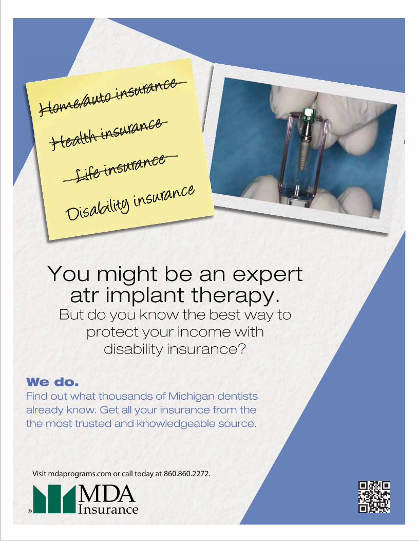
We do.Find out what thousands of Michigan dentistsalready know. Get all your insurance from thethe most trusted and knowledgeable source.
You might be an expertatr implant therapy.
But do you know the best way toprotect your income with
disability insurance?
Genesee District Dental Society smile genesee - spring 2015
ON THE COVER
3
Genesee District Dental Society5225 E. CookGrand Blanc, MI 48439
BOARD OF DIRECTORS
Dr. Bill BeckPresident4252 S. Linden Rd.Flint, MI 48507
Dr. Scott MortimerPresident Elect4495 S. Center Rd.Burton, MI 48519
Dr. Lori ThomasImmediate Past President4500 Town Center Pkwy.Flint, MI 48532
Dr. Traci DantzlerSecretary4170 Lennon Rd.Flint, MI 48507
Dr. Jay WerschkyExecutive Director5225 E. CookGrand Blanc, MI 48439
Dr. Luis PerezEditor2222 S. Linden Rd. Ste. DFlint, MI 48532
ADDITIONAL BOARD MEMBERS
Dr. Zelton JohnsonDr. Paul RacineDr. Patty McGaryDr. Matt LindemannDr. Alan KleinDr. Jori LewisDr. John LigasDr. Dan LechotaDr. Jack MedemarDr. Fred ThompsonDr. Martin Werschky
contents
President’s/Editor’s Notes Page 4
News from the ADA Page 6
Cancer Screening Protocol Page 8 - 12
Dental Trivia Page 13
Tax Benefits of HiringFamily Members Page 15
Pre-Retir ment Planning Page 16 - 17
EventsDr. Pete YamanRestorative Dentistry Page 19-20
Mark Your Calender Page 21
Genessee District Dental Society smile genesee - spring 2015
FROM THE DESK
4
president’s notes
It has truly been an honor and a privilege to serve as President of the
Board of Directors of the Genesee District Dental Society this past year.
The dental year passed very quickly. A major highlight was our
receiving the Golden Apple Award from the American Dental
Association for our January 2014 Diaper Drive, “Dental Health from
the Bottom Up.” We conducted our second annual Diaper Drive this
April as a service and educational event for our community.
The GDDS Board of Directors has been very fortunate to have great
leaders throughout its history, and that tradition continues with our
present Board. Dr. Jay Werschky, our Executive Director, provides
guidance, advice and direction with his passion for and knowledge of
organized dentistry at the national, state and local levels. Our retiring
MDA Trustee, Dr. Jim Cantwil, has been a steady, knowledgeable voice
for us as the new MDA governance model moved into place. The
monthly dinner meetings with CE credits have featured a variety of
interesting and informative speakers thanks to our Program Chair, Dr.
Marty Werschky.
Our quarterly publication, “Smile Genesee,” continues to grow through
the efforts of our Editor, Dr. Luis Perez. I speak for all of our members
when I thank them for their time and commitment.
A special thanks goes to our Board Members who have given much
of their personal time to attend meetings and serve on committees.
I think most would agree that it has been an interesting and rewarding
experience. I would encourage all members, especially our newer
members, who have never been on the Board to volunteer to serve.
I look forward to passing on the Presidency to Dr. Scott Mortimer. Dr.
Traci Dantzler will be our new President-elect. With their leadership
the GDDS will continue to move forward in service to our members
and our community.
Thank you for allowing me to serve as your President, and for your
continued participation in our Dental Society. Have a wonderful
Summer!
editor’s notes
Dr. Bill Beck
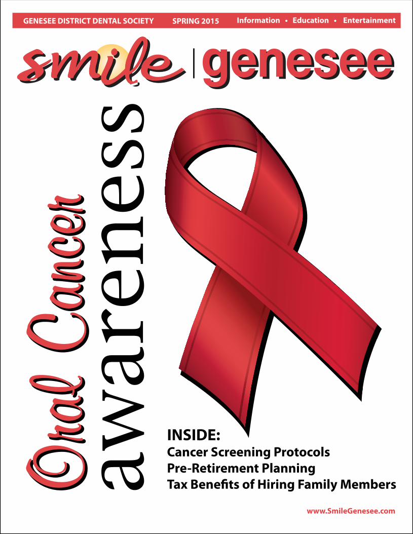
As the front page shows, this edition, is dedicated to oral cancer.
April is the month of the Oral Cancer awareness. Therefore, we
have lined-up a dinner meeting on April 14th with Eva Grayzel
(see last page). I had the fortune to listen to one of her lectures;
this international speaker will inspire you to be a better dentist
and to look at the Oral Cancer with a different perspective
(the patient’s one). I hope you don’t miss this incredible
opportunity.
Warmest regards,
Luis Perez
Greetings,
Another year has gone by!
I would like to start this editorial by expressing our appreciation to our
President Dr. Bill Beck and our MDA Trustee Dr. Jim Cantwil as well as
our Board Members of the GDDS for their time and efforts to organize
dentistry to the benefit of our Society. Many achievements have been
reached during this year, as mentioned on the President’s notes.
Certainly we are so fortunate to count on their leadership and
commitment towards organized dentistry.
Don’t forget to show your appreciation to Bill on April 30th at the end
of the year’s party. Dr. Scott Mortimer will assume the next term
Presidency. I am sure that he will know how to guide us for another
amazing year!

You’re invited to
The Year End Celebration and Tribute to outgoing President,
Dr. Bill Beck!!
April 30, 2015, from 5:00pm - 8:00pm Flint Golf Club
Heavy hors d'oeuvres will be served. There will be a cash bar.

Genesee District Dental Society smile genesee - spring 20156
NEWS
NEWS FROM THE ADAMarch 02, 2015 By Jean Williams
SCIENCE:Symptoms of measles may appear in oral cavity before other manifestations of diseasePolio. Tuberculosis. Whooping cough.
In the 20th century, thanks to vaccines and drugs, these diseases were under control
in the U.S. But the needle is moving again on infections worldwide among certain
thought-to-be-contained diseases. One of them is measles. The first signs of measles
occur typically in the head and neck region and in the oral cavity. But some dentists
may not have had the occasion to actually see these symptoms due to years of
successful control of the disease.
“It’s been rare among oral diseases,” said Dr. Catherine Flaitz, a spokeswoman
for American Academy of Pediatric Dentistry. “All of a sudden, this viral infection
has resurfaced. In the past, we educated our students about this disease with
the caveat that it is unlikely that they will diagnose a case among their patients.
And now with this lack of universal vaccination, we’re beginning to see these
oral manifestations present themselves again.”
Measles outbreaks have been reported in pockets around the country. Officials traced a California outbreak to a popular theme park. In a Jan. 23
health advisory on its website, the Centers for Disease Control and Prevention said of that incidence, “The United States is experiencing a large
multi-state measles outbreak that started in California in December 2014 and has spread to six additional states and Mexico. The initial confirmed
case-patients reported visiting Disneyland Resort Theme Parks in Orange County, CA, from December 17 through December 20, 2014.”
Later in February, five babies reportedly acquired the disease at a suburban Chicago day care center. In the backdrop of the outbreaks, a national
debate rages on between pro- and anti-vaccination components.
In the wake of the Disneyland outbreak, the California Dental Association began issuing advisories on its website to remind members to be vigilant,
including information about reducing risk to aerosol transmissible diseases and sharing recommendations from the California Department of Public
Health that health care workers get the measles, mumps and rubella combination vaccination, if they have not already been immunized.
Meanwhile, Dr. Flaitz advises dentists to be prepared in the event a patient shows up with certain signs and symptoms of the disease. Because the
disease presents in the head and neck region and/or the mouth first, a parent may head to the dentist for care, Dr. Flaitz said.
“I’m a pediatric dentist and an oral and maxillofacial pathologist,” she said. “So I see a number of children who have viral infections, and you never
know what gets them in the front door. It may be that the child’s running a fever, but it’s not so high. But then the parents look inside the mouth
and they see these sores, so they assume that these sores are actually the cause for the fever. Sometimes they will go with a fever straight to their
primary health care provider, such as a pediatrician, who notices the oral problems and makes the referral to the dentist. But other times, if there
are sores or irritations that are present in the mouth, they may seek the advice of the dentist first.”
Dr. Flaitz described three main signs in the oral cavity indicative of measles: Koplik spots; atypical gingivitis with pustules and necrosis; and operculitis.
“Some of these signs and symptoms are subtle,” she said. “So it’s very important to do a thorough soft tissue examination along with the dental
examination. Lots of times, a number of these oral lesions are the first indication that a person has either an infectious disease or an underlying
systemic disease. Sometimes this is the very first sign besides maybe more nondescript findings, such as a fever.”
Other signs and symptoms can help dentists recognize patients with undiagnosed measles.
“They typically at this time, too, will have a high fever, often 104 or greater,” Dr. Flaitz said. “They also will have malaise; they don’t eat well; they are
very fussy. Those are the primary, nondescript signs and symptoms that they may have.”
Dr. Flaitz said dentists should note, too, the three Cs in the head and neck region: conjunctivitis, coryza and cough. “Photophobia, periorbital swelling
and myalgia may be concurrently seen,” she said. “This is followed by an itchy, red rash that starts on the face and then moves down the neck to the
rest of the body.”
As outbreaks continue, dentists should brush up on all potential signs and symptoms for measles in the head and neck region and the oral cavity,
Dr. Flaitz advises.
The ADA Center for Professional Success offers additional guidance on measles for dental professionals. See “Infectious Disease and Workplace
Liability” at Success.ADA.org.
Clinical case: Koplik spots indicate measles inside a mouth. Becausethe disease shows up in the head and neck region and/or the mouthfirst, a patient may head to the dentist for care.
Reprinted from the ADA News
Clio DentalClio DentalRacine & AcklyRacine & Ackly
DiwakarDiwakarKinraKinra
MOVIEHERE
CancerCancerScreeningScreeningProtocolsProtocols
Genesee District Dental Society smile genesee - spring 20158
PRACTICE RESOURCES
IntroductionSquamous cell carcinoma of the oral cavity and pharynx accounts for
over 43,250 cases per year in the United States with approximately 8,000
deaths per year(1,2). Unfortunately, the diagnosis continues to rely on
patient presentation and physical examination with biopsy confirmation.
This may result in delay in diagnosis accounting for the fact that the
majority of these cancers are diagnosed at a late stage (1, 3-5). Studies
confirm that survival does correlate with stage, making early diagnosis
and treatment optimal for this disease (1). Despite advances in surgical
techniques, radiation therapy technology, and the addition of combined
chemotherapy and radiation therapy to the treatment regimen, survival
data has not shown appreciable change in decades (1,6,7). Five-year
survival data reveal overall disease specific survival rates of less than
60% although those that do survive often endure major functional,
cosmetic, and psychological burden due to dysfunction of the ability
to speak, swallow, breathe, and chew. Seventy-five percent of all head
and neck cancers begin in the oral cavity. According to the National
Cancer Institute’s Surveillance, Epidemiology, and Ends Results (SEER)
program, 30 percent of oral cancers originate in the tongue, 17 percent
in the lip, and 14 percent in the floor of the mouth (11). Many other
studies support this finding that oral cancers appear most often on
the tongue, and floor of the mouth (12,13). New data related to the
HPV16 virus may indicate that these trends are changing with the
poster mouth including the tonsils, tonsillar pillar and crypt, the base
of the tongue, and the oropharynx increasing rapidly in incidence rates.
A thorough, systematic examination of the mouth and neck need only
take a few minutes and can detect these cancers at an early and curable,
stage. Our goal is to discover oral, head and neck cancers early, before
patients present complaining of pain, a mass, bleeding, otalgia, or
dysphagia. Errors in diagnosis are most often ones of omission, and
therefore the importance of a systematic approach to the oral, head
and neck cancer examination cannot be overstated.
HistoryAlthough this report is based on examination technique, it is critical to
remember that any person with a history of tobacco and alcohol use
CancerScreeningProtocols
or prior head and neck malignancy has a significant risk of developing an
oral, head and neck cancer. In fact historically 75 percent of these cancers
are related to alcohol and tobacco use (10). These individuals may deserve
more frequent examinations as described to follow. Bear in mind that 1
out of 4 oral, head and neck cancers particularly in patients over the age
of 50 are detected in patients who do not smoke or drink alcohol;
obviously all patients, regardless of their history, need to be screened
at least once a year by their physician or dentist. Current research
indicates that HPV positive disease is rapidly changing these ratios and
age groups. Younger, non smoking patients under the age of 50 are the
fastest growing segment of the oral cancer population. Unfortunately,
this increase in the number or oral, head and neck cancers found in men
and women in their 20’s and 30’s is rapidly replacing those caused by
tobacco since the use of tobacco products has declined in the US every
year for more than a decade. During this same period of time the
incidence rate of OSCC has actually increased. This HPV 16-18 presence,
makes determination of at risk populations much more difficult, and
opportunistic screening of ALL patients must become the norm if the
death rate is to be reduced. As in many cancers, the symptoms and
history will often lead the dentist/physician to not only the presence of
a cancer but also the likely site of the lesion. Tobacco/alcohol lesions
tend to favor the anterior tongue and mouth, and HPV positive lesions
tend to favor the posterior oral cavity.
ApplicationThis examination protocol has been developed for use by both dental
and medical professionals. The different environments of practice will
dictate obvious differences in the equipment and manner in which the
examination is conducted. However all aspects of this thorough
examination are applicable to both types of practitioners to ensure
that a complete assessment of the patient has been accomplished.
Instruments Used For Oral, Head andNeck Cancer ExaminationClinicians need certain instruments and supplies in order to conduct a
Reprinted from www.oralcancerfoundation.org
Genesee District Dental Society smile genesee - spring 20159
PRACTICE RESOURCES
thorough and time efficient examination. Suggested tools for the oral,
head and neck cancer exam include: an adequate light source, mirrors
(laryngeal and nasopharyngeal), gloves, tongue blades, 2x2 gauze pads,
anesthetic nasal spray, flexible nasopharyngolaryngoscope, otoscope,
and nasal speculum.
General ExaminationA thorough oral, head and neck cancer examination can easily be
completed in less than 5 minutes. It primarily consists of inspection
and palpation. Once good rapport has been established with the
patient, the clinician is ready to begin the exam. It is important to
explain to the patient exactly what you are doing before doing it.
Not only will this help put the patient at ease, but it also gives you the
opportunity to educate your patient about the signs and symptoms of
oral, head and neck cancer and how to detect it at an early stage. It is
important for clinicians to understand the complex systemic effects of
malignancy on the body. Commonly changes noticed in a person’s face
and body pertaining to weight loss, anorexia and/or fatigue, may be
the first sign of a malignancy. The initial physical evaluation of a
patient actually begins as soon as you meet the patient. While taking
the patient’s history it is helpful to note any facial asymmetry, masses,
skin lesions, facial paralysis, swelling or temporal wasting. Inspection
of the lips, both moving and at rest, can also be performed while first
meeting the patient. Again, look for any asymmetry or gross lesions on
the lips. Listening in an important part of this examination. The sound
of one’s voice and speech are important in consideration of the location
of tumors as a “hot potato” voice may signal the presence of an
oropharyngeal tumor whereas a raspy, hoarse voice could be the
first sign of a laryngeal neoplasm. Throughout this oral, head and
neck cancer examination, it is helpful to remember to look, listen,
AND feel every site that is being examined.
The FacePosition the patient so that he or she is comfortably sitting and is at
your eye level. Inspect the face for asymmetry, swelling, discoloration
or ulceration. The entire face should be examined with an external light
source (overhead light or headlight) to evaluate for pigmented (red,
brown, black), raised, ulcerated, or firm areas of the skin, including the
hair bearing regions of the face and scalp. The facial bones, skeleton and
soft tissue should be palpated particularly noting asymmetry or masses.
EyesAs part of the cranial nerve examination, extraocular movements should
be tested in each direction as well as visual acuity. Any deficits should be
carefully noted, as they may result from an invasive cancer. Any swelling
of the eye or periorbital area should be noted and can be a late sign of a
cancer which may have started in the palate, maxillary or ethmoid
sinuses. Drainage from the lacrimal system (epiphora), may be a sign
of an obstructing mass in the maxillary sinus, nose, or facial soft tissue.
NoseRoutine nasal examination should include palpation of the external nose
and paranasal region overlying the maxilla and maxillary sinus. Anterior
rhinoscopy can be performed with an external light source, otoscope or
even a penlight to evaluate for lesions of the anterior septum, columella,
nasal vestibule and nasal floor. Be careful not to mistake a nasal turbinate
for a polyp or tumor. A nasal speculum may be of aid during this portion
of the examination. If used, it should be carefully opened in an up-and-
down direction to avoid causing discomfort to your patient (A flexible
nasopharyngolaryngoscope can also be used to exam the nasal cavity.
This technique will be discussed later.)
EarsAs part of the cranial nerve examination, hearing should be tested. By
conversing with the patient during the physical exam, one can generally
assess the integrity of the acoustic nerve. Carefully inspect the auricle,
noting any pigmented, erythematous, or ulcerous lesions. Note that skin
cancers often appear on the superior, sun-exposed portion of the auricle.
Next, examine the external auditory canal for masses or lesions; cerumen
may need to be removed to facilitate this portion of the exam. For
completeness, the tympanic membranes should be inspected with a
pneumatic otoscope.
The Oral CavityFor this portion of the exam patient positioning can vary. Dental patients
tend to lie on their backs, while their dentist exams their oral cavity.
Physicians, on the other hand, usually have their patients sit up straight
and face them eye-to-eye during the exam. It is imperative that the
mouth be examined with an external light source, which allows both
hands free for bimanual palpation or to hold gauze or tongue blade(s)
for improved visualization. If a hands free light source is not available,
an assistant may provide invaluable help in visualization of difficult
areas such as the posterolateral border of the tongue and floor of
mouth. Before beginning this part of the examination, ask the patient
to remove all dental appliances. When examining mucosal surfaces,
it is important to gently dry those surfaces with a gauze or air syringe,
so that color or texture changes will become more obvious. Multiple
studies have consistently shown that the earliest manifestation of
many oral and oropharyngeal squamous cell cancers is a persistent
erythroplastic lesion (3). Clinicians must therefore be on the lookout
for both red and white (leukoplakia) lesions on the oral mucosa, as
well as detection through palpation of indurated and fixated masses
with in the tissues.
IMPORTANT FACT
More than half of oral cancershave already spread to lymphnodes or other areas by thetime they are found.Source: National Cancer Institute
Genesee District Dental Society smile genesee - spring 201510
PRACTICE RESOURCES
The Buccal MucosaThe inside of the cheek or buccal mucosa must be spread away from the
teeth and gums to visualize the sulcus which connects this area to the
gums (gingiva). Examine one side and then the other. It is not uncommon
to see a white line here from a habit of biting the inside of the cheek.
Any irregularity in texture or color or other signs listed above should
be noted. Be sure to examine the entire buccal mucosa from the labial
commissure back to the anterior tonsillar pillar. Stenson’s duct from
the parotid gland is a small protrusion in this area opposite the upper
second molar and should secrete clear saliva from both sides when
the parotid gland is milked. This area is easy to examine when two
tongue blades are used to spread the lining from the gingiva or
gauze is used to pull the lips apart. With your index and middle
fingers inside the patient’s mouth on the buccal mucosa and with
your thumb on the cheeks, carefully pull laterally, and inspect both
gutters along the upper and lower jaws. Next gently pinch the cheek
between your fingers and thumb; this allows you to palpate the buccal
mucosa for any hidden masses.
TongueAsk the patient to open wide while relaxing the tongue; note any
ulcerations, swellings, or other abnormalities. Then have the patient
stick out his or her tongue and move it from side to side. It should move
easily and completely to both sides without spasm or asymmetry. When
there is a nerve paralysis of the hypoglossal nerve, the tongue will usually
deviate to the side of the lesion. Observe the dorsum of the tongue,
noting any discolorations, irregularities, or limitations to movement, all
of which may be a sign of cancer. Notice the circumvallate papillae and
lingual tonsils, which are often mistaken for pathologic lesions. One of
the most common sites of oral cancer is on the lateral aspect of the
tongue, and it must be evaluated completely. This often requires using
gauze to pull the tongue out and roll it from side to side while retracting
the cheek with a tongue blade. Alternatively, two tongue blades can be
use to push the tongue away from the lower teeth allowing visualization
of every part of the mucosal lining to the tonsil and base of tongue. A
dental mirror may be necessary to visualize the base of the tongue
(part of the oropharynx). This area is best viewed by pulling the tongue
forward while holding it with 2X2 gauze, and will roll it up in to a position
enabling clearer view. Next, palpate the dorsum and lateral margins of
the tongue, paying special attention to any masses or firm/fixated areas.
Being careful not to gag the patient, palpate the lingual tonsils. Finally,
have the patient touch the roof of their mouth with the tip of their tongue.
This will allow the examiner to inspect the ventral surface of tongue.
Floor of the MouthThe floor of the mouth is the horseshoe-shaped area that extends from
the alveolar ridge of the mandible to the ventral aspect of the tongue.
Inspect this area while the tongue is elevated. If needed, wrap a piece of
gauze around the tip of the tongue and pull the tongue gently forward
and to one side. With the other hand, use a tongue blade or gloved finger
to push the middle of the tongue up and out of the way. Notice the
frenulum in the midline and the ducts from the submandibular glands
symmetrically on either side. Also note the sublingual glands. It is
helpful to first dry this area with a gauze before looking for any surface
abnormalities. Next, insert a gloved finger beneath the tongue, and
another under the chin on the exterior skin, and bimanually palpate
the submandibular glands and the entire submental region. Keep in
mind that this one of the most common places for oral cancers to
hieratical attention should be paid to any mass that feels firm or
fixated in position.
LipsThe lips should not be overlooked as part of the oral cavity. They may be
involved with squamous cell carcinoma (SCC) of the aerodigestive tract
or both SCC and basal cell carcinomas (BCC) of the skin. The lips should
be evaluated with the mouth open and closed noting any abnormalities
in symmetry, contour, color or texture. Pay special attention to the
vermilion border of the lower lip, as this is a prime site for oral cancers.
First revert the lower lip and inspect the inner surface. The labial mucosa
should be smooth and uniform in color. Notice the frenum of the lip in
the midline. Note any signs of smokeless tobacco use (ulcers, red or white
discolorations, texture variations) on the labial mucosa. With the lip still
retracted, one can also inspect the gingivolabial sulcus, the gingival
mucosa, and the teeth. Next palpate the lip with your thumb and
index finger, noting any firm or nodular submucosal areas. Repeat
these steps for the upper lip.
IMPORTANT FACT
There has been a tendency to watchsuspicious areas over an extendedperiod to determine if they aredangerous or not. Unfortunately,this philosophy leads to a situationin which a dangerous lesion maycontinue to prosper and grow intoa later stage, hard to cure cancer.Any sore, discoloration, induration,prominent tissue, irritation, hoarseness,which does not resolve within a twoweek period on its own, with or withouttreatment, should be considered suspectand worthy of further examinationor referral.
Source: Oral Cancer Foundation
Genesee District Dental Society smile genesee - spring 201511
PRACTICE RESOURCES
Thyroid: First inspect the thyroid gland before proceeding to palpation.
In normal patients the thyroid gland is often difficult to feel. Some
clinicians prefer to palpate the thyroid while positioned behind their
patients, but it is perfectly acceptable to examine the gland from the
front as well. Attempt to palpate the entire gland, and note the
characteristics of any nodules or masses. Having the patient swallow
while your fingers are positioned adjacent to the gland, will elevate
the thyroid gland and may facilitate your examination. Note and
record tenderness. Check the consistency of any abnormality. Is it hard?
Cystic?
Have the patient deviate his head toward the examining side to
relax the muscles during palpation. After the lobe has been palpated,
and with the fingers still, ask the patient to swallow. The gland will move
upward during deglutition and any abnormality will become more
apparent. On swallowing, the inferior pole of the lobes is elevated and
can be outlined. Inability to palpate the inferior pole may suggest
substernal extension of the thyroid gland on that side. Examine each
lobe in this manner. If the patient has a very heavy neck, it may be
helpful to stand behind him and palpate each lobe with his head
deviated toward the examining side.
Hard and Soft Palate(Although not technically part of the oral cavity, the soft palate will be
considered here.) Ask the patient to open widely and tilt his or her head
backward to provide an adequate view of the hard and soft palate. If
needed, depress the base of the tongue with a tongue blade to provide
a better view of the soft palate. Loose teeth, red spots, white spots,
ulcerations, rough areas, asymmetry, growths, or other masses may be
the first sign of a cancer in this area as in all areas of the head and neck.
The uvula should hang down in the midline. Its deviation may indicate a
vagal nerve palsy. Some patients have a torus palatinus, or bony
outgrowth from the midline of their hard palate. This should not be
mistaken for a malignancy.
The OropharynxNote that the examination of the oropharynx is essentially a continuation
of the oral cavity examination, and the two are often completed
simultaneously.
TonsilsAsk the patient to open widely, relax and slowly breathe in and out saying
“Ahhh”. This relaxes the tongue allowing a view of the oropharynx. It may
be necessary to press down on the back of the tongue to get a full view
of the oropharynx. Simply depressing the tongue with a tongue blade
and having the patient say “ah,” often gives only a superficial view of the
oropharynx and many early cancers may go unnoticed. The palatine
tonsils can be seen on both sides behind the retromolar trigone.
Examine the anterior and posterior tonsillar pillars and tonsillar fossa
for any exophytic mass, asymmetry, ulceration, or redness.
Soft PalateThe soft palate is part of the oropharynx and should be evaluated for
symmetry at rest and on elevation as well as for abnormal lesions. The
mobility of the soft palate can be examined by having the patient say
“Ahhh”. (See previous discussion of soft palate examination.)
The NeckHave the patient sit so that his face is at your eye level; support the head
with a headrest. Bimanually palpate the neck, comparing both sides
simultaneously for signs of enlargement. Palpate carefully for enlarged
lymph nodes. Examine the jugular chain first. With two deeply placed
fingers, palpate along the course of the sternomastoid muscles,
underneath the mandible and down to the clavicle. Palpate the
supraclavicular spaces on either side. Next, examine the parotid groups
lying anterior and inferior to the ears, the submental, and finally the
submaxillary chain. To palpate a mass in the submaxillary area, insert
a gloved finger in the patient’s mouth and press structures against your
other hand, positioned under his chin. Next, palpate along the course of
the larynx for signs of immobility or enlargement. Please note that it is
the foundation's opinion that in a patient over the age of 40 that presents
with a neck mass that is painless - that the first differential diagnosis is
oral cancer, and this not being the site of the primary, require a
reexamination of the interior of the mouth to ensure that primary
locations are identified. Enlarged nodes that are painless are seldom
the result of an infectious process.
IMPORTANT FACT
There is no standard or routinescreening test for oral cancer.
• Toluidine blue stain.• Fluorescence staining.• Exfoliative cytology.• Brush biopsy.
No studies have shown thatscreening would decreasethe risk of dying fromthis disease.Source: National Cancer Institute
Genesee District Dental Society smile genesee - spring 201512
PRACTICE RESOURCES
Nasopharynx: Examination of the nasopharynx is one of the more
difficult portions of the oral, head and neck cancer examination.
Instruct the patient to open widely and breathe through his or her
mouth. This should cause the soft palate to rise. With a tongue blade,
carefully depress the mid-portion of the tongue. Then insert a warmed
nasopharyngeal mirror over the tongue blade and into the oropharynx.
Ask the patient to now breathe through his or her nose. This should
cause the soft palate to fall forward, allowing the examiner to see the
nasopharyngeal region reflecting in the mirror. First inspect the
posterior choanae and posterior part of the nasal septum. The inferior
and middle turbinates should also be visible, and note the superior
posterior surface of the soft palate. By slowly rotating the mirror,
visualize both eustachian tube openings, the pharyngeal tonsil, and
walls of the nasopharynx. Look for any masses, swellings, ulcerations,
or discolorations. If your patient is unable to tolerate this procedure,
then some topical anesthetic spray can be used. Please note that the
nasopharynx can also be examined via a fiberoptic scope.
(This will be discussed below).
Hypopharynx and LarynxLike the nasopharyngeal examination, this part of the exam can be
challenging. A thorough inspection of the hypopharynx and larynx is a
critical component to the oral, head and neck cancer examination. All
of the key laryngeal structures need to be closely inspected for any
signs of malignancy. This examination can be accomplished with
either a laryngeal mirror or a fiber optic scope.
Mirror ExamTraditionally the laryngeal mirror has been the instrument of choice
for examining the hypopharynx and larynx. Ask the patient to sit up
straight and slightly protrude the chin upward and forward. Next have
the patient open widely and protrude the tongue. Grasp the tip of the
tongue with a gauze and gently pull it forward. The patient should be
concentrating on breathing in and out through the mouth. Carefully
insert a warmed laryngeal mirror into the oropharynx, using the back
of the mirror to elevate the soft palate. If the patient cannot tolerate
this maneuver without gagging, some anesthetic spray can be used
as a last resort. Be aware that the topical anesthetic will suppress the
patient’s gag reflex, increasing the risk of aspiration. Once the mirror is
in place, examine the base of the tongue. By tilting the mirror, examine
the pharyngeal walls, vallecula, and piriform sinuses noting any masses
or other abnormalities. The epiglottis should be seen in the midline;
closely inspect both surfaces for any lesions. The laryngeal structures
are often brought into better view by having the patient say a high
pitched “e-e-e-e-e”. Examine the arytenoids, aryepiglottic folds, false
vocal cords, and true vocal cords for any hints of malignancy. It is
important to assess the mobility of the true vocal cords by having
the patient phonate. The superior portion of the trachea is often
visible below the vocal cords. Once you are satisfied that all of the
important structures have been visualized, slowly remove
the mirror and release the patient’s tongue.
Fiber Optic NasopharyngolaryngoscopeExamThe flexible fiber optic nasopharyngolaryngoscope has become an
invaluable tool for detecting head and neck cancers. The nasal cavity,
nasopharynx, a portion of the oropharynx, hypopharynx, and larynx
can all be thoroughly inspected with the help of a flexible fiber optic
scope. The flexible scope is passed transnasally into the nasopharynx
after topical vasoconstricting and anesthetic agents have been sprayed
into the nasal cavity. As the scope passes through the nasal cavity and
nasopharynx, look for any mucosal lesions or other abnormalities.
Gently advance the scope into the oropharynx and then down into the
hypopharynx. Examine all of the laryngeal structures as you would for
the mirror exam. Then slowly remove the scope. People’s lives can be
saved through early detection of oral, head and neck cancers. Dentists
and physicians both must do a better job of screening their patients for
these malignancies. By following a systematic approach to the oral, head
and neck cancer physical examination, clinicians will be more effective at
diagnosing such cancers at an early and more treatable stage.
Common Cancer MimicsMany benign lesions mimic oral cancer. This article reviews the most
common, aphthous lesions.
- See more at:
http://www.oralcancerfoundation.org/discovery-diagnosis/screening.php#sthash.ar0USJNv.dpuf
IMPORTANT FACT
The most important stepin reducing the death ratefrom oral cancer is earlydiscovery. No group hasa better opportunity tohave a positive impacton this than membersof the dental community. Source: Oral Cancer Foundation
Genesee District Dental Society smile genesee - spring 201513
Do You or Your Office Staff NeedCPR/AED Certification or OSHA Training?CertificationMade Painless for Health Care Providers
• CPR/AED Certification• Individual use manikins• NO SHAR ING, NO WAIT ING, HANDS ON• 2 Year Health Professional Card• $35 per person• 4 CE Credits
•OSHA Compliance• $50 per person• 3 CE Credits
• Day or early evenings• Held in your officefor your convenience
Contact:Linda Gadioli(586) 786-9652cell: (586) 823-0205fax: (586) 786-1460E-mail: [email protected]
NEW! AGD CE Creditsfor all classes!
Approved FACE Program ProviderFAGD/MAGD Credit7-1-13 TO 6-30-17
Affiliated withAmerican Safety & Health Institute
Gadioli
• According to a Time magazine survey, 59% of Americans would rather sit in a dentist's chair than sit next
to someone on a cell phone.
• The average human produces 25,000 quarts of saliva in a lifetime – enough to fill 2 swimming pools!
• A sneeze zooms out of your mouth at over 600 mph!
• Athletes are 60 times more likely to damage their teeth when not wearing a mouth guard during
athletic activities.
• Most tooth loss in people under 35 years of age is caused by athletic trauma, fights or accidents.
• Most tooth loss in people over age 35 is from periodontal disease.
• In the middle ages, people believed that dogs' teeth boiled in wine made an excellent mouth rinse for
tooth decay prevention.
• Egyptians used a form of toothpaste over 5000 years ago.
JUST FOR FUN
DENTAL TRIVIAFlossing once a DAY INCREASESYOUR LifeLife expectancy by 6 years
UNCERTAINTY
Feel confident about how you will be treated.Every day you are in control—of pain, of options, of caring treatment. DentistCare® is about you and treating you fairly when it comes to professional liability protection:
• Enhanced Coverage —designed to suit your needs
• PracticeGuard®—helps protect your practice if a covered dentist becomes disabled (Available for groups of ten or fewer dentists or specialists.)
• Innovative Risk Resource Services—tap into our experienced advisors and helpful resourcesFind out how DentistCare can help you reduce risk, keep from getting pulled in the wrong direction, and maintain the control you want—call us today at 800.625.7814.
Professional Liability Insurance & Risk Resource ServicesProAssurance Group is rated A+ (Superior) by A.M. Best. ProAssurance.com • 800.625.7814

Genesee District Dental Society smile genesee - spring 201515
FYI
As a business owner, you should be aware that you can save family
income and payroll taxes by putting junior family members on the
payroll. You may be able to turn high-taxed income into tax-free or
low-taxed income, achieve social security tax savings (depending on
how your business is organized), and even make retirement plan
contributions for your child. Here are the key considerations.
Turning high-taxed income into tax-free or low-taxed income. You can
turn some of your high-taxed income into tax-free or low-taxed income
by shifting some of your business earnings to a child as wages. In order
for your business to deduct the wages as a business expense, the work
done by the child must be legitimate and the child's salary must be
reasonable.
For example, suppose you are a sole proprietor in the 39.6% tax bracket.
You hire your 17-year-old daughter to help with office work and pay her
$6,100 during the year and she doesn't have any other earnings. You
save $2,415.60 (39.6% of $6,100) in income taxes at no tax cost to your
daughter, who can use her $6,300 standard deduction (for 2015.)
What about income tax withholding? Your business probably will have
towithhold federal income taxes on your child's wages if 1) their income
exceeds $1,050 for 2015, and includes more than $350 of unearned
income (such as dividends) for 2015, and 2) the child/employee can be
claimed as a dependent on someone else’s return.
Social security tax saving, too. If your practice isn’t incorporated, you
can also save some self-employment (i.e., social security) tax dollars by
shifting some of your earnings to a child. For example, if you usually
take $120,000 of earnings from the business and pay $5,700 to your
17-year-old child. Your self-employment income would be reduced by
$5,700, saving you $165.30. This doesn't take into account a sole
proprietor's income tax deduction for one-half of his or her own
social security taxes.
A similar but more liberal exemption applies for FUTA (unemployment)
tax, which exempts earnings paid to a child under age 21 while employed
by his or her parent. The FICA and FUTA exemptions also apply if a child is
employed by a partnership consisting solely of his parents.
Tax Benefits of HiringFamily MembersProvided by Cass Beals and John Caruana, CPAs -- Beals, Caruana & Company, P.c.
Note that there is no FICA or FUTA exemption for employing a child if
your business is incorporated or a partnership that includes non-parent
partners. However, there's no extra cost to your business if you're paying
a child for work you'd pay someone else to do, anyway.
Retirement benefits. Your business also may be able to provide your
child with retirement benefits, depending on the type of plan it has and
how it defines qualifying employees. For example, if it has a simplified
employee pension (SEP), a contribution can be made for the child up to
25% of his or her earnings but the contribution cannot exceed $52,000
for 2014.
Get advice. If you have any questions about how these rules apply to
your particular situation, please don’t hesitate to call. Also keep in
mind that some of the rules about employing children change from
year to year, and may require your income-shifting strategy to change,
too.
Reprinted from the Journal of the Macomb Dental Society - Winter 2015
Genesee District Dental Society smile genesee - spring 201516
FYI
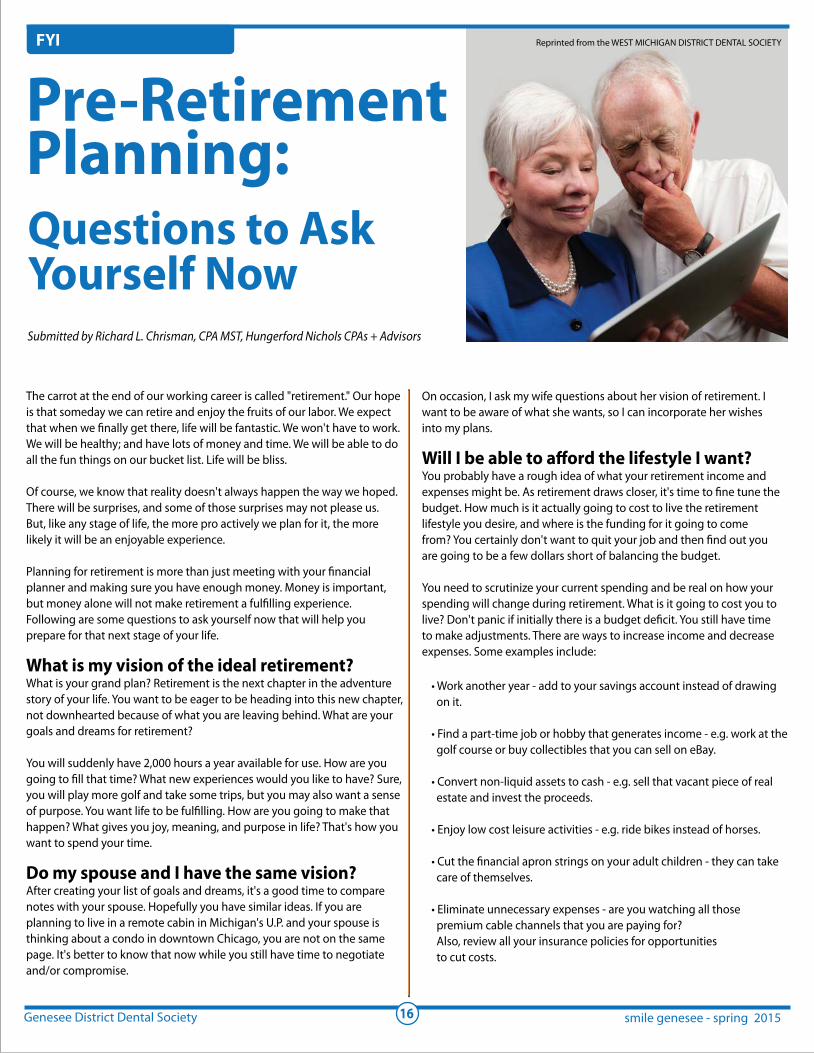
The carrot at the end of our working career is called "retirement." Our hope
is that someday we can retire and enjoy the fruits of our labor. We expect
that when we finally get there, life will be fantastic. We won't have to work.
We will be healthy; and have lots of money and time. We will be able to do
all the fun things on our bucket list. Life will be bliss.
Of course, we know that reality doesn't always happen the way we hoped.
There will be surprises, and some of those surprises may not please us.
But, like any stage of life, the more pro actively we plan for it, the more
likely it will be an enjoyable experience.
Planning for retirement is more than just meeting with your financial
planner and making sure you have enough money. Money is important,
but money alone will not make retirement a fulfilling experience.
Following are some questions to ask yourself now that will help you
prepare for that next stage of your life.
What is my vision of the ideal retirement?What is your grand plan? Retirement is the next chapter in the adventure
story of your life. You want to be eager to be heading into this new chapter,
not downhearted because of what you are leaving behind. What are your
goals and dreams for retirement?
You will suddenly have 2,000 hours a year available for use. How are you
going to fill that time? What new experiences would you like to have? Sure,
you will play more golf and take some trips, but you may also want a sense
of purpose. You want life to be fulfilling. How are you going to make that
happen? What gives you joy, meaning, and purpose in life? That's how you
want to spend your time.
Do my spouse and I have the same vision?After creating your list of goals and dreams, it's a good time to compare
notes with your spouse. Hopefully you have similar ideas. If you are
planning to live in a remote cabin in Michigan's U.P. and your spouse is
thinking about a condo in downtown Chicago, you are not on the same
page. It's better to know that now while you still have time to negotiate
and/or compromise.
Submitted by Richard L. Chrisman, CPA MST, Hungerford Nichols CPAs + Advisors
Reprinted from the WEST MICHIGAN DISTRICT DENTAL SOCIETY
• Work another year - add to your savings account instead of drawing
on it.
• Find a part-time job or hobby that generates income - e.g. work at the
golf course or buy collectibles that you can sell on eBay.
• Convert non-liquid assets to cash - e.g. sell that vacant piece of real
estate and invest the proceeds.
• Enjoy low cost leisure activities - e.g. ride bikes instead of horses.
• Cut the financial apron strings on your adult children - they can take
care of themselves.
• Eliminate unnecessary expenses - are you watching all those
premium cable channels that you are paying for?
Also, review all your insurance policies for opportunities
to cut costs.
Pre-RetirementPlanning:Questions to AskYourself Now
On occasion, I ask my wife questions about her vision of retirement. I
want to be aware of what she wants, so I can incorporate her wishes
into my plans.
Will I be able to afford the lifestyle I want?You probably have a rough idea of what your retirement income and
expenses might be. As retirement draws closer, it's time to fine tune the
budget. How much is it actually going to cost to live the retirement
lifestyle you desire, and where is the funding for it going to come
from? You certainly don't want to quit your job and then find out you
are going to be a few dollars short of balancing the budget.
You need to scrutinize your current spending and be real on how your
spending will change during retirement. What is it going to cost you to
live? Don't panic if initially there is a budget deficit. You still have time
to make adjustments. There are ways to increase income and decrease
expenses. Some examples include:

Genesee District Dental Society smile genesee - spring 201517
FYI
When should I retire?Every individual is on their own timeline. There is no right or wrong
answer. It's just a decision that you need to make based on your own
set of circumstances. Talk to your advisors and family and get their
input. Some variables to consider include:
• Do I enjoy my job?
• Is going part time an option?
• Am I financially able to leave my job?
•When do I qualify for Social Security?
• What health insurance options do I have?
• How is my health?
Should I downsize my house?Most retirement planners focus on your investment portfolio.
Although this is important, for many people, a large percentage of their
wealth is the equity in their homes. As you approach retirement, consider
selling your existing home and buying a smaller home or condo. This
especially makes sense if the mortgage has been paid off and the property
has significant value.
Your goal is to convert the equity in your home to
cash that you can invest and live on during retirement. What good
is having a home worth a million dollars if you can't afford to take that
vacation you always dreamed of? Other reasons to sell your current
home include:
• You may not need as much space in retirement.
• A smaller home or condo is much easier to maintain.
• You may want to relocate.
When should I take Social Security?When to start taking Social Security is not as obvious as you
would think. Your goal is to maximize the benefits for you
and your spouse. Everyone's circumstances are different so it
may take some research to fully understand your options and
choose the one that's best for you. Consider meeting with a
financial advisor to help you sort it out.
What is my plan for the debt that I have?Prepare for retirement by paying off your debt. Eliminating
debt is a 'simple strategy to lower your expenses without
changing your life style.
A lesson I learned from my parents - once your mortgage/
debt is paid off (and your children are financially supporting
themselves), your monthly cost of living will drop significantly.
Is my estate plan current?If you die without proper estate planning, it can create
unnecessary problems and expense for your heirs. Have your
accountant and lawyer review your estate plan to make sure
everything is up to date and appropriate for the size of your
estate and the stage you are in life. I have seen many families
get into major squabbles over their parents' estate. If the estate
plan had been done properly, there would have been nothing
for the children to argue about.
Also, after you are gone, your spouse or children will have
to sort out your financial affairs. Do them a huge favor by
simplifying your finances and organizing your documents.
ConclusionYou have been working your whole life with the hope that
someday you will be able to retire. Now that you are in the
home stretch, it's important that you prepare for it. You may
still have a few more years to work, so take advantage of the
time by dealing with the issues that will make your retirement
as enjoyable as possible.
Richard 1. Chrisman, CPA, MST is the Managing Shareholder of
Hungerford Nichols CPAs + Advisors, a Tax, Auditing and Business
Advisory firm with offices in Grand Rapids and Greenville, Michigan.
The firm is celebrating 73 years of helping local businesses,
including many area dental practices.
DiwakarDiwakarKinraKinra
Impact your future and your profession with
The Michigan Dental Association is looking for leaders and invites you to be
part of LEAD: Leadership Exploration And Development.
An 18-month pilot program
Designed to allow member participants to experience the MDA’s many
facets
Develops and enhances leadership and volunteering skills
Participants will experience various conferences, committee meetings,
workshops, group projects, and a trip to an ADA conference —
expenses paid
It takes time to develop as a leader. As a participant, you’ll make a monthly
time investment throughout the 18 months, including two multi-day events.
But it’s time well spent when you consider the impact you can have on your
future and your profession.
To learn more and request an application,
call Josh Lord at 800-589-2632 or 517-346-9415
or visit smilemichigan.com/LEADprogram
Genesee District Dental Society smile genesee - spring 201519
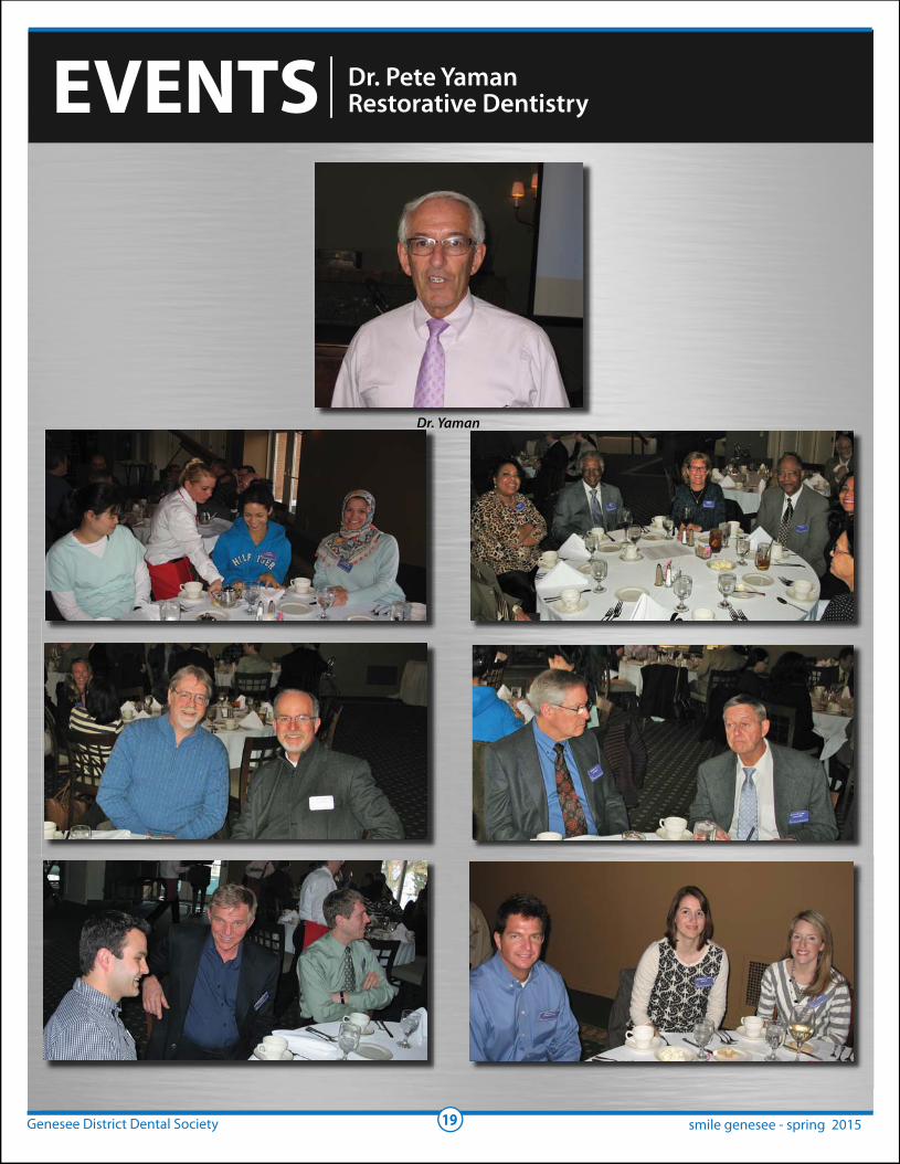
EVENTS Dr. Pete YamanRestorative Dentistry
Dr. YamanDr Yaman
Genesee District Dental Society smile genesee - spring 201520
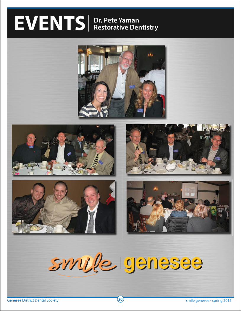
EVENTS Dr. Pete YamanRestorative Dentistry
Genesee District Dental Society smile genesee - spring 201521
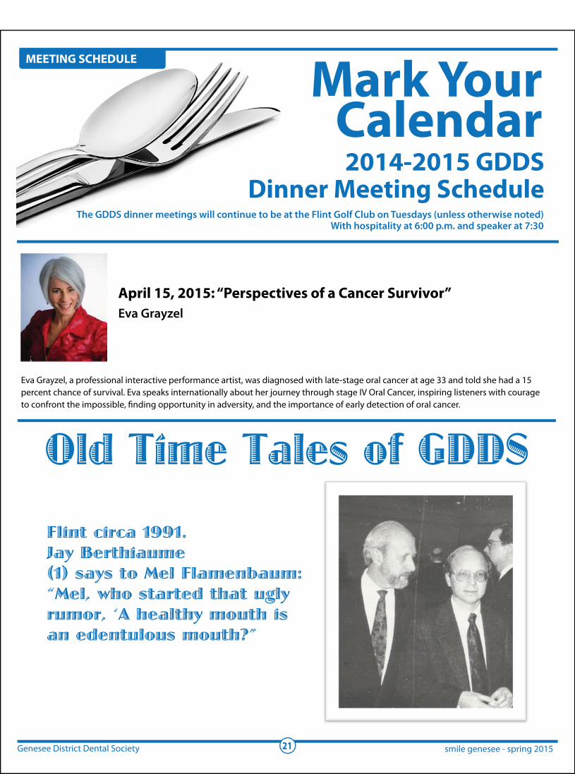
MEETING SCHEDULE
2014-2015 GDDSDinner Meeting Schedule
Mark YourCalendar
The GDDS dinner meetings will continue to be at the Flint Golf Club on Tuesdays (unless otherwise noted)With hospitality at 6:00 p.m. and speaker at 7:30
Eva Grayzel, a professional interactive performance artist, was diagnosed with late-stage oral cancer at age 33 and told she had a 15
percent chance of survival. Eva speaks internationally about her journey through stage IV Oral Cancer, inspiring listeners with courage
to confront the impossible, finding opportunity in adversity, and the importance of early detection of oral cancer.
April 15, 2015: “Perspectives of a Cancer Survivor”Eva Grayzel
Old Time Tales of GDDSFlint circa 1991.Jay Berthiaume(1) says to Mel Flamenbaum:“Mel, who started that uglyrumor, ‘A healthy mouth isan edentulous mouth?”